Fine structure of the mineralized teeth of the chitonAcanthopleura echinata (Mollusca:...
11
Fine Structure of the Mineralized Teeth of the Chiton Acanthopleura echinata (Mollusca: Polyplacophora) Rosamund J. Wealthall, 1 Lesley R. Brooker, 2 David J. Macey, 2 * and Brendan J. Griffin 1 1 Centre for Microscopy and Microanalysis, University of Western Australia, Nedlands, WA 6907, Australia 2 Division of Science and Engineering, Murdoch University, Murdoch, WA 6150, Australia ABSTRACT The major lateral teeth of the chiton Acan- thopleura echinata are composite structures composed of three distinct mineral zones: a posterior layer of magne- tite; a thin band of lepidocrocite just anterior to this; and apatite throughout the core and anterior regions of the cusp. Biomineralization in these teeth is a matrix- mediated process, in which the minerals are deposited around fibers, with the different biominerals described as occupying architecturally discrete compartments. In this study, a range of scanning electron microscopes was uti- lized to undertake a detailed in situ investigation of the fine structure of the major lateral teeth. The arrangement of the organic and biomineral components of the tooth is similar throughout the three zones, having no discrete borders between them, and with crystallites of each min- eral phase extending into the adjacent mineral zone. Along the posterior surface of the tooth, the organic fibers are arranged in a series of fine parallel lines, but just within the periphery their appearance takes on a “fish scale”-like pattern, reflective of the cross section of a series of units that are overlaid, and offset from each other, in adjacent rows. The units are approximately 2 m wide and 0.6 m thick and comprise biomineral plates sepa- rated by organic fibers. Two types of subunits make up each “fish scale”: one is elongate and curved and forms a trough, in which the other, rod-like unit, is nestled. Adja- cent rod and trough units are aligned into large sheets that define the fracture plane of the tooth. The alignment of the plates of rod-trough units is complex and exhibits extreme spatial variation within the tooth cusp. Close to the posterior surface the plates are essentially horizontal and lie in a lateromedial plane, while anteriorly they are almost vertical and lie in the posteroanterior plane. An understanding of the fine structure of the mineralized teeth of chitons, and of the relationship between the or- ganic and mineral components, provides a new insight into biomineralization mechanisms and controls. J. Mor- phol. 265:165–175, 2005. © 2005 Wiley-Liss, Inc. KEY WORDS: biomineralization; iron; lepidocrocite; mag- netite The major lateral teeth of chitons (Mollusca: Polyplacophora) are composite structures contain- ing a number of different biominerals, and thus present a unique opportunity for the study of bi- omineralization in situ. The posterior, or cutting surface of the teeth is composed of magnetite, while lepidocrocite, limonite, ferrihydrite, and apatite have been reported from other regions of the tooth cusp, depending on the species (Lowenstam, 1962; Lowenstam and Weiner, 1985; Evans et al., 1992; Lee et al., 2003a,b). The biomineralization process in these teeth is matrix mediated, with the minerals being deposited around a complex series of fibers and forming what has been described as architectur- ally discrete compartments (Lowenstam, 1967; van der Wal et al., 2000). The radula represents a “con- veyor belt” of tooth formation since the teeth are continuously replaced as they are abraded during the feeding process (Lowenstam and Weiner, 1989; Shaw et al., 2002). Conventional terminology di- vides the radula into five stages of development (Lo- wenstam and Weiner, 1989), although this scheme has been subject to recent debate (Brooker et al., 2003). Stage I teeth are unmineralized, Stage II encompasses the onset of mineralization, Stage III the deposition of magnetite in the tooth cap, and Stages IV and V the deposition of lepidocrocite and the infilling of the tooth core with minerals such as apatite. A number of investigations have dealt with the nature of these minerals, the way in which the various mineralization processes take place, and the structure of the organic matrix (Evans et al., 1990, 1992; Macey et al., 1996; Macey and Brooker, 1996; Lee et al., 1998, 2000, 2003a,b; Brooker et al., 2003). However, for two reasons, studies of the biominerals and the associated organic matrix have mainly been restricted to the early stages of mineralization. First, as this is where the minerals are formed, evidence of precursors can most easily be gathered in this region, and second, the extreme hardness of the teeth in the later stages of mineralization pre- cludes the utilization of many conventional exami- nation techniques. In an attempt to alleviate many of the problems associated with tooth hardness, we Contract grant sponsor: ARC Discovery Scheme. *Correspondence to: D.J. Macey, Division of Science and Engineer- ing, Murdoch University, Murdoch, WA 6150, Australia. E-mail: [email protected] Published online 15 June 2005 in Wiley InterScience (www.interscience.wiley.com) DOI: 10.1002/jmor.10348 JOURNAL OF MORPHOLOGY 265:165–175 (2005) © 2005 WILEY-LISS, INC.
Transcript of Fine structure of the mineralized teeth of the chitonAcanthopleura echinata (Mollusca:...

Fine Structure of the Mineralized Teeth of the ChitonAcanthopleura echinata (Mollusca: Polyplacophora)Rosamund J. Wealthall,1 Lesley R. Brooker,2 David J. Macey,2* and Brendan J. Griffin1
1Centre for Microscopy and Microanalysis, University of Western Australia, Nedlands, WA 6907, Australia2Division of Science and Engineering, Murdoch University, Murdoch, WA 6150, Australia
ABSTRACT The major lateral teeth of the chiton Acan-thopleura echinata are composite structures composed ofthree distinct mineral zones: a posterior layer of magne-tite; a thin band of lepidocrocite just anterior to this; andapatite throughout the core and anterior regions of thecusp. Biomineralization in these teeth is a matrix-mediated process, in which the minerals are depositedaround fibers, with the different biominerals described asoccupying architecturally discrete compartments. In thisstudy, a range of scanning electron microscopes was uti-lized to undertake a detailed in situ investigation of thefine structure of the major lateral teeth. The arrangementof the organic and biomineral components of the tooth issimilar throughout the three zones, having no discreteborders between them, and with crystallites of each min-eral phase extending into the adjacent mineral zone.Along the posterior surface of the tooth, the organic fibersare arranged in a series of fine parallel lines, but justwithin the periphery their appearance takes on a “fishscale”-like pattern, reflective of the cross section of a seriesof units that are overlaid, and offset from each other, inadjacent rows. The units are approximately 2 �m wideand 0.6 �m thick and comprise biomineral plates sepa-rated by organic fibers. Two types of subunits make upeach “fish scale”: one is elongate and curved and forms atrough, in which the other, rod-like unit, is nestled. Adja-cent rod and trough units are aligned into large sheetsthat define the fracture plane of the tooth. The alignmentof the plates of rod-trough units is complex and exhibitsextreme spatial variation within the tooth cusp. Close tothe posterior surface the plates are essentially horizontaland lie in a lateromedial plane, while anteriorly they arealmost vertical and lie in the posteroanterior plane. Anunderstanding of the fine structure of the mineralizedteeth of chitons, and of the relationship between the or-ganic and mineral components, provides a new insightinto biomineralization mechanisms and controls. J. Mor-phol. 265:165–175, 2005. © 2005 Wiley-Liss, Inc.
KEY WORDS: biomineralization; iron; lepidocrocite; mag-netite
The major lateral teeth of chitons (Mollusca:Polyplacophora) are composite structures contain-ing a number of different biominerals, and thuspresent a unique opportunity for the study of bi-omineralization in situ. The posterior, or cuttingsurface of the teeth is composed of magnetite, whilelepidocrocite, limonite, ferrihydrite, and apatite
have been reported from other regions of the toothcusp, depending on the species (Lowenstam, 1962;Lowenstam and Weiner, 1985; Evans et al., 1992;Lee et al., 2003a,b). The biomineralization process inthese teeth is matrix mediated, with the mineralsbeing deposited around a complex series of fibersand forming what has been described as architectur-ally discrete compartments (Lowenstam, 1967; vander Wal et al., 2000). The radula represents a “con-veyor belt” of tooth formation since the teeth arecontinuously replaced as they are abraded duringthe feeding process (Lowenstam and Weiner, 1989;Shaw et al., 2002). Conventional terminology di-vides the radula into five stages of development (Lo-wenstam and Weiner, 1989), although this schemehas been subject to recent debate (Brooker et al.,2003). Stage I teeth are unmineralized, Stage IIencompasses the onset of mineralization, Stage IIIthe deposition of magnetite in the tooth cap, andStages IV and V the deposition of lepidocrocite andthe infilling of the tooth core with minerals such asapatite. A number of investigations have dealt withthe nature of these minerals, the way in which thevarious mineralization processes take place, and thestructure of the organic matrix (Evans et al., 1990,1992; Macey et al., 1996; Macey and Brooker, 1996;Lee et al., 1998, 2000, 2003a,b; Brooker et al., 2003).However, for two reasons, studies of the biomineralsand the associated organic matrix have mainly beenrestricted to the early stages of mineralization.First, as this is where the minerals are formed,evidence of precursors can most easily be gatheredin this region, and second, the extreme hardness ofthe teeth in the later stages of mineralization pre-cludes the utilization of many conventional exami-nation techniques. In an attempt to alleviate manyof the problems associated with tooth hardness, we
Contract grant sponsor: ARC Discovery Scheme.
*Correspondence to: D.J. Macey, Division of Science and Engineer-ing, Murdoch University, Murdoch, WA 6150, Australia.E-mail: [email protected]
Published online 15 June 2005 inWiley InterScience (www.interscience.wiley.com)DOI: 10.1002/jmor.10348
JOURNAL OF MORPHOLOGY 265:165–175 (2005)
© 2005 WILEY-LISS, INC.

developed a technique of resin-embedding theradula, followed by grinding and polishing to exposethe teeth in longitudinal section, which providesexcellent material for the study and analysis of theanatomical structures found within the developingteeth (Macey and Brooker, 1996; Lee et al., 2000;Brooker et al., 2003). Recent advances in the designand operating conditions of electron microscopes,particularly environmental scanning machines,have facilitated the examination of biological mate-rial with minimal damage to the specimens, or al-teration to their chemical constituents. Images, ac-quired using the microscopes in backscatteredelectron mode, exhibit high-contrast definition be-tween regions composed of different average atomicweight materials. As such, this mode facilitates thein situ visualization of different biominerals, andtheir relationship to organic components, within asample. Thus, in this study we present a detailed insitu investigation of the fine structure of well-mineralized major lateral teeth of the chiton Acan-thopleura echinata, using low-pressure scanningelectron microscopy.
MATERIALS AND METHODS
Specimens of Acanthopleura echinata (Barnes, 1824) were col-lected from Quintay, Santiago (33°S, 71°W) and preserved wholein 70% ethanol prior to transfer to our laboratories. Followingtheir extraction, radulae were cleaned in 5% NaOCl, dehydratedthrough a graded series of alcohols to 100%, then dried andembedded in Epon-Araldite resin (Macey and Brooker, 1996). Theresin blocks were then trimmed so as to present the entire lengthof the radula in longitudinal section, ground using graded siliconcarbide paper (P300-P4000), polished (Dialux), and ultrasonicallycleaned. Radulae were examined using several environmentalscanning electron microscopes (ESEMs), including a Philips FEI
Fig. 1. Mineralized cusp of a major lateral tooth of Acanthop-leura echinata composed of three distinct mineral zones: m, mag-netite; l, lepidocrocite; a, apatite.
Fig. 2. Backscattered electron micrograph of the tooth cusp of Acanthopleura echinata showingindiscrete nature of mineral zone borders with crystallites (arrow) of one mineral phase extendinginto the adjacent zone. a, apatite; l, lepidocrocite.
166 R.J. WEALTHALL ET AL.

XL30, an ElectroScan E-3, and a Jeol 6400. The uncoated radulablocks were viewed in the ESEMs using backscattered electron(BSE) and gaseous secondary electron (SE) imaging modes at 30KeV and 1 nA beam current. For examination in the Jeol 6400,the radulae were lightly carbon-coated and viewed at 1.0 nA and15–30 KeV in both SE and BSE modes.
This investigation focuses on late Stage III/early Stage IVteeth, as the process of mineralization, throughout the tooth cusp,is most dynamic in this region of the radula. In order to provideadditional information on the internal structure, some teeth weredeliberately fractured prior to embedding and grinding, whileother fractures were unintentionally introduced during process-ing. In addition, subsequent to viewing the blocks in longitudinalsection, a subsection of one of the Acanthopleura echinata blocks,comprising seven Stage III/IV teeth, was removed and reembed-ded, then reground to facilitate examination of the teeth in trans-verse section.
RESULTSMinerals in the Tooth Cusp
As previously described for this species of chiton(Brooker et al., 2003), low-magnification BSE im-ages of Stage III tooth cusps reveal the presence ofthree distinct mineral zones: magnetite, lepidocroc-ite, and apatite (Fig. 1). The magnetite layer coversthe entire posterior surface, wrapping around theperiphery and just extending onto the anterior sur-face. In addition, a “tab” of magnetite continues fromthe central distal tooth cusp, extending down theanterior surface for approximately one-quarter ofthe cusp height. The thickness of the magnetite re-gion is greatest distally, where it approximates 70�m, gradually decreasing towards the cusp/basejunction (Fig. 1). A thin layer of lepidocrocite liesproximal to the magnetite region, decreasing inthickness from �15 �m superiorly to 4 �m inferi-orly. The remainder of the tooth cusp is comprised ofapatite, initially deposited at the interface with lepi-docrocite, with the mineralizing front moving infe-riorly and anteriorly in progressively more matureteeth.
Interfaces Between Mineral Regions
High-magnification BSE images reveal that theboundaries between mineral regions are not dis-crete, but rather exhibit a tendency to overlap (Fig.2). In teeth that are not fully mature magnetitecrystallites, on the order of 50–100 nm in width, andof variable length, are found well within what wouldformally be described as the lepidocrocite layer,while lepidocrocite crystallites occur within the apa-tite region (Fig. 2).
An additional feature of the BSE images is a sharpincrease in brightness at the boundaries betweenthe magnetite/lepidocrocite and lepidocrocite/apatite regions. This extends for a distance of 2–3�m (Fig. 3A), and is supported by a pixel intensityscan (Fig. 3B), suggesting higher elemental densi-ties along these interfaces.
Organic/Biomineral Units
BSE images of the magnetite region reveal thepresence of dark lines, corresponding to the poorlymineralized fibers of the organic matrix. The ar-rangement of these lines differs both in reference totheir location within the magnetite region and theorientation of the tooth. Along the posterior surfacethey form a 5 �m-wide band of fine, parallel linesthat are oblique with respect to the tooth surface(Fig. 4). However, anterior to this band the fibers arearranged in a pattern reminiscent of “fish scales.”
The “fish scale”-like pattern is not limited to themagnetite region, but extends throughout the distalregion of the cusp and is continuous from one min-eral zone to the next (Fig. 5A). The individual “fishscales” are approximately 2 �m wide, overlap eachother by �0.4 �m, and are offset from each other inadjacent rows (Fig. 4). Each “fish scale” is composedof two subunits: an oval, rod-like unit, and an elon-
Fig. 3. A: Backscattered electron micrograph of the distaltooth cusp of Acanthopleura echinata showing three mineralzones: magnetite (m), lepidocrocite (l), apatite (a), with an in-crease in brightness at the mineral borders. B: Averaged pixelintensity of the boxed region in A showing peaks of intensity atthe borders of the mineral zones (arrows).
167FINE STRUCTURE OF CHITON TEETH

Fig. 4. Backscattered electron image of the posterior (p) tooth region of Acanthopleura echinatataken near the cusp tip, showing a thin layer of fine parallel fibers along the posterior surface, and“fish scale”-like units (f-s) anterior to this.
Fig. 5. Backscattered electron images of the distal tooth cusp of Acanthopleura echinata. A: The continuous nature of “fishscale”-like units throughout the three different mineral zones of the tooth. m, magnetite; l, lepidocrocite; a, apatite. B: Higher-powerimage of the boxed region in A showing the border of two mineral zones with two different mineral phases (arrows) present within asingle “fish scale” unit.
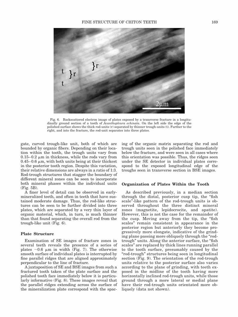
gate, curved trough-like unit, both of which arebounded by organic fibers. Depending on their loca-tion within the tooth, the trough units vary from0.15–0.2 �m in thickness, while the rods vary from0.45–0.6 �m, with both units being at their thickestin the posterior tooth region. Despite this variation,their relative dimensions are always in a ratio of 1:3.Rod-trough structures that stagger the boundary ofdifferent mineral zones can be seen to incorporateboth mineral phases within the individual units(Fig. 5B).
A finer level of detail can be observed in early-mineralized teeth, and often in teeth that have sus-tained moderate damage. Thus, the rod-like struc-tures can be seen to be further divided into threeplates, which are separated by a very thin layer oforganic material, which, in turn, is much thinnerthan that found separating the overall rod from thetrough-like unit (Fig. 6).
Plate Structure
Examination of SE images of fracture zones inseveral teeth reveals the presence of a series ofplates �0.6 �m in width (Fig. 7). The otherwisesmooth surface of individual plates is interrupted byfine parallel ridges that are aligned approximatelyperpendicular to the line of fracture.
A juxtaposition of SE and BSE images from such afractured tooth taken of the plate surface and thepolished tooth face immediately below it is particu-larly informative (Fig. 8). These images reveal thatthe parallel ridges extending across the surface ofthe mineralization plate correspond with the spac-
ing of the organic matrix separating the rod andtrough units seen in the polished face immediatelybelow the fracture, and were seen in all cases wherethis orientation was possible. Thus, the ridges seenunder the SE detector in individual plates corre-spond to the exposed longitudinal edge of thetroughs seen in transverse section in BSE images.
Organization of Plates Within the Tooth
As described previously, in a median sectionthrough the distal, posterior cusp tip, the “fishscale”-like pattern of the rod-trough units is ob-served throughout the three distinct mineralzones (magnetite, lepidocrocite, and apatite).However, this is not the case for the remainder ofthe cusp. Moving away from the tip, the “fishscales” remain consistent in appearance in theposterior region but anteriorly they become pro-gressively more elongate, indicative of the grind-ing plane passing more obliquely through the “rod-trough” units. Along the anterior surface, the “fishscales” are replaced by thick lines running parallelto the tooth surface, presumably caused by the“rod-trough” structures being seen in longitudinalsection (Fig. 9). The orientation of the rod-troughunits relative to the posterior surface also variesaccording to the plane of grinding, with teeth ex-posed in the midline of the tooth having morehorizontally inclined rod-trough units, while thoseground through a more lateral or medial planehave their rod-trough units orientated more ob-liquely (data not shown).
Fig. 6. Backscattered electron image of plates exposed by a transverse fracture in a longitu-dinally ground section of a tooth of Acanthopleura echinata. On the left side the edge of thepolished surface shows the thick rod-units (r) separated by thinner trough-units (t). Further to theright, and into the fracture, the rod-unit separates into three plates.
169FINE STRUCTURE OF CHITON TEETH

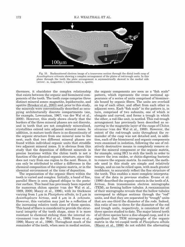
When the teeth are reoriented and ground topresent a transverse section of the cusp, a muchmore complex picture of the organization of the or-ganic matrix emerges, with variation in the align-ment of the plates of “rod-trough” units dependingon their position within the tooth. Again, the platescan be seen to be continuous through all mineralzones (Fig. 10). However, while the plates arealigned horizontally and parallel to the cutting sur-face in the posterior magnetite region, anterior to
this, and also to either side (laterally and medially),they are aligned progressively more obliquely. Inaddition, their overall arrangement is asymmetri-cal, being skewed towards the medial side of thecusp. The alignment of the plates again alters dra-matically, from oblique to vertical, as they passthrough the lepidocrocite zone and into the apatitemineral zone, while finally along the anterior of thecusp they are vertical and perpendicular to the sur-face (Fig. 10).
Fig. 7. SE images of fracture zones in the teeth of Acanthopleura echinata, both revealingplates with parallel ridges. A: A fracture transverse to the longitudinal polished face (pf) of a tooth.B: A chip in a transverse polished face (pf) of a tooth revealing plates running oblique to thesurface.
170 R.J. WEALTHALL ET AL.

Fracture Planes
SE images of polished longitudinal sectionsthrough the magnetite region of the teeth often re-veal the presence of distinct fracture lines (Fig.11A). A higher-magnification BSE image (Fig. 11B)reveals that these oblique cracks pass between therod and trough subunits and are propagated,through the organic material, from one trough unit
to the next, thus defining the fracture plane of thetooth.
DISCUSSION
This study reveals, for the first time, the in situstructure of the mineralized cusps of the major lat-eral teeth of the chiton Acanthopleura echinata. Fur-
Fig. 8. Combination image of a transverse fracture in the magnetite region of a mature toothof Acanthopleura echinata. A: SE image of the fracture showing parallel ridges across the surface.B: Backscattered image of the polished longitudinal face showing rod-trough units. The arrowsdemonstrate that the parallel ridges align precisely with the ends of the trough units.
Fig. 9. Backscattered electron image of a longitudinal section through the distal tooth cusp of Acanthopleura echinata showing thechange in orientation of the rod-trough units from horizontal and perpendicular to the posterior surface in the magnetite (m),progressively more oblique through the lepidocrocite (l) and posterior apatite (a), to almost parallel and vertical along the anteriorsurface.
171FINE STRUCTURE OF CHITON TEETH
thermore, it elucidates the complex relationshipthat exists between the organic and biomineral com-ponents of the tooth. The tooth cusps comprise threedistinct mineral zones: magnetite, lepidocrocite, andapatite (Brooker et al., 2003) and, prior to this study,the minerals were conventionally described as occu-pying architecturally discrete compartments (see,for example, Lowenstam, 1967; van der Wal et al.,2000). However, this study shows clearly that theborders of the three mineral phases are not discrete,and in teeth that are not completely mineralized,crystallites extend into adjacent mineral zones. Inaddition, in mature teeth there is no discontinuity ofthe organic structure from one mineral zone to thenext, such that two different mineral phases arefound within individual organic units that straddletwo adjacent mineral zones. It is obvious from thisstudy that the deposition of different minerals inprecise locations within the chiton tooth is not afunction of the physical organic structure, since thisdoes not vary from one region to the next. Hence, itcan only be attributed to either a difference in thechemical composition of the organic matrix, or in thechemical environment at the time of deposition.
The organization of the organic fibers within thetooth is varied and complex. Initially, a band of fine,parallel fibers is seen aligned oblique to the poste-rior surface. This band has previously been reportedfor numerous chiton species (van der Wal et al.,1989, 2000; Macey et al., 1996), with its thicknessvarying from 1 �m in Plaxiphora albida, to 2 �m inAcanthopleura hirtosa, and 4 �m in A. echinata.However, this variation may just be a reflection ofthe increasing relative tooth sizes of these species.This band of fibers is considered to provide the struc-tural integrity of the tooth shape, since it is moreresistant to chemical etching than the internal en-vironment (van der Wal et al., 1989; Evans et al.,1990; Macey et al., 1996). Throughout much of theremainder of the tooth, when seen in medial section,
the organic components are seen as a “fish scale”pattern, which represents the cross sectional ap-pearance of a series of units comprised of biominer-als bound by organic fibers. The units are overlaidon top of each other, and offset from each other inadjacent rows. Each “fish scale” in the pattern is, inturn, comprised of two subunits, one of which iselongate and curved, and forms a trough in whichthe other, a rod-like unit, is nestled. This rod-trougharrangement has previously been described as oc-curring in the magnetite layer of the cusps of Chitonolivaceus (van der Wal et al., 1989). However, theextent of the rod-trough units throughout the re-mainder of the cusp was not detailed and, in addi-tion, each of the biomineral and organic componentswere examined in isolation, following the use of rel-atively destructive means to completely remove ei-ther the mineral component or the organic matrix,for example, using HCl to etch the teeth in order toremove the iron oxides, or chitin-digesting bacteriato remove the organic matrix. In contrast, the meth-ods used in this study are simple and relativelybenign, and so can be relied upon, with much greaterconfidence, to accurately reflect the fine structure ofthe teeth. This enables a more complete interpreta-tion of the data in previous studies. Evans et al.(1990) described the organic matrix in the teeth of A.hirtosa, from transmission electron micrographs(TEM), as forming hollow tubules. A reexaminationof their micrographs reveals that the hollow tubulescorrespond to oblique sections through the “rod”units, and that they are separated by “trough” unitsthat are one-third the diameter of the rods. Indeed,this ratio of one to three for the diameter of the rodand trough units, respectively, is consistent for allthree species studied to date. The major lateral teethof all three species have a disc-shaped cusp, and it issignificant that TEM micrographs of the organicmatrix in the tri-cuspid teeth of Plaxiphora albida(Macey et al., 1996) do not exhibit the alternating
Fig. 10. Backscattered electron image of a transverse section through the distal tooth cusp ofAcanthopleura echinata showing a complex arrangement of the plates of rod-trough units. In thisplane through the tooth the plate arrangement is asymmetrically skewed to the medial side(arrow). m, magnetite; l, lepidocrocite; a, apatite.
172 R.J. WEALTHALL ET AL.

thin and thick tubules found in A. hirtosa (Evans etal., 1990). Instead, the tubules are all of a similardiameter, indicating that the rod-trough structuremay not be common to all chitons. This corroboratesprevious findings where is has been proposed thatthe organic structure of discoid and tri-cuspid teethis fundamentally different (van der Wal, 1990; vander Wal et al., 2000).
Scanning electron micrographs of damaged toothregions reveal that the rod-trough units are further
organized into large sheets, which in turn possessdistinctive parallel ridges. These ridges are almostcertainly due to the oblique, and offset, alignment ofadjacent rod-trough units, which exposes one side ofthe trough subunit. The relationship between therod and trough units and their organization intosheets is most readily appreciable in a diagrammaticrepresentation (Fig. 12). The layer of organic mate-rial between the sheets of rod-trough units presum-ably presents a weaker medium for separation and,
Fig. 11. Polished longitudinal section through the magnetite region of a tooth of Acanthopleuraechinata. A: SE image of an oblique crack. B: Higher-magnification BSE image of the crack in A,revealing the crack to be propagated from one trough unit to the next (arrows), along the organicinterface between the rod-trough plates
173FINE STRUCTURE OF CHITON TEETH

as such, the orientation of the sheets within thetooth determines the fracture planes along whichcracks will propagate.
While it is not possible to determine the actuallength of the rod-trough units, where they are seenin longitudinal section in the anterior regions of thetooth, lengths of up to 30 �m have been recorded.Evans et al. (1990) determined that Acanthopleurahirtosa has large, rope-like fibers in the anteriorregions of the tooth, and Macey and Brooker (1996)further demonstrated that these fibers originate inthe posterior base of the tooth, just below the cusp,and pass vertically through the junction of the toothbase and cusp. They then progress anteriorly in ahorizontal plane, prior to aligning vertically as theyprogress into the anterior regions of the tooth. It isreasonable to surmise that these fibers, and hencethe rod-trough units, are actually continuousthroughout much of the tooth, merely changing di-rection, and thus orientation, in different regions ofthe tooth. Certainly in SEM images of the teeth incross-section, a complex pattern of continuous platesof rod-trough units is obvious (Fig. 10). This contin-uous nature is most easily represented in a diagram(Fig. 13), which illustrates that the planes of therod-trough units, when viewed in a longitudinal me-dian section, are essentially horizontal and lie in alateromedial plane close to the posterior surface,while anteriorly they are almost vertical and lie inthe posteroanterior plane. Hence, relative to the pos-terior surface, the plates in the distal half of the cusprotate �90° in both the longitudinal and transverseplanes, across the thickness of the tooth. Theseplates then descend down the anterior surface be-fore changing to a horizontal orientation above thejunction zone, from where they then proceed poste-riorly, before finally descending into the tooth base.
The transverse section suggests a further level ofcomplexity in the plate arrangement, with an inter-nal structure that is skewed to the medial side of thecusp. In this regard it is probably significant thatthe tooth cusps of Acanthopleura echinata are notperfectly discoid, being described as mediallyskewed (L.R. Brooker, pers. commun.), which may,in turn, relate to their alignment, with regard to thesubstrate, when used in feeding. In addition, studiesin progress have revealed that the working teeth ofA. hirtosa show unsymmetrical wear in a lateral-medial plane, with more extreme wear on the medialside indicating that the teeth are not held perfectlyperpendicular to the substrate during the feedingstroke. As such, the medial extrusion of the distalcusp allows for a greater contact area with the sub-strate. van der Wal et al. (2000) proposed that it isthe orientation of the rod-trough units at the cusptip that ensures the maintenance of a sharp, wedge-shaped cutting surface as the tooth is worn down.Similarly, it would appear that the intricate pat-terns of the rod-trough plates, seen on the medialside in transverse sections, allow for the perpetua-tion of the medially skewed shape of the cusp.
This study has elucidated the fine structure of themineralized teeth of the chiton Acanthopleura echi-nata, and while there appear to be similarities withthat of Chiton olivaceus (van der Wal et al., 1989),these two species have close taxonomic ties andthere is a body of evidence that suggests that thisstructure is not ubiquitous to all chitons (Macey etal., 1996; van der Wal et al., 2000). In addition,chitons exhibit a variety of biomineralization strat-egies, incorporating a range of biominerals into theirteeth with different spatial organization. As such,the techniques used in this study need to be ex-tended to incorporate an in situ analysis of the finestructure of the teeth of chiton species that are bothtaxonomically diverse and that incorporate differentbiominerals into their teeth.
Fig. 13. Diagrammatic representation of the complex arrange-ment of the plates of rod-trough units in Acanthopleura echinataseen in (A) longitudinal section through the tooth, and (B) trans-verse section.
Fig. 12. Diagrammatic representation of the three-dimensional arrangement of the rod and trough subunits in theteeth of Acanthopleura echinata, comprising the fish scale units,with each rod defined by four surrounding troughs. The rod-trough units are elongated away from the viewer and are ar-ranged in sheets with the exposed edge of each trough unit (whitearrows) forming parallel ridges in the respective sheets surface.The long black arrow indicates the axis of extension of the rod-trough units.
174 R.J. WEALTHALL ET AL.

ACKNOWLEDGMENT
The authors thank Jeremy Shaw for help withillustrations.
LITERATURE CITED
Brooker LR, Lee AP, Macey DJ, Webb J, van Bronswijk W. 2003.Multiple-front iron mineralisation in chiton teeth (Acanthop-leura echinata: Mollusca: Polyplacophora). Mar Biol 142:447–454.
Evans LA, Macey DJ, Webb J. 1990. Characterization and struc-tural organization of the organic matrix of the radula teeth ofthe chiton Acanthopleura hirtosa. Philos Trans R Soc Lond B329:87–96.
Evans LA, Macey DJ, Webb J. 1992. Calcium biomineralization inthe radula teeth of the chiton Acanthopleura hirtosa. CalcifTissue Int 51:78–82.
Lee AP, Webb J, Macey DJ, van Bronswijk W, Savarese AR, deWitt GC. 1998. In situ Raman spectroscopic studies of the teethof the chiton Acanthopleura hirtosa. J Biol Inorg Chem 3:614–619.
Lee AP, Brooker LR, Macey DJ, van Bronswijk W, Webb J. 2000.Apatite mineralization in teeth of the chiton Acanthopleuraechinata. Calcif Tissue Int 67:408–415.
Lee AP, Brooker LR, Macey DJ, Webb J, van Bronswijk W. 2003a.A new biomineral identified in the cores of teeth from the chitonPlaxiphora albida (Mollusca: Polyplacophora). J Biol InorgChem 8:256–262.
Lee AP, Brooker LR, van Bronswijk W, Macey DJ, Webb J. 2003b.Contribution of Raman spectroscopy to identification of biomin-erals present in teeth of Acanthopleura rehderi, Acanthopleura
curtisiana, and Onithochiton quercinus. Biopolymers 72:299–301.
Lowenstam HA. 1962. Magnetite in denticle capping in recentchitons (Polyplacophora). Geol Soc Am Bull 73:435–438.
Lowenstam HA. 1967. Lepidocrocite, an apatite mineral, andmagnetite in teeth of chitons (Polyplacophora). Science 156:1373–1375.
Lowenstam HA, Weiner S. 1985. Transformation of amorphouscalcium phosphate to crystalline dahllite in the radula teeth ofchitons. Science 227:51–52.
Lowenstam HA, Weiner S. 1989. On biomineralization. Oxford:Oxford University Press.
Macey DJ, Brooker LR. 1996. The junction zone: initial site ofmineralization in radula teeth of the chiton Cryptoplax striata(Mollusca: Polyplacophora). J Morphol 230:33–42.
Macey DJ, Brooker LR, Webb J, St. Pierre TG. 1996. Structuralorganization of the radular teeth of the chiton Plaxiphora al-bida. Acta Zool (Stockh) 77:287–294.
Shaw JA, Brooker LR, Macey DJ. 2002. Radula tooth turnover inthe chiton, Acanthopleura hirtosa. Moll Res 22:93–99.
van der Wal P. 1990. Structure and formation of the magnetite-bearing cap of the polyplacophoran tricuspid radula teeth. In:Frankel RB, Blakemore RP, editors. Iron biominerals. NewYork: Plenum Press. p 221–229.
van der Wal P, Videler JJ, Havinga P, Pel R. 1989. Architectureand chemical composition of the magnetite-bearing layer in theradula teeth of Chiton olivaceus (Polyplacophora). In: Crick RE,editor. Origin, evolution, and modern aspects of biomineraliza-tion in plants and animals. New York: Plenum Press. p 153–166.
van der Wal P, Giesen H, Videler J. 2000. Radular teeth asmodels for the improvement of industrial cutting devices. MaterSci Eng C: Biomim Mater Sens Syst 7:129–142.
175FINE STRUCTURE OF CHITON TEETH