Fine Structure, Physiology and Biochemistry of Arthrospore Germination in Streptomyces antibioticus
Upload
khangminh22Category
view
0download
0
THE F I N E S T R U C T U R E OF Streptomyces coelicolor
II. The Nuclear Material
D A V I D A. t t O P W O O D , Ph.D., and A U D R E Y M. G L A U E R T
From the University Botany School and the Strangeways Research Laboratory, Cambridge, England
A B S T R A C T
Colonies and spore suspensions of Streptomyces coelicolor were fixed for electron microscopy by the method of Kellenberger, Ryter, and S5chaud (1958). In thin sections the nuclear regions have a lower average density than the cytoplasm and the outlines of these regions correspond well with the profiles of the chromatinic bodies observed with the light micro- scope. The nuclear regions contain fibrils, about 5 m# in diameter. In contrast, after fixation by the method of Palade (1952) the nuclear material is coagulated into irregular dense masses and tubular structures about 20 m/~ in diameter, lying in a nuclear "vacuole ." The significance of these observations is discussed in relation to the observations of other workers on the fine structure of the nuclear material of other bacteria and the chromosomes of higher cells.
I N T R O D U C T I O N
Observations on stained preparations of Strep- tomyces coelicolor with the light microscope (Hop- wood and Glauert, 1960) indicated that there is a characteristic sequence of changes in the configurations of the chromatinic material during the development of the spores. Elongated chro- matinic structures in the young aerial hyphae separate into a number of subunits and a single round chromatinic body is included in each spore. It was therefore of interest to study the nuclear material in thin sections in the electron microscope to see if there were any corresponding alterations in fine structure.
Kellenberger and his coworkers (Kellenberger, Ryter, and S6chaud, 1958; Ryter and Kellen- berger, 1958) have developed a method of fixation for electron microscopy specifically designed to preserve the nuclear structure of eubacteria. In
Miss Glauert is a Sir Halley Stewart Research Fellow. Received for publication, January 26, 1960.
this study we have applied this method to Strepto- myces coelicolor and have compared the results with those obtained with the conventional Palade technique (Palade, 1952).
M A T E R I A L S A N D M E T H O D S
The methods of growth of the organism and the preparation of thin sections for examination in the electron microscope have been described in detail in the first paper of this series (Glauert and Hopwood, 1960).
The methods of staining the organism and taking the photomicrographs are described in the preceding paper (Hopwood and Glauert, 1960).
R E S U L T S
The general organisation of the hyphae of Strep- tomyces coelicolor as seen in thin sections of material
267
Dow
nloaded from http://rupress.org/jcb/article-pdf/8/1/267/1074889/267.pdf by guest on 31 January 2022
fixed by the method of Kellenberger, Ryter, and S6chaud (1958) has already been described (Glauer t and Hopwood, 1960). The nuclear mater ia l occupies the central regions of the hyphae ; it has a somewhat lower average density than the granular and membranous mater ia l of the cytoplasm (Fig. 1), so tha t the boundar ies of the nuclear regions are seen very clearly in photographs at magnifications of 70,000 or less (Figs. 1, 9, 10). These profiles correspond well wi th those of the chromat in ic bodies in stained prepara t ions viewed in the l ight mi- croscope. Thus, the outline of the nuclear region in a th in section of a major hypha of the substrate mycel ium (Fig. 1) closely resembles tha t of the irregularly lobed chromat in ic bodies in stained preparat ions (Fig. 2). Compare also the nuclear
region of Fig. 5, which occupies a large propor t ion of the area of a section of an aerial hypha, with the rod-shaped chromat in ic bodies of a young aerial hypha in Fig. 6, and the nuclear regions of nearly mature spores in Fig. 7 with the eccentric chromat in ic bodies of spores in Fig. 8.
Al though the configurations of the nuclear regions vary characterist ically in the different parts of the mycel ium and in the spores, the fine structure of the nuclear mater ia l is very similar at all stages of development . The commonest images in the nuclear regions are very dense circular profiles, approximately 5 m/~ in diameter , and less dense, elongated, irregularly curved profiles, approximately 5 m/~ wide and of varying length and density (Figs. 9a, 10a). Mater ia l of low density occupies the space between the dense
E X P L A N A T I O N OF F I G U R E S
FIGURES 1 to 15, except FIGURES 2, 6, 8, and l l . Electron micrographs of thin sec- tions of Streptomyces codicolor strain A3(2), fixed by the method of Kellenberger, Ryter, and S6chaud (1958), except where otherwise stated, and embedded in methacrylate. The scale mark represents 0.1 micron.
FIGURES 2, 6, 8, and 11. Light microscope photographs of organisms fixed in the vapour of 2 per cent osmic acid and stained for chromatin. (See Hopwood and Glauert, 1960). X 3,000.
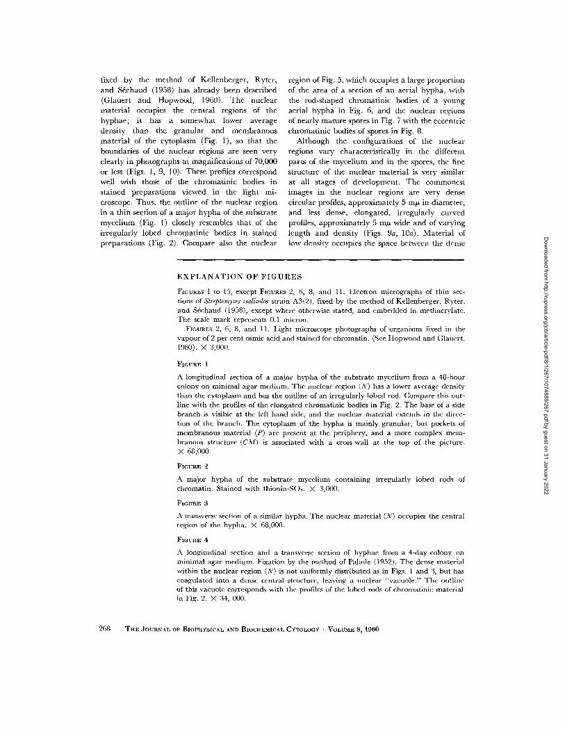
FIGURE 1
A longitudinal section of a major hypha of the substrate mycelium from a 48-hour colony on minimal agar medium. The nuclear region (N) has a lower average density than the cytoplasm and has the outline of an irregularly lobed rod. Compare this out- line with the profiles of the elongated chromatinic bodies in Fig. 2. The base of a side branch is visible at the left hand side, and the nuclear material extends in the direc- tion of the branch. The cytoplasm of the hypha is mainly granular, but pockets of membranous material (P) are present at the periphery, and a more complex mem- branous structure (CM) is associated with a cross-wall at the top of the picture. X 68,000.
FIGURE
A major hypha of the substrate mycelium containing irregularly lobed rods of chromatin. Stained with thionln-SO~. X 3,000.
FIGURE 3
A transverse section of a similar hypha. The nuclear material (N) occupies the central region of the hypha. X 68,000.
FIGURE 4,
A longitudinal section and a transverse section of hyphae from a 4-day colony on minimal agar medium. Fixation by the method of Palade (1959). The dense material within the nuclear region (N) is not uniformly distributed as in Figs. 1 and 3, but has coagulated into a dense central structure, leaving a nuclear "vacuole." The outline of this vacuole corresponds with the profiles of the lobed rods of chromatinic material in Fig. 2. >( 34, 000.
268 THE JOURNAL OF BIOPHYSICAL .aND BIOCHEMICAL CYTOLOGY • VOLUME 8~ 1960
Dow
nloaded from http://rupress.org/jcb/article-pdf/8/1/267/1074889/267.pdf by guest on 31 January 2022
H o P w o o D AND GLAUE~T Fine Structure. I I 2 6 9
Dow
nloaded from http://rupress.org/jcb/article-pdf/8/1/267/1074889/267.pdf by guest on 31 January 2022

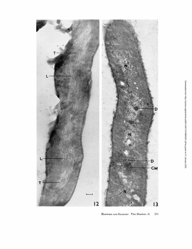
structures. These observations suggest tha t the nucleoplasm contains fibrils of about 5 m~ diameter , which run in different directions within a matr ix of low density. Sectioning of the fibrils in different planes gives rise to profiles of varying length and density. The amoun t of dense ma- terial in different nuclear regions varies more widely than would be expected from variat ions in the thickness of the sections. In particular, the nuclear regions of the spores have a low electron density compared wi th those of some hyphae (compare Figs. 9 and 10). In m a n y nuclear regions, the individual fibrils appear to have a r a n d o m orientat ion with respect to one another and to the axes of the hypha (Figs. 1, 3, 5, 9), but in other regions they have an ordered arrange- ment . Thus, in a section of the elongated nuclear region of a young aerial hypha (Fig. 12), places in the nucleoplasm in which most of the fibrils are cut transversely (T) are separated by areas in which most of them run longitudinal ly (L).
O the r structures are sometimes observed among the fine nuclear fibrils; small e lectron-opaque bodies are found (Fig. 13, D), and vacuoles,
occurr ing singly or in groups, are seen in mature and nearly mature spores (Fig. 15, V). Elements of the intracytoplasmic membrane system are often closely associated wi th or embedded in the nuclear regions of hyphae (Fig. 13, CM; and Glauer t and Hopwood, 1959), and an apparent ly discrete membraneous body is very often embedded in the nuclear region of developing spores (Figs. 10, 15, CM; and Glauer t and Hopwood, 1960); this may explain the appearance of r ing-shaped chromat in ic bodies in the l ight microscope (Fig. 11), since a spherical nuclear region with a core of Feulgen-negative mater ia l would appear as an annulus.
The fine structure of organisms fixed by Palade 's method (1952) differs greatly from that of or- ganisms fixed by the method of Kellenberger , Ryter, and Sfichaud (1958); in the former the cytoplasm is coarsely granular and shows only traces of poorly preserved elements of the intra- cytoplasmic membrane system (Glauer t and Hopwood, 1960) and the nuclear regions of m a n y hyphae appear as "vacuoles" conta ining i r regular masses of dense mater ial (Fig. 4). The
FIGURE 5
Part of a young aerial hypha from a 48-hour colony on minimal agar medium. The nuclear region (N) occupies a large proportion of the cross-section of the hypha, and is uniformly filled with fine fibrillar material. Compare the outline of the nuclear region with the profile of a rod-shaped chromatinic body of a young aerial hypha in Fig. 6. X 100,000.
FIGURE 6
Part of a young aerial hypha. The basal segment (uppermost in the figure) contains a dense rod of chromatinic material. Stained with thionin-SO> X 3,000.
FIGURE 7
A chain of spores which are nearly mature, from a 6-day colony on minimal agar medium. Each spore has a single nuclear region (N). In the central spore the nuclear region is eccentric and can be compared with the eccentric ehromatinic bodies in a chain of spores in Fig. 8. A membranous body is present at CM. Small vacuoles (V) of unknown nature are present in the nuclear regions. X 100,000.
FIGURE 8
A chain of spores which are nearly mature. In several of the spores the chromatinic body is eccentric. Stained with azure A-SO2. X 3,000.
A comparison of Figs. 5 and 7 indicates the different distribution of the nuclear material in the aerial hyphae, before and after spore delimitation. An intermediate stage may be represented by the hypha in Fig. 12; the corresponding appearance in the light microscope is probably the beaded chromatinic rod in the lower segment of the hypha in Fig. 6.
270 THE JOURNAL OF BIOPHYSICAL AND BIOCHEMICAL CYTOLOGY " VOLUME 8, 1960
Dow
nloaded from http://rupress.org/jcb/article-pdf/8/1/267/1074889/267.pdf by guest on 31 January 2022

HOPWOOD AND GLAUERT Fine Structure. I I 271
Dow
nloaded from http://rupress.org/jcb/article-pdf/8/1/267/1074889/267.pdf by guest on 31 January 2022

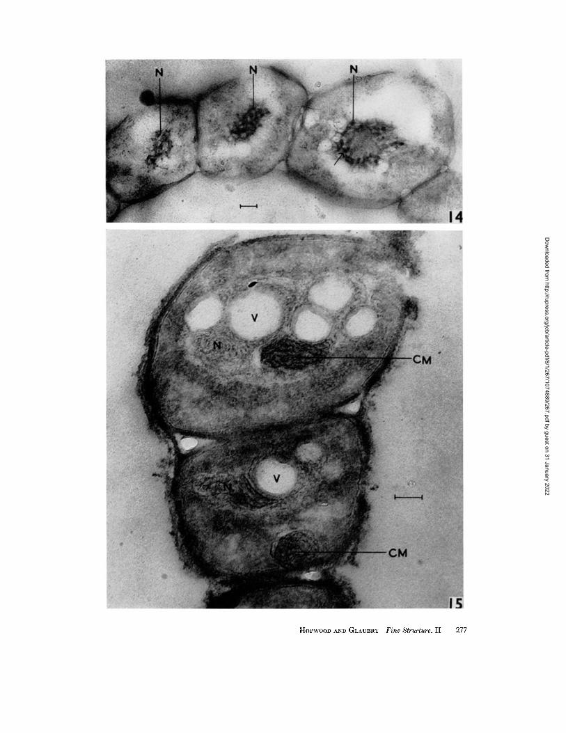
nuclear regions of spores sometimes contain more regular configurations of dense material , which may appear as a series of tubules approximately 20 m# in d iameter (Fig. 14). The outlines of the vacuoles, r a ther than those of the dense masses, correspond to the profiles of the chromat in ic bodies in stained preparat ions.
D I S C U S S I O N
Ryter and Kel lenberger (1958) have shown tha t the fine structure of the nuclear mater ia l of eubac- teria varies widely with the method of fixation, and have given reasons for the view that the appearance produced by their " s t a n d a r d " fixation is the nearest approximat ion so far achieved to the true structure of the nuclear material . This method of fixation applied to Streptomyces coelicolor results in a finely fibrillar nucleoplasm similar to tha t of eubacteria . In our preparat ions the nuclear regions contain a greater concentra t ion of dense mater ial than in most of the pictures of Ryter and Kellen- berger (1958) and of Kellenberger, Ryter, and S6chaud (1958) and also the mater ia l is more uni- formly distr ibuted within the nuclear regions.
Whe the r this reflects a real difference between organisms or is due to a difference in technique cannot be decided at present. The fibrils in the nuclear regions of S. coelicolor usually have a ran- dom ar rangement . Occasionally, they appear to have a parallel a r r angemen t (Fig. 12), which may reflect a movement and segregation of the fibrils into certain regions dur ing the subdivision of the nucleoid. In addi t ion to fibrils of approximately 5 m # diameter , granules of about the same diameter may well be present in the nuclear regions; indeed, the excess of circular over e longated profiles suggests tha t they are. Moreover, the boundary between the cytoplasm and the nuclear regions, well defined as it seems to be at low magnif icat ion (Figs. 9, 10), appears very diffuse at higher magnif icat ion (Figs. 9a, 10a). This possibly reflects an in terchange of g ranular mater ia l between the nuclear regions and the cytoplasm, which may well occur dur ing the synthesis of the cytoplasmic R N A and proteins. I t is possible tha t the difference in density be- tween the nuclear regions of hyphae and of spores is due to a greater amoun t of non-genetic mater ial in the former than in the latter, which
FIGURE 9
Part of an aerial hypha from a spore suspension. The nuclear region occupies a large proportion of the cross-section of the hypha. X 70,000.
FIGURE 9a
An enlargement of part of Fig. 9. The nuclear material of the hypha appears to consist of fine fibrils and granules, about 5 m# in diameter, and there is no clear boundary between the nucleoplasm and the cytoplasm. X 140,000.
FIGURE 10
A developing spore from a spore suspension. A body composed of interconnected membranous elements (CM) is embedded in the nuclear region and is apparently continuous with the main part of the cytoplasm. Such elements may be responsible for the Feulgen-negative core to the chromatinic bodies observed in the light microscope (see Fig. l l , arrow). • 70,000.
FIGURE 10a
An enlargement of part of Fig. 10. The nuclear material appears to consist of fine fibrils and granules, about 5 m# in diameter. The nuclear region of the spore has a lower average density than the nuclear region of the hypha in Fig. 9. Again there is no clear boundary between the nucleoplasm and the cytoplasm. X 300,000.
FIGURE 11
Spores stained by the Feulgen method. One spore contains a ring-shaped chromatinic body (arrow). X 3,000.
272 THE JOURNAL OF BIOPHYSICAL AND BIOCttEMICAL CYTOLOGY • VOLUME 8, 1960
Dow
nloaded from http://rupress.org/jcb/article-pdf/8/1/267/1074889/267.pdf by guest on 31 January 2022

HOPWOOD AND GL.~U~a~ Fine Structure. I1 273
Dow
nloaded from http://rupress.org/jcb/article-pdf/8/1/267/1074889/267.pdf by guest on 31 January 2022
are in a "resting" condition. If this were so, it might account for the difference in staining reaction to Pi6chaud's method shown by the hyphae and spores (Hopwood and Glauert, 1960).
After fixation by the method of Kellenberger, Ryter, and S4chaud (1958), there is no evidence of organised structures larger than the individual fibrils within the nuclear regions. In contrast, after fixation by the method of Palade (1952), which was developed for animal tissues, various thread-like and tubular structures are seen suspended in a nuclear "vacuole" (Figs. 4, 14; Hopwood and Glauert, 1958; Stuart, 1959), as in many of the earlier micrographs of eubac- teria (Birch-Andersen, MaalCe, and Sj6strand, 1953; MaalCe and Birch-Andersen, 1956; Chap- man and Kroll, 1957). Such structures in Bacillus megaterium have been interpreted as chromosomes by Giesbrecht (1958, 1959). Since it is probable that dense structures within a nuclear "vacuole" are produced by the coagulation of a finely fibrillar nucleoplasm during imperfect fixation (Ryter and Kellenberger, 1958), the configurations assumed by this material may not mean very much, and attempts to deduce the structure of bacterial
nuclei from organisms fixed in this way may be misleading. Chapman (1959) suggested that fibrils
and coarser threads may be alternative forms
of the chromatin, but whether it can exist in more than one form during the life of the organism remains to be seen. In view of the fact that tubular
structures 12.5 m# (Kaufmann and De, 1956; Kaufmann and McDonald, 1956) or 20 m/~
(Ris, 1956) in diameter have been described as
the smallest unit of the chromosomes of the cells
of higher organisms fixed by methods resembling that of Palade (1952), it is significant that this method reveals tubules approximately 20 m#
in diameter in the nuclear regions of the spores of S. coelicolor (Fig. 14). If such tubules are fixation
artefacts in this organism, the same may be true for higher cells, although, as pointed out by Kellenberger, Ryter, and S4chaud (1958), the possibility remains that differences in chemical composition between the DNA complexes of bacteria and higher cells (Zubay and Watson, 1959; Wilkins and Zubay, 1959) may be reflected in a different structural organisation. It is inter- esting, however, that fibrils of diameter 3 to 8 m# have been described in the chromosomes of the grasshopper Laplatacris disfar (De Robertis, 1956) and of the dinoflagellate Amphidinium elegans (Grell and Wohlfarth-Bottermann, 1957) when fixed by methods having some features in common with that of Kellenberger and his coworkers. Also in more recent work, Ris (1958) has described 4 m# fibrils of nucleohistone in calf-thymus chromo- somes, but considers them to be associated in pairs with non*histone protein to form larger fibrils 10 m# in diameter; in sperm nuclei the non-histone protein is absent and the 4 m# fibrils fill the whole nucleus.
The nature of the small dense granules in the nuclear regions of some hyphae (Fig. 13) is un- known. Possibly they are analagous with the nucleoli of higher cells and owe their electron density to an accumulation of RNA, or they may be metaphosphate granules comparable to those of mycobacteria (Glauert and Brieger, 1955). Similar granules are present in the nuclear region of an unidentified bacterium studied by Chapman (1959). The vacuoles in the nuclear regions of some spores (Fig. 15) may be artefacts caused by imperfect fixation or embedding of the spores which are more resistant to penetration than the hyphae, or alternatively they may indicate the sites of material that has been extracted.
An apparent association between the nuclear material and elements of the intracytoplasmic membrane system (Glauert and Hopwood, 1960) seems to be characteristic of the spores of Strepto-
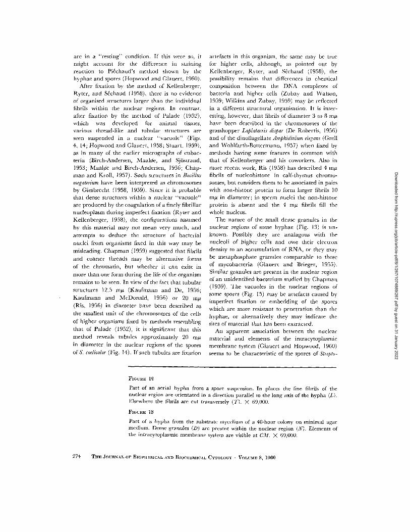
FIGURE 1~
Part of an aerial hypha from a spore suspension. In places the fine fibrils of the nuclear region are orientated in a direction parallel to the long axis of the hypha (L). Elsewhere the fibrils are cut transversely (T). X 69,000.
FIGURE 13
Part of a hypha from the substrate mycelium of a 48-hour colony on minimal agar medium. Dense granules (D) are present within the nuclear region (N). Elements of the intracytoplasmic membrane system are visible at CM. X 69,000.
274 THE JOURNAL OF BIOPHYSICAL AND BIOCHEMICAL CYTOLOGY • VOLUME 8, 1960
Dow
nloaded from http://rupress.org/jcb/article-pdf/8/1/267/1074889/267.pdf by guest on 31 January 2022
HOPWOOD AND GLAUERT Fine Structure. I I 275
Dow
nloaded from http://rupress.org/jcb/article-pdf/8/1/267/1074889/267.pdf by guest on 31 January 2022

rnyces coelicolor, many of which contain a mem- branous body (Figs. 10, 15). A connection between
this body and the main par t of the cytoplasm is sometimes seen (Fig. 10), so that, apart from showing a highly organised membranous structure, these bodies are comparable with the cytoplasmic extensions into the nuclear region that have been observed in many bacteria. These extensions
R E F E R E N C E S
BIRCH-ANDERSEN, A., MAALCE, O., and S.]OSTRAND, F. S., High-resolution electron micrographs of sections of Escherichia coil, Biochim. et Biophysica Acta, 1953, 12, 395.
CHAPMAN, G. B., Electron microscopy of ultrathin sections of bacteria. III. Cell wall, cytoplasmic membrane, and nuclear material, J. Bact., 1959, 78, 96.
CHAPMAN, G. B., and KROLE, A. J., Electron micro- scopy of ultrathin sections of Spirillum serpens, J. Bact,, 1957, 73, 63.
DE ROBERTlS, E., Electron microscopic observations on the submicroscopic morphology of the meiotic nucleus and chromosomes, J. Biophysic. and BiD- chem. Cvtol., 1956, 2, 785.
GIESBRECHT, P., Zur Struktur des Bakterienzellkerns, Naturwissensch., 1958, 45, 473.
GIESBRECHT, P., Elektronenmikroskopische Darstel- lung verschiedener chemischer und physikalisch- chemischer Reaktionen an den DNS-Strukturen eines Chromosoms, Zentr. Bakt. I. Orig., 1959, 176, 413.
GLAtmRT, A. M., and BRIECER, E. M., The electron- dense bodies of Mycobacterium phlei, J. Gen. Micro- biol., 1955, 13, 310.
GLAUERT, A. M., and HoPWOOD, D. A., A mem- branous component of the cytoplasm in Strepto- myces coelicolor, J. Biophysic. and Biochem. Cytol., 1959, 6, 515.
may be responsible for the appearance of an achromatic core in some chromatinic bodies (Fig. 11; Murray, 1953). The significance of the asso- ciation of the membranous bodies with the nuclear material will not be understood until more is known
about the function of the intracytoplasmic mem- brane system as a whole.
GLAUERT, A. M., and HoPwoon, D. A., The fine structure of Streptomyces coelicolor. I. The cyto- plasmic membrane system, J. Biophysic. and BiD- chem. Cytol., 1960, 7,479.
GRELL, K. G., and WOHLFARTH-BOTTERMANN, K. E., Licht und elektronenmikroskopische Unter- suchungen an den Dinoflagellaten Amphidinium elegans n. sp., Z. Zellforsch., 1957, 47, 7.
HoPWOOD, D. A., and GLAUERT, A. M., The electron microscopy of Streptomyces coelicolor, d. Gem Micro- biol., 1958, 18, vi.
HoPWOOD, D. A., and GLAUERT, A. M., Obser- vations on the chromatinic bodies of Streptomyces coelicolor, J. Biophysic. and Biochem. Cvtol., 1960, 8, 257.
KAUFMANN, B. P., and DE, D. N., Fine structure of chromosomes, J. Biophysic. and Biochem. Cytol., 1956, 2, No. 4, suppl., 419.
KAUFMANN, B. P., and McDONALD, M. R., Organi- zation of the chromosome, Cold Spring Harbor Syrup. Quant. Biol., 1956, 21, 233.
KELLENBEROER, E., RYTER, A., and S~CHAUD, J., Electron microscope study of DNA-containing plasms. II. Vegetative and mature phage DNA as compared with normal bacterial nucleoids in different physiological states, J. Biophysic. and Biochem. Cytol., 1958, ~t, 671.
MAALCE, O., and BIRCh-ANDERSEN, A., On the organization of the "nuclear material" in Sal-
FIGURE ]4
A chain of spores which are nearly mature, from a 4-day colony on minimal agar medium. Fixation by the method of Palade (1952). The nuclear region (N) contains a number of tubules about 20 m# in diameter (arrow), within a nuclear "vacuole." >( 60,000.
]~'IG URE 15
A similar chain of spores, from a spore suspension. After fixation by the method of Kellcnberger, Ryter, and S6chaud (1958), the nuclear region (N) contains fine fibrils, about 5 m# in diameter. A number of vacuoles (V) of unknown nature, are present in the nuclear regions. Condensed elements of the intracytoplasmic membrane system are present at CM. X 100,000.
276 THE JOURNAL OF BIOPHYSICAL AND BIOCHEMICAL CYTOLOGY • VOLUME 8, 1960
Dow
nloaded from http://rupress.org/jcb/article-pdf/8/1/267/1074889/267.pdf by guest on 31 January 2022
HOPWOOD AND GLAUERT Fine Structure. I I 277
Dow
nloaded from http://rupress.org/jcb/article-pdf/8/1/267/1074889/267.pdf by guest on 31 January 2022

monella typhimurium, in Bacterial Anatomy, Cam- bridge University Press, 1956, 261.
MURRAY, R. G. E., The problem of fixation for studies of bacterial nuclei, in Bacterial Cytology, Syrup. 6th Cbngr. Internal. Microbiol., Rome, 1953, 136.
PALADE, G. E., A study of fixation for electron microscopy, J. Exp. :gIed., 1952, 93, 285.
RIs, H., A study of chromosomes with the electron microscope, J. Biophysic. and Biochem. Cytol., 1956, 2, No. 4, suppl., 385.
Rls, H., Fine structure of the nucleus during spermio- genesis, Abstracts, 4th Internal. Conf. Electron M~cr., Berlin, 1958, 188.
RYTER, A., and KELLENBERGER, E., Etude au micro- scope 61ectronique de plasmas contenant de l 'acide d6soxyribonucl6ique. 1. Les nucl6oides des bact6ries en croissance active, Z. Natur[orsch., 1958, 13, 597.
STUART, D. C., Fine structure of the nucleoid and internal membrane systems of Streptomyces, J. Bact., 1959, 78, 272.
WILKINS, M. H. F., and ZUBAV, G., The absence of histone in the bacterium Escherichia coll. II. X-ray diffraction of nucleoprotein extract, J. Biophysic. and Biochem. Cytol., 1959, 5, 55.
ZUBAY, G., and WATSON, M. R., The absence of histone in the bacterium Escherichia coll. I. Prepa- ration and analysis of nucleoprotein extracts, J . Biophysic. and Biochem. Cvtol., 1959, 5, 51.
278 THE ~OURNAL OF BIOPHYSICAL AND BIOCttEMICAL CYTOLOGY • VOLUME 8, 1960
Dow
nloaded from http://rupress.org/jcb/article-pdf/8/1/267/1074889/267.pdf by guest on 31 January 2022
Copyright © 2022 FDOKUMEN