
A Series of Fluorene-Based Two-Photon Absorbing Molecules: Synthesis, Linear and Nonlinear...
21
A series of fluorene-based two-photon absorbing molecules: synthesis, linear and nonlinear characterization, and bioimaging Carolina D. Andrade † , Ciceron O. Yanez † , Luis Rodriguez ‡ , and Kevin D. Belfield *,†,‡ † Department of Chemistry, The College of Optics and Photonics, University of Central Florida, Orlando, FL 32816 ‡ CREOL, The College of Optics and Photonics, University of Central Florida, Orlando, FL 32816 Abstract The synthesis, structural, and photophysical characterization of a series of new fluorescent donor– acceptor and acceptor-acceptor molecules, based on the fluorenyl ring system, with two-photon absorbing properties is described. These new compounds exhibited large Stokes shifts, high fluorescent quantum yields, and, significantly, high two-photon absorption cross sections, making them well suited for two-photon fluorescence microscopy (2PFM) imaging. Confocal and two- photon fluorescence microscopy imaging of COS-7 and HCT 116 cells incubated with probe I showed endosomal selectivity, demonstrating the potential of this class of fluorescent probes in multiphoton fluorescence microscopy. Keywords Fluorescent dyes; multiphoton absorption; fluorescence; two-photon; bioimaging Introduction The development of multiphoton absorbing materials has attracted the interest of the scientific community in the last decade due to their potential applications in areas as optical data storage systems, microscopy imaging, optical limiting materials, and photodynamic therapy. In one- photon absorption processes, the absorbed light is directly proportional to the incident light intensity, whereas in two-photon absorption (2PA) processes, the probability of absorbing two photons simultaneously is proportional to the square of the incident light intensity. 1 This nonlinear dependence provides several advantages, including highly confined excitation, increased 3D resolution (Figure 1), and the potential increase of penetration depth in tissue, important features in fields such as optical data storage, 2-4 3D microfabrication, 5-7 and fluorescence microscopy. 1,8 We previously reported a series of fluorescent dyes and photoacid generators that use fluorene as a core structure. 9-14 This core exhibited high thermal and photochemical stability making this class of compounds promising in areas such as lasing applications. 15 [email protected] . Supporting Information Available: Additional experimental descriptions, NMR spectra, and 3D cell images from layer-by-layer reconstruction of two-photon fluorescence micrographs are included. This material is available free of charge via the Internet at http://pubs.acs.org. NIH Public Access Author Manuscript J Org Chem. Author manuscript; available in PMC 2011 June 18. Published in final edited form as: J Org Chem. 2010 June 18; 75(12): 3975–3982. doi:10.1021/jo1005075. NIH-PA Author Manuscript NIH-PA Author Manuscript NIH-PA Author Manuscript
-
Upload
independent -
Category
Documents
-
view
5 -
download
0
Transcript of A Series of Fluorene-Based Two-Photon Absorbing Molecules: Synthesis, Linear and Nonlinear...
A series of fluorene-based two-photon absorbing molecules:synthesis, linear and nonlinear characterization, and bioimaging
Carolina D. Andrade†, Ciceron O. Yanez†, Luis Rodriguez‡, and Kevin D. Belfield*,†,‡†Department of Chemistry, The College of Optics and Photonics, University of Central Florida,Orlando, FL 32816‡CREOL, The College of Optics and Photonics, University of Central Florida, Orlando, FL 32816
AbstractThe synthesis, structural, and photophysical characterization of a series of new fluorescent donor–acceptor and acceptor-acceptor molecules, based on the fluorenyl ring system, with two-photonabsorbing properties is described. These new compounds exhibited large Stokes shifts, highfluorescent quantum yields, and, significantly, high two-photon absorption cross sections, makingthem well suited for two-photon fluorescence microscopy (2PFM) imaging. Confocal and two-photon fluorescence microscopy imaging of COS-7 and HCT 116 cells incubated with probe Ishowed endosomal selectivity, demonstrating the potential of this class of fluorescent probes inmultiphoton fluorescence microscopy.
KeywordsFluorescent dyes; multiphoton absorption; fluorescence; two-photon; bioimaging
IntroductionThe development of multiphoton absorbing materials has attracted the interest of the scientificcommunity in the last decade due to their potential applications in areas as optical data storagesystems, microscopy imaging, optical limiting materials, and photodynamic therapy. In one-photon absorption processes, the absorbed light is directly proportional to the incident lightintensity, whereas in two-photon absorption (2PA) processes, the probability of absorbing twophotons simultaneously is proportional to the square of the incident light intensity.1 Thisnonlinear dependence provides several advantages, including highly confined excitation,increased 3D resolution (Figure 1), and the potential increase of penetration depth in tissue,important features in fields such as optical data storage,2-4 3D microfabrication,5-7 andfluorescence microscopy.1,8
We previously reported a series of fluorescent dyes and photoacid generators that use fluoreneas a core structure.9-14 This core exhibited high thermal and photochemical stability makingthis class of compounds promising in areas such as lasing applications.15
[email protected] .Supporting Information Available: Additional experimental descriptions, NMR spectra, and 3D cell images from layer-by-layerreconstruction of two-photon fluorescence micrographs are included. This material is available free of charge via the Internet athttp://pubs.acs.org.
NIH Public AccessAuthor ManuscriptJ Org Chem. Author manuscript; available in PMC 2011 June 18.
Published in final edited form as:J Org Chem. 2010 June 18; 75(12): 3975–3982. doi:10.1021/jo1005075.
NIH
-PA Author Manuscript
NIH
-PA Author Manuscript
NIH
-PA Author Manuscript
Recently, a computational quantum chemical study of numerous fluorene derivatives wasreported in order to predict and rationalize certain 2PA parameters, such as 2PA cross sectionsand 2PA wavelength. Though insightful, little comparison and validation of computed resultswith experimental data was presented, emphasizing the need to accurately measure 2PA spectraand cross sections of new compounds.16
In the interest of developing more efficient 2PA fluorophores, it is essential to improve the keyphotophysical properties such as quantum yield, absorption maxima, and 2PA cross sectionsin the tuning range of commercial Ti:sapphire lasers (700-1000 nm). In this work, we presentthe design, synthesis, and photophysical (linear and nonlinear) characterization of a new seriesof two-photon absorbing molecules based on a fluorenyl core and demonstration of their usein in vitro confocal and two-photon fluorescence microscopy (2PFM) bioimaging.
In this approach, a series of dyes were developed with the general structural design donor-pi-acceptor (D-π-A) and acceptor-pi-acceptor (A-π-A) using benzothiazole moieties as electron-acceptor groups and diphenylamino groups as electron-donor moieties. To enhance thesolubility of the fluorene core in organic solvents, both protons on the 9-position of fluorenewere substituted by alkyl chains. Functionalities including thiophene and alkynes wereincorporated in order to increase the length of the π-conjugation and to evaluate the effect ofthese functionalities on the linear absorption maxima, fluorescence quantum yields, and 2PAcross sections. The chemical structures of the new fluorescent compounds presented in thispaper are illustrated in Figure 2.
For the work described herein, the thiophene ring was chosen as a electron bridge because ithas a higher degree of aromaticity than other five member rings, e.g., furan, it is stable towardsoxidation, and its chemical reactivity to allow functionalization is higher than that observedfor benzene or furan. Incorporation of these heterocyclic rings in the molecular design ofchromophores has been reported to enhance the optical properties, increasing both themaximum of absorption (λabs
max) and the 2PA cross section values, particulary when thethiophene ring is close to the fluorene core.17,18
One of our goals was to compare this new series of compounds with our previously preparedand studied probes that contain benzothiazole groups. Thus, the bezothiazole ring system wasused as an electron awithdrawing group. Previous studies suggested that compounds containingthis moiety are more robust than analog compounds containing benzoxazole groups. Moreover,benzothiazole groups are relatively strong electron withdrawing groups, and their ability toaccept charge transferred from a diphenylamino moiety appears to be higher than that observedfor benzoxazolyl containing compounds.19,20
Results and DiscussionSynthesis of new fluorescent probes
The model 2PA fluorescent probe I was synthesized starting from the transformation ofbenzothiazole derivative 1 into tin derivative 2, followed by Pd-catalyzed Stille coupling withkey fluorenyl intermediate 3 was accomplished, as shown in Scheme 1, to yield nitrofluorenyl4 in excellent yield.19,21 Reduction of the nitrofluorenyl 4, and subsequent reaction of resultingamine 5 with iodobenzene under Ullman conditions, generated the D-π-A type fluorescent dyeI.19,22 Stille coupling between dibromofluorene derivative 6 and the tin thiophenebenzothiazole intermediate 2 yielded symmetrical dye II11,19,21 (Scheme 1).
The synthesis of alkyne-containing dyes was achieved by means of successive Sonogashiracoupling.23 To prepare the asymmetrical compound III, 2-(5-bromothiophen-2-yl)benzothiazole 1 was converted into protected alkyne 7 which, after hydrolysis in toluene with
Andrade et al. Page 2
J Org Chem. Author manuscript; available in PMC 2011 June 18.
NIH
-PA Author Manuscript
NIH
-PA Author Manuscript
NIH
-PA Author Manuscript
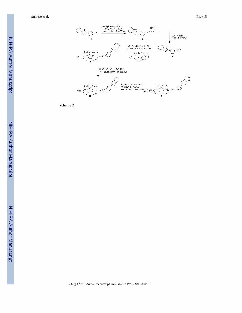
KOH, generated alkyne 8 (Scheme 2).24,25 Pd-catalyzed Sonogashira coupling between thealkyne 8 and key intermediate 3 yielded nitro derivative 9, which was reduced with hydrazineto produce amine 10 in high yield. Amine 10 was treated with iodobenzene under Ullmanconditions to generate the asymmetrical D–π-A III (Scheme 2). 19,22
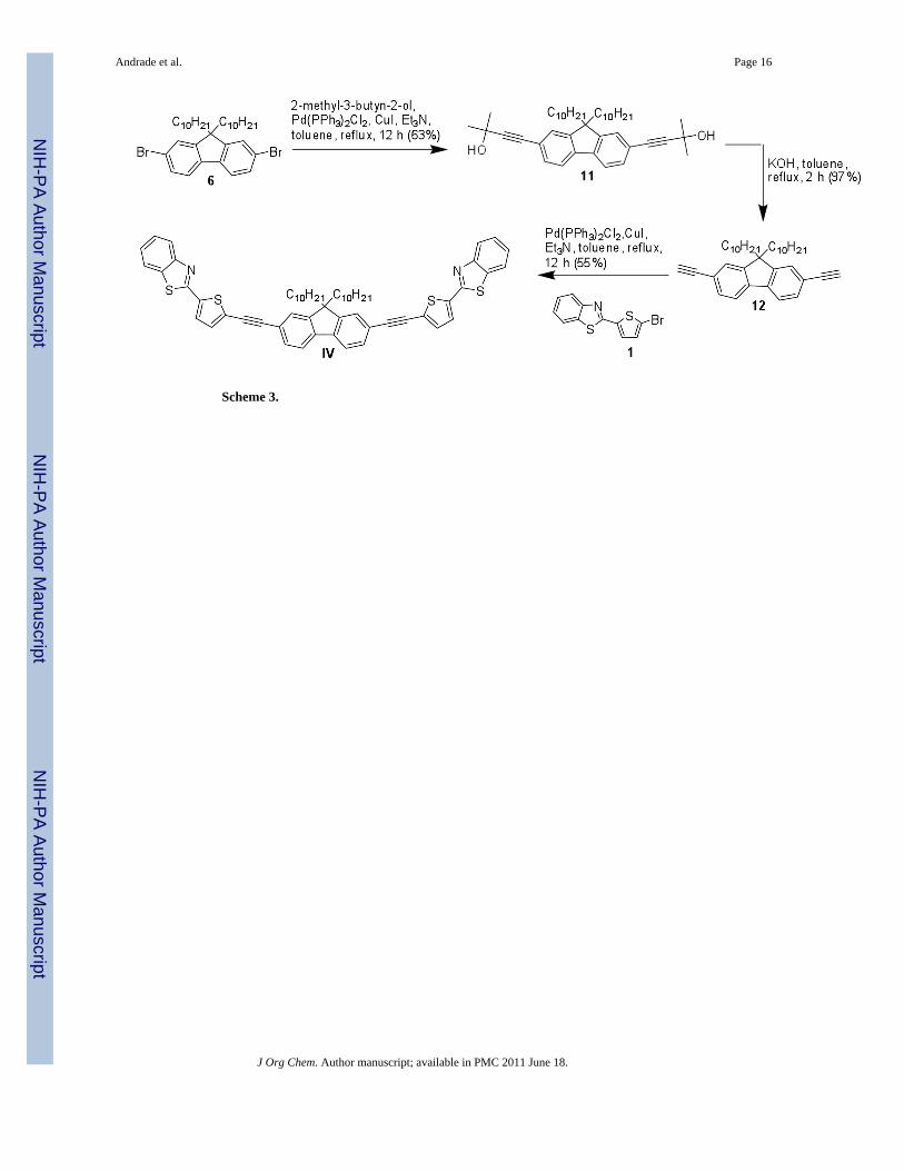
The symmetrical intermediate 11 was prepared using Sonogashira coupling between 9,9-didecyl-2,7-dibromofluorene 6 and 2-methylbut-3-yn-2-ol.24,25 After hydrolysis, the dialkyne12 was coupled through a second Sonogashira reaction with 1, yielding symmetrical A–π-Afluorenyl dye IV (Scheme 3).
Linear photophysical propertiesTable 1 summarizes the photophysical properties of the four new fluorescent dyes (I-IV). Ascan be seen in Figure 3, all molecules exhibited an intense absorption between 397-405 nm,fluorescent maximum in the range of 433-452 nm, and significant Stokes shift values in hexane(see Table 1). The new compounds also exhibited high fluorescence quantum yields(0.86-0.98), while a slight blue shift in the absorption maximum was observed when alkynylmoieties were incorporated in both symmetrical and asymmetrical derivatives.
Nonlinear photophysical propertiesTwo-photon absorption (2PA) values were determined by the fluorescent method, measuringthe two-photon exited fluorescence of the dyes in the range of 730-860 nm in hexane. The 2PAcross section measurements were performed with a tunable Ti:sapphire femtosecond lasersystem (220 fs pulse width, 76 MHz repetition rate) pumped by a 10 W diode laser (532 nm)as the excitation source and a spectrofluorimeter with PMT detectors (Figure S-1; supp data).
The 2PA spectra for this series of compounds are shown in Figure 4. Compound I exhibited amaximum 2PA cross section of ~248 GM at 730 nm which can be attributed to the tail of theband corresponding to the two-photon allowed S0 → S2 transition; a second maximum of ~217GM at 800 nm, assigned to S0 → S1 2PA forbidden transition, was also observed. CompoundII exhibits a maximum 2PA cross section of ~335 GM at 730 nm. Comparing these resultswith our previously reported data for similar molecules that did not contain the thiophenemoiety a significant increase in the 2PA cross section values was observed when the thiophenylmoiety is introduced in the fluorophore.9-14
To observe the effect of the presence of alkynyl triple bonds in the new derivatives,comparisons of compound I to compound III (D-π-A) and compound II to compound IV (A-π-A) were conducted. Compound III exhibited a maximum 2PA cross section of ~563 GM at730 nm while a second maximum of ~356 GM at 800 nm was observed. Comparing thesevalues to those obtained for compound I, it can be inferred that incorporating alkynyl triplebonds in these molecules results in an enhancement in the 2PA cross section for thisunsymmetrical compound.
The 2PA cross section value obtained for compound II (~335 GM at 730 nm) can be comparedwith the large value obtained for compound IV at the same wavelength (~1093 GM). Asignificant increase in the 2PA cross section was observed when the number of alkynyl triplebonds was doubled.
Since the number of electrons in these molecules is different, it is also useful to compare theratios of 2PA cross section (δ) over the number of π-electrons (Ne) in the chromophores(normalized cross section).18,26 When comparing I to III, it was observed that the incorporationof a triple bond in the π system resulted in a two-fold increase in the normalized cross sectionvalue (7 to 14 GM per π electron). Similarly, a three-fold increase of the normalized crosssection was observed when comparing II and IV (9 and 27 GM per π electron, respectively).
Andrade et al. Page 3
J Org Chem. Author manuscript; available in PMC 2011 June 18.
NIH
-PA Author Manuscript
NIH
-PA Author Manuscript
NIH
-PA Author Manuscript

In general, a considerable increase in the value of 2PA cross section was obtained whenthiophene moieties were introduced in both fluorophore archetypes (D-π-A and A-π-A) studiedin this series. In addition, extending the π-conjugation with alkynyl triple bond functionalitiesalso significantly increased the 2PA for this class of molecules.
Fluorescence ImagingIn order to evaluate the efficiency of these archetypes as fluorophores in 2PFM cell imaging,fluorene I was incubated in COS-7 and HCT 116 cell lines at different concentrations andincubation times. Probe I was chosen as model compound due to its relatively constant two-photon absorptivity across a useful NIR wavelength range (750-860 nm for the Ti:sapphirelaser, Figure 4) and because it exhibited better solubility in DMSO than the more symmetricalcompounds II and IV (important for cell incubation) Fluorescence micrographs indicated thedistinct localization the probe in well confined areas within the cytosol in both COS-7 andHCT 116 cell lines (Figures 5 and 6). Probe I was incubated with a commercially availablelysosomal selective dye (Lysotracker Red) and incubated for 10 min, 30 min, 50 min, 1 h 10min, 1 h 30 min and 1 h 50 min, after incubation the cells were fixed, mounted, and imaged.Early colocalization with Lysotracker dye suggests a similar uptake mechanism for both theLysotracker dye and probe I. However, longer incubation times (50 min and 1 h 50 min)indicates a difference in the progression of the probe from the endosomal vesicles to thelysosomes, where the Lysotracker dye reached the lysosomes more quickly than probe I (seeFigure 5). The slower progression of probe I may prove useful for the study of late endosomalprocesses.
The high contrast and good resolution observed in 2PFM images of HCT 116 cells, incubatedwith probe I, support use of the fluorenyl core as an integral part of the fluorescent probe'smolecular architecture. The excellent linear and nonlinear photophysical properties pointstowards their potential as efficient 2PA fluorophores. The 2PFM image, shown in Figure 6,where the vesicles (presumably late endosomes) are clearly visualized in both 2D and 3D (seealso supporting information), is a testament of the efficacy of the 2PA fluorophore. Thepotential of these dyes exceeds that of imaging the endosomes, as further functionalization ofthe dye can be performed to modulate the solubility properties and, more importantly, impartselectivity to other organelles or biomarkers of interest, aspects of current investigation. Thenew fluorenyl derivatives provide the basis for the development of fluorescent probes for2PFM.
ConclusionsEfficient synthetic routes were developed to prepare four new fluorescent compoundscontaining functionalities such as thiophene and alkynyl moieties on a fluorene core, affordingsystematic variation of molecular symmetry and extent of conjugation. Comprehensivecharacterization of the new compounds revealed excellent linear and nonlinear photophysicalproperties, such as high fluorescent quantum yields (0.86-0.98) and high two-photonabsorptivity (220-1060 GM). Incorporation of thiophene and alkynyl moieties resulted in anincrease in the 2PA cross section values. Probe I proved to have low cytotoxocity whenincubated with COS-7 and HCT 116 cells, exhibiting endosomal selectivity, as evidenced byone-photon fluorescence imaging and colocalization studies. The advantageous properties ofprobe I facilitated 2PFM endosomal imaging, with the 2PFM images providing much betterresolution, allowing the visualization of individual endosomes. Due to their linear andnonlinear photophysical properties, these new fluorenyl derivatives should find use in a numberof emerging photonics applications such as targeted two-photon fluorescence imaging and two-photon based optical data storage.
Andrade et al. Page 4
J Org Chem. Author manuscript; available in PMC 2011 June 18.
NIH
-PA Author Manuscript
NIH
-PA Author Manuscript
NIH
-PA Author Manuscript
Experimental sectionMaterials and Methods
2-(5-Bromothiophen-2-yl)benzothiazole 1, 9,9-didecyl-2-iodo-7-nitro-9Hfluorene 3, and 2,7-dibromo-9,9-didecyl-9H-fluorene 6 were prepared as described in the literature.19,27,28 Allreactions were carried out under N2. All other reagents and solvents were used as receivedfrom commercial suppliers. 1H and 13C NMR spectra were recorded in CDCl3 on a NMRspectrometer at 500 and 125 MHz, respectively. MALDI MS analyses were performed at theUniversity of Florida.
Synthesis of 2-(5-(tributylstannyl)thiophen-2-yl)benzothiazole (2)At −78 °C 2-(5-bromothiophen-2-yl)benzothiazole 1 (200 mg, 0.67 mmol) was dissolved indry THF (5 mL). A solution of n-BuLi in hexanes (0.27 mL, 2.5 M, 0.68 mmol) was addeddropwise into the reaction mixture. After stirring for 1 h, Sn(Bu)3Cl (360 mg, 1.10 mmol) wasadded and the mixture was allowed to reach room temperature, and stirred overnight. Themixture was added to water, extracted with Et2O twice, dried over Mg2SO4, and purified bycolumn chromatography using a mixture of hexanes:EtOAc (9:1) as an eluent to yield 240 mg(70%) of viscous oil. 1H NMR (500 MHz, CDCl3) δ 8.02(d, J= 8.2 Hz, 1H, Ph-H), 7.82 (d,J= 8.0 Hz, 1H, Ph-H), 7.74 (d, J= 3.5 Hz, 1H, Thy-H), 7.43-7.47 (m, 1H, Ph-H), 7.31-7.35(m, 1H, Ph-H), 7.18 (d, J= 3.5 Hz, 1H, Thy-H), 1.55-1.63 (m, 6H, CH2), 1.31-1.39 (m, 6H,CH2), 1.12-1.19 (m, 6H, CH2), 0.90 (t, J= 7.34 Hz, 9H, CH3). 13C NMR (125 MHz, CDCl3)δ 161.4, 153.9, 143.5, 142.4, 136.2, 134.7, 129.6, 126.2, 125.1, 122.9, 121.4, 28.9, 27.3, 13.7,11.0. Anal. Calcd for C23H33NS2Sn: C, 54.56; H, 6.57; N, 2.77. Found: C, 54.85; H, 6.67; N,2.86.
Synthesis of 2-(5-(9,9-didecyl-7-nitro-9H-fluoren-2-yl)thiophen-2-yl)benzothiazole (4)9,9-Didecyl-2-iodo-7-nitro-9H-fluorene 3 (200 mg, 0.32 mmol), 2-(5-(tributylstannyl)thiophen-2-yl)benzothiazole 2 (191 mg, 0.38 mmol), and Pd(PPh3)2Cl2 (6 mg, 0.008 mmol)were dissolved in toluene (4 mL). The mixture was heated under reflux for 5 h. The solventwas removed under reduced pressure and the crude was purified by column chromatographyusing a mixture of hexanes:EtOAc (9.5:0.5) as an eluent to yield 222 mg (98%) of yellow oilthat solidified upon standing (m.p. 73.2-74.9 °C). 1H NMR (500 MHz, CDCl3) δ 8.28 (dd,J= 8.3 Hz, J= 2.0 Hz, 1H, Ph-H), 8.22 (d, J= 2.0 Hz, 1H, Ph-H), 8.05 (d, J= 8.0 Hz, 1H, Ph-H), 7.88 (d, J= 7.8 Hz, 1H, Ph-H), 7.79-7.84 (m, 2H, Ph-H), 7.72-7.76 (m, 1H, Ph-H), 7.69(d, J= 1.4 Hz, 1H, Ph-H), 7.66 (d, J= 3.7 Hz, 1H, Thy-H), 7.48-7.53 (m, 1H, Ph-H), 7.46 (d,J= 3.7 Hz, 1H, Thy-H), 7.37-7.42 (m, 1H, Ph-H), 2.01-2.09 (m, 4H, CH2), 0.99-1.24 (m, 28H,CH2), 0.82 (t, J= 7.0 Hz, 6H, CH3), 0.54-0.69 (m, 4H, CH2). 13C NMR (125 MHz, CDCl3)δ 161.0, 153.7, 153.4, 152.2, 147.9, 147.2, 146.8, 139.0, 136.6, 134.7, 134.4, 129.5, 126.6,125.3, 124.40, 124.36, 123.5, 122.0, 121.9, 121.5, 120.4, 120.0, 118.3, 55.9, 40.1, 31.8, 29.8,29.49, 29.47, 29.24, 29.19, 23.8, 22.6, 14.1. Anal. Calcd for C44H54N2O2S2: C, 74.74; H, 7.70;N, 3.96. Found: C, 74.97; H, 7.85; N, 3.97.
Synthesis of 7-(5-(benzothiazol-2-yl)thiophen-2-yl)-9,9-didecyl-9H-fluoren-2-amine (5)2-(5-(9,9-Didecyl-7-nitro-9H-fluoren-2-yl)thiophen-2-yl)benzothiazole 4 (160 mg, 0.23mmol) and 10% Pd/C (16 mg) were dissolved in a mixture 1:1 of THF:EtOH (8 mL).NH2NH2.2H2O (136 mg, 2.7 mmol) was added to the mixture slowly at room temperature,and then heated to 70 °C for 20 h. The mixture was filtered though a silica plug withCH2Cl2, and, after removing the solvent under reduced pressure, the material was purified bycolumn chromatography using as a solvent a mixture of hexanes:EtOAc (9:1), to yield 130 mg(84%) of dark yellow oil that was used directly since the primary amine is prone tooxidation. 1H NMR (500 MHz, CDCl3) δ 8.04 (d, J= 8.0 Hz, 1H, Ph-H), 7.85 (d, J= 8.0 Hz,
Andrade et al. Page 5
J Org Chem. Author manuscript; available in PMC 2011 June 18.
NIH
-PA Author Manuscript
NIH
-PA Author Manuscript
NIH
-PA Author Manuscript
1H, Ph-H), 7.59-7.64 (m, 2H, Ph-H, Thy-H), 7.54-7.58 (m, 2H, Ph-H), 7.45-7.51 (m, 2H, Ph-H), 7.34-7.39 (m, 2H, Ph-H, Thy-H), 6.65-6.69 (m, 2H, Ph-H), 3.80 (s, 2H, NH2), 1.83-2.00(m, 4H, CH2), 0.99-1.28 (m, 28H, CH2), 0.83 (t, J= 7.0 Hz, 6H, CH3), 0.62-0.73 (m, 4H,CH2). 13C NMR (125 MHz, CDCl3) 161.4, 153.8, 153.1, 150.7, 149.6, 146.4, 142.4, 135.0,134.6, 131.6, 130.5, 129.5, 126.4, 125.1, 124.7, 123.0, 122.8, 121.4, 120.8, 120.1, 118.8, 114.1,109.7, 54.9, 40.6, 31.9, 30.1, 29.6, 29.5, 29.29, 29.28, 23.8, 22.7, 14.1.
Synthesis of 7-(5-(benzothiazol-2-yl)thiophen-2-yl)-9,9-didecyl-N,N-diphenyl-9H-fluoren-2-amine (I)
7-(5-(Benzothiazol-2-yl)thiophen-2-yl)-9,9-didecyl-9H-fluoren-2-amine 5 (130 mg, 0.19mmol), iodobenzene (157 mg, 0.77 mmol), Cu-bronze (61 mg, 0.96 mmol), 18-crown-6 (15mg, 0.058 mmol), and K2CO3 (212 mg, 1.54 mmol) were combined with 1,2-dichlorobenzene(3 mL). The mixture was heated to 180 °C for 48 h. The crude product was passed through asilica plug with CH2Cl2. The solvent was removed under reduced pressure and the crude waspurified by column chromatography on silica gel using as a solvent a mixture of hexanes:EtOAc(9:1) to yield 150 mg (94%) of yellow solid (m.p. 107.5-109.5 °C). 1H NMR (500 MHz,CDCl3) δ 8.04 (d, J= 8.0 Hz, 1H, Ph-H), 7.84 (d, J= 8.0 Hz, 1H, Ph-H), 7.58-7.65 (m, 4H, Ph-H, Thy-H), 7.56 (d, J= 8.0 Hz, 1H, Ph-H), 7.45-7.50 (m, 1H, Ph-H), 7.33-7.39 (m, 2H, Ph-H,Thy-H), 7.22-7.30 (m, 5H, Ph-H), 7.11-7.16 (m, 5H, Ph-H), 7.00-7.05 (m, 2H, Ph-H), 1.82-1.95(m, 4H, CH2), 1.01-1.23 (m, 28H, CH2), 0.85 (t, J= 7.0 Hz, 6H, CH3), 0.66-0.75 (m, 4H,CH2). 13C NMR (125 MHz, CDCl3) δ 161.3, 153.8, 152.5, 151.6, 149.2, 147.9, 147.5, 141.6,135.4, 134.6, 131.4, 129.6, 129.5, 129.24, 129.16, 125.2, 125.0, 124.0, 123.9, 123.4, 123.3,122.8, 122.6, 121.5, 121.4, 120.1, 105.0, 55.2, 40.2, 31.9, 30.0, 29.6, 29.6, 29.3, 23.9, 22.7,14.1. Anal. Calcd for C56H64N2S2: C, 81.11; H, 7.78; N, 3.38. Found: C, 80.81; H, 7.74; N,3.30.
Synthesis of 2,2′-(5,5′-(9,9-didecyl-9H-fluorene-2,7-diyl)bis(thiophene-5,2-diyl))dibenzothiazole (II)
2-(5-(Tributylstannyl)thiophen-2-yl)benzothiazole 2 (460 mg, 0.90 mmol), 2,7-dibromo-9,9-didecyl-9H-fluorene 6 (250 mg, 0.41 mmol), and Pd(PPh3)2Cl2 (7.2 mg, 0.01mmol) weredissolved in toluene. The mixture was heated under reflux for 24 h. The solvent was removedunder reduced pressure and the crude was purified by column chromatography using a mixtureof hexanes:EtOAc (9.9:0.1) to yield 126 mg (35%) of yellow oil that solidified upon standing(m.p. 125.2-126.5 °C). 1H NMR (500 MHz, CDCl3) δ 8.05 (d, J= 7.76 Hz, 2H, Ph-H), 7.87(d, J= 7.82 Hz, 2H, Ph-H), 7.74 (d, J= 7.63 Hz, 2H, Ph-H), 7.68-7.71 (m, 2H, Ph-H), 7.64-7.67(m, 4H, Ph-H, Thy-H), 7.47-7.52 (m, 2H, PhH), 7.43 (d, J= 4.10 Hz, 2H, Thy-H), 7.36-7.40(m, 2H, Ph-H), 2.00-2.06 (m, 4H, CH2), 1.04-1.23 (m, 28H, CH2), 0.80 (t, J= 7.04 Hz, 6H,CH3), 0.67-0.74 (m, 4H, CH2). 13C NMR (125 MHz, CDCl3) δ 161.2, 153.7, 152.1, 148.8,140.9, 135.8, 134.7, 132.6, 129.5, 126.5, 125.2, 125.0, 123.7, 122.9, 121.5, 120.5, 120.3, 55.4,40.4, 31.9, 30.0, 29.6, 29.5, 29.3, 29.2, 23.8, 22.6, 14.1. HRMS (MALDI) m/z calcd 877.3712[M+H]+, m/z found 877.3702 [M+H]+.
Synthesis of 4-(5-(benzothiazol-2-yl)thiophen-2-yl)-2-methylbut-3-yn-2-ol (7)2-(5-Bromothiophen-2-yl)benzothiazole 1 (200 mg, 0.67 mmol), 2-methyl-3-butyn-2-ol (170mg, 2.02 mmol), Pd(PPh3)2Cl2 (19 mg, 0.027 mmol), and CuI (5 mg, 0.027 mmol) weredissolved in a 1:4 mixture of Et3N:toluene (5 mL). The mixture was heated under reflux for12 h. After cooling to room temperature, the mixture was filtered through a celite plug, andpurified by column chromatography using a mixture of hexanes:EtOAc (1:1) to yield 192 mg(96%) of pale yellow solid (m.p. 163.9-164.6 °C). 1H NMR (500 MHz, CDCl3) 8.02 (d, J=8.04 Hz, 1H, Ph-H), 7.85 (d, J= 7.87 Hz, 1H, Ph-H), 7.50 (d, J= 3.95 Hz, 1H, Thy-H), 7.46-7.49(m, 1H, Ph-H), 7.35-7.40 (m, 1H, Ph-H), 7.18 (d, J= 3.95 Hz, 1H, Thy-H), 2.11 (s, 1H, -OH),
Andrade et al. Page 6
J Org Chem. Author manuscript; available in PMC 2011 June 18.
NIH
-PA Author Manuscript
NIH
-PA Author Manuscript
NIH
-PA Author Manuscript
1.64 (s, 6H, CH3 ). 13C NMR (125 MHz, CDCl3) δ160.4, 153.6, 137.9, 134.7, 132.9, 128.3,126.4, 125.4, 123.1, 121.5, 121.4, 100.2, 75.2, 65.8, 31.3. . Anal. Calcd for C16H13NOS2: C,64.18; H, 4.38; N, 4.68. Found: C, 64.10; H, 4.33; N, 4.64.
Synthesis of 2-(5-ethynylthiophen-2-yl)benzothiazole (8)4-(5-(Benzothiazol-2-yl)thiophen-2-yl)-2-methylbut-3-yn-2-ol 7 (300 mg, 1.0 mmol) andKOH (300 mg, 5.3 mmol) were heated under reflux in toluene for 1 h. The mixture was filtered,then purified by column chromatography eluting with hexanes to yield 180 mg (75%) of awhite solid (m.p. 117.3-118.3 °C). 1H NMR (500 MHz, CDCl3δ 8.03 (d, J= 8.21Hz, 1H, Ph-H), 7.86 (d, J= 8.21Hz, 1H, Ph-H), 7.51 (d, J= 3.94 Hz, 1H, Thy-H), 7.47-7.50 (m, 1H, Ph-H), 7.37-7.42 (m, 1H, Ph-H), 7.28 (d, J= 3.94 Hz, 1H, Thy-H), 3.50 (s, 1H, C≡C-H). 13C NMR(125 MHz, CDCl3) δ 160.2, 153.6, 138.5, 134.8, 133.9, 133.7, 127.8, 126.5, 125.4, 123.2,121.6, 84.0, 76.5. Anal. Calcd for C13H7NS2: C, 64.70; H, 2.92; N, 5.80. Found: C, 64.64; H,2.91; N, 5.75.
Synthesis of 2-(5-((9,9-didecyl-7-nitro-9H-fluoren-2-yl)ethynyl)thiophen-2-yl)benzothiazole(9)
2-(5-Ethynylthiophen-2-yl)benzothiazole 8 (180 mg, 0.75 mmol), 9,9-didecyl-2-iodo-7-nitro-9Hfluorene 3 (460 mg, 0.75 mmol), Pd(PPh3)2Cl2 (20 mg, 0.03 mmol), and CuI (5.6 mg,0.03 mmol),were dissolved in a 1:4 mixture of Et3N:toluene (5 mL). The mixture was heatedunder reflux for 12 h. After cooling to room temperature, the mixture was filtered through acelite plug and purified by column chromatography eluting with a mixture of hexanes:EtOAc(9:1), and then recrystallized from hexane to yield 300 mg (55%) of yellow solid (m.p.142.3-143.6 °C). 1H NMR (500 MHz, CDCl3) 8.28 (dd, J= 8.40 Hz, J= 2.18 Hz, 1H, Ph-H),8.21 (d, J= 2.05 Hz, 1H, Ph-H), 8.05 (d, J= 7.74 Hz, 1H, Ph-H), 7.88 (d, J= 7.74 Hz, 1H, Ph-H), 7.77-7.82 (m, 2H, Ph-H), 7.55-7.61 (m, 3H, Ph-H, Thy-H), 7.48-7.52 (m, 1H, Ph-H),7.39-7.42 (m, 1H, Ph-H), 7.33 (d, J= 4.08 Hz, 1H, Thy-H), 2.01-2.08 (m, 4H, CH2), 0.99-1.29(m, 28H, CH2), 0.84 (t, J= 6.93 Hz, 6H, CH3), 0.50-0.64 (m, 4H, CH2). 13C NMR (125 MHz,CDCl3) δ 160.3, 153.7, 152.5, 152.3, 147.4, 146.6, 139.3, 138.4, 134.8, 132.8, 131.2, 128.5,128.3, 126.7, 126.2, 126.0, 123.2, 123.1, 121.6, 121.5, 120.2, 118.3, 105.0, 96.3, 83.8, 55.9,40.0, 31.9, 29.9, 29.51, 29.48, 29.2, 23.8, 22.6, 14.14, 14.06. Anal. Calcd forC46H54N2O2S2: C, 75.57; H, 7.45; N, 3.83. Found: C, 75.69; H, 7.47; N, 3.85.
Synthesis of 7-((5-(benzothiazol-2-yl)thiophen-2-yl)ethynyl)-9,9-didecyl-9H-fluoren-2-amine(10)
2-(5-((9,9-Didecyl-7-nitro-9H-fluoren-2-yl)ethynyl)thiophen-2-yl)benzothiazole 9 (180 mg,0.24 mmol) and 10% Pd/C (18 mg) were dissolved in a 1:1 mixture of THF:EtOH (4 mL).NH2NH2.2H2O (146 mg, 2.8 mmol) was added to the mixture dropwise at room temperature,followed by heating to 110 °C for 48 h. The mixture was filtered though a silica plug withCH2Cl2, and, after removing the solvent under reduced pressure, the material was purified bycolumn chromatography using a mixture of hexanes:EtOAc (9:1) as eluent to yield 150 mg(87%) of dark yellow oil that was used immediately due to the oxidative lability of the primaryamine. 1H NMR (500 MHz, CDCl3) δ 8.03 (d, J= 7.45 Hz, 1H, Ph-H), 7.86 (d, J= 7.67 Hz,1H, Ph-H), 7.56 (d, J= 3.99 Hz, 1H, Thy-H), 7.52 (d, J= 7.75 Hz, 1H, PhH), 7.46-7.50 (m,3H, Ph-H), 7.43-7.44 (m, 1H, Ph-H), 7.36-7.40 (m, 1H, Ph-H), 7.27 (d, J= 3.99 Hz, 1H, Thy-H), 6.65-6.68 (m, 2H, Ph-H), 3.83 (s, 2H, NH2), 1.84-1.97 (m, 4H, CH2), 1.03-1.27 (m, 28H,CH2), 0.85 (t, J= 7.16 Hz, 6H, CH3), 0.56-0.68 (m, 4H, CH2). 13C NMR (125 MHz, CDCl3)δ 161.0, 160.6, 153.8, 153.1, 150.2, 149.9, 146.6, 146.0, 144.4, 142.7, 141.6, 136.1, 134.6,133.7, 132.7, 132.2, 131.5, 128.4, 122.9, 121.6, 118.5, 114.0, 109.8, 97.7, 81.9, 54.9, 40.5,31.9, 30.2, 29.7, 29.6, 29.4, 29.3, 23.9, 22.7, 14.2.
Andrade et al. Page 7
J Org Chem. Author manuscript; available in PMC 2011 June 18.
NIH
-PA Author Manuscript
NIH
-PA Author Manuscript
NIH
-PA Author Manuscript

Synthesis of 7-((5-(benzothiazol-2-yl)thiophen-2-yl)ethynyl)-9,9-didecyl-N,N-diphenyl-9Hfluoren-2-amine (III)
7-((5-(Benzothiazol-2-yl)thiophen-2-yl)ethynyl)-9,9-didecyl-9H-fluoren-2-amine 10, (0.135mg, 0.19 mmol), iodobenzene (157 mg, 0.77 mmol), Cu-bronze (61 mg, 0.96 mmol), 18-crown-6 (15 mg, 0.058 mmol), and K2CO3 (212 mg, 1.54 mmol) were combined with, 1,2-dichlorobenzene (3 mL). The mixture was heated at 180 °C for 48 h. The crude product waspassed through a silica plug with CH2Cl2. The solvent was removed under reduced pressure,and the crude product was purified by column chromatography on silica gel using a mixtureof hexanes:EtOAc (9.5:0.5) as eluent to yield 81 mg (50%) of yellow viscous oil. 1H NMR(500 MHz, CDCl3) δ 8.04 (d, J= 8.11 Hz, 1H, Ph-H), 7.85 (d, J= 8.11 Hz, 1H, Ph-H), 7.59 (d,J= 7.90 Hz, 1H, Ph-H), 7.56 (d, , J= 3.72 Hz, 1H, Thy-H), 7.45-7.52 (m, 4H, Ph-H), 7.35-7.39(m, 1H, Ph-H), 7.24-7.30 (m, 5H, Ph-H), 7.10-7.15 (m, 5H, Phe-H), 7.00-7.05 (m, 3H, Phe-H), 1.80-1.92 (m, 4H, CH2), 1.03-1.28 (m, 28H, CH2), 0.85 (t, J= 7.14 Hz, 6H, CH3), 0.62-0.69(m, 4H, CH2). 13C NMR (125 MHz, CDCl3) δ 160.5, 153.7, 152.6, 150.8, 147.8, 147.8, 141.9,137.7, 135.2, 134.7, 132.4, 130.8, 129.2, 128.4, 127.4, 126.6, 125.7, 125.4, 124.0, 123.3, 123.01122.7, 121.5, 120.9, 119.6, 119.1, 118.9, 97.4, 82.2, 55.2, 40.2, 31.9, 30.0, 29.6, 29.6, 29.4,29.3, 23.9, 22.7, 14.1. Anal. Calcd for C58H64N2S2: C, 81.64; H, 7.56; N, 3.28. Found: C,81.63; H, 7.62; N, 3.05.
Synthesis of 4,4′-(9,9-didecyl-9H-fluorene-2,7-diyl)bis(2-methylbut-3-yn-2-ol) (11)2,7-Dibromo-9,9-didecyl-9H-fluorene 6 (1.0 g, 1.65 mmol), 2-methyl-3-butyn-2-ol (0.83 g,9.92 mmol), Pd(PPh3)2Cl2 (95 mg, 0.135 mmol), and CuI (25 mg, 0.13 mmol) were dissolvedin a 1:4 mixture of Et3N:toluene (15 mL). The mixture was heated under reflux for 12 h. Afterit was cooled down, the mixture was filtered through a celite plug and purified by columnchromatography using hexanes as a eluent, followed by a mixture of hexanes:EtOAc (10:1) toyield 0.64 g (63%) of white solid (m.p. 89.0-89.9 °C). 1H NMR (500 MHz, CDCl3) δ 7.58-7.61(m, 2H, Ph-H), 7.35-7.41 (m, 4H, Ph-H), 2.05 (s, 2H, -OH), 1.89-1.96 (m, 4H, CH2), 1.63-1.68(s, 12H, CH3), 0.98-1.27 (m, 28H, CH2), 0.85 (t, J= 6.49 Hz, 6H, CH3), 0.50-0.58 (m, 4H,CH2). 13C NMR (125 MHz, CDCl3) δ 150.9, 140.6, 130.7, 126.0, 121.4, 119.8, 93.9, 83.0,65.7, 55.2, 40.4, 31.9, 31.6, 30.0, 29.6, 29.5, 29.3, 29.2, 23.7, 22.6, 14.1. Anal. Calcd forC43H62O2: C, 84.53; H, 10.23. Found: C, 84.46; H, 10.25.
Synthesis of 9,9-didecyl-2,7-diethynyl-9H-fluorene (12)4,4′-(9,9-Didecyl-9H-fluorene-2,7-diyl)bis(2-methylbut-3-yn-2-ol) 11 (450 mg, 0.736 mmol)and KOH (206 mg, 3.68 mmol) were heated under reflux in toluene for 2 h. The mixture wasfiltered, then purified by column chromatography using hexanes to yield 350 mg (97%) of lightyellow oil. 1H NMR (500 MHz, CDCl3) δ 7.63 (dd, J= 7.73 Hz, J= 0.65 Hz, 2H, Ph-H), 7.48(dd, J= 7.73 Hz, J= 1.42 Hz, 2H, Ph-H), 7.45-7.46 (m, 2H, Ph-H), 3.15 (s, 2H, C≡C-H),1.90-1.96 (m, 4H, CH2), 0.99-1.25 (m, 28H, CH2), 0.85 (t, J= 7.0 Hz, 6H, CH3), 0.52-0.58(m, 4H, CH2). 13C NMR (125 MHz, CDCl3) δ 151.0, 141.0, 131.2, 126.5, 120.8, 119.95, 84.5,77.3, 55.2, 40.2, 31.9, 29.9, 29.5, 29.5, 29.2, 23.7, 22.7, 14.1. Anal. Calcd for C37H50: C, 89.81;H, 10.19. Found: C, 89.79; H, 10.24.
Synthesis of 2,2′-(5,5′-(9,9-didecyl-9H-fluorene-2,7-diyl)bis(ethyne-2,1-diyl)bis(thiophene-5,2-diyl))dibenzothiazole (IV)
9,9-Didecyl-2,7-diethynyl-9H-fluorene 12 (300 mg, 0.61 mmol), 2-(5-bromothiophen-2-yl)benzothiazole 1 (377 mg, 1.27 mmol), Pd(PPh3)2Cl2 (34 mg, 0.05 mmol), and CuI (10 mg,0.05 mmol) were dissolved in a 1:4 mixture of Et3N:toluene (5 mL). The mixture was heatedunder reflux for 12 h. After it was cooled down, the mixture was filtered through a celite plug,and purified by column chromatography using a mixture of hexanes:EtOAc (10:1) as eluentto yield 310 mg (55%) of yellow solid (m.p. 74.0-75.5 °C). 1H NMR (500 MHz, CDCl3) δ
Andrade et al. Page 8
J Org Chem. Author manuscript; available in PMC 2011 June 18.
NIH
-PA Author Manuscript
NIH
-PA Author Manuscript
NIH
-PA Author Manuscript

8.04 (dd, J= 8.09 Hz, J= 0.54 Hz, 2H, Ph-H), 7.87 (dd, J= 8.04 Hz, J= 0.53 Hz, 2H, Ph-H),7.70 (d, J= 7.83 Hz, 2H, Ph-H), 7.57 (d, J= 3.89 Hz, 2H, Thy-H), 7.54-7.56 (m, 2H, Ph-H),7.52-7.53 (m, 2H, Ph-H), 7.48-7.51 (m, 2H, Ph-H), 7.37-7.41 (m, 2H, Ph-H), 7.31 (d, J= 3.89Hz, 2H, Thy-H), 1.97-2.04 (m, 4H, CH2), 1.04-1.23 (m, 28H, CH2), 0.84 (t, J= 6.96 Hz, 6H,CH3), 0.58-0.65 (m, 4H, CH2). 13C NMR (125 MHz, CDCl3) δ 160.5, 153.7, 151.3, 141.1,138.0, 134.7, 132.6, 130.8, 128.4, 127.1, 126.6, 125.9, 125.5, 123.1, 121.5, 121.3, 120.2, 97.0,82.9, 55.4, 40.3, 31.9, 30.0, 29.6, 29.5, 29.3, 29.3, 23.8, 22.7, 14.1. Anal. Calcd forC59H60N2S4: C, 76.58; H, 6.54; N, 3.03. Found: C, 76.79; H, 6.75; N, 2.79.
MeasurementsAbsorption spectra were recorded with an UV–visible spectrophotometer. Steady-statefluorescence spectra were measured with a spectrofluorimeter in the photon counting regimeof the PMT using an L-format configuration. The fluorescence spectra were corrected for thespectral dependence of the PMT. All measurements were performed in hexane at roomtemperature in 1 cm quartz cuvettes, with dye concentrations on the order of 10−6 M.Fluorescence quantum yields were determined relative to 9, 10-diphenylanthracene incyclohexane as a standard.29
Two-photon absorption (2PA) cross sections of the final compounds were determined by thetwo-photon induced fluorescence method.30 In this method, the sample is excitedsimultaneously by two photons of low energy, resulting in fluorescence emission. The numberof photons emitted can be measured by a PMT, and the latter related to the numeric value ofthe 2PA cross section relative to a reference.
Cell culture and incubationCOS-7 and HCT 116 cell were cultured in DMEM, supplemented with 10% FBS, and 1%penicillin, and 1% streptomycin at 37 °C, under 5% CO2 environment. N° 1 round 12 mmcoverslips were treated with poly-D-lysine to improve cell adhesion, and washed (3x) withPBS buffer solution. The treated cover slips were placed in 24-well plates, seeded with 80,000cells/well, and incubated at the same conditions as indicated above until 75-85% confluencywas reached on the coverslips. From a 4.10 × 10⁻⁴ M stock solution of dye I a series of 0.1, 1,10, 20, and 50 μM solutions in DMSO were prepared with all solutions also containing 75 nMof Lysotracker Red. These solutions were used to incubate the cells for 10 min to 3h. The dyesolutions were extracted and the coverslipped cells were washed abundantly with PBS (4x).
Cell fixing and mountingCells were fixed with 3.7% solution of paraformaldehyde in pH=7.4 PBS buffer for 10 min.The fixing agent was extracted and washed (2x) with PBS. To reduce autofluorescence, a freshsolution of NaBH4 (1 mg/mL) in pH=8 PBS buffer was used to treat the fixed cells (2x). Thecoverslipped cells were then washed with buffer PBS (2x) and mounted on microscope slidesusing an antifade mounting media.
Confocal one-photon fluorescence imagingOne-photon (conventional) fluorescence microscopy images were recorded on an invertedconfocal microscope equipped with a EM-CCD digital camera. One-photon confocalfluorescence images of the fixed cells were taken using a custom made filter cube (Ex:377/50;DM: 409; Em:525/40) and a Texas Red filter cube (Ex:562/40; DM: 593; Em:624/40) for probeI and Lysotracker Red, respectively.
Andrade et al. Page 9
J Org Chem. Author manuscript; available in PMC 2011 June 18.
NIH
-PA Author Manuscript
NIH
-PA Author Manuscript
NIH
-PA Author Manuscript
Two-photon upconverted fluorescence imagingTwo-photon fluorescence microscopy (2PFM) imaging was performed on a modified laserscanning confocal microscopy system equipped with a broadband, tunable Ti:sapphire laser(220 fs pulse width, 76 MHz repetition rate), pumped by a 10 W frequency doubled Nd:YAGlaser. The Ti:sapphire laser, tuned 700 nm and modelocked, was used as the two-photonexcitation source. Two-photon induced fluorescence was collected by a 60x microscopeobjective (UPLANSAPO 60x, N.A.=1.35). A high transmittance (>95%) short-pass filter(cutoff 685 nm) was placed in front of the PMT detector within the scanhead in order to filteroff background radiation from the laser source (700 nm).
Supplementary MaterialRefer to Web version on PubMed Central for supplementary material.
AcknowledgmentsThe authors wish to acknowledge the National Science Foundation (CHE-0832622 and CHE-0840431) and theNational Institutes of Health (1 R15 EB008858-01) for support of this work.
References1. Denk W, Strickler JH, Webb WW. Science 1990;248:73–76. [PubMed: 2321027]2. Parthenopoulos DA, Rentzepis PM. Science 1989;245:843–845. [PubMed: 17773360]3. Belfield KD, Schafer KJ. Chem. Mater 2002;14:3656–3662.4. Yanez CO, Andrade CD, Yao S, Luchita G, Bondar MV, Belfield KD. ACS Appl. Mater. Interfaces
2009;1:2219–2229. [PubMed: 20355856]5. Belfield KD, Schafer KJ, Liu YU, Liu J, Ren XB, Van Stryland EW. J. Phys. Org. Chem 2000;13:837–
849.6. Maruo S, Nakamura O, Kawata S. Opt. Lett 1997;22:132–134. [PubMed: 18183126]7. Belfield KD, Ren X, Hagan DJ, Van Stryland EW, Dubikovsky V, Miesak EJ. J. Am. Chem. Soc
2000;122:1217–1218.8. Schafer-Hales KJ, Belfield KD, Yao S, Frederiksen PK, Hales JM, Kolattukudy PE. J. Biomed. Opt
2005;10:015402-1–8.9. Belfield KD, Bondar MV, Morales AR, Yavuz O, Przhonska OV. J. Phys. Org. Chem 2003;16:194–
201.10. Belfield KD, Morales AR, Hales JM, Hagan DJ, Van Stryland EW, Chapela VM, Percino J. Chem.
Mater 2004;16:2267–2273.11. Belfield KD, Morales AR, Kang BS, Hales JM, Hagan DJ, Van Stryland EW, Chapela VM, Percino
J. Chem. Mater 2004;16:4634–4641.12. Yao S, Belfield KD. J. Org. Chem 2005;70:5126–5132. [PubMed: 15960514]13. Belfield KD, Yao S, Bondar MV. Adv. Polym. Sci 2008;213:97–156.14. Yanez CO, Andrade CD, Belfield KD. Chem. Commun 2009:827–829.15. Belfield KD, Bondar MV, Yanez CO, Hernandez FE, Przhonska OV. J. Mater. Chem 2009;19:7498–
7502.16. Moura GLC, Simas AM. J. Phys. Chem. C 2010;114:6106–6116.17. Reinhardt BA, Brott LL, Clarson SJ, Dillard AG, Bhatt JC, Kannan R, Yuan LX, He GS, Prasad PN.
Chem. Mater 1998;10:1863–1874.18. Mongin O, Porres L, Charlot M, Katan C, Blanchard-Desce M. Chem. Eur. J 2007;13:1481–1498.19. Belfield KD, Schafer KJ, Mourad W, Reinhardt BA. J. Org. Chem 2000;65:4475–4481. [PubMed:
10959847]20. Kannan R, He GS, Yuan LX, Xu FM, Prasad PN, Dombroskie AG, Reinhardt BA, Baur JW, Vaia
RA, Tan LS. Chem. Mater 2001;13:1896–1904.
Andrade et al. Page 10
J Org Chem. Author manuscript; available in PMC 2011 June 18.
NIH
-PA Author Manuscript
NIH
-PA Author Manuscript
NIH
-PA Author Manuscript

21. Kosugi M, Koshiba M, Atoh A, Sano H, Migita T. Bull. Chem. Soc. Jpn 1986;59:677–679.22. Yang CP, Lin JH. J. Polym. Sci., Part A-Polym. Chem 1994;32:369–382.23. Sonogashira K, Tohda Y, Hagihara N. Tetrahedron Lett 1975:4467–4470.24. Umezawa H, Okada S, Oikawa H, Matsuda H, Nakanishi H. J. Phys. Org. Chem 2005;18:468–472.25. Mongin O, Krishna TR, Werts MHV, Caminade AM, Majoral JP, Blanchard-Desce M. Chem.
Commun 2006:915–917.26. Pawlicki M, Collins HA, Denning RG, Anderson HL. Angew. Chem. Int. Edit 2009;48:3244–3266.27. Zeng DX, Chen Y. J. Photochem. Photobiol. A 2007;186:121–124.28. Belfield KD, Yao S, Morales AR, Hales JM, Hagan DJ, Van Stryland EW, Chapela VM, Percino J.
Polym. Adv. Technol 2005;16:150–155.29. Belfield KD, Bondar MV, Przhonska OV, Schafer KJ. J. Fluores 2002;12:445–450.30. Belfield KD, Bondar MV, Yanez CO, Hernandez FE, Przhonska OV. J. Phys. Chem. B
2009;113:7101–7106. [PubMed: 19388691]
Andrade et al. Page 11
J Org Chem. Author manuscript; available in PMC 2011 June 18.
NIH
-PA Author Manuscript
NIH
-PA Author Manuscript
NIH
-PA Author Manuscript
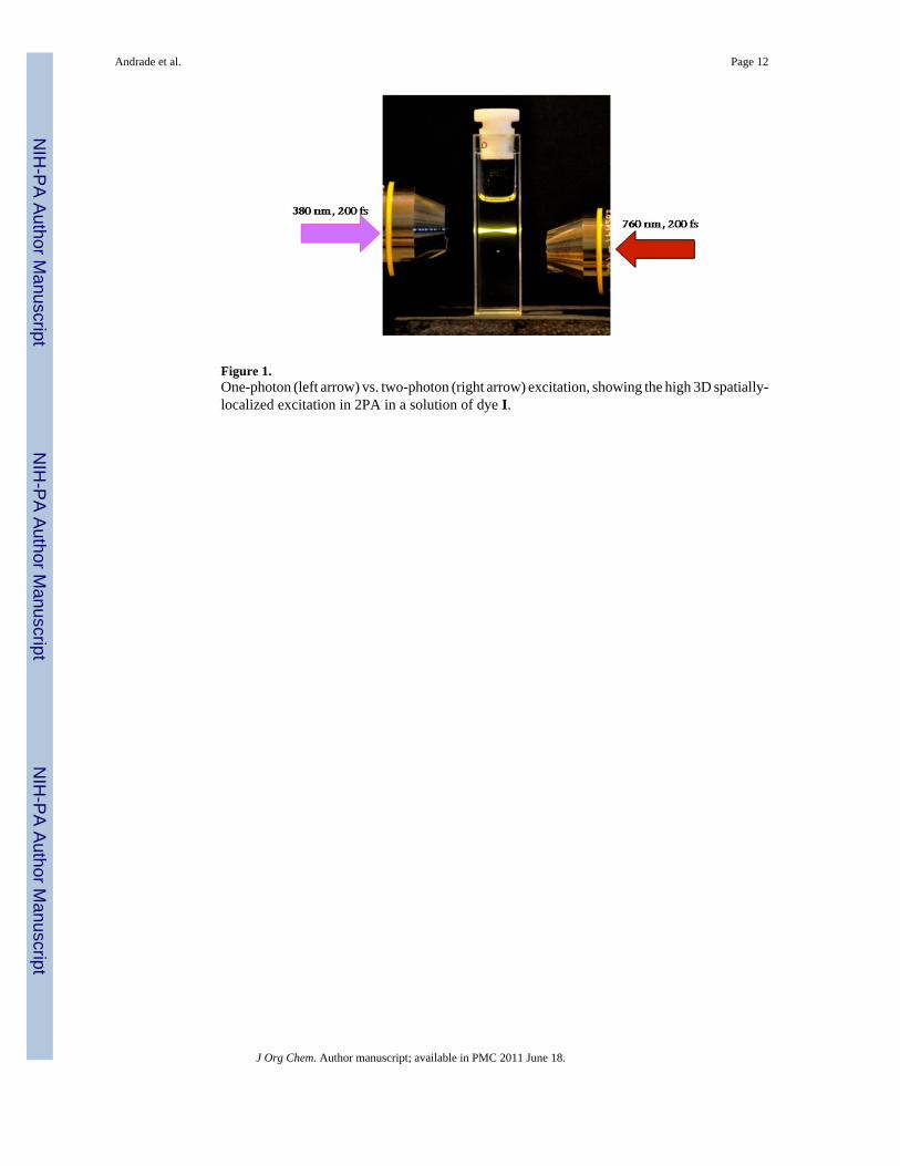
Figure 1.One-photon (left arrow) vs. two-photon (right arrow) excitation, showing the high 3D spatially-localized excitation in 2PA in a solution of dye I.
Andrade et al. Page 12
J Org Chem. Author manuscript; available in PMC 2011 June 18.
NIH
-PA Author Manuscript
NIH
-PA Author Manuscript
NIH
-PA Author Manuscript

Figure 2.Structures of new donor-pi-acceptor (D-π-A) (I and III) and acceptor-pi-acceptor (A-π-A)(II and IV) fluorenyl derivatives. Benzothiazole moieties were used as acceptor groups whilediphenylamino groups were used as donor moieties. Thiophene groups and alkynes wereincorporated to extend the π-conjugation.
Andrade et al. Page 13
J Org Chem. Author manuscript; available in PMC 2011 June 18.
NIH
-PA Author Manuscript
NIH
-PA Author Manuscript
NIH
-PA Author Manuscript

Scheme 1.
Andrade et al. Page 14
J Org Chem. Author manuscript; available in PMC 2011 June 18.
NIH
-PA Author Manuscript
NIH
-PA Author Manuscript
NIH
-PA Author Manuscript
Scheme 2.
Andrade et al. Page 15
J Org Chem. Author manuscript; available in PMC 2011 June 18.
NIH
-PA Author Manuscript
NIH
-PA Author Manuscript
NIH
-PA Author Manuscript
Scheme 3.
Andrade et al. Page 16
J Org Chem. Author manuscript; available in PMC 2011 June 18.
NIH
-PA Author Manuscript
NIH
-PA Author Manuscript
NIH
-PA Author Manuscript

Figure 3.Normalized absorption (left) and fluorescence (right) spectra for I, II, III, and IV in hexane.Absorption and emission spectra were collected at room temperature using hexanes solutionswith optical density of ~0.1 (approximately 10−6 M) without degassing the solutions. Emissionwas collectad by exciting samples at their λabs
max value.
Andrade et al. Page 17
J Org Chem. Author manuscript; available in PMC 2011 June 18.
NIH
-PA Author Manuscript
NIH
-PA Author Manuscript
NIH
-PA Author Manuscript
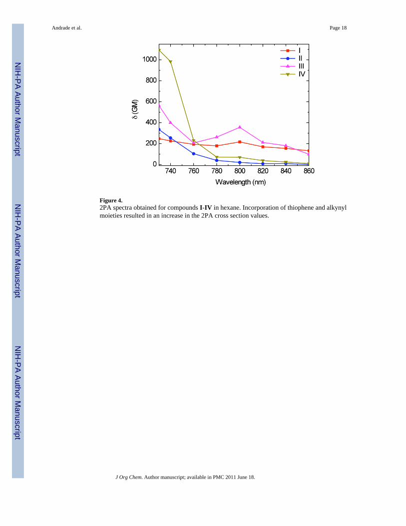
Figure 4.2PA spectra obtained for compounds I-IV in hexane. Incorporation of thiophene and alkynylmoieties resulted in an increase in the 2PA cross section values.
Andrade et al. Page 18
J Org Chem. Author manuscript; available in PMC 2011 June 18.
NIH
-PA Author Manuscript
NIH
-PA Author Manuscript
NIH
-PA Author Manuscript
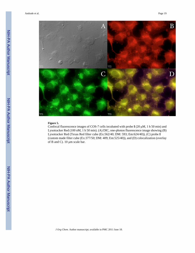
Figure 5.Confocal fluorescence images of COS-7 cells incubated with probe I (20 μM, 1 h 50 min) andLysotracker Red (100 nM, 1 h 50 min). (A) DIC, one-photon fluorescence image showing (B)Lysotracker Red (Texas Red filter cube (Ex:562/40; DM: 593; Em:624/40)), (C) probe I(custom made filter cube (Ex:377/50; DM: 409; Em:525/40)), and (D) colocalization (overlayof B and C). 10 μm scale bar.
Andrade et al. Page 19
J Org Chem. Author manuscript; available in PMC 2011 June 18.
NIH
-PA Author Manuscript
NIH
-PA Author Manuscript
NIH
-PA Author Manuscript
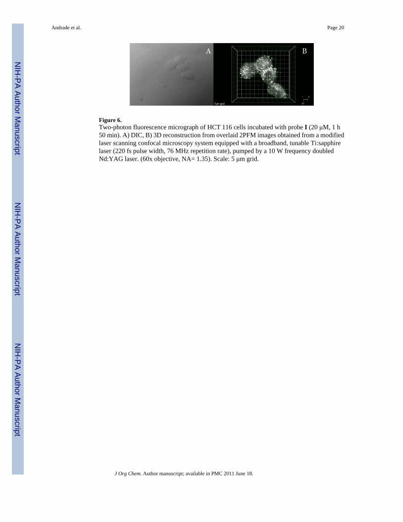
Figure 6.Two-photon fluorescence micrograph of HCT 116 cells incubated with probe I (20 μM, 1 h50 min). A) DIC, B) 3D reconstruction from overlaid 2PFM images obtained from a modifiedlaser scanning confocal microscopy system equipped with a broadband, tunable Ti:sapphirelaser (220 fs pulse width, 76 MHz repetition rate), pumped by a 10 W frequency doubledNd:YAG laser. (60x objective, NA= 1.35). Scale: 5 μm grid.
Andrade et al. Page 20
J Org Chem. Author manuscript; available in PMC 2011 June 18.
NIH
-PA Author Manuscript
NIH
-PA Author Manuscript
NIH
-PA Author Manuscript

NIH
-PA Author Manuscript
NIH
-PA Author Manuscript
NIH
-PA Author Manuscript
Andrade et al. Page 21
Tabl
e 1
Phot
ophy
sica
l cha
ract
eriz
atio
n of
new
fluo
rene
der
ivat
ives
I, II
, III
, and
IV in
hex
ane.
Com
poun
dλa
bsm
ax, n
mλe
mm
ax, n
mΔλ
St, n
mϕ
1εm
ax.1
0−3 ,
M−1
. cm−1
I40
3±1
452±
149
±20.
86±0
.05
53
II40
5±1
446±
141
±20.
98±0
.05
31
III
398±
144
4±1
46±2
0.92
±0.0
550
IV39
7±1
433±
136
±20.
95±0
.05
135
1 Fluo
resc
ence
qua
ntum
yie
ld m
easu
red
rela
tive
to 9
,10-
diph
enyl
anth
race
ne in
cyc
lohe
xane
.
J Org Chem. Author manuscript; available in PMC 2011 June 18.