Matls licensing package for license 05-29035-01 for Univ Corp for ...

Upconverting nanophosphors for bioimaging
This article has been downloaded from IOPscience. Please scroll down to see the full text article.
2009 Nanotechnology 20 405701
(http://iopscience.iop.org/0957-4484/20/40/405701)
Download details:
IP Address: 128.112.84.33
The article was downloaded on 30/04/2010 at 13:48
Please note that terms and conditions apply.
View the table of contents for this issue, or go to the journal homepage for more
Home Search Collections Journals About Contact us My IOPscience
IOP PUBLISHING NANOTECHNOLOGY
Nanotechnology 20 (2009) 405701 (6pp) doi:10.1088/0957-4484/20/40/405701
Upconverting nanophosphors forbioimagingShuang Fang Lim1,2,3, Robert Riehn2,3, Chih-kuan Tung3,William S Ryu4, Rui Zhuo1, Joanna Dalland3 and Robert H Austin3
1 Department of MAE, Princeton University, Princeton, NJ 08544, USA2 Department of Physics, North Carolina State University, Raleigh, NC 27695, USA3 Department of Physics, Princeton University, Princeton, NJ 08544, USA4 Lewis-Sigler Institute for Integrative Genomics, Princeton University, Princeton,NJ 08544, USA
Received 10 May 2009, in final form 11 August 2009Published 8 September 2009Online at stacks.iop.org/Nano/20/405701
AbstractUpconverting nanoparticles (UCNPs) when excited in the near-infrared (NIR) region displayanti-Stokes emission whereby the emitted photon is higher in energy than the excitation energy.The material system achieves that by converting two or more infrared photons into visiblephotons. The use of the infrared confers benefits to bioimaging because of its deeperpenetrating power in biological tissues and the lack of autofluorescence. We demonstrate heresub-10 nm, upconverting rare earth oxide UCNPs synthesized by a combustion method that canbe stably suspended in water when amine modified. The amine modified UCNPs show specificsurface immobilization onto patterned gold surfaces. Finally, the low toxicity of the UCNPs isverified by testing on the multi-cellular C. elegans nematode.
S Supplementary data are available from stacks.iop.org/Nano/20/405701
1. Introduction
Upconverting nanophosphors (UCNPs) consist of rare earthatoms that are embedded in a crystalline host matrix. The rareearths absorb two or more infrared photons and upconvert toemit in the visible spectrum, whereby the emitted photon ishigher in energy than the excitation energy. The upconversionmechanism can be described as either sequential excitation ofthe same atom or excitation of two centers and subsequentenergy transfer [1–4]. The emission of UCNPs consistsof sharp lines characteristic of atomic transitions in a well-ordered matrix. UCNPs have been shown in numerousstudies to be good candidates for bioimaging and havedemonstrated applications such as reporters [2, 5–10], bio-labels [3, 11, 12], emissive displays [13], in solar cells [14]and even microbarcodes [15]. There are several advantagesin using UCNPs for bioimaging, in that they are non-toxic [16, 17] and do not bleach like organic dyes andfluorescent proteins. That is because UCNPs emit from the4f shells of the rare earths, which are efficiently shieldedfrom their chemical environment by the outerlying 5s and 5pelectrons. As a result, the emission lines are quite sharp,10 nm or less in width. Because of the large number of narrowemission lines, much more information can be conveyed thanby the broad and featureless spectra obtained from quantum
dots or organic dyes. This enables multiplex detection froma single sample under a single excitation [4]. Further, bychanging the ratios of the rare earth dopants and combiningvarious rare earth dopants, the spectral lines can be varied fromred through blue, the blue lines being three-photon transitions.Since IR excitation followed by upconversion only occurs inthe UCNPs, autofluorescence is absent [18]. The multi-photondependence of the upconversion luminescence at relativelylow power levels in the range of W mm−2 also ensures ahigher signal to contrast ratio. Solution-stable, unaggregated,bright upconverting, oxide based phosphors have previouslybeen demonstrated for submicron sized yttrium oxide [3, 8, 9]and for NaYF4 nanoparticles [17, 19]. Recently, sub-cellularbioimaging was demonstrated with 40 nm bioconjugatedyttrium oxide nanoparticles [20, 21] and 50 nm NaYF4
nanoparticles [22]. The solubilization and functionalization ofthe ultrasmall UCNPs is typically more challenging due to theirlarge surface area-to-volume ratio and the large surface energythey express.
In this report, we show synthesis and characterizationof Y2O3: Yb, Er which are not only of sub-10 nm scalein particle size, but also low aggregating and upconverting.The subsequent part of the study describes our efforts tointroduce amine groups onto the nanoparticle surface with a
0957-4484/09/405701+06$30.00 © 2009 IOP Publishing Ltd Printed in the UK1
Nanotechnology 20 (2009) 405701 S F Lim et al
demonstration of specific binding. Finally, we demonstrate thelow toxicity of the UCNPs in the nematode worm C. elegans.
2. Experimental details
The yttrium oxide based UCNPs were synthesized using acustom-built flame combustion apparatus, which is based ona custom-built 10 cm long alumina Laval tube [24]. Thedesign of the apparatus was chosen such that nanoparticleswere cooled very rapidly immediately after oxide formation inthe hot zone of the Laval tube. The temperature of combustionas measured by a K type thermocouple was roughly 1200 ◦Cat a distance of one inch downstream from the apex of theLaval tube. The collection end of the Laval tube was fittedwith a copper water-cooled expansion nozzle in order to allowemerging gases to expand and thus cool the nanoparticlesrapidly.
As the precursor for the sub-10 nm nanophosphors, 9.6 gof Y(NO3)3·6H2O was dissolved in 50 ml of ethanol and11.9 ml of 2-ethylhexanoic acid (2-EHA) (99%, Sigma-Aldrich) was added [23], followed by heating at 60 ◦C todissolve. Separately, 11.2 g of Yb(NO3)3·5H2O was dissolvedin 50 ml of ethanol with 11.9 ml of 2-ethylhexanoic acidadded. The same procedure was performed with 11.1 g ofEr(NO3)3·5H2O dissolved in 50 ml of ethanol and 11.9 ml of2-ethylhexanoic acid. Before use, a 60 ml precursor mixtureconsisting of 0.1 M yttrium 2-ethylhexanoate (Y(2-EHA)3)solution in ethanol was prepared. To that solution, 0.5 mmoleach of ytterbium 2-ethylhexanoate (Yb(2-EHA)3) and erbium2-ethylhexanoate (Er(2-EHA)3) were added as dopants.
The crystal structure of the UCNPs was determined by x-ray diffraction (XRD) (Rigaku Miniflex x-ray diffractometer)over a 2θ range of 15◦–75◦, with a step size of 0.04◦ and a scanspeed of 0.8◦ min−1. The crystal size d was calculated from thefull width at half maximum (FWHM) of the (222) peak usingScherrer’s equation [25].
For the solution stabilization of UCNPs, about 1–2 mgof nanoparticles were added to about 100 ml of sodium dode-cyl sulfate (SDS)/tetrahydrofuran (THF), and stirred overnight.The UCNPs were surface functionalized with aminopropyl-methyldiethoxysilane (APS, Sigma-Aldrich). 10 mg of thenanophosphors were dispersed in 50 ml of toluene by probetip sonication at 7–8 W (RMS) output power for 15 min(Model 100, Fisher Scientific Sonic Dismembrator). The reac-tion temperature was raised to 100 ◦C under a nitrogen atmo-sphere followed by the slow addition of 1 ml APS. The suspen-sion was stirred and refluxed for 24 h after which the mixturewas cooled down and isolated by centrifugation. The productwas washed twice with toluene, twice with ethanol and twicewith DI water and dried under reduced pressure.
The particle size distribution of the modified UCNPssuspended in water was measured using dynamic lightscattering on a ZetaPALS 90plus particle size analyzer(Brookhaven Instruments Corporation, Holtsville, NY). TheFourier transform infrared (FTIR) spectra were obtained ona Nicolet 730 FTIR spectrometer (Wilmington, DE) and thesamples were prepared as KBr pellets. FTIR spectra were usedto confirm the existence of the amine groups on the surface ofthe modified UCNPs.
We tested the functionality of our surface coating byspecific binding of amine-UNCPs to a surface pattern ofsulfo-SMCC activated gold. Fused silica wafers were coatedwith Shipley 1818 photoresist, and patterned using an i-linecontact mask aligner (Karl Suss). After developing, titanium(5 nm) and gold (50 nm) were thermally evaporated. Liftoffwas performed in 1165 photoresist remover, and inorganicresidues were removed by means of a RCA 1 clean witha H2O:NH3:H2O2 = 5:1:1 solution heated to between 65and 75 ◦C for at least 15 min in order to remove organicresidues. The substrates were rinsed with ultrapure waterand blown dry. A 5 mM solution of 1,3-propanedithiol(99% Sigma-Aldrich) was prepared in hexane. The gold-patterned substrates were immersed in this solution overnightat 4 ◦C. After that, the substrates were rinsed with more hexaneand then blown dry. A 1 mM of sulfosuccinimidyl 4-(N-maleimidomethyl)cyclohexane-1-carboxylate (Sulfo-SMCC)was prepared by dissolving 0.436 mg of Sulfo-SMCC in 100 μlof dimethylsulfoxide (DMSO) and then 900 μl of 0.01 Mphosphate buffer (PBS). 120 μl aliquots of the Sulfo-SMCCsolution was then pipetted onto each 1 cm × 1 cm square ofthe gold-patterned substrate and allowed to react for 1 h atroom temperature. After that, the substrates were rinsed threetimes with 0.01 M PBS. Finally, 200 μl of amine modifiedUCNPs in 0.01 M PBS were pipetted onto the surface ofeach gold-patterned substrate and allowed to react for 2 h atroom temperature. The substrates were rinsed three times with0.01 M PBS and prepared for imaging.
Imaging of the gold-patterned substrates was performedon a Nikon TE2000 inverted microscope with an IR excitationpower density of about 200 W cm−2.
For the toxicity tests on C. elegans, solutions of theUCNPs, at concentrations of 10 and 1 mg ml−1, were madeby mixing with a nematode growth medium buffer (NGM,0.3% NaCl, 1 mM CaCl2, 1 mM MgSO4, 25 mM potassiumphosphate buffer). This process was also repeated for carboxylcoated six nanometer gold nanocrystals. Each solution wasdelivered to its respective centrifuge tube in aliquots of 30 μl,and then 30 μl of E. coli OP50 was added to each centrifugetube. The final solutions were mixed and 30 μl of eachwere delivered onto the center of respective NGM agar plates.An NGM agar plate received 15 μl of E. coli OP50 in thecenter to serve as a control. The test plates were left at roomtemperature to dry overnight. In order to ensure consistency inthe worms tested, we had synchronized the ages of the worms,such that they were all the same size and at similar stages ofdevelopment. This was done by adding a mixture of sodiumhydroxide and bleach in order to extract the eggs in the worms.The extracted eggs were washed and redeposited onto freshlyseeded plates [26]. A determined number of worms were thentransferred onto the test plates three days after hatching andobserved for 6 h.
3. Results and discussion
Figure 1 shows a high-resolution transmission electronmicroscope (HRTEM) image of the UCNPs and the Fouriertransform diffraction pattern. The HRTEM image shows
2
Nanotechnology 20 (2009) 405701 S F Lim et al
Figure 1. HRTEM image of sub-10 nm UCNPs. The insets show amagnified image of a single nanoparticle and its correspondingFourier transform diffraction pattern. The UCNPs for electronmicroscopy were collected during spray pyrolysis directly onto TEMCu-grids coated with holey carbon film.
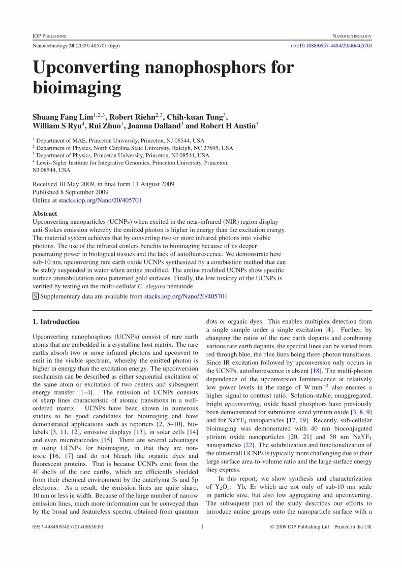
facetted spheroidal nanocrystals with well-defined latticestructures. The measured lattice spacing of the selectednanocrystal is 3.16 A, consistent with the known latticespacing of the (222) plane of cubic Y2O3 [27]. Thatorientation also appears to be the most frequently observedlattice orientation among the other nanocrystals shown. Thecorresponding particle size distribution, calculated from over220 nanoparticles, is shown in figure 2(A). The distributionappears to demonstrate highly uniformly sized nanocrystals,with a mean size of 6.3 nm ± 1.3 nm.
We investigated aggregation of the as-synthesized UCNPsby suspending them in THF, with and without SDS,and measuring the size using dynamical light scattering(figure 2(B)). We had earlier determined the zeta-potentialof about 3.9 mV for unmodified UCNPs in deionized water,which indicates a strong aggregation tendency. Addition ofSDS appears to stabilize the UCNPs electrostatically, and whenadded at a concentration 0.01 M, aggregation of the UCNPscan be reproducibly controlled. We found that in the absenceof SDS, the UCNPs agglomerate into clusters of size 100 nmor more, while when SDS was used, no peaks were observedin the 100–1000 nm range. Typically, at 0.01 M SDS and aUCNPs concentration of 0.01 mg ml−1, a size distribution upto 30 nm was observed, showing that a significant proportion of
A
B
Figure 2. (A) Particle size distribution of UCNPs, based on imagestaken on a Philips CM200 FEG-TEM. (B) Volume distribution ofsub-10 nm nanophosphors suspended in THF, with and without SDS,measured using dynamic light scattering on a Malvern ZetasizerNano-ZS (Malvern Instruments Inc., Worcestershire, UK).
the UCNPs are single nanocrystals and others were in two- orthree-nanocrystal clusters.
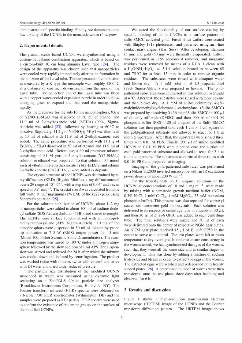
The x-ray diffraction (XRD) spectrum in figure 3(A)shows several diffraction peaks that can be indexed to thestandard Y2O3 body centered cubic (bcc) phase [27]. Thebcc phase combined with the spheroidal shape of the UCNPsobtained are a result of the high synthesis temperature ofour combustion system. Figure 3(A) also shows the XRDspectrum for 40 nm sized UCNPs synthesized using a glycol-based precursor. The sub-10 nm UCNPs XRD peak widths arebroadened compared to the 40 nm one, while the position andrelative strength of the peaks are unchanged. The nanocrystalsize as calculated from Scherrer’s equation [25, 28] was about9 nm. The Y2O3 lattice constant, obtained by calculationsusing XRD data of the nanocrystals is 10.5 A, which isconsistent with that of bulk Y2O3 [29]. The disparity of thesizes as measured by TEM and XRD arises from the differentquantities collected for TEM and XRD measurements. InTEM, samples are collected for about 15 s onto a holey carbon
A B
Figure 3. (A) XRD spectrum of sub-10 nm particles (broken line) and 40 nm particles (solid line). (B) Upconversion spectrum (bold line) of asub-10 nm nanophosphor at 980 nm excitation and cathodoluminescence spectrum (dotted line) at 27 keV acceleration.
3
Nanotechnology 20 (2009) 405701 S F Lim et al
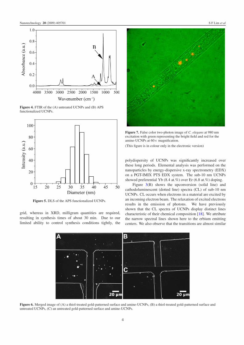
Figure 4. FTIR of the (A) untreated UCNPs and (B) APSfunctionalized UCNPs.
Figure 5. DLS of the APS functionalized UCNPs.
grid, whereas in XRD, milligram quantities are required,resulting in synthesis times of about 30 min. Due to ourlimited ability to control synthesis conditions tightly, the
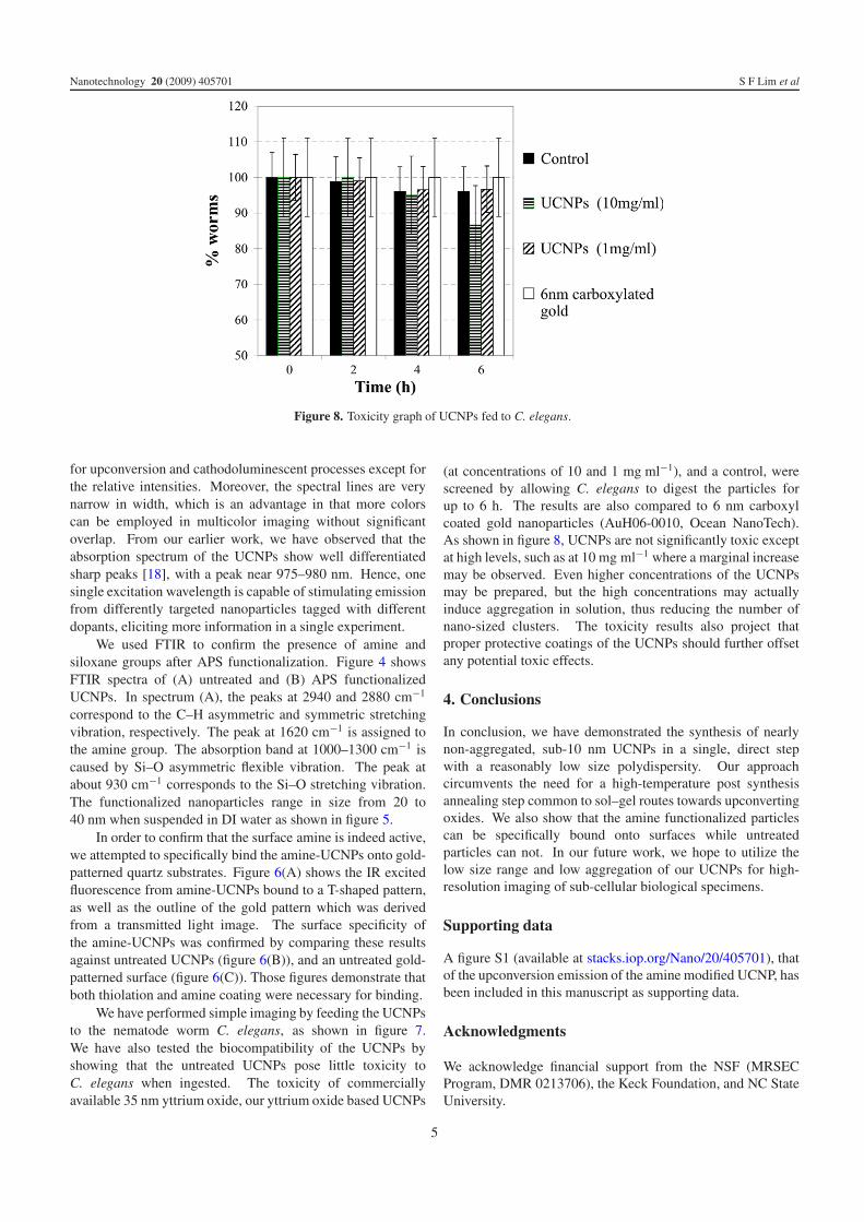
Figure 7. False color two-photon image of C. elegans at 980 nmexcitation with green representing the bright field and red for theamine-UCNPs at 60× magnification.
(This figure is in colour only in the electronic version)
polydispersity of UCNPs was significantly increased overthese long periods. Elemental analysis was performed on thenanoparticles by energy-dispersive x-ray spectrometry (EDX)on a PGT-IMIX PTS EDX system. The sub-10 nm UCNPsshowed preferential Yb (8.4 at.%) over Er (6.8 at.%) doping.
Figure 3(B) shows the upconversion (solid line) andcathodoluminescent (dotted line) spectra (CL) of sub-10 nmUCNPs. CL occurs when electrons in a material are excited byan incoming electron beam. The relaxation of excited electronsresults in the emission of photons. We have previouslyshown that the CL spectra of UCNPs display distinct linescharacteristic of their chemical composition [18]. We attributethe narrow spectral lines shown here to the erbium emittingcenters. We also observe that the transitions are almost similar
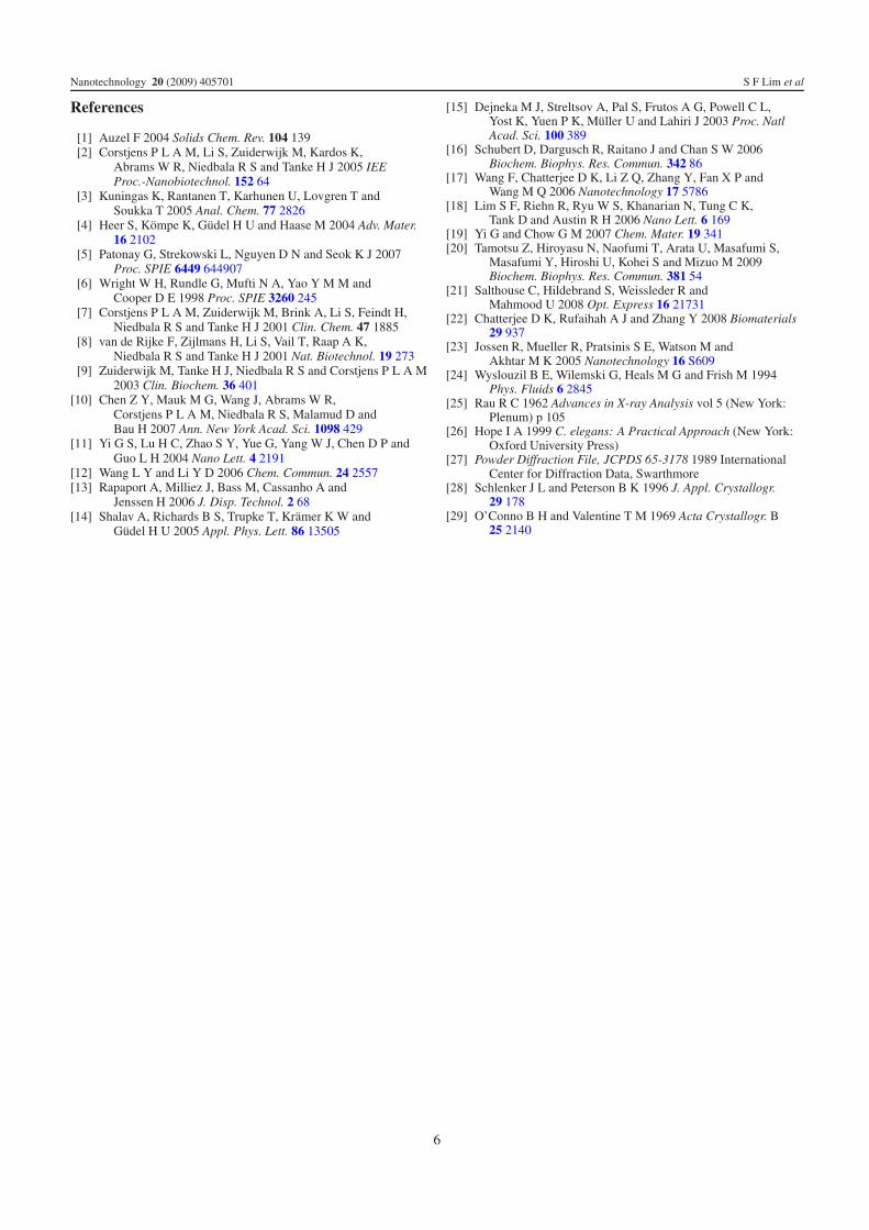
Figure 6. Merged image of (A) a thiol-treated gold-patterned surface and amine-UCNPs, (B) a thiol-treated gold-patterned surface anduntreated UCNPs, (C) an untreated gold-patterned surface and amine-UCNPs.
4
Nanotechnology 20 (2009) 405701 S F Lim et al
Figure 8. Toxicity graph of UCNPs fed to C. elegans.
for upconversion and cathodoluminescent processes except forthe relative intensities. Moreover, the spectral lines are verynarrow in width, which is an advantage in that more colorscan be employed in multicolor imaging without significantoverlap. From our earlier work, we have observed that theabsorption spectrum of the UCNPs show well differentiatedsharp peaks [18], with a peak near 975–980 nm. Hence, onesingle excitation wavelength is capable of stimulating emissionfrom differently targeted nanoparticles tagged with differentdopants, eliciting more information in a single experiment.
We used FTIR to confirm the presence of amine andsiloxane groups after APS functionalization. Figure 4 showsFTIR spectra of (A) untreated and (B) APS functionalizedUCNPs. In spectrum (A), the peaks at 2940 and 2880 cm−1
correspond to the C–H asymmetric and symmetric stretchingvibration, respectively. The peak at 1620 cm−1 is assigned tothe amine group. The absorption band at 1000–1300 cm−1 iscaused by Si–O asymmetric flexible vibration. The peak atabout 930 cm−1 corresponds to the Si–O stretching vibration.The functionalized nanoparticles range in size from 20 to40 nm when suspended in DI water as shown in figure 5.
In order to confirm that the surface amine is indeed active,we attempted to specifically bind the amine-UCNPs onto gold-patterned quartz substrates. Figure 6(A) shows the IR excitedfluorescence from amine-UCNPs bound to a T-shaped pattern,as well as the outline of the gold pattern which was derivedfrom a transmitted light image. The surface specificity ofthe amine-UCNPs was confirmed by comparing these resultsagainst untreated UCNPs (figure 6(B)), and an untreated gold-patterned surface (figure 6(C)). Those figures demonstrate thatboth thiolation and amine coating were necessary for binding.
We have performed simple imaging by feeding the UCNPsto the nematode worm C. elegans, as shown in figure 7.We have also tested the biocompatibility of the UCNPs byshowing that the untreated UCNPs pose little toxicity toC. elegans when ingested. The toxicity of commerciallyavailable 35 nm yttrium oxide, our yttrium oxide based UCNPs
(at concentrations of 10 and 1 mg ml−1), and a control, werescreened by allowing C. elegans to digest the particles forup to 6 h. The results are also compared to 6 nm carboxylcoated gold nanoparticles (AuH06-0010, Ocean NanoTech).As shown in figure 8, UCNPs are not significantly toxic exceptat high levels, such as at 10 mg ml−1 where a marginal increasemay be observed. Even higher concentrations of the UCNPsmay be prepared, but the high concentrations may actuallyinduce aggregation in solution, thus reducing the number ofnano-sized clusters. The toxicity results also project thatproper protective coatings of the UCNPs should further offsetany potential toxic effects.
4. Conclusions
In conclusion, we have demonstrated the synthesis of nearlynon-aggregated, sub-10 nm UCNPs in a single, direct stepwith a reasonably low size polydispersity. Our approachcircumvents the need for a high-temperature post synthesisannealing step common to sol–gel routes towards upconvertingoxides. We also show that the amine functionalized particlescan be specifically bound onto surfaces while untreatedparticles can not. In our future work, we hope to utilize thelow size range and low aggregation of our UCNPs for high-resolution imaging of sub-cellular biological specimens.
Supporting data
A figure S1 (available at stacks.iop.org/Nano/20/405701), thatof the upconversion emission of the amine modified UCNP, hasbeen included in this manuscript as supporting data.
Acknowledgments
We acknowledge financial support from the NSF (MRSECProgram, DMR 0213706), the Keck Foundation, and NC StateUniversity.
5
Nanotechnology 20 (2009) 405701 S F Lim et al
References
[1] Auzel F 2004 Solids Chem. Rev. 104 139[2] Corstjens P L A M, Li S, Zuiderwijk M, Kardos K,
Abrams W R, Niedbala R S and Tanke H J 2005 IEEProc.-Nanobiotechnol. 152 64
[3] Kuningas K, Rantanen T, Karhunen U, Lovgren T andSoukka T 2005 Anal. Chem. 77 2826
[4] Heer S, Kompe K, Gudel H U and Haase M 2004 Adv. Mater.16 2102
[5] Patonay G, Strekowski L, Nguyen D N and Seok K J 2007Proc. SPIE 6449 644907
[6] Wright W H, Rundle G, Mufti N A, Yao Y M M andCooper D E 1998 Proc. SPIE 3260 245
[7] Corstjens P L A M, Zuiderwijk M, Brink A, Li S, Feindt H,Niedbala R S and Tanke H J 2001 Clin. Chem. 47 1885
[8] van de Rijke F, Zijlmans H, Li S, Vail T, Raap A K,Niedbala R S and Tanke H J 2001 Nat. Biotechnol. 19 273
[9] Zuiderwijk M, Tanke H J, Niedbala R S and Corstjens P L A M2003 Clin. Biochem. 36 401
[10] Chen Z Y, Mauk M G, Wang J, Abrams W R,Corstjens P L A M, Niedbala R S, Malamud D andBau H 2007 Ann. New York Acad. Sci. 1098 429
[11] Yi G S, Lu H C, Zhao S Y, Yue G, Yang W J, Chen D P andGuo L H 2004 Nano Lett. 4 2191
[12] Wang L Y and Li Y D 2006 Chem. Commun. 24 2557[13] Rapaport A, Milliez J, Bass M, Cassanho A and
Jenssen H 2006 J. Disp. Technol. 2 68[14] Shalav A, Richards B S, Trupke T, Kramer K W and
Gudel H U 2005 Appl. Phys. Lett. 86 13505
[15] Dejneka M J, Streltsov A, Pal S, Frutos A G, Powell C L,Yost K, Yuen P K, Muller U and Lahiri J 2003 Proc. NatlAcad. Sci. 100 389
[16] Schubert D, Dargusch R, Raitano J and Chan S W 2006Biochem. Biophys. Res. Commun. 342 86
[17] Wang F, Chatterjee D K, Li Z Q, Zhang Y, Fan X P andWang M Q 2006 Nanotechnology 17 5786
[18] Lim S F, Riehn R, Ryu W S, Khanarian N, Tung C K,Tank D and Austin R H 2006 Nano Lett. 6 169
[19] Yi G and Chow G M 2007 Chem. Mater. 19 341[20] Tamotsu Z, Hiroyasu N, Naofumi T, Arata U, Masafumi S,
Masafumi Y, Hiroshi U, Kohei S and Mizuo M 2009Biochem. Biophys. Res. Commun. 381 54
[21] Salthouse C, Hildebrand S, Weissleder R andMahmood U 2008 Opt. Express 16 21731
[22] Chatterjee D K, Rufaihah A J and Zhang Y 2008 Biomaterials29 937
[23] Jossen R, Mueller R, Pratsinis S E, Watson M andAkhtar M K 2005 Nanotechnology 16 S609
[24] Wyslouzil B E, Wilemski G, Heals M G and Frish M 1994Phys. Fluids 6 2845
[25] Rau R C 1962 Advances in X-ray Analysis vol 5 (New York:Plenum) p 105
[26] Hope I A 1999 C. elegans: A Practical Approach (New York:Oxford University Press)
[27] Powder Diffraction File, JCPDS 65-3178 1989 InternationalCenter for Diffraction Data, Swarthmore
[28] Schlenker J L and Peterson B K 1996 J. Appl. Crystallogr.29 178
[29] O’Conno B H and Valentine T M 1969 Acta Crystallogr. B25 2140
6
Copyright © 2022 FDOKUMEN