Effect of suction/blowing on heat-absorbing unsteady radiative ...

Artificial Biomelanin: Highly Light-Absorbing Nano-Sized Eumelaninby Biomimetic Synthesis in Chicken Egg WhiteNicola Fyodor della Vecchia,† Pierfrancesco Cerruti,‡ Gennaro Gentile,‡ Maria Emanuela Errico,‡
Veronica Ambrogi,§ Gerardino D’Errico,† Sara Longobardi,† Alessandra Napolitano,† Luigi Paduano,†
Cosimo Carfagna,‡ and Marco d’Ischia*,†
†Department of Chemical Sciences, University of Naples “Federico II”, Via Cintia 4, 80126, Naples, Italy‡Institute for Polymers, Composites and Biomaterials (IPCB−CNR), Via Campi Flegrei 34, 80078, Pozzuoli, Italy§Department of Chemical, Materials and Production Engineering, University of Naples, Piazzale Tecchio 80, 80125, Naples, Italy
*S Supporting Information
ABSTRACT: The spontaneous oxidative polymerization of0.01−1% w/w 5,6-dihydroxyindole (DHI) in chicken eggwhite (CEW) in the absence of added solvents leads to a black,water-soluble, and processable artificial biomelanin (ABM)with robust and 1 order of magnitude stronger broadband lightabsorption compared to natural and synthetic eumelaninsuspensions. Small angle neutron scattering (SANS) andtransmission electron microscopy (TEM) analysis indicatedthe presence in the ABM matrix of isolated eumelaninnanoparticles (≤100 nm) differing in shape from pure DHImelanin nanoparticles (SANS evidence). Electron para-magnetic resonance (EPR) spectra showed a slightlyasymmetric signal (g ∼ 2.0035) similar to that of solid DHImelanin but with a smaller amplitude (ΔB), suggesting hindered spin delocalization in biomatrix. Enhanced light absorption,altered nanoparticle morphology and decreased free radical delocalization in ABM would reflect CEW-induced inhibition ofeumelanin aggregation during polymerization accompanied in part by covalent binding of growing polymer to the proteins (SDS-PAGE evidence). The technological potential of eumelanin nanosizing by biomimetic synthesis within a CEW biomatrix isdemonstrated by the preparation of an ABM-based black flexible film with characteristics comparable to those of commerciallyavailable polymers typically used in electronics and biomedical applications.
■ INTRODUCTION
Black insoluble eumelanin biopolymers are produced in variousorgans of human body and organisms via a complex oxidativepolymerization process initiated by tyrosinase-catalyzed oxida-tion of tyrosine.1 Supposed biological roles of eumelaninsinclude photoprotection and free radical scavenging in the skinand eyes, and charge transport and detoxification in internal ornonexposed organs, such as inner ear and melanizeddopaminergic neurons of the substantia nigra. Most of thebiological roles and functions of eumelanins are attributed totheir unique physicochemical properties, including broadbandabsorption, efficient energy dissipation by nonradiative excitedstate deactivation (high photon−phonon conversion effi-ciency), free radical, redox, antioxidant and hybrid elec-tronic−ionic semiconductor behavior, and metal and drugbinding,2 which make them highly attractive candidates for abroad range of technological and biomedical applications.3
These can be exemplified by the use of Sepia melaninnanoparticles to produce photoresponsive hydrogel networks.4
Because of the growing interest raised by eumelaninbiopolymers for several technological and biomedical applica-
tions, and considering the notorious difficulties in isolating andpurifying large scale amounts of melanins from naturalsources,1c particular attention is currently being devoted tothe engineering of biocompatible mimics with optimized,enhanced, and/or tailored physicochemical properties.5
Synthetic eumelanins from dopa, 5,6-dihydroxyindole (DHI)and 5,6-dihydroxyindole-2-carboxylic acid (DHICA) have beenused to prepare thin films for organic electronics and bio-optoelectronic interfaces,6 hybrids, and other biomaterials forvarious applications,6d including antioxidants and thermoox-idative stabilization of polymers.7 Another eumelanin-likematerial, dopamine melanin or polydopamine, first describedby Messersmith, Lee, and their associates in 2007 for its abilityto adhere to virtually any kind of surface, has been inspired bythe mussel adhesion process via cross-linking of dopa-richproteins8 and has been used for a broad variety of applications,e.g., to generate biocompatible multifunctional coatings, for the
Received: August 4, 2014Revised: September 15, 2014
Article
pubs.acs.org/Biomac
© XXXX American Chemical Society A dx.doi.org/10.1021/bm501139h | Biomacromolecules XXXX, XXX, XXX−XXX

production of functional nanoparticles, for whole cell coating,drug delivery, and water decontamination.9 Because of theattractive properties and opportunities provided by eumelaninsand the underlying catechol chemistry10 in biomaterials science,increasing efforts are currently directed to develop efficientstrategies to control and tailor structural heterogeneityassociated with chemical disorder, insolubility and tendencyto aggregation. Although aggregation is an important require-ment for the application of eumelanins as thin functionalcoatings, under certain circumstances, it may make may maketheir manipulation a most difficult task, limiting moreover thelight absorption efficiency for optical use. In this connection,disentangling intrinsic absorption from scattering effects hasbeen recognized as a major issue in melanin opticalspectroscopy,2,11 which requires nondestructive disassemblyof supramolecular architecture and/or efficient control overaggregation during synthesis. Two different strategies have beendeveloped in our laboratory, which are based on glycation ofmonomer precursor DHI12 and on the use of poly(vinylalcohol) (PVA) to inhibit aggregation and precipitation duringpolymerization.13 Proteins such as human serum albuminprovide also a viable means of controlling eumelanin growthand properties, as reported in the case of polydopamine.14
Inspired by the natural process of melanogenesis within theproteinaceous matrix of melanosome organelles,15 (Scheme 1),
we disclose herein a new concept in synthetic eumelaninengineering, which is built on the discovery that broadbandabsorption, paramagnetic properties, and aggregate size andshape are strongly affected by molecular embedment in chickenegg white (CEW) as a biomatrix in the absence of additionalsolvents. CEW was selected because of its easy availability,solubility and viscosity properties, biocompatibility andbiodegradability, and especially for its intrinsically alkaline pHoptimally suited to induce efficient catechol autoxidationwithout added oxidants or additives. Moreover, CEW can beeasily converted into insoluble hydrogels by simple heating, andhas been proposed as a soft dielectric component in transistorsand in carbon dot preparation,16 suggesting convenient use inbiomaterials science and bioelectronics.
■ EXPERIMENTAL SECTIONMaterials and Methods. Artificial biomelanin (ABM) was
synthesized by the addition of DHI, prepared as previouslydescribed,17 at 0.01−1% w/w concentration to chicken egg albumenunder stirring at room temperature. The pH of the egg white wastypically in the range between 8 and 8.5 as determined by a pH meter.UV spectra of ABM were run after a 50-fold dilution in water, leadingto a final pH of 8.0.EPR Spectroscopy. Spectra were recorded by an experimental
procedure recently set up for paraffin-embedded sections of melanoma
samples18 and used in the characterization of DHI and DHICAeumelanins.7a The samples were measured using an X-band (9 GHz)Bruker Elexys E-500 spectrometer (Bruker, Rheinstetten, Germany),equipped with a superhigh sensitivity probe head. Samples of ABMfrom 0.01 and 0.1% DHI were transferred to flame-sealed glasscapillaries which, in turn, were coaxially inserted in a standard 4 mmquartz sample tube. Measurements were performed at roomtemperature. The instrumental settings were as follows: sweepwidth, 100 G; resolution, 1024 points; modulation frequency, 100kHz; modulation amplitude, 1.0 G. The amplitude of the fieldmodulation was preventively checked to be low enough to avoiddetectable signal overmodulation. Preliminarily, EPR spectra weremeasured with a microwave power of ∼1.4 mW to avoid microwavesaturation of resonance absorption curve. Several scans, typically 16,were accumulated to improve the signal-to-noise ratio. Successively,for power saturation experiments, the microwave power was graduallyincremented from 0.02 to 160 mW. The g value was evaluated bymeans of two internal standards (2,2-diphenyl-1-picrylhydrazyl,DPPH,18,19 and 4-hydroxy-2,2,6,6-tetramethylpiperidin-1-oxyl, TEM-POL, as ethanol solution20), which were inserted in the quartz sampletube coaxially with the capillary containing the samples.
Bright Field Transmission Electron Microscopy (TEM).Experiments were performed on a FEI TECNAI G12 Spirit-Twin(120 kV, LaB6) microscope equipped with a FEI Eagle 4k CCDcamera (Eindhoven, The Netherlands). Prior to observations, thinsections of the samples (nominal thickness 100 nm) were obtained bya Leica UC7 ultramicrotome (Wien, Austria) and placed on 400 meshcopper grids.
Small Angle Neutron Scattering Measurements (SANS).Measurements were carried out at the LOQ instrument sited at theISIS facility of the Rutherford Appleton Laboratory (Chilton, UK). Atthe ISIS pulsed neutron source, the LOQ instrument uses neutrons ofwavelengths ranging between 2.2 and 10 A detected by a time-of-flightanalysis on a 64 cm2 two-dimensional detector placed 4.1 m from thesample, giving a q range of 0.006−0.24 A−1.13a Raw data werecorrected for wavelength-dependent sample transmissions, incidentspectrum, and detector efficiency and then put into absolute scatteringcross sections I(q) in comparison with scattering from a partiallydeuterated polystyrene standard.
Sodium dodecyl sulfate gel electrophoresis (SDS-PAGE) wasperformed using Laemmli method21 with Bio-Rad Mini Protean IIIapparatus, with 7% polyacrylamide gel. Unstained Protein marker(Thermo Scientific-26610) was used as standard. The gel was stainedby Comassie brilliant blue.
Preparation of Flexible Films. Albumen (10.0 g), 1.0 g ofpolyglycerol-3 (Solvay, average number of monomer units =3), 10 mgof DHI were mixed under magnetic stirring at room temperature for t= 30 min. The resulting mixture was poured onto a PTFE Petri dishand oven heated at T = 90 °C for 90 min. After thermal treatment, theobtained film was dried for 24 h at T = 25 °C.
Mechanical Characterization of Plasticized ABM. Tensile testswere performed on five specimens, using an Instron model 5564dynamometer equipped with a 1 kN load cell at 23 ± 2 °C and 45 ±5% RH with a 5 mm min−1 clamp separation rate.
■ RESULTS AND DISCUSSION
Addition of DHI (0.1% w/w) to raw CEW under stirring atroom temperature resulted in the development, after completesubstrate consumption at 24 h, of a black eumelanin-likepigment, here referred to as artificial biomelanin (ABM)(Figure 1A). Brief heating of ABM in a steam bath led to ablack hydrogel insoluble in water (Figure 1B, see alsoSupporting Information (SI) Figure S1). Thorough drying ofthe ABM hydrogel produced a hard solid material (Figure 1C).Spectrophotometric monitoring of the reaction course with
0.1% w/w DHI in CEW (Figure 2) showed the generation ofan intense purple chromophore peaking at ca. 1 h, which couldbe efficiently stabilized by cautious heating of the mixture under
Scheme 1. Schematic Illustration of Eumelanin Biosynthesisand Embedment in Proteins during Melanosome Maturation
Biomacromolecules Article
dx.doi.org/10.1021/bm501139h | Biomacromolecules XXXX, XXX, XXX−XXXB
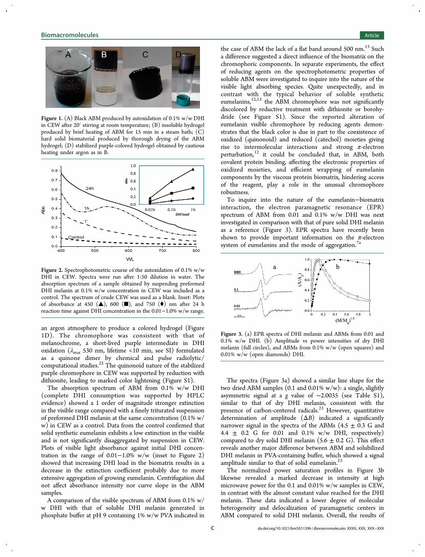
an argon atmosphere to produce a colored hydrogel (Figure1D). The chromophore was consistent with that ofmelanochrome, a short-lived purple intermediate in DHIoxidation (λmax 530 nm, lifetime <10 min, see SI) formulatedas a quinone dimer by chemical and pulse radiolytic/computational studies.22 The quinonoid nature of the stabilizedpurple chromophore in CEW was supported by reduction withdithionite, leading to marked color lightening (Figure S1).The absorption spectrum of ABM from 0.1% w/w DHI
(complete DHI consumption was supported by HPLCevidence) showed a 1 order of magnitude stronger extinctionin the visible range compared with a finely triturated suspensionof preformed DHI melanin at the same concentration (0.1% w/w) in CEW as a control. Data from the control confirmed thatsolid synthetic eumelanin exhibits a low extinction in the visibleand is not significantly disaggregated by suspension in CEW.Plots of visible light absorbance against initial DHI concen-tration in the range of 0.01−1.0% w/w (inset to Figure 2)showed that increasing DHI load in the biomatrix results in adecrease in the extinction coefficient probably due to moreextensive aggregation of growing eumelanin. Centrifugation didnot affect absorbance intensity nor curve slope in the ABMsamples.A comparison of the visible spectrum of ABM from 0.1% w/
w DHI with that of soluble DHI melanin generated inphosphate buffer at pH 9 containing 1% w/w PVA indicated in
the case of ABM the lack of a flat band around 500 nm.13 Sucha difference suggested a direct influence of the biomatrix on thechromophoric components. In separate experiments, the effectof reducing agents on the spectrophotometric properties ofsoluble ABM were investigated to inquire into the nature of thevisible light absorbing species. Quite unexpectedly, and incontrast with the typical behavior of soluble syntheticeumelanins,12,13 the ABM chromophore was not significantlydiscolored by reductive treatment with dithionite or borohy-dride (see Figure S1). Since the reported alteration ofeumelanin visible chromophore by reducing agents demon-strates that the black color is due in part to the coexistence ofoxidized (quinonoid) and reduced (catechol) moieties givingrise to intermolecular interactions and strong π-electronperturbation,12 it could be concluded that, in ABM, bothcovalent protein binding, affecting the electronic properties ofoxidized moieties, and efficient wrapping of eumelanincomponents by the viscous protein biomatrix, hindering accessof the reagent, play a role in the unusual chromophorerobustness.To inquire into the nature of the eumelanin−biomatrix
interaction, the electron paramagnetic resonance (EPR)spectrum of ABM from 0.01 and 0.1% w/w DHI was nextinvestigated in comparison with that of pure solid DHI melaninas a reference (Figure 3). EPR spectra have recently beenshown to provide important information on the π-electronsystem of eumelanins and the mode of aggregation.7a
The spectra (Figure 3a) showed a similar line shape for thetwo dried ABM samples (0.1 and 0.01% w/w): a single, slightlyasymmetric signal at a g value of ∼2.0035 (see Table S1),similar to that of dry DHI melanin, consistent with thepresence of carbon-centered radicals.23 However, quantitativedetermination of amplitude (ΔB) indicated a significantlynarrower signal in the spectra of the ABMs (4.5 ± 0.3 G and4.4 ± 0.2 G for 0.01 and 0.1% w/w DHI, respectively)compared to dry solid DHI melanin (5.6 ± 0.2 G). This effectreveals another major difference between ABM and solubilizedDHI melanin in PVA-containing buffer, which showed a signalamplitude similar to that of solid eumelanin.23
The normalized power saturation profiles in Figure 3blikewise revealed a marked decrease in intensity at highmicrowave power for the 0.1 and 0.01% w/w samples in CEW,in contrast with the almost constant value reached for the DHImelanin. These data indicated a lower degree of molecularheterogeneity and delocalization of paramagnetic centers inABM compared to solid DHI melanin. Overall, the results of
Figure 1. (A) Black ABM produced by autoxidation of 0.1% w/w DHIin CEW after 20′ stirring at room temperature; (B) insoluble hydrogelproduced by brief heating of ABM for 15 min in a steam bath; (C)hard solid biomaterial produced by thorough drying of the ABMhydrogel; (D) stabilized purple-colored hydrogel obtained by cautiousheating under argon as in B.
Figure 2. Spectrophotometric course of the autoxidation of 0.1% w/wDHI in CEW. Spectra were run after 1:50 dilution in water. Theabsorption spectrum of a sample obtained by suspending preformedDHI melanin at 0.1% w/w concentration in CEW was included as acontrol. The spectrum of crude CEW was used as a blank. Inset: Plotsof absorbance at 450 (▲), 600 (■), and 750 (⧫) nm after 24 hreaction time against DHI concentration in the 0.01−1.0% w/w range.
Figure 3. (a) EPR spectra of DHI melanin and ABMs from 0.01 and0.1% w/w DHI. (b) Amplitude vs power intensities of dry DHImelanin (full circles), and ABMs from 0.1% w/w (open squares) and0.01% w/w (open diamonds) DHI.
Biomacromolecules Article
dx.doi.org/10.1021/bm501139h | Biomacromolecules XXXX, XXX, XXX−XXXC

EPR analysis suggest that CEW directly affects eumelanin π-electron system and paramagnetic properties.In a subsequent series of experiments, the nature and
morphology of eumelanin components in ABM was inves-tigated by microscopy techniques. Preliminarily, 40−50 μmthick transparent ABM films directly cast on microscope slideswere examined by optical microscopy under transmissionmode. No detectable aggregates within the instrumental limitsof the technique could be observed (Figure S2). Bright fieldtransmission electron microscopy (TEM) analysis of driedABM from 0.1% w/w DHI revealed, however, sparse particles,with average size in the range of 15−65 nm (Figure 4), that
were absent in the CEW as control. This data indicates that thebiomatrix effectively inhibits aggregation processes in growingeumelanin, allowing only for the formation of nanosizedparticles.The size and shape of eumelanin aggregates in ABM was
further investigated by small angle neutron scattering (SANS).Data in Figure 5 show that at low values (q) of the scatteringvector the profile of the ABM displays an upturn, suggesting thepresence of aggregates with size around 100 nm. Notably,whereas in phosphate buffer DHI polymerizes to form two-dimensional objects, with a power law of −2 at low value of
q,13a in the present case, the power law was markedly lower,indicating the absence of pure DHI melanin and suggesting adifferent nature and shape of the aggregates, likely to involveinteraction with protein chains and their conformationalmodification. The apparent interaction of DHI and itsoligomers with the CEW matrix would cause cross-linkingand conformational changes in proteins, which in turn wouldaffect the aggregate shape. This entails that DHI oligomers tendmainly to grow on the protein matrix components serving aspseudotemplates to form small and undefined objects, ratherthan self-assemble to form the typical bidimensional aggregatesobserved with pure melanin.In support of this conclusion, SDS-PAGE analysis of a freshly
prepared ABM sample from 0.1% DHI indicated detectablemodification of the protein pattern, with the formation of newhigh molecular weight bands, suggesting covalent cross-linkingbetween eumelanin components and CEW proteins (Figure 6).
In particular, bands A and B (lanes 1, 2, and 3) were notdetectable in ABM samples (lanes 4, 5, and 6), while a minorintensity of band C was observed. Based on previousobservations,24 bands A and B (MW above 150 kDa) couldcorrespond to ovomucin and/or ovostatin, while band C (MWabout 75 kDa) is ascribable to ovotransferrin.The nature of the covalent-binding responsible for melanin
cross-linking to proteins could not be elucidated on the basis ofthe present data. CEW major proteins apparently involved incross-linking (Figure 6) contain cysteine and lysine residues,among others, so these amino acids are likely candidates fornucleophilic attack to DHI melanin quinone groups or otherelectrophilic functionalities, but further work is necessary toprove this hypothesis. Based on these results, it can beconcluded that DHI polymerization in CEW proceeds withsubstantial inhibition of aggregation processes leading tonanosized eumelanin particles partly cross-linked to proteincomponents of the embedding viscous biomatrix (Scheme 2).In a final series of experiments the potential application of
ABM to the preparation of new functional materials was brieflyexplored. Mixing freshly prepared ABM from 0.1% w/w DHIwith 10% w/w polyglycerol-3 as plasticizer and heating at 90 °Cfor 90 min resulted in an flexible film (Figure 7). Mechanical
Figure 4. Bright field TEM image of a thermally treated and driedsample of CEW loaded with 0.1% w/w DHI.
Figure 5. SANS analysis of ABM (upper trace) versus CEW as acontrol (lower trace).
Figure 6. SDS-PAGE (Coomassie stained) of 100, 50, and 10 μg ofCEW proteins (lanes 1, 2, and 3, respectively) versus the sameamounts of ABM proteins (lanes 4, 5, and 6, respectively); M:molecular weight markers. Arrows denote main protein bandsmodified by covalent binding to melanin.
Biomacromolecules Article
dx.doi.org/10.1021/bm501139h | Biomacromolecules XXXX, XXX, XXX−XXXD

tests of plasticized ABM (Young modulus E = 3.0 ± 0.3 MPa,stress at break σb = 0.24 ± 0.03 MPa, strain at break εb = 35 ±6%) yielded values similar to those of CEW-derived films in theabsence of DHI (Young modulus, E = 2.9 ± 0.5 MPa, stress atbreak, σb = 0.26 ± 0.04 MPa, Strain at break, εb = 33 ± 5%).These features were comparable to those of a commerciallyavailable polymer, such as polydimethylsiloxane (PDMS),which is a material typically used in electronics and biomedicalapplications.
■ CONCLUSIONSSpontaneous polymerization of DHI in CEW is disclosedherein as a new promising approach for enhancing and tailoringeumelanin optical properties via chromophore disentanglementthrough controlled aggregation. Although proteins such ashuman serum albumin, hen egg white lysozyme, or bovine α-lactalbumin have recently been shown to accelerate the kineticsof polydopamine (dopamine−eumelanin) formation and tocontrol particle size,14 this is the first paper to report theinfluence of a crude natural protein matrix on the optical andparamagnetic properties of growing eumelanins. ABM is thusproposed as a superior low-cost bioinspired material forsustainable eumelanin-based technologies for the followingreasons: it is produced by a spontaneous reaction without theaddition of chemical oxidants; it displays a robust soluble andbiodegradable black chromophore which compares favorably toprevious soluble eumelanins based on glycation12,25 or onsynthesis in PVA;13 it can be processed to produce a variety oflight-absorbing systems amenable to manipulation for diverseapplications.
■ ASSOCIATED CONTENT*S Supporting InformationABM characterization: EPR spectral parameters, opticalmicroscopy, and mechanical characterization of plasticized
ABM. This material is available at free of charge via the Internetat http://pubs.acs.org.
■ AUTHOR INFORMATION
Corresponding Author*Fax: +39 081674393; e-mail: [email protected].
NotesThe authors declare no competing financial interest.
■ ACKNOWLEDGMENTS
This work was supported by grants from the Italian MIUR,PRIN 2010-2011 project, and was carried out within the aimsof the EuMelaNet group (http://www.espcr.org/eumelanet/).
■ REFERENCES(1) (a) Simon, J. D.; Peles, D.; Wakamatsu, K.; Ito, S. Pigm. CellMelanoma Res. 2009, 22, 563−579. (b) Ito, S.; Wakamatsu, K.;d’Ischia, M.; Napolitano, A.; Pezzella, A. In Melanins and Melanosomes:Biosynthesis, Biogenesis, Physiological, and Pathological Functions;Borovansky, J., Riley, P. A., Eds.; Wiley-Blackwell: Weinheim,Germany, 2011; pp 167−185. (c) d’Ischia, M.; Wakamatsu, K.;Napolitano, A.; Briganti, S.; Garcia-Borron, J. C.; Kovacs, D.;Meredith, P.; Pezzella, A.; Picardo, M.; Sarna, T.; Simon, J. D.; Ito,S. Pigm. Cell Melanoma Res. 2013, 26, 616−633.(2) Meredith, P.; Sarna, T. Pigm. Cell Res. 2006, 19, 572−594.(3) (a) d’Ischia, M.; Napolitano, A.; Pezzella, A.; Meredith, P.; Sarna,T. Angew. Chem., Int. Ed. 2009, 48, 3914−3921. (b) Meredith, P.;Bettinger, C. J.; Irimia-Vladu, M.; Mostert, A. B.; Schwenn, P. E. Rep.Prog. Phys. 2013, 034501.(4) Ninh, C.; Cramer, M.; Bettinger, C. J. Biomater. Sci. 2014, 2,766−774.(5) Fisher, O. Z.; Larson, B. L.; Hill, P. S.; Graupner, D.; Nguyen-Kim, M.-T.; Kehr, N. S.; De Cola, L.; Langer, R.; Anderson, D. G. Adv.Mater. 2012, 24, 3032−3036.(6) (a) Bettinger, C. J.; Bruggeman, P. P.; Misra, A.; Borenstein, J. T.;Langer, R. Biomaterials 2009, 30, 3050−3057. (b) Bloisi, F.; Pezzella,A.; Barra, M.; Alfe, M.; Chiarella, F.; Cassinese, A.; Vicari, L. Appl.Phys. A: Mater. 2011, 105, 619−627. (c) Bothma, J. P.; de Boor, J.;Divakar, U.; Schwenn, P. E.; Meredith, P. Adv. Mater. 2008, 20, 3539−3542. (d) Prasetyanto, E. A.; Manini, P.; Napolitano, A.; Crescenzi, O.;d’Ischia, M.; De Cola, L. Chem.Eur. J. 2014, 20, 1597−1601.(7) (a) Panzella, L.; Gentile, G.; D’Errico, G.; Della Vecchia, N. F.;Errico, M. E.; Napolitano, A.; Carfagna, C.; d’Ischia, M. Angew. Chem.,Int. Ed. 2013, 52, 12684−12687. (b) Shanmuganathan, K.; Cho, J. H.;Iyer, P.; Baranowitz, S.; Ellison, C. J. Macromolecules 2011, 44, 9499−9507.(8) Lee, H.; Dellatore, S. M.; Miller, W. M.; Messersmith, P. B.Science 2007, 318, 426−430.(9) (a) Liu, Y.; Ai, K.; Lu, L. Chem. Rev. 2014, 114, 5057−5115.(b) Sedo, J.; Saiz-Poseu, J.; Busque, F.; Ruiz-Molina, D. Adv. Mater.2013, 25, 653−701. (c) Kim, J. H.; Lee, M.; Park, C. B. Angew. Chem.,Int. Ed. Engl. 2014, 53, 6364−6368. (d) Cui, J.; Yan, Y.; Such, G. K.;Liang, K.; Ochs, C. J.; Postma, A.; Caruso, F. Biomacromolecules 2012,13, 2225−2228. (e) Kang, S. M.; Hwang, N. S.; Yeom, J.; Park, S. Y.;Messersmith, P. B.; Choi, I. S.; Langer, R.; Anderson, D. G.; Lee, H.Adv. Funct. Mater. 2012, 22, 2949−2955.(10) (a) Saiz-Poseu, J.; Sedo, J.; García, B.; Benaiges, C.; Parella, T.;Ramon Alibes, R.; Hernando, J.; Busque, F.; Ruiz-Molina, D. Adv.Mater. 2013, 25, 2066−2070. (b) Chen, C.-T.; Ball, V.; de AlmeidaGracio, J. J.; Singh, M. K.; Toniazzo, V.; Ruch, D.; Buehler, M. J. ACSNano 2013, 7, 1524−1532. (c) Shafiq, Z.; Cui, J.; Pastor-Perez, L.; SanMiguel, V.; Gropeanu, R. A.; Serrano, C.; del Campo, A. Angew. Chem.,Int. Ed. Engl. 2012, 51, 4332−4335. (d) Garcia-Fernandez, L.; Cui, J.;Serrano, C.; Shafiq, Z.; Gropeanu, R. A.; San Miguel, V.; Ramos, J. I.;Wang, M.; Auernhammer, G. K.; Ritz, S. L.; Golriz, A. A.; Berger, R.;Wagner, M.; del Campo, A. Adv. Mater. 2013, 25, 529−533.
Scheme 2. Schematic View of ABM Synthesis in CEWa
aEumelanin growth proceeds via a purple melanochrome intermediate(representative structure in the frame) and leads to particle nanosizingdue to segregation in the viscous biomatrix.
Figure 7. Flexible film of plasticized ABM containing 0.1% w/w DHI.
Biomacromolecules Article
dx.doi.org/10.1021/bm501139h | Biomacromolecules XXXX, XXX, XXX−XXXE

(11) Piletic, I. R.; Matthews, T. E.; Warren, W. S. J. Chem. Phys. 2009,131, 181106.(12) Pezzella, A.; Iadonisi, A.; Valerio, S.; Panzella, L.; Napolitano, A.;Adinolfi, M.; d’Ischia, M. J. Am. Chem. Soc. 2009, 131, 15270−15275.(13) (a) Arzillo, M.; Mangiapia, G.; Pezzella, A.; Heenan, R. K.;Radulescu, A.; Paduano, L.; d’Ischia, M. Biomacromolecules 2012, 13,2379−2390. (b) Ascione, L.; Pezzella, A.; Ambrogi, V.; Carfagna, C.;d’Ischia, M. Photochem. Photobiol. 2013, 89, 314−318.(14) Chassepot, A.; Ball, V. J. Colloid Interface Sci. 2014, 414, 97−102.(15) Raposo, G.; Marks, M. S. Nat. Rev. Mol. Cell Biol. 2007, 8, 786−797.(16) (a) Chang, J.-W.; Wang, C.-G.; Huang, C.-Y.; Tsai, T.-D.; Guo,T.-F.; Wen, T. C. Adv. Mater. 2011, 23, 4077−4081. (b) Wang, J.;Wang, C.-F.; Chen, S. Angew. Chem., Int. Ed. 2012, 51, 9297−9301.(17) Edge, R.; d’Ischia, M.; Land, E. J.; Napolitano, A.; Navaratnam,S.; Panzella, L.; Pezzella, A.; Ramsden, C. A.; Riley, P. A. Pigm. Cell Res.2006, 19, 443−450.(18) Cesareo, E.; Korkina, L.; D’Errico, G.; Vitiello, G.; Aguzzi, M. S.;Passarelli, F.; Pedersen, J. Z.; Facchiano, A. PLoS One 2012, 7, e48849.(19) Wertz, J. E.; Bolton, J. R. Electron Spin Resonance: ElementaryPractical Applications; McGraw Hill: New York, 1972(20) Ottaviani, M. F. J. Phys. Chem. 1987, 91, 119−784.(21) Laemmli, U. K. Nature 1970, 227, 680−685.(22) Pezzella, A.; Panzella, L.; Crescenzi, O.; Napolitano, A.;Navaratman, S.; Edge, R.; Land, E. J.; Barone, V.; d’Ischia, M. J. Am.Chem. Soc. 2006, 128, 15490−15498.(23) Mostert, A. B.; Hanson, G. R.; Sarna, T.; Gentle, I. R.; Powell, B.J.; Meredith, P. J. Phys. Chem. B 2013, 117, 4965−4972.(24) Desert, C.; Guerin-Dubiard, C.; Nau, F.; Jan, G.; Val, F.;Mallard, J. J. Agric. Food Chem. 2001, 49, 4553−4561.(25) Manini, P.; d’Ischia, M.; Prota, G. J. Org. Chem. 2001, 66, 5048−5053.
Biomacromolecules Article
dx.doi.org/10.1021/bm501139h | Biomacromolecules XXXX, XXX, XXX−XXXF
Copyright © 2022 FDOKUMEN