Hierarchical integration of individual motions in locally paired-dot stimuli
Upload
independentCategory
view
0download
0
Cell Reports
Article
A Paired RNAi and RabGAP Overexpression ScreenIdentifies Rab11 as a Regulatorof b-Amyloid ProductionVinod Udayar,1,2,3,4 Virginie Buggia-Prevot,6 Rita L. Guerreiro,7,13 Gabriele Siegel,1,2 Naresh Rambabu,8
Amanda L. Soohoo,9 Moorthi Ponnusamy,8 Barbara Siegenthaler,1,2 Jitin Bali,1,2,3 AESG,7 Mikael Simons,10,11
Jonas Ries,12 Manojkumar A. Puthenveedu,9 John Hardy,7,13 Gopal Thinakaran,6 and Lawrence Rajendran1,2,3,5,*1Systems and Cell Biology of Neurodegeneration, University of Zurich, Wagistrasse 12, 8952 Schlieren, Switzerland2Division of Psychiatry Research, University of Zurich, Wagistrasse 12, 8952 Schlieren, Switzerland3Graduate Program of the Zurich Neuroscience Center, University of Zurich, Wagistrasse 12, 8952 Schlieren, Switzerland4Erasmus Mundus Joint Doctorate Program, University of Zurich, Wagistrasse 12, 8952 Schlieren, Switzerland5Graduate Program of the Zurich Center for Integrative Human Physiology, University of Zurich, Wagistrasse 12, 8952 Schlieren, Switzerland6Departments of Neurobiology, Neurology and Pathology, The University of Chicago, Chicago, IL 60637, USA7Alzheimer’s Exome Sequencing Group, University College London, London WC1E 6BT, UK8Raise.Rural Foundation for Promoting Research Awareness in Student Environment in Rural India, 60045 Chennai, India9Department of Biological Sciences, Carnegie Mellon University, Pittsburgh, PA 15213, USA10Department of Neurology, University of Gottingen, 37075 Gottingen, Germany11Max-Planck Institute for Experimental Medicine, 37075 Gottingen, Germany12Cell Biology and Biophysics, European Molecular Biology Laboratory, 69117 Heidelberg, Germany13Reta Lila Weston Research Laboratories, Department of Molecular Neuroscience, UCL Institute of Neurology, London WC1N 3BG, UK
*Correspondence: [email protected]://dx.doi.org/10.1016/j.celrep.2013.12.005
This is an open-access article distributed under the terms of the Creative Commons Attribution-NonCommercial-No Derivative Works
License, which permits non-commercial use, distribution, and reproduction in any medium, provided the original author and source are
credited.
SUMMARY
Alzheimer’s disease (AD) is characterized by cerebraldeposition of b-amyloid (Ab) peptides, which aregenerated from amyloid precursor protein (APP)by b- and g-secretases. APP and the secretasesare membrane associated, but whether membranetrafficking controls Ab levels is unclear. Here, we per-formed an RNAi screen of all human Rab-GTPases,which regulatemembrane trafficking, complementedwith a Rab-GTPase-activating protein screen, andpresent a road map of the membrane-traffickingevents regulating Ab production. We identify Rab11andRab3 as key players. Although retromers and ret-romer-associated proteins control APP recycling, weshow that Rab11 controlled b-secretase endosomalrecycling to the plasmamembrane and thus affectedAb production. Exome sequencing revealed a signif-icant genetic association of Rab11A with late-onsetAD, and network analysis identified Rab11A andRab11B as components of the late-onset AD risknetwork, suggesting a causal link between Rab11and AD. Our results reveal trafficking pathways thatregulate Ab levels and show how systems biologyapproaches can unravel the molecular complexityunderlying AD.
1536 Cell Reports 5, 1536–1551, December 26, 2013 ª2013 The Aut
INTRODUCTION
Alzheimer’s disease (AD) is the most common form of dementia
and is characterized by the cerebral deposition of b-amyloid (Ab)
peptides in the form of amyloid plaques (De Strooper, 2010; Fri-
soni et al., 2011). The amyloid cascade hypothesis postulates
that Ab peptides trigger a series of pathological events leading
to neurodegeneration (Huang and Mucke, 2012; Selkoe,
2011b). Ab peptides are liberated from the transmembrane
amyloid precursor protein (APP) by the sequential actions of
b-secretase and g-secretase (Thinakaran and Koo, 2008; Willem
et al., 2009). b-secretase activity is conferred by a transmem-
brane aspartyl protease, also termed BACE1 (b-site APP-
cleaving enzyme 1) (Vassar et al., 1999), whereas g-secretase
is a multimeric transmembrane protein complex composed of
presenilin-1 (PS1)/PS2, nicastrin, Aph-1, and PEN-2 (Annaert
and De Strooper, 2002; Selkoe and Wolfe, 2007). Familial
mutations in APP, PS1, or PS2 that increase the production of
the amyloidogenic Ab42 peptide have been associated with
early-onset AD (Borchelt et al., 1996; Duff et al., 1996). However,
there is only limited insight into the cause of late-onset AD
(LOAD), which contributes to more than 95% of cases. Genetic
modifiers of LOAD may also regulate Ab production, raising the
possibility that genes regulating APP metabolism might impact
the risk for AD (Andersen et al., 2005; Rogaeva et al., 2007;
Selkoe, 2011a).
Several lines of evidence support an important role for
membrane trafficking in the amyloidogenic processing of APP
hors
and hence in AD pathogenesis (Rajendran and Annaert, 2012;
Thinakaran andKoo, 2008). APP andBACE1 are transmembrane
proteins that are synthesized in the endoplasmic reticulum (ER),
matured in the Golgi complex, and then transported to the
plasma membrane and into endosomes via endocytosis (Small
and Gandy, 2006; Thinakaran and Koo, 2008). The endolysoso-
mal compartment has been implicated as one of the major sites
for Ab generation (Cataldo et al., 2000; Haass et al., 1992; Koo
and Squazzo, 1994). Recent work has revealed that BACE1
cleavage of APP occurs predominantly in early endosomes,
and endocytosis of APP and BACE1 is essential for b cleavage
of APP, and Ab production (Kinoshita et al., 2003; Rajendran
et al., 2006; Sannerud et al., 2011). The pH of endosomes
(pH 4.0–5.0) is optimal for BACE1 activity, which also explains
the requirement for endocytosis (Kalvodova et al., 2005; Vassar
et al., 2009). In contrast, a-secretase cleavage of APP, which
precludes Ab production, occurs at the plasmamembrane (Lich-
tenthaler, 2011). Components of the g-secretase complex are
also synthesized in the ER, but their assembly and maturation
require the coordinated regulation of the ER-Golgi-recycling cir-
cuit (Spasic and Annaert, 2008).
We previously showed that b cleavage of APP occurs in a
Rab5-EEA1-positive membrane compartment and that endocy-
tosis is essential for Ab generation (Rajendran et al., 2006). Tar-
geting a transition-state BACE1 inhibitor to endosomes inhibited
Ab production in cultured cells and mice (Rajendran et al., 2008).
Interestingly, proteins that belong to the retromer family, which
transport cargo from early endosomes to the Golgi, have also
been implicated in AD (Rogaeva et al., 2007; Small et al.,
2005). These AD risk genes regulate the residency of APP and
BACE1 in the early endosomal compartment, therefore regu-
lating Ab generation (Siegenthaler and Rajendran, 2012). Simi-
larly, proteins of the GGA family have been shown to traffic
BACE1 from endosomes to the Golgi, and their depletion led
to increased amyloidogenic processing of APP (He et al., 2005;
Tesco et al., 2007; von Arnim et al., 2006). Although APP is
known to be routed from endosomes to Golgi via the retromer
and retromer-associated proteins including SORL1 and VPS26
(Andersen et al., 2005; Morel et al., 2013; Small and Gandy,
2006; Rogaeva et al., 2007; Small et al., 2005; Siegenthaler
and Rajendran, 2012), nothing much is known about BACE1 re-
cycling from endosomes. A better understanding of the specific
trafficking mechanisms involved in Ab production will thus pro-
vide further insights into disease pathogenesis and potentially
provide novel therapeutic strategies for treating this currently un-
treatable disease.
Rab GTPases regulate many aspects of membrane protein
trafficking, acting as membrane organizers on cellular compart-
ments that mediate vesicular trafficking and aid in vesicle fusion
(Seabra et al., 2002). They regulate vesicular trafficking both in
the biosynthetic and endocytic routes, enabling cargo sorting
within the different membrane compartments (Zerial and
McBride, 2001). Rab GTPases switch between an inactive
GDP-bound form and an active GTP-bound form, which enables
vesicular fusion (Stenmark and Olkkonen, 2001). Although
GDP-GTP exchange is mediated by Rab-specific GTP exchange
factors (GEFs), GTP hydrolysis is achieved by the GTPase-acti-
vating proteins (GAPs) (Barr and Lambright, 2010). In humans,
Cell Re
there are 60 Rab proteins and 39 RabGAPs. Overexpression of
RabGAPs depletes the active formof Rab proteins and increases
the inactive, GDP-bound form, thereby preventing their normal
function (Yoshimura et al., 2007). Here, we systematically
analyzed the role of the cell’s major membrane-trafficking pro-
cesses in Ab production by the combined use of RNAi-mediated
silencing of all Rab GTPase proteins and an overexpression
screen of the RabGAPs.
RESULTS
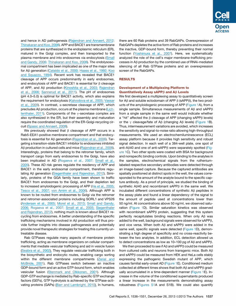
Development of a Multiplexing Platform toQuantitatively Assay sAPPb and Ab LevelsWe first developed a multiplexing assay to quantitatively screen
for Ab and soluble ectodomain of APP b (sAPPb), the two prod-
ucts of the amyloidogenic processing of APP (Figure 1A), from a
single sample. Simultaneous measurements of Ab and sAPPb
from a single sample in the same well would indicate whether
a ‘‘hit’’ affected the b cleavage of APP (changing sAPPb levels)
or the g cleavage/fate of Ab (changing Ab levels) (Figure 1B).
Thus, intermeasurement variations are avoided, which increases
the sensitivity and signal-to-noise ratio allowing high-throughput
measurements. We used an electrochemiluminescence (ECL)
assay platform because it provides high sensitivity and robust
signal detection. In each well of a 384-well plate, one spot of
anti-Ab40 and one of anti-sAPPb were separately spotted (Fig-
ure 1C). Two other spots were coated with BSA for background
and nonspecific binding controls. Upon binding to the analytes in
the samples, electrochemical signals from the ruthenium-
labeled respective secondary antibodies were detected through
an image-based capture. Because the captured antibodies are
spatially positioned at distinct spots in the well, the values corre-
sponded to the amount of the analyte bound to the specific cap-
ture antibody. As a proof of principle, we studied the binding of
synthetic Ab40 and recombinant sAPPb in the same well. We
incubated different concentrations of synthetic Ab peptides in
the assay plate and found a linear dependency of the signal to
the amount of peptide used at concentrations lower than
50 ng/ml. At concentrations above 50 ng/ml, we observed satu-
ration (Figure 1D). Similar saturation kinetics was observed
with recombinant sAPPb protein, suggesting that this system
perfectly recapitulates binding reactions. When only Ab was
added to the well, background signals were observed for sAPPb,
and vice versa. When both Ab and sAPPb were added in the
same well, specific signals were detected (Figure 1D), demon-
strating a high degree of specificity and no cross-reactivity be-
tween the two analytes. In addition, ECL detection allowed us
to detect concentrations as low as 10–100 pg of Ab and sAPPb.
We then proceeded to see if Ab and sAPPb could bemeasured
from cultured cells and neurons from transgenic mice. Both Ab
and sAPPb could be measured from HEK and HeLa cells stably
expressing the pathogenic Swedish mutant of APP, which
causes familial early-onset AD (Figure 1E). Conditioned medium
collected at different times shows that both Ab and sAPPb grad-
ually accumulated in a time-dependent manner (Figure 1E). In-
crease in the volume of the conditioned supernatants produced
a linear increase in the measurements demonstrating assay
robustness (Figures S1A and S1B). We could also quantify
ports 5, 1536–1551, December 26, 2013 ª2013 The Authors 1537
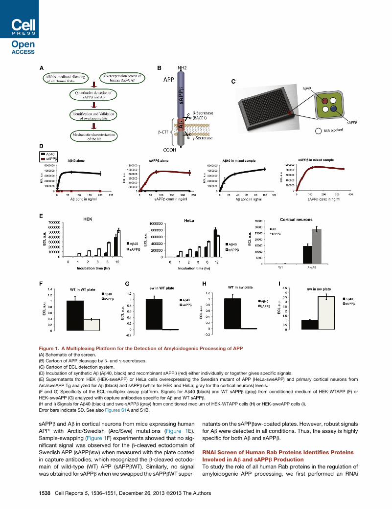
Figure 1. A Multiplexing Platform for the Detection of Amyloidogenic Processing of APP
(A) Schematic of the screen.
(B) Cartoon of APP cleavage by b- and g-secretases.
(C) Cartoon of ECL detection system.
(D) Incubation of synthetic Ab (Ab40, black) and recombinant sAPPb (red) either individually or together gives specific signals.
(E) Supernatants from HEK (HEK-sweAPP) or HeLa cells overexpressing the Swedish mutant of APP (HeLa-sweAPP) and primary cortical neurons from
Arc/sweAPP Tg analyzed for Ab (black) and sAPPb (white for HEK and HeLa; gray for the cortical neurons) levels.
(F and G) Specificity of the ECL-multiplex assay platform. Signals for Ab40 (black) and WT sAPPb (gray) from conditioned medium of HEK-WTAPP (F) or
HEK-sweAPP (G) analyzed with capture antibodies specific for Ab and WT sAPPb.
(H and I) Signals for Ab40 (black) and swe-sAPPb (gray) from conditioned medium of HEK-WTAPP cells (H) or HEK-sweAPP cells (I).
Error bars indicate SD. See also Figures S1A and S1B.
sAPPb and Ab in cortical neurons from mice expressing human
APP with Arctic/Swedish (Arc/Swe) mutations (Figure 1E).
Sample-swapping (Figure 1F) experiments showed that no sig-
nificant signal was observed for the b-cleaved ectodomain of
Swedish APP (sAPPbsw) when measured with the plate coated
in capture antibodies, which recognized the b-cleaved ectodo-
main of wild-type (WT) APP (sAPPbWT). Similarly, no signal
was obtained for sAPPbwhenwe swapped the sAPPbWT super-
1538 Cell Reports 5, 1536–1551, December 26, 2013 ª2013 The Aut
natants on the sAPPbsw-coated plates. However, robust signals
for Ab were detected in all conditions. Thus, the assay is highly
specific for both Ab and sAPPb.
RNAi Screen of Human Rab Proteins Identifies ProteinsInvolved in Ab and sAPPb ProductionTo study the role of all human Rab proteins in the regulation of
amyloidogenic APP processing, we first performed an RNAi
hors

screen (Table S1) in cells that robustly produce Ab and sAPPb
(Rajendran et al., 2006, 2008) and assayed these products using
the multiplexing ECL assay system (Figures 2A and 2B). We
included APP, BACE1, and Pen2 (a subunit of the g-secretase)
as positive controls. As expected, silencing of APP and BACE1
decreased both Ab and sAPPb levels, whereas silencing of
Pen2 decreased Ab levels, but not sAPPb levels (Figures 2A
and 2B), further validating the assay. Quantification was based
on the ECL counts, normalized to the cell viability counts, and
relative to that of the scrambled control (medium GC containing
siRNA oligo [MedGC]). Apart from Rab36, silencing of the other
Rab proteins did not significantly alter cell viability (Figure S1C).
The screen identified Rabs that significantly decreased both Ab
and sAPPb (Figures 2A and 2B), including Rab3A, Rab11A,
Rab36, and Rab17 (Figures S2A and S2B). Rab36, a GTPase
involved in late endosome positioning, was a top hit in the
screen, decreasing both Ab and sAPPb (Figure 2C). A secondary
validation screen reproduced all the selected hits (Figure S2C).
Knockdown efficiency of the relevant genes was confirmed by
RT-PCR (Figure S2D). Although our analysis accounts for
toxicity, and quantifies Ab and sAPPb as a relative count of viable
cells, Rab36 depletion was consistently toxic in all the screens
(Figure S1C). Therefore, we removed it from further analysis.
Rab11A, the major Rab protein involved in the slow recycling
of cargo proteins from early endosomes to the cell surface (Son-
nichsen et al., 2000), was the second top hit in our analysis. In
addition, silencing of the isoform, Rab11B, also reduced Ab
levels (Figure 2A). Interestingly, all the isoforms of Rab3, except
Rab3C, decreased both Ab and sAPPb levels. Rab3 proteins are
involved in synaptic function (Schluter et al., 2004) and in the fast
axonal transport of APP (Szodorai et al., 2009). Silencing of
Rab3A and Rab3B decreased overall APP levels, suggesting
that Rab3 plays a role in the trafficking and maintenance of
APP levels (Figure S2E).
Silencing of Rab44, Rab6A, and Rab10 decreased Ab levels
without affecting sAPPb (Figure 2C), implying that these
GTPases affect either g-secretase cleavage or the fate of Ab.
Interestingly, knockdowns of several Rabs also increased both
sAPPb and Ab levels. To understand how these trafficking path-
ways contributed to both Ab and sAPPb levels, we plotted the
normalized values of Ab and sAPPb from the screen in a 2D
plot and observed a largely correlative curve for both the values,
indicating that the Rab proteins responsible for regulating b
cleavage are also responsible for controlling Ab levels and that
any perturbation at the level of BACE1 cleavage of APP is the
rate-limiting step in Ab production (Figure 2D).
RabGAP-Overexpression Screen Identifies OverlappingHits with the Rab RNAi ScreenBecause RNAi can have off-target effects leading to false-posi-
tive results, we complemented our RNAi screen with an overex-
pression screen of all 39 RabGAPs in the human genome, which
suppress Rab function by accelerating the hydrolysis of GTP. In
this screen, we overexpressed each RabGAP in the same
cellular system used for the RNAi screen and quantitatively
measured Ab, sAPPb levels, and cell viability (Figures 3 and
S1D). Many RabGAPs were found to affect sAPPb and Ab levels
(Figure 3). RN-TRE, a GAP that has previously been shown to
Cell Re
affect amyloidogenic processing of APP (Ehehalt et al., 2003),
reduced both Ab and sAPPb. TBC1D10B, a GAP for Rab35,
Rab27A, Rab22A, Rab31, and Rab3A, decreased both Ab and
sAPPb (Frasa et al., 2012). However, only Rab3A silencing via
RNAi decreased Ab and sAPPb (Figures 2 and 3). EVI5, a
RabGAP for Rab11 (Frasa et al., 2012; Laflamme et al., 2012),
decreased both Ab and sAPPb. In addition, one other RabGAP
for Rab11, namely TBC1D15, decreased both Ab and sAPPb.
Intriguingly, TBC1D14, which alters Rab11 localization and
subsequently delays recycling of transferrin to the plasma mem-
brane (Longatti et al., 2012), decreased Ab levels (Figure 3C).
These results independently identified Rab11 as an important
regulator of sAPPb and Ab production.
Epistasis Mini Array Profiling Identifies DistinctTrafficking RoutesWe next analyzed whether the identified Rabs regulated Ab pro-
duction through the same trafficking pathway or through distinct
mechanisms. To this end, we performed an epistasis mini array
profiling (EMAP) screen (Schuldiner et al., 2005) wherein we
silenced all the hits against all the hits in a 14 3 14 matrix to
determine whether a combined knockdown gives rise to an
aggravating (noninteracting) or alleviating (interacting) pheno-
type (i.e., b cleavage of APP). Here, again, single knockdowns
of either Rab3A or Rab11A decreased sAPPb levels (Figure S3).
However, when they were silenced together, a further decrease
(an aggravating phenotype) was observed, which indicates that
Rab3A and Rab11A act independently and contribute to b cleav-
age via distinct trafficking routes. Most of the genes that were
identified as hits did not interact with each other, with the excep-
tion that a double knockdown of Rab10/Rab23 or Rab10/Rab25
showed an alleviating phenotype, suggesting that these Rabs
regulate the same membrane-trafficking pathways.
Validation of Rab11A as a HitBecause Rab11A was identified as one of the strongest hits in
both the RNAi screen and the RabGAP screen, we independently
validated this finding in a different cellular model. siRNA-medi-
ated silencing of Rab11A in HEK cells stably expressing WTAPP
led to a strong reduction of both Ab and sAPPb levels (Fig-
ure S4A), similar to the effect observed upon silencing of
Rab11A in HeLa cells stably expressing sweAPP (Figure 4A).
Consistent with this, western blotting analysis with APP C termi-
nus antibodies revealed a strong reduction in b-C-terminal
fragment (CTF) levels (C99) with an increase in a-CTF levels (Fig-
ure 4B). This suggests that Rab11A silencing markedly affected
b cleavage of APP and also Ab levels. Western blotting with
Rab11A antibodies showed that Rab11A silencing efficiently
knocked down the protein (Figure 4B) (with >90% efficiency).
In addition, Rab11A silencing led to a decrease in all three spe-
cies of Ab (Ab38, Ab40, and Ab42) (Figure 4C) and reduced
amyloid levels in the cell lysates, suggesting that Rab11A regu-
lates Ab production rather than secretion (Figure S4B). Silencing
of Rab11A using four different siRNAs singly also led to a
decrease in Abwithout affecting cell viability, thus demonstrating
the specificity of the observed effect (Figure 4D). All siRNAs
against Rab11A also silenced Rab11A expression (Figure 4D,
inset).
ports 5, 1536–1551, December 26, 2013 ª2013 The Authors 1539

(legend on next page)
1540 Cell Reports 5, 1536–1551, December 26, 2013 ª2013 The Authors
To further validate these results, we employed a different strat-
egy to interfere with Rab11 function. Because Rab proteins exist
in both a GTP-bound and GDP-bound form, we overexpressed
the GDP-locked (dominant negative; DN) mutant forms of
Rab11A (the ubiquitously expressed isoform) and Rab11B (the
isoform highly expressed in neurons) in cells to phenocopy
the effect of Rab11 silencing. Similar to silencing of Rab11A,
overexpression of Rab11A DN reduced Ab and sAPPb levels sig-
nificantly. Overexpression of Rab11B DN (which would also
interfere with the function of WT Rab11) also significantly
reduced Ab and sAPPb levels (Figure 4E).
Finally, we assessed the effect of silencing the expression of
evolutionarily conserved Rab11 family-interacting proteins on b
cleavage of APP in the cells (Lindsay and McCaffrey, 2002).
Knockdown of four of the five FIP family proteins (Shiba
et al., 2006), with the exception of Rab11FIP1 (retromer-like),
decreased sAPPb levels (Figure 4F). Thus, not only Rab11 but
also Rab11-interacting proteins, which were identified as
Rab11-effector proteins, reduced b cleavage of APP.
Rab11 Silencing in Primary Neurons from WT and APPTransgenic Mice Reduces Ab LevelsWe next studied if Rab11 also regulated Ab production in the
neuronal context. Silencing Rab11A and Rab11B in primary neu-
rons isolated from APP transgenic mice significantly decreased
both sAPPb and Ab levels (Figure 4G). Both RT-PCR andwestern
blotting analysis show efficient silencing of Rab11A and Rab11B
(Figures S4C, S4D, and 4H; Table S2). Knockdown of Rab11A
and Rab11B in primary neurons isolated from WT mice again
reduced Ab (both Ab40 and Ab42) levels (Figures 4I and 4J).
Moreover, silencing either Rab11A or Rab11B also significantly
decreased Ab levels (Figure S4E). Thus, Rab11 is crucial for b
cleavage and Ab generation.
Rab11 Controls Membrane Trafficking of BACE1We hypothesized that Rab11 must regulate either at the level of
APP or at the level of BACE1. Rab11 silencing or Rab11A DN
expression did not affect the total cellular levels of APP or
BACE1 (Figures 4B and S4F, respectively). To analyze protein
localization, we performed live-cell imaging of cells transfected
with either APP-YFP or BACE1-YFP using a wide-field micro-
scope maintained under a temperature-controlled environment.
BACE1-YFP prominently localized to the perinuclear endocytic-
recycling compartment (ERC) and was also found in numerous
highly motile vesicular and tubular structures dispersed
throughout the cell (Figure 5A; Movie S1). Inspection of individual
carriers revealed rapid transport of tubulo-vesicular structures
along a curvy linear trajectory, suggesting active transport along
microtubules (Movie S1). Indeed, brief treatment of cells with the
Figure 2. RNAi Screen of Genome-wide Rabs Identifies Rabs Involved
(A and B) Graph showing the levels of Ab (A) and sAPPb (B) from the Rab siRNA
control), or pooled siRNA against APP, BACE1, PEN2, and the 60 Rabs and ass
(C) List of genes that had the strongest effect on Ab and sAPPb levels after siRN
(D) 2D plot representing the levels of Ab and sAPPb from the Rabs siRNA screen.
control Scrambled is indicated in blue. Rabs silencing that led to the strongest de
using the two-tailed t tests and one-way ANOVA (Table S4).
See also Figures S1C, S2, and S3.
Cell Re
microtubule-disrupting reagent nocodazole inhibited dynamic
transport of BACE1 and induced the formation of enlarged
immobile vesicles (data not shown). The distribution and dy-
namics of BACE1 trafficking, but not of APP, were markedly
altered by expression of Rab11AS25N, the DN mutant of Rab11
(Rab11A DN) (Figure 5A; Movies S1 and S2). The length of
the BACE1-positive tubular carriers emanating from the ERC
was significantly enhanced by Rab11A DN overexpression but
remained highly dynamic, suggesting that GTPase activity of
Rab11 is involved in the fission of BACE1 tubular carriers and/or
their tethering and fusion with the target.
Rab11 GTPase is involved in recycling many cargos from the
ERC, including the recycling endosome marker transferrin re-
ceptor (TfR) (Sonnichsen et al., 2000). TfR as well as internalized
transferrin become associated with a distinct tubular network in
cells expressing Rab11 DN (Choudhury et al., 2002; Wilcke et al.,
2000). To address whether BACE1 follows the recycling route
from recycling endosomes, we first checked whether BACE1
colocalizes with TfR. BACE1 significantly colocalized with TfR
(Figure 5B). Not only the steady-state levels of TfR but also inter-
nalized transferrin colocalized with BACE1 in Rab11 compart-
ments (data not shown). In control cells and those transfected
with Rab11A WT, BACE1-YFP colocalized with TfR in the ERC
as well as in numerous vesicles and short tubulo-vesicular car-
riers (Figure 5B). However, in cells expressing Rab11A DN,
BACE1 and TfR were found in elongated tubular structures
that emanated from the ERC (Figure 5B). Neither the prominent
perinuclear localization of APP in the TGN nor the dynamics of
APP-positive structures was disturbed by Rab11A DN expres-
sion (Figure 5A). Thus, BACE1 localization and trafficking are
akin to that of TfR, which requires recycling via the Rab11-medi-
ated pathway for transferrin internalization. Because Rab11
regulates the recycling of TfR through the slow recycling route,
it may also regulate the recycling of BACE1, thus affecting
both the cell surface as well as the early endosomal trafficking
of BACE1.
Rab11 Affects the Recycling of BACE1Because BACE1 accumulated in elongated tubulo-vesicular
structures in cells expressing Rab11A DN, we hypothesized
that under these conditions, BACE1 is unable to be trafficked
to the plasma membrane and thus is unavailable for reinternal-
ization to early endosomes, the site of Ab production. To test
this, we performed two different experiments. First, we tested
if Rab11A regulated cell surface levels of BACE1 using FLAG-
tagged BACE1 (Figure S5A). Here, we used both FACS (fluores-
cence-activated cell sorting) as well as immunofluorescence on
cells transfected with either control or Rab11A DN-expressing
plasmids. Cells expressing Rab11A DN indeed displayed
in Regulating the Levels of Ab and sAPPb
screen. HeLa-sweAPP cells were transfected with Scrambled oligo (negative
ayed for Ab and sAPPb. Error bars indicate SEM.
A-mediated silencing along with the Z score and t test values.
Positive controls APP, BACE1, and PEN2 are indicated in green, and negative
crease in both Ab and sAPPb is indicated in red. All statistics were performed
ports 5, 1536–1551, December 26, 2013 ª2013 The Authors 1541
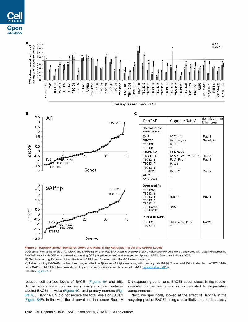
Figure 3. RabGAP Screen Identifies GAPs and Rabs in the Regulation of Ab and sAPPb Levels
(A) Graph showing the levels of Ab (black) and sAPPb (gray) after RabGAP plasmid overexpression. HeLa-sweAPP cells were transfectedwith plasmid expressing
RabGAP fused with GFP or a plasmid expressing GFP (negative control) and assayed for Ab and sAPPb. Error bars indicate SEM.
(B) Graphs showing Z scores of the effects on sAPPb and Ab levels after RabGAP overexpression.
(C) Table showing RabGAPs that had the strongest effect on Ab and/or sAPPb levels alongwith their cognate Rab(s). The asterisk (*) indicates that the TBC1D14 is
not a GAP for Rab11 but has been shown to perturb the localization and function of Rab11 (Longatti et al., 2012).
See also Figure S1D.
reduced cell surface levels of BACE1 (Figures 6A and 6B).
Similar results were obtained using imaging of cell surface-
labeled BACE1 in HeLa (Figure 6C) and primary neurons (Fig-
ure 6D). Rab11A DN did not reduce the total levels of BACE1
(Figure S4F), in line with the observations that under Rab11A
1542 Cell Reports 5, 1536–1551, December 26, 2013 ª2013 The Aut
DN-expressing conditions, BACE1 accumulates in the tubulo-
vesicular compartments and is not rerouted to degradative
compartments.
Next, we specifically looked at the effect of Rab11A in the
recycling pool of BACE1 using a quantitative ratiometric assay
hors
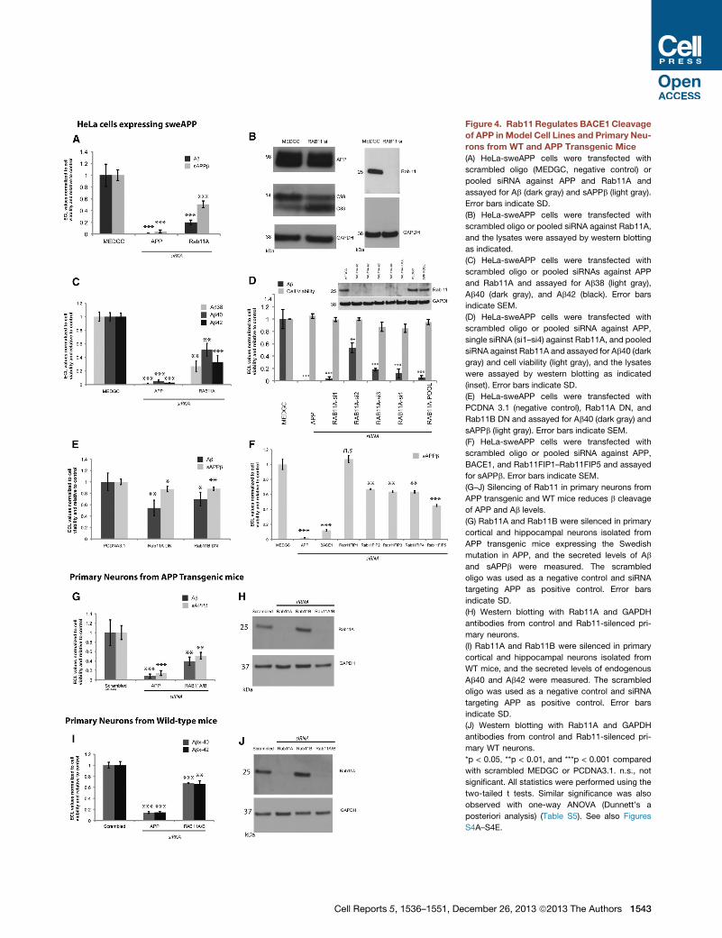
Figure 4. Rab11 Regulates BACE1 Cleavage
of APP in Model Cell Lines and Primary Neu-
rons from WT and APP Transgenic Mice
(A) HeLa-sweAPP cells were transfected with
scrambled oligo (MEDGC, negative control) or
pooled siRNA against APP and Rab11A and
assayed for Ab (dark gray) and sAPPb (light gray).
Error bars indicate SD.
(B) HeLa-sweAPP cells were transfected with
scrambled oligo or pooled siRNA against Rab11A,
and the lysates were assayed by western blotting
as indicated.
(C) HeLa-sweAPP cells were transfected with
scrambled oligo or pooled siRNAs against APP
and Rab11A and assayed for Ab38 (light gray),
Ab40 (dark gray), and Ab42 (black). Error bars
indicate SEM.
(D) HeLa-sweAPP cells were transfected with
scrambled oligo or pooled siRNA against APP,
single siRNA (si1–si4) against Rab11A, and pooled
siRNA against Rab11A and assayed for Ab40 (dark
gray) and cell viability (light gray), and the lysates
were assayed by western blotting as indicated
(inset). Error bars indicate SD.
(E) HeLa-sweAPP cells were transfected with
PCDNA 3.1 (negative control), Rab11A DN, and
Rab11B DN and assayed for Ab40 (dark gray) and
sAPPb (light gray). Error bars indicate SEM.
(F) HeLa-sweAPP cells were transfected with
scrambled oligo or pooled siRNA against APP,
BACE1, and Rab11FIP1–Rab11FIP5 and assayed
for sAPPb. Error bars indicate SEM.
(G–J) Silencing of Rab11 in primary neurons from
APP transgenic and WT mice reduces b cleavage
of APP and Ab levels.
(G) Rab11A and Rab11B were silenced in primary
cortical and hippocampal neurons isolated from
APP transgenic mice expressing the Swedish
mutation in APP, and the secreted levels of Ab
and sAPPb were measured. The scrambled
oligo was used as a negative control and siRNA
targeting APP as positive control. Error bars
indicate SD.
(H) Western blotting with Rab11A and GAPDH
antibodies from control and Rab11-silenced pri-
mary neurons.
(I) Rab11A and Rab11B were silenced in primary
cortical and hippocampal neurons isolated from
WT mice, and the secreted levels of endogenous
Ab40 and Ab42 were measured. The scrambled
oligo was used as a negative control and siRNA
targeting APP as positive control. Error bars
indicate SD.
(J) Western blotting with Rab11A and GAPDH
antibodies from control and Rab11-silenced pri-
mary WT neurons.
*p < 0.05, **p < 0.01, and ***p < 0.001 compared
with scrambled MEDGC or PCDNA3.1. n.s., not
significant. All statistics were performed using the
two-tailed t tests. Similar significance was also
observed with one-way ANOVA (Dunnett’s a
posteriori analysis) (Table S5). See also Figures
S4A–S4E.
Cell Reports 5, 1536–1551, December 26, 2013 ª2013 The Authors 1543

Figure 5. BACE1 Colocalizes with Rab11 Compartments, and Its Trafficking Is Altered upon Rab11 Dysfunction
(A) Live-cell images of COS cells transfected with BACE1-YFP or YFP-APP along with empty vector (control) or Rab11A DN. Expression of Rab11A DN protein is
shown in the insets.
(B) COS cells were transiently cotransfected with BACE1-YFP (green) and empty vector, HA-tagged Rab11A WT or S25N DNmutant, and fixed and stained with
HA (data not shown) and TfR antibodies (red) to label recycling endosomes. The colors in the first two panels are green for BACE1-YFP and red for TfR. Insets are
the zoomed-in images of the corresponding images.
See also Figure S5A and Movies S1 and S2.
to measure recycling by differential labeling. To this end,
cells were labeled with Alexa 647-conjugated M1 anti-FLAG
for 30 min at 37�C, to allow antibodies to bind surface
BACE1 and be internalized. After internalization, cells were
incubated with Alexa 488-conjugated secondary antibodies
for 30 min at 37�C, to detect internalized anti-FLAG-bound
receptors that recycled to the surface. In the Rab11A DN-
expressing cells, BACE1 recycling was significantly decreased,
confirming that Rab11 functions to recycle BACE1 from the
endosomes to the plasma membrane (Figure 6E). Quantitation
of the fluorescence signal clearly revealed that the recycling
pool of BACE1 is markedly decreased in cells expressing
Rab11A DN. Two parallel controls were used: a surface con-
trol, where both incubations were performed at 4�C; and an
endocytosis control, where the first incubation was at 37�Cand the second at 4�C (Figure S5B). Cells without BACE1
did not show any labeling under these conditions. Together,
these results suggest that vesicular trafficking regulated by
Rab11 GTPase is critically important for the homeostatic regu-
lation of BACE1 trafficking and Ab production in endocytic
organelles.
Rab11 Genetic Variability in LOADWe used exome-sequencing data from 170 neuropathologically
assessed controls and 185 patients with LOAD to study if vari-
ants in Rab11A were associated with AD. Our results revealed
a significant association of a variant in Rab11A (rs117150201;
uncorrected p value = 0.01), thus establishing a potential associ-
ation between Rab11 and AD (Table S3).
1544 Cell Reports 5, 1536–1551, December 26, 2013 ª2013 The Aut
Network Analysis of GWAS Genes Linked to LOADIdentifies Rab11A and Rab11B as Interacting ProteinsTo determine whether other genes linked with LOAD risk also
acted via Rab11, we used protein-protein interaction (PPI)
network analysis to study the interaction partners of proteins
linked to LOAD risk. We used the protein products of top ten
genes linked to LOAD (AlzGene; http://www.alzgene.org) and
created a two-level depth interactome of these proteins. In the
cluster with Bin1, a risk gene that is strongly associated with
LOAD risk, we identified both Rab11A and Rab11B and their
interacting proteins, Rab11FIP1–Rab11FI5 (Figure S6). In our
RNAi-based screen, Rab11A, Rab11B, and all the FIPs (except
for FIP1) regulated Ab levels, further supporting the findings
that the LOAD risk gene, Bin1, could contribute to amyloid pro-
duction via Rab11. Interestingly, the Bin1 cluster also included
Arf6, a known player in BACE1 endocytosis to early endosomal
compartments (Sannerud et al., 2011) and GGA3, which regu-
lates early endosomal residency of BACE1 (Tesco et al., 2007),
further supporting the link between Bin1, Rab11 and BACE1
endocytosis and recycling. These results from this unbiased
network analysis strongly link Rab11 to LOAD risk.
Rabs and Membrane-Trafficking Pathways in AmyloidSecretionUsing a systems approach, our screen also identified other
regulatory active Rabs, and it provides insights into mem-
brane-trafficking pathways that play crucial roles in amyloid
production and secretion. This allowed us to present a compre-
hensive road map for APP processing (Figure 7). Most of the hits
hors

were Rabs involved in the regulation of the trafficking either to or
from early endosomes (Galvez et al., 2012; Pfeffer and Aivazian,
2004), confirming the critical role of early endosomes in Ab
generation (Kinoshita et al., 2003; Koo and Squazzo, 1994;
Rajendran et al., 2006; Small and Gandy, 2006). In addition to
Rab11, several other Rabs were identified to also regulate Ab
levels. Silencing of Rab2A, for instance, increased Ab and sAPPb
levels (Figure 2), and the cognate GAP, TBC1D11, showed a
similar effect (Figure 3). Rab2A is involved in the maintenance
of Golgi structure and function. It interacts with the atypical pro-
tein kinase C (aPKC) (Tisdale, 2003) that positively regulates a
cleavage of APP via a-secretase (Nitsch et al., 1993).
On the other side, Rab5C silencing significantly increased
sAPPb and Ab, whereas the other Rab5 isoforms did not.
Rab5C is involved in the endocytosis and recycling of cell sur-
face molecules (Onodera et al., 2012). Silencing of Rab5C
dramatically increased the cellular levels of APP (Figure S2E),
which explains the increase in sAPPb and Ab.
The Rab proteins that have been implicated in exosome
release, namely Rab27 and Rab35 (Hsu et al., 2010; Ostrowski
et al., 2010), negatively regulated Ab and sAPPb levels
(Figure 2). This is consistent with our previous work showing
that very small amounts of Ab can be released via exosomal ves-
icles, and suggests that the exosomal pathway is connected to an
intracellular degradation mechanism and that inhibition of the
exosomal pathway reroutes the cargo for effective secretion.
DISCUSSION
To rule out potential off-target effects of the RNAi screen, we
complemented it with five independent experimental conditions:
(1). The first was an unbiased Rab-GAP overexpression screen; a
paired RNAi screen (of all human Rab GTPases) and an overex-
pression screen (of RabGAPs) were performed; (2) Rab11 DN
phenocopied the effect of RNAi of Rab11. (3) All four different
siRNAs against Rab11 decreased Ab levels. (4) Rab11 RNAi
had the same effect in many different cells, including primary
neurons. Finally, (5) silencing Rab11-associated proteins such
as EHD or FIPs produced similar effect.
The results from our RNAi, RabGAP, and EMAP screens pro-
vide a valuable list of all the human Rab proteins that affect Ab
and sAPPb levels and their nature of interaction. We also
silenced different isoforms of a particular Rab by means of
RNAi, and indeed, in many cases, we found isoform-specific
effects on Ab levels.
Silencing of Rab4A, Rab6A, and Rab10 decreased only Ab.
Conversely, silencing of Rab8A, Rab25, Rab14, and Rab34
significantly increased only Ab. Such Ab-specific effects are
likely the result of either an altered g-secretase cleavage of
APP or a change in secretion and/or degradation of the pro-
duced Ab. Of particular interest is Rab8, which increased Ab
levels when depleted in cells. Rab8 is involved in various physi-
ological functions such as exocytosis and cilia formation. Inter-
estingly, expression of a mutant PS1 (A260V) in PC12 cells
dramatically decreased Rab8 levels, thus affecting trafficking
via Golgi (Kametani et al., 2004). We also identified some hits
that specifically decreased sAPPb levels without affecting Ab.
Although this at first seems counterintuitive, it implies the possi-
Cell Re
bility that specific Rabs play a role in the release/secretion of
sAPPb without affecting the processing of APP. However, we
see a clear overall correlation of sAPPb and Ab, which suggests
two things: (1) b cleavage is rate limiting and thus determines
cellular and secreted Ab levels, or (2) compartments that define
b cleavage are also involved in g cleavage of APP to generate Ab
(Kaether et al., 2006; Rajendran et al., 2006).
We identified Rab11 as a main regulator of APP processing.
Both by ECL assays and through western blotting to study
APP processing, we show that BACE1-mediated processing of
APP is affected in Rab11-silenced cells. Interestingly, whereas
we see a significant reduction in C99 levels, C83 levels are
increased. This could be due to an increased a cleavage of
APP or C99 or could also represent a defect in g-secretase
cleavage, demonstrating that Rab11-recycling endosomes
play a crucial role in APP processing and Ab production. Indeed,
Rab11 has been shown to interact with g-secretase component,
PS (Dumanchin et al., 1999; Wakabayashi et al., 2009). However,
we did not observe any influence of Rab11A on the activity or
localization of g-secretase (data not shown).
APP endocytosis is mediated by the YENPTY cytosolic motif
and is regulated by both cholesterol-dependent and clathrin-
mediated endocytic pathways (Perez et al., 1999; Schneider
et al., 2008). Similarly, endocytosis of BACE1 has been shown
to be both clathrin dependent and clathrin independent via the
ADP-ribosylation factor 6 pathway (Prabhu et al., 2012; San-
nerud et al., 2011). So far, early endosomes, TGN, and the
plasmamembrane have been suggested to contribute to Ab pro-
duction (Choy et al., 2012; Chyung and Selkoe, 2003; Rajendran
et al., 2006; Sannerud et al., 2011). Although APP is known to be
routed from endosomes to Golgi via the retromer and associated
proteins (Andersen et al., 2005; Morel et al., 2013; Small and
Gandy, 2006; Rogaeva et al., 2007; Small et al., 2005; Sie-
genthaler and Rajendran, 2012), nothing much is known about
BACE1 recycling from endosomes.
Our results now identify the Rab11-dependent slow recycling
pathway as a trafficking route involved in Ab generation. A
recent study showed that synaptic activity mediates the conver-
gence of APP and BACE1 in acidicmicrodomains that colocalize
with TfR (Das et al., 2013). Because this study did not look at
colocalization of APP and BACE1 in Rab11 compartments and
that a significant proportion of TfR is also found in early endo-
somes (Sonnichsen et al., 2000), the interaction most likely
occurs in early endosomes because TfR is internalized from
the plasma membrane to early endosomes and then is recycled
to the plasma membrane via Rab11-positive vesicles. Several
studies suggest that early endosomes are the compartments
where BACE1 cleaves APP, also in the neuronal context (Choy
et al., 2012; Chyung and Selkoe, 2003; Rajendran et al., 2006;
Sannerud et al., 2011). The fact that retromer-associated pro-
teins or GGA3 regulates Ab levels is due to its involvement in
sorting of APP or BACE1, respectively, from early endosomes
and not recycling endosomes (Andersen et al., 2005; Morel
et al., 2013; Siegenthaler and Rajendran, 2012; Tesco et al.,
2007). Furthermore, early endosomes are acidic (Maxfield and
Yamashiro, 1987), and recycling endosomes are not because
they lack functional vacuolar ATPase (Gagescu et al., 2000;
Schmidt and Haucke, 2007). In addition, we do not observe
ports 5, 1536–1551, December 26, 2013 ª2013 The Authors 1545
(legend on next page)
1546 Cell Reports 5, 1536–1551, December 26, 2013 ª2013 The Authors
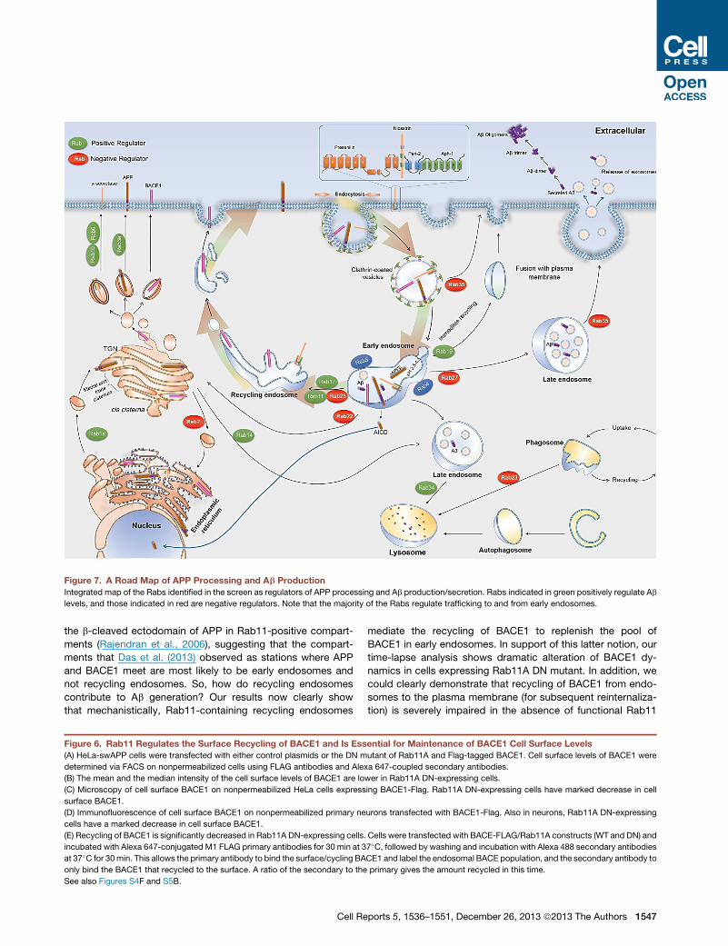
Figure 7. A Road Map of APP Processing and Ab Production
Integrated map of the Rabs identified in the screen as regulators of APP processing and Ab production/secretion. Rabs indicated in green positively regulate Ab
levels, and those indicated in red are negative regulators. Note that the majority of the Rabs regulate trafficking to and from early endosomes.
the b-cleaved ectodomain of APP in Rab11-positive compart-
ments (Rajendran et al., 2006), suggesting that the compart-
ments that Das et al. (2013) observed as stations where APP
and BACE1 meet are most likely to be early endosomes and
not recycling endosomes. So, how do recycling endosomes
contribute to Ab generation? Our results now clearly show
that mechanistically, Rab11-containing recycling endosomes
Figure 6. Rab11 Regulates the Surface Recycling of BACE1 and Is Ess
(A) HeLa-swAPP cells were transfected with either control plasmids or the DN m
determined via FACS on nonpermeabilized cells using FLAG antibodies and Alex
(B) The mean and the median intensity of the cell surface levels of BACE1 are lo
(C) Microscopy of cell surface BACE1 on nonpermeabilized HeLa cells express
surface BACE1.
(D) Immunofluorescence of cell surface BACE1 on nonpermeabilized primary ne
cells have a marked decrease in cell surface BACE1.
(E) Recycling of BACE1 is significantly decreased in Rab11A DN-expressing cells.
incubated with Alexa 647-conjugated M1 FLAG primary antibodies for 30 min at 3
at 37�C for 30min. This allows the primary antibody to bind the surface/cycling BA
only bind the BACE1 that recycled to the surface. A ratio of the secondary to the
See also Figures S4F and S5B.
Cell Re
mediate the recycling of BACE1 to replenish the pool of
BACE1 in early endosomes. In support of this latter notion, our
time-lapse analysis shows dramatic alteration of BACE1 dy-
namics in cells expressing Rab11A DN mutant. In addition, we
could clearly demonstrate that recycling of BACE1 from endo-
somes to the plasma membrane (for subsequent reinternaliza-
tion) is severely impaired in the absence of functional Rab11
ential for Maintenance of BACE1 Cell Surface Levels
utant of Rab11A and Flag-tagged BACE1. Cell surface levels of BACE1 were
a 647-coupled secondary antibodies.
wer in Rab11A DN-expressing cells.
ing BACE1-Flag. Rab11A DN-expressing cells have marked decrease in cell
urons transfected with BACE1-Flag. Also in neurons, Rab11A DN-expressing
Cells were transfected with BACE-FLAG/Rab11A constructs (WT and DN) and
7�C, followed by washing and incubation with Alexa 488 secondary antibodies
CE1 and label the endosomal BACE population, and the secondary antibody to
primary gives the amount recycled in this time.
ports 5, 1536–1551, December 26, 2013 ª2013 The Authors 1547

using both FACS and recycling assays. In addition, Rab17,
another protein involved in the recycling of internalized cargo,
also regulated sAPPb and Ab levels, thus underscoring the
importance of recycling endosomes in APP processing.
Our screen and validation results uncover a trafficking route
involved in Ab production. Because BACE1 cleavage of APP is
critically involved in the pathogenesis of early-onset AD (Yang
et al., 2003) and in certain forms of sporadic AD (Jonsson
et al., 2012), we propose that the Rab11-mediated recycling
route represents a drug target for AD, which is supported by
our exome-sequencing results and the unbiased PPI network
analysis. Interestingly, Eps15 homology domain-containing
(EHD) proteins, which form complexes with Rab11 via Rab11-
FIP2, were independently identified as critical regulators of
dynamic BACE1 trafficking and axonal sorting in hippocampal
neurons. Moreover, knockdown of EHD1 or EHD3 significantly
reduced Ab levels in primary neurons (Buggia-Prevot et al.,
2013). The network model suggests that the interaction of Bin1
with Rab11 is via Arf6, a known player in BACE1 endocytosis
to early endosomal compartments (Sannerud et al., 2011), and
GGA3, a protein that regulates early endosomal residency of
BACE1, (Tesco et al., 2007) is also involved in the network,
further supporting the link between AD risk and Rab11-mediated
BACE1 endocytosis and recycling.
Taken together, our results clearly establish a link for recycling
endosomes in BACE1 recycling, Ab generation, and AD and
underscore the importance of systems level analysis in under-
standing the complexity of AD. An extended version of the
Experimental Procedures can be found in the Supplemental
Experimental Procedures.
EXPERIMENTAL PROCEDURES
cDNA Constructs
Plasmids encoding dsRed-tagged humanWTRab11A and DNmutant (S25N)
(Choudhury et al., 2002) were purchased from Addgene. C-terminally EYFP-
tagged mouse BACE1 was generated by subcloning BACE1 cDNA (provided
by Nabil G. Seidah), in-frame into the pEYFP-N1 vector. HA-tagged Rab11A
constructs have been described by Ren et al. (1998). The sequence corre-
sponding to the Flag epitope in the BACE1-FLAG construct was introduced
after the propeptide cleavage site within the luminal domain of mouse BACE1
using the following primers: 50-CCGGGAGACCGACTACAAGGACGATGAT
GACAAGGGGGGAGGATC-30 and 50-CCGGGATCCTCCCCCCTTGTCATCA
TCGTCCTTGTAGTCGGTCTC-30.
siRNA
siRNAs were purchased from Invitrogen (stealth siRNA). The sequences and
the accession codes are supplied in Table S1.
RNAi Screen
RNAi screen was performed using HeLa cells expressing the Swedish APP
mutation (HeLa-sweAPP). siRNAs were transfected with a final concentration
of 5 nM using Oligofectamine (Invitrogen) as transfection reagent at a concen-
tration of 0.3 ml in a total volume of 100 ml followingmanufacturer’s instructions.
Each siRNA transfection was performed in quadruplicate. After 24 hr, the
transfection mix on the cells was replaced with fresh culture medium. At
69 hr after transfection, medium was again replaced with 100 ml fresh medium
containing 10% Alamar blue (AbD Serotec). At 72 hr after transfection, Alamar
blue measurements were taken using SpectraMAX GeminiXS (Molecular
Devices) at an excitation wavelength of 544 nm and emission at 590 nm.
Supernatant was collected and assayed for Ab and sAPPb. The cells in the
transfected plate were lysed with 50 ml of lysis buffer for 20 min on ice.
1548 Cell Reports 5, 1536–1551, December 26, 2013 ª2013 The Aut
RabGAP Screen
The RabGAP screen was performed in HeLa-sweAPP cells. Cells were
seeded in 96-well plates at a density of 6,000 cells/well 1 day before trans-
fection. Effectene (QIAGEN) was used as transfection reagent following
manufacturer’s instruction using 2.5 ml of Effectene and 0.8 ml of Enhancer
in a total volume of 100 ml. Transfection mix was replaced with fresh medium
after 3.5 hr. At 24 hr after transfection, medium was again replaced with
fresh medium containing 10% Alamar blue. At 27 hr after transfection,
Alamar blue measurements were taken using SpectraMAX GeminiXS at an
excitation wavelength of 544 nm and emission at 590 nm. Supernatant was
collected and assayed for Ab and sAPPb using the ECL assay (see Supple-
mental Experimental Procedures). The cells in the transfected plate were
lysed with 30 ml of lysis buffer, incubated for 20 min on ice, and stored
at �20�C.
Live-Cell Image Acquisition and Processing
Images were acquired on a motorized NikonTe2000 microscope equipped
with a Cascade II:512 CCD Camera (Photometrics) using a 1003 objective
(NA 1.4). During live imaging, cells weremaintained at 37�C in imagingmedium
(140 mM NaCl, 5 mM KCl, 3 mM CaCl2, 2 mM MgCl2, 1.5 mM D-glucose, and
10 mM HEPES [pH 7.4]) in a custom-designed environment chamber. Time-
lapse images were acquired at the rate of two frames/s. Images of fixed cells
were acquired as 200 nm z stacks, deconvolved using Huygens software (Sci-
entific Volume Imaging). Extended Depth of Field plugin of ImageJ software
(National Institutes of Health) was used to generate single-plane projections
from processed z stacks (Rasband, 1997–2012).
Recycling Assay
Cells were labeled with Alexa 647-conjugated M1 anti-FLAG for 30 min at
37�C, to allow antibodies to bind surface BACE1 and be internalized. Cells
were washed twice in media, and incubated with Alexa 488-conjugated sec-
ondary antibodies for 30 min at 37�C, to detect labeled internalized receptors
that recycled to the surface. Cells were fixed in 4% paraformaldehyde for
15 min, washed, mounted, and imaged. Cells were imaged using an Andor
Revolution XD spinning disk system with a Nikon Eclipse Ti automated in-
verted microscope, using a 603 TIRF 1.49 NA objective. Images were
acquired using an Andor iXon+ EM-CCD camera using Andor IQ with 488,
561, and 647 nm solid-state lasers as light sources, using identical parameters
for all images. Confocal images were collected as tiff stacks and analyzed in
ImageJ. All fluorescence quantitations were performed on images directly
acquired from the camera with no manipulation or adjustments. To measure
fluorescence, a region of interest was drawn around each cell and the mean
fluorescence in each channel recorded. In each field, a region of the field
without cells was chosen to estimate background. Percent recycling was
calculated as the ratio of 488/647 fluorescence, after background correction.
Statistical analyses were done using Microsoft Excel or GraphPad Prism
(GraphPad Software).
SUPPLEMENTAL INFORMATION
Supplemental Information includes Supplemental Experimental Procedures,
six figures, five tables, and twomovies and can be found with this article online
at http://dx.doi.org/10.1016/j.celrep.2013.12.005.
CONSORTIA
The members of the AESG (Alzheimer’s Exome Sequencing Group) are Rita
Guerreiro, Jose Bras, Celeste Sassi, J. Raphael Gibbs, Dena Hernandez, Mi-
chelle K. Lupton, Kristelle Brown, Kevin Morgan, John Powell, Andrew
Singleton, and John Hardy.
AUTHOR CONTRIBUTIONS
L.R designed the research; V.U. performed all the RNAi, RabGAP screens, and
validation experiments. B.S. and J.B. performed the ECL optimization assays.
G.S. performed the optimization of siRNA transfection in mouse primary
hors

neurons. V.B.-P. and G.T. performed plasmid cloning and live-cell imaging ex-
periments. M.S. provided reagents. N.R. performed the PPI network analysis
of GWAS AD gene products. M.P. integrated all the data to curate the road
map for APP processing. A.L.S. and M.A.P. performed and analyzed the recy-
cling experiments. R.L.G., J.H., and the AESG group provided the exome-
sequencing data on Rab11. L.R., V.U., V.B.-P., and G.T. analyzed the data.
L.R. wrote the paper, and all the authors participated in the editing of the
manuscript.
ACKNOWLEDGMENTS
We thank G. Yu for the HeLa-swAPP cells, T.C. Sudhof for the generous gift of
Rab11 constructs, and F. Barr for the generous gift of the RabGAP library. We
thankM. Schwab,W. Annaert, H.Mohler, R. Nitsch, C. Hock, and themembers
of the L.R. Lab for critical input into the study.We thank R. Paolicelli for the help
with statistics and E. Schwarz for help with the editing of the manuscript. L.R.
acknowledges financial support from the Swiss National Science Foundation
grant, the Novartis Foundation grant, the Velux Foundation, Bangerter Stiftung,
Baugarten Stiftung, and the Synapsis Foundation. L.R. and V.U. acknowledge
funding support from the European Neuroscience Campus of the Erasmus
Mundus Program. G.S. was supported by an EMBO long-term fellowship
and is a recipient of a Postdoc-Forschungskredit of the University of Zurich.
G.T. acknowledges grant support from the National Institutes of Health
(AG019070 and AG021495) and Cure Alzheimer’s Fund. V.B.-P. was partially
supported by a fellowship from Alzheimer’s Disease Research Fund of Illinois
Department of Public Health. This exome-sequencing work was supported in
part by the Alzheimer’s Research UK (ARUK), by an anonymous donor, by
the Wellcome Trust/MRC Joint Call in Neurodegeneration award (WT089698)
to the UK Parkinson’s Disease Consortium whose members are from the
UCL/Institute of Neurology, the University of Sheffield and the MRC Protein
Phosphorylation Unit at the University of Dundee, by the Big Lottery (to
K.M.), and by a fellowship from ARUK to R.L.G.. The exome-sequencing
component was also supported in part by the Intramural Research Programs
of the National Institute on Aging and the National Institute of Neurological
Disease and Stroke, National Institutes of Health, Department Of Health and
Human Services Project number ZO1 AG000950-10. Some samples and path-
ological diagnoses were provided by theMRC London Neurodegenerative Dis-
eases Brain Bank and the Manchester Brain Bank from Brains for Dementia
Research, jointly funded from ARUK and AS via ABBUK Ltd.
Received: July 24, 2012
Revised: November 7, 2013
Accepted: December 3, 2013
Published: December 26, 2013
REFERENCES
Andersen, O.M., Reiche, J., Schmidt, V., Gotthardt, M., Spoelgen, R., Behlke,
J., von Arnim, C.A., Breiderhoff, T., Jansen, P., Wu, X., et al. (2005). Neuronal
sorting protein-related receptor sorLA/LR11 regulates processing of the amy-
loid precursor protein. Proc. Natl. Acad. Sci. USA 102, 13461–13466.
Annaert, W., and De Strooper, B. (2002). A cell biological perspective on
Alzheimer’s disease. Annu. Rev. Cell Dev. Biol. 18, 25–51.
Barr, F., and Lambright, D.G. (2010). Rab GEFs and GAPs. Curr. Opin. Cell
Biol. 22, 461–470.
Borchelt, D.R., Thinakaran, G., Eckman, C.B., Lee, M.K., Davenport, F.,
Ratovitsky, T., Prada, C.M., Kim, G., Seekins, S., Yager, D., et al. (1996).
Familial Alzheimer’s disease-linked presenilin 1 variants elevate Abeta1-42/
1-40 ratio in vitro and in vivo. Neuron 17, 1005–1013.
Buggia-Prevot, V., Fernandez, C.G., Udayar, V., Vetrivel, K.S., Elie, A.,
Roseman, J., Sasse, V.A., Lefkow, M., Meckler, X., Bhattacharyya, S., et al.
(2013). A function for EHD family proteins in unidirectional retrograde dendritic
transport of BACE1 and Alzheimer’s disease Ab production. Cell Rep. 5, this
issue, 1552–1563.
Cataldo, A.M., Peterhoff, C.M., Troncoso, J.C., Gomez-Isla, T., Hyman, B.T.,
and Nixon, R.A. (2000). Endocytic pathway abnormalities precede amyloid
Cell Re
beta deposition in sporadic Alzheimer’s disease and Down syndrome: differ-
ential effects of APOE genotype and presenilin mutations. Am. J. Pathol.
157, 277–286.
Choudhury, A., Dominguez, M., Puri, V., Sharma, D.K., Narita, K., Wheatley,
C.L., Marks, D.L., and Pagano, R.E. (2002). Rab proteins mediate Golgi trans-
port of caveola-internalized glycosphingolipids and correct lipid trafficking in
Niemann-Pick C cells. J. Clin. Invest. 109, 1541–1550.
Choy, R.W., Cheng, Z., and Schekman, R. (2012). Amyloid precursor protein
(APP) traffics from the cell surface via endosomes for amyloid b (Ab) produc-
tion in the trans-Golgi network. Proc. Natl. Acad. Sci. USA 109, E2077–E2082.
Chyung, J.H., and Selkoe, D.J. (2003). Inhibition of receptor-mediated endocy-
tosis demonstrates generation of amyloid beta-protein at the cell surface.
J. Biol. Chem. 278, 51035–51043.
Das, U., Scott, D.A., Ganguly, A., Koo, E.H., Tang, Y., and Roy, S. (2013).
Activity-induced convergence of APP and BACE-1 in acidic microdomains
via an endocytosis-dependent pathway. Neuron 79, 447–460.
De Strooper, B. (2010). Proteases and proteolysis in Alzheimer disease: a
multifactorial view on the disease process. Physiol. Rev. 90, 465–494.
Duff, K., Eckman, C., Zehr, C., Yu, X., Prada, C.M., Perez-tur, J., Hutton, M.,
Buee, L., Harigaya, Y., Yager, D., et al. (1996). Increased amyloid-beta42(43)
in brains of mice expressing mutant presenilin 1. Nature 383, 710–713.
Dumanchin, C., Czech, C., Campion, D., Cuif, M.H., Poyot, T., Martin, C.,
Charbonnier, F., Goud, B., Pradier, L., and Frebourg, T. (1999). Presenilins
interact with Rab11, a small GTPase involved in the regulation of vesicular
transport. Hum. Mol. Genet. 8, 1263–1269.
Ehehalt, R., Keller, P., Haass, C., Thiele, C., and Simons, K. (2003). Amyloido-
genic processing of the Alzheimer beta-amyloid precursor protein depends on
lipid rafts. J. Cell Biol. 160, 113–123.
Frasa, M.A., Koessmeier, K.T., Ahmadian, M.R., and Braga, V.M. (2012). Illumi-
nating the functional and structural repertoire of human TBC/RABGAPs. Nat.
Rev. Mol. Cell Biol. 13, 67–73.
Frisoni, G.B., Hampel, H., O’Brien, J.T., Ritchie, K., and Winblad, B. (2011).
Revised criteria for Alzheimer’s disease: what are the lessons for clinicians?
Lancet Neurol. 10, 598–601.
Gagescu, R., Demaurex, N., Parton, R.G., Hunziker, W., Huber, L.A., and
Gruenberg, J. (2000). The recycling endosome of Madin-Darby canine kidney
cells is a mildly acidic compartment rich in raft components. Mol. Biol. Cell 11,
2775–2791.
Galvez, T., Gilleron, J., Zerial, M., and O’Sullivan, G.A. (2012). SnapShot:
mammalian Rab proteins in endocytic trafficking. Cell 151, 234–234.e232.
Haass, C., Koo, E.H., Mellon, A., Hung, A.Y., and Selkoe, D.J. (1992). Targeting
of cell-surface beta-amyloid precursor protein to lysosomes: alternative pro-
cessing into amyloid-bearing fragments. Nature 357, 500–503.
He, X., Li, F., Chang, W.P., and Tang, J. (2005). GGA proteins mediate the re-
cycling pathway of memapsin 2 (BACE). J. Biol. Chem. 280, 11696–11703.
Hsu, C., Morohashi, Y., Yoshimura, S., Manrique-Hoyos, N., Jung, S.,
Lauterbach, M.A., Bakhti, M., Grønborg, M., Mobius, W., Rhee, J., et al.
(2010). Regulation of exosome secretion by Rab35 and its GTPase-activating
proteins TBC1D10A-C. J. Cell Biol. 189, 223–232.
Huang, Y., and Mucke, L. (2012). Alzheimer mechanisms and therapeutic stra-
tegies. Cell 148, 1204–1222.
Jonsson, T., Atwal, J.K., Steinberg, S., Snaedal, J., Jonsson, P.V., Bjornsson,
S., Stefansson, H., Sulem, P., Gudbjartsson, D., Maloney, J., et al. (2012). A
mutation in APP protects against Alzheimer’s disease and age-related cogni-
tive decline. Nature 488, 96–99.
Kaether, C., Schmitt, S., Willem, M., and Haass, C. (2006). Amyloid precursor
protein and Notch intracellular domains are generated after transport of their
precursors to the cell surface. Traffic 7, 408–415.
Kalvodova, L., Kahya, N., Schwille, P., Ehehalt, R., Verkade, P., Drechsel, D.,
and Simons, K. (2005). Lipids as modulators of proteolytic activity of BACE:
involvement of cholesterol, glycosphingolipids, and anionic phospholipids
in vitro. J. Biol. Chem. 280, 36815–36823.
ports 5, 1536–1551, December 26, 2013 ª2013 The Authors 1549
Kametani, F., Usami, M., Tanaka, K., Kume, H., and Mori, H. (2004). Mutant
presenilin (A260V) affects Rab8 in PC12D cell. Neurochem. Int. 44, 313–320.
Kinoshita, A., Fukumoto, H., Shah, T., Whelan, C.M., Irizarry, M.C., and
Hyman, B.T. (2003). Demonstration by FRET of BACE interaction with the
amyloid precursor protein at the cell surface and in early endosomes. J. Cell
Sci. 116, 3339–3346.
Koo, E.H., and Squazzo, S.L. (1994). Evidence that production and release of
amyloid beta-protein involves the endocytic pathway. J. Biol. Chem. 269,
17386–17389.
Laflamme, C., Assaker, G., Ramel, D., Dorn, J.F., She, D., Maddox, P.S., and
Emery, G. (2012). Evi5 promotes collective cell migration through its Rab-GAP
activity. J. Cell Biol. 198, 57–67.
Lichtenthaler, S.F. (2011). a-secretase in Alzheimer’s disease: molecular iden-
tity, regulation and therapeutic potential. J. Neurochem. 116, 10–21.
Lindsay, A.J., andMcCaffrey, M.W. (2002). Rab11-FIP2 functions in transferrin
recycling and associates with endosomal membranes via its COOH-terminal
domain. J. Biol. Chem. 277, 27193–27199.
Longatti, A., Lamb, C.A., Razi, M., Yoshimura, S., Barr, F.A., and Tooze, S.A.
(2012). TBC1D14 regulates autophagosome formation via Rab11- and
ULK1-positive recycling endosomes. J. Cell Biol. 197, 659–675.
Maxfield, F.R., and Yamashiro, D.J. (1987). Endosome acidification and the
pathways of receptor-mediated endocytosis. Adv. Exp. Med. Biol. 225,
189–198.
Morel, E., Chamoun, Z., Lasiecka, Z.M., Chan, R.B., Williamson, R.L., Vetano-
vetz, C., Dall’Armi, C., Simoes, S., Point Du Jour, K.S., McCabe, B.D., et al.
(2013). Phosphatidylinositol-3-phosphate regulates sorting and processing
of amyloid precursor protein through the endosomal system. Nat. Commun.
4, 2250.
Nitsch, R.M., Slack, B.E., Farber, S.A., Borghesani, P.R., Schulz, J.G., Kim, C.,
Felder, C.C., Growdon, J.H., and Wurtman, R.J. (1993). Receptor-coupled
amyloid precursor protein processing. Ann. N Y Acad. Sci. 695, 122–127.
Onodera, Y., Nam, J.M., Hashimoto, A., Norman, J.C., Shirato, H., Hashimoto,
S., and Sabe, H. (2012). Rab5c promotes AMAP1-PRKD2 complex formation
to enhance b1 integrin recycling in EGF-induced cancer invasion. J. Cell Biol.
197, 983–996.
Ostrowski, M., Carmo, N.B., Krumeich, S., Fanget, I., Raposo, G., Savina, A.,
Moita, C.F., Schauer, K., Hume, A.N., Freitas, R.P., et al. (2010). Rab27a and
Rab27b control different steps of the exosome secretion pathway. Nat. Cell
Biol. 12, 19–30.
Perez, R.G., Soriano, S., Hayes, J.D., Ostaszewski, B., Xia, W., Selkoe, D.J.,
Chen, X., Stokin, G.B., and Koo, E.H. (1999). Mutagenesis identifies new
signals for beta-amyloid precursor protein endocytosis, turnover, and the
generation of secreted fragments, including Abeta42. J. Biol. Chem. 274,
18851–18856.
Pfeffer, S., and Aivazian, D. (2004). Targeting Rab GTPases to distinct mem-
brane compartments. Nat. Rev. Mol. Cell Biol. 5, 886–896.
Prabhu, Y., Burgos, P.V., Schindler, C., Farıas, G.G., Magadan, J.G., and
Bonifacino, J.S. (2012). Adaptor protein 2-mediated endocytosis of the b-sec-
retase BACE1 is dispensable for amyloid precursor protein processing. Mol.
Biol. Cell 23, 2339–2351.
Rajendran, L., and Annaert, W. (2012). Membrane trafficking pathways in
Alzheimer’s disease. Traffic 13, 759–770.
Rajendran, L., Honsho, M., Zahn, T.R., Keller, P., Geiger, K.D., Verkade, P.,
and Simons, K. (2006). Alzheimer’s disease beta-amyloid peptides are
released in association with exosomes. Proc. Natl. Acad. Sci. USA 103,
11172–11177.
Rajendran, L., Schneider, A., Schlechtingen, G., Weidlich, S., Ries, J.,
Braxmeier, T., Schwille, P., Schulz, J.B., Schroeder, C., Simons, M., et al.
(2008). Efficient inhibition of the Alzheimer’s disease beta-secretase by mem-
brane targeting. Science 320, 520–523.
Ren,M., Xu, G., Zeng, J., De Lemos-Chiarandini, C., Adesnik, M., and Sabatini,
D.D. (1998). Hydrolysis of GTP on rab11 is required for the direct delivery of
1550 Cell Reports 5, 1536–1551, December 26, 2013 ª2013 The Aut
transferrin from the pericentriolar recycling compartment to the cell surface
but not from sorting endosomes. Proc. Natl. Acad. Sci. USA 95, 6187–6192.
Rogaeva, E., Meng, Y., Lee, J.H., Gu, Y., Kawarai, T., Zou, F., Katayama, T.,
Baldwin, C.T., Cheng, R., Hasegawa, H., et al. (2007). The neuronal sortilin-
related receptor SORL1 is genetically associated with Alzheimer disease.
Nat. Genet. 39, 168–177.
Sannerud, R., Declerck, I., Peric, A., Raemaekers, T., Menendez, G., Zhou, L.,
Veerle, B., Coen, K., Munck, S., De Strooper, B., et al. (2011). ADP ribosylation
factor 6 (ARF6) controls amyloid precursor protein (APP) processing by
mediating the endosomal sorting of BACE1. Proc. Natl. Acad. Sci. USA 108,
E559–E568.
Schluter, O.M., Schmitz, F., Jahn, R., Rosenmund, C., and Sudhof, T.C. (2004).
A complete genetic analysis of neuronal Rab3 function. J. Neurosci. 24, 6629–
6637.
Schmidt, M.R., and Haucke, V. (2007). Recycling endosomes in neuronal
membrane traffic. Biol. Cell 99, 333–342.
Schneider, A., Rajendran, L., Honsho, M., Gralle, M., Donnert, G., Wouters, F.,
Hell, S.W., and Simons, M. (2008). Flotillin-dependent clustering of the amyloid
precursor protein regulates its endocytosis and amyloidogenic processing in
neurons. J. Neurosci. 28, 2874–2882.
Schuldiner, M., Collins, S.R., Thompson, N.J., Denic, V., Bhamidipati, A.,
Punna, T., Ihmels, J., Andrews, B., Boone, C., Greenblatt, J.F., et al. (2005).
Exploration of the function and organization of the yeast early secretory
pathway through an epistatic miniarray profile. Cell 123, 507–519.
Seabra, M.C., Mules, E.H., and Hume, A.N. (2002). Rab GTPases, intracellular
traffic and disease. Trends Mol. Med. 8, 23–30.
Selkoe, D.J. (2011a). Alzheimer’s disease. Cold Spring Harb. Perspect. Biol. 3,
a004457.
Selkoe, D.J. (2011b). Resolving controversies on the path to Alzheimer’s ther-
apeutics. Nat. Med. 17, 1060–1065.
Selkoe, D.J., and Wolfe, M.S. (2007). Presenilin: running with scissors in the
membrane. Cell 131, 215–221.
Shiba, T., Koga, H., Shin, H.W., Kawasaki, M., Kato, R., Nakayama, K., and
Wakatsuki, S. (2006). Structural basis for Rab11-dependent membrane
recruitment of a family of Rab11-interacting protein 3 (FIP3)/Arfophilin-1.
Proc. Natl. Acad. Sci. USA 103, 15416–15421.
Siegenthaler, B.M., and Rajendran, L. (2012). Retromers in Alzheimer’s dis-
ease. Neurodegener. Dis. 10, 116–121.
Small, S.A., and Gandy, S. (2006). Sorting through the cell biology of Alz-
heimer’s disease: intracellular pathways to pathogenesis. Neuron 52, 15–31.
Small, S.A., Kent, K., Pierce, A., Leung, C., Kang, M.S., Okada, H., Honig, L.,
Vonsattel, J.P., and Kim, T.W. (2005). Model-guided microarray implicates the
retromer complex in Alzheimer’s disease. Ann. Neurol. 58, 909–919.
Sonnichsen, B., De Renzis, S., Nielsen, E., Rietdorf, J., and Zerial, M. (2000).
Distinct membrane domains on endosomes in the recycling pathway visual-
ized by multicolor imaging of Rab4, Rab5, and Rab11. J. Cell Biol. 149,
901–914.
Spasic, D., and Annaert, W. (2008). Building gamma-secretase: the bits and
pieces. J. Cell Sci. 121, 413–420.
Stenmark, H., and Olkkonen, V.M. (2001). The Rab GTPase family. Genome
Biol. 2, REVIEWS3007.
Szodorai, A., Kuan, Y.H., Hunzelmann, S., Engel, U., Sakane, A., Sasaki, T.,
Takai, Y., Kirsch, J., Muller, U., Beyreuther, K., et al. (2009). APP anterograde
transport requires Rab3A GTPase activity for assembly of the transport
vesicle. J. Neurosci. 29, 14534–14544.
Tesco, G., Koh, Y.H., Kang, E.L., Cameron, A.N., Das, S., Sena-Esteves, M.,
Hiltunen, M., Yang, S.H., Zhong, Z., Shen, Y., et al. (2007). Depletion of
GGA3 stabilizes BACE and enhances beta-secretase activity. Neuron 54,
721–737.
Thinakaran, G., and Koo, E.H. (2008). Amyloid precursor protein trafficking,
processing, and function. J. Biol. Chem. 283, 29615–29619.
hors

Tisdale, E.J. (2003). Rab2 interacts directly with atypical protein kinase C
(aPKC) iota/lambda and inhibits aPKCiota/lambda-dependent glyceralde-
hyde-3-phosphate dehydrogenase phosphorylation. J. Biol. Chem. 278,
52524–52530.
Vassar, R., Bennett, B.D., Babu-Khan, S., Kahn, S., Mendiaz, E.A., Denis, P.,
Teplow, D.B., Ross, S., Amarante, P., Loeloff, R., et al. (1999). Beta-secretase
cleavage of Alzheimer’s amyloid precursor protein by the transmembrane
aspartic protease BACE. Science 286, 735–741.
Vassar, R., Kovacs, D.M., Yan, R., and Wong, P.C. (2009). The beta-secretase
enzyme BACE in health and Alzheimer’s disease: regulation, cell biology, func-
tion, and therapeutic potential. J. Neurosci. 29, 12787–12794.
von Arnim, C.A., Spoelgen, R., Peltan, I.D., Deng, M., Courchesne, S., Koker,
M., Matsui, T., Kowa, H., Lichtenthaler, S.F., Irizarry, M.C., and Hyman, B.T.
(2006). GGA1 acts as a spatial switch altering amyloid precursor protein traf-
ficking and processing. J. Neurosci. 26, 9913–9922.
Wakabayashi, T., Craessaerts, K., Bammens, L., Bentahir, M., Borgions, F.,
Herdewijn, P., Staes, A., Timmerman, E., Vandekerckhove, J., Rubinstein,
E., et al. (2009). Analysis of the gamma-secretase interactome and validation
Cell Re
of its association with tetraspanin-enriched microdomains. Nat. Cell Biol. 11,
1340–1346.
Wilcke, M., Johannes, L., Galli, T., Mayau, V., Goud, B., and Salamero, J.
(2000). Rab11 regulates the compartmentalization of early endosomes
required for efficient transport from early endosomes to the trans-golgi
network. J. Cell Biol. 151, 1207–1220.
Willem, M., Lammich, S., and Haass, C. (2009). Function, regulation and thera-
peutic propertiesof beta-secretase (BACE1). Semin.Cell Dev. Biol. 20, 175–182.
Yang, L.B., Lindholm,K., Yan,R., Citron,M., Xia,W., Yang, X.L., Beach, T., Sue,
L., Wong, P., Price, D., et al. (2003). Elevated beta-secretase expression and
enzymatic activity detected in sporadic Alzheimer disease. Nat. Med. 9, 3–4.
Yoshimura, S., Egerer, J., Fuchs, E., Haas, A.K., and Barr, F.A. (2007). Func-
tional dissection of Rab GTPases involved in primary cilium formation.
J. Cell Biol. 178, 363–369.
Zerial, M., andMcBride, H. (2001). Rab proteins asmembrane organizers. Nat.
Rev. Mol. Cell Biol. 2, 107–117.
ports 5, 1536–1551, December 26, 2013 ª2013 The Authors 1551
Copyright © 2022 FDOKUMEN