A method to develop mock arteries suitable for cell seeding and in-vitro cell culture experiments
8
JOURNAL OF THE MECHANICAL BEHAVIOR OF BIOMEDICAL MATERIALS 3 (2010) 470–477 available at www.sciencedirect.com journal homepage: www.elsevier.com/locate/jmbbm Technical note A method to develop mock arteries suitable for cell seeding and in-vitro cell culture experiments A. Colombo a,b , H. Zahedmanesh a , D.M. Toner a , P.A. Cahill b , C. Lally a,b,* a School of Mechanical & Manufacturing Engineering, Dublin City University, Glasnevin, Dublin 9, Ireland b Vascular Health Research Centre, Faculty of Science and Health, Dublin City University, Glasnevin, Dublin 9, Ireland ARTICLE INFO Article history: Received 24 March 2009 Received in revised form 31 March 2010 Accepted 23 April 2010 Published online 26 May 2010 Keywords: Sylgard R PDMS Mock artery Mechanotransduction Smooth muscle cell (SMC) ABSTRACT Sylgard R is a biocompatible elastomer which has been widely used in biomedical appli- cations including in simulations of the mechanical response of soft tissues and mechan- otransduction investigations. In this study the effect of fabrication parameters including base to curing agent ratio and curing time on the mechanical response of Sylgard R was investigated and a novel fabrication technique for the production of mock arteries with highly uniform thickness, which is essential for mechanotransduction studies, is de- scribed. Finally a method for the surface treatment of Sylgard R using sulphuric acid and fibronectin to enhance smooth muscle cell (SMC) adhesion is proposed and examined in vitro. Sylgard R mock coronary arteries fabricated using the proposed technique exhibited a mechanical response close to arterial tissue with cell adhesion enhanced using the surface treatment techniques described. c 2010 Elsevier Ltd. All rights reserved. 1. Introduction Mock coronary arteries are widely used in many biomedical sectors. The main applications are in validation of numerical simulations of the vascular system, and interaction of medical devices such as stents with the arterial wall (Ramesh et al., 2008). The increased number of mechanotransduction studies has required the use of mock coronary arteries as a sub- strate for mechanical stimulation of cells in a 3D environ- ment. A real artery is a complex, multilayered anisotropic soft tissue with chemical, biological and biomechanical factors involved in its development and maintenance. For the major- ity of mechanotransduction applications, however, the strain cells undergo locally is the most important biomechanical * Corresponding author. Tel.: +353 0 1 7007608; fax: +353 0 1 7007148. E-mail addresses: [email protected], [email protected] (C. Lally). parameter under consideration. A mock coronary artery may therefore have a simplified structure on condition that it can maintain the cyclic strain that cells undergo in vivo. Sylgard R Elastomer 184 (poly-dimethyl siloxane (PDMS) — (C 2 H 6 OSi) n , Dow Corning, Midland, MI, United States) is a widely used silicon elastomer in many biomedical applica- tions (Brown et al., 2005; Hecker et al., 2005; Moore et al., 1994; Oláh and Vancso, 2005; Rajesh et al., 2007). It is often em- ployed for cell and tissue culture applications due to its bio- compatibility and easy-to-mould characteristics (Brown et al., 2005; Hecker et al., 2005; Oláh and Vancso, 2005) and it has been extensively used in experimental flow studies due to its compliance and transparency (Moore et al., 1994; Liepsch et al., 1992). Sylgard R is supplied as a two part kit consist- ing of a base and a curing agent which are mixed together 1751-6161/$ - see front matter c 2010 Elsevier Ltd. All rights reserved. doi:10.1016/j.jmbbm.2010.04.003
-
Upload
independent -
Category
Documents
-
view
2 -
download
0
Transcript of A method to develop mock arteries suitable for cell seeding and in-vitro cell culture experiments

J O U R N A L O F T H E M E C H A N I C A L B E H AV I O R O F B I O M E D I C A L M A T E R I A L S 3 ( 2 0 1 0 ) 4 7 0 – 4 7 7
available at www.sciencedirect.com
journal homepage: www.elsevier.com/locate/jmbbm
Technical note
A method to develop mock arteries suitable for cell seedingand in-vitro cell culture experiments
A. Colomboa,b, H. Zahedmanesha, D.M. Tonera, P.A. Cahillb, C. Lallya,b,∗
a School of Mechanical & Manufacturing Engineering, Dublin City University, Glasnevin, Dublin 9, IrelandbVascular Health Research Centre, Faculty of Science and Health, Dublin City University, Glasnevin, Dublin 9, Ireland
A R T I C L E I N F O
Article history:
Received 24 March 2009
Received in revised form
31 March 2010
Accepted 23 April 2010
Published online 26 May 2010
Keywords:
Sylgard R©
PDMS
Mock artery
Mechanotransduction
Smooth muscle cell (SMC)
A B S T R A C T
Sylgard R© is a biocompatible elastomer which has been widely used in biomedical appli-
cations including in simulations of the mechanical response of soft tissues and mechan-
otransduction investigations. In this study the effect of fabrication parameters including
base to curing agent ratio and curing time on the mechanical response of Sylgard R© was
investigated and a novel fabrication technique for the production of mock arteries with
highly uniform thickness, which is essential for mechanotransduction studies, is de-
scribed. Finally a method for the surface treatment of Sylgard R© using sulphuric acid and
fibronectin to enhance smooth muscle cell (SMC) adhesion is proposed and examined in
vitro. Sylgard R© mock coronary arteries fabricated using the proposed technique exhibited a
mechanical response close to arterial tissue with cell adhesion enhanced using the surface
treatment techniques described.c© 2010 Elsevier Ltd. All rights reserved.
.a
d
1. Introduction
Mock coronary arteries are widely used in many biomedicalsectors. The main applications are in validation of numericalsimulations of the vascular system, and interaction ofmedical devices such as stents with the arterial wall (Rameshet al., 2008).
The increased number of mechanotransduction studieshas required the use of mock coronary arteries as a sub-strate for mechanical stimulation of cells in a 3D environ-ment. A real artery is a complex, multilayered anisotropic softtissue with chemical, biological and biomechanical factorsinvolved in its development and maintenance. For the major-ity of mechanotransduction applications, however, the straincells undergo locally is the most important biomechanical
∗ Corresponding author. Tel.: +353 0 1 7007608; fax: +353 0 1 7007148E-mail addresses: [email protected], [email protected] (C. L
1751-6161/$ - see front matter c© 2010 Elsevier Ltd. All rights reservedoi:10.1016/j.jmbbm.2010.04.003
lly).
parameter under consideration. A mock coronary artery maytherefore have a simplified structure on condition that it canmaintain the cyclic strain that cells undergo in vivo.
Sylgard R© Elastomer 184 (poly-dimethyl siloxane (PDMS) —(C2H6OSi)n, Dow Corning, Midland, MI, United States) is awidely used silicon elastomer in many biomedical applica-tions (Brown et al., 2005; Hecker et al., 2005; Moore et al., 1994;Oláh and Vancso, 2005; Rajesh et al., 2007). It is often em-ployed for cell and tissue culture applications due to its bio-compatibility and easy-to-mould characteristics (Brown et al.,2005; Hecker et al., 2005; Oláh and Vancso, 2005) and it hasbeen extensively used in experimental flow studies due toits compliance and transparency (Moore et al., 1994; Liepschet al., 1992). Sylgard R© is supplied as a two part kit consist-ing of a base and a curing agent which are mixed together
.

J O U R N A L O F T H E M E C H A N I C A L B E H AV I O R O F B I O M E D I C A L M A T E R I A L S 3 ( 2 0 1 0 ) 4 7 0 – 4 7 7 471
to obtain the required elastic material. Depending on the ra-tio between the base and curing agent, the level of elastic-ity can be altered to represent the mechanical characteristicsof a real vessel. This makes it a suitable choice to simulatethe behaviour of human tissues such as coronary arteries inmechanotransduction applications (Brown et al., 2005; Oláhand Vancso, 2005). Sylgard R© cylinders have previously beengenerated into mock-vessels with varying elasticity and di-mensions (Hecker et al., 2005; Rajesh et al., 2007).
Due to interest from other researchers and the absence ofsuch a protocol in the literature, especially for mechanobi-ology applications, this paper describes a simple and re-producible method of creating Sylgard R© sheets and mockarteries. Vessels with an internal diameter of 3 mm, a con-stant wall thickness of 0.4 mm and a length of 4 cm have beendeveloped as a simplified representation of human coronaryarteries, suitable for controlled 3D mechanobiology cell cul-ture studies. The method presented can, however, be appliedto represent arterial segments or complete arteries of variousdiameters, thickness and stiffness and the mechanical prop-erties of various Sylgard R© ratios are presented.
2. Methods
2.1. Sylgard R© preparation
The base and curing agent were weighed on a laboratory scaleand ratios of 10:1, 11:1, 14:1 and 16:1 were prepared. The baseand curing agent were mixed for 30 s in a small container.During this process air bubbles formed inside the mixture.To remove all air bubbles from the mixture, the containerwas placed in a Nalgene vacuum desiccator attached to avacuum pump. Following removal of the air bubbles theSylgard R© material was moulded into flat sheets and curedin an oven at 120 ◦C for 1 h. To study the effect of curingtime, Sylgard R© with a base to curing ratio of 16:1 was retainedin the oven for two different time periods, 1 h and 16 h. Inaddition Sylgard R© , with base to curing ratios of 10:1 and 16:1,was moulded into a custom built rig for mock coronary arterypreparation and cured for 1 h and 16 h at 120 ◦C.
2.2. Mock coronary artery rig design and functionality
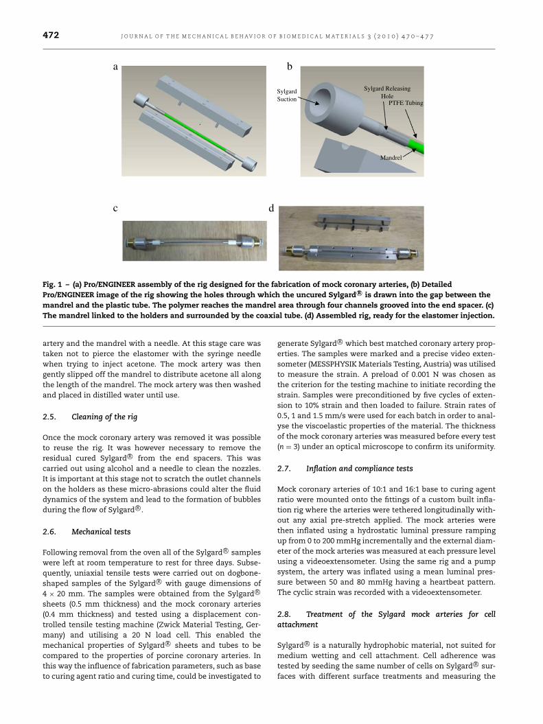
A simple rig was designed in Pro/ENGINEER (ParametricTechnology Corporation, Needham, MA, USA) to create mockarteries with an inner diameter of 3 mm and outer diameterof 3.8 mm. The rig was designed such that idealised coronaryartery geometries could be manufactured consistently andwith no artefacts or damage to the Sylgard R© surface. Theconstruction rig consisted of two stainless steel end spacerswithin which a polished stainless steel inner mandrel wasplaced. The stainless steel mandrel measured 3 mm indiameter. Both end spacers were tapered and slots were cuton the tapered ends to allow for transfer of fluid throughthese predefined channels at either end of the mandrel.Plastic outer moulds were manufactured from PTFE shrinktubing such that they had an inner diameter of 3.8 mm. Toachieve this, 6 mm shrink tubing (Tyco Electronics, Kessel,Belgium) was heated onto a 3.8 mm diameter polished
stainless steel pin using a heat gun at 150 ◦C. The pin wasshaped like a hook at one end to enable the release of themould. This 3.8 mm PTFE cylindrical tubing was then used toform the outer shell of the Sylgard R© mould and was placedover the end spacers as shown in Fig. 1. A non-stick coatingwas also sprayed onto the stainless steel pin prior to heatshrinking the PTFE tubing. This made it easier to release theheat shrink tubing once it had moulded to the diameter ofthe pin.
The mock artery mould was constructed by inserting the3 mm stainless steel mandrel inside the PTFE shell andfixing both ends with stainless steel end spacers such thatboth cylinders were concentric (Fig. 1). The end spacers werespecially designed to fix the mandrel in the centre such thatthe resulting mock artery had a uniform thickness along itslength. The plastic mould was cut such that it was longerthan the mandrel and covered the four holes cut into the endspacers. This ensured that the elastomer was able to passthrough these holes to fill out the space between the innermandrel and the outer PTFE tubing. PTFE isolating tape wasused to avoid air infiltration between the plastic mould andthe holders (Fig. 1).
2.3. Creation of the mock artery mould
The prepared elastomer was drawn into the rig by suctionof a vacuum pump connected to one end of the rig with apneumatic connector. Upon suction, the Sylgard R© was ableto pass from the container into the mould cavity through foursmall holes created on the stainless steel end spacers untilthe cavity was filled. The rig was then placed inside an ovenat 120 ◦C for 16 h to allow the Sylgard R© mould to cure. Whenthe entire mould was filled up with Sylgard R© an aluminiumcover was fastened on top of the base plate to ensure thatthe concentricity of the inner mandrel and the outer PTFEtubing was maintained (Fig. 1). It was important at this stagenot to close off the holders at either end as this prevents theelastomer from expanding longitudinally during curing andcould result in non-uniform thickness along the mandrel.
The Sylgard R© Product Datasheet (Dow Corning, MidlandMI, USA) suggests curing the polymer for one hour at 100 ◦C;however this temperature was found to be inadequate in thiscase due to the presence of a plastic tube around the poly-mer which prevents homogenous heat diffusion. In fact, thelevel of curing was lower in the centre of the mock artery ifthe Sylgard R© manual instructions were followed. Heat diffu-sion was found to be higher at the open inlet and outlet incomparison to the surrounding mould.
2.4. Removal of the mock coronary artery from the rig
Once the polymer was cured the rig was taken out of the ovenand left to cool down for 30 min at room temperature. Toremove the Sylgard R© compliant mock artery from the mould,the end spacers were first removed and the PTFE outer shellwas gently peeled off. The external PTFE tube was removed bygently unwinding it with a spiral movement taking care notto damage the Sylgard R© artery.
Themock coronary artery was subsequently removed fromthe inner mandrel by injecting acetone between the mock
472 J O U R N A L O F T H E M E C H A N I C A L B E H AV I O R O F B I O M E D I C A L M A T E R I A L S 3 ( 2 0 1 0 ) 4 7 0 – 4 7 7
SylgardSuction
Sylgard ReleasingHole
PTFE Tubing
Mandrel
a b
c d
Fig. 1 – (a) Pro/ENGINEER assembly of the rig designed for the fabrication of mock coronary arteries, (b) DetailedPro/ENGINEER image of the rig showing the holes through which the uncured Sylgard R© is drawn into the gap between themandrel and the plastic tube. The polymer reaches the mandrel area through four channels grooved into the end spacer. (c)The mandrel linked to the holders and surrounded by the coaxial tube. (d) Assembled rig, ready for the elastomer injection.
artery and the mandrel with a needle. At this stage care wastaken not to pierce the elastomer with the syringe needlewhen trying to inject acetone. The mock artery was thengently slipped off the mandrel to distribute acetone all alongthe length of the mandrel. The mock artery was then washedand placed in distilled water until use.
2.5. Cleaning of the rig
Once the mock coronary artery was removed it was possibleto reuse the rig. It was however necessary to remove theresidual cured Sylgard R© from the end spacers. This wascarried out using alcohol and a needle to clean the nozzles.It is important at this stage not to scratch the outlet channelson the holders as these micro-abrasions could alter the fluiddynamics of the system and lead to the formation of bubblesduring the flow of Sylgard R©.
2.6. Mechanical tests
Following removal from the oven all of the Sylgard R© sampleswere left at room temperature to rest for three days. Subse-quently, uniaxial tensile tests were carried out on dogbone-shaped samples of the Sylgard R© with gauge dimensions of4 × 20 mm. The samples were obtained from the Sylgard R©
sheets (0.5 mm thickness) and the mock coronary arteries(0.4 mm thickness) and tested using a displacement con-trolled tensile testing machine (Zwick Material Testing, Ger-many) and utilising a 20 N load cell. This enabled themechanical properties of Sylgard R© sheets and tubes to becompared to the properties of porcine coronary arteries. Inthis way the influence of fabrication parameters, such as baseto curing agent ratio and curing time, could be investigated to
generate Sylgard R© which best matched coronary artery prop-erties. The samples were marked and a precise video exten-someter (MESSPHYSIK Materials Testing, Austria) was utilisedto measure the strain. A preload of 0.001 N was chosen asthe criterion for the testing machine to initiate recording thestrain. Samples were preconditioned by five cycles of exten-sion to 10% strain and then loaded to failure. Strain rates of0.5, 1 and 1.5 mm/s were used for each batch in order to anal-yse the viscoelastic properties of the material. The thicknessof the mock coronary arteries was measured before every test(n = 3) under an optical microscope to confirm its uniformity.
2.7. Inflation and compliance tests
Mock coronary arteries of 10:1 and 16:1 base to curing agentratio were mounted onto the fittings of a custom built infla-tion rig where the arteries were tethered longitudinally with-out any axial pre-stretch applied. The mock arteries werethen inflated using a hydrostatic luminal pressure rampingup from 0 to 200 mmHg incrementally and the external diam-eter of the mock arteries was measured at each pressure levelusing a videoextensometer. Using the same rig and a pumpsystem, the artery was inflated using a mean luminal pres-sure between 50 and 80 mmHg having a heartbeat pattern.The cyclic strain was recorded with a videoextensometer.
2.8. Treatment of the Sylgard mock arteries for cellattachment
Sylgard R© is a naturally hydrophobic material, not suited formedium wetting and cell attachment. Cell adherence wastested by seeding the same number of cells on Sylgard R© sur-faces with different surface treatments and measuring the

J O U R N A L O F T H E M E C H A N I C A L B E H AV I O R O F B I O M E D I C A L M A T E R I A L S 3 ( 2 0 1 0 ) 4 7 0 – 4 7 7 473
number of adherent cells after 48 h in culture inside an in-cubator.
The silicon elastomer was dipped into a 70% sulphuric acidbath inside a fume hood for 5 min similar to that previouslydescribed by Moore et al. (1994). Care was taken to completelywet the artery surface without any bubble formation. Theartery was then washed in distilled water. The ends of themock arteries were attached to Polyvinylidene Fluoride (PVDF)plastic connectors (Value Plastics, Fort Collins, CO, USA) andthe system was sterilised in an autoclave.
The mock coronary artery was dipped into PBS (phos-phate buffer solution) containing 8µg/ml of bovine plasmafibronectin (BioSciences, Ireland) for 30 min to improve celladherence. The mock coronary artery was then inserted intoa 15 ml tube and was rotated overnight in a spinning de-vice (STR4/4, Stuart) to obtain a uniform distribution of fi-bronectin. Thereafter, PBS was removed and bovine aorticsmooth muscle cells (SMC) were injected into the 15 ml tubefor cell seeding. The tube was returned to the spinning devicefor SMC seeding for additional 8 h.
2.9. Comparison of Sylgard R© mock arteries with differentsurface treatments
Bovine aortic smooth muscle cell adherence on theSylgard R© was tested for different surface treatments. Treat-ment of a polymer surface with sulphuric acid increases thenumber of charged surface groups favouring cell attachmentand proliferation by hydrophilising the Sylgard R© (Moore et al.,1994). BASMC were cultured to 80/90% confluence in a T-75flask and seeded on autoclaved flat Sylgard R© layers treatedwith/without sulphuric acid andwith/without fibronectin. Af-ter 24 h of culture the cells were fixed in 3% formaldehyde andtheir membranes made permeable with 0.2% Triton X-100 toenable the nuclei to be stained with DAPI (4’,6-diamidino-2-phenylindole). Pictures were taken under a fluorescence mi-croscope (Olympus DP-50) and the images were analysed. Thenumber of cells in constant regions of 300 × 300 pixels wasanalysed. Cells were blind counted in three random areas ofthe Sylgard R© surface (n = 3) for the different surface treat-ments and results were compared.
In a separate test, the resistance of cell attachment to10 dyne/cm2 shear stress was also measured to establishthe suitability of the mock coronary artery for cell seedingunder culture medium or blood flow conditions. BASMC werecultured on Sylgard R© sheets which were previously treatedwith sulphuric acid and fibronectin. Some sheets were left instatic conditions while other sheets were placed on a rotatorat 10 dyne/cm2.
Images were taken using a fluorescence microscope afterDAPI staining of SMCs and cells were counted (n = 3) in aregion of 300× 300 pixels with magnification 40×.
3. Results
3.1. Mechanical tests
The mechanical tests revealed that both the base to curingagent ratio and the curing time have a significant influence
Fig. 2 – Stress–strain response of dogbone-shaped samplesof Sylgard R© sheets cured at different base to curing agentratios.
Fig. 3 – Stress–strain response of dogbone-shaped samplesof Sylgard R© sheets at different strain rates cured for 16 hand fabricated with different base to curing agent ratios of16:1 and 10:1.
on themechanical properties of the Sylgard R© . While a higherbase to curing agent ratio softens the material (Figs. 2 and 3),longer curing duration considerably stiffens the Sylgard R© forboth the 10:1 and 16:1 ratios, see Figs. 4 and 5 respectively.The variation in the mechanical properties of Sylgard R© dueto curing time is more significant for a base to curing agentratio of 16:1 than for 10:1, see Figs. 4 and 5. In addition, therole of strain rate within a physiologically relevant range wasinvestigated and proved insignificant, see Figs. 4 and 5.
Given that the curing level depends on the heat transferfrom the preparation rig throughout the Sylgard R© , the rigdesign was also found to have an important effect on thefinal mechanical properties. In Fig. 6, the flat specimens showconsiderably higher stiffness compared to the tubular mockcoronary arteries fabricated with the same ratio and curingtime for both ratios of 10:1 and 16:1. The use of the 16:1 ratioand a curing time of 16 h at 120 ◦C produced more compliantmock coronary arteries. The mechanical properties of thesetubes is in the range of the circumferential stress–strainresponse of porcine coronary arteries, most notably in the lowphysiological stress range (10–13 kPa or 80–120 mmHg), see

474 J O U R N A L O F T H E M E C H A N I C A L B E H AV I O R O F B I O M E D I C A L M A T E R I A L S 3 ( 2 0 1 0 ) 4 7 0 – 4 7 7
Fig. 4 – Stress–strain response of dogbone-shaped samplesof Sylgard R© sheets at different strain rates cured fordifferent durations and fabricated with a base to curingagent ratio of 10:1.
Fig. 5 – Stress–strain response of dogbone-shaped samplesof Sylgard R© sheets at different strain rates cured fordifferent durations and fabricated with a base to curingagent ratio of 16:1.
Fig. 7. It is clear, however, that specimens can be fabricated torepresent a range of arterial properties.
The thickness of all mock coronary arteries was measuredusing an optical microscope and found to be 0.4± 0.04 mm.
3.2. Inflation and compliance tests
Further investigation of the mechanical properties of themock coronary arteries using inflation tests showed resultsconsistent with the tensile test results. Mock coronary arter-ies prepared with a base to curing agent ratio of 16:1 werefound to be more compliant than arteries prepared with abase to curing agent ratio of 10:1, see Fig. 8.
In addition, a physiologically realistic strain pattern, with5% cyclic strain amplitude, was recorded using a videoexten-someter in the 16:1 mock coronary artery in response to aheartbeat pressure waveform, see Fig. 9.
Fig. 6 – Stress–strain response of dogbone-shaped samplesof Sylgard R© sheets compared to dogbone-shaped samplesof Sylgard R© tubes, both prepared with a base to curingagent ratio of 16:1 and 10:1 for 16 h curing duration, n = 3.
3.3. Treatment of the Sylgard arteries for cell attachment
In order to optimise cell attachment to the mock coronaryartery, the 16:1 Sylgard R© surface was treated with 70% sul-phuric acid solution and/or fibronectin. BASMC were seededon the Sylgard R© surfaces and after 12 h they were fixed informaldeyde and stained with DAPI. Cells were counted in astandard region of 300 × 300 pixels under a fluorescent mi-croscope with magnification 40×. Where the Sylgard R© sur-face was untreated with sulphuric acid solution, 18.66 ± 0.57cells were counted. When Sylgard R© was treated with the sul-phuric acid solution, 21.33 ± 1.52 cells were found in the se-lected region. The use of fibronectin increased the numberof BASMC to 27.66 ± 1.15 whilst treatment with sulphuricacid followed by fibronectin resulted in maximum cell adhe-sion whereby 36 ± 2 BASMC attached to the Sylgard R© sur-face. Cell counts were statistically significant (ANOVA, p <
0.01) showing that treatment of mock coronary arteries withsulphuric acid improved cell adherence and was further en-hanced when bovine plasma fibronectin was used as a bind-ing protein (Fig. 10).
The influence of shear stress on cell adhesion to theSylgard R© surface was assessed by exposing the cells to aphysiologically relevant shear stress of 10 dyne/cm2. Cellnumbers showed no statistically significant difference insamples exposed to shear stress (92.66 ± 2.51 cells per300×300 pixels) and samples whichwere not exposed to shear(90.66 ± 2.08 cells in 300× 300 pixels), see Fig. 11.
4. Discussion
The increasing number of mechanotransduction studies hasled to a need for 3D cell straining platforms. Sylgard R© mockcoronary arteries are a suitable option where a cell monolayeris cyclically strained. In spite of the fact that Sylgard R© mockarteries are uniform and homogeneous, they can mimic thecyclic strain levels that cells experience in vivo (Wedding et al.,2002) during the physiological intraluminal pressure range,see Fig. 9.

J O U R N A L O F T H E M E C H A N I C A L B E H AV I O R O F B I O M E D I C A L M A T E R I A L S 3 ( 2 0 1 0 ) 4 7 0 – 4 7 7 475
Fig. 7 – Strain/stress response of dogbone-shaped samples of Sylgard R© sheets at base to curing agent ratios of 10:1, 11:1,14:1 and 16:1, and cured for 16 h at 120 ◦C in comparison to porcine coronary arterial tissue. The strain/stress response of adogbone-shaped sample obtained from a 16:1 ratio mock coronary artery (MCA) is also shown.
Fig. 8 – Pressure–strain response of Sylgard R© mock coronary arteries cured for 16 h and fabricated with different base tocuring agent ratios of 16:1 and 10:1. (Error bars represent standard deviation among 3 tested tubes).
Fig. 9 – Strain pattern of a Sylgard R© mock coronary arteryexposed to a heartbeat pressure waveform recorded with avideoextensometer.
This paper provides a detailed description of a simple andrepeatable method to prepare mock coronary arteries using acommon polymer, Sylgard R© 184. To the authors’ knowledgea simple way to construct such a mock coronary artery hasnever been thoroughly described. In fact, several requestshave come to the authors for a full process description, thegoal of this work. The in-house system described enablesa specific base to curing agent ratio and curing time to beselected to represent the different mechanical properties thatmay be required. The process can be replicated and adoptedfor segments or complete arteries and veins of different sizesand with different mechanical properties.
Mock arteries are widely employed in research and in thebiomedical industry, especially for the mechanical evaluationof endovascular stents (Hecker et al., 2005; Rajesh et al.,2007) or for the evaluation of imaging techniques suchas ultrasound, CT, and angiography (Douville et al., 1983;McDicken, 1986). Most of these studies, however, do not givea clear explanation of the production process or focus more

476 J O U R N A L O F T H E M E C H A N I C A L B E H AV I O R O F B I O M E D I C A L M A T E R I A L S 3 ( 2 0 1 0 ) 4 7 0 – 4 7 7
Sulphuric acid treatment
No treatment
Sulphuric acid + fibronectin treatment
Fibronectin treatment
Fig. 10 – Influence of surface treatment with sulphuric acid and fibronectin coating on SMC adhesion to Sylgard R© . Everyblue dot represents the nucleus of a cell stained with DAPI under the fluorescence microscope (magnification 40×).
No shear stress 10 Dynes/cm2 shear stress
Fig. 11 – Resistance to physiological shear stress of adherent SMCs on the Sylgard R© surface treated with sulphuric acid andfibronectin (magnification 40×).
on the fluid dynamics inside the lumen than on the controlof the cyclic strain of the arterial wall. The focus of this workwas to obtainmock coronary arteries with cyclic strains in thecircumferential direction close to the physiological values bytuning the mechanical properties of Sylgard R© .
Sylgard R© was found to have minimal strain rate depen-dence but its mechanical properties are strongly dependenton the curing time. As the Sylgard R© curing level depends onthe temperature obtained, the preparation rig can also playan important role in the final mechanical properties. Giventhat in the mock coronary artery preparation rig Sylgard R© isencapsulated within PTFE tubing, the heat transfer to theSylgard R© is significantly lower than in the rig used for the
preparation of flat sheets of Sylgard R© where the elastomeris moulded between two thin stainless steel plates. As a re-sult, the mock coronary artery is much more compliant thanthe flat sheets of Sylgard R© produced with the same base tocuring agent ratio and curing time. This may be explained bylooking at the influence of curing time on the properties of theelastomer. For the same set-up the longer the curing time thestiffer the elastomer obtained, indicating that higher temper-atures leads to stiffening of the material. Using an insulatingpolymer as opposed to steel for the mould in the mock arteryrig insulates the curing elastomer from the surrounding heatof the oven and therefore reduces the temperature reached bythe Sylgard R©. The different thermal diffusion rates however

J O U R N A L O F T H E M E C H A N I C A L B E H AV I O R O F B I O M E D I C A L M A T E R I A L S 3 ( 2 0 1 0 ) 4 7 0 – 4 7 7 477
do not affect the final composition and uniformity of the elas-tomer provided that the material is completely cured. This isas evidenced by the repeatability of the mechanical test data.
The structure of a real artery is very complex compared toa mock coronary artery. The inflation test results presentedfor the Sylgard R© tubes are different from those given forporcine and human coronary arteries under various staticpressures (Van Andel et al., 2003). These differences are dueto the use of a simple homogeneous and isotropic elastomerto represent the anisotropic structure of coronary arteries.However, selecting the right parameters, such as the baseto curing agent ratio and the curing temperature and time,it was possible to create mock coronary arteries with cyclicstrains close to physiological values for specific pressurevalues. As shown in Fig. 9, when exposed to a physiologicalpressure waveform the mock coronary artery exhibited aperiodical strain waveform close to that of a real coronaryartery (Wedding et al., 2002).
The system described thus allows the creation of mockcoronary arteries of constant wall thickness suitable for cellculture bioreactors where the artery deformation is caused bythe pulsatile pressure of flowing culture media. The constantwall thickness is of extreme importance for mechanotrans-duction studies to obtain uniform cell elongation. Mooreet al. (1994) produced mock coronary arteries by pouringSylgard R© on a 6 mm diameter rotating cylinder maintainedat 100 ◦C in the oven. Although Moore et al. outlined thatthe application of liquid Sylgard R© was carefully controlled tomaintain the mock coronary artery wall thickness constantthe system cannot always guarantee a uniform distributionand we encountered many difficulties in attempts to repli-cate this protocol. Furthermore, for small diameters, as in thecase of coronary arteries, the Sylgard R© distribution is evenmore critical. An attempt was made to use both a stainlesssteel internal mandrel and outer casing for the mould butthis was unsuccessful because of the difficulty in removingthe artery from the inner and outer steel casings. The proto-col presented in this work overcomes all of these problems byprecisely encasing the mock artery between an internal man-drel and an external plastic wall which can be easily removed.
Mock coronary arteries were treated with sulphuric acidand coated with fibronectin as suggested by Moore et al.(1994). Sulphuric acid alone, however, had only a marginaleffect on SMC attachment but it increased cell adhesionwhen used in association with fibronectin. Cell number wassignificantly increased in all tests where the Sylgard R© wascoated with fibronectin. Tests have also demonstrated thatcell adherence on Sylgard R© using this protocol is resistant tophysiological levels of shear stress (10 dyne/cm2).
In conclusion, the presentedmethod enables fabrication ofbiocompatible and transparent mock coronary arteries withmechanical properties which can be easily tuned tomimic themechanical response of any specific vessel for mechanotran-duction studies. In addition, with some minor adaptation ofthe rig it would be possible to generate composite vessels withspecific properties to represent the multilayered nature ofarteries.
The Sylgard R© can also be used for 2D or 3D cell studiesto investigate the influence of strain on cell activity, usingsheets and tubes respectively, but care must clearly be taken
to consider the influence of the mould material on the finalstress–strain properties of the material as demonstrated inthis study.
Acknowledgements
The authors would like to acknowledge the help of thetechnicians in the School of Mechanical & ManufacturingEngineering at Dublin City University and Dr. Ronan Murphyand Dr. Philip Cummins from the School of Biotechnologyin Dublin City University. The funding for this study wasprovided by Enterprise Ireland, Science Foundation Ireland,Medtronic AVE, and the Irish Research Council for ScienceEngineering and Technology.
R E F E R E N C E S
Brown, X.Q., Ookawa, K., Wong, J.W., 2005. Evaluation ofpolydimethylsiloxane scaffolds physiologically relevant elasticmoduli: interplay of substrate mechanics and surfacechemistry effects on vascular smooth muscle cell response.Biomaterials 26, 3123–3129.
Douville, Y., Johnston, K.W., Kassam, M., Zuech, P., Cobbold, R.S.C.,Jares, A., 1983. An in vitro model and its application for thestudy of carotid Doppler spectral broadening. Ultrasound inMedicine and Biology 9, 347–356.
Hecker, L., Baar, K., Dennis, R.G., Bitar, K.N., 2005. Developmentof a three-dimensional physiological model of the internalanal sphincter bioengineered in vitro from isolated smoothmuscle cells. American Journal of Physiology-Gastrointestinaland Liver Physiology 289, 563–580. G188-196. Biorheology. 29.
Liepsch, D., Moravec, S., Baumgart, R., (1992). Some flowvisualization and laser-Doppler-velocity measurements in atrue-to-scale elastic model of a human aortic arch—a newmodel technique.
McDicken, W.N., 1986. A versatile test object for the calibration ofultrasonic Doppler flow instruments. Ultrasound in Medicineand Biology 12, 245–249.
Moore Jr., J.E., Bürki, E., Suciu, A., Zhao, S., Burnier, M., Brunner,H.R., Meister, J.J., 1994. A device for subjecting vascularendothelial cells to both fluid shear stress and circumferentialcyclic stretch. Annals of Biomedical Engineering. 22, 416–422.
Oláh, A., Vancso, G.J., 2005. Characterization of adhesion atsolid surfaces: development of an adhesion-testing device.European Polymer Journal 41, 2803–2823.
Ramesh, R., Conti, J, Strope, E.R., Thompson, M., Price, K., Murray,D., 2008. Comparison of radial expansion of stents withinmock vessels molded with a target bent radius versus straightmock vessels bent to a target radius. Biomedical SciencesInstrumentation 44, 189–194.
Rajesh, R., Conti, J.C., Strope, E.R., 2007. Linear elastic mechanicsof mock arteries: empirical versus theoretically predicted pul-satile stent deflection. Biomedical Sciences Instrumentation43, 54–62.
Van Andel, C.J., Pistecky, P.V., Borst, C., 2003. Mechanicalproperties of porcine and human arteries: implications forcoronary anastomotic connectors. The Annals of ThoracicSurgery 76 (1), 58–64.
Wedding, K.L., Draney, M.T., Herfkens, R.J., Zarins, C.K., Taylor,C.A., Pelc, N.J., 2002. Measurement of vessel wall strain usingcine phase contrast MRI. Journal of Magnetic ResonanceImaging 15 (4), 418–428.