A Comparative Study of the Appendicular Musculature of ...
56
A Comparative Study of the Appendicular Musculature of Penguins (Aves: Sphenisciformes) DONALD O. SCHREIWEIS , SMITHSONIAN CONTRIBUTIONS TO ZOOLOGY • NUMBER 341
-
Upload
khangminh22 -
Category
Documents
-
view
4 -
download
0
Transcript of A Comparative Study of the Appendicular Musculature of ...
A Comparative Study ofthe Appendicular Musculature ofPenguins (Aves: Sphenisciformes)
DONALD O. SCHREIWEIS,
SMITHSONIAN CONTRIBUTIONS TO ZOOLOGY • NUMBER 341

SERIES PUBLICATIONS OF THE SMITHSONIAN INSTITUTION
Emphasis upon publication as a means of "diffusing knowledge" was expressedby the first Secretary of the Smithsonian. In his formal plan for the Institution, JosephHenry outlined a program that included the following statement: "It is proposed topublish a series of reports, giving an account of the new discoveries in science, andof the changes made from year to year in all branches of knowledge." This themeof basic research has been adhered to through the years by thousands of titles issuedin series publications under the Smithsonian imprint, commencing with SmithsonianContributions to Knowledge in 1848 and continuing with the following active series:
Smithsonian Contributions to AnthropologySmithsonian Contributions to Astrophysics
Smithsonian Contributions to BotanySmithsonian Contributions to the Earth SciencesSmithsonian Contributions to the Marine Sciences
Smithsonian Contributions to PaleobiologySmithsonian Contributions to Zoo/ogySmithsonian Studies in Air and Space
Smithsonian Studies in History and Technology
In these series, the Institution publishes small papers and full-scale monographsthat report the research and collections of its various museums and bureaux or ofprofessional colleagues in the world of science and scholarship. The publications aredistributed by mailing lists to libraries, universities, and similar institutions throughoutthe world.
Papers or monographs submitted for series publication are received by theSmithsonian Institution Press, subject to its own review for format and style, onlythrough departments of the various Smithsonian museums or bureaux, where themanuscripts are given substantive review. Press requirements for manuscript and artpreparation are outlined on the inside back cover.
S. Dillon RipleySecretarySmithsonian Institution

S M I T H S O N I A N C O N T R I B U T I O N S T O Z O O L O G Y • N U M B E R 3 4 1
A Comparative Study ofthe Appendicular Musculature ofPenguins (Aves: Sphenisciformes)
Donald 0. Schreiweis
SMITHSONIAN INSTITUTION PRESS
City of Washington
1982

A B S T R A C T
Schreiweis, Donald O. A Comparative Study of the Appendicular Muscu-lature of Penguins (Aves: Sphenisciformes). Smithsonian Contributions to Zoology,number 341, 46 pages, 19 figures, 1982.—The gross anatomy of all theappendicular muscles is compared in 14 species, representing the 6 livinggenera of Sphenisciformes. Particular emphasis is placed on the interrelation-ships indicated by variations in the taxa. Eudyptes pachyrhynchus is used as atype for which all the appendicular muscles are described and most areillustrated. The salient features of other taxa, as they differ from the conditionfound in E. pachyrhynchus, are given. Similarities and differences are comparedquantitatively in respect to 47 items in the wing and and 41 in the leg, usingthe method of Hudson et al. (1966:9-11). The results are given in the form ofcumulative scores of differences and correlation coefficients.
The results of the comparison of the wing musculature support those forthe leg. The cumulative scores and correlation coefficients give closely parallelresults.
The Sphenisciformes constitute a rather uniform group of birds. The resultsof this study support the present classification of the order, in which 14 speciesare grouped into 6 genera. A tentative phylogeny of penguins is projected onthe basis of variations in the wing and leg muscles.
OFFICIAL PUBLICATION DATE is handstamped in a limited number of initial copies and is recordedin the Institution's annual report, Smithsonian Year. SERIES COVER DESIGN: The coral Montastreacavemosa (Linnaeus).
Library of Congress Cataloging in Publication DataSchreiweis, Donald O.A comparative study of the appendicular musculature of penguins (Aves: Sphenisciformes)(Smithsonian contributions to zoology ; no. 341)Bibliography: p.Includes index.Supt. of Doc. no.: SI 1.27:3411. Penguins—Anatomy. 2. Wing—Muscles. 3. Leg—Muscles. 4. Anatomy, Comparative.
I. Title. II. Series.QL1.S54 no. 341 [QL696.S473] 591s 81-607820 [598.4'41] AACR2

Contents
Page
Introduction 1Materials and Methods 1Acknowledgments 2
Muscles of the Pectoral Appendage 2Muscles of the Pelvic Appendage 14Position of the Flexor Tendons Passing the Intertarsal Joint 21Discussion and Conclusions 25Appendix: Interesting Features of the Appendicular Muscles 29Literature Cited 31Abbreviations on Illustrations 33Figures 1-19 34Index 45
m


A Comparative Study ofthe Appendicular Musculature ofPenguins (Aves: Sphenisciformes)
Donald 0. Schreiweis
Introduction
Penguins are an ancient group of birds thathad become flightless and highly specialized forunderwater locomotion by the Eocene. Simpson(1975:35) considered penguins related to Procel-lariiformes, basing this relationship on the struc-ture of the skull and mandible. In the oldestknown fossil penguins, the structure of the man-dible, the shape of the foramen magnum, and thearrangement of palatal bones are similar to theprocellariiform type. Sibley and Ahlquist (1972)recently concluded that the egg-white proteinpatterns of penguins were more like those of theProcellariiformes than those of any other group,although they also resembled several other groupsof aquatic birds. The descriptive myology of pen-guins presented in this paper may help to resolvethe problem of phylogeny when comparable stud-ies on other water birds become available.
Although the appendicular muscles have beenstudied in a few sphenisciform birds by variousinvestigators, no one has made a concerted at-tempt to work out in detail the similarities anddifferences between the various genera and spe-cies. Such differences and similarities may serveas a basis for a study of functional morphology,
Donald 0. Schreiweis, Premedical Studies Program, Saint LouisUniversity, Saint Louis, Missouri 63103.
as well as a study of phylogenetic and systematicrelationships among these taxa.
A detailed study of all appendicular muscles,using descriptive and numerical methods, wasmade on a series of 28 specimens, representing 6genera and 14 species. The pectoral musculatureis unlike that of any other group of birds. Most ofthe muscles in the wing are reduced to tendons,a condition not encountered in any other group,while the leg muscles deviate less from the generalavian pattern. The more interesting features ofthe appendicular muscles of penguins are sum-marized in the appendix.
MATERIALS AND METHODS.—The classificationfollowed is that of Falla and Mougin (1979). Idissected the following species: Aptcnodytes patagon-icus (2); A.forsteri (2); Pygoscelis papua (2); P. adeliae(3); P. antarctica (2); Eudyptes pachyrhynchus (2); E.chrysocome (2); E. chrysolophus schlegeli (1); E. c.chrysolophus (2); Megadyptes antipodes (1); Eudyptulaminor minor (1); E. m. albosignata (1); Spheniscusdemersus (2); S. humboldti (1); S. magellanicus (2); S.mendiculus (2).
Fresh specimens were initially injected in allparts with a 1:10 solution of formalin. Afterseveral weeks, specimens were opened and washedin tap water for 3 to 5 days to remove most of theformalin. Subsequently, all were stored in 65%ethyl alcohol.
To facilitate study of the musculature in most
1

SMITHSONIAN CONTRIBUTIONS TO ZOOLOGY
specimens, the coracoid was separated from theclavicle proximally and from the sternum distally,and the femur was disarticulated at the aceta-bular joint. The extremities of the limb boneswere exposed by partially dissecting the jointcapsules without complete disarticulation. Eachmuscle was exposed from origin to insertion bycutting bellies or tendons of certain muscles atprescribed points.
Descriptions of the origin, belly, and insertionof each muscle are given for Eudyptes pachyrhynchus.The positional (anatomical) axes utilized arethose related to the functional axes: for the wing,the extended position of underwater flight; forthe leg, the normal standing position. The loca-tion of muscle attachments (e.g., "at about 0.63tibiotarsus") were obtained by measuring thepoint of attachment from the proximal end of thebone holding the calipers parallel to the long axisof the bone. These measurements were then di-vided by the length of the bone.
Following each description, a comparison ofthe muscle is given for other species as these differfrom E. pachyrhynchus. Included in the comparisonare remarks concerning observations made byother workers. In many instances, observationsmade in this study clarify those of earlier workers.
Numerical comparisons were made using themethod of Hudson et al. (1966:9-11). Tables ofquantitative data are available from the authoron request. Anatomical nomenclature followsthat of the Nomina Anatomica Avium (Baumel et al.,1979).
ACKNOWLEDGMENTS.—I wish to express my ap-preciation and thanks to Dr. George E. Hudsonunder whose guidance and direction this studywas completed. He made available many of thespecimens and, through him, various specimenswere loaned from other institutions. I also wish tothank Drs. Herbert L. Eastlick, C. M. McNeil,and Richard A. Parker of Washington State Uni-versity, who critically read the manuscript andoffered suggestions.
Specimens examined for this study are part ofan extensive alcoholic collection at WashingtonState University, representing all living orders of
birds. Dr. Richard L. Zusi of the National Mu-seum of Natural History, Smithsonian Institu-tion, loaned 12 specimens. Dr. Dean Amadon ofthe American Museum of Natural History loanedone specimen each of Eudyptes pachyrhynchus andSpheniscus humboldti. Mr. Warwich M. Howe of theNew Zealand Department of Internal Affairs sup-plied one specimen each of Eudyptes pachyrhynchus,Megadyptes antipodes, and Eudyptula minor albosig-nata. Mr. T. H. Barry of the South African Mu-seum supplied a specimen of Spheniscus demersus.
This study was supported in part by a grant-in-aid (GB-6088) from the National ScienceFoundation.
Muscles of the Pectoral Appendage
M . LATISSIMUS DORSI
The latissimus dorsi consists of three distinctparts: latissimus dorsi cranialis, latissimus dorsicaudalis, and latissimus dorsi metapatagialis.
Latissimus dorsi cranialis
(LAT. DOR. CRAN.)
FIGURES 1, 2, 8
DESCRIPTION.—This large flat muscle (Figure1) arises from the dorsal spines of about the lastcervical and first five thoracic vertebrae. Theorigin is aponeurotic. Fleshy fibers of this muscleconverge as they pass forward and outward, andterminate on a long, narrow tendon. The tendonis about one-fourth the length of the humerus. Itpasses through a fibrous pulley (Figure 8) muchlike a thread passes through the eye of a needle.This pulley, unique to the Spheniscidae, is at-tached to the axillary border of the scapula justbehind the glenoid fossa. Insertion is on the pos-terior border of the humeral shaft, near the distalborder of the pneumatic fossa.
COMPARISON.—An accessory slip from the cra-nial edge of the latissimus dorsi cranialis occursin Aptenodytes and Pygoscelis. This accessory slip

NUMBER 341
inserts on the dorsal surface of the dorsal head ofthe triceps scapularis.
and Eudyptula and intermediate in Eudyptes, Sphen-iscus, and Megadyptes.
Latissimus dorsi caudalis
(LAT. DOR. CAUD.)
FIGURES 1, 2, 8
DESCRIPTION.—The latissimus dorsi caudalis(Figure 2) is distinct and separated by a wideinterval from the latissimus dorsi cranialis. Itarises by means of a delicate aponeurosis from theinferior edge of the ilium and the abdominalmuscles immediately posterior to the last thoracicrib. Fleshy fibers on the band-like belly passforward toward the shoulder and terminate on along tendon which, after passing through thefibrous pulley mentioned above, is inserted on theposterior border of the humeral shaft adjacent tothe insertion of the latissimus dorsi cranialis. Thetendons of the latissimus dorsi cranialis and latis-simus dorsi caudalis are partly fused.
COMPARISON.—The latissimus dorsi cranialisand latissimus dorsi caudalis are fused along theircontiguous borders in Aptenodytes and Pygoscelis.All other genera have these two parts widelyseparated.
Latissimus dorsi metapatagialis
(LAT. DOR. MET.)
FIGURE 1
DESCRIPTION.—This muscle is quadrilateral inform. It arises by a narrow aponeurosis from thedorsal spines of thoracic vertebrae 4, 5, and 6.Insertion is along a broad line on the skin of thelateral line of the trunk. The belly of the latissi-mus dorsi metapatagialis is much wider than thatof the latissimus dorsi caudalis and nearly as wideas the belly of the latissimus dorsi cranialis.
COMPARISON.—The latissimus dorsi metapata-gialis exhibits a wide range of development inpenguins. In Aptenodytes this muscle is very wide(1.01 scapula), reaching from the base of the neckto the lumbar region. It is narrowest in Pygoscelis
M . RHOMBOIDEUS SUPERFICIALIS
(RHOM. SUP.)
FIGURES 1, 2, 7
DESCRIPTION.—This wide, flat muscle (Figure2) arises by means of a narrow aponeurosis fromthe dorsal spines of the last one or two cervicalvertebrae and the first three or four thoracicvertebrae. The fibers pass transversely andslightly forwards; they are inserted on about theanterior half of the vertebral border of the scapulaand the scapular process of the clavicle.
COMPARISON.—This muscle is very similar inall species. In Eudyptes chrysocome, Watson (1883:76) found this muscle arising from the dorsalspines of the three anterior thoracic vertebrae andthe last two cervical vertebrae. In this penguin,as well as in Pygosceles taeniatus ( = Pygoscelis papua)and Spheniscus minor (= Eudyptula minor), he foundthe insertion to be confined to the cranial two-thirds of the vertebral border of the scapula. Theinsertion is not this extensive in any of the pen-guins studied. Gervais and Alix (1877:444) foundthe rhomboideus superficialis in Eudyptes chrysolo-phus attached to the dorsal spines of the last twocervical vertebrae, as well as to the dorsal spinesof thoracic vertebrae 1 through 5. Such a wideorigin was not present in any specimen of penguinstudied.
M . RHOMBOIDEUS PROFUNDUS
(RHOM. PRO.)
FIGURES 2, 3, 7
DESCRIPTION.—The rhomboideus profundus(Figure 3) arises from the dorsal spines of aboutfive or six thoracic vertebrae (thoracic vertebrae2-7) by means of an aponeurosis. Fleshy fiberspass transversely and somewhat obliquely back-wards to a fleshy insertion on about the caudalthree-fourths of the vertebral border of the scap-
SMITHSONIAN CONTRIBUTIONS TO ZOOLOGY
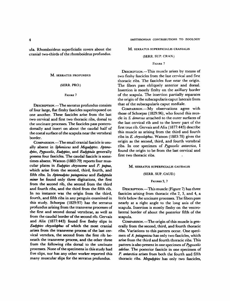
ula. Rhomboideus superficialis covers about thecranial two-thirds of the rhomboideus profundus.
M . SERRATUS PROFUNDUS
(SERR. PRO.)
FIGURE 7
DESCRIPTION.—The serratus profundus consistsof four large, flat fleshy fascicles superimposed onone another. These fascicles arise from the lasttwo cervical and first two thoracic ribs, dorsal tothe uncinate processes. The fascicles pass postero-dorsally and insert on about the caudal half ofthe costal surface of the scapula near the vertebralborder.
COMPARISON.—The small cranial fascicle is usu-ally absent in Spheniscus and Megadyptes. Apteno-dytes, Pygoscelis, Eudyptes, and Eudyptula generallypossess four fascicles. The caudal fascicle is some-times absent. Watson (1883:79) reports four mus-cular plates in Eudyptes chrysocome and P. papua,which arise from the second, third, fourth, andfifth ribs. In Aptenodytes patagonicus and Eudyptulaminor he found only three digitations, the firstfrom the second rib, the second from the thirdand fourth ribs, and the third from the fifth rib.In no instance was the origin from the third,fourth, and fifth ribs in any penguin examined inthis study. Schoepss (1829:97) has the serratusprofundus arising from the transverse processes ofthe first and second dorsal vertebrae, as well asfrom the caudal border of the second rib. Gervaisand Alix (1877:443) found five fleshy slips inEudyptes chrysolophus of which the most cranialarises from the transverse process of the last cer-vical vertebra, the second from the first rib be-neath the transverse process, and the other threefrom the following ribs dorsal to the uncinateprocesses. None of the specimens in this study hadfive slips, nor has any other worker reported thismany muscular slips for the serratus profundus.
M . SERRATUS SUPERFICIALIS CRANIALIS
(SERR. SUP. CRAN.)
FIGURE 7
DESCRIPTION.—This muscle arises by means oftwo fleshy fascicles from the last cervical and firstthoracic ribs. The fascicles fuse near the origin.The fibers pass obliquely anterior and dorsal.Insertion is mostly fleshy on the axillary borderof the scapula. The insertion partially separatesthe origin of the subscapularis caput laterale fromthat of the subscapularis caput mediale.
COMPARISON.—My observations agree withthose of Schoepss (1829:96), who found this mus-cle in S. demersus attached to the outer surfaces ofthe last cervical rib and to the lower part of thefirst true rib. Gervais and Alix (1877:443) describethis muscle as arising from the third and fourthribs in E. chrysolophus. Watson (1883:78) gives theorigin as the second, third, and fourth vertebralribs. In one specimen of Pygoscelis antarctica, Ifound the origin to be from the last cervical andfirst two thoracic ribs.
M . SERRATUS SUPERFICIALIS CAUDALIS
(SERR. SUP. CAUD.)
FIGURES 3, 7
DESCRIPTION.—This muscle (Figure 7) has threefascicles arising from thoracic ribs 2, 3, and 4, alittle below the uncinate processes. The fibers passnearly at a right angle to the long axis of thescapula. Insertion is mostly fleshy on the ventro-lateral border of about the posterior fifth of thescapula.
COMPARISON.—The origin of this muscle is gen-erally from the second, third, and fourth thoracicribs. Variations to this pattern occur. One speci-men of A. patagonicus has only two fascicles, whicharise from the third and fourth thoracic ribs. Thispattern is also present in one specimen of Pygoscelisadeliae. The posterior fascicle in one specimen ofP. antarctica arises from both the fourth and fifththoracic ribs. Megadyptes has only two fascicles,

NUMBER 341
which arise from the second and third thoracicribs. Watson (1883:78) found this muscle arisingby two digitations from the fourth and fifth ribsin P. papua. Schoepss (1829:94) reports this musclearising from four ribs in penguins. Watson (1883:78) never found the serratus superficialis caudalisarising from more than three ribs, and I found itarising from three ribs in all except one specimenin which the posterior slip arose from both thefourth and fifth ribs.
M . SCAPULOHUMERALIS CAUDALIS
(SCAP. HUM. CAUD.)
FIGURES 1-3, 5-7
DESCRIPTION.—The scapulohumeralis caudalis(Figure 3) has an extensive origin from about theposterior two-thirds of the dorsal and lateral sur-faces of the scapula. The fibers pass obliquelyforward and end on a strong tendon, which isinserted on the posterior border of the humerus(0.33 humerus).
COMPARISON.—The scapulohumeralis caudalisis a very uniform muscle in the Spheniscidae. Thecranial end of the origin shows only slight varia-tion between genera of penguins, ranging from0.33 scapula in Aptenodytes to 0.44 scapula inEudyptula.
of the sternum, the interosseus membrane be-tween this process and the corpus sterni, a linealong the entire length of the sternal keel ventralto the origin of the supracoracoideus, the lateralsurface of the clavicle below the shoulder joint,the cranioventral edge of the membrana sterno-coraco-clavicularis, and the aponeurosis coveringthe supracoracoideus. The belly is composed ofthree distinct parts. Fibers of the clavicular partpass caudolaterally. Those of the caudolateralpart pass craniolaterally, while those of the largemiddle part pass obliquely at increasing anglesfrom the sternum. The clavicular fibers end on aspecialized part of the tendon of insertion, whichends on about the proximal fifth of the cranialborder of the humeral shaft, joining the tendonof the propatagialis near the proximal end of thehumerus. The remaining fibers insert by meansof a wide, curving tendon on the proximal thirdof the cranioventral edge of the humerus.
COMPARISON.—The pectoralis thoracica showsno significant variations in the Spheniscidae.Reid (1835:140) described the muscle in Apteno-dytes patagonicus as arising from "the cartilages ofthe ribs, and from the anterior part of the cora-coid bone" in addition to the origin given above.Such is not the case in any of the penguinsexamined in this study or in the study by Watson(1883:81).
M . PECTORALIS
This muscle consists of three slips: pectoralisthoracica, pectoralis propatagialis (which is notpresent in penguins), and pectoralis pars subcu-tanea abdominalis.
Pectoralis thoracica
(PECT. THO.)
FIGURES 2-7
DESCRIPTION.—The pectoralis thoracica (Fig-ure 4) is a very powerful muscle which arises fromthe lateral edge of the sternum posterior to thecoracobrachialis caudalis, the trabecula lateralis
Pectoralis pars subcutanea abdominalis
(PECT. SUB. ABD.)
FIGURE 4
DESCRIPTION.—Pars subcutanea abdominalis isa wide, flat, ribbon-like muscle that arises fromthe skin along the flank. The caudal end of theorigin is opposite the region of the acetabulum.The muscle passes cranially and somewhat dor-sally to insert on the dorsal border of the pecto-ralis thoracica caudal to the insertion of the latteron the humerus.
COMPARISON.—The maximum width of thismuscle relative to the humeral length shows somevariation between genera of penguins. It is nar-

SMITHSONIAN CONTRIBUTIONS TO ZOOLOGY
rowest in Aptenodytes and Eudyptes, and interme-diate in Megadyptes, Spheniscus, and Pygoscelis. Wat-son (1883:81) describes the pectoralis pars sub-cutanea abdominalis as inserting on the anteriormargin of the humerus. This is not the case inany species of penguin examined in this study.
M . SUPRACORACOIDEUS
(SUP. COR.)
FIGURES 1-3, 5
DESCRIPTION.—The supracoracoideus (Figure5) arises from the entire surface of the sternumnot occupied by the pectoralis thoracica, from theproximal part of the trabecula lateralis of thesternum, from the keel of the sternum, and fromthe basal end of the coracoid. It also has a consid-erable attachment to the membrana sterno-cor-aco-clavicularis. From these origins the fibers con-verge to a stout, flattened tendon which, afterpassing through the canalis triosseus, inserts (Fig-ures 1-3) on an oblique ridge on the proximodor-sal part of the humerus between the insertions ofthe deltoideus major and deltoideus minor. Thesupracoracoideus is very well developed in com-parison to the pectoralis thoracica, a fact notedby every anatomist who has worked on any mem-ber of the group.
COMPARISON.—The small, separate anteriorpart of the supracoracoideus described bySchoepss (1829:124), Gervais and Alix (1877:447), and Watson (1883:82) is the long head ofthe deltoideus minor and will be described laterwith that muscle. The supracoracoideus is a verystrong, bipennate muscle in penguins and is verysimilar in all.
M . CORACOBRACHIALIS CRANIALIS
(COR. BRA. CRAN.)
FIGURES 5, 6
DESCRIPTION.—This muscle (Figure 5) is smalland embedded in a very dense investment offascia. It arises from the cranioventral surface of
the distal apex of the coracoid. The belly is veryweak. Insertion is tendinous on the extreme prox-imal end of the cranioventral humeral shaft andis covered by the pectoralis thoracica.
COMPARISON.—The coracobrachialis cranialis ispresent in all species of penguins studied andshows the same relative degree of development.It is not described in the major treatises on pen-guin myology.
M . CORACOBRACHIALIS CAUDALIS
(COR. BRA. CAUD.)
FIGURES 3, 5-7
DESCRIPTION.—The coracobrachialis caudalishas a fleshy origin from a short, narrow line alongthe cranial third of the craniolateral edge of thecorpus sterni and from about the proximal thirdof the lateral surface of the coracoid (Figure 6).The belly is large and tapered to a point at eachend. Insertion is by a strong tendon on the dorsalsurface of the internal tuberosity of the humerus(Figure 3).
COMPARISON.—This muscle shows remarkableuniformity in penguins.
M . STERNOCORACOIDEUS
(STER. COR.)
FIGURE 7
DESCRIPTION.—This muscle has a fleshy originfrom the craniolateral sternal spine and the cra-nial border of the sternum. The sternocoracoideusis a strong, triangular muscle. Insertion is fleshyon a large triangular area of the proximal end ofthe dorsal surface of the coracoid.
COMPARISON.—A very uniform muscle amongpenguins but of varying length. It is longest inPygoscelis and shortest in Spheniscus. The remain-ing genera are of intermediate length. This mus-cle is not described by Watson (1883).

NUMBER 341
M . SUBCORACOIDEUS
(SUBCOR.)
FIGURE 7
DESCRIPTION.—The subcoracoideus is a large,powerful muscle arising fleshy from the dorsalsurface of the caudal end of the spina externa ofthe sternum and partly tendinous from the adja-cent cranial edge of the corpus sterni. As it passestoward the shoulder, it is further attached to thedorsomedial surface of the coracoid and adjacentmembrana sterno-coraco-clavicularis. Near theshoulder joint the subcoracoideus fuses with thesubscapularis. Insertion is by a common, shortbut strong, tendon at the proximal end of thebicipital crest of the humerus. The tendon isstrongly attached to the adjacent joint capsule.
COMPARISON.—The subcoracoideus is very sim-ilar in all penguins.
M . SUBSCAPULARIS
(SUBSCP.)
FIGURES 2, 3, 5-7
DESCRIPTION.—The subscapularis is dividedinto two heads, which are partially separated bythe serratus superficialis cranialis (Figure 7). Thesubscapularis caput mediale (SUBSCP. (CAP. MED.))
arises fleshy from about the proximal third of thecostal surface of the scapula, from a very smallarea on the medial surface of the scapular processof the clavicle, and a small cranial part from themedial surface of the coracoid distal to the sub-coracoideus. Hudson et al. (1969:464) refer tothat part of the subscapularis arising from thecoracoid, clavicle, and adjacent scapula as a cra-nial head of the subcoracoideus. Only the slightestindication of a division of this muscle mass existsin penguins. Because of the very intimate associ-ation with the caput mediale, I have chosen toconsider it a part of the latter. The fibers of thecaput mediale almost at once unite with those ofthe subcoracoideus and subscapularis caput lat-erale (SUBSCP. (CAP LAT.)). The caput laterale
(Figure 3) arises fleshy from about the cranialtwo-fifths of the lateral surface of the scapula.Insertion of the subscapularis is by means of atendon common to it and the subcoracoideus.This tendon inserts on the proximal end of thebicipital crest of the humerus. The caput medialeis smaller than the caput laterale.
COMPARISON.—The caput mediale reaches far-thest distally on the scapula in Megadyptes and isshortest in Pygoscelis; the remaining genera areintermediate. Gervais and Alix (1877:445) statethat the caput laterale {petit rond) inserts abovethe caput mediale (sous-scapulaire). Every speci-men in this study and those studied by Watson(1883:85) have the caput laterale and caput me-diale inserted by means of a common tendon.
M . PROPATAGIALIS
(PROPAT.)
FIGURES 1-4
DESCRIPTION.—The propatagialis (Figure I)arises fleshy from the dorsal apex of the coracoid,the craniolateral edge of the clavicle, and theintervening coracoclavicular ligament. Its belly isstout and partly separable into two layers. Inser-tion is by means of a very heavy tendon alongnearly the entire length of the cranial edge of thehumerus. The tendon continues along the cranialborder of the ventral surface of the radius andcontributes to the formation of the alar aponeu-rosis. Beyond the wrist, the tendon becomes pro-gressively less distinct.
COMPARISON.—The propatagialis exhibits littlevariation among penguins. No indication of adivision into superficial and deep layers is presentin Eudyptula, P. antarctica, P. adeliae, and one spec-imen of E. pachyrhynchus. Watson (1883:88) de-scribes a deep and superficial part to this muscle.The superficial part {deltoides posterieur of Gervaisand Alix) corresponds to the deltoideus major inWatson's opinion. Schoepss (1829:82, 86) figuresa cranial and a caudal belly for the tensor patagiilongus of penguins. The caudal belly of his de-scription most definitely corresponds to the del-

8 SMITHSONIAN CONTRIBUTIONS TO ZOOLOGY
toideus major. Meckel (1828:337-343) also de-scribes two bellies for the tensor longus, one ofwhich can with difficulty be separated from thepectoralis major. These obviously correspond tothe two partial layers described above for E.pachyrhynchus. Watson describes an accessory slipto the tensor patagii. This slip corresponds to thelong head of the deltoideus minor.
M . DELTOIDEUS MAJOR
(DELT. MAJ.)
FIGURES 1, 2
DESCRIPTION.—The deltoideus major (Figure 2)is a very thin, triangular muscle. It arises fleshyfrom the distal apex of the clavicle, the dorsalsurface of the coracoscapular ligament, and theacromial process of the scapula. The fibers con-verge distally onto a short tendon, which insertson the caudodorsal edge of the humerus veryclose to the insertion of the latissimus dorsi.
COMPARISON.—The belly is triangular in mostgenera. It is strap-like in Spheniscus. The belly isshortest in Aptenodytes and Megadyptes, and longestin Eudyptes; intermediate in the remaining genera.The tendon is shortest in Eudyptes and Spheniscusand is longest in Megadyptes and Pygoscelis. Apteno-dytes and Eudyptula are intermediate. Descriptionsby other workers are very confused.
M . DELTOIDEUS MINOR
(DELT. MIN.)
FIGURES 2, 3, 6
DESCRIPTION.—This muscle has two distinctheads in the Spheniscidae. The short dorsal head(Figure 3) arises fleshy from the acromial processof the scapula and from the coraco-scapular lig-ament. The short head fuses with the long headalmost from the origin of the former. The longventral head (Figure 6) arises from the caudalthird of the membrana sterno-coraco-calvicularis,from the cranial edge of the manubrial spine, andfrom a long, narrow line along most of the ven-
tromedial edge of the coracoid. The fibers con-verge on a short, strong tendon, which inserts incommon with the short head on the externaltuberosity of the humerus deep to the propatagi-alis.
COMPARISON.—In most penguins the origin alsoarises from the clavicular process of the coracoidinside the canalis triosseum. Eudyptula and mostEudyptes lack this part of the origin.
M . TRICEPS BRACHII
DESCRIPTION.—The triceps brachii consists oftwo distinct parts, a triceps scapularis from thescapula and clavicle and a triceps humeralis fromthe caudal surface of the humerus.
Triceps scapularis
(TRI. SCAP.)
FIGURES 1-3
The triceps scapularis is very large and com-posed of a dorsal and ventral head. The muchlarger dorsal head (Figure 1) arises fleshy fromthe acromial process of the scapula, the adjacentjoint capsule, and a long, narrow line on themedial and dorsal surfaces of the clavicle dorsalto the origin of the propatagialis. This head ispartially divided into a superficial and deep layer.At about 0.35 humerus the dorsal head ends ona strong tendon common to it and the ventralhead. The smaller ventral head (Figure 2) arisesfleshy from the axillary border of the scapulaimmediately behind the glenoid fossa. This bellyis firmly attached to the fibrous loop throughwhich the tendons of the latissimus dorsi caudalispass. The vental head ends on the common ten-don at 0.37 humerus. The triceps scapularis ten-don almost immediately fuses with the tendon ofthe ventral head of the triceps humeralis. Theresulting tendon is attached to the caudal surfaceof the dorsal head of the triceps humeralis andcan be traced to the elbow. At the elbow two verylarge sesamoids develop in relation to the twoparts of the triceps tendon. Distal to these sesa-

NUMBER 341
moids two tendinous slips are evident, one fromeach of the sesamoids. These short, strong tendonsinsert on the dorsoposterior edge of the ulnaproximal to 0.17 ulna.
Triceps humeralis
(TRI. HUM.)
FIGURES 1-6
The triceps humeralis is less massive than thetriceps scapularis but is also composed of twoheads. The shorter ventral head (Figure 3) arisesfleshy from the region of the pneumatic foramenof the humerus. Its fibers end on a tendon atabout 0.46 humerus. The very long, slender dorsalhead (Figure 1) arises fleshy from the caudalborder of the humerus distal to about 0.39 hu-merus. Its proximal end lies dorsal to the distalend of the ventral head. Fibers of the dorsal headpass caudodistally, ending on the common tendonof the triceps scapularis and triceps humeralis. Afew of the distal fibers end on the inner sesamoidof the elbow.
COMPARISON.—In all specimens studied therewas some exchange of fleshy fibers between thetwo heads of the triceps humeralis. Watson (1883:91) decribes such interchange of fibers for Eudypteschrysolophus. The dorsal head of the triceps hu-meralis is longest in Aptenodytes, Eudyptula, andSpheniscus; shortest in Pygoscelis; intermediate inEudyptes and Megadyptes.
M. BICEPS BRACHII
This muscle is generally absent in the Sphen-iscidae. A vestigial biceps brachii was present ina single specimen of Megadyptes antipodes. It aroseby a short delicate tendon from the dorsal surfaceof the humerus deep to the pectoralis thoracica.A very small, fleshy belly was present near thedistal end of the humerus. Insertion was by adelicate tendon on the deep surface of the pro-patagialis tendon over the brachialis. Filhol(1885) does not describe this muscle for any ofthe penguins he studied, including M. antipodes.
M . BRACHIALIS
(BRACH.)
FIGURES 1-6
DESCRIPTION.—The brachialis (Figure 3) arisesfleshy from the cranioventral border of the hu-merus distal to about 0.59 humerus. The belly isquadrilateral and well developed for this part ofthe wing. Insertion is mainly fleshy on the proxi-mal border of the radius, extending onto theventral surface and slightly onto the dorsal sur-face.
COMPARISON.—The brachialis is very well de-veloped in all penguins. There has been a fairamount of controversy over the correct designa-tion for this muscle. Gervais and Alix (1877:450)consider this muscle not homologous with thebrachialis anticus (M. brachialis inferior of Ga-dow and Selenka, 1891) of other birds because ofits insertion on the radius. Nor do they considerit representative of a biceps, since it does notinsert on the interosseus border of the radius.
Schoepss (1829:141) describes a muscle inSpheniscus demersus that arises from the lower partof the cranial border of the humerus and insertson the radial border of the ulna near the liga-mentous capsule of the elbow joint. Watson(1883:92) failed to recognize the presence of thismuscle in any species of penguin that he dissected.Gervais and Alix (1877:450) omit all reference toit in their description of E. chrysolophus. Such amuscle slip arising from the brachialis is some-times present in penguins. Of the 26 specimensexamined in this study, only 8 specimens of sevenspecies had such a muscle slip. This slip ocurredin one specimen each of Aptenodytes forsteri ( IS) ,A. patagonicus (1 S), Eudyptes chrysolophus schlegeli( 2 W ) , £ c. chrysolophus (1 S), E. pachyrhynchus (1S), Spheniscus demersus (2 W), S. mendiculus ( 1 W ) ,and S. magellanicus (2 W). This small slip of muscleis a definite part of the brachialis. It separatesfrom the caudal border of the brachialis andinserts by means of a short tendon on the radialborder of the ulna near the proximal end. Thepresence of this slip in some specimens reinforces
10 SMITHSONIAN CONTRIBUTIONS TO ZOOLOGY
the idea that the entire muscle is indeed thebrachialis of other birds. It has shifted its point ofinsertion, a result of the extreme dorsoventralcompression of the sphenisciform wing.
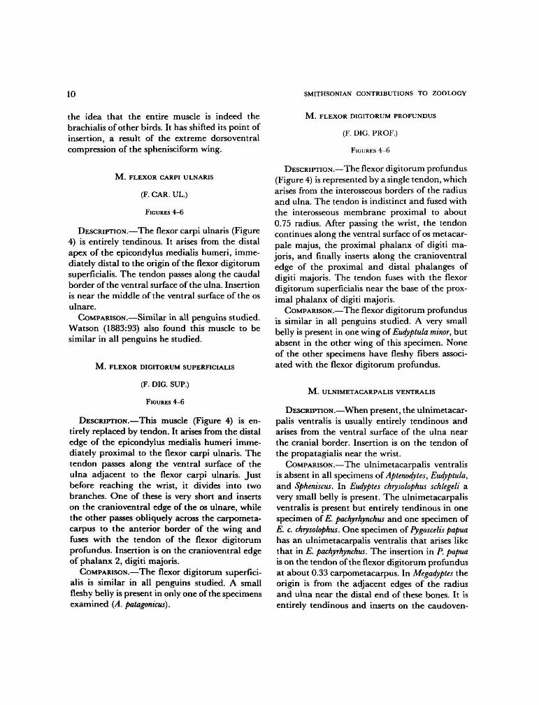
M . FLEXOR CARPI ULNARIS
(F. CAR. UL.)
FIGURES 4-6
DESCRIPTION.—The flexor carpi ulnaris (Figure4) is entirely tendinous. It arises from the distalapex of the epicondylus medialis humeri, imme-diately distal to the origin of the flexor digitorumsuperficialis. The tendon passes along the caudalborder of the ventral surface of the ulna. Insertionis near the middle of the ventral surface of the osulnare.
COMPARISON.—Similar in all penguins studied.Watson (1883:93) also found this muscle to besimilar in all penguins he studied.
M . FLEXOR DIGITORUM SUPERFICIALIS
(F. DIG. SUP.)
FIGURES 4-6
DESCRIPTION.—This muscle (Figure 4) is en-tirely replaced by tendon. It arises from the distaledge of the epicondylus medialis humeri imme-diately proximal to the flexor carpi ulnaris. Thetendon passes along the ventral surface of theulna adjacent to the flexor carpi ulnaris. Justbefore reaching the wrist, it divides into twobranches. One of these is very short and insertson the cranioventral edge of the os ulnare, whilethe other passes obliquely across the carpometa-carpus to the anterior border of the wing andfuses with the tendon of the flexor digitorumprofundus. Insertion is on the cranioventral edgeof phalanx 2, digiti majoris.
COMPARISON.—The flexor digitorum superfici-alis is similar in all penguins studied. A smallfleshy belly is present in only one of the specimensexamined (A. patagomcus).
M . FLEXOR DIGITORUM PROFUNDUS
(F. DIG. PROF.)
FIGURES 4-6
DESCRIPTION.—The flexor digitorum profundus(Figure 4) is represented by a single tendon, whicharises from the interosseous borders of the radiusand ulna. The tendon is indistinct and fused withthe interosseous membrane proximal to about0.75 radius. After passing the wrist, the tendoncontinues along the ventral surface of os metacar-pale majus, the proximal phalanx of digiti ma-joris, and finally inserts along the cranioventraledge of the proximal and distal phalanges ofdigiti majoris. The tendon fuses with the flexordigitorum superficialis near the base of the prox-imal phalanx of digiti majoris.
COMPARISON.—The flexor digitorum profundusis similar in all penguins studied. A very smallbelly is present in one wing of Eudyptula minor, butabsent in the other wing of this specimen. Noneof the other specimens have fleshy fibers associ-ated with the flexor digitorum profundus.
M . ULNIMETACARPALIS VENTRALIS
DESCRIPTION.—When present, the ulnimetacar-palis ventralis is usually entirely tendinous andarises from the ventral surface of the ulna nearthe cranial border. Insertion is on the tendon ofthe propatagialis near the wrist.
COMPARISON.—The ulnimetacarpalis ventralisis absent in all specimens of Aptenodytes, Eudyptula,and Spheniscus. In Eudyptes chrysolophus schlegeli avery small belly is present. The ulnimetacarpalisventralis is present but entirely tendinous in onespecimen of E. pachyrhynchus and one specimen ofE. c. chrysolophus. One specimen of Pygoscelis papuahas an ulnimetacarpalis ventralis that arises likethat in E. pachyrhynchus. The insertion in P. papuais on the tendon of the flexor digitorum profundusat about 0.33 carpometacarpus. In Megadyptes theorigin is from the adjacent edges of the radiusand ulna near the distal end of these bones. It isentirely tendinous and inserts on the caudoven-

NUMBER 341 11
tral edge of the carpometacarpus near the proxi-mal end of the fused pollex. Schoepss (1829:152)describes this muscle in S. demersus as having anorigin from the inner border of the ulna andinserting into the inner side of the base of the firstradial phalanx. Neither Watson (1883:97) nor Ifound this muscle in specimens of S. demersus.Gervais and Alix (1877) and Filhol (1885) gaveno reference to such a muscle in penguins.
M . EXTENSOR METACARPI RADIALIS
(E. META. RAD.)
FIGURES 1-3
DESCRIPTION.—This is a small, weak, proxi-mally situated muscle (Figure 1) arising tendinousfrom the cranial border of the dorsal surface ofthe humerus immediately proximal to the bra-chialis. The origin is strongly attached to thetendon of the propatagialis. The muscular fibersare short and end on a strong tendon which, afterpassing through a shallow groove near the dor-socranial border of the radius, inserts on theproximal end of the carpometacarpus near thecranial border. Insertion is in common with theextensor longus alulae.
COMPARISON.—The muscle is longest in Eudyp-tula (0.21 radius) and shortest in Pygoscelis (0.02radius). In the remaining genera it is of interme-diate length. Schoepss (1829:145) reports that thismuscle arises from the humerus by two distinctheads in penguins. I failed to find this arrange-ment in any species of penguin, nor has any otherworker reported two heads of origin in penguins.
Watson (1883:94) reports a separate insertionfor the extensor metacarpi radialis longus (exten-sor metacarpi radialis) and extensor metacarpiradialis brevis in all the specimens that he dis-sected. Such an arrangement was not present inany specimen examined in this study. Gervaisand Alix (1877:499) and Filhol (1885:176) are inagreement with my findings.
M. SUPINATOR
(SUPIN.)
FIGURES 1-3
DESCRIPTION.—This very weak, triangular mus-cle (Figure 1) arises by means of a short, delicatetendon from the heavy tendon of the extensordigitorum communis, which arises from the dor-sal surface of the distal end of the humeral shaft.The muscular fibers pass distally and cranially toinsert on the dorsal surface of the radius proximalto 0.29 radius.
COMPARISON.—The supinator is longest inSpheniscus, intermediate in Pygoscelis and Eudyp-tula, and shortest in Megadyptes, Aptenodytes andEudyptes. This muscle is absent in one specimen ofP. antarctica. It lacks muscular fibers in two spec-imens of P. adeliae.
M . EXTENSOR DIGITORUM COMMUNIS
(E. DIG. COM.)
FIGURES 1-3
DESCRIPTION.—This muscle (Figure 1) is rep-resented by a strong tendinous band, which arisesfrom the cranial edge of a wide tendinous sheetfrom the epicondylus lateralis humeri. There isno branch to the region of the fused pollex.Insertion is on the craniodorsal surface of osmetacarpale major near the distal end, and onboth phalanges of digiti majoris nearly to the tipof the wing. The tendon is completely fused withthe tendon of the extensor longus digiti majorisnear the middle of the major metacarpal.
COMPARISON.—A very small, fleshy belly is pre-sent in only two of the specimens examined, oneof Eudyptes pachyrhynchus and one of Spheniscushumboldti. In one specimen of P. papua the tendonsends a small branch to the region of the fusedpollex. This branch ends in the fascia over thepollex.

12 SMITHSONIAN CONTRIBUTIONS TO ZOOLOGY
M . EXTENSOR METACARPI ULNARIS
(E. META. UL.)
FIGURES 1-3
DESCRIPTION.—The extensor metacarpi ulnaris(Figure 1) is entirely tendinous. It arises from thecaudal edge of the tendinous sheet in commonwith the extensor digitorum communis. The ten-don passes along the craniodorsal edge of the ulnaand, after crossing the wrist, inserts on the caudaledge of the major metacarpal at about 0.40 car-pometacarpus.
COMPARISON.—In most specimens the insertionis confined to the major metacarpal. Watson(1883:96) found this muscle in Eudyptes crestatus toinsert only on the cranial border of os metacarpaleminus. None of the specimens examined in thisstudy have this arrangement. In all other speci-mens, he found the muscle inserting on the caudalborder of the major metacarpal. Schoepss (1829:150) reports the insertion on both the major andminor metacarpals. Such an arrangement is oc-casionally present. I have found an insertion onboth metacarpals in Megadyptes antipodes (IS),Aptenodytes forsteri (IS), Eudyptula minor (IS), Eu-dyptes c. chrysolophus (IS), and Spheniscus mendiculus(IS).
M . ECTEPICONDYLO-ULNARIS
(ECT.-ULN.)
FIGURE 3
DESCRIPTION.—The ectepicondylo-ulnaris is en-tirely tendinous. It arises from the middle of thetendinous sheet in common with the extensordigitorum communis and extensor metacarpi ul-naris. The tendinous sheet arises from the dorsalsurface of the distal end of the humerus. Insertionis on the cranial border of about the proximalhalf of the ulnar shaft.
COMPARISON.—This muscle is shortest in Eu-dyptes and longest in Eudyptula and Pygoscelis.There is a small belly in A. patagonicus (IS), Eu-dyptes chrysolophus schlegeli (2W), P. papua (IS),
Eudyptula m. albosignata (1W), S. demersus (1W),and S. humboldti (1W); tendinous in all others.
The ectepicondylo-ulnaris (anconaeus) muscledescribed by Reid (1835:142) and "l'ancone ex-terne" described by Gervais and Alix (1877:449)is either a ligament of the elbow joint or a part ofthe triceps tendon. The structure described bythese authors cannot be homologized with anymuscle in other birds. They completely omit ref-erence to the tendon that I call "ectepicondylo-ulnaris." Watson (1883:92) did not find this mus-cle in any species of penguin he dissected, andFilhol (1885:175) reports the muscle to be absentin Eudyptes crestatus.
M . EXTENSOR LONGUS ALULAE
(E. LON. AL.)
FIGURES 1-3
DESCRIPTION.—The extensor longus alulae(Figure 2) arises by means of two heads; a smallradial head from the caudodorsal surface of theradius beginning at about 0.43 radius and a largerulnar head from about the proximal half of thecranial border of the ulnar shaft. The two belliesfuse about midway the forearm, and the commonbelly extends obliquely across the radius endingon a tendon at about 0.80 radius. After crossingthe wrist, this tendon fuses with that of the exten-sor metacarpi radialis. Insertion is on the proxi-mal end of the carpometacarpus near the cranialborder of this bone.
COMPARISON.—The radial head is absent inboth wings of E. m. minor and one wing of E. m.albosignata. Whether the absence of the radialhead in Eudyptula is significant cannot be deter-mined on the basis of only two specimens. Allother species are similar to Eudyptes.
M . EXTENSOR LONGUS DIGITI MAJORIS
(E. LON. DIG. MAJ.)
FIGURES 2, 3
DESCRIPTION.—This muscle (Figure 2) is re-
NUMBER 341 13
placed by a tendon that arises from the distal halfof the caudal surface of the radius between about0.53 radius and 0.82 radius. The tendon thenpasses between the distal ends of the radius andulna, crosses the wrist, and fuses with the tendonof the extensor digitorum communis about mid-way of the major metacarpal. There is no trace ofa distal head.
COMPARISON.—The extensor longus digiti ma-joris is absent in one specimen of A. forsteri andone of A. patagonicus. It is small but partly fleshyin one specimen of A. forsteri and very small andtendinous in the other specimen of A. patagonicus.In one specimen of Eudyptes pachyrhynchus thereare a few fleshy fibers associated with this muscle.It is entirely tendinous in all other penguinsstudied.
Watson (1883:97) described the extensor lon-gus digiti majoris as being a very slender musclearising from the contiguous borders of the radiusand ulna and inserting into the outer side of thesecond or terminal radial phalanx. Such an ar-rangement does not occur in any of the specimensexamined in the current study. There is a con-nection to the posterior edge of the major meta-carpal, as well as to the extensor digitorum com-munis in Eudyptula. Gervais and Alix (1877:451)describe the extensor longus digiti majoris inEudyptes chrysocome as a very small fleshy bundlethat arises from the distal half of the interosseusspace between the radius and ulna.
My findings are in agreement with those ofMeckel and Schoepss. According to Meckel(1828:344), this muscle is represented entirely bytendon. Schoepss (1829:159) found that its originwas confined to the distal end of the radius.
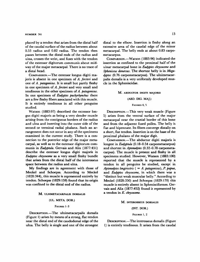
M . ULNIMETACARPALIS DORSALIS
(UL. META. DOR.)
FIGURES 1-3
DESCRIPTION.—The ulnimetacarpalis dorsalis(Figure 1) arises by means of a strong, flat tendonnear the distal end of the caudodorsal edge of theulna. The belly is single and one of the strongest
distal to the elbow. Insertion is fleshy along anextensive area of the caudal edge of the minormetacarpal. The belly ends at about 0.83 carpo-metacarpus.
COMPARISON.—Watson (1883:96) indicated theinsertion as confined to the proximal half of theulnar metacarpal bone in Eudyptes chrysocome andSpheniscus demersus. The shortest belly is in Mega-dyptes (0.76 carpometacarpus). The ulnimetacar-palis dorsalis is a very uniformly developed mus-cle in the Spheniscidae.
M . ABDUCTOR DIGITI MAJORIS
(ABD. DIG. MAJ.)
FIGURES 4, 5
DESCRIPTION.—This very weak muscle (Figure5) arises from the ventral surface of the majormetacarpal near the cranial border of this boneand from the adjacent fused pollex. The belly isflat and bipennate. Its fibers converge distally ona short, flat tendon. Insertion is on the base of theproximal phalanx of the major digit.
COMPARISON.—The abductor digiti majoris islongest in Eudyptula (0.18-0.54 carpometacarpus)and shortest in Aptenodytes (0.32-0.78 carpometa-carpus). The muscle is present and fleshy in allspecimens studied. However, Watson (1883:100)reported that the muscle is represented by atendon in all penguins he studied, except inAptenodytes longirostris ( = A. patagonicus), P. papua,and Eudyptes chrysocome, in which there was a"distinct but weak muscular belly." According toMeckel (1828:350) and Schoepss (1829:170) thismuscle is entirely absent in Sphenisciformes. Ger-vais and Alix (1877:452) found it represented bya tendon in E. chrysocome.
M . INTEROSSEUS DORSALIS
(INT. DOR.)
FIGURES 1, 2
DESCRIPTION.—The interosseus dorsalis (Figure1) is entirely tendinous. It arises from the caudal

14 SMITHSONIAN CONTRIBUTIONS TO ZOOLOGY
border of the major metacarpal and the cranialborder of the minor metacarpal. The tendinoussheet that replaces the belly gives rise to a smalltendon near the distal end of the carpometacar-pus. Insertion is on the middle of the dorsalsurface of the base of the distal phalanx of themajor digit.
COMPARISON.—A small, very weak belly is pre-sent in one specimen of A. patagonicus and one ofS. mendiculus. Watson (1883:101) reports the mus-cle entirely absent in one specimen of A. patagon-icus and Eudyptula minor. In P. papua he found itrepresented by a tendon without a muscularbelly. According to Schoepss (1829:172) this mus-cle is absent in S. demersus. Gervais and Alix (1877:453) found "the muscle, seldom fleshy."
M . INTEROSSEUS VENTRALIS
(INT. VEN.)
FIGURES 1, 2, 4, 5
DESCRIPTION.—The interosseus ventralis (Fig-ure 5) is a weak, fleshy, bipennate muscle thatarises from the caudal border of the major meta-carpal and the cranial border of the minor met-acarpal on the ventral side of the carpometacar-pus. The muscular fibers end on a slender tendonthat passes to the dorsal surface and then insertsalong the entire caudal border of the distal pha-lanx of the major digit.
COMPARISON.—The belly is longest in Apteno-dytes, Megadyptes, and Eudyptula; shortest in Pygos-celis.
M . FLEXOR DIGITI MINORIS
(F. DIG. MIN.)
FIGURES 4, 5
DESCRIPTION.—This muscle (Figure 5) has afleshy origin from the ventral surface proximallyand the caudal surface distally of the minor meta-carpal. The tendon is mainly on the ventral sur-face of the belly. Insertion is mostly tendinous on
the proximally projecting tubercle on the caudaledge of the minor digit.
COMPARISON.—The belly is longest in Apteno-dytes and Eudyptes; shortest in Megadyptes. Thelocation of the distal end of the muscle is veryuniform, at 0.92 and 0.94 carpometacarpus.
The following muscles are absent in the sphen-isciform wing: M. serratus superficialis metapa-tagialis, M. scapulohumeralis cranialis, M. ex-pansor secundariorum. M. pronator superficialis,M. pronator profundus, M. entepicondylo-ul-naris, M. abductor alulae, M. flexor alulae, M.adductor alulae, and M. extensor brevis alulae.
Muscles of the Pelvic Appendage
M . ILIOTROCHANTERICUS CAUDALIS
(IL. TROC. CAUD.)
FIGURES 2, 10, 11
DESCRIPTION.—The iliotrochantericus caudalis(Figure 10) arises fleshy from most of the prea-cetabular ilium (ala preacetabularis). The bellyis very large. Its fibers converge caudally and endon a short broad tendon that inserts on the prox-imocranial edge of the external surface of thefemoral trochanter. The iliofemoralis externus isfused to the iliotrochantericus caudalis except forits extreme distal end and tendon of insertion.
COMPARISON.—The iliotrochantericus caudalisis widest in Aptenodytes, Pygoscelis, and Eudyptula;narrowest in Spheniscus, Megadyptes, and Eudyptes.
According to Watson (1883:103), this muscle"arises from the whole of the external surface ofthe iliac bone as far back as the posterior borderof the acetabulum, as well as from the adjoininghollowed surface formed by the fifth, sixth, andseventh lumbo-sacral vertebrae." In no instancedid I find the iliotrochantericus caudalis attachedto the lumbo-sacral vertebrae, nor has such anarrangement been reported by any other worker.

NUMBER 341 15
M . ILIOTROCHANTERICUS CRANIALIS
(IL. TROC. CRAN.)
FIGURES 10-12
DESCRIPTION.—This muscle (Figure 12), muchsmaller than the iliotrochantericus caudalis, arisespartly fleshy from about the caudal two-thirds ofthe ventrolateral edge of the preacetabular ilium.Carnially the belly is strongly adherent to a fi-brous septum, which separates it from the iliotro-chantericus caudalis. The iliotrochantericus cran-ialis is fused to the iliotrochantericus medius, thetwo muscles being barely distinguishable. Inser-tion is by means of a short, flat tendon on thelateral surface of the femoral trochanter, in com-mon with the iliotrochantericus medius.
COMPARISON.—Only in Eudyptula is the originseparate from that of iliotrochantericus medius.In this genus the insertion, though separate, isjust distal to that of the iliotrochantericus medius.Watson (1883:104) observed a tendency in Apteno-dytes for the iliotrochantericus cranialis to divideinto two distinct portions, an upper and a lower,with a cellular interval lying between them. Hefailed to recognize these as representing the ili-otrochantericus cranialis and iliotrochantericusmedius. According to Gervais and Alix (1877:454), the iliotrochantericus cranialis in Eudypteschrysolophus attaches to the external border of theilium, "et, sur la face interne du femur, au dela dutrochanter"
M . ILIOTROCHANTERICUS MEDIUS
(IL. TROC. MED.)
FIGURES 10-12
DESCRIPTION.—The iliotrochantericus medius(Figure 12) arises fleshy from the ventrolateraledge of the preacetabular ilium immediately cau-dal to the iliotrochantericus cranialis. The iliotro-chantericus medius and iliotrochantericus crani-alis are fused and nearly indistinguishable. Inser-tion is in common with the iliotrochantericuscranialis on the lateral surface of the femoral
trochanter. The femorotibialis medius partly cov-ers this insertion.
COMPARISON.—Watson (1883:104), referring tothe iliotrochantericus medius, states, "Of thismuscle the Penguins do not possess the slightesttrace." In Aptenodytes he observed a tendency forthe iliotrochantericus cranialis "to divide into twodistinct portions, an upper and a lower, a cellularinterval lying between them." His two portionsof the iliotrochantericus cranialis probably rep-resent the iliotrochantericus cranialis and iliotro-chantericus medius. The bellies are fused to somedegree in all penguins. In Eudyptula the origins areseparate.
M . ILIOFEMORALIS EXTERNUS
(ILFEM. EXT.)
FIGURES 10, 11
DESCRIPTION.—The iliofemoralis externus (Fig-ure 11) is very weakly developed. It arises fleshyfrom the lateral dorsal ridge of the ilium imme-diately caudal to the iliotrochantericus caudalis.The origin is situated cranial to the acetabulum,an arrangement unusual among birds. The belliesof iliofemoralis externus and iliotrochantericuscaudalis are almost completely fused, but theextreme distal end of the belly and the tendon ofinsertion are not fused to iliotrochantericus cau-dalis. The short, flat tendon of insertion passesover the iliotrochanterici tendons before insertingon the femoral trochanter. The tendon barelyreaches the ischiofemoralis, which covers the dis-tal end of the insertion of iliofemoralis externus.
COMPARISON.—The belly of this muscle is sep-arate from that of iliotrochantericus caudalis inonly one specimen (Megadyptes antipodes) exam-ined. Gervais and Alix (1877:454) found the in-sertion to be in common with the iliotrochanter-icus caudalis, but the insertions were separate inall penguins that I examined.

16 SMITHSONIAN CONTRIBUTIONS TO ZOOLOGY
M . ILIOFEMORALIS INTERNUS
(ILFEM. INT.)
FIGURE 14
DESCRIPTION.—The iliofemoralis internus is asmall muscle arising fleshy from the ventral edgeof the preacetabular ilium just medial to theiliotrochantericus medius. It passes caudodistallyto insert fleshy on the medial surface of the femurnear the proximal end of the femorotibialis inter-nus.
COMPARISON.—This muscle is very uniformlydeveloped, showing no significant variations inpenguins. However, Meckel (1828:353) thoughtit to be absent in the Spheniscidae.
M. AMBIENS
FIGURES 10-14
DESCRIPTION.—The ambiens (Figure 13) is alarge muscle, arising mostly fleshy from the ven-tral border of the acetabulum. The flattened bellylies on the medial side of the thigh, and terminateson a strong, flat tendon at about 0.86 femur. Thetendon passes in a shallow groove (Figure 14) onthe cranial surface of the patella, then throughthe patellar tendon to the lateral side of the knee.The tendon ends opposite the proximal part ofthe fibula by inserting (Figure 12) on the tendonof origin of the cranial head of the perforatedflexors.
COMPARISON.—The ambiens is shortest in Py-goscelis and Eudyptula, and is longest in Aptenodytes.
M . ILIOTIBIALIS CRANIALIS
(ILTIB. CRAN.)
FIGURES 1,9, 13
DESCRIPTION.—The iliotibialis cranialis (Figure9) is a very powerful muscle, making up theanterior limit of the thigh. The origin is aponeu-rotic from the caudal end of the fused thoracicspinous ridge and fleshy from the cranial andcranio ventral edges of the ilium. There is a strong
tendinous connection with the aponeurosis of theiliotibialis. Insertion (Figure 13) is fleshy on themedial and cranial surfaces of the patella andpatellar tendon.
COMPARISON.—No major differences werenoted other than variations in relative width, thebelly being broadest in Eudyptula and Megadyptes.
M. ILIOTIBIALIS
(IL. TIB.)
FIGURES 2, 9, 13
DESCRIPTION.—The iliotibialis (Figure 9) is aweakly developed, thin sheet of muscle that arisesby means of an aponeurosis from the spinousprocesses opposite the acetabulum. This aponeu-rosis is attached to the belly of the iliotibialiscranialis cranially and the biceps femoris cau-dally. The postacetabular part of the iliotibialisis absent. The belly is thin along its cranialmargin and much thicker along its caudal border.The center of this muscle is aponeurotic distally.Insertion is tendinous on the crista cnemialis cran-ialis of the tibia in common with the femorotibi-alis medius and femorotibialis externus. Theiliotibialis tendon forms a part of the patellartendon.
COMPARISON.—The iliotibialis is a uniformlydeveloped muscle in all species examined. Ac-cording to Garrod (1873:643), the postacetabularportion of this muscle is absent in penguins, whichis consistent with my observations. Watson (1883:112) reports the presence of this portion, althoughreduced to a minimum size, in every species heexamined.
M . FEMOROTIBIALIS EXTERNUS
(FEM. TIB. EXT.)
FIGURE 12
DESCRIPTION.—The femorotibialis externus(Figure 12) is a very small muscle. It arises fromthe caudolateral surface of the femur beginning
NUMBER 341 17
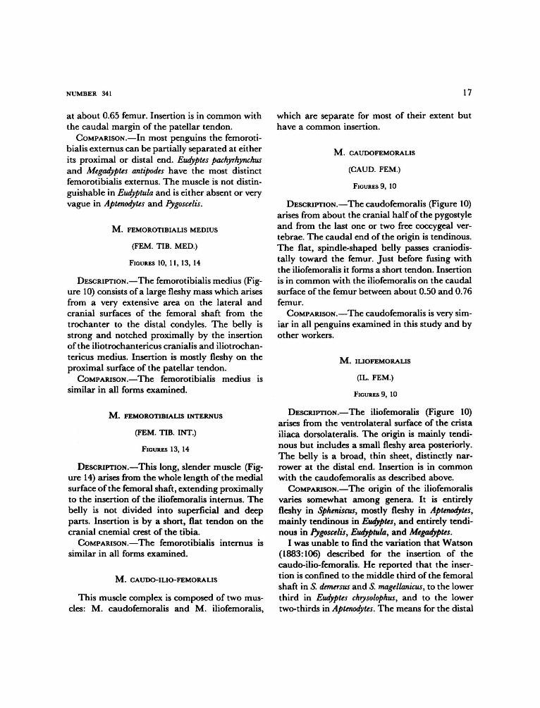
at about 0.65 femur. Insertion is in common withthe caudal margin of the patellar tendon.
COMPARISON.—In most penguins the femoroti-bialis externus can be partially separated at eitherits proximal or distal end. Eudyptes pachyrhynchusand Megadyptes antipodes have the most distinctfemorotibialis externus. The muscle is not distin-guishable in Eudyptula and is either absent or veryvague in Aptenodytes and Pygoscelis.
M . FEMOROTIBIALIS MEDIUS
(FEM. TIB. MED.)
FIGURES 10, 11, 13, 14
DESCRIPTION.—The femorotibialis medius (Fig-ure 10) consists of a large fleshy mass which arisesfrom a very extensive area on the lateral andcranial surfaces of the femoral shaft from thetrochanter to the distal condyles. The belly isstrong and notched proximally by the insertionof the iliotrochantericus cranialis and iliotrochan-tericus medius. Insertion is mostly fleshy on theproximal surface of the patellar tendon.
COMPARISON.—The femorotibialis medius issimilar in all forms examined.
M . FEMOROTIBIALIS INTERNUS
(FEM. TIB. INT.)
FIGURES 13, 14
DESCRIPTION.—This long, slender muscle (Fig-ure 14) arises from the whole length of the medialsurface of the femoral shaft, extending proximallyto the insertion of the iliofemoralis internus. Thebelly is not divided into superficial and deepparts. Insertion is by a short, flat tendon on thecranial cnemial crest of the tibia.
COMPARISON.—The femorotibialis internus issimilar in all forms examined.
M . CAUDO-ILIO-FEMORALIS
This muscle complex is composed of two mus-cles: M. caudofemoralis and M. iliofemoralis,
which are separate for most of their extent buthave a common insertion.
M . CAUDOFEMORALIS
(CAUD. FEM.)
FIGURES 9, 10
DESCRIPTION.—The caudofemoralis (Figure 10)arises from about the cranial half of the pygostyleand from the last one or two free coccygeal ver-tebrae. The caudal end of the origin is tendinous.The flat, spindle-shaped belly passes craniodis-tally toward the femur. Just before fusing withthe iliofemoralis it forms a short tendon. Insertionis in common with the iliofemoralis on the caudalsurface of the femur between about 0.50 and 0.76femur.
COMPARISON.—The caudofemoralis is very sim-iar in all penguins examined in this study and byother workers.
M . ILIOFEMORALIS
(IL. FEM.)
FIGURES 9, 10
DESCRIPTION.—The iliofemoralis (Figure 10)arises from the ventrolateral surface of the cristailiaca dorsolateralis. The origin is mainly tendi-nous but includes a small fleshy area posteriorly.The belly is a broad, thin sheet, distinctly nar-rower at the distal end. Insertion is in commonwith the caudofemoralis as described above.
COMPARISON.—The origin of the iliofemoralisvaries somewhat among genera. It is entirelyfleshy in Spheniscus, mostly fleshy in Aptenodytes,mainly tendinous in Eudyptes, and entirely tendi-nous in Pygoscelis, Eudyptula, and Megadyptes.
I was unable to find the variation that Watson(1883:106) described for the insertion of thecaudo-ilio-femoralis. He reported that the inser-tion is confined to the middle third of the femoralshaft in S. demersus and S. magellanicus, to the lowerthird in Eudyptes chrysolophus, and to the lowertwo-thirds in Aptenodytes. The means for the distal
18 SMITHSONIAN CONTRIBUTIONS TO ZOOLOGY
end of this muscle's insertion ranged from 0.71 to0.79 femur.
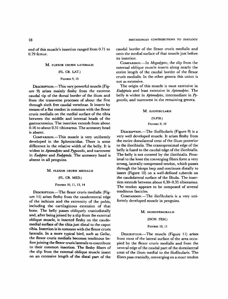
M . FLEXOR CRURIS LATERALIS
(FL. CR. LAT.)
FIGURES 9, 10
DESCRIPTION.—This very powerful muscle (Fig-ure 9) arises mainly fleshy from the extremecaudal tip of the dorsal border of the ilium andfrom the transverse processes of about the firstthrough sixth free caudal vertebrae. It inserts bymeans of a flat tendon in common with the flexorcruris medialis on the medial surface of the tibiabetween the middle and internal heads of thegastrocnemius. The insertion extends from about0.16 to about 0.31 tibiotarsus. The accessory headis absent.
COMPARISON.—This muscle is very uniformlydeveloped in the Spheniscidae. There is somedifference in the relative width of the belly. It iswidest in Aptenodytes and Pygoscelis, and narrowestin Eudyptes and Eudyptula. The accessory head isabsent in all penguins.
M . FLEXOR CRURIS MEDIALIS
(FL. CR. MED.)
FIGURES 10, 11, 13, 14
DESCRIPTION.—The flexor cruris medialis (Fig-ure 11) arises fleshy from the caudoventral edgeof the ischium and the extremity of the pubis,including the cartilaginous extension of thatbone. The belly passes obliquely craniodistallyand, after being joined by a slip from the externaloblique muscle, is inserted fleshy on the caudo-medial surface of the tibia just distal to the caputtibia. Insertion is in common with the flexor crurislateralis. In a more typical bird, such as Gallus,the flexor cruris medialis becomes tendinous be-fore joining the flexor cruris lateralis to contributeto their common insertion. The fleshy fibers ofthe slip from the external oblique muscle inserton an extensive length of the distal part of the
caudal border of the flexor cruris medialis andonto the medial surface of that muscle just beforeits insertion.
COMPARISON.—In Megadyptes, the slip from theexternal oblique muscle inserts along nearly theentire length of the caudal border of the flexorcruris medialis. In the other genera this union isnot as extensive.
The origin of this muscle is most extensive inEudyptula and least extensive in Aptenodytes. Thebelly is widest in Aptenodytes, intermediate in Py-goscelis, and narrowest in the remaining genera.
M . ILIOFIBULARIS
(ILFIB.)
FIGURES 9, 10
DESCRIPTION.—The iliofibularis (Figure 9) is avery well developed muscle. It arises fleshy fromthe entire dorsolateral crest of the ilium posteriorto the iliotibialis. The cranioproximal edge of thebelly is fused to the caudal edge of the iliotibialis.The belly is not covered by the iliotibialis. Prox-imal to the knee the converging fibers form a verystrong, laterally compressed tendon, which passesthrough the biceps loop and continues distally toinsert (Figure 10) on a well-defined tubercle onthe caudolateral surface of the fibula. The inser-tion extends between about 0.30-0.35 tibiotarsus.The tendon appears to be composed of severaltendinous fascicles.
COMPARISON.—The iliofibularis is a very uni-formly developed muscle in penguins.
M . ISCHIOFEMORALIS
(ISCH. FEM.)
FIGURES 10, 11
DESCRIPTION.—The muscle (Figure 11) arisesfrom most of the lateral surface of the area occu-pied by the flexor cruris medialis and from theventral edge of the caudal part of the dorsolateralcrest of the ilium medial to the iliofibularis. Thefibers pass cranially, converging on a stout tendon

NUMBER 341 19
that inserts on the femoral trochanter caudal tothe insertions of the iliotrochantericus cranialisand medius.
COMPARISON.—Except for some variation inlength, this is a very uniformly developed muscle.It is longest in Aptenodytes and shortest in Eudyp-tula.
M . OBTURATORIUS MEDIALIS
(OBT. MED.)
FIGURES 12, 13
DESCRIPTION.—The obturatorius medialis (Fig-ure 13) is an elongate oval muscle. It arises fromthe inner surface of most of the ischium caudal tothe obturator foramen and from most of themedial surface of the pubis. Anteriorly the muscleenters the obturator foramen and passes to theoutside of the pelvis, where the fibers end on astrong tendon that inserts on the caudal edge ofthe femoral trochanter just caudal to the insertionof the ischiofemoralis. Insertion is in commonwith the obturatorius lateralis and is partly fleshy.
COMPARISON.—The width is about 0.25 thelength of the muscle in Aptenodytes. In Spheniscus itis about 0.44 the length of the muscle.
M . OBTURATORIUS LATERALIS
(OBT. LAT.)
FIGURE 12
DESCRIPTION.—The obturatorius lateralis is asmall quadrilateral muscle that arises from thelateral surface of the pelvis adjacent to the obtur-ator foramen. Insertion is on the caudal border ofthe femoral trochanter in common with the ob-turatorius medialis.
COMPARISON.—This muscle is very similar inall forms examined.
M . PUBO-ISCHIO-FEMORALIS
(PUB.-IS.-FEM.)
FIGURES 9-13
DESCRIPTION.—This very powerful muscle (Fig-ure 12) arises mainly fleshy from almost the entire
length of the lateral surface of the pubis caudalto the acetabulum, from part of the cartilaginousextension of this bone, and from a less extensivearea on the ventrolateral edge of the ischium.The belly is clearly divided into lateral and me-dial heads, especially anteriorly. The fibers passcraniodistally to insert on the caudal border ofthe femoral shaft distal to about 0.37 femur. Theinsertion is mostly fleshy, but a few of the caudalfibers insert by means of a tendon immediatelyabove the internal condyle of the femur. Thismuscle is firmly fused to the gastrocnemius parsmedialis for a considerable length.
COMPARISON.—Although the pubo-ischio-fe-moralis shows no major variations in penguins,there is a slight variation in the relative width ofthe belly; it is widest in Aptenodytes, Pygoscelis, andSpheniscus, narrowest in Megadyptes. There is alsosome variation in the length of origin along thepubis. It is longest in Aptenodytes and shortest inEudyptes.
M . TIBIALIS CRANIALIS
(TIB. CRAN.)
FIGURES 10, 11, 13-16
DESCRIPTION.—The tibialis cranialis (Figure10) arises by two distinct heads. The lateral headarises by means of a strong tendon from thecranial surface of the lateral condyle of the femur.The medial head has a fleshy origin from a verysmall part of the lower border of the patella, theintermuscular septum, which separates the tibi-alis cranialis from the gastrocnemius pars medi-alis, and from the cranial cnemial crest of thetibia. The medial head is also tendinous from avery narrow area on about the proximal fourthof the cranial border of the tibia. The heads uniteand form a single tendon opposite the tibiotarsus.The tendon passes beneath a heavy fibrous loop(Figure 15) just above the distal malleoli, tra-verses the intertarsal joint, and inserts on a tu-bercle of metatarsus III at about 0.34 tarsometa-tarsus. It sends a smaller branch laterally to meta-tarsus II.

20 SMITHSONIAN CONTRIBUTIONS TO ZOOLOGY
COMPARISON.—Watson (1883:118) describes asingle insertion on metatarsus III in penguins.Only in S. mendiculus did he find a branch tometatarsus II. I found a branch to metatarsus IIin most penguins. Only in Pygoscelis papua (IS), P.antarctica (IS), Eudyptes chrysolophus schlegeli (IS),and E. chrysocome (IS) did I find a single insertionon metatarsus III.
M . EXTENSOR DIGITORUM LONGUS
(EXT. DIG. L.)
FIGURES 9, 12, 14, 15
DESCRIPTION.—This muscle (Figure 12) occu-pies the cranial surface of the tibiotarsus imme-diately caudal to the tibialis cranialis. The originis fleshy from a small area on the distal border ofthe patella and from about the proximal half orless of the craniomedial surface of the tibia. Theweak bipennate belly tapers distally; ending on atendon that passes under the retinaculum exten-sorium tibiotarsi in common with the tibialiscranialis and through a bony canal on the distalend of the tibiotarsus, crosses the intertarsal joint,slides under the retinaculum extensorium tarso-metatarsi, and expands into an aponeurotic tri-angle that, upon reaching the base of the threedigits, divides sending a branch to each digit(Figure 15). The middle branch bifurcates moreor less vaguely near the base of digit III; these areconnected by an aponeurosis.
First digit: There is no branch to the first digit.Second digit: The branch to the second digit
inserts mainly on the distal end of phalanx 1 andthe bases of the second and third phalanges.
Third digit: The medial branch inserts mainlyon the distal end of phalanx 1 and proximal endof phalanx 2. The long lateral branch gives off abranch laterally to the base of phalanx 3, thencontinues distally. It inserts mainly on the basesof the second, third, and ungual phalanges.
Fourth digit: The branch to the fourth digitinserts mainly on the bases of the second, third,fourth, and fifth phalanges.
COMPARISON.—This is a very uniformly devel-oped muscle in penguins.
M . FIBULARIS LONGUS
(FIB. LONG.)
FIGURES 9-11, 15, 18
DESCRIPTION.—The fibularis longus (Figure 9)occupies the cranial and craniolateral surfaces ofthe shank. It arises tendinous from the cranialsurface of the cranial cnemial crest of the tibia,by a strong aponeurosis from a long narrow lineon the craniolateral border of the tibia, and partlyfleshy from about the middle third of the lateralborder of the fibula. The muscular fibers end ona tendon above the intertarsal joint at about 0.78tibiotarsus. Just above the malleolus, the tendongives off a very broad flattened expansion to thelateral side of the proximal end of the tibialcartilage. This expansion is strongly attached tothe lateral surface of the lateral malleolus. Afterpassing the intertarsal joint, the tendon dividesinto a cranial and a caudal branch. The cranialtendon passes down the lateral side of metatarsusIV and the fourth digit, ending on the penulti-mate phalanx of digit IV. The caudal branch(Figure 18) passes obliquely caudodistally andunites with the tendon of the flexor perforatusdigiti III.
COMPARISON.—No significant variations werenoted in penguins.
M. FIBULARIS BREVIS
(FIB. BREV.)
FIGURES 9-12
DESCRIPTION.—The fibularis brevis (Figure 12)is a very slender muscle that arises from aboutthe distal half of the fibula and the adjacentborder of the tibia. Its tendon passes caudodistallyacross the lateral malleolus and, after crossing theintertarsal joint, inserts on the lateral edge of thetarsometatarsus.
COMPARISON.—No significant differences occur

NUMBER 341 21
in the Spheniscidae. The fibularis brevis is anextremely weak muscle in penguins. Watson(1883:119) has this muscle arising from the uppertwo-thirds of the fibula. The most extensive originoccurs in Megadyptes, in which it arises from mostof the length of the fibula. In most of the otherpenguins, the origin is confined to the distal two-thirds of the fibula.
M . GASTROCNEMIUS
(GAS.)
FIGURES 9-13
DESCRIPTION.—The gastrocnemius forms thesuperficial musculature of most of the medial andposterior surface of the shank and arises by threedistinct heads: pars lateralis, pars intermedia, andpars medialis.
Pars lateralis (p. LAT.: Figures 9, 12, 13): Thishead (Figure 9) arises by a strong tendon fromthe femur just above the proximolateral edge ofthe external condyle and partly fleshy from thelength of the biceps loop. The belly broadens overthe caudolateral surface of the shank and ends ona tendon at about 0.74 tibiotarsus. This tendonjoins that of the pars medialisjust above the tibialcartilage.
Pars intermedia (p. INT.: Figures 12, 13): Theintermediate head of the gastrocnemius (Figure12) has a tendinous origin from the caudal edgeof the proximal end of the medial condyle of thefemur. Proximally the belly is strongly fused tothe pubo-ischio-femoralis. Distally the belly ofthe pars intermedia joins the pars medialis atabout 0.40 tibiotarsus. This union is partly fleshy.
Pars medialis (p. MED.: Figures 10-13): The me-dial head of the gastrocnemius (Figure 13) arisesmainly fleshy from the cranial cnemial crest ofthe tibia, from the medial edge of the patella, andfrom the aponeurosis, separating it from the ti-bialis cranialis. The common tendon of the inter-mediate and medial heads joins the tendon of thelateral head just above the tibial cartilage form-ing a very strong tendon that, after passing theintertarsal joint, divides into two stout branches.
The heavier intermediate branch has a broadinsertion near the proximal end of metatarsus II;the lateral branch inserts more distally on thecaudolateral border of metatarsus IV, a littlebelow the middle.
COMPARISON.—Very little variation was notedin the gastrocnemius, although the length of thebelly shows some variation. It is longest in Mega-dyptes and much shorter in the other genera.
M. PLANTARIS
(PLAN.)
FIGURE 14
DESCRIPTION.—The plantaris is a short, flatmuscle that arises fleshy from about the proximalthird of the caudal border of the tibia. Abouthalfway down the shank, the belly ends on aflattened tendon, which rapidly tapers to a smalloval form. Insertion is on the proximal end of themedial side of the tibial cartilage.
COMPARISON.—The belly is longest in Mega-dyptes and shortest in Aptenodytes and Pygoscelis.
Position of the Flexor Tendons Passing theIntertarsal Joint
DESCRIPTION.—Removal of the gastrocnemiustendon from the posterior surface of the jointcapsule exposes a bundle of three tendons on thecaudolateral side. The most cranial of these is aflexor perforatus digiti III, which forms a sheatharound the flexor perforans et perforatus digitiIII medially and the flexor perforatus digiti IVlaterally. The remaining four tendons traverseseparate canals. On the caudomedial side is acanal for the flexor perforans et perforatus digitiII. Cranial and slightly lateral there is a canal forthe flexor perforatus digiti II. Lateral to the flexorperforatus digiti II is a canal for the flexor hallucislongus. The most cranial canal encloses the ten-don of the flexor digitorum longus.
COMPARISON.—Most penguins adhere to theabove pattern. However, some species of Eudyptes,Spheniscus, and Eudyptula have the flexor perforans

22 SMITHSONIAN CONTRIBUTIONS TO ZOOLOGY
et perforatus digiti III caudal to the flexor perfor-atus digiti IV.
M . FLEXOR PERFORANS ET PERFORATUS DIGITI II
(F. P. ET P. D. II)
FIGURES 9, 18
DESCRIPTION.—This muscle (Figure 9) has avery broad origin extending from the gastrocne-mius pars lateralis to the cranial cnemial crest,tendinous from the external condyle of the femur,the joint capsule, the underlying biceps loop, andthe cranial cnemial crest. The belly is stronglyattached to the gastrocnemius pars lateralis nearthe extreme proximal end of this muscle and tothe flexor perforans et perforatus digiti HI andthe fibularis longus. The belly ends a little abovethe middle of the shank on a flat tendon, whichnarrows as it passes distally. Near the base of thesecond digit the tendon perforates that of theflexor perforatus digiti II before inserting mainlyon the distal end of phalanx 1 and proximal endof phalanx 2. There is also a strong attachmentto the base of phalanx 1.
COMPARISON.—This muscle in Eudyptes chrysolo-phus has an additional origin from the tibia andfibula, according to Gervais and Alix (1877:460).I failed to find such an origin in any specimenexamined. They also describe two fleshy sheetscomprising this muscle. Such an arrangementoccurred in several specimens ofSpheniscus, Apteno-dytes, and Pygoscelis, but shows intraspecific vari-ation.
M . FLEXOR PERFORANS ET PERFORATUS DIGITI III
(F. P. ET P. D. HI)
FIGURES 9, 18
DESCRIPTION.—This muscle (Figure 9) arisespartly fleshy from the head of the fibula, fleshyfrom a narrow line on the lateral surface of thefibula, to a point just distal to the iliofibularisinsertion, partly tendinous from the lateral edgeof the cranial cnemial crest, the lateral side of the
joint capsule, and from the patellar tendon. Thebelly is larger than that of the flexor perforans etperforatus digiti II. Just distal to the middle ofthe shank, the belly ends on a tendon that isbroad proximally. Near the base of the thirddigit, the tendon perforates that of the flexorperforatus digiti HI and, in turn, is perforated bythe tendon of the flexor digitorum longus nearthe proximal end of phalanx 2. Insertion is on thedistal end of phalanx 2 and proximal end ofphalanx 3 by two branches that result from theperforation by the flexor digitorum longus.
COMPARISON.—The flexor perforans et perfor-atus digiti HI is very similar in all penguins.
M . FLEXOR PERFORATUS DIGITI II
(FLEX. PER. D. II)
FIGURES 11, 18
DESCRIPTION.—This muscle arises fleshy fromthe intercondylar space in common with the me-dial and lateral heads of flexor perforatus digitiHI and flexor perforatus digiti IV The bellyseparates from the common muscle mass at ap-proximately the middle of the shank. At about0.75 tibiotarsus, the belly terminates on a tendonthat, after being perforated by the flexor perfor-ans et perforatus digiti II near the base of phalanx1 digit II, is inserted on the base of the proximalphalanx.
COMPARISON.—The belly is longest in Apteno-dytes and shortest in Eudyptes. Except for thisdifference in length, the flexor perforatus digiti IIis very similar in all penguins.
M . FLEXOR PERFORATUS DIGITI III
(FLEX. PER. D. Ill)
FIGURES 9-11, 14, 18
DESCRIPTION.—The flexor perforatus digiti III(Figure 10) arises from the regio intercondyloideaof the femur by means of a broad, flat tendon.This tendon is strongly attached to the cranialsurface of the flexor perforatus digiti IV belly. It

NUMBER 341 23
also arises in common with the flexor perforatusdigiti II and flexor perforatus digiti IV from thehead of the fibula and lateral condyle of thefemur by means of a short tendon. The belly isnotched slightly proximally. A tendinous sheetextends between these two parts of the belly. Theflexor perforatus digiti III is the longest of theperforated flexors. Near the distal end of thetibiotarsus, the tendon forms a sheath around themore caudally situated tendons of the flexor per-forans et perforatus digiti III and flexor perforatusdigiti IV. Near the middle of the tarsometatarsus,the tendon is very strongly attached to the caudalbranch of the fibularis longus tendon. Near thebase of phalanx 1, the tendon is perforated bythat of the flexor perforans et perforatus digitiIII. Insertion is on the distal end of phalanx 1.There is no vinculum between the tendons of theflexor perforatus digiti III and flexor perforans etperforatus digiti HI.
COMPARISON.—The flexor perforatus digiti IIIis a very uniformly developed muscle except forsome variation in the length of the belly. Thereis no vinculum between the tendons of the flexorperforatus digiti III and flexor perforans et per-foratus digiti III in any penguin examined.
M . FLEXOR PERFORATUS DIGITI I V
(FLEX. PER. D. IV)
FIGURES 10, 11, 18
DESCRIPTION.—The flexor perforatus digiti IV(Figure 10) has a fleshy origin from the regiointercondyloidea of the femur. It also arises by ashort tendon from the head of the fibula andlateral condyle of the femur in common with theflexor perforatus digiti II and flexor perforatusdigiti III. This short tendon passes medial to thetendon of insertion of the iliofibularis. The bellygives rise to a tendon at about 0.63 tibiotarsus.Near the base of the fourth digit, the tendondilates before dividing into four branches (Figure18). The lateral branch (branch 1) fuses with themedial branch (branch 4) near the middle ofphalanx 1 before inserting on the proximal end
of phalanx 2. The two middle branches (branches2 and 3) fuse near the base of phalanx 2 andinsert on the base of phalanx 3. The flexor digi-torum longus tendon to the fourth digit passesbetween branches 1 and 4 and then betweenbranches 2 and 3.
COMPARISON.—The insertion of the flexor per-foratus digiti IV exhibits several variations. InEudyptes, Spheniscus, and Megadyptes, the tendonsexhibit the same pattern as that described for E.pachyrhynchus. Branch 1 remains separate in Eu-dyptula and inserts on the distal end of phalanx 1and proximal end of phalanx 2, while branches2 and 4 fuse near the distal half of phalanx 1before inserting on the proximal end of phalanx2. In Eudyptula, branch 3 remains separate andinserts on the base of phalanx 3, the tendon ofthe flexor digitorum longus to the fourth toepassing lateral to branch 3. In Pygoscelis, branches1 and 4 fuse before inserting on the base ofphalanx 2. Branch 2 remains separate and insertson the proximal end of phalanx 3. Branch 3likewise remains separate but inserts on the baseof phalanx 4. The flexor digitorum longus tendonto the fourth digit passes between branches 1 and4 and then between branches 2 and 3. None ofthe branches fuse in Aptenodytes. Branches 1 and4 insert on the base of phalanx 2; while branches2 and 3 insert on the bases of phalanx 3 andphalanx 4, respectively. The flexor digitorum lon-gus tendon passes medial to branch 1 and lateralto branches 2, 3, and 4. The belly is longest inMegadyptes and shortest in Pygoscelis and Eudyptes.
M . FLEXOR HALLUCIS LONGUS
(F. HAL. L.)
FIGURES 10-12, 14, 15, 18
DESCRIPTION.—The flexor hallucis longus (Fig-ure 12) arises fleshy from the regio intercondyloi-dea of the femur in common with the perforatedflexors. Its belly is large in relation to the size ofthe hallux. About midway the tibiotarsus, theshort belly ends on a broad, flat tendon, whichtapers as it passes distally toward the intertarsal

24 SMITHSONIAN CONTRIBUTIONS TO ZOOLOGY
joint. The tendon trifurcates opposite the tarso-metatarsus just before fusing with the flexor dig-itorum longus. On the medial side, there is afibrous connection between the tendons of theflexor hallucis longus and flexor digitorum longusimmediately proximal to the trifurcation. Thisconnection has some resemblance to a vinculum.Of the three very short branches of the flexorhallucis longus, the one that fuses with the flexordigitorum longus tendon to the fourth digit is thestrongest. The branch to the third digit is veryweak. In addition to these three tendons, a fourthvery delicate tendon arises from the medial edgeof the flexor hallucis longus opposite the trifur-cation. It inserts on the base of the ungual pha-lanx of the hallux.
COMPARISON.—This muscle shows very littlevariation among penguins. In several specimensof Pygoscelis, I was unable to find the branch tothe hallux. Poor preservation may account forthis omission. The belly is longest in Spheniscusand shortest in Aptenodytes.
M . FLEXOR DIGITORUM LONGUS
(F. DIG. L.)
FIGURES 9-12
DESCRIPTION.—This powerful deeply situatedmuscle (Figures 12, 14) arises from most of thecaudal and medial borders of the tibia distal tothe popliteus, from the interosseus membrane,and the adjacent border of the fibula. The bellygives rise to a strong tendon near the distal endof the tibiotarsus. The tendon trifurcates near theproximal end of the tarsometatarsus. The threebranches, after uniting with those of the flexorhallucis longus, insert on the bases of the ungualphalanges of digits 2, 3, and 4. There is a shortfibro-elastic branch to the base of each phalanxexcept phalanx 1.
COMPARISON.—No important variations werenoted.
M. POPLITEUS
DESCRIPTION.—This small, quadrilateral mus-cle has a fleshy origin from the proximal end of
the caudomedial border of the fibula. Insertion isfleshy near the proximal end of the caudal surfaceof the tibial shaft. The muscle broadens beforereaching its insertion and is fleshy throughout.
COMPARISON.—The popliteus is a very uni-formly developed muscle in penguins.
M . EXTENSOR HALLUCIS LONGUS
(EXT. HAL. L.)
FIGURES 15, 16
DESCRIPTION.—The origin of this small muscle(Figure 15) is fleshy from the dorsal and medialsurfaces of the rudimentary metatarsus I andfrom the ligament that connects the first to thesecond metatarsal. The belly ends near the prox-imal end of phalanx 1. The tendon of insertion isseparated from the surrounding connective tissuewith difficulty. It traverses the dorsal surface ofthe hallux and is inserted along most of the lengthof phalanx 1 and on the base of the ungualphalanx.
COMPARISON.—The origin extends onto meta-tarsus III in a specimen of A. forsteri (1 S), P.antarctica (1 S), E. pachyrhynchus (1 S), and M.antipodes ( I S ) .
M . EXTENSOR BREVIS DIGITI H I
(EXT. BREV. D III)
FIGURES 15, 16
DESCRIPTION.—The origin (Figure 16) is fleshyfrom most of the cranial surface of metatarsus IIIand the groove between metatarsus II and meta-tarsus III. The broad belly terminates on a short,flat tendon that inserts on the base of the firstphalanx of digit III. The broad tendon is growninto the joint capsule.
COMPARISON.—This muscle varies only inlength. The belly is shortest in Pygoscelis andlongest in Aptenodytes and Eudyptula.

NUMBER 341 25
M . EXTENSOR BREVIS DIGITI I V
(EXT. BREV D. IV)
FIGURES 14-16
DESCRIPTION.—The origin (Figure 16) is fleshyfrom the cranial and medial surfaces of metatar-sus IV and the lateral edge of metatarsus III. Itsbelly is wide and occupies most of the cranialsurface of metatarsus IV. Insertion is by a shorttendon on the medial edge, proximal end, ofphalanx 1, digit IV. The tendon passes througha groove between metatarsus III and metatarsusIV. There is no bony canal.
COMPARISON.—The belly of extensor brevis dig-iti IV is longest in Eudyptula and shortest inAptenodytes.
M . ABDUCTOR DIGITI II
(ABD. DIG. II)
FIGURES 13-18
DESCRIPTION.—This muscle (Figure 16) arisesfleshy from most of the cranial and craniomedialborders of metatarsus II. The belly is wide prox-imally and narrow distally. It ends on a short,strong tendon at about 0.66 tarsometatarsus. In-sertion is on the dorsomedial edge of the base ofphalanx 1, digit II.
COMPARISON.—The abductor digiti II is a uni-formly developed muscle in penguins. In somespecimens there is a division in the belly distally.This division tends to be most obvious in Apteno-dytes.
M . FLEXOR HALLUCIS BREVIS
DESCRIPTION.—The flexor hallucis brevis is pre-sent in only five of the six specimens of Pygoscelisexamined. In these it has a fleshy origin from avery small area on metatarsus II at the base ofmetatarsus I and from most of the plantar surfaceof metatarsus I. The belly is very small. Insertionis by a short tendon on the base of the distalphalanx.
COMPARISON.—This muscle is absent in allspecimens of Eudyptes examined. Watson (1883:126) was unable to find this muscle in any of thespecimens examined in his study. Meckel (1828)does not describe this muscle for penguins. Reid(1835:145) and Gervais and Alix (1877:461) de-scribe this muscle in A. patagonicus and E. chryso-iophus, respectively. The muscle is apparently pre-sent in only a small percentage of penguins. Thehigh frequency of its occurrence in Pygoscelis sug-gests that this muscle may be characteristic inthis genus and, hence, of taxonomic importance.A larger sample is needed for verification.
M . LUMBRICALIS
DESCRIPTION.—The lumbricalis arises from thecranial surface of the deep flexor tendon justdistal to the point of trifurcation. The muscle isvery vague and appears mostly fibrous. Insertionis on the joint pulley near the base of digit III.
COMPARISON.—The lumbricalis is very weak inpenguins, and most specimens have only a verysmall vestige when present. Poor preservationmay account for its absence in several specimens.
M . ABDUCTOR DIGITI I V
(ABD. DIG. IV)
FIGURES 17, 18
DESCRIPTION.—The origin (Figure 18) is fleshyfrom most of the caudal border of metatarsus IV.The belly is wider proximally and narrow distally.Insertion is by a short tendon on the lateroplantaredge of the base of the first phalanx of digit IV.The tendon is strongly fused with the connectivetissue over the joint.
COMPARISON.—This is a uniformly developedmuscle in penguins.
The following muscles are absent in the sphen-isciform leg: M. extensor proprius digiti III, M.adductor digiti II, and M. adductor digiti IV.
Discussion and Conclusions
The pectoral musculature of penguins is unlikethat of any other group of birds. A number of

26 SMITHSONIAN CONTRIBUTIONS TO ZOOLOGY
wing muscles are absent, most remarkably thebiceps. The biceps is absent only in penguins.Several other wing muscles, the expansor secun-dariorum, scapulohumeralis cranialis, and serra-tus superflcialis metapatagialis, are also absent inpenguins. The pronator superflcialis and pronatorprofundus are likewise absent, a loss associatedwith the inability of the wing to be rotated at theelbow. Somewhat surprising, because of the lackof a well-developed patagial wing fold, is thepresence of a well-developed propatagialis musclewhose tendon inserts along much of the length ofthe wing and is strongly attached to the alaraponeurosis. Beddard (1898:397) and Watson(1883:88) have this tendon passing all the way tothe extremity of the last phalanx. The latissimusdorsi is peculiar in that its two parts end in long,slender tendons that pass through a fibrous loopfrom the scapula. The latissimus dorsi metapa-tagialis varies greatly in development. It is anextremely wide muscle in Aptenodytes and verynarrow in Pygoscelis and Eudyptula. With the ex-ception of Pygoscelis, the development seems to berelated to the size of the bird. The insertion ofthis muscle in penguins is on the skin along thelateral line of the trunk instead of near the hu-meral feather tract as in most birds. Most of themuscles acting across the shoulder joint are verywell developed, a reflection of the peculiar actionof the wings during underwater flight. The pec-toralis thoracica is so large that it meets its fellowin the midline over the carina, a condition similarto that in Tinamous (Watson, 1883:397). Thesupracoracoideus is very strongly developed inrelation to the pectoralis. These two muscles arethe prime movers of the wing during underwaterflight. Although the short head of the deltoideusminor is very small and weak, the long head islarge and aids the supracoracoideus in raising thewing. The deltoideus major, on the other hand,is a very weak muscle.
Only nine muscles distal to the shoulder havefleshly fibers. The triceps has a peculiar arrange-ment in penguins. Both the triceps scapularis andtriceps humeralis consist of two heads. One headof the triceps scapularis arises from the clavicle
and acromial process of the scapula; the otherhead arises fleshly from the dorsolateral border ofthe scapula in much the same position as theaponeurotic anchor (George and Berger, 1966:335) arises in many other birds. The triceps hu-meralis has both a dorsal and ventral head arisingfrom the humerus. The brachialis is peculiar inpenguins in that it inserts on the radius as aconsequence of the extreme dorsoventral com-pression of the wing, an adaptation to underwaterflight. Occasionally a small slip separates fromthe caudal edge of the muscle and inserts on theulna in a position reminiscent of other birds. Theextensor metacarpi radialis is a small, weak mus-cle showing no indication of a division. Its tendonfuses with that of the extensor longus alulae.These two muscles act as extensors of the wrist,counteracting the powerful flexor, the ulnimeta-carpalis dorsalis, of the wrist.
Three muscles arising from the carpometacar-pus control flexion and extension of the phalan-ges. Extension is mediated by the very weakabductor digiti majoris and interosseus ventralismuscles. Flexion is the function of the strongflexor digiti minoris.
The arrangement of the muscles of the leg isless characteristic than that of the wing. Gervateand Alix (1877:465) and Watson (1883:137) per-ceived it as being similar to the arrangement thatcharacterizes the palmipedes (swimming birds) ingeneral. There are a number of deviations fromthe arrangement found in these birds. The simi-larities can be accounted for by adaptations tosimilar modes of life. The iliotrochantericus cran-ialis and iliotrochantericus medius are fused. Inpenguins the iliofemoralis externus is very weakand fused to the iliotrochantericus caudalis exceptat the extreme distal end. This muscle is absentin Podiceps, but present in most other swimmingbirds, such as Gavia, Sula, and Chen (Hudson,1937:13). The ambiens is a very strong, broadmuscle approaching the condition reported byHudson (1937:15) for Gavia. It is also well devel-oped in other swimming birds but is absent ingrebes (Watson, 1883:129). The caudal part ofthe iliotibialis is absent in penguins, but shows a

NUMBER 341 27
wide range of variation among other groups ofswimming birds. The caudo-ilio-femoralis is welldeveloped in all penguins, and both parts arepresent. The insertion of this muscle is far distalon the femur. The absence of the caudal part ofthe iliotibialis leaves the iliofibularis uncoveredsuperficially.
A slip of muscle from the external obliqueinserts on an extensive area of the caudal borderof the flexor cruris medialis, an arrangementfound in no other group of birds. The flexor crurismedialis shares a common insertion with theflexor cruris lateralis. The insertion is by meansof a fleshy sheet medially and tendinous sheet onthe lateral side. The accessory flexor cruris later-alis is absent in all penguins.
The weak condition of the short muscles of thehallux and the delicate nature of the tendon fromthe flexor hallucis longus are most likely relatedto the vestigial nature of the hallux. The flexorhallucis brevis is absent in most penguins exam-ined. It is present in only five of the six specimensof Pygoscelis. It has, however, been described inAptenodytes patagonicus (Reid, 1835:145) and Eu-dyptes chrysolophus (Gervais and Alix, 1877:461).Watson (1883:126) was unable to find any traceof this muscle in the penguins he examined.
The absence of the adductor digiti II and weakcondition of the lumbricalis are also specializedfeatures of the leg myology of penguins.
Data in the present study were treated numer-ically by two methods, cumulative scores of dif-ference using weighted data and correlation coef-ficients giving equal weight to all characters. Thetwo methods give closely parallel results. This isevident in most interspecific and intergenericcomparisons. Low scores of difference are usuallyassociated with high positive correlation coeffi-cients and high scores with high negative corre-lation coefficients.
In comparing various taxa, wing scores areusually larger than leg scores. The mean for allwing scores is 15.46 points and of leg scores 11.39points. Only in the genus Eudyptes, does the meanscore for the leg exceed the wing score: 8.97 forthe legs compared to 8.10 for the wings. Many
other groups of birds show just the opposite, thewings being least variable, e.g., in Galliformes(Hudson and Lanzillotti, 1964), Lari and Alcae(Hudson et al., 1969), Ciconiiformes (VandenBerge, 1970), Strigiformes and Caprimulgiformes(Hoff, 1966), and Tinamous (Hudson et al.,1972). The greater difference in wings comparedto legs is not surprising in view of the greatmodification of the wing for underwater propul-sion and differences in absolute size in differentgenera. In the largest penguins, Aptenodytes andPygoscelis, the latissimus dorsi cranialis and cau-dalis have increased so tremendously in widththat their adjacent borders have fused. This nu-merical comparison indicates that the antarctic-subantarctic genera Aptenodytes and Pygoscelis ex-hibit the least interspecific variation, suggestingthat species in these genera are very closely re-lated. The two genera, although quite different,are more closely related to each other than toother genera, as Peters (1931:29-31) indicates byplacing Pygoscelis immediately after Aptenodytes;they are not necessarily more primitive than othergenera as his classification indicates. They areperhaps less "primitive," being very specializedin structure and behavior to endure the severeenvironment of the antarctic region. Most likelythese genera evolved from forms inhabiting tem-perate regions. Fossil evidence (Simpson, 1971)suggests that the Sphenisciformes evolved in tem-perate regions and have moved into the moreinhospitable antarctic regions. Selective pressuresmost likely limit the amount of variation possible,thus accounting for the similarity between theseantarctic-subantarctic species.
All correlation coefficients are low, i.e., below0.60. Correlation coefficients of 0.60 or higher aregenerally used in biological work to indicate closerelationship between two entities; two specimensof the same species would therefore be expectedto have a correlation coefficient of 0.60 or higher.Conversely, as the coefficient approaches — 1, en-tities exhibit less similarity and hence a moredistant relationship. Low correlation coefficientssupport the presently recognized genera.
Eudyptula m. minor and E. m. albosignata are very

28 SMITHSONIAN CONTRIBUTIONS TO ZOOLOGY
similar to each other. The scores of difference forthe wings, however, are higher than might beexpected for subspecies so strongly correlated.This discrepancy may be a reflection of the verysmall sample used in this study.
Spheniscus is a rather heterogeneous assemblage,though the species of Spheniscus clearly constitutea definite group. Of the four species comprisingthe genus, S. demersus and S. humboldti (0.47) andS. humboldti and S. mendiculus (0.43) show thestrongest correlation. Spheniscus demersus and S.mendiculus are somewhat less well correlated.Spheniscus magellanicus is most different. The simi-larity between Eudyptes chrysolophus schlegeli and S.humboldti suggests the possibility that Spheniscusrepresents a radiation of penguins from the Aus-tralian-New Zealand area into the South Ameri-can-South African area. The derivation of Eu-dyptes from Spheniscus seems unlikely, since thefossil record (Simpson, 1946) indicates that pen-guins arose in the Australian-New Zealand area.
With myological data and geographical distri-bution, it is possible to construct a tentative phy-logeny of sphenisciform birds. In such a phylog-eny (Figure 19) three main divisions may berecognized: (1) Aptenodytes, Pygoscelis; (2) Eudyptes,Megadyptes; and (3) Spheniscus. Eudyptula represents
a small division with similarities to both theSpheniscus and Eudyptes assemblages. This is verysimilar to the phylogeny devised by Zusi (1975)based on osteology.
Separation of species within each division isalso possible (Figure 19). Of the Spheniscus group,S. magellanicus is most different, having divergedalong a separate pathway. S. humboldti, S. demersus,and S. mendiculus are relatively closely related.Penguins of the Eudyptes-Megadyptes lineage showgreater divergence. Megadyptes has diverged sig-nificantly from the early members of this group,as has E. chrysolophus schlegeli. The remainingmembers of this group, being somewhat moreclosely related, have perhaps diverged more re-cently or occupied habitats with similar selectivefactors.
The antarctic-subantarctic assemblage is com-posed of two distinct genera, Aptenodytes and Py-goscelis. Within these genera, species exhibit ratherclose affinities. A. forsteri and A. patagonicus sharea correlation of 0.54. P. papua shares a correlationof 0.60 with P. antarctica and a correlation of 0.62with P. adeliae. The last two species share a cor-relation of only 0.56. These relationships suggestthat P. papua is closest to the original species ofPygoscelis, while P. adeliae and P. antarctica havediverged to a greater degree.
Appendix
Interesting Features of the Appendicular Muscles
The following list considers the myological features of appendicular musclesthat show important differences among various groups of birds. Musclesdiscussed in the text but not listed here exhibit no unusual modification inpenguins. List is arranged in the same order as the muscles are discussed inthe text.
MUSCLES OF THE PECTORAL APPENDAGE
LATISSIMUS DORSI CRANIALIS. Accessory slip from cranial edgeof triceps scapularis present in Aptenotfytes and Pygoscelis;absent in other genera.
LATISSIMUS DORSI CAUDALIS. Arises from thoracic ribs inpenguins. Additional origin from spinous processes inAptenodyUs and Pygoscelis.
LATISSIMUS DORSI METAPATAGIAUS. Extreme intergeneric var-
iation in width. Insertion on skin along lateral line of thetrunk instead of near the humeral feather tract as in mostbirds.
SERRATUS SUPERFICIALIS CRANIALIS. TWO fleshy fascicles. Veryextensive insertion on the scapula.
SERRATUS SUPERFICIALIS METAPATAGIAUS. Absent in all pen-
guins.SCAPULOHUMERALIS CRANIALIS. Absent in all penguins .
PECTORALJS PARS SUBCUTANEA ABDOMINAUS. Cranial part pre-
sent and very large; caudal part absent.SUPRACORACOIDEUS. Very well developed in relation to the
pectoralis thoracica.CORACOBRACHIALIS CRANIALIS. A very weak (vestigial) mus-
cle; not previously described for penguins.SUBCORACOIDEUS. Belly large, single.SUBSCAPULARIS, CAPUT LATERALE. Very large, much larger
than caput mediate; caput mediate has origin from cora-coid, clavicle, and adjacent scapula (•« cranial head ofsubcoracoideus, Hudson, et al., 1969:464).
PROPATAGIAUS. Belly single, tendon extends length of wingand contributes to alar aponeurosis.
DELTOIDEUS MAJOR. A very weak, flat, usually triangularmuscle in penguins.
DELTOIDEUS MINOR. Short head very small and weak; longhead large, not connected to supracoracoideus.
TRICEPS SCAPULARIS. TWO distinct heads of origin, one fromclavicle and acromial process of scapula, other from lateralborder of scapular shaft.
TRICEPS HUMERALIS. TWO heads of origin, one from the
pneumatic fossa, the other from about the distal 0.60 ofthe caudal border of the humeral shaft.
BICEPS BRACHII. Generally absent in penguins; rarely, avestigial biceps is present.
EXPANSOR SECUNDARIORUM. Absent in all penguins.BRACHIALIS. Inserts on proximal end of radius instead of on
ulna as in other birds.PRONATOR SUPERFICIALIS. Absent in all penguins.PRONATOR PROFUNDUS. Absent in all penguins.ENTEPICONDYLO-ULNARIS. Absent in all penguins.FLEXOR CARPI ULNARIS. Represented by a tendon in all
penguins.FLEXOR DIGITORUM SUPERFICIALIS. Represented by a single
tendon in all penguins.ULNIMETACARPALIS VENTRAUS. Absent in penguins, except as
an infrequent vestige in less than one-sixth of the speci-mens examined.
EXTENSOR METACARPI RADIALJS. Belly small, weak, single.SupwATOR. A very small, weak, fan-shaped muscle, inserts
on dorsal surface of radius.EXTENSOR DIGITORUM COMMUNIS. Represented by a tendinous
band in penguins.EXTENSOR METACARPI ULNARIS. Represented by a tendon in
all penguins; insertion usually on the major metacarpal.ECTEPICONDYLO-ULNARIS. Replaced by a tendon, occasionally
a few fleshy fibers are present.EXTENSOR LONGUS ALULAE. TWO fleshy heads of origin; ten-
don fused with extensor metacarpi radialis at wrists; radialhead absent in most Evufyptula wings.
EXTENSOR LONGUS DIGITI MAJORIS. Represented by a tendon
in all penguins; no distal head.ULNIMETACARPALIS DORSAUS. Belly large, single.ABDUCTOR ALULAE. Absent in all penguins.FLEXOR ALULAE. Absent in all penguins.ADDUCTOR ALULAE. Absent in all penguins.EXTENSOR BREVIS ALULAE. Absent in all penguins.
ABDUCTOR DIGITI MAJORIS. Flat, weak; arises from the ventralsurface of the major metacarpal near cranial border.
29

30 SMITHSONIAN CONTRIBUTIONS TO ZOOLOGY
INTEROSSEUS DORSALIS. Represented by a tendon in all pen-guins.
FLEXOR DIGITI MINORIS. Belly well developed.
MUSCLES OF THE PELVIC APPENDAGE
ILIOTROCHANTERICUS CRANIALIS. Fused to iliotrochantericusmedius, the two muscles are barely distinguishable.
ILIOFEMORALIS EXTERNUS. Very weak; fused to iliotrochanter-icus caudalis except extreme distal end of belly and tendonof insertion; origin cranial to acetabulum.
AMBIENS. Belly very strong; inserts on the tendon of origin ofthe cranial head of the perforated flexors.
ILIOTIBIALIS. Cranial part present; postacetabular portionabsent.
FEMOROTIBIALIS EXTERNUS. Completely separate in sometaxa; partially fused with the femorotibialis medius inothers; not separable in others.
FEMOROTIBIALIS MEDIUS. Origin partly covers common inser-tion of iliotrochantericus cranialis and iliotrochantericusmedius.
FEMOROTIBIALIS INTERNUS. NO division of belly distally.CAUDO-ILIO-FEMORALIS. M. caudofemoralis and M. iliofe-
moralis well developed; insertion far distal on femur.FLEXOR CRURIS LATERALIS. Origin fleshy from transverse proc-
esses of about the first six free caudal vertebrae, no attach-ment to ilium; accessory portion absent in all penguins.
FLEXOR CRURIS MEDIALIS. Insertion fleshy, in common withflexor cruris lateralis; a tendinous sheet on the lateral sideof common insertion (on medial side in a more typicalbird, e.g., Gallus).
ILIOFIBULARIS. Belly not covered by iliotibialis; tendon ofinsertion appears to be composed of two or more fibrousfascicles fused together and compressed laterally.
ISCHIOFEMORALIS. Extensive origin including much of ventraledge of the caudal part of the dorsolateral crest of theilium.
OBTURATORIUS MEDIALIS. Insertion in common with obtura-torius lateralis on caudal border of femoral trochanter.
OBTURATORIUS LATERALIS. Single; origin nearly surroundsforamen obturatorium.
PUBO-ISCHIO-FEMORALIS. Belly clearly divided into lateraland medial heads, especially cranially.
TIBIALIS CRANIALIS. Tendon of insertion sends a very weakbranch laterally to metatarsus II.
EXTENSOR DIGITORUM LONGUS. Tendon forms aponeurotic
triangle opposite tarsometatarsus.FIBULARIS LONGUS. Tendon sends branch along lateral side
of metatarsus IV and digit IV, ending on the penultimatephalanx.
FIBULARIS BREVIS. An extremely weak muscle.GASTROCNEMIUS. All three heads well developed; tendon
branches near the proximal end of the tarsometatarsus;does not ensheath flexor tendons over tarsometatarsus;pars intermedia strongly fused to pubo-ischio-femoralis.
M U S C L E S O F T H E F L E X O R T E N D O N S
FLEXOR PERFORANS ET PERFORATUS DIGITI II. Very broadproximally.
FLEXOR PERFORANS ET PERFORATUS DIGITI III. No vinculumbetween tendon of flexor perforans et perforatus digiti IIIand flexor perforatus digiti III; belly notched slightlyproximal with a tendinous sheet extending between thesetwo parts.
FLEXOR PERFORATUS DIGITI IV. The tendon of insertion showsfour patterns of branching and attachment in differenttaxa.
FLEXOR HALLUCIS LONGUS. The tendon trifurcates oppositethe tarsometatarsus, the three short branches fuse to thoseof the flexor digitorum longus; a fibrous connection on themedial side resembles a vestigial vinculum; branch tohallux very delicate, ending on the ungual phalanx; bellylarge in relation to size of hallux.
POPLITEUS. Muscle fleshy throughout.EXTENSOR HALLUCIS LONGUS. Very weak; origin from rudi-
mentary metatarsus and phalanx 1, tendon strongly at-tached to length of phalanx 1, ending on ungual phalanx.
EXTENSOR PROPRIUS DIGITI III. Absent in all penguins.EXTENSOR BREVIS DIGITI IV. Tendon does not pass through a
bony canal.FLEXOR HALLUCIS BREVIS. Absent in Aptenodytes, Eudyptes,
Megadyptes, Eudyptula, and Spheniscus; present in five of sixspecimens of Fygoscelis examined, minute.
ADDUCTOR DIGITI II. Absent in penguins.LUMBRICALIS. Weak when present; most specimens have a
very small vestige of this muscle.ADDUCTOR DIGITI IV. Absent in all penguins.

Literature Cited
Baumel, J. J., A. S. King, A. M. Lucas, J. E. Breazile, andH. E. Evans, editors
1979. Nomina Anatomica Avium. 637 pages. London: Aca-demic Press.
Beddard. F. E.1898. The Structure and Classification of Birds. 548 pages.
New York: Longmans, Green, and Co.Falla, R. A., and J.-L. Mougin
1979. Sphenisciformes. In E. Mayr and G. W. Cottrell,editors, Check-list of Birds of the World, second edi-tion, 1:121-134. Cambridge, Mass.: Museum ofComparative Zoology.
Filhol, H.1885. Observations anatomiques, relative a diverses es-
peces de Manchots. Memoires sur la Passage de Venus,3 (2):35-339.
Gadow, H., and E. Selenka1891. Vogel, 1: Anatomischer Theil. In H.G. Bronn, Die
Klassen und Ordnungen des Their-Reichs., 6(4): 1008pages. Leipzig.
Garrod, A. H.1873. On Certain Muscles of the Thigh of Birds, and
Their Value in Classification. Proceedings of theZoological Society of London, 1:626-644.
George, J. C , and A. J. Berger1966. Avian Myology. 500 pages. New York: Academic
Press.Gervais, P., and E. Alix
1877. Osteologie et Myologie des Manchots. Journal deZoologie (Paris), 6:424-472
Hoff, K. M.1966. A Comparative Study of the Appendicular Mus-
cles of Strigiformes and Caprimulgiformes. Ph.D.dissertation, Washington State University, Pull-man.
Hudson, G. E.1937. Studies on the Muscles of the Pelvic Appendage
in Birds. American Midland Naturalist, 18:1-108.Hudson, G. E., and P. J. Lanzillotti
1964. Muscles of the Pectoral Limb in Galliform Birds.American Midland Naturalist, 7 1 : 1 - 1 1 3 .
Hudson, G. E., R. A. Parker, J. Vanden Berge, and P. J.Lanzillotti
1966. A Numerical Analysis of the Modifications of theAppendicular Muscles in Various Genera of Gal-linaceous Birds. American Midland Naturalist, 76:1-73.
Hudson, G. E., K. M. Hoff, J. Vanden Berge, and E. C.Trivette
1969. A Numerical Study of the Wing and Leg Musclesof Lari and Alcae. Ibis, 111:459-524.
Hudson, G. E., D. O. Schreiweis, S. Y. C. Wang, and D. A.Lancaster
1972. A Numerical Study of the Wing and Leg Musclesof Tinamous (Tinamidae). Northwest Science, 46:207-255.
Meckel, J. F.1828. System der vergleichenden Anatomic Halle, 3:
289-392.Peters, J. L.
1931. Check List of the Birds of the World. Volume 1.Cambridge: Harvard University Press.
ReidJ.1835. Anatomical Description of the Patagonian Pen-
guin (Aptenodytes patachonica). Proceedings of the Zoo-logical Society of London, 132-148.
Schoepss, C. G.1829. Beschreibung der Flugelmuskeln der Vogel. In
J. F. Meckel, Archiv fur Anatomic und Physiologic, 72 -176. Leipzig.
Sibley, C. G., and J. E. Ahlquist1972. A Comparative Study of the Egg-White Proteins
of Non-Passerine Birds. Peabody Museum of NaturalHistory Bulletin, 39: 276 pages, 37 figures.
Simpson, G. G.1946. Fossil Penguins. Bulletin of the American Museum of
Natural History, 87:9-99.1971. A Review of the Pre-Pliocene Penguins of New
Zealand. Bulletin of the American Museum of NaturalHistory, 114:319-378.
1975. Fossil Penguins. In B. Stonehouse, editor, Biology ofPenguins, pages 19-41. New York: Macmillan Pub-lishing Company.
Vanden Berge, J. C.1970. A Comparative Study of the Appendicular Mus-
culature of the Order Ciconiiformes. American Mid-land Naturalist, 84:289-364.
Watson, M.1883. Report on the Anatomy of the Spheniscidae Col-
lected during the Voyage of H.M.S. Challenger. InReport on the Scientific Results of the Voyage of H. M.S.Challenger during the Years 1873-76: Zoology, 7(18):244 pages. Edinburgh: Neil and Company.
Zusi, R.1975. An Interpretation of Skull Structure in Penguins.
In B. Stonehouse, editor. Biology of Penguins, pages59-84. New York: Macmillan Publishing Com-pany.
31


Abbreviations on IllustrationsArabic numbers and lower case letters on illustrations denote tendons thathave been cut; each number or letter corresponds to the superscript ofcorresponding muscle; roman numeral denotes number of digit or musclerelated to that digit.
ABD. DIG. IIABD. DIG. IVABD. DIG. MAJ.BRACH.CAUD. FEM.COR. BRA. CAUD.COR. BRA. CRAN.COS. STER. MAJ.COS. STER. MIN.DELT. MAJ.DELT. MIN.DEPR. CAUDAEECT.-ULN.E. DIG. COM.E. LON. AL.E. LON. DIG. MAJ.E. META. RAD.E. META. UL.EXT. BREV. D. III.EXT. BREV. D. IVEXT. DIG. L.EXT. HAL. L.F. CAR. UL.F. DIG. L.F. DIG. MIN.F. DIG. PROF.F. DIG. SUP.FEM. TIB. EXT.FEM. TIB. INT.FEM. TIB. MED.F. HAL. L.FIB. BREV.FIB. LONG.FL. CR. LAT.FL. CR. MED.FLEX. PER. D. IIFLEX. PER. D. HIFLEX. PER. D. IVF. P. ET P. D. II
F. P. ET P. D. Ill
GAS.GAS. (P. INT.)
Abductor digiti IIAbductor digiti IVAbductor digiti majorisBrachialisCaudofemoralisCoracobrachialis caudalisCoracobrachialis cranialisCostosternalis majorisCostosternalis minorisDeltoideus majorDeltoideus minorDepressor caudaeEctepicondylo-ulnarisExtensor digitorum communisExtensor longus alulaeExtensor longus digiti majorisExtensor metacarpi radialisExtensor metacarpi ulnarisExtensor brevis digiti HIExtensor brevis digiti IVExtensor digitorum longusExtensor hallucis longusFlexor carpi ulnarisFlexor digitorum longusFlexor digiti minorisFlexor digitorum profundusFlexor digitorum superficialisFemorotibialis externusFemorotibialis internusFemorotibialis mediusFlexor hallucis longusFibularis brevisFibularis longusFlexor cruris lateralisFlexor cruris medialisFlexor perforatus digiti IIFlexor perforatus digiti IIIFlexor perforatus digiti IVFlexor perforans et perforatus
digiti IIFlexor perforans et perforatus
digiti HIGastrocnemiusGastrocnemius pars intermedia
GAS. (P. LAT.)GAS. (P. MED.)IL. FEM.ILFEM. EXT.ILFEM. INT.ILFIB.IL. TIB.ILTIB. CRAN.IL. TROC. CAUD.IL. TROC. CRAN.IL. TROC. MED.INT. COS. EXT.INT. COS. INT.INT. DOR.INT. VEN.ISCH. FEM.LAT. DOR. CAUD.LAT. DOR. CRAN.LAT. DOR. MET.OBT. LAT.OBT. MED.PECT. SUB. ABD.
PECT. THO.PLAN.PROPAT.PUB.-IS.-FEM.RHOM. PRO.RHOM. SUP.SCAP. HUM. CAUD.SERR. PRO.SERR. SUP. CAUD.SERR. SUP. CRAN.STER. COR.STER. TRACH.SUBCOR.SUBSCP. (CAP. LAT.)SUBSCP. (CAP. MED.)SUP. COR.SUPIN.TIB. CRAN.TRI. HUM.TRI. SCAP.UL. META. DOR.
Gastrocnemius pars lateralisGastrocnemius pars medialisIliofemoralisIliofemoralis externusIliofemoralis internusIliofibularisIliotibialisIliotibialis cranialisIliotrochantericus caudalisIliotrochantericus cranialisIliotrochantericus mediusIntercostalis externusIntercostalis internusInterosseus dorsalisInterosseus ventralisIschiofemoralisLatissimus dorsi caudalisLatissimus dorsi cranialisLatissimus dorsi metapatagialisObturatorius lateralisObturatorius medialisPectoralis pars subcutanea
abdominalisPectoralis thoracicaPlantarisPropatagialisPubo-ischic-femoralisRhomboideus profundusRhomboideus superficialisScapulohumeralis caudalisSerratus profundusSerratus superficialis caudalisSerratus superficialis cranialisSternocoracoideusStemotrachialisSubcoracoideusSubscapularis caput lateraleSubscapularis caput mediateSupracoracoideusSupinatorTibialis cranialisTriceps humeralisTriceps scapular isUlnimetacarpalis dorsalis
33

34 SMITHSONIAN CONTRIBUTIONS TO ZOOLOGY
RHOM. SUP
LAT. DOR. CRAN
PAT.
T R I . SCAP. (DOR.)
LT. MAJ.
,SUP. COR.
E. META. RAD.
LAT. DOR. M E T . _
ILTIB. CRAN
E. LON. AL. E ( N £ TA.UL.
. DIG. COM.
NT. DOR.
FIGURE 1.—Eudyptes pachyrhynchus, dorsal view of superficial muscles of the right wing.
DELT. MAJ.
PROPAT,.
TRI. SCAP. (DOR.
TRI. SCAP. (VEN.
LAT. DOR. CRAN
SIBSCP. (CAP. LAT.]
PHOM. SUP
SCAP. HIM. CAUD
RHOM. PRO
MIN
IL. TROC. CAUD
FIGURE 2.—Eudyptes pachyrhynchus, dorsal view of second layer of muscles of the right wing.

NUMBER 341 35
PROPAT.
T R I . SCAP, (DOR.).
COR, BRA. CAUD,
SUBSCP. (CAP. LAT.)
RHOM. PRO.
SCAP. HUM. CAUD
.PECT. THO.
,DELT. MIN.
.SUP. COR.
T R I . HUM. (VEN.)
UPIN.ECT.-ULN.
E. LON. AL.
E. META. RAD.
. DIG. COM.
T R I . HUM. (DOR.)
. THO.
LON. DIG. MAJ.
UL. META. DOR,
FIGURE 3.—Eudyptes pachyrhynchus, dorsal view of deepest muscles of the right wing.
PECT. THO.
/ R I . HUM. (VEN.)
TRI. HUM. (DOR.)
. DIG. MIN.
FIGURE 4.—Eudyptes pachyrhynchus, ventral view of superficial muscles of the left wing.

36 SMITHSONIAN CONTRIBUTIONS TO ZOOLOGY
PECT. THO,
SUBSCP. (CAP. LAT.)
COR. BRA. CAUD
SUP. COR. -
.COR. BRA. CRAN.
,PECT. THO.
JRI. HUM. (VEN.)
tl, HUM. (DOR.)
F. DIG. PROF.
. DIG. MAJ.
INT. VEN.
FIGURE 5.—Eudyptes pachyrhynchus, ventral view of second layer of muscles of the left wing.
PECT. THO
SUBSCP. (CAP. LAT.)
DELT, MIN. (VEN.
COR. BRA. CAUD.
BRA. CRAN.
,TRI. HUM, (VEN.)
I. HUM. (DOR.)
F. DIG. PROF.
FIGURE 6.—Eudyptes pachyrhynchus, ventral view of deepest muscles of the thorax.

NUMBER 341 37
SCAP, HUM. CAUD.
. SUP. CRAN.
SERR. SUP. CAUD
INT. COS. EXT,
INT. COS. INT
COS. STER. MAJ.
COS. STER. MIN.
STER. TRACH,
FIGURE 7.—Eudyptes pachyrhynchus, medial view of deep muscles of the thorax.
SCAPULA
FIBROUS PULLEY
LAT. DOR. CRAN
HWERUS
LAT. DOR.
FIGURE 8.—EtufypUs pachyrhynchus, dorsal view of fibrous pul-ley and insertion of the latissimus dorsi cranialis and cau-dalis.

38 SMITHSONIAN CONTRIBUTIONS TO ZOOLOGY
ILTIB. CRAN
IL. TIB.
PUB.- IS. -FEM
EXT. DIG. L . e
IL FEM.
ILFIB.
FL. CR. LAT,
FEM.
IV III
FIGURE 9.—Eudyptes pachyrhynchus, lateral view of superficial muscles of the left leg (caudalpart of abdomen and most of tail muscles are not shown).

NUMBER 341 39
IL TROC. CAUD,
IL. TROC. CRAN, AND MED,
AMBIENS.
FEM. TIB. MED
PUB. - IS . -FEM.
F. HAL. L.
. FEM.
I L F I B . a
FLEX. PER. D . I l l
F . D I G . L
L. CR. LAT.b
FIGURE 10.—Eudyptes pachyrhynchus, lateral view of a second layer of muscles of the left leg.

40 SMITHSONIAN CONTRIBUTIONS TO ZOOLOGY
I L . TROC. CAUD,
IL . TROC. CRAN. AND MED
PUB.-IS.-FEM
FEM. T I B . MED
FLEX. PER. D. I
F
FLEX.
AMBIEN
. HAL.
PER.
GAS.
FLEX.
S
L.
D.
(P.
38
\\C
MED.
PER. D.
T IB .
F.
Hi f "
CRAN-
DIG. L
\, LONG
FIB. BREV.
ILFEM. EXT.
FIGURE 11.—Eudyptes pachyrhynchus, lateral view of a third layer of muscles of the left leg.

NUMBER 341 41
I L . TROC. CRAN. AND MED
FEM. T I B . EXT.
ANT. ORIGIN OFPERFORATED FLEXORS
EXT. DIG. L
F. HAL. L
OBT. MED. AND LAT.
PUB.- IS. -FEM.
FIGURE 12.—Eudyptes pachyrhynchus, lateral view of deepest muscles of the left leg.

42 SMITHSONIAN CONTRIBUTIONS TO ZOOLOGY
OBT. MED
DEPR. CAUDAE
L. TIB.
LTIB. CRAN,
FEM. T IB . MED,
AMBIENS
FEM. T IB . INT.
PUB.-IS.-FEM.
U P FROM EXTEXT. OBL.
II
III
FIGURE 13.—Eudyptes pachyrhynchus, medial view of superficial muscles of the left leg.

NUMBER 341
FEM. T IB . MED.
ILFEM. INT.
FEM. T IB. INT'.
F. HAL. L.
PLAN.
FLEX. PER, D. I I
F. DIG. L
43
FL, CR. MED.
ABD. DIG. II
NOTCH FORAMBIENS TENDON
IV
FIGURE 14.—Eudyptes pachyrhynchus, medial view of deepermuscles of the left leg.
ABD, DIG. I
EXT. HAL. L.
J I B . CRAN.
EXT. BREV. D. IV
EXT. BREV. D. I l l
: _ F I B . LONG.
I B . CRAN.
. BREV. D. IV
EXT. DIG. L . a
E X T . B R E V . D . I l l
II
IV
ICURE 15.—Eudyptes pachyrhynchus, dorsal view of superficialmuscles of the left foot.
ABD. DIG. IV
ABD. DIG. I I
F. HAL. L.
ICURE 16.—Eudyptes pachyrhynchus, cranial view of deep :IGURE 17.—Eudyptes pachyrhynchus, caudal view of tarsusmuscles of the left tarsus. showing short foot muscles.

44 SMITHSONIAN CONTRIBUTIONS TO ZOOLOGY
F. DIG. L.a
FIB, LONG
ABD. DIG. IV
FLEX. PER. D. I l l
FLEX. PER. D. IV -~T
HAL. L.b
ABD. DIG. I I
P. ET P. D. I I I C
. P. ET P. D. I I d
FLEX. PER. D. I I
II
FIGURE 18.—Eudyptes pachyrhynchus, plantar view of left footwith most tendons displaced.
S. MAGELLAMCUS
S-KMERSUS
5 HUMBOLOTI
6 MENUCULUS
EU MINOR
M. ANTIPOOES
E. CHRYSOLOPHUS
E. CHRYSOCOME
E. PACHYRHYNCHUS
R ANTARCTICA
P. AOELIAE
R PAPUA
A. FORSTERI
A PATA60MCUS
FIGURE 19.—Proposed phylogeny of the Spheniscidae (Sphen-iscus, Eudyptula, Megadyptes, Eudyptes, JPygoscelis, and Apteno-
dytes) based on total scores of difference between species andgenera derived from numerical analysis.

Index to Muscles(Pages of primary descriptions in italics.)
Abductor alulae, 14, 29Abductor digiti II, 25Abductor digiti IV, 25Abductor digiti majoris, 13, 26, 29Adductor alulae, 14, 29Adductor digiti II, 25, 27, 30Adductor digiti IV, 25, 30Ambiens, 16, 26, 30Biceps brachii, 9, 26, 29Biceps femoris, 16Brachialis, 9, 11,26,29Caudofemoralis, 17Caudo-ilio-femoralis, 17, 27, 30Coracobrachialis caudalis, 5, 6Coracobrachialis cranialis, 6, 29Deltoideus major, 6, 7, 8, 26, 29Deltoideus minor, 6, 8, 26, 29Ectepicondylo-ulnaris, 12, 29Entepicondylo-ulnaris, 14, 29Expansor secundariorum, 14, 26, 29Extensor brevis alulae, 14, 29Extensor brevis digiti III, 24Extensor brevis digiti IV, 25, 30Extensor digitorum communis, / / , 12, 13, 29Extensor digitorum longus, 20, 30Extensor hallucis longus 24, 30Extensor longus alulae, 11, 12, 26, 29Extensor longus digiti majoris, 11, 12,29Extensor metacarpi radialis, / / , 12, 26, 29Extensor metacarpi ulnaris, 12, 29Extensor proprius digiti III, 25, 30Femorotibialis externus, 16, 30Femorotibialis internus, 16, 17, 30Femorotibialis medius, 15, 17, 30Fibularis brevis, 20, 30Fibularis longus, 20, 22, 23, 30Flexor alulae, 14, 29Flexor carpi ulnaris, 10, 29Flexor cruris lateralis, 18, 27, 30Flexor cruris medialis, 18, 27, 30Flexor digiti minoris (Flexor digiti HI), 14, 26, 30Flexor digitorum longus, 21, 22, 23, 24Flexor digitorum profundus, 10Flexor digitorum superficial^ 10, 29Flexor hallucis brevis, 25, 27, 30Flexor hallucis longus, 21, 23, 27, 30Flexor perforans et perforatus digiti II, 21, 22, 30
Flexor perforans et perforatus digiti III, 21, 22, 23, 30Flexor perforatus digiti II, 21, 22, 23Flexor perforatus digiti III, 20, 21, 22, 23Flexor perforatus digiti IV, 21, 22, 23, 30Gastrocnemius, 18, 19, 21, 22, 30Iliofemoralis, 17Iliofemoralis externus, 14, 15, 26, 30Iliofemoralis internus, 16, 17Iliofibularis, 18, 22, 23, 27, 30Iliotibialis, 16, 18, 26, 30Iliotibialis cranialis, 16Iliotrochantericus caudalis, 14, 15, 26Iliotrochantericus cranialis, 15, 17, 19, 26, 30Iliotrochantericus medius, 15, 16, 17, 19, 26Interosseus dorsalis, 13, 30Interosseus ventral is, 14, 26Ischiofemoralis, 15, 18, 19, 30Latissimus dorsi, 2, 8, 26Latissimus dorsi caudalis, 2, 3, 8, 27, 29Latissimus dorsi cranialis, 2, 3, 27, 29Latissimus dorsi matapatagialis, 2, 3, 26, 29Lumbricalis, 25, 27, 30Obturatorius lateralis, 19, 30Obturatorius medialis, 19, 30Pectoraiis, 5Pectoraiis pars subcutanea abdominalis, 5, 6, 29Pectoraiis propatagialis, 5Pectoraiis thoracica, 5, 6, 9, 26Plantaris, 21Popliteus, 24, 30Pronator profundus, 14, 26, 29Pronator superficial is, 14, 26, 29Propatagialis, 5, 7, 8, 9, 10, 11, 26, 29Pubo-ischio-femoralis, 19, 21, 30Rhomboideus profundus, 3Rhomboideus superficialis, 3, 4Scapulohumeralis caudalis, 5Scapulohumeralis cranialis, 14, 26, 29Serratus profundus, 4Serratus superficialis caudalis, 4, 5Serratus superficialis cranialis, 4, 7, 29Serratus superficialis metapatagialis, 14, 26, 29Sternocoracoideus, 6Subcoracoideus, 7, 29Subscapularis, 7Subscapularis caput laterale, 4, 7, 29Subcapularis caput mediate, 4, 7
45

46 SMITHSONIAN CONTRIBUTIONS TO ZOOLOGY
Supinator, // , 29Supracoracoideus, 5, 6, 26, 29Tensor patagii longus, 7, 8Tibialis cranialis, 19, 20, 21, 30Triceps brachii, 8, 26
Triceps humeralis, 8, 9, 26, 29Triceps scapularis, 3, 8, 9, 26, 29Ulnimetacarpalis dorsalis, 13, 26, 29Ulnimetacarpalis ventralis, 10, 29


REQUIREMENTS FOR SMITHSONIAN SERIES PUBLICATION
Manuscripts intended for series publication receive substantive review within theiroriginating Smithsonian museums or offices and are submitted to the Smithsonian InstitutionPress with approval of the appropriate museum authority on Form SI-36. Requests forspecial treatment—use of color, foldouts, casebound covers, etc.—require, on the sameform, the added approval of designated committees or museum directors.
Review of manuscripts and art by the Press for requirements of series format and style,completeness and clarity of copy, and arrangement of all material, as outlined below, willgovern, within the judgment of the Press, acceptance or rejection of the manuscripts and art.
Copy must be typewritten, double-spaced, on one side of standard white bond paper,with iyA" margins, submitted as ribbon copy (not carbon or xerox), in loose sheets (notstapled or bound), and accompanied by original art. Minimum acceptable length is 30 pages.
Front matter (preceding the text) should include: title page with only title and authorand no other information, abstract page with author/title/series/etc., following the establish-ed format, table of contents with indents reflecting the heads and structure of the paper.
First page of text should carry the title and author at the top of the page and an unnum-bered footnote at the bottom consisting of author's name and professional mailing address.
Center heads of whatever level should be typed with initial caps of major words, withextra space above and below the head, but with no other preparation (such as all caps orunderline). Run-in paragraph heads should use period/dashes or colons as necessary.
Tabulations within text (lists of data, often in parallel columns) can be typed on the textpage where they occur, but they should not contain rules or formal, numbered table heads.
Formal tables (numbered, with table heads, boxheads, stubs, rules) should be sub-mitted as camera copy, but the author must contact the series section of the Press for edito-rial attention and preparation assistance before final typing of this matter.
Taxonomic keys in natural history papers should use the alined-couplet form in thezoology and paleobiology series and the multi-level indent form in the botany series. Ifcross-referencing is required between key and text, do not include page references within thekey, but number the keyed-out taxa with their corresponding heads in the text.
Synonymy in the zoology and paleobiology series must use the short form (taxon,author, year:page), with a full reference at the end of the paper under "Literature Cited."For the botany series, the long form (taxon, author, abbreviated journal or book title, volume,page, year, with no reference in the "Literature Cited") is optional.
Footnotes, when few in number, whether annotative or bibliographic, should be typedat the bottom of the text page on which the reference occurs. Extensive notes must appear atthe end of the text in a notes section. If bibliographic footnotes are required, use the shortform (author/brief title/page) with the full reference in the bibliography.
Text-reference system (author/year/page within the text, with the full reference in a"Literature Cited" at the end of the text) must be used in place of bibliographic footnotes inall scientific series and is strongly recommended in the history and technology series:"(Jones, 1910:122)" or ".. . . Jones (1910:122)."
Bibliography, depending upon use, is termed "References," "Selected References," or"Literature Cited." Spell out book, journal, and article titles, using initial caps in all majorwords. For capitalization of titles in foreign languages, follow the national practice of eachlanguage. Underline (for italics) book and journal titles. Use the colon-parentheses systemfor volume/number/page citations: "10(2):5-9." For alinement and arrangement ofelements, follow the format of the series for which the manuscript is intended.
Legends for illustrations must not be attached to the art nor included within the text butmust be submitted at the end of the manuscript—with as many legends typed, double-spaced, to a page as convenient.
Illustrations must not be included within the manuscript but must be submitted sepa-rately as original art (not copies). All illustrations (photographs, line drawings, maps, etc.)can be intermixed throughout the printed text. They should be termed Figures and shouldbe numbered consecutively. If several "figures" are treated as components of a single largerfigure, they should be designated by lowercase italic letters (underlined in copy) on the illus-tration, in the legend, and in text references: "Figure 9b.." If illustrations are intended to beprinted separately on coated stock following the text, they should be termed Plates and anycomponents should be lettered as in figures: "Plate 9.b_." Keys to any symbols within anillustration should appear on the art and not in the legend.
A few points of style: (1) Do not use periods after such abbreviations as "mm, ft,yds, USNM, NNE, AM, BC." (2) Use hyphens in spelled-out fractions: "two-thirds." (3)Spell out numbers "one" through "nine" in expository text, but use numerals in all othercases if possible. (4) Use the metric system of measurement, where possible, instead ofthe English system. (5) Use the decimal system, where possible, in place of fractions.(6) Use day/month/year sequence for dates: "9 April 1976." (7) For months in tabular list-ings or data sections, use three-letter abbreviations with no periods: "Jan, Mar, Jun," etc.
Arrange and paginate sequentially EVERY sheet of manuscript—including ALL frontmatter and ALL legends, etc., at the back of the text—in the following order: (1) title page,(2) abstract, (3) table of contents, (4) foreword and/or preface, (5) text, (6) appendixes,(7) notes, (8) glossary, (9) bibliography, (10) index, (11) legends.

«•
,111/.,