A COMPARATIVE STUDY OF THE ANGLES BETWEEN ...
10
ORIGINAL ARTICLE KOREA. J. ORTHOD. 1996. 26(6) : 657-666 A COMPARATIVE STUDY OF THE ANGLES BETWEEN CROWN AXIS AND ROOT AXIS IN MESIODISTAL 이RECTION BY USING ORTHOPANTOMOGRAM Young Joon Kim, Hy n Sil Choi Orthopantomogram is commonly used to evaluate root parallelism. "Good parallelism" between roots is widely accepted as one of the guidelines of a successful orthodontic treatment. In case there was a large angle between crown axis and root axis, and if we valued only the position of crown in establishing occlusal relationship without considering of the situation of root, the problem of root arrangement between adjacent teeth would be occurred. The estimate of root parallelism in mesiodistal direction before and after orthodontic treatment must be emphasized. The intent of this study was to determine the clinical importance and correlation of the angle between crown axis and root axis. Orthopantomograms of 105 orthodontic patients being treated in Yonsei university were used in this study. Twenty-eight teeth in both maxilla and mandible were selected and analyzed quantitively to evaluate the angle between crown axis and root axis, and obtain the correlati ᄋ nship among the individual teeth. The results are as follows: 1. Among the teeth presenting normal distribution, the maxillary right canine showed the largest mean value( 5.73 土 4.42°), which was composed of the crown-root angles, and the mandibular left lateral incisor showed the smallest mean value( 0.60±3.76°). 2. The crown-root angles of the maxillary incisors and the first molars, and the mandibular central incisors and the first molars didn't show normal distribution and the ranges of these angles were dispersed. 3. Significant differences were present between the crown axis and the root axis except for lower first premolars. (pCO.05) 4. No significant difference was present for the crown-root angle between right and left side, (pく0.05) 5. No significant difference was present for the crown-root angle between male and female except for lower left first premolar. (p<0.05) 6. In the upper right quadrant, significant correlations were present between crown-root angles of the central incisor and lateral incisor, lateral incisor and canine. In the upper left quadrant, significant correlations were present between crown-root angles of the central incisor and lateral incisor. In the lower right quadrant, significant correlations were present between crown-root angles of the central incisor and lateral incisor, first molar and second molar. In the lower left quadrant, significant correlations were present between crown-root angles of the central incisor and lateral incisor, lateral incisor and canine, first molar and second molar. (p<0.05) Key words : crown axis, root axis, crown-root angle he position of brackets on tooth with fixed SWA), which serves to apply the first, the second and appliance should be accurate and since the the third order bend, more importance has been placed introduction of the straight wire appliance( in the positioning of the brackets. In 1952, Holdaway22) - 657 -
-
Upload
khangminh22 -
Category
Documents
-
view
1 -
download
0
Transcript of A COMPARATIVE STUDY OF THE ANGLES BETWEEN ...
ORIGINAL ARTICLE
KOREA. J. ORTHOD. 1996. 26(6) : 657-666
A COMPARATIVE STUDY OF THE ANGLES BETWEEN CROWN AXIS AND ROOT AXIS IN MESIODISTAL 이RECTION
BY USING ORTHOPANTOMOGRAM
Young Joon Kim, Hy니n Sil Choi
Orthopantomogram is commonly used to evaluate root parallelism. "Good parallelism" between roots is widely
accepted as one of the guidelines of a successful orthodontic treatment.
In case there was a large angle between crown axis and root axis, and if we valued only the position of crown in
establishing occlusal relationship without considering of the situation of root, the problem of root arrangement between
adjacent teeth would be occurred. The estimate of root parallelism in mesiodistal direction before and after orthodontic
treatment must be emphasized.
The intent of this study was to determine the clinical importance and correlation of the angle between crown axis
and root axis. Orthopantomograms of 105 orthodontic patients being treated in Yonsei university were used in this study.
Twenty-eight teeth in both maxilla and mandible were selected and analyzed quantitively to evaluate the angle between
crown axis and root axis, and obtain the correlatiᄋnship among the individual teeth. The results are as follows:
1. Among the teeth presenting normal distribution, the maxillary right canine showed the largest mean value( 5.73 土
4.42°), which was composed of the crown-root angles, and the mandibular left lateral incisor showed the smallest
mean value( 0.60±3.76°).
2. The crown-root angles of the maxillary incisors and the first molars, and the mandibular central incisors and the first
molars didn't show normal distribution and the ranges of these angles were dispersed.
3. Significant differences were present between the crown axis and the root axis except for lower first premolars.
(pCO.05)
4. No significant difference was present for the crown-root angle between right and left side, (pく0.05)
5. No significant difference was present for the crown-root angle between male and female except for lower left first
premolar. (p<0.05)
6. In the upper right quadrant, significant correlations were present between crown-root angles of the central incisor
and lateral incisor, lateral incisor and canine.
In the upper left quadrant, significant correlations were present between crown-root angles of the central incisor and
lateral incisor.
In the lower right quadrant, significant correlations were present between crown-root angles of the central incisor
and lateral incisor, first molar and second molar.
In the lower left quadrant, significant correlations were present between crown-root angles of the central incisor and
lateral incisor, lateral incisor and canine, first molar and second molar. (p<0.05)
Key words : crown axis, root axis, crown-root angle
he position of brackets on tooth with fixed SWA), which serves to apply the first, the second and
appliance should be accurate and since the the third order bend, more importance has been placed
introduction of the straight wire appliance( in the positioning of the brackets. In 1952, Holdaway22)
- 657 -

Yᄋ니ng-Joon Kim, Hyun-Sil Choi A Comparative Study Of The Angles Between Crown Axis And Root Axis
used pre-angulated bracket to eliminate the second
order bend, and in 1960 Jarabak, Fizzell24) introduced
the modified edgewise technique which carries the
second and the third order bend. In 1972,Andrews2)
developed the SW A system which applies all three
order bends to satisfiy the six keys to optimal
occlusion and in 1972, Roth35> included the concept
with gnathologic centric relation.
The main goal of orthodontic treatment is to
arrange the upper and lower teeth, basal bone and
allows the jaws in to a position which surrounding
muscle tissues to be in harmony of each other, and to
position the teeth which satisfy both cephalometric
and occlusal standard in three dimensional terms.
u‘20’33) ^ mong criteria of ideal position of tooth, the
mesiodistal angulation should be given a place for it
is needed to distribute the occlusal forces approxi
mately through tight interproximal contacts. 씨,10’
Robert32) suggested that in order to prevent the recur
rence of malocclusion you should either correct the
tooth axis or adjust them to resist the stress force.
When orthodontic treatment is completed, one should
always confirm the tooth angulation by clinical
radiography. Ricketts Roth35̂ Schwaninger40),Yoon
52) etc. measured the angulations of 14 teeth of upper
and lower jaws in relation to the crowns. Kim °이
measured the tooth axis angulation including the root
by using 45° lateral cephalogram, Andrews
postulated that the reference in measuring the tooth
axis angulation is more accurate by using Facial Axis
of Clinical Crown(FACC) than the conventionally used
factors such as the axis of tooth, incisal edge,
marginal ridge and contact point etc. The axis of teeth
and contact point is not clearly defined clinically and
incisal edge is susceptible to attrition,fracture which
would result in deviation. This was most obvions in
maxillary lateral incisors where large amount of
curvature existed. The marginal ridge is readily
observed but there is distance the bracket being
placed not making it a good reference line.
If the crown-root axis angulation is not considered
during the treatment and only the clinical crown is
allowed to participate in the occlusion, it will result in
misalignment between the adjacent roots. Therefore,
it is essential to include the mesiodistal angulations
of the roots in the process of diagnosis and evaluation
of parallelism of roots after treatment. Thus, in our
study we used orthopantomography to quantitatively
analyze the crown-root axis difference, to investigate
which particular tooth has the greatest variation and
its relation with adjacent roots. We also think that it
will help in the evaluation of the alignment of the
roots during treatment planning and after treatment.
SUBJECT & METHOD
1. Subject
The present study consist of 105 patients (43 males,
62 Females) who have completed the treatment at
Yonsei Dental College, Department of Orthodontics
from August of 1994 to October of 1996,has fully
erupted second molars and experience no root
resorption during the course of the treatment.
2. Method
1) Radiographic method
In our study, we used PANOURA 10-C from
Yoshida Co. and radiographs were taken with patients
in centric occlusion.
2) Measuring points
Tracing paper was to trace the panorama film and
the following measuring points were located.
a) midpoint of mesiodistal distance of crown portion
b) midpoint of cementoenamel junction line(CEJ)
c) incisor, canine, premolar : midpoint of mesiodistal
width of apical third of the root
molar : furcation fornix
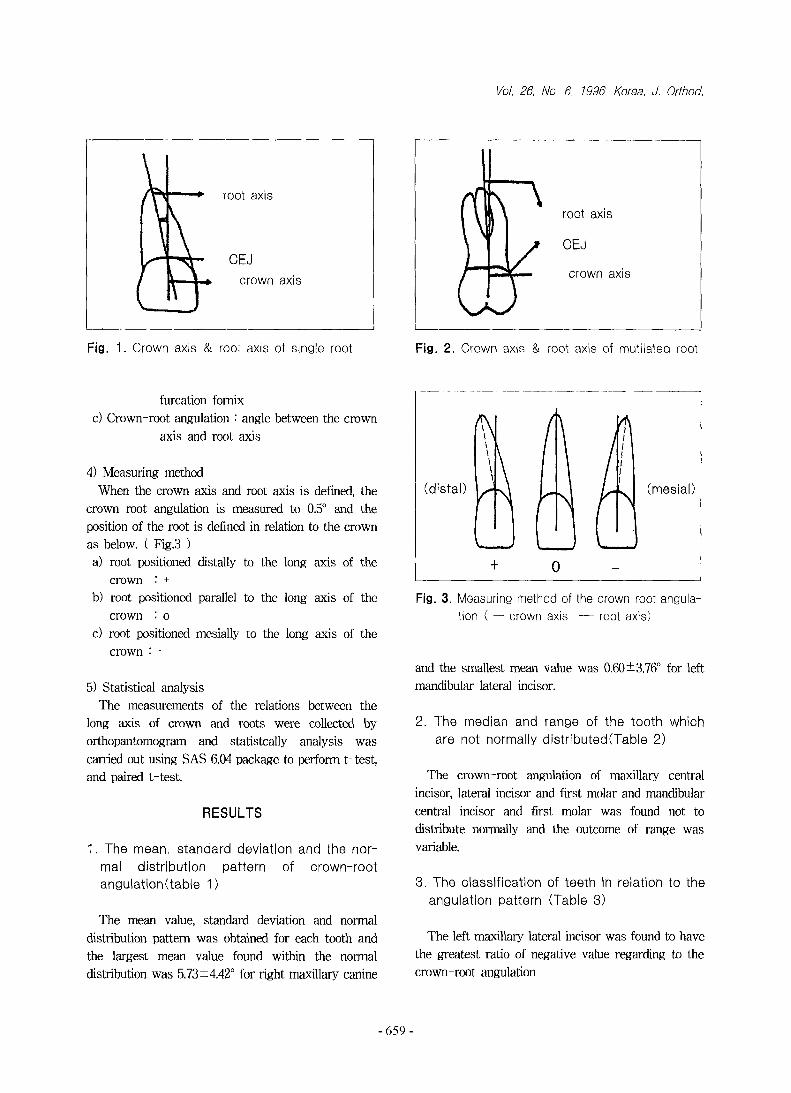
3) Definition of the terms (Fig. 1, 2)
a) Crown axis
incisor, canine, premolar : line joining the mid
points of the distance of the mesiodistal
width of crown and CEJ
molars : line joining the midpoint of mesiodistal
width of crown and midpoint of CEJ line
b) Root axis
incisor, canine, premolar : line joining the mid
point of CEJ line and midpoint of
mesiodistal width of apical third of the
root
molar : line joining the midpoint of CEJ line and
- 658 -
to the long axis of the
to the long axis of the
to the long axis of the
furcation fornix
c) Crown-root angulation : angle between the crown
axis and root axis
4) Measuring method
When the crown axis and root axis is defined, the
crown-root angulation is measured to 0.5° and the
position of the root is defined in relation to the crown
as below. ( Fig.3 )
a) root positioned distaiiy
crown : +
b) root positioned parallel
crown : o
c) root positioned mesially
crown ; -
5) Statistical analysis
The measurements of the relations between the
long axis of crown and roots were collected by
orthopantomogram and statistcally analysis was
carried out using SAS 6.04 package to perform t-test,
and paired t-test
RESULTS
1. The mean, standard deviation and the nor
mal d is tribu tion pattern of c row n-roo t
angulati 이ᄀ (tab le 1)
The mean value, standard deviation and normal
distribution pattern was obtained for each tooth and
the largest mean value found within the normal
distribution was 5.73 ±4.42° for right maxillary canine
Fig. 3. Meas니ring method of the crownᅳroot ang니laᅩ
tion ( — crown axis root axis)
and the smallest mean value was 0.60±3.76o for left
mandibular lateral incisor.
2. The median and range of the too th which
are not normally d is tr ib니tecKTable 2)
The crown- root angulation of maxillary central
incisor, lateral incisor and first molar and mandibular
central incisor and first molar was found not to
distribute normally and the outcome of range was
variable.
3. The c lassifica tion of teeth in relation to the
ang니latiᄋn pattern (Table 3)
The left maxillaiy lateral incisor was found to have
the greatest ratio of negative value regarding to the
crown-root angulation
659

Yo니ng'Joon Kim, Hyun-Sil Choi A Comparative Study Of The Angles Between Crown Axis And Root Axis
Table 1. The mean vah」e, standard deviation and normal distribution pattern of the crown-root angulation (degree)
Right MEAN SD ND Left MEAN SD ND
Upper Upper
1 1.27 3.92 .9111* 1 0.98 3.92 .9280*
2 -0.71 3.86 .884ず 2 -0.82 3.79 .8896*
3 5.73 4.42 .9679 3 5.40 4.44 .9676
4 3.96 3.02 .9631 4 3.61 2.89 .9629
5 3.92 2.41 .9637 5 3.45 3.12 .9647
6 4.09 5.03 .9425* 6 3.84 4.91 .9281*
7 5.10 4.47 .9457 7 4.68 4.22 .9548
Lower Lower
1 0.90 2.90 .9046* 1 0.71 2.95 .8930*
2 0.66 2.12 .9776 2 0.60 3.76 .9548
3 3.47 3.94 .9649 3 3.23 4,21 .9641
4 0.83 428 .9677 4 0,79 4.45 .9631
5 2.47 3.85 .9658 5 2.14 4.00 .9678
6 3.57 4.60 .9340* 6 3.39 4.67 .9654*
7 5.23 4.38 .9691 7 5.52 5.16 .9654
* statistically significant difference at p<0.05
Table 2. The median and range of the teeth not distributed normally. (degree)
Right MEDIAN RANGE Left MEDIAN RANGE
Upper Upper
1 0 25.5 1 0 19.7
2 0 19.3 2 0 18.8
6 4.5 28.3 6 4.5 27.3
Lower Lower
1 0 16.5 1 0 16.5
6 3.5 26.5 6 3.3 26.5
Table 3. The classifcation in relation to the angulation pattern ( % )
. Right — O 十 Left O +
Upper Upper
1 18.1 41.9 40.0 1 17.0 40.3 42.7
2 30.5 55.2 14.3 2 37.1 48.6 14.3
3 4.8 13.3 81.9 3 4.8 16.2 79.0
4 15.2 15.2 69.6 4 13.3 24.8 61.9
5 8.6 22.9 68.5 5 12.4 24.8 62.8
6 9.5 28.6 61.9 6 5.7 34.3 60.0
7 2.9 26.7 70.4 7 2.9 31.4 65.7
Lower Lower
1 20.0 45.7 34,3 1 21.9 47.6 30.5
2 20.0 41.9 38.1 2 22.9 41.0 36.1
3 5.7 28.6 65.7 3 9.5 27.6 62.9
4 23.8 41.0 35.2 4 24.8 39,0 36.2
5 13.3 29.5 57.2 5 17.1 30.5 52.4
6 5.7 34.3 60.0 6 6.7 36.2 57.1
7 3.8 20.0 76.2 7 3.8 21.0 75.2
- 660 -

Vol. 26, No. 6, 1996. Korea. J. Orthod.
Table 4. The statistically significant difference of
crown-root angle (degree)
Right t-value Left t-value
Upper
1 3.29 "
Upper
1 2.89 "
2 -2.85 * 2 -2.13 *
3 13.15 " 3 12.39 "
4 11.90 " 4 10.03 "
5 7.68 " 5 5.99 "
6 8.13 " 6 7.98 **
7 11.62 ** 7 10.08 "
Lower
1 3.05 **
Lower
1 2.36 *
2 2.87 ** 2 2.23 *
3 8.97 " 3 7.75 **
4 1.89 4 1.81
5 6.43 " 5 5.3B "
6 7.88 " 6 7.37
7 11.70 ** 7 10.92 **
statistically significant difference at p<0.05
statistically significant difference at p<0.01
4. S tatis tica lly s ign ificant d iffe rence of the
crown—root ang니lation (Table 4)
There was significant difference between the crown
and root angulation for all teeth except for the right
and left mandibular first premolar.
5. The comparison o f the c row n-roo t ang니I一
ation between the left and the right teeth
(Table 5)
There are no significant difference between the left
and the right teeth in relation to the crown-root
angulation values.
6. The comparison o f the crown—root ang니l_
ation between male and female (Table 6)
There was no statistically significant difference in
crown-root angulation between male and female for
all teeth except the right mandibular first premolar.
7. The relationship between the crown a n d 「ᄋ이:
angulation (Table 7, 8, 9, 10)
1) Maxillary right side : central and lateral incisor,
lateral incisor and canine
2) Maxillary left side : central and lateral incisor
3) Mandibular right side : central and lateral incisor,
first and second molar
4) Mandibular left side : central and lateral incisor,
fable 5. The comparison of the crown-root angulation between the left and the right teeth (degree)
Right MEAN SD Left, MEAN SD t-value
Upper
1 1.27 3.92
Upper
1 0.98 3.47 1.28
2 -0.71 3.86 2 -0.82 3.79 -0.76
3 5.73 4.42 3 5.40 4.44 1.26
4 3.96 3.02 4 3.61 2.89 1.50
5 3.92 2.41 5 3.45 3,12 1.00
6 4.09 5.03 6 3.84 4.91 0.89
7 5.10 4.47 7 4.68 4.22 1.74
Lower
1 0.90 2.90
Lower
1 0.71 2.95 -1.76
2 0.66 2.12 2 0.60 3.76 -1.63
3 3.47 3.94 3 3.23 4.21 -1.69
4 0.83 428 4 0.79 4.25 -0.96
5 2.47 3.85 5 2.14 4.00 1.40
6 3.57 4.60 6 3.39 4.67 -1.16
7 5.23 4.38 7 5.52 5.16 1.82
statistically significant difference at p<0.05
- 661 -

YoungᅳJoon Kim, Hyun-Sil Choi A Comparative Study Of The Angles Between Crown Axis And Root Axis
Table 6. The comparison of crown-root angulation between male and female
rightmale female
leftmale female
MEAN S.D. MEAN S.D. t-vaiue MEAN S.D. MEAN S.D. t-value
U 1 1.80 3.97 0.63 3.76 1.65 U 1 1.00 3.24 0.97 3.49 1.83
2 -0.36 4.04 -0.78 3.39 0.60 2 - 0.74 4.06 -1.24 3.59 -0.23
3 6.47 4.71 4.80 4.15 1.94 3 6.14 4.38 3.21 4.10 1.87
4 5.00 4.04 3.56 4.25 1.83 4 4.16 4.10 2.95 5.06 1.41
5 4.34 5.28 3.12 5.06 L24 5 4.08 4.92 3.21 5.20 0.17
6 3,03 4.66 4.42 4.38 - 0.68 6 3.87 5.61 3.83 4.15 0.03
7
L
4,85 4.66 4.87 4.45 -0.39 7
L
4.90 4.54 3.53 4.59 0.29
1 0.72 2.00 0.75 3.23 -0.05 1 0.97 2.65 0.31 2.620.45
-0.282 1.11 2.86 0.54 3.85 0.84 2 1.36 3.52 0.56 3.75
0.90
0.77
-1.51
-1.04
1.49
3 3.08 4.00 3.43 3.91 -0.44 3 3.26 4.62 2.27 5.38
4 1.75 4.65 0.13 3.45 2.04* 4 1.66 4.41 -0.14 3.97
5 1.82 4.01 2.49 3.66 -0.89 5 1.97 3.17 1.60 3,42
6 4.03 4.65 3.02 4.92 1.07 6 4.33 3.01 2.02 5.21
7 4.77 4.54 6.51 4.60 ᅳ 1.71 7 5.52 3.80 4.07 3.65
statistically significant difference at p<0.05
Table 7. Correlation vah」e of crown-root angulation of
right maxillary teeth
Tooth 1 2 3 4 5 .6
2 .2991*
3 .0001 .2457'
4 .0976 -.0946 .0773
5 .0649 .0871 .1708 .1642
6 .1008 .2214* -.0525 ,0541 ,1759
7 .0221 .1303 .1400 -.1129 .0326 .1329
* statistically significant difference at p<0.05
Table 9. Correlation value of crown-root angulation of
left mandibular teeth
Tooth 1 2 3 4 5 6
2 .3596
3 -.0769 •2019*
4 ,0531 1200 .0537
5 .0154 -.0312 -.0098 .1866
6 -.0199 .0162 -.0180 .0671 -.0711
7 -.0642 .0353 -.0129 -.0842 ,1992 .2564**
* statistically significant difference at p くᄋ.05
** statistically significant difference at p<0.01
Table 8. Correlation vahje of crown—root angulation of
left maxillary teeth
Tooth 1 2 3 4 5 6
2 .2028*
3 .0489 .0957
4 .0096 .0999 .1233
5 .0292 .0421 •0425 -.0055
6 .0616 .9590 -0839 -.1340 -.0500
7 -.1136 .0395 .0829 .0009 .1041 .0742
* statistically significant difference at p<0.05
Table 10, Correlation val니e of crown—root angulation
of right mandibular teeth
Tooth 1 2 3 4 5
2 .4583**
3 -.0446 .1232
4 .0462 .0852 .0535
5 -.0000 -.0206 .0482 .1351
6 -.0400 .0679 -.0038 .0218 .7440
7 .0225 .1047 .0361 .1655 .0417 .2178*
* statistically significant difference at p<0.05
林 statistically significant difference at p<0.01
- 662 -
Vol. 26, No. 6, 1996. Korea. J. Orthod.
lateral incisor and canine, first and second molar.
From the above relationship, positive correlation
was obtained.
DISCUSSION
Root formation may be influenced by environmental
factors and is neither affected by the size of the
crown nor the jaw1,13,14,15). The number and structure
of root is known to show wide range of variation7,18,34).
From the previous studies carried out on patients with
normal occlusion it can be concluded that the tip of
root is generally directed distally in relation to the
crown and the distal displacement of the root is
extremely variable depending on the tooth2’20). As
such, the crown of the tooth is more or less
perpendicular to the occlusal plane. The axis of the
individual tooth is not a straight line but a curvature
disecting distally, when extending from the crown
down to the root10'4®. The individual tooth is aligned
mesiodistally in an angle unique to the tooth to
achieve a parallel relationship with the adjacent
teeth2,5,9,10>. The parallelism of the root is essential in
achieving proper alignment of the teeth within the
basal bone and obtaining normal occlusion between
the maxillary and the mandibular teeth, in addition to
retaining a stable treatment result28).
Taylor411 postulated that the relationship between
the crown and the root is extremely variable and the
line extended from the root tip towards the crown will
not pass the center of the cementoenamel junction."
Dewel10> stated that the axis of the crown Is very
different to the axis of the root and the line dividing
the tooth tends to direct distally as you move down to
the root tip. W heeler^ strongly suggested that the
root curvature of individual tpeth is relatively constant
and has significant relationship w ith the periodontal
health regarding its physiological importance.
Magness271 suggested that orthodontists using the
SW A should have in mind that not all teeth are
morphologically ideal and small adjustments in regard
to such deviation is required.
On the other hand, studies of crown-root axis from
buccolingual surface have been reported while
Charlsson and Ronnerman6) measured the difference
of crown and root axis of extracted teeth and
Delivanis, Kuftnecs> studied the crown-root angulation
in Class II malocclusion patients and found that the
value of the angulation was high and when maxillary
incisors are moved, either by intrusion or applying
torque, the root will be in contact with the lingual
compact bone and subsequently induce root resorp
tion. Harris19) et al had also reported large values of
crown-root angulation in Class III malocclusion
patients.
The evaluation of tooth axis is generally measured
by using 45° lateral cephalogram but when the
parallelism of molar root was compared for reliability
was compared by using either the orthopantomogram
or the 45° lateral cephalogram, orthopantomogram
showed to produce better reliability for testing the
parallelism of molar crown-root parallelism51'.
Graber,Mayoral, Hauck, Thorpe, Tronje advocated
the use of panorama film in evaluating tooth axis
angutation16,21,28,42,43;. The panoramic radiography pro
jects all images into a single film without superim
position of the maxillary, mandibular and adjacent
structures between the left and the right arch. It also
has advantages of low radiation exposure, comfortable
patiens manipulation and simple handling21’30’44). The
image of a structure from orthopantomogram may be
magnified depending on the geometrical principle and
linear measurement such as horizontal distance may
be influenced by projection factors and object move
ment, and thus lowers the reliability of the measure
ment whereas the measurement of angulation between
adjacent structures is clincally acceptable26,2S,30,38). In
order to achieve a clinical objective a difference of 5°
between adjacent structures or of 5° after another
consecutive radiographic taking, may be acceptable
13,38'49). The reliability of orthopantomogram in the
measurement of angulation is not only reliable but
taking into account of the fact that there is ± 5° of
error after repeated exposures of film, it is reasonable
to suggest that it is an acceptable instrument to be
used clinically12,칭’3914̂ In a recent study, 4 different
models of panorama radiographic machine were
compared for the angulation of tooth axis of dry skull,
in different position and angle. The result was that
w ith a 土 50 error tolerlance, abnormal positioning of
patient w ill not dramatically influence the tooth axis
measurement .
FACC( Facial Axis of Clinical Crown ) is defined as
the buccal groove that divides the buccal cusps of
- 663 -
Young-Joon Kim, Hyun-Sil Choi A Comparative Study Of The Angles Between Crown Axis And Root Axis
molar tooth and other teeth that pass through the
most prominent part of the central lobe. However, this
landmark does not show up radiographically and after
times, the line joining the middle of the greatest
height of contour and the midpoint of the cemen-
toenamel junction is used. The reason for using the
anatomical crown by extending the cementoenamel
junction is based on the fact that clinical crown is
defiened as 1.8mm shorter in height301. The reference
point for single rooted tooth is placed at the center of
the apical third which accounts for the fact that the
morphological deviation of the root is greatest at that
point. The reason for selecting the furcation apex as
the reference point in multirooted teeth is because
roots of molar teeth originate from one root trunk
located at the base of the crown, thus providing a
reference for judging the axis of the root48).
W ith respect to the normal distribution pattern of
the crownᅳroot angulation, all the teeth except for the
maxillary central incisor, lateral incisor, and first
molar, and mandibular central incisor and first molar
showed normal distribution. The teeth that are
normally distributed possess a mean value that is
significant and otherwise, only the median and the
range will describe the pattern of the distribution.
Among the teeth that normally distribute, the
largest mean value of the crown-root angulation was
found for right maxillary canine as 5.73±4.42° and the
smallest value was for the left mandibular lateral
incisor as 0.60 ±3.76°.
Among the teeth that arre not normally distributed,
the median value of the maxillary and mandibular
anterior teeth was 0° and the range was variable
between 16.5° and 25.5°. W ith respect to the
angulation pattern of the teeth, the maxillary lateral
incisor that had relatively high incidence of mesially
tilted root also showed deformations of the crown and
for the root, although higher incidence of root with
distal dilaceration generally occurred, there were also
roots that dilacerated mesially47). In our study, the
appearence of negative value of maxillary central
incisors was 17% and in reference to Ingle's report'
of 4% for mesial tiling of the tooth, our study showed
greater percentage.
A ll the teeth except the left and right mandibular
premolars showed significant difference in the
angulation of tooth axis, which supports the fact that
the axis of the crown and roots are different. The left
and right maxillary lateral incisor and left mandibular
lateral incisor had 95% significance whereas in other
teeth the significance was greater than 99%.
There was no significant difference between the left
and right teeth for measurements comparing crown-
root angulation.
There was no significant difference with respect to
the difference in the sex in all the teeth except for the
left and right mandibular first premolar.
Correlation was used to determine the relationship
between the teeth and positive correlations were
found for the following pairs of teeth :
Right maxillary quadrant: between central and lateral
incisor, lateral incisor and canine
Left maxillary quadrant ■' between central and lateral
incisor
Right mandibular quadrant : between central and
lateral incisor,first and second molar
Left mandibular quadrant: between central and lateral
incisor, lateral incisor and canine, first and
second molar
Therefore, even if there is large difference of the
angulation of tha axis of the crown and root, the pairs
of teeth that show positive relationship tends to
maintain a constant spacial relationship between each
other.
In conclusion, there was a significant difference
between the axis of the crown and the root and the
amount of difference varied depending on the tooth.
Therefore by understanding the pattern of deviation of
roots in their axial relationships, the evaluation of the
alignment of roots may be possible. As discussed
previously, the crown-root angulation for different
malocclusions have been studied by others but data
for such measurements in mesiᄋ-distal dimension is
lacking and more research is needed in this field.
Moreover, our study using the orthopantomogram is a
quantitative analysis which inherently retaines errors
with true values. As such, we hope to improve such
deficiency by measuring the angulation of crown-root
axis of extracted teeth.
CONCLUSION
The following conclusions were made for the
study of crown-root angulation,
- 664

Vol. 26, No. 6, 1996. Korea. J. Orthod.
1. Among the teeth presenting normal distribution, the
maxillary right canine showed the largest mean
value( 5.73 土 4.42° ),which was composed of the
crown-root angles, and the mandibular left lateral
incisor showed the smallest mean value( 0.60 士
3.76° X
2. The crown-root angles of the maxillary incisors
and the first molars, and the mandibular central
incisors and the first molars didn't show normal
distribution and the ranges of these angles were
dispersed.
3. Significant differences were present between the
crown axis and the root axis except for lower first
premolars, (pく0.05)
4. No significant difference was present for the crown
-root angle between right and left side, (pく0.05)
5. No significant difference was present for the crown
-root angle between male and female except for
lower left first premolar, (pく0.05)
6. In the upper right quadrant, significant correlations
were present between crown-root angles of the
central incisor and lateral incisor, lateral incisor and
canine.
In the upper left quadrant, significant correlations
were present between crown-root angles of the
central incisor and lateral incisor.
In the lower right quadrant, significant correlations
were present between crown-root angles of the
central incisor and lateral incisor, first molar and
second molar.
In the lower left quadrant, significant correlations
were present between crown-root angles of the
central incisor and lateral incisor, lateral incisor and
canine, first molar and second molar. (p<0.05)
REFERENCE
1. Anderson, D. L., Thampson, G. W.,Popoich, F, : Tooth,
chin, bone and bony correlations., Am. J. Phys. Anthro-
pol. 48 : 305 - 314,1978.
2. Andrews, L. F. : The six keys to nomal occlusion., Am.
J. Orthod. 62 : 296 - 309, 1972.
3. : The diagnostic system : Occlusion analysis.,
Dent. Clin, North. Am. 20 ■ 671 - 690. 1976.
4 . : Stright Wire. The concept and appliance., San
Dieagᄋ,L.A. Wells Co. 1976.
5. Bums, R. D. : A cephalometric study of the mesiodistal
axial inclinations of the teeth., Am. J. Orthod. 56 : 309
(Abst.), 1969.
6. Carlsson, R., Rfinnerman, A. • Crown-root angles of
upper central incisors., Am, J. Orthod. 64 : 147 - 154,
1973.
7. Chenail, B. L, Teplitsky, P. E. : Endersonics in curved
root canals. Part II.,J. Endodont. 14 : 214 - 217, 1988.
8. Delivanis, H, P., Kuftinec, M. M. : Variation in mor
phology of the maxillary central incisors found in class
II div 2 malocclusions.,Am. J. Orthod. 78 '• 438 - 443,
1980.
9. Dempster, W, T.,Adams, W. J. and Duddles, R. A, :
Arrangement in the jaws of the roots of the teeth., J.
Am. Dent. Assoc. 67 : 779 - 797, 1963.
10. Dewel, B. F. : Clinical observations on the axial in
clination of teeth,,Am. J. Orthod. 35 : 98 - 115,1949.
11. Edwards. J. G. : The prevention of relapse in extraction
case.,Am.J. Orthod. 60 : 128 - 141,1971.
12. Frykholm, A. : Angular measurements in orthopanto
mography.’ Dentomaxillofac. Radiol. 6 • 77 - 81,1977.
13. Gam, S. M., Smith, B. H.수 Cole, P, E. : Correlations
between root length and face size., J. Dent. Res. 59 •
141,1980. ᅵ
14. Gam, S. M.,Van Alstine, W. L,,Cole, P. E. : Intrain
dividual root length correlations., J. Dent. Res. 57 : 270,
1978.
15 . : Relationship between root length and crown
diameters of correspanding teeth., J. Dent. Res. 57 ■■ 636,
1978.
16. Graber, T. M. : Panoramic radiography in orthodontic
diagnosis., Am. J. Orthod. 53 : 799 - 821, 1967.
17. Graber, T, M., Swain, B. F. •• Orthodontic bands. In
Current orthodontic concepts and techniques.,Vol 1. 2d
ed. Philadelphia, W.B. Saunders Co. 1975.
18. Greenfield, R. S., Cambruzzi,J. V. : Complexities of
endodontic treament of maxillary lateral incisors with
anomalous root formation,’ Oral Surg. Oral Med Oral
Pathol. 62 : 82 - 88,1986.
19. Harris, E. F., Hassankiadeh, S., Harris, J. T. : Maxillary
incisor crown-root relationships in different angles
malocclusion., Am. J. Orthd. Dentofac. Orthop. 103 : 48 -
53,1993.
20. Hatasaka, H. H. : A radiographic study of roots in
extraction site.,Angle Orthod. 46 : 64 - 68,1976.
21. Hauck, R. M. : Documentation of tooth movement by
means of panoral radiography., Am. J. Orthod. 57 : 386
— 392,1970.
22. Holdaway, R. A. : Bracket angulation as applied to the
edgewise appliance., ^ngle Orthod. 22 : 227 - 236, 1952.
23. Ingle, J. I. : Endodontics, 2d ed Lea & Febiger Co.
Philadelphia. 1986.
24. Jarabak, J. R.,Fizzell,J, A. : Technique and treatment
with light~wire edgewise appliance, StLouise, The C.V.
Mosby Company, Vol.2, chap.7, 1972.
665

Young-Joon Kim, Hyun-Sil Choi A Comparative Study Of The Angles Between Crown Axis And Root Axis
25. Larheim, T. A. • Reproducibility of rotational panoramic
radiography.* Mandibular linear dimensions and angles.,
Am. J. Orthod. 90 : 54 - 51, 1986.
26. Lucchesi, M. V. : Suitability of the panoramic radiograph
for assessment of mesiodistal angulation of teeth in the
buccal segment of the mandible., Am. J. Orthod. 94 :
303-310,1988.
27. Magness, W. B. : The straight - wire concept., Am. J.
Orthod. 73 : 541 - 550, 1978.
28. Majoral, G. : Treatment results with light wires studied
by panoramic radiography., Am. J. Orthod. 81 •• 489 -
497,1982.
29. Ohba, T. : Comparision of orthopantomography with
conventional periapical dental radiography.,Oral Surg.
Oral Med Oral Pathol. 34 : 524-529, 1972.
30. Orban, B. * Oral histology and embryology, 4th ed St
Louise,The C.V. Mosby Co.1957.
31. Paatero,Y. V. * Pantomography and orthopantomogra
phy., Oral Surg Oral Med. Oral Pathol. 14 : 947 — 953,
1961.
32. Ricketts, R. H, * Bioprogressive therapy as an answer to
orthodontic needs., Am. J. Orthod. 70 : 241 - 268, 1976.
33. Robert, R. H. W. * Fators associated with successful
orthodontic treatment., Am. J. Ortho. 38 : 790 - 800,
1952.
34. Rohlin, M., Rundquist, L. • Apical root anatomy of
impacted maxillary canines., Oral Surg. Oral Med. Oral
Pathol. 58 : 141 - 147, 1984.
35. Roth. R, H. : Five year clinical evaluation of the Andrews
straight - wire appliance., J. Clin. Orthod. 10 : 836 - 850,
1976.
36. Samfors, K. A. * Angle distortion in narrow beam rot
ational radiography., Acta. Radiol. 15 : 570 ~ 76,1974.
37. Saltzman, J. A. : Principles of orthodontics. 2d ed. Phil
adelphia, J. B. Lippincott Co., 1950.
38. Samawi, S. S. B. and Burlce, P. H. : Angle distortion in
the orthopantomogram., Br, J. Orhod. 11 • 100 - 107,
1984.
39. Schudy, F. F., Rushing. C. H. and Sims, M. A.: The
angle of axial inclination of teeth., Angle Orthod. 33 : 69
- 82,1963.
40. Schwaninger, B, : Evaluation of the straight arch wire
concept.,Am. J. Orthod. 74 : 188 - 196,1978.
41. Taylor., R. M. S. : Variation in form of human teeth : An
anthropologic and forensic study of maxillary incisors., J.
Dent. Res. 48 : 5 - 16,1969.
42. Thorpe, J. O.,Charlotte, N. C. • Panoramic radiography
in the general practice of dentistry .Oral Surg.Oral Med.
Oral Pathol. 24 : 781,1967.
43. Tronje, G. ' Image distortion in rotational radiography.
Thesis. Dentomaxillofac. Radiol. 3 : 180-183,1982.
44. Tweed, C. H. : Clinical orthodontics. Vol.l.St. Louis,The
C.V.Mosby Co., 1966.
45. Updegrave, W. J. : The role of panoramic dental radio
graphy., Oral Surg. Oral Med Oral Pathol. 22 : 49 - 57,
1966.
46. Vrsi, W.,Almeda, R., Tavano,O. • Assessment of mesio
distal axial inclination through panoramic radiography.,
J. Clin. Orthod. Vol. 24 No. 3,166-173,1990.
47. Welander, U. '• Image distortion in narrow beam rotation
radiography Acta. Radiol. Diagnosis. 19 • 507-511, 1978,
48. Wheeler, R. C. : Dental anatomy, physiology, and occlu
sion., ed 5. Pilladelphia, W.B. Saunders Company, 1974.
49. Byung-Cheol Kang : Comparison of four panoramic
dental radiographic systems for tooth angulation
measurement accuracy under different folerences.
50. Kyung-Ho Kim : 45° oblique cephalometric analysis of
mesiodistal axial incclination in normal occlusion.
51. Young Gyu Min : A comparative study on reliability of
the root parallelism of the posterior theeth projected on
the orthopantomogram with the 45° oblique cephalogram.
52. Don Young Jeong : A study of the crown inclination in
normal occlusions.
KOREA. J ORTHOD. 1996 ; 26 : 657-666