Fossil Musculature of the Most Primitive Jawed Vertebrates
6
Reports / http://www.sciencemag.org/content/early/recent / 13 June 2013 / Page 1 / 10.1126/science.1237275 The most fundamental anatomical divide among living vertebrates is that between the jawless cyclostomes (lampreys and hagfishes) and the jawed gnathostomes (all other vertebrates). Although early development in the two groups is similar, differences in migration and proliferation patterns of cell populations produce profoundly different adult architectures in the head region, encompassing not only the skeleton but also the muscu- lature (1). In conjunction with the presence or absence of jaws, a major difference between the groups is the absence of a shoulder girdle, paired appendages, and a differentiated neck region in cyclostomes. The gnathostome stem group contains jawless as well as jawed clades, suggesting the jawless condition is primitive for vertebrates (2). Some jawless stem gnathostomes, such as osteostracans, have pectoral fins and “shoulder girdles,” but the latter are immovably attached to the skull (2). Head-shoulder separation, and the origin of distinctive neck muscles such as the cucullaris, seems to have occurred at approximately the same time as the origin of jaws. The phylogenetically deepest and morphologically most primitive versions of jawed vertebrate anatomy are found in the placoderms, a paraphyletic array of Silurian-Devonian armored fishes that form the upper part of the gnathostome stem group (3, 4) (Fig. 1A). Data on placoderm muscular anatomy would add considerably to the knowledge of gnathostome origins, given the functional and anatomical importance of the musculature in the transition from jawless to jawed vertebrates, as well as its significance as a probable indicator of cell population identity (5). Until now, such data have not been available, and conclusions about placoderm musculature have been based entirely on inferences from skeletal anatomy and functional morphology. Studies of muscular evolution in gnathostomes have often proceeded from the assumption that extant sharks are adequate models for the primitive gna- thostome condition (6–8). We present here placoderm fossils with preserved three-dimensional mus- culature, from the Upper Devonian Gogo Formation of Western Australia. The presence of neck and trunk muscles in the eubrachythoracid arthrodires Eastmanosteus, Compagopiscis and Incisoscutum allow detailed reconstruc- tion of this musculature (Figs. 1, B to D, 2, and 3, and figs. S1 and S2). Addi- tional information about tail muscles comes from the ptyctodont Austroptyc- todus (fig. S2, H and I). These speci- mens present an anatomy that differs radically from the shark model (6). Presence of a dermal neck joint (Fig. 2A) between the skull and trunk armor of placoderms implies vertical pivoting movement of the head, requir- ing elevator and depressor muscles (9, 10). Such muscles are confirmed in our specimens (Figs. 1, B and C, and 3, A to F and J, and fig. S1). A large medial levator capitis major and a small lateral levator capitis minor have palmately arranged muscle fibers radiating from the area of origin. Lateral to the articu- lation of the neck joint (Fig. 2A), an oblique muscle that originates on the inner face of the dermal shoulder girdle and inserts in a hollow on the inner face of the skull roof is identified as the cucullaris muscle (see supplementary text and fig. S1F). It represents the phylogenetically deepest record of this characteristic gnathostome mus- cle. In mammals the cucullaris reaches the dorsal midline of the neck, whereas in sharks it is narrower and bounded dorsally by somitic epaxial muscles. The latter morphology is often viewed as primitive for gnatho- stomes (11), but both differ from the placoderm condition in forming part of a flexible neck region rather than operating a constrained pivoting joint. The cranial musculature is less well preserved. However, the levator arcus palatini (11) is observed attached to the ventral postocular process of Compagopiscis (Fig. 3, K and L, fig. S2G, and movie S1). In addition, part of the dorsal branchial constrictor muscle can be seen on the viscer- al surface of the paranuchal plate, lateral to the cucullaris (fig. S1J). Normal segmented body muscles with anteroposteriorly oriented fi- bers are present under the dorsolateral parts of the trunk armor and in the tail. However, the posterior part of the ventral abdominal musculature contains two anteroposteriorly elongated muscle bands with transversely oriented fibers (Figs. 1, C and D, and 2, C to F; see supplementary in- formation), representing areas of stronger fiber development within a continuum of transverse fibers extending across the ventral midline. Dorsolaterally, the bands are in contact with the ventral ends of the my- omeres. The existence of these transverse muscles had not been predict- ed (9). Mineralized tendinous attachments for the somitic muscles, inclined anterodorsally in a manner reflecting the W shape of the myocommata, are present in the median septum above the neural spines (Fig. 2B). In the eubrachythoracids, the myocommata are incompletely preserved (Fig. 3, D to I, and fig. S2F), but in Austroptyctodus they have a shallow w-shaped curvature (12) (fig. S2, H and I). The medial myoseptal at- tachment probably spanned only two segments, in contrast to at least Fossil Musculature of the Most Primitive Jawed Vertebrates Kate Trinajstic, 1,2 Sophie Sanchez, 3,4 Vincent Dupret, 4 Paul Tafforeau, 3 John Long, 5,6 Gavin Young, 6 Tim Senden, 7 Catherine Boisvert, 8 Nicola Power, 6 Per Erik Ahlberg 4 * 1 Western Australian Organic and Isotope Geochemistry Centre, Department of Chemistry, Curtin University, Perth, Western Australia 6102, Australia. 2 Earth and Planetary Sciences, Western Australian Museum, Perth, Western Australia 6000, Australia. 3 European Synchrotron Radiation Facility, 38043 Grenoble Cedex, France. 4 Subdepartment of Evolution and Development, Department of Organismal Biology, Uppsala University, Norbyvägen 18A, 752 36 Uppsala, Sweden. 5 School of Biological Sciences, Flinders University, PO Box 2100, Adelaide, South Australia 5001. 6 Research School of Earth Sciences, The Australian National University, Canberra 0200, Australia. 7 Department of Applied Mathematics, Research School of Physics and Engineering, The Australian National University, Canberra 0200, Australia. 8 Australian Regenerative Medicine Institute, Monash University, Victoria 3800, Australia. *Corresponding author. E-mail: [email protected] The transition from jawless to jawed vertebrates (gnathostomes) resulted in the reconfiguration of the muscles and skeleton of the head, including the creation of a separate shoulder girdle with distinct neck muscles. We describe here the only known examples of preserved musculature from placoderms (extinct armored fishes), the phylogenetically most basal jawed vertebrates. Placoderms possess a regionalized muscular anatomy differing radically from the musculature of extant sharks, which is often viewed as primitive for gnathostomes. The placoderm data suggest that neck musculature evolved together with a dermal joint between skull and shoulder girdle, not as part of a broadly flexible neck as in sharks, and that transverse abdominal muscles are an innovation of gnathostomes rather than of tetrapods. on June 24, 2013 www.sciencemag.org Downloaded from
Transcript of Fossil Musculature of the Most Primitive Jawed Vertebrates
Reports
/ http://www.sciencemag.org/content/early/recent / 13 June 2013 / Page 1 / 10.1126/science.1237275
The most fundamental anatomical divide among living vertebrates is that between the jawless cyclostomes (lampreys and hagfishes) and the jawed gnathostomes (all other vertebrates). Although early development in the two groups is similar, differences in migration and proliferation patterns of cell populations produce profoundly different adult architectures in the head region, encompassing not only the skeleton but also the muscu-lature (1). In conjunction with the presence or absence of jaws, a major difference between the groups is the absence of a shoulder girdle, paired appendages, and a differentiated neck region in cyclostomes.
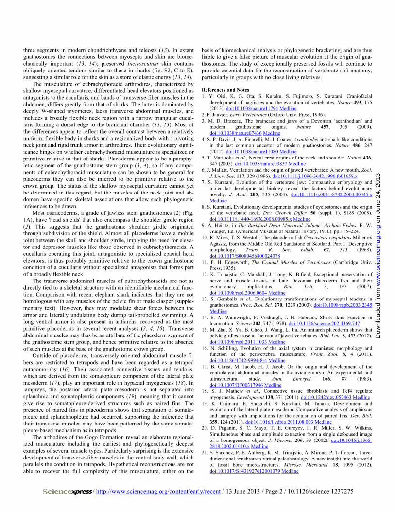
The gnathostome stem group contains jawless as well as jawed clades, suggesting the jawless condition is primitive for vertebrates (2). Some jawless stem gnathostomes, such as osteostracans, have pectoral fins and “shoulder girdles,” but the latter are immovably attached to the skull (2). Head-shoulder separation, and the origin of distinctive neck muscles such as the cucullaris, seems to have occurred at approximately the same time as the origin of jaws. The phylogenetically deepest and morphologically most primitive versions of jawed vertebrate anatomy are found in the placoderms, a paraphyletic array of Silurian-Devonian armored fishes that form the upper part of the gnathostome stem group (3, 4) (Fig. 1A).
Data on placoderm muscular anatomy would add considerably to the knowledge of gnathostome origins, given the functional and anatomical importance of the musculature in the transition from jawless to jawed vertebrates, as well as its significance as a probable indicator of cell population identity (5). Until now, such data have not been available, and conclusions about placoderm musculature have been based entirely on inferences from skeletal anatomy and functional morphology. Studies of muscular evolution in gnathostomes have often proceeded from the assumption that extant sharks are adequate models for the primitive gna-thostome condition (6–8).
We present here placoderm fossils with preserved three-dimensional mus-culature, from the Upper Devonian Gogo Formation of Western Australia. The presence of neck and trunk muscles in the eubrachythoracid arthrodires Eastmanosteus, Compagopiscis and Incisoscutum allow detailed reconstruc-tion of this musculature (Figs. 1, B to D, 2, and 3, and figs. S1 and S2). Addi-tional information about tail muscles comes from the ptyctodont Austroptyc-todus (fig. S2, H and I). These speci-mens present an anatomy that differs radically from the shark model (6).
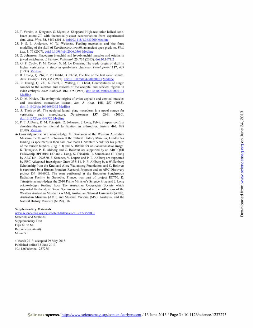
Presence of a dermal neck joint (Fig. 2A) between the skull and trunk armor of placoderms implies vertical pivoting movement of the head, requir-ing elevator and depressor muscles (9, 10). Such muscles are confirmed in our specimens (Figs. 1, B and C, and 3, A to F and J, and fig. S1). A large medial levator capitis major and a small lateral levator capitis minor have palmately arranged muscle fibers radiating from the area of origin. Lateral to the articu-lation of the neck joint (Fig. 2A), an oblique muscle that originates on the inner face of the dermal shoulder girdle and inserts in a hollow on the inner face of the skull roof is identified as the cucullaris muscle (see supplementary text and fig. S1F). It represents the
phylogenetically deepest record of this characteristic gnathostome mus-cle. In mammals the cucullaris reaches the dorsal midline of the neck, whereas in sharks it is narrower and bounded dorsally by somitic epaxial muscles. The latter morphology is often viewed as primitive for gnatho-stomes (11), but both differ from the placoderm condition in forming part of a flexible neck region rather than operating a constrained pivoting joint.
The cranial musculature is less well preserved. However, the levator arcus palatini (11) is observed attached to the ventral postocular process of Compagopiscis (Fig. 3, K and L, fig. S2G, and movie S1). In addition, part of the dorsal branchial constrictor muscle can be seen on the viscer-al surface of the paranuchal plate, lateral to the cucullaris (fig. S1J).
Normal segmented body muscles with anteroposteriorly oriented fi-bers are present under the dorsolateral parts of the trunk armor and in the tail. However, the posterior part of the ventral abdominal musculature contains two anteroposteriorly elongated muscle bands with transversely oriented fibers (Figs. 1, C and D, and 2, C to F; see supplementary in-formation), representing areas of stronger fiber development within a continuum of transverse fibers extending across the ventral midline. Dorsolaterally, the bands are in contact with the ventral ends of the my-omeres. The existence of these transverse muscles had not been predict-ed (9).
Mineralized tendinous attachments for the somitic muscles, inclined anterodorsally in a manner reflecting the W shape of the myocommata, are present in the median septum above the neural spines (Fig. 2B). In the eubrachythoracids, the myocommata are incompletely preserved (Fig. 3, D to I, and fig. S2F), but in Austroptyctodus they have a shallow w-shaped curvature (12) (fig. S2, H and I). The medial myoseptal at-tachment probably spanned only two segments, in contrast to at least
Fossil Musculature of the Most Primitive Jawed Vertebrates Kate Trinajstic,1,2 Sophie Sanchez,3,4 Vincent Dupret,4 Paul Tafforeau,3 John Long,5,6 Gavin Young,6 Tim Senden,7 Catherine Boisvert,8 Nicola Power,6 Per Erik Ahlberg4* 1Western Australian Organic and Isotope Geochemistry Centre, Department of Chemistry, Curtin University, Perth, Western Australia 6102, Australia. 2Earth and Planetary Sciences, Western Australian Museum, Perth, Western Australia 6000, Australia. 3European Synchrotron Radiation Facility, 38043 Grenoble Cedex, France. 4Subdepartment of Evolution and Development, Department of Organismal Biology, Uppsala University, Norbyvägen 18A, 752 36 Uppsala, Sweden. 5School of Biological Sciences, Flinders University, PO Box 2100, Adelaide, South Australia 5001. 6Research School of Earth Sciences, The Australian National University, Canberra 0200, Australia. 7Department of Applied Mathematics, Research School of Physics and Engineering, The Australian National University, Canberra 0200, Australia. 8Australian Regenerative Medicine Institute, Monash University, Victoria 3800, Australia.
*Corresponding author. E-mail: [email protected]
The transition from jawless to jawed vertebrates (gnathostomes) resulted in the reconfiguration of the muscles and skeleton of the head, including the creation of a separate shoulder girdle with distinct neck muscles. We describe here the only known examples of preserved musculature from placoderms (extinct armored fishes), the phylogenetically most basal jawed vertebrates. Placoderms possess a regionalized muscular anatomy differing radically from the musculature of extant sharks, which is often viewed as primitive for gnathostomes. The placoderm data suggest that neck musculature evolved together with a dermal joint between skull and shoulder girdle, not as part of a broadly flexible neck as in sharks, and that transverse abdominal muscles are an innovation of gnathostomes rather than of tetrapods.
on
June
24,
201
3w
ww
.sci
ence
mag
.org
Dow
nloa
ded
from
/ http://www.sciencemag.org/content/early/recent / 13 June 2013 / Page 2 / 10.1126/science.1237275
three segments in modern chondrichthyans and teleosts (13). In extant gnathostomes the connections between myosepta and skin are biome-chanically important (13, 14); preserved Incisoscutum skin contains obliquely oriented tendons similar to those in sharks (fig. S2, C to E), suggesting a similar role for the skin as a store of elastic energy (13, 14).
The musculature of eubrachythoracid arthrodires, characterized by shallow myoseptal curvature, differentiated head elevators positioned as antagonists to the cucullaris, and bands of transverse-fiber muscles in the abdomen, differs greatly from that of sharks. The latter is dominated by deeply W-shaped myomeres, lacks transverse abdominal muscles, and includes a broadly flexible neck region with a narrow triangular cucul-laris forming a dorsal edge to the branchial chamber (11, 13). Most of the differences appear to reflect the overall contrast between a relatively uniform, flexible body in sharks and a regionalized body with a pivoting neck joint and rigid trunk armor in arthrodires. Their evolutionary signif-icance hinges on whether eubrachythoracid musculature is specialized or primitive relative to that of sharks. Placoderms appear to be a paraphy-letic segment of the gnathostome stem group (3, 4), so if any compo-nents of eubrachythoracid musculature can be shown to be general for placoderms they can also be inferred to be primitive relative to the crown group. The status of the shallow myoseptal curvature cannot yet be determined in this regard, but the muscles of the neck joint and ab-domen have specific skeletal associations that allow such phylogenetic inferences to be drawn.
Most ostracoderms, a grade of jawless stem gnathostomes (2) (Fig. 1A), have 'head shields' that also encompass the shoulder girdle region (2). This suggests that the gnathostome shoulder girdle originated through subdivision of the shield. Almost all placoderms have a mobile joint between the skull and shoulder girdle, implying the need for eleva-tor and depressor muscles like those observed in eubrachythoracids. A cucullaris operating this joint, antagonistic to specialized epaxial head elevators, is thus probably primitive relative to the crown gnathostome condition of a cucullaris without specialized antagonists that forms part of a broadly flexible neck.
The transverse abdominal muscles of eubrachythoracids are not as directly tied to a skeletal structure with an identifiable mechanical func-tion. Comparison with recent elephant shark indicates that they are not homologous with any muscles of the pelvic fin or male clasper (supple-mentary text). However, they may modulate shear forces between the armor and laterally undulating body during tail-propelled swimming. A long ventral armor is also present in antiarchs, recovered as the most primitive placoderms in several recent analyses (3, 4, 15). Transverse abdominal muscles may thus be an attribute of the placoderm segment of the gnathostome stem group, and hence primitive relative to the absence of such muscles at the base of the gnathostome crown group.
Outside of placoderms, transversely oriented abdominal muscle fi-bers are restricted to tetrapods and have been regarded as a tetrapod autapomorphy (16). Their associated connective tissues and tendons, which are derived from the somatopleure component of the lateral plate mesoderm (17), play an important role in hypaxial myogenesis (18). In lampreys, the posterior lateral plate mesoderm is not separated into splanchnic and somatopleuric components (19), meaning that it cannot give rise to somatopleure-derived structures such as paired fins. The presence of paired fins in placoderms shows that separation of somato-pleure and splanchnopleure had occurred, supporting the inference that their transverse muscles may have been patterned by the same somato-pleure-based mechanism as in tetrapods.
The arthodires of the Gogo Formation reveal an elaborate regional-ized musculature including the earliest and phylogenetically deepest examples of several muscle types. Particularly surprising is the extensive development of transverse-fiber muscles in the ventral body wall, which parallels the condition in tetrapods. Hypothetical reconstructions are not able to recover the full complexity of this musculature, either on the
basis of biomechanical analysis or phylogenetic bracketing, and are thus liable to give a false picture of muscular evolution at the origin of gna-thostomes. The study of exceptionally preserved fossils will continue to provide essential data for the reconstruction of vertebrate soft anatomy, particularly in groups with no close living relatives.
References and Notes 1. Y. Oisi, K. G. Ota, S. Kuraku, S. Fujimoto, S. Kuratani, Craniofacial
development of hagfishes and the evolution of vertebrates. Nature 493, 175 (2013). doi:10.1038/nature11794 Medline
2. P. Janvier, Early Vertebrates (Oxford Univ. Press, 1996). 3. M. D. Brazeau, The braincase and jaws of a Devonian ‘acanthodian’ and
modern gnathostome origins. Nature 457, 305 (2009). doi:10.1038/nature07436 Medline
4. S. P. Davis, J. A. Finarelli, M. I. Coates, Acanthodes and shark-like conditions in the last common ancestor of modern gnathostomes. Nature 486, 247 (2012). doi:10.1038/nature11080 Medline
5. T. Matsuoka et al., Neural crest origins of the neck and shoulder. Nature 436, 347 (2005). doi:10.1038/nature03837 Medline
6. J. Mallatt, Ventilation and the origin of jawed vertebrates: A new mouth. Zool. J. Linn. Soc. 117, 329 (1996). doi:10.1111/j.1096-3642.1996.tb01658.x
7. S. Kuratani, Evolution of the vertebrate jaw: Comparative embryology and molecular developmental biology reveal the factors behind evolutionary novelty. J. Anat. 205, 335 (2004). doi:10.1111/j.0021-8782.2004.00345.x Medline
8. S. Kuratani, Evolutionary developmental studies of cyclostomes and the origin of the vertebrate neck. Dev. Growth Differ. 50 (suppl. 1), S189 (2008). doi:10.1111/j.1440-169X.2008.00985.x Medline
9. A. Heintz, in The Bashford Dean Memorial Volume: Archaic Fishes, E. W. Gudger, Ed. (American Museum of Natural History, 1930). pp.115–224.
10. R. Miles, T. S. Westoll, The placoderm fish Coccosteus cuspidatus Miller ex Agassiz, from the Middle Old Red Sandstone of Scotland. Part 1. Descriptive morphology. Trans. R. Soc. Edinb. 67, 373 (1968). doi:10.1017/S0080456800024078
11. F. H. Edgeworth, The Cranial Muscles of Vertebrates (Cambridge Univ. Press, 1935).
12. K. Trinajstic, C. Marshall, J. Long, K. Bifield, Exceptional preservation of nerve and muscle tissues in Late Devonian placoderm fish and their evolutionary implications. Biol. Lett. 3, 197 (2007). doi:10.1098/rsbl.2006.0604 Medline
13. S. Gemballa et al., Evolutionary transformations of myoseptal tendons in gnathostomes. Proc. Biol. Sci. 270, 1229 (2003). doi:10.1098/rspb.2003.2345 Medline
14. S. A. Wainwright, F. Vosburgh, J. H. Hebrank, Shark skin: Function in locomotion. Science 202, 747 (1978). doi:10.1126/science.202.4369.747
15. M. Zhu, X. Yu, B. Choo, J. Wang, L. Jia, An antiarch placoderm shows that pelvic girdles arose at the root of jawed vertebrates. Biol. Lett. 8, 453 (2012). doi:10.1098/rsbl.2011.1033 Medline
16. N. Schilling, Evolution of the axial system in craniates: morphology and function of the perivertebral musculature. Front. Zool. 8, 4 (2011). doi:10.1186/1742-9994-8-4 Medline
17. B. Christ, M. Jacob, H. J. Jacob, On the origin and development of the ventrolateral abdominal muscles in the avian embryo. An experimental and ultrastructural study. Anat. Embryol. 166, 87 (1983). doi:10.1007/BF00317946 Medline
18. S. J. Mathew et al., Connective tissue fibroblasts and Tcf4 regulate myogenesis. Development 138, 371 (2011). doi:10.1242/dev.057463 Medline
19. K. Onimaru, E. Shoguchi, S. Kuratani, M. Tanaka, Development and evolution of the lateral plate mesoderm: Comparative analysis of amphioxus and lamprey with implications for the acquisition of paired fins. Dev. Biol. 359, 124 (2011). doi:10.1016/j.ydbio.2011.08.003 Medline
20. D. Paganin, S. C. Mayo, T. E. Gureyev, P. R. Miller, S. W. Wilkins, Simultaneous phase and amplitude extraction from a single defocused image of a homogeneous object. J. Microsc. 206, 33 (2002). doi:10.1046/j.1365-2818.2002.01010.x Medline
21. S. Sanchez, P. E. Ahlberg, K. M. Trinajstic, A. Mirone, P. Tafforeau, Three-dimensional synchrotron virtual paleohistology: A new insight into the world of fossil bone microstructures. Microsc. Microanal. 18, 1095 (2012). doi:10.1017/S1431927612001079 Medline
on
June
24,
201
3w
ww
.sci
ence
mag
.org
Dow
nloa
ded
from
/ http://www.sciencemag.org/content/early/recent / 13 June 2013 / Page 3 / 10.1126/science.1237275
22. T. Varslot, A. Kingston, G. Myers, A. Sheppard, High-resolution helical cone-beam micro-CT with theoretically-exact reconstruction from experimental data. Med. Phys. 38, 5459 (2011). doi:10.1118/1.3633900 Medline
23. P. S. L. Anderson, M. W. Westneat, Feeding mechanics and bite force modelling of the skull of Dunkleosteus terrelli, an ancient apex predator. Biol. Lett. 3, 76 (2007). doi:10.1098/rsbl.2006.0569 Medline
24. Z. Johanson, Placoderm branchial and hypobranchial muscles and origins in jawed vertebrates. J. Vertebr. Paleontol. 23, 735 (2003). doi:10.1671/2
25. G. F. Couly, P. M. Coltey, N. M. Le Douarin, The triple origin of skull in higher vertebrates: a study in quail-chick chimeras. Development 117, 409 (1993). Medline
26. R. Huang, Q. Zhi, C. P. Ordahl, B. Christ, The fate of the first avian somite. Anat. Embryol. 195, 435 (1997). doi:10.1007/s004290050063 Medline
27. R. Huang, Q. Zhi, K. Patel, J. Wilting, B. Christ, Contributions of single somites to the skeleton and muscles of the occipital and cervical regions in avian embryos. Anat. Embryol. 202, 375 (1997). doi:10.1007/s004290000131 Medline
28. D. M. Noden, The embryonic origins of avian cephalic and cervical muscles and associated connective tissues. Am. J. Anat. 168, 257 (1983). doi:10.1002/aja.1001680302 Medline
29. S. Theis et al., The occipital lateral plate mesoderm is a novel source for vertebrate neck musculature. Development 137, 2961 (2010). doi:10.1242/dev.049726 Medline
30. P. E. Ahlberg, K. M. Trinajstic, Z. Johanson, J. Long, Pelvic claspers confirm chondrichthyan-like internal fertilization in arthrodires. Nature 460, 888 (2009). Medline
Acknowledgments: We acknowledge M. Siversson at the Western Australian Museum, Perth and Z. Johanson at the Natural History Museum, London for lending us specimens in their care. We thank I. Montero Verdú for his picture of the muscle bundles (Fig. 3D) and A. Ritchie for an Eastmanosteus image. K. Trinajstic, P. E. Ahlberg and C. Boisvert are supported by an ARC QEII Fellowship DP110101127 and J. Long, K. Trinajstic, T. Senden and G. Young by ARC DP 1092870. S. Sanchez, V. Dupret and P. E. Ahlberg are supported by ERC Advanced Investigator Grant 233111, P. E. Ahlberg by a Wallenberg Scholarship from the Knut and Alice Wallenberg Foundation, and C. Boisvert is supported by a Human Frontiers Research Program and an ARC Discovery project DP 1096002. The scan performed at the European Synchrotron Radiation Facility in Grenoble, France, was part of project EC770. K. Trinajstic acknowledges the 2010 Prime Minister’s Science Prize and J. Long acknowledges funding from The Australian Geographic Society which supported fieldwork at Gogo. Specimens are housed in the collections of the Western Australian Museum (WAM), Australian National University (ANU), Australian Museum (AMF) and Museum Victoria (MV), Australia, and the Natural History Museum (NHM), UK.
Supplementary Materials www.sciencemag.org/cgi/content/full/science.1237275/DC1 Materials and Methods Supplementary Text Figs. S1 to S4 References (20–30) Movie S1
4 March 2013; accepted 29 May 2013 Published online 13 June 2013 10.1126/science.1237275
on
June
24,
201
3w
ww
.sci
ence
mag
.org
Dow
nloa
ded
from
/ http://www.sciencemag.org/content/early/recent / 13 June 2013 / Page 4 / 10.1126/science.1237275
Fig. 1. Preserved musculature and placoderm phylogeny. (A) Phylogeny showing position of placoderms relative to major extant vertebrate groups. Placoderm paraphyly indicated schematically by inclusion of two groups, antiarchs and arthrodires. Topology from references (3, 4). (B to D) Preserved musculature of eubrachythoracid placoderms. (B) Dorsal, (C) lateral, and (D) ventral views. Muscle fiber orientations indicated diagrammatically. Scale bar, 10 mm.
on
June
24,
201
3w
ww
.sci
ence
mag
.org
Dow
nloa
ded
from
/ http://www.sciencemag.org/content/early/recent / 13 June 2013 / Page 5 / 10.1126/science.1237275
Fig. 2. Dermal skeleton, mineralized tendons and transverse abdominal muscles. (A) Eastmanosteus (AMF82185) lateral view showing dermal neck joint (indicated by red ring). (B) Incisoscutum (WAM 03.3.28), mineralized tendinous attachments. (C) Incisoscutum (NHM P57636A) dorsal view; red box enlarged in (E). (D) Incisoscutum (WAM 03.3.24), in ventral view showing body cavity infill; red box enlarged in (F). (E) transverse and longitudinal abdominal muscles in internal view (NHM P 57636A). (F) corresponding muscles in external view (WAM 03.3.24). Scale bars for (A) to (D), 10 mm; for (E) and (F), 1 mm. ADL, anterior dorsolateral plate; AL, anterior lateral plate; AVL, anterior ventrolateral plate; df, dorsal fin; lf, longitudinal muscle fibers; MD, median dorsal plate; MV, median ventral plate; mineralized tendons; nsp, neural spine; Nu, nuchal plate; mit, PNu, paranuchal plate; PVL, posterior ventrolateral plate; tf, transverse muscle fibers.
on
June
24,
201
3w
ww
.sci
ence
mag
.org
Dow
nloa
ded
from
/ http://www.sciencemag.org/content/early/recent / 13 June 2013 / Page 6 / 10.1126/science.1237275
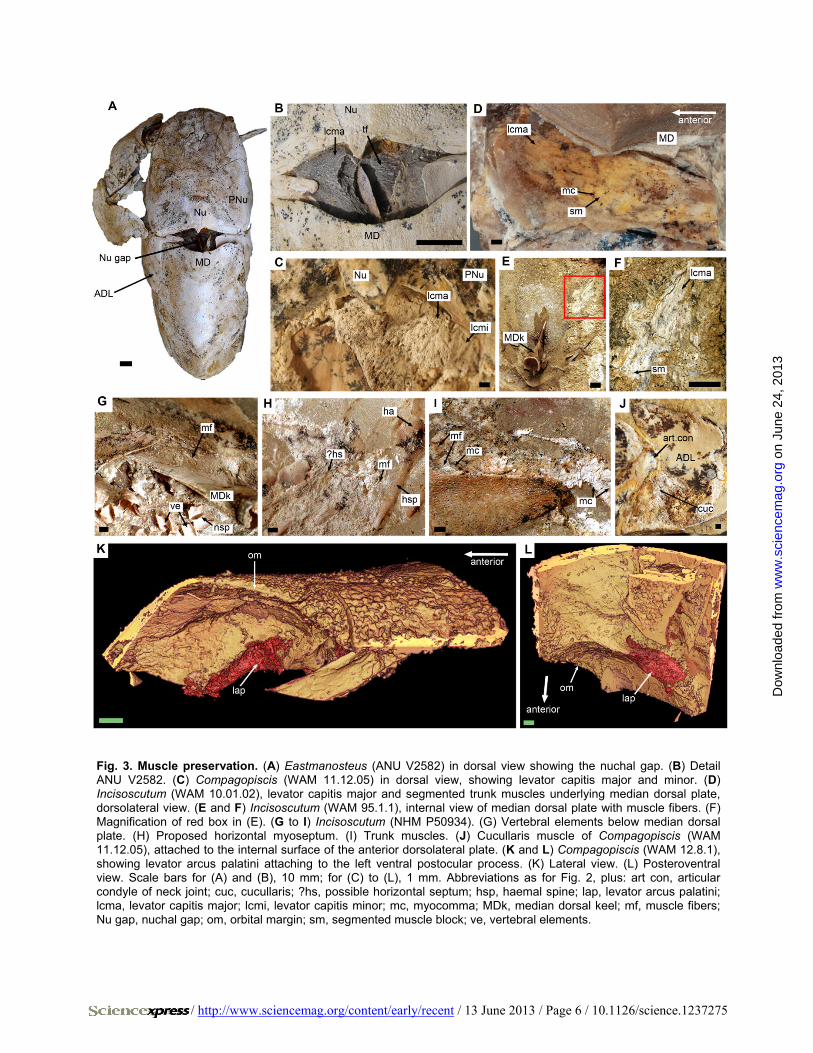
Fig. 3. Muscle preservation. (A) Eastmanosteus (ANU V2582) in dorsal view showing the nuchal gap. (B) Detail ANU V2582. (C) Compagopiscis (WAM 11.12.05) in dorsal view, showing levator capitis major and minor. (D) Incisoscutum (WAM 10.01.02), levator capitis major and segmented trunk muscles underlying median dorsal plate, dorsolateral view. (E and F) Incisoscutum (WAM 95.1.1), internal view of median dorsal plate with muscle fibers. (F) Magnification of red box in (E). (G to I) Incisoscutum (NHM P50934). (G) Vertebral elements below median dorsal plate. (H) Proposed horizontal myoseptum. (I) Trunk muscles. (J) Cucullaris muscle of Compagopiscis (WAM 11.12.05), attached to the internal surface of the anterior dorsolateral plate. (K and L) Compagopiscis (WAM 12.8.1), showing levator arcus palatini attaching to the left ventral postocular process. (K) Lateral view. (L) Posteroventral view. Scale bars for (A) and (B), 10 mm; for (C) to (L), 1 mm. Abbreviations as for Fig. 2, plus: art con, articular condyle of neck joint; cuc, cucullaris; ?hs, possible horizontal septum; hsp, haemal spine; lap, levator arcus palatini; lcma, levator capitis major; lcmi, levator capitis minor; mc, myocomma; MDk, median dorsal keel; mf, muscle fibers; Nu gap, nuchal gap; om, orbital margin; sm, segmented muscle block; ve, vertebral elements.
on
June
24,
201
3w
ww
.sci
ence
mag
.org
Dow
nloa
ded
from