A COMPARATIVE STUDY OF ENDOSCOPIC COBLATION ...
104
A COMPARATIVE STUDY OF ENDOSCOPIC COBLATION ADENOIDECTOMY AND CONVENTIONAL CURETTAGE ADENOIDECTOMY Dissertation Submitted to THE TAMIL NADU DR.M.G.R. MEDICAL UNIVERSITY In partial fulfilment of the requirements for the degree of M.S. DEGREE BRANCH – IV M.S. OTORHINOLARYNGOLOGY DEPARTMENT OF OTORHINOLARYNGOLOGY KILPAUK MEDICAL COLLEGE CHENNAI - 600010. MAY – 2018
-
Upload
khangminh22 -
Category
Documents
-
view
3 -
download
0
Transcript of A COMPARATIVE STUDY OF ENDOSCOPIC COBLATION ...
A COMPARATIVE STUDY OF ENDOSCOPIC COBLATION
ADENOIDECTOMY AND CONVENTIONAL CURETTAGE
ADENOIDECTOMY
Dissertation Submitted to
THE TAMIL NADU DR.M.G.R. MEDICAL UNIVERSITY
In partial fulfilment
of the requirements for the degree of
M.S. DEGREE
BRANCH – IV
M.S. OTORHINOLARYNGOLOGY
DEPARTMENT OF OTORHINOLARYNGOLOGY
KILPAUK MEDICAL COLLEGE
CHENNAI - 600010.
MAY – 2018
2
DECLARATION
I, Dr.J.Dianitta Devapriya Veronica, solemnly declare that the
dissertation titled, “A COMPARATIVE STUDY OF ENDOSCOPIC
COBLATION ADENOIDECTOMY AND CONVENTIONAL
CURETTAGE ADENOIDECTOMY” is a bonafide work done by me
at Government Kilpauk Medical College under the guidance and
supervision of Prof.Dr.T.INDRA, MS(ENT), Professor, Department of
Otorhinolaryngology.
This dissertation is submitted to The Tamil Nadu Dr.M.G.R.
Medical University towards the partial fulfilment of the requirements of
M.S. Branch – IV, Otorhinolaryngology degree examination.
Place : Chennai
Date:
Dr. J. DIANITTA DEVAPRIYA VERONICA

2
CERTIFICATE
This is to certify that Dr.J.Dianitta Devapriya Veronica,
postgraduate student (2015 – 2018) in the Department of
Otorhinolaryngology, Government Kilpauk Medical College and
Hospital, Chennai has done this dissertation titled “A COMPARATIVE
STUDY OF ENDOSCOPIC COBLATION ADENOIDECTOMY
AND CONVENTIONAL CURETTAGE ADENOIDECTOMY”
under the direct guidance and supervisionin partial fulfilment of the
regulations laid down by The Tamil Nadu Dr.M.G.R. Medical University,
Chennai, for M.S. Branch–IV Otorhinolaryngology Degree Examination.
Prof. Dr. T. INDRA, MS(ENT) Prof.Dr.VASANTHA MANI, MD(OG)
Professor and HOD DGO. MNAMS. Dpsy.MBA
Department of Otorhinolaryngology DEAN
Govt. Kilpauk Medical College and Govt. Kilpauk Medical College
Hospital, Chennai. and Hospital, Chennai.

3
CERTIFICATE - II
This is to certify that this dissertation work titled A
COMPARATIVE STUDY OF ENDOSCOPIC COBLATION
ADENOIDECTOMY WITH CONVENTIONAL CURETTAGE
ADENOIDECTOMY of the candidate Dr. J. DIANITTA
DEVAPRIYA VERONICA with registration Number
221514151for the award of M.S. DEGREE in the branch of
OTORHINOLARYNGOLOGY. I personally verified the
urkund.com website for the purpose of plagiarism Check. I found
that the uploaded thesis file contains from introduction to
conclusion pages and result shows 3% percentage of plagiarism in
the dissertation.
Guide & Supervisor sign with Seal.
4
ACKNOWLEDGEMENT
I am extremely thankful to Prof.Dr.VASANTHA MANI, DEAN,
Government Kilpauk Medical college and Hospital for having permitted
me to utilize the facilities of the hospital for conducting this study.
I am immensely grateful to Prof. Dr. T. INDRA MS(ENT) for her
concern, meticulous guidance, expert advice and constant encouragement
in preparing this dissertation.
I am very grateful to express my sincere gratitude to the Assistant
Professors, to Dr.V.Prithviraj, MS(ENT), Dr.K.Sanjay Kumar,
MS(ENT), Dr.P.Thamizharasan, MS(ENT), DLO, Dr.S.Vignesh,
MS(ENT) and Dr.G.Udhaya Chandrika, MS(ENT), for their constant
motivation and valuable suggestions.
I am extremely grateful and indebted to my former professors
Prof.Dr.K.Ravi, MS(ENT), DLO, DNB, Prof.Dr.P.Ilangovan
MS(ENT), DLO and Dr.S.Rajasekar, MS(ENT), DLO for their
inspiration, constant encouragement and guidance.
I am thankful to the Institutional Ethical Committee for their
guidance and approval for this study.

5
I am thankful to all my colleagues and friends for their help and
advice in carrying out this dissertation.
I am grateful to my husband Dr.Prem, my son Sammy and other
family members for their moral support and encouragement.
I am grateful to the Almighty God who gave me an opportunity
and blessed me to finish the work.
Last but not the least, I thank all the patients for willingly
submitting themselves for this study.
6
CONTENTS
Sl.No. Title Page
No.
1. INTRODUCTION 1
2. AIM OF THE STUDY 4
3. ANATOMY AND PHYSIOLOGY 5
4. HISTORY AND EVOLUTION OF
ADENOIDECTOMY
15
5. SURGICAL TECHNIQUES OF
ADENOIDECTOMY
19
6. REVIEW OF LITERATURE 30
7. MATERIALS AND METHODOLOGY 44
8. RESULTS 57
9. DISCUSSION 73
10. CONCLUSION 81
11. ANNEXURES
a) BIBLIOGRAPHY
b) APPENDIX (PROFORMA)
c) PATIENT CONSENT FORM
d) PATIENT INFORMATION FORM
e) MASTER CHART
f) URKUND ORIGINALITY REPORT
g) ETHICAL COMMITTEE APPROVAL
CERTIFICATE

7
ABSTRACT
Adenoid hypertrophy with presenting complaints such as chronic
nasal obstruction, snoring, mouth breathing and earache are some of the
most common problems encountered in otorhinolaryngological practice.
The increased prevalence of adenoid hypertrophy in paediatric patients
has led to the wide practice of adenoidectomy. As with all surgical
interventions, adenoidectomy is associated with certain complications. In
the recent years, different surgical techniques for adenoidectomy have
been proposed to reduce the morbidity and the incidence of
complications.
Though conventional curettage is still practised, the endoscopic
approach is gaining popularity. Conventional technique is a blind
procedure and can cause injury to the surrounding structures and has the
disadvantage of leaving residual tissue.
The endoscopic approach offers solution to the above mentioned
problems. Adenoidectomy can be carried out under direct visualisation
thereby minimising the risk of injury to the surrounding structures. A step
ahead is the use of the coblator in which the tissue is not exploded but is
molecularly broken down into simpler hydrocarbons. The present study is
to compare the efficacy of endoscopic assisted coblation adenoidectomy
with conventional curettage adenoidectomy with respect to the duration
of surgery, intraoperative bleeding, postoperative pain, the time taken for
recovery in the postoperative period and completeness of removal of
adenoid.
Keywords: coblation, adenoidectomy, curettage

1
INTRODUCTION
Children constitute a major percentage of the population presenting
to the ENT OPD. Children presenting with complaints such as nasal
obstruction, sleeping with the mouth open, snoring and earache arouse the
suspicion of adenoid enlargement. Though adenoid enlargement is
physiologically normal in this age group, the resulting airway obstruction
is troublesome and the children can present with distressing symptoms.
Adenoid hypertrophy can cause eustachian tube dysfunction
leading to otitis media with effusion which if not treated adequately may
lead to a chronic discharging ear. Chronic adenoiditis can act as a septic
focus with the child falling ill often with repeated respiratory infections.
This remains one of the common causes for a poor appetite leading
to a poor nutritional status. Lack of good sleep leads to poor
concentration and poor school performance. This creates an alarming
state for parents who rush for medical consultation. Studies have revealed
that adenoid hypertrophy causing chronic airway obstruction can even
lead to cor pulmonale.1
Though a physiological hypertrophy, adenoid enlargement when
causing airway compromise or interfering with the development of the

2
facial skeleton needs to be addressed. Surgical removal of the adenoid
tissue is the solution and is being practised for years. A simple curettage
adenoidectomy offers a solution.
The recent past has witnessed great advances in the field of ENT.
One such advancement is the development of newer surgical techniques
for adenoidectomy. These techniques that have been proposed seem to
have a significant effect on the morbidity and the incidence of
complications following the procedure.
The advent of endoscopes has made ENT surgeries more attractive,
with a reduction in the list of indications for conventional techniques. The
use of endoscopes combined with the newer surgical techniques has
gained popularity in the recent days.
One such combination is the endoscopy assisted coblation
technique. The coblator works on the principle of controlled ablation
which cause disintegration of cells molecule by molecule causing
volumetric reduction of tissue. This technique of coblation
adenoidectomy has considerable influence on the duration of surgery, the
blood lost intraoperatively, pain in the postoperative period, the time
taken for recovery in the postoperative period and the complete removal
of adenoid tissue.

3
The present study was carried out to compare the efficacy of
endoscopic assisted coblation adenoidectomy with conventional curettage
adenoidectomy.
4
AIM OF THE STUDY
The aim of the study was to compare the efficacy of endoscopic
coblation adenoidectomy with conventional curettage adenoidectomy.
The techniques were compared on the basis of
Primary outcome:
1. Completeness of removal
2. Intraoperative blood loss
Secondary outcome:
1. Operative duration
2. Postoperative pain
3. Recovery time
5
ANATOMY
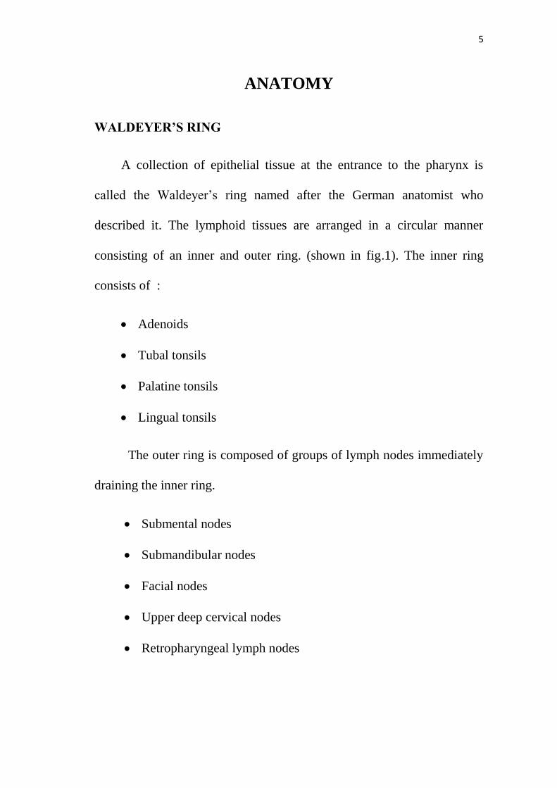
WALDEYER’S RING
A collection of epithelial tissue at the entrance to the pharynx is
called the Waldeyer‟s ring named after the German anatomist who
described it. The lymphoid tissues are arranged in a circular manner
consisting of an inner and outer ring. (shown in fig.1). The inner ring
consists of :
Adenoids
Tubal tonsils
Palatine tonsils
Lingual tonsils
The outer ring is composed of groups of lymph nodes immediately
draining the inner ring.
Submental nodes
Submandibular nodes
Facial nodes
Upper deep cervical nodes
Retropharyngeal lymph nodes
6
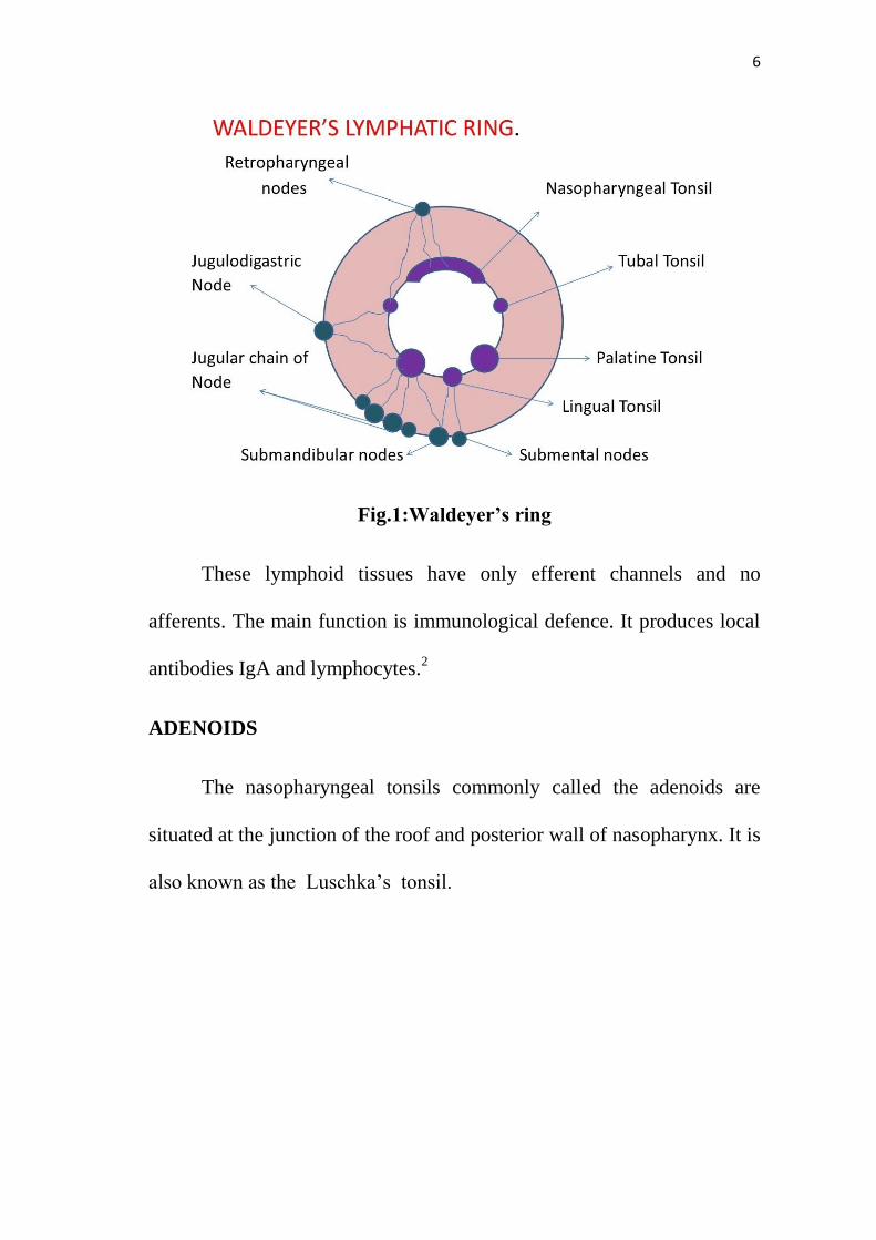
Fig.1:Waldeyer’s ring
These lymphoid tissues have only efferent channels and no
afferents. The main function is immunological defence. It produces local
antibodies IgA and lymphocytes.2
ADENOIDS
The nasopharyngeal tonsils commonly called the adenoids are
situated at the junction of the roof and posterior wall of nasopharynx. It is
also known as the Luschka‟s tonsil.
7
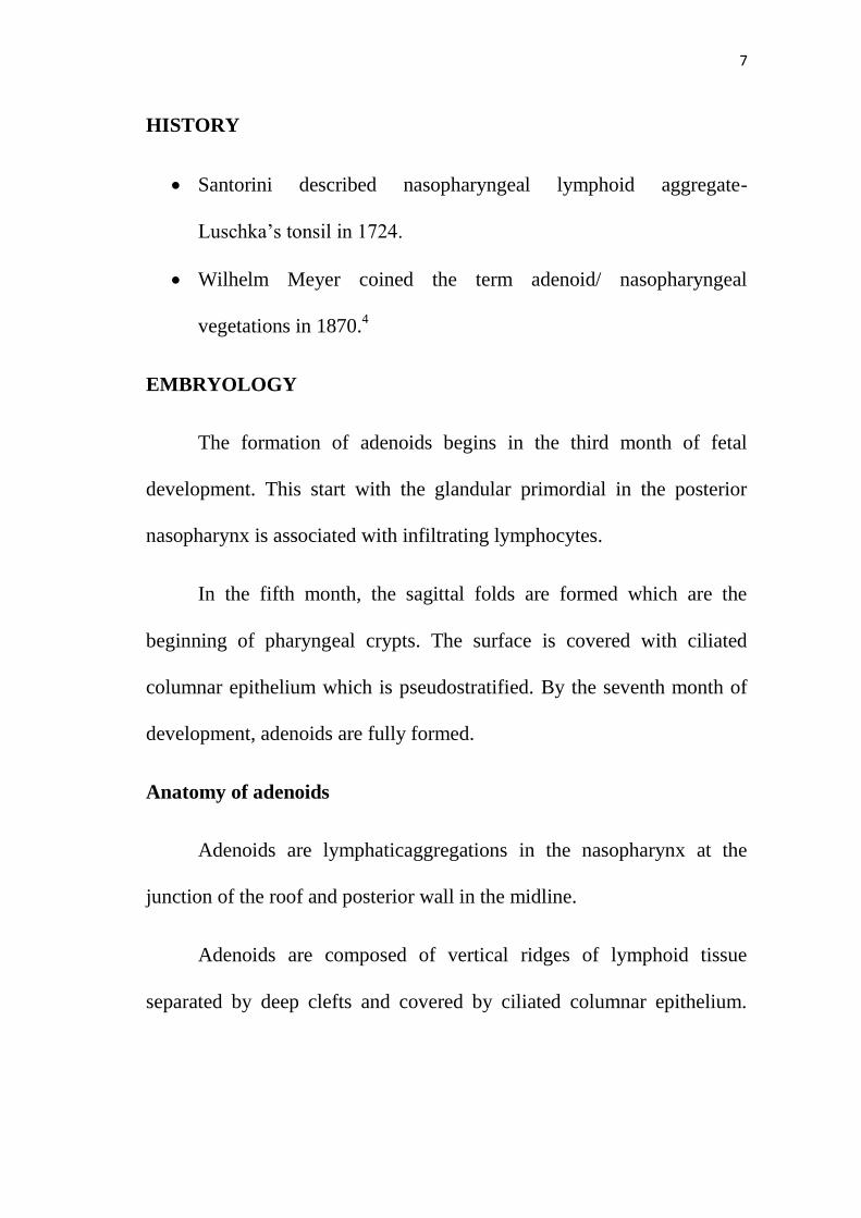
HISTORY
Santorini described nasopharyngeal lymphoid aggregate-
Luschka‟s tonsil in 1724.
Wilhelm Meyer coined the term adenoid/ nasopharyngeal
vegetations in 1870.4
EMBRYOLOGY
The formation of adenoids begins in the third month of fetal
development. This start with the glandular primordial in the posterior
nasopharynx is associated with infiltrating lymphocytes.
In the fifth month, the sagittal folds are formed which are the
beginning of pharyngeal crypts. The surface is covered with ciliated
columnar epithelium which is pseudostratified. By the seventh month of
development, adenoids are fully formed.
Anatomy of adenoids
Adenoids are lymphaticaggregations in the nasopharynx at the
junction of the roof and posterior wall in the midline.
Adenoids are composed of vertical ridges of lymphoid tissue
separated by deep clefts and covered by ciliated columnar epithelium.

8
The deep crypts penetrate adenoid tissue from the surface. The crypts are
lined by epithelium specialized for the uptake of antigens. (fig 2)
The adenoids are present from birth and increase in size with age
and undergo spontaneous regression after puberty.
Fig.2: Lateral view of nasopharynx showing adenoids.
Blood supply
Ascending palatine of facial artery
Pharyngeal branch of internal maxillary artery
Ascending pharyngeal branch of external carotid
Ascendingcervical branch of inferior thyroid artery of
thyrocervical trunk.3
9
Venous drainage
Venous drainage is through the pharyngeal plexus and pterygoid
plexus flowing ultimately into the facial and internal jugular veins.
Nerve supply
Innervation is derived from pharyngeal plexus.
Lymphatic drainage
Lymphatic drainage is to the upper jugulodigastric nodes directly
and indirectly via retropharyngeal and parapharyngeal nodes.
PHYSIOLOGY
The crypts of adenoids are lined by specialized epithelium for the
uptake of antigens.
B and T cells are present in the adenoids with B cells
predominating comprising 60% while T cells contributing to 40%. IgA,
IgM, IgD are the immunoglobulins produced by the B cells of adenoids.
Radiologically adenoids are not demonstrable in infants less than 4
weeks of age. They become radiologically visible from the age of 4
months. Growth continues and plateaus between 2 and 14 years of age.
The size of adenoid appears to be at its largest at 7 years of age.
Regression occurs after the age of 15 years in most of the children.4

10
MICROBIOLOGY
The human nasopharynx harbours bacterial species such as
streptococcus pneumoniae, Haemophilus influenzae and Moraxella
catarrhalis which adhere to the epithelial cells. The colonization gets
established early in childhood.
Pathological manifestations of the adenoid
Upper airway obstruction and OSA
Rhinosinusitis
Recurrent otitis media
Otitis media with effusion
Olfactory disturbances4
GRADING OF ADENOID ENLARGEMENT
Clemens and McMurray
GRADE 1 : adenoid tissue filling 1:3 of the vertical height of
choana (Fig 3a)
GRADE 2 : adenoid tissue filling 2:3 of the vertical height of
choana (Fig 3b)
GRADE 3 : adenoid tissue filling 2:3 to nearly all but not complete
filling of the choana (Fig 3c)
GRADE 4 : complete choanal obstruction (Fig 3d)18

11
Clinical symptoms are common in the younger age group due to
the relative small size of nasopharynx and increased incidence of upper
respiratory tract infections. The criterion for adenoid sufficient to cause
nasal obstruction was tissue occupying more than 40% of the
nasopharynx.
Fig 3a Fig 3b
Fig3c Fig 3d
AETIOLOGY OF ADENOID HYPERTROPHY
Physiological hypertrophy in children
Associated with upper respiratory tract infections
Allergy of upper respiratory tract
Unsuspected neoplasia

12
Clinical features of adenoid hypertrophy
Nasal symptoms
Nasal obstruction
Nasal discharge
Epistaxis
Sinusitis
Voice change
Aural symptoms
Tubal obstruction – retracted TM and conductive hearing loss
Recurrent attacks of acute otitis media
Serous otitis media
CSOM refractory to medical management2
Others
Snoring and sleep apnoea
Growth problems
Cardiopulmonary problems
Child presents with characteristic adenoid facies (fig.4)

13
The features include
Open mouth and mouth breathing
Pinched nostrils
Loss of nasolabial fold
Crowded teeth and gum hyperplasia
Underslung mandible
High arched „V‟ shaped palate
Short upper lip
Hypoplasia of maxilla
Vacant expression
Pectus excavatum
Voice changes
Fig.4: Adenoid facies.

14
DIAGNOSIS
1. Posterior rhinoscopy
2. X ray soft tissue neck -lateral view
3. Diagnostic nasal endoscopy – gold standard
4. Acoustic rhinomanometry
5. MRI4
Fig.5: Endoscopic picture of adenoids.
Fig.6: X-Ray soft tissue neck lateral view showing hypertrophied
adenoids.

15
HISTORY AND EVOLUTION OF ADENOIDECTOMY
The earliest description of adenoid surgery was by Paul of Aegina
who removed adenoids by passing a ligature transnasally.
In1967, William meyer performed the first adenoidectomy using a
specially designed ring knife.
Voltilini removed adenoids using galvanic cauterization.
Beckmann modified the ring knife designed by Gottstein and
invented the curette.
Sir St. Clair Thompson designed the curette with the cage which
entangles the tissue fragments which is still in use.
Punch forceps and adenotome were also developed for adenoid
removal.
Kwok and Hawke developed the suction diathermy for the purpose
of haemorrhage control but subsequently the whole procedure was
performed using this technique.
Following the advent of endoscopes, difficulties such as visualizing
the adenoids and accessing the adenoids were overcome.
Conventional adenoidectomy was done followed by endoscopic
assessment of the residual tissue which was removed under
endoscopic visualisation.

16
Power assisted adenoidectomy was initially designed for
arthroscopic surgeries and then used for sinonasal procedures. First
used for adenoid removal by Yanagisawa.5
INDICATIONS FOR ADENOIDECTOMY
Adenoid hypertrophy causing
Obstructed nasal airway
Sleep apnoea syndrome
Recurrent rhinosinusitis
Chronic eustachian tube dysfunction
Otitis media with effusion
Recurrent otitis media
CSOM
Dental occlusion abnormalities
Speech abnormalities
Children with chronic adenoid hypertrophy can present with
craniofacial morphology problems, excessive snoring, poor olfaction and
are candidates for adenoidectomy. Patients with history of chronic
sinusitis/ chronic recurrent sinusitis/chronic purulent sinusitis which may
occur secondary to chronic adenoiditis mat show response to
adenoidectomy.

17
Coticchia and colleagues showed that 94.9% of the mucosal
surface of the adenoids removed for chronic sinusitis was covered with
dense mature biofilms which may act as a natural reservoir for resistant
bacteria. Hence their removal may be the reason for the benefit of the
procedure. Patients with hyponasal speech are also candidates for
adenoidectomy.
In patients with cleft palate / submucous cleft palate, conservative
adenoidectomy is performed leaving the lower portion of the adenoid pad
to decrease velopharyngeal insufficiency.
Adenoidectomy is combined with myringotomy and ventilation
tube placement to reduce the incidence of further episodes of otitis media.
Surgical removal of adenoids may remove a nasopharyngeal nidus
of contaminated tissue that may secondarily act as a source of infection in
the middle ear. Simply adenoidectomy removes the anatomic obstruction
of eustachian tube.
CONTRAINDICATIONS FOR ADENOIDECTOMY
Overt cleft palate
Submucous cleft palate
Haemorrhagic diathesis
Acute infection of upper respiratory tract

18
COMPLICATIONS OF ADENOIDECTOMY
Haemorrhage
Bleeding usually occurs within 6-20 hours in less than 0.7%.
postnasal packing is the preferred management. Secondary haemorrhage
occurs from an aberrant ascending pharyngeal artery. Suspicion of a
coagulation defect occurs in unusual reactionary or secondary
haemorrhage.
Injury to eustachian tube opening
Injury to pharyngeal musculature and vertebrae
Dental trauma
Airway obstruction due to nasopharyngeal blood clot ( coroner‟s
clot)
Velopharyngeal insufficiency – occurs as a result of incomplete
closure of palate to the posterior and lateral nasopharyngeal wall.
Torticollis – because adenoids are removed from the posterior wall
of nasopharynx over the spine and superior constrictor muscle-
children can have stiff neck/ spasm
Nasopharyngeal stenosis due to scarring
Atlantoaxial subluxation (Grisel syndrome) commonly in Down‟s
syndrome
Regrowth of adenoid
Infection4

19
SURGICAL TECHNIQUES OF ADENOIDECTOMY
Different techniques of adenoidectomy have been described since
the earliest techniques. All these techniques are based on the principle of
complete tissue removal with less damage to the surrounding tissues.
They are:-
Conventional curettage adenoidectomy
Adenoidectomy using electrocautery
Microdebrider assisted adenoidectomy
Laser adenoidectomy
Coblation adenoidectomy
Positioning
Initially the procedure was performed with the patients in sitting
position and the surgeon facing the patient. The adenoids were removed
with either the curette or adenotome. Nowadays the Rose position is used
with the patient in supine position with head and neck extended with sand
bag under the shoulders and the surgeon sitting behind the patient at the
head end. Illumination is by using a head light. Visualisation can be done
using a laryngeal mirror.
20
CURETTAGE ADENOIDECTOMY
Curettage adenoidectomy dates back to the earliest attempts at
removal of adenoid tissue. Curettes of various sizes are available which
are all based on the curette originally designed by Jacob Gottenstein. The
curette has a sharp horizontal knife edge which is necessary for cutting
through the adenoid bed. The side arms of the curette form the vertical
posts of the window and hold the blade. The vertical posts converge to
extend as the handle which is framed with a grip at the end helps in
applying pressure while passing the knife through the adenoid bed. The
procedure was initially performed by confirming by digital palpation.
Most use a mirror to scan the full adenoid bed when the child is in Rose
position with the palate retracted. An appropriate sized curette is selected
and placed such that it rests against the vomer which is then pushed
through the spongy adenoid tissue to the underlying muscle layer. The
handle is then pulled backward with the thumb acting as the fulcrum at
the upper incisor level. With the movement of the curette‟s handle, the
blade of the knife sweeps in an arc through the base of the adenoid tissue
to terminate with the removal of tissue at Passavant‟s ridge. The residual
tissue that remains is removed using curette of the necessary size which
usually happens when dull curette knives are used. After tissue removal,
the nasopharynx is pressure packed for hemostasis.

21
ADENOTOME
The adenotome was one of the earliest instruments used for
adenoid removal. The adenotome is a basket like device with a concave
open curved face into which the adenoid tissue fits before being sliced off
by a flexible blade made of steel that acts as the lid of the basket. The
various sizes of the adenotome with the rounded geometry of the opening
fit perfectly into the nasopharynx. Under direct vision, the adenotome is
placed in the nasopharynx by using the palatal retractor to elevate the
velum and the surgeon sits by the side of the patient. The adenotome is
pressed into the midline of the bed against the pharyngeal wall and is
rotated downward as the blade closes.
Fig.7 : Adenoidectomy using Adenotome.

22
ELECTROCAUTERY
Suction cautery was initially used to achieve hemostasis after the
traditional methods of adenoidectomy. Further, this led to the
development of using suction cautery for the whole procedure. With the
patient in rose position, a malleable suction cautery is introduced into the
nasopharynx and the adenoids are ablated in the superoinferior direction.
The power is set between 30 – 40 watts and the adenoids are charred on
ablation. Hemostasis can be achieved using the same suction cautery and
this preludes the use of nasopharyngeal packs.
Fig.8 : Adenoidectomy by suction cautery.

23
LASER ADENOIDECTOMY
Laser adenoidectomy is performed with a CO2 laser attached to the
surgical microscope. The lips, teeth and face are covered with wet towels
after the palate and jaw have been retracted. The laser light is reflected
into the nasopharynx using a polished metal mirror and a suction is used
to evacuate the smoke plumes. The commonest complication is acquired
nasopharyngeal stenosis.
POWER ASSISTED ADENOIDECTOMY
This procedure which was advocated by Dr.Eji Yanagisawa was
initially reserved for the superiorly situated adenoid tissue that caused
choanal obstruction and couldn‟t be removed using the standard
techniques. An endoscopic set is kept ready, the nasal cavity is
decongested and the patient is positioned as for endoscopic sinus surgery.
The microdebrider is introduced through one of the nostrils to reach the
nasopharynx and the adenoid tissue is removed in a side to side fashion
by the oscillating blade starting superiorly progressing inferiorly. It is
essential that the blade is kept in view at all times. This method was
considered to be faster with less blood loss but quite expensive.6

24
Fig.9 : Microdebrider assisted adenoidectomy.
DISCOVERY OF COBLATION
The technology of using plasma to ablate biological tissue was first
described by Woloszko and Gilbride. By their pioneering work in this
field they proved that radiofrequency current could be passed through
local regions of the body without discharge taking place.
Radiofrequency technology for medical use (for cutting,
coagulation and tissue dessication) was popularized by Cushing and

25
Bovie. Cushing an eminent neurosurgeon found this technology excellent
for his neurosurgical procedures. First use of this technology inside the
operating room took place on October 1st 1926 at Peter Bent Brigham
hospital in Boston, Massachusetts. It was Dr.Cushing who removed a
troublesome intracranial tumour using this equipment.
Coblation was first discovered by Hira V. Thapliyal and Philip E.
Eggers. This was a fortuitous discovery in their quest for unblocking
coronary arteries using electrosurgical energy. Coblation was initially
used for arthroscopic surgeries.
COBLATION TECHNOLOGY EXPLAINED
Coblation is non-thermal volumetric tissue removal through
molecular dissociation. This action is more or less similar to Excimer
laser. This technology uses the principle that when electric current is
passed through a conducting fluid, a charged layer of particles known as
plasma is released. These charged particles have the tendency to
accelerate through tissue and gains energy to break the molecular bonds
within the cells. This ultimately cause disintegration of cellsmolecule by
molecule causing volumetric reduction of tissue.

26
Fig.10 : Plasma generation in coblator.
Fig.11 : Coblator
THE COBLATION SYSTEM
• Radiofrequency generator
• Foot pedal system
• Irrigation system
• Coblation wand

27
The coblation wand has two electrodes, the base electrode and the
active electrode which are separated by ceramic. The current generated
flows between these electrodes via the saline medium. This saline is
broken down into ions which forms active plasma which appears as as a
orange glow at the tip of the wand. The thickness of the plasma created is
100-200µm.
The modes of operation include ablation and coagulation. The
operating frequency is 100kHz with a power consumption of 110/240
MV.
Once the wand is connected the microprocessor senses the type of
wand and displays the default settings of that particular wand. The flow
control valve unit is clamped to the IV stand to ensure automatic saline
flow. The saline flows only when the surgeon presses the pedal. Smoke
generated during the procedure indicates the presence of tissue between
the electrodes which can be flushed out. During surgery the tissue turns
brown which indicates tissue oxidation and not heat induced charring.
Intermittent application of coblation with copious irrigation of cold
saline improves the efficiency of ablation.

28
STAGES OF PLASMA GENERATION
FIRST STAGE
Vapour gas piston formation
This stage is characterised by transition from bubble to film
boiling. This decreases heat emission and causes increase in surface
temperature
SECOND STAGE
Stage of vapour film pulsation
Tissue ablation occurs during this stage
THIRD STAGE
Reduction of amplitude of current across the electrodes.
FOURTH STAGE
Dissipation of electron energy at the metal electrode surface.
FIFTH STAGE
Stage of thermal dissipation of energy
This stage is essentially due to recombination of plasma ions,
active atoms and molecules.
These stages explain why coblation is effective if applied
intermittently. This ensures constant presence of stage of vapour film
pulsation which is important for tissue ablation.

29
The effect of plasma on tissue is purely chemical and not thermal.
Plasma generates OH &H+ ions. These make plasma destructive. OH
radical causes protein disintegration.
Advantages of coblation
• Very limited depth of penetration
• Minimal collateral tissue damage
• Localized effect
• Controlled volumetric tissue removal
• Surface temperatures 40-70 degrees C
• By products / gases that form are different from those of
conventional devices.7
COBLATION
BASED
DEVICES
CONVENTIONAL
ELECTROSURGICAL
DEVICES
CONVENTIONAL
LASER DEVICES
TEMPERATURE 40-70º >400º >400º
THERMAL
PENETRATION
MINIMAL DEEP MODERATE TO
DEEP
EFFECTS ON
TARGET TISSUE
GENTLE
REMOVAL
DISSOLUTION
RAPID HEATING
CHARRING
BURNING
CUTTING
RAPID HEATING
AND
VAPOURIZATION
EFFECTS ON
SURROUNDING
TISSUE
MINIMAL
COLLATERAL
EFFECT
INADVERTENT
CHARRING OR BURNING
INADVERTENT
BURNING AND
BLEEDING
Table.1 : Comparison of different electrosurgical devices.

30
REVIEW OF LITERATURE
Literature search was done and the studies comparing conventional
curettage adenoidectomy and endoscopic coblation adenoidectomy were
reviewed.
Di Riezo Businco et al studied 40 children with adenoid
hypertrophy of the age group 4-16 years. The children were divided into
2 groups with group A to receive adenoidectomy by cold curettage and
group B by coblation technology. The outcomes evaluated after surgery
were pain score on the first day, days reporting pain and requiring
analgesia, days of absenteeism, adenoid grade by endoscopic evaluation
and intraoperative blood loss. The children were also subjected to basal
rhinomanometry and nasal decongestion test 40 days after surgery. The
results showed that the cold curettage group showed higher adenoid grade
and high values of nasal resistance at rhinomanometry postoperatively.
Endoscopic coblation adenoidectomy was a technique considered to
ensure complete removal of adenoids and was also safer as it was done
under endoscopic control.8
Jheong Whun Kin studied 388 children who were classified into 3
groups on the basis of the adenoidectomy technique employed. The
techniques employed were power assisted adenoidectomy with

31
cauterization- microdebrider with electrocauterization for haemostasis,
power assisted adenoidectomy without cauterization- microdebrider with
just packing for haemostasis. Though the power assisted technique is
commonly used for adenoidectomy, coblation was found to be
advantageous because it performs multiple functions including ablation,
coagulation, suction and saline irrigation. This prospective multicentre
study demonstrated that coblation adenoidectomy was superior to
microdebrider adenoidectomy with regards to the amount of
intraoperative bleeding 9
Mahmut Ozkiris et al studied 60 consecutive patients in the age
group of 4-8 years undergoing adenoidectomy. Two groups each
consisting of 30 patients were subjected to two different adenoidectomy
techniques, the coblation technique and cold curettage technique. The
techniques were compared by means of intraoperative blood loss,
operative time and pre and postoperative nasal mucociliary clearance
rates (NMCR). The nasal mucociliary clearance was described as the
velocity of nasal mucociliary transport of the 99Tc-MAA droplet.
Radioscintigraphy was performed before and after surgery in all the
patients on the right nasal cavity. The average nasal mucociliary
clearance rates showed no significant statistical difference before surgery
but at 6 weeks postoperatively, the NMCR was 1.80+/- 0.21 mm/min and
32
2.06+/- 0.31 mm/min for the curettage and coblation groups respectively.
The coblation group offered better NMCR values and lesser bleeding but
a longer operating time when compared to curettage techniques.10
N. E. Jonas et al conducted a prospective randomized study to
compare suction diathermy to curettage adenoidectomy with regard to
operating time and adenoid regrowth at 6 months postoperatively. 100
children were included in the study and underwent surgery by either
technique by a single surgeon. There was no significant difference in the
duration of surgery between the two techniques. The postoperative
assessment at 6 months showed a significant difference in the residual
adenoid tissue with the suction diathermy group presenting with less
adenoid tissue. Although the difference was statistically significant it did
not seem to be of any clinical significance.11
Nebil Ark and colleagues conducted a prospective study on 99
patients comparing blind curettage with curettage under indirect mirror
visualization. By the blind technique, only 20.2% patients showed no
residual adenoid. Residual adenoid tissue was seen along the torus tubaris
on either side of the nasopharynx and on the pharyngeal roof near the
choanal opening and on the posterior wall of nasopharynx. The study
revealed that visualization of nasopharynx is essential for complete
removal in adenoidectomy.12

33
Vijayakrishnan and colleagues conducted a randomized study of
adenotonsillectomy for children with OSA comparing conventional and
coblation methods. 50 children with OSA of the age group 5-17 years
were randomly selected and were subjected to either of the technique.
Comparison of the surgical procedures on the basis of blood loss,
postoperative pain and postoperative reactionary and secondary
haemorrhage were done. Blood loss was found to be more in the
conventional group whereas postoperative bleeding was found to be
minimal in both the techniques.13
Nina L. Shapiro et al compared cold dissection with coblation
assisted adenotonsillectomy in a prospective randomized study of
pediatric patients aged 2-16 years. Intraoperative parameters measured
were surgical duration and intraoperative blood loss. Postoperative
parameters measured were daily pain rating using Wong-Baker FACES
pain scale, days of use of pain medication, days to return to normal diet.
Postoperative complications were also included. Operative duration was
shorter in the coblation group (11.2 min vs 17.0 min) (P< 0.001).
Intraoperative blood loss was lower in the coblation group whereas no
significant difference was reported in the postoperative pain scores
analysed daily and both the groups returned to normal activity on similar
postoperative days.14

34
In a study by Benninger and colleagues, two different techniques
of adenotonsillectomy were compared, the cold dissection method and
coblation adenotonsillectomy. The study revealed that coblation
technique had less reports of postoperative pain and decreased
postoperative narcotic usage leading to early recovery.15
In yet another study by Stanislaw P Jr and colleagues, curettage
adenoidectomy was compared with power assisted adenoidectomy. 90
patients aged 1-13 years and 87 patients aged 1- 12 years underwent
power assisted adenoidectomy and curettage adenoidectomy respectively.
The parameters evaluated were operative duration, intraoperative blood
loss, completeness of removal, complications and postoperative recovery
period. The results showed that power assisted adenoidectomy was 20%
faster with 27% decreased blood loss with more complete removal of
adenoid tissue P < 0.001.16
In a study by Elluru R G et al, electrocautery adenoidectomy was
compared with curettage and power assisted methods for adenoidectomy.
Results showed that electrocautery adenoidectomy as well as power
assisted adenoidectomy offered shorter operating times and less blood
loss but power assisted adenoidectomy appears to be more expensive with
a longer learning curve.17

35
In a study by Clemens and McMurray and colleagues,
electrocautery adenoidectomy using suction cautery was compared with
curettage adenoidectomy. Blood loss, postoperative adenoid grade and
postoperative complications were recorded. No differences in
postoperative adenoid grade or postoperative complications were noted.
However blood loss was lower in the electrocautery group compared to
curettage group.18
Wong L and colleagues studied the effectiveness of
electrosurgical adenoid ablation by measuring the reduction of adenoid
size, blood loss and postoperative complications. The average blood loss
was 2.6 ml and no significant postoperative complications were
encountered. It was significant that 19 out of 23 children studied showed
no evidence of residual adenoid tissue.19
Somani S S and colleagues studied the efficacy of power assisted
adenoidectomy on 44 children with adenoid hypertrophy. A powered
microdebrider was used under endoscopic guidance and operative time,
blood loss, complications, completeness of removal and recovery period
were evaluated. The average operative time was 12 min and average
blood loss was 30 ml. Good visualization ensured complete removal of
adenoid tissue.20

36
Pagella F et al studied the evolution of different power assisted
techniques for adenoidectomy. They have performed a literature search
and have discussed the surgical techniques with review of the advantages
and disadvantages of each method. They concluded that all the methods
seemed to be safe and effective but the endoscopic techniques were
considered to be of greater value.21
In yet another study, Pagella F and colleagues studied 143
patients by employing a combined method of traditional curettage and
endoscopic adenoidectomy with microdebrider. A classical
adenoidectomy was performed initially followed by endoscopic
assessment of the residual tissue which was then removed using a
microdebrider under endoscopic control. 70 children had to undergo
transnasal removal of residual adenoid tissue establishing the advantage
of the use of an endoscope.22
Al Mazrou and colleagues conducted a study on 40 children
within the age group of 3-17 years who presented with symptoms
suggestive of obstructive sleep apnoea. They were randomly selected and
distributed into two groups. Group A was subjected to transnasal
endoscopic powered adenoidectomy and group B was taken up for
curettage adenoidectomy. Both the groups were subjected to preoperative
and postoperative nasal endoscopy. Comparison between the two groups

37
was made with regards to operative time, amount of blood loss,
postoperative morbidity, postoperative complications and resolution of
symptoms. The results showed that the mean blood loss was 8.2 ml in
group A compared to 22.1 ml in group B. The operative times were 6.1
min and 12.3 min in groups A and B respectively. Postoperative
endoscopy showed resolution of symptoms with evidence of adenoid
remnants. Transnasal endoscopic powered adenoidectomy was
considered to be a safe method.24
Havas and colleagues conducted a study on 130 consecutive
patients in the paediatric age group who had obstructive adenoid
hypertrophy. The aim of their study was to evaluate the efficacy of
traditional curettage adenoidectomy and the usefulness of intraoperative
endoscopic examination. The degrees of postnasal obstruction and
intranasal adenoid tissue after curettage adenoidectomy and powered
shaver adenoidectomy were assessed using endoscopic examination.
Following traditional curettage adenoidectomy 39% (51 out of 130
patients) had residual adenoid tissue. Having determined the presence of
residual tissue causing obstruction, complete patency was achieved with
powered shaver adenoidectomy. They concluded that traditional
adenoidectomy is ineffective in removing intranasal adenoid tissue and

38
the use of endoscope intraoperatively allows the assessment of complete
removal at staging.25
Datta R and colleagues studied 60 consecutive cases requiring
adenoidectomy. They conducted a comparative study of conventional
versus endoscopic powered adenoidectomy. 60 patients were divided into
2 groups, Group A and Group B. Group A underwent conventional
adenoidectomy using the traditional curettage method. Group B
underwent microdebrider adenoidectomy under endoscopic control. The
two groups were studied with regards to intraoperative duration,
intraoperative bleeding, completeness of resection, damage to adjacent
structure, postoperative pain and recovery time. The intraoperative time
in group A varied from 22-29 min whereas in group B from 27-55
minutes. The average blood loss in group A was 21 ml and 31.67 ml in
group B. Group B showed complete removal of adenoid tissue whereas
more than 50% in group A showed residual adenoid. Patients in group A
showed collateral damage. Postoperative pain was studied in cases
undergoing adenoidectomy alone. Group A had a pain score of 1.64-3.63.
group B had a pain score of 1.19-3.06 but this difference was not
statistically significant. The mean time for recovery was 3.5 days in
group A and 2.93 days in group B. The results showed that conventional
adenoidectomy group showed lesser operative time and less

39
intraoperative bleeding. The endoscopic powered adenoidectomy group
fared better with complete removal, accurate resection, lesser collateral
damage and faster recovery time.26
Das T and colleagues conducted a prospective study on 60
patients who underwent combined conventional and endoscopic
microdebrider assisted adenoidectomy. The study was conducted in a
tertiary care centre. Only grade 3 and grade 4 adenoid hypertrophy was
included in the study. The adenoid hypertrophy was graded again at the
end of conventional adenoidectomy and after combined procedure.
Postoperative complications, duration of surgery and amount of blood
loss were recorded. The endoscopic microdebrider assisted
adenoidectomy technique ensured complete clearance of adenoid tissue in
all cases. The average duration of the conventional procedure was 5 min
12 sec, while that of the combined technique was 14 min 45 sec. The
average blood loss was 15+/- 3 ml approximately. There were no major
complications. They concluded that the combined approach offered an
effective method for complete and accurate removal of adenoids.27
Feng Y and Yin S conducted a study in order to compare the
outcomes of powered assisted adenoidectomy with adenoid curette
adenoidectomy. 34 cases were retrospectively analysed and all the
patients were followed up for 6-12 months. Out of the 34 children, 18
40
cases underwent powered assisted adenoidectomy and the rest 16 patients
underwent conventional adenoidectomy by curettage method. The results
were assessed by acoustic rhinometry. The average time taken for
endoscopic powered assisted adenoidectomy was 5 min and 15 sec, while
the mean time for conventional adenoidectomy was 8 min 22 seconds (p<
0.01). The mean blood loss during powered assisted and conventional
adenoidectomy was 50 ml and 75 ml respectively. Comparing the
preoperative and postoperative cross-sectional area at adenoid in both
groups by acoustic rhinometry, a significant difference ( p < 0.01) was
found. Hence it was concluded that acoustic rhinometry is a useful
objective/ parameter for assessing the outcome after adenoidectomy.28
Ucar C conducted a study on 125 patients (67 boys and 58 girls)
for evaluating the efficacy of endoscopic adenoidectomy. The children
included in the study were those who presented with complaints such as
nasal obstruction, mouth breathing, snoring, loss of appetite and slower
growth development. Preoperatively endoscopic rhinoscopy was done on
48 patients and in the remaining patients, lateral cranial radiographs were
done. Postoperative controls were done at one and four weeks. At the end
of 4 weeks, none of the patients had any complaints. 39 patients showed
complete removal of adenoids. Thus endoscopic adenoidectomy offers a

41
satisfactory method as it controls the amount of adenoid tissue being
removed.29
Regmi D and colleagues conducted a study to evaluate the results
of conventional curettage adenoidectomy by postoperative endoscopic
evaluation of the nasopharynx. 41 consecutive patients with symptomatic
adenoid hypertrophy was taken up for the study. Endoscopic evaluation
was done before and after curettage adenoidectomy and following
subsequent endoscopy assisted adenoidectomy. Endoscopic evaluation
done postoperatively demonstrated that conventional curettage method
failed to remove the adenoid tissue completely. Residual tissue was seen
in the superomedial part of choanae, eustachian tube opening,
nasopharyngeal roof and Fossa of Rosenmuller. Subsequent endoscopy
assisted adenoidectomy successfully removed the residual tissue from all
the sites. The study concluded that endoscopy assisted adenoidectomy
fared better over the curettage technique.30
Pearl A J and colleagues conducted a retrospective review of
adenoidectomies in a pediatric hospital. In the study, 330
adenoidectomies were analyzed. The adenoidectomies were done with or
without concurrent tonsillectomy/ pressure equalization / ventilation tube
insertion. In all the above said cases, adenoidectomy was performed with
proper retraction of soft palate and indirect visualization using laryngeal

42
mirror. A subgroup of 31 patients corresponding to 9.4% showed residual
adenoid tissue extending from the nasopharynx into the choana and
posterior nasal cavity. The technique of indirect visualization has been
proposed so that residual adenoid tissue can be avoided after
adenoidectomy surgery.
Wright E D and colleagues conducted a prospective study on 138
patients in order to evaluate a new cautery technique for adenoid removal.
The operative technique involved indirect visualization using a laryngeal
mirror combined with cautery liquefaction and suction ablation of the
adenoid tissue. Conventional cold curettage adenoidectomy cases served
as controls. The parameters studied were duration of surgery, blood loss,
position of adenoid hypertrophy and concurrent procedures. The results
demonstrated a significant reduction of blood loss and operative time
with low incidence of postoperative infection and recurrent adenoid
hypertrophy with the new cautery technique.31
Lowe D and colleagues undertook a prospective cohort study to
investigate the incidence of postoperative hemorrhage and its risk factors
after adenoidectomy. A total of 33921 patients underwent tonsillectomy.
Of these 9900 patients underwent adjunctive adenoidectomy. Traditional
curettage was performed in 6871 patients. Adenoidectomy using suction
diathermy was done in 1489 patients. Hemorrhage rates were calculated

43
for both the techniques and were compared by calculation of risk ratios.
The early and late hemorrhage rates for the suction diathermy group was
0.07% and 0.07% and in the traditional curettage group, the rates were
0.3% and 0.2% respectively. The risk ratio for hemorrhage overall was
3.6 for curettage adenoidectomy, compared with suction diathermy group
(0.86-14.9, P = 0.06). The data suggest that the hemorrhage rates were
comparable and suction diathermy in adenoidectomy appears to have a
similar safety profile compared to conventional techniques.32
44
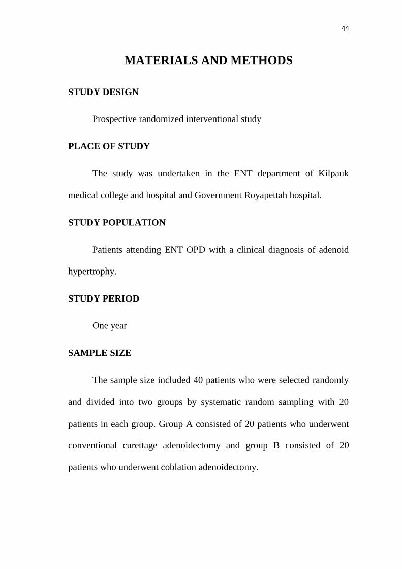
MATERIALS AND METHODS
STUDY DESIGN
Prospective randomized interventional study
PLACE OF STUDY
The study was undertaken in the ENT department of Kilpauk
medical college and hospital and Government Royapettah hospital.
STUDY POPULATION
Patients attending ENT OPD with a clinical diagnosis of adenoid
hypertrophy.
STUDY PERIOD
One year
SAMPLE SIZE
The sample size included 40 patients who were selected randomly
and divided into two groups by systematic random sampling with 20
patients in each group. Group A consisted of 20 patients who underwent
conventional curettage adenoidectomy and group B consisted of 20
patients who underwent coblation adenoidectomy.
45
SAMPLING and ALLOCATION METHOD
Sampling was done by systematic random sampling. An
independent researcher allocated interventions through sequentially
numbered sealed envelopes marked according to the allocation schedule
generated by computer.
INCLUSION CRITERIA
o Age group of 5-15 years with symptoms of adenoid hypertrophy
such as snoring, mouth breathing, earache.
o Adenoid hypertrophy confirmed by radiological investigation and
diagnostic nasal endoscopy.
EXCLUSION CRITERIA
o Previous history of surgery for adenoidectomy
o Bleeding disorders
o Cases with cleft palate or previous history of cleft palate repair
o Neuromuscular/craniofacial anomalies
o DOWN‟S syndrome patients
Ethical committee approval : obtained
Consent : Informed written consent obtained
Financial support : Nil
Conflict of interest : Nil

46
METHODOLOGY
After obtaining institutional ethical committee approval, patients
attending the ENT OPD with complaints of snoring, mouth breathing and
ear block were randomly selected. The patients selected for the study
were those in the age group of 5-15 years and who had symptoms such as
snoring, mouth breathing and ear ache. They were then subjected to
radiological investigation and diagnostic nasal endoscopy for the
confirmation of adenoid hypertrophy.
Patients with history of previous surgery for adenoidectomy,
bleeding disorders, cleft palate, neuromuscular disorders, craniofacial
abnormalities and Down‟s syndrome were excluded from the study.
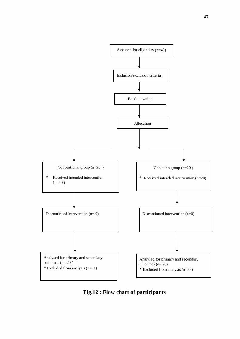
Of those who satisfied the inclusion criteria of the study,40 patients
were selected and were divided into two groups (group A and group B)
by systematic random sampling. Group A consisted of 20 patients who
underwent conventional curettage adenoidectomy and group B consisted
of 20 patients who underwent endoscopic assisted coblation
adenoidectomy. The flow chart illustrating the stages of the study is
shown in Fig 12.
47
Fig.12 : Flow chart of participants
Assessed for eligibility (n=40)
Inclusion/exclusion criteria
Analysed for primary and secondary
outcomes (n= 20 )
* Excluded from analysis (n= 0 )
Discontinued intervention (n= 0)
Conventional group (n=20 )
* Received intended intervention
(n=20 )
Discontinued intervention (n=0)
Coblation group (n=20 )
* Received intended intervention (n=20)
Analysed for primary and secondary
outcomes (n= 20)
* Excluded from analysis (n= 0 )
Randomization
Allocation

48
PREOPERATIVE EVALUATION
The selected patients were radiologically investigated in the
preoperative period. X-Ray soft tissue neck lateral view was done to
confirm adenoid enlargement. Diagnostic nasal endoscopy was done
preoperatively for grading the adenoid enlargement.
The grading was according to the scale given by Clemens and
McMurray.
Grade 1 adenoid tissue filling 1/3 of the vertical height of choana
Grade 2 adenoid tissue filling upto 2/3 of the vertical height of
choana
Grade 3 adenoid tissue filling 2/3 to nearly all but not complete
filling of the choana
Grade 4complete choanal obstruction
SURGICAL TECHNIQUES
CONVENTIONAL CURETTAGE ADENOIDECTOMY:
Under general anaesthesia with orotracheal intubation with the
patient in Rose position, Boyle Davis mouth gag was applied and
retracted. Adenoids were palpated digitally. Upon palpation, adenoids
were curetted using St. Clair Thompson adenoid curette with cage. Then
49
curettage was done using adenoid curette without cage. Nasopharynx was
packed with gauze.
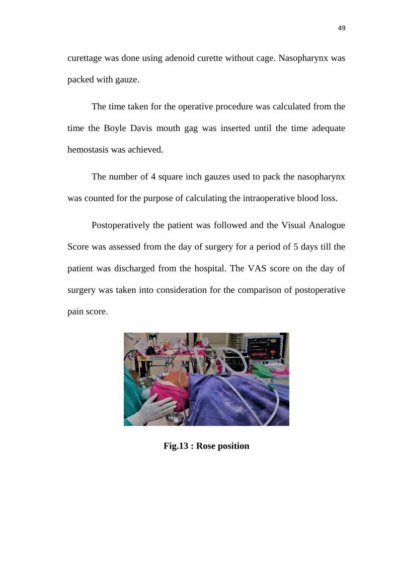
The time taken for the operative procedure was calculated from the
time the Boyle Davis mouth gag was inserted until the time adequate
hemostasis was achieved.
The number of 4 square inch gauzes used to pack the nasopharynx
was counted for the purpose of calculating the intraoperative blood loss.
Postoperatively the patient was followed and the Visual Analogue
Score was assessed from the day of surgery for a period of 5 days till the
patient was discharged from the hospital. The VAS score on the day of
surgery was taken into consideration for the comparison of postoperative
pain score.
Fig.13 : Rose position

50
Fig.14 : Curettage adenoidectomy
Fig.15 : Adenoid tissue after removal
Fig.16 :St. Clair Thompson adenoid Fig 17: Adenoid curette without
curette. cage.
51
ENDOSCOPIC ASSISTED COBLATION ADENOIDECTOMY
Under general anaesthesia with orotracheal intubation and with the
patient in supine position, Boyle Davis mouth gag was applied and
retracted. Disposable nasal catheters were introduced through both the
nostrils and were brought through the mouth in order to retract the soft
palate superiorly.
Under endoscopic visualization using a 70 degree Hopkin‟s rod
endoscope, the coblation PROCISE MAX wand was used to coblate the
adenoid tissue. As the procedure involved visualization of the adenoids
using an endoscope, the extent of tissue as well as the intraoperative
bleeding was assessed easily. The coblation system included suction and
irrigation thereby providing a clear surgical field. The coblation wand for
adenoidectomy has a flat tissue configuration for fast tissue ablation. The
built in suction effectively clears away debris without risk of clogging.
The shaft can be bended which provides easy access to the choanae
during adenoidectomy. The adenoid wand is shown in fig: 15.The number
of gauze pieces used to pack the nasopharynx was counted. The number
of blood soaked gauze pieces along with the amount of blood in the
suction apparatus, subtracting the amount of irrigating fluid used was
taken into account for the calculation of intraoperative blood loss.
Postoperative pain was assessed using the VAS scale.

52
Fig.18 : coblation adenoid wand.
Fig.19 : Endoscopic coblation adenoidectomy

53
Fig.20 : At the end of the procedure
The comparison of the two groups was done on the basis of:
PRIMARY OUTCOMES
1. Completeness of removal
2. Intraoperative blood loss
SECONDARY OUTCOMES
1. Operative duration
2. Postoperative pain
3. Recovery time
1. COMPLETENESS OF REMOVAL
Preoperatively, the patients were subjected to diagnostic nasal
endoscopy and the adenoids were graded according to Clemens and
McMurray scale of adenoid grading.

54
Postoperatively, diagnostic nasal endoscopy was done at the time
of discharge and one month later and was compared with the preoperative
adenoid grading.
Postoperatively, diagnostic nasal endoscopy showing less than
20% of adenoid tissue was considered as complete removal.
2. INTRAOPERATIVE BLOOD LOSS
At the time of surgery, intraoperative blood loss was calculated by
counting the number of 4 square inch gauze pieces used for packing
thenasopharynx. Each fully soaked gauze piece was assumed to
correspond to a blood loss of10 ml. The number of blood soaked gauze
pieces along with the amount of blood in the suction apparatus,
subtracting the amount of irrigating fluid used was taken into account for
the calculation of intraoperative blood loss.
3. OPERATIVE DURATION
Operative duration was calculated from the time the Boyle Davis
mouth gag was inserted until the time adequate hemostasis was achieved.
4. POSTOPERATIVE PAIN
Postoperative pain was analysed using the Visual Analog Scale
(VAS) and the scores were observed over a period of 5 days. The VAS
55
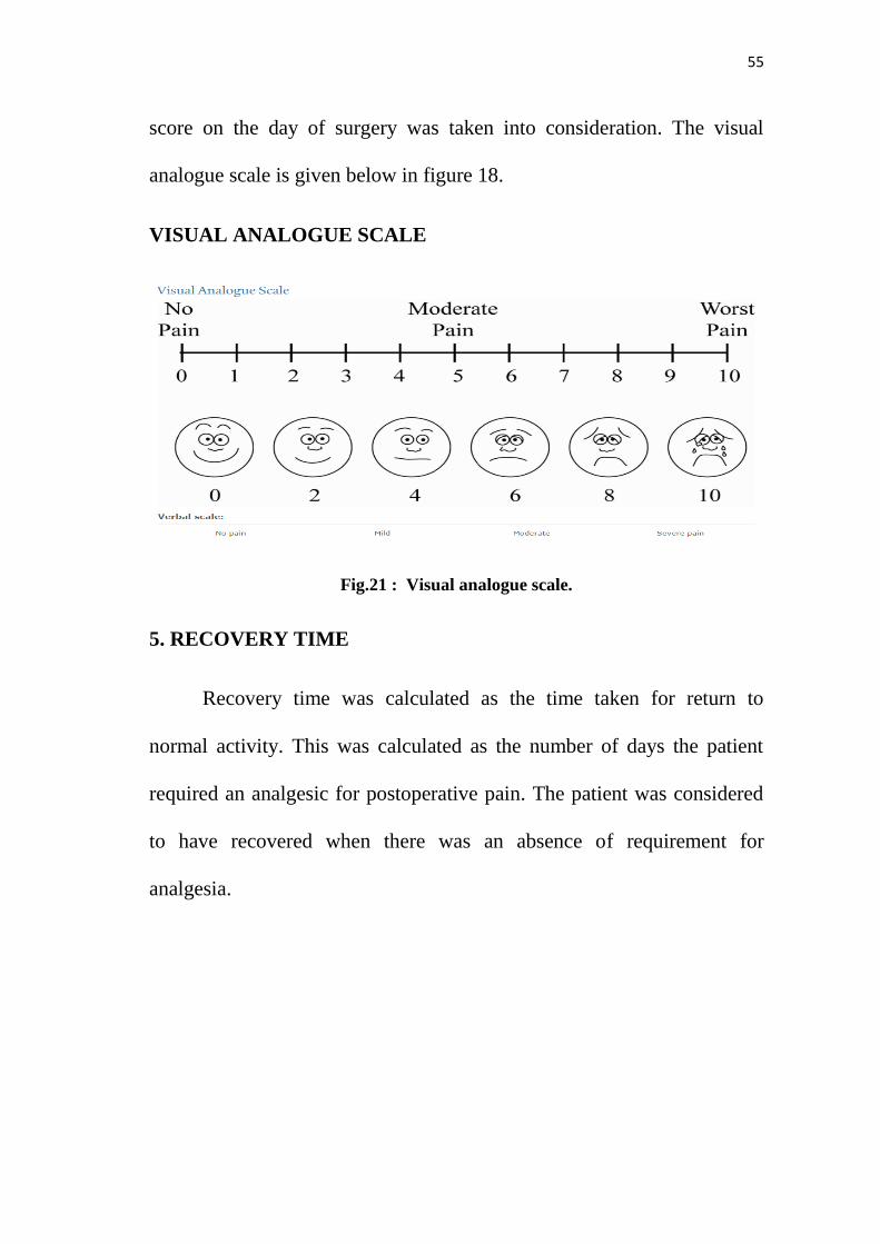
score on the day of surgery was taken into consideration. The visual
analogue scale is given below in figure 18.
VISUAL ANALOGUE SCALE
Fig.21 : Visual analogue scale.
5. RECOVERY TIME
Recovery time was calculated as the time taken for return to
normal activity. This was calculated as the number of days the patient
required an analgesic for postoperative pain. The patient was considered
to have recovered when there was an absence of requirement for
analgesia.

56
STATISTICAL ANALYSIS
Statistical analyses were carried out using SPSS for Windows
version 15.0. Primary outcome measure (completeness of removal) was
compared using Fischer‟s exact test and Primary outcome measure
(intraoperative blood loss) and Secondary outcome measures (operative
duration, postoperative pain score, recovery time) was compared using
student‟s t-test and results are expressed as mean and standard deviation.
To have a power of 80 % to detect 25% difference in the proportion with
respect to the primary outcome(completeness of removal) between the
two groups with an accepted type I error of 0.05 and type II error of 0.20,
a sample size of 20 patients was required in each group.

57
RESULTS
A total of 43 patients were evaluated, out of which 3 patients were
excluded because they had a history of previous surgery for
adenoidectomy. A total of 40 patients were taken up for the study who
were then divided into two groups, group A and group B with 20 in each
group.
Patients in group A underwent adenoidectomy by the conventional
curettage method. Those in group B underwent adenoidectomy by the
endoscopic assisted coblation method.
Age distribution
All the patients included in the study were between 5 and 15 years
of age. The mean and SD in group A was 10.33 ± 3.18 and in group B
was 9.69 ± 1.93. The youngest in group A and B was 6 years of age. The
oldest patient in group A was 15 years and in group B was 13 years. The
P value was 0.089 which was not statistically significant, implying that
the group were comparable to each other in terms of age distribution
which is given in Fig 19 and table 2.

58
Fig.19 : Age characteristics.
AGE CHARACTERISTICS:
GROUP A
GROUP B
P-VALUE
AGE
CHARACTERISTICS
10.33 ± 3.18
9.69 ± 1.93
0.089
Table – 2 : Age characteristics.*All the values are expressed in mean ±SD.
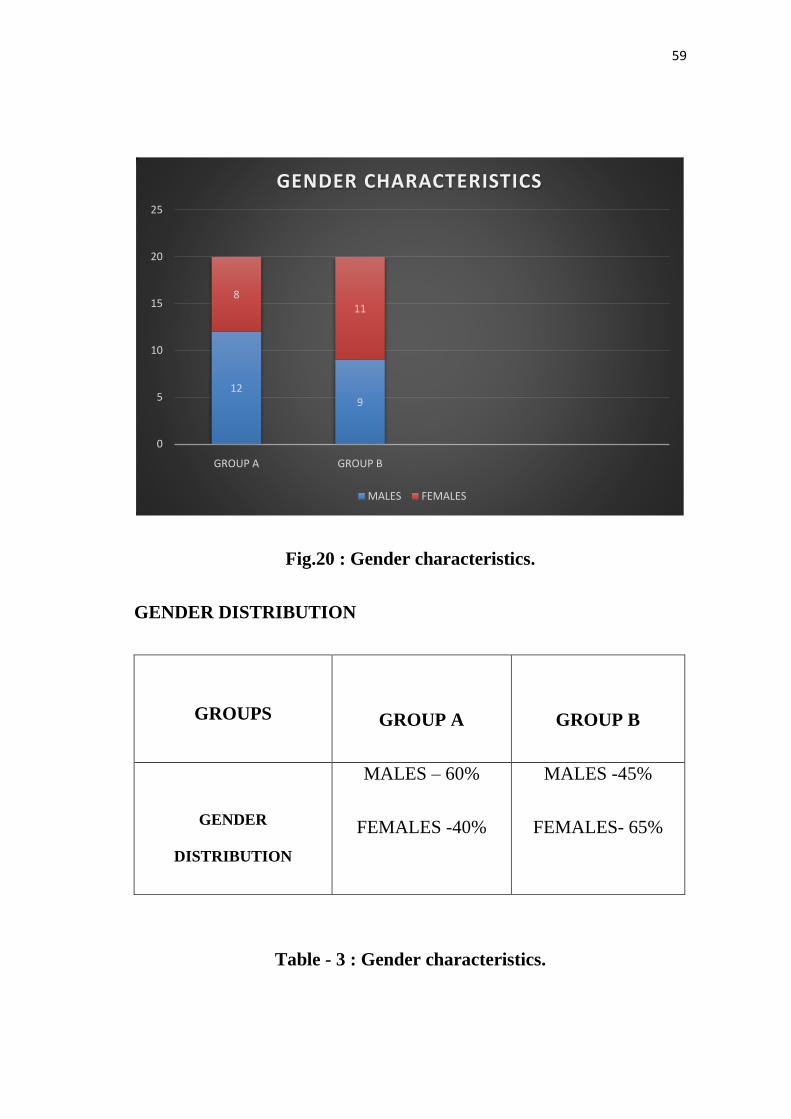
Gender distribution:
The gender distribution in both the groups was as follows:-12 male
children were included in group A and 9 male children in group B. Group
A had 8 female children and group B had 11 female children. Gender
characteristics are given in Fig 20 and table 3.
10.339.69
0
2
4
6
8
10
12
14
AGE
AGE CHARACTERISTICS
GROUP A GROUP B
59
Fig.20 : Gender characteristics.
GENDER DISTRIBUTION
GROUPS
GROUP A
GROUP B
GENDER
DISTRIBUTION
MALES – 60%
FEMALES -40%
MALES -45%
FEMALES- 65%
Table - 3 : Gender characteristics.
129
811
0
5
10
15
20
25
GROUP A GROUP B
GENDER CHARACTERISTICS
MALES FEMALES

60
Preoperative adenoid grading
The patients included in both the groups were subjected to
endoscopic evaluation for grading the adenoid hypertrophy. The grading
was done according to Clemens and Mc Murray scale.
3 patients in group B presented with grade 1 while none in group A
had grade 1. Preoperatively grade 2 was seen in 15 patients in group A
and 14 patients in group B. 5 patients in group A and 3 patients in group
B had grade 3. This analysis is given in table 5.
Thus 75% in group A and 70% in group B had grade 2. The mean
and SD were 2 ± 0.44 in group A and 2 ± 0.56 in group B. The P value
was 0.126. This is given in table 4 and fig 21.
PREOPERATIVE ADENOID GRADING:
GROUP A GROUP B
P-
VALUE
PREOPERATIVE
ADENOID GRADE
2 ± 0.44 2 ± 0.56 0.126
Table.4 : Preoperative adenoid grade.*all the values are expressed as
mean ± SD

61
Fig.21 : Preoperative adenoid grading.
Table - 5 : PREOPERATIVE ADENOID GRADING
GROUPS PERCENTAGE GROUP A GROUP B
GRADE 1
COUNT
PERCENTAGE
0
0
3
15%
GRADE 2
COUNT
PERCENTAGE
15
75%
14
70%
GRADE 3
COUNT
PERCENTAGE
5
25%
3
15%
GRADE 4
COUNT
PERCENTAGE
0
0
0
0
2 2
0
0.5
1
1.5
2
2.5
PREOP GRADE
PREOPERATIVE GRADE
GROUP A GROUP B
62
Intraoperative blood loss
Both the surgical techniques included in the study had a certain
amount of blood loss during the procedure. This parameter was compared
between the two groups.
The amount of blood lost during the surgical procedure was
calculated by counting the number of 4 square inch gauze pieces used for
packing the nasopharynx. To it was added the amount of blood in the
suction apparatus. Each fully soaked gauze piece was considered to
correspond to a blood loss of 10 ml.
In the conventional group none of the patients had a blood loss of
less than 10 ml. 5% of the 20 patients had blood loss in the range of 11-
20 ml. About 20 -30 ml was the range of blood loss in 50% of patients.
45% of the patients had blood loss between 30 – 40 ml.
In the coblation group the amount of intraoperative blood loss was
calculated by counting the number of 4 square inch gauze pieces used for
packing the nasopharynx. Each fully soaked gauze piece was counted as a
blood loss of 10 ml. If partially soaked, it was considered to correspond
to 5ml of blood loss.

63
To the amount of blood loss calculated by counting the gauze
pieces, the amount of blood in the suction apparatus was added after
subtracting the amount of fluid used for irrigation.
In the coblation group 2 out of 20 patients had a blood loss of 5 ml
contributing 10%. 13 out of the 20 patients had a blood loss in the range
of 5-10 ml contributing to 65%. 5 patients had blood loss in the range of
11-20 ml contributing to 25% which is illustrated in table 6 and fig 22.
The mean and SD were 31 ± 5.52 and 10.75 ± 2.93 in groups A and B
respectively (p - 0.0001) which is given in table 7.
INTRAOPERATIVE BLOOD LOSS
BLOOD LOSS IN ML GROUP A GROUP B
≤ 5 ML 0 10%
5-10 0 65%
11-20 5% 25%
21-30 50% 0
31-40 45% 0
Table – 6 : Intraoperative blood loss.

64
Fig.22: Intraoperative blood loss.
INTRAOPERATIVE BLOOD LOSS:
GROUPS GROUP A GROUP B P- VALUE
BLOOD LOSS
31 ± 3.23
10.75 ± 2.93
0.0001
Table – 7 : Intraoperative blood loss.*all the values are expressed in
mean ± SD
31
10.75
GROUP A
GROUP B
0 5 10 15 20 25 30 35
INTRAOPERATIVE BLOOD LOSS(ml)
GROUP A GROUP B
65
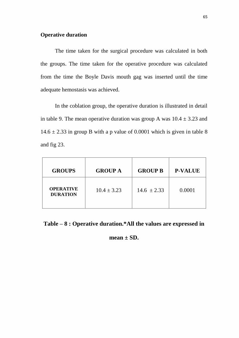
Operative duration
The time taken for the surgical procedure was calculated in both
the groups. The time taken for the operative procedure was calculated
from the time the Boyle Davis mouth gag was inserted until the time
adequate hemostasis was achieved.
In the coblation group, the operative duration is illustrated in detail
in table 9. The mean operative duration was group A was 10.4 ± 3.23 and
14.6 ± 2.33 in group B with a p value of 0.0001 which is given in table 8
and fig 23.
GROUPS
GROUP A
GROUP B
P-VALUE
OPERATIVE
DURATION
10.4 ± 3.23
14.6 ± 2.33
0.0001
Table – 8 : Operative duration.*All the values are expressed in
mean ± SD.

66
Fig.23 : Operative duration.
OPERATIVE DURATION
TIME IN MINUTES GROUP A GROUP B
<5
0 0
5-10 65% 5%
11-15 35% 75%
16-20 0 20%
Table – 9 : Operative duration.
0
5
10
15
20
25
OPERATIVE DURATION(minutes)
GROUP A GROUP B

67
Completeness of removal
A postoperative endoscopic evaluation was done in both the groups
in order to assess the completeness of removal after the surgical
procedure. The patients were followed up for a month and the adenoid
grading at the end of one month was taken into consideration for
comparison between the two groups.
Postoperative adenoid tissue filling < 20% of the vertical height of
the choana was considered as complete removal.
3 patients in group A showed complete removal of adenoids
following the procedure with <20% of residual adenoid tissue detected in
the postoperative period. The remaining 17 patients in group A showed
partial removal of adenoids in the postoperative endoscopic evaluation
with either grade 2 or grade 3 adenoids.
Conversely in group B, 15 patients showed complete removal of
adenoids demonstrating adenoid tissue occupying less than 20 percent of
the vertical height of choana. Only 5 patients in group B showed partial
removal, with remnant adenoid tissue occupying more than 20 percent of
the vertical height of choana.
On comparison of the two groups with respect to this parameter,
coblation adenoidectomy showed a greater percentage of complete
removal of adenoid tissue when compared to conventional
adenoidectomy(p – 0.0003) which is shown in table 10 and fig 24.

68
Fig.24 : Completeness of removal.
COMPLETENESS OF REMOVAL
COMPLETENESS OF
REMOVAL
GROUP A
GROUP B
COMPLETE REMOVAL
15%
75%
PARTIAL REMOVAL
85%
25%
Table – 10 : Completeness of removal.
15%
85%
75%
25%
0%
10%
20%
30%
40%
50%
60%
70%
80%
90%
100%
COMPLETE PARTIAL
COMPLETENESS OF REMOVAL
GROUP A GROUP B

69
Postoperative pain
Following the procedure, the postoperative pain score was assessed
using the visual analogue scale for a period of 5 days. However the
postoperative pain score on day 0, i.e the day of surgery was taken into
consideration for comparison between the two groups.
80% of patients in the conventional adenoidectomy group had a
pain score of 4, while 85 % of patients in the coblation group had a pain
score of 3. One patient in group A had a pain score of 5 on the day of
surgery. The median VAS score for group A was 4 ± 0.44 and for group
B was 3 ± 0.36 (p - 0.0001).This is illustrated in table 11 and fig 25 and
postoperative pain score on POD – 0 is shown in table 12.
GROUPS
GROUP A
GROUP B
P-VALUE
PAIN SCORE
4 ± 0.44
3 ±0.36
0.0001
Table – 11 : Postoperative VAS score.

70
Fig.25 : Pain score
POSTOPERATIVE PAIN
SCORE ON POD - 0
GROUP A
GROUP B
VAS SCORE 1 0 0
VAS SCORE 2 0 0
VAS SCORE 3 15% 85%
VAS SCORE 4 80% 15%
VAS SCORE 5 5% 0
Table – 12 : Postoperative pain score.
0
1
2
3
4
5
6
0 5 10 15 20 25
PAIN SCORE(VAS)
GROUP A GROUP B

71
Recovery period
Recovery period was considered as the time required for the patient
toreturn to normal activity following surgery. It was calculated as the
number of analgesia requirement days. 60 % of patients in group A
required analgesia for 3 postoperative days. Hence the recovery period
for this 60% of patients was taken as 3 postoperative days. 30% of
patients in group A had a recovery period of 4 postoperative days.
60% of patients in group B (coblation adenoidectomy group) had a
recovery period of 3 postoperative days and 40% had a recovery period of
2 postoperative days which is shown in table 14.
The mean recovery period in group A was 3.15 ± 0.61 days and
2.65 ± 0.63 days for group B (P - 0.001) shown in table 13 and fig 26.The
coblation group was found to recover earlier than the conventional group.
GROUPS
GROUP A
GROUP B
P-VALUE
RECOVERY
DAYS
3.15 ±0.61
2.65 ± 0.63
0.0017
Table – 13 : Recovery days.*All values are expressed in mean ± SD

72
Fig.26 : Recovery days.
RECOVERY PERIOD
ANALGESIA
REQUIREMENT DAYS
GROUP A GROUP B
1 DAY 0 40%
2 DAYS 10% 60%
3 DAYS 60% 0
4 DAYS 30% 0
5 DAYS 0 0
Table – 14 : Recovery days.
0
1
2
3
4
5
RECOVERY DAYS
GROUP A GROUP B

73
DISCUSSION
Adenoidectomy is one of the commonly performed surgeries in
children. Various techniques have been proposed in order to reduce the
amount of bleeding during the procedure and to facilitate the easy and
safe removal of adenoid tissue. Adenoidectomy can be done using an
adenoid curette, bipolar cautery, power assisted microdebrider and the
coblator. Though there are many options emphasis should be laid on the
efficacy technique and the postoperative outcome. Inspite of the
numerous options available, it has been noted that the recurrence rates
following adenoidectomy are very high. These are attributed to certain
factors like difficult access of adenoids and the non-visualization of
adenoids during removal.
The use of an endoscope during adenoidectomy has the advantage
of visualizing the adenoid tissue as well as the surrounding structures.
Direct visualization enables a better removal of the whole adenoid tissue
without injuring the other structures in the vicinity. Coblation is a non
heat driven process of soft tissue dissolution using bipolar radiofrequency
energy under a conductive medium like normal saline. Plasma not only
has the physical effects of cutting and coagulation but it also
decontaminates the surgical wound thereby facilitating better wound

74
healing. This non thermal dissolution technique produces very minimal
bleeding. The length of the wand combined with the use of the endoscope
gives access to the whole nasopharynx so that all the adenoid tissue can
be coblated without fear of injuring the adjacent structures. Endoscopic
assisted coblation adenoidectomy ensures the complete removal of
adenoids with less injury to the surrounding structures and minimal
bleeding. This in turn decreases the postoperative pain hence facilitating
earlier recovery. However the disadvantages of coblation technique
include a longer learning curve which requires considerable skill and
expertise, increased duration of coblation technique due to various factors
and the cost factor of the equipment.
Murat Songuand colleagues compared endoscopic assisted
adenoidectomy with curettage adenoidectomy in 38 patients in the age
group of 8-12 years in their study. The children were randomly divided
into 2 groups and the parameters evaluated were nasopharyngoscopy,
symptom improvement scale, midsagittal reformatted images of
computed tomography of temporal bone, blood loss and operative time.
The evaluation of computerized tomographies of temporal bone by
adenoidal divided by nasopharyngeal ratios revealed a mean ratio of 0.41
in the curettage group and 0.30 in the endoscopic adenoid group. They
demonstrated that endoscopic adenoidectomy was superior to curettage

75
adenoidectomy in terms of completeness of removal. In our study,
postoperative grading of adenoids was done at the end of 1 month post
surgery. Only 15% in the conventional group showed complete removal
of adenoids whereas 75% in the endoscopic coblation group showed
complete removal of adenoids (P - 0.0003) showing the superiority of
endoscopic assisted coblation adenoidectomy compared to conventional
curettage adenoidectomy in terms of completeness of removal.33
The results of our study with respect to removal of adenoid tissue
were similar to a study done by Ismail Elnashar and colleagues. They
studied the effectiveness of endoscopic assisted adenoidectomy by
measuring the volume of adenoid tissue removed after blind curettage and
endoscopy assisted adenoidectomy. The volume of adenoid removed by
curettage adenoidectomy ranged from 1 to 3.6 ml with a mean of 2.45 ml.
The volume of adenoid tissue removed post curettage adenoidectomy
ranged from 0 - 2.9 ml (mean 0.67 ± 0.58ml). They concluded that
conventional curettage adenoidectomy left a substantial volume of
adenoid tissue showing the advantage of endoscopic assisted
adenoidectomy over curettage technique.34
Guo Xiao and colleagues compared traditional adenoidectomy with
coblation adenoidectomy in 54 children. They divided the children into
two groups, the coblation group and the control /cold curettage group.
76
The parameters compared were the operative time, intraoperative
bleeding, postoperative pain scale, postoperative pain duration and cure
rates. They demonstrated that the coblation group showed increased
operative time, less intraoperative bleeding with less postoperative pain
with a shorter duration of postoperative pain. Our results were similar to
this study. The mean operative duration were 10.4 ± 3.23 minutes for
group A and 14.6 ± 2.33 minutes for group B(p - 0.0001) showing an
increased intraoperative duration with the coblation group. The mean
intraoperative bleeding was less in the coblation group. The mean
intraoperative bleeding were 31 ± 5.52 ml for group A and 10.75 ± 2.93
ml for group B (p - 0.0001). The median VAS score in group A was 4 ±
0.44 and 3 ± 0.36 in group B (p - 0.0001) indicating lesser postoperative
pain in coblation group compared with the conventional group. The mean
duration of postoperative pain was less in the coblation group compared
to the conventional group. The mean duration of postoperative pain in the
conventional group was 3.15 ± 0.61 days whereas in the coblation group
it was 2.65 ± 0.63 days (p - 0.0017).
Abd El Rahman El Tahan and colleagues compared the coblation
technique with standard conventional curettage technique with end points
as operative time and intraoperative blood loss as primary outcomes. The
patients were asked to record their pain and discomfort on standardized

77
Wong Baker Faces pain rating scale from 0 (no pain) to 10 (severe pain).
The postoperative complications and the recurrences were also recorded.
In their study, the conventional group showed less operative time whereas
the coblation group showed less intraoperative blood loss, less
postoperative bleeding and adenoid recurrence. However the two groups
showed insignificant difference in postoperative pain. The results of our
study were similar to the above study by El Rahman and colleagues with
the coblation group showing less intraoperative blood loss and superior
completeness of removal but with a longer operative time.35
Liyun yang and colleagues conducted a meta analysis comparing
endoscopic assisted coblation adenoidectomy and conventional curettage
adenoidectomy. The parameters compared were operative effectiveness,
total operative time, blood loss and complications. There were 7 studies
which fitted in the inclusion criteria, out of which 331 patients were
treated with endoscopic assisted coblation adenoidectomy and 251
patients with conventional curettage adenoidectomy. The results showed
that the endoscopic method had a shorter operative time (SMD-1.09,
95%CI -1.29-.90. P< 0.00001), less blood loss (SMD-19.74, 95%CI -
22.75-16.73, P<0.00001) and fewer complications (0.15,95% CI, 0.07-
0.35), P<0.00001.They concluded that the endoscopic method was
advantageous over the conventional curettage method. The results of our

78
study were similar to this study with regards to less intraoperative blood
loss and fewer complications in the coblation group.36
Michael S Timms and colleagues compared the use of coblator
with the conventional and other newer methods of adenoidectomy. They
concluded that coblator use showed a relatively bloodless field with
precision of tissue removal and less damage to the surrounding structures
which was similar to the results of our study.37
Our study in similarity to the above quoted studies provides
evidence that the use of an endoscope is superior to the conventionally
practised curettage method of adenoidectomy when completeness of
adenoid removal is considered. Moreover the curettage method being a
blind technique has an inherent risk of injury to the surrounding
structures whenever an enthusiastic effort may be undertaken to ensure
completeness of removal. A fear of injuring the associated structures may
itself be the cause for a partial or an incomplete removal in case of the
traditional method. Whereas the use of an endoscope offers a good
visualization of the entire nasopharynx and hence careful removal can be
done upon visualization.
Secondly on considering the blood lost during the procedure, the
coblation technique proves to be superior to the conventional method.

79
The plasma released as a result of passage of electric current through the
conducting fluid breaks down the molecular bonds causing disintegration
of tissue molecule by molecule. This technique produces negligible
amount of bleeding.
Similarly the coblation method showed a lesser postoperative pain
score and hence the patients in this group recovered early.

80
LIMITATIONS OF THE STUDY
1. The improvement of symptoms following the procedures was not
compared postoperatively which can make a significant
contribution while comparing the efficacy of the two techniques.
2. Though the completeness of removal was assessed endoscopically
after 1 month in the postoperative period, the patient was not
followed up for parameters such as recurrence and postoperative
hemorrhage (secondary/reactionary).
3. The postoperative pain score was assessed by VAS score which
can cause judgmental bias due to subjective assessment.
4. Lastly, the surgeon‟s skill may be a potential confounding factor
while comparing the two techniques.

81
CONCLUSION
The endoscopic assisted coblation adenoidectomy is superior to the
conventional method in terms of
Completeness of adenoid removal
Lessintraoperative blood loss
Less postoperative pain
Shorter period of time for recovery.
However it is more time consuming when compared to the
conventional adenoidectomy method.

82
BIBLIOGRAPHY
1. Karanov, J & Minić, Predrag & Subarević, Vladan & Baljosevic,
Ivan. (2000). [Cor pulmonale caused by hypertrophic adenoid
glands and tonsils: indications for tonsillectomy and
adenoidectomy in a 2-year-old child]. 128. 208-10.
2. Diseases of Ear, Nose and throat and head and neck surgery-sixth
edition-2014.
3. Cummings otolaryngology head and neck surgery fifth edition,
2010
4. Scott-Brown Otolaryngology, seventh edition, 1997 and Manual of
middle ear surgery.
5. Dilesh M. Adenoidectomy- A Historical Review. Glob J Oto 2017;
8(3): 555740. DOI:10.19080/GJO.2017.08.555740.
6. Operative Techniques In Otolaryngology--Head And Neck
Surgery, Vol 12, No 4 (Dec), 2001: PP 199-203
7. Thiagarjan Balasubramanian.Use of Coblation Technology In
Otolaryngology ,Otolaryngology online.

83
8. Di Rienzo Businco, L. et al. “Paediatric Adenoidectomy:
Endoscopic Coblation Technique Compared to Cold Curettage.”
Acta Otorhinolaryngologica Italica 32.2 (2012): 124–129.
9. Kim J-W, Kim HJ, Lee WH, Kim D-K, Kim SW, Kim YH, et al.
(2015) Comparative Study for Efficacy and Safety of
Adenoidectomy according to the Surgical Method: A Prospective
Multicenter Study. PLoS ONE 10(8): e0135304.
10. Özkiriş M, Karaçavuş S, Kapusuz Z, Saydam L. Comparison of
two different adenoidectomy techniques with special emphasize on
postoperative nasal mucociliary clearance rates: coblation
technique vs. cold curettage. Int J Pediatr Otorhinolaryngol. 2013;
77: 389–393. pmid:23266160.
11. Jonas NE, Sayed R, Prescott CA. Prospective, randomized, single-
blind, controlled study to compare two methods of performing
adenoidectomy. Int J Pediatr Otorhinolaryngol. 2007;71(10):1555–
1562.
12. Ark, Nebil & Kurtaran, Hanifi & Ugur, Kadriye & Yilmaz, Turker
& Altug Ozboduroglu, Akin & Mutlu, Cemil. (2010). Comparison
of adenoidectomy methods: Examining with digital palpation vs.
visualizing the placement of the curette. International journal of
84
pediatric otorhinolaryngology. 74. 649-51. 10.1016 /j.ijporl.
2010.03.012.
13. Paramasivan, Vijaya Krishnan et al. Randomised comparative
study of adenotonsillectomy by conventional and coblation method
for children with obstructive sleep apnoea. International Journal of
Pediatric Otorhinolaryngology, Volume 76, Issue 6, 816 – 82.
14. Shapiro, N. L. and Bhattacharyya, N. (2007), Cold Dissection
Versus Coblation-Assisted Adenotonsillectomy in Children. The
Laryngoscope, 117: 406–410.
15. Benninger M, Walner D. Coblation: improving outcomes for
children following adenotonsillectomy. Clin Cornerstone. 2007
16. Stanislaw P, Jr, Koltai PJ, Feustel PJ. Comparison of power-
assisted adenoidectomy vs adenoid curette adenoidectomy. Arch
Otolaryngol Head Neck Surg. 2000;126(7):845–849.
17. Elluru, R. G., Johnson, L. and Myer, C. M. (2002), Electrocautery
Adenoidectomy Compared With Curettage and Power-Assisted
Methods. The Laryngoscope, 112: 23–25.
18. Clemens J, McMurray JS, Willging JP. Electrocautery versus
curette adenoidectomy: comparison of postoperative results. Int J
Pediatr Otorhinolaryngol 1998;43:115–22.

85
19. Wong L, Moxham JP, Ludemann JP. Electrosurgical adenoid
ablation. J Otolaryngol 2004;33:104–6.
20. Somani SS, Naik CS, Bangad SV. Endoscopic adenoidectomy with
microdebrider. Indian J Otolaryngol Head Neck Surg.
2010;62(4):427–431. doi: 10.1007/s12070-011-0118-9.
21. Pagella F, Pusateri A, Canzi P, Caputo M, Marseglia A, Pelizzo G,
Matti E. The evolution of the adenoidectomy: analysis of different
power-assisted techniques. Int J Immunopathol Pharmacol. 2011
Oct;24(4 Suppl):55-9. Review. PubMed PMID: 22032788.
22. Pagella F, Mati E, Colombo A, et al. How we do it: A combined
method of traditional curette and power-assisted endoscopic
adenoidectomy. Acta Otolaryngol. 2009;129:556–59
23. Costantini F, Salamanca F, Amaina T. Videoendoscopic
adenoidectomy with microdebrider. Acta Otorhinolaryngol Ital.
2008;28(1):26–29
24. Al-Mazrou KA, Al-Qahtani A, Al-Fayez AI. Effectiveness of
transnasal endoscopic powered adenoidectomy in patients with
choanal adenoids. Int J Pediatr Otorhinolaryngol.
2009;73(12):1650–1652.

86
25. Havas T, Lowinger D. Obstructive adenoid tissue: an indication for
powered-shaver adenoidectomy. Arch Otolaryngol Head Neck
Surg. 2002;128(7):789–791. doi: 10.1001/archotol.128.7.789.
26. Datta R, Singh V, Deshpal. Conventional Versus Endoscopic
Powered Adenoidectomy: A Comparative Study. Medical Journal,
Armed Forces India. 2009;65(4):308-312.
27. Das AT, Prakash SB, Priyadarshini V. Combined Conventional
and Endoscopic Microdebrider-Assisted Adenoidectomy: A
Tertiary Centre Experience. Journal of Clinical and Diagnostic
Research: JCDR. 2017;11(2):MC05-MC07.
28. Feng YH, Ying SK. Comparison on the effectiveness between
endoscopic assisted adenoidectomy and conventional curettage
adenoidectomy. J Clin Otorhinolaryngol. 2006;20(3):54–57
29. Uçar C (2008): Endoscopic adenoidectomy. Kulak Burun Bogaz
Ihtis Derg. MarApr;18(2):66-8.
30. Regmi D, Mathur NN, Bhattarai M. Rigid endoscopic evaluation of
conventional curettage adenoidectomy. J Laryngol Otol.
2011;125(1):53–58.
31. Wright ED, Manoukian JJ, Shapiro RS. Ablative adenoidectomy: a
new technique using simultaneous liquefaction/aspiration. J
Otolaryngol 1997;26:36–43.

87
32. Lowe D, Brown P, Yung M. Adenoidectomy technique in the
United Kingdom and postoperative hemorrhage. Otolaryngol Head
Neck Surg. 2011; 145: 314–318.
33. Songu M, Altay C, Adibelli ZH, Adibelli H. Endoscopicassisted
versus curettage adenoidectomy: a prospective, randomized,
double-blind study with objective outcome measures.
Laryngoscope. 2010;120:1895–1899.
34. Elnashar, Ismail et al. “Objective assessment of endoscopy assisted
adenoidectomy.” International journal of pediatric
otorhinolaryngology 78 8 (2014): 1239-42.
35. Yang L, Shan Y, Wang S, Cai C, Zhang H. Endoscopic assisted
adenoidectomy versus conventional curettage adenoidectomy: a
meta-analysis of randomized controlled trials. SpringerPlus.
2016;5:426.
36. Timms MS, Ghosh S, Roper A. Adenoidectomy with the coblator:
a logical extension of radiofrequency tonsillectomy. J Laryngol
Otol. 2005;119:398–399.
37. Anand V, Sarin V, Singh B. Changing trends in adenoidectomy.
Indian J Otolaryngol Head Neck Surg. 2014;66(4):375–380.
38. Jonas NE, Sayed R, Prescott CA. Prospective, randomized, single-
blind, controlled study to compare two methods of performing
adenoidectomy. Int J Pediatr Otorhinolaryngol. 2007; 71: 1555–
1562.
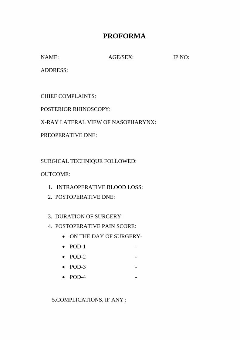
88
PROFORMA
NAME: AGE/SEX: IP NO:
ADDRESS:
CHIEF COMPLAINTS:
POSTERIOR RHINOSCOPY:
X-RAY LATERAL VIEW OF NASOPHARYNX:
PREOPERATIVE DNE:
SURGICAL TECHNIQUE FOLLOWED:
OUTCOME:
1. INTRAOPERATIVE BLOOD LOSS:
2. POSTOPERATIVE DNE:
3. DURATION OF SURGERY:
4. POSTOPERATIVE PAIN SCORE:
ON THE DAY OF SURGERY-
POD-1 -
POD-2 -
POD-3 -
POD-4 -
5.COMPLICATIONS, IF ANY :

89
PATIENT CONSENT FORM
STUDY DETAIL : A COMPARATIVE STUDY OF ENDOSCOPIC
COBLATION ADENOIDECTOMY AND CONVENTIONAL
CURETTAGE ADENOIDECTOMY
STUDY CENTRE: KMC AND GRH, CHENNAI.
PATIENT NAME: PATIENT AGE:
IDENTIFICATION NUMBER:
PATIENT TO TICK ( )THESE BOXES
I conform that I have understood the purpose of procedure for the above study.
I have the opportunity to ask the question and all my questions and doubts have been
answered to my satisfaction.
I understand that my participation in the study is voluntary and that I am free to
withdraw at anytime without giving any reasons, without my legal rights being
affected.
I understand that investigator, regulatory authorities and the ethics committee will not
need my permission to look at my health records both in respect to the current study
and any further research that may be conducted in relation to it, even if withdraw
from the study, I understand that my identity will not be revealed in any information
released to third parties or published, unless as required under the law. I agree not to
restrict the use of any data or results that arise from the study.
I agree to take part in the above study and to comply with the instructions given
during the study and faithfully cooperative with the study team and to immediately
inform the study staff if I suffer from any deterioration in my health or wellbeing or
any unexpected or unusual symptoms.
I hereby give consent to participate in this study.
I hereby give permission to undergo complete clinical examination and diagnostic
test.
Signature/Thumb impression: Place:
Date:
Patient/guardian name and address:
Signature of the investigator: Place:
Date:
Study investigator‟s name:
90

91
PATIENT INFORMATION FORM
TITLE OF THE STUDY: A comparative study of endoscopic
coblation adenoidectomy and conventional curettage adenoidectomy.
PLACE OF STUDY: KMC and GRH , Chennai.
The subjects who have consented to participate in the study will be
grouped into two groups and two different techniques of the surgical
procedure (adenoidectomy) will be undertaken.
The investigations done, the surgical procedure and the
postoperative status will be recorded and used for the study.
The subjects will be followed up for a period of one week after the
surgery.
The details collected will be documented and analysed.

92

93
MASTER CHART

94
NAME AGE SEX PREOPERATIVE
GRADE TECHNIQUE
BLOOD
LOSS
IN ML
TIME IN
MINUTES
POSTOPERATIVE
GRADE
PAIN
SCORE
RECOVERY
DAYS
Rahim basha 11 mch 2 conventional 35 7 0 3 4
Arthi 13 fch 2 conventional 35 10 1 4 4
Priya 14 fch 2 conventional 35 8 1 3 4
Jaisurya 12 mch 2 conventional 30 7 1 4 3
Manishkumar 10 mch 2 conventional 25 10 1 4 3
Kamesh 12 mch 2 conventional 30 8 1 4 3
Sugumar 14 mch 2 conventional 25 10 1 4 3
Jaswan singh 9 mch 2 conventional 20 10 1 4 3
Abinesh 13 mch 2 conventional 25 12 1 4 3
Kanimozhi 11 fch 3 conventional 30 15 1 4 3
Tamilnesan 17 mch 3 conventional 30 15 1 4 3
Bavatharani 5 fch 2 conventional 40 15 1 4 4
Guna 13 mch 2 conventional 25 15 1 4 2
Sangeetha 10 fch 2 conventional 35 15 1 4 3
Pavithra 5 fch 3 conventional 35 7 0 4 4
Karthick 12 mch 3 conventional 35 10 1 5 3
Divyadharshini 9 fch 2 conventional 25 5 0 4 4
Deepa 6 fch 2 conventional 35 7 1 4 3
Manikanda
prabhu 10 mch 2 conventional 30 10 1 3 2
Karthick 15 mch 3 conventional 40 12 1 4 3
Vidhyashri 13 fch 3 coblation 15 15 1 4 3
Naresh 12 mch 2 coblation 15 12 0 4 3
Sathish 11 mch 2 coblation 10 15 0 3 3
Swathi 12 fch 2 coblation 10 10 0 3 3
Trisha 11 fch 2 coblation 15 13 0 3 2
Sam 10 mch 2 coblation 10 12 0 3 2
Jeeva 11 mch 1 coblation 10 16 0 4 3
Rishi 7 mch 3 coblation 10 15 0 3 3
Rakshan 8 mch 2 coblation 10 15 1 3 3
Vinoth 6 mch 2 coblation 10 15 0 3 2
Rema 7 fch 2 coblation 15 18 0 3 2
Shiny 9 fch 1 coblation 10 14 0 3 3
Jemi 10 fch 2 coblation 5 20 1 3 3
Sathish 10 mch 2 coblation 10 14 1 3 2
Rahul 7 mch 1 coblation 10 15 0 3 2
Rachel 8 fch 3 coblation 10 18 0 3 3
Srisha 9 fch 2 coblation 10 15 0 3 2
Akshara 10 fch 2 coblation 10 15 0 3 3
Vidya 11 fch 2 coblation 5 12 0 3 2
Sangeetha 10 fch 2 coblation 15 13 1 3 3
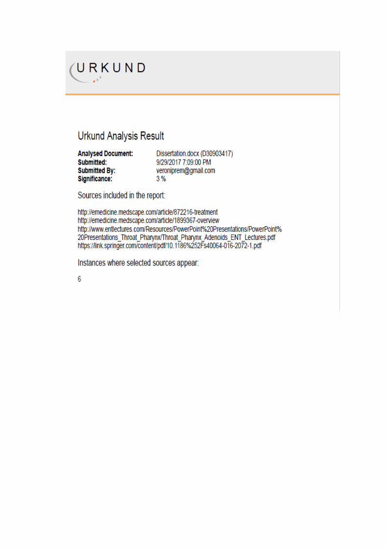
95
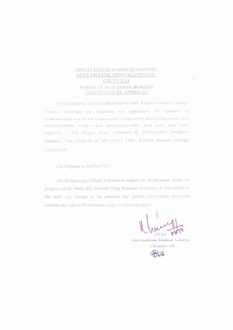
96