Des pathologies encéphaliques à connaître — Syndrome d’encéphalopathie postérieure réversible
Upload
khangminh22Category
view
2download
0
A COMPARATIVE STUDY OF VERTEBRAL PATHOLOGIES AND ANOMALIES IN TWO MEDIEVAL
BRITISH POPULATIONS
A thesis submitted in partial fulfilment of the requirements of Liverpool John Moores University for the degree of Master of
Philosophy
Clair Richardson
February 2018

1
I would like to dedicate this work to my grandfather Roy
1938 – 2015
“Do you not know that a man is not dead while his name is still spoken?”
– Terry Pratchett, Going Postal

2
Disclaimer: All of the photographic images used within this study were taken by
and are owned by the author of this thesis.

3
Abstract
A holistic approach to palaeopathological studies using historical
documentation and clinical, archaeological and epidemiological literature can
provide important information as to the health, lifestyles, socioeconomic and
occupational status of individuals from the past. Applying this approach, the study
provides an overview and comparative analysis of the spinal health of two
contemporaneous British skeletal samples from the medieval period; St Owen’s
Cemetery, an urban based population from Gloucester (n=68) and Poulton a rural,
agrarian community from Cheshire (n=70). Sex and age at death were estimated
using a variety of osteological techniques and descriptive statistics and Chi-square
statistical tests were computed to identify and assess inter- and intra-population
differences. Although some significant differences were observed, both skeletal
samples had similar types and anatomical locations of the pathological conditions
observed. St Owen’s Cemetery exhibits higher frequencies of degenerative
conditions and vertebral fractures, whereas Poulton displays higher frequencies of
congenital conditions such as lumbosacral anomalies and cervical vertebral
synostosis (possible Klippel-Feil Syndrome). Age of onset of degenerative
conditions is markedly earlier in Poulton indicating occupational status from a
younger age than their contemporaries in St Owen’s. Females are more affected by
degenerative conditions and vertebral fractures than the males in both samples. The
frequency of the aforementioned conditions increases dramatically with age
suggesting the fractures could be related to osteoporosis. Significant differences
were observed between population samples in the frequency of osteoarthritis in the
thoracic segment of middle adult males and females in which St Owen’s exhibit
double the amount. This indicates that biomechanical stresses were more likely to
have been placed on the thoracic segment such as carrying heavier loads and
repetitive bending and twisting in a singular occupational role in an urban setting
such as a craft (cordwainer) or working for a merchant guild. Frequency of infectious
lesions is comparable in both samples, indicating similar exposure to pathogens
albeit there may be differences in the type of infections present. Males are more
affected than the females in the rural sample suggesting that pathogens in the soil
and livestock may be the cause of the lesions observed. The findings of this study
are generally consistent with other published data, although frequency of vertebral

4
trauma in the urban sample far exceeds other contemporary sites. The study
presented here provides a glimpse into medieval life in both the large town and the
rural farming community in the British Isles. Further studies include comparing data
from more contemporary urban and rural populations to produce a more holistic
study on medieval health and lifestyle in the British Isles including further analysis
of larger skeletal samples from Poulton and St Owen’s. Additional observation of
complete skeletons including body mass and stature and radiographic analyses will
give further supportive evidence for some diagnoses.
5
Table of Contents
List of Figures ................................................................................................... 8
Abbreviations .................................................................................................. 12
Acknowledgments .......................................................................................... 13
Chapter 1: Introduction................................................................................... 14
Chapter 2: Literature Review .......................................................................... 19
2.1 Anatomy of the Spine ........................................................................... 19
2.2 Significance of the Spine in Palaeopathology.................................... 21
2.3 Sex and Gender in Palaeopathology ................................................... 21
2.4 The Osteological Paradox .................................................................... 22
2.5 Joint Conditions .................................................................................... 22
2.5.1 Osteoarthritis (OA) / Degenerative Joint Disease (DJD) ......................... 22
2.5.2 Intervertebral (IDD) / Degenerative Disc Disease (DDD) ......................... 25
2.5.3 Schmӧrl’s Nodes (SN) ............................................................................... 26
2.5.4 Diffuse Idiopathic Skeletal Hyperostosis (DISH) ..................................... 27
2.5.5 Vertebral Osteophytosis (VO) ................................................................... 28
2.6 Infectious Diseases .............................................................................. 29
2.6.1 Tuberculosis (TB) ...................................................................................... 30
2.6.2 Brucellosis ................................................................................................. 30
2.6.3 Spinal Osteomyelitis (SO) ......................................................................... 31
2.7 Metabolic Diseases ............................................................................... 31
2.7.1 Osteoporosis ............................................................................................. 32
2.7.2 Paget’s Disease of Bone (PDB) ................................................................ 33
2.8 Congenital Conditions and Vertebral Anomalies ............................... 33
2.8.1 Transitional Vertebrae, Sacralisation and Lumbarisation ...................... 33
2.8.2 Spina bifida ................................................................................................ 34
2.8.3 Scoliosis .................................................................................................... 34
2.8.4 Kyphosis .................................................................................................... 35
2.8.5 Klippel-Feil Syndrome (KFS)/ Cervical Vertebral Synostosis ................ 35
2.9 Trauma ................................................................................................... 36
2.9.1 Spondylolysis and Spondylolisthesis ...................................................... 36
2.9.2 Stress, Crush and Burst Fractures .......................................................... 37
2.10 Summary ............................................................................................. 37
Chapter 3: Site Information and Hypotheses ................................................ 39

6
3.1 Poulton, Cheshire ................................................................................. 39
3.2 St Owen’s Cemetery, Gloucester ......................................................... 42
3.3 Medieval Burial Practices ..................................................................... 48
Chapter 4: Materials and Methods ................................................................. 50
4.1 Skeletal Samples ................................................................................... 50
4.2 Biological Profiling ............................................................................... 51
4.2.1 Sex Estimation ........................................................................................... 51
4.2.2 Age Estimation .......................................................................................... 53
4.3 Observation of Pathological Lesions .................................................. 55
4.3.1 Joint Conditions ........................................................................................ 55
4.3.2 Infectious Diseases ................................................................................... 61
4.3.3 Congenital Conditions and Vertebral Anomalies .................................... 62
4.3.4 Trauma ....................................................................................................... 68
4.3.5 Data Analysis ............................................................................................. 70
Chapter 5: Results .......................................................................................... 71
5.1 Skeletal Samples ................................................................................... 71
5.2 Joint Conditions .................................................................................... 72
5.2.1 Osteoarthritis (OA) .................................................................................... 72
5.2.2 Intervertebral Disc Disease (IDD) ............................................................. 81
5.2.3 Schmörl’s Nodes (SN) ............................................................................... 84
5.2.4 Diffuse Idiopathic Skeletal Hyperostosis (DISH) ..................................... 90
5.2.5 Vertebral Osteophytes .............................................................................. 90
5.3 Infectious Diseases .............................................................................. 99
5.3.1 Tuberculosis, Brucellosis and Spinal Osteomyelitis .............................. 99
5.4 Congenital Conditions and Vertebral Anomalies ............................. 100
5.4.1 Spina bifida .............................................................................................. 100
5.4.2 Klippel-Feil Syndrome/ Vertebral Synostosis ........................................ 100
5.4.3 Vertebral Border Shifting including Sacralisation and Lumbarisation 101
5.4.4 Scoliosis .................................................................................................. 104
5.4.5 Kyphosis .................................................................................................. 104
5.5 Trauma ................................................................................................. 106
5.6 Summary ............................................................................................. 107
Chapter 6: Discussion .................................................................................. 108
6.1 Introduction ......................................................................................... 108
6.2 Joint Conditions .................................................................................. 108
7
6.3 Metabolic Disorders ............................................................................ 119
6.4 Infectious Diseases ............................................................................ 120
6.5 Congenital Conditions and Vertebral Anomalies ............................. 121
6.6 Trauma ................................................................................................. 127
6.7 Low Back Pain and Socioeconomic Costs in Medieval Urban and Rural
Communities ............................................................................................. 131
Chapter 7: Conclusion .................................................................................. 133
7.1 Limitations ........................................................................................... 133
7.2 Hypotheses and Significant Findings ............................................... 134
7.3 Summary ............................................................................................. 138
7.4 Recommendations for Further Research ......................................... 139
Bibliography .................................................................................................. 141
Appendices .................................................................................................... 168

8
List of Figures
Figure 1 - Lateral (side-on) view of the spinal column. Diagram of anatomical directions, segmental junctions and vertebral segments. .................... 20
Figure 2 - Posterior aspect of a "typical" 3rd cervical vertebra. Red - superior uncofacets. Green – superior an inferior apophyseal facets. ............... 24
Figure 3 - Lateral aspect of a “typical” mid-thoracic vertebra. Red – transverse process facets. Green – superior and inferior apophyseal facets. Blue – superior and inferior costovertebral facets .................... 24
Figure 4 – Lateral aspect of a "typical" lumbar vertebra. Green – superior and inferior apophyseal facets. ............................................................ 24
Figure 5 - Map of Great Britain showing positions of Chester (black) and Poulton (Red) in the county of Cheshire. ............................................. 40
Figure 6 - “Glocester. Glovernia. Claudiocestria” town plan of Gloucester by Pierre Van Der Aa (1720) based on original John Speed (1610). (Arrow – Church of St Owen). ............................................................. 45
Figure 7 - Osteoarthritis of the left superior apophyseal facet (red) and left and right uncovertebral facets (white) of a mid-cervical vertebra (GLC0036). .......................................................................................... 56
Figure 8 - Intervertebral disc disease on the inferior aspect of a fourth lumbar vertebra (GLC0110). Red – severe osteophytyes on the vertebral body rim. Blue – pitting on vertebral endplate ..................................... 58
Figure 9 - Schmӧrl’s node on the inferior endplate of a mid-thoracic vertebra of a young adult male (GLC0030)........................................................ 58
Figure 10 - DISH affecting the mid thoracic spine of an old adult male individual in the St Owen’s Cemetery population (GLC0028). (a) ossified longitudinal ligament. (b) unaffected intervertebral disc space. .................................................................................................. 59
Figure 11 - Pictorial diagram of vertebral osteophyte severity grading scale, including osteophyte size ranges. ....................................................... 61
Figure 12 – (a) Infectious lesions on the inferior surface of a mid-thoracic vertebra of an old adult male from the St Owen’s Cemetery sample (GLC0043). (b) Infectious lesion on the anterior rim of a lumbar vertebra (GLC0036). ............................................................................ 62
Figure 13 - Spina bifida (GLC0164) ................................................................. 63
Figure 14 - Illustrations of spinal disorders kyphosis and scoliosis .................. 64
Figure 15 - Klippel-Feil Syndrome in skeleton POU539. (a) anterior aspect showing fusion of the vertebral bodies; (b) posterior aspect showing fusion of pedicles; (c) lateral aspect showing fusion of C2 inferior apophyseal facets and C3 superior apophyseal facets. ...................... 65
Figure 16 - Transitional C7 with cervical rib (GLC0031) .................................. 66
Figure 17 - Complete bilateral sacralisation of L5 to S1 (GLC0030) ............... 67
Figure 18 - Bilateral lumbarisation of the 1st sacral vertebra (GLC0135) ........ 67
9
Figure 19 - Healed burst fracture on superoanterior body of the 4th lumbar vertebra of GLC0058 from St Owen's Cemetery (Red arrow points to fracture). .............................................................................................. 69
Figure 20 - (a) Bilateral spondylolysis of 5th lumbar vertebra (GLC0011) and (b) unilateral spondylolysis of 5th lumbar vertebra (POU490). ............. 69
Figure 21 - Sex distribution of spinal OA in the St Owen’s Cemetery sample . 73
Figure 22 - Sex distribution of spinal OA in the Poulton sample ...................... 73
Figure 23 - Percentage of eburnation per vertebra in the Poulton (green) and St Owen’s Cemetery (blue) males. ...................................................... 77
Figure 24 - Percentage of eburnation per vertebra in the Poulton (purple) and St Owen’s Cemetery (red) females. .............................................. 77
Figure 25 - Advanced osteoarthritis of the superior apophyseal facets of a thoracic vertebra in an old adult male from St Owen’s Cemetery (GLC0043). .......................................................................................... 78
Figure 26 – (a) Osteoarthritis of the right inferior facet of the atlas vertebra of an old adult female from the Poulton sample (POU86). (b) Anterolateral macro view of eburnation on the right inferior facet of the atlas vertebra ................................................................................. 80
Figure 27 -Percentage of IDD per vertebra in St Owen's Cemetery subadults, adult males and females .................................................... 83
Figure 28 - Percentage of IDD per vertebra in Poulton subadults, adult males and females ......................................................................................... 83
Figure 29 – Raw values and distribution of Schmörl’s nodes in the adults of Poulton and St Owen's Cemetery samples ......................................... 85
Figure 30 - Sex distribution, anatomical location and frequency of SNs in the Poulton (a) and St Owen’s Cemetery (b) samples............................... 86
Figure 31 – Age, anatomical distribution and percentage of individuals with Schmörl's nodes in St Owen's young (YAM, YAF), middle (MAM, MAF) and old adult (OAM, OAF) males and females. ......................... 87
Figure 32 – Age and anatomical distribution of Individuals with Schmörl's nodes in Poulton young adult (YAM, YAF), middle adult (MAM, MAF) and old adult (OAM, OAF) males and females .................................... 88
Figure 33 - Osteophyte severity on the superior and inferior vertebral margins in the Poulton males and females .......................................... 93
Figure 34 – Osteophyte severity on the superior and inferior vertebral margins in the St Owen’s Cemetery males and females. .................... 93
Figure 35 – Highest grade recorded for osteophyte severity of the superior and inferior vertebral margins in young adult males in Poulton (a – superior, b - inferior) and St Owen’s (c – superior, d – inferior) ........... 95
Figure 36 – Highest grade recorded for osteophyte severity of the superior and inferior vertebral margins in young adult females in Poulton (a – superior, b - inferior) and St Owen’s (c – superior, d – inferior) ........... 95
10
Figure 37 – Highest grade recorded for osteophyte severity of the superior and inferior vertebral margins in middle adult males in Poulton (a – superior, b - inferior) and St Owen’s (c – superior, d – inferior). .......... 97
Figure 38 – Highest grade recorded for osteophyte severity of the superior and inferior vertebral margins in middle adult females in Poulton (a – superior, b - inferior) and St Owen’s (c – superior, d – inferior). .......... 97
Figure 39 – Highest grade recorded for osteophyte severity of the superior and inferior vertebral margins in old adult males in Poulton (a – superior, b - inferior) and St Owen’s (c – superior, d – inferior) ........... 98
Figure 40 – Highest grade recorded for osteophyte severity of the superior and inferior vertebral margins in old adult females in Poulton (a – superior, b - inferior) and St Owen’s (c – superior, d – inferior) ........... 98
Figure 41 – Sex and age distribution of infectious diseases observed in the Poulton and St Owen's Cemetery samples ......................................... 99
Figure 42 - Distribution of border shifting in the St Owen's Cemetery sample 103
Figure 43 - Distribution of border shift types in the Poulton sample .............. 103
Figure 44 - Age and sex distribution of kyphosis in the St Owen's Cemetery sample ............................................................................................... 105
Figure 45 - Age and sex distribution of kyphosis in the Poulton sample ........ 105
Figure 46 – Burial locations of individuals with lumbosacral vertebral anomalies to infer familial links in the Poulton sample. Anomalies; Klippel-Feil Syndrome,Sacralisation, Lumbarisation……………………125
11
List of Tables
Table 1 - Medieval farming, hunting and household practices likely to have been carried out at Poulton ................................................................. 42
Table 2 - List of trades in Gloucester in mid-late medieval, 1396 A.D. to 1595 A.D. (Herbert, 1988) ............................................................................ 47
Table 3 - Number of spinal segments and vertebrae observed in both skeletal samples .................................................................................. 51
Table 4 - Age range categories ........................................................................ 54
Table 5 – Traumatic time frames differentiation in trauma identification .......... 68
Table 6 - Vertebral fracture types (adapted from Lovell, 1997) ........................ 68
Table 7 – Demographics of St Owen's Cemetery sample ................................ 71
Table 8 - Significant Chi-square findings and calculated percentages of OA in Poulton and St Owen’s Cemetery........................................................ 74
Table 9 - Spinal articulations affected by OA in the St Owen's Cemetery sample ................................................................................................. 75
Table 10 - Spinal articulations affected by OA in the Poulton sample .............. 75
Table 11 – Number and percentage of superior and inferior apophyseal facets affected by osteoarthritis in both skeletal samples. ................... 78
Table 12 – Age, sex and segmental distribution of IDD in the St Owen's and Poulton samples .................................................................................. 82
Table 13 – Frequency count and percentage of SNs on the inferior and superior vertebral endplates in Poulton and St Owen’s males and females ................................................................................................ 84
Table 14 – Sex and age categories affected by SNs in the St Owen’s Cemetery and Poulton skeletal samples ............................................. 84
Table 15 – Number of vertebrae with SNs in the Poulton and St Owen’s samples (Sex and age distribution) ..................................................... 85
Table 16 – Frequency, anatomical location and distribution of SNs in Poulton and St Owen’s Cemetery subadults .................................................... 87
Table 17 – Diffuse Idiopathic Skeletal Hyperostosis in the Poulton and St Owen's samples .................................................................................. 90
Table 18 - Age and sex distribution of severity of the osteophytes observed in the St Owen’s Cemetery sample ..................................................... 90
Table 19 - Age and sex distribution of severity of the osteophytes observed in the Poulton sample .......................................................................... 91
Table 20 - Sex and age distribution of infectious diseases in both samples .. 100
Table 21 – Frequency of congenital conditions and vertebral anomalies in the Poulton and St Owen's Cemetery samples ....................................... 101
Table 22 – Sex distribution and frequency of fractures by type in the Poulton and St Owen's Cemetery samples .................................................... 106

12
Abbreviations
AD Anno Domini SCg1 Sacralisation of the 1st coccygeal vertebra
BC Before Christ SL5 Sacralisation of the 5th lumbar vertebra
CM Centimetre SN Schmörl’s Node
CS Caudal Shift SO Spinal Osteomyelitis DDD Degenerative Disc Disease SPSS IBM Statistical Package 21
DISH Diffuse Idiopathic Skeletal Hyperostosis TB Tuberculosis
IDD Intervertebral Disc Disease TL Thoracolumbar border shift KFS Klippel-Feil Syndrome YAM Young Adult Male
L Lumbarisation MAM Middle Adult Male mm Millimetre OAM Old Adult Male
OA Osteoarthritis YAF Young Adult Female
POU Poulton MAF Middle Adult Female SBO Spina Bifida Occulta OAF Old Adult Female
SOC St Owen’s Cemetery SA Subadult VO Vertebral Osteophytosis Ind Indeterminate sex

13
Acknowledgments
I would like to thank my director of studies Professor Joel Irish for his
unrelenting support and guidance throughout the duration of my research. I would
also like to extend my gratitude to the rest of my supervisory team, Dr Constantine
Eliopoulos and Dr Isabelle De Groote.
This research would not have been possible had it not been for the financial
help of my parents, Carolyn and Peter and my grandmother, Helen. Their unrivalled
support, not just in financial terms but through their consistent positivity is what has
kept me going.
I would like to thank Carole Davenport and Carla Burrell for aiding in my
research of the Poulton and St Owen’s Cemetery sites and allowing me to use their
previously unpublished site and skeletal data which made my life a lot easier. I will
forever be indebted to Sharon Martin for keeping a roof over my head and for being
a really good friend before and during this study. I cannot thank you enough.

14
Chapter 1: Introduction
The study of palaeopathological conditions that affected the spinal health of
medieval British skeletal samples is beneficial to several fields of study, such as
medicine, archaeology and anthropology. Palaeopathological conditions of the
spine can provide skeletal evidence as to the diet and socioeconomic lifestyles of
our ancestors (Hussien et al., 2009). Observations regarding the spinal health
differences between urban and rural skeletal populations from the medieval period
can potentially provide further supportive evidence to current bioarchaeological and
anthropological research, in addition to providing information regarding the daily
activities, diet and overall health of our recent ancestors.
A significant benefit of epidemiological studies is the advancement of
potential identification of a disease, the aetiology of diseases considered idiopathic
and, in the control and prevention of diseases. This possibility can be achieved by
observing health trends and the prevalence of disease in modern and ancient
populations (Hussien et al., 2009). Anthropological research can apply social
constructs and historical documentation to palaeopathological and epidemiological
studies to produce a holistic interpretation of past lifestyles and health of our
ancestors.
Pathologies of the human skeleton are generally sorted into categories such
as joint, infectious, neoplastic, congenital (including developmental anomalies),
metabolic and traumatic (Waldron, 2005; Roberts and Manchester, 2010).
Palaeopathological studies are useful in providing macroscopic and microscopic
data that can indicate possible lifestyle factors, including, diet, social activities,
occupational patterns as well as the socioeconomic standing of individuals from past
populations (Roberts and Manchester, 2010; Larsen, 2015). The history of disease
can provide an understanding and insight into the origin, possible prevention,
disease spread and ultimately the treatment of many diseases that still affect
humans today (David and Zimmerman, 2010).
The human skeleton is undoubtedly the most effective source of information
about the history of certain diseases. However, this is a limitation when considering
prevalence rates of diseases, as few conditions leave visible manifestations on
human bone (Ortner, 2003; Halperin, 2004; David and Zimmerman, 2010). A good

15
example is cancer, an acute disease that in many cases is fatal prior to bone
remodelling (Halperin, 2004). Additional limitations include pseudopathologies,
whereby the ‘lesion’ observed is the result of post-mortem taphonomic alteration.
Differential diagnoses combined with vague and conflicting literature are significant
inhibitors that can skew the identification and overall prevalence of conditions that
may appear similar in morphology (Halperin, 2004, Kumar and Tubbs, 2011). Small
sample sizes, archaeological skeletal assemblages excavated from archaeological
sites dating from multiple historical eras and containing various taphonomic
conditions; poor preservation of skeletal elements and burial practices such as
truncation are all challenges that palaeopathologists encounter (Waldron, 2007;
DeWitte and Stojanowski, 2015). Even with the multitude of limitations, many
studies have been conducted and drawn interpretive conclusions about the lifestyle
and health/disease of past populations.
Mild to severe joint conditions such as spinal arthritis, intervertebral disc
disease, osteophytosis, Schmörl’s nodes and spondylolysis are commonly
associated with repetitive hyperextension in addition to habitual and continuous
axial loading: see Chapter Two for further details (Jurmain and Kilgore, 1995; Mann
and Hunt, 2005; Weiss, 2009). Obesity and occupational activities that place
significant stress on the body may also affect the severity of joint conditions,
especially vertebral osteophytosis, intervertebral disc disease and spinal arthritis
(Liuke et al., 2005; Samartzis et al., 2012; Larsen, 2015). In modern clinical
literature, joint conditions such as diffuse idiopathic skeletal hyperostosis (DISH)
have been linked to obesity and type-2 diabetes (Verlaan et al., 2007). In
palaeopathological studies DISH has been considered a condition of the wealthy
and is commonly observed in monastic populations (Van der Merwe et al., 2012).
This condition may indicate individuals of higher social status within a given
community that have access to an abundance of rich foods.
Some studies have shown differences in the distribution of spinal lesions
between populations (Sofaer-Derevenski, 2000; Larsen, 2015). Lovell (1994)
conducted a study on spinal arthritis in Harappan skeletons and discovered very
high frequencies of severe osteophytosis in the cervical vertebral segment when
compared to thoracic and lumbar segments of the spine. This pattern indicates that
high levels of stress was being applied to that area of the spine (Lovell, 1994).

16
The marked differences in joint conditions such as osteoarthritis and
osteophytosis between males and females are due to variances in gendered roles
in communities (Larsen and Thomas, 1982; Klaus et al., 2009; Larsen, 2015).
Sofaer-Derevenski (2000) conducted a comparative study on two sites: a 16th-19th
century site in the Outer Hebrides, Scotland and a medieval site in Yorkshire,
England with well documented gendered differences in occupational activities. Also,
having taken into account the natural variation in osseous changes in males and
females, due to a variety of factors, it was concluded that the changes highlighted
were the result of occupational stress rather than differences due to biological sex.
Various studies have concluded that variation in the prevalence and
distribution of fractures is predominantly due to environmental differences between
urban and rural communities in both the United Kingdom and abroad (Mays et al.,
2006). A study conducted by Mays et al., (2006) established differences in the
frequency of osteoporotic injury between a rural English population and an urban
Norwegian population. It was concluded that the Norwegian sample exhibit a higher
rate of fractures compared to their English contemporaries, which was possibly due
to differences in living and working environments and climatic conditions.
A clinical study indicated that geographical location, water supply to the
mother, season of the year and infant’s sex can influence the prevalence of
congenital anomalies (Dorsch et al., 1984). The prevalence of congenital conditions
such as Spina bifida occulta may provide an insight into medieval dietary
deficiencies such as a lack of dark green foods that provide folic acid that protects
against certain congenital conditions from developing (De Wals et al., 2007).
Infectious diseases of the spine can produce visible lesions on bone and
examples of such diseases are: tuberculosis, brucellosis and spinal osteomyelitis.
Brucellosis is a zoonotic infection in humans attributed to prolonged and close
contact with animals that have been infected with bacteria and the consumption of
unpasteurised, infected milk. Mycobacterium bovis, a strain of tuberculosis is
another infectious disease caused by close and prolonged contact with livestock
(Roberts and Manchester, 2010; D’Anastasio et al., 2011). Mycobacterium
tuberculosis, a strain of tuberculosis that can affect humans, is commonly attributed
to large and dense populations where the bacterium can be spread easily by
infected persons (Mays et al., 2001). Urban and rural communities may have

17
suffered from different strains of the infectious diseases highlighted above due to
the varying occupational activities and diets.
Thousands of human skeletons are currently housed in both museums and
university collections in the United Kingdom, many of which have yet to be analysed
in detail and have results published. The human skeletal samples observed and
analysed within this study come from two relatively unknown medieval sites in
Gloucester and Cheshire, England. The St Owen’s Cemetery (Gloucester) skeletal
collection used in this study comprises approximately 300 individuals, whereas the
Poulton (Cheshire) skeletal collection, consists of over 800 individuals and is still an
active archaeological site today. The skeletal samples used for this research are
housed at Byrom Street Campus, Liverpool John Moores University.
Comparative studies of all spinal diseases in palaeopathological literature are
uncommon and usually only consider one area or a specific type of disease such as
joint conditions with an emphasis on osteoarthritis, congenital conditions and/or
infectious diseases, to name a few. Using current literature, the Poulton and
Gloucester collections will be analysed and interpreted in detail.
1.1 Organisation of the Thesis
The main objectives of this research is to 1) provide an extensive overview
of the history of the two medieval sites, including possible identification of
occupations that may have been employed by the individuals from both cemeteries
under study 2) analyse modern clinical and palaeopathological literature to explore
spinal pathologies and the impact of these on urban and rural medieval communities
3) compare and highlight significant differences in the distribution, severity and
frequency of spinal pathologies and trauma between urban and rural communities
and 4) discuss in detail the differences in spinal pathologies and trauma between
the two samples and compare the findings with previously published literature.
Chapter Two explores the rationale and literature behind the study. Past and
current palaeopathology, epidemiology, bioarchaeology and clinical studies are
provided for a comprehensive overview of the study of disease on human skeletal
remains.
Chapter Three outlines the histories of St Owen’s Cemetery, Gloucester and
Poulton (medieval monastic cemetery), Cheshire. The historical data and published

18
literature regarding the medieval age of both sites is presented. In addition, the
occupations of the medieval individuals and the use of the landscape of the two sites
is analysed to produce an interpretation of the lives of the occupants buried in the
two cemeteries under study.
Chapter Four highlights the numerous methodologies used to estimate sex
and age, and the current literature used to identify the spinal pathologies observed
during the course of this research.
Chapter Five displays the results obtained in a series of graphs, pictorial
diagrams and tables. Descriptive statistics and comparative statistical tests such as
Chi-Squared Test of Independence and additional contingency coefficient tests, for
samples <5, are implemented to assess the differences in the health of the two
samples. All statistical analyses are conducted using Miscrosoft Excel and IBM
statistical package SPSS.
Chapter Six discusses and interprets the results that have been obtained in
this study to published palaeopathological, anthropological and clincal literature.
Chapter Seven provides a conclusive statement of the findings of this
research and will offer additional research areas for the future.

19
Chapter 2: Literature Review
2.1 Anatomy of the Spine
A typical human spine consists of seven cervical vertebrae, twelve thoracic
vertebrae, five lumbar vertebrae, five sacral vertebrae and one to five coccygeal
vertebrae (tail bones). However, the thoracic, lumbar, sacral and coccygeal
segments can be variable in the number of vertebrae present.
The main functions of the spine are to protect the spinal cord, which consists
of many spinal nerves transporting information from around the body to the brain
and back again and to aid in mobility and in the transference of weight to the pelvis
from the head and torso (Ferguson, 2008; Hofmann et al., 2008). In mammals, the
number of vertebrae generally stays the same, but some variation can occur in the
number of vertebrae in each segment. However, the cervical spinal segment rarely
differs from the standard seven vertebrae and only size and morphology are
differentiating factors (Gray, 1977; Galis, 1999).
Anatomical directions used when referring to the spine are dorsal
(posterior/back), ventral (anterior/front), lateral (sides), superior (towards the
cranium) and inferior (towards the feet). Caudal (towards the feet) and cranial
(towards the cranium) may also be used when describing certain types of conditions
such as border shifting. When referring to areas of the spine, the column is
separated into segmental junctions which are atlanto-occipital, cervicothoracic,
thoracolumbar and lumbosacral and this terminology is employed throughout this
study (Figure 1).
Due to the transference of weight in the upper body to the pelvis, the spine
has curves unique to upright bipedal walkers. The normal outward curve of the
thoracic segments of the spine is called kyphosis. The inward curve of the lumbar
and cervical segments is called lordosis and is apparent in all upright mammalian
spinal columns. Lordosis of the lumbar segment acts as a stabiliser of the upper
body by directing the torso’s centre of mass to just above the pelvis (Abitbol, 1995;
Kalichman and Hunter, 2007; Whitcome et al., 2007; Been et al., 2012). Lumbar
lordosis is the only spinal curve that is acquired after birth due to weight that is

20
transferred to the lower back when upright walking commences (Abitbol, 1995; Been
et al., 2012).
Intervertebral discs are cartilaginous pads that lie between each vertebral
endplate and make up approximately a third of the spinal column (Urban and
Roberts, 2003). The job of the intervertebral disc is to act as a “shock absorber”
when a load is applied and to allow flexibility and movements such as; flexion and
torsion (Urban and Roberts, 2003; Adams et al., 2006).
Cranial
Superior
Ventral/Anterior
Dorsal/Posterior
Caudal
Inferior
Figure 1 - Lateral (side-on) view of the spinal column. Diagram of anatomical directions, segmental junctions and vertebral segments
Cervical segment
Cervicothoracic junction
Thoracic segment
Thoracolumbar junction
Lumbar segment
Lumbosacral junction
Sacral segment

21
2.2 Significance of the Spine in Palaeopathology
The spine can provide a wealth of knowledge as to a person’s diet and
lifestyle (Hussien et al., 2009). The spinal column supports the torso and cranium
and the spinal curves allow for upright walking and maintenance of balance
(Ferguson, 2008; Been et al., 2012). Therefore, the spine can withstand a multitude
of forces being applied to it which will inevitably leave a mark on the vertebrae.
The frequency, distribution and type of spinal lesions can be observed to
possibly interpret social status and gender differences in occupational activities of
individuals in the past (Sofaer-Derevenski, 2000; Robb et al., 2001; Hussien et al.,
2009; Üstündağ, 2009). In addition, the close anatomical proximity to the lungs and
other internal organs allow for chronic infectious diseases to spread to the spine,
thus enabling inferences as to the environment, occupational activities and diets of
individuals in past populations (Manchester, 1984; Mays et al., 2001).
2.3 Sex and Gender in Palaeopathology
Sex and gender in anthropology has long been an area for debate. Most
agree that gender is a social construct and that sex refers to biological variations
(genetics and skeletal development, etc.) between males and females (Armelagos,
1998). Aside from reproductive differences, genetics, hormones and metabolic
variances affect male and female disease predispositions (Doyal, 2001). Therefore,
the assumption that differences will occur between the sexes in spinal lesions is
acceptable. However, not only is biological sex a cause for differences between
males and females but gendered occupational roles in communities also affect the
spinal column in different ways (Sofaer-Derevenski, 2000).
Very few osteological studies have been conducted on gendered
occupational roles in past populations. This is mainly due to the lack of well-
documented occupational roles in the skeletal communities recovered from the
excavated archaeological sites and cemeteries (Sofaer-Derevenski, 2000).
However, Sofaer-Derevenski (2000) discovered that differences were apparent in
spinal degenerative conditions between the males and females in a medieval site in
Ensay, Outer Hebrides, indicating a gendered division of labour. Females carried
creels of peat which accounted for the higher levels of stress observed in the spinal
column.

22
2.4 The Osteological Paradox
Skeletal “stress” markers and pathologies on human remains are
indisputably an excellent source of information about past societies, especially with
the inclusion of archaeological artefacts (Ortner, 2003). However, the “osteological
paradox” as first described by Wood et al., (1992) highlights issues in
palaeopathology and palaeodemography such as, demographic nonstationarity,
selective mortality, and hidden heterogeneity in risks. Is the sample under study
representative of the community and/or population as a whole? Most skeletal
samples provide a biased overview of the “health” of a community, mainly due to
the limitations already discussed in Chapter 1. Some of the limitations are as follows;
• assessing samples that span large time periods, essentially inhibiting the
analysis of age-specific mortality rates, thus eliminating the opportunity to
assess disease prevalence and spread
• the omission in data collection of acute illnesses
• overestimation of the prevalence of skeletal lesions in a population
• small sample sizes
• poor preservation of skeletal elements
• migratory patterns within a single community with varying degrees of disease
susceptibility due to differences in occupation, diet, genetic factors and
socioeconomic status
2.5 Joint Conditions
Degenerative diseases of the spinal column are considered one of the most
commonly observed lesions on human skeletons in the bioarchaeological record
(Waldron, 1991; Aufderheide and Rodríguez-Martín, 1998; Navitainuck et al., 2013).
In modern Western society, it is estimated that 80% of adults will suffer back pain in
their lives due to disc herniation and other degenerative spinal conditions (Gallucci
et al., 2005). Continuous axial loading is considered the main contributor to
degenerative conditions of the spine and is therefore a common pathological finding
in middle aged and old adults (Gallucci et al., 2007).
2.5.1 Osteoarthritis (OA) / Degenerative Joint Disease (DJD)
The most commonly studied degenerative skeletal condition in antiquity is
OA (Waldron, 1991; Lieverse et al., 2007). It is also considered the most frequent
23
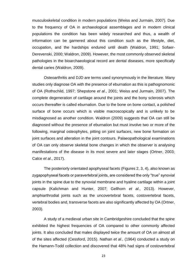
musculoskeletal condition in modern populations (Weiss and Jurmain, 2007). Due
to the frequency of OA in archaeological assemblages and in modern clinical
populations the condition has been widely researched and thus, a wealth of
information can be garnered about this condition such as the lifestyle, diet,
occupation, and the hardships endured until death (Waldron, 1991; Sofaer-
Derevenski, 2000; Waldron, 2009). However, the most commonly observed skeletal
pathologies in the bioarchaeological record are dental diseases, more specifically
dental caries (Waldron, 2009).
Osteoarthritis and DJD are terms used synonymously in the literature. Many
studies only diagnose OA with the presence of eburnation as this is pathognomonic
of OA (Rothschild, 1997; Shepstone et al., 2001; Weiss and Jurmain, 2007). The
complete degeneration of cartilage around the joints and the bony sclerosis which
occurs thereafter is called eburnation. Due to the bone on bone contact, a polished
surface of bone occurs which is visible macroscopically and is unlikely to be
misdiagnosed as another condition. Waldron (2009) suggests that OA can still be
diagnosed without the presence of eburnation but must involve two or more of the
following, marginal osteophytes, pitting on joint surfaces, new bone formation on
joint surfaces and alteration in the joint contours. Palaeopathological examinations
of OA can only observe skeletal bone changes in which the observer is analysing
manifestations of the disease in its most severe and later stages (Ortner, 2003;
Calce et al., 2017).
The posteriorly orientated apophyseal facets (Figures 2, 3, 4), also known as
zygapophyseal facets or paravertebral joints, are considered the only “true” synovial
joints in the spine due to the synovial membrane and hyaline cartilage within a joint
capsule (Kalichman and Hunter, 2007; Gellhorn et al., 2013). However,
amphiarthrodial joints such as the uncovertebral facets, costovertebral facets,
vertebral bodies and, transverse facets are also significantly affected by OA (Ortner,
2003).
A study of a medieval urban site in Cambridgeshire concluded that the spine
exhibited the highest frequencies of OA compared to other commonly affected
joints. It also concluded that males displayed twice the amount of OA on almost all
of the sites affected (Cessford, 2015). Nathan et al., (1964) conducted a study on
the Hamann-Todd collection and discovered that 48% had signs of costovertebral

24
OA and that no correlation was established between the sexes and ancestral origin.
This is consistent with the study by Plomp and Boylston (2016) in which it was
suggested that sex did not influence the rate of costovertebral OA in two British
medieval skeletal samples.
2.5.1.1 Causes of Osteoarthritis
Osteoarthritis is considered a heterogeneous condition. The causes of OA
that have been identified are as follows: damage to surrounding ligaments and
tendons, obesity and ageing (Shepstone et al., 2001; Roos, 2005; Calce et al.,
2017). Most studies of OA of the skeleton and or, more specifically, the spine
conclude that the frequency and severity of OA increases with age (Weiss and
Jurmain, 2007; Plomp and Boylston, 2016). Osteoarthritis and DJD can be
indicators of mechanical stress that has been applied to the body through strenuous
and repetitive movements and physical manual labour over a long period of time
(Klaus et al., 2009).
A multitude of studies have contributed compelling evidence to infer activity
and occupational patterns from the distribution and severity of degenerative
conditions (Merbs, 1983; Stirland and Waldron, 1997; Sofaer-Derevenski, 2000).
Young crewmates of the Mary Rose ship exhibit accelerated degenerative changes
due to the harsh, military lifestyles (Stirland and Waldron, 1997) and, as already
stated, Sofaer-Derevenski (2000) discovered higher levels of OA in the spines of
females that carried creels of peat on their back.
Figure 2 - Posterior aspect of a "typical" 3rd cervical vertebra. Red - superior uncofacets. Green – superior and inferior apophyseal facets.
Figure 3 - Lateral aspect of a “typical” mid-thoracic vertebra. Red – transverse process facets. Green – superior and inferior apophyseal facets. Blue – superior and inferior costovertebral facets
Figure 4 – Lateral aspect of a "typical" lumbar vertebra. Green – superior and inferior apophyseal facets.

25
2.5.1.2 Symptoms of Spinal Osteoarthritis
Osteoarthritis may have profound effects on the lives on the sufferers, as
symptoms can include, chronic pain and sciatica (Mizuguchi, 1976), joint stiffness
(Kean et al., 2004) but also depression and anxiety (Stubbs et al., 2016). Severe
cervical vertebral OA can be potentially life-changing with physical symptoms such
as spinal stenosis, paraplegia, loss of muscle mass and motor control (Wilkinson,
1960; Teraguchi et al., 2014). Therefore, it would be acceptable to suggest that this
condition would have affected the lives of medieval people through loss of income
and in their social spheres. It is worth noting that OA is generally observed in
association with other degenerative conditions, such as intervertebral disc
degeneration (Rogers et al., 1985; Fujiwara et al., 1999), and therefore, the
symptoms may in fact be due or be partly due to the other conditions present.
2.5.2 Intervertebral (IDD) / Degenerative Disc Disease (DDD)
Intervertebral disc disease and degenerative disc disease are terms used
synonymously to name the same type of condition in the literature. It is, along with
OA, considered one of the most common musculoskeletal conditions affecting
people today (Annunen et al., 1999). Intervertebral disc disease is known to affect
individuals from as early as the 2nd decade of life, which then continues to become
more discernible as aging takes place (Antoniou et al., 1996).
The aetiology of IDD is elusive, like most spinal conditions, and can be
caused by many factors. Lack of nutrient supply to the disc cells, trauma,
mechanical loading and a genetic predisposition have been ascertained as factors
leading to disc disease (Urban and Roberts, 2003). Intervertebral disc disease in a
clinical setting has been considered to fall in to two separate categories; “Endplate
driven” and “Annulus driven”. Endplate driven IDD generally occurs from spinal
injury and can be seen in individuals below the age of 30 years. Annulus driven is a
progressive condition which tends to affect individuals 30 years and above and is
considered to stem from habitual and repetitive bending, lifting and spinal loading
(Adams and Dolan, 2012; Adams et al., 2015). The annulus driven type is
considered the more debilitating of the two types, but both are linked to lower back
pain (Adams and Dolan, 2012).
Degeneration of the intervertebral discs and vertebral endplates is widely
regarded as a major cause of low back pain (Smith et al., 2011) and is a contributing

26
factor to the formation of OA in the spinal facet joints (Brain et al., 1952; Fujiwara et
al., 2000). Axial rotation due to disc degeneration is greatly diminished according to
a study by Fujiwara et al., (2000).
A study by Waldron examines the relationship between spinal conditions
observed in the Christ Church Spitalfields skeletons and noted that the cervical
(males had the highest reported frequency in the C6 vertebra with 27% and females
in the C5 vertebra with 26%) and lower lumbar vertebrae (males exhibit
approximately 8% in the L5 vertebra and females exhibit 4%) exhibit the highest
concentrations of IDD (Waldron, 1991). The results are similar to that of clinical
studies and the pattern of IDD very closely follows that of OA in that the cervical and
lumbar regions are commonly the most affected spinal segments (Brain et al., 1952;
Waldron, 1991; Fujiwara et al., 2000). The lack of thoracic involvement of OA and
IDD could be due to lack of movement and stress applied to that region in everyday
life (Briggs et al., 2009; Teraguchi et al., 2014).
2.5.3 Schmӧrl’s Nodes (SN)
Schmӧrl’s nodes are described as vertebral lesions caused by the herniation
of the intervertebral disc (nucleus pulposus), through the endplate of the
neighbouring vertebral body. As a result of this herniation the vertebral body
exhibits, on the inferior and superior vertebral bodies, either a central, posterior or
anterior depression with a sclerotic margin (Ortner and Putschar, 1981; Pfirrmann
and Resnick, 2001; Ortner, 2003; Williams et al., 2007; Waldron, 2009; Dar et al,
2009; Dar et al, 2010; Kyere et al., 2012).
The origin of SNs is continually debated in the literature. However, most
would agree that like OA, SNs are a heterogeneous lesion that can affect any age,
sex and ancestral background. Trauma, genetics (including developmental),
infectious and degenerative conditions have all been inferred as likely precursors to
the formation of SNs (Fahey et al., 1998; Dar et al., 2009; Dar et al., 2010). A study
conducted by Dar et al., (2009) suggested that SNs could be genetic in origin due
to the significant difference between the prevalence in European Americans (60.3%)
and African Americans (36.7%). The same study also concluded that the males
exhibited more SN’s than their female counterparts irrespective of ancestral origin.
Some of the earliest observed SN was found within the vertebral column of
Australopithecus africanus dating back to approximately 2.4 – 2.8 million years

27
(D’Anastasio et al., 2009). Unlike some conditions of the human spine that are
considered unique to humans, Schmӧrl’s nodes are seen in other non-humans such
as; domestic dogs (Canis lupus familiaris – Linnaeus, 1758) (Gaschen et al., 1995)
and sheep (Ovis aries – Linnaeus, 1758) (Fews et al., 2006), among others.
Schmӧrl’s nodes as indicators of habitual and repetitive activity have been
reported in the literature. Stirland and Waldron (1997) discovered a high prevalence
of the lesions in the adolescent and young adult crew of the Mary Rose ship. Military
recruits buried in Snake Hill cemetery in New York had extensive and multiple SNs
throughout their entire spinal columns. The individuals discovered in both sites were
subjected to intensive manual labour, rigorous exercise regimes and excessive
mechanical loading of the spine on a daily basis (Stirland, 1996; Larsen, 2015).
2.5.4 Diffuse Idiopathic Skeletal Hyperostosis (DISH)
Diffuse Idiopathic Skeletal Hyperostosis is a multisystem hormonal condition
that affects the spine and other extra-spinal areas such as the patellae and calcanei
(Resnick et al., 1978; Rogers and Waldron, 2001; Sarzi-Puttini and Atzeni, 2004).
However, it is commonly referred to in the bioarchaeological literature as a joint
condition (Aufderheide and Rodríguez-Martín, 1998; Roberts and Manchester,
2010).
Prior to modern day living, DISH was considered a condition that affects
wealthy, high-status and monastic individuals due to the access to an abundance of
rich foods that the lower classes could not afford (Verlaan et al., 2007; Van der
Merwe et al., 2012). However, DISH is most commonly found in elderly individuals
and men are more susceptible to the condition than females (Rogers and Waldron,
2001; Jankauskas, 2003; Verlaan et al., 2007). Individuals who were overweight as
children and/or individuals with a high body mass index and who have a diet rich in
purines are more likely to develop DISH from the sixth decade of life onwards
(Cammisa et al., 1998; Kiss et al., 2002; Kagotani et al., 2013). Kiss et al., (2002)
established a correlation between high levels of serum uric acid and DISH. High
levels of uric acid in the blood is an indicator of ingesting large amounts of food
products containing organic compounds called purines and once ingested are
broken down into uric acid. Purines are found in high levels in beer, wine, liver,
kidneys, trout, mackerel, scallops and dried beans amongst many other foods (Kiss
et al., 2002).

28
There is strong association between DISH and insulin-independent diabetes
(Verlaan et al., 2007). Obesity is a frequent observation in the DISH sufferers (Kiss
et al., 2002; Verlaan, et al., 2007). A genetic origin has been previously inferred by
Weinfeld et al., (1997) which indicated in the outcome of a populational based study
in America suggested that American whites had a far greater prevalence of DISH
than African Americans, Native Americans and Asians. However, this study failed
to point out changes in diet and lifestyle that may play a role in the occurrence of
DISH (Kiss et al., 2002; Waldron, 2009).
Diffuse idiopathic skeletal hyperostosis may appear rather severe due to the
ossification of the anterior longitudinal ligament to four or more contiguous
vertebrae. However, the symptoms of the condition are relatively minimal and is
frequently first noted in radiographs taken for something else (Verlaan et al., 2007).
Sometimes, if symptoms do occur they tend to take the form of dysphagia (difficulty
in swallowing), sleep apnea, stiffness and pain, amongst others (Cammisa et al.,
1998; Verlaan et al., 2007).
2.5.5 Vertebral Osteophytosis (VO)
Vertebral osteophytes are the lipping of bone on the superior and inferior rim
of the vertebral body that occurs with age and is generally attributed to DJD/OA but
also to repeated episodes of stress and pressure transmitted through the
intervertebral disc (Reid et al., 1991; Lovell, 1994; Sofaer-Derevenski, 2000;
Kumaresan et al., 2001).
Many researchers agree that osteophytes are secondary abnormalities
attributed to OA and IDD and to heavy manual labour early on in life (O’Neill et al.,
1999; Kumaresan et al., 2001; Klaassen et al., 2011). However, some have
established little correlation between the formation of VO and OA and have
attributed them to a high body mass index (BMI) and body weight (Oishi et al., 2003).
A high body mass index in medieval peasants was highly unlikely given the organic
foods being eaten and the highly strenuous farming practices being undertaken on
a daily basis (Resor, 2010).
The disintegration of the nucleus pulposus and the annulus fibrosus result in
less disc space which in turn affects the normal biomechanics of the spine resulting
in osteophyte formation over time (Kumaresan at al., 2001). Vertebral osteophytosis
can lead to decreased mobility in the areas affected and large osteophytes in the

29
cervical segment can lead to dysphagia due to the obstruction of the normal
elevation and anterior movements of the larynx (Mosher, 1926; Seidler et al., 2009).
Even though VO is a common condition affecting the aging and elderly population,
the clinical literature gives clear indication that this can be detrimental to the
everyday life of the individuals affected. It is clear from the literature that severe
osteophytes can have life affecting neurological symptoms and can impair mobility
such as spinal stenosis (Kumaresan at al., 2001).
There are numerous studies that specifically grade vertebral body
osteophytes by severity in an attempt to assess age (Stewart, 1958; Snodgrass,
2004; Kim et al., 2012). In addition, there are numerous clinical and
palaeopathological studies involving the grading of the severity of apophyseal facet
osteophytes and other spinal degenerative conditions such as OA and DJD
(Fujiwara et al., 2000; Sofaer-Derevenski, 2000). Snodgrass (2004) examines
osteophyte development between sexes in order to establish if there is a similar
pattern of formation with advancing age. The author concludes that males and
females exhibit very similar age-related osteophyte development and that the
degree of osteophyte development could be used as a general indicator of age.
2.6 Infectious Diseases
Infectious diseases of the spine can infer dietary habits, living environment
and conditions and occupational patterns (Mays et al., 2001; Roberts and
Manchester, 2010). Infectious diseases of the human skeleton can be classified into
nonspecific and specific. Nonspecific infections; relating to an infection not known
to be caused by a specific pathogen such as periostitis and osteomyelitis (including
spinal), and specific; an infection pertaining to a known pathogen, for example
tuberculosis caused by the bacteria known as Mycobacterium tuberculosis,
amongst other bacterial strains (Roberts and Manchester, 2010).
Infectious epidemics such as “The Black Death” caused by the bacterium
Yersinia pestis rarely give the infected host time for bones to remodel before death
and that is the case for most infectious diseases that affect humans. However, some
blastic (bone forming) and lytic (destructive) skeletal lesions are readily identifiable
in the archaeological record and are usually a result of prolonged exposure to a
certain pathogen and the subsequent chronic infection (Galasko, 1982; Larsen,
2015).

30
2.6.1 Tuberculosis (TB)
Tuberculosis is a chronic infectious disease that can affect any part of the
body and is caused by the bacteria of the genus Mycobacterium. Two types affect
humans; the bovine type and the human type (Manchester, 1984; Mays et al., 2001).
In post-medieval Britain, tuberculosis has been concluded to be a significant cause
of death. In 1667, TB was at epidemic proportions and was concluded to account
for 20% of all recorded deaths for that year (Santos and Roberts, 2001). It is possible
that overcrowding, close contact of infected individuals and the unsanitary
conditions of the city were the likely causes of this epidemic (Mays et al., 2001).
In modern populations, TB is considered to only cause osseous changes in
less than 10% of individuals suffering from the chronic condition (Zink et al., 2001;
Roberts and Cox, 2003). In archaeological assemblages, the actual number of
individuals with TB would be much higher. Observation of skeletal remains may only
give a small insight in to the prevalence of some chronic infectious diseases within
archaeological assemblages.
2.6.2 Brucellosis
Brucellosis is a disease of ancient repute (Pappas et al., 2006). It is a
common, rarely fatal, zoonotic infection caused by bacteria from close and
prolonged contact with livestock such as: pigs (Sus scrofa domesticus; B. suis),
goats (Capra aegagrus hircus; B. melitensis), sheep (Ovis aries; B. melitensis),
cattle (Bos taurus; B. abortus) and horses (Equus ferus caballus; B. abortus) (Al
Dahouk et al., 2002; Roberts and Manchester, 2010).
Approximately 90% of recorded cases of brucellosis are caused by B.
melitensis (Memish and Balkhy, 2004). Approximately 500,000 cases are diagnosed
annually, and the majority of those cases are in developing countries. Abbatoir
workers in the USA in the 1970’s suffered from over 2,000 cases of B. suis within a
ten-year period (Pappas et al., 2006). One of the earliest cases of brucellosis was
discovered in Australopithecus africanus dating back to 2.3 – 2.5 million years ago
and this finding offers an insight in to the health and lifestyles of our direct ancestors.
During the Roman era and the Middle Ages brucellosis was considered to be at
epidemic levels in Europe (D’Anastasio et al., 2009).
The skeletal manifestation of brucellosis can cause similar lesions to OA and
other degenerative conditions due to the large osteophyte formation (brucellar
31
epiphysitis) that can occur in advanced cases (D’Anastasio et al., 2009). It can also
be confused with other infectious conditions of the spine such as tuberculosis and
thus, bacterial DNA testing should be conducted if absolutely necessary for
identification purposes (Zink et al., 2001).
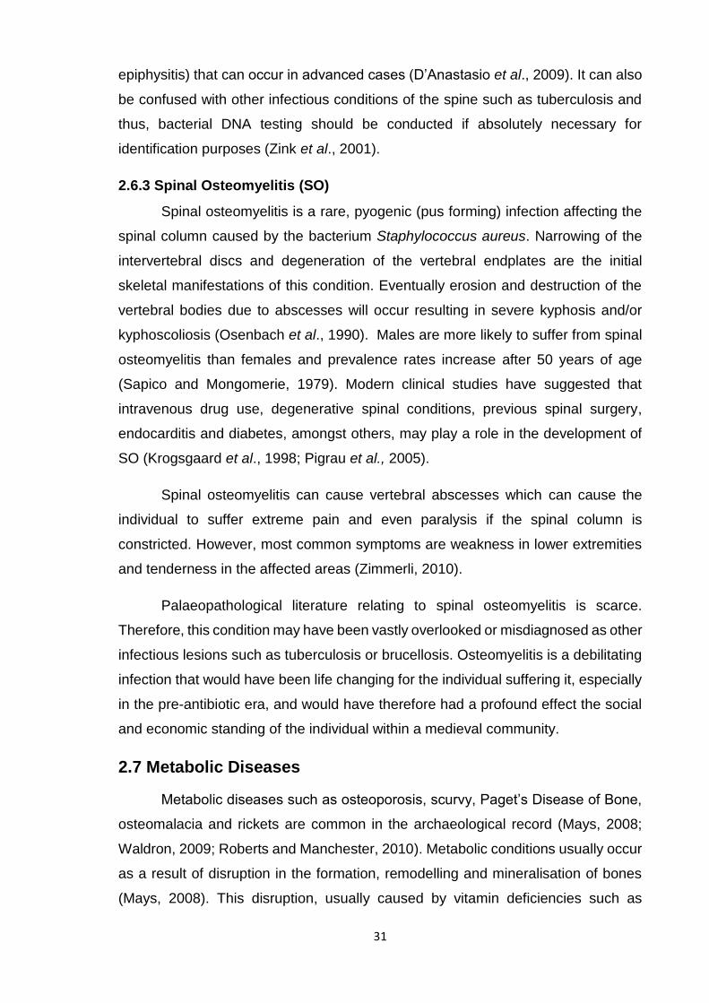
2.6.3 Spinal Osteomyelitis (SO)
Spinal osteomyelitis is a rare, pyogenic (pus forming) infection affecting the
spinal column caused by the bacterium Staphylococcus aureus. Narrowing of the
intervertebral discs and degeneration of the vertebral endplates are the initial
skeletal manifestations of this condition. Eventually erosion and destruction of the
vertebral bodies due to abscesses will occur resulting in severe kyphosis and/or
kyphoscoliosis (Osenbach et al., 1990). Males are more likely to suffer from spinal
osteomyelitis than females and prevalence rates increase after 50 years of age
(Sapico and Mongomerie, 1979). Modern clinical studies have suggested that
intravenous drug use, degenerative spinal conditions, previous spinal surgery,
endocarditis and diabetes, amongst others, may play a role in the development of
SO (Krogsgaard et al., 1998; Pigrau et al., 2005).
Spinal osteomyelitis can cause vertebral abscesses which can cause the
individual to suffer extreme pain and even paralysis if the spinal column is
constricted. However, most common symptoms are weakness in lower extremities
and tenderness in the affected areas (Zimmerli, 2010).
Palaeopathological literature relating to spinal osteomyelitis is scarce.
Therefore, this condition may have been vastly overlooked or misdiagnosed as other
infectious lesions such as tuberculosis or brucellosis. Osteomyelitis is a debilitating
infection that would have been life changing for the individual suffering it, especially
in the pre-antibiotic era, and would have therefore had a profound effect the social
and economic standing of the individual within a medieval community.
2.7 Metabolic Diseases
Metabolic diseases such as osteoporosis, scurvy, Paget’s Disease of Bone,
osteomalacia and rickets are common in the archaeological record (Mays, 2008;
Waldron, 2009; Roberts and Manchester, 2010). Metabolic conditions usually occur
as a result of disruption in the formation, remodelling and mineralisation of bones
(Mays, 2008). This disruption, usually caused by vitamin deficiencies such as
32
vitamins C and D, produces architecturally unstable and weak bone (Mays, 2008).
It is difficult to correctly diagnose certain metabolic conditions in palaeopathological
studies, due to common multi factorial aetiologies and similar morphological
appearances on skeletal remains such as, porotic and hypertrophic areas of bone
and fractures (Brickley and Ives, 2006; Geber and Murphy, 2012).
2.7.1 Osteoporosis
Osteoporosis is a systematic skeletal disease that affects thousands of
people worldwide and is considered a major health problem in modern aging
populations due to the associated fracture risk (Kanis et al., 2002; Holroyd et al,
2008). Osteoporosis is characterised by loss of bone mineral density and poor bone
microstructure due to a disruption in bone formation, remodelling and mineralisation
(Brickley, 2002; Brickley and Agarwal, 2003; Mays et al., 2006; Mays, 2008).
People suffering from osteoporosis are more likely to suffer fractures due to
loss of bone density and the most common fracture types associated with
osteoporosis are femoral neck, distal radius (Colle’s fracture) and spinal
compression fractures (Kanis et al., 2002; Mays, 2006). Fractures of the femoral
neck and extensive vertebral compression fractures are considered the most
clinically significant aspect of osteoporosis as these fractures are potentially life
threatening and/or life changing (Holroyd et al., 2008).
Women are at least twice as likely to be affected as men after the age of 35
due to post-menopausal hormonal changes (Jordan and Cooper, 2002; Mays et al.,
2006). Other contributors and exacerbating factors of osteoporosis is smoking, lack
of exercise and being deficient in calcium (Iki, 2005; Mays et al., 2006; Kapetanovic
and Avdic, 2014). Data from a study conducted by the General Practice Research
Database in the United Kingdom between 1988-1998 concluded that one in two
women over the age of 50 will at some point in their lives develop an osteoporotic
fracture compared to one in five men (Van Staa et al., 2001).
Mays et al. (2006) conducted a study on contemporary medieval skeletons
from an urban community from Norway (Trondheim) and a rural community from
England (Wharram Percy). It was concluded that the Norwegian sample overall
exhibited a higher prevalence of osteoporotic fractures. However, due to the small
sample sizes the results remained tentative, but the authors suggested that a
combination of factors such as cold conditions and an urban environment could
33
explain the higher frequency of osteoporotic fractures due to harder surfaces and
colder/icy climate.
2.7.2 Paget’s Disease of Bone (PDB)
Paget’s disease of bone is characterised by the over production of
osteoclasts and the subsequent disarray of bone remodelling causing poor structure
and loss of strength in the bone affected (Whyte, 2006). The axial skeleton is the
most commonly affected area in PDB with the pelvis being the most frequently
affected area (Ralston, 2013).
PDB is considered to have originated in the British Isles due to the high
frequencies recorded and thus, indicates a likely genetic origin. However, although
there appears to be a likely genetic component to PDB it is considered a
heterogeneous disease as environmental factors may also play a role (Mays, 2010).
2.8 Congenital Conditions and Vertebral Anomalies
Most developmental anomalies occur in the spinal column and more
specifically in the lumbosacral region (Groza et al., 2016). Genetics plays an
important role in the development of congenital conditions and developmental
anomalies. Changes in genetic signalling affect the initial stages of skeletal
development. Therefore, it is possible to connect genetic affinity between individuals
in skeletal assemblages as developmental anomalies tend to pass through familial
lineages (Barnes, 2012). It has been stated that consanguineous parents have a
higher probability of bearing children with skeletal anomalies than parents that are
not closely related (Devor, 1993; Sarry El-Din and El Banna, 2006; Hussien et al.,
2009). However, environmental factors and dietary deficiencies such as,
geographical area, pollution, contaminated water supplies and lack of folic acid and
other vitamins and minerals have also been associated with the development of
congenital anomalies (Dorsch et al., 1984; De Wals et al., 2007).
2.8.1 Transitional Vertebrae, Sacralisation and Lumbarisation
Transitional vertebrae are common spinal anomalies in the present day and
in archaeological skeletal assemblages and the incidence of these anomalies
greatly varies between populations (Delport et al., 2006). However, there is a
probable genetic factor involved in the expression of transitional vertebrae and this
could explain the variability in the frequencies observed in different populations (Tini

34
et al., 1977; Delport et al., 2006). Lumbosacral transitional vertebrae (sacralisation
and lumbarisation) are not considered to be pathological conditions. However, some
studies have shown that fusion of L5 and/or L6 to the sacrum can predispose an
individual to degenerative spondylolisthesis and worsen other degenerative spinal
conditions due to the extra force applied to the lumbosacral junction (Kong, 2008).
2.8.2 Spina bifida
Spina bifida occulta is one of the most commonly observed spinal
dysraphisms in the archaeological record (Groza et al., 2012). Spina bifida is a
developmental defect in which the caudal neural tube fails to fuse leaving an open
midline (Saluja, 1988; Mitchell et al., 2004). Life expectancy decreased dramatically
for anyone born with spina bifida cystica, the most severe form of spina bifida, which
had a mortality rate of 10% – 12% (Roberts and Manchester, 2010; Pruitt, 2012).
Spina bifida cystica is often associated with other spinal abnormalities which
include hemi-vertebrae, vertebral synostosis, congenital scoliosis or kyphosis and
developmental lordosis (Zimmerman and Kelly, 1982; Kumar and Singh, 2003;
Kumar and Tubbs, 2011). Spina bifida occulta is so called due to the fact it can go
unnoticed by many individuals throughout life and causes no problems with day to
day living (Roberts and Manchester, 2010).
2.8.3 Scoliosis
Scoliosis is the curvature of the spine that differs from the normal lordosis
and kyphosis. It can be characterised by vertebral wedging, asymmetrical
apophyseal facets and the abnormal angulation of the transverse processes and
spinous processes creating an ‘S’ or ‘C’ curve to the spine (Appleby et al., 2014).
Scoliosis is subcategorised in to seven types; congenital, early on-set, adolescent
idiopathic, degenerative (senile), neuromuscular, syndromic and Scheuermann’s
kyphosis (Scoliosis Association UK, 2017). Hemi-vertebrae are a common
occurrence in people with congenital scoliosis (Ortner, 2003).
The last Plantagenet King Richard III famously exhibits scoliotic curvature in
the thoracic region of the spine and was said to have one shoulder higher than the
other and walked with a slight limp (Appleby et al., 2014). Playwright Sir William
Shakespeare was less than complimentary about the Plantagenet king and within a
play, titled ‘Richard III’, the king was described as “deformed” and was generally
mocked and acted with a limp arm and obvious kyphoscoliosis. The use of the

35
adjectives “deformed” and “hunchback” may give an insight into the negative public
perceptions of individuals that had visible spinal conditions in the medieval times
(Lund, 2015). However, it is possible that the accusation that King Richard III
participated in the disappearance of the ‘princes in the tower’ may have influenced
Shakespeare’s portrayal of the king in a negative and villainous light.
2.8.4 Kyphosis
Kyphosis or hyperkyphosis is the exaggeration of the already present
kyphotic curvature of the thoracic spine. It is usually considered a secondary
abnormality as a result of trauma and infectious, degenerative, congenital and
metabolic conditions such as tuberculosis, osteoporosis and Scheuermann’s
disease (Fon et al., 1979; Holloway et al., 2011; Üstündağ and Deveci, 2011).
A radiographic study on a modern population discovered the degree of
kyphosis increases with age and that females are more affected than males (Fon et
al., 1979). Due to the increase in the degree of kyphosis with aging, it is now
considered a “geriatric syndrome” and it is estimated to affect approximately 30% of
the elderly population (Kado et al., 2014). Kyphosis becomes more apparent with
excessive biomechanical loading and poor musculature (Lorgerbs et al., 2017).
2.8.5 Klippel-Feil Syndrome (KFS)/ Cervical Vertebral Synostosis
First described by Klippel and Feil in 1912, Klippel-Feil Syndrome (KFS) is
the congenital ankylosis of the cervical vertebrae due to abnormal division of
somites during foetal development (Fernandes and Costa, 2007). Klippel-Feil
Syndrome in living patients can be seen in shortening of the neck, limited neck
movement and low hairlines (Larson et al., 2001; Fernandes and Costa, 2007). It
also commonly occurs with other congenital conditions such as Sprengel deformity,
hemi-vertebrae and spina bifida but can also be present separately to the
aforementioned conditions (Aufderheide and Rodríguez-Martín, 1998). The physical
symptoms of KFS and other commonly associated congenital conditions may have
caused an inability to be employed in strenuous and physically demanding
occupations such as many agricultural roles.
Cases of possible KFS have been observed in many archaeological skeletal
assemblages and historical figures, such as the Mayan sacrificial skeletal
assemblage in the Midnight Terror cave in Belize (Kieffer, 2015), Cardinal Carlo de’
Medici (Giuffra et al., 2009), a medieval cemetery in Portugal (Fernandes and Costa,

36
2007) and in Neolithic Vietnam (Tilley and Oxenham, 2011). A study conducted by
Kieffer (2015) stated that the individuals discovered in the Midnight Terror cave may
have been sacrificed due to their visible manifestations of KFS and thus, were
considered “social outcasts”.
2.9 Trauma
Incidence, anatomical location, and trauma type provide insight into the
health status and lifestyles of individuals in past populations, but can also indicate
conflict including possible warfare, domestic and interpersonal violence and murder
(Larsen, 2015). Vertebral trauma such as compression fractures are indicative of
chronic stress and heavy loading of the spine and are particularly common in the
thoracolumbar spine (Myers and Wilson, 1997; Agnew and Justus, 2014). Fractures
are more likely to occur in the mid-thoracic and thoracolumbar segments and when
the load applied to the vertebrae is equal to or greater than its strength (Duan et al.,
2001; Bruno et al., 2017).
2.9.1 Spondylolysis and Spondylolisthesis
Spondylolysis is a degenerative defect/weakening and subsequent stress
fracture of the pars interarticularis of a vertebra (Merbs, 1996; Larsen, 2015).
Spondylolysis is a unique condition that affects hominins and has been solely
attributed to bipedal, upright walking (Merbs, 1996; Merbs, 2002). In addition to
general degeneration of the spinal facet joints and vertebral bodies, this lesion tends
to occur due to strenuous and continual loading and hyper extension and lateral
flexion of the lumbar and lower thoracic region of the spine (Dietrich and Kurowski,
1985; Merbs, 1996; Fibiger and Knüsel, 2005). The main vertebrae affected are the
4th and 5th and the occasional 6th lumbar and 1st sacral vertebrae (Merbs, 1996;
Fibiger and Knüsel, 2005). Even though this condition points to strenuous and
habitual physical activity in certain individuals, it cannot be attributed to any single
occupation and thus, makes it difficult to interpret occupational activities in skeletal
assemblages (Mays, 2006).
It has been stated that approximately 25% of individuals who have
spondylolysis have lower back pain. Spondylolysis in individuals who engage in
strenuous physical activity, including weight lifters, are more likely to suffer lower
back pain than other sufferers of the same condition (Kalichman et al., 2009).
Modern clinical studies have highlighted possible symptoms of spondylolysis and

37
spondylolisthesis as tightness in the biceps femoris, semitendinosus and the
semimembranosus muscles (hamstrings), tenderness in the lower spine and pain
whilst conducting activities (Cavalier et al., 2006).
Spondylolisthesis is the anterior slippage of a vertebra leading to
compression in the posterior portion of a vertebral endplate (Merbs, 1996).
Degenerative spondylolisthesis, caused by general degeneration of the apophyseal
facets and vertebral body, and spondylolytic spondylolisthesis or “isthmic”
spondylolisthesis, a predisposition to spondylolisthesis due to spondylolysis, are
considered the two main aetiologies (Whitesides et al., 2005; Kalichman et al.,
2009).
2.9.2 Stress, Crush and Burst Fractures
Stress fractures are indicative of repeated episodes of stress and pressure
put on the spinal column, but crush and burst fractures are excellent indicators of a
sudden traumatic incident (Lovell, 1997). Vertebral fractures are common
complications of osteoporosis and are frequently observed in elderly women
(Cummings et al., 1985). Stress fractures occur when repeated episodes of force
are placed on the bone and is most commonly seen in the tibiae, calcanei and
metatarsals (Lovell, 1997). Crush fractures are generally categorised into three
types; depression, compression and pressure. Burst fractures are almost
exclusively seen in the spine and is the result of vertical compression damaging the
intervertebral disc (Lovell, 1997).
2.10 Summary
Many of the spinal conditions observed within this study would have possibly
affected everyday living for the individuals that suffered from them. Infectious
diseases, advanced metabolic conditions, degenerative conditions and trauma
would have most probably caused mobility problems and back pain for the
individuals, especially in the more advanced stages of the conditions. The reality of
the effects of the symptoms would have likely attributed to loss of income for the
manual labourers due to not being able to work to their full potential. The general
public’s lack of education and the social attitudes towards people with visible
skeletal conditions in the medieval period may have caused people with obvious
spinal malformations to have been treated in an unfavourable way.

38
It may be possible to infer activity and occupational patterns in the distribution
and severity of degenerative conditions. However, an in-depth account of the
population’s occupational patterns within that region and/or specific community is
essential for a compelling case. In review of the palaeopathological studies
observed, it is clear that a more holistic and interdisciplinary approach is needed,
using archaeological, medical and ethnographical research to provide a more
accurate assessment of the health in past populations.

39
Chapter 3: Site Information and
Hypotheses
3.1 Poulton, Cheshire
3.1.1 Medieval Chester, Cheshire
Chester was founded in the decade succeeding 70 A.D. during the reign of
Roman Emperor Vespasian. Chester was a “castrum” or fortress and was
considered one of the main army strongholds in the Britain (Emery, 2010; Historic
England, 2018). Chester is the county town of Cheshire and situated on the River
Dee and close to the border of north Wales. Chester relied on the production and
sale of livestock to fund the city’s agrarian economy in the medieval times. Chester’s
market was closely linked with pastures near the Dee and Mersey estuaries but was
also linked to mixed farming to the city’s south and east (Lewis and Thacker, 2003).
Poulton may have been closely linked to the Chester’s market. Unfortunately, no
historical documentation has been retrieved to acknowledge this.
3.1.2 Poulton
To the west of the river Dee and five miles to the south of Chester city centre,
lies the hamlet of Poulton (Figure 5) (Fair, 2010). In the beginning of the medieval
period, the hamlet of Poulton had a population of approximately 50 individuals or
ten households. From 1153 A.D., the land of Poulton, more specifically the river Dee
floodplains, was transformed into an agricultural landscape. The transformation of
the land was mainly attributed to the Cistercian monks that inhabited Poulton Abbey
from 1153 A.D. to 1220 A.D. who then relocated to Dieulacres Abbey, Staffordshire
(McGuicken, 2006; Emery, 2010).
In opposition to the religious beliefs of austerity and humility, the Cistercian
monks became wealthy through their great skill in transforming marshy and boggy
landscapes into areas of crop cultivation and sheep farming. Shortly after 1220 A.D.
and the relocation of the monastic community to Staffordshire, Poulton abbey was
converted and used as a monastic grange; a manor or farming estate used as a
place for food production (Emery, 2010).

40
Figure 5 - Map of Great Britain showing positions of Chester (black) and Poulton (Red) in the county of Cheshire.
It is likely that the local lay people were peasants and conducted agriculture
as a means of subsistence, and this is in keeping with other contemporaneous rural
villages in England. The Cistercian monks that inhabited the Poulton Abbey would
have stayed away from the local peasant community and more specifically the lay
peasant agriculture as this was strictly forbidden. The Cistercian monks were
exempt from paying annual taxes (payment equating to 10% of annual earnings) to
the church which became a source of ill will between the villagers and monks at
Poulton and neighbouring Pulford and Dodleston (Emery, 2010).
The monks that relocated to Dieulacres Abbey still owned the Poulton
grange. In the 15th century, Poulton and the surrounding farmland were leased to
the local, wealthy Manley family. Sir Nicholas Manley and/or his father John, were
responsible for the extension and reconstruction of the Poulton chapel and in Sir
Nicholas Manley’s last will and testament he announced that he should like to be
buried within the chancel of the chapel. The assumed burial of Sir Nicholas was
discovered in 1997 and was located facing the altar. Additionally, the burial was
41
complete and undisturbed unlike most burials within medieval cemeteries (Emery,
2010).
3.1.3 Archaeological Excavation of Poulton Chapel and Cemetery
Poulton is a renowned ancient settlement of archaeological potential. There
is a multitude of archaeological evidence recovered from the Poulton site and from
ancient sites overlooking the “Old Pulford Brook” such as microliths, worked bone
and antlers made in to harpoons, barbs and arrow heads. The site itself can be
accurately dated to the Neolithic era (Emery, 2010; Fair, 2010). Due to the vast
amount of Roman era roof tiles, window glass, pottery and brooches excavated
within the site, it is suggested that there is a Roman temple or villa within the locale.
However, this supposed villa has remained elusive, even with extensive excavations
carried out over the past 20 years (Emery, 2010).
Excavations that were carried out over four seasons from 1995 to 1998
uncovered a medieval chapel and cemetery. To date over 800 individuals (pre-natal
to old aged) have been excavated from this extensive cemetery. The cemetery is
assumed to have the native local people buried within it, and no distinction has been
made as to the social status and wealth of the individuals. However, one adult male
skeleton was recovered from within the chapel walls and is reported to be of high
status and is believed to be Sir Nicholas Manley (1468 A.D. – 1506 A.D.) (Ormerod,
1819; Emery, 2010).
3.1.4 Medieval Farming Practices
Farming is an overarching occupation that hosts a multitude of tasks and is
considered one of the most hazardous and physically strenuous occupations in
modern society (Judd and Roberts, 1999). Therefore, it is acceptable to assume that
farming in the medieval times would have been equally, if not more physically
demanding without the use of heavy machinery as a farming aid.
“The Labors of the Months” as illustrated in many medieval manuscripts and
Renaissance art, and dependent on socioeconomic and social status, is the cycle
of subsistance strategies employed each month of the year as a means to effectively
farm the land (Judd and Roberts, 1999). Males generally completed heavy manual
jobs and designated household chores, child care and less physical farming roles
to the females (ibid.). However, farming roles were not always divided by gender

42
and certain tasks such as haymaking would be completed by both males and
females in equal measure (Vardi, 1996). As stated above, farming is an umbrella
term used to name a multitude of agricultural tasks (Table 1) and it is unlikely that
an individual had just one mode of employment, unlike the specialist crafts and
guilds within the major towns and cities.
Table 1 - Medieval farming, hunting and household practices likely to have been carried out at Poulton
Farming Practices Hunting Household
Plowing Fowling Food preparation
Threshing Child care
Transporting Butchery
Tree felling Gardening
Harvesting
Planting
Herding
3.1.5 Medieval Rural Diets
Medieval diets were mainly determined by social status and wealth. Peasants
largely relied on foods they could produce themselves, good weather and a plentiful
harvest (Resor, 2010). The diets would have chiefly consisted of ale and “pottage”,
a stew made from vegetables, meat (most probably mutton) and from cereals such
as oats (Spencer, 2008; Resor, 2010): a healthy diet by the processed standards of
today. Meat would have been eaten periodically and significantly made up of old
cows, sheep and pigs that were no longer useful for working the fields and producing
milk (Spencer, 2008). The rural diet may be considered a healthy diet, but harsh
winter frosts and wet summers may have ruined the crops and thus, malnutrition
may have been a sporadic occurrence. The “Little Ice Age” (14th to 17th centuries
A.D.) would have had disastrous effects on both urban and rural communities
(Lewis, 2003).
3.2 St Owen’s Cemetery, Gloucester
3.2.1 Medieval Gloucester
Gloucester is a city in the southwest of England which is situated close to the
border of southeast Wales. At the beginning of the medieval period, circa 1066 A.D.,
Gloucester was a thriving, urban trading centre but was heavily reliant on Bristol as
the closest major town that conducted trading with continental Europe. In the early
medieval period much of Gloucester’s trading was to Ireland and south Wales

43
(Herbert, 1988). It was also considered a very important town in the Plantagenet
period (1154 A.D. – 1485 A.D.) and was the only city to crown a king, Henry III,
outside of Westminster in London (Moss, 2005). During the latter part of the
medieval period many towns and cities were in economic decline, but Gloucester
remained a relatively affluent town and was rated 16th (1334 A.D.), 15th (1377 A.D.)
and 17th (1523 A.D.) in terms of overall wealth in all of the towns in England (Herbert,
1988).
Gloucester was a hub of iron working and hosted one of thirteen English coin
producers. However, many different trades were conducted within Gloucester
during the Medieval times (Table 2), most of which would have involved physically
strenuous labour (Herbert, 1988). Many of the trades listed in Table 2 such as,
drapers, mercers and tailors were considered wealthy trades that many higher
status families sent their male children to become apprentices. Poorer families
would likely have sent their children to apprentice in lesser crafts such as wood
working and weaving (Lewis, 2016). Age at which many children enrolled in
apprenticeships could be from approximately seven to sixteen which ultimately
depended on family wealth, their father’s trade/craft, location, health and physical
and mental maturity (Dunlop, 1912; Lewis, 2016).
Each of the main streets, Southgate, Eastgate, Westgate and Northgate,
were occupied by concentrations of crafts for example, Northgate and Southgate
Streets were busy with cordwainers (shoemakers) (Langton, 1977; Herbert, 1988).
Southgate Street was also home to fishmongers and wheat sellers (Herbert, 1988).
According to Herbert (1988), trades such as cordwainers and weavers were most
numerous throughout the entire medieval period, closely followed by butchers and
tailors (Table 2).
The occupations and lifestyles of medieval urban females is less transcribed
in historical records in comparison to the males. The stereotype that women were
homemakers is most probably true. However, what is sometimes overlooked is the
probable increase in labour and wealth of medieval females after the decrease in
the male-dominated labourer population by large scale pandemics such as the
plague (Bardsley, 1999). Some young adult and adolescent females may have
migrated to Gloucester in search of employment from the local townships and
44
villages in the aftermath of the plague yet, many of these migrant workers would
have returned to their homes after their short-term contracts ran out (Lewis, 2016).
Bennett (1992) argues that urban women’s socioeconomic standing
remained relatively consistent throughout the latter part of the medieval period and
thus, many women worked in low paid jobs and were almost always denied entrance
into the crafts and merchant guilds. Therefore, the occupations of the females from
St Owen’s remains somewhat elusive. Given the likelihood that the St Owen’s
Cemetery skeletal material is from the local lay community the females would have
likely been domestic servants, homemakers, assisted in making and selling wares
and to have been employed in menial, low wage occupations.
3.2.2 Medieval Urban Diets
Urban diets would have been entirely different to the rural peasants as the
urban markets would have offered a multitude of different foods. Urban traders and
craftsmen had diets largely consisting of meat, fish, milk and cheese. The servants
of merchants and craftsmen were, by law, given meat and fish once per day as part
payment for their services (Spencer, 2008). Gloucester had a series of butchers,
bakeries, spicers, salters and greengrocers, thus enabling the inhabitants to have a
varied diet if they could afford it (Langton, 1977; Herbert, 1988). Excavations carried
out in St Mary Spital, London, discovered remnants of urban foodstuffs such as,
herbs, spices, nuts and a variety of fruits (Connell et al., 2012; Towle et al., 2017).
It is likely that the inhabitants of Gloucester ate similar produce too. Being a port
town, there is a likelihood that seafood was also consumed by the general populace.

45
3.2.3 St Owen’s Cemetery and Archaeological Excavation of Site 3/89,
Southgate Street
The chapel and cemetery of St Owen was situated just outside the Southgate
of Gloucester city walls (Figure 6) and is thought to have been commissioned by
Roger of Gloucester, an illegitimate son of Henry I of England (Herbert, 1988). The
cemetery of St Owen was in use for approximately 500 years, from 1100 A.D. to
1643 A.D. (Herbert, 1988; Atkin and Garrod, 1990). More than 300 medieval burials
have been excavated from St Owens cemetery, which included later burials from
the Independent Chapel and Royal Infirmary (Atkin and Garrod, 1990; Martin et al.,
2016).
The site code 3/89 stands for site three of the archaeological excavation
carried out in 1989. The excavations were carried out on the west side of Southgate
Street and revealed a row of timber buildings that may have been used as a possible
Figure 6 - “Glocester. Glovernia. Claudiocestria” town plan of Gloucester by Pierre Van Der Aa (1720) based on original John Speed (1610). (Arrow – Church of St Owen).
46
area of iron working in the 1st and 2nd centuries A.D. Subsequently, it is believed that
this suburb of Gloucester lay abandoned from the 4th century until the 10/11th
centuries A.D. when a more recent series of timber dwellings were unearthed (Atkin
and Garrod, 1990). In 1137 A.D. the site of St Owen was obtained by Llanthony
Priory and a row of stone framed buildings were laid down (Herbert, 1988; Atkin and
Garrod, 1990). Archaeological evidence established that there was a disruption in
the building sequence for some of the late-medieval dwellings which also coincided
with an increase in burials in the cemetery. It is suggested that an epidemic such as
the “Black Death” was the reason for the increase in burial density in the 1500’s A.D.
and the disruption in building sequence (Atkin and Garrod, 1990). The site of the
Church of St Owen was completely obliterated in the 19th century to make way for
an extension of the docks. However, the church and cemetery were demolished and
abandoned prior to the siege of Gloucester in 1643 A.D. The 3/89 excavation
recovered building material from the church in the foundations of an 18th century
house (Herbert, 1988; Atkin and Garrod, 1990).

47
Table 2 - List of trades in Gloucester in mid-late medieval, 1396 A.D. to 1595 A.D. (Herbert, 1988)
Metal Workers Leather
Workers
Textile
Workers
Clothing
Trades
Distributive
Trades
Food and
Drink
Building
Trades
Other Trades
Iron Mongers Tanners Weavers Tailors Drapers Bakers Masons Trowmen
Farriers Curriers Walkers Hosiers Mercers Fishmongers Carpenters Carriers
Smiths Skinners Dyers Cappers Retailers Brewers Turners Wheelwrights
Braziers Whittawers Shearman Glovers Tranters Maltmakers Glaziers Hoopers
Locksmiths Cordwainers Cardmakers Pursers Chapmen Salters Painters Ropers
Bladesmiths Saddlers Chaloners Hatmakers Spicers Sawyers Bowyers
Cutlers Sheathers Woadmen Cooks Tilers Fletchers
Spoonmakers Tuckers Innkeepers Sieve Makers
Pinners Butchers Patten Makers
Wiredrawers Charcoal
Lorimers Barbers
Spurriers Millwards
Nailers Boatmen
Pewterers Watermen
Latten Makers Mariners
Bellmakers Mattress Makers
Furbishers Burners
Goldsmiths
48
3.3 Medieval Burial Practices
The skeletons recovered and analysed within this study were retrieved from
burials that conformed to the Christian interment of west-east alignment in an
extended supine position: the head towards the west and feet towards to the east in
accordance with the belief that the soul will arise and face God on the “Day of
Judgement” (Parker-Pearson, 1999). The burials within the Poulton cemetery have
been categorised into shroud, coffin and no coffin. However, no distinction was
made as to the shroud burials within the St Owen’s Cemetery and thus only coffin
and no coffin burials were noted. Shroud burial individuals at the Poulton site have
been categorised as such due to the positioning of the human remains in situ and
the lack of post-mortem displacement of the skeletons.
Grave goods, in terms of personal artefacts and grave markers, were none
existent in both cemeteries, indicating probable poorer status but many medieval lay
cemeteries of the general populous generally lack grave goods (Cessford, 2015).
However, biodegradable grave markers and artefacts may have been used at the
time of burial and thus there is no longer any trace of the original goods at the time
of excavation. In medieval England, many wealthy and high-status individuals had
a choice to have more permanent memorials whether it be a grave marker or a wall
plaque (Daniell, 2005).
Truncation, the cutting of an existing burial for interment of another individual,
within medieval cemeteries is commonplace due to compact nature of the burials
and is commonly observed in the Poulton cemetery (Daniell, 2005). This practice is
not, however, limited to medieval cemeteries and such techniques have also been
observed in Anglo-Saxon and Roman sites amongst others (Egging-Dinwiddy and
Stoodley, 2016).

49
3.4 Hypotheses
Based on previous studies, highlighted in previous chapters, a number of
hypotheses have been outlined for testing in this study; they are stated below in the
form of null (H0) and alternative hypotheses (Ha):
H0: There will be no difference in the frequency of joint conditions observed
in males from Poulton and St Owen’s Cemetery due to strenuous and
physical activities employed in daily life in both urban and rural settings.
Ha: There will be a difference in the distribution of spinal lesions on the
vertebral column due to differences in living and working conditions, and
employment type between rural and urban communities.
H0: There will be no difference in the severity of spinal lesions in both
samples due to strenuous and physical activities employed in daily life.
Ha: There will be a difference in the distribution and frequency of spinal
pathologies in the females due to differences in gendered roles in urban
and rural settings.
Ha: There will be a difference in congenital conditions and developmental
anomalies between the urban and rural samples due to differing
population density, environment and diet.
Ha: There will be a difference in the frequency and type of infectious disease
between a rural, agricultural community and an urban community.
Ha: There will be a difference in vertebral fractures between samples due to
differences in working conditions, prevalence of metabolic conditions
and lifestyles.

50
Chapter 4: Materials and Methods
4.1 Skeletal Samples
The skeletal samples under study are from two British medieval communities:
from urban, market town Gloucester and rural, agricultural hamlet Poulton in
Cheshire. Poulton cemetery and the cemetery of St Owen are contemporaneous
burial grounds that were primarily in use in the medieval times (Herbert, 1988;
Emery et al., 1995). The church and cemetery of St Owen were established in the
year 1100 A.D. and continued to be used as a burial ground until it was demolished
prior to the siege of Gloucester in 1643 A.D., thus it is likely that some of the
individuals recovered (especially in the shallowest layers) are potentially post-
medieval (Stuart era, 1603 A.D. – 1714 A.D.) (Herbert, 1988). Similarly, Poulton was
also in use from the 12th century A.D. until the 17th century A.D. (Emery et al., 1995).
The Poulton skeletal collection housed at Liverpool John Moores University
currently has approximately 850 individuals but unfortunately, many of these
individuals are truncated and/or severely eroded, thus many of the skeletons
analysed do not have complete spinal columns. However, great care was taken to
procure complete spinal segments (cervical, thoracic, lumbar, and sacral). To
provide a representative cross section of the population from each sample, 10
individuals from each sex and age category established for this study were selected,
to yield a total of 70 individuals (30 males, 30 females and 10 subadults) (Table 3).
The St Owen’s Cemetery collection housed at Liverpool John Moores
University is composed of approximately 300 individuals. The poor preservation of
the skeletons was influential in the number of individuals observed. To obtain a
sufficient sample size to analyse, individuals with at least one vertebral segment
present were selected. A total of 68 individuals were analysed (27 females, 38
males, and 3 subadults) (Table 3).

51
Table 3 - Number of spinal segments and vertebrae observed in both skeletal samples
St Owen’s Cemetery, Gloucester (n=68)
Cervical Thoracic Lumbar Sacral Total
No. vertebrae
observed 361 672 279 237
1549
No. segments
observed 43 44 46 46
179
Poulton, Cheshire (n=70)
Cervical Thoracic Lumbar Sacral Total
No. vertebrae
observed 447 795 331 267
1840
No. segments
observed 58 61 63 50
232
4.2 Biological Profiling
In archaeological skeletal assemblages, biological profiling is conducted to
achieve demographic data of a site. The biological information gathered can then
provide a background for palaeopathological and biocultural analysis (Larsen,
2015). In a forensic setting, biological profiling can help to provide a minimum
number of individuals (MNI) in the remains recovered and reduce the number of
potential individuals for identification of murder victims and/or missing persons
(White and Folkens, 2005).
4.2.1 Sex Estimation
The estimation of sex of an individual is generally achieved through
observation of the traits of the pelvis and skull along with metric data collected from
other post-cranial elements (White and Folkens, 2005). The pelvis is the most
sexually dimorphic area of the skeleton when estimating sex of human skeletal
remains, followed by differences in cranial morphology (Spradley and Jantz, 2011).
However, a study conducted by Spradley and Jantz (2011) indicated that using the
skull was far less reliable in estimating sex than the post-cranial skeleton. DNA
profiling using PCR amplification of the amelogenin gene is a method to obtain
accurate sex determination (Manjunath, 2011). However, the reliability of this
method is limited in aged samples and time and funding would not permit this
technique to be employed.

52
Non-metric traits of the pelvis such as the greater sciatic notch (Walker,
2005), concavity and overall shape of the sacrum (Flanders, 1978) and the Phenice
method (1969) were assessed for each skeleton in accordance with the overall
preservation of the remains. The Phenice method (1969) for sex estimation using
the pelvis includes the observation of the medial aspect of the ischiopubic ramus,
sub-pubic concavity, and ventral arc. Non-metric traits of the skull were also
observed, which included a one to five grading scale (one - hyper feminine, five -
hyper masculine and three - indeterminate) of the glabella, mastoid processes,
nuchal crest, mental eminence and supraorbital margins (Walker in Buikstra and
Ubelaker, 1994; White and Folkens, 2005).
In conjunction with the observation of skeletal traits achieved after puberty,
measurements can be obtained from long bones to aid in the estimation of the sex
of an individual, however, metric assessments for sex estimation is population
specific but when used in tandem with non-metric analysis it can produce viable
results (Martin et al., 2016). Even with highly fragmented skeletal collections, there
were still a multitude of employable measurements that could be taken, for example,
humeral head diameter and maximum humerus length (Stewart, 1979; Bass, 2005),
the maximum length of the femur, popliteal length and bicondylar width of the femur
(Bass, 2005).
The accuracy of estimating juvenile sex is limited due to the under- developed
sexually dimorphic traits. Many studies have tried to establish a method for sexing
subadult skeletons with varying degrees of success (Loth and Henneberg, 2001;
Vlak et al., 2008). Franklin et al., (2007) conducted research on estimating sex of
subadults using geometric morphometric data of the mandible. The classification
accuracy was concluded to be 59% and regression formulas indicated no significant
difference between the sexes. Vlak et al., (2008) concluded that a study conducted
by Schutkowski (1993) on the sexual dimorphism in morphological changes in
subadult greater sciatic notches showed no significant difference between the sexes
from a well-documented skeletal sample from Portugal. Low accuracy in sex
estimation of subadults is a reoccurring problem in physical anthropology and
forensic research and thus, sex estimation of subadults was not employed in this
study but labelled as indeterminate sex.

53
4.2.2 Age Estimation
Estimating approximate age at the time of death is an extremely important
aspect of identifying individuals in archaeological settings and in forensic cases.
However, it is fraught with difficulties due to the variation in severity of degenerative
changes of individuals similar in chronological age (White and Folkens, 2005). No
two skeletons are the same and thus, estimating the age at death of adult individuals
in archaeological contexts fall into large age categories. Table 4 shows the age
groups used in this study.
Degeneration of articular surfaces such as the pubic symphysis and auricular
surface of the ilium and the morphological changes that occur during the aging
process are observed and graded to estimate chronological age of adult skeletal
remains (Todd, 1920; McKern and Stewart, 1957; Ogden et al., 1978; Lovejoy et al.,
1985b; Ubelaker, 1987; Brooks and Suchy, 1990; Coqueugniot and Weaver, 2007;
AlQuatani et al., 2009; Schaefer et al., 2009). In addition, other non-metric methods
employed in this study were cranial vault and lateral-anterior suture closure (Meindl
and Lovejoy, 1985) and attrition of the maxillary and mandibular dentition (Brothwell,
1981; Lovejoy, 1985).
Tooth eruption rates, if the preservation of the remains allow this technique
to be employed, is the most commonly applied method for estimating the age of
subadult remains (White and Folkens, 2005). AlQuatani (2009) produced a chart
that enabled age estimation to an accuracy of ± one year. The chart established
known tooth eruption rates but also included the degree of root formation to narrow
the age range down considerably. Epiphyseal fusion of the long bones can give a
relatively accurate age estimation when applied in conjunction with other methods.
McKern and Stewart (1957) developed standards that show fusion times of long
bone epiphyses and pelvic elements. Subadult age was achieved by observing
skeletal development (length of the diaphysis), the fusion of the epiphyses and
vertebral annular rings and other skeletal elements and tooth development rates
(Maresh, 1970; Schaefer, 2008; AlQuatani, 2009).

54
Table 4 - Age range categories
Poulton St Owen's
Sample Min. Age Max. Age n n
Subadults 13 18 10 3
Young Adults 19 34 20 11
Middle Adults 35 49 20 27
Old Adults 50+ 20 27
Total 70 68
Subadults younger than approximately 13 years of age were excluded from
the study due to the stark contrast in skeletal development with older subadult
individuals. Additionally, preservation of skeletal elements made gathering
individuals younger than approximately 13 years of age difficult. Many small and
fragmented skeletal elements are either poorly preserved and/or the inexperience
of excavator may hinder the recovery of the tiny elements. Areas of the spinal
column and individual vertebrae develop and fuse at different rates and time frames.
The sacrum begins to fuse at approximately two years of age (left and right alae to
the centrum) until 25+ years of age (fusion of the 1st centrum to the 2nd centrum)
(Schaefer et al., 2009, p.342). However, most of the sacrum is completely fused by
approximately 12 to 14 years of age (ibid) therefore, a starting point of 13 years of
age allowed for observation of skeletal elements that had fused and/or were in the
process of fusing. The unfused nature of the neural arches (fusion tends to occur
between ages 7 and 15) (ibid) can inhibit analysis and promote misdiagnosis of
certain congenital abnormalities like spina bifida occulta (Henneberg and
Henneberg, 1999).
This study adapted the age categories from Buikstra and Ubelaker’s
Standards for Data Collection from Human Skeletal Remains (1994) (Table 4). Many
authors consider individuals aged 17 years and below as “non-adults” or children
(Perry, 2005; Lewis, 2007). Vertebral annular rings fuse at approximately 16 to 18
years of age and at approximately 17 and 18 years of age long bone epiphyses
begin to fuse, apart from the medial clavicle which begins to fuse at approximately
23 years of age (Schaefer et al., 2009). Additionally, third molars (if not congenitally
absent) start to appear at approximately 18 years of age. These biological
developments were the reasoning behind establishing the minimum age for young
adults in this study (White and Folkens, 2005).
55
4.3 Observation of Pathological Lesions
4.3.1 Joint Conditions
Aside from dental caries, joint conditions are the most frequently examined
pathologies in the archaeological record (Waldron, 1991; Navitainuck et al., 2013).
Bone remodelling is an active response to hardship and stress that has been applied
to the spine over relatively long periods of time (Sofaer-Derevenski, 2000). A
number of joint conditions have been examined and are as follows;
1. Osteoarthritis/Degenerative joint disease
2. Intervertebral disc disease
3. Schmörl’s nodes
4. Diffuse idiopathic skeletal hyperostosis
5. Vertebral Osteophytosis
4.3.1.2 Osteoarthritis (OA)
Osteoarthritis (Greek; osteo- of the bone, arthr – joint and itis – inflammation
of) is considered to be a condition of the articular cartilage of the joint (Waldron,
2009). Eburnation, “shiny” areas of bone located on joint surfaces, caused by the
complete destruction of the synovial fluid and hyaline cartilage within and around
the two joint surfaces, is pathognomonic of OA and is characteristic of the latter
stages of the condition (Waldron, 1991; Ortner, 2003; Lagier, 2006; Calce et al.,
2017). However, if eburnation is not present, two or more symptoms must be
present to diagnose OA; marginal osteophyte formation, new bone development on
joint surfaces, pitting on the articular surfaces and changes in the joint contours
(Waldron, 2009).

56
Osteoarthritis can affect any joint in the skeleton, however, as noted this
study only examines the presence of it in the spinal articulations. All vertebral
articular surfaces were analysed, and OA was concluded to be either present or
absent and a separate number was assigned for the presence of eburnation
(advanced OA). Osteoarthritis commonly affects the superior and inferior
uncovertebral joints located on the lateral rims of the vertebral body (Figure 7), the
C1 odontoid facet, C2 odontoid process and the superior and inferior apophyseal
facets. Unlike cervical vertebrae, thoracic vertebrae do not have uncovertebral
joints. Osteoarthritis affects the superior and inferior costovertebral facets, the
transverse facets and the superior and inferior apophyseal facets. Osteoarthritis
mainly affects the superior and inferior apophyseal facets of all vertebrae. The
condition was assessed on a presence (number 1) or absence (number 2) basis.
However, if eburnation was present a separate number was assigned (number 3).
Figure 7 - Osteoarthritis of the left superior apophyseal facet (red) and left and right uncovertebral facets (white) of a mid-cervical vertebra (GLC0036).
57
4.3.1.3 Intervertebral Disc Disease (IDD)
Intervertebral Disc Disease and Degenerative Disc Disease are terms used
synonymously in the literature. Osteophytosis (“lipping” of bone) of the vertebral
margins and pitting are defining factors of this pathological condition (Figure 8)
(Mann and Hunt, 2005; Waldron, 2009; Larsen, 2015). The operational criteria
established by Waldron (2009) was assessed and a number 1 was assigned for the
presence of IDD or allocated a number 2 if IDD was absent from the vertebra.
4.3.1.4 Schmörl’s Nodes (SN)
Schmörl’s nodes have been described as vertebral lesions caused by the
herniation of the intervertebral disc (nucleus pulposus), through the cartilaginous
and bony endplate of the neighbouring vertebral body (Ortner and Putschar, 1981;
Ortner, 2003; Williams et al., 2007; Waldron, 2009; Dar et al., 2009; Dar et al., 2010;
Kyere et al., 2012). They are indentations on the vertebral endplate produced by
prolapsed tissue of the intervertebral disc. The prolapsed disc causes a
characteristic depression and is therefore rarely misdiagnosed and can be easily
studied in the bioarchaeology record (Figure 9) (Saluja et al., 1986). Schmӧrl’s
nodes present on the vertebral endplates were assigned a number 1 and a number
2 was given for endplates without. Superior and inferior endplate nodes were
recorded separately.

58
Figure 8 - Intervertebral disc disease on the inferior aspect of a fourth lumbar vertebra (GLC0110). Red – severe osteophytes on the vertebral body rim. Blue – pitting on vertebral endplate.
Figure 9 - Schmӧrl’s node on the inferior endplate of a mid-thoracic vertebra of a young adult male (GLC0030)

59
4.3.1.5 Diffuse Idiopathic Skeletal Hyperostosis (DISH)
Diffuse idiopathic skeletal hyperostosis commonly referred to as DISH but
also known as Forestier’s disease, is usually categorised as a joint disease.
However, it has been noted that this is not technically true as it does not affect
synovial fluid and cartilage within the joint (Aufderheide and Rodríguez-Martín,
1998; Roberts and Manchester, 2010). Diffuse idiopathic skeletal hyperostosis is
easily diagnosable due to the characteristic “candle-wax” appearance of the ossified
anterior longitudinal ligament on the right anterior aspect of the vertebrae (Figure
10a). Presence of intervertebral disc space is another differentiating factor from
other similar spinal disorders such as ankylosing spondylitis (Figure 10b). For the
purposes of this study operational criteria established by Resnick and Niwayama
(1976) was employed; the synostosis of four or more contiguous vertebrae with the
“candle wax” ossification solely confined to the right lateral anterior aspect of the
thoracic vertebral bodies and preserved intervertebral disc space.
(a) (b)
Figure 10 - DISH affecting the mid thoracic spine of an old adult male individual in the St Owen’s Cemetery population (GLC0028). (a) ossified longitudinal ligament. (b) unaffected intervertebral disc space.

60
4.3.1.6 Vertebral Osteophytosis (VO)
Vertebral osteophytes (“osteo” – bone, “phyte” – plant) are growths of bone
on the superior and inferior margins of the vertebral bodies and the spinal facets
(Klaassen et al., 2011). In this study, a scale has been established to classify the
severity of osteophytes of the vertebral bodies (Figure 11). For this study, VO have
been graded to enable identification of lesions that could be considered life altering
to the individual and have thus been examined as a separate entity to IDD and OA.
All osteophytes on the vertebral margins have been measured from the margin to
the furthest point of the osteophyte on the superior and inferior margins, using digital
sliding callipers.
• No visible lipping (0 on grading scale) indicates that no discernible
osteophyte could be recorded which is generally found in subadult
individuals.
• 1 and 2 on grading scale indicates normal age-related growth usually
observed in young adults and middle adults.
• 3 on grading scale indicates age related growth and is normally
observed in middle and old adults.
• 4 on grading scale indicates advanced disc degeneration and
osteoarthritis including other pathologies (infections and trauma) and
is more commonly found in old aged individuals. Bridging osteophytes
(synostosis) were classed as severe on the grading scale.

61
4.3.2 Infectious Diseases
Infectious diseases are difficult to identify by macroscopic methods as many
of these conditions present with very similar manifestations. Areas of localised bone
destruction, porosity and crush and compression fractures creating exaggerated
kyphotic curvature are all features of infectious diseases of the spinal column
(Mutolo et al., 2012). One of the most accurate ways to identify which bacterial
infection is present is to conduct bacterial DNA testing (Zink et al., 2001; Mutolo et
al., 2012). However, this is highly destructive, expensive and time consuming and
has not been conducted in this study.
Due to the possibility of misdiagnosis all vertebrae exhibiting signs of possible
infectious diseases, have been classed as an infectious lesion, and no evaluation
has been made to identify the exact condition present (Figures 12a and 12b).
Possible infectious diseases that may leave visible evidence in the vertebrae (refer
to chapter 2 for more details on infectious conditions mentioned) include:
1. Spinal tuberculosis (Pott’s disease)
2. Brucellosis
3. Spinal Osteomyelitis
Grade 0 (no lipping)
Grade 1 (0.1 -1mm)
Grade 2 (1-3mm)
Figure 11 - Pictorial diagram of vertebral osteophyte severity grading scale, including osteophyte size ranges.
Grade 3 (3 – 5mm) Grade 4 (5mm+)

62
4.3.3 Congenital Conditions and Vertebral Anomalies
Congenital conditions in the spine, also known as deformities, malformations,
defects and developmental abnormalities/anomalies are conditions that occur
during intrauterine development but can also manifest around the time of birth with
the outcome usually resulting in defects in skeletal development (Sarry El-Din and
El Banna, 2006; Hannon, 2010). The anomalies may be observable at birth, but
some could take years to become noticeable (Sarry El-Din and El Banna, 2006). A
series of congenital conditions were observed;
1. Spina bifida
2. Scoliosis
3. Klippel-Feil Syndrome
4. Transitional vertebrae, including sacralisation and lumbarisation
4.3.3.1 Spina bifida occulta (SBO)
Spina bifida (“spina” – spine and “bifida” – split) is the incomplete fusion of
the posterior segments of the vertebrae, also known as the neural arches (Kumar
and Tubbs, 2011). Spina bifida occulta (“occulta” – secret/asymptomatic) is the most
commonly observed form in many skeletal assemblages (Masnicová and Beňuš,
2003). Normally this is seen in the sacrum but can be observed less frequently in
the lumbar vertebrae (Roberts and Manchester, 2010). Bradtmiller (1984) suggests
that three or more neural arch segments should be unfused in succession (Figure
(a) (b)
Figure 12 – (a) Infectious lesions on the inferior surface of a mid-thoracic vertebra of an old adult male from the St Owen’s Cemetery sample (GLC0043). (b) Infectious lesion on the anterior rim of a lumbar vertebra (GLC0036).

63
13) before spina bifida can be diagnosed and this is the diagnostic criterion that has
been employed within this study.
4.3.3.2 Scoliosis
Scoliosis can be characterised by vertebral wedging, asymmetrical
apophyseal facets and the abnormal angulation of the transverse processes and
spinous processes creating an ‘S’ or ‘C’ curve to the spine (Appleby et al., 2014).
Ortner (2003) states that scoliosis is the lateral deviation of the spinal column from
the midsagittal plane. The Cobb angle is a method that measures the degree of
angularity of the spinal curve. A deviation of 10° or more in a lateral direction would
be considered scoliosis in a clinical setting. The Cobb method was not employed in
this study due to the preservation of the remains.
The criteria as described by Ortner (2003) and Appleby et al., (2014) was
followed throughout this study: asymmetrical superior and inferior apophyseal
facets, vertebral body wedging and an observable “S” or C”” curve were assessed
to diagnose scoliosis (Figure 14). A number 1 was assigned for the presence of
scoliosis and a number 2 if absent.
Figure 13 - Spina bifida (GLC0164)

64
4.3.3.3 Kyphosis
Kyphosis is the exaggerated curvature in the anterior plane of the thoracic
spine which is generally a result of other metabolic, congenital, infectious and
degenerative conditions and may also be secondary to a traumatic episode or
repeated episodes (Waldron, 2009). Kyphosis was assessed in a similar way to
scoliosis and the presence of anterior vertebral wedging caused by compression
fractures and anterior Schmӧrl’s nodes was noted (such as found in Scheuermann’s
Kyphosis) (Alexander, 1977; Üstündağ and Deveci, 2011). Any exaggerated
anteriorly oriented curve (Figure 14) was diagnosed as kyphosis and presence or
absence were assigned a number 1 and a number 2 respectively.
Exaggerated anterior
curvature that occurs in
the thoracic segment is
indicative of kyphosis.
Kyphosis
“S” or “C” bend in spine
in the lateral plane are
indicative of scoliosis
Scoliosis
Figure 14 - Illustrations of spinal disorders kyphosis and scoliosis

65
4.3.3.4 Klippel-Feil Syndrome (KFS)
Klippel-Feil syndrome or “block vertebrae” is the synostosis of some, if not
all, of the cervical vertebrae (Figure 15). The condition presents itself in living
individuals with shortening and lack of motion in the neck (Fernandes and Costa,
2007; Urunuela and Alvarez, 2015). All evidence of synostosis in the cervical
vertebrae that appears congenital in origin will be recorded, for example; if the
vertebrae appear to be fused but there is a lack of evidence of trauma.
4.3.3.5 Transitional Vertebrae (Border shifting)
Transitional vertebrae also fall in to the categories of lumbarisation and
sacralisation (Konin and Walz, 2010). Transitional vertebrae occur around
segmental junctions in the spine such as the cervicothoracic (Figure 16),
thoracolumbar and lumbosacral. The vertebra generally takes on characteristics of
the vertebrae in the adjacent segment for example; a thoracic vertebra may appear
to look morphologically similar to a lumbar vertebra and as such will lack
costovertebral facets and have medially and laterally orientated superior and inferior
apophyseal facets. All transitional vertebrae identified have been labelled as present
(number 1) or if not observed (number 2).
(a) (b) (c)
Figure 15 - Klippel-Feil Syndrome in skeleton POU539. (a) anterior aspect showing fusion of the vertebral bodies; (b) posterior aspect showing fusion of pedicles; (c) lateral aspect showing fusion of C2 inferior apophyseal facets and C3 superior apophyseal facets.
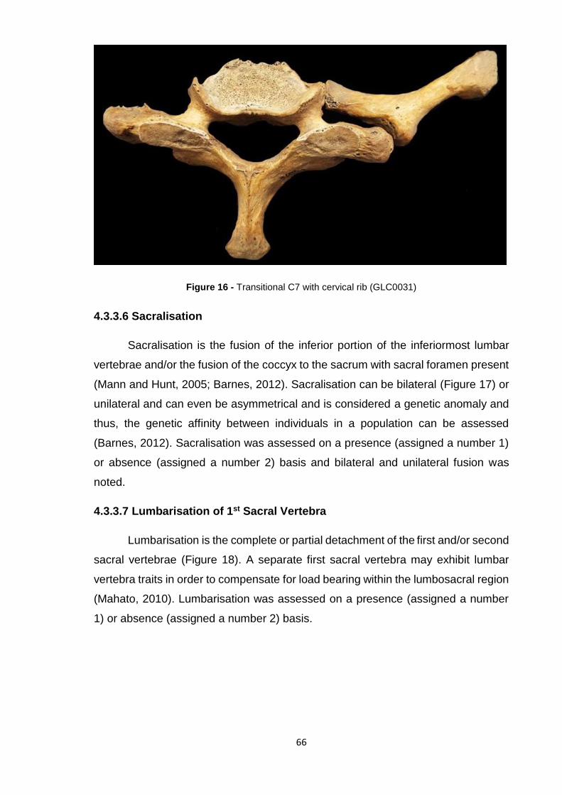
66
Figure 16 - Transitional C7 with cervical rib (GLC0031)
4.3.3.6 Sacralisation
Sacralisation is the fusion of the inferior portion of the inferiormost lumbar
vertebrae and/or the fusion of the coccyx to the sacrum with sacral foramen present
(Mann and Hunt, 2005; Barnes, 2012). Sacralisation can be bilateral (Figure 17) or
unilateral and can even be asymmetrical and is considered a genetic anomaly and
thus, the genetic affinity between individuals in a population can be assessed
(Barnes, 2012). Sacralisation was assessed on a presence (assigned a number 1)
or absence (assigned a number 2) basis and bilateral and unilateral fusion was
noted.
4.3.3.7 Lumbarisation of 1st Sacral Vertebra
Lumbarisation is the complete or partial detachment of the first and/or second
sacral vertebrae (Figure 18). A separate first sacral vertebra may exhibit lumbar
vertebra traits in order to compensate for load bearing within the lumbosacral region
(Mahato, 2010). Lumbarisation was assessed on a presence (assigned a number
1) or absence (assigned a number 2) basis.

67
Figure 18 - Bilateral lumbarisation of the 1st sacral vertebra (GLC0135)
Figure 17 - Complete bilateral sacralisation of L5 to S1 (GLC0030)

68
4.3.4 Trauma
The types of injury often observed in palaeopathological studies of the
vertebrae and other skeletal elements are direct and indirect trauma, fractures that
occur as a result of a disease/pathology and stress (Lovell, 1997). The identification
of ante, peri and post-mortem traumatic time frame injury types is essential to
accurate analysis of skeletal trauma (Table 5). Current and well-established criterion
was employed in this study (Quatrehomme and Işcan, 1997; Galloway et al., 1999;
Kimmerle and Baraybar, 2008).
Table 5 – Traumatic time frames differentiation in trauma identification
Traumatic time frame Description/Preservation
Ante-mortem trauma
Callus formation Evidence of bone remodelling (healing) Smooth edges
Peri-mortem trauma “butterfly fractures” Diagonal and radiating fracture lines
Post-mortem trauma Right-angled edges Flaking Colour difference in broken area of bone
4.3.4.1 Fractures
Fractures of the vertebrae can be caused by a variety of issues. Metabolic
diseases such as osteoporosis make individuals more susceptible to vertebral
fractures (Brickley, 2002; Old and Calvert, 2004). A sudden traumatic event could
also yield vertebral body burst fractures (Figure 19), which can cause long term
suffering for the individual (Lovell, 1997). A variety of fracture types are listed in
Table 6. Presence and absence of skeletal injury was concluded, and each injury
observed was examined to establish traumatic time frame and injury/fracture type.
Table 6 - Vertebral fracture types (adapted from Lovell, 1997)
Injury type Fracture type Description
Direct Trauma Crush
Compression Depression
Force applied to both sides of the bone Force applied to one side of the bone
Indirect Trauma Burst Vertical compression
Secondary to pathology Secondary fracture due to metabolic and systemic conditions
Stress Repeated episodes of force placed on the spine

69
2.3.4.2 Spondylolysis
Spondylolysis (“spondylos” – vertebra and “lysis” – dissolution) is a
defect/weakening and subsequent bilateral (Figure 20a) or unilateral fracture
(Figure 20b) of the pars interarticularis of a vertebra. All stress fractures of the
posterior arch will be diagnosed as spondylolysis. This particular lesion could be
considered to originate in a non-traumatic episode and be attributed to a natural
and/or congenital weakening in the neural arch and has therefore been assessed
seperately to crush, burst and stress fractures (of the vertebral bodies). The nature
of the fracture such as whether it is unilateral and bilateral (complete fracture) has
been noted. All fractures observed were labelled as either present (number 1) or
absent (number 2).
Figure 19 - Healed burst fracture on superoanterior body of the 4th lumbar vertebra of GLC0058 from St Owen's Cemetery (Red arrow points to fracture).
Figure 20 - (a) Bilateral spondylolysis of 5th lumbar vertebra (GLC0011) and (b) unilateral spondylolysis of 5th lumbar vertebra (POU490).
(a) (b)

70
4.3.5 Data Analysis
4.3.5.1 – Frequency of Pathological Lesions
The frequency of the bone lesions and pathological conditions observed were
divided by the total number of individuals at each site and then compared by sex
and age. All data were input and analysed in Microsoft Excel 2013 (Windows, 2013)
and IBM Statistical Package SPSS-23 (IBM, Corp, 2015).
4.3.5.2 – Statistical Analysis
Each bone lesion and pathological condition was observed and concluded to
be present (number 1) or absent (number 2) for each vertebra of each individual
from both archaeological sites. Absent segments and vertebrae were coded
separately (number 0). All of the variables were either dichotomous (present/absent
for pathological condition) or polytomous/nominal (sex – male, female and
indeterminate, age ranges – subadult, young adult, middle adult and old adult and
severity scale for osteophytes – 0, 1, 2, 3 and 4).
Normality testing of the samples was conducted in SPSS using the Shapiro-
Wilk test due to sample sizes of <50. Interpopulation variance, including variation
between the sexes and ages were evaluated. The chi-square test for independence
was used to compare samples because it can be used on non-normal and normal
data regardless including categorical data and counts of frequencies. This test was
also employed to observe significant differences between variables. In addition to
chi-square, a contingency coefficient was applied to sample sizes <5 to achieve a
more conservative result. A series of tables, bar charts, line graphs and pictorial
diagrams have been completed using Microsoft Excel to convey the results
obtained.
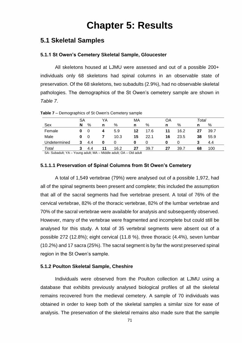
71
Chapter 5: Results
5.1 Skeletal Samples
5.1.1 St Owen’s Cemetery Skeletal Sample, Gloucester
All skeletons housed at LJMU were assessed and out of a possible 200+
individuals only 68 skeletons had spinal columns in an observable state of
preservation. Of the 68 skeletons, two subadults (2.9%), had no observable skeletal
pathologies. The demographics of the St Owen’s cemetery sample are shown in
Table 7.
Table 7 – Demographics of St Owen's Cemetery sample
Sex SA YA MA OA Total N % n % n % n % n %
Female 0 0 4 5.9 12 17.6 11 16.2 27 39.7
Male 0 0 7 10.3 15 22.1 16 23.5 38 55.9
Undetermined 3 4.4 0 0 0 0 0 0 3 4.4
Total 3 4.4 11 16.2 27 39.7 27 39.7 68 100 SA- Subadult; YA – Young adult; MA – Middle adult; OA – Old adult
5.1.1.1 Preservation of Spinal Columns from St Owen’s Cemetery
A total of 1,549 vertebrae (79%) were analysed out of a possible 1,972, had
all of the spinal segments been present and complete; this included the assumption
that all of the sacral segments had five vertebrae present. A total of 76% of the
cervical vertebrae, 82% of the thoracic vertebrae, 82% of the lumbar vertebrae and
70% of the sacral vertebrae were available for analysis and subsequently observed.
However, many of the vertebrae were fragmented and incomplete but could still be
analysed for this study. A total of 35 vertebral segments were absent out of a
possible 272 (12.8%); eight cervical (11.8 %), three thoracic (4.4%), seven lumbar
(10.2%) and 17 sacra (25%). The sacral segment is by far the worst preserved spinal
region in the St Owen’s sample.
5.1.2 Poulton Skeletal Sample, Cheshire
Individuals were observed from the Poulton collection at LJMU using a
database that exhibits previously analysed biological profiles of all the skeletal
remains recovered from the medieval cemetery. A sample of 70 individuals was
obtained in order to keep both of the skeletal samples a similar size for ease of
analysis. The preservation of the skeletal remains also made sure that the sample

72
was limited in number as truncation is a common issue within this particular
medieval cemetery. Out of the 70 skeletons analysed, four had no observable
skeletal pathologies; all of the latter were sub-adults (5.7%). Ten skeletons from
each age and sex category were analysed and thus, a demographics table has not
been created.
5.1.2.1 Preservation of Spinal Columns from Poulton
The number of vertebrae observed totalled 1,839 (91%) out of a possible
2,030 had all segments been present and complete; this amount also included the
assumption of five vertebrae per sacrum. A total of 91% of the cervical vertebrae,
95% of the thoracic vertebrae, 95% of the lumbar vertebrae and 76% of the sacral
vertebrae were available for analysis. Many of those were fragmented, but still in an
observable state of preservation that could be used for the study. A total of 17
segments were absent out of a possible 280 (6.1%) with many of those segments
incomplete; four cervical segments (5.7%), one thoracic segment (1.4%), three
lumbar segments (4.3%) and nine sacral segments (12.9%) were absent.
5.2 Joint Conditions
5.2.1 Osteoarthritis (OA)
Osteoarthritic changes, such as pitting, marginal osteophytes, new bone
formation on joint contours and eburnation on the vertebral articulations in the St
Owen’s cemetery sample is highly prevalent in both males and females (Figure 21).
St Owen’s individuals exhibited more OA than their counterparts in Poulton with OA
reaching in excess of 80.0% in females (84.0% on the T1 vertebra) and 69.0% in
males (68.9% on T1) vs. 65.5% (T11) in the Poulton females and 57.0% (57.1% on
T1) in the males. Interestingly, 89.4% (n=34/38) of males and 100.0% (n=27/27)
females from St Owen’s and 80.0% (n=24/30) of males and 96.6% (n=29/30) of
females in Poulton had arthritic changes on at least one vertebra
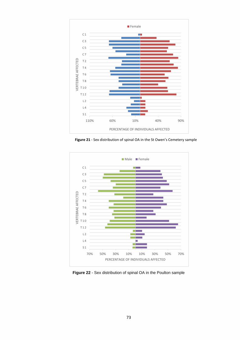
73
Figure 21 - Sex distribution of spinal OA in the St Owen’s Cemetery sample
Figure 22 - Sex distribution of spinal OA in the Poulton sample
110% 60% 10% 40% 90%
S 1
L 4
L 2
T 1 2
T 1 0
T 8
T 6
T 4
T 2
C 7
C 5
C 3
C 1
PERCENTAGE OF INDIVIDUALS AFFECTED
VER
TEB
RA
E A
FFEC
TED
Female
70% 50% 30% 10% 10% 30% 50% 70%
S 1
L 4
L 2
T 1 2
T 1 0
T 8
T 6
T 4
T 2
C 7
C 5
C 3
C 1
PERCENTAGE OF INDIVIDUALS AFFECTED
VER
TEB
RA
E A
FFEC
TED
Male Female

74
The distribution of arthritic changes appears to be very similar, in both
samples, with the same vertebrae exhibiting the highest incidence of OA such as
T1 and T12. However, there is a difference in the lumbar segment; females in
Poulton exhibit the highest amounts of OA whereas males in St Owen’s Cemetery
have the highest frequency (Figure 22).
No significant differences were observed in the presence of OA in the young
adults and old adult females. However, the tests highlighted a significant difference
between middle adult males; St Owen’s Cemetery exhibits much higher
percentages of OA on four mid-thoracic vertebrae (T6-T9) (Table 8).
Table 8 - Significant Chi-square findings and calculated percentages of OA in Poulton and St Owen’s
Cemetery
Poulton St Owen’s Cemetery
Vertebra χc2 p n/N % n/N %
Middle Adult Males
T6 11.461 p=<.00 2/9 22.2 11/13 84.6
T7 5.091 p=.010 1/9 11.1 9/13 69.2
T8 4.583 p=.015 2/10 20.0 9/12 75.0
T9 3.694 p=.026 2/10 20.0 8/13 61.5
Middle Adult Females
T1 4.267 p=.016 5/10 50.0 10/11 90.9
T6 4.237 p=.019 3/10 30.0 7/10 70.0
T7 2.991 p=.040 4/10 40.0 8/10 80.0
Old Adults Males
L4 2.869 p=.040 0/10 0.0 5/11 45.5
χc2 – Chi-Squared with continuity correction p – P- Value n= number of vertebrae with OA N= number of vertebrae observed in total
Old adult males exhibit a significant difference in the L4 vertebra; the St
Owen’s Cemetery sample shows five instances whereas no observation of OA was
made in the L4 vertebrae in Poulton males. No OA in subadults was recorded for
the St Owen’s sample. However, Poulton exhibited OA in two subadults. Although
there is an observable difference, there is no statistically significant difference
between subadults in the two samples (Table 9).
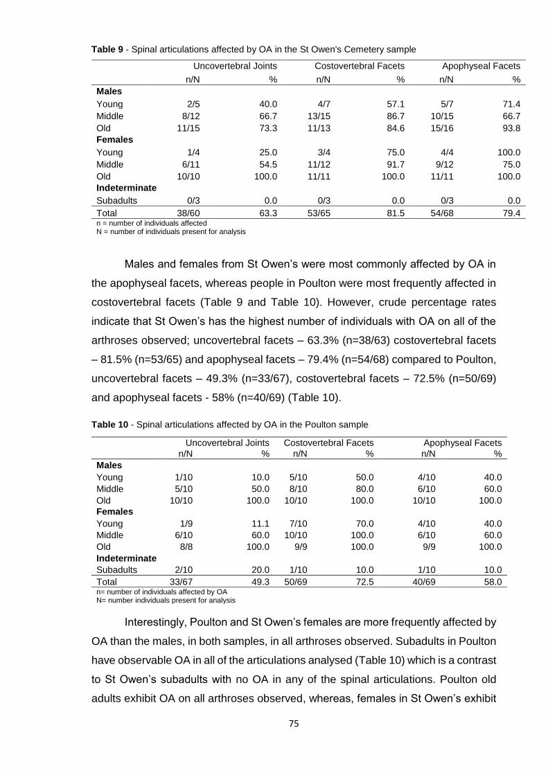
75
Table 9 - Spinal articulations affected by OA in the St Owen's Cemetery sample
Uncovertebral Joints Costovertebral Facets Apophyseal Facets
n/N % n/N % n/N %
Males
Young 2/5 40.0 4/7 57.1 5/7 71.4
Middle 8/12 66.7 13/15 86.7 10/15 66.7
Old 11/15 73.3 11/13 84.6 15/16 93.8
Females
Young 1/4 25.0 3/4 75.0 4/4 100.0
Middle 6/11 54.5 11/12 91.7 9/12 75.0
Old 10/10 100.0 11/11 100.0 11/11 100.0
Indeterminate
Subadults 0/3 0.0 0/3 0.0 0/3 0.0
Total 38/60 63.3 53/65 81.5 54/68 79.4 n = number of individuals affected N = number of individuals present for analysis
Males and females from St Owen’s were most commonly affected by OA in
the apophyseal facets, whereas people in Poulton were most frequently affected in
costovertebral facets (Table 9 and Table 10). However, crude percentage rates
indicate that St Owen’s has the highest number of individuals with OA on all of the
arthroses observed; uncovertebral facets – 63.3% (n=38/63) costovertebral facets
– 81.5% (n=53/65) and apophyseal facets – 79.4% (n=54/68) compared to Poulton,
uncovertebral facets – 49.3% (n=33/67), costovertebral facets – 72.5% (n=50/69)
and apophyseal facets - 58% (n=40/69) (Table 10).
Table 10 - Spinal articulations affected by OA in the Poulton sample
Uncovertebral Joints Costovertebral Facets Apophyseal Facets
n/N % n/N % n/N %
Males
Young 1/10 10.0 5/10 50.0 4/10 40.0
Middle 5/10 50.0 8/10 80.0 6/10 60.0
Old 10/10 100.0 10/10 100.0 10/10 100.0
Females
Young 1/9 11.1 7/10 70.0 4/10 40.0
Middle 6/10 60.0 10/10 100.0 6/10 60.0
Old 8/8 100.0 9/9 100.0 9/9 100.0
Indeterminate
Subadults 2/10 20.0 1/10 10.0 1/10 10.0
Total 33/67 49.3 50/69 72.5 40/69 58.0 n= number of individuals affected by OA N= number individuals present for analysis
Interestingly, Poulton and St Owen’s females are more frequently affected by
OA than the males, in both samples, in all arthroses observed. Subadults in Poulton
have observable OA in all of the articulations analysed (Table 10) which is a contrast
to St Owen’s subadults with no OA in any of the spinal articulations. Poulton old
adults exhibit OA on all arthroses observed, whereas, females in St Owen’s exhibit
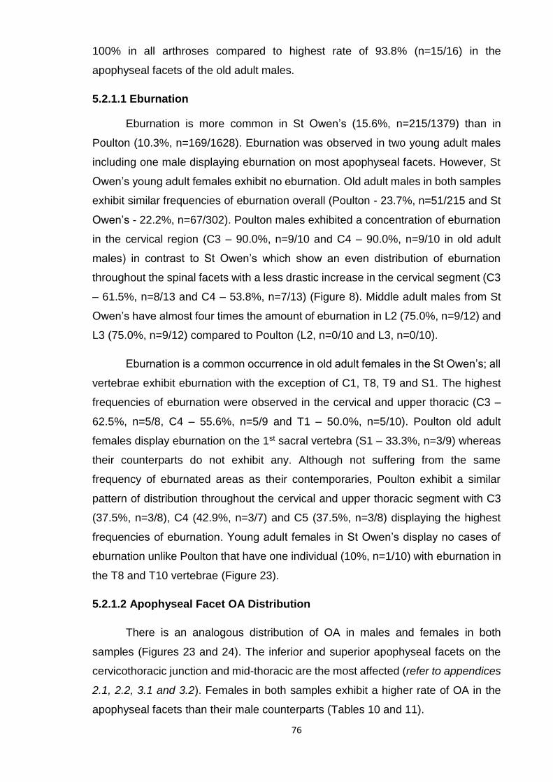
76
100% in all arthroses compared to highest rate of 93.8% (n=15/16) in the
apophyseal facets of the old adult males.
5.2.1.1 Eburnation
Eburnation is more common in St Owen’s (15.6%, n=215/1379) than in
Poulton (10.3%, n=169/1628). Eburnation was observed in two young adult males
including one male displaying eburnation on most apophyseal facets. However, St
Owen’s young adult females exhibit no eburnation. Old adult males in both samples
exhibit similar frequencies of eburnation overall (Poulton - 23.7%, n=51/215 and St
Owen’s - 22.2%, n=67/302). Poulton males exhibited a concentration of eburnation
in the cervical region (C3 – 90.0%, n=9/10 and C4 – 90.0%, n=9/10 in old adult
males) in contrast to St Owen’s which show an even distribution of eburnation
throughout the spinal facets with a less drastic increase in the cervical segment (C3
– 61.5%, n=8/13 and C4 – 53.8%, n=7/13) (Figure 8). Middle adult males from St
Owen’s have almost four times the amount of eburnation in L2 (75.0%, n=9/12) and
L3 (75.0%, n=9/12) compared to Poulton (L2, n=0/10 and L3, n=0/10).
Eburnation is a common occurrence in old adult females in the St Owen’s; all
vertebrae exhibit eburnation with the exception of C1, T8, T9 and S1. The highest
frequencies of eburnation were observed in the cervical and upper thoracic (C3 –
62.5%, n=5/8, C4 – 55.6%, n=5/9 and T1 – 50.0%, n=5/10). Poulton old adult
females display eburnation on the 1st sacral vertebra (S1 – 33.3%, n=3/9) whereas
their counterparts do not exhibit any. Although not suffering from the same
frequency of eburnated areas as their contemporaries, Poulton exhibit a similar
pattern of distribution throughout the cervical and upper thoracic segment with C3
(37.5%, n=3/8), C4 (42.9%, n=3/7) and C5 (37.5%, n=3/8) displaying the highest
frequencies of eburnation. Young adult females in St Owen’s display no cases of
eburnation unlike Poulton that have one individual (10%, n=1/10) with eburnation in
the T8 and T10 vertebrae (Figure 23).
5.2.1.2 Apophyseal Facet OA Distribution
There is an analogous distribution of OA in males and females in both
samples (Figures 23 and 24). The inferior and superior apophyseal facets on the
cervicothoracic junction and mid-thoracic are the most affected (refer to appendices
2.1, 2.2, 3.1 and 3.2). Females in both samples exhibit a higher rate of OA in the
apophyseal facets than their male counterparts (Tables 10 and 11).

77
80% 60% 40% 20% 0% 20% 40% 60% 80%
C1
C2
C3
C4
C5
C6
C7
T1
T2
T3
T4
T5
T6
T7
T8
T9
T10
T11
T12
L1
L2
L3
L4
L5
S1
Percentage of Individuals with Eburnation
Vert
ebra
e w
ith E
burn
ation
YAF_G MAF_G OAF_G YAF_P MAF_P OAF_P
100% 50% 0% 50% 100%
C1
C2
C3
C4
C5
C6
C7
T1
T2
T3
T4
T5
T6
T7
T8
T9
T10
T11
T12
L1
L2
L3
L4
L5
S1
Percentage of Individuals with Eburnation
Vert
ebra
e w
ith E
burn
ation
YAM_G MAM_G OAM_G YAM_P MAM_P OAM_P
Figure 23 - Percentage of eburnation per vertebra in the Poulton (purple) and
St Owen’s Cemetery (red) females.
Figure 24 - Percentage of eburnation per vertebra in the Poulton (green) and St Owen’s Cemetery (blue) males.
Figure key – G = Gloucester; P = Poulton; YAF = Young adult female; MAF = Middle adult female; OAF = Old adult female; YAM = Young Adult Male; MAM
= Middle adult male; OAM = Old adult male

78
Figure 25 - Advanced osteoarthritis of the superior apophyseal facets of a thoracic vertebra in an old adult male from St Owen’s Cemetery (GLC0043).
Table 11 – Number and percentage of superior and inferior apophyseal facets affected by osteoarthritis in both skeletal samples.
St Owen's Cemetery Poulton
Males Females Males Females
n/N % n/N % nN % n/N %
Superior Facets 179/1434 12.5 144/1180 12.2 122/1370 8.9 125/1396 9.0 Inferior Facets 157/1434 10.9 152/1180 12.9 138/1370 10.1 152/1396 10.9 n = number of superior and inferior facets with OA (left and right facets) N = number of superior and inferior facets observed in total.
St Owen’s Cemetery males have a tendency towards OA on the left superior,
left inferior and right inferior apophyseal facets in cervical vertebrae. Young adult
females exhibit OA in the inferior apophyseal facets in the upper-mid thoracic
vertebrae (T4 – 75.0%, n=3/4 and T5 – 50.0%, n=2/4) yet do not exhibit any in the
superior facets at all. Old adult females appear to have a tendency towards OA on
the left side in the cervical thoracic junction with T1 displaying the highest
frequencies (T1 left superior facet – 70.0%, n=7/10 and left inferior facet 20.0%,
n=2/10). The left side of the vertebral column appears to exhibit the highest amount
of OA in St Owen’s males and females (see Appendix 1.1 and 1.2).

79
Poulton old adult males and females seem to exhibit the highest amounts of
OA in the cervical vertebrae (Figure 26) on the inferior and superior facets (males
exhibit the highest percentage in the C3 vertebra – 70.0%, n=7/10 and females in
the C3 vertebra 42.9%, n=3/7) (see Appendix 2.1 and 2.2). Middle adult females
exhibit the most OA in the lumbar apophyseal facets (a high of 20.0%, n=2/10 in L1,
L2, L4 and L5 vertebrae) and old adult females have a high amount of OA in the L5
facets on all superior and inferior facets (22.2%, n=2/9) (Table 11).

80
(a)
(b)
Figure 26 – (a) Osteoarthritis of the right inferior facet of the atlas vertebra of an old adult female from the Poulton sample (POU86). (b) Anterolateral macro view of eburnation on the right inferior facet of the atlas vertebra

81
5.2.2 Intervertebral Disc Disease (IDD)
Degeneration of the intervertebral discs, identified by pitted and porotic
vertebral endplates and marginal osteophytes, is common in both samples. The
vertebrae T1 (Poulton – 12.1%, n=8/66 and St Owen’s – 17.5%, n=10/57), T2
(Poulton – 10.4%, n=7/67 and St Owen’s – 20.4%, n=11/54) and T3 (Poulton –
10.8%, n=7/65 and St Owen’s – 20.8%, n=11/53) have the lowest frequencies of
IDD whereas the vertebrae located in the mid-cervical and the thoracolumbar
junction exhibit the highest frequencies (Figures 27 and 28).
Intervertebral disc disease is present in the cervical (n=9/9) and thoracic
segments in 100.0% (n=10/10) of the old adult females in St Owen’s and in 100.0%
of the lumbar segments of the old females in Poulton (Figures 27 and 28). However,
males and females in both samples exhibit high frequencies in all spinal segments
(Table 12).
The occurrence of IDD increases with age but middle adult males exhibit the
highest frequencies of IDD in the mid and lower thoracic segments in St Owen’s
males (Figure 27). The increase with age of sacral IDD is distinct in males and
females in both samples with old females exhibiting the highest counts. However,
there is an observable difference between old adult males (54.5%, n=6/11) and
females (72.7%, n=8/11) in the crude prevalence of sacral IDD. Young adult males
in both Poulton (70.0%, n=7/10) and St Owen’s (57.1%, n=4/7) exhibit a high rate
of IDD in the thoracic segment (Table 12).
Chi-square tests show no significant differences in IDD between young adult
females, young adult males, middle adult females, old adult females and old adult
males. However, there is a significant difference in the prevalence of IDD in the T12
vertebrae of middle adult males with St Owen’s males exhibiting 91.7% (n=11/12)
compared to 40.0% (n=4/10) in Poulton males (χc2 (1, n=22) =3.188, p=.037).

82
Table 12 – Age, sex and segmental distribution of IDD in the St Owen's and Poulton samples
Male Female Ind.
Young Adult Middle Adult Old Adult Young Adult Middle Adult Old Adult Subadult St Owen’s Cemetery
Spinal segment n/N % n/N % n/N % n/N % n/N % n/N % n/N %
Cervical 1/5 20.0 8/12 66.7 13/15 86.7 1/4 25.0 7/12 58.3 10/10 100.0 0/3 0.0
Thoracic 4/7 57.1 13/15 86.7 11/13 84.6 2/4 50.0 9/12 75.0 11/11 100.0 0/3 0.0
Lumbar 4/7 57.1 11/13 84.6 11/13 84.6 1/4 25.0 8/11 72.7 9/10 90.0 0/3 0.0
Sacral 0/7 0.0 5/10 50.0 6/11 54.5 0/4 0.0 5/11 45.5 8/11 72.7 0/3 0.0
Poulton
n/N % n/N % n/N % n/N % n/N % n/N % n/N %
Cervical 4/10 40.0 6/10 60.0 8/10 80.0 2/9 22.2 6/10 60.0 8/9 88.8 1/10 10.0
Thoracic 7/10 70.0 6/10 60.0 8/10 80.0 3/9 33.3 6/10 60.0 8/9 88.8 1/10 10.0
Lumbar 3/10 30.0 7/10 70.0 7/9 77.7 2/10 20.0 7/70 70.0 9/9 100.0 0/10 0.0
Sacral 2/9 22.2 3/8 37.5 6/8 75.0 0/10 0.0 6/10 60.0 7/9 77.7 0/10 0.0
n = number of instances of IDD per segment N = number of segments observed
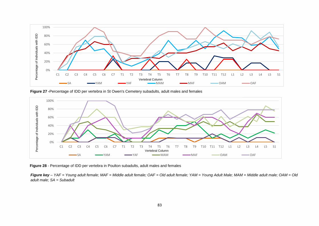
83
Figure 28 - Percentage of IDD per vertebra in Poulton subadults, adult males and females
0%
20%
40%
60%
80%
100%
C1 C2 C3 C4 C5 C6 C7 T1 T2 T3 T4 T5 T6 T7 T8 T9 T10 T11 T12 L1 L2 L3 L4 L5 S1
Perc
enta
ge o
f In
div
iduals
with I
DD
Vertebral Column
SA YAM YAF MAM MAF OAM OAF
0%
20%
40%
60%
80%
100%
C1 C2 C3 C4 C5 C6 C7 T1 T2 T3 T4 T5 T6 T7 T8 T9 T10 T11 T12 L1 L2 L3 L4 L5 S1
Perc
enta
ge o
f In
div
iduals
with I
DD
Vertebral Column
SA YAM YAF MAM MAF OAM OAF
Figure 27 -Percentage of IDD per vertebra in St Owen's Cemetery subadults, adult males and females
Figure key – YAF = Young adult female; MAF = Middle adult female; OAF = Old adult female; YAM = Young Adult Male; MAM = Middle adult male; OAM = Old
adult male; SA = Subadult

84
5.2.3 Schmörl’s Nodes (SN)
There appears to be consistency in the frequency and distribution of SNs
observed in both samples (Figure 29). No SNs were observed in the cervical
segment at all. Vertebrae T11 (11.4% of the total SNs observed, n=62/542) and T12
(11.8% of the total SNs observed, n=64/542) exhibit the highest frequency of SN’s
in Poulton and T10 (10.9% of the total SNs observed, n=59/541) and T11 (10.9% of
the total SNs observed, n=59/541) in St Owen’s.
Table 13 – Frequency count and percentage of SNs on the inferior and superior vertebral endplates in Poulton and St Owen’s males and females
St Owen’s Cemetery Poulton
Males Females Males Females
n/N % n/N % n/N % n/N
Superior 163/519 31.4 73/443 16.5 150/480 31.3 109/513 21.2
Inferior 196/519 37.8 109/443 24.6 155/480 32.2 128/513 25.0
Total 359/1038 34.6 182/886 20.5 305/960 31.8 237/1026 23.1
n = number of SNs on inferior and superior endplates N = number of endplates observed
Males and females have a tendency towards SNs on the inferior vertebral
endplates in the mid-thoracic segment which then changes to the superior surface
in the thoracolumbar junction. St Owen’s males have the highest frequency of SNs
overall on the superior and inferior vertebral endplates (34.6%, n=359/1038).
However, St Owen’s females have the lowest frequencies (20.5%, n=182/886)
(Table 13).
Table 14 – Sex and age categories affected by SNs in the St Owen’s Cemetery and Poulton skeletal samples
St Owens
Poulton
n N % n N %
Indeterminate
Subadult 1 3 33.3 7 10 70.0
Males
Young Adult 7 7 100.0 9 10 90.0
Middle Adult 14 15 93.3 10 10 100.0
Old Adult 12 16 75.0 7 8 87.5
Total 33 38 86.8 26 28 92.9
Females
Young Adult 4 4 100.0 9 10 90.0
Middle Adult 10 12 83.3 9 10 90.0
Old Adult 8 11 72.8 9 9 100.0
Total 22 27 81.5 27 29 93.1 n = number of individuals with SNs N = number of individuals observed

85
Males and females are similarly affected by SNs but males have the highest
frequencies of SNs in total (Poulton – 43%, n=207/480 and St Owens – 46%,
n=241/519) (Table 14 and Table 15). The percentages of SNs located on the
superior and inferior surfaces of the spinal column are very similar between
samples. There is a predilection for inferior vertebral endplate SNs in the thoracic
segment and a tendency for superior SNs in the thoracolumbar and lumbar regions
(Figures 30a and 30b).
Table 15 – Number of vertebrae with SNs in the Poulton and St Owen’s samples (Sex and age distribution)
St Owens Poulton
n N % n N %
Indeterminate Subadult 3 54 5.56 42 180 23.3
Males Young Adult 43 100 43.0 87 179 48.6 Middle Adult 113 211 53.6 61 154 39.6 Old Adult 85 208 40.9 59 147 40.1 Total 241 519 46.4 207 480 43.0
Females Young Adult 26 69 37.7 67 173 38.7 Middle Adult 52 193 26.9 63 180 35.0 Old Adult 56 181 30.9 43 160 26.9 Total 134 443 30.2 173 513 33.7 n = number of vertebrae observed N = number of vertebrae observed with SNs (superior and inferior endplates) % = frequency of SNs on number of vertebrae observed
0
10
20
30
40
50
60
70
T1 T2 T3 T4 T5 T6 T7 T8 T9 T10 T11 T12 L1 L2 L3 L4 L5 S1
SN
Count
Vertebral Column
Poulton St Owen's Cemetery
Figure 29 – Raw values and distribution of Schmörl’s nodes in the adults of Poulton and St Owen's Cemetery samples

86
Figure 30 - Sex distribution, anatomical location and frequency of SNs in the Poulton (a) and St Owen’s Cemetery (b) samples.
Inf. = Inferior; Sup. = Superior
20 15 10 5 0 5 10 15 20
T1
T2
T3
T4
T5
T6
T7
T8
T9
T10
T11
T12
L1
L2
L3
L4
L5
S1
Schmorl's Node Frequency (%)
Vert
ebra
l C
olu
mn
Male Sup. Male Inf. Female Sup. Female Inf.(a)
25 20 15 10 5 0 5 10 15
T1
T2
T3
T4
T5
T6
T7
T8
T9
T10
T11
T12
L1
L2
L3
L4
L5
S1
Schmorl's Node Frequency (%)
Vert
ebra
l C
olu
mn
Males Sup. Males Inf. Females Sup. Females Inf.(b)
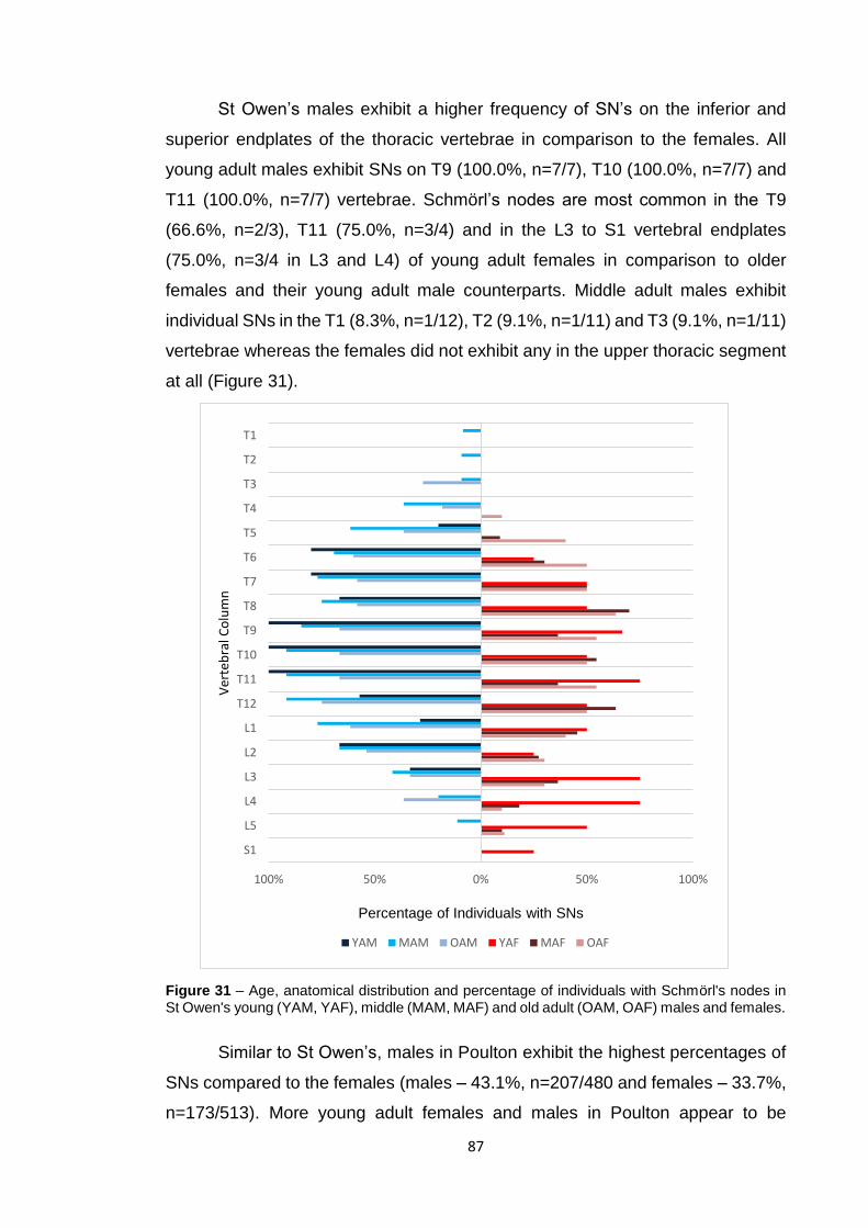
87
St Owen’s males exhibit a higher frequency of SN’s on the inferior and
superior endplates of the thoracic vertebrae in comparison to the females. All
young adult males exhibit SNs on T9 (100.0%, n=7/7), T10 (100.0%, n=7/7) and
T11 (100.0%, n=7/7) vertebrae. Schmörl’s nodes are most common in the T9
(66.6%, n=2/3), T11 (75.0%, n=3/4) and in the L3 to S1 vertebral endplates
(75.0%, n=3/4 in L3 and L4) of young adult females in comparison to older
females and their young adult male counterparts. Middle adult males exhibit
individual SNs in the T1 (8.3%, n=1/12), T2 (9.1%, n=1/11) and T3 (9.1%, n=1/11)
vertebrae whereas the females did not exhibit any in the upper thoracic segment
at all (Figure 31).
Figure 31 – Age, anatomical distribution and percentage of individuals with Schmörl's nodes in St Owen's young (YAM, YAF), middle (MAM, MAF) and old adult (OAM, OAF) males and females.
Similar to St Owen’s, males in Poulton exhibit the highest percentages of
SNs compared to the females (males – 43.1%, n=207/480 and females – 33.7%,
n=173/513). More young adult females and males in Poulton appear to be
100% 50% 0% 50% 100%
T1
T2
T3
T4
T5
T6
T7
T8
T9
T10
T11
T12
L1
L2
L3
L4
L5
S1
Percentage of Individuals with SNs
Ver
teb
ral C
olu
mn
YAM MAM OAM YAF MAF OAF

88
affected by SNs than other age categories and this is concentrated in the mid-
thoracic segment and thoracolumbar areas (Figure 32). Young adult males,
similar to their urban counterparts, exhibit the highest frequencies of SNs in T7
(90.0%, n=9/10), T9 (90.0%, n=9/10), T10 (90.0%, n=9/10) and, T11 (90.0%,
n=9/10). Young adult females display the highest frequencies of SNs in T10
(80.0%, n=8/10) and T11 (80.0%, n=8/10) vertebrae. This is comparable to the
young adult females in St Owen’s where 75.0% (n=3/4) exhibit SNs in the T11
vertebrae (Figure 14). Old adult females are the least affected by SNs in all of the
vertebrae in all of the sex and age categories apart from in the T3 (12.5%, n=1/8),
T4 (11.1%, n=1/9) and L5 (11.1%, n=1/9) vertebrae. Old adult males, similar to
the females, appear to be less affected by SNs apart from in the lumbar and
sacral segments.
Figure 32 – Age and anatomical distribution of Individuals with Schmörl's nodes in Poulton young adult (YAM, YAF), middle adult (MAM, MAF) and old adult (OAM, OAF) males and females
100% 50% 0% 50% 100%
T1
T2
T3
T4
T5
T6
T7
T8
T9
T10
T11
T12
L1
L2
L3
L4
L5
S1
Percentage of Individuals with SNs
Vert
ebra
l C
olu
mn
YAM MAM OAM YAF MAF OAF
89
Table 16 – Frequency, anatomical location and distribution of SNs in Poulton and St Owen’s Cemetery subadults
Poulton St Owen’s Cemetery
n Sup. Inf. Total n Sup. Inf. Total
T1 10 0 0 0 3 0 0 0
T2 10 0 0 0 3 0 0 0
T3 10 0 0 0 3 0 0 0
T4 10 0 1 1 3 0 0 0
T5 10 2 3 5 3 0 0 0
T6 10 3 4 7 3 0 0 0
T7 10 2 5 7 3 1 0 1
T8 10 4 5 9 3 1 1 2
T9 10 3 5 8 3 1 0 1
T10 10 4 2 6 3 0 0 0
T11 10 3 4 7 3 0 0 0
T12 10 2 2 4 3 0 0 0
L1 10 3 3 6 3 0 0 0
L2 10 3 3 6 3 0 0 0
L3 10 2 1 3 3 0 0 0
L4 10 0 1 1 3 0 0 0
L5 10 0 0 0 3 0 0 0
S1 10 0 0 0 3 0 0 0
Total 180 30 41 70 54 3 1 4
n = number of vertebrae observed
Seven subadults (70%, n=7/10) exhibit SNs in the Poulton compared to
only one individual in St Owen’s (33.3%, n=1/3). Schmӧrl’s nodes were far more
prevalent in the Poulton subadults on both superior and inferior endplates (19.4%,
n=70/360) in comparison to St Owen’s Cemetery subadults (3.7%, n=4/108).
Comparably to the adults, subadults in both samples exhibited SNs in the mid-
thoracic and thoracolumbar junction (Table 16).

90
5.2.4 Diffuse Idiopathic Skeletal Hyperostosis (DISH)
Diffuse idiopathic skeletal hyperostosis was observed in two old adult
males in the St Owen’s sample (2.9%, n=2/68) and in one middle adult female in
Poulton (1.4%, n=1/70) (Table 17)
Table 17 – Diffuse Idiopathic Skeletal Hyperostosis in the Poulton and St Owen's samples
St Owen's Cemetery Poulton
Male Female Male Female
n n n n
Young Adult 0 0 0 0
Middle Adult 0 0 0 1
Old Adult 2 0 0 0
5.2.5 Vertebral Osteophytes
5.2.5.1 St Owen’s Cemetery
Table 18 - Age and sex distribution of severity of the osteophytes observed in the St Owen’s Cemetery sample
Age Sex
0 1 2 3 4
n/N % n/N % n/N % n/N % n/N %
Sub- adult I 132/138 95.7 6/138 4.3 0/0 0.0 0/0 0.0 0/0 0.0 Young Adult M 166/258 64.3 88/258 34.1 14/258 5.4 0/0 0.0 0/0 0.0 F 68/169 40.2 83/169 49.1 17/169 10.1 0/0 0.0 1/169 0.6 Middle Adult M 114/555 20.5 191/555 34.4 187/555 33.7 32/555 5.8 31/555 5.6 F 118/468 25.2 153/468 32.7 153/468 32.7 32/468 6.8 12/468 2.6 Old Adult M 54/604 8.9 179/604 30 196/604 32.5 63/604 10.4 101/604 16.7 F 38/479 7.9 157/479 32.8 200/479 41.8 51/479 10.6 33/479 6.9 n = number of endplates with osteophytes N= number of endplates I = Indeterminate sex; M = Male; F = Female 0 = no osteophyte; 1 = 0.1-1mm; 2 = 1.-3mm; 3 = 3-5mm; 4 = 5mm+
The severity of vertebral osteophytes became more apparent with
increasing age. Subadults exhibit grade zero osteophytes in 95.7% (n=132/138)
of the vertebrae analysed (superior and inferior margins). Young adult males
mainly exhibit zero on the grading scale (64.3%, n=166/258) in St Owen’s
Cemetery, although some had grade one (34.1%, n=88/258) and grade two
osteophytes (5%, n=14/258) formations (≤ 3mm). Young adult females exhibited
over 85% of grade zero (40.2%, n=68/169) to grade one (49.1%, n=83/169)
osteophytes. Nevertheless, one individual (0.6%, n=1/169) exhibits large and
severe osteophytes which is most likely due to trauma or disease (grade four).

91
Old adult males and females exhibit the highest percentages of grade four
osteophytes (males – 16.7%, n=101/604 and females – n=6.9%, n=33/479)
(>5mm) (Table 18).
5.2.5.2 Poulton
Table 19 - Age and sex distribution of severity of the osteophytes observed in the Poulton sample
Age Sex
0 1 2 3 4
n/N % n/N % n/N % n/N % n/N %
Sub-adult I 403/471 85.6 61/471 13 5/471 1.1 2/471 0.4 0/0 0.0 Young Adult M 189/489 38.7 215/489 44 65/489 13.3 10/489 2.0 10/489 2.0 F 225/447 50.3 181/447 40.5 40/447 8.9 1/447 0.2 0/447 0.0 Middle Adult M 93/454 20.5 154/454 33.9 150/454 33 25/454 5.5 32/454 7.0 F 62/488 12.7 147/488 30.1 181/488 37.1 57/488 11.7 41/488 8.4 Old Adult M 42/417 10.1 126/417 30.2 151/417 36.2 54/417 12.9 44/417 10.6
F 26/417 6.2 134/417 32.1 174/417 41.7 58/417 13.9 25/417 6.0 n = number of endplates with osteophytes N= number of endplates I = Indeterminate sex; M = Male; F = Female 0 = no osteophyte; 1 = 0.1-1mm; 2 = 1.-3mm; 3 = 3-5mm; 4 = 5mm+
Subadults in Poulton exhibit more grade one osteophytes (13.0%,
n=61/471) compared to St Owen’s (Grade one – 4.3%, n=6/138) but still have a
large amount of grade zero osteophytes on the inferior and superior vertebral
margins (85.6%, n=403/471). Young adult males display osteophytes from all of
the severity grades. However, 83% of the osteophyte scores fell within grade zero
(38.7%, n=189/489) and grade one (44.0%, n=215/489). Young adult females
exhibited 90% grade zero (50.3%, n=225/447) and grade one vertebral
osteophytes (40.5%, n=181/447) and unlike their male counterparts, did not
exhibit any severe grade four osteophytes but did have one individual with grade
three osteophytes (0.2%, n=1/447). Middle adult females in Poulton (11.7%,
n=57/488) have a tendency towards grade three osteophytes over their
counterparts in St Owen’s Cemetery (5.5%, n=25/454). Old adult males show the
highest prevalence of grade four osteophytes in 10.6% (n=44/417) of the sample
in comparison to only 6.0% (n=25/417) of old adult females (Table 19), this
frequency of severity is very similar to that of their urban counterparts (males –
16.7%, n=101/604 and females – 6.9%, n=33/479).
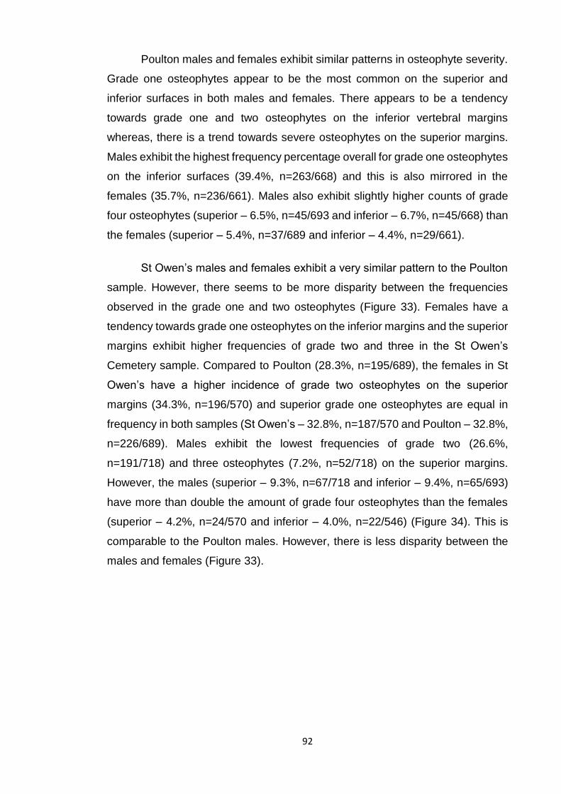
92
Poulton males and females exhibit similar patterns in osteophyte severity.
Grade one osteophytes appear to be the most common on the superior and
inferior surfaces in both males and females. There appears to be a tendency
towards grade one and two osteophytes on the inferior vertebral margins
whereas, there is a trend towards severe osteophytes on the superior margins.
Males exhibit the highest frequency percentage overall for grade one osteophytes
on the inferior surfaces (39.4%, n=263/668) and this is also mirrored in the
females (35.7%, n=236/661). Males also exhibit slightly higher counts of grade
four osteophytes (superior – 6.5%, n=45/693 and inferior – 6.7%, n=45/668) than
the females (superior – 5.4%, n=37/689 and inferior – 4.4%, n=29/661).
St Owen’s males and females exhibit a very similar pattern to the Poulton
sample. However, there seems to be more disparity between the frequencies
observed in the grade one and two osteophytes (Figure 33). Females have a
tendency towards grade one osteophytes on the inferior margins and the superior
margins exhibit higher frequencies of grade two and three in the St Owen’s
Cemetery sample. Compared to Poulton (28.3%, n=195/689), the females in St
Owen’s have a higher incidence of grade two osteophytes on the superior
margins (34.3%, n=196/570) and superior grade one osteophytes are equal in
frequency in both samples (St Owen’s – 32.8%, n=187/570 and Poulton – 32.8%,
n=226/689). Males exhibit the lowest frequencies of grade two (26.6%,
n=191/718) and three osteophytes (7.2%, n=52/718) on the superior margins.
However, the males (superior – 9.3%, n=67/718 and inferior – 9.4%, n=65/693)
have more than double the amount of grade four osteophytes than the females
(superior – 4.2%, n=24/570 and inferior – 4.0%, n=22/546) (Figure 34). This is
comparable to the Poulton males. However, there is less disparity between the
males and females (Figure 33).

93
Figure 33 - Osteophyte severity on the superior and inferior vertebral margins in the Poulton males and females
Figure 34 – Osteophyte severity on the superior and inferior vertebral margins in the St Owen’s Cemetery males and females.
0%
5%
10%
15%
20%
25%
30%
35%
40%
0 1 2 3 4
Perc
enta
ge o
f V
ert
ebra
e
Osteophyte Severity Scale
Male Superior Male Inferior Female Superior Female Inferior
0%
5%
10%
15%
20%
25%
30%
35%
40%
45%
0 1 2 3 4
Perc
enta
ge o
f vert
ebra
e
Osteophyte Severity Scale
Male Superior Male Inferior Female Superior Female Inferior
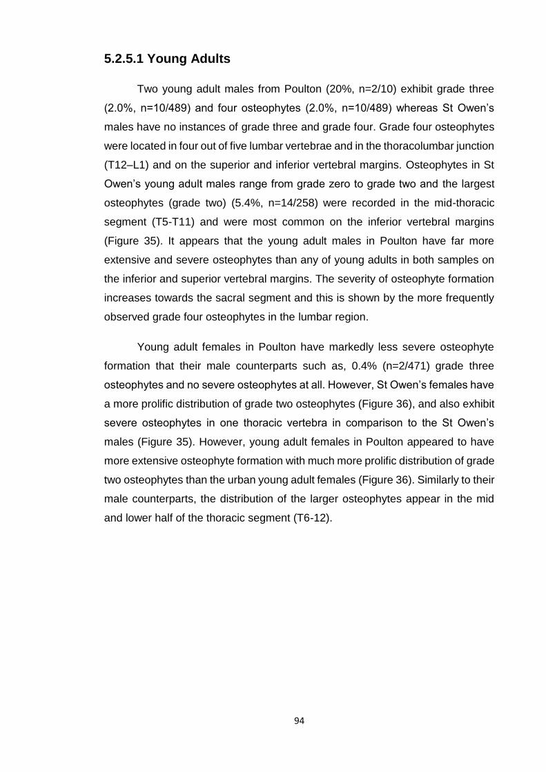
94
5.2.5.1 Young Adults
Two young adult males from Poulton (20%, n=2/10) exhibit grade three
(2.0%, n=10/489) and four osteophytes (2.0%, n=10/489) whereas St Owen’s
males have no instances of grade three and grade four. Grade four osteophytes
were located in four out of five lumbar vertebrae and in the thoracolumbar junction
(T12–L1) and on the superior and inferior vertebral margins. Osteophytes in St
Owen’s young adult males range from grade zero to grade two and the largest
osteophytes (grade two) (5.4%, n=14/258) were recorded in the mid-thoracic
segment (T5-T11) and were most common on the inferior vertebral margins
(Figure 35). It appears that the young adult males in Poulton have far more
extensive and severe osteophytes than any of young adults in both samples on
the inferior and superior vertebral margins. The severity of osteophyte formation
increases towards the sacral segment and this is shown by the more frequently
observed grade four osteophytes in the lumbar region.
Young adult females in Poulton have markedly less severe osteophyte
formation that their male counterparts such as, 0.4% (n=2/471) grade three
osteophytes and no severe osteophytes at all. However, St Owen’s females have
a more prolific distribution of grade two osteophytes (Figure 36), and also exhibit
severe osteophytes in one thoracic vertebra in comparison to the St Owen’s
males (Figure 35). However, young adult females in Poulton appeared to have
more extensive osteophyte formation with much more prolific distribution of grade
two osteophytes than the urban young adult females (Figure 36). Similarly to their
male counterparts, the distribution of the larger osteophytes appear in the mid
and lower half of the thoracic segment (T6-12).
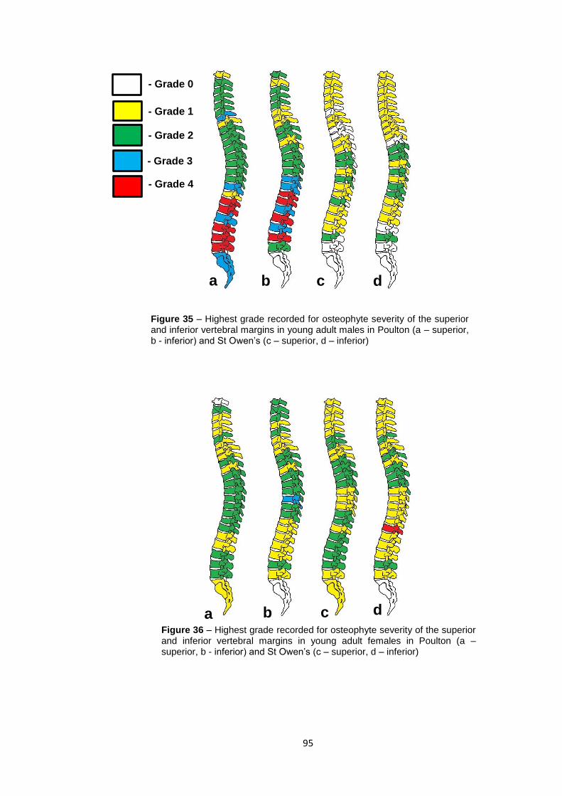
95
- Grade 3
- Grade 0
- Grade 1
- Grade 2
- Grade 4
a b c d
Figure 35 – Highest grade recorded for osteophyte severity of the superior and inferior vertebral margins in young adult males in Poulton (a – superior, b - inferior) and St Owen’s (c – superior, d – inferior)
a b c d Figure 36 – Highest grade recorded for osteophyte severity of the superior and inferior vertebral margins in young adult females in Poulton (a – superior, b - inferior) and St Owen’s (c – superior, d – inferior)
96
5.2.5.2 Middle Adults
Osteophyte formation and severity increases with age, especially in the
weight bearing vertebrae of the thoracic and lumbar segments (Figures 37 and
Figure 38). A larger distribution of severe osteophytes is apparent in the males
from St Owen’s. However, both Poulton and St Owen’s males seem similarly
affected by severe osteophytes (Poulton – 7.5%, n=32/454 and St Owen’s –
5.6%, n=31/555). Superior and inferior vertebral margins are similarly affected by
large osteophytes.
Poulton females appear to exhibit higher frequencies of grades three
(11.6%, n=57/488) and four (8.4%, n=41/488) than their male counterparts (grade
three – 5.5%, n=25/454 and grade four – 7.1%, n=32/454) (Figure 37). Poulton
females exhibit more than double the amount of grade four osteophytes than the
St Owen’s Cemetery females (2.6%, n=12/468). Poulton females have
concentration of grade three osteophytes in the cervico-thoracic junction that also
appear to be affecting the atlas vertebrae. The superior vertebral margins are
more affected by severe grade four osteophytes than the inferior margins in
Poulton females whereas the females in St Owen’s frequently have grades three
and four osteophytes on the inferior vertebral margins (Figure 38).
5.2.5.3 Old Adults
Osteophytes severity increases with age in the old adult males in both
samples (Figure 39 and Figure 40). However, this is reversed in the old adult
females with less grade four osteophytes (6.0%, n=25/417) but a higher rate of
grade three osteophytes (13.9%, n=58/417) than the middle adult females in
Poulton (Figure 23). St Owen’s Cemetery males display prolific grade four
osteophytes (16.7%, n=101/604) throughout the entire spine with the exception
of the upper cervical vertebrae on the superior and inferior vertebral margins. The
Poulton old adult males have an area of condensed grade four osteophytes
(10.6%, n=44/417) around the mid-thoracic and thoracolumbar segments but still
have grades two and three osteophytes in the thoracic and cervical segments. St
Owen’s Cemetery old adult females exhibit grade four osteophytes (6.0%,
n=33/479) in the mid/lower cervical segment with the Poulton sample showing no
instances of any grade four osteophytes within this spinal segment. Grades three
and four osteophytes appear to mainly affect the mid and lower thoracic and the

97
lumbar and sacral segments. St Owen’s females exhibit a tendency towards
severe osteophytes on the inferior vertebral margins (Figure 39).
a b
a b
c d
Figure 37 – Highest grade recorded for osteophyte severity of the superior and inferior vertebral margins in middle adult males in Poulton (a – superior, b - inferior) and St Owen’s (c – superior, d – inferior).
c d Figure 38 – Highest grade recorded for osteophyte severity of the superior
and inferior vertebral margins in middle adult females in Poulton (a –
superior, b - inferior) and St Owen’s (c – superior, d – inferior).

98
a b
a b
c
c d
d
Figure 39 – Highest grade recorded for osteophyte severity of the superior and inferior vertebral margins in old adult males in Poulton (a – superior, b - inferior) and St Owen’s (c – superior, d – inferior)
Figure 40 – Highest grade recorded for osteophyte severity of the superior and inferior vertebral margins in old adult females in Poulton (a – superior, b - inferior) and St Owen’s (c – superior, d – inferior)

99
5.3 Infectious Diseases
5.3.1 Tuberculosis, Brucellosis and Spinal Osteomyelitis
Infectious lesions were commonly observed in the Poulton and St Owen’s
Cemetery samples. Fourteen cases were identified in Poulton (20.0%, n=14/70)
and 14 cases in St Owen’s Cemetery (20.6%, n=14/68). Males were more
affected by infectious lesions than the females; nine St Owen’s cemetery males
(23.7%, n=9/38) compared to five females (18.5%, n=5/27) and nine males in
Poulton (30.0%, n=9/30) compared to four females (13.3%, n=4/30) (Table 20).
Figure 41 – Sex and age distribution of infectious diseases observed in the Poulton and St
Owen's Cemetery samples
Subadults (10.0%, n=1/10) and young adult males (30.0%, n=3/10) exhibit
infectious bone alterations in the Poulton sample whilst no cases in those age
ranges were recorded in the St Owen’s sample (Figure 41). No infectious lesions
were recorded in young adult females in both samples.
St Owen’s middle adult females show the highest frequency of infectious
lesions out of all the age and sex samples (33.3%, n=4/12). This is a stark
contrast to the 10.0% (n=1/10) of middle adult females in Poulton. This is then
reversed in old adult females with increasing prevalence in Poulton (30.0%,
n=3/10) and a decreasing prevalence rate in St Owen’s Cemetery to 9.1%
(n=1/11) (Table 20).
0%
10%
20%
30%
40%
50%
60%
70%
SA YAM MAM OAM YAF MAF OAF
Perc
enta
ge o
f In
div
iduals
Sex and Age
St Owen's Poulton

100
Table 20 - Sex and age distribution of infectious diseases in both samples
N
Poulton
N
St Owen's Cemetery
n % n %
Indeterminate Subadult 10 1 10.0 3 0 0.0
Males Young Adult 10 3 30.0 7 0 0.0
Middle Adult 10 3 30.0 15 4 26.6
Old Adult 10 3 30.0 16 5 31.3
Females Young Adult 10 0 0.0 4 0 0.0
Middle Adult 10 1 10.0 12 4 33.3
Old Adult 10 3 30.0 11 1 9.1
Total 70 14 20.0 68 14 20.6 N = Number of individuals in sample n = number of individuals with infectious lesions
There doesn’t appear to be a correlation with age in the manifestation of
infectious diseases on the bone. The Poulton sample shows that all of the age
categories, bar young adult females, have cases of infectious lesions on the
spine. However, young adults (males and females) in the St Owen’s sample show
no instances of any infectious lesions and only begin to manifest in the middle
adult age range (Table 20).
5.4 Congenital Conditions and Vertebral Anomalies
5.4.1 Spina bifida
Spina bifida was observed in two individuals (2.9%, n=2/68), a young adult
female and an old adult male in the St Owen’s Cemetery. Spina bifida was
observed in one subadult individual in the Poulton sample (1.4%, n=1/70) (Table
21).
5.4.2 Klippel-Feil Syndrome/ Vertebral Synostosis
Vertebral synostosis was observed in three male individuals (one young
adult and two old adult males) within the Poulton sample (4.3%, n=3/70),
highlighting possible male partiality. No cases were recorded in the St Owen’s
sample (Table 21).

101
Table 21 – Frequency of congenital conditions and vertebral anomalies in the Poulton and St Owen's Cemetery samples
Poulton (n=70) St Owen's Cemetery (n=68)
n % n %
Conditions
Spina bifida 1 1.4 2 2.9
Klippel-Feil Syndrome/Vertebral Synostosis 3 4.3 0 0.0
Scoliosis 5 7.1 4 5.9
Kyphosis 16 22.9 19 27.9
Anomalies
Cervicothoracic Transitional 0 0.0 2 2.9
Thoracolumbar Transitional 1 1.4 3 4.4
Sacralisation of lumbar 3 4.3 4 5.9
Sacralisation Cg1 12 17.1 11 16.2
Lumbarisation 6 8.6 3 4.4
Total 31 44.3 29 42.6
5.4.3 Vertebral Border Shifting including Sacralisation and Lumbarisation
Seven males exhibit 12 instances of some form of border shifting in St
Owen’s. Sacralisation of the fifth and/or sixth lumbar is the most common border
shift among the males at 11.4% (n=4/35). Three male individuals were concluded
to have two or more instances of vertebral border shifting with one of the males
exhibiting a thoracolumbar shift, sacralisation of L5 and sacralisation of Cg1
(sacralisation of the 1st coccygeal vertebra) (Figure 42).
The females in St Owen’s exhibit ten cases of border shifting (Figure 42).
One female had two forms of border shifting; lumbarisation and sacralisation of
Cg1. Sacralisation of Cg1 (n=8/23) is the most frequent form of border shift and
was observed in 34.7% of the females. One young adult female and one middle
adult male exhibit a caudal shift of C7 in which the vertebra displays cervical and
thoracic traits, this includes a cervical rib and thoracic costovertebral facets.
Subadults did not exhibit any border shifting.
The Poulton sample exhibit 22 examples of vertebral border shifting. Ten
males show ten instances of vertebral border shifting overall. Sacralisation of Cg1
is the most frequent border shift amongst the males at 28.0% (n=7/25) and
females (n=5/27) (18.5%). Females (10.0%, n=3/29) and males have an equal
amount of lumbarisation (10.3%, n=3/28). Ten females (30.0%, n=10/30) exhibit
11 instances of border shifting. One middle adult female exhibits a caudal

102
thoracolumbar shift of T12, in which the 12th thoracic vertebra appears lumbar
like in morphology; the vertebra lacks rib facets and the apophyseal facets appear
broader. One old adult female has two separate border shifts (lumbarisation and
sacralisation of the coccyx) and one subadult individual exhibits sacralisation of
L5 (10.0%, n=1/10) (Figure 43).
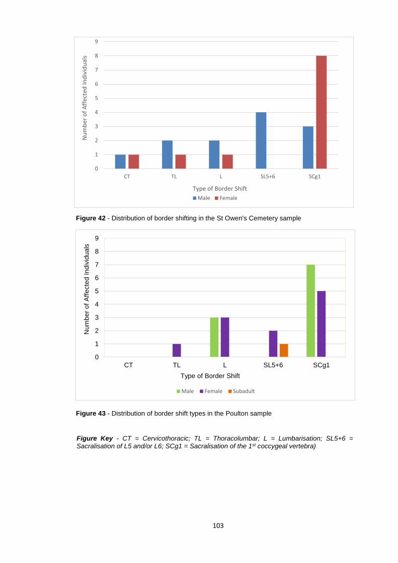
103
Figure 42 - Distribution of border shifting in the St Owen's Cemetery sample
Figure 43 - Distribution of border shift types in the Poulton sample
0
1
2
3
4
5
6
7
8
9
CT TL L SL5+6 SCg1
Num
ber
of
Aff
ecte
d Indiv
iduals
Type of Border Shift
Male Female Subadult
0
1
2
3
4
5
6
7
8
9
CT TL L SL5+6 SCg1
Nu
mb
er o
f A
ffec
ted
In
div
idu
als
Type of Border Shift
Male Female
Figure Key - CT = Cervicothoracic; TL = Thoracolumbar; L = Lumbarisation; SL5+6 = Sacralisation of L5 and/or L6; SCg1 = Sacralisation of the 1st coccygeal vertebra)
104
5.4.4 Scoliosis
Scoliotic curvature, ‘C’ or an ‘S’ curve, was observed in four individuals in
the St Owen’s sample; in a young adult female, an old adult female and two old
adult males (5.8%, n=4/68). In Poulton, five cases of scoliosis were observed
(7.1%, n=5/70); two young adult females, one old adult female, one middle adult
male and one old adult male (Table 21).
5.4.5 Kyphosis
Kyphosis was accounted for in 19 individuals (27.9%, n=19/68) in the St
Owen’s sample (Table 21). Middle adult males (n=8/15, 53.3%) exhibit more
cases of kyphosis than the females (16.7%, n=2/12) and old adult females
(45.5%, n=5/11) suffered more than their male counterparts (25.0%, n=4/16).
Kyphosis was not observed in any individual less than 35 years of age (Figure
44).
Kyphosis was observed 16 individuals in the Poulton sample (22.9%,
n=16/70); ten males (four young adults, five middle adults and one old adult)
(33.3%, n=10/30) and six females (one young adult, two middle adults and three
old adults) (20.0%, n=6/30). Each display curvature in the thoracic segment.
There is a tendency towards kyphosis in males compared to females in the young
and middle adult age ranges. Poulton has younger adults exhibiting kyphosis
compared to their urban counterparts that do not display kyphotic curvature until
the fourth decade of life. However, no subadults had kyphosis (Figure 45).

105
Figure 44 - Age and sex distribution of kyphosis in the St Owen's Cemetery sample
Figure 45 - Age and sex distribution of kyphosis in the Poulton sample
0
1
2
3
4
5
6
7
8
9
Non-adult Young Adult Middle Adult Old Adult
Num
ber
of
Indiv
iduals
Aff
ecte
d
Age Categories
Male Female Indeterminate
0
1
2
3
4
5
6
7
8
9
Non-adult Young Adult Middle Adult Old Adult
Num
ber
of
Indiv
iduals
Aff
ecte
d
Age Categories
Male Female Indeterminate
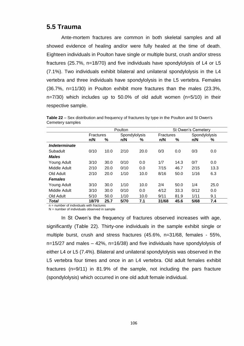
106
5.5 Trauma
Ante-mortem fractures are common in both skeletal samples and all
showed evidence of healing and/or were fully healed at the time of death.
Eighteen individuals in Poulton have single or multiple burst, crush and/or stress
fractures (25.7%, n=18/70) and five individuals have spondylolysis of L4 or L5
(7.1%). Two individuals exhibit bilateral and unilateral spondylolysis in the L4
vertebra and three individuals have spondylolysis in the L5 vertebra. Females
(36.7%, n=11/30) in Poulton exhibit more fractures than the males (23.3%,
n=7/30) which includes up to 50.0% of old adult women (n=5/10) in their
respective sample.
Table 22 – Sex distribution and frequency of fractures by type in the Poulton and St Owen's Cemetery samples
Poulton St Owen’s Cemetery
Fractures Spondylolysis Fractures Spondylolysis n/N % n/N % n/N % n/N %
Indeterminate
Subadult 0/10 10.0 2/10 20.0 0/3 0.0 0/3 0.0
Males Young Adult 3/10 30.0 0/10 0.0 1/7 14.3 0/7 0.0
Middle Adult 2/10 20.0 0/10 0.0 7/15 46.7 2/15 13.3
Old Adult 2/10 20.0 1/10 10.0 8/16 50.0 1/16 6.3
Females Young Adult 3/10 30.0 1/10 10.0 2/4 50.0 1/4 25.0
Middle Adult 3/10 30.0 0/10 0.0 4/12 33.3 0/12 0.0
Old Adult 5/10 50.0 1/10 10.0 9/11 81.9 1/11 9.1
Total 18/70 25.7 5/70 7.1 31/68 45.6 5/68 7.4 n = number of individuals with fractures N = number of individuals observed in sample
In St Owen’s the frequency of fractures observed increases with age,
significantly (Table 22). Thirty-one individuals in the sample exhibit single or
multiple burst, crush and stress fractures (45.6%, n=31/68, females - 55%,
n=15/27 and males – 42%, n=16/38) and five individuals have spondylolysis of
either L4 or L5 (7.4%). Bilateral and unilateral spondylolysis was observed in the
L5 vertebra four times and once in an L4 vertebra. Old adult females exhibit
fractures (n=9/11) in 81.9% of the sample, not including the pars fracture
(spondylolysis) which occurred in one old adult female individual.

107
5.6 Summary
5.6.1 Joint Conditions
The urban sample appear to be affected by degenerative joint conditions more
than their rural counterparts. Joint conditions such as Schmörl’s nodes, vertebral
osteophytes and diffuse idiopathic skeletal hyperostosis are more common in the
males than females in both urban and rural cemeteries. Females displayed a
higher rate of intervertebral disc disease and osteoarthritis in both samples with
St Owen’s exhibing the most. All degenerative joint conditions increase in
prevalence and severity with age.
5.6.2 Infectious Diseases
Urban and rural samples display the same frequency of infectious diseases
and twice the amount of males are affected by infectious lesions than the females.
There appears to be little correlation with age as all age groups are affected in
the Poulton sample. However, infectious lesions do not begin to appear until the
middle adult range (35 – 50 yrs) in St Owen’s.
5.6.3 Congenital Conditions
Transitional vertebrae such as sacralisation of the coccyx and the fifth and
sixth lumbar are the most common congenital anomalies and there is no obvious
correlation with age. Females in St Owen’s exhibit almost three times the amount
of sacralisation of the coccyx than the males but males and females are both
similarly affected in Poulton. Lumbosacral anomalies are more prevalent in
Poulton and Klippel-Feil syndrome (fusion of the cervical vertebrae) is only seen
in Poulton and has only affected males.
5.6.4 Trauma
Stress, burst and crush fractures are the most common traumatic injury
observed in both St Owen’s Cemetery and Poulton. However, fractures are far
more prevalent in the urban community than the rural yet spondylolysis is seen
in equal amounts in both samples but at varying vertebral levels.
108
Chapter 6: Discussion
6.1 Introduction
The purpose of this study was to assess and compare differences in
vertebral pathologies and anomalies of two British medieval skeletal samples
between the 11th century and 16th centuries A.D. The skeletal remains are from
the lay cemetery of St Owen, an urban site in Gloucester and from a medieval
cemetery in Poulton, a hamlet in Cheshire. It was hypothesised that the frequency
and severity of degenerative conditions will be similar due to both communities
being involved in physically demanding occupational roles, but differences will
occur in the distribution of the lesions due to the variation in occupational roles
and a probable gendered division of labour. It was also hypothesised that there
will be a difference in the type of infectious diseases and frequency of congenital
conditions, developmental anomalies and vertebral fractures due to a variation in
diet, environment and lifestyles. All vertebral pathologies and developmental
anomalies were analysed, and results and conclusions have been drawn using
previously published clinical and archaeological studies.
6.2 Joint Conditions
6.2.1 Osteoarthritis (OA)
Osteoarthritis is very common in both samples following the diagnostic
criteria established by Waldron (2009). St Owen’s has higher frequencies of
individuals exhibiting OA on at least one vertebra in the spine (89.4%, n=34/38)
of males and 100.0% of females, n=27/27). However, OA is still very common in
the Poulton sample with 80.0% of males (n=24/30) and 96.6% of females
(n=29/30) exhibiting osteoarthritic changes. The extremely high prevalence rates
may be due to the number of diarthrodial and amphiarthrodial joints observed
within this study (costovertebral, apophyseal and uncovertebral joints). Most
clinical and archaeological studies only refer to the apophyseal joints when
determining OA in living samples and skeletal assemblages even though
diarthrodial and amphiarthdrial joints are both commonly affected by OA (Ortner,
2003). Additionally, the St Owen’s skeletal sample has an under-representation
of young adult specimens in comparison to middle and old adults. Osteoarthritis
109
is widely accepted as an age-related condition and therefore a high prevalence
in the St Owen’s sample is not unexpected (Waldron, 1991; Waldron 2009).
6.2.1.1 Spinal Distribution of Osteoarthritis in both Skeletal Samples
Males and females are equally and most frequently affected in the T1, T11
and T12 in both skeletal samples; 84.0% of the T1 observed had OA in the St
Owen’s females. The distribution of all of the vertebrae affected by OA is similar
in both samples. The most frequently affected vertebrae are consistent in each
sex category; St Owen’s females are most affected in the C4 (77.8%, n=14/18),
T1 (84%, n=21/25), T4 (83.3%, n=20/24) and T12 (80%, n=20/25) vertebrae and
the males in C3 (67.9%, n=19/28), C4 (67.9%, n=19/28), T1 (69%, n=20/29) and
T12 (67.7%, n=21/31). The Poulton sample exhibited a very similar pattern; the
females in the T1 (57.1%, n=16/28), T11 (65.5%, n=19/29) and T12 (62.1%,
n=18/29) and the males in the C3 (48.3%, n=14/29), C4 (50%, n=15/30), T1
(57.1%, n=16/28) and T12 (46.4%, n=13/28). The vertebrae all appear to be in
the weight bearing mid-cervical, cervicothoracic and thoracolumbar junctions.
Crubézy et al., (2002) presented similar results in the distribution of OA
throughout the spines of Neolithic skeletons. Mid-cervical vertebrae were the
most commonly affected (C2-C3 – 55.6%, C3-C4 – 61.8% and C4-C5 – 95.0%)
but the author states that a number of arthritic and degenerative lesions were
recorded between T1 – T4 and T7 – T11 on both the vertebral bodies and facet
joints (Crubézy et al., 2002). Although Crubézy et al., (2002) observed vertebral
bodies and apophyseal facet joints for OA, the results bear similarities indicating
that minimal change has occurred in the distribution of OA throughout the spine
in pre-modern populations.
6.2.1.2 Arthroses Affected by Osteoarthritis
Individuals in Poulton and St Owen’s are mostly affected by OA in the
costovertebral joints (Poulton, 72.5%, n=50/69 and St Owen’s, 81.5%, n=53/65).
Consistent with other studies, frequency and severity increases dramatically with
age (Nathan et al., 1964; Plomp and Boylston, 2016). Plomp and Boylston (2016),
discovered that 22% of adults (27-35 years of age) and 47% of elderly adults (45+
years of age) exhibit eburnation on at least one costovertebral joint surface in two
British medieval skeletal collections. However, this study applied different age
categories, considered eburnation to be the only definitive evidence of OA and
110
included the analysis of extra spinal rib facets and thus the results cannot be
compared fully. The results by the above authors do, however, present an
observable trend in the increase in OA with advancing age. Clinical studies
suggest that costovertebral OA can cause significant pain in the thoracic region,
which can lead to a loss in quality of life for the individual affected (Sales et al.,
2007; Plomp and Boylston, 2016) so it can be hypothesised that this could have
caused considerable pain in the individuals in Poulton and St Owen’s. The cause
of OA in the costovertebral joints is considered multifactorial and repetitive lifting
and heavy weight applied to the thoracic region (Plomp and Boylston, 2016) may
be a contributing factor to the high prevalence within the Poulton and St Owen’s
samples.
Individuals in St Owen’s were similarly affected by OA in the
costovertebral facets and apophyseal facets (costovertebral facets – 81.5%,
n=53/65 and apophyseal facets – 79.4%, n=54/68). There appears to be no
association with age as 100.0% (n=4/4) of young adult females and 71.4%
(n=5/7) of young adult males exhibit OA which then decreases in the middle adult
range (females – 75.0%, n=9/12 and males – 66.7%, n=10/15) (Table 10). The
osteological paradox (Wood et al., 1992) may offer reasons as to the results
obtained: small sample sizes (n=4 and n=7) and late onset of OA in the
individuals who lived longer. The St Owen’s sample mainly consists of middle
and old adults of both sexes.
Both sexes show that the upper thoracic and upper, mid-cervical vertebrae
are most affected by apophyseal facet joint OA. Interestingly, the lumbar
vertebrae exhibit the lowest frequencies of OA in comparison to the rest of the
spine which contrasts with many clinical (Gellhorn, 2013) and archaeological
studies (Waldron, 1991). Waldron (1991) concluded that the L5 was affected by
OA in 12% of females and 10% in males. The results may appear to exhibit lower
frequencies than is recorded in Poulton and St Owen’s, but the results still
conclude that the lumbar vertebrae are the most affected in the spinal columns
of individuals at Christ Church, Spitalfields.
The cause of OA of the apophyseal facets has long been a cause for
debate. Clinical studies have suggested that anatomical variance of the facet
joints and the lack of core strength can cause facet joint arthritis and subsequently
111
pain in the areas affected (Weiss and Jurmain, 2007). Others have concluded
that repetitive strenuous movements and heavy manual occupations are causes
of spinal arthritis (Gallucci et al., 2007; Waldron, 2009).
Uncovertebral joint OA in the cervical vertebrae was extremely common in
both skeletal samples (St Owen’s - 63.3%, n=38/60 and Poulton – 49.3%,
n=33/67). Similarly to the apophyseal and costovertebral facets, the prevalence
and severity of OA increases with age (St Owen’s – young adults, 33.3%, n=3/9,
middle adults, 60.9%, n=14/23 and old adults, 84.0%, n=21/25 and Poulton –
young adults, 10.5%, n=2/19, middle adults, 55.0%, 11/20 and old adults,
100.0%, n=18/18). The mid-cervical, C3 to C6, exhibit the highest prevalence of
OA including eburnation. Osteoarthritis of the neck can be very debilitating and
cause pain for the individual affected including spinal stenosis, paraplegia and
loss of muscle mass and motor control if vertebral osteophytes become so severe
(Wilkinson, 1960; Teraguchi et al., 2014).
6.2.1.3 Inter-Sample Differences between Subadults from both Skeletal
Samples
Adolescent subadults (13 – 18 years of age) from St Owen’s have no
observable manifestations of degenerative conditions, but Poulton exhibits two
individuals with evidence of osteoarthritis (20.0%, n=2/10) in the cervical
uncovertebral joints and thoracic apophyseal facets. However, the St Owen’s
sample size for subadults (n=3) is small and the results will likely have been
affected. Additionally, the chi-square test for independence found no significant
difference between the two sites. In modern population studies, OA in young
adults and adolescents is commonly seen in the clinically obese or in the very
active such as athletes and gymnasts (Goulding et al., 2002; Sjolie, 2004; Briggs
et al., 2009). Medieval adolescents, especially from rural communities like
Poulton, would have likely worked the fields and around the farmstead from
childhood resulting in an active lifestyle from an early age. Obesity and a high
BMI could also be valid explanation given that soft tissue has not survived to be
analysed in detail, but it is unlikely given the agricultural mode of employment
(Delgado-López and Castilla-Díez, 2018). It is likely that the subadults in Poulton
were considered able to work from a much younger age than St Owen’s
subadults, thus enabling bone remodelling from as early as the subadult age
112
range (13 - 18 years of age). Stirland and Waldron (1997) concluded that the
crew from the Mary Rose exhibited accelerated degenerative changes due to
intensive work from an early age and Rojas-Sepúlveda et al., (2008) determined
that Pre-Columbian Muisca skeletal remains exhibited degenerative changes
from approximately 15 years of age due to socioeconomic pressures.
6.2.1.4 Sex Differences: Inter and Intra-Sample Observations
Differences in the prevalence of osteoarthritis between males and females
have been previously reported upon in the early subsections however, this
section provides a detailed account of inter and intra-sample differences in the
two skeletal samples.
Females in both skeletal samples exhibit more OA than their male
counterparts. The result is an indicator that the females were equally, if not more,
involved in physically demanding labour than their male counterparts. St Owen’s
females exhibit a peak in OA of 84.0% (n=21/25) in the T1 vertebra whereas
males display a peak of 68.9% (n=20/29) on the T1 vertebra. Medieval urban
females were less likely to have been involved in the highly documented crafts
and guilds that males were employed in, making the cause of the high levels of
OA observed less clear. However, it is likely that the females would have carried
out assistant roles in certain crafts, helped in selling market wares, been involved
in low wage occupations, domestic services and child rearing (Bennett, 1992;
Shapland et al., 2015).
Literature regarding degenerative conditions of the thoracic vertebrae is
limited in comparison to the cervical and lumbar segments due to the rigidity and
stability created by the thoracic cage (Briggs et al., 2009; Teraguchi et al., 2014).
However, there appears to be a significant difference in the prevalence of
degenerative spinal conditions in the thoracic segment between the urban and
rural communities within this study. St Owen’s middle adult males had a
significantly higher frequency of OA in the T6 (p=<.05, Poulton 22.2% and St
Owen’s 84.6%), T7 (p=<.05, 11.1% and 69.2%), T8 (p=<.05, 20.0% and 75.0%),
T9 (p=<.05, 20.0% and 61.5%) vertebrae compared to the middle adult males in
Poulton. This was also the case for middle adult females in St Owen’s who
exhibited significantly greater amounts of OA in the T1 (p=<.05, Poulton 50.0%
and St Owen’s 90.9%), T6 (p=<.05, 30.0% and 70.0%), T7 (p=<.05, 40.0% and

113
80.0%) vertebrae than the Poulton middle adult females. The difference in
prevalence was most noticeable in the apophyseal facets as there was only a
minimal difference in the costovertebral joints. As OA is generally linked to
excessive loading and mechanical forces placed on the spine (Gallucci et al.,
2007; Waldron, 2009), it might be acceptable to assume that the individuals from
St Owen’s carried heavy objects on the thoracic level of their backs or conducted
continuously repetitive hyperextension and flexion movements. Repetitive spinal
movements would have been a normal part of working in a singular occupational
role and repetitive strain may be one of a number of factors as to the high level
of OA seen in the males in St Owen’s sample. The males from Gloucester would
likely have lived physically active lifestyles, especially if they worked in a
specialist craft (fullers, weavers and dyers) and/or as general traders selling their
wares on the markets present at the time. However, merchant guilds such as
clothiers, tailors and hosiers may have been slightly less physically demanding
and more sedentary (Herbert, 1988). Southgate Street in Gloucester was
occupied by a number of shoemakers (cordwainers) so perhaps the high thoracic
involvement of OA can be attributed to consistent sitting and/or standing with poor
posture and twisting of the back to work leather (Langton, 1977). The comparable
frequency of thoracic OA in the females may also point to assisting in the making
of shoes and other similar crafts.
6.2.2 Intervertebral/Degenerative Disc Disease (IDD)
Intervertebral disc disease is extremely common in Poulton and St
Owen’s. The mid-cervical (C3-C6) and the thoracolumbar junction exhibited the
highest frequencies of IDD with up to 100.0% of old adult females (n=9/9)
exhibiting it on at least one vertebra in each vertebral segment (Figure 10). The
vertebrae T1 to T3 exhibit the lowest frequencies of IDD, likely due to less weight
being applied to these vertebrae. Intervertebral disc disease is commonly
associated with osteoarthritis and facet joint degeneration (Brain et al., 1952;
Fujiwara et al., 2000) and this is consistent with the results obtained from St
Owen’s and Poulton. However, there appears to be a greater frequency of OA in
comparison to IDD in the cervicothoracic junction in both skeletal samples.
Waldron (2009) states that intervertebral disc disease is very common in
individuals above 70 years of age but an unusual observation in people younger
than 40 years. However, Antoniou et al. (1996) states that IDD can be observed

114
in individuals from as early as the second decade of life. Using criteria established
by Waldron (2009) to aid in the diagnosis of IDD in both samples and the results
obtained in this study conform to theory that IDD can be observed in individuals
from as early as the second decade of life (Antoniou et al., 1996); evidence of
intervertebral disc disease is present from as early as the subadult range (13 - 18
years of age) in Poulton and from the young adult range (18 - 35 years of age) in
St Owen’s. Adams and Dolan (2012) suggest that IDD observed in younger
individuals may be due to a spinal injury and overloading but also to congenital
weakening of the vertebral endplates. Therefore, the instances of IDD in the
Poulton young adults can likely be attributed to occupational stresses from an
earlier age compared to their contemporaries in St Owen’s.
Females in both skeletal samples are affected by IDD more than their male
counterparts, especially in the mid-cervical segment (St Owen’s females – C4 -
61.1%, n=11/18, C5 - 71.4%, n=15/21 and C6 - 63.6%, n=14/22 and St Owen’s
males – C4 – 53.6%, n=15/28, C5 – 53.3%, n=16/30 and, C6 – 57.1%, n=16/28)
(Poulton females – C4 - 42.3%, n=11/26, C5 - 51.9%, n=14/27, C6 - 55.6%,
n=15/27and Poulton males – C4 – 46.7%, n=14/30, C5 – 41.4%, n=12/29 and C6
– 33.3%, n=10/30). However, the difference is not significant and males and
females both exhibit very similar distributions of IDD throughout the spine. The
distribution of IDD, although seen at much higher level in Poulton and St Owen’s,
is comparable to results seen in the Christ Church Spitalfields skeletons with
peaks in the cervical (males had the highest reported frequency in the C6
vertebra with 27% and females in the C5 vertebra with 26%) and lumbar
segments (males exhibit approximately 8% in the L5 vertebra and females exhibit
4%) (Waldron, 1991).
Literature on IDD in archaeological assemblages is sparse, in part due to
the variety of nomenclature employed in clinical and osteological studies and the
vague interpretation of what constitutes a disease of the intervertebral discs and
not just amphiarthrodial joint osteoarthritis.
6.2.3 Schmörl’s Nodes (SNs)
The aetiology of Schmörl’s nodes, to this day, is unknown even with the
multitude of scientific research surrounding this spinal lesion. Schmörl’s nodes
appear to be heterogeneous lesions that are caused by strenuous, stressful and

115
repetitive movements, development of the vertebrae in infancy, degenerative,
neoplastic and metabolic diseases and traumatic episodes to name a few
(Resnick and Niwayama, 1978; Takahashi et al., 1994; Dar et al., 2010). Adams
and Dolan (2012) and Williams et al. (2007) state that Schmörl’s nodes are highly
heritable. Dar et al., (2010) concluded that there was no correlation between sex,
age and ancestral origin in the anatomical location of SNs in the T4 to L5
vertebrae. Schmörl’s nodes are most common in males in both Poulton and St
Owen’s, but the lesions are independent of age and were observed on the
vertebrae of subadults through to old adults in varying frequencies and
anatomical locations. Again, this is consistent with other clinical and
archaeological studies (Hilton et al., 1976; Saluja et al., 1986; Adams and Dolan,
2012).
The lower thoracic and lumbar vertebrae are the most frequently affected
areas of the spinal column in the St Owen’s and Poulton samples (Figure 12) and
this appears to be consistent with other studies (Resnick and Niwayama, 1978;
Faccia and Williams, 2008; Dar et al., 2010). Males in both samples exhibit more
SNs on both superior and inferior vertebral endplates than their female
counterparts (St Owen’s males – 34.6%, n=359/1038 and females – 20.5%,
n=182/886 and Poulton males – 31.8%, n=305/960 and females – 23.1%,
n=237/1026). However, the percentage of males and females that exhibit SNs on
at least one vertebral endplate is similar in each sample (St Owen’s males –
86.8%, n=33/38 and females – 81.5%, n=22/27 and Poulton males - 92.9%,
n=26/28 and females – 93.1%, n=27/29). The results of this study are consistent
with other contemporary, post-medieval and modern skeletal studies (Dar et al.,
2009; Plomp et al., 2012). Dar et al., (2009) discovered that 69.6% of the 511
SNs observed were located in male spines and also established that males were
more likely to have multiple endplate SNs compared to the females. The same
author also considers a genetic aetiology for SNs.
Both skeletal samples indicate there is a predilection for inferior SNs in the
thoracic segment and superior SNs in the lumbar segment which is in-keeping
with numerous clinical studies (Hilton et al., 1976; Williams et al., 2007; Dar et
al., 2010). An explanation for this remains unclear. However, one theory that has
been suggested is that during foetal development the inferior surfaces of the
vertebrae are weaker than the superior, which allows for the nucleus pulposus to
116
herniate the less well developed vertebral endplate (Dar et al., 2010). Lumbar
vertebrae experience more pressure and tensile strain and due to the direction of
the stress being applied the superior endplates are the most likely to develop SNs
(Dar et al., 2010).
Schmӧrl’s nodes can be symptomatic and asymptomatic and this solely
depends on the size and proximity of the lesion to various blood vessels and
nerve endings. Symptomatic SNs can cause significant pain and tenderness in
the area affected (Takahashi et al., 1995). If the modern social attitude towards
back pain existed in the medieval times, it is likely that the individuals would have
been dependent on others for a time (Faccia and Williams, 2008). However, living
in a peasant community, the notion of taking time off work to recover would have
most likely been non-existent, especially when farming is season dependent.
SN frequency and anatomical distribution was examined in this study, but
depth and width were not recorded. Had depth and width been recorded, a more
holistic analysis of health implications could have been achieved.
6.2.4 Diffuse Idiopathic Skeletal Hyperostosis (DISH)
Diffuse idiopathic skeletal hyperostosis was observed in both samples; two
old adult males in St Owen’s (2.9%, n=2/68) and one middle adult female in
Poulton (1.4%, n=1/70) (Table 21). Usually considered a condition of the wealthy
and high-ranking, DISH has been associated with obesity and type-2 diabetes
and the ease of access to rich foods (Jankauskas, 2003; Verlaan et al., 2007;
Van der Merwe et al., 2012). The elderly, the male sex, individuals with a high
body mass index and/or who were overweight as children and individuals with a
purine rich diet are more likely to develop DISH from the sixth decade of life
onwards (Cammisa et al., 1998; Kiss et al., 2002; Kagotani et al., 2013). A link
has been established between serum uric and acid and DISH (Kiss et al., 2002).
Purines once ingested are broken down in to uric acid which can then be traced
in the blood. Foods containing high levels of purines such as beer, wine, liver,
kidneys, trout, mackerel, scallops and dried beans amongst many other foods
(ibid) would have been readily available in an urban environment.
The two old adult males exhibiting DISH appeared to have no discernible
grave goods and/or evidence of higher social ranking. The burial of GLC0028
was located in the second layer and truncated by a modern concrete foundation

117
and thus any grave goods that may have been in the feet portion of the grave
would have been removed (Atkin and Garrod, 1989). The location of the burial
and the lack of a grave marker and coffin indicates that the individual was
probably not of higher social ranking than the general population buried within
the cemetery. The burial of GLC110 was the earlier of the two burials and was
recovered in the third layer. Again, evidence of grave goods were not observed,
thus indicating the individual was probably not of higher status. However, the
DISH diagnosis does suggest that the individuals had a diet rich in foods
containing higher levels of purine and had easy and frequent access to these
kinds of foods. Gloucester as a market town would have sold a variety of foods
and there was a multitude of butchers, alehouses, bakers and fishmongers
(Herbert, 1988). Therefore, observing a slightly higher prevalence in the urban
sample is not unexpected. Other health conditions that can be hypothesised for
the individuals exhibiting DISH are diabetes mellitus (type-2) and obesity. In
addition to DISH, POU166 exhibits very advanced OA and widespread
eburnation throughout the cervical, thoracic and lumbar segments. Interestingly,
POU166 is female and a middle adult (aged 35-49 years of age), both of which
are uncommon in the usual demographic of DISH sufferers (Cammisa et al.,
1998).
The middle adult female, exhibiting DISH in the thoracic segment,
recovered from Poulton may be an individual of higher status. The skeleton was
recovered in the southwest corner of the cemetery and coffin nails were located
in the grave fill suggesting the individual was buried in a coffin, an uncommon
finding at this particular medieval cemetery. The skeleton was also complete
indicating a more recent burial or that an obvious grave marker protected the
location, however no grave goods were discovered.
Mader and Lavi (2009) conducted a study comparing back pain in DISH
sufferers. It was concluded that individuals diagnosed with DISH before 50 years
of age were more likely to suffer back pain in the thoracic and lumbar segments
and be affected by extra-spinal enthesopathies than elderly individuals diagnosed
with DISH after the age of 60. However, a study by Schlapbach et al. (1989)
indicates that back pain in DISH sufferers was similar to that of the unaffected
control sample. This is also consistent with a study by Faccia et al., (2016) who
asserts that DISH is largely asymptomatic. Although many patients display no

118
symptoms, some attribute stiffness and reduced mobility in the thoracic and/or
cervical segments to DISH (Mata et al., 1997; Rogers and Waldron, 2001). The
disease in modern populations has little clinical significance (Rogers and
Waldron, 2001). Therefore, it would be acceptable to assume that DISH, as an
isolated condition, would not have affected the daily lives of the medieval
individuals.
Prevalence rates of DISH in both samples remain tentative. Due to the
diagnostic criteria by Resnick and Niwayama (1976) of the ossification of four or
more contiguous vertebrae, any possible cases of DISH (such as the early signs
of DISH) were omitted from the study indicating that a higher prevalence may
have been recorded.
6.2.5 Vertebral Osteophytosis
Spinal osteophytes are common in individuals with intervertebral disc
disease, ankylosing spondylitis and osteoarthritis (Rogers et al., 1985). In the
Poulton and St Owen’s samples, osteophytes were graded separately to OA and
IDD. However, there appears to be an association between spinal osteophytes,
IDD and OA. No cases of ankylosing spondylitis were observed.
Severe osteophytes are frequently located in the mid-cervical,
thoracolumbar and lumbosacral regions in both skeletal samples. However, in old
adults the anatomical distribution of severe osteophytes encompasses the
majority of the spinal column with the exception of a few upper cervical vertebrae.
These findings are consistent with numerous studies, including a study
conducted by Kim, et al. (2012) on pre-modern Korean skeletons which
discovered that C5, T9, T10 and L4 exhibit the most severe osteophyte growth;
the vertebrae furthest from the line of gravity. The same study also discovered
that males exhibit the highest frequency of severe osteophytes and suggested
that this is a result of strenuous manual labour in comparison to the females which
is also observed in the results from Poulton (males – 6.3%, n=86/1360 and
females – 4.9%, n=66/1352) and St Owen’s (males - 8.9%, n=132/1479 and
females – 4.1%, n=46/1116). Severe apophyseal facet osteophytes can cause a
multitude of health problems; sleep apnea, dysphagia and vocal fold paralysis
(Klaasen et al., 2011).
119
The development of osteophytes is, amongst other factors, caused by
repetitive and chronic biomechanical stress applied to a particular area of a joint
(Zemirline et al., 2013). The males on average have the largest and most severe
osteophytes in both Poulton and St Owen’s, indicating more pronounced
mechanical stress associated with manual labour in the city and/or peasant
farming. Young adult males (18 - 35 years) in Poulton (20.0%, n=2/10) show
severe osteophytes in the thoracolumbar junction and lumbar vertebral segment
(severe osteophytes – 2.0%, n=10/489). Although two young adult males did
exhibit severe osteophytes, one must point out that 83% of the sample display
zero (none) (38.7%, n=189/489) and one (normal) (44.0%, n=215/489) on the
grading scale. However, the young adult individuals exhibiting severe
osteophytes had other degenerative spinal conditions, which may be attributed
to being employed in physically demanding occupational roles from a very early
age (Schmitt et al., 2004; Klaasen et al., 2011). Although severe osteophytes
were observed in some young adults, the frequency and severity in the size of
the osteophytes increases with age, again suggesting that osteophytes are not
only a by-product of intensive manual labour but also of the aging process and of
spinal degeneration and these results are consistent with other osteophyte
development studies (Snodgrass, 2004).
On average, females in comparison to the males exhibit a lower frequency
of severe osteophytes (Poulton males – 6.3%, n=86/1360 and females – 4.9%,
n=66/1352 and St Owen’s males - 8.9%, n=132/1479 and females – 4.1%,
n=46/1116). However, middle adult females (8.4%, n=41/488) in Poulton display
a higher degree of severity compared to their male counterparts (7%, n=32/454).
Perhaps childbearing years influenced the severity somewhat. Sanderson and
Fraser (1996) suggest that pregnancy and childbirth is a factor in the formation
of degenerative spondylolisthesis. Therefore, assuming that degeneration of the
spinal column can occur in women of childbearing age that this could be a
contributing factor to the development of severe osteophytes. The 0
6.3 Metabolic Disorders
Metabolic disorders occur when the body is deprived of or cannot absorb
vitamins and minerals that are essential for the maintenance of healthy bone
(Mays, 2008). Metabolic disorders comprise a multitude of conditions, some of

120
which include; scurvy, Paget’s disease of bone, osteomalacia and rickets and,
osteoporosis (Mays, 2008).
The poor preservation of the skeletal elements from the St Owen’s skeletal
collection unfortunately hindered the assessment of metabolic conditions. Most
of the vertebrae under analysis were lightweight and porous which could lead to
misdiagnosis. An example of a metabolic condition that could easily be
misdiagnosed is osteoporosis. Osteoporosis is characterised by a reduction in
total bone mass and poor architectural arrangement of the underlying trabecular
bone (Brickley, 2002; Brickley and Agarwal, 2003; Mays et al., 2006). As a result
of poor bone architecture, porosity, crush, and stress fractures are features of
osteoporosis (Brickley, 2002).
6.4 Infectious Diseases
Infectious lesions are common in both samples with Poulton displaying a
crude prevalence rate of 20.0% (n=14/70) and St Owen’s Cemetery exhibiting a
rate of 20.6% (n=14/68). The diagnoses of infectious lesions were identified
following descriptions by Waldron (2009) and Ortner (2003). However, both
authors state that infectious lesions of the spine are difficult to distinguish
macroscopically and thus, no evaluation has been made to identify the exact
conditions present within Poulton and St Owen’s. Waldron (2009) also states that
Pott’s disease (late stage skeletal involvement of tuberculosis) is relatively easy
to diagnose due to the complete destruction of vertebral bodies and apparent
extreme kyphosis. No cases of Pott’s disease were identified in Poulton or St
Owen’s.
Infectious diseases such as brucellosis and certain strains of tuberculosis,
such as Mycobacterium bovis, are spread to humans through prolonged and
close contact with livestock and the consumption of infected milk and animal
products (Mays et al., 2001; Roberts and Manchester, 2010; D’Anastasio et al.,
2011). This could explain the prevalence of infectious diseases in both samples.
There is a great likelihood that both Poulton and St Owen’s Cemetery individuals
would have had close contact with livestock due to the rural animal farming
practices (Poulton) and trading and consumption of animals and/or animal
products in the household and urban markets. Until the 20th century bovine type
tuberculosis was a common infectious disease before the introduction of

121
antibiotics, disease control and the pasteurisation of milk (Mays et al., 2001; US
Department of Health & Human Services, 2011). However, pulmonary
tuberculosis (Mycobacterium tuberculosis) could be attributed to the infectious
lesions observed in the urban sample due to close contact to infected individuals
in a densely populated setting (Mays et al., 2001). Medieval Gloucester was a
thriving trading town and it is likely that the lay community came in to contact with
people from further afield and were more likely to be infected from outsiders as
well as local individuals. Shared housing was widespread due to the limited
availability of accommodation (Langton, 1977). Cramped living and working
conditions would have easily spread infectious diseases.
Infectious lesions have a clear predilection for males (30.0%, n=9/30) in
comparison to females (13.3%, n=4/30) in the Poulton sample (Table 24). In
addition, sub-adults and young adults all show lesions too (10.0%, n=1/10).
Perhaps pathogens in the soil contributed to this, given that the males tended to
work the fields in a more frequent capacity than the females. However, this does
not begin to explain the high percentage of old adult females exhibiting infectious
vertebral lesions. Females on average tend to have more effective immune
systems than males, which then becomes less effective in old age (Stini, 1985;
Roberts et al., 1998). The expression of infectious lesions in the females may
have taken longer to manifest. Similarly, to their rural counterparts, infectious
lesions are more prevalent in the males (23.7%, n=9/38) from St Owen’s
compared to the females (18.5%, n=5/27).
6.5 Congenital Conditions and Vertebral Anomalies
6.5.1 Spina bifida
Spina bifida is a neural tube defect in which the posterior arches of the
vertebrae fail to fuse (Kumar and Tubbs, 2011). This condition is a common
observation in the sacral vertebrae but can occur anywhere on the spinal column
(Groza et al., 2012). Spina bifida can be subdivided into numerous forms. Spina
bifida occulta, the most common form and is generally asymptomatic and, spina
bifida cystica and aperta (meningocele and myelomeningocele), both of which
can be fatal within the first year of life if not treated. The rarest form is rachischisis
and is identified by a completely open spinal column (Waldron, 2009).
122
Myelomeningocele, the deadliest form of spina bifida cystica, had a survival rate
of 10-12% according to the historical record (Roberts and Manchester, 2010).
The lack of soft tissue in archaeological remains make it difficult to
establish what type of spina bifida the individuals in both samples exhibit.
Therefore, it is problematic to interpret the impact of this condition on the
individuals in the two medieval communities. However, following criteria by
Bradtmiller (1984), spina bifida was observed in both samples, one subadult
individual in Poulton (n=1/70, 1.4%) and one young adult female and one old
adult male in St Owen’s (n=2/68, 2.9%). There appears to be no association with
age and sex in the expression of spina bifida in the two samples. The percentages
of spina bifida observed in both skeletal samples are similar to that of other
medieval sites (Cessford, 2015).
Many congenital anomalies have a genetic component and spina bifida is
no different. However, the genetic factor of spina bifida and other neural tube
defects is poorly understood (Copp et al., 2017). Environment and diet are the
most widely studied causes of spina bifida (Mitchell et al., 2004). A lack of folic
acid during the mother’s pregnancy is considered a major contributor to the
development of this congenital condition. Up to 70% of reported cases could have
been prevented had the supplementation of folic acid been considered before
and during pregnancy (Hewitt et al., 1992; Mitchell et al., 2004; Bassuk and Kibar,
2009). Folic acid can be obtained from dark green plants (broccoli, cabbage,
kale, and spinach), fortified cereals, beans and citrus fruits such as oranges
(BDA, 2016). Therefore, the observation of spina bifida in archaeological samples
could indicate lack of a balanced diet and limited access to a variety of foods.
6.5.2 Vertebral Synostosis/Klippel-Feil Syndrome
Klippel-Feil syndrome (KFS) is characterised by the fusion of two or more
cervical vertebrae. This condition usually manifests in atlanto-occipital fusion and
or the fusion of C1 and C2 or the fusion of C2 and C3, although it can occur
between any of the cervical vertebrae (Fernandes and Costa, 2007; Lasanianos
et al., 2015). Three cases of cervical vertebral synostosis were observed in the
rural Poulton sample (4.3%, n=3/70) whereas no instances were discovered in St
Owen’s. All of the cases examined included the fusion of C2 to C3, but one
individual also has fusion of C6 to C7. Additionally, all of the cases are from

123
skeletons estimated to be male. The skeleton exhibiting C2 to C3 and C6 to C7
fusion also exhibits severe osteophytosis in the lumbar segment and eburnation
of C1 and C2 (POU443).
Klippel-Feil Syndrome is commonly associated with other anomalies such
as, Sprengel deformity (congenital elevation of the scapula), spina bifida, hemi-
vertebrae and cleft palates, amongst others. However, KFS can be present
separately to the aforementioned anomalies (Aufderheide and Rodríguez-Martín,
1998; Fernandes and Costa, 2007; Samartzis et al., 2008). The Poulton
individuals bore no other congenital spinal anomalies apart from one case of
coccygeal sacralisation. Therefore, KFS can be inferred but not truly identified.
The high rate of cervical synostosis observed in Poulton (4.3%, n=3/70) is much
higher than is reported in modern clinical studies with prevalence rates ranging
from 0.5% to 1% of the given population (Fernandes and Costa, 2007;
Lasanianos et al., 2015). Further analysis of the skeletons in relation to other
extra spinal anomalies commonly observed with KFS may provide more robust
identification. Differential diagnoses could include juvenile rheumatoid arthritis
and trauma (Kulkarni and Ramesh, 2012). However, the fusion observed in
Poulton was only confined to the cervical vertebrae and there was no evidence
of rheumatoid arthritis and the only trauma (compression fracture) observed was
in a T9 vertebra of a young adult male.
Klippel-Feil Syndrome is a heterogeneous condition. However, a study
conducted by Tassabehji et al. (2008) concluded that mutations of the gene loci
GDF6 and GDF3, amongst others, are known causes of some phenotypes of
KFS. These are autosomal inherited mutations and thus, may infer genetic affinity
between individuals in the Poulton sample. Environmental factors such as parent
alcoholism and diabetes can also cause chromosomal translocations and
deletions that can cause KFS (Klimo et al., 2007; Krakow, 2018). However,
environmental factors do not yield a conclusive answer as to why the condition is
solely observed in the rural sample. The isolation of KFS in Poulton may suggest
that autosomal dominant inheritance is much more likely or that the phenotype of
KFS seen in this sample may be passed through paternal lineages. Interestingly,
two individuals that display KFS anomalies were buried within the chapel walls
indicating a likely familial linkage (Figure 46).
124
6.5.3 Vertebral Border Shifting/ Transitional Vertebrae
Transitional vertebrae or vertebral border shifting are anomalies affecting
vertebral junctions, examples of which are caudal shifts of the seventh cervical
vertebra, lumbarisation of the twelfth thoracic vertebra, lumbarisation of the first
sacral vertebra, sacralisation of the fifth or sixth lumbar vertebrae and
sacralisation of the coccyx. Sacralisation of the coccyx was the most commonly
observed anomaly and exhibits a crude prevalence rate 34.7% in the females in
St Owen’s compared to 12.0% (n=3/25) in males. Males (28%, n=7/25) and
females (18.5%, n=5/27) in Poulton both exhibit relatively high prevalence rates
of sacralisation of the coccyx. Sacralisation of the coccyx is underreported in the
archaeological literature and therefore, the crude prevalence displayed in this
study cannot be compared to contemporaneous skeletal assemblages.
Sacralisation is the most common vertebral anomaly in both skeletal samples,
which is in contrast to other studies that state that spina bifida is the most
commonly observed in skeletal assemblages (Barnes, 1994; Sarry El-Din and El
Banna, 2006).
Sacralisation of the fifth or sixth lumbar vertebra is studied on a much
larger scale (Barnes, 1994; Bonaiuti et al., 1997; Delport et al., 2006). The
prevalence rates of lumbosacral transitional vertebrae (lumbarisation and
sacralisation of L5 and L6) in modern samples ranges from 4% - 24% (Delport et
al., 2006). The frequencies of lumbosacral transitional vertebrae observed in this
study (Poulton – 4.3%, n=3/70 and St Owen’s – 5.9%, n=4/68) falls within the
lower range of modern prevalence rates suggesting that the incidence of this
congenital anomaly is minimally affected by environmental factors and lifestyles
but is more likely affected by genetic predisposition. Due to a probable genetic
component of lumbosacral anomalies, burial locations of the individuals were
examined (Figure 46). However, there appears to be little correlation between
burials, thus no familial connection can be made. DNA analysis may provide
further evidence.

125
Figure 46 – Burial locations of individuals with lumbosacral vertebral anomalies to infer familial links in the Poulton sample. Anomalies; Klippel-Feil Syndrome, Sacralisation, Lumbarisation
Figure 2 – Healed burst fracture on superoanterior body of the 4th lumbar vertebra of GLC0058 from St Owen's Cemetery (Red arrow points to fracture).

126
6.5.4 Scoliosis
Scoliotic curvature is prevalent in 7.1% (n=5/70) of the Poulton sample and
5.8% (n=4/68) of the St Owen’s sample. Clinical and epidemiological literature
states that 2.5% of the population exhibit adolescent idiopathic scoliosis (Asher
and Burton, 2006). The reason for the high prevalence in this study is due to the
fact that any curve that differs from the “normal” curvature of the spine and
exhibits an “S” or a “C” shape was diagnosed here as scoliosis which may also
include, degenerative scoliosis, congenital and other types of scoliosis.
Prevalence rates of all scoliosis types in a single population have yet to be
published so a comparison cannot be accurately made. The degree of the curve
was not analysed due to poor preservation of the remains and thus, had the Cobb
angle method (Cobb, 1948) been applied in this study then the prevalence rate
may drop significantly. The Cobb angle, developed by Dr John Cobb in 1948, is
a method that measures the degree of angularity of the spinal curve and is
generally applied in clinical studies. An angle of 10° or more in a lateral direction
would be considered scoliosis in a clinical setting (ibid.).
Clinically, scoliosis is categorised into seven types, degenerative,
adolescent idiopathic, congenital, early onset, neuromuscular, Scheuermann’s
kyphosis and syndromic (Scoliosis Association UK, 2017). However, these types
are generally hard to differentiate in the archaeological record and especially in
older individuals. It is clear that some of the individuals from both Poulton and St
Owen’s had degenerative or adolescent idiopathic scoliosis. Vertebral fractures
and intervertebral disc disease created scoliotic curves in most of the elderly
individuals. Younger individuals that display abnormal curvature, including
abnormal orientation of spinal articulations and processes were considered to be
exhibiting the idiopathic type.
Out of all of the spinal conditions, severe cases of scoliosis and
kyphoscoliosis are probably the most noticeable. King Richard III displayed
scoliotic curvature. Adjectives, in which Shakespeare referred to his abnormality
such as, “deformed”, may provide evidence as to how people with visible
conditions were treated in the medieval period (Lund, 2015). Felt (2012) states
that modern society still measures individual worth on exterior aesthetics.

127
Shakespeare proved with his portrayal of Richard III that the audience would
assume his guilt due to his visible “deformities” (ibid).
6.5.5 Kyphosis
Thoracic kyphosis is the exaggeration of the already present kyphotic
curvature in the thoracic vertebral segment. Kyphosis is a heterogeneous
condition that is caused by multitude of congenital, degenerative, infectious and
metabolic diseases and acute traumatic episodes (Brickley, 2002; Anderson,
2003; Holloway et al., 2011). Kyphotic curvature is present in 19 individuals
(27.9%, n=19/68) in St Owen’s and in 16 individuals (23.5%, n=16/70) in Poulton.
Middle adult males appear to be the most affected by kyphosis in both samples,
but females appear to be most affected in the old adult age range. The degree of
kyphosis was hard to interpret with poorly preserved and incomplete spinal
columns. The high prevalence of kyphosis in old adult females is most likely due
to disc degeneration and osteoporotic vertebral compression fractures. The most
obvious cause of the kyphosis seen in St Owen’s is vertebral compression
fractures (78.9%, n=15/19) and all of the individuals have been estimated to be
middle and old adults. This suggests that working conditions were hazardous
and/or metabolic conditions such as osteoporosis were prevalent. However, two
of the kyphotic individuals (GLC031 and GLC177) display evidence of an
infectious disease such as brucellosis or tuberculosis in the thoracic segment.
Kyphosis is present in males and one female, from as early as the third
decade of life in Poulton, indicating that males are most likely to conduct the more
physically demanding farming roles from an early age. All the young adult
individuals expressing kyphosis exhibit other congenital (scoliosis), degenerative
conditions (OA, SNs and IDD). There was no evidence of infectious lesions in
any of the young adult individuals.
6.6 Trauma
Evidence of vertebral trauma was commonplace in both Poulton and St
Owen’s Cemetery. The individuals recovered from St Owen’s exhibit the highest
amounts of trauma/injury (45.6%, n=31/68), excluding spondylolysis (7.4%,
n=5/68). Individuals from Poulton exhibit far less vertebral fractures than their
urban counterparts (25.7%, n=18/70), but spondylolysis was equally apparent

128
(7.1%, n=5/70). The frequency of spondylolysis observed in Poulton and St
Owen’s is consistent with other studies (Stirland, 1996; Masnicová and Beňuš,
2003). A study conducted by Stirland (1996) on individuals from the Mary Rose
recorded 29 cases of spondylolysis in 413 individuals (7.0%) in the L3-L5
vertebrae and Masnicová and Beňuš (2003) also recorded that 7% of individuals
exhibit spondylolysis in the Devin-Hrad archaeological site in Slovakia. However,
Stirland only observed eight cases of other vertebral trauma (1.9%) such as
fractures of other vertebral parts rather than the pars interarticularis. Stirland’s
results are a stark contrast to the numbers observed in Poulton and St Owen’s
and this could be due to the younger age of the individuals in the sample and
thus, less chance of osteoporotic fractures occurring.
Spondylolysis in subadults (aged less than 18 years of age) commonly
occurs due to repetitive and strenuous movements (Cavalier et al., 2006). In
modern clinical studies spondylolysis and spondylolisthesis has been frequently
observed in subadults that take part in gymnastics and other highly repetitive and
stressful sports (Kruse and Lemmen, 2009). Spondylolysis was observed twice
in the Poulton subadults which was higher than any of the adult age categories.
This points to possible strenuous physical activity as young children in an
agricultural setting.
The rate of individuals exhibiting vertebral fractures in the St Owen’s
sample (45.6%, n=31/68) is almost double that of Poulton (25.7%, n=18/70). Judd
and Roberts (1999) study of the long bone fractures between a rural farming
community and an urban skeletal sample concluded that farming was far more
hazardous. Mays et al. (2006) and Djurić et al. (2006) discovered that urban
individuals sustained the most fractures. It is unclear as to why the individuals in
St Owen’s exhibit the most fractures. However, low bone density due to metabolic
conditions (osteoporosis) is one of a multitude of factors that influences the
likelihood of sustaining fractures to the spine (Brickley, 2002). The menopause
and the sudden drop in levels of the hormone eostrogen can cause low bone
density and an increased risk of developing osteoporosis (Cummings and Melton,
2002). Middle and old adult females are twice as likely to be affected by low bone
density than males due to the menopause and attributed hormonal changes
(Jordan and Cooper, 2002). Vertebral fractures were observed in 81.9% (n=9/11)
of St Owen’s and 50.0% (n=5/10) of Poulton old adult females; a significant

129
amount more than their male counterparts in each skeletal sample (St Owen’s -
50.0%, n=8/16 and Poulton – 20.0%, n=2/10). It is likely that osteoporosis is a
factor in the number of spinal fractures observed in the old adult females and
males. Osteoporosis can be caused by limited vitamin D intake. Vitamin D helps
in the absorption of calcium which is essential for the development of healthy
bones throughout the life course (Holick, 2004; Lips et al., 2006). Therefore, a
lack of vitamin D through underexposure to sunlight due to consistent indoor
working may be the reason for the high prevalence of vertebral fractures in both
males and females of St Owen’s.
Differential diagnoses may account for the high prevalence of vertebral
“trauma” observed. Degenerative conditions such as intervertebral
osteochondrosis can mimic anteriorly oriented compression fractures. Bone
diagenesis and low bone density could indicate why the old adult individuals in
both samples exhibit the highest frequencies of vertebral trauma. In a study
conducted by Osman et al., (1994) it is suggested that prevalence rates of
vetebral fractures, in many epidemiological studies, may be over represented.
Anterior vertebral wedging can be created by osteoarthritic bone remodelling and
can mimic changes occurring as a result of a osteoporotic fracture. This study,
however, was published as an aid to clinicians using radiographs. Taphonomic
alteration and the lengthy storage of the vertebrae in St Owen’s Cemetery
sample, excavated in the later 1980’s, may be the reason for the high number of
vertebral lesions assessed. A high percentage of the vertebrae analysed in this
sample exhibited stress fractures and this particular kind of lesion is difficult to
diagnose on dry bone without the use of an X-Ray machine to analyse the
underlying trabecular bone. Further examination of vertebral trauma with the use
of radiography could influence the rate of vertebral fractures recorded.
The ramifications of vertebral fractures in the medieval period may not
have been as problematic as one would assume. All of the fractures seen in St
Owen’s and Poulton were fully healed indicating a full recovery was made.
Fractures of the femoral neck are far more serious and have greater mortality rate
than other common osteoporotic fractures sites (Brickley, 2002). It is estimated
that up to a quarter of individuals suffering from femoral fractures die within the
first year (Spector, 1991; Brickley, 2002). However, vertebral compression
fractures are considered far less serious and are often asymptomatic. Kyphosis

130
and loss in stature are the most frequent results of vertebral compression
fractures (Brickley, 2002).
6.6.2 Other Trauma
6.6.2.1 – Spinous process fractures
Well-healed fractures of the spinous processes of the T5 and T6 vertebrae
were observed on one old adult male (POU711). In addition, the spinous process
of T7 appears to be flattened and T8 has a depression that appears
compensatory for the flattening and movement of the spinous process of T7
(Figure 47).
Very similar fractures to this type are labelled “clay shoveler” fractures in
clinical studies. However, this term usually encompasses the cervicothoracic
junction (C6-T3) (Kirsch et al., 2015). The diagnostic term “Clay-shoveler’s
fracture” originated in Australia during the construction of the highways and is
caused by direct trauma to the cervicothoracic junction by a clay heavy shovel
(Hall, 1940). In modern clinical case studies, this kind of fracture is most
frequently observed in victims of motor vehicle and sporting accidents (Kang et
al., 2009; Han and Sohn, 2014). Published studies on thoracic spinous process
fractures are rare which may in part be due to the clinical insignificance of the
fracture. The fractures highlighted here (Figure 47) occurred in the most rigid area
of the thoracic spine and was likely to have been caused by hyperextension and
flexion but also by direct trauma to the site for example, from the action of
manoeuvring a shovel or scythe used in peasant farming and/or the direct impact
of the aforementioned tools. The flattening of the surrounding spinous processes
suggest that a direct impact/ force was applied to that area of the spine. Fractures
that occur anywhere on the human skeleton will inevitably cause pain for an initial
period and it has been noted that is causes an instantaneous, “knife-like” stabbing
pain (Jordana et al., 2006). However, this type of fracture is considered a stable,
minor injury and generally, a full recovery is made (Denis, 1983).
6.6.2.2 Sacral fracture
An old adult male observed from St Owen’s Cemetery exhibits a well-
healed Denis classification zone III sacral fracture. These types of fractures
normally occur between the second and third sacral vertebrae and create a

131
kyphotic curve to the sacrum (Denis et al., 1988). Without radiographs of the
sacrum an estimate has been made using macroscopic methods; using sub-
categories by Roy-Camille et al. (1985), and Strange-Vognsen and Lebech
(1991) of the Denis classification zone III, a type 2 fracture is the most likely
diagnosis for the injury observed in this old adult male (GLC110).
Type 2 sacral fractures are usually a result of falling or jumping from a
height and can cause life-long problems such as bladder and bowel dysfunction
(Roy-Camille et al., 1985; Strange-Vognsen and Lebech, 1991; Lindahl, 2013).
The case highlighted here has been observed in an old adult male and is well
healed with obvious callus formation. The fully healed nature of the fracture
indicates that it was stable and/or that the individual received social aid and was
possibly cared for during the healing process. The vertebral column of this
individual is completely remodelled due to extensive osteophytosis, osteoarthritis
and DISH. Unfortunately, it is not possible to tell if this fracture occurred as a
result of an occupational accident.
6.7 Low Back Pain and Socioeconomic Costs in Medieval Urban
and Rural Communities
The frequency and severity of degenerative conditions in the middle and
old adult skeletal samples would have caused chronically reoccurring pain
throughout their lives. Although many of these conditions would have most likely
caused dull pain they probably would not have inhibited the social and economic
lifestyles of the individuals and this is shown in the increasing severity with age
in both samples.
The skeletal trauma observed in both skeletal samples would likely have
inhibited, temporarily, the occupational activities of the individuals affected. Yet,
most of the fractures, especially cases observed in young adults are considered
stress and fatigue fractures and would have likely occurred over an extended
period of time. Osteoporotic fractures would have likely been asymptomatic
(Brickley, 2002). All of the fractures examined in this study, aside from the sacral
fracture, are considered stable in modern clinical literature, thus suggesting that
the individuals in both skeletal samples made a full recovery and were able to
continue to contribute to society at a social and economic scale.

132
There are very few studies on back pain in the medieval period and the
socioeconomic costs of this. This may in part be due to the fact that survival
outweighed chronic pain, especially in a rural lay community. Clinical studies can
provide an insight into the causes of back pain but they provide little evidence to
infer how this might have affected medieval people.

133
Chapter 7: Conclusion
7.1 Limitations
7.1.1 Preservation
As with many palaeopathological studies preservation of the skeletal
remains is paramount for successful analysis. Taphonomic alteration that occurs
to human remains after death can become a significant limitation for osteological
analyses. Overall, the Poulton collection was well preserved for the time period,
although a number of vertebral segments were absent. The preservation of the
St Owen’s skeletal material was categorised as poor. Vertebrae are lightweight,
porous and extremely fragile in comparison to the Poulton material. The spine is
notoriously hard to preserve over long periods of time and thus, fragmentation
and erosion occurs (Willey et al., 1997; Holloway et al., 2011). Truncation, a
common burial practice in the medieval period, also made the procurement of
complete spinal columns difficult. Unfortunately, the preservation of the St
Owen’s skeletal material made impossible the accurate assessment of metabolic
conditions such as osteoporosis and thus, metabolic conditions were omitted
from the study. The St Owen’s site was excavated in the 1980’s and therefore,
the lengthy storage of the skeletal remains may have contributed further to the
post-mortem damage.
7.1.2 Limitations of the Samples
Variables such as age and sex decreased the sample sizes dramatically.
However, due to the preservation of the remains, this could not be rectified. The
burial practice of truncation, which is often observed in tightly packed medieval
cemeteries, reduced the number of observable skeletons for this study.
The most visible bias within this study is the omission of subadults below
the estimated age of 13. The reason behind this was to eliminate problems arising
in data collection due to subadults under the age of 18 varying dramatically in
stages of skeletal development. The obtainment of skeletal material from all of
the age categories also meant that sample bias was to occur. It was felt that in
order to assess vertebral pathologies in a population study that age was a critical
variable.

134
This entire study was based on the assumption that the skeletons were of
people from the local lay community. However, during the medieval period there
were thousands of economic migrations from rural villages to towns and cities
and vice versa (Lewis, 1999; Lewis, 2016). It is almost impossible to know
whether the individuals within Poulton and St Owen’s migrated for employment
and who were natives of the area. A lack of available birth and burial records
adds to the difficulty in assessing possible migrations. Stable isotope analysis
may provide evidence for migratory habits within a single population.
7.2 Hypotheses and Significant Findings
A number of hypotheses were established in order to assess differences
between the two samples. The results indicate that overall, type and distribution
of vertebral pathologies and anomalies were very similar between samples.
However, some differences were observed. The null and alternate hypotheses
are answered below. However, for more in-depth discussion refer back to
Chapter 6.
7.2.1 Differences in the Frequency of Joint Conditions between Males
The peasant agrarian occupations such as sheep farming and crop
cultivation, comparably to their urban counterparts, who would likely have
conducted physically strenuous occupational roles working for crafts and guilds,
would have been physically demanding and thus would exhibit similar types,
frequencies and severity of conditions. However, there are differences in the
frequency of vertebral pathologies between samples. Joint conditions were highly
prevalent in both samples but were observed at a higher rate in the urban sample.
Osteoarthritis was observed in 89.4% of individuals (T1 vertebra exhibiting a peak
of 69%), DISH was recorded in two individuals, IDD in 92% of the individuals,
severe osteophytes in 17% of old adult males and the highest prevalence of SNs
(35%).
A chi-square test for independence was calculated to compare the
frequency of osteoarthritis between the two male samples. A significant
difference was observed in the frequency of mid-thoracic osteoarthritis in middle
adult males (T6 - p=<.05, Poulton 22.2% and St Owen’s 84.6%, T7 - p=<.05,
11.1% and 69.2%, T8 - p=<.05, 20.0% and 75.0%, T9 - p=<.05, 20.0% and

135
61.5%) and in the L4 vertebra of old adult males (p=<.05, Poulton, 0.0% and St
Owen’s 45.0%). The differences observed between the middle adult individuals
in Poulton and St Owen’s has been attributed to the possibility of working in a
singular occupational role such as shoemaking, which was a common craft in
Gloucester in the medieval period.
The hypothesis states that there will be no difference in the frequency of
joint conditions observed in males from Poulton and St Owen’s Cemetery due to
strenuous and physical activities employed in daily life in both urban and rural
settings. The null hypothesis can therefore be rejected as some significant
differences were observed.
7.2.2 Differences in the Distribution of Spinal Lesions between Skeletal
Samples
Differences only occur between skeletal samples in the distribution of OA.
The results are similar to the differences seen in the frequency of the spinal
lesions between the middle adult males and females. The significant differences
occur in the thoracic segment, with St Owen’s displaying a greater frequency of
OA. Occupational status in a singular role such as shoemaking has been
attributed to the increase this specific area of the spine due to the repetitive,
habitual movements one would endure. The alternate hypothesis states that
there will be a difference in the distribution of spinal lesions on the vertebral
column due to differences in living and working conditions, and employment type
between rural and urban communities. There is a significant difference in the
distribution of OA in the skeletal samples and therefore the null hypothesis can
be rejected.
7.2.3 Differences in Severity of Spinal Lesions due to Strenuous and
Physical Activities Employed in Daily Life
Osteophytes were the only spinal lesion to have severity assessed. There
was no significant difference between the two sites but what was noted was that
subadults in Poulton exhibit a higher prevalence of grades one (subadults, 13.3%,
n=65/489) and two osteophytes (1.1%, n=5/471) in comparison to St Owen’s
(4.3%, n=6/138 and 0.0% respectively). The severity then increased in young
adults from Poulton, with 2.0% (n=10/489) of the sample exhibit severe, grade

136
four osteophytes. Grade three and four were completely non-existent in the urban
sample. The results indicate mechanical stresses from an earlier age in Poulton
due to working in a more intensive manual occupational role. There were no
significant differences observed in the older males and females but there is a
marked increase in severity in subadults and young adult males. Therefore, the
null hypothesis that there will be no difference in the severity of spinal lesions in
both samples due to strenuous and physical activities employed in daily life can
be rejected.
7.2.4 Differences in the Expression and Frequency of Spinal Pathologies in
the Females due to Gendered Occupational Roles
It is likely that there was a gendered division of labour in both sites.
However, there is little historical documentation of the two sites to determine this.
Using archaeological and historical evidence from other established sites, it has
been assumed that the working conditions for females were relatively similar, i.e.
in a domestic setting. The females in both sites exhibit greater amounts of IDD
and OA than the males probably due to the variety and extensive amount of
different chores to be completed on a regular/daily basis. Urban females were
most likely to suffer from vertebral fractures compared to their rural counterparts.
The higher prevalence in the urban females suggests they were exposed to
greater vitamin D deficiencies from indoor occupational roles, risks of injury
and/or violence in an occupational or domestic setting. The females in Poulton
would have had access to more sunlight whilst working outdoors and to calcium
rich foods such as cheese and milk.
There is a significant difference between OA in the thoracic segment of
middle adult females too (T1 – p=<.05, 50.0% and 90.9%, T6 – p=<.05, 30.0%
and 70.0%, T7 – p=<.05, 40.0% and 80.0%). The comparable distribution of OA
in St Owen’s females to their male counterparts also indicates that it was likely
they assisted in the crafts with the males but also worked in domestic roles too.
The females in Poulton likely helped in household chores, feeding livestock,
childrearing and assisting in crop planting and harvesting.
The alternate hypothesis states that there will be a difference in the
expression and frequency of spinal pathologies in the females due to gendered
roles in urban and rural settings. There is significant difference in vertebral trauma

137
and OA, which can most likely be attributed to differences in environment and to
differences in some occupational roles therefore the hypothesis fails to be
rejected.
7.2.5 Differences in congenital conditions and developmental anomalies
between skeletal samples
St Owen’s (n=31) and Poulton (n=29) are affected by a multitude of
congenital conditions and developmental anomalies. Some individuals exhibit
more than one congenital condition and/or anomaly but both skeletal samples
exhibit very similar frequencies of many of the conditions. However, there is a
greater rate of some lumbosacral anomalies (lumbarisation, 8.6%, n=6/70) and
Klippel-Feil Syndrome (KFS) (4.3%, n=3/70) in Poulton. There was not a single
case of KFS observed in St Owen’s. Therefore, the alternate hypothesis fails to
be rejected due to the clear difference in the observation of KFS. The isolation of
KFS in the Poulton sample indicates that a genetic inheritance is probable and
may be due to being in a relatively “close-knit” community. The prevalence rate
of other developmental and congenital anomalies are very similar between the
two sites, and to other medieval British sites, which may indicate deficiencies in
folic acid and other vitamins and minerals as well as a minimal genetic component
(Delport et al., 2006; Cessford, 2015).
7.2.6 Differences in Infectious Lesions between Skeletal Samples
It appears that both skeletal samples are equally affected by infectious
diseases and thus, rejects the alternate hypothesis that there will be a difference
in the frequency and type of infectious disease between a rural, agricultural
community and an urban community. Unfortunately, diagnosis of the types of
infections present was not achieved. Identifying specific and non-specific
infections using macroscopic methods is difficult due to the often-identical lesions
manifested on the skeletal element. Brucellosis, spinal osteomyelitis and
tuberculosis all present with vertebral destruction in thoracic and lumbar
vertebrae (Ortner, 2003). The only differentiating factor between brucellosis and
tuberculosis is Pott’s disease. However, this only manifests in late stage
tuberculosis (Waldron, 2009). Further study using bacterial DNA testing could
provide additional evidence to consider differences between the two samples.

138
7.2.7 Differences in Vertebral Fractures between Skeletal Samples
St Owen’s display almost double the amount of individuals (45.6%,
n=31/68) exhibiting vertebral fractures than their rural counterparts (25.7%,
n=18/70). The results are inconsistent with the general consensus that rural,
farming practices were far more hazardous than within an urban environment
(Judd and Roberts, 1999). However, studies on urban and rural differences in
vertebral fractures are rare as most of the studies focus long bone injuries (Judd
and Roberts, 1999; Djurić et al., 2006). St Owen’s old adult females exhibit the
highest amount (81.9%, n=9/11) which has been attributed to probable
osteoporosis. The males exhibit a high prevalence of vertebral fractures too
(50.0%, n=8/16). As osteoporosis is linked to poor vitamin D intake, the high
prevalence of vertebral fractures seen in St Owen’s could be attributed to limited
sunlight due to prolific indoor working. Spondylolysis was seen in two subadults
in Poulton but not a single case was observed in St Owen’s subadults. The results
add supportive evidence to infer the subadults in Poulton were socioeconomically
viable from an earlier age. However, the occurrence of spondylolysis in both
skeletal samples is within the range of other British and European medieval
skeletal samples (Stirland, 1996; Masnicová and Beňuš, 2003). The hypothesis
that there will be a difference in vertebral fractures between samples due to
differences in working conditions, prevalence of metabolic conditions and
lifestyles cannot be rejected, as there is a clear difference in the frequency of
vertebral fractures between the two sites.
7.3 Summary
In general, the spinal health among the St Owen’s sample can be
described as worse than in Poulton, although both samples exhibit similar types
and distributions of spinal conditions. Whilst nearly all of the vertebral pathologies
observed do not point to a singular occupation, the severity, frequency and
anatomical locations of the lesions indicate that the individuals, especially the
females, lived physically active and strenuous lifestyles but managed to endure
the probable pain and discomfort of the conditions present. The individuals with
no skeletal lesions would most likely have died from an acute disease or traumatic
episode and would have been considered the “unhealthiest” of the community.

139
However, this is a prime example of the “osteological paradox” (Wood et al.,
1992).
The most likely evidence for occupational activities discovered in this study
are the fractures of the spinous processes of an old adult male from Poulton.
These fractures are almost exclusively found in farmhands and construction
workers that participate in digging using a spade/shovel before the advent of
motor vehicles. Motor vehicle crashes are the main reason for spinous process
fractures in modern clinical studies. Middle adult males and females in St Owen’s
exhibit significantly higher levels of osteoarthritis in the thoracic segment
indicating a likely repetitive and occupational cause. Literature discussing
thoracic osteoarthritis is scarce and thus, the results could not be compared to
similar studies. Therefore, these findings highlight a rare case of possible
occupational stresses solely attributed to an urban craft and guild. Differences in
environment and occupation may play a role in the dramatic increase in vertebral
fractures in the urban sample. A lack of vitamin D due to working indoors for a
singular craft may provide evidence to the increase.
Interpopulation differences occurred between the females indicating a
likely difference in occupational roles within their communities. However, the
females in each sample exhibit relatively similar types and distributions of
vertebral lesions to their male counterparts albeit the frequencies were observed
in higher levels. The higher levels observed point to females undertaking a variety
of occupational and domestic tasks than the males.
The onset of osteoarthritis and intervertebral disc disease in Poulton was
markedly earlier than in St Owen’s, indicating that socioeconomic pressure and
employment were apparent from an earlier age in the rural community.
Additionally, a higher rate of spondylolysis and a greater prevalence of larger
vertebral osteophytes were apparent which provides further evidence. However,
with increasing age, degenerative conditions affecting both skeletal samples are
highly prevalent.
7.4 Recommendations for Further Research
This study calls for the standardisation in the naming and recording of
degenerative conditions of the spinal column for example, intervertebral disc
140
disease has a multitude of nomenclature which makes the analysis of this
condition fraught with difficulty. Sound diagnostic criteria and grading scale for
osteophytosis and osteoarthritis would be significantly beneficial to further
palaeopathological studies. Should osteoarthritis only be diagnosed in the
presence of eburnation? Conflicting views of what constitutes as osteoarthritis
makes the comparison of skeletal data extremely difficult and ultimately ensuring
an inaccurate representation of the prevalence of the condition in a single sample
and in a comparative study of multiple samples. Perhaps a grading scale will
wean out unlikely cases of osteoarthritis, and/ or completely eliminating cases
without the presence of eburnation would create a more conservative result.
The isolation of possible Klippel-Feil Syndrome in the Poulton sample
presents an interesting case. Further studies of rural skeletal assemblages may
shed light on this anomaly in bioarchaeology in England and further afield. DNA
analysis would provide a more accurate assessment of this condition and provide
standards for recording this condition in archaeological assemblages as there is
possibly a genetic component observed in the Poulton sample. The observation
of Klippel-Feil Syndrome provides an insight into the lifestyles of “close-knit”, rural
communities.
Further studies including more contemporary urban and rural skeletal
samples would produce a well-rounded and robust picture of medieval health and
lifestyle in the British Isles. Larger skeletal samples from Poulton and St Owen’s
and observation of the skeleton in its entirety, including the estimation of body
size and stature will give further supportive evidence for some diagnoses. The
use of radiographs may provide further evidence to support fracture frequency
and aid in diagnosis of Paget’s disease of bone and osteoporosis.

141
Bibliography
Abitbol, M. (1995) Lateral view of Australopithecus afarensis: primitive
aspects of bipedal positional behavior in the earliest hominids. Journal of
Human Evolution, 28:211-229.
Adams, M., Burton, K., Bogduk, N (2006) The biomechanics of back pain (Vol.
55). Elsevier health sciences.
Adams, M., and Roughley, P (2006) What is intervertebral disc degeneration,
and what causes it? Spine, 31:2151-2161.
Adams, M., Dolan, P (2012) Intervertebral disc degeneration: evidence for two
distinct phenotypes. Journal of Anatomy, 221:497-506.
Adams, M., Lama, P., Zehra, U., Dolan, P (2015) Why Do Some Intervertebral
Discs Degenerate, When Others (in the Same Spine) Do Not? Clinical
Anatomy, 28:195-204.
Agnew, A., Justus, H (2014) Preliminary investigations of the bioarchaeology
of Medieval Giecz (XI–XII c.): examples of trauma and stress. Anthropological
Review, 77:189-203.
Al Dahouk, S., Tomaso, H., Nöckler, K., Neubauer, H., Frangoulidis, D (2002)
Laboratory-based diagnosis of brucellosis--a review of the literature. Part II:
serological tests for brucellosis. Clinical laboratory, 49:577-589.
Alexander, C (1977) Scheuermann's disease. Skeletal Radiology, 1:209-221.
AlQahtani, S (2009) Atlas of tooth development and eruption. Barts and the
London School of Medicine and Dentistry. London, Queen Mary University of
London: MClinDent.
Anderson, T (2000) Congenital conditions and neoplastic disease in British
palaeopathology. Human Osteology in Archaeology and Forensic Science.
London: Greenwich Medical Media, 99-226.
Anderson, T (2003) A medieval example of a sagittal cleft or
‘butterfly’vertebra. International Journal of Osteoarchaeology, 13:352-357.
Annunen, S., Paassilta, P., Lohiniva, J., Perälä, M., Pihlajamaa, T.,
Karppinen, J., Tervonen, O., Kröger, H., Lähde, S., Vanharanta, H., Ryhänen,
142
L (1999) An allele of COL9A2 associated with intervertebral disc disease.
Science, 285:409-412.
Antoniou, J., Steffen, T., Nelson, F., Winterbottom, N., Hollander, A., Poole,
R., Aebi, M., Alini, M (1996) The Human Lumbar Intervertebral Disc: Evidence
of Changes in the Biosynthesis and Denaturation of the Extracellular Matrix
with Growth, Maturation, Ageing, and Degeneration. Journal of Clinical
Investigation, 98:996-1003.
Appleby, J., Mitchell, P., Robinson, C., Brough, A., Rutty, G., Harris, R.,
Thompson, D., Morgan, B (2014) The scoliosis of Richard III, last Plantagenet
King of England: diagnosis and clinical significance. The Lancet, 383:1944.
Abitbol, V., Roux, C., Chaussade, S., Guillemant, S., Kolta, S., Dougados, M.,
Couturier, D., Amor, B (1995) Metabolic bone assessment in patients with
inflammatory bowel disease. Gastroenterology, 108:417-422.
Armelagos, G (1998) Introduction: sex, gender and health status in prehistoric
and contemporary populations. In: Grauer, A., Stuart-Macadam, P ed. Sex
and Gender in Paleopathological Perspective. Cambridge University Press,
pp.1-10.
Atkin, M., Garrod, A. (1990) Archaeology in Gloucester 1989. Transactions of
the Bristol and Gloucestershire Archaeological Society, 108:185-192.
Aufderheide, A, Rodríguez-Martín, C., Langsjoen, O (1998) The Cambridge
Encyclopedia of Human Paleopathology (Vol. 478). Cambridge: Cambridge
University Press.
Bardsley, S (1999) Women's work reconsidered: gender and wage
differentiation in late medieval England. Past & present, Oxford University
Press 165:3-29.
Barnes, E (2012) Atlas of Developmental Field Anomalies of the Human
Skeleton: A Palaeopathology Perspective. John Wiley & Sons, Hoboken, New
Jersey.
Bass, W (2005) Human Osteology: A Laboratory and Field Manual. Missouri
Archaeological Society.
Bassuk, A., Kibar, Z (2009) Genetic Basis of Neural Tube Defects. Seminars
in Pediatric Neurology, 16:101-110.

143
Been, E., Gómez‐Olivencia, A., Kramer, P (2012) Lumbar lordosis of extinct
hominins. American Journal of Physical Anthropology, 147:64-77.
Bennett, J (1992) Medieval Women, Modern Women: Across the Great
Divide," in Culture and History 1350-1600: Essays on English Communities,
Identities, and Writing, David Aers, ed. (Harvester Wheatsheaf, 1992), 147-
175.
Bonaiuti, D., Faccenda, I., Flores, A (1997) Sacralization of the 5th lumbar
vertebra and backache: what's the possible relationship?. La Medicina del
lavoro, 88:226-236.
Bradtmiller, B (1984) Congenital anomalies of the lower spine in two Ankara
skeletal series. Plains Anthropological Society, 29:327-333.
Brain, W., Northfield, D., Wilkinson, M (1952) The neurological manifestations
of cervical spondylosis. Brain, 75:187-225.
Brickley, M (2002) An investigation of historical and archaeological evidence
for age‐related bone loss and osteoporosis. International Journal of
Osteoarchaeology, 12:364-371.
Brickley, M., Agarwal, S (2003) Techniques for the investigation of age-related
bone loss and osteoporosis in archaeological bone. In Bone Loss and
Osteoporosis (pp. 157-172) Springer US.
Brickley, M., Ives, R (2006) Skeletal manifestations of infantile scurvy.
American Journal of Physical Anthropology, 129:163-172.
Briggs, A., Smith, A., Straker, L., Bragge, P (2009) Thoracic spine pain in the
general population: prevalence, incidence and associated factors in children,
adolescents and adults. A systematic review. BMC Musculoskeletal
Disorders, 10:77.
Brooks, S., Suchey, J (1990) Skeletal age determination based on the os
pubis: a comparison of the Acsádi-Nemeskéri and Suchey-Brooks methods.
Human Evolution, 5:227-238.
Brothwell, D (1981) Digging up bones: the excavation, treatment, and study
of human skeletal remains. Cornell University Press.
Bruno, A., Burkhart, K., Allaire, B., Anderson, D., Bouxsein, M (2017) Spinal
Loading Patterns From Biomechanical Modeling Explain the High Incidence

144
of Vertebral Fractures in the Thoracolumbar Region. Journal of Bone and
Mineral Research, 32:1282-1290.
Buikstra, J., Ubelaker, D (1994) Standards for data collection from human
skeletal remains: proceedings of a seminar at the Field Museum of Natural
History.
Calce, S., Kurki, H., Weston, D., Gould, L (2017) Principal component analysis
in the evaluation of osteoarthritis. American Journal of Physical Anthropology,
162:476-490.
Cammisa, M., De Serio, A., Guglielmi, G (1998) Diffuse idiopathic skeletal
hyperostosis. European Journal of Radiology, 27:S7-S11.
Cardoso, H., Abrantes, J., Humphrey, L (2014) Age estimation of immature
human skeletal remains from the diaphyseal length of the long bones in the
postnatal period. International Journal of Legal Medicine, 128:809-824.
Cavalier, R., Herman, M., Cheung, E., Pizzutillo, P (2006) Spondylolysis and
spondylolisthesis in children and adolescents: I. Diagnosis, natural history,
and nonsurgical management. Journal of the American Academy of
Orthopaedic Surgeons, 14:417-424.
Cessford, C (2015) The St. John’s Hospital cemetery and environs,
Cambridge: contextualizing the Medieval urban dead. Archaeological Journal,
172:52-120.
Cobb, R (1948) Outline for the study of scoliosis. In: American Academy of
Orthopaedic Surgeons Instructional Course Lectures. Ann Arbor: J Edwards,
5:261–75.
Connell, B., Jones, A., Redfern, R., Walker, D (2012) A Bioarchaeological
Study of Medieval Burials on the Site of St Mary Spital: Excavations at
Spitalfields Market, London E1, 1991-2007. Museum of London Archaeology.
Cootes, K., Emery, M (2014) The Excavation of Ring-Ditches Two and Three
At Poulton, Cheshire. 2010-2013. Poulton Research Project, Interim Report.
Copp, A., Stanier, P., Greene, N (2017) Genetic Basis of Neural Tube Defects.
Textbook of Pediatric Neurosurgery, 1-28.
Coqueugniot, H., Weaver, T (2007) Brief communication: infracranial
maturation in the skeletal collection from Coimbra, Portugal: new aging

145
standards for epiphyseal union. American Journal of Physical Anthropology,
134:424-437.
Cummings, S., Kelsey, J., Nevitt, M., O'Dowd, K (1985) Epidemiology of
osteoporosis and osteoporotic fractures. Epidemiologic Reviews, 7:178-208.
Cummings, S., Melton, L (2002) Epidemiology and outcomes of osteoporotic
fractures. The Lancet, 359:1761-1767.
Crubézy, E., Goulet, J., Jaruslav, B., Jan, J., Daniel, R., Bertrand, L (2002)
Epidemiology of osteoarthritis and enthesopathies in a European population
dating back 7700 years. Joint Bone Spine, 69:580-588.
D’Anastasio, R., Zipfel, B., Moggi-Cecchi, J., Stanyon, R., Capasso, L (2009)
Possible Brucellosis in an Early Hominin Skeleton from Sterkfontein, South
Africa. PLoS One, 4:e6439
D'anastasio, R., Staniscia, T., Milia, M., Manzoli, L., Capasso, L (2011) Origin,
evolution and paleoepidemiology of brucellosis. Epidemiology and Infection,
139:149-156.
Daniell, C (2005) Death and burial in medieval England 1066-1550.
Routledge.
Dar, G., Peleg, S., Masharawi, Y., Steinberg, N., May, H., Herschkovitz, I
(2009) Demographical Aspects of Schmorl’s Nodes: A Skeletal Study. Spine,
9:312-315.
Dar, G., Masharawi, Y., Peleg, S., Steinberg, N., May, H., Medlej, B., Peled,
N., Herschkovitz, I (2010) Schmorl’s nodes distribution in the human spine
and its possible etiology. European Spine Journal, 19:670-675.
David, A., Zimmerman, M (2010) Cancer: an old disease, a new disease or
something in between? Nature Reviews Cancer, 10:728-733.
Delgado-López, P., Castilla-Díez, J (2018) Impact of obesity in the
pathophysiology of degenerative disk disease and in the morbidity and
outcome of lumbar spine surgery. Neurocirugía.
Delport, E., Cucuzzella, T., Kim, N., Marley, J., Pruitt, C., Delport, A (2006)
Lumbosacral transitional vertebrae: incidence in a consecutive patient series.
Pain physician, 9:53.

146
Denis, F (1983) The three column spine and its significance in the
classification of acute thoracolumbar spinal injuries. Spine, 8:817-831.
Denis, F., Davis, S., Comfort, T (1988) Sacral Fractures: An Important
Problem Retrospective Analysis of 236 Cases. Clinical Orthopaedics and
Related Research, 227:67-81.
Devor, E (1993) Genetic disease. The Cambridge world history of disease,
p113-126.
De Wals, P., Tairou, F., Van Allen, M., Uh, S., Lowry, R., Sibbald, B., Evans,
J., Van den Hof, M., Zimmer, P., Crowley, M., Fernandez, B (2007) Reduction
in neural-tube defects after folic acid fortification in Canada. New England
Journal of Medicine, 357:135-142.
DeWitte, S., Stojanowski, C (2015) The osteological paradox 20 years later:
past perspectives, future directions. Journal of Archaeological Research,
23:397-450.
Deyo, R., Cherkin, D., Conrad, D., Volinn, E (1991) Cost, controversy, crisis:
low back pain and the health of the public. Annual Review of Public Health,
12:141-156.
Dietrich, M., Kurowski, P (1985) The Importance of Mechanical Factors in the
Etiology of Spondylolysis: A Model Analysis of Loads and Stresses in Human
Lumbar Spine. Spine, 10:532-542.
Djurić, M., Roberts, C., Rakočević, Z., Djonić, D., Lešić, A (2006) Fractures in
late medieval skeletal populations from Serbia. American Journal of Physical
Anthropology, 130:167-178.
Dorsch, M., Scragg, R., Mcmichael, A., Baghurst, P., Dyer, K (1984)
Congenital malformations and maternal drinking water supply in rural South
Australia: a case-control study. American Journal of Epidemiology, 119:473-
486.
Doyal, L (2001) Sex, gender, and health: the need for a new approach. BMJ:
British Medical Journal, 323:1061.
Duan, Y., Seeman, E., Turner, C (2001) The biomechanical basis of vertebral
body fragility in men and women. Journal of Bone and Mineral Research,
16:2276-2283.

147
Dunlop, O (1912) English apprenticeship & child labour: a history. Macmillan.
Egging-Dinwiddy, K., Stoodley, N (2016) An Anglo-Saxon Cemetery at
Collingbourne Ducis, Wiltshire. Wessex Archaeology Ltd, Salisbury.
Emery, M (2010) Poulton-Pulford – 7500BC to 1650AD. In: Pulford and
Poulton Local History Group. ed. Pulford and Poulton in Cheshire: Through
the Ages. United Kingdom, Arima Publishing.
Faccia, K., Williams, R (2008) Schmorl's nodes: clinical significance and
implications for the bioarchaeological record. International Journal of
Osteoarchaeology, 18:28-44.
Faccia, K., Waters-Rist, A., Lieverse, A., Bazaliiskii, V., Stock, J., Katzenberg,
M (2016) Diffuse idiopathic skeletal hyperostosis (DISH) in a middle Holocene
forager from Lake Baikal, Russia: Potential causes and the effect on quality
of life. Quaternary International, 405:66-79.
Fahey, V., Opeskin, K., Silberstein, M., Anderson, R., Briggs, C (1998) The
pathogenesis of Schmorl's nodes in relation to acute trauma: an autopsy
study. Spine, 23:2272-2275.
Fair, M (2010) Introduction. In: Pulford and Poulton Local History Group. ed.
Pulford and Poulton in Cheshire: Through the Ages. United Kingdom, Arima
Publishing.
Felt, C (2012) Richard III: An Outer Deformity Defines Inner Self-Perception.
Ferguson, S. (2008) Biomechanics of the Spine. In: Spinal Disorders (pp. 41-
66). Springer Berlin Heidelberg.
Fernandes, T., Costa, C (2007) Klippel‐Feil syndrome with other associated
anomalies in a medieval Portuguese skeleton (13th–15th century). Journal of
Anatomy, 211:681-685.
Fews, D., Brown, P., Alterio, G (2006) A case of invertebral disc degeneration
and prolapse with Schmorl’s node formation in a sheep. Veterinary and
Comparative Orthopaedics and Traumatology, 19:187-189
Fibiger, L., Knüsel, C. (2005) Prevalence rates of spondylolysis in British
skeletal populations. International Journal of Osteoarchaeology, 15:164-174.

148
Flander, L (1978) Univariate and multivariate methods for sexing the sacrum.
American Journal of Physical Anthropology, 49: 103-110.
Fon, G., Pitt, M., Thies Jr, A (1980) Thoracic kyphosis: range in normal
subjects. American Journal of Roentgenology, 134:979-983.
Franklin, D., Oxnard, C., O'higgins, P., Dadour, I (2007) Sexual dimorphism
in the subadult mandible: quantification using geometric morphometrics.
Journal of Forensic Sciences, 52:6-10.
Fujiwara, A., Tamai, K., Yamato, M., An, H., Yoshida, H., Saotome, K.,
Kurihashi, A (1999) The relationship between facet joint osteoarthritis and disc
degeneration of the lumbar spine: an MRI study. European Spine Journal,
8:396-401.
Fujiwara, A., Lim, T., An, S., Tanaka, N., Jeon, C., Andersson, G., Haughton,
V (2000) The effect of disc degeneration and facet joint osteoarthritis on the
segmental flexibility of the lumbar spine. Spine, 25:3036-3044.
Galasko, C (1982) Mechanisms of lytic and blastic metastatic disease of bone.
Clinical Orthopaedics and Related Research, 169:20-27.
Galis, F (1999) Why do all mammals have seven cervical vertebrae?
Developmental constraints, Hox genes, and cancer. Journal of Experimental
Zoology, 285:16-26.
Galloway, A., Symes, S., Haglund, W., and France, D (1999) The Role of
Forensic Anthropology in Trauma Analysis. In: Galloway, A, editor. Broken
Bones: Anthropological Analysis of Blunt Force Trauma. Springfield, IL:
Charles C. Thomas; 5-31.
Gallucci, M., Puglielli, E., Splendiani, A., Pistoia, F., Spacca, G (2005)
Degenerative disorders of the spine. European Radiology, 15:591-598.
Gallucci, M., Limbucci, N., Paonessa, A., Splendiani, A (2007) Degenerative
disease of the spine. Neuroimaging Clinics of North America, 17:87-103.
Garland, A., R. Janaway (1989). The taphonomy of inhumation burials. In:
Roberts, C., Lee, F. and Bintliff, J. (eds.) Burial Archaeology: Current
Research, Methods and Developments. BAR British Series, 211:15-37.

149
Gaschen, L., Lang, J., Haeni, H (1995) Intravertebral Disc Herniation
(Schmorl’s Nodes) in Five Dogs. Veterinary Radiology & Ultrasound, 36:509-
516
Geber, J., Murphy, E (2012) Scurvy in the Great Irish Famine: Evidence of
vitamin C deficiency from a mid‐19th century skeletal population. American
Journal of Physical Anthropology, 148:512-524.
Gellhorn, A., Katz, J., Suri, P (2013) Osteoarthritis of the spine: the facet
joints. Nature Reviews Rheumatology, 9:216-224.
Giuffra, V., Vitiello, A., Giusiani, S., Fornaciari, A., Caramella, D., Villari, N.
and Fornaciari, G (2009) Rheumatoid arthritis, Klippel-Feil syndrome and
Pott’s disease in Cardinal Carlo de’Medici (1595-1666). Clinical Experimental
Rheumatology, 27:594-602.
Goulding, A., Taylor, R., Jones, I., Manning, P., Williams, S (2002) Spinal
overload: a concern for obese children and adolescents? Osteoporosis
International, 13:835-840.
Gray, H (1977) Gray’s Anatomy. New York, Crown Publishers, p.34.
Groza, V., Simalcsik, A., Bejenaru, L (2012) Frequency of spina bifida occulta
and other occultspinal dysraphisms in the medieval population of Iaşi city:
skeleton paleopathology in the necropolis discovered in the eastern part of
the Princely Court (“Curtea Domneasă”), 17th century. Biologie animală,
LVIII:195-204.
Groza, V., Bejenaru, A., Simalcsik, R (2016) spina bifida occulta in medieval
and postmedieval times in eastern romania. Memoirs of the Scientific Sections
of the Romanian Academy, 39.
Hall, R (1940) Clay-shoveler's fracture. Journal of Bone and Joint Surgery,
22:63-75.
Halperin, E (2004) Paleo-oncology: the role of ancient remains in the study of
cancer. Perspectives in Biology and Medicine, 47:1-14.
Han, S., Sohn, M (2014) Twelve contiguous spinous process fracture of
cervico-thoracic spine. Korean Journal of Spine, 11:212-213.

150
Hannon, R (2010) Porth Pathophysiology: concepts of altered health states
(1st Canadian ed.). Philadelphia, PA: Wolters Kluwer Health/Lippincott
Williams & Wilkins. p. 128.
Hansen, J.T., 2014. Netter's Clinical Anatomy. Elsevier Health Sciences.
Henneberg, R., Henneberg, M (1999) Variation in the closure of the sacral
canal in the skeletal sample from Pompeii, Italy, 79 AD. Perspectives in
Human Biology, 4:177-88.
Herbert, N (1988) 'Medieval Gloucester: Trade and Industry 1327-1547', in A
History of the County of Gloucester: Volume 4, the City of Gloucester, ed. pp.
41-54, London.
Herbert, N (1988) Victoria County History of Gloucestershire 4. The City of
Gloucester.
Hewitt, S., Crowe, C., Navin, A., Miller, M. (1992) Recommendations for the
use of folic acid to reduce the number of cases of spina bifida and other neural
tube defects. Atlanta GA USA, 41:980-4.
Hilton R., Ball J., Benn R (1976) Vertebral end-plate lesions (Schmorl’s nodes)
in the dorsolumbar spine. Annals of Rheumatic Diseases, 35:127–132.
Historic England (2018) “Chester City Walls, Chester”. Accessed: 09/01/2018,
<https://historicengland.org.uk/services-skills/grants/visit/chester-city-wall-
chester-ch1-2nr/>.
Hofmann, M., Böni, T., Alt, K., Woitek, U., Rühli, F (2008) Paleopathologies
of the Vertebral Column in Medieval Skeletons. Anthropologischer Anzeiger,
1:1-17.
Holick, M (2004) Vitamin D: importance in the prevention of cancers, type 1
diabetes, heart disease, and osteoporosis. The American journal of clinical
nutrition, 79:362-371.
Holloway, K., Henneberg, R., de Barros Lopes, M., Henneberg, M (2011)
Evolution of human tuberculosis: a systematic review and meta-analysis of
paleopathological evidence. HOMO-Journal of Comparative Human Biology,
62:402-458.

151
Holroyd, C., Cooper, C., Dennison, E (2008) Epidemiology of osteoporosis.
Best Practice and Research in Clinical Endocrinology and Metabolism,
22:671-685.
Hussien, F., Sarry El-Din, A., El Samie Kandeel, W., El Banna, R (2009)
Spinal Pathological Findings in Ancient Egyptians of the Greco-Roman Period
Living in Bahriyah Oasis. International Journal of Osteoarchaeology, 19:613-
627.
Webster, H (1917) Early European History. DC Heath & Company.
Iki, M (2005) Osteoporosis and smoking. Clinical Calcium, 15:156-158.
Jankauskas, R (2003) The incidence of diffuse idiopathic skeletal
hyperostosis and social status correlations in Lithuanian skeletal materials.
International Journal of Osteoarchaeology, 13:289-293.
Jordan, K., Cooper, C (2002) Epidemiology of osteoporosis. Best Practice and
Research in Clinical Rheumatology, 16:795-806.
Jordana, X., Galtés, I., Busquets, F., Isidro, A., Malgosa, A., (2006) Clay‐
shoveler's fracture: an uncommon diagnosis in palaeopathology. International
Journal of Osteoarchaeology, 16:366-372.
Judd, M., Roberts, C (1999) Fracture trauma in a medieval British farming
village. American Journal of Physical Anthropology, 109:229-243.
Jurmain, R., Kilgore, L (1995) Skeletal evidence of osteoarthritis: a
palaeopathological perspective. Annals of the rheumatic diseases, 54:443.
Kado, D., Miller‐Martinez, D., Lui, L., Cawthon, P., Katzman, W., Hillier, T.,
Fink, H., Ensrud, K (2014) Hyperkyphosis, Kyphosis Progression, and Risk of
Non‐Spine Fractures in Older Community Dwelling Women: The Study of
Osteoporotic Fractures (SOF). Journal of Bone and Mineral Research,
29:2210-2216.
Kagotani, R., Yoshida, M., Muraki, S., Oka, H., Hashizume, H., Yamada, H.,
Enyo, Y., Nagata, K., Ishimoto, Y., Teraguchi, M., Tanaka, S (2014)
Prevalence of diffuse idiopathic skeletal hyperostosis (DISH) of the whole
spine and its association with lumbar spondylosis and knee osteoarthritis: the
ROAD study. Journal of Bone and Mineral Metabolism, 33:221-229.
152
Kalichman, L., Hunter, D (2007 October) Lumbar facet joint osteoarthritis: a
review. In Seminars in Arthritis and Rheumatism, 37:69-80. WB Saunders.
Kalichman, L., Kim, D., Li, L., Guermazi, A., Berkin, V., Hunter, D (2009)
Spondylolysis and spondylolisthesis: prevalence and association with low
back pain in the adult community-based population. Spine, 34:199.
Kang, D., Lee, S (2009) Multiple spinous process fractures of the thoracic
vertebrae (Clay-Shoveler’s Fracture) in a beginning Golfer: a case report.
Spine, 34:E534-E537.
Kanis, J.A., Black, D., Cooper, C., Dargent, P., Dawson-Hughes, B., De Laet,
C., Delmas, P., Eisman, J., Johnell, O., Jonsson, B., Melton, L (2002) A new
approach to the development of assessment guidelines for osteoporosis.
Osteoporosis International, 13:527-536.
Kapetanovic, A., Avdic, D (2014) Influence of cigarette smoking on bone
mineral density in postmenopausal women with estrogen deficiency in
menstrual history. Journal of Health Sciences, 4(1).
Kean, W., Kean, R., Buchanan, W (2004) Osteoarthritis: symptoms, signs and
source of pain. Inflammopharmacology, 12:3-31.
Kieffer, C (2015) Sacrifice of the social outcasts: two cases of Klippel–Feil
syndrome at Midnight Terror Cave, Belize. International Journal of
Osteoarchaeology, 27:45-55.
Kim, D., Kim, M., Kim, Y., Oh, C., Shin, D (2012) Vertebral osteophyte of pre-
modern Korean skeletons from Joseon tombs. Anatomy & Cell Biology,
45:274-281.
Kimmerle, E., Baraybar, J (2008) Skeletal trauma: identification of injuries
resulting from human rights abuse and armed conflict. CRC Press.
Kirsch, J., Nathani, A., Patel, R (2015) Multiple Adjacent Isolated Thoracic
Spinous Process Fractures in High-Energy Trauma. Case Reports in
Orthopedics, 2015.
Kiss, C., Szilagyi, M., Paksy, A., Poor, G (2002) Risk factors for diffuse
idiopathic skeletal hyperostosis: a case–control study. Rheumatology, 41:27-
30.

153
Klaassen, Z., Tubbs, R., Apaydin, N., Hage, R., Jordan, R., Loukas, M (2011)
Vertebral spinal osteophytes. Anatomical Science International, 86:1-9.
Klaus, H., Spencer Larsen, C., Tam, M (2009) Economic Intensification and
Degenerative Joint Disease: Life and Labor on the Postcontact North Coast
of Peru. American Journal of Physical Anthropology, 139:204-221.
Klimo, P., Rao, G., Brockmeyer, D (2007) Congenital anomalies of the cervical
spine. Neurosurgery Clinics of North America, 18:463-478.
Kong, C., Park, J., Park, J (2008) Sacralization of L5 in radiological studies of
degenerative spondylolisthesis at L4-L5. Asian Spine Journal, 2:34-37.
Konin, G., Walz, D (2010) Lumbosacral transitional vertebrae: classification,
imaging findings, and clinical relevance. American Journal of Neuroradiology,
31:1778-1786.
Krakow, D (2018) Spinal Abnormalities and Klippel-Feil Syndrome. In
Obstetric Imaging: Fetal Diagnosis and Care (Second Edition), 295-297.
Kramer, P (2006) Prevalence and distribution of spinal osteoarthritis in
women. Spine, 31:2843-2848.
Krogsgaard, M., Wagn, P., Bengtsson, J (1998) Epidemiology of acute
vertebral osteomyelitis in Denmark: 137 cases in Denmark 1978–1982,
compared to cases reported to the National Patient Register 1991–1993. Acta
Orthopaedica Scandinavica, 69:513-517.
Kruse, D., Lemmen, B (2009) Spine injuries in the sport of gymnastics.
Current sports medicine reports, 8:20-28.
Kulkarni, V., Ramesh, B (2012) A spectrum of Vertebral Synostosis.
International Journal of Basic and Applied Medical Sciences, 2:71-77.
Kumar R., Singh S (2003) Spinal dysraphism: Trends in northern India.
Paediatric Neurosurgery, 38:133–145.
Kumar, A., Tubbs, R (2011) Spina bifida: a diagnostic dilemma in
paleopathology. Clinical Anatomy, 24:19-33.
Kumaresan, S., Yoganandan, N., Pintar, F., Maiman, D., Goel, V., (2001)
Contribution of disc degeneration to osteophyte formation in the cervical

154
spine: a biomechanical investigation. Journal of Orthopaedic Research,
19:977-984.
Kyere, K., Khoi, T., Wang, A., Rahman, S., Valdivia-Valdivia, J., La Marca, F.,
Park, P (2012) Schmorl’s Nodes. European Spine Journal, 21:2115-2121.
Lagier, R (2006) Bone eburnation in rheumatic diseases: a guiding trace in
today’s radiological diagnosis and in paleopathology. Clinical Rheumatology,
25:127.
Langton, J (1977) Late medieval Gloucester: some data from a rental of 1455.
Transactions of the Institute of British Geographers, 259-277.
Larsen, C., Thomas, D (1982) The anthropology of St. Catherines Island. 3,
Prehistoric human biological adaptation. Anthropological papers of the
AMNH; 57:157-276.
Larsen, C (2015) Bioarchaeology: Interpreting Behavior from the Human
Skeleton (2nd ed). Cambridge, Cambridge University Press.
Larson, A., Josephson, K., Pauli, R., Opitz, J., Williams, M (2001) Klippel‐Feil
anomaly with Sprengel anomaly, omovertebral bone, thumb abnormalities,
and flexion‐crease changes: Novel association or syndrome? American
Journal of Medical Genetics Part A, 101:158-162.
Lasanianos, N., Triantafyllopoulos, G., Pneumaticos, S (2015) Klippel Feil
Syndrome. In Trauma and Orthopaedic Classifications (pp. 231-233).
Springer London.
Lewis, C., Thacker, A (2003) A History of the County of Chester: Volume 5
Part 1, the City of Chester: General History and Topography. London, Victoria
County History.
Lewis, M (2003) in: The environmental archaeology of industry’ Murphy, P.,
Wiltshire, P (eds). Symposia of the Association for Environmental
Archaeology no. 20. Oxbow Books: Oxford.
Lewis, M (2007) The Bioarchaeology of Children: perspectives from biological
and forensic anthropology (Vol. 50). Cambridge University Press.
Lewis, M (2016) Work and the Adolescent in Medieval England ad 900–1550:
The Osteological Evidence. Medieval Archaeology, 60:138-171.
155
Lieverse, A., Weber, A., Bazaliiskiy, V., Goriunova, O., Savel’ev, N (2007)
Osteoarthritis in Siberia’s Cis-Baikal: Skeletal Indicators of Hunter-Gatherer
Adaptation and Cultural Change. American Journal of Physical Anthropology,
132:1-16.
Lips, P., Hosking, D., Lippuner, K., Norquist, J., Wehren, L., Maalouf, G.,
RAGI‐EIS, S., Chandler, J (2006) The prevalence of vitamin D inadequacy
amongst women with osteoporosis: an international epidemiological
investigation. Journal of internal medicine, 260:245-254.
Liuke, M., Solovieva, S., Lamminen, A., Luoma, K., Leino-Arjas, P.,
Luukkonen, R., Riihimäki, H (2005) Disc degeneration of the lumbar spine in
relation to overweight. International Journal of Obesity, 29:903.
Lorbergs, A., Murabito, J., Jarraya, M., Guermazi, A., Allaire, B., Yang, L.,
Kiel, D., Cupples, L., Bouxsein, M., Travison, T., Samelson, E (2017) Thoracic
Kyphosis and Physical Function: The Framingham Study. Journal of the
American Geriatrics Society, 65:2257-2264.
Loth, S., Henneberg, M (2001) Sexually dimorphic mandibular morphology in
the first few years of life. American Journal of Physical Anthropology, 115:179-
186.
Lovejoy, C (1985) Dental wear in the Libben population: its functional pattern
and role in the determination of adult skeletal age at death. American Journal
of Physical Anthropology, 68:47-56.
Lovell, N (1994) Spinal arthritis and physical stress at Bronze Age Harappa.
American Journal of Physical Anthropology, 93:149-164.
Lund, M (2015) Richard's back: death, scoliosis and myth making. Medical
Humanities, 41:89-94.
Mader, R., Lavi, I (2009) Diabetes mellitus and hypertension as risk factors
for early diffuse idiopathic skeletal hyperostosis (DISH). Osteoarthritis and
Cartilage, 17:825-828.
Mahato, N (2010) Morphological traits in sacra associated with complete and
partial lumbarization of first sacral segment. The Spine Journal, 10:910-915.
Manchester, K (1984) Tuberculosis and Leprosy in Antiquity: An
Interpretation. Journal of Medical History, 28:162-173.

156
Manjunath, B., Chandrashekar, B., Mahesh, M., Rani, R (2011) DNA profiling
and forensic dentistry–A review of the recent concepts and trends. Journal of
forensic and legal medicine, 18:191-197.
Mann, R., Hunt, D (2005). Photographic Regional Bone Atlas of Bone
Disease: A Guide to Pathologic and Normal Variation in the Human Skeleton
(3rd ed). Illinois, Charles C Thomas Publisher LTD.
Maresh, M (1970) Measurements from roentogenograms. In: Human growth
and development. (R.W. McCammon Ed.), 157-200. Springfield, Illinois: C.C
Thomas.
Martin, S., Eliopoulos, C., Borrini, M (2016) Metric Methods of Skeletal Sex
Determination using the Arm Bones of Two British Medieval Populations.
Global Journal of Anthropological Research, 3:41-49.
Masnicová, S., Beňuš, R (2003) Developmental anomalies in skeletal remains
from the Great Moravia and Middle Ages cemeteries at Devin (Slovakia).
International Journal of Osteoarchaeology, 13:266-274.
Mata, S., Fortin, P., Fitzcharles, M., Starr, M., Joseph, L., Watts, C., Gore, B.,
Rosenberg, E., Chhem, R., Esdaile, J (1997) A Controlled Study of Diffuse
Idiopathic Skeletal Hyperostosis Clinical Features and Functional Status.
Medicine, 76:104-117.
Mays, S., Taylor, G., Legge, A., Young, D., Turner-Walker, G (2001)
Palaeopathological and Biomolecular Study of Tuberculosis in a Medieval
Skeletal Collection from England. American Journal of Physical Anthropology,
114:298-311.
Mays, S (2006) Spondylolysis, spondylolisthesis, and lumbo‐sacral
morphology in a medieval English skeletal population. American journal of
physical anthropology, 131:352-362.
Mays, S., Turner‐Walker, G., Syversen, U (2006) Osteoporosis in a population
from medieval Norway. American Journal of Physical Anthropology, 131:343-
351.
Mays, S (2007) Metabolic Bone Disease, in Advances in Human
Palaeopathology (eds R. Pinhasi and S. Mays), John Wiley & Sons Ltd,
Chichester, UK.

157
Mays, S (2008) Metabolic bone disease. Advances in Human
Palaeopathology, 215-251.
Mays, S (2010) The Archaeology of Human Bones. Routledge.
Mays, S (2010) Archaeological skeletons support a northwest European origin
for Paget's disease of bone. Journal of Bone and Mineral Research, 25:1839-
1841.
McGuicken, R (2006) II: Castle in Context? Redefining the Significance of
Beeston Castle, Cheshire. Journal of Chester Archaeological Society, 81:65-
82.
McKern, T., Stewart, T (1957) Skeletal age changes in young American
males. Massachusetts: Quartermaster Research and Development
Command Technical Report EP-45.
Meindl, R., Lovejoy, C (1985) Ectocranial suture closure: A revised method
for the determination of skeletal age at death based on the lateral‐anterior
sutures. American Journal of Physical Anthropology, 68:57-66.
Memish, Z., Balkhy, H (2004) Brucellosis and International Travel. Journal of
Travel Medicine, 11:49-55.
Merbs, C (1983) Patterns of activity-induced pathology in a Canadian Inuit
population. Musée National de l'Homme. Collection Mercure. Commission
Archéologique du Canada. Publications d'Archéologie. Dossier Ottawa, 1-
199.
Merbs, C (1996) Spondylolysis of the Sacrum in Alaskan and Canadian Inuit
Skeletons, American Journal of Physical Anthropology, 101:357-367.
Merbs, C (2002) Asymmetrical Spondylolysis. American Journal of Physical
Anthropology, 119:156-174.
Mitchell, L., Adzick, S., Melchionne, J., Pasquariello, P., Sutton, L.,
Whitehead, A (2004) Spina bifida. The lancet, 364:1885-1895.
Mizuguchi, T (1976) Division of the pyriformis muscle for the treatment of
sciatica: postlaminectomy syndrome and osteoarthritis of the spine. Archives
of Surgery, 111:719-722.
158
Mosher, H (1926) Exostoses of the cervical vertebrae as a cause for difficulty
in swallowing. The Laryngoscope, 36:181-182.
Moss, P (2005) Historic Gloucester: An Illustrated Guide to the City & Its
Buildings. Nonsuch.
Mutolo, M., Jenny, L., Buszek, A., Fenton, T., Foran, D (2012) Osteological
and molecular identification of brucellosis in ancient Butrint, Albania.
American Journal of Physical Anthropology, 147:254-263.
Myers, E., Wilson, S (1997) Biomechanics of osteoporosis and vertebral
fracture. Spine, 22:25S-31S.
Nathan, H., Weinberg, H., Robin, G., Aviad, I (1964) The costovertebral joints.
Anatomical‐clinical observations in arthritis. Arthritis & Rheumatology, 7:228-
240.
Navitainuk, D., Meyer, C., Alt, K (2013) Degenerative alterations of the spine
in an Early Mediaeval population from Mannheim-Seckenheim, Germany.
Homo – Journal of Comparative Human Biology, 64:179-189.
Ogden, J., Conlogue, G., Jensen, P (1978) Radiology of postnatal skeletal
development: the proximal humerus. Skeletal Radiology, 2:153-160.
Oishi, Y., Shimizu, K., Katoh, T., Nakao, H., Yamaura, M., Furuko, T.,
Narusawa, K., Nakamura, T (2003) Lack of association between lumbar disc
degeneration and osteophyte formation in elderly Japanese women with back
pain. Bone, 32:405-411.
Old, J., Calvert, M (2004) Vertebral Compression Fractures in the Elderly.
American Academy of Family Physicians, 69:111-116.
O'Neill, T., McCloskey, E., Kanis, J., Bhalla, A., Reeve, J., Reid, M., Todd, C.,
Woolf, A., Silman, A (1999) The distribution, determinants, and clinical
correlates of vertebral osteophytosis: a population based survey. The Journal
of Rheumatology, 26:842-848.
Ormerod, G (1819) The history of the county palatine and city of Chester.
London, Printed for Lackington, Hughes. Harding, Mavor, and Jones.
Ortner, D., Putschar, W (1981) Identification of pathological conditions in
human skeletal remains. Washington, DC: Smithsonian Institution Press.

159
Ortner, D (2003) Identification of Pathological Conditions in Human Skeletal
Remains. San Diego: Academic Press.
Osenbach, R., Hitchon, P., Menezes, A (1990) Diagnosis and management
of pyogenic vertebral osteomyelitis in adults. Surgical neurology, 33:266-275.
Osman, A., Bassiouni, H., Koutri, R., Nijs, J., Geusens, P., Dequeker, J (1994)
Aging of the thoracic spine: distinction between wedging in osteoarthritis and
fracture in osteoporosis—a cross-sectional and longitudinal study. Bone,
15:437-442.
Pappas, G., Papadimitriou, P., Akritidis, N., Christou, L., Tsianos, E (2006)
The new global map of human brucellosis. The Lancet Infectious Diseases,
6:91-99.
Parker-Pearson, M (1999) The Archaeology of Death and Burial. Phoenix Mill,
UK: Sutton.
Perry, M (2005) Redefining childhood through bioarchaeology: Toward an
archaeological and biological understanding of children in antiquity.
Archeological Papers of the American Anthropological Association, 15:89-
111.
Pfirrmann C., Resnick D (2001) Schmorl nodes of the thoracic and lumbar
spine: radiographic-pathologic study of prevalence, characterization, and
correlation with degenerative changes of 1, 650 spinal levels in 100 cadavers.
Radiology, 219:368–374.
Phenice, T.W (1969) A newly developed visual method of sexing the os pubis.
American Journal of Physical Anthropology, 30:297-301.
Pigrau, C., Almirante, B., Flores, X., Falco, V., Rodríguez, D., Gasser, I.,
Villanueva, C. and Pahissa, A (2005) Spontaneous pyogenic vertebral
osteomyelitis and endocarditis: incidence, risk factors, and outcome. The
American Journal of Medicine, 118:1287-e17.
Plomp, K., Roberts, C., Viðarsdóttir, U (2012) Vertebral morphology
influences the development of Schmorl's nodes in the lower thoracic
vertebrae. American Journal of Physical Anthropology, 149:572-582.

160
Plomp, K., Boylston, A (2016) Frequency and patterns of costovertebral
osteoarthritis in two Medieval English populations. International Journal of
Paleopathology, 14:64-68.
Pruitt, L (2012) Living With Spina Bifida: A Historical Perspective. Pediatrics,
130:181-183.
Quatrehomme, G., Işcan, M (1997) Postmortem skeletal lesions. Forensic
Science International, 89:155-165.
Ralston, S (2013) Paget's disease of bone. New England Journal of Medicine,
368:644-650.
Rees, G (2016) Food Fact Sheet: Folic Acid. British Dietetic Association.
Reid, I., Evans, M., Ames, R., Wattie, D (1991) The influence of osteophytes
and aortic calcification on spinal mineral density in postmenopausal women.
The Journal of Clinical Endocrinology and Metabolism, 72:1372-1374.
Resnick D, Niwayama G (1976) Radiographic and pathologic features of
spinal involvement in diffuse idiopathic skeletal hyperostosis (DISH).
Radiology, 119:559–568.
Resnick, D., Niwayama, G (1978) Intervertebral disc abnormalities associated
with vertebral metastasis: observations in patients and cadavers with prostatic
cancer. Investigative Radiology, 13:182-190.
Resnick, D., Shapiro, R., Wiesner, K., Niwayama, G., Utsinger, P., Shaul, S
(1978, February). Diffuse idiopathic skeletal hyperostosis (DISH) [ankylosing
hyperostosis of Forestier and Rotes-Querol]. In Seminars in Arthritis and
Rheumatism, 7:153-187.
Resor, C (2010) Food as a theme in social studies classes: Connecting daily
life to technology, economy, and culture. The Social Studies, 101:236-241.
Robb, J., Bigazzi, R., Lazzarini, L., Scarsini, C., Sonego, F (2001) Social
“Status” and Biological “Status”: A Comparison of Grave Goods and Skeletal
Indicators From Pontecagnano. American Journal of Physical Anthopology,
115:213-222.
Roberts, C., Lewis, M., Boocock, P (1998) Infectious disease, sex, and
gender: the complexity of it all. In: Grauer, A., Stuart-Macadam, P (Ed’s.) Sex
and gender in paleopathological perspective, pp.93-113.

161
Roberts, C., Cox, M (2003) Health and disease in Britain: from prehistory to
the present day. Sutton publishing.
Roberts, C., Manchester, K (2010) The Archaeology of Disease. The History
Press, Gloucestershire.
Rogers, J., Watt, I., Dieppe, P (1985) Palaeopathology of spinal
osteophytosis, vertebral ankylosis, ankylosing spondylitis, and vertebral
hyperostosis. Annals of the Rheumatic Diseases, 44:113-120.
Rogers, J., Waldron, T (2001) DISH and the monastic way of life. International
Journal of Osteoarchaeology, 11:357-365.
Rojas‐Sepúlveda, C., Ardagna, Y., Dutour, O (2008) Paleoepidemiology of
vertebral degenerative disease in a Pre‐Columbian Muisca series from
Colombia. American Journal of Physical Anthropology, 135:416-430.
Roos, E (2005) Joint injury causes knee osteoarthritis in young adults. Current
Opinion in Rheumatology, 17:195-200.
Rothschild, B (1997) Porosity: a curiosity without diagnostic significance.
American Journal of Physical Anthropology, 104:529-533.
Roy-camille, R., Saillant, G., Gagna, G., Mazel, C (1985) Transverse Fracture
of the Upper Sacrum: Suicidal Jumper's Fracture. Spine, 10:838-845.
Sales, J., Beals, R., Hart, R (2007) Osteoarthritis of the costovertebral joints.
Bone and Joint Journal, 89:1336-1339.
Saluja, G., Fitzpatrick, K., Bruce, M., Cross, J (1986) Schmorl's nodes
(intravertebral herniations of intervertebral disc tissue) in two historic British
populations. Journal of anatomy, 145:87-96
Saluja, P (1988) The incidence of spina bifida occulta in a historic and a
modern London population. Journal of Anatomy, 158:91-93.
Samartzis, D., Lubicky, J., Shen, F (2008) "Bone Block" and Congenital Spine
Deformity. Annals Academy of Medicine Singapore, 37:624.
Samartzis, D., Karppinen, J., Chan, D., Luk, K., Cheung, K (2012) The
association of lumbar intervertebral disc degeneration on magnetic resonance
imaging with body mass index in overweight and obese adults: A population‐
based study. Arthritis & Rheumatology, 64:1488-1496.

162
Sanderson, P., Fraser, R (1996) The influence of pregnancy on the
development of degenerative spondylolisthesis. British Editorial Society of
Bone and Joint Surgery, 78:951-954.
Santos, A L., Roberts, C (2001) A Picture of Tuberculosis in Young
Portuguese People in the Early 20th Century: A Multidisciplinary Study of the
Skeletal and Historical Evidence. American Journal of Physical Anthropology,
115:38-49.
Sapico, F., Montgomerie, J (1979) Pyogenic vertebral osteomyelitis: report of
nine cases and review of the literature. Reviews of Infectious Diseases, 1:754-
776.
Sarry El‐Din, A.M., El Banna, R.A.E.S (2006) Congenital anomalies of the
vertebral column: a case study on ancient and modern Egypt. International
Journal of Osteoarchaeology, 16:200-207.
Sarzi-Puttini, P., Atzeni, F (2004) New developments in our understanding of
DISH (diffuse idiopathic skeletal hyperostosis). Current opinion in
rheumatology, 16:287-292.
Schaefer, M (2008) A summary of epiphyseal union timings in Bosnian males.
International Journal of Osteoarchaeology, 18:536-545.
Schaefer, M., Black, S., Scheuer, L (2009) Juvenile Osteology: A Laboratory
and Field Manual. Academic Press, USA.
Schlapbach, P., Beyeler, C., Gerber, N., VAN DER LINDEN, S., Bürgi, U.,
Fuchs, W., Ehrengruber, H (1989) Diffuse idiopathic skeletal hyperostosis
(DISH) of the spine: a cause of back pain? A controlled study. Rheumatology,
28:299-303.
Schutkowski, H (1993) Sex determination of infant and juvenile skeletons. I.
Morphognostic features. American Journal of Physical Anthropology, 90:199–
205.
“Types of scoliosis” (2017), Scoliosis Association UK, Viewed 26/10/17,
<http://www.sauk.org.uk/types-of-scoliosis/types-of-scoliosis>.
Seidler, T., Àlvarez, J., Wonneberger, K., Hacki, T (2009) Dysphagia caused
by ventral osteophytes of the cervical spine: clinical and radiographic findings.
European Archives of Oto-Rhino-Laryngology, 266:285-291.
163
Shapland, F., Lewis, M., Watts, R (2015) The Lives and Deaths of Young
Medieval Women: The Osteological Evidence. Medieval Archaeology,
59:272-289.
Shepstone, L., Rogers, J., Kirwan, J., Silverman, B (2001) Shape of the
intercondylar notch of the human femur: a comparison of osteoarthritic and
non-osteoarthritic bones from a skeletal sample. Annals of the Rheumatic
Diseases, 60:968-973.
Sjolie, A (2004) Low‐back pain in adolescents is associated with poor hip
mobility and high body mass index. Scandinavian Journal of Medicine &
Science in Sports, 14:168-175.
Smith, L., Nerurkar, N., Choi, K., Harfe, B., Elliott, D (2011) Degeneration and
regeneration of the intervertebral disc: lessons from development. Disease
models & mechanisms, 4:31-41.
Snodgrass, J (2004) Sex differences and aging of the vertebral column.
Journal of Forensic Science, 49:JFS2003198-6.
Sofaer-Derevenski, J (2000) Sex differences in activity‐related osseous
change in the spine and the gendered division of labor at Ensay and Wharram
Percy, UK. American Journal of Physical Anthropology, 111:333-354.
Spector T (1991) The epidemiology of osteoporosis. In Osteoporosis,
Stevenson JC (ed.). Reed Healthcare: Guildford; 7–9.
Spencer, R (2008) Testing hypotheses about diffuse idiopathic skeletal
hyperostosis (DISH) using stable isotope and aDNA analysis of late medieval
British populations (Doctoral dissertation, Durham University).
Spradley, M., Jantz, R (2011) Sex estimation in forensic anthropology: skull
versus postcranial elements. Journal of Forensic Sciences, 56:289-296.
Stewart, T (1958) The rate of development of vertebral osteoarthritis in
American whites and its significance in skeletal age identification. Leech,
28:144-151.
Stewart, T (1979) Essentials of Forensic Anthropology. Springfield, Illinois.
Stini, W (1985) Growth rates and sexual dimorphism in evolutionary
perspective. The Analysis of Prehistoric Diets Academic Press: Orlando, 191-
226.

164
Stirland, A (1996) Patterns of trauma in a unique medieval parish cemetery.
International Journal of Osteoarchaeology, 6:92-100.
Stirland, A., Waldron, T (1997) Evidence for Activity Related Markers in the
Vertebrae of the Crew of the Mary Rose. Journal of Archaeological Science,
24:329-335.
Strange-Vognsen, H., Lebech, A (1991) An unusual type of fracture in the
upper sacrum. Journal of Orthopaedic Trauma, 5:200-203.
Stubbs, B., Aluko, Y., Myint, P., and Smith, T (2016) Prevalence of depressive
symptoms and anxiety in osteoarthritis: a systematic review and meta-
analysis. Age and ageing, 45:228-235.
Tassabehji, M., Fang, Z., Hilton, E., McGaughran, J., Zhao, Z., de Bock, C.,
Howard, E., Malass, M., Donnai, D., Diwan, A., Manson, F (2008) Mutations
in GDF6 are associated with vertebral segmentation defects in Klippel‐Feil
syndrome. Human Mutation, 29:1017-1027.
Teraguchi, M., Yoshimura, N., Hashizume, H., Muraki, S., Yamada, H.,
Minamide, A., Oka, H., Ishimoto, Y., Nagata, K., Kagotani, R., Takiguchi, N
(2014) Prevalence and distribution of intervertebral disc degeneration over the
entire spine in a population-based cohort: the Wakayama Spine Study.
Osteoarthritis and cartilage, 22:104-110.
Tilley, L., Oxenham, M (2011) Survival against the odds: Modeling the social
implications of care provision to seriously disabled individuals. International
Journal of Paleopathology, 1:35-42.
Tini, P., Wieser, C., Zinn, W (1977) The transitional vertebra of the
lumbosacral spine: its radiological classification, incidence, prevalence, and
clinical significance. Rheumatology, 16:180-185.
Todd, T (1920) Age changes in the pubic bone. I. The male white pubis.
American Journal of Physical Anthropology, 3:285-334.
Towle, I., Davenport, C., Irish, J., De Groote, I (2017) Dietary and behavioral
inferences from dental pathology and non-masticatory wear on dentitions from
a British medieval town. bioRxiv, 222091.
Ubelaker, D (1987) Estimating age at death from immature human skeletons:
an overview. Journal of Forensic Science, 32:1254-1263.

165
Urban, J., Roberts, S (2003) Degeneration of the intervertebral disc. Arthritis
Research and Therapy, 5:120-130.
Urunela, G., Alvarez, R (2015) A report of Klippel-Feil syndrome in
prehispanic remains from Cholula, Puebla, Mexico. Journal of
Paleopathology, 6:63-67.
Üstündağ, H (2009) Schmorl’s Nodes in a Post-Medieval Skeletal Sample
from Klostermarienberg, Austria. International Journal of Osteoarchaeology,
19:695-710.
Üstündağ, H., Deveci, A (2011) A possible case of Scheuermann's disease
from Akarçay Höyük, Birecik (Şanlıurfa, Turkey). International Journal of
Osteoarchaeology, 21:187-196.
US Department of Health and Human Services (2011). Mycobacterium bovis
(Bovine Tuberculosis) in Humans, National Center for HIV/AIDS, Viral
Hepatitis, STD, and TB Prevention.
Van der Merwe, A., Maat, G., and Watt, I (2012) Diffuse idiopathic skeletal
hyperostosis: diagnosis in a palaeopathological context. HOMO-Journal of
Comparative Human Biology, 63:202-215.
Van Staa, T., Dennison, E., Leufkens, H., Cooper, C (2001) Epidemiology of
fractures in England and Wales. Bone, 29:517-522.
Vardi, L (1996) Imagining the harvest in early modern Europe. The American
Historical Review, 101:1357-1397.
Verlaan, J.J., Oner, F., Maat, G (2007) Diffuse idiopathic skeletal hyperostosis
in ancient clergymen. European Spine Journal, 16:1129-1135.
Vlak, D., Roksandic, M., Schillaci, M (2008) Greater sciatic notch as a sex
indicator in juveniles. American Journal of Physical Anthropology, 137:309-
315.
Waldron, H., Cox, M (1989) Occupational arthropathy: evidence from the past.
Occupational and Environmental Medicine, 46:420-422.
Waldron, H (1991) Prevalence and distribution of osteoarthritis in a population
from Georgian and early Victorian London. Annals of Rheumatic Diseases,
50:301-307.

166
Waldron, T (1991) The prevalence of, and the relationship between some
spinal diseases in a human skeletal population from London. International
Journal of Osteoarchaeology, 1:103-110.
Waldron, T (1995) Changes in the distribution of osteoarthritis over historical
time. International Journal of Osteoarchaeology, 5:385-389.
Waldron, T (2007) Palaeoepidemiology: The measure of disease in the
human past. Left Coast Press.
Waldron, T. (2009) Palaeopathology. Cambridge University Press,
Cambridge.
Walker, P. (2005) ‘Greater sciatic notch morphology: sex, age, and population
differences’. American Journal of Physical Anthropology, 127:385-391.
Weinfeld, R., Olson, P., Maki, D., Griffiths, H (1997) The prevalence of diffuse
idiopathic skeletal hyperostosis (DISH) in two large American Midwest
metropolitan hospital populations. Skeletal Radiology, 26:222-225.
Weiss, E., Jurmain, R (2007) Osteoarthritis revisited: a contemporary review
of aetiology. International Journal of Osteoarchaeology, 17:437-450.
Weiss, E (2009) Spondylolysis in a Pre-Contact San Francisco Bay
Population: Behavioural and Anatomical Sex Differences. International
Journal of Osteoarchaeology, 19:375-385.
Whitcome, K., Shapiro, L., Lieberman, D. (2007) Fetal load and the evolution
of lumbar lordosis in bipedal hominins. Nature, 450:1075-1078.
White, T., Folkens, P (2005) The human bone manual. Academic Press.
Whitesides, T., Horton, W., Hutton, W., Hodges, L (2005) Spondylolytic
Spondylolisthesis. Spine, 30:512-521.
Whyte, M (2006) Paget's disease of bone. New England Journal of Medicine,
355:593-600.
Wilkinson, M (1960) The morbid anatomy of cervical spondylosis and
myelopathy. Brain, 83:589-617.
Willey, P., Galloway, A., Snyder, L (1997) Bone mineral density and survival
of elements and element portions in the bones of the Crow Creek massacre
victims. American Journal of Physical Anthropology, 104:513-528.

167
Williams, F., Manek, N., Sambrook, P., Spector, T., Macgregor, A (2007)
Schmorl’s Nodes: Common, Highly Heritable, and Related to Lumbar Disc
Disease. Arthritis Care & Research, 5:855-860.
Wood, W., Milner, G., Harpending, H., Weiss, K., Cohen, M., Eisenberg, L.,
Hutchinson, D., Jankauskas, R., Česnys, G., Katzenberg, M., Lukacs, J.,
McGrath, J., Roth, E., Ubelaker, D., Wilkinson, R (1992) "The Osteological
Paradox: Problems of Inferring Prehistoric Health from Skeletal Samples [and
Comments and Reply]." Current Anthropology 33:343-70.
Zemirline, A., Vincent, J., Sid-Ahmed, S., Le Nen, D., Dubrana, F (2013)
Lumbo-sacral malformations and spina bifida occulta in medieval skeletons
from Brittany. European Journal of Orthopaedic Surgery & Traumatology,
23:149-153.
Zimmerli, W (2010) Vertebral Osteomyelitis. New England Journal of
Medicine, 362:1022-1029.
Zimmerman M., Kelly M (1982) Atlas of Human Paleopathology. New York:
Praeger.
Zink, A., Haas, C., Reischl, U., Szeimies, U., Nerlich, A (2001) Molecular
analysis of skeletal tuberculosis in an ancient Egyptian population. Journal of
Medical Microbiology, 50:355-366.

168
Appendices

169
Appendix 1: Laboratory Marking
Sheets

170
Skeleton
Number Sex Schmorl’s nodes Osteophytes
(Sev scale 0 - 4)
IDD (Intervertebral
Disc Disease)
Age
Spinal Column Sup Inf Sup Inf Sup Inf
C1
C2
C3
C4
C5
C6
C7
T1
T2
T3
T4
T5
T6
T7
T8
T9
T10
T11
T12
L1
L2
L3
L4
L5
S1
S2
S3
S4
S5

171
Other Conditions
Present Absent
Spina bifida
DISH
Scoliosis
TB (Pott’s Disease)
Paget’s Disease
Ankylosing Spondylitis
Klippel-Feil Syndrome
Spondylolisthesis
Spondylolysis
Cervical ribs
Extra vertebrae: Cervical…………… Thoracic…………….. Lumbar…………… Sacral………………
Trauma:
Preservation of spinal column (%):
Number of Vertebrae Present:
Cervical:
Thoracic:
Lumbar:
Sacral:
Description

172
Spinal Column
Uncovertebral Joint OA
Left Sup Right Sup Left Inf Right Inf General
C1
C2
C3
C4
C5
C6
C7
Costal Facet OA
T1
T2
T3
T4
T5
T6
T7
T8
T9
T10
T11
T12

173
Spinal Column
Apophyseal Joint Left Sup Right Sup Left Inf Right Inf General
C1
C2
C3
C4
C5
C6
C7
T1
T2
T3
T4
T5
T6
T7
T8
T9
T10
T11
T12
L1
L2
L3
L4
L5
S1

174
Appendix 2: St Owen’s Cemetery
Results
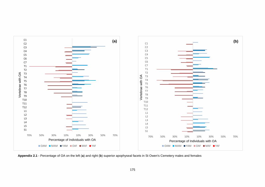
175
70% 50% 30% 10% 10% 30% 50% 70%
S1
L5
L4
L3
L2
L1
T12
T11
T10
T9
T8
T7
T6
T5
T4
T3
T2
T1
C7
C6
C5
C4
C3
C2
C1
Percentage of Individuals with OA
Vert
ebra
e w
ith O
A
OAM MAM YAM OAF MAF YAF
70% 50% 30% 10% 10% 30% 50% 70%
S1
L5
L4
L3
L2
L1
T12
T11
T10
T9
T8
T7
T6
T5
T4
T3
T2
T1
C7
C6
C5
C4
C3
C2
C1
Percentage of Individuals with OAV
ert
ebra
e w
ith O
A
OAM MAM YAM OAF MAF YAF
Appendix 2.1 - Percentage of OA on the left (a) and right (b) superior apophyseal facets in St Owen's Cemetery males and females
(a)
Ap
pe
ndi
x
2.1
-
Per
cen
tag
e of
OA
on
the
left
(a)
and
rig
ht
(b)
sup
eri
or
apo
phy
sea
l
fac
ets
in
St
(b)
(b)
(b)
(b)

176
70% 50% 30% 10% 10% 30% 50% 70%
S1
L5
L4
L3
L2
L1
T12
T11
T10
T9
T8
T7
T6
T5
T4
T3
T2
T1
C7
C6
C5
C4
C3
C2
C1
Percentage of individuals with OA
Vert
ebra
e w
ith O
A
OAM MAM YAM OAF MAF YAF
(a)
80% 60% 40% 20% 0% 20% 40%
S1
L5
L4
L3
L2
L1
T12
T11
T10
T9
T8
T7
T6
T5
T4
T3
T2
T1
C7
C6
C5
C4
C3
C2
C1
Percentage of individuals with OA
Vert
ebra
e w
ith O
A
OAM MAM YAM OAF MAF YAF
(b)
Appendix 2.2 - Percentage of OA on the left (a) and right (b) inferior apophyseal facets of St Owen's Cemetery males and females
Appendix 2.2 - Percentage of OA on the left (a) and right (b) inferior apophyseal facets of St Owen's Cemetery males and females

177
Appendix 2.3 - Percentages of OA on each vertebra within the St Owen's Cemetery sample
Y A M M A M O A M Y A F M A F O A F SA
N n % N n % N n % N n % N n % N n % N n %
C1 5 0 0 10 0 0 12 1 8 3 0 0 9 1 11 10 0 0 3 0 0
C2 5 1 20 9 4 44 13 6 46 3 0 0 9 2 22 10 6 60 2 0 0
C3 5 3 60 10 6 60 13 10 77 3 1 33 9 4 44 8 8 100 2 0 0
C4 5 2 40 10 7 70 13 10 77 1 0 0 8 5 63 9 9 100 2 0 0
C5 5 0 0 11 7 64 14 11 79 3 0 0 9 6 67 9 7 78 3 0 0
C6 4 0 0 10 5 50 14 10 71 3 0 0 10 6 60 9 7 78 2 0 0
C7 4 0 0 11 3 27 15 6 40 3 1 33 10 8 80 9 7 78 3 0 0
T1 5 2 40 12 9 75 12 9 75 4 1 25 11 10 91 10 10 100 3 0 0
T2 5 2 40 11 6 55 12 3 25 3 1 33 11 7 64 9 7 78 3 0 0
T3 5 2 40 11 4 36 11 5 45 3 1 33 11 8 73 9 8 89 3 0 0
T4 4 2 50 11 7 64 11 5 45 4 3 75 10 8 80 10 9 90 3 0 0
T5 5 2 40 13 11 85 11 6 55 4 3 75 11 7 64 10 7 70 3 0 0
T6 5 1 20 13 11 85 10 6 60 4 0 0 10 7 70 10 6 60 3 0 0
T7 5 1 20 13 9 69 12 4 33 4 0 0 10 8 80 10 7 70 3 0 0
T8 6 1 17 12 9 75 12 4 33 4 1 25 10 7 70 11 6 55 3 0 0
T9 6 2 33 13 8 62 12 2 17 3 0 0 11 3 27 11 7 64 3 0 0
T10 6 1 17 12 4 33 12 9 75 4 2 50 11 4 36 10 9 90 3 0 0
T11 6 3 50 12 9 75 12 8 67 4 1 25 11 5 45 11 10 91 3 0 0
T12 7 3 43 12 9 75 12 9 75 4 2 50 11 9 82 10 9 90 3 0 0
L1 7 0 0 13 1 8 13 6 46 4 0 0 11 0 0 10 1 10 3 0 0
L2 6 0 0 12 0 0 13 4 31 4 0 0 11 1 9 10 2 20 3 0 0
L3 6 0 0 12 1 8 12 5 42 4 0 0 11 0 0 10 3 30 3 0 0
L4 6 0 0 10 3 30 11 5 45 4 0 0 11 2 18 10 1 10 3 0 0
L5 5 1 20 9 2 22 9 3 33 4 0 0 10 1 10 9 3 33 3 0 0
S1 5 1 20 10 1 10 11 3 27 4 0 0 11 1 9 11 2 18 3 0 0 N = number of vertebrae present in sample; n = number of vertebrae with OA; YAM – young adult male; MAM – middle adult male; OAM – old adult male; YAF – young adult female; MAF – middle adult female; OAF – old adult female;
SA – subadult

178
Appendix 3: Poulton Results
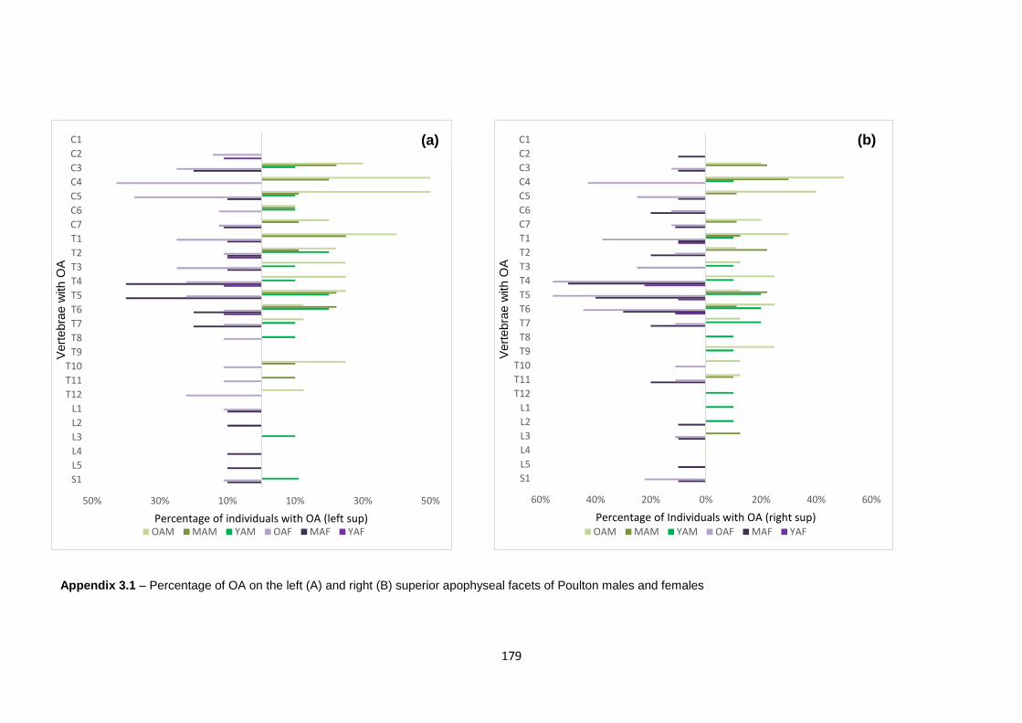
179
50% 30% 10% 10% 30% 50%
S1
L5
L4
L3
L2
L1
T12
T11
T10
T9
T8
T7
T6
T5
T4
T3
T2
T1
C7
C6
C5
C4
C3
C2
C1
Percentage of individuals with OA (left sup)
Vert
ebra
e w
ith O
A
OAM MAM YAM OAF MAF YAF
60% 40% 20% 0% 20% 40% 60%
S1
L5
L4
L3
L2
L1
T12
T11
T10
T9
T8
T7
T6
T5
T4
T3
T2
T1
C7
C6
C5
C4
C3
C2
C1
Percentage of Individuals with OA (right sup)V
ert
ebra
e w
ith O
A
OAM MAM YAM OAF MAF YAF
Appendix 3.1 – Percentage of OA on the left (A) and right (B) superior apophyseal facets of Poulton males and females
Appendix 3.1 – Percentage of OA on the left (A) and right (B) superior apophyseal facets of Poulton males and females
(a)
(a)
(a)
(a)
(b)
App
end
ix
3.1
–
Per
cen
tag
e of
OA
on
the
left
(A)
and
righ
t (B)
sup
erio
r
apo
phy
seal
face
ts
of
Pou
lton
mal
es
and
fem

180
50% 30% 10% 10% 30% 50%
S1
L5
L4
L3
L2
L1
T12
T11
T10
T9
T8
T7
T6
T5
T4
T3
T2
T1
C7
C6
C5
C4
C3
C2
C1
Percentage of Individuals with OA (left inf)
Vert
ebra
e w
ith O
A
OAM MAM YAM OAF MAF YAF
(a)
50% 30% 10% 10% 30% 50% 70%
S1
L5
L4
L3
L2
L1
T12
T11
T10
T9
T8
T7
T6
T5
T4
T3
T2
T1
C7
C6
C5
C4
C3
C2
C1
Percentage of Individuals with OA (right inf)
Vert
ebra
e w
ith O
A
OAM MAM YAM OAF MAF YAF
Appendix 3.2 - Percentage of OA on the left (A) and right (B) inferior apophyseal facets of Poulton males and females
Appendix 3.2 - Percentage of OA on the left (A) and right (B) inferior apophyseal facets of Poulton males and females
(b)
App
endi
x 3.2
-
Perc
enta
ge
of
OA
on
the
left
(A)
and
right
(B)
infer
ior
apo
phys
eal
face
ts of
Poul
ton
male
s
and
fem
ales(
b)

181
Appendix 3.3 - Percentages of OA on each vertebra within the Poulton sample
Y A M M A M O A M Y A F M A F O A F SA
N n % N n % N n % N n % N n % N n % N n %
C1 10 0 0 10 0 0 9 1 11 9 1 11 10 0 0 7 1 14 10 0 0
C2 10 1 10 10 2 20 9 4 44 9 2 22 10 4 40 7 4 57 10 0 0
C3 10 1 10 9 4 44 10 9 90 9 1 11 10 5 50 8 5 63 10 2 20
C4 10 1 10 10 4 40 10 10 100 9 1 11 10 4 40 7 6 86 8 1 13
C5 10 1 10 9 3 33 10 7 70 9 1 11 10 4 40 8 8 100 9 0 0
C6 10 1 10 10 3 30 10 5 50 9 1 11 10 5 50 8 8 100 9 0 0
C7 10 1 10 9 3 33 10 6 60 9 1 11 9 2 22 8 7 88 9 0 0
T1 10 3 30 8 6 75 10 7 70 10 5 50 10 5 50 8 6 75 10 0 0
T2 10 2 20 9 2 22 9 5 56 10 1 10 10 3 30 9 4 44 10 0 0
T3 10 3 30 9 0 0 8 2 25 10 3 30 10 5 50 8 4 50 10 0 0
T4 10 3 30 9 3 33 8 5 63 9 3 33 10 4 40 9 5 56 10 0 0
T5 10 3 30 9 3 33 8 3 38 10 2 20 10 6 60 9 6 67 10 1 10
T6 10 3 30 9 2 22 8 6 75 9 1 11 10 3 30 9 7 78 10 1 10
T7 10 4 40 9 1 11 8 4 50 10 1 10 10 4 40 9 3 33 10 0 0
T8 10 4 40 10 2 20 8 4 50 10 1 10 10 4 40 9 4 44 10 1 10
T9 10 2 20 10 2 20 8 5 63 10 0 0 10 2 20 9 3 33 10 1 10
T10 10 3 30 10 3 30 8 5 63 10 3 30 10 4 40 9 8 89 10 1 10
T11 10 2 20 10 7 70 8 3 38 10 5 50 10 6 60 9 8 89 10 1 10
T12 10 2 20 10 7 70 8 4 50 10 5 50 10 5 50 9 8 89 10 1 10
L1 10 1 10 8 0 0 8 0 0 10 0 0 10 2 20 9 1 11 10 0 0
L2 10 1 10 8 1 13 8 0 0 9 0 0 10 3 30 9 1 11 10 0 0
L3 10 1 10 8 1 13 8 0 0 9 0 0 10 2 20 9 1 11 10 1 10
L4 10 0 0 6 0 0 8 0 0 9 0 0 10 1 10 9 0 0 10 0 0
L5 10 1 10 6 0 0 8 0 0 9 0 0 10 2 20 9 3 33 10 0 0
S1 9 1 11 6 0 0 8 0 0 9 0 0 10 2 20 9 3 33 10 0 0 N = number of vertebrae present in sample; n = number of vertebrae with OA; YAM – young adult male; MAM – middle adult male; OAM – old adult male; YAF – young adult female; MAF – middle adult female; OAF – old adult female, SA –
subadult
Copyright © 2022 FDOKUMEN