Rotation compatibility approach to moment redistribution for ...
Upload
independentCategory
view
1download
0
BioMed CentralScoliosis
ss
Open AcceReviewVertebral rotation measurement: a summary and comparison of common radiographic and CT methodsGabrielle C Lam1, Doug L Hill1,2, Lawrence H Le3, Jim V Raso1,2 and Edmond H Lou*1,2Address: 1Department of Surgery, University of Alberta, Edmonton, T6G 2B7, Canada, 2Department of Rehabilitation Technology, Glenrose Rehabilitation Hospital, Alberta Health Services, Edmonton, T5G 0B7, Canada and 3Department of Radiology and Diagnostic Imaging, University of Alberta, Edmonton, T6G 2B7, Canada
Email: Gabrielle C Lam - [email protected]; Doug L Hill - [email protected]; Lawrence H Le - [email protected]; Jim V Raso - [email protected]; Edmond H Lou* - [email protected]
* Corresponding author
AbstractCurrent research has provided a more comprehensive understanding of Adolescent IdiopathicScoliosis (AIS) as a three-dimensional spinal deformity, encompassing both lateral and rotationalcomponents. Apart from quantifying curve severity using the Cobb angle, vertebral rotation hasbecome increasingly prominent in the study of scoliosis. It demonstrates significance in bothpreoperative and postoperative assessment, providing better appreciation of the impact of bracingor surgical interventions. In the past, the need for computer resources, digitizers and customsoftware limited studies of rotation to research performed after a patient left the scoliosis clinic.With advanced technology, however, rotation measurements are now more feasible. Whilenumerous vertebral rotation measurement methods have been developed and tested, thoroughcomparisons of these are still relatively unexplored. This review discusses the advantages anddisadvantages of six common measurement techniques based on technology most pertinent inclinical settings: radiography (Cobb, Nash-Moe, Perdriolle and Stokes' method) and computertomography (CT) imaging (Aaro-Dahlborn and Ho's method). Better insight into the clinicalsuitability of rotation measurement methods currently available is presented, along with adiscussion of critical concerns that should be addressed in future studies and development of newmethods.
BackgroundAdolescent Idiopathic Scoliosis (AIS) is a lateral and rota-tional deformity of the spine, predominantly affectingindividuals of age 10 to 17. The progression of AIS occursduring the rapid growth stage due to factors stillunknown. Traditionally, measurement of Cobb angleswas the primary means of quantifying the severity of AISin scoliotic patients. However, this method is limited to
assessment of the spine in the saggital and coronal planes.More current investigation of vertebral rotation in theaxial plane has provided better understanding of AIS as athree-dimensional condition.
Recent studies suggest that the coupling relation betweenvertebral rotation and lateral motion may provide insightinto an indicative characteristic of scoliotic spines [1-4].
Published: 2 November 2008
Scoliosis 2008, 3:16 doi:10.1186/1748-7161-3-16
Received: 13 August 2008Accepted: 2 November 2008
This article is available from: http://www.scoliosisjournal.com/content/3/1/16
© 2008 Lam et al; licensee BioMed Central Ltd. This is an Open Access article distributed under the terms of the Creative Commons Attribution License (http://creativecommons.org/licenses/by/2.0), which permits unrestricted use, distribution, and reproduction in any medium, provided the original work is properly cited.
Page 1 of 10(page number not for citation purposes)

Scoliosis 2008, 3:16 http://www.scoliosisjournal.com/content/3/1/16
Other literature has examined rotation of the vertebralcolumn in connection to the etiology of AIS [5,6], as dis-cussed in the Spine-Rib Hypothesis [7] and in evaluatingthe Neurocentral Junction Hypothesis [8]. Above all,measurement of vertebral rotation is of key significance inthe prognosis and treatment of scoliotic curves [9-11]. Itmay act as an indicator of curve progression, thus beingclinically applicable for both preoperative and postopera-tive assessment [9,12]. The association of vertebral rota-tion with rib hump has led to techniques that may beapplied to school screening programs [13,14]. Further-more, vertebral rotation measurement is becoming prom-inent in assisting pre-surgical planning. Inaccurateknowledge of vertebral rotation may lead to unnecessarysurgical operations and, in the case of pedicle screws, mis-placements that incur risks of spinal cord injury [15]. Atthe same time, axial rotation has been equally valuable inbetter understanding the effect of brace treatment or sur-gical interventions, as evidenced in the studies evaluatingCotrel-Dubousset [16-19] and Harrington instrumenta-tion [20,21].
While numerous methods have been developed to meas-ure axial rotation, the techniques explored most exten-sively are those involving landmark identification. Theposition of the spinous process (Cobb method) [22],pedicle shadows (Nash and Moe, Perdriolle, Drerup,Stokes method) [9,10,23-25], or a combination of land-marks (Mehta) [26] in relation to the vertebral body areoften clinically used to quantify the extent of vertebralrotation. The methods mentioned above are performedon images obtained using radiography – a technologythat, though popular, is limited in various aspects. In thepedicle-shadow offset technique initiated by Nash andMoe, measurements taken from radiographic images onlyrepresent a projected, not actual, rotation [27]. Further-more, the hazardous health implications associated withfrequent and prolonged exposure to radiation has been ofprimary concern for scoliotic patients, who are oftenundergoing critical growth and developmental stages.Consequently, there has been growing emphasis on devel-oping new technology that does not involve patient expo-sure to ionizing radiation. Such examples include real-time ultrasound [28,29], the AUSCAN system [30,31],magnetic resonance imaging (MRI) [32-34], and otherinnovations [35]. The real-time ultrasound method[28,29] used the Aloka SDD 500 portable ultrasound unit(Olympus, Medical and Industrial Equipment Ltd.) toidentify the laminae and the rib to determine the rotationwhile the patient was lying prone on a couch with theirforehead supported. Kirby et al [28] reported that theworst estimation on both rib and vertebral rotation were± 3.6°. The AUSCAN System (AUtomatic SColiosis ANa-lyser) (BTS Bioengineering Technology & Systems Inc.,Italy) is an automatic optoelectronic device that consists
of two pairs of CCD TV-cameras, a FPSR (Fast Processorfor Shape Recognition) image processor and a speciallydeveloped software package for data processing. Twentyseven body landmarks placed on the skin: 19 on the pos-terior side and 8 on the anterior side, which included thespinous processes from C7 to S1 were used to reconstructthe internal alignment. The vertebral rotation would thenbe estimated. To use MR images to estimate the vertebralrotation, a specific MRI images technique was need [32].Using the Birchall et al method [32], the segmental axialrotation was comparable to the conventional CT method.However, MR images were not commonly requested dur-ing scoliosis clinic. The Ortelius 800 machine uses a mag-netic field fingertip sensor to identify the spinal processand then to reconstruct the internal spinal structure wasanother non-ionization method. The vertebral rotationcould be estimated based on the location of the spinousprocess; however, no clinical study has concluded thatthis method was accurate. The aim of these new develop-ments is to reduce the number of false positives referred toscoliosis clinics through school screening programs aswell as unnecessary exposure to radiation. Another tech-nology that is becoming increasingly popular for assessingaxial rotation is computer tomography. Aaro and Dahl-born [36] and Ho et al [37] developed techniques of rota-tion measurement from CT images. Despite manyadvantages of this technology, radiographic methodsremain most standard, to which current developing tech-niques are compared for evaluating their accuracy [27,38-41].
This review encapsulates six common methods of measur-ing vertebral rotation based on technology demonstratinggreatest relevance in present clinical settings: radiographyand CT imaging. An assessment of each technique,founded upon accuracy, economic, and health considera-tions, forms the focus of this paper. Our findings aim toprovide better insight into the clinical suitability of pres-ently available rotation measurement methods, as well asto underscore critical concerns that should be addressedin future development of new techniques.
Summary of Radiographic and CT Methods
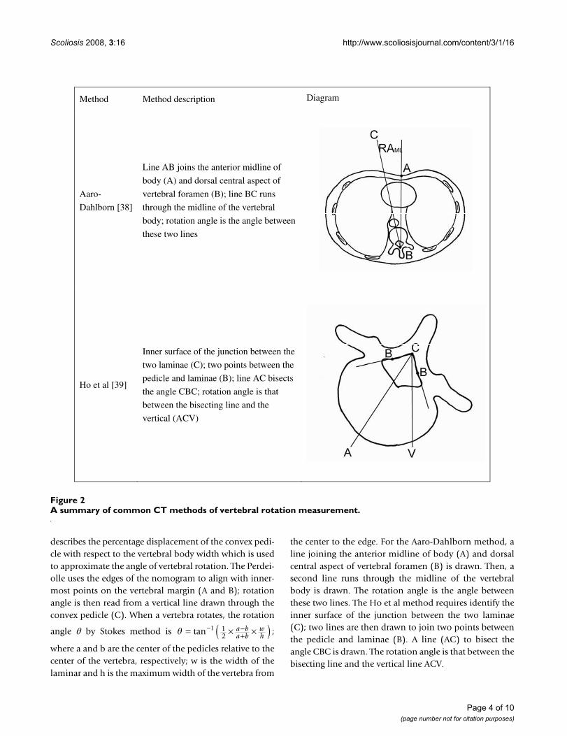
The following charts (Figures 1 and 2) summarize theradiographic and CT methods of vertebral rotation meas-urement of interest in this review, respectively. Figure 1describes and illustrates how the vertebral rotation meas-ured using the Cobb, Nash-Moe, Perdeiolle and Stokesmethods with radiographs. Figure 2 describes and illus-trates how the vertebral rotation is measured using theAaro-Dahlborn and Ho et al. methods with CT images.The Cobb method divides the vertebral body into six sec-tions; the region in which the spinous process is aligneddetermines the grade assigned. The Nash-Moe method
Page 2 of 10(page number not for citation purposes)

Scoliosis 2008, 3:16 http://www.scoliosisjournal.com/content/3/1/16
Page 3 of 10(page number not for citation purposes)
A summary of common radiographic methods of vertebral rotation measurementFigure 1A summary of common radiographic methods of vertebral rotation measurement.
Method Method description Diagram
Cobb [24]
The vertebral body is divided into
six sections; the region in which
the spinous process is aligned
determines the grade assigned
Nash-Moe
[25]
The percentage displacement of
the convex pedicle with respect to
the vertebral body width is used
to approximate the angle of
vertebral rotation
Perdriolle
[10]
The edges of the nomogram are
aligned with innermost points on
the vertebral margin (A and B);
rotation angle is read from a
vertical line drawn through the
convex pedicle (C)
Stokes [27]
The projected distances of both
pedicles from the vertebral center
(a and b) are measured from the
radiographic film; fixed width-to-
depth ratios for each vertebral
level is applied to Stokes’
formula to determine rotation
angle
Scoliosis 2008, 3:16 http://www.scoliosisjournal.com/content/3/1/16
describes the percentage displacement of the convex pedi-cle with respect to the vertebral body width which is usedto approximate the angle of vertebral rotation. The Perdei-olle uses the edges of the nomogram to align with inner-most points on the vertebral margin (A and B); rotationangle is then read from a vertical line drawn through theconvex pedicle (C). When a vertebra rotates, the rotation
angle θ by Stokes method is ;
where a and b are the center of the pedicles relative to thecenter of the vertebra, respectively; w is the width of thelaminar and h is the maximum width of the vertebra from
the center to the edge. For the Aaro-Dahlborn method, aline joining the anterior midline of body (A) and dorsalcentral aspect of vertebral foramen (B) is drawn. Then, asecond line runs through the midline of the vertebralbody is drawn. The rotation angle is the angle betweenthese two lines. The Ho et al method requires identify theinner surface of the junction between the two laminae(C); two lines are then drawn to join two points betweenthe pedicle and laminae (B). A line (AC) to bisect theangle CBC is drawn. The rotation angle is that between thebisecting line and the vertical line ACV.
θ = × ×( )− −+tan 1 1
2a ba b
wh
A summary of common CT methods of vertebral rotation measurementFigure 2A summary of common CT methods of vertebral rotation measurement.
Method Method description Diagram
Aaro-
Dahlborn [38]
Line AB joins the anterior midline of
body (A) and dorsal central aspect of
vertebral foramen (B); line BC runs
through the midline of the vertebral
body; rotation angle is the angle between
these two lines
Ho et al [39]
Inner surface of the junction between the
two laminae (C); two points between the
pedicle and laminae (B); line AC bisects
the angle CBC; rotation angle is that
between the bisecting line and the
vertical (ACV)
Page 4 of 10(page number not for citation purposes)

Scoliosis 2008, 3:16 http://www.scoliosisjournal.com/content/3/1/16
Advantages and Disadvantages of Individual MethodsRadiographic measures are obtained routinely in a stand-ing posture whereas CT measures are obtained in a supineposition. It is noteworthy that scoliotic curves appear lesssevere when supine, both in terms of curvature and rota-tion. This imperfect relationship complicates compari-sons when considering both modalities for longitudinalfollow-up of an individual.
Cobb methodOne weakness of Cobb's method is its ability to providean approximation of vertebral rotation. The gradingscheme is limited to five grades and does not allow quan-tification of the angle of axial rotation [23]. Nash and Moelater improved on this limitation by suggesting a methodof obtaining a rotation degree from the percentage dis-placement of the landmark with regards to the vertebralbody.
Another disadvantage of Cobb's method involves the ver-tebral landmark used to determine axial rotation. Verte-brae of severe scoliotic cases often exhibit intravertebralrotation, as evidenced by the distortion of the spinousprocess tip from the frontal center of the vertebral body.The vertebral model referred to in Cobb's study, however,disregarded such asymmetry. Moreover, surgical tech-niques often alter the spinous process, obscuring visibilityfollowing operation. Stokes, Nash and Moe argued thatmeasurements using the spinous process may result ininaccurate apparent rotation [23,25], which motivatedtheir later studies of pedicle shadows to determine verte-bral rotation.
Nash and Moe reported the difficulty of visualizing thespinous process on spinal radiographs to be a major chal-lenge [23]. Mehta also discovered the limited visibility ofthe spinous process problematic in measuring large rota-tion angles [26]. In addition to the limited visibility of thechosen landmark, Nash and Moe observed inconsisten-cies in the grading scheme and measurement method.Their study reported a 10–20° underestimation usingCobb's method as well as inconsistencies in the angleinterval represented by each grade.
Due to Cobb's method being one of the earliest recordedtechniques used for measuring vertebral rotation, most lit-erature focused upon its limitations. Despite these criti-cisms, the concept of measuring axial rotation usingvertebral landmarks from anteroposterior radiographswas adopted by many later methods and remains popularin clinical settings. Cobb's method is valuable in beingsimple to use and requiring no additional patient expo-sure to radiation in comparison to more modern methods[42,43]. Moreover, the spinous process is used as a land-mark in numerous other improved methods. In 1985,
Bunnell developed a measurement method involving thedistance of the spinous process with respect to the widthof the vertebral body [44]. Drerup also suggested investi-gating the position of pedicles with relation to the spinousprocess and vertebral body [9,24]. Both these techniqueswere found to yield relatively accurate results [45].
Nash-Moe methodNash and Moe claimed their method to be muchimproved from that of Cobb. One advantage was the bet-ter visibility of the particular chosen anatomic landmarkover a greater range of angles. Their study was able toinvestigate rotations of the convex pedicle of up to 90°[23]. Additionally, pedicle shadows could be better seeneven after surgery, rendering the method applicable inpostoperative assessment. The Nash-Moe method seemedto overcome the concern of intravertebral deformityaffecting measurement reliability, which became a majorfactor in the criticism of Cobb's technique. In comparisonto the spinous process, the pedicles are located closer tothe vertebral body and, consequently, are not subject to asmuch distortion in severe scoliotic cases [23,25].
Despite these improvements, there exist some drawbacksof the technique. Foremost, the suggested method ofangle determination only provided a rough approxima-tion of axial rotation. Ho observed that grade 0, a neutralposition, represented rotation of up to 11° determined byCT scans [37]. Like Cobb's method, the Nash-Moe tech-nique neglected vertebral asymmetries such as non-paral-lel endplates, concave vertebral walls, and ellipticaldiameters [9,24]. Other factors overlooked were men-tioned by Stokes, including the effect of distance from thex-ray, vertebral body shape and symmetry [25].
The better visibility of pedicle shadows was emphasized asthe main advantage of the Nash-Moe method. However,Mehta contradicted this assertion, finding that using a sin-gle anatomical landmark limited accurate measurementsto small rotations [26]. This same realization was made bythe authors of this report, who examined 115 spinal radi-ographs in two separate sittings. The convex pediclebecame difficult to visualize at rotation angles greaterthan 30°. Furthermore, surgical implantations such asHarrington and Cotrel-Dubousset instrumentationobstructed visibility of pedicles on spinal radiographs.[46,47]
Perdriolle methodConflicting views were presented concerning the accuracyand reliability of rotation measurements performed usingthe Perdriolle torsion meter. Richards' study [47] foundthe average individual observer error to be 6°. Only about50% of observers obtained measurements within 5° ofthe actual value. Richards also discussed the difficulty of
Page 5 of 10(page number not for citation purposes)

Scoliosis 2008, 3:16 http://www.scoliosisjournal.com/content/3/1/16
visualizing pedicle landmarks in 20% of postoperativespinal radiographs, for which determination of vertebralrotation is currently most relevant. Another complicationinvolved marking the pedicles on spinal radiographs,where a 2 mm error amounts to 5° of rotation. Richardsalso recognized that a rotated patient body during radio-graphic examination further increased measurementerror.
Despite Richards' negative findings, most studies high-lighted advantages of using the torsion meter. In examin-ing interobserver and intraobserver errors, Barsantishowed that over 92% of errors made were within ± 5°[46]. Omeroglu reached similar conclusions, with 98% ofintraobserver measurements within ± 5° [48]. In Weiss'investigation, intraobserver error was reported to be ± 1°and interobserver error ± 3° [49]. These studies also agreethat such accuracy can only be observed for mild to mod-erate rotation, due to difficulty of point selection for ver-tebrae with large rotation. However, this does not seem tobe a limitation, since typical scoliosis incidences involve15–20° rotation, rarely exceeding 40° [46,49].
Another issue connected to the controversy concerningaccuracy and reliability of the torsion meter is its ability toaccount for irregular vertebral geometry, saggital andcoronal inclination. This challenge is more inconclusive,although Weiss noted that their effect on measurementaccuracy was insignificant when studying the apical verte-bra. He also suggested measuring a second vertebra as ref-erence, so to reduce measurement error due to rotatedbody position [27]. Overall, many studies reported thatthe Perdriolle torsion meter is suitable for use in the clinic.It is affordable, non-invasive and simple to use, making itapplicable in clinical settings [46,48,50]. Barsanti alsohighlighted its ability to measure rotation from a singleanteroposterior radiograph to be an advantage. In con-trast to newly developed techniques like stereoradiogra-phy, the torsion meter minimized patient exposure toharmful radiation.
Stokes methodStokes' method accounted for vertebral asymmetry anddimension by determining pedicle-offset with respect to acenter point rather than the vertebral edges. His techniquesimulates biplanar radiography, which involves takingtwo radiographic images, frontal and lateral, to obtainmeasurement of vertebral dimensions. Stokes reportedsimilar accuracy, with the added advantage of one fewer x-ray exposure and a less complicated measurement scheme[25].
In a comparative study of four methods used to determineaxial rotation from radiographic images [45], the tech-niques proposed by Bunnell, Drerup, Koreska and Stokes
were examined. Results showed close correlation betweenvalues obtained by the former three methods. However,Stokes' method demonstrated significant deviation forrotation greater than 5°. Even after Stokes corrected thewidth-to-depth ratios by a factor of two [51], the results ofthe comparative study suggested that his method was leastaccurate among the four techniques tested. Stokes ration-alized that using the vertebral center to measure pedicledisplacement should be more accurate. However, thehigher precision of marking vertebral edges might offsetthis accuracy. His method incurred least systematic error,but greatest random error.
Another concern regards the use of an averaged width-to-depth ratio for different vertebra levels. The geometry ofscoliotic vertebrae can vary greatly among individual con-ditions. Consequently, these values might be poorly rep-resented in severely distorted vertebrae, affecting accuracyof calculations obtained from Stokes' formula.
Aaro-Dahlborn methodA significant advancement of the Aaro-Dahlborn methodwas its use of CT technology. In comparison with radiog-raphy, CT produces clearer and more detailed images.Errors due to poor clarity of vertebral structures, an obsta-cle for many radiographic techniques, are reduced. Fur-thermore, in the transverse plane, landmarks are easilyseen even with large rotation. This technology overcomesa major challenge faced by radiography: measuring largedegrees of rotation.
One major drawback, however, of using CT technology isincreased measurement inaccuracy associated with verte-brae inclined in the saggital and coronal planes[11,36,52]. A study by Skalli et al [11] found that ininclined vertebrae with small degrees of axial rotation(less than 10°), the difference between the actual three-dimensional rotation and the projected rotation is rela-tively small (approximately 2°). Not only does vertebralorientation change with different body positions, scolioticvertebrae often exhibit rotations in various planes. There-fore, Skalli et al stated that rotation measurements fromtransverse CT images can be misleading.
Additionally, CT scans require more time and are moreexpensive than spinal radiographs, limiting its use in clin-ical settings. If the entire vertebral column is examined, asin radiography, CT involves greater patient exposure toionizing radiation, particularly in the thoracolumbarregion [53]. As discussed earlier, increased exposure toradiation can be particularly harmful for patients under-going crucial developmental stages.
Consequently, a CT scan of the apex vertebra is conven-tionally used to indicate rotation severity. CT, however,
Page 6 of 10(page number not for citation purposes)

Scoliosis 2008, 3:16 http://www.scoliosisjournal.com/content/3/1/16
cannot entirely replace the role of radiography, since a spi-nal radiograph is still necessary for identification of theapex vertebra. An associated problem was highlighted byone recent study [41], which showed that the apical verte-bra did not always exhibit maximum rotation, a challengein tracing the rotation severity using CT. Furthermore, thesupine position required for CT scans also reduces theCobb angle, rotation angle, and rib hump [36,50]. Meas-urements taken from CT scans will therefore under repre-sent the true rotation.
The disadvantages discussed above mainly concern thechoice of measurement technology. Amongst other CTmethods, such as that of Ho et al, the Aaro-Dahlbornmethod reached measurements with greater correlation tothe actual rotation value, even with tilt in the saggital andcoronal planes [54]. In a separate study, both Aaro-Dahl-born and Ho's methods showed similar accuracy. Theformer method was found to be more difficult to use forinexperienced observers [52]. Gocen et al propose thatthis is due to the less obvious reference points used, suchas the anterior midline, in determining RAML. Ho et alreported higher intraobserver and interobserver errors inthe use of the Aaro-Dahlborn method when compared totheir technique [37].
Ho et al methodThe method by Ho et al determines rotation from CTscans using the laminae and laminae junction. Theirresults reported a 95% clinical success rate and 1.2° errorratio [52] compared three methods (Ho et al, Aaro-Dahl-born and Krismer), reaching similar conclusions to that ofHo et al. The former two methods showed closest correla-tion with the actual rotation value, but Ho et al's methodwas preferred. The more clearly defined reference pointsused in this method allowed less experienced observers toreach accurate measurements from the CT images. Theirresults revealed that Ho's method reduced errors andmeasurement variability due to reference point selection.While both Aaro-Dahlborn and Ho et al's methodsproved clinically applicable and accurate, the study alsoshowed that interobserver reliability was significantly bet-ter in the latter technique.
One concern related to axial rotation measurements fromCT is the effect of saggital and coronal tilt on accuracy ofmeasurement values, as described earlier. Krismer et alconducted a study [54] investigating the accuracy of Aaro-Dahlborn and Ho et al's methods in measuring axial rota-tion of vertebra with significant anatomic deformationand rotated in different planes. The measurementsobtained using Ho's method showed less correlation toactual values in comparison to the Aaro-Dahlbornmethod. Krismer et al suggested that the high accuracy
found from Ho's study only signifies its applicability inidealistic conditions, not so in clinical settings.
DiscussionThe key advantages and disadvantages associated witheach method of vertebral rotation measurement are sum-marized in the chart below (Table 1). A review of the var-ious methods of measuring vertebral rotation indicatessome of the significant hurdles encountered in this area ofresearch. The objective of obtaining accurate measure-ments seems to be hampered by vertebral irregularitiestypical of scoliotic cases. Such factors include intraverte-bral rotation, inclination in different planes, and largerotation angles – the prominence and effect of each beingdependent on the particular method used. Although thesechallenges have long been recognized, a conclusive solu-tion has yet to be reached and remains a primary goal incontinuing studies.
Aside from measurement accuracy and precision, clinicalapplicability of each method is examined in this paper onthe basis of its ease of usage, health and financial implica-tions. Radiation exposure is of critical importance in thisevaluation, keeping in mind the young age of AIS patientsbeing considered and their vulnerability during crucialgrowth stages. CT images are insufficient for identifyingthe apex vertebra, therefore unable to entirely replace therole of radiography in rotation assessment. While radiog-raphy yields images with poorer clarity and involves expo-sure of a larger bodily region to radiation, it entails lessradiation exposure overall. Furthermore, accuracy of CTmeasurements is more severely affected by inclinationand tilt, outweighing limitations encountered with radio-graphic measurement of large rotations when consideringrealistic conditions. For these two reasons, radiography isstill the more clinically suitable alternative.
The literature examined did not allow for entirely trans-parent conclusions to be made concerning the most clini-cally applicable rotation measurement method. Manyreviews of Perdriolle's torsion meter express consensus inits measurement precision and simplicity of use. Stokes'calculation may also be a promising method. It aims toovercome the difficulty associated with asymmetrical ver-tebral geometry, accounting for vertebral dimensions inaddition to projected distances. However, there presentlyexist limited studies evaluating Stokes' method, renderingit difficult to reach fair judgments. For instance, the com-parative study by Russel et al [45] draws conclusions fromtesting a small number of vertebrae. Comparison ofStokes' calculations with stereoradiographic techniquesmay be valuable, since his concept had been motivated bythis improved technology. More extensive comparativestudies are perhaps an area requiring greater attention in
Page 7 of 10(page number not for citation purposes)

Scoliosis 2008, 3:16 http://www.scoliosisjournal.com/content/3/1/16
the future, building better groundwork for evaluatingmethods with potential clinical utility, like that of Stokes.
ConclusionUp to this point, there have been many methods pro-posed to measure vertebral rotation such as Radiography,CT method, ultrasound, MRI and magnetic sensors.Among these, radiography is still the more commonlyused method as all orthopaedic surgeons are very familiarwith radiographic images. At our site CT and MR imagesare generally reserved for cases with unusual presenta-tions, neurological symptoms or deficits, rapidly progress-ing curves and surgical planning for complex congenitalcases. The choice between CT and MR rests on the purposeof the intervention with CT scans showing bony detailbetter whereas MR images are superior for soft tissue iden-tification. As the technology becomes more advanced, andradiation exposure is reduced, either low dose x-raymachines or a better 3D ultrasound imaging machine willbe more commonly used to measure vertebral rotationaccurately.
Competing interestsThe authors declare that they have no competing interests.
Authors' contributionsGL conducted the study and prepared the manuscript. DHinvolved in editing the manuscript. LL conceived of thestudy. JR conceived of the study. EL conceived of thestudy, coordinated the project and edited the manuscript.All authors approved the final manuscript.
AcknowledgementsThe authors would like to thank Glenrose Rehabilitation Hospital for pro-viding necessary resources and funding for this project.
References1. Cole AA, Burwell RG, Webb JK: Lateral flexion induced axial
rotation in adolescent idiopathic scoliosis (AIS). Studies inHealth Technology and Informatics: Research into Spinal Deformities 11997, 37:93-96.
2. Lafage V, Leborgne P, Mitulescu A, Dubousset J, Lavaste F, Skalli W:Comparison of mechanical behavior of normal and scolioticvertebral segment: a preliminary numerical approach. StudHealth Technol Inform 2002, 88:340-344.
3. Farahpour N, Allard P, Labelle H, Rivard C, Duhaime M: Couplingmechanisms in the scoliotic spine. Studies in Health Technology
Table 1: Advantages and disadvantages associated with radiographic and CT methods of rotation measurement
Method Advantages Disadvantages
Cobb ▪ Simple procedure ▪ No means to quantify rotation from gradation scheme▪ Little patient exposure to radiation (one anteroposterior radiograph)
▪ Distortion of spinous process tip in scoliotic vertebrae may decrease accuracy▪ Limited visibility of spinous process on radiographs of vertebrae with large rotation
Nash-Moe ▪ Position of pedicles are less affected by intravertebral rotation; more reliable landmark for measuring rotation
▪ Provides over-estimation of rotation (can be adjusted by 10° as suggested by Drerup)▪ Pedicles are poorly visible on vertebrae rotated severely or on spines with surgical instrumentation
Perdriolle ▪ Affordable ▪ Difficulty in making precise markings on radiographs – a 2 mm error corresponds to 5° rotation
▪ Non-invasive▪ Simple procedure▪ Little patient exposure to radiation (one anteroposterior radiograph)
▪ Reduced accuracy when measuring large degrees of rotation
▪ General findings report accurate measurements to within ± 5°
Stokes ▪ Accounts for three-dimensionality of vertebra; similar accuracy to stereoradiograph
▪ Greater random error in comparison to methods involving marking of vertebral edges
▪ Little exposure to radiation▪ Simple measuring procedure
Aaro-Dahlborn ▪ Better measurement accuracy even when measuring vertebrae tilted in the coronal and saggital planes
▪ More difficult to use for inexperienced observers due to less obvious landmark definitions
Ho ▪ Clearly defined reference points; simple procedure ▪ Less correlation to actual rotation value when measuring distorted and
▪ Better interobserver reliability in comparison to Aaro-Dahlborn method when assessing normal vertebrae
tilted vertebrae; less applicable in realistic conditions
Page 8 of 10(page number not for citation purposes)

Scoliosis 2008, 3:16 http://www.scoliosisjournal.com/content/3/1/16
and Informatics: Three-dimensional Analysis of Spinal Deformities 1995,15:119-121.
4. Beuerlein MJ, Raso VJ, Hill DL, Moreau MJ, Mahood JK: The rela-tionship between axial rotation and lateral bending. Studies inHealth Technology and Informatics: Research into Spinal Deformities 21999, 59:105-108.
5. Heidari B, Fitzpatrick D, McCormack D, Synnott K: Correlation ofan induced rotation model with the clinical categorization ofscoliotic deformity – a possible platform for prediction ofscoliosis progression. Stud Health Technol Inform 2006,123:169-175.
6. Grivas TB, Vasiliadis E, Malakasis M, Mouzakis V, Segos D: Interver-tebral disc biomechanics in the pathogenesis of idiopathicscoliosis. Stud Health Technol Inform 2006, 123:80-83.
7. Burwell RG, Dangerfield PH: Hypotheses on pathogenesis ofAdolescent Idiopathic Scoliosis (AIS) a spine-rib hypothesis.In International Research Society of Spinal Deformities Symposium 2004:10–12 June 2004; Vancouver, Canada Edited by: Sawatzky J. Bonita:University of British Columbia; 2004:297-301.
8. Al-Adra D, Arnett J, Rajwani T, Bagnall K: Rotation, curvature andwedging in Adolescent Idiopathic Scoliosis patients. In Inter-national Research Society of Spinal Deformities Symposium 2004: 10–12June 2004; Vancouver, Canada Edited by: Sawatzky J. Bonita: Universityof British Columbia; 2004:282-285.
9. Drerup B: Improvements in measuring vertebral rotationfrom the projections of the pedicles. Journal of Biomechanics1985, 18:369-378.
10. Perdriolle R, Vidal J: Thoracic idiopathic scoliosis curve evalua-tion and prognosis. Spine 1985, 10:785-791.
11. Skalli W, Lavaste F, Descrimes J: Quantification of three-dimen-sional vertebral rotations in scoliosis: what are the true val-ues? Spine 1995, 20(5):546-553.
12. Kuklo T, Potter BK, Lawrence L: Vertebral Rotation and Tho-racic Torsion in Adolescent Idiopathic Scoliosis: What is theBest Radiographic Correlate? Journal of Spinal Disorders and Tech-niques 2005, 18(2):139-147.
13. Kotwicki T, Krawczynski A, Lorkowska M, Frydryk K: Clinical andradiological assessment of spinal rotation. InternationalResearch Society of spinal deformities 2004:207-211.
14. Ho E, Upadhyay SS, Chan FL, Hsu L, Leong J: New Methods ofMeasuring Vertebral Rotation From Computed Tomo-graphic Scans: An Intraobserver and Interobserver Study onGirls with Scoliosis. Spine 1993, 18(9):1173-1177.
15. Sugimoto Y, Tanaka M, Nakanishi K, Misawa H, Takigawa T, Ozaki T:Predicting Intraoperative Vertebral Rotation in PatientsWith Scoliosis Using Posterior Elements as AnatomicalLandmarks. Spine 2007, 32(25):E761-E763.
16. Cundy PJ, Paterson DC, Hillier TM, Sutherland AD, Stephen JP, FosterBK: Cotrel-Dubousset instrumentation and vertebral rota-tion in adolescent idiopathic scoliosis. J Bone Joint Surg Br 1990,72(4):670-674.
17. Lenke LG, Bridwell KH, Baldus C, Blanke K: Analysis of pulmonaryfunction and axis rotation in adolescent and young adult idi-opathic scoliosis patients treated with Cotrel-Duboussetinstrumentation. Journal of Spinal Disorders 1992, 5:16-25.
18. Krismer M, Bauer R, Sterzinger W: Scoliosis correction byCotrel-Dubbousset instrumentation: The effect of derota-tion and three dimensional correction. Spine 1992,17:2635-2695.
19. Wood KB, Transfeldt EE, Ogilvie JW, Schendel MJ, Bradford DS:Rotational changes of the vertebral-pelvic axis followingCotrel-Dubousset instrumentation. Spine 1991, 16:S404-408.
20. Aaro S, Dahlborn M: The effect of Harrington instrumentationon the longitudinal axis rotation of the apical vertebra andon the spinal and ribcage deformity in idiopathic scoliosisstudied by computer tomography. Spine 1982, 7:456-462.
21. Marchesi DG, Transfeldt EE, Bradford DS: Changes in vertebralrotation after Harrington and Luque instrumentation for idi-opathic scoliosis. Spine 1992, 17:775-780.
22. Cobb JR: Outline for the study of scoliosis. Instructional CourseLectures. American Academy of Orthopaedic Surgeons 1948, 5:261-275.
23. Nash C, Moe JH: A study of vertebral rotation. Journal of Boneand Joint Surgery 1969, 51:223-229.
24. Drerup B: Principles of measurement of vertebral rotationfrom frontal projection of the pedicles. Journal of Biomechanics1984, 17:923-935.
25. Stokes IAF, Bigalow LC, Moreland MS: Measurement of axial rota-tion of vertebrae in scoliosis. Spine 1986, 11:213-218.
26. Mehta MH: Radiographic estimation of vertebral rotation inscoliosis. J Bone Joint Surg Br 1973, 55(3):513-520.
27. Weiss HR: Technical error of vertebral rotation measure-ments. Studies in Health Technology and Informatics: Three-DimensionalAnalysis of Spinal Deformities 1995, 15:243-249.
28. Kirby AS, Aujla RK, Burwell RG, Cole AA, Moulton A, Polak FJ, WebbJK: A preliminary study of a new real-time ultrasoundmethod of measuring rib and spinal rotation in scoliosis.Studies in Health Technology and Informatics: Research into Spinal Deform-ities 1 1997, 37:335-337.
29. Kirby AS, Burwell RG, Cole AA, Pratt RK, Webb JK, Moulton A:Evaluation of a new real-time ultrasound method for meas-uring segmental rotation of vertebrae and ribs in scoliosis.Studies in Health Technology and Informatics: Research into Spinal Deform-ities 2 1999, 59:316-320.
30. Negrini S, Negrini A, Rainero G, Sibilla P, Santambrogio GC: Corre-lation between trunk gibbosity and spinal torsion measuredby the AUSCAN system. Studies in Health Technology and Informat-ics: Three-Dimensional Analysis of Spinal Deformities 1995, 15:279-283.
31. Negrini S, Negrini A, Atansaio S, Santambrogio GC: Three-dimen-sional easy morphological (3-DEMO) classification of scolio-sis, part 1. Scoliosis 2006, 1:20.
32. Birchall D, Hughes D, Williamson B: Demonstration of vertebraland disc mechanical torsion in adolescent idiopathic scoliosisusing three-dimensional magnetic resonance imaging. Scolio-sis 2007, 2(Suppl 1):S27.
33. Dangerfield PH, Roche CJ, King CE, Carty HM, Dorgan JC: Rotationof the atlantico-axial joint, investigated using CT and MRI.Stud Health Technol Inform 2002, 88:336-339.
34. Birchall D, Hughes DG, Hindle J, Robinson L, Williamson JB: Meas-urement of Vertebral Rotation in Adolescent Idiopathic Sco-liosis Using Three-Dimensional Magnetic ResonanceImaging. Spine 1997, 22(20):2403-2407.
35. Parisini P, Lolli F, Greggi T, Di Silvestre M, Cioni A, Giacomini S, Baka-loudis G: An innovative diagnostic procedure of vertebraldeformities without exposure to X-rays. Stud Health TechnolInform 2006, 123:527-532.
36. Aaro S, Dahlborn M, Svensson L: Estimation of vertebral rota-tion in structural scoliosis by computer tomography. ActaRadiologica Diagnosis 1978, 19:990-992.
37. Ho EKW, Upadlyay SS, Chan FL, Hsu LC, Leong JC: New methodsof measuring vertebral rotation from computed tomo-graphic scans. Spine 1993, 18:1173-1177.
38. Thometz J, Liu XC, Lyon R: Axial rotation in idiopathic scoliosis:a comparison of the Perdriolle, Scoliometer, Quantec SpinalImage System. Studies in Health Technology and Informatics: Researchinto Spinal Deformities 2 1999, 59:329-331.
39. Merolli A, Leali PT, Aulisa L, Guidi PL, Impagnatello M: A newmethod for clinical measurement of vertebral rotation. Stud-ies in Health Technology and Informatics: Research into Spinal Deformities1 1997, 37:147-149.
40. Adam C, Askin G: Automatic Measurement of Vertebral Rota-tion in Idiopathic Scoliosis. Spine 2006, 31(3):E80-E83.
41. Ho EK, Upadhyay SS, Ferris L, Chan F, Leong J: A ComparativeStudy of Computed Tomographic and Plain RadiographicMethods to Measure Vertebral Rotation in Adolescent Idio-pathic Scoliosis. Spine 1992, 17(7):771-774.
42. Brown RH, Burnstein AH, Nash CL, Schock CC: Spinal analysisusing a three-dimensional radiographic technique. Journal ofBiomechanics 1976, 9:355-365.
43. Stokes IAF, Wilder DG, Frymoyer JW, Pope MH: Assessment ofpatients with low back pain by biplanar radiographic meas-urement of intervertebral motion. Spine 1981, 6:233-240.
44. Bunnell WP: Simple method of measurement on routine radi-ographs. Orthopaedic Transactions 1985, 9:114.
45. Russell GG, Raso VJ, Hill D, McIvor J: A comparison of four com-puterized methods for measuring vertebral rotation. Spine1990, 15(1):24-27.
46. Barsanti CM, DeBari A, Covino BM: The torsion meter: a criticalreview. Journal of Pediatric Orthopaedics 1990, 10:527-531.
47. Richards BS: Measurement error in assessment of vertebralrotation using the perdriolle torsionmeter. Spine 1992,17(5):513-517.
Page 9 of 10(page number not for citation purposes)

Scoliosis 2008, 3:16 http://www.scoliosisjournal.com/content/3/1/16
Publish with BioMed Central and every scientist can read your work free of charge
"BioMed Central will be the most significant development for disseminating the results of biomedical research in our lifetime."
Sir Paul Nurse, Cancer Research UK
Your research papers will be:
available free of charge to the entire biomedical community
peer reviewed and published immediately upon acceptance
cited in PubMed and archived on PubMed Central
yours — you keep the copyright
Submit your manuscript here:http://www.biomedcentral.com/info/publishing_adv.asp
BioMedcentral
48. Omeroglu H, Ozekin O, Bicimoglu A: Measurement of vertebralrotation in didiopathic scoliosis using the Perdriolle torsion-meter: a clinical study on intraobserver and interobservererror. European Spine Journal 1996, 5:167-171.
49. Weiss HR: Measurement of vertebral rotation: Perdriolle ver-sus Raimondi. European Spine Journal 1995, 4:34-38.
50. Yazici M, Acaroglu E, Alanay A, Deviren V, Cila A, Surat A: Measure-ment of vertebral rotation in standing versus supine positionin adolescent idiopathic scoliosis. Journal of Pediatric Orthopaedics2001, 21(2):252-256.
51. Stokes IAF: Letters to the Editor. Spine 1991, 16(5):599-600.52. Gocen S, Aksu MG, Baktiroglu L, Ozcan O: Evaluation of com-
puted tomographic methods to measure vertebral rotationin adolescent idiopathic scoliosis: an intraobserver and inter-observer analysis. Journal of Spinal Disorders 1998, 11(3):210-214.
53. Birchall D, Hughes DG, Robinson L, Williamson JB: Analysis ofintravertebral axial rotation in adolescent idiopathic scolio-sis using three-dimensional MRI. Studies in Health Technology andInformatics: Research into Spinal Deformities 2 1999, 59:61-64.
54. Krismer M, Chen AM, Steinlechner M, Haid C, Lener M, Wimmer C:Measurement of vertebral rotation: a comparison of twomethods based on CT scans. Journal of Spinal Disorders 1999,12(2):126-130.
Page 10 of 10(page number not for citation purposes)
Copyright © 2022 FDOKUMEN