A cholesterol-enriched diet induces ultrastructural changes in retinal and macroglial rabbit cells
10
A cholesterol-enriched diet induces ultrastructural changes in retinal and macroglial rabbit cells Alberto Trivin ˜o a , Ana I. Ramı ´rez a , Juan J. Salazar a , Rosa de Hoz a , Blanca Rojas a , Eugenia Padilla b , Teresa Tejerina b , Jose M. Ramı ´rez a, * a Instituto de Investigaciones Oftalmolo ´gicas Ramon Castroviejo, Facultad de Medicina, Universidad Complutense, 28040 Madrid, Spain b Department of Pharmacology, School of Medicine, Complutense University, 28040 Madrid, Spain Received 21 July 2005; accepted in revised form 22 December 2005 Available online 31 March 2006 Abstract The purpose of this study was to ascertain whether the excess of cholesterol in rabbits induces ultrastructural retinal changes similar to those observed in human age-related macular degeneration (AMD). New Zealand rabbits were divided into two groups: Control (GO; n ¼ 10), fed standard diet for 8 months; hypercholesterolemic (G1; n ¼ 10), fed with 0.5% cholesterol-enriched diet for 8 months. Eyes were processed for transmission electron microscopy (TEM) and immunohistochemistry (anti-glial fibrillary acidic protein, GPAP). In comparison with GO, G1 exhibited alterations in all the retinal layers that were more intense in areas overlying altered retinal pigment epithelium (RPE). RPE changes showed no preferential location. In G1, Bruch’s membrane was thicker as a result particle build-up in the collagen layers; the cytoplasm of RPE showed dense bodies, debris from cell membranes, vacuoles and numerous clumps of lipids; necrosis and apoptosis were detected in different retinal layers; Mu ¨ller cells and astrocytes were reactive with instances of apoptosis and necrosis; some Mu ¨ller cells filled up the empty spaces left by degenerated neurons in all retinal layers; some Mu ¨ller cell nuclei were displaced to the nerve-fiber layer (NFL); epiretinal perivascular astrocytes contained drops of lipids; the NFL had very few astrocytes and the basal membranes of capillaries in the NFL was thicker. Excess cholesterol induces ultrastructural changes in the rabbit retina similar to those in human AMD. Given that lipid intake is most dependent on food composition, dietary regimen could help induce or prevent retinal disease. Ó 2006 Elsevier Ltd. All rights reserved. Keywords: cholesterol-fed rabbit; retina; electron microscopy; Bruch’s membrane; astrocytes; Mu ¨ller cells; AMD 1. Introduction Recent studies have demonstrated that fatty acids are fun- damental for normal visual function (SanGiovanni and Chew, 2005). Humans are unable to synthesize essential fatty acids (EFAs) and must acquire them through the food intake. Once acquired through the diet, EFAs are transformed in the endoplasmic reticulum of hepatic and retinal cells (Su et al., 1999) into long-chain polyunsaturated fatty acids (LCPUFAs). LCPUFAs perform various functions, such as to serve as ligands for gene-transcription factors for cell growth and dif- ferentiation, to participate in the metabolisms of lipids, carbo- hydrates and proteins, and to intervene in the inter- and intracellular signal cascades that influence vascular, neural and immune functions (SanGiovanni and Chew, 2005). In the neural retina, the richest LCPUFA-containing lipids are the phospholipids of the cell membranes (Gordon and Ba- zan, 1997), and the most abundant LCPUFAs in the retina are docosahexaenoic acid (DHA) and arachidonic acid (AA). Brain astrocytes (Moore, 2001) and retinal tissue can produce DHA, but in a limited way (Wang and Anderson, 1991; Wetzel et al., 1991), given that the synthesis process is slow (Wetzel et al., 1991) and restricted to the retinal pigment epithelium (RPE) and the endothelial cells of the retinal vessels * Corresponding author. Tel.: þ34 91 394 7080; fax: þ34 91 394 1359. E-mail address: [email protected] (J.M. Ramı ´rez). 0014-4835/$ - see front matter Ó 2006 Elsevier Ltd. All rights reserved. doi:10.1016/j.exer.2005.12.020 Experimental Eye Research 83 (2006) 357e366 www.elsevier.com/locate/yexer
-
Upload
ingenieria110 -
Category
Documents
-
view
0 -
download
0
Transcript of A cholesterol-enriched diet induces ultrastructural changes in retinal and macroglial rabbit cells

Experimental Eye Research 83 (2006) 357e366www.elsevier.com/locate/yexer
A cholesterol-enriched diet induces ultrastructural changes in retinaland macroglial rabbit cells
Alberto Trivino a, Ana I. Ramırez a, Juan J. Salazar a, Rosa de Hoz a, Blanca Rojas a,Eugenia Padilla b, Teresa Tejerina b, Jose M. Ramırez a,*
a Instituto de Investigaciones Oftalmologicas Ramon Castroviejo, Facultad de Medicina, Universidad Complutense, 28040 Madrid, Spainb Department of Pharmacology, School of Medicine, Complutense University, 28040 Madrid, Spain
Received 21 July 2005; accepted in revised form 22 December 2005
Available online 31 March 2006
Abstract
The purpose of this study was to ascertain whether the excess of cholesterol in rabbits induces ultrastructural retinal changes similar to thoseobserved in human age-related macular degeneration (AMD). New Zealand rabbits were divided into two groups: Control (GO; n ¼ 10), fedstandard diet for 8 months; hypercholesterolemic (G1; n ¼ 10), fed with 0.5% cholesterol-enriched diet for 8 months. Eyes were processedfor transmission electron microscopy (TEM) and immunohistochemistry (anti-glial fibrillary acidic protein, GPAP). In comparison with GO,G1 exhibited alterations in all the retinal layers that were more intense in areas overlying altered retinal pigment epithelium (RPE). RPE changesshowed no preferential location. In G1, Bruch’s membrane was thicker as a result particle build-up in the collagen layers; the cytoplasm of RPEshowed dense bodies, debris from cell membranes, vacuoles and numerous clumps of lipids; necrosis and apoptosis were detected in differentretinal layers; Muller cells and astrocytes were reactive with instances of apoptosis and necrosis; some Muller cells filled up the empty spacesleft by degenerated neurons in all retinal layers; some Muller cell nuclei were displaced to the nerve-fiber layer (NFL); epiretinal perivascularastrocytes contained drops of lipids; the NFL had very few astrocytes and the basal membranes of capillaries in the NFL was thicker. Excesscholesterol induces ultrastructural changes in the rabbit retina similar to those in human AMD. Given that lipid intake is most dependent on foodcomposition, dietary regimen could help induce or prevent retinal disease.� 2006 Elsevier Ltd. All rights reserved.
Keywords: cholesterol-fed rabbit; retina; electron microscopy; Bruch’s membrane; astrocytes; Muller cells; AMD
1. Introduction
Recent studies have demonstrated that fatty acids are fun-damental for normal visual function (SanGiovanni andChew, 2005). Humans are unable to synthesize essential fattyacids (EFAs) and must acquire them through the food intake.Once acquired through the diet, EFAs are transformed in theendoplasmic reticulum of hepatic and retinal cells (Su et al.,1999) into long-chain polyunsaturated fatty acids (LCPUFAs).LCPUFAs perform various functions, such as to serve as
* Corresponding author. Tel.: þ34 91 394 7080; fax: þ34 91 394 1359.
E-mail address: [email protected] (J.M. Ramırez).
0014-4835/$ - see front matter � 2006 Elsevier Ltd. All rights reserved.
doi:10.1016/j.exer.2005.12.020
ligands for gene-transcription factors for cell growth and dif-ferentiation, to participate in the metabolisms of lipids, carbo-hydrates and proteins, and to intervene in the inter- andintracellular signal cascades that influence vascular, neuraland immune functions (SanGiovanni and Chew, 2005).
In the neural retina, the richest LCPUFA-containing lipidsare the phospholipids of the cell membranes (Gordon and Ba-zan, 1997), and the most abundant LCPUFAs in the retina aredocosahexaenoic acid (DHA) and arachidonic acid (AA).Brain astrocytes (Moore, 2001) and retinal tissue can produceDHA, but in a limited way (Wang and Anderson, 1991; Wetzelet al., 1991), given that the synthesis process is slow (Wetzelet al., 1991) and restricted to the retinal pigment epithelium(RPE) and the endothelial cells of the retinal vessels

358 A. Trivino et al. / Experimental Eye Research 83 (2006) 357e366
(Delton-Vandenbroucke et al., 1997). Consequently, retinal re-quirements of LCPUFAs depend on input from the liver (themain site of LCPUFA biosynthesis) (Li et al., 2001) and henceon transportation of LCPUFAs from the choriocapillaris to theouter segments of the RPE-photoreceptor.
Cell-membrane permeability is thought to depend on thebalance between LCPUFAs and cholesterol (Hussain andRoots, 1994; Serougne et al., 1976). Ocular DHA levels arelower in high-cholesterol diets, a fact that could influencethe development of ocular disease (Puskas et al., 2004). Re-cently, Seddon et al. (2003) reported the relationship betweenlipid intake and age-related macular degeneration (AMD) inpatients with low intake of linoleic acid (a LCPUFA).
With age the diffusion characteristics of the choriocapilla-ris-Bruch’s membrane-RPE-photoreceptor complex (Ambatiet al., 2003) change, RPE density decreases (Dorey et al.,1989; Gao and Hollyfield, 1992; Panda-Jonas et al., 1996),and the cytoarchitecture of RPE cells changes (Wazke et al.,1993). Such morphological and functional changes lead toAMD in some patients. Additionally, there may be age-relatedchanges in the specific activities of the lysosomal enzymes ofthe RPE (Boulton et al., 1994). Elner (2002) reported that an-imals fed a fish-oil-enriched diet presented higher activity oflysosomal acid lipase. This could increase the hydrolysis ofthe intralysosomal lipids of the RPE, thus reducing lipofuscindeposits and oxidative damage of the RPE, which would pre-vent the development of AMD.
Retinal astrocytes are also affected by aging in two mainways: the density decreases and the number of reactive astro-cytes increases (Ramırez et al., 2001). These features are inten-sified in AMD where in addition there appears to be a majorchange in retinal macroglial activity; this is deduced from theobservation that Muller cells and astrocytes migrate to the vit-reous in search of metabolic substrates (Ramirez et al., 2001).
Laboratory rabbits have been widely used in studies of ath-erosclerosis for several reasons: they exhibit hypercholesterol-emia within a few days of administration of a high-cholesteroldiet and they are very sensitive to the inducement of athero-sclerotic lesions similar to human atherosclerosis (Finkingand Hankeh, 1997; Yanni, 2004). Because of their similarityto human fatty streaks, the cholesterol-fed rabbit model of aor-tic atherosclerotic has been adopted as a universal test modelfor determining the anti-atherogenic activity of a large varietydrugs such as calcium antagonist, hypolipidemic agents, andassorted natural and synthetic compounds (Daugherty et al.,1989; Del Rio et al., 1995; Huff and Carroll, 1980; Zaubermanand Livni, 1981).
The aim of the present work was to study the effect of a cho-lesterol-enriched diet on the retina of New Zealand rabbits andto ascertain whether excess cholesterol induces ultrastructuralretinal changes similar to those observed in human AMD.
2. Materials and methods
Twenty male adult New Zealand rabbits weighing2.5 � 0.5 Kg were housed separately in cages in an air-condi-tioned room with a 12-h light/dark cycle. All animals were
fed rabbit maintenance diet (Carbohydrates (N.F.E.) 50%, Fi-bers 15,5%, Proteins 13,5%, Moisture 11%, Minerals 7%, andLipids 3%) (Panlab S.L. Barcelona, Spain) at least 7 days be-fore the beginning of the experiment and allowed ad libitum ac-cess to water. The animals were divided into two groups:Control group (GO; n ¼ 10): rabbits fed a rabbit maintenancediet for 8 months; hypercholesterolemic rabbits (G1; n ¼ 10):rabbits fed a rabbit maintenance diet enriched with 0.5% cho-lesterol (U.A.R., Paris, France) for 8 months. Weight was mon-itored before the beginning of the experiment and monthlythereafter. The same schedule was used to monitor serum valuesof total cholesterol, triglycerides, phospholipids, high-densitylipoproteins (HDL), low-density lipoproteins (LDL), very low-density lipoproteins (VLDL) (Table 1). Blood samples weretaken from the marginal vein of the ear and analyzed by a colori-metric reaction with a commercially available kit (BioMerieux,France). The animals were killed with an overdose of sodiumpentobarbital in the 8th month of the experiment. Care anduse of animals conformed to the ARVO statements for the useof animals in ophthalmic and vision research.
Eyes (n ¼ 40) were enucleated immediately after death andcut behind the limbus with a razor blade in order to facilitatepenetration of the fixative. For each animal, one of the eyeswas used for immunohistochemistry (n ¼ 20) and the otherone for electron microscopy (n ¼ 20).
2.1. Electron microscopy
Eyes were immersed in 2% glutaraldehyde in 0.1 M phos-phate buffer (PB), pH 7.4 at 4 �C for 5 h. After washing in0.1 M PB, the wall of the posterior segment of the eyes wascut into small pieces comprising all regions of the retina.These fragments were refixed in 1% osmium tetroxide in0.1 M PB for 2 h at 4 �C. The tissues were then dehydratedin graded acetone and embedded in araldite. The semi-thin
Table 1
Serum values for lipids in the two groups studied
Group 0 (mg/100 ml) Group 1A (mg/100 ml)
Cholesterol 43 � 15.8 1753 � 303
Triglycerides 167 � 46.4 352 � 109.6
Phospholipids 68 � 18 576 � 84.6
c-VLDL 16 � 18 1130 � 310
Tg-VLDL 97 � 23.1 274 � 104.1
Ph-VLDL 20.3 � 7 332 � 84.9
c-HDL 10 � 2.1 28.6 � 4.8
Ph-HDL 30 � 2 42 � 4.8
c-LDL 6.3 � 2 259 � 27.2
Ph-LDL 7.3 � 1.4 100 � 8.3
c-IDL 10.6 � 9.2 350 � 80.1
Tg-IDL 24.7 � 19.2 57 � 28.7
Ph-IDL 9.3 � 8.3 114.7 � 25.5
Total cholesterol, cholesterol-very low density lipoproteins (cVLDL), choles-
terol-high density lipoproteins (cHDL), cholesterol-low density lipoproteins
(cLDL), cholesterol-intermediate density lipoproteins (cIDL) at the end of ex-
periments (n ¼ 10 animals). Each data point shows the mean of 10 experi-
ments and vertical lines indicate S.E.M. [Control group (standard diet),
Group 1A (hypercholesterolemic group, 0.5% cholesterol enriched-diet) and
Group 1B (hypercholesterolemic diet þ standard diet)].

359A. Trivino et al. / Experimental Eye Research 83 (2006) 357e366
sections were stained with toluidine blue, and after selectionthe blocks were further trimmed for ultramicrotomy (ReichertOM-V3 ultramicrotome, Leica, Germany). The thin sectionswere contrasted with uranyl acetate and lead citrate, and exam-ined by transmission electron microscopy (TEM; Zeiss 902electron microscope).
2.2. Immunohistochemistry
Eyes were fixed with 4% paraformaldehyde in 0.1 M phos-phate buffer pH 7.4 for 4 h and then processed as ret-inal whole-mounts with the immunohistochemical protocoldescribed elsewhere (Ramırez et al., 1994). A monoclonalmouse antibody directed against GFAP (clone GA-5, Sigma,St. Louis, Missouri, USA) was used as the primary antibodyin a 1/300 dilution.
3. Results
In comparison with the control group (GO), hypercholester-olemic rabbits (G1) exhibited several alterations in all the ret-inal layers.
3.1. Semi-thin sections
The semi-thin sections of G1 rabbits showed numerous hy-pertrophic RPE cells (Fig. 1A). In some instances, RPE nucleiwere absent. In areas where the RPE was more altered, thephotoreceptor discs were mostly absent (Fig. 1A). The thick-ness of the outer nuclear layer (ONL), inner nuclear layer(INL), inner plexiform layer (IPL) and nerve-fiber layer(NFL) were apparently reduced with respect to control(Fig. 1). The outer plexiform layer (OPL), which in control an-imals appeared as a clearly defined band between the ONL andthe INL, was not so clearly defined in the hypercholesterol-emic group (G1) (Fig. 1A,B). Apparently empty spaces werevisible at different retinal levels, specifically at the ONL,INL, IPL and NFL. Additionally, G1 rabbits exhibited fewerganglion cells than did control (Fig. 1). Retinal changes de-scribed in G1 animals were not uniformly distributed through-out the retina and were more intense in the retinal areasoverlying the most altered RPE cells. RPE changes did notshow a preferential location for a specific retinal area.
3.2. Transmission electron microscopy
Bruch’s membrane was thicker in rabbits from Group 1than in control. (Fig. 2A, B). The inner collagen layer wasthicker as a result of the build-up of electrodense and electro-lucent particles (Fig. 2B). To a lesser extent, theses particleswere also found in the outer collagen layer. No changeswere detected in the elastic layer (Fig. 2B).
The cytoplasm of RPE cells in G1 animals showed numer-ous dense bodies, debris from cell membranes, and numerousclumps of lipids (Fig. 2C). In some cases, these lipids coa-lesced, filling the cytoplasm and replacing the nucleus and or-ganelles (Fig. 2D). In the basal zone of some RPE cells,
vacuoles, electrodense deposits and debris from cell mem-branes were visible (Fig. 2C,D). Also visible was a sectorialdisappearance of the basal infolding and apical microvillisfrom some RPE cells (Fig. 2C,D). Additionally, the plasmamembrane of the basal infolding folded back on itself, forminglamellar structures in some instances (Fig. 2C).
The empty spaces observed at the ONL, INL and GCL onsemi-thin sections consisted of different stages of cell degen-eration due to necrosis and apoptosis (Fig. 3). These formsof cell death affected numerous retinal cells. Overall, in cellsexhibiting necrosis, the nucleoplasm, cytoplasm and cytoplas-mic organelles underwent progressive hydropic degeneration(swelling, vacuolization and disappearance of specific ultra-structural features) (Fig. 3C). In more advanced stages of ne-crosis, the nuclear and cytoplasmic membranes ruptured andreleased their contents into the intercellular space (Fig. 3D).The remains were taken up and absorbed by neighboringcells-essentially Muller cells (Fig. 3E) and astrocytes, the lat-ter only in the NFL. The cells undergoing apoptosis showedprogressive condensation and shrinkage of the nucleoplasmand cytoplasm (Fig. 3B,DeF). Cells in an advanced stage ofapoptosis shed part of their substance, which was observedas dense inclusion bodies in neighboring cells (Fig. 3D). Thecompact bodies appeared surrounded by or engulfed withinMuller cells and astrocytes.
The axons of the OPL and IPL were more distant from oneanother than in control. Inside these, numerous dense bodieswere observed (Fig. 3A,C). In some cases the Muller cell pro-cesses filled the empty spaces left by degenerated nerve fibers(Fig. 3A).
On the other hand, the empty spaces detected at the NFLwere free of tissue as a result of axonal and cell degeneration(mainly astrocytes) (Fig. 4B,C). This feature was more evidentin the portion of the NFL where the axons were myelinated ethat is, the medullated nerve-fiber region (two wing-shapedareas spreading horizontally on either side of the optic nerve).TEM revealed microglial cells in this layer (Fig. 4C).
3.3. Retinal astrocytes and Muller cells
Muller cells from G1 rabbits were reactive under TEM, ex-hibiting large amounts of rough endoplasmic reticulum andabundant glial filaments in their cytoplasm (Fig. 5D). Theseglial filaments were GFAP(þ) in G1 and GFAP(�) in the con-trol group (G0) (Fig. 5AeC). In G1, some of these cells werehypertrophic and filled up the empty spaces left by degener-ated neurons in all retinal layers (Fig. 3A). This feature wasmore evident at the ONL, INL and IPL (Fig. 3A). SomeMuller cell nuclei were displaced to the NFL from their orig-inal position in the INL (Fig. 5DeF).
The astrocytes from G1 rabbits showed features of cellularactivation: large amounts of rough endoplasmic reticulum andabundant glial filaments in the cytoplasm. The latter wereidentified as these cells presented a higher GFAP(þ) immu-noreactivity than controls (Fig. 5A,B). Additionally, the astro-cyte cytoplasm contained numerous dense bodies. Theastrocytes surrounding the epi-retinal vessels contained drops

360 A. Trivino et al. / Experimental Eye Research 83 (2006) 357e366
Fig. 1. Semi-thin sections (optic microscopy): A,C Hypercholesterolemic rabbits (G1). [Bar ¼ 25 mm]. B,D Control group (G0). The figure illustrates the overall
thinning of the retinal layers, the loss of photoreceptor discs, hypertrophy of RPE cells (*) and the empty spaces (arrows) secondary to cell degeneration and loss
observed in G1 with respect to G0. [Bar ¼ 25 mm]. [Ganglion-cell layer (GCL); Glial tuft (GT); Ganglion cell (GC); Inner nuclear layer (INL); Inner plexiform
layer (IPL); Inner limiting membrane (ILM); Nerve fiber layer (NFL); Outer nuclear layer (ONL); Outer plexiform layer (OPL); Photoreceptor disc (arrowhead);
Retinal pigment epithelium (RPE); Receptor layer (RL)].
of lipids (Fig. 5G). Overall, very few astrocytes were detectedin the medullated nerve-fiber region of the NFL.
We observed instances of apoptosis and necrosis affectingMuller cells and astrocytes (Fig. 5F,G).
3.4. Retinal capillaries
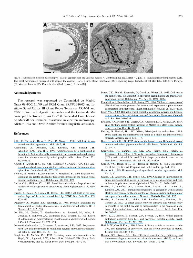
In G1 rabbits, the capillaries in the NFL and in the vitreoushumor had thicker basal membranes (Fig. 6B) than in control(Fig. 6A). The vascular lumen showed a normal feature as inthe controls; however, in the cytoplasm of the endothelialcells, some dense bodies and only a few vacuoles were ob-served (Fig. 6B).
4. Discussion
Most studies of age-related macular degeneration (AMD)have focused on the outer retina. However, it has recentlybeen reported that in AMD the inner retina is affected by anintense retinal ischemia (Ramirez et al., 2001).
The abundant lipids found in the cytoplasm of some RPEcells could contribute to the hypertrophy and degeneration ofthe RPE of our hypercholesterolemic rabbits. Additionally,the basal zone of some RPE cells revealed autophagic vesi-cles, vacuoles, electrodense deposits, and debris from cellmembranes that could correspond to the laminar deposits de-scribed by Curcio and Millican (1999). As in human AMD(Adler et al., 1999; Green, 1999), changes of RPE could con-tribute to the degeneration of the photoreceptors of G1 rab-bits, whose metabolism depends on normal RPE functionand integrity.
The cholesterol source for RPE and photoreceptors are theplasma lipids. Given that there is no direct contact between thephotoreceptors and the choroidal circulation, adjacent celltypes (RPE cells and Muller cells) must facilitate the transferof lipids to the photoreceptors. In fact, the expression of nativereceptors for LDL on RPE cells has been reported (Gor-diyenko et al., 2004; Hayes et al., 1989); this could be con-nected with local production of apoE by RPE cells. Anabnormal metabolism of lipids secondary to a cholesterol-enriched diet and/or apoE deficiencies could upset the

361A. Trivino et al. / Experimental Eye Research 83 (2006) 357e366
Fig. 2. Transmission electron microscopy (TEM) of the Bruch’s membrane and RPE cells. A: Choriocapillaris-Bruch’s membrane-RPE complex from control (G0).
Detail of Bruch’s membrane (insert) showing the outer collagen layer (OCL), elastic layer (E) and inner collagen layer (ICL). [Bar ¼ 2.2 mm. Insert bar ¼ 1.1 mm].
B: Bruch’s membrane from G1 with electrodense (black arrowhead) and electroluminescent (white arrowhead) particles at the ICL. [Bar ¼ 0.5 mm]. C: The cy-
toplasm of RPE cell in G1 shows dense bodies (white arrows), debris from cell membranes (*) and droplets of lipids (L). The apical microvilli have disappeared
and the basal infolding forms lamellar structures (black arrow). [Bar ¼ 2.2 mm]. D: Lipids (L) filling up the cytoplasm of a RPE cell. Apical microvilli and basal
infolding have disappeared. [Bar ¼ 3.4 mm]. [Choroid (CH), Basal membrane of the RPE (BM), Receptor layer (RL), Retinal pigment epithelium (RPE)].
cholesterol balance in these retinal layers. However, it appearsthat other retinal components can produce heterogeneous par-ticles locally containing apoE (Amaratunga et al., 1996).These particles are synthesized mainly by Muller cells, al-though astrocytes associated with ganglion cells axons couldbe involved in their production (Amaratunga et al., 1996).This local metabolism and transport of cholesterol (probablyimpaired in rabbits from Group 1 as a result of a thickenedBruch’s membrane with changes in its collagen layers) couldplay an important role in the contribution of lipids requiredfor retinal neurons to maintain and remodel their membranes.
Changes observed in the nuclear layers of the retina of G1rabbits were similar to those described by our group in humanAMD (Ramirez et al., 2001) e that is, apoptosis and necrosiswith the remaining neurons containing clumps of lipofuscin.
As in human AMD, rabbits from group G1 exhibited a lossof ganglion cells and had cell features of apoptosis and necro-sis as well as electrodense inclusions (probably lipofuscin) inthe cytoplasm of this cell type. This ganglion-cell loss couldbe caused, at least partly, by a local disruption of cholesterolhomeostasis (Ong et al., 2001). A reduced population of gan-glion cells could secondarily impair the neurotrophic supportof the retinal neurons as a consequence of reduced secretionof brain-derived neurotrophic factor (BDNF) by ganglioncells. This scenario is feasible, given that amacrine cells
express the TrkB receptor for BDNF (Cusato et al., 2002)and that BDNF improves the survival of bipolar cells upon ac-tivation of the p75 receptor, which then induces secretion offibroblast growth factor b (bFGF) (Wesler et al., 1998). Thesituations described could contribute to the axon loss observedin our hypercholesterolemic rabbits; this loss parallels humanAMD, in which a considerable axonal degeneration has beenreported (Ramirez et al., 2001).
In these results the Muller glia in group 1 contained moreintermediate filaments and endoplasmic reticulum than controlgroup and was anti-glial fibrillary acidic protein positive[GFAP(þ)]. The major intermediate filament expressed by re-active Muller cells appears to be GFAP. This protein is the bestknown marker for reactive gliosis throughout the nervous sys-tem. GFAP expression in Muller cells displays a unique fea-ture. Normally, GFAP is expressed at a low level or is notdetectable in mammalian Muller cells, as in our group G0.However, loss of retinal integrity as a result of mechanical in-jury, detachment, or photoreceptor degeneration provokes theappearance of intense GFAP-immunoreactivity in Muller cells,and an increase in GFAP content of the retina (Erickson et al.,1987; Guerin et al., 1990; Eisenfeld et al., 1984; Sarthy andFu, 1989; Tyler and Burns, 1991). Additionally Muller cells’reactivity transducers an increase in cell metabolism (Sarthyand Ripss, 2001). As described by Lindsay (1986), the

362 A. Trivino et al. / Experimental Eye Research 83 (2006) 357e366
Fig. 3. Transmission electron microscopy (TEM) in G1. A: General view of changes in the ONL, OPL, INL and IPL. Numerous dense bodies (black arrows) and
empty spaces (*) are visible in all layers. The processes of Muller cells (M) fill the empty spaces left by degenerated cells. [Bar ¼ 10 mm]. Insert: At greater mag-
nification the empty spaces consist of degenerate cytoplasm with numerous dense bodies (black arrow) and cellular debris (black arrowhead). [Bar ¼ 3.4 mm]. B:
Apoptosis of photoreceptors (white arrows) in the ONL. [Bar ¼ 5 mm]. C: Cell necrosis (*) in the INL and dense bodies (black arrows) in the OPL. [Bar ¼ 3.4 mm].
D: A cell in advanced stage of necrosis (*), a cell in apoptosis (white arrow) and dense bodies (black arrow) in the INL. Muller cell (M). Neuronal nucleus (N).
[Bar ¼ 2.2 mm]. E: Cells in apoptosis (white arrow) in the INL. Dense bodies (black arrows) inside the Muller cell processes (M). [Bar ¼ 3.4 mm]. F: Apoptosis
(white arrow) in the GCL. Cellular debris (black arrowhead) and dense bodies (black arrows). [Bar ¼ 2.2 mm]. [Axon (ax); Ganglion cell (GC); Inner nuclear layer
(INL); Inner plexiform layer (IPL); Outer nuclear layer (ONL); Outer plexiform layer (OPL)].
processes of Muller cells filled most of the spaces that ap-peared in the NFL, ganglion-cell layer and nuclear layers asa result of degeneration and cell loss. According to Sharty(2001), this situation also affected the outer retina of our hy-percholesterolemic rabbits, where we observed Muller pro-cesses extending to the empty spaces left by degeneratedphotoreceptors.
Differences between rabbit and human retinas and astro-cytes must be taken into account when comparing the two
species (Haddad et al., 2001, 2003; Ramirez et al., 1996;Trivino et al., 1997). The rabbit retina has epiretinal vascu-larization and possesses perivascular astrocytes which are ab-sent in humans. However, in both species, astrocytes arelocated at the NFL and GCL. Additionally, astrocytes andMuller cells are intermediate cells between neurons and ves-sels; they are located on the basal membrane of capillariesseparating them from neurons. Like Muller cells, astrocytesfrom G1 rabbits exhibited increased cellular activity, as

363A. Trivino et al. / Experimental Eye Research 83 (2006) 357e366
Fig. 4. Transmission electron microscopy (TEM) of NFL and ILM in the med-
ullary nerve-fiber region. A: Control group (G0). Axons in the vicinity of the
ILM. No empty spaces are observed. [Bar ¼ 2.5 mm]. B,C: Hypercholesterol-
emic rabbits (G1): Empty spaces in the NFL as a result of axonal and cellular
degeneration. [B: Bar ¼ 2.2 mm. C: Bar ¼ 5mm]. [Astrocyte (A); Axons (ax).
Basal membrane of the ILM (BM). Muller cells (M). Microglial cell (m). Vit-
reous humor (V). Empty spaces (*). Dense bodies (black arrows). Cellular
debris (black arrowhead). Axonal debris (white arrowhead)].
deduced by the increment of GFAP immunoreactivity and in-termediate filaments and organelles examined under TEM.This increased cellular activity, in conjunction with an al-tered lipid homeostasis, could explain the accumulationof electrodense particles, probably lipofuscin and lipids(Winkler et al., 1999) that we observed in the cytoplasm ofastrocytes of G1 animals. The exposure of these electrodenseparticles to light and high oxygen concentrations provideideal conditions for the formation of reactive oxygen speciesthat damage cellular proteins and lipid membranes (Winkleret al., 1999). If we add to this the higher concentrations ofextracellular toxic substances (e.g. glutamate) which coulddamage the neurons by cytotoxic mechanisms (Iadecola,1999; Wilson, 1997), the possibilities of keeping the cellularmechanisms intact against ischemia diminish in favor of celldeath (Liu et al., 1999).
The alterations in the retinal vessels in G1 rabbits were alsoobserved in hypercholesterolemic rats (Yamakawa et al.,2001). These alterations consisted of the thickening of thebasal membrane, dense bodies, and cytoplasm vacuoles.
The thickening of the basal membrane together with thealterations of the endothelial cells of the intraretinal and epire-tinal capillaries, combined with the changes in Bruch’smembrane and the build-up of lipids in the outer retina, couldhave contributed to a situation of chronic ischemia in G1rabbits.
The ischemia caused the depletion of cellular energy stores,the release of free radicals, and subsequent neuronal injury(Dingledine and McBain, 1993).
The task of reactive astrocytes is assumed to be to protectthe neurons from the ischemia. It has been shown that astro-cytes are more resistant to oxidative damage and can protectneurons from it because they possess such antioxidant mech-anisms as high concentrations of reduced glutation and vita-min C (Wilson, 1997). We found few astrocytes in the NFLof hypercholesterolemic rabbits, the remaining ones being hy-per-reactive. Both features have been described in humanAMD (Ramırez et al., 2001). A reduction in astrocytes andtheir antioxidant activity and capacity to remove glutamatefrom the extracellular space may contribute to neuronaldysfunction.
In the experimental model of hypercholesterolemia pre-sented in this study, we detected ultrastructural changes incells of the outer and inner retina similar to human AMD,which could be secondary to chronic ischemia. In both in-stances, the bodies of Muller cells are displaced from theINL, to the vitreous in the case of human AMD (Ramırezet al., 2001) and to the NFL and ILM in the present study. Itis possible that in both situations Muller cells migrate in an at-tempt to reach the metabolic reserve in the vitreous. This couldbe an adaptive system for transporting nutrients and energeticsubstrates to those areas of the retina exposed to the chronicischemic insult.
It is now recognized that lipids play a key role as structuraland signaling molecules. Given that lipid intake is most depen-dent on food composition, the dietary regimen could contrib-ute to induction or prevention of retinal disease.

364 A. Trivino et al. / Experimental Eye Research 83 (2006) 357e366
Fig. 5. Retinal astrocytes and Muller cells. AeC: Immunohistochemistry anti-GFAP of retinal astrocytes and Muller cells in control and G1. A,B: Astrocytes in
hypercholesterolemic rabbits (G1) (B), present a higher GFAP (þ) immunoreactivity than controls (A). C: GFAP (þ) Muller cells in G1(C). In the control group,
there is no immunoreactivity for GFAP (insert). [A,B,C and insert: Bar ¼ 50 mm]. DeG: Transmission electron microscopy (TEM) of retinal astrocytes and Muller
cells in G1. D: Reactive Muller cells with abundant rough endoplasmic reticulum (ER) and glial filaments (F) in their cytoplasm. [Bar ¼ 1.2 mm]. E: Three nuclei
of Muller cell displaced to the NFL. One of the Muller cells participates in the formation of the ILM (white asterisk). Astrocytes in advanced stage of necrosis
(black asterisk). [Bar ¼ 2.2 mm]. F: Displaced Muller cell to the NFL-ILM with numerous dense bodies in the cytoplasm. Nuclei of glial cells in apoptosis (white
arrow). [Bar ¼ 3.4 mm]. G: Perivascular astrocytes of the glial tufts in the vitreous humor. Astrocytes in the process of necrosis with rupture of the plasma mem-
brane (curved arrow). The cytoplasm shows abundant endoplasmic reticulum (ER), droplets of lipids (L) and dense bodies. [Bar ¼ 1.2 mm]. [Astrocyte (A); Axon
(Ax); Basal membrane of the ILM (BM); Dense bodies (black arrow); Filaments (F); Muller cell (M); Vitreous humor (V)].
365A. Trivino et al. / Experimental Eye Research 83 (2006) 357e366
Fig. 6. Transmission electron microscopy (TEM) of capillaries in the vitreous humor. A: Control animal (G0). [Bar ¼ 2 mm]. B: Hypercholesterolemic rabbit (G1).
The basal membrane is thickened with respect the control. [Bar ¼ 2 mm]. [Basal membrane (BM); Capillary (cap); Endothelial cell (E); Glial tuft (GT); Pericyte
(P); Vitreous humour (V); Dense bodies (black arrows); Retina (R)].
Acknowledgements
The research was supported by Comunidad de MadridGrant 08.4/0017.1/99 and UCM Grant PR48/01-9905 and In-stituto Salud Carlos III Grant Redes Tematicas CO3/01 andC03/13. We thank Agustın Fernandez and the Centro de Mi-croscopia Electronica ‘‘Luis Bru’’ (Universidad Complutensede Madrid) for technical assistance in electron microscopy;Alistair Ross and David Nesbitt for their linguistic assistance.
References
Adler, R., Curcio, C., Hicks, D., Price, D., Wong, F., 1999. Cell death in age-
related macular degeneration. Mol. Vis 5, 31.
Amaratunga, A., Abraham, C.R., Edwards, R.B., Sandell, J.H.,
Schreiber, B.M., Fine, R.E., 1996. Apolipoprotein E is synthesized in
the retina by Muller glial cells, secreted into the vitreous, and rapidly trans-
ported into the optic nerve by retinal ganglion cells. J. Biol. Chem. 271,
5628e5632.
Ambati, J., Ambati, B.K., Yoo, S.H., Lanchulev, S., Adamis, A.P., 2003. Age-
related macular degeneration: etiology, pathogenesis, and therapeutic strat-
egies. Surv. Ophthalmol. 48, 257e293.
Boulton, M., Moriarty, P., Jarvis-Evans, J., Maryiniuk, B., 1994. Regional var-
iation and age-related changed of lysosomal enzymes in the human retinal
pigment epithelium. Br. J. Ophthalmol. 78, 125e129.
Curcio, C.A., Millican, C.L., 1999. Basal linear deposit and large drusen are
specific for early age-related maculopathy. Arch. Ophthalmol. 117, 329e
339.
Cusato, K., Bosco, A., Linden, R., Reese, B.E., 2002. Cell death in the inner
nuclear layer of the retina is modulated by BDNF. Dev. Brain Res. 139,
325e330.
Daugherty, A., Zweifel, B.S., Schonfeld, G., 1989. Probucol attenuates the
development of aortic atherosclerosis in cholesterol-fed rabbits. Br. J.
Pharmacol. 2, 612e618.
Del Rio, M., Chulea, T., Merchan-Perez, A., Remezal, M., Valor, S.,
Gonzalez, J., Gutierrez, J.A., Lasuncion, M.A., Tejerina, T., 1995. Effects
of indapamide on Atherosclerosis Development in cholesterol-fed rabbits.
J. Cardiol. Pharmacol. 25, 973e978.
Delton-Vandenbroucke, I., Grammas, P., Anderson, R.E., 1997. Polyunsatu-
rated fatty acid metabolism in retinal and cerebral microvascular endothe-
lial cells. J. Lipid Res. 38, 147e159.
Dingledine, R., McBain, C.J., 1993. Excitatory amino acid transmitters. In:
Siegel, G.J., Agranoff, B.W., Albers, R.W., Molinoff, P.B. (Eds.), Basic
Neurochemistry, fifth ed. Raven Press, New York, pp. 367e387.
Dorey, C.K., Wu, G., Ebenstein, D., Garsd, A., Weiter, J.J., 1989. Cell loss in
the aging retina. Relationship to lipofucsin accumulation and macular de-
generation. Invest. Ophthalmol. Vis. Sci. 30, 1691e1699.
Eisenfeld, A.J., Bunt-Milam, A.H., Sarthy, P.V., 1984. Muller cell expression of
glial fibrillary acidic protein after genetic and experimental photoreceptor
degeneration in the rat retina. Invest. Ophthalmol. Vis. Sci. 25, 1321e1328.
Elner, V.M., 2002. Retinal pigment epithelial acid lipase activity and lipopro-
tein receptors: effects of dietary omega-3 fatty acids. Trans. Am. Ophthal-
mol. Soc. 100, 301e338.
Erickson, P.A., Fisher, S.R., Guerin, C.J., Anderson, D.H., Kaska, D.D., 1987.
Glial fibrillary acidic protein increases in Muller cells after retinal detach-
ment. Exp. Eye Res. 44, 37e48.
Finking, G., Hankeh, H., 1997. Nikolaj Nikolajewitsch Anitschkow (1885-
1964) stablished the cholesterol-fed rabbit as a model for atherosclerosis
research. Atherosclerosis 135, 1e7.
Gao, H., Hollyfield, J.G., 1992. Aging of the human retina. Differential loss of
neurons and retinal pigment epithelial cells. Invest. Ophthalmol. Vis. Sci.
33, 1e17.
Gordiyenko, N., Campos, M., Lee, J.W., Fariss, R.N., Sztein, J.,
Rodriguez, I.R., 2004. RPE cells internalize low-density lipoprotein
(LDL) and oxidized LDL (oxLDL) in large quantities in vitro and in
vivo. Invest. Ophthalmol. Vis. Sci. 45, 2822e2829.
Gordon, W.C., Bazan, N.G., 1997. Retina. In: Harding, J.J. (Ed.), Biochemis-
try of the Eye, first ed. Chapman and Hall, London, pp. 144e275.
Green, W.R., 1999. Histopathology of age-related macular degeneration. Mol.
Vis. 5, 27.
Guerin, C.J., Anderson, D.H., Fisher, S.K., 1990. Changes in intermediate fil-
ament immunolabeling occur in response to retinal detachment and reat-
tachment in primates. Invest. Ophthalmol. Vis. Sci. 31, 1474e1482.
Haddad, A., Ramırez, A.I., Laicine, E.M., Salazar, J.J., Trivino, A.,
Ramırez, J.M., 2001. Immunohistochemistry in association with scanning
electron microscopy for the morphological characterization and location of
astrocytes of the rabbit retina. J. Neurosci. Methods 106, 131e137.
Haddad, A., Salazar, J.J., Laicine, E.M., Ramırez, A.I., Ramırez, J.M.,
Trivino, A., 2003. A direct contact between astrocyte and vitreous body
is possible in the rabbit eye due to discontinuities in the basement mem-
brane of the retinal inner limiting membrane. Braz. J. Med. Biol. Res.
36, 207e211.
Hayes, K.C., Lindsey, S., Stephan, Z.F., Brecker, D., 1989. Retinal pigment
epithelium possesses both LDL and scavenger receptor activity. Invest.
Ophthalmol. Vis. Sci. 30, 225e232.
Huff, M.W., Carroll, K.K., 1980. Effects of dietary protein on turnover, oxida-
tion, and absorption of cholesterol, and on steroid excretion in rabbits.
J. Lipid Res. 21, 546e548.
Hussain, S.T., Roots, B.I., 1994. Effects of essential fatty deficiency and
immunopathological stresses on bloodebrain-barrier (BBB) in Lewis
rats: a biochemical study. Biochem. Soc. Trans. 2, 338S.

366 A. Trivino et al. / Experimental Eye Research 83 (2006) 357e366
Iadecola, C., 1999. Mechanisms of cerebral ischemic damage. In: Walz, W.
(Ed.), Cerebral Ischemia. Molecular and Cellular Pathophysiology.
Humana Press Inc, Totowa, pp. 3e32.
Li, F., Chen, H., Anderson, R.E., 2001. Biosynthesis of docosahexaenoate-
containing glycerolipid molecular species in the retina. J. Mol. Neurosci
16, 205e214 (Discussion 215-221).
Liu, D., Smith, C.L., Barone, F.C., Ellison, J.A., Lysco, P.G., Li, K.,
Simpson, I.A., 1999. Astrocytic demise precedes delayed neuronal death
in focal ischemic rat brain. Mol. Brain Res. 68, 29e41.
Moore, S.A., 2001. Polyunsaturated fatty acid synthesis and release by brain-
derived cells in vitro. J. Mol. Neurosci 16, 195e200 (Discussion 215-221).
Ong, J.M., Zorapapel, N.C., Rich, K.A., Wagstaff, R.E., Lambert, R.W.,
Rosenberg, S.E., Moghaddas, F., Pirouzmanesh, A., Aoki, A.M.,
Kenney, C., 2001. Effects of cholesterol and Apolipoprotein E on retinal
abnormalities in ApoE-deficient mice. Invest. Ophthalmol. Vis. Sci. 42,
1891e1900.
Panda-Jonas, S., Jonas, J.B., Jakobczyk-Zmija, M., 1996. Retinal pigment ep-
ithelial cell count, distribution, and correlations in normal human eyes.
Am. J. Ophthalmol 121, 181e189.
Puskas, L.G., Bereczki, E., Santha, M., Vigh, L., Csanadi, G., Spener, F.,
Ferdinandy, P., Onochy, A., Kitajka, K., 2004. Cholesterol and cholesterol
plus DHA diet-induced gene expression and fatty acid changes in mouse
eye and brain. Biochimie 86, 817e824.
Ramırez, J.M., Trivino, A., Ramırez, A.I., Salazar, J.J., Garcıa-Sanchez, J.,
1994. Immunohistochemical study of human retinal astroglia. Vis. Res.
34, 1935e1946.
Ramırez, J.M., Trivino, A., Ramırez, A.I., Salazar, J.J., Garcıa-Sanchez, J.,
1996. Structural specializations of human retinal glial cells. Vis. Res. 36,
2029e2036.
Ramırez, J.M., Ramırez, A.I., Salazar, J.J., De Hoz, R., Trivino, A., 2001.
Changes of astrocytes in retinal ageing and Age-Related Macular Degen-
eration. Exp. Eye Res. 73, 601e615.
SanGiovanni, J.P., Chew, E.Y., 2005. The role of omega-3 long-chain polyun-
saturated fatty acids in health and disease of the retina. Prog. Ret. Eye Res.
24, 87e138.
Sarthy, P.V., Fu, M., 1989. Transcriptional activation of an intermediate fila-
ment protein gene in mice with retinal dystrophy. DNA J. Mol. Cell.
Biol. 8, 437e446.
Sarthy, V., Ripss, H., 2001. The Retinal Muller Cell. Structure and Function.
Kluwer Academic/Plenum Publishers, New York.
Seddon, J.M., Rosner, B., Sperduto, R.D., Yannuzzi, L., Haller, J.A.,
Blair, N.P., Willett, W., 2003. Dietary fat and risk for advanced age-related
macular degeneration. Arch. Ophthalmol 119, 1191e1199.
Serougne, C., Lefevre, C., Chevalier, F., 1976. Cholesterol transfer between
brain and plasma in the rat: a model for the turnover of cerebral choles-
terol. Exp. Neurol 51, 229e240.
Su, H.M., Bernardo, L., Mirmiran, M., Ma, X.H., Corso, T.N.,
Nathanieslz, P.W., Brena, J.T., 1999. Bioequivalence of dietary alpha-lino-
leic and docosahexaenoic acids as sources of docosanexaenoate accretion
in brain and associated organs of neonatal baboons. Pediatr. Res. 45,
87e93.
Trivino, A., Ramırez, J.M., Ramırez, A.I., Salazar, J.J., Garcıa-Sanchez, J.,
1997. Comparative study of astrocytes in human and rabbit retinae. Vis.
Res. 37, 1707e1711.
Tyler, N.K., Burns, M.S., 1991. Alterations in glial cells morphology and glial
fibrillary acidic protein expression in urethane-induced retinopathy. Invest.
Ophthalmol. Vis. Sci. 32, 246e256.
Wang, N., Anderson, R.E., 1991. Synthesis of docosahexaeonoic acid by retina
and retinal pigment epithelium. Biochemistry 32, 13703e13709.
Wazke, R.C., Soldevilla, J.D., Trune, D.R., 1993. Morphometric analysis of
human retinal pigment epithelium: correlation with age and location.
Curr. Eye Res. 12, 133e142.
Wesler, E.M., Berkovich, O., Nawy, S., 1998. Role of the low-affinity NGF
receptor (p75) in survival of retinal bipolar cells. Visual Neurosci 15,
211e218.
Wetzel, M.G., Li, J., Alvarez, R.A., Anderson, R.E., O’Brien, P.J., 1991. Me-
tabolism of linolenic acids and docosahexaenoic acid in rat retina and rod
outer segments. Exp. Eye Res. 53, 437e446.
Wilson, J.X., 1997. Antioxidant defense of the brain: a role for astrocytes. Can.
J. Physiol. Pharmacol 75, 1149e1163.
Winkler, B.S., Boulton, M.E., Gottsch, J.D., Sternberg, P., 1999. Oxidative
damage and age-related macular degeneration. Mol. Vis. 5, 32. <http://
www.molvis.org/molvis/v5/p32/>.
Yamakawa, K., Bhutto, I.A., Lu, Z., Watanabe, Y., Amemiya, T., 2001. Retinal
vascular changes in rats with inherited hypercholesterolemia e corrosion
cast demonstration. Curr. Eye Res. 22, 258e265.
Yanni, A.E., 2004. The laboratory rabbit: an animal model of atherosclerosis
research. Lab. Anim 38, 246e256.
Zauberman, H., Livni, 1981. Experimental vascular occlusion in hypercholes-
terolemic rabbits. Invest. Ophthalmol. Vis. Sci. 2, 248e255.