A brief review on molecular, genetic and imaging techniques for HCV fibrosis evaluation
16
REVIEW Open Access A brief review on molecular, genetic and imaging techniques for HCV fibrosis evaluation Waqar Ahmad, Bushra Ijaz, Sana Gull, Sultan Asad, Saba Khaliq, Shah Jahan, Muhammad T Sarwar, Humera Kausar, Aleena Sumrin, Imran Shahid, Sajida Hassan * Abstract Background: Chronic HCV is one of the major causes of morbidity and mortality in the present day world. The assessment of disease progression not only provides useful information for diagnosis and therapeutic supervision judgment but also for monitoring disease. Different invasive and non invasive methods are applied to diagnose the disease from initial to end stage (mild fibrosis to cirrhosis). Although, liver biopsy is still considered as gold standard to identify liver histological stages, an assessment of the disease development based on non-invasive clinical findings is also emerging and this may replace the need of biopsy in near future. This review gives brief insight on non-invasive methods currently available for predicting liver fibrosis in HCV with their current pros and cons to make easier for a clinician to choose better marker to assess liver fibrosis in HCV infected patients. Methods: More than 200 studies regarding invasive and noninvasive markers available for HCV liver disease diagnosis were thoroughly reviewed. We examined year wise results of these markers based on their sensitivity, specificity, PPV, NPV and AUROCs. Results: We found that in all non-invasive serum markers for HCV, FibroTest, Forn’s Index, Fibrometer and HepaScore have high five-year predictive value but with low AUROCs (0.60~0.85) and are not comparable to liver biopsy (AUROC = 0.97). Even though from its beginning, Fibroscan is proved to be best with high AUROCs (> 0.90) in all studies, no single noninvasive marker is able to differentiate all fibrosis stages from end stage cirrhosis. Meanwhile, specific genetic markers may not only discriminate fibrotic and cirrhotic liver but also differentiate individual fibrosis stages. Conclusions: There is a need of marker which accurately determines the stage based on simplest routine laboratory test. Genetic marker in combination of imaging technique may be the better non invasive diagnostic method in future. 1. Introduction Chronic Hepatitis C (HCV) is one of the major causes of liver fibrosis, with distortion of the hepatic architec- ture, and ultimate progression to cirrhosis. Approxi- mately more than 3% of the total world population is chronically infected with HCV and due to gradual increase in the prevalence of HCV; future burden of chronic HCV is predicted to raise at least 3 fold by the year 2020. Common causes of liver fibrosis are viral hepatitis and steato hepatitis with alcohol or obesity. Fibrosis caused by excessive deposition of extracellular matrix (ECM) by histological and molecular reshuffling of various components like collagens, glycoproteins, pro- teoglycans, matrix proteins and matrix bound growth factors. These changes can lead to metabolic and synth- esis impairment to hepatocytes, epithelial cells and hepatic stellate cells (HSC). HSC activation the main step leading to fibrosis, involves several changes in liver like fibrogenesis, proliferation, contractility, chemotaxis, matrix degradation and cytokine release. Fibrosis can be defined as net result of the balance between ECM pro- duction and degradation. As ECM tissues not only involve matrix production but also matrix degradation leading to ECM remodeling, fibrosis is potentially a reversible process in early stages (advance stages in some cases) [1-6]. * Correspondence: [email protected] Applied and Functional Genomics Laboratory, Centre of Excellence in Molecular Biology, University of the Punjab, Lahore, Pakistan Ahmad et al. Virology Journal 2011, 8:53 http://www.virologyj.com/content/8/1/53 © 2011 Ahmad et al; licensee BioMed Central Ltd. This is an Open Access article distributed under the terms of the Creative Commons Attribution License (http://creativecommons.org/licenses/by/2.0), which permits unrestricted use, distribution, and reproduction in any medium, provided the original work is properly cited.
-
Upload
independent -
Category
Documents
-
view
2 -
download
0
Transcript of A brief review on molecular, genetic and imaging techniques for HCV fibrosis evaluation
REVIEW Open Access
A brief review on molecular, genetic and imagingtechniques for HCV fibrosis evaluationWaqar Ahmad, Bushra Ijaz, Sana Gull, Sultan Asad, Saba Khaliq, Shah Jahan, Muhammad T Sarwar, Humera Kausar,Aleena Sumrin, Imran Shahid, Sajida Hassan*
Abstract
Background: Chronic HCV is one of the major causes of morbidity and mortality in the present day world. Theassessment of disease progression not only provides useful information for diagnosis and therapeutic supervisionjudgment but also for monitoring disease. Different invasive and non invasive methods are applied to diagnosethe disease from initial to end stage (mild fibrosis to cirrhosis). Although, liver biopsy is still considered as goldstandard to identify liver histological stages, an assessment of the disease development based on non-invasiveclinical findings is also emerging and this may replace the need of biopsy in near future. This review gives briefinsight on non-invasive methods currently available for predicting liver fibrosis in HCV with their current pros andcons to make easier for a clinician to choose better marker to assess liver fibrosis in HCV infected patients.
Methods: More than 200 studies regarding invasive and noninvasive markers available for HCV liver diseasediagnosis were thoroughly reviewed. We examined year wise results of these markers based on their sensitivity,specificity, PPV, NPV and AUROCs.
Results: We found that in all non-invasive serum markers for HCV, FibroTest, Forn’s Index, Fibrometer andHepaScore have high five-year predictive value but with low AUROCs (0.60~0.85) and are not comparable to liverbiopsy (AUROC = 0.97). Even though from its beginning, Fibroscan is proved to be best with high AUROCs (> 0.90)in all studies, no single noninvasive marker is able to differentiate all fibrosis stages from end stage cirrhosis.Meanwhile, specific genetic markers may not only discriminate fibrotic and cirrhotic liver but also differentiateindividual fibrosis stages.
Conclusions: There is a need of marker which accurately determines the stage based on simplest routinelaboratory test. Genetic marker in combination of imaging technique may be the better non invasive diagnosticmethod in future.
1. IntroductionChronic Hepatitis C (HCV) is one of the major causesof liver fibrosis, with distortion of the hepatic architec-ture, and ultimate progression to cirrhosis. Approxi-mately more than 3% of the total world population ischronically infected with HCV and due to gradualincrease in the prevalence of HCV; future burden ofchronic HCV is predicted to raise at least 3 fold by theyear 2020. Common causes of liver fibrosis are viralhepatitis and steato hepatitis with alcohol or obesity.Fibrosis caused by excessive deposition of extracellular
matrix (ECM) by histological and molecular reshufflingof various components like collagens, glycoproteins, pro-teoglycans, matrix proteins and matrix bound growthfactors. These changes can lead to metabolic and synth-esis impairment to hepatocytes, epithelial cells andhepatic stellate cells (HSC). HSC activation the mainstep leading to fibrosis, involves several changes in liverlike fibrogenesis, proliferation, contractility, chemotaxis,matrix degradation and cytokine release. Fibrosis can bedefined as net result of the balance between ECM pro-duction and degradation. As ECM tissues not onlyinvolve matrix production but also matrix degradationleading to ECM remodeling, fibrosis is potentially areversible process in early stages (advance stages insome cases) [1-6].
* Correspondence: [email protected] and Functional Genomics Laboratory, Centre of Excellence inMolecular Biology, University of the Punjab, Lahore, Pakistan
Ahmad et al. Virology Journal 2011, 8:53http://www.virologyj.com/content/8/1/53
© 2011 Ahmad et al; licensee BioMed Central Ltd. This is an Open Access article distributed under the terms of the Creative CommonsAttribution License (http://creativecommons.org/licenses/by/2.0), which permits unrestricted use, distribution, and reproduction inany medium, provided the original work is properly cited.
Fibrosis stages information not only indicate treatmentresponse but also reflect/indicate cirrhosis developmentdisaster. We can evaluate fibrosis in HCV infectedpatients invasively or non-invasively. Liver biopsy aninvasive method is used for histological scoring and stillused as reference test for fibrosis staging. With theincreasing knowledge of molecular biology, genetics andavailability of modern imaging techniques, many clini-cians and related scientists developed several non-invasivemethods to assess liver fibrosis and cirrhosis.These markers need to be more precise, reproducibleand non-invasive to evaluate liver fibrosis in HCVinfected patients. Therefore, an assessment of the diseasedevelopment based on clinical findings is still critical forpatients infected with HCV. The accuracy of a serologicaltest either individually or in combination is given as thearea under the curve (AUC) of the receiver operatorcharacteristic (ROC) of specific serum diagnosis test.In the meantime, genetic marker should reflect
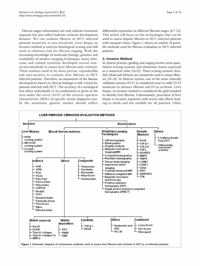
differential expression in different fibrosis stages [4,7-13].This article will focus on the technologies that can beused to assess hepatic fibrosis in HCV infected patientswith unequal values. Figure 1 shows an outline of possi-ble methods used for fibrosis evaluation in HCV infectedpatients.
2. Invasive MethodIn clinical practice, grading and staging involve semi-quan-titative scoring systems, and elementary lesion expressedas a numerical value [14,15]. Three scoring systems, Kno-dell, Ishak and Metavir are extensively used to assess fibro-sis [16-18]. In Metavir system, one of the most clinicallyvalidated systems; F0-F1 is considered none to mild, F2-F3moderate to advance fibrosis and F4 as cirrhosis. Liverbiopsy, an invasive method is considered the gold standardto identify liver fibrosis. Unfortunately, procedure of liverbiopsy is invasive, expensive with severe side effects lead-ing to death and not suitable for all patients. Other
Figure 1 Schematic diagram of noninvasive methods used to assess liver fibrosis and cirrhosis in HCV or co-infected patients.
Ahmad et al. Virology Journal 2011, 8:53http://www.virologyj.com/content/8/1/53
Page 2 of 16
limitations of liver biopsy comprises sampling error, intraand inter observer variation and somehow static, not accu-rately predict disease progression [19,20].
3. Non-invasive MethodsNon-invasive methods can be classified as serum,genetic and imaging techniques. These markers areaddressed below in detail.
4. Serum markersSerological markers refer to the measurement of one ormore molecules within blood or serum correlating tohepatic fibrosis [21-23]. There are several proposed ser-ological markers or combinations of serum markers forhepatic fibrosis measurement. Their levels vary bychanges in their clearance, metabolism, and excretion,and their significant contribution from non-hepaticsources, such as, bones, joints, lungs, kidneys and skin[24,25]. Proposed hepatic fibrosis serological markerscan be divided in three categories as direct, indirect orcomposite. Combinations of both direct and indirect,markers are taking place as an emerging and promisingalternative to liver biopsy [26-29]. Figure 1 gives a briefidea about the non-invasive methods used for fibrosisand cirrhosis prediction in HCV infected patients.
4.1. Direct serum markersDirect serum markers reflect ECM turnover, balancebetween hepatic fibrogenesis and fibrolysis, and in thedeposition and removal of ECM. Levels of direct serummarkers are elevated during disease progression and anindependent association between stage of fibrosis anddirect markers was observed [30-32]. Some of the mar-kers reported are discussed below.4.1.1. Matrix deposition and removal markersThese may be classified into followingProcollagen I carboxy terminal peptide (PICP), Pro-collagen III amino-terminal peptide (PIIINP) andType IV collagen PICP and PIIINP released into theserum during matrix removal and deposition. PIIINPreflects the stage of fibrosis and known to be elevated inchronic liver disease. PIIINP is a good inflammatoryscore predictor as compared to fibrosis. PICP usuallyindicates cirrhosis and used for quantifying diseaseseverity. However, it reflects alcohol etiology better thandiagnosis of chronic liver disease. Type IV serum col-lagen reflects matrix degradation and increased inchronic liver disease. Murawaki et al (1996) establishedthe cutoff value of 110 ng/mL for stages greater than F2and 130 ng/mL for F3 fibrosis stage [33-37].Matrix metalloproteinase (MMP’s) and tissue inhibi-tor of metalloproteinases (TIMPs) MMP’s enzymes pro-duced intracellularly and secreted in a pro-enzyme formthat requires cleavage by cell surface mechanisms control
matrix degradation. Although these proteins act both todegrade and deposition of ECM, also involve in activationof growth factor, effect on cell proliferation and inhibitionof apoptosis; their association with liver fibrosis is notclear [4,23]. TIMPs also increased during HCV infection,while a decrease is reported after interferon therapy.These have high diagnostic ability to detect cirrhosis [38].Cytokines Two types of cytokines TGF-b 1 (transform-ing growth factors b 1) and PDGF (platelet derivedgrowth factor) are mainly used to assess the fibrosis pro-gression. TGF-b 1 is the dominant stimulus for produ-cing extracellular matrix and it showed a significantcorrelation with degree of hepatic fibrosis. A significantassociation was found between TGF-b 1 serum levelsand fibrosis progression. Serum level of PDGF has alsoshowed high ability as serum marker for fibrosis pro-gression [39-41].4.1.2.Combined direct markersFibroSpect FibroSpect assay is a combination of threeparameters: HA, TIMP-1 and alpha-2-macroglobulinand can differentiate between no/mild and moderate/severe fibrosis [42,43]. Maximum sensitivity and specifi-city of this assay was observed at two extreme stages(F0 and F4). This assay was further developed by addingYKL-40 serum marker for assessing Ishak stages anddigital quantification of fibrosis [23].ELF European liver fibrosis group (ELF) developed analgorithm consisted of HA, PIIINP, TIMP-1 and age.However this assay showed low performance while pre-dicting fibrosis in chronic HCV patients [44].Leroy ScoreThis score was developed by Leroy et al and contains
PIIINP and MMP-1 as basic components. It can differ-entiate between mild and significant fibrosis [45].4.1.3.OthersHyaluronic acid (HA) HA is best validated, an essentialcomponent of extracellular matrix of body tissues. HAlevels increases with the fibrosis progression and corre-late with the degree of fibrosis and inflammation inchronic HCV patients. The diagnostic accuracy of HA isbetter than that of PIIINP [32,35,46-49].Chondrex, human cartilage glycoprotein (YKL-40) Inliver fibrosis, YKL-40 plays role in tissue degradationand extracellular matrix remodeling. YKL-40 level isobserved to decrease after interferon therapy. In a com-bination of different direct serum markers, HA andYKL-40 were more useful for monitoring fibrosis pro-gression with 80% PPV of predicting stage specific fibro-sis. A significant association of HA with liver fibrosiswas observed when compared with TGF-b1 [50-53].Table 1 briefly describes a year wise overview of the
AUROCs, PPV, NPV, sensitivity and specificity of directserum markers used in various studies to predict fibrosisand cirrhosis in HCV infected patients. Direct serum mar-kers; HA, YKL-40 and ELF were able to predict significant
Ahmad et al. Virology Journal 2011, 8:53http://www.virologyj.com/content/8/1/53
Page 3 of 16
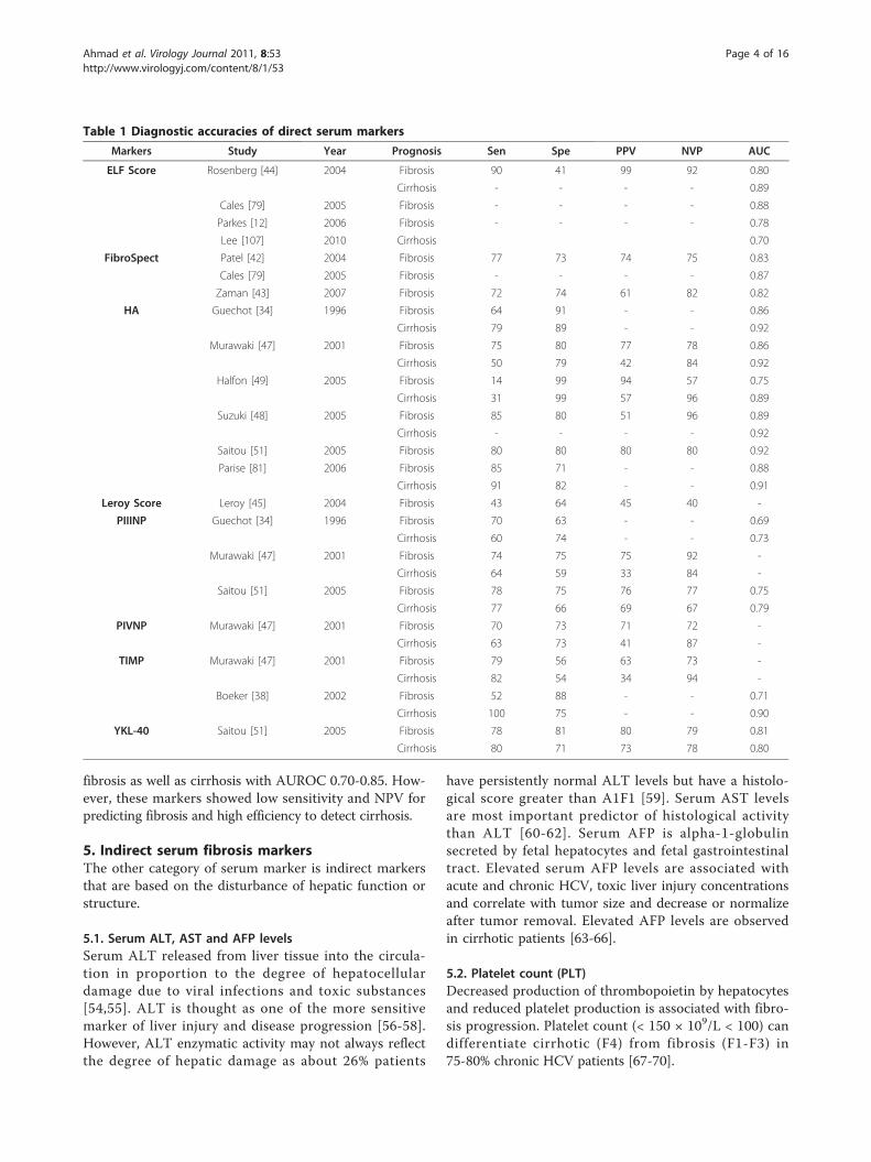
fibrosis as well as cirrhosis with AUROC 0.70-0.85. How-ever, these markers showed low sensitivity and NPV forpredicting fibrosis and high efficiency to detect cirrhosis.
5. Indirect serum fibrosis markersThe other category of serum marker is indirect markersthat are based on the disturbance of hepatic function orstructure.
5.1. Serum ALT, AST and AFP levelsSerum ALT released from liver tissue into the circula-tion in proportion to the degree of hepatocellulardamage due to viral infections and toxic substances[54,55]. ALT is thought as one of the more sensitivemarker of liver injury and disease progression [56-58].However, ALT enzymatic activity may not always reflectthe degree of hepatic damage as about 26% patients
have persistently normal ALT levels but have a histolo-gical score greater than A1F1 [59]. Serum AST levelsare most important predictor of histological activitythan ALT [60-62]. Serum AFP is alpha-1-globulinsecreted by fetal hepatocytes and fetal gastrointestinaltract. Elevated serum AFP levels are associated withacute and chronic HCV, toxic liver injury concentrationsand correlate with tumor size and decrease or normalizeafter tumor removal. Elevated AFP levels are observedin cirrhotic patients [63-66].
5.2. Platelet count (PLT)Decreased production of thrombopoietin by hepatocytesand reduced platelet production is associated with fibro-sis progression. Platelet count (< 150 × 109/L < 100) candifferentiate cirrhotic (F4) from fibrosis (F1-F3) in75-80% chronic HCV patients [67-70].
Table 1 Diagnostic accuracies of direct serum markers
Markers Study Year Prognosis Sen Spe PPV NVP AUC
ELF Score Rosenberg [44] 2004 Fibrosis 90 41 99 92 0.80
Cirrhosis - - - - 0.89
Cales [79] 2005 Fibrosis - - - - 0.88
Parkes [12] 2006 Fibrosis - - - - 0.78
Lee [107] 2010 Cirrhosis 0.70
FibroSpect Patel [42] 2004 Fibrosis 77 73 74 75 0.83
Cales [79] 2005 Fibrosis - - - - 0.87
Zaman [43] 2007 Fibrosis 72 74 61 82 0.82
HA Guechot [34] 1996 Fibrosis 64 91 - - 0.86
Cirrhosis 79 89 - - 0.92
Murawaki [47] 2001 Fibrosis 75 80 77 78 0.86
Cirrhosis 50 79 42 84 0.92
Halfon [49] 2005 Fibrosis 14 99 94 57 0.75
Cirrhosis 31 99 57 96 0.89
Suzuki [48] 2005 Fibrosis 85 80 51 96 0.89
Cirrhosis - - - - 0.92
Saitou [51] 2005 Fibrosis 80 80 80 80 0.92
Parise [81] 2006 Fibrosis 85 71 - - 0.88
Cirrhosis 91 82 - - 0.91
Leroy Score Leroy [45] 2004 Fibrosis 43 64 45 40 -
PIIINP Guechot [34] 1996 Fibrosis 70 63 - - 0.69
Cirrhosis 60 74 - - 0.73
Murawaki [47] 2001 Fibrosis 74 75 75 92 -
Cirrhosis 64 59 33 84 -
Saitou [51] 2005 Fibrosis 78 75 76 77 0.75
Cirrhosis 77 66 69 67 0.79
PIVNP Murawaki [47] 2001 Fibrosis 70 73 71 72 -
Cirrhosis 63 73 41 87 -
TIMP Murawaki [47] 2001 Fibrosis 79 56 63 73 -
Cirrhosis 82 54 34 94 -
Boeker [38] 2002 Fibrosis 52 88 - - 0.71
Cirrhosis 100 75 - - 0.90
YKL-40 Saitou [51] 2005 Fibrosis 78 81 80 79 0.81
Cirrhosis 80 71 73 78 0.80
Ahmad et al. Virology Journal 2011, 8:53http://www.virologyj.com/content/8/1/53
Page 4 of 16
5.3. Prothrombin time (PT)PT reflects the synthesis capacity of the liver and essen-tial mechanism of blood coagulation. Its clinical refer-ence range is usually around 12-15 seconds. ProlongedPT is associated with esophageal varices and is one ofthe earliest indicators of liver cirrhosis [71-73].
5.4. AST/ALT ratio (AAR)Sheth et al. reported an AST/ALT ratio ≥ 1 having100% PPV for the presence of cirrhosis in chronic HCVpatients [74]. Reedy et al. observed that AAR failed topredict cirrhosis accurately in HCV patients [75], whileGiannini et al. reported high diagnostic accuracy of theAAR for prediction of cirrhosis in HCV infected patients[76]. However, many authors could not able to find highaccuracy of this marker [4,70,77].
5.5. AST to platelet ratio Index (APRI)APRI was the simplest and accurate test for significantliver fibrosis and cirrhosis [28]. Several authors verifiedthis marker for fibrosis and cirrhosis and found it betterthan AAR. However, APRI was unable to identify indivi-dual stages of fibrosis [77-86].
5.6. PGA and PGAA IndexPGA was known to be the original index of hepaticfibrosis in 1990 s and combines gamma glutamyl trans-ferase (gGT), apolipoprotein A1 (PGA) and prothrombinindex. PGAA index is modified form of PGA index bythe addition of alpha-2-macroglobulin, resulted in itsimproved version. The diagnostic accuracy of the PGAand PGAA for detecting cirrhosis reported between 66-72% and 80%, respectively [87-92].
5.7. FibroTest/FibroSureFibroTest is the combination of five markers: alpha-2-macroglobulin, haptoglobin, apolipoprotein A1, GGTand total bilirubin [26,80]. This marker has 75% sensi-tivity and 85% specificity with reproducibility for fibrosisdiagnosis [83-85]. However, Rossi et al. reported lowAUROC (0.739) for significant fibrosis with NPV andPPV 85% and 78%, respectively. Meanwhile, FibroTest isvalidated and suggested as an alternative to liver biopsyin chronic HCV patients [93-105].
5.8. Fibro IndexIt combines three markers; AST, platelet count andgamma globulin. AUROC for prediction of significantfibrosis was 0.83 [106].
5.9. Forns IndexThis index is based on four available variables; age,GGT, platelet count and cholesterol levels in a study
on HCV patients, included both test and validationcohorts [27]. The limitation of this index was the iden-tification of advance liver disease with minimal fibrosis[79,80,106,107].
5.10. ActiTestActiTest reflects both necroinflamatory activity and livercirrhosis. It is modified form of Fibrotest with additionof ALT level (26). Fibrotest and ActiTest were found tobe potential non-invasive assays for the assessment ofhepatic fibrosis and necro-inflammatory activity inpediatric patients with chronic HCV in comparison withliver biopsy [90,91,108].
5.11. SteatoTestIt incorporates the FibroTest, ALT, body mass index,serum cholesterol, triglycerides and glucose adjusted forage and gender. It has 63% PPV for steatosis prevalencewith 93% NPV [109].
5.12. Model 3This model is based on AST, platelet count and pro-thrombin time expressed as international normalizedration (INR). Patients with liver cirrhosis can be excludedat cutoff value of < 0.20 with 99% NPV [110,111].
5.13. Goteborg University Cirrhosis Index (GUCI)Islam et al. found strong association between AST, pro-thrombin-INR and platelet count. By using a cutoffvalue 1.0, the sensitivity and specificity for the diagnosisof cirrhosis was 80% and 78% respectively, while theNPV and PPV were 97% and 31%, respectively [112].
5.14. Fibrosis IndexThis index comprises of platelet count and albumincontents. It can differentiate significant fibrosis and cir-rhosis from mild fibrosis [113].
5.15. Phol ScoreThis index comprises of AST, ALT and platelet count. Itshowed great accuracy for discriminating significantfibrosis and cirrhosis with high PPV and NPV. However,it showed limited ability to predict fibrosis in later study[114,115].
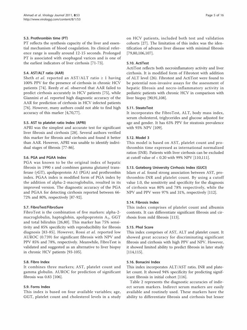
5.16. Bonacini IndexThis index incorporates ALT/AST ratio, INR and plate-let count. It showed 94% specificity for predicting signif-icant fibrosis in initial cohort [116].Table 2 represents the diagnostic accuracies of indir-
ect serum markers. Indirect serum markers are easilyavailable and routinely used. These markers have theability to differentiate fibrosis and cirrhosis but lesser
Ahmad et al. Virology Journal 2011, 8:53http://www.virologyj.com/content/8/1/53
Page 5 of 16
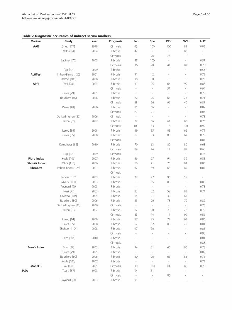
Table 2 Diagnostic accuracies of indirect serum markers
Markers Study Year Prognosis Sen Spe PPV NVP AUC
AAR Sheth [74] 1998 Cirrhosis 53 100 100 81 0.85
Afdhal [4] 2004 Fibrosis 47 - - 88 -
Cirrhosis - 96 74 - -
Lackner [70] 2005 Fibrosis 53 100 - - 0.57
Cirrhosis 36 90 41 87 0.73
Fuji [77] 2009 Fibrosis - - - - 0.56
ActiTest Imbert-Bismut [26] 2001 Fibrosis 91 42 - - 0.79
Halfon [100] 2008 Fibrosis 90 38 - - 0.75
APRI Wai [28] 2003 Fibrosis 41 95 64 90 0.88
Cirrhosis - - 57 - 0.94
Cales [79] 2005 Fibrosis - - - - 0.79
Bourliere [80] 2006 Fibrosis 22 95 63 76 0.71
Cirrhosis 38 96 96 40 0.81
Parise [81] 2006 Fibrosis 85 66 - - 0.82
Cirrhosis 73 81 - - 0.84
De Ledinghen [82] 2006 Cirrhosis - - - - 0.73
Halfon [83] 2007 Fibrosis 77 66 61 80 0.76
Cirrhosis 100 83 18 100 0.92
Leroy [84] 2008 Fibrosis 39 95 88 62 0.79
Cales [85] 2008 Fibrosis 62 83 80 67 0.78
Cirrhosis - - - - 0.84
Kamphues [86] 2010 Fibrosis 70 63 80 80 0.68
Cirrhosis 89 44 14 97 0.63
Fuji [77] 2009 Cirrhosis - - - - 0.76
Fibro Index Koda [106] 2007 Fibrosis 36 97 94 59 0.83
Fibrosis Index Ohta [113] 2006 Fibrosis 68 71 75 81 0.85
FibroTest Imbert-Bismut [26] 2001 Fibrosis 87 59 63 85 0.87
Cirrhosis
Bedosa [102] 2003 Fibrosis 27 97 90 55 -
Myers [101] 2003 Fibrosis - 95 88 - 0.83
Poynard [90] 2003 Fibrosis - - - - 0.73
Rossi [97] 2003 Fibrosis 83 52 52 83 0.74
Colletta [103] 2005 Fibrosis 64 31 33 62 -
Bourliere [80] 2006 Fibrosis 55 90 73 79 0.82
De Ledinghen [82] 2006 Cirrhosis - - - - 0.73
Halfon [83] 2007 Fibrosis 67 80 70 78 0.79
Cirrhosis 85 74 11 99 0.86
Leroy [84] 2008 Fibrosis 57 85 78 68 0.80
Cales [85] 2008 Fibrosis 67 82 80 70 0.81
Shaheen [104] 2008 Fibrosis 47 90 - - 0.81
Cirrhosis - - - - 0.90
Cales [105] 2010 Fibrosis - - - - 0.81
Cirrhosis - - - - 0.88
Forn’s Index Forn [27] 2002 Fibrosis 94 51 40 96 0.78
Cales [79] 2005 Fibrosis - - - - 0.82
Bourliere [80] 2006 Fibrosis 30 96 65 83 0.76
Koda [106] 2007 Fibrosis - - - - 0.79
Model 3 Lok [110] 2005 Cirrhosis 10 100 100 86 0.78
PGA Teare [87] 1993 Fibrosis 94 81 - - -
Cirrhosis - - 86 - -
Poynard [90] 2003 Fibrosis 91 81 - - -
Ahmad et al. Virology Journal 2011, 8:53http://www.virologyj.com/content/8/1/53
Page 6 of 16
extent to direct serum markers. APRI and FibroTest aremost validated serum markers with AUROC rangebetween 0.60-0.85 for predicting fibrosis and cirrhosis.
6. Composite fibrosis markers6.1. FibroMeterFibroMeter can differentiate fibrosis progression in viraldisease consist of combination of HA, AST, plateletcount, prothrombin index, alpha-2-macroglobulin, ureaand age of the patients [105].
6.2. HepascoreHepascore is a model consisting of bilirubin, GGT, HA,alpha-2-macroglobulin, gender and age. AUROC of thistest is 0.85, 0.96 and 0.94 for significant fibrosis,advanced fibrosis and cirrhosis, respectively [117-120].
6.3. Shasta IndexIt combines HA, AST and albumin. Optimal results ofthis assay are observed in extreme conditions. Thisassay showed similar accuracy with FibroTest [121].
Table 2 Diagnostic accuracies of indirect serum markers (Continued)
Poynard [91] 2004 Fibrosis 79 89 - - -
PGAA Naveau [92] 2005 Cirrhosis 89 79 - - 0.93
Phol Score Pohl [114] 2001 Fibrosis 41 99 93 85 -
Cheung [115] 2008 Fibrosis - - - - 0.53
Table 3 Prognosis accuracies of combined serum markers
Markers Study Year Prognosis Sen Spe PPV NVP AUC
FIB-4 Sterling [122] 2006 Fibrosis 70 74 42 71 0.80
Cirrhosis
De Ledingh [82] 2006 Cirrhosis - - - - 0.73
Vallet-Pichard [123] 2007 Fibrosis 74 80 82 95 0.85
Cales [85] 2008 Fibrosis 74 72 74 71 0.80
Cirrhosis - - - - 0.87
Mallet [124] 2009 Fibrosis 71 73 52 86 0.81
Cirrhosis - - - - 0.87
Lee [107] 2010 Cirrhosis - - - - 0.71
Fibrometer Halfon [83] 2007 Fibrosis 92 87 21 100 0.94
Cirrhosis 62 87 21 100 0.94
Cales [85] 2008 Fibrosis - - - - 0.90
Cirrhosis - - - - 0.90
Cales [105] 2010 Fibrosis - - - - 0.88
Cirrhosis - - - - 0.88
Fortunato Score Fortunato [127] 2001 Fibrosis - 94 - - -
HepaScore Adams [117] 2005 Fibrosis 63 89 88 90 0.82
Cirrhosis 71 89 - - 0.90
Bourliere [80] 2006 Fibrosis - - - - 0.82
Cirrhosis - - - - 0.90
Halfon [83] 2007 Fibrosis 77 63 59 80 0.76
Cirrhosis 92 72 11 100 0.89
Leroy [118] 2007 Fibrosis 54 84 78 64 0.79
Leroy [84] 2008 Fibrosis 63 80 75 70 0.78
Cales [85] 2008 Fibrosis 66 79 77 68 0.78
Cirrhosis - - - - 0.90
Becker [119] 2009 Fibrosis 82 65 70 78 0.81
Cirrhosis - - - - 0.88
Cales [105] 2010 Fibrosis - - - - 0.78
Cirrhosis - - - - 0.89
Guechot [120] 2010 Fibrosis 77 70 71 77 0.81
Cirrhosis 86 74 37 97 0.88
Shasta Index Kelleher [121] 2005 Fibrosis 88 72 55 94 0.87
Sud Index Sud [125] 2004 Fibrosis 42 98 97 54 0.84
Testa Index Testa [126] 2006 Fibrosis 78 79 - - 0.80
Ahmad et al. Virology Journal 2011, 8:53http://www.virologyj.com/content/8/1/53
Page 7 of 16
6.4. Apricot (FIB-4)This assay combines four markers: AST, ALT, plateletcount and age. This index can predict significant fibrosisin patients infected with HIV/HCV [122]. Later studiesvalidated this index not only in co-infected patients butalso in HCV infected patients [85,123,124].
6.5. Sud IndexThis assay is also known as FPI comprises of age, AST,cholesterol, insulin resistance and alcohol intake. Thisindex showed high specificity and PPV for detectingadvance fibrosis [125].
6.6. Testa IndexThis index relate platelet count and spleen diameter.This ratio showed 78% concordance with the histologi-cal score [126].
6.7. Fortunato scoreThis model contains fibronectin, prothrombin time,PCHE, ALT, Mn-SOD and b-NAG as essential compo-nents. It has ability to classify cirrhotic from chronicpatients with high accuracy in initial and validationcohort [127].Table 3 gives an idea about the prediction levels of
combined serum markers. These markers showed highAUROCs (0.80-0.90) for predicting fibrosis and cirrhosisin HCV infected patients. FIB-4, Fibrometer and Hepa-score are most precise and validated serum markers.Combined serum markers are easily available and mostpreferable non invasive serum markers now a day.
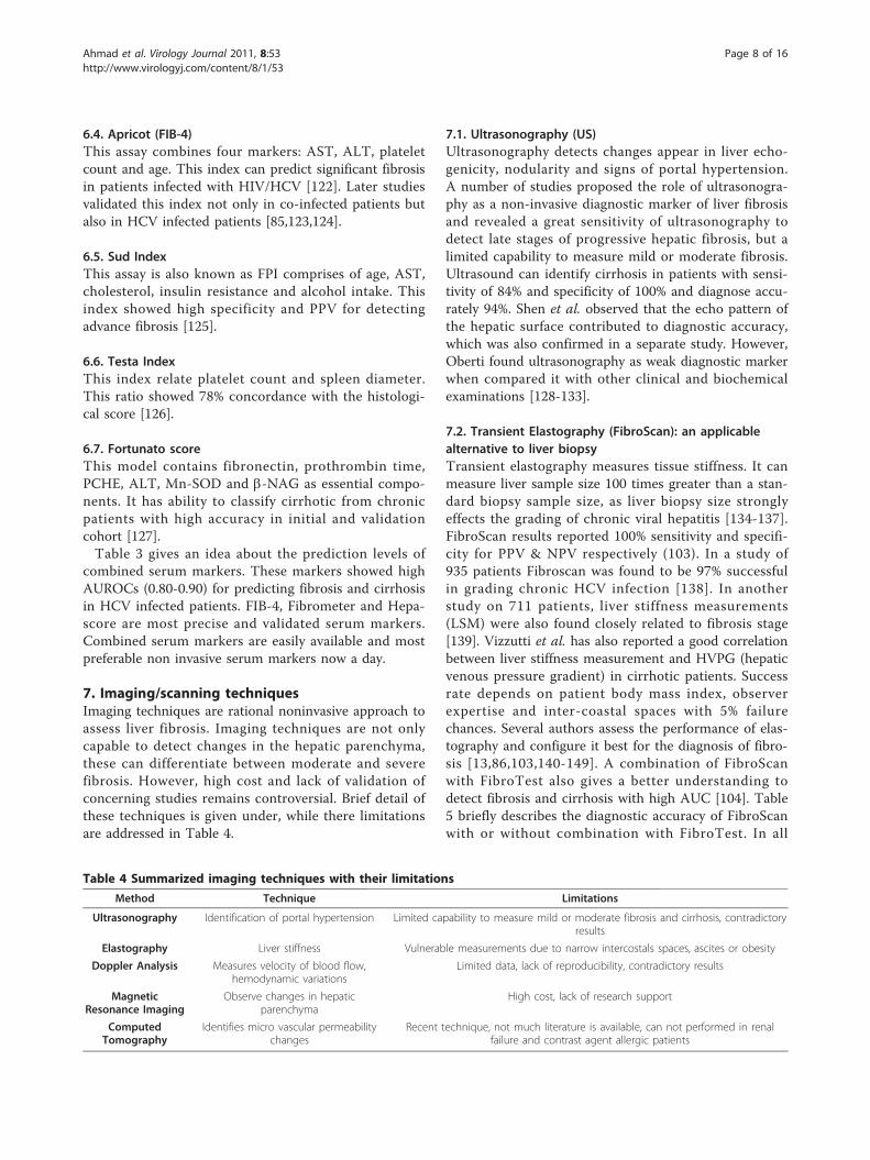
7. Imaging/scanning techniquesImaging techniques are rational noninvasive approach toassess liver fibrosis. Imaging techniques are not onlycapable to detect changes in the hepatic parenchyma,these can differentiate between moderate and severefibrosis. However, high cost and lack of validation ofconcerning studies remains controversial. Brief detail ofthese techniques is given under, while there limitationsare addressed in Table 4.
7.1. Ultrasonography (US)Ultrasonography detects changes appear in liver echo-genicity, nodularity and signs of portal hypertension.A number of studies proposed the role of ultrasonogra-phy as a non-invasive diagnostic marker of liver fibrosisand revealed a great sensitivity of ultrasonography todetect late stages of progressive hepatic fibrosis, but alimited capability to measure mild or moderate fibrosis.Ultrasound can identify cirrhosis in patients with sensi-tivity of 84% and specificity of 100% and diagnose accu-rately 94%. Shen et al. observed that the echo pattern ofthe hepatic surface contributed to diagnostic accuracy,which was also confirmed in a separate study. However,Oberti found ultrasonography as weak diagnostic markerwhen compared it with other clinical and biochemicalexaminations [128-133].
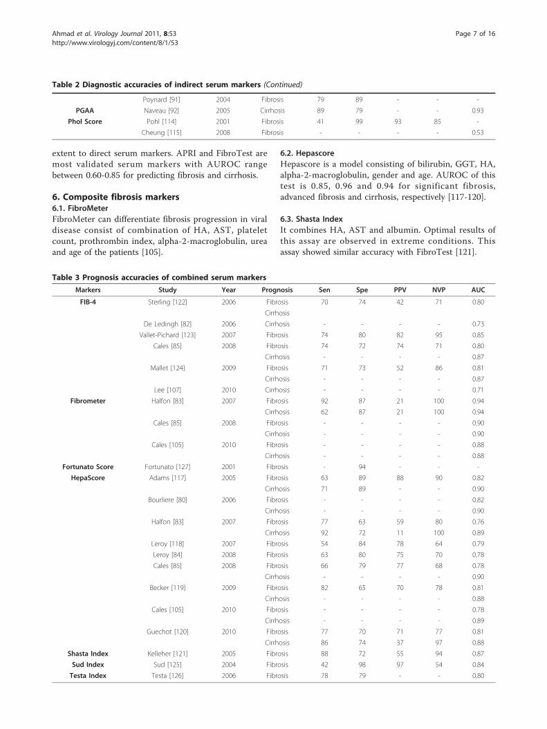
7.2. Transient Elastography (FibroScan): an applicablealternative to liver biopsyTransient elastography measures tissue stiffness. It canmeasure liver sample size 100 times greater than a stan-dard biopsy sample size, as liver biopsy size stronglyeffects the grading of chronic viral hepatitis [134-137].FibroScan results reported 100% sensitivity and specifi-city for PPV & NPV respectively (103). In a study of935 patients Fibroscan was found to be 97% successfulin grading chronic HCV infection [138]. In anotherstudy on 711 patients, liver stiffness measurements(LSM) were also found closely related to fibrosis stage[139]. Vizzutti et al. has also reported a good correlationbetween liver stiffness measurement and HVPG (hepaticvenous pressure gradient) in cirrhotic patients. Successrate depends on patient body mass index, observerexpertise and inter-coastal spaces with 5% failurechances. Several authors assess the performance of elas-tography and configure it best for the diagnosis of fibro-sis [13,86,103,140-149]. A combination of FibroScanwith FibroTest also gives a better understanding todetect fibrosis and cirrhosis with high AUC [104]. Table5 briefly describes the diagnostic accuracy of FibroScanwith or without combination with FibroTest. In all
Table 4 Summarized imaging techniques with their limitations
Method Technique Limitations
Ultrasonography Identification of portal hypertension Limited capability to measure mild or moderate fibrosis and cirrhosis, contradictoryresults
Elastography Liver stiffness Vulnerable measurements due to narrow intercostals spaces, ascites or obesity
Doppler Analysis Measures velocity of blood flow,hemodynamic variations
Limited data, lack of reproducibility, contradictory results
MagneticResonance Imaging
Observe changes in hepaticparenchyma
High cost, lack of research support
ComputedTomography
Identifies micro vascular permeabilitychanges
Recent technique, not much literature is available, can not performed in renalfailure and contrast agent allergic patients
Ahmad et al. Virology Journal 2011, 8:53http://www.virologyj.com/content/8/1/53
Page 8 of 16
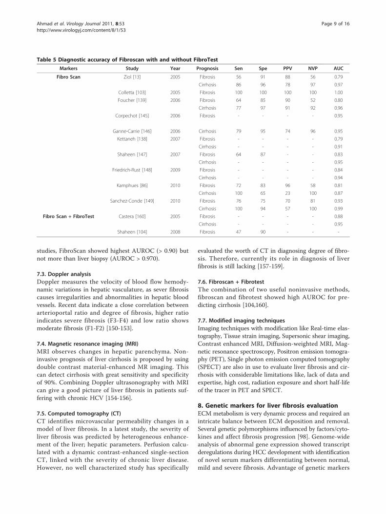
studies, FibroScan showed highest AUROC (> 0.90) butnot more than liver biopsy (AUROC > 0.970).
7.3. Doppler analysisDoppler measures the velocity of blood flow hemody-namic variations in hepatic vasculature, as sever fibrosiscauses irregularities and abnormalities in hepatic bloodvessels. Recent data indicate a close correlation betweenarterioportal ratio and degree of fibrosis, higher ratioindicates severe fibrosis (F3-F4) and low ratio showsmoderate fibrosis (F1-F2) [150-153].
7.4. Magnetic resonance imaging (MRI)MRI observes changes in hepatic parenchyma. Non-invasive prognosis of liver cirrhosis is proposed by usingdouble contrast material-enhanced MR imaging. Thiscan detect cirrhosis with great sensitivity and specificityof 90%. Combining Doppler ultrasonography with MRIcan give a good picture of liver fibrosis in patients suf-fering with chronic HCV [154-156].
7.5. Computed tomography (CT)CT identifies microvascular permeability changes in amodel of liver fibrosis. In a latest study, the severity ofliver fibrosis was predicted by heterogeneous enhance-ment of the liver; hepatic parameters. Perfusion calcu-lated with a dynamic contrast-enhanced single-sectionCT, linked with the severity of chronic liver disease.However, no well characterized study has specifically
evaluated the worth of CT in diagnosing degree of fibro-sis. Therefore, currently its role in diagnosis of liverfibrosis is still lacking [157-159].
7.6. Fibroscan + FibrotestThe combination of two useful noninvasive methods,fibroscan and fibrotest showed high AUROC for pre-dicting cirrhosis [104,160].
7.7. Modified imaging techniquesImaging techniques with modification like Real-time elas-tography, Tissue strain imaging, Supersonic shear imaging,Contrast enhanced MRI, Diffusion-weighted MRI, Mag-netic resonance spectroscopy, Positron emission tomogra-phy (PET), Single photon emission computed tomography(SPECT) are also in use to evaluate liver fibrosis and cir-rhosis with considerable limitations like, lack of data andexpertise, high cost, radiation exposure and short half-lifeof the tracer in PET and SPECT.
8. Genetic markers for liver fibrosis evaluationECM metabolism is very dynamic process and required anintricate balance between ECM deposition and removal.Several genetic polymorphisms influenced by factors/cyto-kines and affect fibrosis progression [98]. Genome-wideanalysis of abnormal gene expression showed transcriptderegulations during HCC development with identificationof novel serum markers differentiating between normal,mild and severe fibrosis. Advantage of genetic markers
Table 5 Diagnostic accuracy of Fibroscan with and without FibroTest
Markers Study Year Prognosis Sen Spe PPV NVP AUC
Fibro Scan Ziol [13] 2005 Fibrosis 56 91 88 56 0.79
Cirrhosis 86 96 78 97 0.97
Colletta [103] 2005 Fibrosis 100 100 100 100 1.00
Foucher [139] 2006 Fibrosis 64 85 90 52 0.80
Cirrhosis 77 97 91 92 0.96
Corpechot [145] 2006 Fibrosis - - - - 0.95
Ganne-Carrie [146] 2006 Cirrhosis 79 95 74 96 0.95
Kettaneh [138] 2007 Fibrosis - - - - 0.79
Cirrhosis - - - - 0.91
Shaheen [147] 2007 Fibrosis 64 87 - - 0.83
Cirrhosis - - - - 0.95
Friedrich-Rust [148] 2009 Fibrosis - - - - 0.84
Cirrhosis - - - - 0.94
Kamphues [86] 2010 Fibrosis 72 83 96 58 0.81
Cirrhosis 100 65 23 100 0.87
Sanchez-Conde [149] 2010 Fibrosis 76 75 70 81 0.93
Cirrhosis 100 94 57 100 0.99
Fibro Scan + FibroTest Castera [160] 2005 Fibrosis - - - - 0.88
Cirrhosis - - - - 0.95
Shaheen [104] 2008 Fibrosis 47 90 - - -
Ahmad et al. Virology Journal 2011, 8:53http://www.virologyj.com/content/8/1/53
Page 9 of 16
over liver biopsy is intrinsic and long life while liver biopsyrepresents only one time point [161-163].Huang and colleagues developed an assay known as
cirrhosis risk score (CRS), a set of seven marker genesto predict cirrhosis risk in HCV infected patients. Ofthe seven genes, AZIN1 and TLR4 have an identifiedrole in hepatic fibrosis, while the identification of func-tional mechanism of the other 5 genes is under process.The authors suggested that fibrosis risk can be identifiedby host genetic factors like single nucleotide polymorph-ism (SNP’s) [164,165].A strong association between CXCR3-associated che-
mokines CXCL9 and CXCL10 with liver fibrosis sug-gested that they may have promise as new non-invasivemarkers of liver fibrosis in HCV infected patients[166,167].CTGF expression is significantly correlated with fibro-
sis stages and remarkably increased in advanced stagesin HCV patients. The AUROC of CTGF to discriminatebetween mild and advanced fibrosis is 0.842 for HCVinfected patients [168].Sharma et al. reported the significant association and
elevated interleukin-18 (IL-18) levels in fibrotic and cir-rhotic liver stages, severity of disease and necrosis inHCV patients [169].A recent study by Caillot et al. used microarray tech-
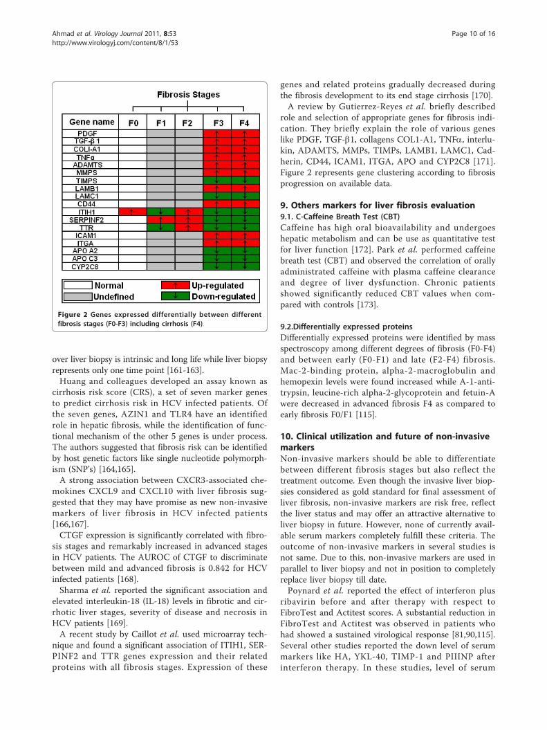
nique and found a significant association of ITIH1, SER-PINF2 and TTR genes expression and their relatedproteins with all fibrosis stages. Expression of these
genes and related proteins gradually decreased duringthe fibrosis development to its end stage cirrhosis [170].A review by Gutierrez-Reyes et al. briefly described
role and selection of appropriate genes for fibrosis indi-cation. They briefly explain the role of various geneslike PDGF, TGF-b1, collagens COL1-A1, TNFa, interlu-kin, ADAMTS, MMPs, TIMPs, LAMB1, LAMC1, Cad-herin, CD44, ICAM1, ITGA, APO and CYP2C8 [171].Figure 2 represents gene clustering according to fibrosisprogression on available data.
9. Others markers for liver fibrosis evaluation9.1. C-Caffeine Breath Test (CBT)Caffeine has high oral bioavailability and undergoeshepatic metabolism and can be use as quantitative testfor liver function [172]. Park et al. performed caffeinebreath test (CBT) and observed the correlation of orallyadministrated caffeine with plasma caffeine clearanceand degree of liver dysfunction. Chronic patientsshowed significantly reduced CBT values when com-pared with controls [173].
9.2.Differentially expressed proteinsDifferentially expressed proteins were identified by massspectroscopy among different degrees of fibrosis (F0-F4)and between early (F0-F1) and late (F2-F4) fibrosis.Mac-2-binding protein, alpha-2-macroglobulin andhemopexin levels were found increased while A-1-anti-trypsin, leucine-rich alpha-2-glycoprotein and fetuin-Awere decreased in advanced fibrosis F4 as compared toearly fibrosis F0/F1 [115].
10. Clinical utilization and future of non-invasivemarkersNon-invasive markers should be able to differentiatebetween different fibrosis stages but also reflect thetreatment outcome. Even though the invasive liver biop-sies considered as gold standard for final assessment ofliver fibrosis, non-invasive markers are risk free, reflectthe liver status and may offer an attractive alternative toliver biopsy in future. However, none of currently avail-able serum markers completely fulfill these criteria. Theoutcome of non-invasive markers in several studies isnot same. Due to this, non-invasive markers are used inparallel to liver biopsy and not in position to completelyreplace liver biopsy till date.Poynard et al. reported the effect of interferon plus
ribavirin before and after therapy with respect toFibroTest and Actitest scores. A substantial reduction inFibroTest and Actitest was observed in patients whohad showed a sustained virological response [81,90,115].Several other studies reported the down level of serummarkers like HA, YKL-40, TIMP-1 and PIIINP afterinterferon therapy. In these studies, level of serum
Figure 2 Genes expressed differentially between differentfibrosis stages (F0-F3) including cirrhosis (F4).
Ahmad et al. Virology Journal 2011, 8:53http://www.virologyj.com/content/8/1/53
Page 10 of 16
markers continue to fall following treatment but mostoften return to permanent levels with biochemical andvirological relapse. These findings suggest that theseassays may be useful for initial staging of disease pro-gression as well as histological response to therapy[174-177]. Fibroscan showed positive correlation withfibrosis stages. However, it is reported that AUROCvalue of Fibroscan and FibroTest must be improved astheir values fall in treated patients irrespective of theirvirological response [178,179]. Furthermore, HCV clear-ance is associated with a significant reduction in non-invasive fibrosis serological markers like FibroTest,Forns Index, age-platelet ratio index, Shasta, FIB-4, Hepa-score and FibroMeter [180]. Patel et al. compared twocommercially available serum marker panels Fibrosureand Fibrospect-II in HCV patients during interferon-basedtherapy. Both assays showed comparable performance fordifferentiating mild fibrosis from moderate-severe stage[181]. Imaging techniques also have some technical limita-tions. These are very expensive and are not easy to handle.Their presence in each hospital or laboratory is not possi-ble especially in poor countries. On the other hand geneticmarkers showed a great variability for detecting cirrhosisand fibrosis. They are also able to differentiate amongfibrosis stages. But a lot of work is needed for them tobecome an integral part of hepatic analysis.
11. ConclusionsOur study showed that there are only three to four mar-kers or set of marker that are used continuously basedon their precision and accuracy in various studies forfibrosis and cirrhosis prediction. In serum non-invasivemarkers, FibroTest, Forn’s Index, Fibrometer and Hea-paScore have a high five-year prognostic value but notcompared to liver biopsy (AUROC = 0.97), while Fibros-can showed maximum accuracy nearer to liver biopsy(AUROC > 0.90). Recently, genetic markers showed dif-ferential gene expression in different fibrosis stages, butthese are not frequently available in all labs. Imagingtechniques like ultrasound and elastography not onlyused to diagnose liver fibrosis but also monitor diseaseprogression. However, genetic markers showed highability to distinguish not only mild and advance stagesof liver fibrosis but also differentiate between intermedi-ate fibrosis stages. Although present published literaturedo not provide any evidence for non-invasive markers tobecome an integrated part of the complete assessmentof liver fibrosis in HCV patients, a combination of twoor more serum markers with imaging techniques mayimprove the accuracy of diagnosis.
AcknowledgementsFinancial support by Higher Education Commission (Grant # 863) is highlyacknowledged.
Authors’ contributionsAW, IB, GS, AS and HS designed the study, analyze the data and wrotepaper. JS, KS, SMT, KH, SA and SS checked the revised manuscriptthoroughly and confirmed all the data given in manuscript. All work wasperformed under supervision of HS. All authors read and approved the finalmanuscript.
Authors’ informationShah Jahan, Saba Khaliq and Samrin A (PhD in Molecular biology), BushraIjaz (M Phil Molecular Biology), Waqar Ahmad (M Phil Chemistry) and Gull S(MSc Biochemistry) are Research Officer; Sawar MT and Shahid I are Phdscholars, Asad S is MPhil scholars, while Sajida Hassan (PhD MolecularBiology) is Principal Investigator at CEMB, University of the Punjab, Lahore.
Competing interestsThe authors declare that they have no competing interests.
Received: 18 January 2011 Accepted: 8 February 2011Published: 8 February 2011
References1. Memon MI, Memon MA: Hepatitis C: an epidemiological review. J Viral
Hepat 2002, 9:84-100.2. WHO: Global distribution of hepatitis A, B and C, 2001. Weakly
Epidimiological Records 2002, 77, 41,48.3. Marcellin P, Asselah T, Boyer N: Fibrosis and disease progression in
hepatitis C. Hepatology 2002, 36:S47-56.4. Afdhal NH, Nunes D: Evaluation of liver fibrosis: a concise review. Am J
Gastroenterol 2004, 99:1160-1174.5. Dienstag JL, McHutchison JG: American Gastroenterological Association
technical review on the management of hepatitis C. Gastroenterology2006, 130:231-264, quiz 214-237.
6. Clark JM: The epidemiology of nonalcoholic fatty liver disease in adults.J Clin Gastroenterol 2006, 40(Suppl 1):S5-10.
7. Harbin WP, Robert NJ, Ferrucci JT Jr: Diagnosis of cirrhosis based onregional changes in hepatic morphology: a radiological and pathologicalanalysis. Radiology 1980, 135:273-283.
8. Gressner AM: The cell biology of liver fibrogenesis - an imbalance ofproliferation, growth arrest and apoptosis of myofibroblasts. Cell TissueRes 1998, 292:447-452.
9. Arthur MJ: Reversibility of liver fibrosis and cirrhosis following treatmentfor hepatitis C. Gastroenterology 2002, 122:1525-1528.
10. Adinolfi LE, Gambardella M, Andreana A, Tripodi MF, Utili R, Ruggiero G:Steatosis accelerates the progression of liver damage of chronichepatitis C patients and correlates with specific HCV genotype andvisceral obesity. Hepatology 2001, 33:1358-1364.
11. El-Serag HB: Hepatocellular carcinoma and hepatitis C in the UnitedStates. Hepatology 2002, 36:S74-83.
12. Parkes J, Guha IN, Roderick P, Rosenberg W: Performance of serum markerpanels for liver fibrosis in chronic hepatitis C. J Hepatol 2006, 44:462-474.
13. Ziol M, Handra-Luca A, Kettaneh A, Christidis C, Mal F, Kazemi F, deLedinghen V, Marcellin P, Dhumeaux D, Trinchet JC, Beaugrand M:Noninvasive assessment of liver fibrosis by measurement of stiffness inpatients with chronic hepatitis C. Hepatology 2005, 41:48-54.
14. Saadeh S, Cammell G, Carey WD, Younossi Z, Barnes D, Easley K: The roleof liver biopsy in chronic hepatitis C. Hepatology 2001, 33:196-200.
15. Booth JC, O’Grady J, Neuberger J: Clinical guidelines on the managementof hepatitis C. Gut 2001, 49(Suppl 1):I1-21.
16. Knodell RG, Ishak KG, Black WC, Chen TS, Craig R, Kaplowitz N, Kiernan TW,Wollman J: Formulation and application of a numerical scoring systemfor assessing histological activity in asymptomatic chronic activehepatitis. Hepatology 1981, 1:431-435.
17. group TFMcs: Intraobserver and interobserver variations in liver biopsyinterpretation in patients with chronic hepatitis C. The French METAVIRCooperative Study Group. Hepatology 1994, 20:15-20.
18. Ishak K, Baptista A, Bianchi L, Callea F, De Groote J, Gudat F, Denk H,Desmet V, Korb G, MacSween RN, et al: Histological grading and stagingof chronic hepatitis. J Hepatol 1995, 22:696-699.
19. Goldin RD, Goldin JG, Burt AD, Dhillon PA, Hubscher S, Wyatt J, Patel N:Intra-observer and inter-observer variation in the histopathologicalassessment of chronic viral hepatitis. J Hepatol 1996, 25:649-654.
Ahmad et al. Virology Journal 2011, 8:53http://www.virologyj.com/content/8/1/53
Page 11 of 16
20. Westin J, Lagging LM, Wejstal R, Norkrans G, Dhillon AP: Interobserverstudy of liver histopathology using the Ishak score in patients withchronic hepatitis C virus infection. Liver 1999, 19:183-187.
21. Friedman SL: Molecular regulation of hepatic fibrosis, an integratedcellular response to tissue injury. J Biol Chem 2000, 275:2247-2250.
22. Friedman SL: Liver fibrosis – from bench to bedside. J Hepatol 2003,38(Suppl 1):S38-53.
23. Kelleher TB, Afdhal N: Noninvasive assessment of liver fibrosis. Clin LiverDis 2005, 9:667-683, vii.
24. Idobe Y, Murawaki Y, Ikuta Y, Koda M, Kawasaki H: Post-prandial serumhyaluronan concentration in patients with chronic liver disease. InternMed 1998, 37:568-575.
25. Saif MW, Alexander D, Wicox CM: Serum Alkaline Phosphatase Level as aPrognostic Tool in Colorectal Cancer: A Study of 105 patients. J Appl Res2005, 5:88-95.
26. Imbert-Bismut F, Ratziu V, Pieroni L, Charlotte F, Benhamou Y, Poynard T:Biochemical markers of liver fibrosis in patients with hepatitis C virusinfection: a prospective study. Lancet 2001, 357:1069-1075.
27. Forns X, Ampurdanes S, Llovet JM, Aponte J, Quinto L, Martinez-Bauer E,Bruguera M, Sanchez-Tapias JM, Rodes J: Identification of chronic hepatitisC patients without hepatic fibrosis by a simple predictive model.Hepatology 2002, 36:986-992.
28. Wai CT, Greenson JK, Fontana RJ, Kalbfleisch JD, Marrero JA,Conjeevaram HS, Lok AS: A simple noninvasive index can predict bothsignificant fibrosis and cirrhosis in patients with chronic hepatitis C.Hepatology 2003, 38:518-526.
29. Thabut D, Simon M, Myers RP, Messous D, Thibault V, Imbert-Bismut F,Poynard T: Noninvasive prediction of fibrosis in patients with chronichepatitis C. Hepatology 2003, 37:1220-1221, author reply 1221.
30. Guechot J, Loria A, Serfaty L, Giral P, Giboudeau J, Poupon R: Serumhyaluronan as a marker of liver fibrosis in chronic viral hepatitis C: effectof alpha-interferon therapy. J Hepatol 1995, 22:22-26.
31. Pares A, Deulofeu R, Gimenez A, Caballeria L, Bruguera M, Caballeria J,Ballesta AM, Rodes J: Serum hyaluronate reflects hepatic fibrogenesis inalcoholic liver disease and is useful as a marker of fibrosis. Hepatology1996, 24:1399-1403.
32. McHutchison JG, Blatt LM, de Medina M, Craig JR, Conrad A, Schiff ER,Tong MJ: Measurement of serum hyaluronic acid in patients withchronic hepatitis C and its relationship to liver histology. ConsensusInterferon Study Group. J Gastroenterol Hepatol 2000, 15:945-951.
33. Hayasaka A, Saisho H: Serum markers as tools to monitor liver fibrosis.Digestion 1998, 59:381-384.
34. Guechot J, Laudat A, Loria A, Serfaty L, Poupon R, Giboudeau J:Diagnostic accuracy of hyaluronan and type III procollagen amino-terminal peptide serum assays as markers of liver fibrosis in chronicviral hepatitis C evaluated by ROC curve analysis. Clin Chem 1996,42:558-563.
35. Fabris C, Falleti E, Federico E, Toniutto P, Pirisi M: A comparison of fourserum markers of fibrosis in the diagnosis of cirrhosis. Ann Clin Biochem1997, 34(Pt 2):151-155.
36. George DK, Ramm GA, Walker NI, Powell LW, Crawford DH: Elevated serumtype IV collagen: a sensitive indicator of the presence of cirrhosis inhaemochromatosis. J Hepatol 1999, 31:47-52.
37. Murawaki Y, Ikuta Y, Koda M, Yamada S, Kawasaki H: Comparison of serum7 S fragment of type IV collagen and serum central triple-helix of typeIV collagen for assessment of liver fibrosis in patients with chronic viralliver disease. J Hepatol 1996, 24:148-154.
38. Boeker KH, Haberkorn CI, Michels D, Flemming P, Manns MP,Lichtinghagen R: Diagnostic potential of circulating TIMP-1 and MMP-2as markers of liver fibrosis in patients with chronic hepatitis C. Clin ChimActa 2002, 316:71-81.
39. Zhang BB, Cai WM, Weng HL, Hu ZR, Lu J, Zheng M, Liu RH: Diagnosticvalue of platelet derived growth factor-BB, transforming growth factor-beta1, matrix metalloproteinase-1, and tissue inhibitor of matrixmetalloproteinase-1 in serum and peripheral blood mononuclear cellsfor hepatic fibrosis. World J Gastroenterol 2003, 9:2490-2496.
40. Johansen JS, Christoffersen P, Moller S, Price PA, Henriksen JH, Garbarsch C,Bendtsen F: Serum YKL-40 is increased in patients with hepatic fibrosis.J Hepatol 2000, 32:911-920.
41. Kanzler S, Baumann M, Schirmacher P, Dries V, Bayer E, Gerken G,Dienes HP, Lohse AW: Prediction of progressive liver fibrosis in hepatitis
C infection by serum and tissue levels of transforming growth factor-beta. J Viral Hepat 2001, 8:430-437.
42. Patel K, Gordon SC, Jacobson I, Hezode C, Oh E, Smith KM, Pawlotsky JM,McHutchison JG: Evaluation of a panel of non-invasive serum markers todifferentiate mild from moderate-to-advanced liver fibrosis in chronichepatitis C patients. J Hepatol 2004, 41:935-942.
43. Zaman A, Rosen HR, Ingram K, Corless CL, Oh E, Smith K: Assessment ofFIBROSpect II to detect hepatic fibrosis in chronic hepatitis C patients.Am J Med 2007, 120:280, e289-214.
44. Rosenberg WM, Voelker M, Thiel R, Becka M, Burt A, Schuppan D,Hubscher S, Roskams T, Pinzani M, Arthur MJ: Serum markers detect thepresence of liver fibrosis: a cohort study. Gastroenterology 2004,127:1704-1713.
45. Leroy V, Monier F, Bottari S, Trocme C, Sturm N, Hilleret MN, Morel F,Zarski JP: Circulating matrix metalloproteinases 1, 2, 9 and theirinhibitors TIMP-1 and TIMP-2 as serum markers of liver fibrosis inpatients with chronic hepatitis C: comparison with PIIINP and hyaluronicacid. Am J Gastroenterol 2004, 99:271-279.
46. Murawaki Y, Ikuta Y, Nishimura Y, Koda M, Kawasaki H: Serum markers forconnective tissue turnover in patients with chronic hepatitis B andchronic hepatitis C: a comparative analysis. J Hepatol 1995, 23:145-152.
47. Murawaki Y, Ikuta Y, Okamoto K, Koda M, Kawasaki H: Diagnostic value ofserum markers of connective tissue turnover for predicting histologicalstaging and grading in patients with chronic hepatitis C. J Gastroenterol2001, 36:399-406.
48. Suzuki A, Angulo P, Lymp J, Li D, Satomura S, Lindor K: Hyaluronic acid, anaccurate serum marker for severe hepatic fibrosis in patients with non-alcoholic fatty liver disease. Liver Int 2005, 25:779-786.
49. Halfon P, Bourliere M, Penaranda G, Deydier R, Renou C, Botta-Fridlund D,Tran A, Portal I, Allemand I, Rosenthal-Allieri A, Ouzan D: Accuracy ofhyaluronic acid level for predicting liver fibrosis stages in patients withhepatitis C virus. Comp Hepatol 2005, 4:6.
50. Hakala BE, White C, Recklies AD: Human cartilage gp-39, a major secretoryproduct of articular chondrocytes and synovial cells, is a mammalianmember of a chitinase protein family. J Biol Chem 1993, 268:25803-25810.
51. Saitou Y, Shiraki K, Yamanaka Y, Yamaguchi Y, Kawakita T, Yamamoto N,Sugimoto K, Murata K, Nakano T: Noninvasive estimation of liver fibrosisand response to interferon therapy by a serum fibrogenesis marker,YKL-40, in patients with HCV-associated liver disease. World JGastroenterol 2005, 11:476-481.
52. Sanvisens A, Serra I, Tural C, Tor J, Ojanguren I, Barluenga E, Rey-Joly C,Clotet B, Muga R: Hyaluronic acid, transforming growth factor-beta1and hepatic fibrosis in patients with chronic hepatitis C virus andhuman immunodeficiency virus co-infection. J Viral Hepat 2009,16:513-518.
53. Schiavon LL, Carvalho-Filho RJ, Narciso-Schiavon JL, Medina-Pestana JO,Lanzoni VP, Ferraz ML, Silva AE: YKL-40 and hyaluronic acid (HA) asnoninvasive markers of liver fibrosis in kidney transplant patients withHCV chronic infection. Scand J Gastroenterol 2010, 45:615-622.
54. Felig P: The glucose-alanine cycle. Metabolism 1973, 22:179-207.55. Daxboeck F, Gattringer R, Mustafa S, Bauer C, Assadian O: Elevated serum
alanine aminotransferase (ALT) levels in patients with serologicallyverified Mycoplasma pneumoniae pneumonia. Clin Microbiol Infect 2005,11:507-510.
56. Sherman KE: Alanine aminotransferase in clinical practice. A review. ArchIntern Med 1991, 151:260-265.
57. Dufour DR, Lott JA, Nolte FS, Gretch DR, Koff RS, Seeff LB: Diagnosis andmonitoring of hepatic injury. I. Performance characteristics of laboratorytests. Clin Chem 2000, 46:2027-2049.
58. Akkaya O, Kiyici M, Yilmaz Y, Ulukaya E, Yerci O: Clinical significance ofactivity of ALT enzyme in patients with hepatitis C virus. World JGastroenterol 2007, 13:5481-5485.
59. Kim HJ, Oh SW, Kim DJ, Choi EY: Abundance of immunologically activealanine aminotransferase in sera of liver cirrhosis and hepatocellularcarcinoma patients. Clin Chem 2009, 55:1022-1025.
60. Shiffman ML, Diago M, Tran A, Pockros P, Reindollar R, Prati D, Rodriguez-Torres M, Lardelli P, Blotner S, Zeuzem S: Chronic hepatitis C in patientswith persistently normal alanine transaminase levels. Clin GastroenterolHepatol 2006, 4:645-652.
61. Okuda M, Li K, Beard MR, Showalter LA, Scholle F, Lemon SM, Weinman SA:Mitochondrial injury, oxidative stress, and antioxidant gene expression
Ahmad et al. Virology Journal 2011, 8:53http://www.virologyj.com/content/8/1/53
Page 12 of 16

are induced by hepatitis C virus core protein. Gastroenterology 2002,122:366-375.
62. Zechini B, Pasquazzi C, Aceti A: Correlation of serum aminotransferaseswith HCV RNA levels and histological findings in patients with chronichepatitis C: the role of serum aspartate transaminase in the evaluationof disease progression. Eur J Gastroenterol Hepatol 2004, 16:891-896.
63. Cedrone A, Covino M, Caturelli E, Pompili M, Lorenzelli G, Villani MR, Valle D,Sperandeo M, Rapaccini GL, Gasbarrini G: Utility of alpha-fetoprotein (AFP)in the screening of patients with virus-related chronic liver disease: doesdifferent viral etiology influence AFP levels in HCC? A study in 350western patients. Hepatogastroenterology 2000, 47:1654-1658.
64. Chu CW, Hwang SJ, Luo JC, Lai CR, Tsay SH, Li CP, Wu JC, Chang FY,Lee SD: Clinical, virologic, and pathologic significance of elevated serumalpha-fetoprotein levels in patients with chronic hepatitis C. J ClinGastroenterol 2001, 32:240-244.
65. Chen TM, Huang PT, Tsai MH, Lin LF, Liu CC, Ho KS, Siauw CP, Chao PL,Tung JN: Predictors of alpha-fetoprotein elevation in patients withchronic hepatitis C, but not hepatocellular carcinoma, and itsnormalization after pegylated interferon alfa 2a-ribavirin combinationtherapy. J Gastroenterol Hepatol 2007, 22:669-675.
66. Tamura Y, Yamagiwa S, Aoki Y, Kurita S, Suda T, Ohkoshi S, Nomoto M,Aoyagi Y: Serum alpha-fetoprotein levels during and after interferontherapy and the development of hepatocellular carcinoma in patientswith chronic hepatitis C. Dig Dis Sci 2009, 54:2530-2537.
67. Aster RH: Pooling of platelets in the spleen: role in the pathogenesis of“hypersplenic” thrombocytopenia. J Clin Invest 1966, 45:645-657.
68. Kawasaki T, Takeshita A, Souda K, Kobayashi Y, Kikuyama M, Suzuki F,Kageyama F, Sasada Y, Shimizu E, Murohisa G, et al: Serum thrombopoietinlevels in patients with chronic hepatitis and liver cirrhosis. Am JGastroenterol 1999, 94:1918-1922.
69. Adinolfi LE, Giordano MG, Andreana A, Tripodi MF, Utili R, Cesaro G,Ragone E, Durante Mangoni E, Ruggiero G: Hepatic fibrosis plays a centralrole in the pathogenesis of thrombocytopenia in patients with chronicviral hepatitis. Br J Haematol 2001, 113:590-595.
70. Lackner C, Struber G, Liegl B, Leibl S, Ofner P, Bankuti C, Bauer B,Stauber RE: Comparison and validation of simple noninvasive tests forprediction of fibrosis in chronic hepatitis C. Hepatology 2005,41:1376-1382.
71. Croquet V, Vuillemin E, Ternisien C, Pilette C, Oberti F, Gallois Y, Trossaert M,Rousselet MC, Chappard D, Cales P: Prothrombin index is an indirectmarker of severe liver fibrosis. Eur J Gastroenterol Hepatol 2002,14:1133-1141.
72. Pilette C, Oberti F, Aube C, Rousselet MC, Bedossa P, Gallois Y, Rifflet H,Cales P: Non-invasive diagnosis of esophageal varices in chronic liverdiseases. J Hepatol 1999, 31:867-873.
73. Craxi A, Camma C, Giunta M: Clinical aspects of bleeding complications incirrhotic patients. Blood Coagul Fibrinolysis 2000, 11(Suppl 1):S75-79.
74. Sheth SG, Flamm SL, Gordon FD, Chopra S: AST/ALT ratio predictscirrhosis in patients with chronic hepatitis C virus infection. Am JGastroenterol 1998, 93:44-48.
75. Reedy DW, Loo AT, Levine RA: AST/ALT ratio > or = 1 is not diagnostic ofcirrhosis in patients with chronic hepatitis C. Dig Dis Sci 1998,43:2156-2159.
76. Giannini E, Risso D, Botta F, Chiarbonello B, Fasoli A, Malfatti F,Romagnoli P, Testa E, Ceppa P, Testa R: Validity and clinical utility of theaspartate aminotransferase-alanine aminotransferase ratio in assessingdisease severity and prognosis in patients with hepatitis C virus-relatedchronic liver disease. Arch Intern Med 2003, 163:218-224.
77. Fujii H, Enomoto M, Fukushima W, Ohfuji S, Mori M, Kobayashi S, Iwai S,Morikawa H, Tamori A, Sakaguchi H, et al: Noninvasive laboratory testsproposed for predicting cirrhosis in patients with chronic hepatitis C arealso useful in patients with non-alcoholic steatohepatitis. J Gastroenterol2009, 44:608-614.
78. Poynard T, Morra R, Halfon P, Castera L, Ratziu V, Imbert-Bismut F, Naveau S,Thabut D, Lebrec D, Zoulim F, et al: Meta-analyses of FibroTest diagnosticvalue in chronic liver disease. BMC Gastroenterol 2007, 7:40.
79. Cales P, Oberti F, Michalak S, Hubert-Fouchard I, Rousselet MC, Konate A,Gallois Y, Ternisien C, Chevailler A, Lunel F: A novel panel of blood markersto assess the degree of liver fibrosis. Hepatology 2005, 42:1373-1381.
80. Bourliere M, Penaranda G, Renou C, Botta-Fridlund D, Tran A, Portal I,Lecomte L, Castellani P, Rosenthal-Allieri MA, Gerolami R, et al: Validation
and comparison of indexes for fibrosis and cirrhosis prediction inchronic hepatitis C patients: proposal for a pragmatic approachclassification without liver biopsies. J Viral Hepat 2006, 13:659-670.
81. Parise ER, Oliveira AC, Figueiredo-Mendes C, Lanzoni V, Martins J, Nader H,Ferraz ML: Noninvasive serum markers in the diagnosis of structural liverdamage in chronic hepatitis C virus infection. Liver Int 2006, 26:1095-1099.
82. de Ledinghen V, Douvin C, Kettaneh A, Ziol M, Roulot D, Marcellin P,Dhumeaux D, Beaugrand M: Diagnosis of hepatic fibrosis and cirrhosis bytransient elastography in HIV/hepatitis C virus-coinfected patients.J Acquir Immune Defic Syndr 2006, 41:175-179.
83. Halfon P, Bacq Y, De Muret A, Penaranda G, Bourliere M, Ouzan D, Tran A,Botta D, Renou C, Brechot MC, et al: Comparison of test performanceprofile for blood tests of liver fibrosis in chronic hepatitis C. J Hepatol2007, 46:395-402.
84. Leroy V, Halfon P, Bacq Y, Boursier J, Rousselet MC, Bourliere M, de Muret A,Sturm N, Hunault G, Penaranda G, et al: Diagnostic accuracy,reproducibility and robustness of fibrosis blood tests in chronic hepatitisC: a meta-analysis with individual data. Clin Biochem 2008, 41:1368-1376.
85. Cales P, de Ledinghen V, Halfon P, Bacq Y, Leroy V, Boursier J, Foucher J,Bourliere M, de Muret A, Sturm N, et al: Evaluating the accuracy andincreasing the reliable diagnosis rate of blood tests for liver fibrosis inchronic hepatitis C. Liver Int 2008, 28:1352-1362.
86. Kamphues C, Lotz K, Rocken C, Berg T, Eurich D, Pratschke J, Neuhaus P,Neumann UP: Chances and limitations of non-invasive tests in theassessment of liver fibrosis in liver transplant patients. Clin Transplant2010, 24:652-659.
87. Teare JP, Sherman D, Greenfield SM, Simpson J, Bray G, Catterall AP,Murray-Lyon IM, Peters TJ, Williams R, Thompson RP: Comparison of serumprocollagen III peptide concentrations and PGA index for assessment ofhepatic fibrosis. Lancet 1993, 342:895-898.
88. Naveau S, Poynard T, Benattar C, Bedossa P, Chaput JC: Alpha-2-macroglobulin and hepatic fibrosis. Diagnostic interest. Dig Dis Sci 1994,39:2426-2432.
89. Oberti F, Valsesia E, Pilette C, Rousselet MC, Bedossa P, Aube C, Gallois Y,Rifflet H, Maiga MY, Penneau-Fontbonne D, Cales P: Noninvasive diagnosisof hepatic fibrosis or cirrhosis. Gastroenterology 1997, 113:1609-1616.
90. Poynard T, McHutchison J, Manns M, Myers RP, Albrecht J: Biochemicalsurrogate markers of liver fibrosis and activity in a randomized trial ofpeginterferon alfa-2b and ribavirin. Hepatology 2003, 38:481-492.
91. Poynard T, Munteanu M, Imbert-Bismut F, Charlotte F, Thabut D, LeCalvez S, Messous D, Thibault V, Benhamou Y, Moussalli J, Ratziu V:Prospective analysis of discordant results between biochemical markersand biopsy in patients with chronic hepatitis C. Clin Chem 2004,50:1344-1355.
92. Naveau S, Raynard B, Ratziu V, Abella A, Imbert-Bismut F, Messous D,Beuzen F, Capron F, Thabut D, Munteanu M, et al: Biomarkers for theprediction of liver fibrosis in patients with chronic alcoholic liverdisease. Clin Gastroenterol Hepatol 2005, 3:167-174.
93. Sebastiani G, Alberti A: Non invasive fibrosis biomarkers reduce but notsubstitute the need for liver biopsy. World J Gastroenterol 2006,12:3682-3694.
94. Friedrich-Rust M, Rosenberg W, Parkes J, Herrmann E, Zeuzem S, Sarrazin C:Comparison of ELF, FibroTest and FibroScan for the non-invasiveassessment of liver fibrosis. BMC Gastroenterol 2010, 10:103.
95. Halfon P, Imbert-Bismut F, Messous D, Antoniotti G, Benchetrit D, Cart-Lamy P, Delaporte G, Doutheau D, Klump T, Sala M, et al: A prospectiveassessment of the inter-laboratory variability of biochemical markers offibrosis (FibroTest) and activity (ActiTest) in patients with chronic liverdisease. Comp Hepatol 2002, 1:3.
96. Poynard T, Imbert-Bismut F, Ratziu V, Chevret S, Jardel C, Moussalli J,Messous D, Degos F: Biochemical markers of liver fibrosis in patientsinfected by hepatitis C virus: longitudinal validation in a randomizedtrial. J Viral Hepat 2002, 9:128-133.
97. Rossi E, Adams L, Prins A, Bulsara M, de Boer B, Garas G, MacQuillan G,Speers D, Jeffrey G: Validation of the FibroTest biochemical markers scorein assessing liver fibrosis in hepatitis C patients. Clin Chem 2003,49:450-454.
98. Ngo Y, Munteanu M, Messous D, Charlotte F, Imbert-Bismut F, Thabut D,Lebray P, Thibault V, Benhamou Y, Moussalli J, et al: A prospective analysisof the prognostic value of biomarkers (FibroTest) in patients withchronic hepatitis C. Clin Chem 2006, 52:1887-1896.
Ahmad et al. Virology Journal 2011, 8:53http://www.virologyj.com/content/8/1/53
Page 13 of 16
99. Halfon P, Bourliere M, Deydier R, Botta-Fridlund D, Renou C, Tran A, Portal I,Allemand I, Bertrand JJ, Rosenthal-Allieri A, et al: Independent prospectivemulticenter validation of biochemical markers (fibrotest-actitest) for theprediction of liver fibrosis and activity in patients with chronic hepatitisC: the fibropaca study. Am J Gastroenterol 2006, 101:547-555.
100. Halfon P, Munteanu M, Poynard T: FibroTest-ActiTest as a non-invasivemarker of liver fibrosis. Gastroenterol Clin Biol 2008, 32:22-39.
101. Myers RP, De Torres M, Imbert-Bismut F, Ratziu V, Charlotte F, Poynard T:Biochemical markers of fibrosis in patients with chronic hepatitis C: acomparison with prothrombin time, platelet count, and age-plateletindex. Dig Dis Sci 2003, 48:146-153.
102. Bedossa P, Dargere D, Paradis V: Sampling variability of liver fibrosis inchronic hepatitis C. Hepatology 2003, 38:1449-1457.
103. Colletta C, Smirne C, Fabris C, Toniutto P, Rapetti R, Minisini R, Pirisi M:Value of two noninvasive methods to detect progression of fibrosisamong HCV carriers with normal aminotransferases. Hepatology 2005,42:838-845.
104. Shaheen AA, Myers RP: Systematic review and meta-analysis of thediagnostic accuracy of fibrosis marker panels in patients with HIV/hepatitis C coinfection. HIV Clin Trials 2008, 9:43-51.
105. Cales P, Boursier J, Bertrais S, Oberti F, Gallois Y, Fouchard-Hubert I, Dib N,Zarski JP, Rousselet MC: Optimization and robustness of blood tests forliver fibrosis and cirrhosis. Clin Biochem 2010, 43:1315-1322.
106. Koda M, Matunaga Y, Kawakami M, Kishimoto Y, Suou T, Murawaki Y:FibroIndex, a practical index for predicting significant fibrosis in patientswith chronic hepatitis C. Hepatology 2007, 45:297-306.
107. Lee MH, Cheong JY, Um SH, Seo YS, Kim DJ, Hwang SG, Yang JM, Han KH,Cho SW: Comparison of surrogate serum markers and transientelastography (Fibroscan) for assessing cirrhosis in patients with chronicviral hepatitis. Dig Dis Sci 2010, 55:3552-3560.
108. El-Shabrawi MH, Mohsen NA, Sherif MM, El-Karaksy HM, Abou-Yosef H, El-Sayed HM, Riad H, Bahaa N, Isa M, El-Hennawy A: Noninvasive assessmentof hepatic fibrosis and necroinflammatory activity in Egyptian childrenwith chronic hepatitis C virus infection using FibroTest and ActiTest. EurJ Gastroenterol Hepatol 2010, 22:946-951.
109. Poynard T, Ratziu V, Naveau S, Thabut D, Charlotte F, Messous D,Capron D, Abella A, Massard J, Ngo Y, et al: The diagnostic value ofbiomarkers (SteatoTest) for the prediction of liver steatosis. CompHepatol 2005, 4:10.
110. Lok AS, Ghany MG, Goodman ZD, Wright EC, Everson GT, Sterling RK,Everhart JE, Lindsay KL, Bonkovsky HL, Di Bisceglie AM, et al: Predictingcirrhosis in patients with hepatitis C based on standard laboratory tests:results of the HALT-C cohort. Hepatology 2005, 42:282-292.
111. Cheong JY, Um SH, Seo YS, Kim DJ, Hwang SG, Lee YJ, Cho M, Yang JM,Kim YB, Park YN, Cho SW: Non-Invasive Index for Predicting SignificantLiver Fibrosis: Comparison of Diagnostic Performances in Patients withChronic Hepatitis B and C. Dig Dis Sci 2010, 56:555-563.
112. Islam S, Antonsson L, Westin J, Lagging M: Cirrhosis in hepatitis C virus-infected patients can be excluded using an index of standardbiochemical serum markers. Scand J Gastroenterol 2005, 40:867-872.
113. Ohta T, Sakaguchi K, Fujiwara A, Fujioka S, Iwasaki Y, Makino Y, Araki Y,Shiratori Y: Simple surrogate index of the fibrosis stage in chronichepatitis C patients using platelet count and serum albumin level. ActaMed Okayama 2006, 60:77-84.
114. Pohl A, Behling C, Oliver D, Kilani M, Monson P, Hassanein T: Serumaminotransferase levels and platelet counts as predictors of degree offibrosis in chronic hepatitis C virus infection. Am J Gastroenterol 2001,96:3142-3146.
115. Cheung RC, Currie S, Shen H, Bini EJ, Ho SB, Anand BS, Hu KQ, Wright TL,Morgan TR: Can we predict the degree of fibrosis in chronic hepatitis Cpatients using routine blood tests in our daily practice? J ClinGastroenterol 2008, 42:827-834.
116. Bonacini M, Hadi G, Govindarajan S, Lindsay KL: Utility of a discriminantscore for diagnosing advanced fibrosis or cirrhosis in patients withchronic hepatitis C virus infection. Am J Gastroenterol 1997, 92:1302-1304.
117. Adams LA, Bulsara M, Rossi E, DeBoer B, Speers D, George J, Kench J,Farrell G, McCaughan GW, Jeffrey GP: Hepascore: an accurate validatedpredictor of liver fibrosis in chronic hepatitis C infection. Clin Chem 2005,51:1867-1873.
118. Leroy V, Hilleret MN, Sturm N, Trocme C, Renversez JC, Faure P, Morel F,Zarski JP: Prospective comparison of six non-invasive scores for the
diagnosis of liver fibrosis in chronic hepatitis C. J Hepatol 2007,46:775-782.
119. Becker L, Salameh W, Sferruzza A, Zhang K, ng Chen R, Malik R, Reitz R,Nasser I, Afdhal NH: Validation of hepascore, compared with simpleindices of fibrosis, in patients with chronic hepatitis C virus infection inUnited States. Clin Gastroenterol Hepatol 2009, 7:696-701.
120. Guechot J, Lasnier E, Sturm N, Paris A, Zarski JP: Automation of theHepascore and validation as a biochemical index of liver fibrosis inpatients with chronic hepatitis C from the ANRS HC EP 23 Fibrostarcohort. Clin Chim Acta 2010, 411:86-91.
121. Kelleher TB, Mehta SH, Bhaskar R, Sulkowski M, Astemborski J, Thomas DL,Moore RE, Afdhal NH: Prediction of hepatic fibrosis in HIV/HCV co-infected patients using serum fibrosis markers: the SHASTA index.J Hepatol 2005, 43:78-84.
122. Sterling RK, Lissen E, Clumeck N, Sola R, Correa MC, Montaner J, M SS,Torriani FJ, Dieterich DT, Thomas DL, et al: Development of a simplenoninvasive index to predict significant fibrosis in patients with HIV/HCVcoinfection. Hepatology 2006, 43:1317-1325.
123. Vallet-Pichard A, Mallet V, Nalpas B, Verkarre V, Nalpas A, Dhalluin-Venier V,Fontaine H, Pol S: FIB-4: an inexpensive and accurate marker of fibrosisin HCV infection. comparison with liver biopsy and fibrotest. Hepatology2007, 46:32-36.
124. Mallet V, Dhalluin-Venier V, Roussin C, Bourliere M, Pettinelli ME, Giry C,Vallet-Pichard A, Fontaine H, Pol S: The accuracy of the FIB-4 index for thediagnosis of mild fibrosis in chronic hepatitis B. Aliment Pharmacol Ther2009, 29:409-415.
125. Sud A, Hui JM, Farrell GC, Bandara P, Kench JG, Fung C, Lin R,Samarasinghe D, Liddle C, McCaughan GW, George J: Improved predictionof fibrosis in chronic hepatitis C using measures of insulin resistance ina probability index. Hepatology 2004, 39:1239-1247.
126. Testa R, Testa E, Giannini E, Borro P, Milazzo S, Isola L, Ceppa P, Lantieri PB,Risso D: Noninvasive ratio indexes to evaluate fibrosis staging in chronichepatitis C: role of platelet count/spleen diameter ratio index. J InternMed 2006, 260:142-150.
127. Fortunato G, Castaldo G, Oriani G, Cerini R, Intrieri M, Molinaro E, Gentile I,Borgia G, Piazza M, Salvatore F, Sacchetti L: Multivariate discriminantfunction based on six biochemical markers in blood can predict thecirrhotic evolution of chronic hepatitis. Clin Chem 2001, 47:1696-1700.
128. Aube C, Oberti F, Korali N, Namour MA, Loisel D, Tanguy JY, Valsesia E,Pilette C, Rousselet MC, Bedossa P, et al: Ultrasonographic diagnosis ofhepatic fibrosis or cirrhosis. J Hepatol 1999, 30:472-478.
129. Mathiesen UL, Franzen LE, Aselius H, Resjo M, Jacobsson L, Foberg U,Fryden A, Bodemar G: Increased liver echogenicity at ultrasoundexamination reflects degree of steatosis but not of fibrosis inasymptomatic patients with mild/moderate abnormalities of livertransaminases. Dig Liver Dis 2002, 34:516-522.
130. Colli A, Fraquelli M, Andreoletti M, Marino B, Zuccoli E, Conte D: Severeliver fibrosis or cirrhosis: accuracy of US for detection–analysis of 300cases. Radiology 2003, 227:89-94.
131. Zheng RQ, Wang QH, Lu MD, Xie SB, Ren J, Su ZZ, Cai YK, Yao JL: Liverfibrosis in chronic viral hepatitis: an ultrasonographic study. World JGastroenterol 2003, 9:2484-2489.
132. Colli A, Colucci A, Paggi S, Fraquelli M, Massironi S, Andreoletti M, Michela V,Conte D: Accuracy of a predictive model for severe hepatic fibrosis orcirrhosis in chronic hepatitis C. World J Gastroenterol 2005, 11:7318-7322.
133. Shen L, Li JQ, Zeng MD, Lu LG, Fan ST, Bao H: Correlation betweenultrasonographic and pathologic diagnosis of liver fibrosis due tochronic virus hepatitis. World J Gastroenterol 2006, 12:1292-1295.
134. Sandrin L, Tanter M, Gennisson JL, Catheline S, Fink M: Shear elasticityprobe for soft tissues with 1-D transient elastography. IEEE Trans UltrasonFerroelectr Freq Control 2002, 49:436-446.
135. Sandrin L, Fourquet B, Hasquenoph JM, Yon S, Fournier C, Mal F,Christidis C, Ziol M, Poulet B, Kazemi F, et al: Transient elastography: anew noninvasive method for assessment of hepatic fibrosis. UltrasoundMed Biol 2003, 29:1705-1713.
136. Colloredo G, Guido M, Sonzogni A, Leandro G: Impact of liver biopsy sizeon histological evaluation of chronic viral hepatitis: the smaller thesample, the milder the disease. J Hepatol 2003, 39:239-244.
137. Cobbold JF, Morin S, Taylor-Robinson SD: Transient elastography for theassessment of chronic liver disease: ready for the clinic? World JGastroenterol 2007, 13:4791-4797.
Ahmad et al. Virology Journal 2011, 8:53http://www.virologyj.com/content/8/1/53
Page 14 of 16
138. Kettaneh A, Marcellin P, Douvin C, Poupon R, Ziol M, Beaugrand M, deLedinghen V: Features associated with success rate and performance ofFibroScan measurements for the diagnosis of cirrhosis in HCV patients: aprospective study of 935 patients. J Hepatol 2007, 46:628-634.
139. Foucher J, Chanteloup E, Vergniol J, Castera L, Le Bail B, Adhoute X,Bertet J, Couzigou P, de Ledinghen V: Diagnosis of cirrhosis by transientelastography (FibroScan): a prospective study. Gut 2006, 55:403-408.
140. Maor Y, Halfon P, Bashari D, Penaranda G, Morali G, Klar R, Bar-Meir S,Martinowitz U, Oren R: Fibrotest or Fibroscan for evaluation of liverfibrosis in haemophilia patients infected with hepatitis C. Haemophilia2010, 16:148-154.
141. Friedrich-Rust M, Ong MF, Herrmann E, Dries V, Samaras P, Zeuzem S,Sarrazin C: Real-time elastography for noninvasive assessment of liverfibrosis in chronic viral hepatitis. AJR Am J Roentgenol 2007, 188:758-764.
142. Fraquelli M, Rigamonti C, Casazza G, Conte D, Donato MF, Ronchi G,Colombo M: Reproducibility of transient elastography in the evaluationof liver fibrosis in patients with chronic liver disease. Gut 2007,56:968-973.
143. Vizzutti F, Arena U, Romanelli RG, Rega L, Foschi M, Colagrande S,Petrarca A, Moscarella S, Belli G, Zignego AL, et al: Liver stiffnessmeasurement predicts severe portal hypertension in patients with HCV-related cirrhosis. Hepatology 2007, 45:1290-1297.
144. de Ledinghen V, Vergniol J: Transient elastography for the diagnosis ofliver fibrosis. Expert Rev Med Devices 2010, 7:811-823.
145. Corpechot C, El Naggar A, Poujol-Robert A, Ziol M, Wendum D,Chazouilleres O, de Ledinghen V, Dhumeaux D, Marcellin P, Beaugrand M,Poupon R: Assessment of biliary fibrosis by transient elastography inpatients with PBC and PSC. Hepatology 2006, 43:1118-1124.
146. Ganne-Carrie N, Ziol M, de Ledinghen V, Douvin C, Marcellin P, Castera L,Dhumeaux D, Trinchet JC, Beaugrand M: Accuracy of liver stiffnessmeasurement for the diagnosis of cirrhosis in patients with chronic liverdiseases. Hepatology 2006, 44:1511-1517.
147. Shaheen AA, Wan AF, Myers RP: FibroTest and FibroScan for theprediction of hepatitis C-related fibrosis: a systematic review ofdiagnostic test accuracy. Am J Gastroenterol 2007, 102:2589-2600.
148. Friedrich-Rust M, Wunder K, Kriener S, Sotoudeh F, Richter S, Bojunga J,Herrmann E, Poynard T, Dietrich CF, Vermehren J, et al: Liver fibrosis inviral hepatitis: noninvasive assessment with acoustic radiation forceimpulse imaging versus transient elastography. Radiology 2009,252:595-604.
149. Sanchez-Conde M, Montes-Ramirez ML, Miralles P, Alvarez JM, Bellon JM,Ramirez M, Arribas JR, Gutierrez I, Lopez JC, Cosin J, et al: Comparison oftransient elastography and liver biopsy for the assessment of liverfibrosis in HIV/hepatitis C virus-coinfected patients and correlation withnoninvasive serum markers. J Viral Hepat 2010, 17:280-286.
150. Abu-Yousef MM: Duplex Doppler sonography of the hepatic vein intricuspid regurgitation. AJR Am J Roentgenol 1991, 156:79-83.
151. Albrecht T, Blomley MJ, Cosgrove DO, Taylor-Robinson SD, Jayaram V,Eckersley R, Urbank A, Butler-Barnes J, Patel N: Non-invasive diagnosis ofhepatic cirrhosis by transit-time analysis of an ultrasound contrastagent. Lancet 1999, 353:1579-1583.
152. Hirata M, Akbar SM, Horiike N, Onji M: Noninvasive diagnosis of thedegree of hepatic fibrosis using ultrasonography in patients withchronic liver disease due to hepatitis C virus. Eur J Clin Invest 2001,31:528-535.
153. Bonekamp S, Kamel I, Solga S, Clark J: Can imaging modalities diagnoseand stage hepatic fibrosis and cirrhosis accurately? J Hepatol 2009,50:17-35.
154. Lucidarme O, Baleston F, Cadi M, Bellin MF, Charlotte F, Ratziu V,Grenier PA: Non-invasive detection of liver fibrosis: Is superparamagneticiron oxide particle-enhanced MR imaging a contributive technique? EurRadiol 2003, 13:467-474.
155. Numminen K, Tervahartiala P, Halavaara J, Isoniemi H, Hockerstedt K: Non-invasive diagnosis of liver cirrhosis: magnetic resonance imagingpresents special features. Scand J Gastroenterol 2005, 40:76-82.
156. Aguirre DA, Behling CA, Alpert E, Hassanein TI, Sirlin CB: Liver fibrosis:noninvasive diagnosis with double contrast material-enhanced MRimaging. Radiology 2006, 239:425-437.
157. Taura T, Nakamura K, Takashima S, Kaminou T, Yamada R, Shuto T,Wakasa K: Heterogeneity of hepatic parenchymal enhancement oncomputed tomography during arterial portography: quantitative analysis
of correlation with severity of hepatic fibrosis. Hepatol Res 2001,20:182-192.
158. Materne R, Annet L, Dechambre S, Sempoux C, Smith AM, Corot C,Horsmans Y, Van Beers BE: Dynamic computed tomography with low-and high-molecular-mass contrast agents to assess microvascularpermeability modifications in a model of liver fibrosis. Clin Sci (Lond)2002, 103:213-216.
159. Wang S, Fu D, Xu M, Hu D: Advanced fuzzy cellular neural network:application to CT liver images. Artif Intell Med 2007, 39:65-77.
160. Castera L, Vergniol J, Foucher J, Le Bail B, Chanteloup E, Haaser M,Darriet M, Couzigou P, De Ledinghen V: Prospective comparison oftransient elastography, Fibrotest, APRI, and liver biopsy for theassessment of fibrosis in chronic hepatitis C. Gastroenterology 2005,128:343-350.
161. Asselah T, Bieche I, Laurendeau I, Paradis V, Vidaud D, Degott C, Martinot M,Bedossa P, Valla D, Vidaud M, Marcellin P: Liver gene expression signatureof mild fibrosis in patients with chronic hepatitis C. Gastroenterology2005, 129:2064-2075.
162. Smith MW, Walters KA, Korth MJ, Fitzgibbon M, Proll S, Thompson JC,Yeh MM, Shuhart MC, Furlong JC, Cox PP, et al: Gene expression patternsthat correlate with hepatitis C and early progression to fibrosis in livertransplant recipients. Gastroenterology 2006, 130:179-187.
163. Huang H, Shiffman ML, Cheung RC, Layden TJ, Friedman S, Abar OT, Yee L,Chokkalingam AP, Schrodi SJ, Chan J, et al: Identification of two genevariants associated with risk of advanced fibrosis in patients withchronic hepatitis C. Gastroenterology 2006, 130:1679-1687.
164. Machida K, Cheng KT, Sung VM, Levine AM, Foung S, Lai MM: Hepatitis Cvirus induces toll-like receptor 4 expression, leading to enhancedproduction of beta interferon and interleukin-6. J Virol 2006, 80:866-874.
165. Huang H, Shiffman ML, Friedman S, Venkatesh R, Bzowej N, Abar OT,Rowland CM, Catanese JJ, Leong DU, Sninsky JJ, et al: A 7 gene signatureidentifies the risk of developing cirrhosis in patients with chronichepatitis C. Hepatology 2007, 46:297-306.
166. Zeremski M, Petrovic LM, Chiriboga L, Brown QB, Yee HT, Kinkhabwala M,Jacobson IM, Dimova R, Markatou M, Talal AH: Intrahepatic levels ofCXCR3-associated chemokines correlate with liver inflammation andfibrosis in chronic hepatitis C. Hepatology 2008, 48:1440-1450.
167. Zeremski M, Dimova R, Brown Q, Jacobson IM, Markatou M, Talal AH:Peripheral CXCR3-associated chemokines as biomarkers of fibrosis inchronic hepatitis C virus infection. J Infect Dis 2009, 200:1774-1780.
168. Kovalenko E, Tacke F, Gressner OA, Zimmermann HW, Lahme B, Janetzko A,Wiederholt T, Berg T, Muller T, Trautwein C, et al: Validation of connectivetissue growth factor (CTGF/CCN2) and its gene polymorphisms asnoninvasive biomarkers for the assessment of liver fibrosis. J Viral Hepat2009, 16:612-620.
169. Sharma A, Chakraborti A, Das A, Dhiman RK, Chawla Y: Elevation ofinterleukin-18 in chronic hepatitis C: implications for hepatitis C viruspathogenesis. Immunology 2009, 128:e514-522.
170. Caillot F, Hiron M, Goria O, Gueudin M, Francois A, Scotte M, Daveau M,Salier JP: Novel serum markers of fibrosis progression for the follow-upof hepatitis C virus-infected patients. Am J Pathol 2009, 175:46-53.
171. Gutierrez-Reyes G, Gutierrez-Ruiz MC, Kershenobich D: Liver fibrosis andchronic viral hepatitis. Arch Med Res 2007, 38:644-651.
172. Desmond PV, Patwardhan RV, Johnson RF, Schenker S: Impairedelimination of caffeine in cirrhosis. Dig Dis Sci 1980, 25:193-197.
173. Park GJ, Katelaris PH, Jones DB, Seow F, Le Couteur DG, Ngu MC: Validityof the 13C-caffeine breath test as a noninvasive, quantitative test ofliver function. Hepatology 2003, 38:1227-1236.
174. Manns MP, McHutchison JG, Gordon SC, Rustgi VK, Shiffman M,Reindollar R, Goodman ZD, Koury K, Ling M, Albrecht JK: Peginterferonalfa-2b plus ribavirin compared with interferon alfa-2b plus ribavirin forinitial treatment of chronic hepatitis C: a randomised trial. Lancet 2001,358:958-965.
175. Kumar D, Farrell GC, Fung C, George J: Hepatitis C virus genotype 3 iscytopathic to hepatocytes: Reversal of hepatic steatosis after sustainedtherapeutic response. Hepatology 2002, 36:1266-1272.
176. Abe S, Tabaru A, Ono M, Tai M, Narita R, Moriyama A, Otsuki M: High-doseinterferon-alpha therapy lowers the levels of serum fibrogenesis markersover 5 years in chronic hepatitis C. Hepatol Res 2003, 25:22-31.
177. Derbala MF, Al Kaabi SR, El Dweik NZ, Pasic F, Butt MT, Yakoob R, Al-Marri A, Amer AM, Morad N, Bener A: Treatment of hepatitis C virus
Ahmad et al. Virology Journal 2011, 8:53http://www.virologyj.com/content/8/1/53
Page 15 of 16
genotype 4 with peginterferon alfa-2a: impact of bilharziasis and fibrosisstage. World J Gastroenterol 2006, 12:5692-5698.
178. Takemoto R, Nakamuta M, Aoyagi Y, Fujino T, Yasutake K, Koga K,Yoshimoto T, Miyahara T, Fukuizumi K, Wada Y, et al: Validity of FibroScanvalues for predicting hepatic fibrosis stage in patients with chronic HCVinfection. J Dig Dis 2009, 10:145-148.
179. Vergniol J, Foucher J, Castera L, Bernard PH, Tournan R, Terrebonne E,Chanteloup E, Merrouche W, Couzigou P, de Ledinghen V: Changes ofnon-invasive markers and FibroScan values during HCV treatment. J ViralHepat 2009, 16:132-140.
180. Halfon P, Carrat F, Bedossa P, Lambert J, Penaranda G, Perronne C, Pol S,Cacoub P: Effect of antiviral treatment on serum markers of liver fibrosisin HIV-hepatitis C virus-coinfected patients: the Fibrovic 2 Study - ANRSHC02. Antivir Ther 2009, 14:211-219.
181. Patel K, Benhamou Y, Yoshida EM, Kaita KD, Zeuzem S, Torbenson M,Pulkstenis E, Subramanian GM, McHutchison JG: An independent andprospective comparison of two commercial fibrosis marker panels (HCVFibroSURE and FIBROSpect II) during albinterferon alfa-2b combinationtherapy for chronic hepatitis C. J Viral Hepat 2009, 16:178-186.
doi:10.1186/1743-422X-8-53Cite this article as: Ahmad et al.: A brief review on molecular, geneticand imaging techniques for HCV fibrosis evaluation. Virology Journal2011 8:53.
Submit your next manuscript to BioMed Centraland take full advantage of:
• Convenient online submission
• Thorough peer review
• No space constraints or color figure charges
• Immediate publication on acceptance
• Inclusion in PubMed, CAS, Scopus and Google Scholar
• Research which is freely available for redistribution
Submit your manuscript at www.biomedcentral.com/submit
Ahmad et al. Virology Journal 2011, 8:53http://www.virologyj.com/content/8/1/53
Page 16 of 16