Human DNA Ligase III Recognizes DNA Ends by Dynamic Switching between Two DNA-Bound States
Upload
independentCategory
view
0download
0
Free Radical Biology and Medicine 61 (2013) 384–394
Contents lists available at SciVerse ScienceDirect
Free Radical Biology and Medicine
0891-58http://d
Abbre2′-deoxmidinichydroxyexchangtathioneMantGTPMantGDPN-acetyglycosylbotulinuGDP-bo
n Corrof MedBouleva
E-m1 Pe
Center,2 Pe
Educati3 Pe
Univers
journal homepage: www.elsevier.com/locate/freeradbiomed
Original Contribution
8-Oxoguanine DNA glycosylase-1 links DNA repair to cellular signalingvia the activation of the small GTPase Rac1
Gyorgy Hajas a, Attila Bacsi a,1, Leopoldo Aguilera-Aguirre a, Muralidhar L. Hegde c,K.Hazra Tapas b,d,c, Sanjiv Sur b,d, Zsolt Radak a,2, Xueqing Ba a,3, Istvan Boldogh a,d,n
a Department of Microbiology and Immunology, University of Texas Medical Branch at Galveston, Galveston, TX 77555, USAb Department of Internal Medicine, University of Texas Medical Branch at Galveston, Galveston, TX 77555, USAc Department of Biochemistry & Molecular Biology, University of Texas Medical Branch at Galveston, Galveston, TX 77555, USAd Sealy Center for Molecular Medicine, University of Texas Medical Branch at Galveston, Galveston, TX 77555, USA
a r t i c l e i n f o
Article history:Received 28 October 2012Received in revised form24 February 2013Accepted 9 April 2013Available online 21 April 2013
Keywords:8-Oxoguanine DNA glycosylase-18-OxoguanineRac1 GTPase
49/$ - see front matter & 2013 Elsevier Inc. Ax.doi.org/10.1016/j.freeradbiomed.2013.04.011
viations: 8-oxoG, 8-oxo-7,8-dihydroguanine;yguanosine; 8-OH-Ade, 8-oxo-7,8-dihydroadenendonuclease 1; DNA BER, base excision rep-5-formamidopyrimidine; FU, fluorescence une factor; GC/MS, gas chromatography/mass s-S-transferase; H2DCF-DA, 2′-7′-dihydrodichlo, GTP (2′-(or-3′)-O-(N-methylanthraniloyl)gua, GDP (2′-(or-3′)-O-(N-methylanthraniloyl)gual-L-cysteine; NOX4, NADPH oxidase type 4; Oase-1; p22phox, regulatory subunit of NOXs; Rm toxin substrate 1; Rac1-GTP, GTP bound Rund Rac1-GTPase.esponding author at: Department of Microbioicine, University of Texas Medical Branch ardd, Galveston, TX 77555. Fax: +1 409 747 68ail address: [email protected] (I. Boldogh).rmanent address: Department of ImmunologyUniversity of Debrecen, Debrecen, H-4012, Hurmanent address: Research Institute of Sporon and Sport Science, Semmelweis Universityrmanent address: Institute of Genetics andity, Changchun, 130024, China.
a b s t r a c t
8-Oxo-7,8-dihydroguanine (8-oxoG) is one of the most abundant DNA base lesions induced by reactiveoxygen species (ROS). Accumulation of 8-oxoG in the mammalian genome is considered a marker ofoxidative stress, to be causally linked to inflammation, and is thought to contribute to aging processesand various aging-related diseases. Unexpectedly, mice that lack 8-oxoguanine DNA glycosylase-1(OGG1) activity and accumulate 8-oxoG in their genome have a normal phenotype and longevity; infact, they show increased resistance to both inflammation and oxidative stress. OGG1 excises andgenerates free 8-oxoG base during DNA base-excision repair (BER) processes. In the present study, wereport that in the presence of the 8-oxoG base, OGG1 physically interacts with guanine nucleotide-freeand GDP-bound Rac1 protein. This interaction results in rapid GDP-GTP, but not GTP-GDP, exchangein vitro. Importantly, a rise in the intracellular 8-oxoG base levels increases the proportion of GTP-boundRac1. In turn Rac1-GTP mediates an increase in ROS levels via nuclear membrane-associated NADPHoxidase type 4. These results show a novel mechanism by which OGG1 in complex with 8-oxoG is linkedto redox signaling and cellular responses.
& 2013 Elsevier Inc. All rights reserved.
Introduction
Environmental pollutants, accidental chemical exposures, andionizing and ultraviolet radiation increase the cellular levels of
ll rights reserved.
8-oxodG, 8-oxo-7,8-dihydro-ine; APE1, apurinic/apyri-air; FapyG, 2,6-diamino-4-it; GEF, guanine nucleotide
pectrometry; GST, glu-rofluorescein diacetate;nosine 5′-triphosphate;nosine 5′-diphosphate; NAC,GG1, 8-oxoguanine DNAac1-GTPase, Ras-related C3ac1-GTPase; Rac1-GDP,
logy and Immunology, Schoolt Galveston, 301 University69.
, Medical and Health Sciencengary.t Science, Faculty of Physical, Budapest, H-1025, Hungary.Cytology, Northeast Normal
reactive oxygen species (ROS), directly or via activation of oxido-reductases and/or induction of mitochondrial dysfunction. ROSfrom various sources including mitochondria (with or withoutmitochondrial dysfunction) are modulators of cellular signalingpathways [1]. On the other hand, they modify proteins, lipids, andDNA (reviewed in [2]). Among DNA and RNA bases, guanine is themost susceptible to oxidation [3]. One of the most frequentoxidation products of guanine in DNA is 8-oxo-7,8-dihydroguanine(8-oxoG) [4]. It is recognized and excised as a base by 8-oxoguanine DNA glycosylase-1 (OGG1) during the DNA baseexcision repair (BER) pathway, both in the nucleus and in mito-chondria [5,6]. OGG1-initiated BER encompasses four key steps,including damaged base recognition and excision, 2′-deoxyribose-3′-phosphate end processing by AP endonuclease 1 (APE1), fillingin the nucleotide gap by DNA polymerase β, and nick-sealing byDNA ligase [5]. OGG1's repair activity is modulated by posttransla-tional modifications, including phosphorylation [7], acetylation[8], and by interactions with canonical repair and nonrepairproteins [9]. Studies have also unveiled a redox-dependentmechanism for the regulation of OGG1 activity [10].
Accumulation of 8-oxoG in DNA has conventionally beenassociated with inflammatory processes, various diseases, acceler-ated telomere shortening, and aging processes [11–13]. In addition,unrepaired 8-oxoG is potentially one of the most mutagenic

G. Hajas et al. / Free Radical Biology and Medicine 61 (2013) 384–394 385
lesions among oxidatively modified DNA bases, because its pairingwith A will cause a GC¼>TA mutation [14]. Unexpectedly, Ogg1knock out (Ogg1–/–) mice have an unaltered life span, show onlymoderate increases in tumor formation, and exhibit slight patho-physiological changes despite the supraphysiological levels of 8-oxoG in their DNA [15,16]. Furthermore, Ogg1–/– mice showed anincreased resistance to inflammation [17,18] and lack of Ogg1activity protected mice from the trinucleotide repeat expansionsunderlying Huntington's disease [19]. It also appears that a lack ofOgg1 activity is accompanied by dysfunction of signaling pathway(s) linking oxidant stress to cellular responses. These observationsraise the possibility that the 8-oxoG base released from thegenome by OGG1 could have physiological/pathophysiologicalrelevance. Previous studies have implied roles for OGG1 in multi-ple cellular processes in addition to that of being a canonical DNABER protein [6,20]. For example, it has been shown that OGG1colocalizes with centrioles (microtubule organizing center), micro-tubule networks, and mitotic chromosomes [7,21,22]. Moreover,we have previously shown that OGG1 binds its excision product,the 8-oxoG base, but not 8-oxo-7,8-dihydro-2′-deoxyguanosine(8-oxodG) or 2,6- diamino-4-hydroxy-5-formamidopyrimidine(FapyG) or other intact or oxidized nucleotides and nucleosides,at a site independent from its catalytic active site. In complex withthe 8-oxoG base, OGG1 interacts with canonical Ras familymembers and induces guanine nucleotide exchange [23]. ActivatedRas then initiates signal transduction via Raf1-MEK1,2/ERK1,2,leading to the transcriptional activation of genes as shown bymicroarray analysis (NCBI, GEO No. GSE26813) [23]. These dataand those showing the implication of OGG1 in chromatin remo-deling and transcriptional initiation [24,25] imply a role of OGG1in 8-oxoG-dependent regulation of cellular signaling. Elegantstudies have demonstrated that a DNA repair product of nucleo-tide excision repair (NER) plays a role in the cellular response toDNA damage [26] and a failure in degradation of this NER productby endo/exonuclease(s) has been associated with various immunedisorders [27].
In a previous study using biochemical and cell biological assays,we showed that in the presence of molecular oxygen the 8-oxoGbase is transformed to a catalase- and glutathione peroxidase-sensitive hydroperoxide. It rapidly oxidizes Amplex red to resor-ufin, H2DCF to DCF, Fe2+ to Fe3+, and GSH to GSSG and when addedto cells caused an oxidative burst [28]. Interestingly, the additionof 8-oxoG base to cells also increases cellular ROS levels; however,the kinetics are different from those induced by the hydroperoxideform of 8-oxoG. The present study hypothesizes that the increasein cellular ROS levels by 8-oxoG base is due to the activation ofcellular oxido-reductases, which may require small GTPase(s).Here we document that in the presence of 8-oxoG base theOGG1 protein physically interacted with GDP-bound Rac1. Thisinteraction catalyzed a guanine nucleotide exchange and increasedthe GTP-bound form of Rac1 in vitro, in cultured cells and thetissue environment (animal model). In turn, Rac1-GTP resulted in atransient increase in ROS levels via nuclear membrane-associatedNADPH oxidase type 4. Our novel observations suggest that theOGG1-mediated repair of 8-oxoG not only prevents mutagenesis butalso induces cellular responses via a Rac1-dependent mechanism.
Materials and methods
Cells cultures
A549 type II alveolar epithelial cells (ATCC No. CCL-185) werecultured in Ham's F12 (GIBCO-BRL). Human embryonic fibroblasts(MRC-5) were grown in EMEM (GIBCO-BRL). All media weresupplemented with 10% FBS (Atlanta Biologicals, Lawrenceville,
GA), penicillin (100 units/ml; GIBCO-BRL), and streptomycin(100 μg/ml; GIBCO-BRL).
Animals and challenge
Six- to 8-week-old female BALB/c mice (purchased from HarlanSprague-Dawley; San Diego, CA) were used for these studies.While mice were under mild anesthesia, their lungs were chal-lenged intranasally with 8-oxoG [29]. The animals were sacrificedand their lungs were homogenized in a buffer (25 mM Tris-HCl, pH7.5, 150 mM NaCl, 1% NP-40, 1 mM DTT, 5% glycerol, 20 mM NaF,1 mM sodium orthovanadate, 1 μg/ml leupeptin, and 1 μg/mlaprotinin) and GTP bound levels of Rac determined as describedunder Assessment of GTP-bound Rac1 levels. All experiments wereperformed according to the NIH Guidelines for the Care and Use ofExperimental Animals. The protocol used was approved by theUniversity of Texas Medical Branch Animal Care and Use Commit-tee (No. 0807044A).
Reagents
We obtained reagents from the following sources: 8-oxoG(Cayman Chemical, Ann Arbor, MI); diphenyleneiodonium chloride(DPI), 8-oxo-7,8-dihydro-2′-deoxyguanosine (8-oxodG), and guanine(Sigma-Aldrich, St. Louis, MO); 8-oxo-7,8-dihydroadenine (8-OH-Ade;Biolog Life Science Institute, Bremen, Germany); 2,6-diamino-4-hydroxy-5-formamidopyrimidine (FapyG) from Dr. Miral Dizdaroglu(Chemical Science and Technology Laboratory, National Institute ofStandards and Technology, Gaithersburg, MD); N-acetyl-L-cysteine,Rac1 antibody (Ab; Thermo Fisher Scientific, Rockford, IL); Rac2 andRac3-, NADPH oxidase subunit Abs (Epitomics, Burlingame, CA);recombinant human Rac1 protein (Cytoskeleton, Denver, CO); OGG1Ab (Abcam, Cambridge, MA); recombinant human OGG1 (NovusBiological, Littleton, CO); HRP-conjugated anti-rabbit Ab (SouthernBiotech, Birmingham, AL); 3,3′,5,5′-tetramethylbenzidine (TMB)substrate (eBioscience, San Diego, CA); H2SO4 (Fisher Scientific, FairLawn, NJ); (Mant)-GTP (2′-(or-3′)-O-(N-methylanthraniloyl)guanosine5′-triphosphate, trisodium salt, MantGTP) and (Mant)-GDP (2′-(or-3′)-O-(N-methylanthraniloyl)guanosine 5′-diphosphate, disodium salt,MantGDP) (Invitrogen, Carlsbad, CA). GDP and GTPγS were purchasedfrom Cytoskeleton.
Assessment of cellular ROS levels
To assess cellular ROS generation the hydrogen peroxide sensorpHyPer (Evrogen, Axxora Inc., Farmingdale, NY) was used [30,31].Vectors include pHyPer-Cyto (without targeting signal), pHyPer-dMito (mitochondria targeting signal fused to HyPer N-terminus),or pHyPer-Nuc (nuclear localization signal fused to the C-terminusof HyPer). Vectors were introduced into cells by using Lipofecta-mine 2000 as described by the manufacturer's instructions(Invitrogen, Life Technologies Corporation). Seventy-two hoursafter transfection cells were incubated in medium containing 1%FBS for 4 h and challenged with 8-oxoG (10 μM) or H2O2 (10 μM).At times 0, 12, 20, and 60 min postexposure cells were fixed informalin (3.7%), dried, and mounted on microscope slides. Imageswere taken by NIKON Eclipse Ti System.
Changes in cellular ROS levels were also determined by usingthe fluorogenic probe carboxymethyl 2′-7′-dihydrodichlorofluor-escein-DA (H2DCF; Molecular Probes, Eugene, OR) [32]. Briefly,cells were grown to 70% confluence and starved in mediumcontaining 1% FBS for 4 h, and 50 μM H2DCF-DA was added at371 C for 30 min. Cells were then washed twice with PBS andexposed to nucleotides, nucleosides, or solvent. Changes in DCFfluorescence were recorded in an FLx800 (Bio-Tek InstrumentsInc., Winooski, VT) microplate reader at 485 nm excitation and

G. Hajas et al. / Free Radical Biology and Medicine 61 (2013) 384–394386
528 nm emission. In selected experiments, changes in cellular ROSlevels were determined by flow cytometry (BD FACSCanto flowcytometer; BD Biosciences, Franklin Lakes, NJ).
Protein–protein binding assays
To determine interactions between OGG1 and Rac1, we usedenzyme-linked immunosorbent assays (ELISA). Briefly, Rac1antibody-coated wells were washed with PBS-T (2.68 mM KCl,1.47 mM KH2PO4, 136.8 mM NaCl, 9.58 mM NaH2PO4, 0.05% Tween20), and then guanine nucleotide free (empty) Rac1 (5.3 pmol),GDP-, or GTP-loaded Rac1 protein (5.3 pmol) was added to parallelwells in PBS-T alone or together with OGG1 (5.3 pmol) and 8-oxoG(5.3 pmol) for 1 h at room temperature. Unbound proteins wereremoved by washing before incubation with anti-OGG1 Ab (1 h).HRP-conjugated secondary Ab was added for 45 min, and colorwas developed by using 3,3′,5,5′-tetramethylbenzidine substrate.Absorbance was determined on a SpectraMax 190 MicroplateReader (Molecular Devices, Sunnyvale, CA).
To confirm protein-protein interactions by His-affinity pull-down assays, nickel-nitrilotriacetic acid (Ni-NTA)-agarose beads(Qiagen Inc., Valencia, Ca) were mixed with His-Rac1 protein(6 pmol) (Cytoskeleton) in 300 μl interaction buffer (50 mMNaH2PO4, 300 mM NaCl, 20 mM imidazole, 0.05% Tween 20, pH7.5) [33]. After a 30-min incubation at 41 C, His-Rac1-bound beadswere washed 3 times, and equimolar, nontagged OGG1 alone orOGG1 (6 pmol) plus 8-oxoG (6 pmol) was added to the interactionbuffer. Samples were incubated for 30 min at 41 C and thenwashed twice with interaction buffer, and proteins were elutedwith Laemmli buffer (0.125 M Tris-HCl, 4% SDS, 20% glycerol, 10%2-mercaptoethanol, pH 6.8) at 100 1C for 5 min. The eluants wereanalyzed on Western immunoblots as described below.
Assessment of GTP-bound Rac1 levels
Changes in the levels of GTP-bound Rac1 were analyzed byusing the Active Rac1 pull-down and detection kit (Pierce, ThermoScientific Inc) per the manufacturer's instructions with slightmodifications. Briefly, cells were washed once with ice-cold TBSand lysed with 1X lysis/binding/washing buffer (25 mM Tris-HCl,pH 7.5, 150 mM NaCl, 1% NP-40, 1 mM DTT, 5% glycerol, 20 mMNaF, 1 mM sodium orthovanadate, 1 μg/ml leupeptin, and 1 μg/mlaprotinin). Cell/tissue extracts were cleared by centrifugation, andGTP-bound Rac1 was captured by the Rac-binding domain of p21/Cdc42/Rac1-activated kinase 1 bound to GST beads [34]. GST beadswere washed with lysis buffer and bound proteins were fractio-nated on a 4 to 20% PAGE. Changes in Rac1 levels were determinedby Western immunoblot analysis.
Guanine nucleotide exchange assays
The GDP-GTP and GTP-GDP exchange on Rac1 was determinedby real-time fluorescence spectroscopic analysis [35]. Rac1 (6 pmol)was loaded with the nucleotide analog (2′-(or-3′)-O-(N-methylan-thraniloyl)guanosine 5′-triphosphate (Mant)-GTP (MantGTP) or GDP(MantGDP) in exchange buffer containing 20 mM Tris (pH 7.5),150 mM NaCl, 1 mM dithiothreitol, 50 μg of bovine serum albuminfor 30 min. In the case of GDP-GTP exchange, Rac-1-MantGDP andOGG1 protein (6 pmol) + 8-oxoG base (6 pmol) were mixed withuntagged GTP. A similar strategy was used to monitor GTP-GDPexchange. Kinetic changes in the fluorescence of Rac1-MantGDP orRac1-MantGTP were determined by using a POLARstar Omega reader(BMG: Bio Medical Gurrat; LABTECH). Curves were fitted by usingMS Excel. The half-life of Rac1-MantGDP was determined usingPOLARstar Omega software.
Western blot analysis
Cells were harvested and incubated for 15 min with lysis buffer(25 mM Tris-HCl, pH 7.5, 150 mM NaCl, 1% NP-40, 1 mM DTT, 5%glycerol, 20 mM NaF, 1 mM sodium orthovanadate, 1 μg/mlleupeptin, and 1 μg/ml aprotinin). The lysates were clarified bycentrifugation, and the supernatants were collected. Protein sam-ples were mixed with sample loading buffer, heated for 5 min at951 C, and separated by 4–20% SDS-PAGE. Proteins were trans-ferred to Hybond-ECL nitrocellulose (Amersham Biosciences UKLtd, Uppsala, Sweden) membrane by electroblotting. The mem-branes were then blocked with 3% BSA in TBS containing 0.1%Tween (TBS-T) for 3 h and incubated overnight at 41 C with theprimary anti-Rac1 Ab diluted in 3% BSA in TBS-T. The blots werethen washed 4 times with TBS-T and incubated for 1 h with HRP-conjugated secondary HPR-conjugated Ab (anti-mouse IgG, GEHealthcare UK Ltd, Pittsburgh, PA) in 5% nonfat dry milk in TBS-T.After washing, immunoreactive bands on membranes were visua-lized by chemiluminescence using an ECL substrate (AmershamBiosciences).
siRNA ablation of gene expression
siRNAs were introduced into A549 and MRC-5 cells by usingINTERFERin (Polyplus-transfection Inc., New York, NY) transfectionreagent per the manufacturer's instructions. Briefly, siRNAs (20 nmfinal concentrations, as determined in preliminary studies) weremixed with INTERFERin transfection reagent and added to cells.After 3 h incubation in serum-free medium, growth medium wasadded for 72 h. Control and Rac1 siRNAs (siGENOME SMARTpool)were obtained from Dharmacon (Thermo Fisher Scientific, Inc.);control and p22phox siRNA were purchased from Santa CruzBiotechnology, Inc. (Santa Cruz, CA). siRNA to OGG1 (siGENOMESMARTpool) was obtained from Dharmacon. OGG1 was depletedvia a simultaneous siRNA transfection and plating method [23].Depletion of the target genes mRNA levels was determined byqRT-PCR and Western blot analysis.
Quantitative real-time PCR
qRT-PCR was done by the SYBRGreen method using an ABI7000 System equipment and software (Applied Biosystems, FosterCity, CA) per the manufacturer's recommended protocol. Thethermal profile was: 501 C for 2 min, 951 C for 10 min, and45 cycles of 951 C for 15 s, followed by 601 C for 1 min. A dissociationstage was added at the end of the run to verify the primers'specificity (951 C for 15 s, 601 C for 20 s, and 901 C for 15 s). Expres-sion levels (fold change) were determined by the delta-delta Ctmethod (ΔΔCt) [36,37]. The primers were p22phox: F, 5′-AACGAGCAGGCGCTGGCGTCCG-3′; R, 5′-GCTTGGGCTCGATG GGCGTCCACT-3′;Rac1: F, 5′-CTGATGCAGGCCATCAAGT-3′; R, 5′-CAGGAAATGCATTGGTTG TG-3′; OGG1: F, 5′-GCATCGTACTCTAGCCTCCA-3′ R, 5′-GCTCTTGTCTCCTCGGTACA-3′; NOX4: F, 5′-CTGGAGGAGCTGGCTCGCCAAC-GAAG-3′; R, 5′-GTGATCATGAGGAATAGCACCACCACCATGCAG-3′;GAPDH: F, 5′-GAAGGTGAAGGTCGGAGT-3′; R, 5′-GAAGATGGTGATGG-GATTTC-3′.
Microscopic imaging
Cells on microscope coverslips were mock-treated or pulsed for30 min with 10 μM 8-oxoG base, fixed in 4% paraformaldehyde at41 C, and then permeabilized with Triton X-100 for 30 min at 371 C.The cells were then incubated for overnight at 41C with primaryantibody to OGG1 (1:200), Rac1 (1:400), and NADPH oxidase type4 antibody (1:300). After washing (PBS-Tween 20: PBS-T) cellswere incubated for 1 h at room temperature with Alexa 488-,

G. Hajas et al. / Free Radical Biology and Medicine 61 (2013) 384–394 387
Alexa-594-, and/or Texas Red-conjugated secondary antibodies.Nuclei of cells were stained for 15 min with DAPI (4′6-diamidino-2-phenylindole dihydrochloride; 10 ng/ml). Cells were thenmounted in antifade medium (Dako Inc. Carpinteria, CA) on amicroscope slide. Microscopy was performed on a NIKON EclipseTi System. Magnification: �125. Colocalization was visualized bysuperimposition of green and red images by using Nikon NISElements Version 3.5 (NIKON Instruments, Tokyo, Japan).
The overlap coefficient of proteins was calculated according toManders [38]. R¼ΣS1� S2/√ Σ(S1)2�Σ(S21)2, where S1 representsthe signal intensity of pixels in channel 1 and S2 represents signalintensity of pixels in channel 2. This coefficient is not sensitive tothe limitations of typical fluorescence imaging, such as efficiencyof hybridization, sample photobleaching, and camera quantumefficiency [39]. The overlap coefficients k1 and k2 split the value ofcolocalization into a pair of separate parameters: k1¼ΣS1� S2/Σ(S1)2; k2¼ΣS1� S2/Σ(S2)2, where S1 represents the signal inten-sity of pixels in channel 1 and S2 represents signal intensity ofpixels in channel 2 [38].
Preparation of 8-oxoG solution
8-OxoG is provided as a hydroacetate salt, and was dissolved in10 mM NaOH (4 mM final concentration). An 8-OxoG stock solu-tion was prepared freshly, diluted in PBS (w/o Ca2+/Mg2+, pH: 7.4),and used within 1 h for experiments. All other intact and oxidizednucleotide bases and nucleosides were dissolved in the samemanner.
Assessment of 8-oxoG's cellular uptake
MRC-5 cells at 80% confluence were washed, and PBS contain-ing 10 mM 8-oxoG (final concentration) was added. Aliquots wereremoved at 0, 1, 30, and 60 min, lyophilized, and redissolved in200 ml of 10 mM NaOH. An aliquot of 2.8 nmol of 8-oxoG-13C3, 15Nwas added to 20 ml of this solution as an internal standard. Thesample was lyophilized overnight and then derivatized andanalyzed by gas chromatography/mass spectrometry (GC/MS) todetermine the level of 8-oxoG [40].
Statistical Analysis
The data are expressed as the mean 7 SD. Results wereanalyzed for significant differences using ANOVA procedures andStudent's t tests (Sigma Plot 11.0). Differences were consideredsignificant at Po0.05 (*Po0.05, **Po0.01, ***Po0.001, ****Po or¼0.0001).
Results
Rac1 GDP-GTP exchange by OGG1 in the presence of 8-oxoG
First we examined whether OGG1 in the presence of 8-oxoG[OGG1(8-oxoG)] interacts with Rac1 in vitro. The results summar-ized in Fig. 1A show that OGG1 protein alone interacted poorlywith guanine nucleotide-free (empty) Rac1 protein. However, inthe presence of 8-oxoG, physical interactions between OGG1 andempty Rac1 were significantly increased, as shown by ELISA andNi-NTA pull-down assays (Fig. 1A, and Fig. 1A inset). OGG1(8-oxoG) bound most extensively to GDP-loaded Rac1 (Fig. 1A),while binding to Rac1-GTP was significantly lower than to bothempty and GDP-Rac1 (Fig. 1A). Together these data reveal anunexpected physical interaction of the OGG1(8-oxoG) complexwith Rac1, and also suggest that the conformation of GDP-boundRac1 allows the most stable interaction with OGG1(8-oxoG).
To examine the possibility that OGG1(8-oxoG) may serve as aguanine nucleotide exchange factor (GEF) and increase Rac1-GTPlevels, we performed guanine nucleotide exchange assays utilizingfluorescently labeled guanine nucleotides (MantGDP and MantGTP)[35]. When Rac1 protein was loaded with MantGDP (1:1 molarratio), the fluorescence intensity of MantGDP was increased from1.36�105 fluorescence unit (FU) to 1.9 �105 FU, as determined byspectroscopic analysis (Fig. 1B). On addition of OGG1 and 8-oxoGalong with unlabeled GTP to Rac1-MantGDP, the fluorescencerapidly decreased, indicating that Rac1-bound MantGDP wasreplaced by nonfluorescent GTP (Fig. 1B). The release of MantGDPand replacement with GTP were rapid, as during a 45-s timeperiod more that 50% of MantGDP was exchanged for GTP (Fig. 1B).In controls, OGG1 plus GTP did not change Rac1-MantGDP fluores-cence (Fig. 1B), and OGG1 alone also had no effect (data notshown). Next, we found that the fluorescence intensity of MantGTPwas increased (from 1.38�105 FU to 2.02�105 FU) (Fig. 1C) whenbound to Rac1 in a manner similar to that observed forRac1-MantGDP (Fig. 1B). Addition of OGG1 plus 8-oxoG along withnonlabeled GDP resulted in a slow guanine nucleotide exchange(Fig. 1C). OGG1 caused no change in Rac1-MantGTP fluorescence inthe presence of GDP; OGG1 alone also had no effect (data notshown). These data are in line with the poor interaction of OGG1with Rac1-GTP (Fig. 1A). These results thus show that OGG1 incomplex with 8-oxoG catalyzes the release of GDP efficiently, andmay function as a GEF in the intracellular environment.
To examine this possibility, we further investigated whetherOGG1 protein in the presence of 8-oxoG base increases Rac1-GTPlevels in cell extracts. Our results showed that addition of 8-oxoGbase to the extract from A549 cells increased Rac1-GTP levels in atime-dependent manner (Fig. 1D, upper panel). There was nochange in the level of Rac1-GTP with cell lysates without OGG1(Fig. 1D, lower panel). Our results also showed that the lowestdose of the 8-oxoG base that increased levels of Rac1-GTP was0.1 mM (Fig. 1E). There was no concentration-dependent increasein Rac1-GTP levels above 10 mM 8-oxoG (data not shown), possiblydue to 8-oxoG's low solubility at physiological pH [3]. Togetherthese data suggest that OGG1 in the presence 8-oxoG basecatalyzes the replacement of Rac1-bound GDP by GTP in cellulo.
Increased Rac1-GTP levels in 8-oxoG-exposed cultured cells andan animal model
Next, we explored whether exposing cells to the 8-oxoG basechanges cellular Rac1-GTP levels. MRC-5 and A549 cells weremaintained in low serum (1%)-containing medium for 24 h andexposed to 8-oxoG base (10 mM) for increasing time intervals. Theresults, summarized in Fig. 2A, showed a rapid increase in Rac1-GTP levels in MRC-5 cells between 1 and 5 min of treatment,which peaked at 5 min and then decreased nearly to basal levelsby 30 min. Similar results were obtained by using A549 cells(Fig. 2B). Fig. 2C shows the percentage changes in Rac-1 GTPlevels after 0, 1, 3, 5, 15, and 30 min of 8-oxo-G exposure. At time 0,the percentage of GTP-bound Rac1 was 0.787 0.2 and 0.747 0.1%in MRC-5 and A459 cells, respectively, whereas after a 5-minexposure to 8-oxoG, the percentage of GTP- bound Rac1 levelsincreased to 6.57 2% (MRC-5) and 7.057 1.7% (A459) cells. Tofurther confirm that the 8-oxoG-induced increase in Rac1-GTPlevels in cellulo required OGG1, we decreased its levels with siRNA[23]. In OGG1-depleted cells (MRC-5, A549), there was nosignificant increase in Rac1-GTP levels after 8-oxoG addition (Fig. 2D,upper and lower panels) when compared to Rac1-GTP levels in theOGG1-expressing cells. The extent of OGG1 depletion wasconfirmed by Western blot analysis (Fig. 2E). Importantly, theguanine base, FapyG (another BER product of OGG1), and 8-OH-Ade or uric acid did not increase Rac1-GTP levels in MRC5 and
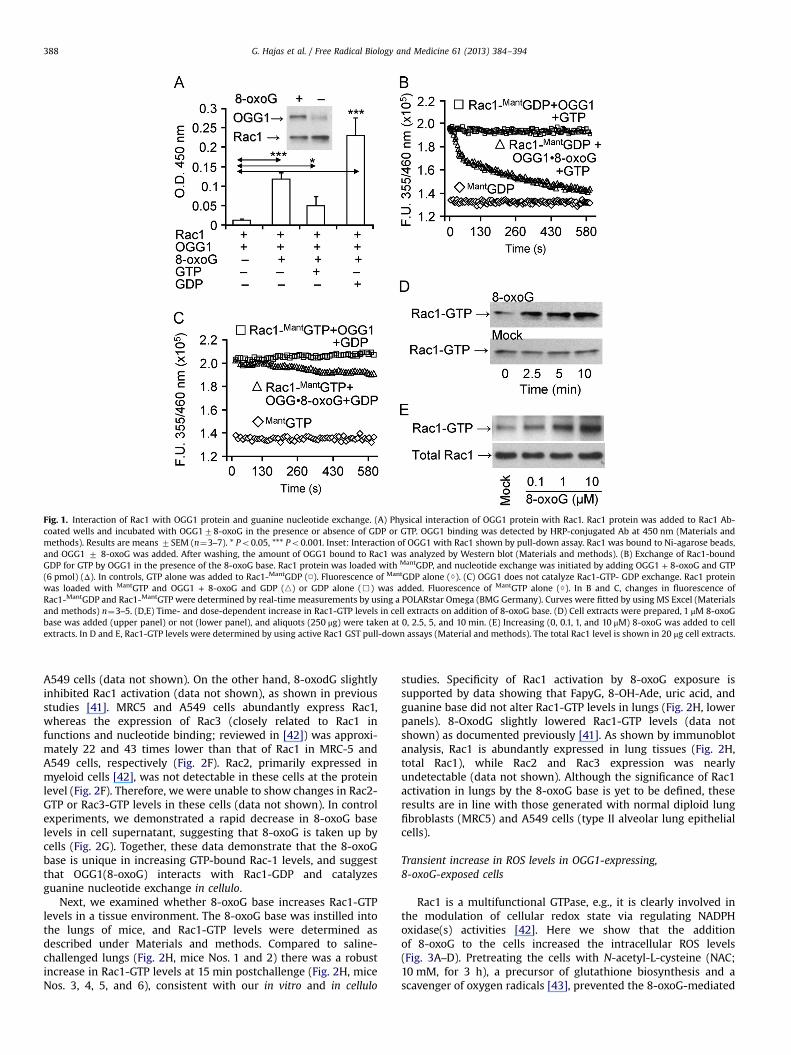
Fig. 1. Interaction of Rac1 with OGG1 protein and guanine nucleotide exchange. (A) Physical interaction of OGG1 protein with Rac1. Rac1 protein was added to Rac1 Ab-coated wells and incubated with OGG178-oxoG in the presence or absence of GDP or GTP. OGG1 binding was detected by HRP-conjugated Ab at 450 nm (Materials andmethods). Results are means 7SEM (n¼3–7). * Po0.05, *** Po0.001. Inset: Interaction of OGG1 with Rac1 shown by pull-down assay. Rac1 was bound to Ni-agarose beads,and OGG1 7 8-oxoG was added. After washing, the amount of OGG1 bound to Rac1 was analyzed by Western blot (Materials and methods). (B) Exchange of Rac1-boundGDP for GTP by OGG1 in the presence of the 8-oxoG base. Rac1 protein was loaded with MantGDP, and nucleotide exchange was initiated by adding OGG1 + 8-oxoG and GTP(6 pmol) (Δ). In controls, GTP alone was added to Rac1-MantGDP (□). Fluorescence of MantGDP alone (◊). (C) OGG1 does not catalyze Rac1-GTP- GDP exchange. Rac1 proteinwas loaded with MantGTP and OGG1 + 8-oxoG and GDP (n) or GDP alone (&) was added. Fluorescence of MantGTP alone (◊). In B and C, changes in fluorescence ofRac1-MantGDP and Rac1-MantGTP were determined by real-time measurements by using a POLARstar Omega (BMG Germany). Curves were fitted by using MS Excel (Materialsand methods) n¼3–5. (D,E) Time- and dose-dependent increase in Rac1-GTP levels in cell extracts on addition of 8-oxoG base. (D) Cell extracts were prepared, 1 μM 8-oxoGbase was added (upper panel) or not (lower panel), and aliquots (250 μg) were taken at 0, 2.5, 5, and 10 min. (E) Increasing (0, 0.1, 1, and 10 μM) 8-oxoG was added to cellextracts. In D and E, Rac1-GTP levels were determined by using active Rac1 GST pull-down assays (Material and methods). The total Rac1 level is shown in 20 μg cell extracts.
G. Hajas et al. / Free Radical Biology and Medicine 61 (2013) 384–394388
A549 cells (data not shown). On the other hand, 8-oxodG slightlyinhibited Rac1 activation (data not shown), as shown in previousstudies [41]. MRC5 and A549 cells abundantly express Rac1,whereas the expression of Rac3 (closely related to Rac1 infunctions and nucleotide binding; reviewed in [42]) was approxi-mately 22 and 43 times lower than that of Rac1 in MRC-5 andA549 cells, respectively (Fig. 2F). Rac2, primarily expressed inmyeloid cells [42], was not detectable in these cells at the proteinlevel (Fig. 2F). Therefore, we were unable to show changes in Rac2-GTP or Rac3-GTP levels in these cells (data not shown). In controlexperiments, we demonstrated a rapid decrease in 8-oxoG baselevels in cell supernatant, suggesting that 8-oxoG is taken up bycells (Fig. 2G). Together, these data demonstrate that the 8-oxoGbase is unique in increasing GTP-bound Rac-1 levels, and suggestthat OGG1(8-oxoG) interacts with Rac1-GDP and catalyzesguanine nucleotide exchange in cellulo.
Next, we examined whether 8-oxoG base increases Rac1-GTPlevels in a tissue environment. The 8-oxoG base was instilled intothe lungs of mice, and Rac1-GTP levels were determined asdescribed under Materials and methods. Compared to saline-challenged lungs (Fig. 2H, mice Nos. 1 and 2) there was a robustincrease in Rac1-GTP levels at 15 min postchallenge (Fig. 2H, miceNos. 3, 4, 5, and 6), consistent with our in vitro and in cellulo
studies. Specificity of Rac1 activation by 8-oxoG exposure issupported by data showing that FapyG, 8-OH-Ade, uric acid, andguanine base did not alter Rac1-GTP levels in lungs (Fig. 2H, lowerpanels). 8-OxodG slightly lowered Rac1-GTP levels (data notshown) as documented previously [41]. As shown by immunoblotanalysis, Rac1 is abundantly expressed in lung tissues (Fig. 2H,total Rac1), while Rac2 and Rac3 expression was nearlyundetectable (data not shown). Although the significance of Rac1activation in lungs by the 8-oxoG base is yet to be defined, theseresults are in line with those generated with normal diploid lungfibroblasts (MRC5) and A549 cells (type II alveolar lung epithelialcells).
Transient increase in ROS levels in OGG1-expressing,8-oxoG-exposed cells
Rac1 is a multifunctional GTPase, e.g., it is clearly involved inthe modulation of cellular redox state via regulating NADPHoxidase(s) activities [42]. Here we show that the additionof 8-oxoG to the cells increased the intracellular ROS levels(Fig. 3A–D). Pretreating the cells with N-acetyl-L-cysteine (NAC;10 mM, for 3 h), a precursor of glutathione biosynthesis and ascavenger of oxygen radicals [43], prevented the 8-oxoG-mediated

Fig. 2. Increased Rac1-GTP levels in OGG1-expressing cells on 8-oxoG exposures. (A,B) Changes in Rac1-GTP levels in MRC-5 (A) and A549 (B) cells. The 8-oxoG base (10 μM)was added to cells and extracts were made at indicated times points. Rac1-GTP levels were determined by GST pull-down assays (Materials and methods). Upper panels:Rac1-GTP levels were determined in 100 mg cell extract. Lower panel: total Rac1 protein levels in 10 μg cell lysate. n¼3–8. (C) Graphical depiction of the percentage of Rac-1GTP levels. Rac1-GTP band intensities were determined by densitometry (Image J 1.44), and the percentage changes were calculated by using MS Excel. (D) Depletion ofOGG1 expression decreased 8-oxoG exposure-induced Rac1-GTP levels. MRC-5 and A549 cells were transfected with OGG1 siRNA or control siRNA (Materials and methods)and then exposed to 8-oxoG (10 μM) for 10 min. Rac1-GTP levels were determined as in the legends to panels A and B. (E) siRNA-mediated decrease in OGG1 levels. GAPDH isshown for equal protein loading. (F) Expression of Rac family proteins in MRC-5 and A549 cells. Cells were lysed and protein levels were determined in 10 μg (Rac1) and25 μg (Rac2 and Rac3) extract per lane by immunoblotting. Results of a representative set of experiments are shown. GAPDH is shown for equal protein loading. (G) Cellularuptake of 8-oxoG base. The medium of MRC5 monolayers was replaced with PBS (pH 7.4), and 10 μM (final concentration) of 8-oxoG was added. Aliquots of the mediumweretaken at 0, 1, 30, and 60 min. In controls (□), 8-oxoG was added to PBS in the absence of cells. The 8-oxoG content in the aliquots was determined by GC/MS [40].(H) Activation of Rac1 by 8-oxoG challenge in lungs. The lungs of mice were challenged intranasally with saline 7 1 μM 8-oxoG. In controls, mice were challenged with 1 μMFapyG, 8-oxo-Ade, uric acid, or guanine. At 15 min thereafter Rac1-GTP levels were determined by GST pull-down assays in 125 μg lung extracts.
G. Hajas et al. / Free Radical Biology and Medicine 61 (2013) 384–394 389
increase in intracellular ROS levels (Fig. 3B). 8-OxoG inducedsignificantly lower levels of ROS in OGG1-depleted cells (Fig. 3C),consistent with decreased Rac1 activation (Fig. 2D). The 8-oxoGbase is unique in increasing ROS levels, because the guanine base,FapyG, 8-OH-Ade, adenine, and uric acid (deaminated form of8-oxoG) do not have this activity (Fig. 3D).
Treating the cells with DPI (10 mM, determined in preliminarystudies), an NADPH oxidase (NOX) inhibitor, before 8-oxoG addi-tion decreased cellular ROS levels by 6273 and 7077% in MRC-5and A549 cells, respectively, compared to cells exposed to 8-oxoGalone (Fig. 4A). p22phox is an essential regulatory subunit of NOX1-4 [42]. siRNA-mediated depletion of p22phox lowered ROS levels by6478% (MRC-5 cells) and 6976% (A549 cells) in 8-oxoG-exposedcells, which further confirmed the involvement of NOXs in ROSgeneration (Fig. 4B). siRNA to NOX 1, 2, and 3 only somewhat (datanot shown) decreased ROS levels, but NOX4 siRNA significantlydecreased (6779.5% for MRC5; 7674% for A549) ROS levels(Fig. 4C). Depletion of Rac1 decreased ROS levels by 7273% inMRC-5 and 6877% in A549 cells after 8-oxoG exposure (Fig. 4B).The siRNA-mediated decrease in Rac1, p22phox, and NOX4 expres-sion is shown in Fig. 4D.
NOX type 4 (NOX4) have been reported to be localized tocytoplasmic compartments and nuclear membranes, and areinvolved in multiple cellular processes, including localized redoxmodulation, and cellular signaling [42,44]. To determine thecellular localization of NOX4 that generate ROS in response to8-oxoG exposure, cells were transfected with the biosensor pHy-Pers [30,31], expressed in cytoplasm (pHyPer-Cyto) or targeted tonucleus (pHyPer-Nuc), and mitochondria (pHyPer-Mito). Seventy
two hours later cells were challenged with H2O2 (as control;10 μM) or 8-oxoG (10 μM). Intracellular sites of fluorescence wererecorded by microscopic imaging. As shown in Fig. 5A, H2O2-induced pHyPer signal was localized to cytoplasm, mitochondria,and nuclei of cells. When cells were exposed to 8-oxoG pHyPer-Nuc fluorescence appeared first at the perinuclear region (10 to12 min postexposure) and then a nuclear fluorescence wasobserved (�20 min). pHyPer-Nuc signal decreased to the basallevel by 60 min. pHyPer-Cyto-mediated fluorescence was initiatedaround the nuclei of cells (10 to 12 min after postexposure) andthen spread to the cytoplasm (20 min; Fig. 5). The pHyPer-Mitofluorescence appeared at 20 min after 8-oxoG addition. Interest-ingly, only few mitochondria showed fluorescence, suggesting thatpHyPer-Mit oxidation is a secondary event and possibly due toROS generated by NOX4. There were no increased fluorescence ofbiosensors observed at 60 min postexposure with 8-oxoG.Together these data suggest that NOX4 generating ROS is localizedto the nuclear membrane and 8-oxoG exposure-induced ROSgeneration is transient.
To obtain an insight on the close proximity of OGG1, Rac1, andNOX4 at perinuclear regions, we immunostained A549 cells byusing specific antibodies to these proteins. Fig. 6A, B, and C showthat OGG1 was primarily localized to the cell nuclei, but a fractionof it was associated with the nuclear membrane. Microscopicimaging showed that like NOX4, Rac1 is associated with thenuclear membrane. Lamin A/C antibody stained the nuclearmembrane exclusively (Fig. 6C). We utilized digital imaging toolsto examine the colocalization of OGG1 with Rac1 and NOX4 inimages obtained by fluorescence microscopy. The fluorescence

Fig. 3. Changes in ROS levels in OGG1-expressing cells after exposure to 8-oxoG base. (A) Kinetic changes in ROS levels in MRC-5 and A459 cells after 8-oxoG treatment. □,A549; ■, MRC-5; Δ, mock-treated A549; ♦, mock-treated MRC-5. (B) 8-OxoG base exposure failed to increase ROS levels in N-acetyl-L-cysteine (NAC)-pretreated cells.(C) Depletion of OGG1 prevented the 8-oxoG-mediated increase in ROS levels. Cells were transfected with OGG1 siRNA (Materials and methods) and exposed to 8-oxoG. ROSlevels were determined at 30 min. (D) Exposure of cells to FapyG, 8-oxo-Ade, adenine, guanine, or uric acid failed to change intracellular ROS levels. In panels A, B, C, and D,cells at 80% confluence were kept in 0.1% serum-containing media for 4 h, H2DCF-DA-loaded, and then exposed for 30 min before DCF fluorescence was assessed (Materialsand methods). 8-OxoG, 8-oxo-7,8-dihydroguanine; FapyG, 2,6-diamino-4-hydroxy-5-formamidopyrimidine; 8-oxoAde, 8-oxo-7,8-dihydroadenine. Results are means 7 SEM(n¼7–15) ***Po0.001, **** Po 0.0001.
G. Hajas et al. / Free Radical Biology and Medicine 61 (2013) 384–394390
intensities emitted by OGG1, Rac1, and NOX4 were different, so weemployed and calculated overlap coefficients according to Manders,a method which allows a reliable estimation of close proximitylocalization of proteins [38,39]. Manders’ formula (Materials andmethods) showed a close proximity localization of OGG1 and Rac1(Manders’ overlap¼0.935431; overlap coefficients k1¼0.886159and overlap coefficients k2¼0.987443) and NOX4 (Manders’ over-lap¼0.909245, overlap coefficients k1¼0.872817, and overlap coef-ficients k2¼0.980908). In controls, OGG1 was localized to thenuclear membrane, like lamin A/C; however, the Manders’equation (overlap¼0.898948, coefficients k1¼1.341340, coefficientsk2¼0.602463) showed that OGG1 and lamin A/C are not in closeproximity. The close proximity of OGG1 with Rac-1 (Fig. 6A) andNOX4 (Fig. 6B) is consistent with an increase in Rac1-GTP levels andthe observed nuclear membrane-associated ROS generation (Fig. 5).
Discussion
Among multiple types of DNA base damage generated by ROS,8-oxoG has received the most attention, as its accumulation in thegenome has been associated with increased mutations, variousdiseases, and aging processes. 8-OxoG is excised from DNA via theOGG1-initiated BER pathway, producing an 8-oxoG base which,when measured in serum and urine, is one of the best markers ofoxidative exposures. Here we show that in the presence of the8-oxoG base, OGG1 interacts with the GDP-bound Rac1 proteinand promotes the exchange of GDP for GTP in vitro. Importantly, inOGG1-expressing cells the 8-oxoG base increased Rac1-GTP levels.Activated Rac1 then facilitated a spatially controlled increase incellular ROS levels via NADPH oxidase type 4, which is associatedwith the nuclear membrane. These results suggest that OGG1, in
association with the 8-oxoG base, has a role in cellular physiolo-gical and/or pathophysiological signaling via the activation of Rac1.
Our recent study showed that OGG1 binds to the free 8-oxoG basewith high affinity [binding constant (Kd) of 0.5670.19 nm] in theabsence of duplex DNA [23], which was unexpected for binding to aproduct of an enzyme. It has also been shown that interaction ofOGG1 with 8-oxoG allowed its binding with canonical Ras familyproteins and, importantly, induced guanine nucleotide exchange [23].In the present study, OGG1 protein alone showed poor binding toguanine nucleotide-free (empty) Rac1 protein, but the 8-oxoG baseincreased this interaction. On the other hand, OGG1 showed poorinteractions with Rac1-GTP in the presence of 8-oxoG—however, lessthan with the empty protein. These observations resemble thoseshowing binding of OGG1(8-oxoG) with canonical Ras family pro-teins [23] and those reporting interaction between Rac1 and GEFssuch as T lymphoma invasion and metastasis (Tiam) protein [45], Sonof sevenless 1 (SOS1) [46], or Vav2 (a homolog of the vav proto-oncogene) [47]. The interaction of OGG1 with GDP-loaded Rac1suggested that 8-oxoG binding results in a conformational changewhich allows OGG1 to bind to Rac1. In support of this hypothesis, ithas been shown that GEFs require either posttranslational modifica-tion(s) or binding to regulatory molecules for interaction andcatalysis of guanine nucleotide exchange on Rac1 GTPases [48,49].Tiam1's interaction with Rac1 and its GEF activity are increased byassociation with phosphoinositides in its N-terminal pleckstrinhomology domain [50]. The GEF activity of Ras guanine nucleotiderelease factor 2 requires Ca2+/calmodulin binding to its IQ motifstructure [48,49]. The activity of Ras guanine nucleotide releaseprotein is regulated by Ca2+- and diacylglycerol [51]. Together thesedata suggest that the OGG1 protein is functionally similar to otherRac1 GEFs, as it requires binding of a cofactor (8-oxoG) to gain theproper conformation necessary for its binding to Rac1-GDP.
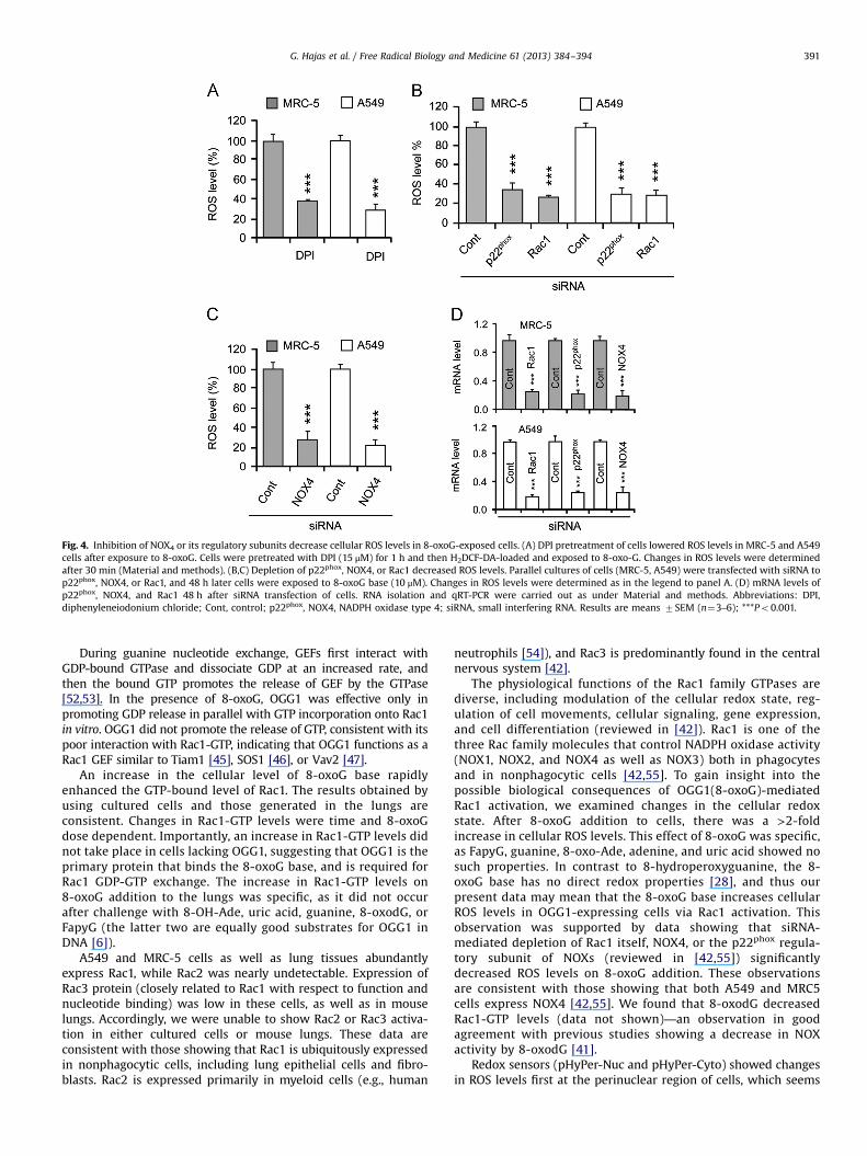
Fig. 4. Inhibition of NOX4 or its regulatory subunits decrease cellular ROS levels in 8-oxoG-exposed cells. (A) DPI pretreatment of cells lowered ROS levels in MRC-5 and A549cells after exposure to 8-oxoG. Cells were pretreated with DPI (15 μM) for 1 h and then H2DCF-DA-loaded and exposed to 8-oxo-G. Changes in ROS levels were determinedafter 30 min (Material and methods). (B,C) Depletion of p22phox, NOX4, or Rac1 decreased ROS levels. Parallel cultures of cells (MRC-5, A549) were transfected with siRNA top22phox, NOX4, or Rac1, and 48 h later cells were exposed to 8-oxoG base (10 μM). Changes in ROS levels were determined as in the legend to panel A. (D) mRNA levels ofp22phox, NOX4, and Rac1 48 h after siRNA transfection of cells. RNA isolation and qRT-PCR were carried out as under Material and methods. Abbreviations: DPI,diphenyleneiodonium chloride; Cont, control; p22phox, NOX4, NADPH oxidase type 4; siRNA, small interfering RNA. Results are means 7SEM (n¼3–6); ***Po0.001.
G. Hajas et al. / Free Radical Biology and Medicine 61 (2013) 384–394 391
During guanine nucleotide exchange, GEFs first interact withGDP-bound GTPase and dissociate GDP at an increased rate, andthen the bound GTP promotes the release of GEF by the GTPase[52,53]. In the presence of 8-oxoG, OGG1 was effective only inpromoting GDP release in parallel with GTP incorporation onto Rac1in vitro. OGG1 did not promote the release of GTP, consistent with itspoor interaction with Rac1-GTP, indicating that OGG1 functions as aRac1 GEF similar to Tiam1 [45], SOS1 [46], or Vav2 [47].
An increase in the cellular level of 8-oxoG base rapidlyenhanced the GTP-bound level of Rac1. The results obtained byusing cultured cells and those generated in the lungs areconsistent. Changes in Rac1-GTP levels were time and 8-oxoGdose dependent. Importantly, an increase in Rac1-GTP levels didnot take place in cells lacking OGG1, suggesting that OGG1 is theprimary protein that binds the 8-oxoG base, and is required forRac1 GDP-GTP exchange. The increase in Rac1-GTP levels on8-oxoG addition to the lungs was specific, as it did not occurafter challenge with 8-OH-Ade, uric acid, guanine, 8-oxodG, orFapyG (the latter two are equally good substrates for OGG1 inDNA [6]).
A549 and MRC-5 cells as well as lung tissues abundantlyexpress Rac1, while Rac2 was nearly undetectable. Expression ofRac3 protein (closely related to Rac1 with respect to function andnucleotide binding) was low in these cells, as well as in mouselungs. Accordingly, we were unable to show Rac2 or Rac3 activa-tion in either cultured cells or mouse lungs. These data areconsistent with those showing that Rac1 is ubiquitously expressedin nonphagocytic cells, including lung epithelial cells and fibro-blasts. Rac2 is expressed primarily in myeloid cells (e.g., human
neutrophils [54]), and Rac3 is predominantly found in the centralnervous system [42].
The physiological functions of the Rac1 family GTPases arediverse, including modulation of the cellular redox state, reg-ulation of cell movements, cellular signaling, gene expression,and cell differentiation (reviewed in [42]). Rac1 is one of thethree Rac family molecules that control NADPH oxidase activity(NOX1, NOX2, and NOX4 as well as NOX3) both in phagocytesand in nonphagocytic cells [42,55]. To gain insight into thepossible biological consequences of OGG1(8-oxoG)-mediatedRac1 activation, we examined changes in the cellular redoxstate. After 8-oxoG addition to cells, there was a >2-foldincrease in cellular ROS levels. This effect of 8-oxoG was specific,as FapyG, guanine, 8-oxo-Ade, adenine, and uric acid showed nosuch properties. In contrast to 8-hydroperoxyguanine, the 8-oxoG base has no direct redox properties [28], and thus ourpresent data may mean that the 8-oxoG base increases cellularROS levels in OGG1-expressing cells via Rac1 activation. Thisobservation was supported by data showing that siRNA-mediated depletion of Rac1 itself, NOX4, or the p22phox regula-tory subunit of NOXs (reviewed in [42,55]) significantlydecreased ROS levels on 8-oxoG addition. These observationsare consistent with those showing that both A549 and MRC5cells express NOX4 [42,55]. We found that 8-oxodG decreasedRac1-GTP levels (data not shown)—an observation in goodagreement with previous studies showing a decrease in NOXactivity by 8-oxodG [41].
Redox sensors (pHyPer-Nuc and pHyPer-Cyto) showed changesin ROS levels first at the perinuclear region of cells, which seems
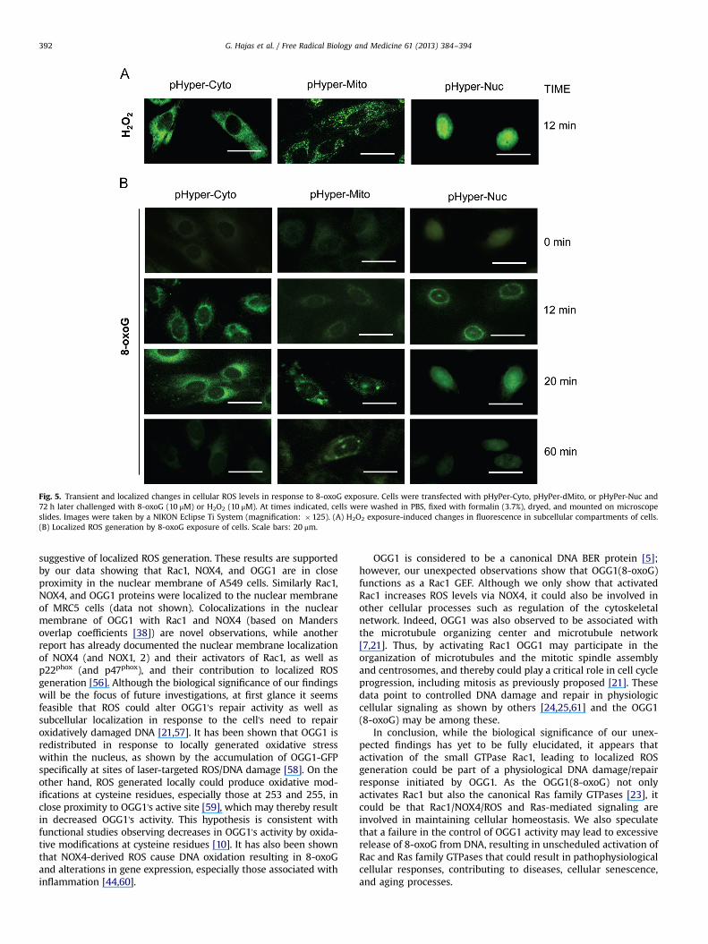
Fig. 5. Transient and localized changes in cellular ROS levels in response to 8-oxoG exposure. Cells were transfected with pHyPer-Cyto, pHyPer-dMito, or pHyPer-Nuc and72 h later challenged with 8-oxoG (10 μM) or H2O2 (10 μM). At times indicated, cells were washed in PBS, fixed with formalin (3.7%), dryed, and mounted on microscopeslides. Images were taken by a NIKON Eclipse Ti System (magnification: �125). (A) H2O2 exposure-induced changes in fluorescence in subcellular compartments of cells.(B) Localized ROS generation by 8-oxoG exposure of cells. Scale bars: 20 μm.
G. Hajas et al. / Free Radical Biology and Medicine 61 (2013) 384–394392
suggestive of localized ROS generation. These results are supportedby our data showing that Rac1, NOX4, and OGG1 are in closeproximity in the nuclear membrane of A549 cells. Similarly Rac1,NOX4, and OGG1 proteins were localized to the nuclear membraneof MRC5 cells (data not shown). Colocalizations in the nuclearmembrane of OGG1 with Rac1 and NOX4 (based on Mandersoverlap coefficients [38]) are novel observations, while anotherreport has already documented the nuclear membrane localizationof NOX4 (and NOX1, 2) and their activators of Rac1, as well asp22phox (and p47phox), and their contribution to localized ROSgeneration [56]. Although the biological significance of our findingswill be the focus of future investigations, at first glance it seemsfeasible that ROS could alter OGG1's repair activity as well assubcellular localization in response to the cell's need to repairoxidatively damaged DNA [21,57]. It has been shown that OGG1 isredistributed in response to locally generated oxidative stresswithin the nucleus, as shown by the accumulation of OGG1-GFPspecifically at sites of laser-targeted ROS/DNA damage [58]. On theother hand, ROS generated locally could produce oxidative mod-ifications at cysteine residues, especially those at 253 and 255, inclose proximity to OGG1's active site [59], which may thereby resultin decreased OGG1's activity. This hypothesis is consistent withfunctional studies observing decreases in OGG1's activity by oxida-tive modifications at cysteine residues [10]. It has also been shownthat NOX4-derived ROS cause DNA oxidation resulting in 8-oxoGand alterations in gene expression, especially those associated withinflammation [44,60].
OGG1 is considered to be a canonical DNA BER protein [5];however, our unexpected observations show that OGG1(8-oxoG)functions as a Rac1 GEF. Although we only show that activatedRac1 increases ROS levels via NOX4, it could also be involved inother cellular processes such as regulation of the cytoskeletalnetwork. Indeed, OGG1 was also observed to be associated withthe microtubule organizing center and microtubule network[7,21]. Thus, by activating Rac1 OGG1 may participate in theorganization of microtubules and the mitotic spindle assemblyand centrosomes, and thereby could play a critical role in cell cycleprogression, including mitosis as previously proposed [21]. Thesedata point to controlled DNA damage and repair in physiologiccellular signaling as shown by others [24,25,61] and the OGG1(8-oxoG) may be among these.
In conclusion, while the biological significance of our unex-pected findings has yet to be fully elucidated, it appears thatactivation of the small GTPase Rac1, leading to localized ROSgeneration could be part of a physiological DNA damage/repairresponse initiated by OGG1. As the OGG1(8-oxoG) not onlyactivates Rac1 but also the canonical Ras family GTPases [23], itcould be that Rac1/NOX4/ROS and Ras-mediated signaling areinvolved in maintaining cellular homeostasis. We also speculatethat a failure in the control of OGG1 activity may lead to excessiverelease of 8-oxoG from DNA, resulting in unscheduled activation ofRac and Ras family GTPases that could result in pathophysiologicalcellular responses, contributing to diseases, cellular senescence,and aging processes.

Fig. 6. Colocalization of Rac1 and OGG1 with NADPH oxidase 4 in nuclear membranes. A549 cells were cultured on microscope coverslips, and then incubated with Abs toOGG1, Rac1 (A), and/or NOX4 (B). Panel C shows OGG1's localization to the nuclear membrane based on Lamin A/C. Binding of primary Abs was visualized by using Alexa488- and Alexa 594-conjugated secondary Abs. Staining of cells was carried out as described under Materials and methods. Images were captured with a NIKON Eclipse TiSystem (magnification: 125). Colocalization was visualized by superimposition of green and red images using Nikon NIS Elements Version 3.5 (NIKON Instruments, Tokyo,Japan). Scale bars: 20 μm. Manders’ overlap coefficient is 0.935431 for OGG1 and Rac1 (upper right panel); 0.909245 for OGG1 and NOX4 (middle right panel). DAPI, 4′6-diamidino-2-phenylindole dihydrochloride.
G. Hajas et al. / Free Radical Biology and Medicine 61 (2013) 384–394 393
Conflicts of interest
The authors declare that there are no conflicts of interest.
Acknowledgments
This work was supported by grants NIEHS RO1 ES018948 (I.B.),NIA/AG 021830 (I.B.), NIAID/AI062885 (I.B.), NINDS R01NS073976(T.H.K., I.B.), and the NHLBI Proteomic Center, N01HV00245(Dr. A. Kurosky). L. Aguilera-Aguirre is an Environmental ToxicologyResearch Training Fellow (NIEHS T32 ES007254). The work wasalso supported by the TAMOP 4.2.2.A-11/1/KONV-2012-2023 pro-ject (A.B.), which is cofinanced by the European Union and theEuropean Social Fund. A.B. was also supported by the Janos BolyaiFellowship from the Hungarian Academy of Sciences. We thankDrs. Miral Dizdaroglu and Pawel Jaruga (Chemical Science andTechnology Laboratory, National Institute of Standards andTechnology, Gaithersburg, MD USA) for GC/MS measurements of the8-oxoG and for the gift of FapyG. We thank Drs. Sankar Mitra andMiral Dizdaroglu for their scientific input, and Dr. David Konkel(Department of Biochemistry and Molecular Biology) and MardelleSusman (Department of Microbiology and Immunology) forcritically editing the manuscript. We also thank the anonymousreviewers for their suggestions, thoughts, and concept, whichimproved the quality of our paper.
References
[1] de Moura, M. B.; dos Santos, L. S.; Van Houten, B. Mitochondrial dysfunction inneurodegenerative diseases and cancer. Environ. Mol. Mutagen. 51:391–405;2010.
[2] D'Autreaux, B.; Toledano, M. B. ROS as signalling molecules: mechanisms thatgenerate specificity in ROS homeostasis. Nat. Rev. Mol. Cell. Biol. 8:813–824; 2007.
[3] Steenken, S. Purine bases, nucleosides, and nucleotides: aqueous solutionredox chemistry and transformation reactions of their radical cations ande− and OH adducts. Chem. Rev. 89:503–520; 1989.
[4] Radak, Z.; Boldogh, I. 8-Oxo-7,8-dihydroguanine: links to gene expression,aging, and defense against oxidative stress. Free Radic. Biol. Med. 49:587–596;2010.
[5] Mitra, S.; Izumi, T.; Boldogh, I.; Bhakat, K. K.; Hill, J. W.; Hazra, T. K.Choreography of oxidative damage repair in mammalian genomes. Free Radic.Biol. Med. 33:15–28; 2002.
[6] Dizdaroglu, M.; Kirkali, G.; Jaruga, P. Formamidopyrimidines in DNA: mechan-isms of formation, repair, and biological effects. Free Radic. Biol. Med.45:1610–1621; 2008.
[7] Dantzer, F.; Luna, L.; Bjoras, M.; Seeberg, E. Human OGG1 undergoes serinephosphorylation and associates with the nuclear matrix and mitotic chroma-tin in vivo. Nucleic Acids Res. 30:2349–2357; 2002.
[8] Bhakat, K. K.; Mokkapati, S. K.; Boldogh, I.; Hazra, T. K.; Mitra, S. Acetylation ofhuman 8-oxoguanine-DNA glycosylase by p300 and its role in 8-oxoguaninerepair in vivo. Mol. Cell. Biol. 26:1654–1665; 2006.
[9] Hegde, ML; Rao KS, H. P.; Mitra, S. Oxidative genome damage and its repair inneurodegenerative diseases: function of transition metals as a double-edgedsword. J. Alzheimers Dis. 24:183–198; 2011.
[10] Bravard, A.; Vacher, M.; Gouget, B.; Coutant, A.; de Boisferon, F. H.; Marsin, S.;Chevillard, S.; Radicella, J. P. Redox regulation of human OGG1 activity inresponse to cellular oxidative stress. Mol. Cell. Biol. 26:7430–7436; 2006.
[11] David, S. S.; O'Shea, V. L.; Kundu, S. Base-excision repair of oxidative DNAdamage. Nature 447:941–950; 2007.
[12] Markesbery, W. R.; Lovell, M. A. DNA oxidation in Alzheimer's disease.Antioxid. Redox Signal. 8:2039–2045; 2006.
[13] Radak, Z.; Bori, Z.; Koltai, E.; Fatouros, I. G.; Jamurtas, A. Z.; Douroudos, II;Terzis, G.; Nikolaidis, M. G.; Chatzinikolaou, A.; Sovatzidis, A.; Kumagai, S.;Naito, H.; Boldogh, I. Age-dependent changes in 8-oxoguanine-DNA glycosy-lase activity are modulated by adaptive responses to physical exercise inhuman skeletal muscle. Free Radic. Biol. Med. 51:417–423; 2011.
[14] Nishimura, S. Involvement of mammalian OGG1(MMH) in excision of the8-hydroxyguanine residue in DNA. Free Radic. Biol. Med. 32:813–821; 2002.
[15] Klungland, A.; Rosewell, I.; Hollenbach, S.; Larsen, E.; Daly, G.; Epe, B.; Seeberg,E.; Lindahl, T.; Barnes, D. E. Accumulation of premutagenic DNA lesions in micedefective in removal of oxidative base damage. Proc. Natl. Acad. Sci. USA96:13300–13305; 1999.

G. Hajas et al. / Free Radical Biology and Medicine 61 (2013) 384–394394
[16] Minowa, O.; Arai, T.; Hirano, M.; Monden, Y.; Nakai, S.; Fukuda, M.; Itoh, M.;Takano, H.; Hippou, Y.; Aburatani, H.; Masumura, K.; Nohmi, T.; Nishimura, S.;Noda, T. Mmh/Ogg1 gene inactivation results in accumulation of 8-hydroxyguanine in mice. Proc. Natl. Acad. Sci. USA 97:4156–4161; 2000.
[17] Mabley, J. G.; Pacher, P.; Deb, A.; Wallace, R.; Elder, R. H.; Szabo, C. Potentialrole for 8-oxoguanine DNA glycosylase in regulating inflammation. FASEB J.19:290–292; 2005.
[18] Li, G.; Yuan, K.; Yan, C.; Fox 3rd J.; Gaid, M.; Breitwieser, W.; Bansal, A. K.; Zeng,H.; Gao, H.; Wu, M. 8-Oxoguanine-DNA glycosylase 1 deficiency modifiesallergic airway inflammation by regulating STAT6 and IL-4 in cells and in mice.Free Radic. Biol. Med. 52:392–401; 2012.
[19] Kovtun, I. V.; Liu, Y.; Bjoras, M.; Klungland, A.; Wilson, S. H.; McMurray, C. T.OGG1 initiates age-dependent CAG trinucleotide expansion in somatic cells.Nature 447:447–452; 2007.
[20] Mitra, S. DNA glycosylases: specificity and mechanisms. Prog. Nucleic Acid Res.Mol. Biol. 68:189–192; 2001.
[21] Conlon, K. A.; Zharkov, D. O.; Berrios, M. Cell cycle regulation of themurine 8-oxoguanine DNA glycosylase (mOGG1): mOGG1 associates withmicrotubules during interphase and mitosis. DNA Repair (Amst.) 3:1601–1615;2004.
[22] Szczesny, B.; Hazra, T. K.; Papaconstantinou, J.; Mitra, S.; Boldogh, I. Age-dependent deficiency in import of mitochondrial DNA glycosylases required forrepair of oxidatively damaged bases. Proc. Natl. Acad. Sci. USA 100:10670–-10675; 2003.
[23] Boldogh, I.; Hajas, G.; Aguilera-Aguirre, L.; Hegde, M. L.; Radak, Z.; Bacsi, A.;Sur, S.; Hazra, T. K.; Mitra, S. Activation of ras signaling pathway by8-oxoguanine DNA glycosylase bound to its excision product, 8-oxoguanine.J. Biol. Chem. 287:20769–20773; 2012.
[24] Perillo, B.; Ombra, M. N.; Bertoni, A.; Cuozzo, C.; Sacchetti, S.; Sasso, A.;Chiariotti, L.; Malorni, A.; Abbondanza, C.; Avvedimento, E. V. DNA oxidationas triggered by H3K9me2 demethylation drives estrogen-induced geneexpression. Science 319:202–206; 2008.
[25] Amente, S.; Bertoni, A.; Morano, A.; Lania, L.; Avvedimento, E. V.; Majello, B.LSD1-mediated demethylation of histone H3 lysine 4 triggers Myc-inducedtranscription. Oncogene 29:3691–3702; 2011.
[26] Kemp, M. G.; Reardon, J. T.; Lindsey-Boltz, L. A.; Sancar, A. Mechanism ofrelease and fate of excised oligonucleotides during nucleotide excision repair.J. Biol. Chem. 287; 2012.
[27] Napirei, M.; Karsunky, H.; Zevnik, B.; Stephan, H.; Mannherz, H. G.; Moroy, T.Features of systemic lupus erythematosus in Dnase1-deficient mice. Nat.Genet. 25:177–181; 2000.
[28] Hajas, G.; Bacsi, A.; Aguilerra-Aguirre, L.; German, P.; Radak, Z.; Sur, S.; Hazra,T. K.; Boldogh, I. Biochemical identification of a hydroperoxide derivative ofthe free 8-oxo-7,8-dihydroguanine base. Free Radic. Biol. Med. 52:749–756;2012.
[29] Boldogh, I.; Bacsi, A.; Choudhury, B. K.; Dharajiya, N.; Alam, R.; Hazra, T. K.;Mitra, S.; Goldblum, R. M.; Sur, S. ROS generated by pollen NADPH oxidaseprovide a signal that augments antigen-induced allergic airway inflammation.J. Clin. Invest. 115:2169–2179; 2005.
[30] Belousov, V. V.; Fradkov, A. F.; Lukyanov, K. A.; Staroverov, D. B.; Shakhbazov,K. S.; Terskikh, A. V.; Lukyanov, S. Genetically encoded fluorescent indicatorfor intracellular hydrogen peroxide. Nat. Methods 3:281–286; 2006.
[31] Malinouski, M.; Zhou, Y.; Belousov, V. V.; Hatfield, D. L.; Gladyshev, V. N.Hydrogen peroxide probes directed to different cellular compartments. PLoSOne 6:e14564; 2011.
[32] Bacsi, A.; Chodaczek, G.; Hazra, T. K.; Konkel, D.; Boldogh, I. Increased ROSgeneration in subsets of OGG1 knockout fibroblast cells. Mech. Ageing Dev.128:637–649; 2007.
[33] Qin, J.; Xie, Y.; Wang, B.; Hoshino, M.; Wolff, D. W.; Zhao, J.; Scofield, M. A.;Dowd, F. J.; Lin, M. F.; Tu, Y. Upregulation of PIP3-dependent Rac exchanger 1(P-Rex1) promotes prostate cancer metastasis. Oncogene 28:1853–1863; 2009.
[34] Benard, V.; Bokoch, G. M. Assay of Cdc42, Rac, and Rho GTPase activation byaffinity methods. Methods Enzymol. 345:349–359; 2002.
[35] Zhang, B.; Zhang, Y.; Wang, Z.; Zheng, Y. The role of Mg2+ cofactor in theguanine nucleotide exchange and GTP hydrolysis reactions of Rho familyGTP-binding proteins. J. Biol. Chem 275:25299–25307; 2000.
[36] Livak, K. J.; Schmittgen, T. D. Analysis of relative gene expression data usingreal-time quantitative PCR and the 2(-Delta Delta C(T)) method. Methods25:402–408; 2001.
[37] Aguilera-Aguirre, L.; Bacsi, A.; Saavedra-Molina, A.; Kurosky, A.; Sur, S.;Boldogh, I. Mitochondrial dysfunction increases allergic airway inflammation.J. Immunol. 183:5379–5387; 2009.
[38] Manders, E. M.; Verbeek, F. J.; Aten, J. A. Measurement of co-localization ofobjects in dual-colour confocal images. J. Microscopy 169:375–382; 1993.
[39] Zinchuk, V.; Zinchuk, O.; Okada, T. Quantitative colocalization analysis ofmulticolor confocal immunofluorescence microscopy images: pushing pixelsto explore biological phenomena. Acta Histochem. Cytochem. 40:101–111; 2007.
[40] Dizdaroglu, M.; Jaruga, P.; Rodriguez, H. Measurement of 8-hydroxy-2’-deoxyguanosine in DNA by high-performance liquid chromatography-massspectrometry: comparison with measurement by gas chromatography-massspectrometry. Nucleic Acids Res. 29:E12; 2001.
[41] Kim, H. J.; Yoon, S. H.; Ryu, H. O.; Yoon, B. H.; Choi, S.; Ye, S. K.; Chung, M. H.8-Oxo-7,8-dihydroguanosine triphosphate(8-oxoGTP) down-regulates respira-tory burst of neutrophils by antagonizing GTP toward Rac, a small GTP bindingprotein. Free Radic. Res. 41:655–662; 2007.
[42] Bedard, K.; Krause, K. H. The NOX family of ROS-generating NADPH oxidases:physiology and pathophysiology. Physiol. Rev. 87:245–313; 2007.
[43] Das, G. C.; Bacsi, A.; Shrivastav, M.; Hazra, T. K.; Boldogh, I. Enhanced gamma-glutamylcysteine synthetase activity decreases drug-induced oxidative stresslevels and cytotoxicity. Mol. Carcinog. 45:635–647; 2006.
[44] Gordillo, G.; Fang, H.; Park, H.; Roy, S. Nox-4-dependent nuclear H2O2 drivesDNA oxidation resulting in 8-OHdG as urinary biomarker and hemangio-endothelioma formation. Antioxid. Redox Signal 12:933–943; 2010.
[45] Haeusler, L. C.; Blumenstein, L.; Stege, P.; Dvorsky, R.; Ahmadian, M. R. Comparativefunctional analysis of the Rac GTPases. FEBS Lett. 555:556–560; 2003.
[46] Khanday, F. A.; Santhanam, L.; Kasuno, K.; Yamamori, T.; Naqvi, A.; Dericco, J.;Bugayenko, A.; Mattagajasingh, I.; Disanza, A.; Scita, G.; Irani, K. Sos-mediatedactivation of rac1 by p66shc. J. Cell Biol. 172:817–822; 2006.
[47] Sauzeau, V.; Sevilla, M. A.; Montero, M. J.; Bustelo, X. R. The Rho/Rac exchangefactor Vav2 controls nitric oxide-dependent responses in mouse vascularsmooth muscle cells. J. Clin. Invest. 120:315–330; 2010.
[48] Fan, W. T.; Koch, C. A.; de Hoog, C. L.; Fam, N. P.; Moran, M. F. The exchangefactor Ras-GRF2 activates Ras-dependent and Rac-dependent mitogen-activated protein kinase pathways. Curr. Biol. 8:935–938; 1998.
[49] Innocenti, M.; Zippel, R.; Brambilla, R.; Sturani, E. CDC25(Mm)/Ras-GRF1regulates both Ras and Rac signaling pathways. FEBS Lett. 460:357–362; 1999.
[50] Fleming, I. N.; Gray, A.; Downes, C. P. Regulation of the Rac1-specific exchangefactor Tiam1 involves both phosphoinositide 3-kinase-dependent and -inde-pendent components. Biochem. J. 351:173–182; 2000.
[51] Buday, L.; Downward, J. Many faces of Ras activation. Biochim. Biophys. Acta1786:178–187; 2008.
[52] Bourne, H. R.; Sanders, D. A.; McCormick, F. The GTPase superfamily:a conserved switch for diverse cell functions. Nature 348:125–132; 1990.
[53] Boriack-Sjodin, P. A.; Margarit, S. M.; Bar-Sagi, D.; Kuriyan, J. The structuralbasis of the activation of Ras by Sos. Nature 394:337–343; 1998.
[54] Hordijk, P. L. Regulation of NADPH oxidases: the role of Rac proteins. Circ. Res.98:453–462; 2006.
[55] Lambeth, J. D.; Kawahara, T.; Diebold, B. Regulation of Nox and Duoxenzymatic activity and expression. Free Radic. Biol. Med. 43:319–331; 2007.
[56] Spencer, N. Y.; Yan, Z.; Boudreau, R. L.; Zhang, Y.; Luo, M.; Li, Q.; Tian, X.; Shah,A. M.; Davisson, R. L.; Davidson, B.; Banfi, B.; Engelhardt, J. F. Control of hepaticnuclear superoxide production by glucose 6-phosphate dehydrogenase andNADPH oxidase-4. J. Biol. Chem. 286:8977–8987; 2011.
[57] Szczesny, B.; Bhakat, K. K.; Mitra, S.; Boldogh, I. Age-dependent modulation ofDNA repair enzymes by covalent modification and subcellular distribution.Mech. Ageing Dev. 125:755–765; 2004.
[58] Zielinska, A.; Davies, O. T.; Meldrum, R. A.; Hodges, N. J. Direct visualization ofrepair of oxidative damage by OGG1 in the nuclei of live cells. J. Biochem Mol.Toxicol. 25:1–7; 2011.
[59] Qi, Y.; Spong, M. C.; Nam, K.; Banerjee, A.; Jiralerspong, S.; Karplus, M.;Verdine, G. L. Encounter and extrusion of an intrahelical lesion by a DNA repairenzyme. Nature 462:762–766; 2009.
[60] Weyemi, U.; Dupuy, C. The emerging role of ROS-generating NADPH oxidaseNOX4 in DNA-damage responses. Mutat. Res. 751:77–81; 2012.
[61] Al-Mehdi, A.B.; Pastukh, V.M.; Swiger, B.M.; Reed, D.J.; Patel, M.R.; Bardwell, G.C.; Pastukh, V.V.; Alexeyev, M.F.; Gillespie, M.N. Perinuclear mitochondrialclustering creates an oxidant-rich nuclear domain required for hypoxia-induced transcription. Sci. Signal. 5:ra47; 2012.
Copyright © 2022 FDOKUMEN