Chapter 4 Studying DNA
25
4/8/2020 Studying DNA - Genomes - NCBI Bookshelf https://www.ncbi.nlm.nih.gov/books/NBK21129/ 1/25 NCBI Bookshelf. A service of the National Library of Medicine, National Institutes of Health. Brown TA. Genomes. 2nd edition. Oxford: Wiley-Liss; 2002. Chapter 4 Studying DNA Learning outcomes When you have read Chapter 4, you should be able to: Give outline descriptions of the events involved in DNA cloning and the polymerase chain reaction ( PCR), and state the applications and limitations of these techniques Describe the activities and main applications of the different types of enzyme used in recombinant DNA research Identify the important features of DNA polymerases and distinguish between the various DNA polymerases used in genomics research Describe, with examples, the way that restriction endonucleases cut DNA and explain how the results of a restriction digest are examined Distinguish between blunt- and sticky-end ligation and explain how the efficiency of blunt- end ligation can be increased Give details of the key features of plasmid cloning vectors and describe how these vectors are used in cloning experiments, using pBR322 and pUC8 as examples Describe how bacteriophage λ vectors are used to clone DNA Give examples of vectors used to clone long pieces of DNA, and evaluate the strengths and weaknesses of each type Summarize how DNA is cloned in yeast, animals and plants Describe how a PCR is performed, paying particular attention to the importance of the primers and the temperatures used during the thermal cycling used by molecular biologists to study DNA molecules was assembled during the 1970s and 1980s. Before then, the only way in which individual genes could be studied was by classical genetics, using the procedures that we will examine in Chapter 5. Classical genetics is a powerful approach to gene analysis and many of the fundamental discoveries in molecular biology were made in this way. The operon theory proposed by Jacob and Monod in 1961 ( Section 9.3.1), which describes how the expression of some bacterial genes is regulated, was perhaps the most heroic achievement of this era of genetics. But the classical approach is limited because it does not involve the direct examination of genes, information on gene structure and activity being inferred from the biological characteristics of the organism being studied. By the late 1960s these indirect methods had become insufficient for answering the more detailed questions that molecular biologists had begun to ask about the expression pathways of individual genes. These questions could only be addressed by examining directly the segments of DNA containing the genes of interest. This was not possible using the current technology, so a new set of techniques had to be invented. The development of these new techniques was stimulated by breakthroughs in biochemical research which, in the early 1970s, provided molecular biologists with enzymes that could be
-
Upload
khangminh22 -
Category
Documents
-
view
2 -
download
0
Transcript of Chapter 4 Studying DNA

4/8/2020 Studying DNA - Genomes - NCBI Bookshelf
https://www.ncbi.nlm.nih.gov/books/NBK21129/ 1/25
NCBI Bookshelf. A service of the National Library of Medicine, National Institutes of Health.
Brown TA. Genomes. 2nd edition. Oxford: Wiley-Liss; 2002.
Chapter 4 Studying DNA
Learning outcomesWhen you have read Chapter 4, you should be able to:
Give outline descriptions of the events involved in DNA cloning and the polymerase chainreaction (PCR), and state the applications and limitations of these techniques
Describe the activities and main applications of the different types of enzyme used inrecombinant DNA research
Identify the important features of DNA polymerases and distinguish between the variousDNA polymerases used in genomics research
Describe, with examples, the way that restriction endonucleases cut DNA and explain howthe results of a restriction digest are examined
Distinguish between blunt- and sticky-end ligation and explain how the efficiency of blunt-end ligation can be increased
Give details of the key features of plasmid cloning vectors and describe how these vectorsare used in cloning experiments, using pBR322 and pUC8 as examples
Describe how bacteriophage λ vectors are used to clone DNA
Give examples of vectors used to clone long pieces of DNA, and evaluate the strengthsand weaknesses of each type
Summarize how DNA is cloned in yeast, animals and plants
Describe how a PCR is performed, paying particular attention to the importance of theprimers and the temperatures used during the thermal cycling
��� ������� �� ���������� used by molecular biologists to study DNA molecules wasassembled during the 1970s and 1980s. Before then, the only way in which individual genescould be studied was by classical genetics, using the procedures that we will examine in Chapter5. Classical genetics is a powerful approach to gene analysis and many of the fundamentaldiscoveries in molecular biology were made in this way. The operon theory proposed by Jacoband Monod in 1961 (Section 9.3.1), which describes how the expression of some bacterial genesis regulated, was perhaps the most heroic achievement of this era of genetics. But the classicalapproach is limited because it does not involve the direct examination of genes, information ongene structure and activity being inferred from the biological characteristics of the organismbeing studied. By the late 1960s these indirect methods had become insufficient for answeringthe more detailed questions that molecular biologists had begun to ask about the expressionpathways of individual genes. These questions could only be addressed by examining directly thesegments of DNA containing the genes of interest. This was not possible using the currenttechnology, so a new set of techniques had to be invented.
The development of these new techniques was stimulated by breakthroughs in biochemicalresearch which, in the early 1970s, provided molecular biologists with enzymes that could be

4/8/2020 Studying DNA - Genomes - NCBI Bookshelf
https://www.ncbi.nlm.nih.gov/books/NBK21129/ 2/25
used to manipulate DNA molecules in the test tube. These enzymes occur naturally in living cellsand are involved in processes such as DNA replication, repair and recombination (see Chapters13 and 14). In order to determine the functions of these enzymes, many of them were purifiedand the reactions that they catalyze studied in the test tube. Molecular biologists then adopted thepure enzymes as tools for manipulating DNA molecules in pre-determined ways, using them tomake copies of DNA molecules, to cut DNA molecules into shorter fragments, and to join themtogether again in combinations that do not exist in nature (Figure 4.1). These manipulations,which are described in Section 4.1, form the basis of recombinant DNA technology, in whichnew or ‘recombinant’ DNA molecules are constructed in the test tube from pieces of naturallyoccurring chromosomes and plasmids. Recombinant DNA methodology led to the developmentof DNA or gene cloning, in which short DNA fragments, possibly containing a single gene, areinserted into a plasmid or virus chromosome and then replicated in a bacterial or eukaryotic host(Figure 4.2). We will examine exactly how gene cloning is performed, and the reasons why thistechnique resulted in a revolution in molecular biology, in Section 4.2.
Figure 4.1
Examples of the manipulations that can be carried out withDNA molecules.
Figure 4.2
DNA cloning. In this example, the fragment of DNA to becloned is inserted into a plasmid vector which is subsequentlyreplicated inside a bacterial host.
Gene cloning was well established by the end of the 1970s. The next major technicalbreakthrough came some 5 years later when the polymerase chain reaction (PCR) was invented(Mullis, 1990). PCR is not a complicated technique - all that it achieves is the repeated copyingof a short segment of a DNA molecule (Figure 4.3) - but it has become immensely important inmany areas of biological research, not least the study of genomes. PCR is covered in detail inSection 4.3.
Figure 4.3
The polymerase chain reaction (PCR) is used to make copiesof a selected segment of a DNA molecule. In this example, asingle gene is copied.
4.1. Enzymes for DNA ManipulationRecombinant DNA technology was one of the main factors that contributed to the rapid advancein knowledge concerning gene expression that occurred during the 1970s and 1980s. The basis ofrecombinant DNA technology is the ability to manipulate DNA molecules in the test tube. This,in turn, depends on the availability of purified enzymes whose activities are known and can becontrolled, and which can therefore be used to make specified changes to the DNA moleculesthat are being manipulated. The enzymes available to the molecular biologist fall into four broadcategories:
DNA polymerases (Section 4.1.1), which are enzymes that synthesize new polynucleotidescomplementary to an existing DNA or RNA template (Figure 4.4A);

4/8/2020 Studying DNA - Genomes - NCBI Bookshelf
https://www.ncbi.nlm.nih.gov/books/NBK21129/ 3/25
Nucleases (Section 4.1.2), which degrade DNA molecules by breaking the phosphodiesterbonds that link one nucleotide to the next (Figure 4.4B);
Ligases (Section 4.1.3), which join DNA molecules together by synthesizingphosphodiester bonds between nucleotides at the ends of two different molecules, or at thetwo ends of a single molecule (Figure 4.4C);
End-modification enzymes (Section 4.1.4), which make changes to the ends of DNAmolecules, adding an important dimension to the design of ligation experiments, andproviding one means of labeling DNA molecules with radioactive and other markers(Technical Note 4.1).
Figure 4.4
The activities of (A) DNA polymerases, (B) nucleases, and(C) ligases. In (A), the activity of a DNA-dependent DNApolymerase is shown on the left and that of an RNA-dependent DNA polymerase on the right. In (B), the activitiesof endonucleases and exonucleases (more...)
Box 4.1
DNA labeling. Attachment of radioactive, fluorescent orother types of marker to DNA molecules. DNA labeling is acentral part of many molecular biology procedures, includingSouthern hybridization (Section 4.1.2), fluorescent in situhybridization (FISH; (more...)
4.1.1. DNA polymerases
Many of the techniques used to study DNA depend on the synthesis of copies of all or part ofexisting DNA or RNA molecules. This is an essential requirement for PCR (Section 4.3), DNAsequencing (Section 6.1), DNA labeling (Technical Note 4.1) and many other procedures that arecentral to molecular biology research. An enzyme that synthesizes DNA is called a DNApolymerase and one that copies an existing DNA or RNA molecule is called a template-dependent DNA polymerase.
The mode of action of a template-dependent DNA polymerase
A template-dependent DNA polymerase makes a new DNA polynucleotide whose sequence isdictated, via the base-pairing rules, by the sequence of nucleotides in the DNA molecule that isbeing copied (Figure 4.5). The mode of action is very similar to that of a template-dependentRNA polymerase (Section 3.2.2), the new polynucleotide being synthesized in the 5′→3′direction: DNA polymerases that make DNA in the other direction are unknown in nature.
Figure 4.5
The activity of a DNA-dependent DNA polymerase. Newnucleotides are added on to the 3′ end of the growingpolynucleotide, the sequence of this new polynucleotidebeing determined by the sequence of the template DNA.
One important difference between template-dependent DNA synthesis and the equivalent processfor synthesis of RNA is that a DNA polymerase is unable to use an entirely single-stranded

4/8/2020 Studying DNA - Genomes - NCBI Bookshelf
https://www.ncbi.nlm.nih.gov/books/NBK21129/ 4/25
molecule as the template. In order to initiate DNA synthesis there must be a short double-stranded region to provide a 3′ end onto which the enzyme will add new nucleotides (Figure4.6A). The way in which this requirement is met in living cells when the genome is replicated isdescribed in Chapter 13. In the test tube, a DNA copying reaction is initiated by attaching to thetemplate a short synthetic oligonucleotide, usually about 20 nucleotides in length, which acts as aprimer for DNA synthesis. At first glance, the need for a primer might appear to be an undesiredcomplication in the use of DNA polymerases in recombinant DNA technology, but nothing couldbe further from the truth. Because annealing of the primer to the template depends oncomplementary base-pairing, the position within the template molecule at which DNA copying isinitiated can be specified by synthesizing a primer with the appropriate nucleotide sequence(Figure 4.6B). A short specific segment of a much longer template molecule can therefore becopied, which is much more valuable than the random copying that would occur if DNAsynthesis did not need to be primed. You will fully appreciate the importance of priming whenwe deal with PCR in Section 4.3.
Figure 4.6
The role of the primer in template-dependent DNA synthesis.(A) A DNA polymerase requires a primer in order to initiatethe synthesis of a new polynucleotide. (B) The sequence ofthis oligonucleotide determines the position at which itattaches to the (more...)
A second general feature of template-dependent DNA polymerases is that many of theseenzymes are multifunctional, being able to degrade DNA molecules as well as synthesize them.This is a reflection of the way in which DNA polymerases act in the cell during genomereplication (Section 13.2.2). As well as their 5′→3′ DNA synthesis capability, DNA polymerasescan also have one or both of the following exonuclease activities (Figure 4.7):
Figure 4.7
The DNA synthesis and exonuclease activities of DNApolymerases. All DNA polymerases can make DNA andmany also have one or both of the exonuclease activities.
A 3′→5′ exonuclease activity enables the enzyme to remove nucleotides from the 3′ end ofthe strand that it has just synthesized. This is called the proofreading activity because itallows the polymerase to correct errors by removing a nucleotide that has been insertedincorrectly.
A 5′→3′ exonuclease activity is less common, but is possessed by some DNA polymeraseswhose natural function in genome replication requires that they must be able to remove atleast part of a polynucleotide that is already attached to the template strand that thepolymerase is copying.
The types of DNA polymerases used in research
Several of the template-dependent DNA polymerases that are used in molecular biology research(Table 4.1) are versions of the Escherichia coli DNA polymerase I enzyme, which plays a central

4/8/2020 Studying DNA - Genomes - NCBI Bookshelf
https://www.ncbi.nlm.nih.gov/books/NBK21129/ 5/25
role in replication of this bacterium's genome (Section 13.2.2). This enzyme, sometimes calledthe Kornberg polymerase, after its discoverer Arthur Kornberg (Kornberg, 1960), has both the3′→5′ and 5′→3′ exonuclease activities, which limits it usefulness in DNA manipulation. Itsmain application is in DNA labeling, as described in Technical Note 4.1.
Table 4.1
Features of template-dependent DNA polymerases used inmolecular biology research.
Of the two exonuclease activities, it is the 5′→3′ version that causes most problems when a DNApolymerase is used to manipulate molecules in the test tube. This is because an enzyme thatpossesses this activity is able to remove nucleotides from the 5′ ends of polynucleotides that havejust been synthesized (Figure 4.8). It is unlikely that the polynucleotides will be completelydegraded, because the polymerase function is usually much more active than the exonuclease,but some techniques will not work if the 5′ ends of the new polynucleotides are shortened in anyway. In particular, DNA sequencing is based on synthesis of new polynucleotides, all of whichshare exactly the same 5′ end, marked by the primer used to initiate the sequencing reactions. Ifany nibbling of the 5′ ends occurs, then it is impossible to determine the correct DNA sequence.When DNA sequencing was first introduced in the late 1970s, it made use of a modified versionof the Kornberg enzyme called the Klenow polymerase. The Klenow polymerase was initiallyprepared by cutting the natural E. coli DNA polymerase I enzyme into two segments with aprotease. One of these segments retained the polymerase and 3′→5′ exonuclease activities, butlacked the 5′→3′ exonuclease of the untreated enzyme. The enzyme is still often called theKlenow fragment in memory of this old method of preparation, but nowadays it is almost alwaysprepared from E. coli cells whose polymerase gene has been engineered so that the resultingenzyme has the desired properties. But in fact the Klenow polymerase is now rarely used insequencing and has its major application in DNA labeling (see Technical Note 4.1). This isbecause an enzyme called Sequenase (see Table 4.1), which has superior properties as far asequencing is concerned, was developed in the 1980s. We will return to the features ofSequenase, and why they make the enzyme ideal for sequencing, in Box 6.1
Figure 4.8
The 5′→3′ exonuclease activity of a DNA polymerase candegrade the 5′ end of a polynucleotide that has just beensynthesized.
The E. coli DNA polymerase I enzyme has an optimum reaction temperature of 37 °C, this beingthe usual temperature of the natural environment of the bacterium, inside the intestines ofmammals such as humans. Test-tube reactions with either the Kornberg or Klenow polymerases,and with Sequenase, are therefore incubated at 37 °C, and terminated by raising the temperatureto 75 °C or above, which causes the protein to unfold or denature, destroying its activity. Thisregimen is perfectly adequate for most molecular biology techniques but, for reasons that willbecome clear in Section 4.3, PCR requires a thermostable DNA polymerase - one that is able tofunction at temperatures much higher than 37 °C. Suitable enzymes can be obtained frombacteria such as Thermus aquaticus, which live in hot springs at temperatures up to 95 °C, andwhose DNA polymerase I enzyme has an optimum working temperature of 72 °C. Thebiochemical basis of protein thermostability is not fully understood, but probably centers onstructural features that reduce the amount of protein unfolding that occurs at elevatedtemperatures.

4/8/2020 Studying DNA - Genomes - NCBI Bookshelf
https://www.ncbi.nlm.nih.gov/books/NBK21129/ 6/25
One additional type of DNA polymerase is important in molecular biology research. This isreverse transcriptase, which is an RNA-dependent DNA polymerase and so makes DNA copiesof RNA rather than DNA templates. Reverse transcriptases are involved in the replication cyclesof retroviruses (Section 2.4.2), including the human immunodeficiency viruses that cause AIDS,these having RNA genomes that are copied into DNA after infection of the host. In the test tube,a reverse transcriptase can be used to make DNA copies of mRNA molecules. These copies arecalled complementary DNAs (cDNAs). Their synthesis is important in some types of genecloning and in techniques used to map the regions of a genome that specify particular mRNAs(Section 7.1.2).
4.1.2. Nucleases
A range of nucleases have found applications in recombinant DNA technology (Table 4.2). Somenucleases have a broad range of activities but most are either exonucleases, removing nucleotidesfrom the ends of DNA and/or RNA molecules, or endonucleases, making cuts at internalphosphodiester bonds. Some nucleases are specific for DNA and some for RNA; some work onlyon double-stranded DNA and others only on single-stranded DNA, and some are not fussy whatthey work on. We will encounter various examples of nucleases in later chapters when we dealwith the techniques in which they used. Only one type of nuclease will be considered in detailhere: the restriction endonucleases, which play a central role in all aspects of recombinant DNAtechnology.
Table 4.2
Features of important nucleases used in molecular biologyresearch.
Restriction endonucleases enable DNA molecules to be cut at defined positions
A restriction endonuclease is an enzyme that binds to a DNA molecule at a specific sequence andmakes a double-stranded cut at or near that sequence. Because of the sequence specificity, thepositions of cuts within a DNA molecule can be predicted, assuming that the DNA sequence isknown, enabling defined segments to be excised from a larger molecule. This ability underliesgene cloning and all other aspects of recombinant DNA technology in which DNA fragments ofknown sequence are required.
There are three types of restriction endonuclease. With Types I and III there is no strict controlover the position of the cut relative to the specific sequence in the DNA molecule that isrecognized by the enzyme. These enzymes are therefore less useful because the sequences of theresulting fragments are not precisely known. Type II enzymes do not suffer from thisdisadvantage because the cut is always at the same place, either within the recognition sequenceor very close to it (Figure 4.9). For example, the Type II enzyme called EcoRI (isolated from E.coli) cuts DNA only at the hexanucleotide 5′-GAATTC-3′. Digestion of DNA with a Type IIenzyme therefore gives a reproducible set of fragments whose sequences are predictable if thesequence of the target DNA molecule is known. Over 2500 Type II enzymes have been isolatedand more than 300 are available for use in the laboratory (Brown, 1998). Many enzymes havehexanucleotide target sites, but others recognize shorter or longer sequences (Table 4.3). Thereare also examples of enzymes with degenerate recognition sequences, meaning that they cutDNA at any of a family of related sites. HinfI (from Haemophilus influenzae), for example,recognizes 5′-GANTC-3′, where ‘N’ is any nucleotide, and so cuts at 5′-GAATC-3′, 5′-GATTC-3′, 5′-GAGTC-3′ and 5′-GACTC-3′. Most enzymes cut within the recognition sequence, but afew, such a BsrBI, cut at a specified position outside of this sequence.
4/8/2020 Studying DNA - Genomes - NCBI Bookshelf
https://www.ncbi.nlm.nih.gov/books/NBK21129/ 7/25
Figure 4.9
Cuts produced by restriction endonucleases. In the top part ofthe diagram, the DNA is cut by a Type I or Type IIIrestriction endonuclease. The cuts are made in slightlydifferent positions relative to the recognition sequence, so theresulting fragments (more...)
Table 4.3
Some examples of restriction endonucleases.
Restriction enzymes cut DNA in two different ways. Many make a simple double-stranded cut,giving a blunt or flush end; others cut the two DNA strands at different positions, usually two orfour nucleotides apart, so that the resulting DNA fragments have short single-stranded overhangsat each end. These are called sticky or cohesive ends because base-pairing between them canstick the DNA molecule back together again (Figure 4.10A). Some sticky-end cutters give 5′overhangs (e.g. Sau3AI, HinfI) whereas others leave 3′ overhangs (e.g. PstI) (Figure 4.10B). Onefeature that is particularly important in recombinant DNA technology is that some pairs ofrestriction enzymes have different recognition sequences but give the same sticky ends, examplesbeing Sau3AI and BamHI, which both give a 5′-GATC-3′ sticky end even though Sau3AI has a4-bp recognition sequence and BamHI recognizes a 6-bp sequence (Figure 4.10C).
Figure 4.10
The results of digestion of DNA with different restrictionendonucleases. (A) Blunt ends and sticky ends. (B) Differenttypes of sticky end: the 5′ overhangs produced by BamHI andthe 3′ overhangs produced by PstI. (C) The same sticky(more...)
Examining the results of a restriction digest
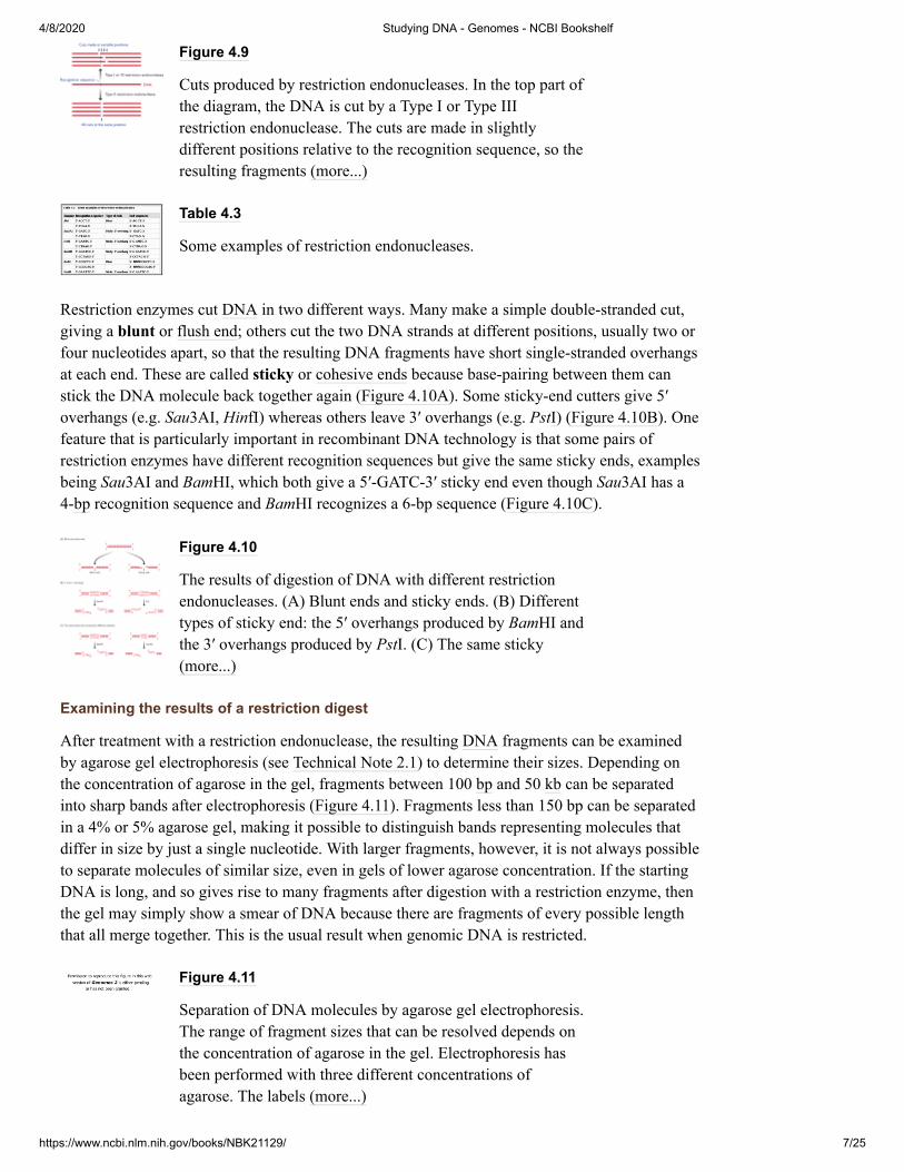
After treatment with a restriction endonuclease, the resulting DNA fragments can be examinedby agarose gel electrophoresis (see Technical Note 2.1) to determine their sizes. Depending onthe concentration of agarose in the gel, fragments between 100 bp and 50 kb can be separatedinto sharp bands after electrophoresis (Figure 4.11). Fragments less than 150 bp can be separatedin a 4% or 5% agarose gel, making it possible to distinguish bands representing molecules thatdiffer in size by just a single nucleotide. With larger fragments, however, it is not always possibleto separate molecules of similar size, even in gels of lower agarose concentration. If the startingDNA is long, and so gives rise to many fragments after digestion with a restriction enzyme, thenthe gel may simply show a smear of DNA because there are fragments of every possible lengththat all merge together. This is the usual result when genomic DNA is restricted.
Figure 4.11
Separation of DNA molecules by agarose gel electrophoresis.The range of fragment sizes that can be resolved depends onthe concentration of agarose in the gel. Electrophoresis hasbeen performed with three different concentrations ofagarose. The labels (more...)

4/8/2020 Studying DNA - Genomes - NCBI Bookshelf
https://www.ncbi.nlm.nih.gov/books/NBK21129/ 8/25
If the sequence of the starting DNA is known then the sequences, and hence the sizes, of thefragments resulting from treatment with a particular restriction enzyme can be predicted. Theband for a desired fragment (for example, one containing a gene) can then be identified, cut outof the gel, and the DNA purified. Even if its size is unknown, a fragment containing a gene oranother segment of DNA of interest can be identified by the technique called Southernhybridization, providing that some of the sequence within the fragment is known or can bepredicted. The first step is to transfer the restriction fragments from the agarose gel to anitrocellulose or nylon membrane. This is done by placing the membrane on the gel and allowingbuffer to soak through, taking the DNA from the gel to the membrane, where it becomes bound(Figure 4.12A). This process results in the DNA bands becoming immobilized in the samerelative positions on the surface of the membrane.
Figure 4.12
Southern hybridization. (A) Transfer of DNA from the gel tothe membrane. (B) The membrane is probed with aradioactively labeled DNA molecule. On the resultingautoradiograph, one hybridizing band is seen in lane 2, andtwo in lane 3.
The next step is to prepare a hybridization probe, which is a labeled DNA molecule whosesequence is complementary to the target DNA that we wish to detect. The probe could, forexample, be a synthetic oligonucleotide whose sequence matches part of an interesting gene.Because the probe and target DNAs are complementary, they can base-pair or hybridize, theposition of the hybridized probe on the membrane being identified by detecting the signal givenout by a label attached to the probe. To carry out the hybridization, the membrane is placed in aglass bottle with the labeled probe and some buffer, and the bottle gently rotated for severalhours so that the probe has plenty of opportunity to hybridize to its target DNA. The membraneis then washed to remove any probe that has not become hybridized, and the signal from thelabel is detected (see Technical Note 4.1). In the example shown in Figure 4.12B the probe isradioactively labeled and the signal is detected by autoradiography. The band that is seen on theautoradiograph is the one that corresponds to the restriction fragment that hybridizes to the probeand which therefore contains the gene that we are searching for.
4.1.3. DNA ligases
DNA fragments that have been generated by treatment with a restriction endonuclease can bejoined back together again, or attached to a new partner, by a DNA ligase. The reaction requiresenergy, which is provided by adding either ATP or NAD to the reaction mixture, depending onthe type of ligase that is being used.
The most widely used DNA ligase is obtained from E. coli cells infected with T4 bacteriophage.It is involved in replication of the phage DNA and is encoded by the T4 genome. Its natural roleis to synthesize phosphodiester bonds between unlinked nucleotides present in onepolynucleotide of a double-stranded molecule (Figure 4.13A). In order to join together tworestriction fragments, the ligase has to synthesize two phosphodiester bonds, one in each strand(Figure 4.13B). This is by no means beyond the capabilities of the enzyme, but the reaction canoccur only if the ends to be joined come close enough to one another by chance - the ligase is notable to catch hold of them and bring them together. If the two molecules have complementarysticky ends, and the ends come together by random diffusion events in the ligation mixture, thentransient base pairs might form between the two overhangs. These base pairs are not particularly

4/8/2020 Studying DNA - Genomes - NCBI Bookshelf
https://www.ncbi.nlm.nih.gov/books/NBK21129/ 9/25
stable but they may persist for sufficient time for a ligase enzyme to attach to the junction andsynthesize phosphodiester bonds to fuse the ends together (Figure 4.13C). If the molecules areblunt ended, then they cannot base-pair to one another, not even temporarily, and ligation is amuch less efficient process, even when the DNA concentration is high and pairs of ends are inrelatively close proximity.
Figure 4.13
Ligation of DNA molecules with DNA ligase. (A) In livingcells, DNA ligase synthesizes a missing phosphodiester bondin one strand of a double-stranded DNA molecule. (B) Tolink two DNA molecules in vitro, DNA ligase must make twophosphodiester bonds, (more...)
The greater efficiency of sticky-end ligation has stimulated the development of methods forconverting blunt ends into sticky ends. In one method, short double-stranded molecules calledlinkers or adaptors are attached to the blunt ends. Linkers and adaptors work in slightly differentways but both contain a recognition sequence for a restriction endonuclease and so produce asticky end after treatment with the appropriate enzyme (Figure 4.14). Another way to create asticky end is by homopolymer tailing, in which nucleotides are added one after the other to the 3′terminus at a blunt end (Figure 4.15). The enzyme involved is called terminal deoxynucleotidyltransferase, which we will meet in the next section. If the reaction contains the DNA, enzyme,and only one of the four nucleotides, then the new stretch of single-stranded DNA that is madeconsists entirely of just that single nucleotide. It could, for example, be a poly(G) tail, whichwould enable the molecule to base-pair to other molecules that carry poly(C) tails, created in thesame way but with dCTP rather than dGTP in the reaction mixture.
Figure 4.14
Linkers are used to place sticky ends on to a blunt-endedmolecule. In this example, each linker contains therecognition sequence for the BamHI restriction endonuclease.DNA ligase attaches the linkers to the ends of the blunt-endedmolecule in a reaction (more...)
Figure 4.15
Homopolymer tailing. In this example, a poly(G) tail issynthesized at each end of a blunt-ended DNA molecule.Tails comprising other nucleotides are synthesized byincluding the appropriate dNTP in the reaction mixture.
4.1.4. End-modification enzymes
Terminal deoxynucleotidyl transferase (see Figure 4.15), obtained from calf thymus tissue, is oneexample of an end-modification enzyme. It is, in fact, a template- independent DNApolymerase, because it is able to synthesize a new DNA polynucleotide without base-pairing ofthe incoming nucleotides to an existing strand of DNA or RNA. Its main role in recombinantDNA technology is in homopolymer tailing, as described above.
Two other end-modification enzymes are also frequently used. These are alkaline phosphataseand T4 polynucleotide kinase, which act in complementary ways. Alkaline phosphatase, which is

4/8/2020 Studying DNA - Genomes - NCBI Bookshelf
https://www.ncbi.nlm.nih.gov/books/NBK21129/ 10/25
obtained from various sources, including E. coli and calf intestinal tissue, removes phosphategroups from the 5′ ends of DNA molecules, which prevents these molecules from being ligatedto one another. Two ends carrying 5′ phosphates can be ligated to one another, and aphosphatased end can ligate to a non-phosphatased end, but a link cannot be formed between apair of ends if neither carries a 5′ phosphate. Judicious use of alkaline phosphatase can thereforedirect the action of a DNA ligase in a pre-determined way so that only desired ligation productsare obtained. T4 polynucleotide kinase, obtained from E. coli cells infected with T4 phage,performs the reverse reaction to alkaline phosphatase, adding phosphates to 5′ ends. Like alkalinephosphatase, the enzyme is used during complicated ligation experiments, but its mainapplication is in the end-labeling of DNA molecules (see Technical Note 4.1).
4.2. DNA CloningDNA cloning is a logical extension of the ability to manipulate DNA molecules with restrictionendonucleases and ligases. Imagine that an animal gene has been obtained as a single restrictionfragment after digestion of a larger molecule with the restriction enzyme BamHI, which leaves5′-GATC-3′ sticky ends (Figure 4.16). Imagine also that a small E. coli plasmid has been purifiedand treated with BamHI, which cuts the plasmid in a single position. The circular plasmid hastherefore been converted into a linear molecule, again with 5′-GATC-3′ sticky ends. Mix the twoDNA molecules together and add DNA ligase. Various recombinant ligation products will beobtained, one of which comprises the circularized plasmid with the animal gene inserted into theposition originally taken by the BamHI restriction site. If the recombinant plasmid is now re-introduced into E. coli, and the inserted gene has not disrupted its replicative ability, then theplasmid plus inserted gene will be replicated and copies passed to the daughter bacteria after celldivision. More rounds of plasmid replication and cell division will result in a colony ofrecombinant E. coli bacteria, each bacterium containing multiple copies of the animal gene. Thisseries of events, as illustrated in Figure 4.16, constitutes the process called DNA or gene cloning.
Figure 4.16
An outline of gene cloning. See the text for details.
4.2.1. Cloning vectors and the way they are used
In the experiment shown in Figure 4.16, the plasmid acts as a cloning vector, providing thereplicative ability that enables the cloned gene to be propagated inside the host cell. Plasmidsreplicate efficiently in bacterial hosts because each plasmid possesses an origin of replicationwhich is recognized by the DNA polymerases and other proteins that normally replicate thebacterium's chromosomes (Section 13.2.1). The host cell's replicative machinery thereforepropagates the plasmid, plus any new genes that have been inserted into it. Bacteriophagegenomes can also be used as cloning vectors because they too possess origins of replication thatenable them to be propagated inside bacteria, either by the host enzymes or by DNA polymerasesand other proteins specified by phage genes. The next two sections describe how plasmid andphage vectors are used to clone DNA in E. coli.
Plasmids are uncommon in eukaryotes, although Saccharomyces cerevisiae possesses one that issometimes used for cloning purposes; most eukaryotic vectors are therefore based on virusgenomes. Alternatively, with a eukaryotic host the replication requirement can be bypassed byperforming the experiment in such a way that the DNA to be cloned becomes inserted into one of

4/8/2020 Studying DNA - Genomes - NCBI Bookshelf
https://www.ncbi.nlm.nih.gov/books/NBK21129/ 11/25
the host chromosomes. These approaches to cloning in eukaryotic cells are described later in thechapter.
Vectors based on E. coli plasmids
The easiest way to understand how a cloning vector is used is to start with the simplest E. coliplasmid vectors, which illustrate all of the basic principles of DNA cloning. We will then be ableto turn our attention to the special features of phage vectors and vectors used with eukaryotes.
One of the first plasmid vectors to be developed was pBR322 (Bolivar et al., 1977), which wasconstructed by ligating restriction fragments from three naturally occurring E. coli plasmids: R1,R6.5 and pMB1. The pBR322 plasmid is small (just 4363 bp) and, as well as the origin ofreplication, it carries genes coding for enzymes that enable the host bacterium to withstand thegrowth-inhibitory effects of two antibiotics: ampicillin and tetracycline (Figure 4.17). Thismeans that cells containing a pBR322 plasmid can be distinguished from those that do not byplating the bacteria onto agar medium containing ampicillin and/or tetracycline. Normal E. colicells are sensitive to these antibiotics and cannot grow when either of the two antibiotics ispresent. Ampicillin and tetracycline resistance are therefore selectable markers for pBR322.
Figure 4.17
pBR322. The map shows the positions of the ampicillin-resistance gene (amp ), the tetracycline-resistance gene (tet
), the origin of replication (ori) and the recognitionsequences for seven restriction endonucleases.
The manipulations shown in Figure 4.16, resulting in construction of a recombinant plasmid, arecarried out in the test tube with purified DNA. Pure pBR322 DNA can be obtained quite easilyfrom extracts of bacterial cells (Technical Note 4.2), but how can the manipulated plasmids bere-introduced into the bacteria? The answer is to make use of the natural processes fortransformation of bacteria, which result in the uptake of ‘naked’ DNA by a bacterial cell. This isthe process studied by Avery and his colleagues in the experiments which showed that bacterialgenes are made of DNA (Section 1.1.1). Transformation is not a particularly efficient process inmany bacteria, including E. coli, but the rate of uptake can be enhanced significantly bysuspending the cells in calcium chloride before adding the DNA, and then briefly incubating themixture at 42 °C. Even after this enhancement, only a very small proportion of the cells take up aplasmid. This is why the antibiotic-resistance markers are so important - they allow the smallnumber of transformants to be selected from the large background of non-transformed cells.
Box 4.2
DNA purification. Techniques for the preparation of puresamples of DNA from living cells play a central role inmolecular biology research. The first step in DNApurification is to break open the cells from which the DNAwill be obtained. With some types (more...)
The map of pBR322 shown in Figure 4.17 indicates the positions of the recognition sequencesfor seven restriction enzymes, each of which cuts the plasmid at just one location. Note that sixof these sites lie within one or other of the genes for antibiotic resistance. This means that if anew fragment of DNA is ligated into one of these six sites, then the antibiotic-resistanceproperties of the plasmid become altered - the plasmid loses the ability to confer either ampicillin
R
R

4/8/2020 Studying DNA - Genomes - NCBI Bookshelf
https://www.ncbi.nlm.nih.gov/books/NBK21129/ 12/25
or tetracycline resistance on the host cells. This is called insertional inactivation of the selectablemarker and is the key to distinguishing a recombinant plasmid - one that contains an insertedpiece of DNA - from a non-recombinant plasmid that has no new DNA. Identifyingrecombinants is important because the manipulations illustrated in Figure 4.16 result in a varietyof ligation products, including plasmids that have recircularized without insertion of new DNA.To identify recombinants, the resistance properties of colonies are tested by transferring cellsfrom agar containing one antibiotic onto agar containing the second antibiotic. For example, ifthe BamHI site has been used then recombinants will be ampicillin resistant but tetracyclinesensitive, because the BamHI site lies within the region that specifies resistance to tetracycline.After transformation, cells are plated onto ampicillin agar (Figure 4.18). All cells that contain apBR322 plasmid, whether recombinant or not, divide and produce a colony. The colonies arethen transferred onto tetracycline agar by replica plating, which results in the colonies on thesecond plate retaining the relative positions that they had on the first plate. Some colonies do notgrow on the tetracycline agar because their cells contain recombinant pBR322 molecules with adisrupted tetracycline-resistance gene. These are the colonies we are looking for because theycontain the cloned gene, so we return to the ampicillin plate, from which samples of the cells canbe recovered.
Figure 4.18
Recombinant selection with pBR322. See text for details. Theinset shows how replica plating is performed.
Replica plating is not a difficult technique but it takes time. It would be much better ifrecombinants could be distinguished from non-recombinants simply by plating onto a single agarmedium. This is possible with most of the modern plasmid cloning vectors, including pUC8(Figure 4.19; Vieira and Messing, 1982). This vector carries the ampicillin-resistance gene frompBR322, along with a second gene, called lacZ′, which is part of the E. coli gene for the enzymeβ-galactosidase. The remainder of the lacZ gene is located in the chromosome of the special E.coli strain that is used when cloning genes with pUC8. The proteins specified by the genesegments on the plasmid and on the chromosome are able to combine to produce a functional β-galactosidase enzyme. The presence of functional β-galactosidase molecules in the cells can bechecked by a histochemical test with a compound called X-gal (5-bromo-4-chloro-3-indolyl-β-�-galactopyranoside), which the enzyme converts into a blue product. The lacZ′ gene contains acluster of unique restriction sites; insertion of new DNA into any one of these sites results ininsertional inactivation of the gene and hence loss of β-galactosidase activity. Recombinants andnon-recombinants can therefore be distinguished simply by plating the transformed cells ontoagar containing ampicillin and X-gal (Figure 4.19). All colonies that grow on this medium aremade up of transformed cells because only transformants are ampicillin resistant. Some coloniesare blue and some are white. Those that are blue contain cells with functional β-galactosidaseenzymes and hence with undisrupted lacZ′ genes; these colonies are therefore non-recombinants.The white colonies comprise cells without β-galactosidase activity and hence with disruptedlacZ′ genes; these are recombinants.
Figure 4.19
Recombinant selection with pUC8. See the text for details.
Cloning vectors based on E. coli bacteriophage genomes

4/8/2020 Studying DNA - Genomes - NCBI Bookshelf
https://www.ncbi.nlm.nih.gov/books/NBK21129/ 13/25
E. coli bacteriophages were developed as cloning vectors back in the earliest days of therecombinant DNA revolution. The main reason for seeking a different type of vector was theinability of plasmids such as pBR322 and pUC8 to handle DNA fragments greater than about 10kb in size, larger inserts undergoing rearrangements or interfering with the plasmid replicationsystem in such a way that the recombinant DNA molecules become lost from the host cells. Thefirst attempts to develop vectors able to handle larger fragments of DNA centered onbacteriophage λ. The infection cycle of λ is similar to that of the T2 phages studied by Hersheyand Chase in the experiments that alerted molecular biologists to the fact that genes are made ofDNA (Section 1.1.1), but there is one important difference. As well as following the lyticinfection cycle (see Figure 1.4B), the λ genome is able to integrate into the bacterialchromosome, where it can remain quiescent for many generations, being replicated along withthe host chromosome every time the cell divides. This is called the lysogenic infection cycle(Figure 4.20).
Figure 4.20
The lysogenic infection cycle of bacteriophage λ. Comparewith the lytic infection cycle of T2 bacteriophage, shown inFigure 1.4B. The special feature of the lysogenic cycle is theinsertion of the phage genome into the bacterium'schromosomal (more...)
The λ genome is 48.5 kb, of which some 15 kb or so is ‘optional’ in that it contains genes thatare only needed for integration of the phage DNA into the E. coli chromosome (Figure 4.21A).These segments can therefore be deleted without impairing the ability of the phage to infectbacteria and direct synthesis of new λ particles by the lytic cycle. Two types of vector have beendeveloped (Figure 4.21B):
Figure 4.21
Cloning vectors based on bacteriophage λ. (A) In the λgenome, the genes are arranged into functional groups. Forexample, the region marked as ‘protein coat’ comprises 21genes coding for proteins that are either components (more...)
Insertion vectors, in which part or all of the optional DNA has been removed and a uniquerestriction site introduced at some position within the trimmed down genome;
Replacement vectors, in which the optional DNA is contained within a stuffer fragment,flanked by a pair of restriction sites, that is replaced when the DNA to be cloned is ligatedinto the vector.
The λ genome is linear, but the two natural ends of the molecule have 12-nucleotide single-stranded overhangs, called cos sites, which have complementary sequences and so can base-pairto one another. A λ cloning vector can therefore be obtained as a circular molecule which can bemanipulated in the test tube in the same way as a plasmid, and re-introduced into E. coli bytransfection, the term used for uptake of naked phage DNA. Alternatively, a more efficient
4/8/2020 Studying DNA - Genomes - NCBI Bookshelf
https://www.ncbi.nlm.nih.gov/books/NBK21129/ 14/25
uptake system called in vitro packaging can be utilized (Hohn and Murray, 1977). Thisprocedure starts with the linear version of the cloning vector, the initial restriction cutting themolecule into two segments, the left and right arms, each with a cos site at one end. The ligationis carried out with carefully measured quantities of each arm and the DNA to be cloned, the aimbeing to produce concatamers in which the different fragments are linked together in the orderleft arm-new DNA-right arm, as shown in Figure 4.22. The concatamers are then added to an invitro packaging mix, which contains all the proteins needed to make a λ phage particle. Theseproteins form phage particles spontaneously, and will place inside the particles any DNAfragment that is between 37 and 52 kb in length and is flanked by cos sites. The packaging mixtherefore cuts left arm-new DNA-right arm combinations of 37–52 kb out of the concatamersand constructs λ phages around them. The phages are then mixed with E. coli cells, and thenatural infection process transports the vector plus new DNA into the bacteria.
Figure 4.22
Cloning with a λ insertion vector. The linear form of thevector is shown at the top of the diagram. Treatment with theappropriate restriction endonuclease produces the left andright arms, both of which have one blunt end and one endwith the (more...)
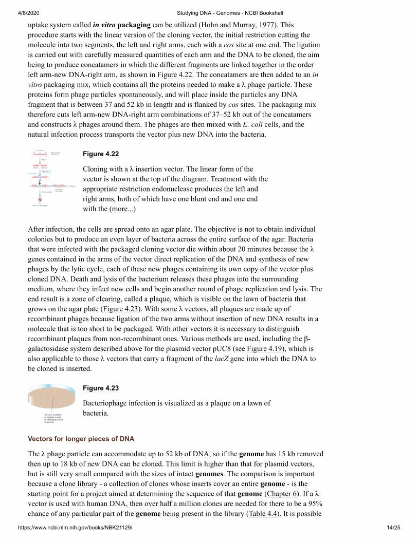
After infection, the cells are spread onto an agar plate. The objective is not to obtain individualcolonies but to produce an even layer of bacteria across the entire surface of the agar. Bacteriathat were infected with the packaged cloning vector die within about 20 minutes because the λgenes contained in the arms of the vector direct replication of the DNA and synthesis of newphages by the lytic cycle, each of these new phages containing its own copy of the vector pluscloned DNA. Death and lysis of the bacterium releases these phages into the surroundingmedium, where they infect new cells and begin another round of phage replication and lysis. Theend result is a zone of clearing, called a plaque, which is visible on the lawn of bacteria thatgrows on the agar plate (Figure 4.23). With some λ vectors, all plaques are made up ofrecombinant phages because ligation of the two arms without insertion of new DNA results in amolecule that is too short to be packaged. With other vectors it is necessary to distinguishrecombinant plaques from non-recombinant ones. Various methods are used, including the β-galactosidase system described above for the plasmid vector pUC8 (see Figure 4.19), which isalso applicable to those λ vectors that carry a fragment of the lacZ gene into which the DNA tobe cloned is inserted.
Figure 4.23
Bacteriophage infection is visualized as a plaque on a lawn ofbacteria.
Vectors for longer pieces of DNA
The λ phage particle can accommodate up to 52 kb of DNA, so if the genome has 15 kb removedthen up to 18 kb of new DNA can be cloned. This limit is higher than that for plasmid vectors,but is still very small compared with the sizes of intact genomes. The comparison is importantbecause a clone library - a collection of clones whose inserts cover an entire genome - is thestarting point for a project aimed at determining the sequence of that genome (Chapter 6). If a λvector is used with human DNA, then over half a million clones are needed for there to be a 95%chance of any particular part of the genome being present in the library (Table 4.4). It is possible
4/8/2020 Studying DNA - Genomes - NCBI Bookshelf
https://www.ncbi.nlm.nih.gov/books/NBK21129/ 15/25
to prepare a library comprising half a million clones, especially if automated techniques are used,but such a large collection is far from ideal. It would be much better to reduce the number ofclones by using a vector that is able to handle fragments of DNA longer than 18 kb. Many of thedevelopments in cloning technology over the last 20 years have been aimed at finding ways ofdoing this.
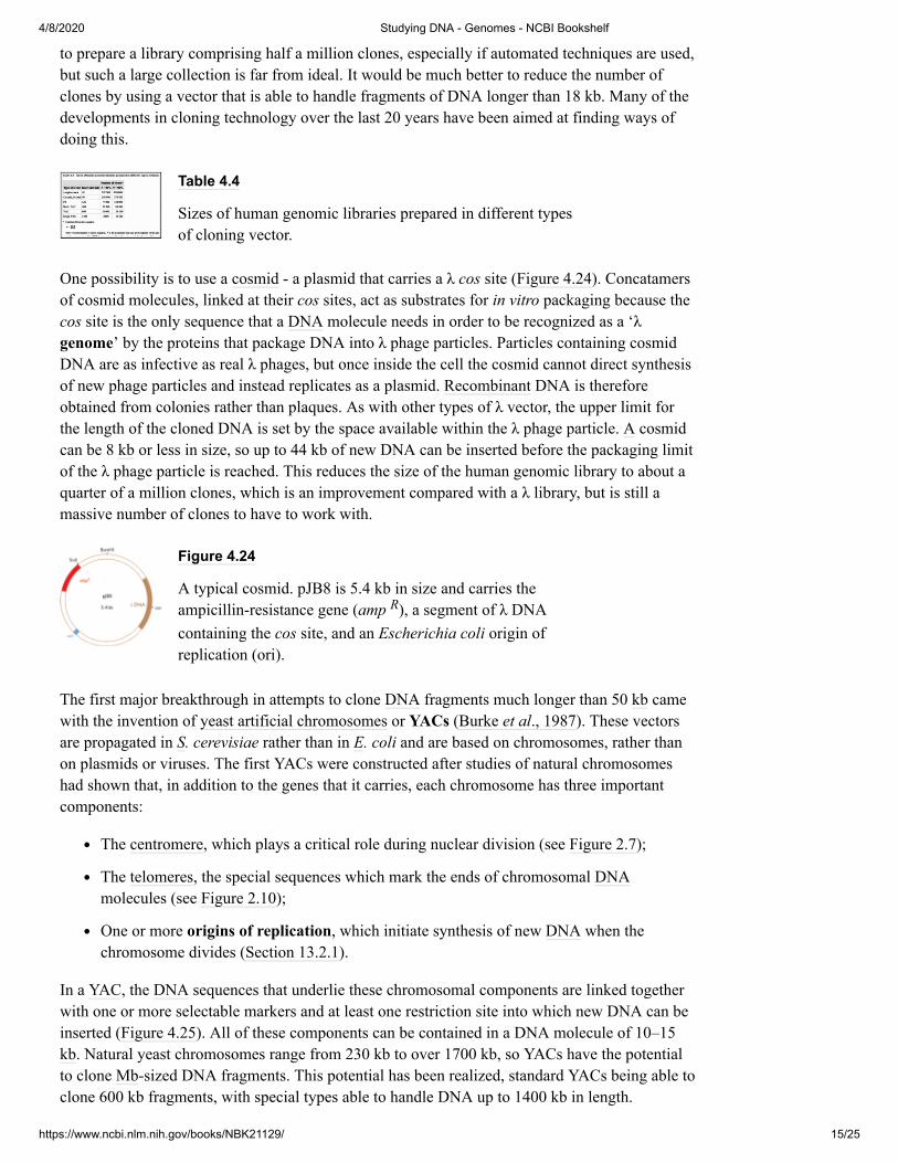
Table 4.4
Sizes of human genomic libraries prepared in different typesof cloning vector.
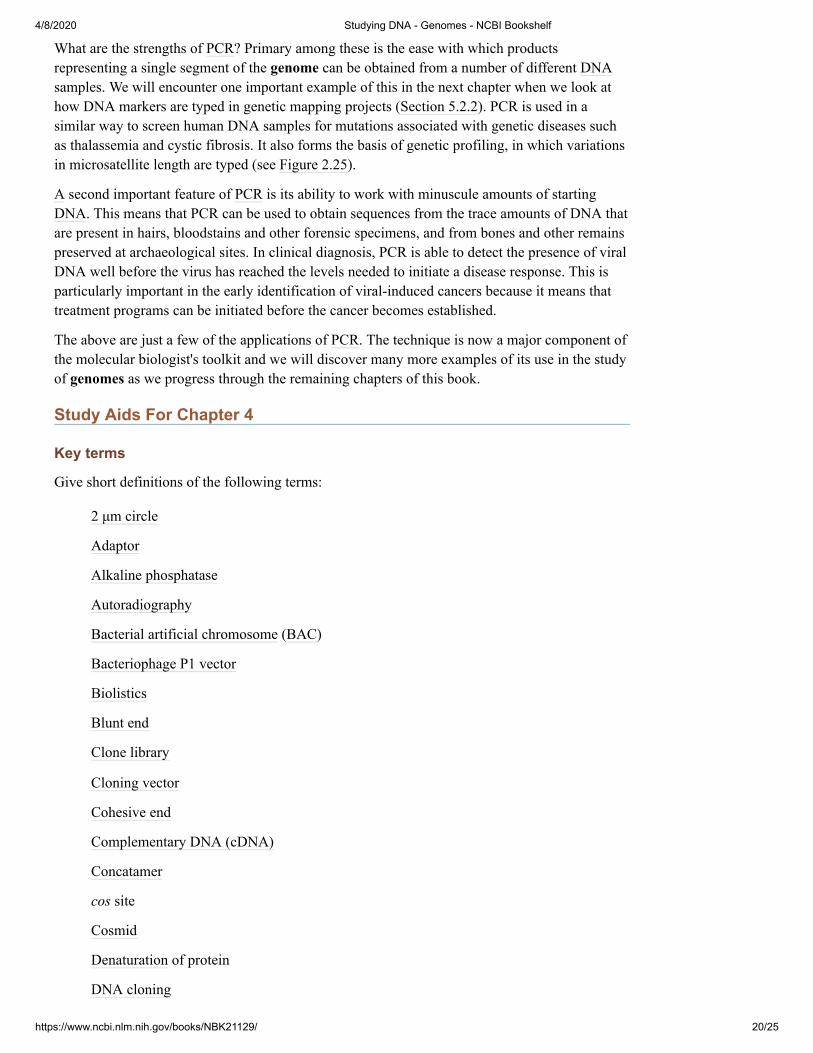
One possibility is to use a cosmid - a plasmid that carries a λ cos site (Figure 4.24). Concatamersof cosmid molecules, linked at their cos sites, act as substrates for in vitro packaging because thecos site is the only sequence that a DNA molecule needs in order to be recognized as a ‘λgenome’ by the proteins that package DNA into λ phage particles. Particles containing cosmidDNA are as infective as real λ phages, but once inside the cell the cosmid cannot direct synthesisof new phage particles and instead replicates as a plasmid. Recombinant DNA is thereforeobtained from colonies rather than plaques. As with other types of λ vector, the upper limit forthe length of the cloned DNA is set by the space available within the λ phage particle. A cosmidcan be 8 kb or less in size, so up to 44 kb of new DNA can be inserted before the packaging limitof the λ phage particle is reached. This reduces the size of the human genomic library to about aquarter of a million clones, which is an improvement compared with a λ library, but is still amassive number of clones to have to work with.
Figure 4.24
A typical cosmid. pJB8 is 5.4 kb in size and carries theampicillin-resistance gene (amp ), a segment of λ DNAcontaining the cos site, and an Escherichia coli origin ofreplication (ori).
The first major breakthrough in attempts to clone DNA fragments much longer than 50 kb camewith the invention of yeast artificial chromosomes or YACs (Burke et al., 1987). These vectorsare propagated in S. cerevisiae rather than in E. coli and are based on chromosomes, rather thanon plasmids or viruses. The first YACs were constructed after studies of natural chromosomeshad shown that, in addition to the genes that it carries, each chromosome has three importantcomponents:
The centromere, which plays a critical role during nuclear division (see Figure 2.7);
The telomeres, the special sequences which mark the ends of chromosomal DNAmolecules (see Figure 2.10);
One or more origins of replication, which initiate synthesis of new DNA when thechromosome divides (Section 13.2.1).
In a YAC, the DNA sequences that underlie these chromosomal components are linked togetherwith one or more selectable markers and at least one restriction site into which new DNA can beinserted (Figure 4.25). All of these components can be contained in a DNA molecule of 10–15kb. Natural yeast chromosomes range from 230 kb to over 1700 kb, so YACs have the potentialto clone Mb-sized DNA fragments. This potential has been realized, standard YACs being able toclone 600 kb fragments, with special types able to handle DNA up to 1400 kb in length.
R

4/8/2020 Studying DNA - Genomes - NCBI Bookshelf
https://www.ncbi.nlm.nih.gov/books/NBK21129/ 16/25
Currently this is the highest capacity of any type of cloning vector, and several genome projectshave made extensive use of YACs. Unfortunately, with some types of YAC there have beenproblems with insert stability, the cloned DNA becoming rearranged into new sequencecombinations (Anderson, 1993). For this reason there is also great interest in other types ofvectors, ones that cannot clone such large pieces of DNA but which suffer less from instabilityproblems. These vectors include the following:
Figure 4.25
Working with a YAC. (A) The cloning vector pYAC3. (B) Toclone with pYAC3, the circular vector is digested withBamHI and SnaBI. BamHI restriction removes the stufferfragment held between the two telomeres in the circularmolecule. SnaBI cuts within (more...)
Bacteriophage P1 vectors (Sternberg, 1990) are very similar to λ vectors, being based on adeleted version of a natural phage genome, the capacity of the cloning vector beingdetermined by the size of the deletion and the space within the phage particle. The P1genome is larger than the λ genome, and the phage particle is bigger, so a P1 vector canclone larger fragments of DNA than a λ vector, up to 125 kb using current technology.
Bacterial artificial chromosomes or BACs (Shizuya et al., 1992) are based on the naturallyoccurring F plasmid of E. coli. Unlike the plasmids used to construct the early cloningvectors, the F plasmid is relatively large and vectors based on it have a higher capacity foraccepting inserted DNA. BACs can be used to clone fragments of 300 kb and longer.
P1-derived artificial chromosomes or PACs (Ioannou et al., 1994) combine features of P1vectors and BACs and have a capacity of up to 300 kb.
Fosmids (Kim et al., 1992) contain the F plasmid origin of replication and a λ cos site.They are similar to cosmids but have a lower copy number in E. coli, which means thatthey are less prone to instability problems.
The sizes of human genome libraries prepared in these various types of vector are given in Table4.4.
Cloning in organisms other than E. coli
Cloning is not merely an aid to DNA sequencing: it also provides a means of studying the modeof expression of a gene and the way in which expression is regulated, of carrying out geneticengineering experiments aimed at modifying the biological characteristics of the host organism,and of synthesizing important animal proteins, such as pharmaceuticals, in a new host cell fromwhich the proteins can be obtained in larger quantities than is possible by conventionalpurification from animal tissue. These multifarious applications demand that genes mustfrequently be cloned in organisms other than E. coli.
Cloning vectors based on plasmids or phages have been developed for most of the well studiedspecies of bacteria such as Bacillus, Streptomyces and Pseudomonas, these vectors being used inexactly the same way as the E. coli analogs. Plasmid vectors are also available for yeasts andfungi. Some of these carry the origin of replication from the 2 μm circle, a plasmid present inmany strains of S. cerevisiae, but other plasmid vectors only have an E. coli origin. An exampleis YIp5, an S. cerevisiae vector that is simply a pBR322 plasmid that contains a copy of the yeastgene called URA3 (Figure 4.26A). What is the logic behind the construction of YIp5? When used

4/8/2020 Studying DNA - Genomes - NCBI Bookshelf
https://www.ncbi.nlm.nih.gov/books/NBK21129/ 17/25
in a cloning experiment, the vector is initially used with E. coli as the host, up to the stage wherethe desired recombinant molecule has been constructed by restriction and ligation. Therecombinant vector is then purified from E. coli and transferred into S. cerevisiae, usually bymixing the DNA with protoplasts - yeast cells whose walls have been removed by enzymetreatment. Without an origin of replication the vector is unable to propagate independently insideyeast cells, but it can survive if it becomes integrated into one of the yeast chromosomes, whichcan occur by homologous recombination (Section 7.2.2) between the URA3 gene carried by thevector and the chromosomal copy of this gene (Figure 4.26B). ‘YIp’ in fact stands for ‘yeastintegrative plasmid’. Once integrated the YIp, plus any DNA that has been inserted into it,replicates along with the host chromosomes.
Figure 4.26
Cloning with a YIp. (A) YIp5, a typical yeast integrativeplasmid. The plasmid contains the ampicillin-resistance gene(amp ), the tetracycline-resistance gene (tet ), the yeastgene URA3, and an Escherichia coli origin of replication(ori). The presence (more...)
Integration into chromosomal DNA is also a feature of many of the cloning systems used withanimals and plants, and forms the basis of the construction of knockout mice, which are used todetermine the functions of previously unknown genes that are discovered in the human genome(Section 7.2.2). The vectors are animal equivalents of YIps. Adenoviruses and retroviruses areused to clone genes in animals when the objective is to treat a genetic disease or a cancer by genetherapy (Lemoine and Cooper, 1998). A similar range of vectors has been developed for cloninggenes in plants. Plasmids can be introduced into plant embryos by bombardment with DNA-coated microprojectiles, a process called biolistics (Klein et al., 1987), integration of the plasmidDNA into the plant chromosomes, followed by growth of the embryo, resulting in a plant thatcontains the cloned DNA in most or all of its cells. Some success has also been achieved withplant vectors based on the genomes of caulimoviruses and geminiviruses (Timmermans et al.,1994; Viaplana et al., 2001), but the most interesting types of plant cloning vector are thosederived from the Ti plasmid (Hansen and Wright, 1999). This is a large bacterial plasmid foundin the soil microorganism Agrobacterium tumefaciens, part of which, the T-DNA, becomesintegrated into a plant chromosome when the bacterium infects a plant stem and causes crownroot disease. The T-DNA carries a number of genes that are expressed inside the plant cells andinduce the various physiological changes that characterize the disease. Vectors such as pBIN19(Figure 4.27) have been designed to make use of this natural genetic engineering system (Bevan,1984). The recombinant vector is introduced into A. tumefaciens cells, which are allowed toinfect a cell suspension or plant callus culture, from which mature transformed plants can beregenerated.
Figure 4.27
The plant cloning vector pBIN19. pBIN19 carries the lacZ′gene (see Figure 4.19), the kanamycin-resistance gene (kan
), an Escherichia coli origin of replication (ori), and the twoboundary sequences from the T-DNA region of the Tiplasmid. These (more...)
4.3. The Polymerase Chain Reaction (PCR)
R R
R

4/8/2020 Studying DNA - Genomes - NCBI Bookshelf
https://www.ncbi.nlm.nih.gov/books/NBK21129/ 18/25
In essence, DNA cloning results in the purification of a single fragment of DNA from a complexmixture of DNA molecules. Cloning is a powerful technique and its impact on our understandingof genes and genomes has been immeasurable. Cloning does, however, have one majordisadvantage: it is a time-consuming and, in parts, difficult procedure. It takes several days toperform the manipulations needed to insert DNA fragments into a cloning vector and thenintroduce the ligated molecules into the host cells and select recombinants. If the experimentalstrategy involves generation of a large clone library, followed by screening of the library toidentify a clone that contains a gene of interest (see Technical Note 4.3), then several more weeksor even months might be needed to complete the project.
Box 4.3
Working with a clone library. Clone collections used as asource of genes and other DNA segments. Clone librarieshave been used in molecular biology research for many yearsand their importance extends well beyond their role as thestarting point for (more...)
PCR complements DNA cloning in that it enables the same result to be achieved - purification ofa specified DNA fragment - but in a much shorter time, perhaps just a few hours (Saiki et al.,1988). PCR is complementary to, not a replacement for, cloning because it has its ownlimitations, the most important of which is the need to know the sequence of at least part of thefragment that is to be purified. Despite this constraint, PCR has acquired central importance inmany areas of molecular biology research. We will examine the technique first and then surveyits applications.
4.3.1. Carrying out a PCR
PCR results in the repeated copying of a selected region of a DNA molecule (see Figure 4.3).Unlike cloning, PCR is a test-tube reaction and does not involve the use of living cells: thecopying is carried out not by cellular enzymes but by the purified, thermostable DNApolymerase of T. aquaticus (Section 4.1.1). The reason why a thermostable enzyme is neededwill become clear when we look in more detail at the events that occur during PCR.
To carry out a PCR experiment, the target DNA is mixed with Taq DNA polymerase, a pair ofoligonucleotide primers, and a supply of nucleotides. The amount of target DNA can be verysmall because PCR is extremely sensitive and will work with just a single starting molecule. Theprimers are needed to initiate the DNA synthesis reactions that will be carried out by the Taqpolymerase (see Figure 4.6). They must attach to the target DNA at either side of the segmentthat is to be copied; the sequences of these attachment sites must therefore be known so thatprimers of the appropriate sequences can be synthesized.
The reaction is started by heating the mixture to 94 °C. At this temperature the hydrogen bondsthat hold together the two polynucleotides of the double helix are broken, so the target DNAbecomes denatured into single- stranded molecules (Figure 4.28). The temperature is thenreduced to 50–60 °C, which results in some rejoining of the single strands of the target DNA, butalso allows the primers to attach to their annealing positions. DNA synthesis can now begin, sothe temperature is raised to 72 °C, the optimum for Taq polymerase. In this first stage of thePCR, a set of ‘long products’ is synthesized from each strand of the target DNA. Thesepolynucleotides have identical 5′ ends but random 3′ ends, the latter representing positions whereDNA synthesis terminates by chance. When the cycle of denaturation-annealing- synthesis isrepeated, the long products act as templates for new DNA synthesis, giving rise to ‘short

4/8/2020 Studying DNA - Genomes - NCBI Bookshelf
https://www.ncbi.nlm.nih.gov/books/NBK21129/ 19/25
products’ whose 5′ and 3′ ends are both set by the primer annealing positions (Figure 4.29). Insubsequent cycles, the number of short products accumulates in an exponential fashion (doublingduring each cycle) until one of the components of the reaction becomes depleted. This means thatafter 30 cycles, there will be over 250 million short products derived from each startingmolecule. In real terms, this equates to several micrograms of PCR product from a fewnanograms or less of target DNA.
Figure 4.28
The first stage of a PCR. See the text for details.
Figure 4.29
The synthesis of ‘short’ products in a PCR. The first cycleproducts from Figure 4.28 are shown at the top. The nextcycle of denaturation-annealing-synthesis leads to fourproducts, two of which are identical to the first cycle products(more...)
The results of a PCR can be determined in various ways. Usually, the products are analyzed byagarose gel electrophoresis, which will reveal a single band if the PCR has worked as expectedand has amplified a single segment of the target DNA (Figure 4.30). Alternatively, the sequenceof the product can be determined, using techniques described in Section 6.1.1.
Figure 4.30
Analysing the results of a PCR by agarose gelelectrophoresis. The PCR has been carried out in a microfugetube. A sample is loaded into lane 2 of an agarose gel. Lane 1contains DNA size markers, and lane 3 contains a sample of aPCR done by a colleague. (more...)
4.3.2. The applications of PCR
PCR is such a straightforward procedure that it is sometimes difficult to understand how it canhave become so important in modern research. First we will deal with its limitations. In order tosynthesize primers that will anneal at the correct positions, the sequences of the boundary regionsof the DNA to be amplified must be known. This means that PCR cannot be used to purifyfragments of genes or other parts of a genome that have never been studied before. A secondconstraint is the length of DNA that can be copied. Regions of up to 5 kb can be amplifiedwithout too much difficulty, and longer amplifications - up to 40 kb - are possible usingmodifications of the standard technique. However, the >100 kb fragments that are needed forgenome sequencing projects are unattainable by PCR.
4/8/2020 Studying DNA - Genomes - NCBI Bookshelf
https://www.ncbi.nlm.nih.gov/books/NBK21129/ 20/25
What are the strengths of PCR? Primary among these is the ease with which productsrepresenting a single segment of the genome can be obtained from a number of different DNAsamples. We will encounter one important example of this in the next chapter when we look athow DNA markers are typed in genetic mapping projects (Section 5.2.2). PCR is used in asimilar way to screen human DNA samples for mutations associated with genetic diseases suchas thalassemia and cystic fibrosis. It also forms the basis of genetic profiling, in which variationsin microsatellite length are typed (see Figure 2.25).
A second important feature of PCR is its ability to work with minuscule amounts of startingDNA. This means that PCR can be used to obtain sequences from the trace amounts of DNA thatare present in hairs, bloodstains and other forensic specimens, and from bones and other remainspreserved at archaeological sites. In clinical diagnosis, PCR is able to detect the presence of viralDNA well before the virus has reached the levels needed to initiate a disease response. This isparticularly important in the early identification of viral-induced cancers because it means thattreatment programs can be initiated before the cancer becomes established.
The above are just a few of the applications of PCR. The technique is now a major component ofthe molecular biologist's toolkit and we will discover many more examples of its use in the studyof genomes as we progress through the remaining chapters of this book.
Study Aids For Chapter 4
Key terms
Give short definitions of the following terms:
2 μm circle
Adaptor
Alkaline phosphatase
Autoradiography
Bacterial artificial chromosome (BAC)
Bacteriophage P1 vector
Biolistics
Blunt end
Clone library
Cloning vector
Cohesive end
Complementary DNA (cDNA)
Concatamer
cos site
Cosmid
Denaturation of protein
DNA cloning

4/8/2020 Studying DNA - Genomes - NCBI Bookshelf
https://www.ncbi.nlm.nih.gov/books/NBK21129/ 21/25
DNA polymerase
End-modification enzyme
Endonuclease
Exonuclease
Flush end
Fosmid
Gene cloning
Gene therapy
Homologous recombination
Homopolymer tailing
Hybridization
Hybridization probe
In vitro packaging
Insertion vector
Insertional inactivation
Klenow polymerase
Knockout mice
Kornberg polymerase
Ligase
Linker
Lysogenic infection cycle
Lysozyme
Lytic infection cycle
Northern hybridization
Nuclease
Oligonucleotide
Origin of replication
P1-derived artificial chromosome (PAC)
Phosphorimaging
Plaque
Polymerase chain reaction (PCR)
Primer
Proofreading

4/8/2020 Studying DNA - Genomes - NCBI Bookshelf
https://www.ncbi.nlm.nih.gov/books/NBK21129/ 22/25
1.
2.
3.
4.
Protoplast
Recombinant
Recombinant DNA technology
Replacement vector
Replica plating
Restriction endonuclease
Reverse transcriptase
Reverse transcriptase-PCR
RNA-dependent DNA polymerase
Selectable marker
Sequenase
Shuttle vector
Southern hybridization
Sticky end
Stuffer fragment
T4 polynucleotide kinase
T-DNA
Template-dependent DNA polymerase
Template-independent DNA polymerase
Terminal deoxynucleotidyl transferase
Thermostable
Ti plasmid
Transfection
Transformation
Yeast artificial chromosome (YAC)
Self study questions
Draw diagrams that outline the events that occur during (a) DNA cloning, and (b) PCR.What are the limitations of each of these two techniques?
List the types of enzyme used in recombinant DNA research.
Distinguish between the two types of exonuclease activity that can be possessed by a DNApolymerase, and explain how these activities influence the potential applications ofindividual DNA polymerases in recombinant DNA research.
Using examples, describe the various types of end produced after digestion of DNA with arestriction endonuclease.

4/8/2020 Studying DNA - Genomes - NCBI Bookshelf
https://www.ncbi.nlm.nih.gov/books/NBK21129/ 23/25
5.
6.
7.
8.
9.
10.
11.
12.
13.
1.
2.
3.
How are agarose gel electrophoresis and Southern hybridization used to examine the results of arestriction digest?
Explain why the efficiency of blunt-end ligation is less than that of sticky-end ligation. Whatsteps can be taken to improve the efficiency of blunt-end ligation?
Draw diagrams of (a) pBR322, and (b) pUC8. Explain how the differences between thesetwo vectors influence the ways in which they are used to clone DNA fragments.
Distinguish between the lytic and lysogenic infection cycles for a bacteriophage.
Write a short description of the way in which a bacteriophage λ vector is used to clone DNA.How does a cosmid differ from a standard λ vector?
Draw a diagram showing a typical YAC. Indicate the key features and explain how a YACis used to clone DNA.
What problems might arise when a YAC is used to clone a large fragment of DNA? To whatextent can these problems be solved by the use of other types of high-capacity cloningvector?
How is DNA cloned in organisms other than Escherichia coli?
Describe how a PCR is carried out, paying particular attention to the role of the primers andthe temperatures used during the thermal cycling.
Problem-based learning
Soon after the first gene cloning experiments were carried out in the early 1970s, a number ofscientists argued that there should be a temporary moratorium on this type of research. Whatwas the basis of these scientists' fears and to what extent were these fears justified?
What would be the features of an ideal cloning vector? To what extent are these requirementsmet by any of the existing cloning vectors?
The specificity of the primers is a critical feature of a successful PCR. If the primers annealat more than one position in the target DNA then products additional to the one being soughtwill be synthesized. Explore the factors that determine primer specificity and evaluate theinfluence of the annealing temperature on the outcome of a PCR.
References
1. Anderson C. Genome shortcut leads to problems. Science. (1993);259:1684–1687.[PubMed: 8456291]
2. Bevan M. Binary Agrobacterium vectors for plant transformation. Nucleic Acids Res.(1984);12:8711–8721. [PMC free article: PMC320409] [PubMed: 6095209]
3. Bolivar F, Rodriguez RL, Greene PJ. et al. Construction and characterisation of newcloning vectors II. A multi-purpose cloning system. Gene. (1977);2:95–113. [PubMed:344137]
4. Brown TA (1998) Molecular Biology Labfax, 2nd edition, Vol. 1. Academic Press,London.
5. Brown TA (2001) Gene Cloning and DNA Analysis: An Introduction, 4th edition.Blackwell Scientific Publishers, Oxford.
6. Burke DT, Carle GF, Olson MV. Cloning of large segments of exogenous DNA into yeastby means of artificial chromosome vectors. Science. (1987);236:806–812. [PubMed:

4/8/2020 Studying DNA - Genomes - NCBI Bookshelf
https://www.ncbi.nlm.nih.gov/books/NBK21129/ 24/25
3033825]7. Hansen G, Wright MS. Recent advances in the transformation of plants. Trends Plant Sci.
(1999);4:226–231. [PubMed: 10366879]8. Hohn B, Murray K. Packaging recombinant DNA molecules into bacteriophage particles
in vitro. Proc. Natl Acad. Sci. USA. (1977);74:3259–3263. [PMC free article:PMC431522] [PubMed: 333431]
9. Ioannou PA, Amemiya CT, Garnes J. et al. P1-derived vector for the propagation of largehuman DNA fragments. Nature Genet. (1994);6:84–89. [PubMed: 8136839]
10. Kim U-J, Shizuya H, de Jong PJ, Birren B, Simon MI. Stable propagation of cosmid andhuman DNA inserts in an F factor based vector. Nucleic Acids Res. (1992);20:1083–1085.[PMC free article: PMC312094] [PubMed: 1549470]
11. Klein RM, Wolf ED, Wu R, Sanford JC. High velocity microprojectiles for deliveringnucleic acids into living cells. Biotechnology. (1992);24:384–7386. [PubMed: 1422046]
12. Kornberg A. Biologic synthesis of deoxyribonucleic acid. Science. (1960);131:1503–1508.[PubMed: 14411056]
13. Lemoine N and Cooper D (1998) Gene Therapy. BIOS Scientific Publishers, Oxford.14. Mullis KB. The unusual origins of the polymerase chain reaction. Sci. Am.
(1990);262(4):56–65. [PubMed: 2315679]15. Saiki RK, Gelfand DH, Stoffel S. et al. Primer-directed enzymatic amplification of DNA
with a thermostable DNA polymerase. Science. (1988);239:487–491. [PubMed: 2448875]16. Shizuya H, Birren B, Kim UJ. et al. Cloning and stable maintenance of 300-kilobase-pair
fragments of human DNA in Escherichia coli using an F-factor-based vector. Proc. NatlAcad. Sci. USA. (1992);89:8794–8797. [PMC free article: PMC50007] [PubMed:1528894]
17. Sternberg N. Bacteriophage P1 cloning system for the isolation, amplification, andrecovery of DNA fragments as large as 100 kilobase pairs. Proc. Natl Acad. Sci. USA.(1990);87:103–107. [PMC free article: PMC53208] [PubMed: 2404272]
18. Timmermans MCP, Das OP, Messing J. Geminiviruses and their uses as extrachromosomalreplicons. Ann. Rev. Plant Physiol. Plant Mol. Biol. (1994);45:79–112.
19. Viaplana R, Turner DS, Covey SN. Transient expression of a GUS reporter gene fromcauliflower mosaic virus replacement vectors in the presence and absence of helper virus.J. Gen. Virol. (2001);82:59–65. [PubMed: 11125159]
20. Vieira J, Messing J. The pUC plasmids, an M13mp7-derived system for insertionmutagenesis and sequencing with synthetic universal primers. Gene. (1982);19:259–268.[PubMed: 6295879]
Further Reading
1. Blackman K. The advent of genetic engineering. Trends Biochem. Sci. (2001);26:268–270.—A personal account of the early days of gene cloning. [PubMed: 11295561]
2. Dale JW (1998) Molecular Genetics of Bacteria, 3rd edition. Wiley, Chichester. —Provides a detailed description of plasmids and bacteriophages.
3. Monaco AP, Larin Z. YACs, BACs, PACs and MACs - artificial chromosomes as researchtools. Trends Biotechnol. (1994);12:280–286. —A good review of high-capacity cloningvectors. [PubMed: 7765076]
4. Southern EM. Blotting at 25. Trends Biochem. Sci. (2000);25:585–588. —The origins ofSouthern hybridization. [PubMed: 11116181]
5. Watson JD, Gilman M, Witkowski J and Zoller M (1992) Recombinant DNA, 2ndedition. W.H. Freeman, New York. —Detailed descriptions of basic recombinant DNAmethodology.

4/8/2020 Studying DNA - Genomes - NCBI Bookshelf
https://www.ncbi.nlm.nih.gov/books/NBK21129/ 25/25
Copyright © 2002, Garland Science.
Bookshelf ID: NBK21129