1alpha,25(OH)2-3-Epi-Vitamin D3, a Natural Physiological Metabolite of Vitamin D3: Its Synthesis,...
11
1a,25(OH) 2 -3-Epi-Vitamin D 3 , a Natural Physiological Metabolite of Vitamin D 3 : Its Synthesis, Biological Activity and Crystal Structure with Its Receptor Ferdinand Molna ´r 1,2 , Rita Sigu ¨ eiro 3 , Yoshiteru Sato 1 , Clarisse Araujo 3 , Inge Schuster 4 , Pierre Antony 1 , Jean Peluso 5 , Christian Muller 5 , Antonio Mourin ˜o 3 , Dino Moras 1 , Natacha Rochel 1 * 1 Institut de Ge ´ne ´ tique et de Biologie Mole ´ culaire et Cellulaire (IGBMC), Institut National de Sante ´ et de Recherche Me ´ dicale (INSERM) U964/Centre National de Recherche Scientifique (CNRS) UMR 7104/Universite ´ de Strasbourg, Illkirch, France, 2 School of Pharmacy, Faculty of Health Sciences, University of Eastern Finland, Kuopio, Finland, 3 Departamento de Quimica Organica, Universidad de Santiago de Compostela and Unidad Asociada al CSIC, Santiago de Compostela, Spain, 4 Institute of Pharmaceutical Chemistry, University of Vienna, Vienna, Austria, 5 Faculty of Pharmacy, Institut Gilbert Laustriat, UMR 7175 CNRS, University of Strasbourg, Illkirch, France Abstract Background: The 1a,25-dihydroxy-3-epi-vitamin-D 3 (1a,25(OH) 2 -3-epi-D 3 ), a natural metabolite of the seco-steroid vitamin D 3 , exerts its biological activity through binding to its cognate vitamin D nuclear receptor (VDR), a ligand dependent transcription regulator. In vivo action of 1a,25(OH) 2 -3-epi-D 3 is tissue-specific and exhibits lowest calcemic effect compared to that induced by 1a,25(OH) 2 D 3 . To further unveil the structural mechanism and structure-activity relationships of 1a,25(OH) 2 -3-epi-D 3 and its receptor complex, we characterized some of its in vitro biological properties and solved its crystal structure complexed with human VDR ligand-binding domain (LBD). Methodology/Principal Findings: In the present study, we report the more effective synthesis with fewer steps that provides higher yield of the 3-epimer of the 1a,25(OH) 2 D 3 . We solved the crystal structure of its complex with the human VDR-LBD and found that this natural metabolite displays specific adaptation of the ligand-binding pocket, as the 3-epimer maintains the number of hydrogen bonds by an alternative water-mediated interaction to compensate the abolished interaction with Ser278. In addition, the biological activity of the 1a,25(OH) 2 -3-epi-D 3 in primary human keratinocytes and biochemical properties are comparable to 1a,25(OH) 2 D 3 . Conclusions/Significance: The physiological role of this pathway as the specific biological action of the 3-epimer remains unclear. However, its high metabolic stability together with its significant biologic activity makes this natural metabolite an interesting ligand for clinical applications. Our new findings contribute to a better understanding at molecular level how natural metabolites of 1a,25(OH) 2 D 3 lead to significant activity in biological systems and we conclude that the C3- epimerization pathway produces an active metabolite with similar biochemical and biological properties to those of the 1a,25(OH) 2 D 3 . Citation: Molna ´r F, Sigu ¨ eiro R, Sato Y, Araujo C, Schuster I, et al. (2011) 1a,25(OH) 2 -3-Epi-Vitamin D 3 , a Natural Physiological Metabolite of Vitamin D 3 : Its Synthesis, Biological Activity and Crystal Structure with Its Receptor. PLoS ONE 6(3): e18124. doi:10.1371/journal.pone.0018124 Editor: Moray Campbell, Roswell Park Cancer Institute, United States of America Received October 24, 2010; Accepted February 21, 2011; Published March 31, 2011 Copyright: ß 2011 Molna ´ r et al. This is an open-access article distributed under the terms of the Creative Commons Attribution License, which permits unrestricted use, distribution, and reproduction in any medium, provided the original author and source are credited. Funding: This study received financial support from CNRS, INSERM, ULP, the European Commission as SPINE2-complexes (contract no. LSHG-CT-2006-031220) under the RDT programme ‘Quality of Life and Management of Living Resources’, the Spanish Ministry of Education and Science (Grant SAF2007-67205) and Xunta de Galicia (Projects INCITE08PXIB-209130PR and ACEUIC-2006/XA050). Dishman-Netherlands provided the gift of vitamin D2. R.S. thanks the Spanish MEC for a predoctoral fellowship. F.M. thanks the Academy of Finland for a postdoctoral fellowship (128226). The funders had no role in study design, data collection and analysis, decision to publish, or preparation of the manuscript. Competing Interests: The authors have declared that no competing interests exist. * E-mail: [email protected] Introduction The 1a,25-dihydroxyvitamin D 3 (1a,25(OH) 2 D 3 or calcitriol), is the most active form of vitamin D 3 and mediates its pleiotropic effects through VDR activation, which heterodi- merizes with retinoid X receptor (RXR). VDR-induced genomic action results in growth inhibition of lymphomas, breast or prostate primary tumor cells, renal osteodystrophy, osteoporosis, psoriasis or autoimmune diseases [1,2]. Conse- quently, VDR is an exquisite therapeutic target to combat human metabolic diseases and uncontrolled cell proliferation in many tissues [3–5]. In addition 1a,25(OH) 2 D 3 is a key regulator of calcium and phosphate homeostasis and bone metabolism but its intrinsic hypercalcemic effect prevents its use in therapeutical applications [6]. 1a,25(OH) 2 D 3 is subjected to enzymatic inactivation via two major pathways leading to C-24 and C-23 hydroxylated metabolites in various tissues [7–17]. While the side chain oxidation is a general pathway associated to inactivation, another metabolite modified at the A-ring, the 1a,25(OH) 2 -3-epi-D 3 , has been shown to retain significant biological activity compared to the natural hormone [18,19]. The 1a,25(OH) 2 -3-epi-D 3 was initially identified in the culture of human neonatal keratinocytes [20,21]. Further in vivo studies have characterized the occurrence of a C-3 PLoS ONE | www.plosone.org 1 March 2011 | Volume 6 | Issue 3 | e18124
-
Upload
independent -
Category
Documents
-
view
3 -
download
0
Transcript of 1alpha,25(OH)2-3-Epi-Vitamin D3, a Natural Physiological Metabolite of Vitamin D3: Its Synthesis,...

1a,25(OH)2-3-Epi-Vitamin D3, a Natural PhysiologicalMetabolite of Vitamin D3: Its Synthesis, BiologicalActivity and Crystal Structure with Its ReceptorFerdinand Molnar1,2, Rita Sigueiro3, Yoshiteru Sato1, Clarisse Araujo3, Inge Schuster4, Pierre Antony1,
Jean Peluso5, Christian Muller5, Antonio Mourino3, Dino Moras1, Natacha Rochel1*
1 Institut de Genetique et de Biologie Moleculaire et Cellulaire (IGBMC), Institut National de Sante et de Recherche Medicale (INSERM) U964/Centre National de Recherche
Scientifique (CNRS) UMR 7104/Universite de Strasbourg, Illkirch, France, 2 School of Pharmacy, Faculty of Health Sciences, University of Eastern Finland, Kuopio, Finland,
3 Departamento de Quimica Organica, Universidad de Santiago de Compostela and Unidad Asociada al CSIC, Santiago de Compostela, Spain, 4 Institute of Pharmaceutical
Chemistry, University of Vienna, Vienna, Austria, 5 Faculty of Pharmacy, Institut Gilbert Laustriat, UMR 7175 CNRS, University of Strasbourg, Illkirch, France
Abstract
Background: The 1a,25-dihydroxy-3-epi-vitamin-D3 (1a,25(OH)2-3-epi-D3), a natural metabolite of the seco-steroid vitaminD3, exerts its biological activity through binding to its cognate vitamin D nuclear receptor (VDR), a ligand dependenttranscription regulator. In vivo action of 1a,25(OH)2-3-epi-D3 is tissue-specific and exhibits lowest calcemic effect comparedto that induced by 1a,25(OH)2D3. To further unveil the structural mechanism and structure-activity relationships of1a,25(OH)2-3-epi-D3 and its receptor complex, we characterized some of its in vitro biological properties and solved itscrystal structure complexed with human VDR ligand-binding domain (LBD).
Methodology/Principal Findings: In the present study, we report the more effective synthesis with fewer steps thatprovides higher yield of the 3-epimer of the 1a,25(OH)2D3. We solved the crystal structure of its complex with the humanVDR-LBD and found that this natural metabolite displays specific adaptation of the ligand-binding pocket, as the 3-epimermaintains the number of hydrogen bonds by an alternative water-mediated interaction to compensate the abolishedinteraction with Ser278. In addition, the biological activity of the 1a,25(OH)2-3-epi-D3 in primary human keratinocytes andbiochemical properties are comparable to 1a,25(OH)2D3.
Conclusions/Significance: The physiological role of this pathway as the specific biological action of the 3-epimer remainsunclear. However, its high metabolic stability together with its significant biologic activity makes this natural metabolite aninteresting ligand for clinical applications. Our new findings contribute to a better understanding at molecular level hownatural metabolites of 1a,25(OH)2D3 lead to significant activity in biological systems and we conclude that the C3-epimerization pathway produces an active metabolite with similar biochemical and biological properties to those of the1a,25(OH)2D3.
Citation: Molnar F, Sigueiro R, Sato Y, Araujo C, Schuster I, et al. (2011) 1a,25(OH)2-3-Epi-Vitamin D3, a Natural Physiological Metabolite of Vitamin D3: ItsSynthesis, Biological Activity and Crystal Structure with Its Receptor. PLoS ONE 6(3): e18124. doi:10.1371/journal.pone.0018124
Editor: Moray Campbell, Roswell Park Cancer Institute, United States of America
Received October 24, 2010; Accepted February 21, 2011; Published March 31, 2011
Copyright: � 2011 Molnar et al. This is an open-access article distributed under the terms of the Creative Commons Attribution License, which permitsunrestricted use, distribution, and reproduction in any medium, provided the original author and source are credited.
Funding: This study received financial support from CNRS, INSERM, ULP, the European Commission as SPINE2-complexes (contract no. LSHG-CT-2006-031220)under the RDT programme ‘Quality of Life and Management of Living Resources’, the Spanish Ministry of Education and Science (Grant SAF2007-67205) andXunta de Galicia (Projects INCITE08PXIB-209130PR and ACEUIC-2006/XA050). Dishman-Netherlands provided the gift of vitamin D2. R.S. thanks the Spanish MECfor a predoctoral fellowship. F.M. thanks the Academy of Finland for a postdoctoral fellowship (128226). The funders had no role in study design, data collectionand analysis, decision to publish, or preparation of the manuscript.
Competing Interests: The authors have declared that no competing interests exist.
* E-mail: [email protected]
Introduction
The 1a,25-dihydroxyvitamin D3 (1a,25(OH)2D3 or calcitriol),
is the most active form of vitamin D3 and mediates its
pleiotropic effects through VDR activation, which heterodi-
merizes with retinoid X receptor (RXR). VDR-induced
genomic action results in growth inhibition of lymphomas,
breast or prostate primary tumor cells, renal osteodystrophy,
osteoporosis, psoriasis or autoimmune diseases [1,2]. Conse-
quently, VDR is an exquisite therapeutic target to combat
human metabolic diseases and uncontrolled cell proliferation in
many tissues [3–5]. In addition 1a,25(OH)2D3 is a key regulator
of calcium and phosphate homeostasis and bone metabolism but
its intrinsic hypercalcemic effect prevents its use in therapeutical
applications [6].
1a,25(OH)2D3 is subjected to enzymatic inactivation via two
major pathways leading to C-24 and C-23 hydroxylated
metabolites in various tissues [7–17]. While the side chain
oxidation is a general pathway associated to inactivation, another
metabolite modified at the A-ring, the 1a,25(OH)2-3-epi-D3, has
been shown to retain significant biological activity compared to the
natural hormone [18,19]. The 1a,25(OH)2-3-epi-D3 was initially
identified in the culture of human neonatal keratinocytes [20,21].
Further in vivo studies have characterized the occurrence of a C-3
PLoS ONE | www.plosone.org 1 March 2011 | Volume 6 | Issue 3 | e18124

epimerization pathway [22]. Indeed, this natural vitamin D3
metabolite was detected in serum of rats treated with pharmaco-
logical doses of 1a,25(OH)2D3, and may therefore play an important
physiological role by buffering the level of 1a,25(OH)2D3. In
addition, significant accumulation of 1a,25(OH)2-3-epi-D3 was
observed in different human adenocarcinoma cell lines such as
colon-derived Caco-2 cells[23] or NCI-H441 pulmonary cells [24].
Moreover, 1a,25(OH)2-3-epi-D3 was readily quantified in bovine
parathyroid cells, [25] rat osteoblastic UMR 106 and Ros17/2.8 cells
[26].
The production of 1a,25(OH)2-3-epi-D3 is initiated via A-ring
C3-epimerization (Figure 1), where the C-3 hydroxyl moiety is
Figure 1. Proposed pathway of the 1a,25(OH)2-3-epi-D3 production [18]. The reaction is initiated via A-ring C3-epimerization, where the C-3hydroxyl moiety is changed from b to its diastereomer a. Two distinct pathways may be employed by cells to generate 1a,25(OH)2-3-epi-D3. The first,more likely used pathway, starts with dehydrogenation catalyzed by yet unidentified enzyme leading to a keto-intermediate, which is converted mostprobably by the same enzyme to the final product 1a,25(OH)2-3-epi-D3. The second one uses dehydration and a subsequent hydroxylation at C-3 aposition.doi:10.1371/journal.pone.0018124.g001
Structure-Function Activity of 1a,25(OH)2-3-Epi-D3
PLoS ONE | www.plosone.org 2 March 2011 | Volume 6 | Issue 3 | e18124

changed from position b to its diastereomer a. The enzymes
responsible for the C3-epimerization have not been identified to
present date. It was also proposed by Reddy et al. that this pathway
might be used for metabolites that resist inactivation through C-24
oxidation [18] a phenomenon well characterized in the bile acid
metabolism where the reaction is catalyzed by bile acid
hydroxysteroid dehydrogenase [27]. This pathway plays also a
major role in the activation and/or inactivation of steroid
hormones such as androgens [28].
Despite a lower binding affinity than calcitriol, 1a,25(OH)2-3-
epi-D3 possess significant biological activity only in specific tissues
where it is produced [29]. The transcriptional response of the
1a,25(OH)2-3-epi-D3 compound varies for different VDR-regu-
lated genes in different tissues. For instance, it shows lower
activation of osteocalcin gene and lower HL60 differentiation [30]
but has almost equipotent activity to 1a,25(OH)2D3 in inhibiting
cellular proliferation in keratinocytes [19] and in suppressing
parathyroid secretion in bovine parathyroid cells [25]. These in
vitro properties associated with its low calcemic activity [31,32]
assign potential therapeutic interest to this compound.
To further unveil, the structural mechanism and structure-
activity relationships of 1a,25(OH)2-3-epi-D3/hVDR-LBD com-
plex, we describe a more effective synthetic route to the synthesis
of 1a,25(OH)2-3-epi-D3, some of its in vitro biological properties
and the crystal structure of its complex with hVDR LBD.
Results and Discussion
Synthesis of the 1a,25(OH)2-3-epi-D3
The synthesis of the target 1a,25(OH)2-3-epi-D3 (1, Scheme S1)
was first described by Okamura’s group at Riverside from (R)-
carvone using the dienyne approach (13 steps, 8.5%) [33]. We
describe here an efficient and alternative convergent synthesis of 1from (S)-carvone (9 steps, 13%) that features a palladium catalyzed
tandem process that produces the vitamin D triene unit
stereoselectively in one pot by coupling enol triflate 3 (A-ring
fragment) with an alkenyl metal intermediate 2 (CD-side chain
fragment) [34]. For reproducibility reasons we employed Indium
intermediates (M = InR2) instead of Zinc intermediates [35–37].
Synthesis of the A-ring fragment 3Our synthesis starts with commercial (S)-carvone (4, Scheme S2),
which was reduced under Luche conditions [38] to alcohol 5a and
its epimer 5b (9:1 ratio as determined by 1H-NMR). The mixture of
alcohols 5 was subjected to Sharpless epoxidation [39] to provide
the desired epoxyalcohol 6a (58% yield, two steps) and the starting
ketone 4 (28%). The formation of 4 can be explained by oxidation
of 5a through the corresponding chair-like equatorially oriented
vanadium ester intermediate. Tert-butyldimethylsilyl protection of
6a gave 6b in 96% yield. Side-chain degradation on 6b by
Daniewski’s method [40] afforded alcohol 7a (71%), which was
protected to 7b in the usual way (91%). Epoxide 7b was converted
in 77% yield to dibromide 8b by the two-step sequence: 1) oxidative
cleavage with periodic acid; 2) Corey-Fuchs side-chain extension
[41]. Finally, consecutive treatment of 8b with lithium diisopropy-
lamide and n-butyllithium followed by trapping of the resulting
enolate with N-(5-Cl-2-pyridil)bis(triflate) gave the desired enol
triflate 3 in 76% yield [42].
Synthesis of the upper fragment 2 and1a,25(OH)2-3-epi-D3 (1b)
Alkenyl bromide 10 was prepared from ketone 9 by a modified
[43] Trost procedure [44]. Treatment of a mixture of bromide 10and indium trichloride with tert-butyllithium, and coupling of the
resulting indium intermediate 2a with enol triflate 3 in the
presence of catalytic amounts of (Ph3P)4Pd and (dppf)PdCl2, gave,
after desilylation, the desired metabolite 1b in 58% yield (Scheme
S3). The detailed synthesis is described in the Methods S1.
1a,25(OH)2D3 and 1a,25(OH)2-3-epi-D3 show similarproperties in coactivator peptide recruitment
The human transcriptional intermediary factor TIF2 coactiva-
tor (NCOA2) has been shown to interact with VDR [45]. The
induced recruitment of TIF2 coactivator peptide bearing the 3rd
LXXLL motif to the hVDR LBD was monitored in the presence
of increasing concentrations of 1a,25(OH)2D3 or 1a,25(OH)2-3-
epi-D3 using the luminescent oxygen channeling assays [46]. Our
results show that EC50 value for both metabolites are in the lower
nanomolar range, 1.2 and 2.5 nM for 1a,25(OH)2D3 and
1a,25(OH)2-3-epi-D3, respectively (Figure 2A).
1a,25(OH)2D3 and 1a,25(OH)2-3-epi-D3 induce expressionof vitamin D target genes in human breast cancer(MCF-7) cells with similar potency
The transactivation potency of 1a,25(OH)2-3-epi-D3 has been
reported for several VDR target genes in different model cell lines
such as MG-63 or ROS17/2.8 osteosarcoma cells [24,30]. While
the transcriptional activity in MG-63 cells using a vitamin D-
responsive element (VDRE) from human osteocalcin (2848/+10) and
rat CYP24 (2291/+9) gene promoters was lower upon stimulation
with 1a,25(OH)2-3-epi-D3 compared to 1a,25(OH)2D3 [47], using
2xVDREs reporter from CYP24 gene promoter in human
melanoma G-361 cells comparable transcriptional activity was
observed [48]. This response is mainly achieved in cells in which the
1a,25(OH)2-3-epi-D3 metabolite is produced [29]. We monitored
the dose-dependent VDR induced transcriptional activity in human
breast cancer cells (MCF-7) cells transfected with human CYP24
promoter (2414 to 264) containing VDRE fused to reporter
luciferase gene (Figure 2B). Here, we show that 1a,25(OH)2-3-epi-D3
is slightly less potent than 1a,25(OH)2D3 in directing transactivation
assay as the EC50 induced by 1a,25(OH)2-3-epi-D3 is twice higher
than that of 1a,25(OH)2D3 (5.9 nM vs 2.9 nM). This difference is in
agreement with our results obtained from cell free coactivator
peptide recruitment assays. Our transactivation assays show that the
dose-dependent comparison between the 1a,25(OH)2-3-epi-D3 and
1a,25(OH)2D3 reveals that at 50% of the dose-response, the
transcriptional activity of the 3-epimer is 65% of that obtained with
1a,25(OH)2D3. Statistical analysis revealed a significant correlation
between both the induced-coactivator recruitment and transactiva-
tion assays (Pearson r = 0.961** and r = 0.986**, respectively),
indicating the similarity in the course of the dose response curves
for both 1a,25(OH)2D3 and 1a,25(OH)2-3-epi-D3. The reason for
the discrepancy from the previously reported lower transactivation
potential of 1a,25(OH)2-3-epi-D3 may have its origin in different
CYP24 promoter fragment used in our experiments. Although, the
EMSA assays with nuclear extracts and in vitro translated full length
VDR and RXR reported by Nakagawa et al. [47] showed decreased
DNA complex formation of VDR-RXR heterodimer in the
presence of 1a,25(OH)2-3-epi-D3 compared to 1a,25(OH)2D3, the
same authors showed using two-hybrid system that the strength of
VDR-RXR heterodimerization in presence of 10nM of the 3-
epimer is 40% compared to that observed for 1a,25(OH)2D3.
Cell specific effects of 1a,25(OH)2-3-epi-D3
The magnitude of 1a,25(OH)2-3-epi-D3-mediated specific
biological outcomes versus that induced by 1a,25(OH)2D3 is cell
line specific. As such, it is established based only on CD11b
Structure-Function Activity of 1a,25(OH)2-3-Epi-D3
PLoS ONE | www.plosone.org 3 March 2011 | Volume 6 | Issue 3 | e18124

antigen positive cell numbers that 1a,25(OH)2-3-epi-D3 is
biologically less potent than 1a,25(OH)2D3 in the human leukemia
anti-proliferation and pro-differentiation cellular model (HL60),
compared to 1a,25(OH)2D3 [47]. We monitor the precise dose-
dependent study of 1a,25(OH)2-3-epi-D3 - directed HL60 cell
anti-proliferation and differentiation by live cell enumeration and
flow cytometry based on the expression of both CD11c and CD14
cell surface markers. In our experiments for both, 1a,25(OH)2D3
and 1a,25(OH)2-3-epi-D3, only the saturating 100 nM concentra-
tion of ligand reduced the numbers of HL60 cells (Figure S1 and
Methods S2). Although for 1a,25(OH)2-3-epi-D3 the related
percentage of single positive or double CD11c/CD14 sub-
populations was higher compared to that observed in control
incubations, it was markedly reduced compared to that induced
with 100 nM 1a,25(OH)2D3, consistent with the previous study
[47].
Further, we hypothesized about the absence of the 1a,25(OH)2-
3-epi-D3 signaling in HL60 cellular model and thus turned to
characterize some of the biological properties of 1a,25(OH)2-3-
epi-D3 in cells where it is produced [20,21]. We first determined
the kinetics of CYP27B1- and CYP24A1-catalyzed oxidation by
monitoring the major lipophilic metabolites arising from a single
pulse of 3H[26,27]-25(OH)D3 at physiological concentration
(20.6 nM). During the first two hours, we observed a rapid
appearance of 1a,25(OH)2D3, from which at a slower rate the 3-
epimer was irreversibly formed. In total, some 60 independent
incubation experiments were performed on the kinetics of3H[26,27]-25(OH)D3 using primary keratinocytes from various
donors and skin sites. In all experiments, highly comparable time
course of 1a,25(OH)2D3 and 1a,25(OH)2-3-epi-D3 were recorded
with 3-epimer exceeding 1a,25(OH)2D3 after longer incubation as
shown in Figure 2C and in the detailed HPLC analysis in Figure
Figure 2. 1a,25(OH)2D3 and 1a,25(OH)2-3-epi-D3 show similar biological properties. (A) Coactivator peptide recruitment assay wasperformed using AlphaScreen method in the presence of increasing concentrations of either 1a,25(OH)2D3 (green circles) or 1a,25(OH)2-3-epi-D3
(blue circles). The data represents two independent measurements in triplicates for which the mean and the S.D. of the mean was calculated. (B)Transactivation assays were performed in human breast cancer cells MCF7 cells with subsequent treatments of the increasing concentrations of either1a,25(OH)2D3 (green circles) or 1a,25(OH)2-3-epi-D3 (blue circles). For every triplicate the mean and the S.D. were calculated. (C) Metabolism of 3H-25(OH)D3 in human keratinocytes. Kinetics of the primary metabolite 1a,25(OH)2D3 and its 3-epimer, is shown. The time point 5 h, where the1a,25(OH)2-3-epi-D3 is the major metabolite is highlighted with red arrow. Confluent keratinocytes derived from lid skin were incubated in KGM(0.06 mM calcium) with 20.6 nM 3H[26,27]-25(OH)D3 for the indicated time periods. CHCl3-extracts of the incubations were analyzed on Zorbax-Siland individual metabolites identified by matching with authentic reference compounds and quantified as described in Materials and Methods. Data(6 SD) was calculated from duplicate experiment. (D) Anti-proliferative cellular effect of 1a,25(OH)2D3 and 1a,25(OH)2-3-epi-D3 in humankeratinocytes. Keratinocytes in serum-free KGM (0.06 mM calcium) were seeded into 96-well plates and 24 h later the indicated metabolites (range0.1–100 nM). After further 24 h, 1 mCi 3H-thymidine was applied to each well and its incorporation determined as described in Methods. Data aremean values (6 SD) from a representative experiment out of two independent studies, each done in triplicates. For all experiments Student’sunpaired t-test was performed and p-values were calculated between values obtained for 1a,25(OH)2D3 and 1a,25(OH)2-3-epi-D3 (* p,0.05,** p,0.01, *** p,0.001).doi:10.1371/journal.pone.0018124.g002
Structure-Function Activity of 1a,25(OH)2-3-Epi-D3
PLoS ONE | www.plosone.org 4 March 2011 | Volume 6 | Issue 3 | e18124

S2 and Methods S2. Since the 1a,25(OH)2-3-epi-D3 is present
steadily up to 5 h in rather high concentration in this tissue and
the fact that the primary genomic effects of hVDR ligands are
exerted in first hours suggested that primary keratinocytes may be
a good cellular model to investigate the anti-proliferative actions of
this metabolite. Therefore we determined the dose-dependent
anti-proliferative effects of 1a,25(OH)2D3 and 1a,25(OH)2-3-epi-
D3 using 3H-thymidine incorporation assay (Figure 2D), and
found that the IC50 values for 1a,25(OH)2D3 and 1a,25(OH)2-3-
epi-D3 were highly similar (41.4 and 66.1 nM, respectively) with
no significant statistical difference (using unpaired t-test p = 0.074).
In addition, we correlated the course of the anti-proliferation data
between the two epimers and find a strong correlation (Pearson
r = 0.940**) between them indicating the similar anti-proliferative
activity for 1a,25(OH)2D3 and 1a,25(OH)2-3-epi-D3. The anti-
proliferative effects of the two metabolites are comparable and
they are in close agreement with our coactivator peptide
recruitment (Figure 2A) and reporter gene assays (Figure 2B).
Although in this assay we cannot totally exclude the possibility that
the potential cell type specific difference in the function of the two
natural ligands may be partly due to the accumulation of
1a,25(OH)2-3-epi-D3 in 1a,25(OH)2D3 treated cells with C3-
epimerization ability leading to additive effect, we consider this
accumulation process as a naturally occurring in vivo physiological
event when 1a,25(OH)2D3 is present in these cells.
Overall structure of the hVDR complexed to1a,25(OH)2-3-epi-D3
The mechanistic action of analogues of 1a,25(OH)2D3 is
unveiled by the determination at high resolution of the crystal
structure of their complexes with the VDR LBD [49–53]. We
solved the crystal structure of the complex formed by 1a,25(OH)2-
3-epi-D3 with the hVDR LBD mutant previously used to solve the
structures of hVDR LBD in complexes with 1a,25(OH)2D3 or
several synthetic agonists [49–54]. The crystal was isomorphous
and the structure of hVDR LBD bound to 1a,25(OH)2-3-epi-D3
determined at a resolution of 1.9 A (PDB ID: 3A78). The
crystallographic data are summarized in Table S1. After
refinement of the protein alone, the map showed an unambiguous
electron density where to fit the ligand (Figure 3B). The complex
formed by the hVDR LBD bound to 1a,25(OH)2-3-epi-D3 adopts
the canonical active conformation as described in all previously
reported agonist-bound nuclear receptor LBDs (Figure 3A). The
conformation of the activation helix 12 is strictly maintained.
When compared to the structure of hVDR LBD-1a,25(OH)2D3
complex, the atomic coordinates of hVDR LBD bound to
1a,25(OH)2-3-epi-D3 show very small root-mean-square deviation
(RMSD) of 0.17 A for all 255 Ca atoms, reflecting its high
structural homology.
Conformation of the 3a-epimer in the ligand-bindingpocket of hVDR
The 1a,25(OH)2-3-epi-D3, is buried in the predominantly
hydrophobic ligand-binding pocket (LBP) of the VDR. The
conformation of the 3-epi-hydroxyl group does not modify the A-
ring chair conformation of the ligand. Furthermore the seco B-, C-,
D- rings, and the aliphatic side chain present conformations similar
to those observed with 1a,25(OH)2D3 (Figure 3B and C).
In the complexes of hVDR LBD bound to 1a,25(OH)2D3
versus 1a,25(OH)2-3-epi-D3, the distance between the C1-OH
and the C25-OH groups varies from 13.1 A to 12.7 A and
between the C3-OH and the C25-OH groups from 15.3 A to
16.0 A, respectively. The adaptation of the hVDR’s LBP to
different ligands can be described with the differential changes in
the volumes of LBPs and bound ligands. In addition the parameter
representing the % of LBP filling with ligand can provide useful
information about the activity of ligand [55]. All these parameters
are summarized in Table 1. Although the two diastereomer have
the same molecular weight and differ only in the position of the
C3-OH group, the 1a,25(OH)2-3-epi-D3 takes a slightly more
compact conformation in the LBP. The graphical 0.2 A mesh
representation of the superimposed LPBs presented in Figure 4A
Figure 3. Overall structure of the VDR-1a,25(OH)2-3-epi-D3 and conformation of the bound ligand. (A) Superimposition of the hVDRLBD– 1a,25(OH)2-3-epi-D3 (blue) and the hVDR LBD–1a,25(OH)2D3 (white). The ligands are shown in stick representation in blue for the 1a,25(OH)2-3-epi-D3 and in green for the 1a,25(OH)2D3. (B) The 1a,25(OH)2-3-epi-D3 is shown in its Fo – Fc electron density omit map contoured at 3 s. The ligandis shown in stick representation with carbon and oxygen atoms in blue and red, respectively. (C) Stereo view of the ligand 3D conformations of1a,25(OH)2-3-epi-D3 (blue) and 1a,25(OH)2D3 (green) in their VDR ligand-binding pockets (LBP).doi:10.1371/journal.pone.0018124.g003
Structure-Function Activity of 1a,25(OH)2-3-Epi-D3
PLoS ONE | www.plosone.org 5 March 2011 | Volume 6 | Issue 3 | e18124

and B show the surface area which is enlarged in case of
1a,25(OH)2D3 (in green) and the one enlarged in case of
1a,25(OH)2-3-epi-D3 bound hVDR LBP (in blue). This suggests
that the hydrophobic residues lining the LBP are closer to the 3-
epimer and may compensate for the canonical hydrogen bonds.
We observed a notable adaptation with the displacement of the
side chain of the residue Tyr147 by 2.0 A compared to the
1a,25(OH)2D3 bound complex and the reorientation of the
Glu277 side chain away from the 1a,25(OH)2-3-epi-D3 due to
the a-position of the C3-OH group (Figure 4B). These specific
rearrangements lead to a more compact conformation resulting in
a 5% decrease in the volume of the LBP compared to
1a,25(OH)2D3.
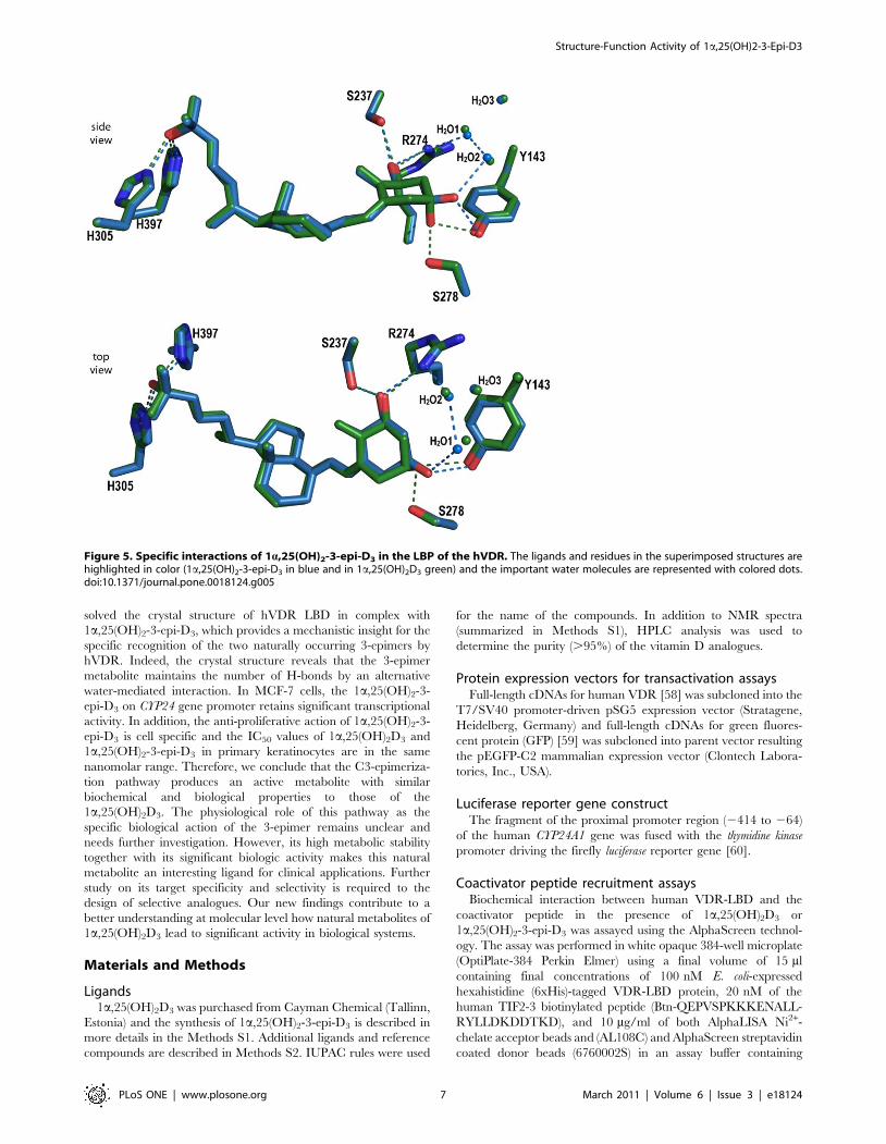
Specific interactions of the 1a,25(OH)2-3-epi-D3
The hydrophobic and electrostatic interactions between the
receptor and the ligand are similar between the two structures
except around the C3-OH group. While the C1-OH and C25-OH
display the canonical hydrogen bonds, the 3-epi-OH of
1a,25(OH)2-3-epi-D3 interacts through hydrogen bonding only
with Tyr143 instead of interacting with both Tyr143 and Ser278
(Figure 5). A significant feature of the 1a,25(OH)2-3-epi-D3 is the
compensation of the loss of interaction with Ser278 by a water-
mediated hydrogen bond with the water molecule H2O1 (W1 in
[50]). As such, the position of water H2O1 is moved 0.7 A towards
1a,25(OH)2-3-epi-D3, thereby facilitating the specific water-
mediated contacts. This water molecule is part of the network
connecting another water molecule H2O2 to Arg274. All these
water molecules are also present in the 1a,25(OH)2D3–hVDR
complex [50]. The C3-OH hydrogen bonds have longer distances
in the 3-epimer (3.0 A instead of 2.8 A with Tyr143 and 3.1 A
with the water molecule instead of 2.9 A with Ser278). A study on
the mutations of the residues forming the hydrogen bonds with the
hydroxyl groups of 1a,25(OH)2-3-epi-D3 revealed that mutated
residues contacting the 3-hydroxyl group are the less affected in
term of activity. Mutation of Ser278 in Ala may result in a lower
binding affinity for 1a,25(OH)2D3 [56] while showing a similar
potency to activate the transcription [57,56]. Due to the shift of the
side chain of Tyr147, a hydrophobic interaction with this residue is
lost in the 3-epimer structure. These structural data agree well
with the lower binding affinity of this compound for VDR and to
its induced biological activity.
In conclusion, we described a more effective synthesis of the
highly stable 1a,25(OH)2-3-epi-D3, a natural metabolite. We have
Figure 4. Adaptability of the hVDR LBP upon 1a,25(OH)2-3-epi-D3 binding. (A) The adaptation of the LBP is depicted by meshrepresentation of the superimposed LBP volumes calculated with Voidoo software. The green surface represent the LBP area where the 1a,25(OH)2D3
bound pocket is larger. The blue area represents similar increase but for 1a,25(OH)2-3-epi-D3 and the two main expanded regions are highlightedwith red circles. (B) Adaptation of the residues Tyr147 and Glu277 in the LBP of the 1a,25(OH)2-3-epi-D3 hVDR complex. The distances between theligand-specific positions of the residues are displayed in A.doi:10.1371/journal.pone.0018124.g004
Table 1. Volume of VDR ligands and their resulting LBPs.
Ligand Ligand volume [A3] * Ligand volume [%]* LBP volume [A3]** LBP volume [%]** Filling of the LBP with ligand [%]
1a,25(OH)2D3 416.56 100.00 667.13 100.00 62.44
1a,25(OH)2-3-epi-D3 407.65 97.86 633.75 95.00 64.32
The absolute values in A3 as well as relative values in reference to those of 1a,25(OH)2D3 (100%) are indicated. From these values the percent filling of the LBP withligands was also calculated.*and ** Connolly solvent accessible surfaces calculated by GRASP and Voidoo respectively The quality of the cubic grid spacing for the surface for both ligands andLBP = 0.5 A.doi:10.1371/journal.pone.0018124.t001
Structure-Function Activity of 1a,25(OH)2-3-Epi-D3
PLoS ONE | www.plosone.org 6 March 2011 | Volume 6 | Issue 3 | e18124
solved the crystal structure of hVDR LBD in complex with
1a,25(OH)2-3-epi-D3, which provides a mechanistic insight for the
specific recognition of the two naturally occurring 3-epimers by
hVDR. Indeed, the crystal structure reveals that the 3-epimer
metabolite maintains the number of H-bonds by an alternative
water-mediated interaction. In MCF-7 cells, the 1a,25(OH)2-3-
epi-D3 on CYP24 gene promoter retains significant transcriptional
activity. In addition, the anti-proliferative action of 1a,25(OH)2-3-
epi-D3 is cell specific and the IC50 values of 1a,25(OH)2D3 and
1a,25(OH)2-3-epi-D3 in primary keratinocytes are in the same
nanomolar range. Therefore, we conclude that the C3-epimeriza-
tion pathway produces an active metabolite with similar
biochemical and biological properties to those of the
1a,25(OH)2D3. The physiological role of this pathway as the
specific biological action of the 3-epimer remains unclear and
needs further investigation. However, its high metabolic stability
together with its significant biologic activity makes this natural
metabolite an interesting ligand for clinical applications. Further
study on its target specificity and selectivity is required to the
design of selective analogues. Our new findings contribute to a
better understanding at molecular level how natural metabolites of
1a,25(OH)2D3 lead to significant activity in biological systems.
Materials and Methods
Ligands1a,25(OH)2D3 was purchased from Cayman Chemical (Tallinn,
Estonia) and the synthesis of 1a,25(OH)2-3-epi-D3 is described in
more details in the Methods S1. Additional ligands and reference
compounds are described in Methods S2. IUPAC rules were used
for the name of the compounds. In addition to NMR spectra
(summarized in Methods S1), HPLC analysis was used to
determine the purity (.95%) of the vitamin D analogues.
Protein expression vectors for transactivation assaysFull-length cDNAs for human VDR [58] was subcloned into the
T7/SV40 promoter-driven pSG5 expression vector (Stratagene,
Heidelberg, Germany) and full-length cDNAs for green fluores-
cent protein (GFP) [59] was subcloned into parent vector resulting
the pEGFP-C2 mammalian expression vector (Clontech Labora-
tories, Inc., USA).
Luciferase reporter gene constructThe fragment of the proximal promoter region (2414 to 264)
of the human CYP24A1 gene was fused with the thymidine kinase
promoter driving the firefly luciferase reporter gene [60].
Coactivator peptide recruitment assaysBiochemical interaction between human VDR-LBD and the
coactivator peptide in the presence of 1a,25(OH)2D3 or
1a,25(OH)2-3-epi-D3 was assayed using the AlphaScreen technol-
ogy. The assay was performed in white opaque 384-well microplate
(OptiPlate-384 Perkin Elmer) using a final volume of 15 ml
containing final concentrations of 100 nM E. coli-expressed
hexahistidine (6xHis)-tagged VDR-LBD protein, 20 nM of the
human TIF2-3 biotinylated peptide (Btn-QEPVSPKKKENALL-
RYLLDKDDTKD), and 10 mg/ml of both AlphaLISA Ni2+-
chelate acceptor beads and (AL108C) and AlphaScreen streptavidin
coated donor beads (6760002S) in an assay buffer containing
Figure 5. Specific interactions of 1a,25(OH)2-3-epi-D3 in the LBP of the hVDR. The ligands and residues in the superimposed structures arehighlighted in color (1a,25(OH)2-3-epi-D3 in blue and in 1a,25(OH)2D3 green) and the important water molecules are represented with colored dots.doi:10.1371/journal.pone.0018124.g005
Structure-Function Activity of 1a,25(OH)2-3-Epi-D3
PLoS ONE | www.plosone.org 7 March 2011 | Volume 6 | Issue 3 | e18124

50 mM MOPS pH = 7.4, 50 mM NaF, 50 mM CHAPS, and
100 mg/ml bovine serum albumin. Different concentrations of
1a,25(OH)2D3 or 1a,25(OH)2-3-epi-D3 dissolved in DMSO
(maintained at a final concentration of 1%) were added as indicated.
The experiment represents two independent measurements in
triplicates for, which the mean and the S.D. of the mean was
calculated.
Transient transfections and luciferase reporter geneassays
MCF-7 cells were seeded into 24-well plates (100,000 cells/well)
and grown overnight in phenol red-free Dulbecco’s modified
Eagle’s medium (DMEM) supplemented with 10% charcoal-
stripped fetal bovine serum (FCS) and 0.6 mg/ml insulin. Plasmid
DNA containing liposomes were formed by incubating 40 ng of an
expression vector for hVDR, 100 ng of reporter plasmid and
10 ng pEGF-C2 with Fugene 6 (Roche Diagnostics, Switzerland)
transfection reagent according to the recommendation of the
manufacturer for 15 min at room temperature. After dilution with
500 ml of phenol red-free DMEM, the liposomes were added to
the cells. Phenol red-free DMEM supplemented with 500 ml of
20% charcoal-stripped FCS was added 4 h after transfection, in
the presence of ligands or solvent. The cells were lysed 16 h after
the onset of stimulation using reporter gene lysis buffer (Roche
Diagnostics, Switzerland). The lysates were assayed for luciferase
activity as recommended by the supplier (Perkin-Elmer, The
Netherlands). The luciferase activities were normalized to GFP
expression. Data represent one triplicate for which the mean and
the S.D. of the mean was calculated.
Data analysis for dose response curvesA non-linear curve fit was performed for the AlphaScreen and
reporter gene assay experimental dose response data and from
sigmoidal dose response curve then the EC50 values for the
respective ligands were calculated using GraphPad Prism 4
(GraphPad Software Inc., San Diego, CA). The Student’s
unpaired t-test and Pearson correlation were performed with the
SPSS software (SPSS Inc., version 14.0, Chicago, IL, USA).
Keratinocyte cell culturesNormal human keratinocytes were isolated from fresh adult skin
obtained from surgery and immediately transported to the
laboratory under sterile conditions. Isolation and culture under
serum-free conditions and without a feeder layer followed a
modified protocol as used by Bikle et al [11]. The isolated epidermis
was incubated in a 0.25% trypsin solution for 45 min at 37uC.
Thereafter, the cells were scraped off and put in 50 ml Hank’s
balanced salt solution (HBSS) containing 10% FCS to block further
trypsin digestion and centrifuged at 2000 rpm/2 min. The resulting
cell pellet was suspended in Keratinocyte Growth Medium (KGM,
Clonetics Corp., San Diego), a defined serum-free medium at low
(0.06 mM) calcium containing 0.1 ng/ml epidermal growth factor,
5 mg/ml insulin, 0.5 mg/ml hydrocortisone, bovine pituitary
extract, antibiotics (gentamycin, amphothericin) gave the primary
culture. After 24 h, the cells were incubated at 37uC in 95% air/5%
CO2 and the attached cells were washed and provided with fresh
KGM medium. The culture medium was changed every other day
and the cells were passaged when they reached 80–90% confluency
(usually 6–10 days after plating).
Incubations of primary keratinocytes with 3H-25(OH)D3
Confluent human keratinocytes in 1 ml KGM and in 6-well
plates were incubated in duplicates at 37uC with 20.6 nM 3H-
25(OH)D3 (around 600 000 dpm/ml) for 1–23 h. Incubations
were stopped with 1 ml methanol/well, the cells were scraped off,
transferred to a test tube together with the supernatant and two
washings (with 1 ml methanol and 0.8 ml water). Unmodified 3H-
25(OH)D3 and most of the products were totally extracted from
the combined solutions plus cell pellet according to the method of
Bligh and Dyer [61] by three subsequent extractions with 2, 1 and
1 ml volumes of CHCl3 at room temperature. 3H-activity in the
CHCl3-phase, in the water and total 3H-yield were determined.
The combined CHCl3 extracts were then evaporated under argon
at 35uC, the residues dissolved in 0.4 ml ethanol and an aliquot
(containing around 250 000 dpm 3H-activity) subjected to HPLC-
analysis (see Methods S2).
3H-Thymidine incorporation (anti-proliferation assay inprimary keratinocytes)
Keratinocytes (second passage) in 200 ml KGM (low calcium)
were plated in 96-well plates at an initial density of 104 cells/well,
kept 24 h at 37uC in an incubator with 95% air/5% CO2.
Thereafter, the test compounds 1,25(OH)2D3 or its 3-epimer were
added in 1 ml ethanol to give final concentrations ranging from 0
to 100 nM, each condition in triplicates. After further 24 h, 50 ml3H-thymidine (1 mCi) were added and incubation continued for
additional 7 h. Then, incubations were stopped by cell harvesting
(Filtermate 196 Harvester, Packard-Canberra) and lysis: After
removing the supernatant (see below), the adherent cells were
released by 5 min treatment with 100 ml 0.125% trypsin in PBS at
37uC, harvested on a filterplate and washed 3 x with redistilled
water. After drying the plates, their bottoms were sealed with a
film and 50 ml scintillation cocktail (MicroScint O, Packard) were
added. The whole plates were sealed with Packard Cover Film and3H-activity counted on a Microplate Scintillation Counter
(TopCount, Packard Canberra). To check whether proliferative
(3H-thymidine incorporating) cells could have been shed off, the
supernatants were soaked through a 96-well filterplate (Unifilter
Plate GF/C) and 3 x washed with redistilled water: in all
conditions, 3H-activity was undetectable on these filterplates (in
order to roughly assess cell numbers and check for substance
related morphological changes/toxic effects, photographs were
taken prior to compound addition and immediately before
harvesting.) Data - used as means 6 SD – were normalized
(incorporated 3H-activity sample vs. blank) and analyzed using the
GRAFIT Erithacus 4.0.19 IC50 software.
Protein purification and CrystallizationPurification and crystallization of the hVDR LBD complexed
with 1a,25(OH)2-3-epi-D3 were performed as previously described
[49]. The LBD of the hVDR (residues 118-427 D166-216) was
cloned in pET-28b expression vector (Novagen) to obtain an N-
terminal 6xHis fusion protein and was overproduced in E. coli
BL21 (DE3) strain. Cells were grown in Luria Bertani medium and
subsequently incubated for 6 h at 20uC with 1 mM isopropyl thio-
b-D-galactoside. The protein purification included a metal affinity
chromatography step on a Co2+-chelating resin (Clontech). The
6xHis tag was removed by thrombin digestion overnight at 4uC,
and the protein was further purified by gel filtration on a Superdex
S200 16/60. The sample buffer prior to protein concentration
contained 10 mM Tris, pH = 7.5, 100 mM NaCl, and 10 mM
dithiothreitol. The protein was concentrated to 3.5 mg/ml and
incubated in the presence of a 1.5-fold molar excess of ligand. The
purity and homogeneity of the protein were assessed by SDS-
PAGE. The protein crystals were obtained at 4uC by vapor
diffusion method using crystals of hVDR LBD-1a,25(OH)2D3 as
Structure-Function Activity of 1a,25(OH)2-3-Epi-D3
PLoS ONE | www.plosone.org 8 March 2011 | Volume 6 | Issue 3 | e18124

microseeds. The reservoir solution contained 0.1 M MES and
1.4 M ammonium sulfate pH = 6.0.
X-Ray data collection and structure determinationThe crystal was mounted in fiber loop and flash cooled in liquid
nitrogen after cryoprotection with a solution containing the reservoir
plus 30% glycerol and 2% polyethylene glycol 400. Data collection
from a single frozen crystal was performed at 100 K on the beamline
ID29 of the European Synchrotron Radiation Facility (Grenoble,
France). The crystal belongs to the orthorhombic space group
P212121 with one monomer per asymmetric unit. Data were
integrated and scaled using MOSFLM [62] (see statistics in Table
S1). A rigid body refinement was used with the structure of the hVDR
LBD complexed to 1a,25(OH)2D3 as a starting model. Refinement
involved iterative cycles of manual building and refinement
calculations. The programs Refmac [63] and COOT [64] were
used throughout structure determination and refinement. The omit
map from the refined atomic model of hVDR LBD was used to fit the
ligand to its electron density, shown in Figure 2A. Individual B-
atomic factors were refined isotropically. Solvent molecules were then
placed according to unassigned peaks in the difference Fourier map.
In the hVDR/1a,25(OH)2-3-epi-D3 complex, refined at 1.9 A with
no s cutoff, the final model consists of residues 118-423 (D166–216),
the ligand, two sulphate ions and 372 water molecules. According to
PROCHECK [65] 92.6% of peptide lies in most favored regions and
7.4% in additional allowed regions. Data are summarized in Table
S1. The volumes of the ligand-binding pockets and ligands were
calculated as previously reported [49].
Supporting Information
Figure S1 Biological properties of 1a,25(OH)2D3 and1a,25(OH)2-3-epi-D3 in HL60 cellular model. (A)
1a,25(OH)2-3-epi-D3-mediated HL60 cell growth. 1a,25(OH)2D3
or 1a,25(OH)2-3-epi-D3-treated HL60 at 1 nM and 100 nM
concentrations are counted. Data are presented as mean6S.D. of
the mean (*, p,0.05; **, p,0.01; ***, p,0.001). (B) 1a,25(OH)2-3-
epi-D3-mediated HL60 cell differentiation into monocyte-like cells.
HL60 cells were treated with either ethanol or 1 nM and 100"nM
concentration of 1a,25(OH)2D3 or 1a,25(OH)2-3-epi-D3. Cells were
labeled with PElabeled anti-human CD11c and FITC-labeled anti-
human CD14, and HL60 cell differentiation was estimated by the
double-positive CD11c/CD14 population. Data are representative of
three distinct experiments.
(PDF)
Figure S2 Dominant production of the 1a,25(OH)2-3-epi-D3 in keratinocytes after 5 h. HPLC profile of the
CHCl3-extract from keratinocytes after 5 h incubation is shown.
The amount of 1a,25(OH)2-3-epi-D3 (blue star) is the highest from
all the metabolites detected with HPLC. The peak of
1a,25(OH)2D3 is highlighted with green star.
(PDF)
Table S1 Data collection and refinement statistics.
(PDF)
Methods S1 Synthesis.
(PDF)
Methods S2
(DOCX)
Scheme S1 Retrosynthesis of 1b.
(PDF)
Scheme S2 Synthesis of enol triflate 3. Si = TBS = Si(t-
Bu)(CH3)2.TBHP = t-BuOOH (a) NaBH4, CeCl3?7H2O, MeOH,
0uC, 30 min. (b) TBHP, VO(acac)2, PhH, reflux, 30 min. (c)
TBSCl, Im, DMF, rt, 12 h. (d) O3, MeOH-CH2Cl2, 278uC;
Ac2O, Et3N, DMAP, 235uC to 28uC, 2 h; NaOAc, MeOH,
37uC, 12 h, (e) H5IO6, Et2O, rt, 2 h. (f) CBr4, Zn, Ph3P, CH2Cl2,
rt, 40 min. (g) LDA, THF, 278uC, 1 h; n-BuLi, 15 min; 5-Cl-
Py-2NTf2, 278uC to rt, 12 h.
(PDF)
Scheme S3 Synthesis of metabolite 1. TES =
Si(CH2CH3)3. (a) (Ph3PCH2Br)Br, KOt-Bu, toluene, 25uC to rt,
1 h, 80%. (b) TESCl, Im, DMAP, DMF, rt, 3 h, 91%. (c) InCl3, t-
BuLi, THF, 278uC to 0uC, 2 h. (d) 3, (Ph3P)4Pd, Et3N, THF,
(dppf)PdCl2, 0uC to rt, 12 h. (e) HF?Py, Et3N, CH2Cl2, CH3CN,
rt, 4 h, 58%.
(PDF)
Acknowledgments
We thank the beam-line staff at the ESRF (Grenoble, France) for help
during data collection and T. Huet for critical reading. We thank N.
Rouleau and J. Hyvarinen from Perkin Elmer for support in AlphaScreen
Assays and V. Prantner for help with statistical analysis.
Author Contributions
Conceived and designed the experiments: AM DM NR. Performed the
experiments: FM RS YS CA IS PA JP. Analyzed the data: FM IS AM NR.
Contributed reagents/materials/analysis tools: CM. Wrote the paper: FM
AM NR.
References
1. Adorini L, Penna G (2008) Control of autoimmune diseases by the vitamin D
endocrine system. Nat Clin Pract Rheumatol 4: 404–412.
2. Bouillon R, Eelen G, Verlinden L, Mathieu C, Carmeliet G, et al. (2006)
Vitamin D and cancer. J Steroid Biochem Mol Biol 102: 156–162.
3. Pinette KV, Yee YK, Amegadzie BY, Nagpal S (2003) Vitamin D receptor as a
drug discovery target. Mini Rev Med Chem 3: 193–204.
4. Campbell MJ, Adorini L (2006) The vitamin D receptor as a therapeutic target.
Expert Opin Ther Targets 10: 735–748.
5. DeLuca HF (2008) Evolution of our understanding of vitamin D. Nutr Rev 66:
S73–S87.
6. Nagpal S, Na S, Rathnachalam R (2005) Noncalcemic actions of vitamin D
receptor ligands. Endocr Rev 26: 662–687.
7. Garabedian M, Lieberherr M, N’Guyen TM, Corvol MT, Du Bois MB, et al.
(1978) The in vitro production and activity of 24, 25-dihydroxycholecalciferol in
cartilage and calvarium. Clin Orthop Relat Res 135: 241–248.
8. Howard GA, Turner RT, Sherrard DJ, Baylink DJ (1981) Human bone cells in
culture metabolize 25-hydroxyvitamin D3 to 1,25-dihydroxyvitamin D3 and
24,25-dihydroxyvitamin D3. J Biol Chem 256: 7738–7740.
9. Jones G, Vriezen D, Lohnes D, Palda V, Edwards NS (1987) Side-chain
hydroxylation of vitamin D3 and its physiological implications. Steroids 49:
29–53.
10. Ishizuka S, Norman AW (1987) Metabolic pathways from 1a,25-dihydroxyvi-
tamin D3 to 1a,25-dihydroxyvitamin D3-26,23-lactone. Stereo-retained and
stereo-selective lactonization. J Biol Chem 262: 7165–7170.
11. Bikle DD, Nemanic MK, Whitney JO, Elias PW (1986) Neonatal human
foreskin keratinocytes produce 1,25-dihydroxyvitamin D3. Biochemistry 25:
1545–1548.
12. Horst RL (1979) 25-OHD3-26,23-lactone: a metabolite of vitamin D3 that is 5
times more potent than 25-OHD3 in the rat plasma competitive protein binding
radioassay. Biochem Biophys Res Commun 89: 286–293.
Structure-Function Activity of 1a,25(OH)2-3-Epi-D3
PLoS ONE | www.plosone.org 9 March 2011 | Volume 6 | Issue 3 | e18124

13. Wichmann JK, DeLuca HF, Schnoes HK, Horst RL, Shepard RM, et al. (1979)
25-Hydroxyvitamin D3 26,23-lactone: a new in vivo metabolite of vitamin D.
Biochemistry 18: 4775–4780.
14. DeLuca HF, Suda T, Schnoes HK, Tanaka Y, Holick MF (1970) 25,26-
dihydroxycholecalciferol, a metabolite of vitamin D3 with intestinal calcium
transport activity. Biochemistry 9: 4776–4780.
15. Reinhardt TA, Napoli JL, Praminik B, Littledike ET, Beitz DC, et al. (1981) 1a-
25,26-trihydroxyvitamin D3: an in vivo and in vitro metabolite of vitamin D3.
Biochemistry 20: 6230–6235.
16. Holick MF, Schnoes HK, DeLuca HF, Gray RW, Boyle IT, et al. (1972)
Isolation and identification of 24,25-dihydroxycholecalciferol, a metabolite of
vitamin D made in the kidney. Biochemistry 11: 4251–4255.
17. Kumar R, Schnoes HK, DeLuca HF (1978) Rat intestinal 25-hydroxyvitamin
D3- and 1a,25-dihydroxyvitamin D3-24-hydroxylase. J Biol Chem 253:
3804–3809.
18. Reddy GS, Rao DS, Siu-Caldera ML, Astecker N, Weiskopf A, et al. (2000)
1a,25-dihydroxy-16-ene-23-yne-vitamin D3 and 1a,25-dihydroxy-16-ene-23-
yne-20-epi-vitamin D3: analogs of 1a,25-dihydroxyvitamin D3 that resist
metabolism through the C-24 oxidation pathway are metabolized through the
C-3 epimerization pathway. Arch Biochem Biophys 383: 197–205.
19. Norman AW, Bouillon R, Farach-Carson MC, Bishop JE, Zhou LX, et al.
(1993) Demonstration that 1 beta,25-dihydroxyvitamin D3 is an antagonist of the
nongenomic but not genomic biological responses and biological profile of the
three A-ring diastereomers of 1a,25-dihydroxyvitamin D3. J Biol Chem 268:
20022–20030.
20. Reddy GS, Muralidharan KR, Okamura WH, Tserng K-Y, McLane JA (1994)
Metabolism of 1a,25-dihydroxyvitamin D3 and one of its A-ring diastereomer
1a,25-dihydroxy-3-epivitamin D3 in neonatal human keratinocytes. In:
Norman AW, Bouillon R, Thomasset M, eds. Vitamin D a pluripotent steroid
hormone: Structural studies, molecular endocrinology and clinical applications,
Walter de Gruyter, NY, USA. Walter de Gruyter, NY, USA. pp 172–173.
21. Reddy GS, Siu-Caldera M-L, Schuster I, Astecker N, Tserng K-Y, et al. (1997)
Target tissue specific metabolism of 1a,25-dihydroxyvitamin D3 through A-ring
modification. In: Norman WA, Bouillon R, Thomasset M, eds. Vitamin D,
chemistry, biology and clinical applications of the steroid hormone, Riverside,
CA, USA. Riverside, CA, USA. pp 139–146.
22. Sekimoto H, Siu-Caldera ML, Weiskopf A, Vouros P, Muralidharan KR, et al.
(1999) 1a,25-dihydroxy-3-epi-vitamin D3: in vivo metabolite of 1a,25-dihydrox-
yvitamin D3 in rats. FEBS Lett 448: 278–282.
23. Bischof MG, Siu-Caldera ML, Weiskopf A, Vouros P, Cross HS, et al. (1998)
Differentiation-related pathways of 1a,25-dihydroxycholecalciferol metabolism
in human colon adenocarcinoma-derived Caco-2 cells: production of 1a,25-
dihydroxy-3epi-cholecalciferol. Exp Cell Res 241: 194–201.
24. Rehan VK, Torday JS, Peleg S, Gennaro L, Vouros P, et al. (2002) 1a,25-
dihydroxy-3-epi-vitamin D3, a natural metabolite of 1a,25-dihydroxy vitamin
D3: production and biological activity studies in pulmonary alveolar type II cells.
Mol Genet Metab 76: 46–56.
25. Brown AJ, Ritter C, Slatopolsky E, Muralidharan KR, Okamura WH, et al.
(1999) 1a,25-dihydroxy-3-epi-vitamin D3, a natural metabolite of 1a,25-
dihydroxyvitamin D3, is a potent suppressor of parathyroid hormone secretion.
J Cell Biochem 73: 106–113.
26. Siu-Caldera ML, Sekimoto H, Weiskopf A, Vouros P, Muralidharan KR, et al.
(1999) Production of 1a,25-dihydroxy-3-epi-vitamin D3 in two rat osteosarcoma
cell lines (UMR 106 and ROS 17/2.8): existence of the C-3 epimerization
pathway in ROS 17/2.8 cells in which the C-24 oxidation pathway is not
expressed. Bone 24: 457–463.
27. Hylemon PB, Sjovall HDAJ (1985) Chapter 12 Metabolism of bile acids in
intestinal microflora. In: New Comprehensive Biochemistry Sterols and Bile
Acids Elsevier. pp 331–343.
28. Penning TM, Bennett MJ, Smith-Hoog S, Schlegel BP, Jez JM, et al. (1997)
Structure and function of 3a-hydroxysteroid dehydrogenase. Steroids 62:
101–111.
29. Reddy GS, Rao DS, Siu-Caldera ML (2000) Natural metabolites of 1a,25-
dihydroxy-vitamin D3 and its analogs. In: Norman AW, Bouillon R, Thomasset ,
eds. Vitamin D Endocrine system, Structural, Biological, Genetic and Clinical
Aspects, University of California, Printing and Reprographics, Riverside, CA,
USA. pp 139–146.
30. Kamao M, Tatematsu S, Hatakeyama S, Sakaki T, Sawada N, et al. (2004) C-3
epimerization of vitamin D3 metabolites and further metabolism of C-3 epimers:
25-hydroxyvitamin D3 is metabolized to 3-epi-25-hydroxyvitamin D3 and
subsequently metabolized through C-1a or C-24 hydroxylation. J Biol Chem
279: 15897–15907.
31. Morrison NA, Eisman JA (1991) Nonhypercalcemic 1,25-(OH)2D3 analogs
potently induce the human osteocalcin gene promoter stably transfected into rat
osteosarcoma cells (ROSCO-2). J Bone Miner Res 6: 893–899.
32. Fleet JC, Bradley J, Reddy GS, Ray R, Wood RJ (1996) 1a,25-(OH)2-vitamin
D3 analogs with minimal in vivo calcemic activity can stimulate significant
transepithelial calcium transport and mRNA expression in vitro. Arch Biochem
Biophys 329: 228–234.
33. Muralidharan KR, De Lera AR, Isaeff SD, Norman AW, Okamura WH (1993)
Studies of vitamin D (calciferol) and its analogs. 45. Studies on the A-ring
diastereomers of 1a,25-dihydroxyvitamin D3. J Org Chem 58: 1895–1899.
34. Gomez-Reino C, Vitale C, Maestro M, Mourino A (2005) Pd-catalyzed
carbocyclization-Negishi cross-coupling cascade: a novel approach to 1a,25-
dihydroxyvitamin D3 and analogues. Org Lett 7: 5885–5887.
35. Zhu G, Okamura WH (1995) Synthesis of Vitamin D (Calciferol). Chem Rev 95:
1877–1952.
36. Krause S, Schmalz HG (2000) Palladium-Catalyzed Synthesis of Vitamin D-
Active Compounds. In: Organic Synthesis Highlights IV. Germany: Wiley and
VCH. pp 212–217.
37. Posner GH, Kahraman M (2003) Organic chemistry of vitamin D analogues
(deltanoids). Eur J Org Chem 2003. pp 3889–3895.
38. Luche JL (1978) Lanthanides in organic chemistry. 1. Selective 1, 2 reductions of
conjugated ketones. J Am Chem Soc 100: 2226–2227.
39. Sharpless KB, Michaelson RC (1973) High stereo-and regioselectivities in the
transition metal catalyzed epoxidations of olefinic alcohols by tert-butyl
hydroperoxide. J Am Chem Soc 95: 6136–6137.
40. Daniewski AR, Garofalo LM, Hutchings SD, Kabat MM, Liu W, et al. (2002)
Efficient synthesis of the A-ring phosphine oxide building block useful for 1a,25-
dihydroxy-vitamin D and analogues. J Org Chem 67: 1580–1587.
41. Corey EJ, Fuchs PL (1972) A synthetic method for formyl--. ethynyl conversion
(RCHO--. RCCH or RCCR’). Tetrahedron Lett 13: 3769–3772.
42. Mourino A, Torneiro M, Vitale C, Fernandez S, Perez-Sestelo J, et al. (1997)
Efficient and versatile synthesis of A-ring precursors of 1a,25-dihydroxy-vitamin
D3 and analogues. Applications to the synthesis of Lythgoe-Roche phospine
oxide. Tetrahedron letters 38: 4713–4716.
43. Gogoi P, Sigueiro R, Eduardo S, Mourino A (2010) An expeditious route to 1
a,25-dihydroxyvitamin D(3) and its analogues by an aqueous tandem palladium-
catalyzed a-ring closure and suzuki coupling to the C/D unit. Eur J Chem 16:
1432–1435.
44. Trost BM, Dumas J, Villa M (1992) New Strategies for the synthesis of vitamin
D metabolites via Pd-catalyzed reactions. J Am Chem Soc 114: 9836–9845.
45. Takeyama K, Masuhiro Y, Fuse H, Endoh H, Murayama A, et al. (1999)
Selective interaction of vitamin D receptor with transcriptional coactivators by a
vitamin D analog. Mol Cell Biol 19: 1049–1055.
46. Ullman EF, Kirakossian H, Singh S, Wu ZP, Irvin BR, et al. (1994) Luminescent
oxygen channeling immunoassay: measurement of particle binding kinetics by
chemiluminescence. Proc Natl Acad Sci U S A 91: 5426–5430.
47. Nakagawa K, Sowa Y, Kurobe M, Ozono K, Siu-Caldera ML, et al. (2001)
Differential activities of 1a,25-dihydroxy-16-ene-vitamin D(3) analogs and their
3-epimers on human promyelocytic leukemia (HL-60) cell differentiation and
apoptosis. Steroids 66: 327–337.
48. Harant H, Spinner D, Reddy GS, Lindley IJ (2000) Natural metabolites of
1a,25-dihydroxyvitamin D(3) retain biologic activity mediated through the
vitamin D receptor. J Cell Biochem 78: 112–120.
49. Rochel N, Wurtz JM, Mitschler A, Klaholz B, Moras D (2000) The crystal
structure of the nuclear receptor for vitamin D bound to its natural ligand. Mol
Cell 5: 173–179.
50. Hourai S, Fujishima T, Kittaka A, Suhara Y, Takayama H, et al. (2006) Probing
a water channel near the A-ring of receptor-bound 1 a,25-dihydroxyvitamin D3
with selected 2a-substituted analogues. J Med Chem 49: 5199–5205.
51. Eelen G, Valle N, Sato Y, Rochel N, Verlinden L, et al. (2008) Superagonistic
fluorinated vitamin D3 analogs stabilize helix 12 of the vitamin D receptor.
Chem Biol 15: 1029–1034.
52. Tocchini-Valentini G, Rochel N, Wurtz JM, Mitschler A, Moras D (2001)
Crystal structures of the vitamin D receptor complexed to superagonist 20-epi
ligands. Proc Natl Acad Sci U S A 98: 5491–5496.
53. Hourai S, Rodrigues LC, Antony P, Reina-San-Martin B, Ciesielski F, et al.
(2008) Structure-based design of a superagonist ligand for the vitamin D nuclear
receptor. Chem Biol 15: 383–392.
54. Rochel N, Tocchini-Valentini G, Egea PF, Juntunen K, Garnier JM, et al.
(2001) Functional and structural characterization of the insertion region in the
ligand binding domain of the vitamin D nuclear receptor. Eur J Biochem 268:
971–979.
55. Molnar F, Perakyla M, Carlberg C (2006) Vitamin D receptor agonists
specifically modulate the volume of the ligand-binding pocket. J Biol Chem 281:
10516–10526.
56. Reddy MD, Stoynova L, Acevedo A, Collins ED (2007) Residues of the human
nuclear vitamin D receptor that form hydrogen bonding interactions with the
three hydroxyl groups of 1a,25-dihydroxyvitamin D3. J Steroid Biochem Mol
Biol 103: 347–351.
57. Choi M, Yamamoto K, Masuno H, Nakashima K, Taga T, et al. (2001) Ligand
recognition by the vitamin D receptor. Bioorg Med Chem 9: 1721–1730.
58. Carlberg C, Bendik I, Wyss A, Meier E, Sturzenbecker LJ, et al. (1993) Two
nuclear signalling pathways for vitamin D. Nature 361: 657–660.
59. Cormack BP, Valdivia RH, Falkow S (1996) FACS-optimized mutants of the
green fluorescent protein (GFP). Gene 173: 33–38.
60. Vaisanen S, Dunlop TW, Sinkkonen L, Frank C, Carlberg C (2005) Spatio-
temporal activation of chromatin on the human CYP24 gene promoter in the
presence of 1a,25-Dihydroxyvitamin D3. J Mol Biol 350: 65–77.
61. Bligh EG, Dyer WJ (1959) A rapid method of total lipid extraction and
purification. Can J Biochem Physiol 37: 911–917.
62. Leslie AGW (1992) Recent changes to the MOSFLM package for processing
film and image plate data. Joint CCP4 + ESF-EAMCB Newsletter on Protein
Crystallography No. 26.
Structure-Function Activity of 1a,25(OH)2-3-Epi-D3
PLoS ONE | www.plosone.org 10 March 2011 | Volume 6 | Issue 3 | e18124
63. Murshudov GN, Vagin AA, Dodson EJ (1997) Refinement of macromolecular
structures by the maximum-likelihood method. Acta Crystallogr D Biol Crystal-logr 53: 240–255.
64. Emsley P, Cowtan K (2004) Coot: model-building tools for molecular graphics.
Acta Crystallogr D Biol Crystallogr 60: 2126–2132.
65. Laskowski RA, MacArthur MW, Moss DS, Thornton JM (1993) PROCHECK:
A program to check the stereochemical quality of protein structures. J Appl
Crystallogr 26: 283–291.
Structure-Function Activity of 1a,25(OH)2-3-Epi-D3
PLoS ONE | www.plosone.org 11 March 2011 | Volume 6 | Issue 3 | e18124