ZOONOSES AND COMMUNICABLE DISEASES COMMON ...
404
ZOONOSES AND COMMUNICABLE DISEASES COMMON TO MAN AND ANIMALS Third Edition Volume III Parasitoses Scientific and Technical Publication No. 580 PAN AMERICAN HEALTH ORGANIZATION Pan American Sanitary Bureau, Regional Office of the WORLD HEALTH ORGANIZATION 525 Twenty-third Street, N.W. Washington, D.C. 20037 U.S.A. 2003
-
Upload
khangminh22 -
Category
Documents
-
view
0 -
download
0
Transcript of ZOONOSES AND COMMUNICABLE DISEASES COMMON ...

ZOONOSES AND COMMUNICABLE DISEASES COMMON TO MAN AND ANIMALS
Third Edition
Volume III
Parasitoses
Scientific and Technical Publication No. 580
PAN AMERICAN HEALTH ORGANIZATIONPan American Sanitary Bureau, Regional Office of the
WORLD HEALTH ORGANIZATION525 Twenty-third Street, N.W.
Washington, D.C. 20037 U.S.A.
2003

Also published in Spanish (2003) with the title:Zoonosis y enfermedades transmisibles comunes al hombre y a los animales:
parasitosisISBN 92 75 31991 X (3 volume set)
ISBN 92 75 31992 8 (Vol. 3)
PAHO HQ Library Cataloguing-in-PublicationPan American Health Organization
Zoonoses and communicable diseases common to man and animals:parasitoses3rd ed. Washington, D.C.: PAHO, © 2003.3 vol.—(Scientific and Technical Publication No. 580)
ISBN 92 75 11991 0—3 volume setISBN 92 75 11993 7—Vol. 3I. Title II. (Series)1. ZOONOSES2. PARASITIC DISEASES3. DISEASE RESERVOIRS4. COMMUNICABLE DISEASE CONTROL5. FOOD CONTAMINATION6. PUBLIC HEALTH VETERINARYNLM WC950.P187 2003 v.3 En
The Pan American Health Organization welcomes requests for permission to reproduce ortranslate its publications, in part or in full. Applications and inquiries should be addressed toPublications, Pan American Health Organization, Washington, D.C., U.S.A., which will beglad to provide the latest information on any changes made to the text, plans for new editions,and reprints and translations already available.
© Pan American Health Organization, 2003
Publications of the Pan American Health Organization enjoy copyright protection in accor-dance with the provisions of Protocol 2 of the Universal Copyright Convention. All rights arereserved.
The designations employed and the presentation of the material in this publication do notimply the expression of any opinion whatsoever on the part of the Secretariat of the PanAmerican Health Organization concerning the status of any country, territory, city or area orof its authorities, or concerning the delimitation of its frontiers or boundaries.
The mention of specific companies or of certain manufacturers’ products does not implythat they are endorsed or recommended by the Pan American Health Organization in prefer-ence to others of a similar nature that are not mentioned. Errors and omissions excepted, thenames of proprietary products are distinguished by initial capital letters.
This volume was updated by Omar O. Barriga, Professor of Parasitology in the Facultyof Medicine of the University of Chile, and consultant for PAHO’s Veterinary PublicHealth Unit from 1997 to 2001.
CONTENTS
Prologue . . . . . . . . . . . . . . . . . . . . . . . . . . . . . . . . . . . . . . . . . . . . . . . . . . . . . . viiPreface to the First Edition . . . . . . . . . . . . . . . . . . . . . . . . . . . . . . . . . . . . . . . . . ixPreface to the Second Edition . . . . . . . . . . . . . . . . . . . . . . . . . . . . . . . . . . . . . . . xiIntroduction . . . . . . . . . . . . . . . . . . . . . . . . . . . . . . . . . . . . . . . . . . . . . . . . . . . . xv
Section A: PROTOZOOSES
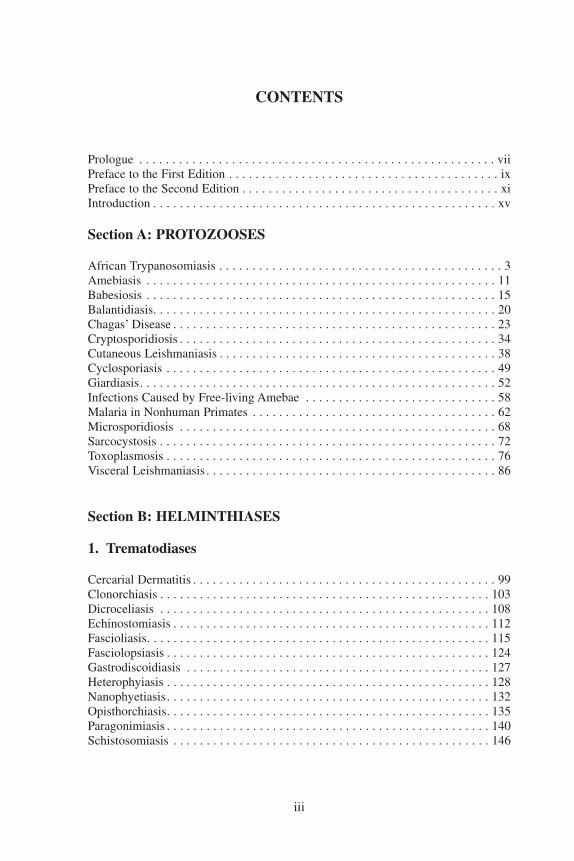
African Trypanosomiasis . . . . . . . . . . . . . . . . . . . . . . . . . . . . . . . . . . . . . . . . . . . 3Amebiasis . . . . . . . . . . . . . . . . . . . . . . . . . . . . . . . . . . . . . . . . . . . . . . . . . . . . . 11Babesiosis . . . . . . . . . . . . . . . . . . . . . . . . . . . . . . . . . . . . . . . . . . . . . . . . . . . . . 15Balantidiasis. . . . . . . . . . . . . . . . . . . . . . . . . . . . . . . . . . . . . . . . . . . . . . . . . . . . 20Chagas’ Disease . . . . . . . . . . . . . . . . . . . . . . . . . . . . . . . . . . . . . . . . . . . . . . . . . 23Cryptosporidiosis . . . . . . . . . . . . . . . . . . . . . . . . . . . . . . . . . . . . . . . . . . . . . . . . 34Cutaneous Leishmaniasis . . . . . . . . . . . . . . . . . . . . . . . . . . . . . . . . . . . . . . . . . . 38Cyclosporiasis . . . . . . . . . . . . . . . . . . . . . . . . . . . . . . . . . . . . . . . . . . . . . . . . . . 49Giardiasis. . . . . . . . . . . . . . . . . . . . . . . . . . . . . . . . . . . . . . . . . . . . . . . . . . . . . . 52Infections Caused by Free-living Amebae . . . . . . . . . . . . . . . . . . . . . . . . . . . . . 58Malaria in Nonhuman Primates . . . . . . . . . . . . . . . . . . . . . . . . . . . . . . . . . . . . . 62Microsporidiosis . . . . . . . . . . . . . . . . . . . . . . . . . . . . . . . . . . . . . . . . . . . . . . . . 68Sarcocystosis . . . . . . . . . . . . . . . . . . . . . . . . . . . . . . . . . . . . . . . . . . . . . . . . . . . 72Toxoplasmosis . . . . . . . . . . . . . . . . . . . . . . . . . . . . . . . . . . . . . . . . . . . . . . . . . . 76Visceral Leishmaniasis . . . . . . . . . . . . . . . . . . . . . . . . . . . . . . . . . . . . . . . . . . . . 86
Section B: HELMINTHIASES
1. Trematodiases
Cercarial Dermatitis . . . . . . . . . . . . . . . . . . . . . . . . . . . . . . . . . . . . . . . . . . . . . . 99Clonorchiasis . . . . . . . . . . . . . . . . . . . . . . . . . . . . . . . . . . . . . . . . . . . . . . . . . . 103Dicroceliasis . . . . . . . . . . . . . . . . . . . . . . . . . . . . . . . . . . . . . . . . . . . . . . . . . . 108Echinostomiasis . . . . . . . . . . . . . . . . . . . . . . . . . . . . . . . . . . . . . . . . . . . . . . . . 112Fascioliasis. . . . . . . . . . . . . . . . . . . . . . . . . . . . . . . . . . . . . . . . . . . . . . . . . . . . 115Fasciolopsiasis . . . . . . . . . . . . . . . . . . . . . . . . . . . . . . . . . . . . . . . . . . . . . . . . . 124Gastrodiscoidiasis . . . . . . . . . . . . . . . . . . . . . . . . . . . . . . . . . . . . . . . . . . . . . . 127Heterophyiasis . . . . . . . . . . . . . . . . . . . . . . . . . . . . . . . . . . . . . . . . . . . . . . . . . 128Nanophyetiasis. . . . . . . . . . . . . . . . . . . . . . . . . . . . . . . . . . . . . . . . . . . . . . . . . 132Opisthorchiasis. . . . . . . . . . . . . . . . . . . . . . . . . . . . . . . . . . . . . . . . . . . . . . . . . 135Paragonimiasis . . . . . . . . . . . . . . . . . . . . . . . . . . . . . . . . . . . . . . . . . . . . . . . . . 140Schistosomiasis . . . . . . . . . . . . . . . . . . . . . . . . . . . . . . . . . . . . . . . . . . . . . . . . 146
iii

iv CONTENTS
2. Cestodiases
Bertielliasis . . . . . . . . . . . . . . . . . . . . . . . . . . . . . . . . . . . . . . . . . . . . . . . . . . . 160Coenurosis . . . . . . . . . . . . . . . . . . . . . . . . . . . . . . . . . . . . . . . . . . . . . . . . . . . . 162Cysticercosis . . . . . . . . . . . . . . . . . . . . . . . . . . . . . . . . . . . . . . . . . . . . . . . . . . 166Diphyllobothriasis . . . . . . . . . . . . . . . . . . . . . . . . . . . . . . . . . . . . . . . . . . . . . . 176Dipylidiasis . . . . . . . . . . . . . . . . . . . . . . . . . . . . . . . . . . . . . . . . . . . . . . . . . . . 181Hydatidosis . . . . . . . . . . . . . . . . . . . . . . . . . . . . . . . . . . . . . . . . . . . . . . . . . . . 184Hymenolepiasis . . . . . . . . . . . . . . . . . . . . . . . . . . . . . . . . . . . . . . . . . . . . . . . . 199Inermicapsiferiasis . . . . . . . . . . . . . . . . . . . . . . . . . . . . . . . . . . . . . . . . . . . . . . 205Mesocestoidiasis . . . . . . . . . . . . . . . . . . . . . . . . . . . . . . . . . . . . . . . . . . . . . . . 206Raillientiniasis . . . . . . . . . . . . . . . . . . . . . . . . . . . . . . . . . . . . . . . . . . . . . . . . . 208Sparganosis . . . . . . . . . . . . . . . . . . . . . . . . . . . . . . . . . . . . . . . . . . . . . . . . . . . 210Taeniasis . . . . . . . . . . . . . . . . . . . . . . . . . . . . . . . . . . . . . . . . . . . . . . . . . . . . . 214
3. Acanthocephaliases and Nematodiases
Acanthocephaliasis. . . . . . . . . . . . . . . . . . . . . . . . . . . . . . . . . . . . . . . . . . . . . . 222Angiostrongyliasis . . . . . . . . . . . . . . . . . . . . . . . . . . . . . . . . . . . . . . . . . . . . . . 225Anisakiasis. . . . . . . . . . . . . . . . . . . . . . . . . . . . . . . . . . . . . . . . . . . . . . . . . . . . 231Ascariasis. . . . . . . . . . . . . . . . . . . . . . . . . . . . . . . . . . . . . . . . . . . . . . . . . . . . . 236Baylisascariasis . . . . . . . . . . . . . . . . . . . . . . . . . . . . . . . . . . . . . . . . . . . . . . . . 241Capillariasis . . . . . . . . . . . . . . . . . . . . . . . . . . . . . . . . . . . . . . . . . . . . . . . . . . . 244Cutaneous Larva Migrans. . . . . . . . . . . . . . . . . . . . . . . . . . . . . . . . . . . . . . . . . 249Dioctophymosis . . . . . . . . . . . . . . . . . . . . . . . . . . . . . . . . . . . . . . . . . . . . . . . . 252Dracunculiasis . . . . . . . . . . . . . . . . . . . . . . . . . . . . . . . . . . . . . . . . . . . . . . . . . 254Esophagostomiasis and Ternidensiasis . . . . . . . . . . . . . . . . . . . . . . . . . . . . . . . 259Gnathostomiasis . . . . . . . . . . . . . . . . . . . . . . . . . . . . . . . . . . . . . . . . . . . . . . . . 262Gongylonemiasis . . . . . . . . . . . . . . . . . . . . . . . . . . . . . . . . . . . . . . . . . . . . . . . 267Lagochilascariasis . . . . . . . . . . . . . . . . . . . . . . . . . . . . . . . . . . . . . . . . . . . . . . 269Mammomonogamiasis . . . . . . . . . . . . . . . . . . . . . . . . . . . . . . . . . . . . . . . . . . . 271Micronemiasis . . . . . . . . . . . . . . . . . . . . . . . . . . . . . . . . . . . . . . . . . . . . . . . . . 273Strongyloidiasis . . . . . . . . . . . . . . . . . . . . . . . . . . . . . . . . . . . . . . . . . . . . . . . . 275Thelaziasis . . . . . . . . . . . . . . . . . . . . . . . . . . . . . . . . . . . . . . . . . . . . . . . . . . . . 283Trichinosis . . . . . . . . . . . . . . . . . . . . . . . . . . . . . . . . . . . . . . . . . . . . . . . . . . . . 285Trichostrongyliasis . . . . . . . . . . . . . . . . . . . . . . . . . . . . . . . . . . . . . . . . . . . . . . 299Trichuriasis of Animal Origin. . . . . . . . . . . . . . . . . . . . . . . . . . . . . . . . . . . . . . 302Visceral Larva Migrans and Toxocariasis . . . . . . . . . . . . . . . . . . . . . . . . . . . . . 305Zoonotic Ancylostomiasis . . . . . . . . . . . . . . . . . . . . . . . . . . . . . . . . . . . . . . . . 312Zoonotic Filariases . . . . . . . . . . . . . . . . . . . . . . . . . . . . . . . . . . . . . . . . . . . . . . 317
Section C: ARTHROPODS
Dermatitis Caused by Mites of Animal Origin . . . . . . . . . . . . . . . . . . . . . . . . . 327Myiases . . . . . . . . . . . . . . . . . . . . . . . . . . . . . . . . . . . . . . . . . . . . . . . . . . . . . . 331Pentastomiases . . . . . . . . . . . . . . . . . . . . . . . . . . . . . . . . . . . . . . . . . . . . . . . . . 345Tick Infestations. . . . . . . . . . . . . . . . . . . . . . . . . . . . . . . . . . . . . . . . . . . . . . . . 350
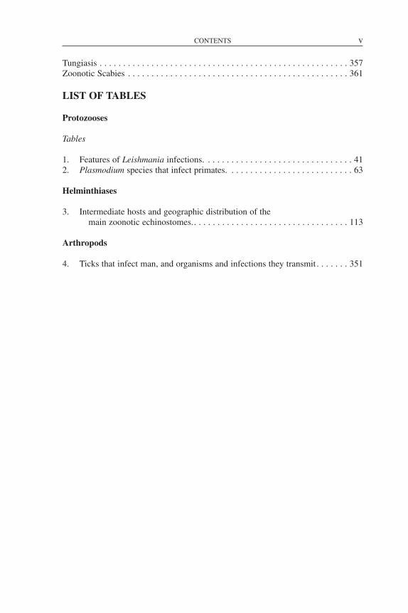
Tungiasis . . . . . . . . . . . . . . . . . . . . . . . . . . . . . . . . . . . . . . . . . . . . . . . . . . . . . 357Zoonotic Scabies . . . . . . . . . . . . . . . . . . . . . . . . . . . . . . . . . . . . . . . . . . . . . . . 361
LIST OF TABLES
Protozooses
Tables
1. Features of Leishmania infections. . . . . . . . . . . . . . . . . . . . . . . . . . . . . . . . 412. Plasmodium species that infect primates. . . . . . . . . . . . . . . . . . . . . . . . . . . 63
Helminthiases
3. Intermediate hosts and geographic distribution of themain zoonotic echinostomes.. . . . . . . . . . . . . . . . . . . . . . . . . . . . . . . . . 113
Arthropods
4. Ticks that infect man, and organisms and infections they transmit . . . . . . . 351
CONTENTS v

vii
PROLOGUE
In recent years, zoonoses and communicable diseases common to man and ani-mals have gained increasing attention worldwide. Human diseases that have theirorigins in infected animals, such as AIDS or Creutzfeldt-Jakob, have highlighted theneed for a better understanding of animal diseases in terms of their epidemiology,mechanism of transmission to man, diagnosis, prevention, and control. Social anddemographic changes have also contributed to the importance of gaining and dis-seminating knowledge about zoonoses. For instance, as people encroach further andfurther on ecological areas with which they had little contact and whose fauna maynot be well known, their exposure to animals—and the infections they transmit—has increased. New knowledge is also being developed in the area of urban ecology.The ease and speed of modern travel also facilitates the spread of diseases once con-fined to specific geographic areas, as recently occurred with severe acute respiratorysyndrome (SARS). Animal migration and trade pose a similar threat, as was shownby the outbreaks in the United States of West Nile fever, and most recently, mon-keypox—two diseases not previously known in the Western Hemisphere. Each ofthese examples highlights the need for improved knowledge and surveillance of andresponse to zoonoses.
The negative effects of zoonoses are far reaching. High incidence rates continueto cause significant morbidity and mortality in both humans and animals. Their eco-nomic impact is seen in lost labor productivity due to illness; reduced travel andtourism to affected areas; reduced livestock and food production; death and destruc-tion of affected animals; and restrictions on and reductions in international trade.Zoonoses can be a serious drain on a country’s economy, which in turn can havewide repercussions for a society’s health.
To help solve these problems, the Pan American Health Organization (PAHO)—an international public health organization that has devoted itself to improving thehealth and living conditions of the people of the Americas for over one hundredyears—established the Veterinary Public Health Unit. The Unit’s overall objective isto collaborate with PAHO’s Member Governments in the development, implemen-tation, and evaluation of policies and programs that lead to food safety and protec-tion and to the prevention, control, or eradication of zoonoses, among them foot-and-mouth disease.
To this end, PAHO’s Veterinary Public Health Unit has two specialized regionalcenters: the Pan American Foot-and-Mouth Disease Center (PANAFTOSA), createdin 1951 in Rio de Janeiro, Brazil, and the Pan American Institute for Food Protectionand Zoonoses (INPPAZ), established on November 15, 1991, in Buenos Aires,Argentina. INPPAZ’s precursor was the Pan American Zoonoses Center(CEPANZO), which was created through an agreement with the Government ofArgentina to help the countries of the Americas combat zoonoses, and which oper-ated from 1956 until 1990.
Since its creation in 1902, PAHO has participated in various technical coopera-tion activities with the countries, among them those related to the surveillance, pre-vention, and control of zoonoses and communicable diseases common to man andanimals, which cause high morbidity, disability, and mortality in vulnerable humanpopulations. PAHO has also collaborated in the strengthening of preventive medi-

viii PROLOGUE
cine and public health through the promotion of veterinary health education in learn-ing, research, and health care centers. An example of this work is the preparation ofseveral publications, among which the two previous Spanish and English editions ofZoonoses and Communicable Diseases Common to Man and Animals stand out.
Scientific knowledge has progressed since the last edition was published in 1986.Also, the countries of the Americas have modified their livestock production strate-gies in recent years, which has affected the transmission of zoonotic infections andtheir distribution. The publication of this third edition is an attempt to address thesechanges. The third edition is presented in three volumes: the first contains bacte-rioses and mycoses; the second, chlamydioses, rickettsioses, and viroses; and thethird, parasitoses.
We believe that this new edition will continue to be useful for professors and stu-dents of public health, medicine, veterinary medicine, and rural development; work-ers in public health and animal health institutions; and veterinarians, researchers,and others interested in the subject. We also hope that this publication is a useful toolin the elaboration of national zoonosis control or eradication policies and programs,as well as in risk evaluation and in the design of epidemiological surveillance sys-tems for the prevention and timely control of emerging and reemerging zoonoses. Insummary, we are confident that this book will contribute to the application of theknowledge and resources of the veterinary sciences for the protection and improve-ment of public health.
MIRTA ROSES PERIAGODIRECTOR

ix
PREFACE TO THE FIRST EDITION
This book considers two groups of communicable diseases: those transmittedfrom vertebrate animals to man, which are—strictly speaking—zoonoses; and thosecommon to man and animals. In the first group, animals play an essential role inmaintaining the infection in nature, and man is only an accidental host. In the sec-ond group, both animals and man generally contract the infection from the samesources, such as soil, water, invertebrate animals, and plants; as a rule, however,animals do not play an essential role in the life cycle of the etiologic agent, but may contribute in varying degrees to the distribution and actual transmission ofinfections.
No attempt has been made to include all infections and diseases comprised inthese two groups. A selection has been made of some 150 that are of principal inter-est, for various reasons, in the field of public health. The number of listed zoonosesis increasing as new biomedical knowledge is acquired. Moreover, as human activ-ity extends into unexplored territories containing natural foci of infection, newzoonotic diseases are continually being recognized. In addition, improved healthservices and better differential diagnostic methods have distinguished zoonoses pre-viously confused with other, more common diseases. A number of diseasesdescribed in this book have only recently been recognized, examples of whichinclude the Argentine and Bolivian hemorrhagic fevers, angiostrongyliasis, rotaviralenteritis, Lassa fever, Marburg disease, and babesiosis.
The principal objective in writing this book was to provide the medical profes-sions a source of information on the zoonoses and communicable diseases commonto man and animals. Toward that end, both medical and veterinary aspects, whichhave traditionally been dealt with separately in different texts, have been combinedin a single, comprehensive volume. As a result, physicians, veterinarians, epidemi-ologists, and biologists can all gain an overview of these diseases from one source.
This book, like most scientific works, is the product of many books, texts, mono-graphs, and journal articles. Many sources of literature in medicine, veterinary med-icine, virology, bacteriology, mycology, and parasitology were consulted, as were alarge number of reports from different biomedical disciplines, in order to provideup-to-date and concise information on each disease. It is expected that any errors oromissions that may have been committed can, with the collaboration of the readers,be corrected in a future edition.
Where possible, explanations were attempted with special emphasis on theAmericas, particularly Latin America. An effort was made, one which was notalways successful, to collect available information on diseases in this Region. Dataon the incidence of many zoonoses are fragmentary and frequently not reliable. It ishoped that the establishment of control programs in various countries will lead toimproved epidemiologic surveillance and disease reporting.
More space has been devoted to those zoonoses having greatest impact on publichealth and on the economy of the countries of the Americas, but information is alsoincluded on those regionally less important or exotic diseases.
The movement of persons and animals over great distances adds to the risk ofintroducing exotic diseases that may become established on the American continentgiven the appropriate ecologic factors for existence of the etiologic agents. Today,

x PREFACE TO THE FIRST EDITION
public health and animal health administrators, physicians, and veterinarians mustbe familiar with the geographic distribution and pathologic manifestations of thevarious infectious agents so that they can recognize and prevent the introduction ofexotic diseases.
We, the authors, would like to give special recognition to Dr. Joe R. Held,Assistant Surgeon-General of the United States Public Health Service and Directorof the Division of Research Services of the U.S. National Institutes of Health, whogave impetus to the English translation and reviewed the bacterioses sections.
We would also like to express our utmost appreciation to the experts whoreviewed various portions of this book and offered their suggestions for improvingthe text. These include: Dr. Jeffrey F. Williams, Professor in the Department ofMicrobiology and Public Health, Michigan State University, who reviewed thechapters dealing with parasitic zoonoses; Dr. James Bond, PAHO/WHO RegionalAdviser in Viral Diseases, who read the viroses; Dr. Antonio Pío, formerlyPAHO/WHO Regional Adviser in Tuberculosis and presently with WHO in Geneva,and Dr. James H. Rust, PAHO/WHO Regional Adviser in Enteric Diseases, both ofwhom reviewed the bacterioses; and Dr. F. J. López Antuñano, PAHO/WHORegional Adviser in Parasitic Diseases, who read the metazooses.
We would like to thank Dr. James Cocozza, PAHO/WHO Veterinary Adviser, forhis review of the translation and Dr. Judith Navarro, Editor in the Office ofPublications of PAHO, for her valuable collaboration in the editorial revision andcomposition of the book.
PEDRO N. ACHABORIS SZYFRES

xi
PREFACE TO THE SECOND EDITION
The fine reception accorded the Spanish, English, and French versions of thisbook has motivated us to revise it in order that it still may serve the purpose forwhich it was written: to provide an up-to-date source of information to the medicalprofession and allied fields. This book has undoubtedly filled a void, judging by itswide use in schools of public health, medicine, and veterinary medicine, as well asby bureaus of public and animal health.
The present edition has been considerably enlarged. In the seven years since thefirst edition was published, our knowledge of zoonoses has increased broadly andrapidly, and new zoonotic diseases have emerged. Consequently, most of the dis-cussions have been largely rewritten, and 28 new diseases have been added to theoriginal 148. Some of these new diseases are emerging zoonoses; others are patho-logic entities that have been known for a long time, but for which the epidemiologicconnection between man and animal has been unclear until recently.
The use this book has had outside the Western Hemisphere has caused us to aban-don the previous emphasis on the Americas in favor of a wider scope and geomed-ical view. Moreover, wars and other conflicts have given rise to the migration ofpopulations from one country or continent to another. A patient with a diseaseheretofore known only in Asia may now turn up in Amsterdam, London, or NewYork. The physician must be aware of these diseases in order to diagnose and treatthem. “Exotic” animal diseases have been introduced from Africa to Europe, theCaribbean, and South America, causing great damage. The veterinary physicianmust learn to recognize them to be able to prevent and eradicate them before theybecome entrenched. It must be remembered that parasites, viruses, bacteria, andother agents of zoonotic infection can take up residence in any territory where theyfind suitable ecologic conditions. Ignorance, economic or personal interests, andhuman customs and needs also favor the spread of these diseases.
Research in recent years has demonstrated that some diseases previously consid-ered to be exclusively human have their counterparts in wild animals, which in cer-tain circumstances serve as sources of human infection. On the other hand, theseanimals may also play a positive role by providing models for research, such as inthe case of natural leprosy in nine-banded armadillos or in nonhuman primates inAfrica. Of no less interest is the discovery of Rickettsia prowazekii in eastern flyingsquirrels and in their ectoparasites in the United States, and the transmission of theinfection to man in a country where epidemic typhus has not been seen since 1922.A possible wild cycle of dengue fever is also discussed in the book. Is Creutzfeldt-Jakob disease a zoonosis? No one can say with certainty, but some researchersbelieve it may have originated as such. In any case, interest is aroused by the sur-prising similarity of this disease and of kuru to animal subacute spongiformencephalopathies, especially scrapie, the first known and best studied of this group.Discussion of human and animal slow viruses and encephalopathies is included inthe spirit of openness to possibilities and the desire to bring the experience of onefield of medicine to another. In view of worldwide concern over acquired immuno-deficiency syndrome (AIDS), a brief section on retroviruses has also been added, inwhich the relationship between the human disease and feline and simian AIDS is

xii PREFACE TO THE SECOND EDITION
noted. Another topic deeply interesting to researchers is the mystery of the radicalantigenic changes of type A influenza virus, a cause of explosive pandemics thataffect millions of persons around the world. Evidence is mounting that thesechanges result from recombination with a virus of animal origin (see Influenza).That this should occur is not surprising, given the constant interaction between manand animals. As a rule, zoonoses are transmitted from animal to man, but the reversemay also occur, as is pointed out in the chapters on hepatitis, herpes simplex, andmeasles. The victims in these cases are nonhuman primates, which may in turnretransmit the infection to man under certain circumstances.
Among emerging zoonoses we cite Lyme disease, which was defined as a clinicalentity in 1977; the etiologic agent was found to be a spirochete (isolated in 1982),for which the name Borrelia burgdorferi was recently proposed. Emerging viralzoonoses of note in Latin America are Rocio encephalitis and Oropouche fever; thelatter has caused multiple epidemics with thousands of victims in northeast Brazil.Outstanding among new viral disease problems in Africa are the emergence of Eboladisease and the spread of Rift Valley fever virus, which has caused tens of thousandsof human cases along with great havoc in the cattle industry of Egypt and has evokedalarm around the world. Similarly, the protozoan Cryptosporidium is emerging asone of the numerous agents of diarrheal diseases among man and animals, and prob-ably has a worldwide distribution.
As the English edition was being prepared, reports came to light of two animaldiseases not previously confirmed in humans. Three cases of human pseudorabiesvirus infection were recognized between 1983 and 1986 in two men and one womanwho had all had close contact with cats and other domestic animals. In 1986, sero-logic testing confirmed infection by Ehrlichia canis in a 51-year-old man who hadbeen suspected of having Rocky Mountain spotted fever. This is the first knownoccurrence of E. canis infection in a human. These two diseases bear watching aspossible emerging zoonoses.
The space given to each zoonosis is in proportion to its importance. Some diseasesthat deserve their own monographs were given more detailed treatment, but noattempt was made to cover the topic exhaustively.
We, the authors, would like to give special recognition to Dr. Donald C. Blenden,Professor in the Department of Medicine and Infectious Diseases, School ofMedicine, and Head of the Department of Veterinary Microbiology, College ofVeterinary Medicine, University of Missouri; and to Dr. Manuel J. Torres, Professorof Epidemiology and Public Health, Department of Veterinary Microbiology,College of Veterinary Medicine, University of Missouri, for their thorough review ofand valuable contributions to the English translation of this book.
We would also like to recognize the support received from the Pan AmericanHealth Organization (PAHO/WHO), the Pan American Health and EducationFoundation (PAHEF), and the Pan American Zoonoses Center in Buenos Aires,Argentina, which enabled us to update this book.
We are most grateful to Dr. F. L. Bryan for his generous permission to adapt hismonograph “Diseases Transmitted by Foods” as an Appendix to this book.
Mr. Carlos Larranaga, Chief of the Audiovisual Unit at the Pan AmericanZoonosis Center, deserves our special thanks for the book’s artwork, as do Ms. IrisElliot and Mr. William A. Stapp for providing the translation into English. We would

PREFACE TO THE SECOND EDITION xiii
like to express our most sincere gratitude and recognition to Ms. Donna J. Reynolds,editor in the PAHO Editorial Service, for her valuable collaboration in the scientificeditorial revision of the book.
PEDRO N. ACHABORIS SZYFRES
xv
INTRODUCTION
This new edition of Zoonoses and Communicable Diseases Common to Man andAnimals is published in three volumes: I. Bacterioses and mycoses; II.Chlamydioses and rickettsioses, and viroses; and III. Parasitoses. Each of the fiveparts corresponds to the location of the etiologic agents in the biological classifica-tion; for practical purposes, chlamydias and rickettsias are grouped together.
In each part, the diseases are listed in alphabetical order to facilitate readersearches. There is also an alphabetical index, which includes synonyms of the dis-eases and the etiologic agents’ names.
In this edition, the numbers and names of the diseases according to theInternational Statistical Classification of Diseases and Related Health Problems,Tenth Revision (ICD-10), are listed below the disease title. However, some zoonosesare not included in ICD-10 and are difficult to classify within the current scheme.
In addition, for each disease or infection, elements such as synonyms; etiology;geographical distribution; occurrence in man and animals; the disease in man andanimals; source of infection and mode of transmission; role of animals in the epi-demiology; diagnosis; and control are addressed. Patient treatment (for man or otherspecies) is beyond the scope of this work; however, recommended medicines areindicated for many diseases, especially where they are applicable to prophylaxis.Special attention is paid to the epidemiological and ecological aspects so that thereader can begin to understand the determining factors of the infection or disease.Some topics include simple illustrations of the etiologic agent’s mode of transmis-sion, showing the animals that maintain the cycle of infection in nature. Similarly,other graphics and tables are included to provide additional information on the geo-graphical distribution or prevalence of certain zoonoses.
The data on the occurrence of the infection in man and animals, along with dataon the geographical distribution, may help the reader judge the relative impact thateach disease has on public health and the livestock economy in the different regionsof the world, given that the importance of different zoonoses varies greatly. Forexample, foot-and-mouth disease is extremely important from an economic stand-point, but of little importance in terms of public health, if animal protein losses arenot considered. In contrast, Argentine and Machupo hemorrhagic fevers are impor-tant human diseases, but their economic impact is minimal, if treatment costs andloss of man-hours are not taken into account. Many other diseases, such as brucel-losis, leptospirosis, salmonellosis, and equine encephalitis, are important from botha public health and an economic standpoint.
Finally, each disease entry includes an alphabetical bibliography, which includesboth the works cited and other relevant works that the reader may consult for moreinformation about the disease.

Section A
PROTOZOOSES

AFRICAN TRYPANOSOMIASIS
ICD-10 B56 African trypanosomiasis; B56.0 Gambiense trypanosomiasis;B56.1 Rhodesiense trypanosomiasis;
B56.9 African trypanosomiasis, unspecified
Synonyms: Sleeping sickness, trypanosomiasis; gambiense trypanosomiasis:infection due to Trypanosoma brucei gambiense, West African sleeping sickness;rhodesiense trypanosomiasis: infection due to Trypanosoma brucei rhodesiense,East African sleeping sickness.
Etiology: African trypanosomiasis in man is caused by two subspecies ofTrypanosoma (Trypanozoon) brucei: T. brucei gambiense and T. brucei rhodesiense,both of which are transmitted by the bite of tsetse flies (genus Glossina) (Bales,1991; Dumas and Bouteille, 1996; Chimelli and Scaravilli, 1997). These try-panosomes are considered to belong to the salivarian group because of the way inwhich they are transmitted through the vector’s bite. Infection caused directly by abite is considered inoculative, or via the anterior station, as opposed to contamina-tive, or via the posterior station, when the infection is transmitted by means of thefly’s excrement (see the chapter on Chagas’ Disease).
The two subspecies that affect man, T. b. gambiense and T. b. rhodesiense, as wellas T. b. brucei, are morphologically indistinguishable. The latter species, while itdoes not affect man, is pathogenic for domestic animals in Africa, such as donkeys,horses, goats, camels, mules, sheep, dogs, and cattle. The forms that are present inblood, cerebrospinal fluid, and lymph are pleomorphic trypomastigotes (seeChagas’ Disease). The forms range from long, thin parasites (measuring 30 µm by1.5 µm on average, with a subterminal kinetoplast, a long flagellum extending fromthe anterior tip of the body, and an undulating membrane between the flagellum andthe body) to short, fat parasites (averaging 15 µm by 3.5 µm, with a near-terminalkinetoplast and no external flagellum). The long forms multiply in the fluids of thedefinitive host by binary longitudinal division. The short forms are the infective ele-ments for the vector and do not divide in the human host.
For a long time, the three subspecies T. b. brucei, T. b. gambiense, and T. b. rhode-siense were distinguished on the basis of their infectivity and pathogenicity for rats,their sensitivity to the drug tryparsamide, and their pathogenicity for man. To makea definitive distinction between T. b. brucei and the human trypanosomes, humanvolunteers were employed, with the consequent risks. In practice, differentiation
3

between the two human pathogens is still fundamentally based on the course andgeographic distribution of the infection. Now more precise techniques are availablefor identifying the parasites. The blood incubation infectivity test (BIIT) consists ofincubating the trypanosomes in human serum or plasma and then inoculating themin rats. In this procedure, T. b. brucei loses its infectivity for rats whereas the sub-species that affect humans maintain it. Nevertheless, studies have revealed widevariation in the susceptibility of these trypanosomes to the effects of human serum,and some evidence exists that T. b. brucei can become resistant to the action of theserum. This would mean that T. b. brucei could become infective for man when fliesinfected by animals feed on human blood (Minter, 1982). Another important andincreasingly used method is characterization of the trypanosomes according to theelectrophoretic movement of their isozymes, which makes it possible to distinguishthe different zymodemes (see definition under Chagas’ Disease). Truc andTybayrenc (1993) have described 23 zymodemes in Central Africa, which can bedivided into two groups, one corresponding to T. b. gambiense and the other to T. b.brucei. Recent findings suggest that the different zymodemes are related not only tothe species but also to the geographic distribution and clinical characteristics of theinfection (Smith and Bailey, 1997). Also, the use of polymerase chain reaction hasmade it possible to identify T. b. gambiense (Schares and Mehlitz, 1996).
In man, the trypanosomes of African trypanosomiasis multiply in the blood,lymph, cerebrospinal fluid, and intercellular spaces, but they do not penetrate cells.In the vector, the short, fat trypanosomes consumed in the process of ingesting ablood meal multiply in the lumen of the mid and hindgut for about 10 days, afterwhich they turn into thin forms and migrate toward the proventriculus, where theymultiply for another 10 days; from there they travel to the salivary glands, where theyattach themselves to the epithelial cells and turn into epimastigotes (see Chagas’Disease). The epimastigotes continue to multiply and are rapidly transformed intoshort, fat, metacyclic trypomastigotes, sometimes without a flagellum, which are theforms that are infective for man. Although the complete cycle of the trypanosomeinside the tsetse fly can range from 15 to 35 days (average 21 days), the infectioncycle up to the formation of metacyclic trypomastigotes is completed in only about10% of the flies that ingest the parasite. The infected flies remain so for the rest oftheir lives and inoculate trypanosomes every time they take a blood meal.
Geographic Distribution: African trypanosomiasis in man occurs between 15°N and 20° S Latitude in Africa, which is the vector’s area of distribution. T. b. rhode-siense is found in multiple foci in eastern Africa over an area stretching fromEthiopia to Botswana, while T. b. gambiense is found in central and western Africa,from northwestern Senegal to northeastern Sudan in the north to Angola in thesouth. Outside the endemic area, there are occasional cases in tourists and immi-grants from endemic countries.
Occurrence in Man: In the past, there were devastating epidemics of gambiensetrypanosomiasis, brought on by the migration of settlers during colonization. At thebeginning of the twentieth century, 500,000 people died within a decade in theCongo basin alone, and there were about 200,000 fatalities (two-thirds of the popu-lation) in the province of Busoga, Uganda (Goodwin, 1970). Following the imple-mentation of control measures, by 1950 and 1960 prevalence of the disease haddropped to very low levels in some areas (0.1% to 2%) and annual incidence was
4 PROTOZOOSES

estimated at less than 10,000 cases. In 1972, there were 4,126 new cases in theDemocratic Republic of Congo (formerly Zaire) and 3,000 in the rest of Africa (DeRaadt, 1976). However, starting in the 1970s, the disease flared up again alarminglyin some of its old foci (Kusoe, 1993; Cattand, 1994; Jusot et al., 1995). This increasewas a reflection of the massive new movements of people both within and outsidethe endemic areas as a result of wars and social and political instability in manyAfrican countries. The situation was aggravated by shortages of human and materialresources for surveillance and medical care programs in the affected countries(Mhlanga, 1996). In 1982, the World Health Organization (WHO) reported thatgambiense trypanosomiasis was endemic in 23 African countries, 45 million inhab-itants were at risk, and that every year there were nearly 10,000 new infections (BullWorld Health Organ, 1982). Currently, African trypanosomiasis in man is endemicin 36 African countries south of the Sahara; the two forms of the disease togetherpose a risk for approximately 50 million people; and about 25,000 new cases arebeing reported annually, with the likelihood that not all cases were being notified(Bales, 1991; Kusoe, 1993).
Gambiense trypanosomiasis, which is chronic, tends to occur in epidemics,whereas rhodesiense trypanosomiasis, which has a more acute course, occurs spo-radically, and gives rise to far fewer epidemics. The latter infection is endemicamong livestock-raising tribes in eastern Africa and frequently affects hunters, fish-ermen, and travelers. Overall incidence is rather low because the people avoid areasinfested by the vector.
The Disease in Man: The human disease usually has three phases: the primarylesion, parasitemia, and invasion of the central nervous system. Two or three daysafter the bite of an infected fly, a painful inflammation (chancre) appears at the inoc-ulation site, and it disappears after two to three weeks (McGovern et al., 1995). Theprimary lesion is observed more frequently in infections caused by T. b. rhodesiensethan in those produced by T. b. gambiense. From the chancre site, the trypanosomesinvade the bloodstream, and the patient suffers from irregular and intermittent fever,mirroring the waves of parasitemia. Other signs during this acute period are painlessadenopathies, especially in the posterior cervical lymph nodes, as well as edema ofthe eyelids and joints. The most common symptoms of the acute phase are cepha-lalgia, insomnia, arthralgia, weight loss, and generalized erythema and pruritus, par-ticularly in the sternal region. In later stages of the disease, the symptomatology isrelated to the affected organ. Invasion of the central nervous system is common, anda large variety of psychological, motor, and sensory perturbations may be seen.Following the meningitis that develops early in the course of the infection, a ruptureoccurs in the choroid plexus which allows the parasites to invade sites in the brain.The result is encephalitis, consisting of generalized inflammation with perivascularinfiltrations of B and T lymphocytes, plasmocytes, and macrophages. The blood-brain barrier becomes permeable, and this condition may give rise to vasogenic cere-bral edema. Astrocytes and microglia are activated, and, together with immune cells,they begin to produce cytokines, which also contribute to progression of the disease(Pentreath et al., 1994). There is irritability, paresthesia, and insomnia, and later on,cerebral edema can cause severe headaches and edema of the optic papillae. Therecan also be neurologic manifestations such as epileptic seizures, chorea, psychoticepisodes, euphoria, somnolence, lethargy, and coma.
AFRICAN TRYPANOSOMIASIS 5
As it was noted earlier, gambiense trypanosomiasis usually follows a slow andchronic course. Weeks or months may elapse between the first and second phase,and months or years may elapse between the second and third phase. Rhodesiensetrypanosomiasis has a more acute course and its phases are less marked; death maycome within a few months, in contrast to patients with T. b. gambiense infection,who can live for many years. Cardiac complications are more common in rhode-siense trypanosomiasis, and some patients die before reaching the neurologic phase(Greenwood and Whittle, 1980; WHO, 1979).
Both forms of African trypanosomiasis severely alter the patient’s immune sys-tem. The main characteristics are synthesis of large amounts of gamma globulin,autoantibody formation, and immunodeficiency (Vincendeau et al., 1996). The par-asites in the bloodstream are covered with variable glycoprotein surface antigen(VGSA), which generates powerful immune responses that rapidly suppress para-sitemia. These responses include antibody production and activation ofmacrophages that produce tumor necrosis factor alpha (TNF-α) and nitric acid(NO). Some parasites, however, manage to express another of the more than 1,000genes coded for this antigen and are covered with a different glycoprotein, therebyinitiating a new wave of parasitemia. These waves recur every 7 to 15 days until thepatient, if left untreated, dies. The succession of new antigens is a powerful stimu-lus for the immune response, which participates in both the defense and the pathol-ogy of the disease. Although there is epidemiologic evidence of protective immunityin gambiense trypanosomiasis (Khonde et al., 1995), individual antigenic variationis effective protection for the parasite against the immunity of the host. In terms ofimmunopathology, there is no evidence that high gamma globulin levels or an abun-dance of immune complexes play an important role in pathology of the human dis-ease. Nevertheless, there is experimental evidence suggesting that autoantibodies tocomponents of the central nervous system, such as anti-galactocerebrosides andtryptophan anti-analogous antibodies, may play a part in the development ofencephalitis (Hunter et al., 1992). Although T lymphocytes diminish the parasite’scapacity to proliferate, they continue to produce gamma interferon (IFN-γ). Themacrophages and astrocytes, for their part, produce TNF-α. Although IFN-γ andTNF-α, together with their specific antibodies and the NO of the macrophages, havepowerful properties to fight the trypanosomes, it has been demonstrated that TNF-αlevels are directly related to the severity of the disease (Okomo-Assoumou et al.,1995).
The Disease in Animals: Infections caused by African trypanosomes in animalshave a variety of local names, but they are most often referred to as nagana. T. b.gambiense has been inoculated in or isolated occasionally from animals, includinglaboratory animals, antelopes, swine, chickens, dogs, and cows, but there is no evi-dence that it causes disease or sustained parasitemia. However, T. b. rhodesiense cancause an infection, which is usually asymptomatic, in domestic animals such as cat-tle and sheep; wild animals, including antelopes, hyenas, and lions; and also labo-ratory animals. T. b. brucei, on the other hand, infects a wide variety of animals,including carnivores, swine, equines, ruminants, and laboratory animals. It causesan important disease in camels, equines, cats, dogs, and small ruminants. The dis-ease is chronic and occasionally fatal in cattle; it is rarely fatal in swine. Man isresistant to the infection. There are other African trypanosomes of great importance
6 PROTOZOOSES

for domestic animals, but none of them infects man (Levine, 1985): T. congolenseaffects carnivores, swine, equines, and ruminants; T. vivax affects equines and rumi-nants; and T. simiae affects camels and swine. The primary symptoms in animals arelymphadenopathy, intermittent fever, anemia, and progressive emaciation (Urquhart,1980). Depending on the species, the age of the host, and the parasite load, the dis-ease may be acute or chronic.
Trypanosomiasis in animals has played a role in configuring African societies:awareness of the parasite’s fatal effect on horses protected the original inhabitantsfrom foreign invasions, while its effect on cattle has prevented ranchers from takingadvantage of 7 million km2 of pastureland to raise high-yield European cattle.Another form of trypanosomiasis that occurs both in Africa and outside the conti-nent is caused by T. evansi. It is transmitted by tabanid flies and is especially path-ogenic for camels, equines, and dogs.
Source of Infection and Mode of Transmission: Man is the main reservoir of T.b. gambiense and the source of infection for the vector. Because the infection is pro-longed and includes intervals between febrile attacks during which the patient feelsrelatively well, affected individuals may move about and propagate the infection innew areas where the vectors exist. There is no evidence that lower animals play arole in human T. b. gambiense infection, even though animal-to-animal transmissionhas been demonstrated in the laboratory (Molyneux, 1983) and parasites fromswine, sheep, and dogs have been shown to be identical to human parasites in theirsensitivity to human sera or their isoenzymatic profile (Scott et al., 1983; Scharesand Mehlitz, 1996). The success of control programs aimed exclusively at eliminat-ing the human parasite would indicate that animal reservoirs are not important ingambiense trypanosomiasis. Nevertheless, the presence of animal reservoirs couldaccount for maintenance of the T. b. gambiense infection in areas where isolatedhuman cases have occurred with long intervals between them.
The main vectors of T. b. gambiense infection are the tsetse flies Glossinafuscipes, G. palpalis, and G. tachinoide. These species belong to the palpalis, orriverine, group of flies, which inhabit dense vegetation along the shores of rivers andlakes. Human infection occurs almost always in the vicinity of watercourses orplaces where water pools in rural settings; tourists are rarely affected. The male andfemale tsetse flies are biological vectors, but they can transmit the infection mechan-ically during epidemics, when there are many patients with parasitemia. In general,the infection rate in the vectors is low. In addition, according to some reports, con-genital transmission can occur in man.
By contrast, in the case of rhodesiense trypanosomiasis, lower animals, especiallycattle, play an important role as reservoirs. T. b. rhodesiense has been isolated froma number of wild and domestic animals; but only antelopes, hyenas, lions, sheep,and cattle develop sufficiently high and prolonged parasitemia to serve as effectivereservoirs. These animals are responsible for persistence of the parasite in areas thathave not been inhabited by humans for years.
The main vectors in eastern Africa are Glossina morsitans, G. pallidipes, and G.swynnertoni. These species belong to the morsitans group of flies, which inhabitsavannahs and forested areas and prefer to feed on cattle and wild animals. The moreacute nature of the human infection, coupled with the fact that the habitat of the vec-tors is not near homes, makes rhodesiense trypanosomiasis more sporadic than the
AFRICAN TRYPANOSOMIASIS 7

gambiense form and less capable of causing epidemics. The main victims of therhodesiense form are hunters, tourists, and persons who have contact with wild ani-mal habitats where the infection is enzootic.
Diagnosis: The disease may be suspected when its main symptoms and signs arepresent, in particular intermittent fever, enlarged posterior cervical lymph glands,and cutaneous erythema. Biochemical tests do not reveal any remarkable alterationsexcept higher cell counts and increased IgM in cerebrospinal fluid, which are con-sidered pathognomonic of invasion of the central nervous system (Bisser et al.,1997). The infection is confirmed by demonstrating the presence of the parasite inaspirate from the chancre or the lymph glands, in bone marrow, or in blood takenduring the acute phase, or cerebrospinal fluid during the chronic phase. The sampleto be observed may be either fresh or fixed and stained. In acute-phase patients, aspi-ration of the lymph glands is more effective for detecting T. b. gambiense than T. b.rhodesiense. On the other hand, peripheral parasitemia is higher in rhodesiense thanin gambiense trypanosomiasis, and it is therefore easier to demonstrate the presenceof T. b. rhodesiense by examining thick blood films. In both cases, however, the lev-els of parasitemia fluctuate and are higher during febrile attacks. It is easier to findparasites in blood by centrifugation in hematocrit tubes and examination of theleukocyte layer, or by minifiltration in DEAE-cellulose, centrifugation, and exami-nation of the exudate (Bailey and Smith, 1994). To demonstrate the presence of T.b. rhodesiense, samples of blood or cerebrospinal fluid can be inoculated intraperi-toneally in mice, which develop detectable parasitemia within the second week. It isdifficult to infect rodents with T. b. gambiense. When the foregoing methods havebeen unsuccessful, an attempt may be made to examine bone marrow or culture it inspecial media such as glucose, lacto-albumin, serum, hemoglobin, or GLSH.Sediment from cerebrospinal fluid should be examined immediately after it is col-lected. Serologic reactions such as the card agglutination test, indirect hemaggluti-nation, enzyme-linked immunosorbent assay (ELISA), and indirect immunofluores-cence are useful for epidemiologic studies, but they are of limited value forindividual diagnosis: healthy individuals may have developed antibodies to animaltrypanosomes inoculated by tsetse flies which did not produce infection, and theseantibodies can cross-react with the antigens of T. b. gambiense and T. b. rhodesiense.
Control: The two main approaches to controlling the African trypanosomiasesare to reduce the principal reservoirs of infection and the presence of the vectors. Indiminishing the reservoirs of gambiense trypanosomiasis, detecting and treating thehuman infection should be emphasized to reduce the source of infection for the vec-tors. The challenge is greater with rhodesiense trypanosomiasis, because measuresmust also be taken to control the livestock population, both wild (e.g., antelopes)and domestic (e.g., cattle). The latter can be reduced by converting the savannahswhere livestock graze into cropland, which is not propitious for the proliferation oftsetse flies. Reduction of the vector population, which is much more efficient in con-trolling rhodesiense trypanosomiasis, can be achieved either through the targeteddestruction of the flies’ habitats or the use of insecticides. Both approaches, how-ever, can cause major ecologic changes. Moreover, the mass use of insecticides iscostly and not very efficient, because the flies are protected by vegetation in theirhabitats. Tsetse fly traps have been developed that are very effective, especiallywhen they are impregnated with insecticides (Langley, 1994). Another approach
8 PROTOZOOSES

would be to saturate the natural environment with male flies sterilized in the labo-ratory, which was successful in eradicating the fly Cochliomyia hominivorax inLibya in 1991. Empirical observations and mathematical models suggest that reduc-ing the vector population is most efficient during epidemics, while reducing thehuman reservoir is more effective in endemic situations (Gouteux and Artzrouni,1996). Other appropriate measures include preventing host-vector contact by the useof protective clothing, netting that keeps out flies, repellants, or simply not goinginto areas where there are high densities of tsetse flies. In highly endemic areas, theindiscriminate donation of blood should be prohibited. Chemoprophylaxis for visi-tors to endemic areas is not recommended because pentamidine and suramin areonly effective against T. b. gambiense, they are somewhat toxic, their use can masksymptoms of the disease until it invades the central nervous system, and generalizedapplication promotes parasite resistance to the drugs. Moreover, most tourists aremore exposed to T. b. rhodesiense than to T. b. gambiense. Wery (1990) considersthat the most important advances in the control of gambiense trypanosomiasis havebeen the improvements in serologic diagnosis, the demonstration of parasitemia, andthe introduction of low-cost, efficient traps for tsetse flies.
The problem of antigenic variation in the African trypanosomes has impeded theproduction of a vaccine, but there is epidemiologic evidence that the disease gener-ates protective immunity: while 30% of the uninfected population in the DemocraticRepublic of Congo is at risk of contracting the infection, only 15% of those previ-ously infected run a similar risk (Khonde et al., 1995). These facts suggest that avaccination is possible.
Bibliography
Bailey, J.W., D.H. Smith. The quantitative buffy coat for the diagnosis of trypanosomes.Trop Doct 24:54–56, 1994.
Bales, J.D. African trypanosomiasis. In: Strickland, G.T., ed. Hunter’s Tropical Medicine,7th ed. Philadelphia: Saunders; 1991:617–628.
Bisser, S., B. Bouteille, J. Sarda, et al. Apport des examens biochimiques dans le diagnos-tic de la phase nerveuse de la trypanosomose humaine africaine. Bull Soc Pathol Exot90:321–326, 1997.
Cattand, P.P. Trypanosomiase humaine africaine. Situation epidemiologique actuelle, unerecrudescence alarmante de la maladie. Bull Soc Pathol Exot 87:307–310, 1994.
Chimelli, L., S. Scaravilli. Trypanosomiasis. Brain Pathol 7:599–611, 1997.Control of sleeping sickness due to Trypanosoma brucei gambiense. Bull World Health
Organ 60:821–825, 1982.De Raadt, P. African sleeping sickness today. Trans R Soc Trop Med Hyg 70:114–116,
1976.Dumas, M., B. Bouteille. Trypanosomose humaine africaine. C R Seances Soc Biol Fil
190:395–408, 1996.Goodwin, L.G. The pathology of African trypanosomiases. Trans R Soc Trop Med Hyg
64:797–817, 1970.Gouteux, P.J., M. Artzrouni. Aut-il ou non un controle des vecteurs dans la lutte contre la
maladie du sommeil? Une approche bio-mathématique du problème. Bull Soc Pathol Exot89:299–305, 1996.
Greenwood, B.M., H.C. Whittle. The pathogenesis of sleeping sickness. Trans R Soc TropMed Hyg 74:716–725, 1980.
AFRICAN TRYPANOSOMIASIS 9
Hunter, C.A., F.W. Jennings, J.F. Tierney, M. Murray, P.G. Kennedy. Correlation of autoan-tibody titres with central nervous system pathology in experimental African trypanosomiasis.J Neuroimmunol 41:143–148, 1992.
Jusot, J.F., S.J. de Vlas, G.J. van Oortmarssen, A. De Muynck. Apport d’un modèle math-ématique dans le controle d’une parasitose: cas de la trypanosomiase humaine africaine àTrypanosoma brucei gambiense. Ann Soc Belg Med Trop 75:257–272, 1995.
Khonde, N., J. Pepin, T. Niyonsenga, F. Milord, P. De Wals. Epidemiological evidence forimmunity following Trypanosoma brucei gambiense sleeping sickness. Trans R Soc Trop MedHyg 89:607–611, 1995.
Kuzoe, F.A. Current situation of African trypanosomiasis. Acta Trop 54:153–162, 1993.Langley, P.A. Understanding tsetse flies. Onderstepoort J Vet Res 61:361–367, 1994.Levine, N.D. Veterinary Protozoology. Ames: Iowa State University Press; 1985.McGovern, T.W., W. Williams, J.E. Fitzpatrick, M.S. Cetron, B.C. Hepburn, R.H. Gentry.
Cutaneous manifestations of African trypanosomiasis. Arch Dermatol 131:1178–1182, 1995.Mhlanga, J.D. Sleeping sickness: Perspectives in African trypanosomiasis. Sci Prog 79 (Pt
3):183–214, 1996.Minter, D.M. Trypanosomes. In: Manson, P., F.I.C. Apted. Manson’s Tropical Diseases,
18th ed. London: Ballière Tindall; 1982.Molyneux, D.M. Selective primary health care: Strategies for control of disease in devel-
oping world. VIII African trypanosomiasis. Rev Infect Dis 5:945–956, 1983.Okomo-Assoumou, M.C., S. Daulouede, J.L. Lemesre, A. N’Zila-Mouanda, P. Vincendeau.
Correlation of high serum levels of tumor necrosis factor-alpha with disease severity in humanAfrican trypanosomiasis. Am J Trop Med Hyg 53:539–543, 1995.
Pentreath, V.W., P.J. Baugh, D.R. Lavin. Sleeping sickness and the central nervous system.Onderstepoort J Vet Res 61:369–377, 1994.
Schares, G., D. Mehlitz. Sleeping sickness in Zaire: A nested polymerase chain reactionimproves the identification of Trypanosoma (Trypanozoon) brucei gambiense by specifickinetoplast DNA probes. Trop Med Int Health 1:59–70, 1996.
Scott, C.M., J.L. Frezil, A. Toudic, D.G. Godfrey. The sheep as a potential reservoir ofhuman trypanosomiasis in the Republic of the Congo. Trans R Soc Trop Med Hyg 77:397–401,1983.
Smith, D.H., J.W. Bailey. Human African trypanosomiasis in south-eastern Uganda:Clinical diversity and isoenzyme profiles. Ann Trop Med Parasitol 91:851–856, 1997.
Truc, P., M. Tibayrenc. Population genetics of Trypanosoma brucei in central Africa:Taxonomic and epidemiological significance. Parasitology 106(Pt 2):137–149, 1993.
Urquhart, G.M. The pathogenesis and immunology of African trypanosomiasis in domes-tic animals. Trans R Soc Trop Med Hyg 74:726–729, 1980.
Vincendeau, P., M.C. Okomo-Assoumou, S. Semballa, C. Fouquet, S. Daulouede.Immunologie et immunopathologie de la trypanosomose Africaine. Med Trop (Mars)56:73–78, 1996.
Wery, M. Les lents progrès du controle de la maladie du sommeil. Ann Parasitol HumComp 65(Suppl 1):89–93, 1990.
World Health Organization (WHO). Parasitic Zoonoses. Report of a WHO ExpertCommittee, with the Participation of FAO. Geneva: WHO; 1979. (Technical Report Series637).
10 PROTOZOOSES

AMEBIASIS
ICD-10 A06
Synonyms: Amebiosis, amebic dysentery, entamebiasis.
Etiology: Of the numerous species of the genus Entamoeba found in mammals,only E. histolytica and E. polecki are of zoonotic interest. E. dispar was recently iden-tified as a separate species, but knowledge about it is still quite limited. E. histolyticais essentially a human parasite which is also capable of infecting a number of non-human primates. In addition, it has been occasionally isolated from dogs, cats, swine,and rats, and it has produced experimental infection in rabbits and other rodents(Tsutsumi, 1994). E. polecki was isolated from swine and goats in 1912 and has alsobeen identified in humans (Giboda et al., 1988), although Levine (1985) contendsthat the original description was inadequate to distinguish it from E. histolytica.Another human species, E. dispar, was thought for many years to be a “small race”of E. histolytica because it is very similar in appearance but does not have the samepathogenic power; it has now been identified as a separate species (Jackson, 1998).
Amebas have two developmental stages: the trophic (or vegetative), during whichthe trophozoite is formed, and the cystic (or resistant) stage, when the cyst appears.The trophozoites live in the large intestine of the host, moving around by means ofpseudopodia and multiplying by binary fission. As they progress through the hostintestine toward the outside, they divide into smaller forms, cease taking in nour-ishment, and develop a thin, resistant wall around themselves in preparation for turn-ing into cysts. At first the cysts are mononuclear; they then subdivide by two con-secutive mitoses, producing two and ultimately four nuclei. At that point the cystsare eliminated in the feces of the host. If they are ingested by another host via con-taminated food or water, upon reaching the small intestine they break up into fournew trophozoites which then migrate to the large intestine, where the multiplicationprocess resumes.
Geographic Distribution: Worldwide.
Occurrence in Man: E. histolytica infection is especially prevalent in tropicaland subtropical areas, and it is more frequent in developing countries than industri-alized ones. An estimated 400 to 500 million people in the world are infected, andbetween 5% and 10% of them present symptoms (García and Bruckner, 1997). Inrecent decades, prevalence of the infection has declined notably in the industrializedcountries. In the US, for example, the rate in the general population fell from 7% in1961 to approximately 2% at the end of the 1990s. On the other hand, in the devel-oping world the disease continues to be an important cause of morbidity and mor-tality. According to reports published in 1996–1997, the infection was found in 0.3%of 1,917 apparently healthy children in Spain, 7.8% of 862 children with diarrhea inKenya, 19.4% of 33,253 hospital fecal samples in Beirut and 1.2% of 11,611 simi-lar samples in Tripoli, between 20% and 29% of 980 normal adults in Egypt, 18.6%of 1,267 individuals in Nicaragua, and 8.7% of a group of 342 persons in Venezuela.
The prevalence of E. dispar is unknown because laboratories only rarely distin-guish it from E. histolytica. Its frequency may be high, however, since symptoms arepresent in only 5% to 10% of the infections attributed to E. histolytica.
AMEBIASIS 11
E. polecki infection is rare in humans, and most reports refer to individual cases.However, the prevalence of this species may be greater than has been reported so farbecause of the difficulty of distinguishing it from E. histolytica (Levine, 1985). Itwould appear to be more frequent in southeastern parts of Asia: the parasite wasfound in 19% of 184 children in Papua New Guinea (Desowitz and Barnish, 1986);4.6% of 1,478 refugees from Cambodia, the Lao People’s Democratic Republic, andViet Nam arriving in the US (DeGirolami and Kimber, 1983); and 3.2% of 435refugees from Cambodia and Viet Nam arriving in France (Chaker et al., 1982).
Occurrence in Animals: Infection with E. histolytica is relatively common innonhuman primates. The parasite has been isolated from dogs and rats, and on occa-sion from naturally infected cats and swine; it has also been reported in cattle(Levine, 1985). Experimental infections have been produced in numerous rodents(mice, rats, guinea pigs, hamsters, and jerboas) and also in rabbits (Tsutsumi, 1994).
Infection with E. polecki appears to be common in swine. Pakandl (1994) reportedhigh prevalence of this parasite among newly weaned swine in the formerCzechoslovakia. It is rarely identified in diagnostic laboratories.
The Disease in Man: Most E. histolytica infections are asymptomatic, but theyshould be regarded as potentially pathogenic because there is always the danger thatthey could develop into a progressive and invasive disease (WHO, 1981). Amebiasisis particularly common in young adults and may be manifested by an invasion of thesmall intestine, liver, or, more rarely, other tissues. In the intestinal disease, the par-asite invades the tissues and produces small ulcers in the intestinal mucosa whichspread underneath in the submucosal tissue by means of lysis. On rare occasions itcan cause perforation of the intestine or produce granulomas in the wall of the largeintestine. The symptoms range from mild abdominal discomfort with bloodymucous diarrhea, alternating with periods of constipation or remission, to acute orfatal dysentery with fever, chills, and bloody or mucous diarrhea (amebic dysentery)(Benenson, 1995). Hematogenic dissemination may carry the parasites to the liver,where they produce a focal necrosis which is often incorrectly referred to as an ame-bic liver abscess. The symptoms of intestinal amebiasis correspond to febrile andpainful hepatosplenomegaly. However, unlike hepatic fascioliasis, there is noperipheral eosinophilia. Occasionally, the parasite may invade the lungs, skin, geni-tal organs, spleen, brain, or pericardium.
Human infection with E. polecki is usually asymptomatic. In the few cases of intes-tinal disease that have been described, the symptoms were considerably milder thanthose produced by E. histolytica and there was no invasion of extraintestinal tissues.
The Disease in Animals: Like human infections, animal infections with E. his-tolytica are usually asymptomatic. Both the clinical intestinal form and the hepaticform occur in lower primates, and spider monkeys are particularly susceptible(Amyx et al., 1978). It is not known whether the disease occurs in swine. In dogs,there have been reports of occasional cases of intestinal disease and, more rarely,invasion of the liver and other tissues. Shimada et al. (1992) described a case of E.histolytica disease in a cat. Among laboratory rodents, the hamster and the jerboaare susceptible to hepatic invasion, but the guinea pig and the rat are resistant.Although combined immunodeficient mice are fully susceptible to hepatic amebia-sis, normal mice are highly resistant.
12 PROTOZOOSES

E. polecki does not appear to be pathogenic for swine (Dunlap, 1975).
Source of Infection and Mode of Transmission: Humans are the reservoir of E.histolytica. There is no evidence of animal-to-human transmission. The infection isacquired by the ingestion of products contaminated with the fecal matter frominfected persons. Although contaminated water (Marshall et al., 1997) and raw veg-etables (Monge and Arias, 1996) are sources of infection, well-documented risk fac-tors include persons who handle contaminated food and those with poor hygienichabits who may contaminate the household food supply. In addition, flies are effi-cient vectors of the cysts. The trophozoites, which are virtually the only forms pres-ent in diarrheic stools, are of little importance as transmitters of the infectionbecause they are not very resistant to desiccation or the action of gastric juices. Thecysts, which are found in abundance in pasty or formed feces, are the principal ele-ments of transmission, since they survive in the soil for eight days at temperaturesbetween 28°C and 34°C and for 40 days at 2°C to 6°C. For this reason, the chronicpatient and the healthy carrier are more effective sources of infection than the acutepatient. In the last two decades it has also been documented that sexual practiceswhich include anal-oral or anal-genital-oral contact are an important risk factor forinfection.
Except in the case of monkeys, it is believed that animals acquire the infectionfrom human reservoirs. Apparently E. histolytica can be propagated among lowerprimates: of 29 chimpanzees admitted to a particular colony, only 2 had E. histolyt-ica on the day of their arrival, whereas 4 years later 10 of the 29 were infected(Miller and Bray, 1966).
The reservoir of E. polecki is swine, and the human infection is contracted eitherby the ingestion of protozoan cysts in contaminated water or food or via the handsof a person who has been in contact with fecal matter from this reservoir. Human-to-human transmission is also suspected: of three patients diagnosed in Venezuela,two had not had any contact with animals (Chacin-Bonilla, 1983).
Diagnosis: Clinical manifestations alone are not sufficient to differentiate dysen-tery caused by amebiasis from other causes of dysentery. Laboratory diagnosis isbased on three fecal examinations, each taken half a day apart, and serologic tests inspecial cases. Direct examination of diarrheic feces almost always reveals tropho-zoites, whereas cysts and occasional trophozoites are found in formed and pastyfeces. Samples of diarrheic fecal matter should be examined as soon as possibleafter collection unless steps are taken to preserve the trophozoites, for which pur-pose trichromic or iron hematoxylin stain is recommended (García and Bruckner,1997). Samples from formed or pasty feces may be examined using stool concen-tration methods and direct microscopic observation of cysts. The diagnosis of E. his-tolytica requires carefully performed procedures and personnel well trained in dis-tinguishing between the macrophages, leukocytes, trophozoites, and cysts of thisand other parasites.
The clinical manifestations of extraintestinal amebiasis are not sufficient for adefinitive diagnosis. Tests such as the enzyme-linked immunosorbent assay make itpossible to identify 90% of all cases, although this technique only detects 10% ofintestinal cases (Restrepo et al., 1996). Tests designed to identify foreign bodies,such as radioisotopic imaging, ultrasound, and computerized tomography, may helpto locate the lesion, but they are not diagnostic of the disease.
AMEBIASIS 13

Differential diagnosis between E. histolytica and E. polecki is difficult and canonly be accomplished by studying the cysts. Although the distinction between thepathogenic species E. histolytica and the nonpathogenic E. dispar cannot be madeon the basis of morphological criteria alone, there are immunologic and isoenzy-matic differences which have recently made it possible to identify the species in spe-cialized laboratories (Jackson, 1998). The E. polecki cysts have a single nucleus,unevenly distributed chromatin in the nuclear periphery, rare glycogen vacuoles inthe cytoplasm, usually an opaque cytoplasmic inclusion body which is much largerthan the nucleus, and up to 30 chromatoidal bars. On the other hand, mature E. his-tolytica cysts have four nuclei, uniformly distributed chromatin, frequent glycogenvacuoles in the cytoplasm, no cytoplasmic inclusion body, and fewer than 10 chro-matoidal bars (Levin and Armstrong, 1970). Giboda et al. (1988) have establishedadditional criteria.
Control: Basically, amebiasis is controlled by avoiding contamination of theenvironment with human feces and educating the general public—children in par-ticular, in order to reach the people in the household who handle food—and com-mercial food handlers about proper hygiene to prevent transmission of the infection.The following measures are essential in order to avoid contamination: proper dis-posal of human excreta, protection of water sources from fecal contamination,treatment of chronic patients and healthy carriers who are spreading cysts, andsupervision of food preparation in public places where raw food is eaten. Foodshould be covered when there are flies or dust in the air. Health education shouldstress the danger of drinking water or eating raw vegetables that might be contami-nated, as well as the importance of washing one’s hands after defecating and beforepreparing food. Education programs should be targeted toward high-risk groupssuch as homosexuals and swineherds in order to prevent infections caused by E.polecki. In endemic areas, water and food should be either boiled or treated withnine drops of 2% tincture of iodine per liter of water for 30 minutes. Travelers vis-iting endemic areas should consume only bottled water (including ice made frombottled water) and cooked food.
Bibliography
Amyx, H.L., D.M. Asher, T.E. Nash, C.J. Gibbs, Jr., D.C. Gajdusek. Hepatic amebiasis inspider monkeys. Am J Trop Med Hyg 27:888–891, 1978.
Benenson, A.S., ed. Control of Communicable Diseases in Man, 16th ed. An official reportof the American Public Health Association. Washington, D.C.: APHA; 1995.
Chaker, E., M. Kremer, T.T. Kien. Quatorze cas d’Entamoeba polecki chez des refugies duSud-Est asiatique: remarques sur l’aspect morphologique du parasite. Bull Soc Pathol ExotFiliales 75:484–490, 1982.
Chacin-Bonilla, L. Entamoeba polecki infection in Venezuela. Report of a new case. TransR Soc Trop Med Hyg 77:137, 1983.
DeGirolami, P.C., J. Kimber. Intestinal parasites among Southeastern Asian refugees inMassachusetts. Am J Clin Pathol 79:502–504, 1983.
Desowitz, R.S., G. Barnish. Entamoeba polecki and other intestinal protozoa in Papua NewGuinea Highland children. Ann Trop Med Parasitol 80:399–402, 1986.
Dunlap, J.S. Protozoa. In: Dunne, H.W., A.D. Leman, eds. Diseases of Swine, 4th ed. Ames:Iowa State University Press; 1975.
14 PROTOZOOSES

García, L.S., D.A. Bruckner. Diagnostic Medical Parasitology, 3rd ed. Washington, D.C.:ASM Press; 1997.
Giboda, M., N. Vokurkova, P. Kopacek, O. Ditrich, J. Gutvirth. Entamoeba polecki:Morphology, immunology, antigen study and clinic of the first infections in Czechoslovakia.Folia Parasitol (Praha) 35:11–16, 1988.
Jackson, T.P. Entamoeba histolytica and Entamoeba dispar are distinct species: Clinical,epidemiological and serological evidence. Int J Parasitol 28:181–186, 1998.
Levin, R.L., D.E. Armstrong. Human infection with Entamoeba polecki. Am J Clin Pathol54:611–614, 1970.
Levine, N.D. Veterinary Protozoology. Ames: Iowa State University Press; 1985.Marshall, M.M., D. Naumowitz, Y. Ortega, C.R. Sterling. Waterborne protozoan pathogens.
Clin Microbiol Rev 10:67–85, 1997.Miller, M.J., R.S. Bray. Entamoeba histolytica infections in the chimpanzee (Pan satyrus).
J Parasit 52:386–388, 1966.Monge, R., M.L. Arias. Presencia de microorganismos patógenos en hortalizas de consumo
crudo en Costa Rica. Arch Latinoam Nutr 46:292–294, 1996.Pakandl, M. The prevalence of intestinal protozoa in wild and domestic pigs. Vet Med
(Praha) 39:377–380, 1994.Restrepo, M.I., Z. Restrepo, C.L. Elsa Villareal, A. Aguirre, M. Restrepo. Diagnostic tests
for amoebic liver abscess: Comparison of enzyme-linked immunosorbent assay (ELISA) andcounterimmunoelectrophoresis (CIE). Rev Soc Bras Med Trop 29:27–32, 1996.
Shimada, A., Y. Muraki, T. Awakura, et al. Necrotic colitis associated with Entamoeba his-tolytica in a cat. J Comp Pathol 106:195–199, 1992.
Tsutsumi, V. Los modelos experimentales in vivo en la amebiasis. Gac Med Mex130:450–453, 1994.
World Health Organization (WHO). Intestinal Protozoan and Helminthis Infections. Reportof a WHO Scientific Group. Geneva: WHO; 1981. (Technical Report No. 666).
BABESIOSIS
ICD-10 B60.0
Synonyms: Piroplasmosis, babesiasis.
Etiology: Of the 73 species of Babesia that have been described as parasites ofmammals, only slightly more than a dozen are important for domestic animals andonly five occasionally infect man: 1) B. microti, a parasite of rodents; 2) B. diver-gens, a parasite of cattle in Europe; 3) a species related to the dog parasite B. gib-soni, isolated in Africa and Asia (but undistinguishable from B. microti) and also inthe state of Washington, US, where it was given the preliminary designation WA1;4) a species related to B. divergens isolated in Missouri, US, and assigned the pre-liminary designation MO1; and 5) a species related to B. microti isolated in Taiwanand identified preliminarily as TW1 (Barriga, 1997; Herwaldt et al., 1996; Shih etal., 1997). Since the diagnosis of Babesia is still based mainly on the morphologyof the parasites, it is possible that man may be infected by other species which havenot yet been identified with certainty.
BABESIOSIS 15

Babesias parasitize the red blood cells of vertebrate hosts and are transmitted innature by ticks. When an infected tick bites a mammal, pyriform parasites (sporo-zoites measuring 1.5–3 µm long) are introduced through the tick’s saliva and rapidlypenetrate the host’s erythrocytes. The majority of the parasites grow inside the redblood cells as pyriform trophozoites or merozoites, the rest as gametocytes. Thetrophozoites or merozoites often divide asexually into two organisms, forming a“V.” B. microti sometimes divides into four parasites that form a tetrad or Maltesecross. When they achieve full growth and measure between 1 µm and 5 µm inlength, the parasites break free of the erythrocytes, often destroying them in theprocess, and invade new ones. This cycle is repeated until either the infection isbrought under control or the host dies. The gametocytes, on the other hand, developinside the host’s erythrocytes until they become an oval or round parasite, at whichpoint they stop growing. These gametocytes are the precursors of the parasite’s sex-ual stage, which continue to multiply inside the tick.
Babesiosis is typically a chronic infection. Even after the infection is controlled,the parasite usually maintains a low-level presence in the host erythrocytes for avery long time. In domestic animals, this period often lasts for the rest of their life.
When a tick vector—Ixodes scapularis (formerly I. dammini) in the case of B.microti, or I. ricinus in the case of B. divergens—sucks blood containing the para-sites, the merozoites are destroyed in the digestive tract, but the gametocytes matureand become male and female gametes, which fuse and form mobile zygotes. The lat-ter in turn become kinetes, which migrate to the hemocele and from there invadenumerous organs of the tick, where they divide asexually and invade even moreorgans. Some of the kinetes invade oocytes; once inside the egg, they can be passedon to the next generation of ticks via transovarial transmission. Other kinetes invadethe salivary glands, where they are transformed into sporozoites after the gland hasundergone certain developmental changes that take place while the arthropodingests its blood meal. Because of the time required for this process to occur, sporo-zoites are not inoculated until a few days after the infected tick begins to feed(Mehlhorn and Schein, 1985).
Geographic Distribution: Animal babesias occurs almost everywhere in theworld where ticks exist, including both the tropical zones and many temperate areasas well. In the US, human infections have been identified and attributed to the fol-lowing: B. microti (especially in New England), WA1 (in Washington State andnorthern California), and MO1 (in Missouri). In Europe, cases of babesiosis causedby B. divergens have been reported in Belgium, France, Ireland, Russia, Scotland,Spain, Sweden, and the former Yugoslavia. In Taiwan, cases due to TW1 were iden-tified. Osorno et al. (1976) found antibodies to an undetermined species of Babesiain 38 of 101 samples taken from residents in a rural area of Mexico, and they suc-cessfully isolated the parasite from three of the samples by inoculation in hamsters.
Occurrence in Man: Clinical human babesiosis is infrequent. The first case wasconfirmed in the former Yugoslavia in 1957 and attributed to B. divergens. Since1990, there have been reports in the US of 100 cases in the states of Massachusettsand New York; 2 in the midwest in the states of Wisconsin and Minnesota; 10 in thestates of Washington and California; and 1 in the state of Missouri. In addition, some22 cases have been identified in Europe and there has been 1 case in Taiwan. The totalnumber of cases described in the world is estimated to be fewer than 200. Although
16 PROTOZOOSES

most of the cases in Europe and the first cases in the US occurred in splenectomizedindividuals, in the last decade numerous cases have been described, especially in theUS, in patients who were immunodeficient for other reasons or previously healthy.However, there is evidence that the infection is much more frequent than the disease.For example, 6.3% of 1,285 residents of Connecticut were found to be seropositivefor B. microti (Krause et al., 1991), 9% of 574 residents of Rhode Island were alsoseropositive for B. microti (Krause et al., 1992), and 17.8% of 230 semirural residentsof northern California were seropositive for WA1 (Fritz et al., 1997).
Occurrence in Animals: Animal babesiosis is widespread throughout the world,with the highest prevalence in the tropics. It is one of the most important diseases ofcattle in Africa, the Middle East and other parts of Asia, Australia, Central America,and the northern half of South America. The disease poses a risk for 50% to 70% ofthe cattle in the world and causes heavy economic losses, and has been compared tomalaria in man. B. divergens occurs only in Europe and some parts of the formerUSSR. It is distinguished from the other major babesias of cattle (B. bigemina, B.bovis, and B. major) by its relatively smaller size, measuring only 1.5 µm x 1.0 µm,compared with at least 2.0 µm in length in the case of all the others (Barriga, 1997).B. microti is found on numerous wild rodents in the US and Europe, and it is a com-mon laboratory research model. In the US, blood examinations of the white-footedmouse (Peromyscus leucopus) on Nantucket Island, Massachusetts, revealed B.microti in 35.4% of the blood smears examined and 67% of the specimens inocu-lated in hamsters (Spielman et al., 1981). In Germany, it was found in 38% of 255field voles (Microtus agrestis) (Krampitz, 1979). Nevertheless, the only knowncases of human infection due to this species have been found on the eastern coast ofthe US.
The Disease in Man: The cases in Europe of B. divergens infection usually occurin splenectomized individuals (80%). They are characterized by severe illness, oftenwith pyrexia, chills, anemia, muscular pain, prostration, hemoglobinuria, and jaun-dice. The case fatality rate is about 50%. The spleen plays a very important role inresistance to the parasite, and splenectomy is undoubtedly a predisposing factor. Bythe same token, the disease may be more severe in immunodeficient individuals. Inthe Americas, the nonsplenectomized patients infected with B. microti had a diseasethat developed gradually, with anorexia, fatigue, fever, sweating, and generalizedmuscle pain. Some patients may have mild splenomegaly and hepatomegaly, andmild to severe hemolytic anemia is common as well. Parasitemia may affect fewerthan 1% or up to 10% or more of the erythrocytes. In blood smears, it is difficult todistinguish the predominant form of B. microti from the small ring forms ofPlasmodium, which they closely resemble. Recovery is slow, with malaise andfatigue persisting for several months (Ruebush, 1984). B. microti rarely causesdeath, even in splenectomized or immunodeficient patients. The incubation periodbetween the tick bite and the appearance of symptoms can range from 7 to 28 days.
Because of epidemiologic similarities with infections caused by Borreliaburgdorferi and Ehrlichia spp., human babesiosis in the US can coexist with infec-tions caused by these other agents (Mitchell et al., 1996).
The Disease in Animals: In the affected domestic species the symptomatology ofbabesiosis is similar, characterized by the triad of fever, anemia, and jaundice.
BABESIOSIS 17

Anemia occurs when the parasites emerge from the erythrocytes and cause theimmunologic destruction of these cells. The increased amount of free hemoglobin inplasma often produces hemoglobinuria. B. bovis tends to attach itself to the capil-lary endothelium in much the same way that Plasmodium falciparum does in man,thereby blocking circulation. The sensitivity of nervous tissue to anoxia often resultsin symptoms of agitation and convulsions. Babesiosis in cattle can range from mildto fatal, and those animals that recover usually harbor a subclinical infection and actas healthy carriers. Calves and young equines 6 to 9 months of age are relativelyresistant to the infection and disease. In endemic areas, most animals acquire anasymptomatic infection when they are young that confers premunition (i.e., resist-ance to subsequent infections). By contrast, animals arriving from parasite-freeareas usually develop a severe form of the disease.
B. microti in rodents appears to be asymptomatic, but no extensive studies havebeen done on the subject.
Source of Infection and Mode of Transmission: The reservoirs for domesticanimals and rodents are other infected animals, which are often healthy carriers. Thereservoirs for man are wild rodents, especially the white-footed mouse P. leucopusand meadow vole Microtus pennsylvanicus, both of which have been demonstratedto be infected with B. microti in the US, and cattle infected with B. divergens inEurope. The B. microti infection is transmitted in nature by the tick I. scapularis andthe B. divergens infection, by I. ricinus.
In the case of B. microti, the larva of I. scapularis acquires the infection from wildrodents and the nymph, which acquires the infection via transstadial transmissionfrom the larva, passes the infection along when it bites a human host. In a study onNantucket Island, Massachusetts, US, 5% of the I. scapularis nymphs collectedwere infected. However, the protozoan is not passed from the nymph to the adulttick. Since the adult tick feeds on deer that are not susceptible to B. microti, it doesnot have the opportunity to become infected and therefore does not transmit theinfection (Lane and Crosskey, 1993). The B. microti enzootic areas are characterizedby an abundance of rodents, which serve as hosts for the larvae and nymphs of thevector, while deer serve as the host for adult ticks. Most of the human infections inthe US occur in July and August, when nymphs are most abundant (Ruebush et al.,1981).
In the case of B. divergens, only the adult I. ricinus tick feeds on cattle and iscapable of acquiring the infection. However, B. divergens is transmitted to the nextgeneration of ticks both through the egg (transovarial transmission) and subse-quently during the different developmental stages of the tick (transstadial transmis-sion). Thus, I. ricinus can transmit B. divergens at any stage in its life cycle.
There have been about eight reports of human Babesia infection due to bloodtransfusion (Mintz et al., 1991). Asymptomatic donors carry the infection for up to12 months after they initially acquire the infection themselves.
Diagnosis: A diagnosis of babesiosis should be suspected when the clinicalsymptoms coincide with an epidemiological history of tick bites or visits to enzooticareas. It is confirmed in febrile or acute cases when parasites are seen inside ery-throcytes on Giemsa-stained thin or thick blood smears. Differentiation fromPlasmodium, especially P. falciparum, may require the help of experts. UnlikePlasmodium, Babesia does not produce pigment (hemozoin) in the parasitized red
18 PROTOZOOSES

blood cells. In more chronic cases with low parasitemia, a diagnosis can be madeusing serologic examinations such as the indirect immunofluorescence test or theinoculation of blood into susceptible animals. In chronic cases, or before babesiasappear in the blood in detectable numbers, the polymerase chain reaction can beused to detect the specific nucleic acids of the parasite (Krause et al., 1996).
Control: For domestic animals in endemic areas, the most effective control meas-ure is to prevent intense infestation by the tick vectors. There is now a vaccine thatinvokes an immunologic response against Boophilus microplus infestation in cattle;as this tick is the main vector of babesiosis in cattle, the reduction of tick infestationalso reduces the transmission of the disease (de la Fuente et al., 1998). Cattle intro-duced into endemic areas may be protected by the administration of commerciallyavailable live vaccines or by artificial premunition, i.e., inoculating them with bloodfrom healthy carrier cattle to induce a mild infection and then treating them subcu-ratively. Since the human infection is usually sporadic and occurs only after visits toendemic areas, it is recommended that on such occasions people use protectiveclothing or tick repellents, and following the visit, that they examine themselvesclosely for nymphs, which are very small. Those living in endemic areas should con-trol rodents inside the home and cut down shrubbery surrounding the dwelling tocontrol the presence of nymphs.
Bibliography
Barriga, O.O. Veterinary Parasitology for Practitioners, 2nd ed. Edina: BurgessInternational Group; 1997.
De la Fuente, J., M. Rodríguez, M. Redondo, C. Montero, J.C. García-García, L. Méndez,et al. Field studies and cost-effectiveness analysis of vaccination with Gavac™ against the cat-tle tick, Boophilus microplus. Vaccine 16(4):366–373, 1998.
Fritz, C.L., A.M. Kjemtrup, P.A. Conrad, et al. Seroepidemiology of emerging tickborneinfectious diseases in a Northern California community. J Infect Dis 175:1432–1439, 1997.
Herwaldt, B., D.H. Persing, E.A. Precigout, et al. A fatal case of babesiosis in Missouri:Identification of another piroplasm that infects humans. Ann Intern Med 124:643–650, 1996.
Krampitz, H.E. Babesia microti: Morphology, distribution and host relationship inGermany. Zentralbl Bakteriol [Orig A] 244:411–415, 1979.
Krause, P.J., S.R. Telford, R. Ryan, et al. Geographic and temporal distribution of babesialinfection in Connecticut. J Clin Microbiol 29:1–4, 1991.
Krause, P.J., S.R. Telford, R.J. Pollack, et al. Babesiosis: An underdiagnosed disease ofchildren. Pediatrics 89 (6 Pt 1):1045–1048, 1992.
Krause, P.J., S.R. Telford, A. Spielman, et al. Comparison of PCR with blood smear andinoculation of small animals for diagnosis of Babesia microti parasitemia. J Clin Microbiol34:2791–2794, 1996.
Lane, R.P., R.W. Crosskey, eds. Medical Insects and Arachnids. London: Chapman & Hall;1993.
Mehlhorn, H., E. Schein. The piroplasms: Life cycle and sexual stages. Adv Parasitol23:37–103, 1984.
Mintz, E.D., J.F. Anderson, R.G. Cable, J.L Hadler. Transfusion-transmitted babesiosis: Acase report from a new endemic area. Transfusion 31:365–368, 1991.
Mitchell, P.D., K.D. Reed, J.M. Hofkes. Immunoserologic evidence of coinfection withBorrelia burgdorferi, Babesia microti, and human granulocytic Ehrlichia species in residentsof Wisconsin and Minnesota. J Clin Microbiol 34:724–727, 1996.
BABESIOSIS 19

Osorno, M., C. Vega, M. Ristic, C. Robles, S. Ibarra. Isolation of Babesia spp. from asymp-tomatic human beings. Vet Parasitol 2:111–120, 1976.
Ruebush, T.K. Babesiosis. In: Warren, K.S., A.A.F. Mahmoud, eds. Tropical andGeographical Medicine. New York: McGraw-Hill; 1984.
Ruebush, T.K., D.D. Juranek, A. Spielman, J. Piesman, G.R. Healy. Epidemiology ofhuman babesiosis on Nantucket Island. Am J Trop Med Hyg 30:937–941, 1981.
Shih, C.M., L.P. Liu, W.C. Chung, S.J. Ong, C.C. Wang. Human babesiosis in Taiwan:Asymptomatic infection with a Babesia microti-like organism in a Taiwanese woman. J ClinMicrobiol 35:450–454, 1997.
Spielman, A., P. Etkind, J. Piesman, T.K. Ruebush, D.D. Juranek, M.S. Jacobs. Reservoirhosts of human babesiosis on Nantucket Island. Am J Trop Med Hyg 30:560–565, 1981.
BALANTIDIASIS
ICD-10 A07.0
Synonyms: Balantidiosis, balantidial dysentery.
Etiology: Balantidium coli is a ciliated protozoan that affects swine, primates(including humans) and, rarely, guinea pigs, dogs, and rats. It has been isolated from27 species of vertebrates (Wenyon, 1926), but its identification is dubious in manycases. The vegetative form (or trophozoite) measures 30–150 µm in length and25–120 µm in width; it is ovoid, with a slightly elongated end in which there is a tri-angular cell mouth, or cytostome, and it is covered with short cilia in a spiral pat-tern. Osmoregulatory and food vacuoles are frequently found in its cytoplasm. Theinfective form (the cyst) measures 45–65 µm in diameter, is round, and contains theciliated organism, sometimes mobile and often with vacuoles, within a thick buttransparent double wall (Neva and Brown, 1994). As is characteristic of ciliates,both forms have a large kidney-shaped nucleus, or macronucleus, which is respon-sible for vegetative functions, and a smaller spherical nucleus, or micronucleus,which is not always visible and is responsible for sexual reproduction, when itoccurs. Unlike other protozoa, the parasite does not multiply inside the cyst. Hence,the Balantidium cyst possesses the same number of nuclei as the trophozoite (Garcíaand Bruckner, 1997).
The trophozoites live in the lumen of the large intestine and, occasionally, invadethe mucosa and other tissues. They replicate by transverse binary fission and, some-times, by budding or conjugation. As they move towards the outside of the body,many become encysted. The cysts form in the fecal matter as it passes through theintestine or in the soft feces that are excreted.
Geographic Distribution: B. coli is found throughout the world, but it is moreprevalent in tropical and temperate regions. In man, it is most often found in indi-viduals who are in contact with swine and those exposed to poor environmentalhygiene conditions.
20 PROTOZOOSES
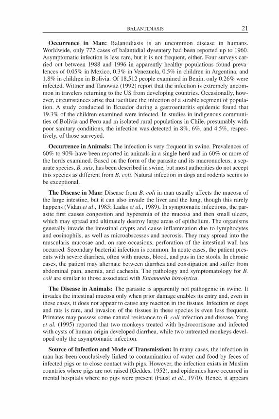
Occurrence in Man: Balantidiasis is an uncommon disease in humans.Worldwide, only 772 cases of balantidial dysentery had been reported up to 1960.Asymptomatic infection is less rare, but it is not frequent, either. Four surveys car-ried out between 1988 and 1996 in apparently healthy populations found preva-lences of 0.05% in Mexico, 0.3% in Venezuela, 0.5% in children in Argentina, and1.8% in children in Bolivia. Of 18,512 people examined in Benin, only 0.26% wereinfected. Wittner and Tanowitz (1992) report that the infection is extremely uncom-mon in travelers returning to the US from developing countries. Occasionally, how-ever, circumstances arise that facilitate the infection of a sizable segment of popula-tion. A study conducted in Ecuador during a gastroenteritis epidemic found that19.3% of the children examined were infected. In studies in indigenous communi-ties of Bolivia and Peru and in isolated rural populations in Chile, presumably withpoor sanitary conditions, the infection was detected in 8%, 6%, and 4.5%, respec-tively, of those surveyed.
Occurrence in Animals: The infection is very frequent in swine. Prevalences of60% to 90% have been reported in animals in a single herd and in 60% or more ofthe herds examined. Based on the form of the parasite and its macronucleus, a sep-arate species, B. suis, has been described in swine, but most authorities do not acceptthis species as different from B. coli. Natural infection in dogs and rodents seems tobe exceptional.
The Disease in Man: Disease from B. coli in man usually affects the mucosa ofthe large intestine, but it can also invade the liver and the lung, though this rarelyhappens (Vidan et al., 1985; Ladas et al., 1989). In symptomatic infections, the par-asite first causes congestion and hyperemia of the mucosa and then small ulcers,which may spread and ultimately destroy large areas of epithelium. The organismsgenerally invade the intestinal crypts and cause inflammation due to lymphocytesand eosinophils, as well as microabscesses and necrosis. They may spread into themuscularis mucosae and, on rare occasions, perforation of the intestinal wall hasoccurred. Secondary bacterial infection is common. In acute cases, the patient pres-ents with severe diarrhea, often with mucus, blood, and pus in the stools. In chroniccases, the patient may alternate between diarrhea and constipation and suffer fromabdominal pain, anemia, and cachexia. The pathology and symptomatology for B.coli are similar to those associated with Entamoeba histolytica.
The Disease in Animals: The parasite is apparently not pathogenic in swine. Itinvades the intestinal mucosa only when prior damage enables its entry and, even inthese cases, it does not appear to cause any reaction in the tissues. Infection of dogsand rats is rare, and invasion of the tissues in these species is even less frequent.Primates may possess some natural resistance to B. coli infection and disease. Yanget al. (1995) reported that two monkeys treated with hydrocortisone and infectedwith cysts of human origin developed diarrhea, while two untreated monkeys devel-oped only the asymptomatic infection.
Source of Infection and Mode of Transmission: In many cases, the infection inman has been conclusively linked to contamination of water and food by feces ofinfected pigs or to close contact with pigs. However, the infection exists in Muslimcountries where pigs are not raised (Geddes, 1952), and epidemics have occurred inmental hospitals where no pigs were present (Faust et al., 1970). Hence, it appears
BALANTIDIASIS 21

that person-to-person transmission is possible where environmental sanitation con-ditions are poor.
The B. coli trophozoite cannot survive for very long in a dry environment, so it isunlikely that infection would be acquired through ingestion of viable trophozoites.The cyst is a much more efficient means of transmission than the trophozoite, sinceit can survive outside the body for two weeks or more at ambient temperatures.Ingested cysts excyst in the intestine and begin to multiply as trophozoites.
Diagnosis: The symptomatology of balantidiasis is such that it cannot be differ-entiated clinically from other causes of dysentery. Similarly, it is not possible to dis-tinguish it from amebiasis through endoscopic observation of intestinal lesions.Diagnosis is based on detection of trophozoites, which are most commonly found inwatery diarrheal stools, or cysts, which are particularly abundant in formed stools.The trophozoite, obtained from stool specimens or endoscopic samples, can be seenby microscopic examination of wet mounts at low magnification (100X). Permanentstained preparations are not recommended because the parasite, owing to its size andthickness, stains deeply and its internal structures then cannot be observed. Thecysts can be visualized by means of fecal parasite concentration methods.
Control: The most efficient control method is probably to educate the publicabout basic personal hygiene practices in areas in which contact between humansand swine is common. On pig farms, care should be taken to prevent animal wastefrom contaminating water used for drinking or irrigation, and manure should not beused as fertilizer on crops of vegetables that are eaten raw. Suspicious water or foodshould be boiled because normal chlorination will not kill the cysts. Where there isa potential for person-to-person transmission, the usual personal hygiene practicesfor preventing infections of fecal origin, combined with effective treatment ofinfected individuals, should reduce the risk of transmission.
Bibliography
Faust, E.C., P.F. Russell, C.R. Jung. Craig and Faust’s Clinical Parasitology. Philadelphia:Lea & Febiger; 1970.
García, L.S., D.A. Bruckner. Diagnostic Medical Parasitology, 3rd ed. Washington, D.C.:ASM Press; 1997.
Geddes, McC.A. Balantidiasis in South Persia. Br Med J 1:629–631, 1952.Ladas, S.D., S. Savva, A. Frydas, A. Kaloviduris, J. Hatzioannou, S. Raptis. Invasive bal-
antidiasis presented as chronic colitis and lung involvement. Dig Dis Sci 34:1621–1623, 1989.Neva, F.A., H.W. Brown. Basic Clinical Parasitology, 6th ed. Norwalk, Connecticut:
Appleton & Lange; 1994.Vidan, J.R., A. Frauca, B. Martínez, F. Borda. Parasitosis hepática por Balantidium coli.
Med Clin (Barc) 85:299–300, 1985.Wenyon, C.M. Protozoology: A Manual for Medical Men, Veterinarians and Zoologists.
London: Baillière, Tindall and Cox; 1926.Wittner, M., H.B. Tanowitz. Intestinal parasites in returned travelers. Med Clin North Am
76:6, 1433–1448, 1992.Yang Y., L. Zeng, M. Li, J. Zhou. Diarrhoea in piglets and monkeys experimentally infected
with Balantidium coli isolated from human faeces. J Trop Med Hyg 98(1):69–72, 1995.
22 PROTOZOOSES
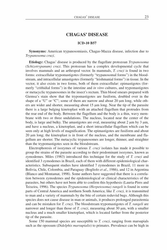
CHAGAS’ DISEASE
ICD-10 B57
Synonyms: American trypanosomiasis, Chagas-Mazza disease, infection due toTrypanosoma cruzi.
Etiology: Chagas’ disease is produced by the flagellate protozoan Trypanosoma(Schizotrypanum) cruzi. This protozoan has a complex developmental cycle thatinvolves mammals and an arthropod vector. In mammals, T. cruzi is found in twoforms: extracellular trypomastigotes (formerly “trypanosomal forms”) in the blood-stream, and intracellular amastigotes (formerly “leishmanial forms”) in tissue. In thevector, it also exists in two forms, both of them extracellular: epimastigotes (for-merly “crithidial forms”) in the intestine and in vitro cultures, and trypomastigotesor metacyclic trypanosomes in the insect’s rectum. Thin blood smears prepared withGiemsa’s stain show that the trypomastigotes are fusiform, doubled over in theshape of a “U” or “C”; some of them are narrow and about 20 µm long, while oth-ers are wider and shorter, measuring about 15 µm long. Near the tip of the parasitethere is a large bulging kinetoplast with an attached flagellum that protrudes fromthe rear end of the body. Between the flagellum and the body is a thin, wavy mem-brane with two or three undulations. The nucleus, located near the center of thebody, is large and bulky. The amastigotes are oval, measuring about 2 µm by 3 µm,and have a nucleus, a kinetoplast, and a short intracellular flagellum which can beseen only at high levels of magnification. The epimastigotes are fusiform and about20 µm long; the kinetoplast is in front of the nucleus, and the membrane and fla-gellum are shorter. The metacyclic trypanosomes are longer, thinner, and straighterthan the trypomastigotes seen in the bloodstream.
Electrophoresis of isozymes of various T. cruzi isolates has made it possible togroup the strains of the parasite according to their predominant isozymes, known aszymodemes. Miles (1983) introduced this technique for the study of T. cruzi andidentified 3 zymodemes in Brazil, each of them with different epidemiological char-acteristics. Subsequent studies have identified 7 zymodemes in Brazil, 11 more inBolivia, Chile, Colombia, and Paraguay (Bogliolo et al., 1986), and 12 in Argentina(Blanco and Montamat, 1998). Some authors have suggested that there is a correla-tion between zymodemes and the epidemiological or clinical characteristics of theparasites, but others have not been able to confirm this hypothesis (Lauria-Pires andTeixeira, 1996). The species Trypanosoma (Herpetosoma) rangeli is found in someparts of Central America and northern South America; like T. cruzi, it is transmittedto man and a variety of mammals by the bite of certain reduviid bugs. Although thisspecies does not cause disease in man or animals, it produces prolonged parasitemiaand can be mistaken for T. cruzi. The bloodstream trypomastigotes of T. rangeli arenarrower and longer than those of T. cruzi, measuring about 30 µm, with a smallernucleus and a much smaller kinetoplast, which is located farther from the posteriortip of the parasite.
Some 150 mammal species are susceptible to T. cruzi, ranging from marsupialssuch as the opossum (Didelphis marsupialis) to primates. Prevalence can be high in
CHAGAS’ DISEASE 23

cats, dogs, rodents, and both domestic and wild lagomorphs, which taken togetherconstitute an important reservoir for human infection. Birds and cold-blooded ver-tebrates are refractory to T. cruzi infection, but domestic birds are important foodsources for the vector.
The vectors are reduviid bugs of the family Triatomidae. It has been confirmedthat about a hundred species are susceptible to the infection, but the most importantvectors are Triatoma infestans in southern Peru; Panstrongylus megistus in northernArgentina, southern Brazil, and Paraguay; and Rhodnius prolixus in northern SouthAmerica, parts of Central America, and Mexico. The vector becomes infected whenit feeds on the blood of an infected mammal and ingests trypomastigotes. Theseforms reach the midgut of the insect, turn into epimastigotes, and divide abundantlyby binary fission. After the bug has been infected for 15 to 30 days, the infectivemetacyclic trypanosomes begin to appear in its rectum. The bug usually remainsinfected for several months or the rest of its life.
Unlike the African trypanosomes (see the chapter on African Trypanosomiasis),which produce infection through the bite of a vector, T. cruzi infects its definitive hostthrough the vector’s excrement. This modality is referred to as contaminative infec-tion, or transmission from the “posterior station,” as opposed to inoculative infection,or transmission from the “anterior station.” Animals may also become infected byingesting infected vectors. The metacyclic trypanosomes penetrate the organismeither through normal healthy mucosa or broken skin, often caused by scratching;they then invade the macrophages of the dermis or subcutaneous tissue, transforminto amastigotes, and multiply by binary fission. Between four and five days afterinfecting the host cell, the amastigotes then turn into trypomastigotes, which destroythe original cell and invade neighboring cells or spread through the bloodstream tothe cells of other organs, especially macrophages, cardiac and striated muscle fibers,and the neuroglia. The trypomastigotes do not multiply in the bloodstream; instead,they turn into amastigotes once again inside the cells and repeat the cycle of intra-cellular multiplication and destruction of the cell. Some authors have described inter-mediate forms between trypomastigotes and amastigotes (promastigotes, epimasti-gotes) when the parasite abandons the cells, but if such forms exist, they are rarelyseen in a mammal host. At the beginning of the infection, there are large numbers oftrypomastigotes in the bloodstream, but over time, the frequency of the blood-cellcycles tapers off, and as a result, after a few weeks the level of parasitemia drops con-siderably and the parasite remains restricted to tissue.
Geographic Distribution: T. cruzi infection exists only in the Americas, withinan area extending from 42° N latitude in the US (from California to Maryland) toapproximately 34° S in Chile and 42° S in Argentina. Vectors and wild reservoirshave also been found throughout much of the Caribbean, which was previously con-sidered free of the infection (PAHO, 1984). Autochthonous Chagas’ disease has notbeen confirmed outside the Americas (Marsden, 1997).
Occurrence in Man: Chagas’ disease is essentially a problem affecting southernMexico and Central and South America. According to estimates based on seroepi-demiologic studies, at the beginning of the 1980s, 10 to 20 million people in LatinAmerica were infected and 65 million were at risk for the infection; in addition, 10%of those infected in South America could be expected to develop symptoms and clin-ical signs of chronic Chagas’ disease (PAHO, 1984). By the mid-1990s, it was esti-
24 PROTOZOOSES
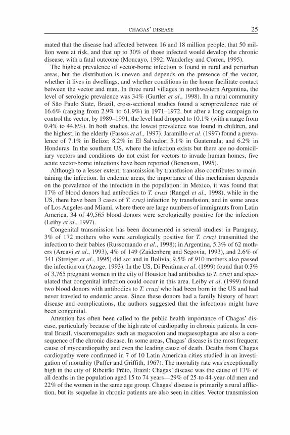
mated that the disease had affected between 16 and 18 million people, that 50 mil-lion were at risk, and that up to 30% of those infected would develop the chronicdisease, with a fatal outcome (Moncayo, 1992; Wanderley and Correa, 1995).
The highest prevalence of vector-borne infection is found in rural and periurbanareas, but the distribution is uneven and depends on the presence of the vector,whether it lives in dwellings, and whether conditions in the home facilitate contactbetween the vector and man. In three rural villages in northwestern Argentina, thelevel of serologic prevalence was 34% (Gurtler et al., 1998). In a rural communityof São Paulo State, Brazil, cross-sectional studies found a seroprevalence rate of16.6% (ranging from 2.9% to 61.9%) in 1971–1972, but after a long campaign tocontrol the vector, by 1989–1991, the level had dropped to 10.1% (with a range from0.4% to 44.8%). In both studies, the lowest prevalence was found in children, andthe highest, in the elderly (Passos et al., 1997). Jaramillo et al. (1997) found a preva-lence of 7.1% in Belize; 8.2% in El Salvador; 5.1% in Guatemala; and 6.2% inHonduras. In the southern US, where the infection exists but there are no domicil-iary vectors and conditions do not exist for vectors to invade human homes, fiveacute vector-borne infections have been reported (Benenson, 1995).
Although to a lesser extent, transmission by transfusion also contributes to main-taining the infection. In endemic areas, the importance of this mechanism dependson the prevalence of the infection in the population: in Mexico, it was found that17% of blood donors had antibodies to T. cruzi (Rangel et al., 1998), while in theUS, there have been 3 cases of T. cruzi infection by transfusion, and in some areasof Los Angeles and Miami, where there are large numbers of immigrants from LatinAmerica, 34 of 49,565 blood donors were serologically positive for the infection(Leiby et al., 1997).
Congenital transmission has been documented in several studies: in Paraguay,3% of 172 mothers who were serologically positive for T. cruzi transmitted theinfection to their babies (Russomando et al., 1998); in Argentina, 5.3% of 62 moth-ers (Arcavi et al., 1993), 4% of 149 (Zaidenberg and Segovia, 1993), and 2.6% of341 (Streiger et al., 1995) did so; and in Bolivia, 9.5% of 910 mothers also passedthe infection on (Azoge, 1993). In the US, Di Pentima et al. (1999) found that 0.3%of 3,765 pregnant women in the city of Houston had antibodies to T. cruzi and spec-ulated that congenital infection could occur in this area. Leiby et al. (1999) foundtwo blood donors with antibodies to T. cruzi who had been born in the US and hadnever traveled to endemic areas. Since these donors had a family history of heartdisease and complications, the authors suggested that the infections might havebeen congenital.
Attention has often been called to the public health importance of Chagas’ dis-ease, particularly because of the high rate of cardiopathy in chronic patients. In cen-tral Brazil, visceromegalies such as megacolon and megaesophagus are also a con-sequence of the chronic disease. In some areas, Chagas’ disease is the most frequentcause of myocardiopathy and even the leading cause of death. Deaths from Chagascardiopathy were confirmed in 7 of 10 Latin American cities studied in an investi-gation of mortality (Puffer and Griffith, 1967). The mortality rate was exceptionallyhigh in the city of Ribeirão Prêto, Brazil: Chagas’ disease was the cause of 13% ofall deaths in the population aged 15 to 74 years—29% of 25-to 44-year-old men and22% of the women in the same age group. Chagas’ disease is primarily a rural afflic-tion, but its sequelae in chronic patients are also seen in cities. Vector transmission
CHAGAS’ DISEASE 25
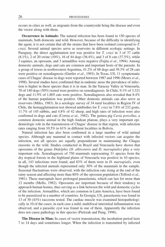
occurs in cities as well, as migrants from the countryside bring the disease and eventhe vector along with them.
Occurrence in Animals: The natural infection has been found in 150 species ofmammals, both domestic and wild. However, because of the difficulty in identifyingthe agent, it is not certain that all the strains that have been isolated correspond to T.cruzi. Several animal species serve as reservoirs in different ecologic settings. InParaguay, the direct agglutination test was positive for T. cruzi in 3 of 37 cattle(8.1%), 2 of 20 swine (10%), 16 of 44 dogs (36.4%), and 3 of 8 cats (37.5%), while3 equines, an opossum, and 3 armadillos were negative (Fujita et al., 1994). Amongdomestic animals, dogs and cats are common and important hosts of the parasite. Ina group of towns in northwestern Argentina, 41.2% of 68 dogs and 39.3% of 28 catswere positive on xenodiagnosis (Gurtler et al., 1993). In Texas, US, 11 symptomaticcases of Chagas’ disease in dogs were reported between 1987 and 1996 (Meurs et al.,1998). Several studies have confirmed that in endemic areas the prevalence of infec-tion is higher in these species than it is in man. In the Yaracuy Valley in Venezuela,70 of 140 dogs (50%) tested were positive on xenodiagnosis. In Chile, 9.1% of 3,321dogs and 11.9% of 1,805 cats were positive. Xenodiagnosis also revealed that 8.4%of the human population was positive. Other domestic animals can also serve asreservoirs (Miles, 1983). In a serologic survey of 34 rural localities in Region IV ofChile, the hemagglutination test showed antibodies for T. cruzi in 7.8% of 232 goats,11.7% of 145 rabbits, and 4.8% of 42 sheep, and high rates of infection were alsoconfirmed in dogs and cats (Correa et al., 1982). The guinea pig Cavia porcellus, acommon domestic animal in the high Andean plateau, plays a very important epi-demiologic role in the transmission of Chagas’ disease in that region, with infectionrates ranging from 10.5% to 61% in different localities in Bolivia.
Natural infection has also been confirmed in a large number of wild animalspecies. Although any mammal in contact with infected vectors can acquire theinfection, not all species are equally preponderant in maintaining the Chagasenzootic in the wild. Studies conducted in Brazil and Venezuela have shown thatopossums of the genus Didelphis (D. albiventris and D. marsupialis) play a veryimportant role. Xenodiagnosis of 750 mammals representing 31 species from thedry tropical forests in the highland plains of Venezuela was positive in 10 species;in all, 143 infections were found, and 83% of them were in D. marsupialis, eventhough the infected animals represented only 30% of the mammals in the sample.Seasonal fluctuations were observed, with the infection rate rising at the end of therainy season and affecting more than 80% of the opossum population (Telford et al.,1981). These marsupials have prolonged parasitemia, which can last for more than12 months (Mello, 1982). Opossums are important because of their tendency toapproach human homes, thus serving as a link between the wild and domestic cyclesof the infection. Armadillos, which are common in Latin America, have been foundto be parasitized in a number of countries. In Georgia, US, parasitemia was found in13 of 30 (43%) raccoons tested. The cardiac muscle was examined histopathologi-cally in 10 of the cases; in each case a mild, multifocal interstitial inflammation wasobserved, and a parasitic cyst was found in one of them. Apparently the infectiondoes not cause pathology in this species (Pietrzak and Pung, 1998).
The Disease in Man: In cases of vector transmission, the incubation period lasts7 to 14 days and sometimes longer. When the infection is transmitted by infected
26 PROTOZOOSES

blood, incubation takes between 30 and 40 days. Three phases of infection are dis-tinguished: acute, indeterminate, and chronic. The acute phase can range from anasymptomatic course, which is most common, to a severe or fatal disease. In 59acute-phase patients treated in Venezuela between 1988 and 1996, the disease pre-sented 19 different forms. In its most frequent manifestation, the symptomatologyincluded fever, myalgia, cephalalgia, and Romaña’s sign (unilateral eyelid swellingwhich seems to be mainly an allergic reaction to the bite), observed in 20% of thepatients (particularly children). Fifteen percent of the cases were asymptomatic, and11.9% manifested fever only. Nearly 50% of the children had an inoculationchagoma (swelling with involvement of a satellite lymph node, apparently caused bylocal multiplication of the parasite), but in about 25% of the patients no signs of aportal of entry were observed. The case fatality rate for the acute form is about 8%,and the deaths occur mainly in children with cardiac or central nervous system com-plications (Anez et al., 1999).
The indeterminate phase consists of a period of latent infection with low para-sitemia and no clinical symptoms, which can last indefinitely or progress to thechronic disease. This period is characterized by positive serology or xenodiagnosiswithout any clinical cardiac, digestive, or central nervous system manifestations andno electrocardiographic or radiologic alterations. In endemic areas, this form is seenespecially in the first three decades of life (Dorea, 1981). Autopsies of persons dyingfrom an accident who were in this phase have revealed foci of myocarditis and areduced number of neurons in the parasympathetic plexus.
The chronic form is seen in 10% to 30% of infected individuals, usually appear-ing 10 to 15 years after the acute phase. Chagas cardiopathy is the most importantchronic form. After the first manifestations, which almost always consist ofextrasystoles and precordialgia, an electrocardiogram will show complete or partialblockage of the right branch of the bundle of His. Signs of heart failure are seen dur-ing this phase, and autopsies show a weakened ventricular wall with aneurysms.Often the chronic phase is manifested only by abnormalities in the electrocardio-gram, with no clinical symptomatology. Histopathologic examination reveals areasof fibrosis and infiltration of mononuclear cells but not the presence of parasites,conditions not usually found in the chronic form of the disease (see hypotheses pre-sented below). The heart lesion corresponds to a microfocal, diffuse, fibrosingmyocarditis. At the same time, there is a significant reduction in the number ofparasympathetic ganglia (González Cappa and Segura, 1982). In Argentina, it isestimated that about 20% of all Chagas patients suffer from myocarditis. In severalendemic areas of Latin America, there is a digestive form of Chagas’ disease thatproduces visceromegalies such as megacolon and megaesophagus, and less fre-quently, neurologic, myxedematous, and glandular forms. Patients with acquiredimmunodeficiency syndrome may experience reactivation of the disease, with nerv-ous (75%) or cardiac (44%) involvement, or myositis of the esophagus and stomach(Ferreira et al., 1997).
The pathogenesis of the chronic phase is not yet understood. The lack of correla-tion between the lesions in the myocardium or digestive apparatus and the presenceof parasites has given rise to three main hypotheses to account for the pathogenesisof these manifestations: 1) when the pseudocysts rupture, T. cruzi “toxin” is releasedand destroys the muscle cells or the neurons; 2) antigen from the T. cruzi pseudo-cysts is absorbed by the adjacent cells, inducing an immune response that destroys
CHAGAS’ DISEASE 27

these cells; or 3) the parasite and the muscle cells or neurons share antigens in com-mon; therefore, immune reactions to the parasite also destroy the host cells. Sinceno toxin has been found that might account for the damage, the autoimmunehypotheses have been gaining ground in recent years, even though the supportingevidence is only circumstantial (Kierszembaum, 1999). Some investigators haveproposed that the lesions may be due to inflammatory reactions to parasites thatremain inside the tissues (Brener and Gazzinelli, 1997).
When immunocompetent individuals acquire the infection from a blood transfu-sion, there are usually no symptoms of the disease, but these people may developprolonged fever, adenopathies, and later, splenomegaly. In immunodeficientpatients, however, the infection can cause a high fever and progressively compro-mise their general state of health.
In the congenital disease, the most frequent signs are hepatosplenomegaly, pre-mature birth (weight under 2.5 kg), changes in the retina, meningoencephalitis, andcardiac insufficiency with alterations in the electroencephalogram. Fever is unusual.
The Disease in Animals: It is generally believed that T. cruzi infection is asymp-tomatic in wild animals, but this impression may be largely due to the lack ofdetailed clinical examination. Electrocardiographic studies and ventricularangiograms of rats (Rattus rattus) naturally infected with T. cruzi have revealedauricular and ventricular arrhythmias, second degree AV block, blockage of the rightoutflow tract, and dilation of the right chambers. The same alterations are seen indogs with chronic T. cruzi infections (Blandon et al., 1995). The acute phase, whichbegins after an incubation period of 5 to 42 days, is characterized by moderate fever,palpebral edema in some cases, pronounced hepatomegaly, multiple adenopathies,cardiac perturbations, and alterations in the nervous system. The acute phase lastsfrom 10 to 30 days and sometimes longer, following which the disease passes to theindeterminate phase, which can extend for years without clinical manifestations.Dogs with acute experimental infections have exhibited alterations in the neurons ofthe Auerbach plexus and myositis in the lower third of the esophagus (Caliari et al.,1996), but they did not have any visceromegalies. As in man, the chronic form ischaracterized by myocarditis. Of 26 dogs experimentally infected with blood trypo-mastigotes, 13 died spontaneously during the acute phase, while 12 of 38 dogsinfected with metacyclic trypanosomes survived to the chronic phase and lived for1 or 2 years. These animals had the same cardiac alterations that are seen in manduring the acute and chronic phase (Lana et al., 1992). Clinical, electrocardio-graphic, and echocardiographic manifestations in dogs with chronic Chagas’ diseasewere compatible with right heart disease. Six dogs survived less than 6 months,while 5 of them lived more than 30 months, the outcome varying according to theage of the animal at the time of initial examination (Meurs et al., 1998). There havealso been occasional reports of alterations in the brain and the peripheral nerves dur-ing the acute and chronic phases. The infection was found in a female dog and sevenof her eight pups in Virginia, US, suggesting that in dogs the infection can be trans-mitted either via the placenta or through the mother’s milk.
Source of Infection and Mode of Transmission: The source of Chagas’ infectionis always the infected mammal. In the case of vector transmission, the reservoir maybe any peridomestic animal that infects the vector, which in turn, infects other ani-mals, including man. In nature, Chagas’ disease appears to exist preferably in the
28 PROTOZOOSES

wild, invading the domestic environment only when there are domiciliary vectors andecological conditions that enable them to live in human homes. Since these condi-tions do not exist in the US, the infection has remained in the wild in that country.However, in many poor rural areas of Latin America, there are vectors that live exclu-sively or preferably inside houses, or at least have the potential to do so, and thedwellings have the kind of cracks that the insect needs in order to reproduce and hideduring the day. The infection is particularly prevalent in these areas. Migrants whomove from the countryside to the outskirts of cities can carry the vectors in their per-sonal effects and infest new residential areas. Often, migrants or T. cruzi-infectedvectors settle in periurban areas where Chagas’ disease is already endemic. Severalstudies have shown that one of the major risk factors for human infection is the pres-ence and number of dogs in the home, and some studies have implicated cats as well,especially when these animals are infected. This observation would indicate that dogsare a primary source of food and infection for the vectors (Gurtler et al., 1998).Chickens in the household are also a risk factor because, even though these animalsare not susceptible to T. cruzi, the vector feeds on them. T. cruzi can also be intro-duced in the human environment through peridomestic wild animals such as armadil-los, guinea pigs, opossums, and others. Rats have visible and prolonged infections,and they can also be a source of infection (Blandon et al., 1995). Moreover, even inthe chronic phase of the disease, a human can be a potential source of infection, asrevealed in a 13-year follow-up study of 202 chronic-phase patients: xenodiagnosisshowed that the levels of parasitemia were consistently maintained in 146 of thepatients and actually rose in 14 of them, while in 42 of the cases did these levelsdecline (Castro et al., 1999). These results notwithstanding, there are statistical stud-ies indicating that the presence of infected dogs is much more important in the infec-tion of vectors than is the presence of infected humans (Gurtler et al., 1991).
A number of the vectors are fully adapted to cohabiting with humans—for exam-ple, Triatoma infestans, which has a wide area of distribution that encompassesArgentina, Bolivia, Brazil, Chile, Paraguay, Peru, and Uruguay. Such species play akey role in human infection because of their facility of contact with people. Thenthere are species, found both in homes and in the wild, that are important becausethey introduce T. cruzi into the domestic environment; an example is Rhodnius pro-lixus, which is widely active in Colombia, Ecuador, Venezuela, much of CentralAmerica, and Mexico. Still other species are in the process of domiciliary adapta-tion—for example, Triatoma sordida in Argentina, Bolivia, and Brazil;Panstrongylus megistus in the eastern part of Brazil; T. brasiliensis in the Braziliannortheast; and T. maculata in Venezuela. Finally, there are species that are funda-mentally wild and rarely invade the peridomestic environment; examples are T. spin-olai in Chile, T. protracta in North America, and T. sanguisuga in the US. Althoughthese species do not play a significant role in human infection, they maintain theendemicity of Chagas’ disease in the wild. Some of the vectors, such as T. infestans,defecate while they feed, thus easily contaminating the skin or mucosae of the hostand facilitating transmission of the infective agent. These species are most impor-tant in transmission to man. Other species, such as T. protracta, defecate later andare therefore less significant for human infection, but they can play a role in the caseof animals that chew and eat them in an effort to get rid of them.
The ecology of Chagas’ disease is closely linked to underdevelopment andpoverty in rural and marginal urban areas of Latin America. Precariously built
CHAGAS’ DISEASE 29

dwellings made of adobe and mud, as well as roofs of palm thatch or straw, affordideal conditions for triatomine colonization. The bugs also take up residence inchicken houses, rabbit hutches, corrals, pigsties, aviaries, sheds, and wood piles inareas surrounding the homes.
Although less prevalent than vector transmission, congenital transmission andtransmission via blood transfusion are also important sources of human infection(see The Disease in Man), especially because they introduce the agent in areaswhere the vectors do not exist. Unlike toxoplasmosis, Chagas’ disease can be passedon congenitally when the mother is in the chronic phase of the infection. Althoughtransmission can also occur from the ingestion of food contaminated with the excre-ment of infected triatomines, the importance of this route in the epidemiology of thedisease remains to be assessed. There have also been accidental infections in labo-ratories and from organ transplants from infected donors.
Diagnosis: The specific diagnostic methods for Chagas’ disease are direct identi-fication of the parasite and testing for immunologic reactions. Recent efforts havefocused on detection of the parasite’s DNA using the polymerase chain reaction(PCR) technique. Since T. cruzi remains in the blood for only a short time, directdemonstration is used mainly in the acute phase, whereas the immunologic tests areused in the indeterminate and chronic phases.
In direct observation, fresh blood is examined either between slide and coverslipor in thin or thick films stained using Giemsa’s method. However, the effectivenessof these diagnostic procedures is limited except in very acute cases and with con-genital infection in children under 6 months old. Microscopic examination of tissuesoften fails to yield any parasites. A technique that is more efficient is the Stroutmethod (Flores et al., 1966), in which the blood sample is allowed to coagulate, theserum is centrifuged at a low speed (200 G) to eliminate the rest of the blood cellsand then at a high speed (600 G) to concentrate the trypanosomes, and finally, thesediment is observed. All the procedures mentioned become less effective as thelevel of parasitemia declines. For borderline cases, the most effective direct methodsare xenodiagnosis, hemoculture (Anez et al., 1999), and inoculation in animals, inall of which the few parasites present in the patient’s blood are multiplied. In xeno-diagnosis, the patient is bitten by uninfected vectors that have been produced in thelaboratory and fed on chickens (to prevent accidental T. cruzi infections), and theinsects’ feces are examined 30 and 60 days later to detect the presence of the para-site. This method is 100% effective in acute-phase patients, but less than 50% effec-tive with those in the indeterminate and chronic phases. Culture of blood or tissuesamples is done preferably using Novy-MacNeal-Nicolle medium, and incubationtakes 30 days. Finally, another method of diagnosis consists of inoculating samplesin uninfested mice or rats and subsequently observing these animals for parasitemia.
As the patient progresses to the indeterminate or chronic phase, the presence ofparasites in the bloodstream is too low to apply direct methods and indirect immuno-logic methods must be used. The complement fixation test (or Guerreiro Machadoreaction) was common in the past, but it is now considered that the most sensitiveand specific tests are direct agglutination, indirect immunofluorescence, and theenzyme-linked immunosorbent assay (Anez et al., 1999). Specificity, and to someextent sensitivity, depends on the antigens used, and recombinant antigens are beingstudied for this purpose (Umezawa et al., 1999). Also being investigated is PCR,
30 PROTOZOOSES

which detects parasite DNA and should obtain reactions that are both highly specificand sufficiently sensitive (Gomes et al., 1999).
Cases of congenital infection in infants up to 6 months of age can be consideredacute cases; thereafter, they should be considered indeterminate or chronic cases.When serology is used in congenital cases, the focus should be on finding IgM orIgA antibodies, because the mother’s IgG antibodies cross the placenta and can sim-ulate an infection in healthy children.
Although T. rangeli can be mistaken for T. cruzi, the two can be differentiated bymorphology, immunologic techniques based on selected antigens (Acosto et al.,1991), or PCR.
Control: The drugs available for the treatment of acute-phase Chagas’ disease aretoxic and unreliable in terms of eradicating the infection, and there is no curativetreatment for chronic infection (Levi et al., 1996); therefore, it is highly importantto control transmission. A number of countries, Brazil in particular, have independ-ently undertaken control campaigns (da Rocha e Silva et al., 1998). In 1991, sixSouthern Cone countries (Argentina, Bolivia, Brazil, Chile, Paraguay, and Uruguay)launched a regional control initiative with the support of the World HealthOrganization (Schofield and Dias, 1999). By 1999, vector transmission had beeninterrupted in Uruguay and significantly reduced in Argentina, Chile, and Brazil, butit had not yet been curtailed in Bolivia or Paraguay.
To ultimately control vector transmission, homes must be improved by eliminat-ing the cracks and crevices in which the vectors establish their colonies. However,since this is a costly, long-term undertaking, a more immediate alternative is to treatthe surfaces of infested dwellings with residual insecticides. Synthetic pyrethroidsare most often used, and the employment of synthetic insect hormones is understudy. It is desirable to remove dogs and cats from the human environment becausethey are not only an important food source for the vector but also a major reservoirfor T. cruzi (Gurtler et al., 1998). The areas to be treated are identified throughreports received, observation of the vector’s presence in homes (often found after thespraying of repellents), and detection of persons with positive serology for T. cruzi.Although the last approach is the most efficient and reliable, its drawback is that itonly identifies a Chagas endemic area after the people have become infected. Theverification of T. cruzi-positive serology in domestic dogs appears to give similarresults (Castanera et al., 1998). One possible approach to identifying endemic areasprior to the appearance of T. cruzi infection might be by detecting antibodies to thevector (instead of the protozoan) in humans or domestic animals (Barriga, 2000). Itis necessary to maintain surveillance following initial eradication of the domiciliaryvectors; it has been shown that they can establish foci outside the home after theapplication of insecticides and return to their original densities in one to six years.Moreover, wild species can occupy the habitats abandoned by the domestic vectors.
Transmission by blood transfusion is prevented through the presumptive identifi-cation of blood donors using questionnaires to find out if they come from Chagasicareas, through blood tests, or by treating the donated blood with gentian violet (250mg/L) for 24 hours or longer, with or without the addition of ascorbic acid and expo-sure to light (Morães-Souza and Bordin, 1996).
Congenital infection is combated through timely treatment of infected mothers,but there are no reliable reports on the effectiveness of this method.
CHAGAS’ DISEASE 31

Although numerous studies have been conducted with a view to producing a vac-cine against Chagas’ disease, success has been impeded by the difficulty of distin-guishing between protective antigens and those that might generate pathology in thelong term.
Bibliography
Acosta, L., A.J. Romanha, H. Cosenza, A. Krettli. Trypanosomatid isolates from Honduras:Differentiation between Trypanosoma cruzi and Trypanosoma rangeli. Am J Trop Med Hyg44:676–683, 1991.
Anez, N., H. Carrasco, H. Parada, et al. Acute Chagas’ disease in western Venezuela: Aclinical, seroparasitologic, and epidemiologic study. Am J Trop Med Hyg 60:215–222, 1999.
Arcavi, M., G. Orfus, G. Griemberg. Incidencia de la infección chagásica en embarazadasy en recién nacidos en área no endémica. Medicina (B Aires) 53:217–222, 1993.
Azogue, E. Women and congenital Chagas’ disease in Santa Cruz, Bolivia: Epidemiologicaland sociocultural aspects. Soc Sci Med 37:503–511, 1993.
Barriga, O.O. Los hospederos generan anticuerpos circulantes y cutáneos contra las pica-das de Triatoma infestans e inhiben su alimentación. In: VII Jornada Anual SOCHIPA, Librode resúmenes, p. 22. Temuco: Sociedad Chilena de Producción Animal; 2000.
Benenson, A.S., ed. Control of Communicable Diseases in Man, 16th ed. An OfficialReport of the American Public Health Association. Washington, D.C.: APHA; 1995.
Blanco, A., E.E. Montamat. Genetic variation among Trypanosoma cruzi populations. JExp Zool 282:62–70, 1998.
Blandon, R., I.M. Leandro, C.M. Johnson. Evaluación clínica, electrocardiográfica yangiográfica de los reservorios naturales de la enfermedad de Chagas en la República dePanama. Rev Med Panama 20:108–115, 1995.
Bogliolo, A.R., E. Chiari, R.O. Silva-Pereira, A.A. Silva-Pereira. A comparative study ofTrypanosoma cruzi enzyme polymorphism in South America. Braz J Med Biol Res19:673–683, 1986.
Brener, Z., R.T. Gazzinelli. Immunological control of Trypanosoma cruzi infection andpathogenesis of Chagas’ disease. Int Arch Allergy Immunol 114:103–110, 1997.
Caliari, E.R., M.V. Caliari, M. de Lana, W.L. Tafuri. Estudo quantitativo e qualitativo dosplexos de Auerbach e Meissner do esôfago de cãos inoculados com o Trypanosoma cruzi. RevSoc Bras Med Trop 29:17–20, 1996.
Castanera, M.B., M.A. Lauricella, R. Chuit, R.E. Gurtler. Evaluation of dogs as sentinelsof the transmission of Trypanosoma cruzi in a rural area of north-western Argentina. Ann TropMed Parasitol 92(6):671–683, 1998.
Castro, C., V. Macêddo, A. Prata. Comportamento da parasitemia pelo Trypanosoma cruziem chagásicos crônicos durante 13 anos. Rev Soc Bras Med Trop 32:157–165, 1999.
Correa, V., J. Briceño, J. Zuñiga, et al. Infección por Trypanosoma cruzi en animalesdomésticos de sectores rurales de la IV Región, Chile. Bol Chil Parasitol 37:27–78, 1982.
da Rocha e Silva, E.O., D.M. Wanderley, V.L. Rodrigues. Triatoma infestans: importancia,controle e eliminação da espécie no Estado de São Paulo, Brasil. Rev Soc Bras Med Trop31:73–88, 1998.
Di Pentima, M.C., L.Y. Hwang, C.M. Skeeter, M.S. Edwards. Prevalence of antibody toTrypanosoma cruzi in pregnant Hispanic women in Houston. Clin Infect Dis 28:1281–1285,1999.
Dorea, R.C.C. Doença de Chagas na Amazônia: aspectos epidemiológicos regionais e con-siderações a propósito de um caso pediátrico. Hileia Méd 3:81–109, 1981.
Ferreira, M.S., S. de Nishioka, M.T. Silvestre, A.S. Borges, F.R. Nunes-Araujo, A. Rocha.Reactivation of Chagas’ disease in patients with AIDS: Report of three new cases and reviewof the literature. Clin Infect Dis 25:1397–1400, 1997.
32 PROTOZOOSES

Flores, M.A., A. Trejos, A. R. Paredes, A.Y. Ramos. El método de concentración de Strouten el diagnóstico de la fase aguda de la enfermedad de Chagas. Bol Chil Parasitol 21:38–39,1966.
Fujita, O., L. Sanabria, A. Inchaustti, A.R. De Arias, Y. Tomizawa, Y. Oku. Animal reser-voirs for Trypanosoma cruzi infection in an endemic area in Paraguay. J Vet Med Sci56:305–308, 1994.
Gomes, M.L., L.M. Galvão, A.M. Macedo, S.D. Pena, E. Chiari. Chagas’ disease diagno-sis: Comparative analysis of parasitologic, molecular, and serologic methods. Am J Trop MedHyg 60:205–210, 1999.
González Cappa, S.M., E.L. Segura. Enfermedad de Chagas. Adel Microbiol Enf Infec (BAires) 1:51–102, 1982.
Gurtler, R.E., M.C. Cecere, D.N. Rubel, et al. Chagas disease in north-west Argentina:Infected dogs as a risk factor for the domestic transmission of Trypanosoma cruzi. Trans R SocTrop Med Hyg 85:741–745, 1991.
Gurtler, R.E., M.C. Cecere, R.M. Petersen, D.N. Rubel, N.J. Schweigmann. Chagas diseasein north-west Argentina: Association between Trypanosoma cruzi parasitaemia in dogs andcats and infection rates in domestic Triatoma infestans. Trans R Soc Trop Med Hyg 87:12–15,1993.
Gurtler, R.E., R. Chuit, M.C. Cecere, M.B. Castanera, J.E. Cohen, E.L. Segura. Householdprevalence of seropositivity for Trypanosoma cruzi in three rural villages in northwestArgentina: Environmental, demographic, and entomologic associations. Am J Trop Med Hyg59:741–749, 1998.
Jaramillo, R., J.P. Bryan, J. Schur, A.A. Pan. Prevalence of antibody to Trypanosoma cruziin three populations in Belize. Am J Trop Med Hyg 57:298–301, 1997.
Kierszenbaum, F. Chagas’ disease and the autoimmunity hypothesis. Clin Microbiol Rev12:210–223, 1999.
Lana, M. de, E. Chiari, W.L. Tafuri. Experimental Chagas’ disease in dogs. Mem InstOswaldo Cruz 87:59–71, 1992.
Lauria-Pires, L., A.R. Teixeira. Virulence and pathogenicity associated with diversity ofTrypanosoma cruzi stocks and clones derived from Chagas’ disease patients. Am J Trop MedHyg 55:304–310, 1996.
Leiby, D.A., E.J. Read, B.A. Lenes, et al. Seroepidemiology of Trypanosoma cruzi, etio-logic agent of Chagas’ disease, in US blood donors. J Infect Dis 176:1047–1052, 1997.
Leiby, D.A., M.H. Fucci, R.J. Stumpf. Trypanosoma cruzi in a low- to moderate-risk blooddonor population: Seroprevalence and possible congenital transmission. Transfusion39:310–315, 1999.
Levi, G.C., I.M. Lobo, E.G. Kallas, V. Amato Neto. Etiological drug treatment of humaninfection by Trypanosoma cruzi. Rev Inst Med Trop Sao Paulo 38:35–38, 1996.
Marsden, P.D. The control of Latin American Chagas’ disease. Rev Soc Bras Med Trop30:521–527, 1997.
Mello, D.A. Aspectos do ciclo silvestre do Trypanosoma cruzi em regiões de cerrado(Município de Formosa, Estado de Goias). Mem Inst Oswaldo Cruz 76:227–246, 1981.
Meurs, K.M., M.A. Anthony, M. Slater, M.W. Miller. Chronic Trypanosoma cruzi infectionin dogs: 11 cases (1987–1996). J Am Vet Med Assoc 213:497–500, 1998.
Miles, M.A. The epidemiology of South American trypanosomiasis—Biochemical andimmunological approaches and their relevance to control. Trans R Soc Trop Med Hyg 77:5–23,1983.
Moncayo, A. Chagas’ disease: Epidemiology and prospects for interruption in theAmericas. World Health Stat Q 45:276–279, 1992.
Morães-Souza, H., J.O. Bordin. Strategies for prevention of transfusion-associated Chagas’disease. Transfus Med Rev 10:161–170, 1996.
Pan American Health Organization (PAHO). Situación de la enfermedad de Chagas en lasAméricas. Bol Oficina Sanit Panam 97:159–165, 1984.
CHAGAS’ DISEASE 33

Passos, A.D., J.L. Nogueira, J.F. de Castro Figueiredo, U.A. Gomes, A.L. Dal-Fabbro.Evolução da positividade sorológica para a doença de Chagas numa comunidade ruralbrasileira. Rev Panam Salud Publica 2:247–252, 1997.
Pietrzak, S.M., O.J. Pung. Trypanosomiasis in raccoons from Georgia. J Wildl Dis34:132–136, 1998.
Puffer, R.R., G. Wynne Griffith. Patterns of Urban Mortality. Washington, D.C.: PanAmerican Health Organization; 1967. (Scientific Publication No. 151.)
Rangel, H., R. Gatica, C. Ramos. Detection of antibodies against Trypanosoma cruzi indonors from a blood bank in Cuernavaca, Morelos, Mexico. Arch Med Res 29:79–82, 1998.
Russomando, G., M.M. de Tomassone, I. de Guillen, et al. Treatment of congenital Chagas’disease diagnosed and followed up by the polymerase chain reaction. Am J Trop Med Hyg59:487–491, 1998.
Schmunis, G.A., F. Zicker, F. Pinheiro, D. Brandling-Bennett. Risk for transfusion-trans-mitted infectious diseases in Central and South America. Emerg Infect Dis 4:5–11, 1998.
Schofield, C.J., J.C. Dias. The Southern Cone Initiative against Chagas’ disease. AdvParasitol 42:1–27, 1999.
Streiger, M., D. Fabbro, M. del Barco, R. Beltramino, N. Bovero. Chagas congénito en laciudad de Santa Fe. Diagnóstico y tratamiento. Medicina (B Aires) 55:125–132, 1995.
Telford, S.R., Jr., R.J. Tonn, J. González, P. Betancourt. Dinámica de las infecciones tri-panosómicas entre la comunidad de los bosques tropicales secos en los llanos altos deVenezuela. Bol Dirección Malariol Saneam Ambient 21:196–209, 1981.
Umezawa, E.S., S.F. Bastos, M.E. Camargo, et al. Evaluation of recombinant antigens forserodiagnosis of Chagas’ disease in South and Central America. J Clin Microbiol37:1554–1560, 1999.
Wanderley, D.M., F.M. Correa. Epidemiology of Chagas’ heart disease. Rev Paul Med113:742–749, 1995.
Zaidenberg, M., A. Segovia. Enfermedad de Chagas congénita en la ciudad de Salta,Argentina. Rev Inst Med Trop Sao Paulo 35:35–43, 1993.
CRYPTOSPORIDIOSIS
ICD-10 A07.2
Synonym: Cryptosporidium infection.
Etiology: The genus Cryptosporidium, together with Isospora, Cyclospora,Sarcocystis, and Toxoplasma, contains protozoa of the coccidia group in the phylumApicomplexa (formerly Sporozoa). Since the genus was recognized in 1907 byTyzzer, more than 20 species of Cryptosporidium have been described, but at pres-ent only 6 are accepted as valid. The only species that affects both humans and othermammals is C. parvum (Barriga, 1997). However, it appears that some varieties ofthis parasite infect only humans and other varieties infect humans, cattle, and mice.C. parvum normally lives in the small intestine, where it forms oocysts that areexcreted in the host’s feces. Each oocyst contains four small banana-shaped sporo-zoites, which are the infective stage of the parasite. When a susceptible host ingests
34 PROTOZOOSES

the oocysts, the sporozoites shed their protective cover and penetrate the epithelialcells of the new host’s intestine. Each sporozoite differentiates into a spherical par-asite, the trophozoite, which in turn multiplies asexually to form two types ofmeronts (formerly called schizonts), each about 5 µm in diameter. Type I merontsproduce six to eight new banana-shaped parasites (merozoites). Type II merontsform four oval-shaped merozoites, the gametocytes. Once mature, the merozoitesleave the host cell and invade new epithelial cells, where they produce more mero-zoites (type I) or gametocytes (type II). The gametocytes also invade new intestinalcells, where they differentiate into male cells (microgametocytes) and female cells(macrogametocytes).
The microgametocytes produce numerous filamentous microgametes 1–2 µm inlength, which leave the host cell and fertilize the macrogametocytes, forming azygote. The zygote matures in the host cell and produces four naked sporozoites—i.e., not inside sporocysts—which are already infective. Most of the mature zygotes(around 80%) develop a tough outer cover measuring 2.5–5 µm in diameter andbecome infective oocysts. These oocysts are excreted by the host in feces and con-taminate the environment. The rest of the mature zygotes have only a thin outermembrane. Because these thin-walled oocysts are easily ruptured, their sporozoitesremain in the intestine, reinfecting the same host (Fayer and Ungar, 1986).
Geographic Distribution: Worldwide. Cases of human cryptosporidiosis havebeen reported from more than 50 countries on 6 continents (Benenson, 1997).
Occurrence in Man: The first two clinical cases of human cryptosporidiosiswere identified in 1976 in two immunodeficient patients. Since then, many cases and numerous epidemics have been recognized. Various surveys have indicated thatthe oocyst prevalence in feces ranges from 1% to 2% in Europe, 0.6% to 4.3% inNorth America, and 10% to 20% in the developing countries. However, serologicevidence of past infections has shown positivity rates of 25% to 35% in industrial-ized countries and up to 65% in developing countries. The most well-known epi-demic occurred in 1993 in Milwaukee, Wisconsin, US, where a total of 1.6 millionpeople were exposed, 403,000 were infected, and 7 died (Lisle and Rose, 1995).Infection is much more common than the clinical disease and most frequentlyoccurs in children under 2 years of age, contacts of infected individuals, livestockhandlers, travelers to developing countries, homosexuals, and, especially, immuno-deficient individuals. In Australia, Cryptosporidium was found in 4.1% of stool sam-ples from 884 patients with gastroenteritis, but not in samples from 320 patientswithout gastroenteritis (Tzipori et al., 1983). In the US, however, whileCryptosporidium was similarly found in the stool samples of around 4% of patientswith gastroenteritis, it was also found in 13% of samples from healthy individuals.
Occurrence in Animals: Several species of Cryptosporidium infect both warm-and cold-blooded animals. C. parvum infects numerous mammals in addition tohumans, in particular other primates, cattle and other ruminants, horses, carnivores,and rodents. In all the affected domestic species, very young unweaned animals aremore susceptible to the infection and the disease than adults, and calves appear to bemost susceptible. The first clinical case of cryptosporidiosis in animals was identifiedin a calf in 1971. Subsequently, the infection has been found in up to 80% of calvesunder 1 month of age and up to 62% of apparently healthy adult cattle. In horses, the
CRYPTOSPORIDIOSIS 35
infection has been found in 15%–31% of suckling foals, but only 0.6% of adulthorses. Of calves with diarrhea studied in the US, 25% were found to be infected.
The Disease in Man: In individuals with healthy immune systems, cryp-tosporidiosis may be asymptomatic or may occur as a self-limiting disease. The ill-ness is characterized by profuse watery diarrhea that begins explosively one or twoweeks after infection and generally lasts 8–20 days, often accompanied by abdomi-nal pain, nausea, vomiting, low-grade fever (under 39°C), and weight loss. Inimmunodeficient individuals, the symptoms are more severe and may include asmany as 71 evacuations per day, with fluid loss of up to 25 liters (Ryan, 1994).Rather than being self-limiting, the disease may persist until the individual’s death.In such patients, the parasite has sometimes been found to invade the respiratory andbiliary tracts (Clavel et al., 1996).
The Disease in Animals: Cryptosporidiosis is fairly common in young calves. Theinfection generally appears during the first three weeks of life and affects animalsbetween 3 and 35 days of age. The clinical manifestations are diarrhea, tenesmus,anorexia, and weight loss. It is difficult to distinguish diarrhea caused byCryptosporidium from diarrhea caused by other agents. Anderson (1982) reported thatin calves aged 1–15 days from 47 herds, only 17 out of 51 were found to be excretingCryptosporidium oocysts, although all had diarrhea. In horses, swine, and domesticcarnivores, the disease has occasionally been reported in very young or immunodefi-cient animals (Barriga, 1997). Infected rodents do not appear to develop signs of dis-ease. Birds are rarely affected by the species of the parasite that infect mammals.
Source of Infection and Mode of Transmission: The sources of infection forhumans are other infected people and infected cattle. There is no solid evidence thatother animals are an important source of human infection. AlthoughCryptosporidium-infected cats have been found in association with AIDS patients,it is not clear who infected whom. However, recent studies indicate that C. parvumexhibits genetic polymorphism, with one genotype infecting only humans andanother infecting humans, cattle, and mice (Peng et al., 1997). These genotypesmight represent different species, but unequivocal identification of Cryptosporidiumspecies is difficult.
The source of infection for domestic animals is other domestic animals. Cross-trans-mission studies have demonstrated that parasites isolated from humans, goat kids,deer, lambs, and calves can infect and cause diarrhea in pigs, lambs, and calves, whilethey produce an asymptomatic infection in chickens, colts, and laboratory animals(Tzipori, 1983). Isolates from humans and calves have also been transmitted to kids,puppies, cats, mice, and calves (Current, 1983). Cryptosporidium species that infectbirds do not infect mammals, and species that infect mammals rarely infect birds.
The infection is transmitted through ingestion of foods and water contaminatedwith fecal matter from an infected individual, direct contact with infected feces, oringestion of water from sources contaminated by effluents from sewerage systemsor cattle farms. Children, childcare workers who change diapers, bed-ridden patientsand their caregivers, people who work with cattle, and individuals who engage inanal sex have a high risk of being infected through direct contact with fecal matter.
Diagnosis: Diarrhea from Cryptosporidium is hard to distinguish clinically fromdiarrheal illnesses due to other causes. Diagnosis of cryptosporidiosis is suspected
36 PROTOZOOSES

on the basis of both clinical symptoms and epidemiological history and is confirmedby demonstrating the presence of oocysts in the patient’s feces. However, becauseoocysts are small (2.5–5 µm in diameter), they are difficult to see with direct micro-scopic examination of fecal samples. They are therefore more easily detected bymeans of techniques involving concentration in sugar solutions, such as Sheather’ssolution, and by phase contrast microscopy. Giemsa or methylene blue stainingmakes the oocysts more visible but also turns yeast contaminants the same color,making it impossible to distinguish them from the parasite. Ziehl-Neelsen stain, onthe other hand, turns oocysts red but does not stain yeast. Auramine-rhodamine andsafranine-methylene blue are also useful for distinguishing oocysts. A recentlydeveloped technique uses fluorescent monoclonal antibodies specific toCryptosporidium to visualize the parasites in fecal or environmental specimens. Thespecificity of serologic diagnosis by means of immunofluorescence assay orenzyme-linked immunosorbent assay was initially dubious, but the tests have beenrefined and now show satisfactory levels of sensitivity and specificity. Althoughserologic diagnosis is useful for epidemiological studies, the antibodies may appeartoo late for clinical purposes in immunocompetent patients or may not appear in suf-ficient quantities in immunodeficient patients.
Procedures for recovering and identifying Cryptosporidium in environmentalwaters are highly variable, inefficient, and time-consuming. The currently recom-mended practice involves passing large volumes of water through special filters,centrifuging the material trapped by the filters to concentrate it, purifying the con-centrate in a Percoll-sucrose gradient, staining with fluorescent antibodies, and,finally, examining the material microscopically.
Control: For an individual, prevention of cryptosporidiosis consists of avoidingthe ingestion of raw foods or water that may be contaminated with human or animalfeces and avoiding contact with feces (Juraneck, 1995). Cooking high-risk foods andwashing hands carefully before eating should also reduce the danger of infection.People should avoid immersion in water containing effluents from sewerage systemsor cattle farms. Cryptosporidium oocysts are highly resistant to all disinfectants. Forexample, the CT value (concentration of disinfectant in milligrams per liter multi-plied by contact time in minutes) for killing 99% to 99.9% of the Cryptosporidiumoocysts present in water is 6 to 10 for ozone and 9,600 for chlorine. In contrast, thecorresponding values for Giardia intestinalis are 0.17 and 15. Exposure to watertemperatures of 25°C and 8°C for 4 weeks kills only 50% and 25% of oocysts,respectively (Barriga, 1997). Under favorable conditions, they are probably capableof surviving for several months in nature.
Treatment of drinking water in well-run plants with good filters removes around99.9% of oocysts.
Bibliography
Anderson, B.C. Cryptosporidiosis: A review. J Am Med Assoc 180:1455–1457, 1982.Barriga, O.O. Veterinary Parasitology for Practitioners, 2nd ed. Edina: Burgess
International Group; 1997.Benenson, A.S., ed. Control of Communicable Diseases Manual, 16th ed. An official report
of the American Public Health Association. Washington, D.C: APHA; 1995.
CRYPTOSPORIDIOSIS 37

Clavel, A., A.C. Arnal, E.C. Sanchez, et al. Respiratory cryptosporidiosis: Case series andreview of the literature. Infection 24:341–346, 1996.
Current, W. L., N.C. Reese, J.V. Ernst, et al. Human cryptosporidiosis in immunocompe-tent and immunodeficient persons. Studies of an outbreak and experimental transmission. NewEngl J Med 308(21):1252–1257, 1983.
Fayer, R., B.L. Ungar. Cryptosporidium spp. and cryptosporidiosis. Microbiol Rev50:458–483, 1986.
Juraneck, D.D. Cryptosporidiosis: Sources of infection and guidelines for prevention. ClinInfect Dis Suppl 1:S57–61, 1995.
Lisle, J.T., J.B. Rose. Cryptosporidium contamination of water in the US and the UK: Amini-review. J Water SRT-Aqua 44:103–117, 1995.
Peng, M.M., L. Xiao, A.R. Freeman, et al. Genetic polymorphism among Cryptosporidiumparvum isolates: Evidence of two distinct human transmission cycles. Emerg Infect Dis3:567–573, 1997.
Ryan, K.J., ed. Sherris Medical Microbiology: An Introduction to Infectious Diseases, 3rded. Norwalk, Connecticut: Appleton & Lange; 1994.
Tzipori, S. Cryptosporidiosis in animals and humans. Microbiol Rev 47:84–96, 1983.Tzipori, S., M. Smith, C. Birch, G. Bames, R. Bishop. Cryptosporidiosis in hospital patients
with gastroenteritis. Am J Trop Med Hyg 32:931–934, 1983.
CUTANEOUS LEISHMANIASIS
ICD-10 B55.1
Synonyms: Chiclero ulcer, espundia, pian-bois, uta, and buba (in the Americas);oriental sore, Aleppo boil, Baghdad sore, Delhi sore, and other local names (in theOld World).
Etiology: Leishmaniasis is caused by flagellate protozoa of the familyTrypanosomatidae, genus Leishmania. The life cycle of leishmanias is relativelysimple. The flagellate forms of the parasite—oval amastigotes measuring 2 to 5 µmin diameter (see the chapter on Chagas’ Disease)—exist within macrophages of adefinitive vertebrate host, including humans. Small flies of the familyPhlebotomidae (genus Phlebotomus in the Old World and Lutzomyia in theAmericas) ingest the parasites when they feed on the host’s blood. Once in the fly’sintestine, the amastigotes become promastigotes—extracellular forms with a flagel-lum emerging from the anterior end, which are fusiform and measure 14 to 20 µmlong and 2 to 4 µm wide. The promastigotes then multiply in the intestine of the vec-tor. In the insect, two promastigote forms can be observed: a wider, relativelyimmotile form that attaches to the wall of the intestine, and another, thinner, motileform that moves freely in the insect’s intestinal lumen and proboscis. The first formincubates easily in human serum; the second does not. Presumably, the latter aremetacyclic infective forms for the vertebrate which, by regurgitation from the intes-tine, are inoculated by the fly through the proboscis at its next blood meal (Kettle,
38 PROTOZOOSES

1995). Once inside the vertebrate, the promastigotes become amastigotes, invade thecutaneous macrophages, and multiply in a parasitophorous vacuole. These parasitesare equipped with several adaptation mechanisms that enable them to overcome thelethal effects of macrophages and lysosomes on microorganisms (Antoine, 1995).Their multiplication eventually causes the host cell to rupture, and the releasedamastigotes then invade new macrophages.
Though there have been reports of morphological differentiation of Leishmaniaby computerized image analysis (Youssef et al., 1997), it is virtually impossible todistinguish between species by means of conventional microscopy. Moreover, leish-manias seem to be undergoing an active process of evolution: isolates from parasitesthat cause identical diseases have shown different biochemical characteristics, whileisolates from parasites that cause different diseases have similar biochemical fea-tures (Barral et al., 1991). Some authors have proposed dividing the genus into twosubgenera: Leishmania, encompassing forms that multiply in the foregut of theirvectors (suprapylaria reproduction), and Vianna, comprising leishmania thatdevelop in the midgut and hindgut (peripylaria reproduction). It is widely acceptedthat very similar groups of leishmanias exist. Those groups have been classified asspecies or complexes, which in turn comprise various lesser categories or sub-species. The complexes associated with cutaneous or visceral leishmaniasis in theAmericas have been identified by polymerase chain reaction (Harris et al., 1998).The species or subspecies of each complex are distinguished mainly by their geo-graphic distribution, the clinical manifestations of the disease they cause, and epi-demiologic characteristics.
Current efforts are endeavoring to classify leishmanias by means of serologicmethods (agglutination, specificity of excretory antigens, and fluorescence withmonoclonal antibodies), biochemical methods (enzymatic profile, nuclear and kine-toplast DNA (kDNA) density, and metabolic characteristics), and molecular biologytechniques (genomic DNA sequencing and karyotype analysis), as a result of whichnew species are being proposed (Kreutzer et al., 1991). Investigators now speak ofserodemes (populations that are differentiated by their reactivity, using antibody bat-teries), zymodemes (populations distinguished by the composition of theirisozymes), and schizodemes (populations that can be distinguished by the size oftheir DNA fragments when they are treated with batteries of restriction enzymes).For example, Chouicha et al. (1997) studied 10 enzymatic systems from 137 isolatesof L. braziliensis obtained in Bolivia, Brazil, and Colombia, and were able to iden-tify 44 closely related zymodemes. Kapoor et al. (1998) distinguished L. tropicafrom L. donovani, the agent of visceral leishmaniasis in Africa and Asia, and iden-tified different isolates of L. donovani using cDNA probes and kDNA fragments.
The complexes that produce cutaneous leishmaniasis in the Americas are L. mexi-cana (which encompasses the species L. mexicana, L. amazonensis, and L. venezue-lensis); L. braziliensis (L. braziliensis, L. panamensis, L. guyanensis); and L. peru-viana. In the Old World, the agents of the disease belong to the L. tropica complex (L.tropica, L. major, and L. aethiopica) (Chulay, 1991). The complexes that causeAmerican cutaneous leishmaniasis can be distinguished by such characteristics as vec-tor, parasite localization in the insect’s intestine, the pathogenicity of the agent onhamster skin, and the growth in culture media (Lainson and Shaw, 1974). The vectorsof the L. mexicana complex leishmanias are phlebotomines of the group Nyssomyia.These parasites undergo suprapylaria development. When inoculated in hamster skin,
CUTANEOUS LEISHMANIASIS 39
they reproduce quickly, forming histiocytomas in which amastigotes are abundant andmetastasis is common. They grow profusely in Novy, MacNeal, and Nicolle (NNN)culture medium. In contrast, leishmanias of the L. braziliensis complex, for which thevectors are phlebotomines of the groups Psychodopygus and Nyssomyia, undergoperipylaria development and multiply very slowly in hamster skin, producing smallnodules or ulcers with few amastigotes that do not metastasize and whose growth inNNN culture media is slow or moderate (Bonfante-Garrido, 1983).
Geographic Distribution (Table 1): Some leishmanias appear to be indigenousto the Americas, while others were most likely imported from the Old World.Momen et al. (1993) found similarities between the isozymes of L. chagasi inCentral and South America and those of L. infantum in the Mediterranean and Asia.They also found zymodemes characteristic of L. major from Africa and Asia in par-asites isolated in the Americas.
Human cutaneous leishmaniasis in the Americas occurs from southern Mexico tonorthern Argentina, with sporadic cases in permanent residents and travelers whovisit northern Mexico (Melby et al., 1992). In the Caribbean islands, indigenousleishmaniasis exists only in the Dominican Republic (Zeledón, 1992). In SouthAmerica, on the other hand, only Chile and Uruguay are free of the parasite.Detailed information on the distribution of leishmanias in the Americas can be foundin Grimaldi et al. (1989). In the Old World, there are known endemic areas along theMediterranean coast and in the Middle East, several countries of Asia (Azerbaijan,Kazakhstan, Tajikistan, Turkmenistan, and Uzbekistan), northern China, and north-western India. In Africa, in addition to the foci on the Mediterranean coast, othersexist in the western-central, eastern-central, and southern parts of the continent.
Within the L. mexicana complex, the subspecies L. mexicana is distributed inBelize, Brazil (Vale do Ribeira in the state of São Paulo) (Machado et al., 1983),Guatemala, Mexico (Yucatan Peninsula and foci in Veracruz and Oaxaca), and theUS (Texas, Oklahoma, Ohio, and possibly Michigan) (Sellon et al., 1993). L. ama-zonensis is distributed in Mato Grosso in the Amazon Basin of Brazil, and L.venezuelensis was described in 1980 and isolated in Venezuela on the banks of theTurbio River in the state of Lara (Bonfante-Garrido, 1984).
Within the L. braziliensis complex, the distribution area of L. braziliensis encom-passes eastern Bolivia, jungle areas of Brazil, Colombia, Ecuador, Paraguay, Peru,and Venezuela (Bonfante-Garrido, 1983). L. guyanensis is distributed north of theAmazon in Brazil and in French Guiana, Guyana, and Suriname. L. panamensis ispresent in Costa Rica, Honduras, Panama, and possibly in other Central Americancountries. L. peruviana is limited to the Peruvian Andes and is found only at alti-tudes of 900 to 3,000 m. In several regions of the Americas, L. braziliensis coexistswith L. mexicana.
As for the complex L. tropica in the Old World, the subspecies L. tropica is foundin some Mediterranean countries (Greece, Tunisia, and Turkey) and in Afghanistan,Iran, Iraq, Israel, Kuwait, and Uganda. In many other countries, such as the formerSoviet Union, its identification is dubious owing to lack of information over the last15 years or because it was eradicated (WHO, 1984). L. major exists in the Arabianpeninsula, Afghanistan, Algeria, Burkina Faso, Egypt, India, Iran, Iraq, Israel,Jordan, Libya, Mali, Morocco, Mauritania, Pakistan, Senegal, Sudan, Syria, andTurkey. L. aethiopica is found in Ethiopia and Kenya (WHO, 1984).
40 PROTOZOOSES

CUTANEOUS LEISHMANIASIS 41
TA
BL
E1.
Fea
ture
s of
Lei
shm
ania
infe
ctio
ns.
Spec
ies
Synd
rom
eG
eogr
aphi
c di
stri
buti
onP
rinc
ipal
res
ervo
irP
rinc
ipal
vec
tor
Lei
shm
ania
mex
ican
a co
mpl
exL
. mex
ican
aC
utan
eous
,rar
ely
Bel
ize,
Bra
zilia
n A
maz
on,
Rod
ents
Lut
zom
yia
olm
eca
diff
use
Gua
tem
ala,
Mex
ico,
Ven
ezue
laL
. am
azon
ensi
sC
utan
eous
and
dif
fuse
Bra
zilia
n A
maz
onR
oden
ts a
nd m
arsu
pial
sL
utzo
myi
a fl
avis
cute
llat
aL
. ven
ezue
lens
isC
utan
eous
Ven
ezue
laU
nkno
wn
Lut
zom
yia
olm
eca
L. b
razi
lien
sis
com
plex
L. b
razi
lien
sis
Cut
aneo
us a
nd
Bol
ivia
,Bra
zil,
Col
ombi
a,Pa
ragu
ay,
Rod
ents
,dog
sL
utzo
myi
a w
ellc
omei
muc
ocut
aneo
usPe
ru,V
enez
uela
L
. pan
amen
sis
Cut
aneo
us,r
arel
y m
ucoc
utan
eous
Col
ombi
a,C
osta
Ric
a,Pa
nam
aSl
oths
Lut
zom
yia
trap
idoi
L. g
uyan
ensi
sC
utan
eous
Fren
ch G
uian
a,G
uyan
a,Su
rina
me
Slot
hs,a
ntea
ters
Lut
zom
yia
umbr
atil
isL
. per
uvia
naC
utan
eous
Arg
entin
a,Pe
ru
Dog
sL
utzo
myi
a pe
ruen
sis
Lut
zom
yia
verr
ucar
umL
. tro
pica
com
plex
L. t
ropi
caC
utan
eous
Mid
dle
Eas
t,M
edite
rran
ean
Man
,dog
sP
hleb
otom
us s
erge
nti
coas
t,So
uthe
ast A
sia
Phl
ebot
omus
pap
atas
iL
. maj
orC
utan
eous
Mid
dle
Eas
t,So
uthe
ast A
sia,
Ger
bils
Phl
ebot
omus
pap
atas
iSu
b-Sa
hara
n A
fric
a P
hleb
otom
us c
auca
sicu
sL
. aet
hiop
ica
Cut
aneo
us a
nd d
iffu
seE
thio
pia,
Ken
yaH
yrax
esP
hleb
otom
us l
ongi
pes
Phl
ebot
omus
ped
ifer
L. d
onov
ani
com
plex
L. d
onov
ani
Vis
cera
lIn
dia,
Nep
al,P
akis
tan,
Sub-
Man
,dog
s,ro
dent
sP
hleb
otom
us a
rgen
tipe
sSa
hara
n A
fric
a,W
este
rn A
fric
a P
hleb
otom
us o
rien
tali
sP
hleb
otom
us m
arti
niL
. inf
antu
mV
isce
ral
Med
iterr
anea
n co
ast,
Cen
tral
Asi
a,D
omes
tic a
nd w
ild c
anid
s P
hleb
otom
us p
erni
cios
usC
hina
,Mid
dle
Eas
t P
hleb
otom
us m
ajor
Phl
ebot
omus
cau
casi
cus
Phl
ebot
omus
chi
nens
isP
hleb
otom
us s
erge
nti
L. c
haga
siV
isce
ral
Cen
tral
and
Sou
th A
mer
ica
Can
ids
Lut
zom
yia
long
ipal
pis
Occurrence: Leishmaniasis is believed to be endemic in 88 countries—72 ofthem developing countries—on four continents. It is estimated that every yearbetween 1.5 and 2 million new cases occur, of which only 600,000 are officiallyreported. An estimated 320–350 million people are at risk of acquiring the infectionand 12 million are already infected (Desjeux, 1992; WHO, 2003). Ninety percent ofall cases of cutaneous leishmaniasis occur in Afghanistan, Brazil, Peru, SaudiArabia, and Syria (WHO, 2003). Despite some local successes in controlling theinfection, it seems to be expanding in range and increasing in prevalence. Cutaneousleishmaniasis prevalence rates vary considerably, but most endemic countries arereporting an increase in cases or an expansion in the disease’s distribution. Forexample, in 1972, a total of 22,368 human cases of leishmaniasis (cutaneous andvisceral) were reported in the Americas, 20,348 of them from Mesoamerica, espe-cially Guatemala (29.6 per 100,000 population), and 2,020 from South America (2.9per 100,000 population) (PAHO, 1975). Since 1987, Brazil has been reportingbetween 23,000 and 26,000 cases of cutaneous leishmaniasis annually, with 2,511cases of visceral leishmaniasis in 1985 alone (Lacerda, 1994). Although part of thisincrease may be due to improved reporting, Jorquera et al. (1998) found a preva-lence of 16.7% in three communities in Venezuela, and prevalences of 47% wererecorded in Ecuador (Armijos et al., 1997), 34% in Brazil (Pignatti et al., 1995),10.7–20% among 11,517 children in Iran (Sharifi et al., 1998), and 64.8% in 4 com-munities in Iran (Yaghoobi-Ershadi and Javadian, 1995).
The Disease in Man: Cutaneous leishmaniasis is a polymorphous disease thatmay affect only the skin or both the skin and the mucous membranes. It manifestsinitially as itchy erythematous lesions, which later form papules and then painlessulcers. The incubation period is between one week and several months. There maybe one or many lesions, and they may sometimes be nonulcerative and diffuse.Though the lesions generally heal spontaneously within weeks or months, they maypersist for as long as a year, or more. Spontaneous healing of leishmaniasis in manhas been shown to depend on cell-mediated immunity and production of gammainterferon (Carvalho et al., 1995). In AIDS patients, the manifestations are atypicaland relapses are frequent (Agostoni et al., 1998). In the Americas, the disease occursin several clinical forms, depending mainly, but not solely, on the species of the eti-ologic agent involved.
a) Leishmaniasis due to L. mexicana mexicana predominates in Central Americaand southeastern Mexico. It causes a benign infection with only one or a few skinulcers, known as chiclero ulcer, chiclero ear, or bay sore. The lesion is usuallylocated on the earflap or, less often, on the face or extremities. It begins with an ery-thematous papule that then ulcerates and, when the scab comes off, bleeds easily.The lesions on the earflap are deforming, tend to be chronic, and may last manyyears, while those on other parts of the body heal spontaneously in about six months.A distinctive feature of this form of cutaneous leishmaniasis is that it may spread tothe lymph nodes, though this very rarely occurs. In Mexico, no cases of mucocuta-neous leishmaniasis have been detected, but two or three cases of cutaneous lesionsthat invaded the contiguous mucosa have been reported. The vectors are not espe-cially attracted to man, and the main victims tend to be people who spend a lot oftime in the forest, the vector’s habitat, such as the gum tappers (chicleros) who workthere during the rainy season when phlebotomine flies are plentiful.
42 PROTOZOOSES

b) L. mexicana amazonensis leishmaniasis occurs in the Amazon basin of Braziland neighboring countries. Human cases due to this agent are rare because the vec-tors are nocturnal and not normally anthropophilic, and they inhabit marshy areaswhere man does not ordinarily live. The infection causes single or multiple lesionsthat rarely heal spontaneously. Around 30% of patients have diffuse cutaneouslesions characterized by thickening of the skin in the form of scattered plaques,papules, or nodules, found mainly on the face and legs.
c) L. mexicana pifanoi causes a diffuse cutaneous leishmaniasis similar to lepro-matous leprosy, with which it is often confused. This diffuse form of leishmaniasishas been described in Venezuela, but it also occurs in other areas. Outside theAmericas, cases have been reported from Ethiopia and Kenya. This form appears inindividuals with immune system deficiencies. Patients with diffuse cutaneous leish-maniasis are anergic and do not react to the Montenegro skin test. The lesions har-bor a large number of parasites. Healthy volunteers inoculated with parasites frompatients with diffuse cutaneous leishmaniasis developed a localized lesion at theinoculation site which healed without sequelae. For that reason, the occurrence ofthis form is believed to be due more to deficient immune response in the host thanto some special property of the parasite. Human cases of diffuse cutaneous leish-maniasis are infrequent.
d) L. mexicana venezuelensis leishmaniasis causes ulcerous lesions and, less fre-quently, nodules or ulceronodular lesions.
e) L. braziliensis braziliensis causes the mucocutaneous form of leishmaniasisknown as espundia. The disease begins with a papular lesion on the face or extrem-ities that may develop into a painless ulcer that seldom heals spontaneously. A char-acteristic feature of this form is metastasis to the mucocutaneous parts of the body.A sizable proportion of untreated patients develop lesions on the nasal septum,mouth, nasopharynx, and, sometimes, even the anorectal region, penis, scrotum, andvulva. These metastases may occur simultaneously with the primary lesion or, moreoften, much later, and may cause severe destruction of the affected tissue, disfigur-ing the patient. The secondary lesions are ulcerous or indurated.
f) Leishmaniasis due to L. braziliensis guyanensis occurs in Guyana, FrenchGuiana, Suriname, and northern Brazil; it causes characteristic lesions on the skin,which frequently spread via the lymph vessels and produce ulcers known as pianbois all over the body.
g) L. braziliensis peruviana leishmaniasis exists in villages in Andean valleys ofPeru and causes a form of cutaneous leishmaniasis called uta, characterized by a sin-gle lesion that tends not to metastasize and heals spontaneously. The disease mainlyaffects children.
h) L. braziliensis panamensis causes ulcerous skin lesions and, occasionally,affects the mucosa.
Despite these descriptions, most clinical physicians indicate that it is very diffi-cult to differentiate between subspecies of leishmanias based only on the lesionsthey cause.
In the Old World, cutaneous leishmaniasis occurs in three main forms:a) L. major causes the rural or wet form of leishmaniasis that occurs in semi-
desert and desert regions. The lesion begins as a papule on the exposed parts of thebody (face and extremities), which develops into a wet ulcer. The lesions may
CUTANEOUS LEISHMANIASIS 43

spread, either directly or via the lymph system. The disease lasts two to eightmonths, and fibrosis during spontaneous healing leaves a permanent scar.
b) L. tropica causes the dry form of leishmaniasis, which occurs in urban and peri-urban areas primarily in the Middle East. The initial papule develops slowly, andulceration, when it occurs, is also slow to develop. The disease has a long course—a year or more—and leaves a permanent scar. In contrast to the wet form, the lesionscontain large numbers of parasites.
c) L. aethiopica causes three types of lesions: the oriental button or furuncle, themucocutaneous form, and diffuse cutaneous leishmaniasis. The lesions developslowly and may or may not ulcerate later. Spontaneous healing occurs after one tothree years, or sometimes longer (WHO, 1984).
The Disease in Animals: In the Americas, until recently only L. braziliensisperuviana, the agent of human cutaneous leishmaniasis, and L. donovani chagasi,the agent of human visceral leishmaniasis, had been identified in dogs. However, L.braziliensis has since been found in dogs in São Paulo, Brazil. Owing to the diffi-culty of identifying the parasite species, the etiologic agent of leishmaniasis in dogsis sometimes called simply L. canis (Santos et al., 1998). The prevalence in dogsmay be high. In 270 dogs from endemic areas of Rio de Janeiro, Brazil, Santos etal. (1998) found that 31.5% had acute or chronic lesions, results of indirect immuno-fluorescence were positive in 25.1%, and the skin test yielded positive results in40.5%.
In the Old World, dogs are affected by L. tropica, which causes a cutaneous dis-ease in man, and by subspecies of L. donovani—except for those isolated in India—which causes a visceral disease in humans. Regardless of which species causes theinfection, dogs often exhibit both cutaneous and visceral manifestations. In endemicareas, leishmaniasis may also occur in equines, which develop nodular lesions andsometimes scabs or ulcers, but only on or around the earflap.
In Venezuela, of 116 donkeys examined, 28 had one or more ulcerous lesions and17 (15%) had positive microscopy. Based on its behavior in hamsters and culturemedia, the authors classified the agent as L. braziliensis (Bonfante-Garrido et al.,1981). In general, infections in wild animals are inapparent. In rodents and otherwild animals, apparent infections by the agents of the L. mexicana complex produceskin alterations, mainly at the base of the tail and, occasionally, on the ears and toes.Lesions consist of swellings with hair loss and, sometimes, ulcers, in which the pres-ence of amastigotes can be demonstrated. Infection in these hosts is prolonged. L.mexicana amazonensis infection has also been found in the rodent Proechimysguyannensis and other animals in the Amazon Basin. In these cases, the skinremains normal in appearance but the parasites are dispersed in the dermis. The par-asites of the L. braziliensis complex produce a systemic infection in wild animals,but skin lesions are rarely seen. The parasites can be cultured from blood, viscera(spleen, liver), and apparently normal skin.
Source of Infection and Mode of Transmission: In the Americas, the reservoirsof cutaneous leishmaniasis are generally rodents or edentate animals (Table 1). Theinfection is transmitted from one wild animal to another by means of phlebotomineflies of the genus Lutzomyia. Humans are infected accidentally by the bite of thesephlebotomines when they enter enzootic areas in the jungle. Recent studies haveshown that a large number of wild mammal species are infected, but not all of these
44 PROTOZOOSES

animals can be considered primary hosts, either because they are not very abundantor their infection rate is too low for them to play that role. The exception is dogs, theonly known nonhuman hosts of L. b. peruviana. However, Lainson (1983) suspectedthat dogs are actually a secondary host of this infection (uta) and that the primary hostis a wild animal. Infected domestic rats (Rattus rattus) have been found in Brazil. InManaus, Brazil, opossums of the species Didelphis marsupialis may also serve as alink between the wild enzootic and the peridomestic cycles (WHO, 1984). It is pos-sible that the infections produced by the L. mexicana complex are maintained innature not by a single specific host, but by a wide variety of species associated witha particular type of habitat (WHO, 1984). In some areas of the Americas, the relativeroles of the various infected animal species have not been clearly defined.
In the Old World, the vectors belong to the genus Phlebotomus. The main reservoirof L. major in the former Soviet Union is the great gerbil Rhombomys opimus. Infectedcolonies of this desert or semidesert rodent have been found in Iran, the southern partof the former Soviet Union, and from northern Afghanistan to Mongolia. In north-western India and in Israel and Morocco, the reservoirs are Meriones spp. The infec-tion in these rodents is quite prolonged. In Algeria, northwestern Libya, and Israel,Psammomys obesus serves as the reservoir, while in Ethiopia and Senegal, the reser-voirs are species of Mastomys, Tatera, and Arvicanthis.
The agent L. tropica has been isolated from dogs and from Rattus rattus, but mostinvestigators believe that the maintenance host is man. Lainson (1982) does notshare that opinion, however, pointing out that person-to-person transmission isunlikely, since this agent causes few skin lesions in humans and those lesions con-tain only scant numbers of amastigotes.
In Ethiopia and Kenya, L. aethiopica infection is maintained by hyraxes, such asProcavia capensis, Heterohyrax brucei, and Dendrohyrax arboreus.
Cutaneous leishmaniasis is a zoonosis. Humans are accidental hosts who acquirethe infection when they enter enzootic forest areas for occupational purposes (e.g.,lumberjacks, gum tappers, oilfield workers, cattlemen, and farmers). Cutaneousleishmaniasis may be a serious problem in rural settlements within the jungle.Permanent human settlements in enzootic areas generate significant ecologicalchanges, especially deforestation, replacement of wildlife with domestic animals,and replacement or modification in the prevalence of some insects as species betteradapted to the new environment become dominant. These ecological changes alsomodify the epidemiology of cutaneous leishmaniasis: in Vale do Ribeira, São Paulo,Brazil, 80% of cutaneous leishmaniasis patients worked near their homes and hadno contact with the jungle. The devastation of the natural environment altered thespecies composition of the phlebotomine population in that region andPsychodopygus intermedius—a species that prefers secondary growth, enters humandwellings, and is anthropophilic—became dominant (Tolezano et al., 1980).
In the western-central region of Venezuela, the disease used to occur exclusivelyamong the inhabitants of villages located near mountainous areas with dense vege-tation. However, cases have been diagnosed in several neighborhoods on the out-skirts of the city of Barquisimeto (Bonfante-Garrido et al., 1984). It is not yet knownwhether this was due to some ecological change, but the appearance of the diseasein an urban environment shows that cutaneous leishmaniasis is not always sylvaticor rural and that its epidemiology is changing. Among the American cutaneousleishmaniases, uta in Peru is exceptional because no wild reservoirs of the parasite
CUTANEOUS LEISHMANIASIS 45

are known. In the Old World, L. major infection is a rural zoonosis, whereas L. trop-ica infections appear to be transmitted between humans in an urban environment.
Diagnosis: The simplest specific diagnostic method consists of confirming thepresence of amastigotes in lesions. For that purpose, the lesion is cleaned with 70%alcohol to remove any necrotic matter. Then, a sample is taken from the edge or baseof the lesion (nodule or ulcer of the skin or mucosa) by aspiration, scraping, orbiopsy. The sample is mounted on a slide and stained using the Giemsa or Wrighttechnique. Numerous amastigotes may be seen in the case of lesions that are recentor active, but in lesions that are chronic or healing, it can be difficult or impossibleto demonstrate the presence of parasites by direct smear microscopy or biopsy.Parasitologic diagnosis is especially difficult in the mucocutaneous form (CubaCuba et al., 1981).
Isolation of the agent can be accomplished by culturing the sample in an appropri-ate medium, such as NNN or Schneider’s Drosophila, with a supplement of 30% fetalbovine serum. The promastigotes grow in these media and can be observed within aweek. Another procedure is intracutaneous or intranasal inoculation of the suspiciousmaterial into hamsters, but it may take two months or more to obtain a positive result.The best results are obtained by culturing and inoculating hamsters simultaneously.Parasites of the L. mexicana complex grow abundantly in laboratory media. Wheninoculated into the nose of a hamster, a histiocytoma containing many amastigotesforms within a few weeks, and the infection spreads by metastasis. In contrast, para-sites of the L. braziliensis complex grow poorly in artificial culture media, and wheninoculated in hamsters produce a small nodule or ulcer that takes six months or moreto form. These lesions contain few amastigotes and do not metastasize.
Numerous immunologic tests have been used to diagnose cutaneous leishmania-sis, including the Montenegro skin test, immunofluorescence, direct agglutination,latex agglutination, gel immunodiffusion, and enzyme-linked immunosorbent assay(ELISA). The Montenegro skin test is a delayed hypersensitivity reaction, which isread 48–72 hours after intradermal injection of a suspension of promastigotes. It isgroup-specific but not species-specific, and it is useful in epidemiologic surveys.Though frequently positive in the cutaneous and mucocutaneous forms, theMontenegro test is ordinarily negative in the visceral and diffuse cutaneous forms.It does not produce cross-reactions with the agents of American or African try-panosomiasis, and its application will not affect the titer for any subsequent sero-logic reactions (Amato Neto et al., 1996). The indirect immunofluorescence test,perhaps the most widely used of the serologic reactions, yields better results with anamastigote antigen than with a promastigote antigen; however, there is no correla-tion between the titer required to produce a reaction and clinical manifestations,duration, or number of lesions (Cuba Cuba et al., 1981). An IgA conjugate provedsuperior to IgG when used to diagnose the mucocutaneous form (Lainson, 1983).
Serology for the cutaneous leishmaniases of the Old World is generally negative(WHO, 1984). In general, serology is positive in only 70% to 80% of cases, at lowtiters—except in the mucosal forms—and only after two to three months followinginitial infection. Polymerase chain reaction had a sensitivity of 86% when usedalone and 93% when used in combination with Southern blotting. In contrast,microscopy of histological sections and impression smears exhibited a sensitivity ofonly 76% and 48%, respectively (Andresen et al., 1996).
46 PROTOZOOSES
Control: Cutaneous leishmaniases in the Americas are mainly diseases of forestareas, with sylvatic reservoirs and vectors. Hence, it is impossible to control themby eliminating reservoirs and vectors. The only effective method of prevention is toavoid endemic areas or use repellents and protective clothing to avoid being bittenby the insect vectors. In special circumstances, the environment may be modified bymeans of deforestation to eliminate vector habitats. In camps or in domestic andperidomestic areas where L. peruviana and L. tropica exist, the walls of rooms canbe sprayed with insecticide and windows can be covered with fine mesh to controlthe vectors. Use of insecticides in antimalaria campaigns in Southeast Asia led tovirtual disappearance of visceral and cutaneous leishmaniasis from the region.Patients infected with L. b. braziliensis should be treated as soon as possible to pre-vent development of the mucocutaneous form, as should those infected with L. mex-icana amazonensis to prevent the occurrence of diffuse cutaneous leishmaniasis. Itis believed that uta could be prevented by eliminating infected dogs in the endemicareas of Peru. However, the elimination of reservoirs has not generally been effec-tive against the urban cutaneous leishmaniasis in the Old World.
In Iran, Israel, and the former Soviet Union, immunization with virulent strains ofL. major has been practiced to prevent infection with that agent and with L. tropica.The inoculation is intended to prevent later infections that cause deforming lesionson the face, and it is applied on a part of the body where the scar will not be visibleor unattractive. Inoculated individuals are advised to remain outside endemic areasuntil immunity is established. This type of immunization is not recommended,though it may be useful for people who must enter high-risk areas. Armijos et al.(1998) achieved 72.9% protection in Ecuadorian children by administering a killedpromastigote vaccine with BCG adjuvant.
Bibliography
Agostoni, C., N. Dorigoni, A. Malfitano, et al. Mediterranean leishmaniasis in HIV-infectedpatients: Epidemiological, clinical, and diagnostic features of 22 cases. Infection 26:93–99, 1998.
Amato Neto, V., C.R. De Marchi, M.C. Guimarães, A. Salebian, L.C. Cuce, L. Matsubara.Avaliação de eventual influência da intradermorreação de Montenegro sobre prova sorologicapara o diagnóstico da Leishmaniose tegumentar americana. Rev Hosp Clin Fac Med Univ SaoPaulo 51:217–219, 1996.
Andresen, K., A. Gaafar, A.M. El-Hassan, et al. Evaluation of the polymerase chain reactionin the diagnosis of cutaneous leishmaniasis due to Leishmania major: A comparison with directmicroscopy of smears and sections from lesions. Trans R Soc Trop Med Hyg 90:133–135, 1996.
Antoine, J.C. Biologie des interactions macrophages Leishmania. Pathol Biol (Paris) 43:215–223, 1995.
Armijos, R.X., M.M. Weigel, R. Izurieta, et al. The epidemiology of cutaneous leishmaniasisin subtropical Ecuador. Trop Med Int Health 2:140–152, 1997.
Armijos, R.X., M.M. Weigel, H. Aviles, R. Maldonado, J. Racines. Field trial of a vaccineagainst New World cutaneous leishmaniasis in an at-risk child population: Safety, immunogenic-ity, and efficacy during the first 12 months of follow-up. J Infect Dis 177:1352–1357, 1998.
Barral, A., D. Pedral-Sampaio, G. Grimaldi, Jr., et al. Leishmaniasis in Bahia, Brazil:Evidence that Leishmania amazonensis produces a wide spectrum of clinical disease. Am J TropMed Hyg 44:536–546, 1991.
Blackwell, J.M. Parasite genome analysis. Progress in the Leishmania genome project. TransR Soc Trop Med Hyg 91:107–110, 1997.
CUTANEOUS LEISHMANIASIS 47

Bonfante-Garrido, R. Leishmanias y leishmaniasis tegumentaria en América Latina. Bol OfSanit Panam 95:418–426, 1983.
Bonfante-Garrido, R., E. Melendez, R. Torres, N. Murillo, C. Arredondo, I. Urdaneta.Enzootic equine cutaneous leishmaniasis in Venezuela. Trans R Soc Trop Med Hyg 75:471,1981.
Bonfante-Garrido, R., S. Barroeta, M. A. Mejía de Alejos, et al. Leishmaniasis tegumentariaurbana en Barquisimeto, Venezuela. Bol Of Sanit Panam 97:105–110, 1984.
Carvalho, E.M., D. Correia Filho, O. Bacellar, R.P. Almeida, H. Lessa, H. Rocha.Characterization of the immune response in subjects with self-healing cutaneous leishmaniasis.Am J Trop Med Hyg 53:273–277, 1995.
Chouicha, N., G. Lanotte, F. Pratlong, C.A. Cuba Cuba, I.D. Velez, J.P. Dedet. Phylogenetictaxonomy of Leishmania (Viannia) braziliensis based on isoenzymatic study of 137 isolates.Parasitology 115:343–348, 1997.
Chulay, J.D. Leishmaniasis, cutaneous leishmaniasis of the Old World, and cutaneous leish-maniasis of the New World. In: Strickland, G.T., ed. Hunter’s Tropical Medicine, 7th ed.Philadelphia: Saunders; 1991.
Cuba Cuba, C.A., P.D. Marsden, A.C. Barreto, R. Rocha, R.R. Sampaio, L. Patzlaff.Parasitologic and immunologic diagnosis of American (mucocutaneous) leishmaniasis. Bull PanAm Health Organ 15:249–259, 1981.
Desjeux P. Human leishmaniases: Epidemiology and public health aspects. World Health StatQ 45:267–275, 1992.
Grimaldi, G., R.B. Tesh, D. McMahon-Pratt. A review of the geographic distribution and epi-demiology of leishmaniasis in the New World. Am J Trop Med Hyg 41:687–725, 1989.
Harris, E., G. Kropp, A. Belli, B. Rodríguez, N. Agabian. Single-step multiplex PCR assayfor characterization of New World Leishmania complexes. J Clin Microbiol 36:1989–1995,1998.
Jorquera, A., E. Ledezma, L. De Sousa, et al. Epidemiologic characterization of Americancutaneous leishmaniasis in an endemic region of eastern Venezuela. Am J Trop Med Hyg 58:589–593, 1998.
Kapoor, G.S, S.K. Arora, S. Sehgal. Genetic polymorphism of Leishmania species using kine-toplast DNA restriction fragment length polymorphism and cDNA probe of Leishmania dono-vani. Med Microbiol Immunol (Berl) 186:209–214, 1998.
Kettle, D.S. Medical and Veterinary Entomology, 2nd ed. Wallingford: CAB International;1995.
Kreutzer, R.D., A. Corredor, G. Grimaldi, et al. Characterization of Leishmania colombiensissp. n (Kinetoplastida: Trypanosomatidae), a new parasite infecting humans, animals, and phle-botomine sand flies in Colombia and Panama. Am J Trop Med Hyg 44:662–675, 1991.
Lacerda, M.M. The Brazilian leishmaniasis control program. Mem Inst Oswaldo Cruz89:489–495, 1994.
Lainson, R. Epidemiología e ecología de leishmaniose tegumentar na Amazonia. Hileia Med(Belem) 3:35–40, 1981.
Lainson, R. Leishmaniasis. In: Jacobs, L., P. Arámbulo, section eds. Vol. 1: CRC HandbookSeries in Zoonoses. Boca Raton: CRC Press; 1982.
Lainson, R. The American leishmaniases: some observations on their ecology and epidemi-ology. Trans R Soc Trop Med Hyg 77:569–596, 1983.
Lainson, R., J.J. Shaw. Las leishmanias y la leishmaniasis del Nuevo Mundo, con particularreferencia al Brasil. Bol Oficina Sanit Panam 76:93–114, 1974.
Machado, M. I., R.V. Milder, G. Grimaldi, H. Momen. Identification of Leishmania mexicanamexicana in the state of Sao Paulo, Brazil. Rev Inst Med Trop S Paulo 25:97, 1983.
Melby, P.C., R.D. Kreutzer, D. McMahon-Pratt, A.A. Gam, F.A. Neva. Cutaneous leishmani-asis: Review of 59 cases seen at the National Institutes of Health. Clin Infect Dis 15:924–937,1992.
48 PROTOZOOSES

Momen, H., R.S. Pacheco, E. Cupolillo, G. Grimaldi, Jr. Molecular evidence for the importa-tion of Old World Leishmania into the Americas. Biol Res 26:249–255, 1993.
Pan American Health Organization (PAHO). Reported Cases of Notifiable Diseases in theAmericas, 1970–1972. Washington, D.C.: PAHO; 1975. (Scientific Publication 308).
Pignatti, MG., R.C. Mayo, M.J. Alves, S.S. Souza, F. Macedo, R.M. Pereira. Leishmaniosetegumentar americana na região nordeste do estado de São Paulo-Brasil. Rev Soc Bras Med Trop28:243–247, 1995.
Santos, E.G., M.C. Marzochi, N.F. Conceição, C.M. Brito, R.S. Pacheco. Epidemiologicalsurvey on canine population with the use of immunoleish skin test in endemic areas of humanAmerican cutaneous leishmaniasis in the state of Rio de Janeiro, Brazil. Rev Inst Med Trop SaoPaulo 40:41–47, 1998.
Sellon, R.K., M.M. Menard, D.J. Meuten, E.J. Lengerich, F.J. Steurer, E.B. Breitschwerdt.Endemic visceral leishmaniasis in a dog from Texas. J Vet Internal Med 7:16–19, 1993.
Sharifi, I., A.R. Fekri, M.R. Aflatonian, A. Nadim, Y. Nikian, A. Kamesipour. Cutaneousleishmaniasis in primary school children in the south-eastern Iranian city of Bam, 1994–95. BullWorld Health Organ 76:289–293, 1998.
Tolezano, J.E., S.A. Macoris, J.M.P. Diniz. Modificação na epidemiologia da leishmaniosetegumentar no Vale do Ribeira, Estado de São Paulo, Brasil. Rev Inst A Lutz 40:49–54, 1980.
World Health Organization (WHO). The Leishmaniases. Report of a WHO ExpertCommittee. Geneva: WHO; 1984. (Technical Report Series 701).
World Health Organization (WHO). Control of the Leishmaniases. Report of a WHO ExpertCommittee. Geneva: WHO; 1990:155. (Technical Report Series 793).
World Health Organization (WHO). The Leishmaniases and Leishmania/HIV Co-infections.May 2000 [web page]. Available at www.who.int/inf-fs/en/fact116.html. Accessed January2003.
Yaghoobi-Ershadi, M.R., E. Javadian. Leishmaniose cutanée zoonotique au nord d’Ispahan.Le point sur l’infection humaine en 1991. Bull Soc Pathol Exot 88:42–45, 1995.
Youssef, M.Y., M.M. Eissa, S.T. el Mansoury. Confirmation of clinical differentiation of threeLeishmania strains by computerized image analyser system. J Egypt Soc Parasitol 27:553–561,1997.
Zeledon, R. Leishmaniasis in the Caribbean Islands. A review. Ann N Y Acad Sci653:154–160, 1992.
CYCLOSPORIASIS
Etiology: Cyclosporiasis is caused by Cyclospora cayetanensis, a coccidiumrelated taxonomically to the genus Eimeria and clinically to the generaCryptosporidium and Isospora. The earliest reports of the disease—from Peru in1986—referred to cyanobacteria-like bodies found in human feces. Subsequentstudies revealed their coccidial nature. Apparently, similar organisms had beenobserved in New Guinea in 1977 and had been confused with Isospora (Sterling andOrtega, 1999).
The life cycle of Cyclospora is not yet fully known, but various observations, aswell as the organism’s similarities to other coccidia, suggest that parasites ingested
CYCLOSPORIASIS 49
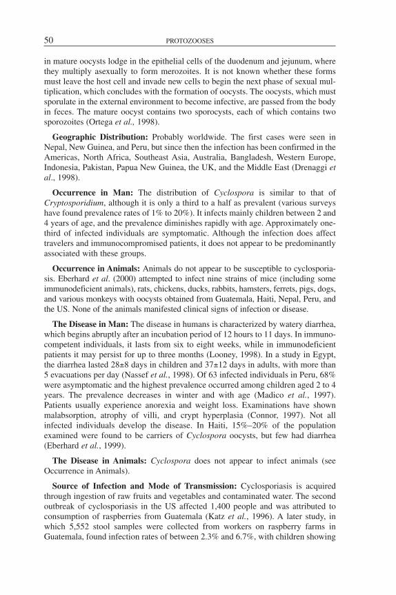
in mature oocysts lodge in the epithelial cells of the duodenum and jejunum, wherethey multiply asexually to form merozoites. It is not known whether these formsmust leave the host cell and invade new cells to begin the next phase of sexual mul-tiplication, which concludes with the formation of oocysts. The oocysts, which mustsporulate in the external environment to become infective, are passed from the bodyin feces. The mature oocyst contains two sporocysts, each of which contains twosporozoites (Ortega et al., 1998).
Geographic Distribution: Probably worldwide. The first cases were seen inNepal, New Guinea, and Peru, but since then the infection has been confirmed in theAmericas, North Africa, Southeast Asia, Australia, Bangladesh, Western Europe,Indonesia, Pakistan, Papua New Guinea, the UK, and the Middle East (Drenaggi etal., 1998).
Occurrence in Man: The distribution of Cyclospora is similar to that ofCryptosporidium, although it is only a third to a half as prevalent (various surveyshave found prevalence rates of 1% to 20%). It infects mainly children between 2 and4 years of age, and the prevalence diminishes rapidly with age. Approximately one-third of infected individuals are symptomatic. Although the infection does affecttravelers and immunocompromised patients, it does not appear to be predominantlyassociated with these groups.
Occurrence in Animals: Animals do not appear to be susceptible to cyclosporia-sis. Eberhard et al. (2000) attempted to infect nine strains of mice (including someimmunodeficient animals), rats, chickens, ducks, rabbits, hamsters, ferrets, pigs, dogs,and various monkeys with oocysts obtained from Guatemala, Haiti, Nepal, Peru, andthe US. None of the animals manifested clinical signs of infection or disease.
The Disease in Man: The disease in humans is characterized by watery diarrhea,which begins abruptly after an incubation period of 12 hours to 11 days. In immuno-competent individuals, it lasts from six to eight weeks, while in immunodeficientpatients it may persist for up to three months (Looney, 1998). In a study in Egypt,the diarrhea lasted 28±8 days in children and 37±12 days in adults, with more than5 evacuations per day (Nassef et al., 1998). Of 63 infected individuals in Peru, 68%were asymptomatic and the highest prevalence occurred among children aged 2 to 4years. The prevalence decreases in winter and with age (Madico et al., 1997).Patients usually experience anorexia and weight loss. Examinations have shownmalabsorption, atrophy of villi, and crypt hyperplasia (Connor, 1997). Not allinfected individuals develop the disease. In Haiti, 15%–20% of the populationexamined were found to be carriers of Cyclospora oocysts, but few had diarrhea(Eberhard et al., 1999).
The Disease in Animals: Cyclospora does not appear to infect animals (seeOccurrence in Animals).
Source of Infection and Mode of Transmission: Cyclosporiasis is acquiredthrough ingestion of raw fruits and vegetables and contaminated water. The secondoutbreak of cyclosporiasis in the US affected 1,400 people and was attributed toconsumption of raspberries from Guatemala (Katz et al., 1996). A later study, inwhich 5,552 stool samples were collected from workers on raspberry farms inGuatemala, found infection rates of between 2.3% and 6.7%, with children showing
50 PROTOZOOSES

the highest rates (Bern et al., 1999). The highest prevalence occurred during thewarm months. In Peru, Ortega et al. (1997) found that 14.5% of vegetables obtainedfrom markets were contaminated with Cryptosporidium and 1.8% with Cyclospora.In two outbreaks in the US, basil seems to have been the vehicle (López et al.,1999), while in Spain, cases have been attributed to consumption of raspberries, buf-falo milk, and raw fish (Gascon et al., 2001). The study by Bern et al. (1999) inGuatemala revealed that the principal risk for infection among those affected wasconsumption of untreated water. A study of the water in domestic containers inEgypt showed that 56% was contaminated with Giardia, 50% withCryptosporidium, 12% with Blastocystis, 9% with Cyclospora, and 3% withmicrosporidia (Khalifa et al., 2001). Using microscopy and molecular biology tech-niques, Sturbaum (1998) identified Cyclospora oocysts in wastewater.
Diagnosis: Cyclospora infection is suggested by the patient’s symptoms and byepidemiological circumstances, especially in travelers who have visited endemicareas. The diagnosis is confirmed by detection of the double-walled oocysts meas-uring 8–10 microns in diameter in stool samples. The oocysts are concentrated byformol-ether sedimentation and flotation in Sheather’s sucrose solution. They can bedetected by staining, autofluorescence under ultraviolet light, phase contrastmicroscopy, or polymerase chain reaction (Ortega et al., 1998). The stains used mostfrequently (to make it easier to visualize the organisms and to differentiate themfrom yeasts) are trichrome stains, Ziehl-Neelsen, Giemsa, safranin with methyleneblue, calcofluor white, and auramine phenol. Safranin has been found to be the mosteffective and appropriate stain for use in diagnostic laboratories (Negm, 1998).
Control: Cyclosporiasis can be prevented by applying the classic measures forcontrol of parasitoses transmitted via the fecal-oral route: washing foods that areeaten raw, boiling suspicious water, and washing hands before eating. Treatment ofwater contaminated with Giardia, Cryptosporidium, Blastocystis, Cyclospora, ormicrosporidia with chlorine at 4 or 8 parts per million (ppm) or with ozone at 1 ppmshowed that ozone was more effective in destroying the parasites, but that it did nottotally inactivate Cyclospora or Blastocystis (Khalifa et al., 2001).
Bibliography
Bern, C., B. Hernandez, M.B. Lopez, et al. Epidemiologic studies of Cyclospora cayeta-nensis in Guatemala. Emerg Infect Dis 5:766–774, 1999.
Drenaggi, D., Cirioni, O., Giacometti, A., et al. Cyclosporiasis in a traveler returning fromSouth America. J Travel Med 5:153–155, 1998.
Connor, B.A. Cyclospora infection: A review. Ann Acad Med Singapore 26(5):632–636,1997.
Eberhard, M.L., E.K. Nace, A.R. Freeman, et al. Cyclospora cayetanensis infections inHaiti: A common occurrence in the absence of watery diarrhea. Am J Trop Med Hyg60(4):584–586, 1999.
Eberhard, M.L., Y.R. Ortega, D.E. Hanes, et al. Attempts to establish experimentalCyclospora cayetanensis infection in laboratory animals. J Parasitol 86(3):577–582, 2000.
Gascon, J., M. Alvarez, E.M. Valls, et al. Ciclosporiasis: estudio clinicoepidemiológico enviajeros con Cyclospora cayetanensis importada. Med Clin (Spain) 116(12):461–464, 2001.
Khalifa, A.M., M.M. El Temsahy, I.F. Abou El Naga. Effect of ozone on the viability ofsome protozoa in drinking water. J Egypt Soc Parasitol 31(2):603–616, 2001.
CYCLOSPORIASIS 51
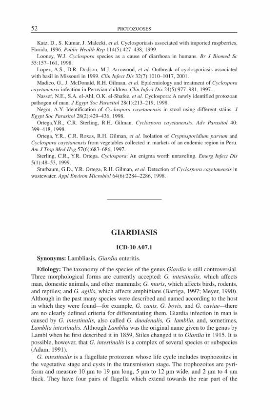
Katz, D., S. Kumar, J. Malecki, et al. Cyclosporiasis associated with imported raspberries,Florida, 1996. Public Health Rep 114(5):427–438, 1999.
Looney, W.J. Cyclospora species as a cause of diarrhoea in humans. Br J Biomed Sc55:157–161, 1998.
Lopez, A.S., D.R. Dodson, M.J. Arrowood, et al. Outbreak of cyclosporiasis associatedwith basil in Missouri in 1999. Clin Infect Dis 32(7):1010–1017, 2001.
Madico, G., J. McDonald, R.H. Gilman, et al. Epidemiology and treatment of Cyclosporacayetanensis infection in Peruvian children. Clin Infect Dis 24(5):977–981, 1997.
Nassef, N.E., S.A. el-Ahl, O.K. el-Shafee, et al. Cyclospora: A newly identified protozoanpathogen of man. J Egypt Soc Parasitol 28(1):213–219, 1998.
Negm, A.Y. Identification of Cyclospora cayetanensis in stool using different stains. JEgypt Soc Parasitol 28(2):429–436, 1998.
Ortega,Y.R., C.R. Sterling, R.H. Gilman. Cyclospora cayetanensis. Adv Parasitol 40:399–418, 1998.
Ortega, Y.R., C.R. Roxas, R.H. Gilman, et al. Isolation of Cryptosporidium parvum andCyclospora cayetanensis from vegetables collected in markets of an endemic region in Peru.Am J Trop Med Hyg 57(6):683–686, 1997.
Sterling, C.R., Y.R. Ortega. Cyclospora: An enigma worth unraveling. Emerg Infect Dis5(1):48–53, 1999.
Sturbaum, G.D., Y.R. Ortega, R.H. Gilman, et al. Detection of Cyclospora cayetanensis inwastewater. Appl Environ Microbiol 64(6):2284–2286, 1998.
GIARDIASIS
ICD-10 A07.1
Synonyms: Lambliasis, Giardia enteritis.
Etiology: The taxonomy of the species of the genus Giardia is still controversial.Three morphological forms are currently accepted: G. intestinalis, which affectsman, domestic animals, and other mammals; G. muris, which affects birds, rodents,and reptiles; and G. agilis, which affects amphibians (Barriga, 1997; Meyer, 1990).Although in the past many species were described and named according to the hostin which they were found—for example, G. canis, G. bovis, and G. caviae—thereare no clearly defined criteria for differentiating them. Giardia infection in man iscaused by G. intestinalis, also called G. duodenalis, G. lamblia, and, sometimes,Lamblia intestinalis. Although Lamblia was the original name given to the genus byLambl when he first described it in 1859, Stiles changed it to Giardia in 1915. It ispossible, however, that G. intestinalis is a complex of several species or subspecies(Adam, 1991).
G. intestinalis is a flagellate protozoan whose life cycle includes trophozoites inthe vegetative stage and cysts in the transmission stage. The trophozoites are pyri-form and measure 10 µm to 19 µm long, 5 µm to 12 µm wide, and 2 µm to 4 µmthick. They have four pairs of flagella which extend towards the rear part of the
52 PROTOZOOSES

organism, two nuclei, two claw-shaped median bodies in the middle of the body, anda convex ventral disk in the front half of the body, with which they cling to the intes-tinal mucosa. Those forms live in the anterior portion of the host’s small intestine,particularly in the duodenum, where they multiply by binary fission. Many of thetrophozoites are carried to the ileum, where they secrete a resistant wall and becomeovoid cysts measuring 7 µm to 10 µm by 8 µm to 13 µm. After encysting, the para-site’s organs divide again. The mature cyst thus has four nuclei, four median bodies,and eight flagella. Division of the cytoplasm does not occur until the parasiteexcysts. The cysts leave the host in feces. They can survive for more than twomonths in water at 8°C and around one month at 21°C; however, they are sensitiveto desiccation, freezing, and sunlight. They are also relatively sensitive to ordinarydisinfectants. Solutions of quaternary ammonium recommended for disinfecting theenvironment will kill them in one minute at 20°C, but normal concentrations ofchlorine in drinking water do not affect them. The mature cyst is the infective ele-ment for a new host. Once ingested, the parasite excysts in the duodenum, divides,and begins to multiply normally.
Geographic Distribution: Worldwide.
Occurrence in Man: Giardiasis is endemic throughout the world. Its prevalencegenerally ranges from 2% to 4% in industrialized countries, but it may be over 15%among children in developing countries. Both the infection and the disease are morecommon in children than in adults. Giardiasis may also occur in epidemic form. Of25 and 22 disease epidemics spread through ingestion of drinking or recreationalwaters, which affected 2,366 and 2,567 people in the US in 1993–1994 and1995–1996, respectively, G. intestinalis was the most common pathogen. In the firstepidemic, together with Cryptosporidium, it caused 40% of the cases, while in thesecond epidemic, together with Shigella sonnei, it was responsible for 9% of thecases (Kramer et al., 1996; Levy et al., 1998). In 1974, in a population of 46,000inhabitants in the state of New York, US, 4,800 people (10.4%) contracted clinicalgiardiasis as a result of contamination of drinking water supplies. In epidemic situ-ations, all age groups are affected equally. In previously uninfected populations,morbidity rates may be as high as 20% or more of the total population (Knight,1980). Outbreaks are relatively common in institutions for children, such as orphan-ages and daycare centers. G. intestinalis is also frequently a cause of “travelers’ diar-rhea.” In a group of 21 people whose feces tested negative for Giardia, the proto-zoan’s cysts were later found in those of 15 out of 17 who became ill after visitingLeningrad (Kulda and Nohynková, 1978). The infection is less frequent in AIDSpatients, perhaps because the virus interferes with the parasite’s activity in the intes-tinal mucosa (Lindo et al., 1998).
Occurrence in Animals: The infection has been confirmed in a wide variety ofdomestic and wild mammal species. Surveys from all over the world have foundprevalences of 20% to 35% in young dogs; 10% to 15% in young cats; 5% to 90%in calves; 6% to 80% in lambs; 17% to 32% in foals; and 7% to 44% in young pigs(Xiao, 1994). As with man, the infection is less frequent in adult animals. In a studyin which feces of 494 dogs were examined for parasites, the infection was detectedin 3.4% of adult males, 7% of adult females, and 53.2% of puppies. A study inColorado, US, found cysts of the parasite in 10% of the cattle, 18% of the beavers,
GIARDIASIS 53

and 6% of the coyotes examined. A giardiasis outbreak among nonhuman primatesand zoo personnel was recorded in Kansas City, Missouri, US. High rates of infec-tion have also been found in rats and other rodents, both synanthropic and wild, butwhether the agent was G. intestinalis or G. muris has not been determined (Meyerand Jarroll, 1982).
The Disease in Man: The majority of infections are subclinical (Flanagan, 1992;Farthing, 1996). Rajeshwari et al. (1996) found that impairment of the humoralimmune response was the deciding factor in whether or not the infection was symp-tomatic in children. In symptomatic individuals, the incubation period is generally3–25 days (Benenson, 1997). The symptomatology consists mainly of diarrhea andbloating, frequently accompanied by abdominal pain. Nausea and vomiting occurless frequently. The acute phase of the disease lasts 3–4 days. In some persons, giar-diasis may be a prolonged illness, with episodes of recurring diarrhea and flatulence,urticaria, and intolerance of certain foods. These and other allergic manifestationsassociated with giardiasis disappear after treatment and cure. Meloni et al. (1995)examined 97 isolates of G. intestinalis from humans and various animals and dif-ferentiated 47 zymodemes. In another study, Cevallos et al. (1995) found a correla-tion between certain zymodemes and the pathology caused by the parasite in rats.However, Rajeshwari et al. (1996) demonstrated that neither the zymodeme of theparasite nor the presence of associated bacterial infections influenced the occurrenceor the pathogenicity of the infection in children.
The Disease in Animals: As in man, the infection is usually asymptomatic. Themanifestations of the disease in dogs and cats are also similar to those in man.However, experimental infections in ruminants produced only mild diarrhea incalves and weight loss in lambs (Zajac, 1992; Olson et al., 1995). The disease ismore frequent in young animals.
Source of Infection and Mode of Transmission: Man is the principal reservoirof human giardiasis. The source of infection is feces containing the parasite’s cysts,which often contaminate water. Although the infection in individuals is often self-limited and disappears within a few months, continuous transmission to other hostsin endemic areas ensures the agent’s persistence. The existence of asymptomaticinfected individuals and chronic patients, coupled with the cysts’ resistance to envi-ronmental factors, are important factors in the epidemiology. The median infectivedose (ID50) of giardiasis for man is only 10 cysts, but infected individuals may laterexcrete up to 900 million cysts per day in their feces. Elimination of cysts can beintermittent and the quantity can vary greatly (Knight, 1980).
The most frequent mode of transmission appears to be ingestion of water con-taminated with cysts (Hill, 1993). Direct hand-to-hand or hand-to-mouth transmis-sion of cysts from an infected person to a susceptible person is also common, espe-cially among children, personnel in institutions that care for children or adults, andfood-handlers. Rezende et al. (1997) studied 264 food-handlers in 57 schools inMinas Gerais (Brazil) at various times of the year and found 8%, 2%, and 3% to beinfected. Indirect transmission from fecal contamination of food is less frequent thandirect transmission from infected food-handlers, but it may occur as a result of irri-gating or washing foods with contaminated water or by means of mechanical vec-tors. However, as the cysts are susceptible to environmental factors such as desicca-
54 PROTOZOOSES
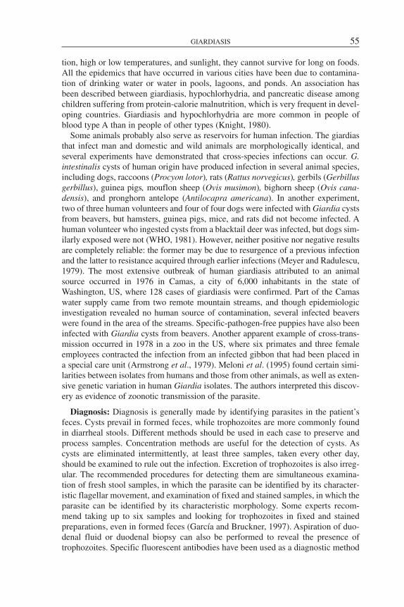
tion, high or low temperatures, and sunlight, they cannot survive for long on foods.All the epidemics that have occurred in various cities have been due to contamina-tion of drinking water or water in pools, lagoons, and ponds. An association hasbeen described between giardiasis, hypochlorhydria, and pancreatic disease amongchildren suffering from protein-calorie malnutrition, which is very frequent in devel-oping countries. Giardiasis and hypochlorhydria are more common in people ofblood type A than in people of other types (Knight, 1980).
Some animals probably also serve as reservoirs for human infection. The giardiasthat infect man and domestic and wild animals are morphologically identical, andseveral experiments have demonstrated that cross-species infections can occur. G.intestinalis cysts of human origin have produced infection in several animal species,including dogs, raccoons (Procyon lotor), rats (Rattus norvegicus), gerbils (Gerbillusgerbillus), guinea pigs, mouflon sheep (Ovis musimon), bighorn sheep (Ovis cana-densis), and pronghorn antelope (Antilocapra americana). In another experiment,two of three human volunteers and four of four dogs were infected with Giardia cystsfrom beavers, but hamsters, guinea pigs, mice, and rats did not become infected. Ahuman volunteer who ingested cysts from a blacktail deer was infected, but dogs sim-ilarly exposed were not (WHO, 1981). However, neither positive nor negative resultsare completely reliable: the former may be due to resurgence of a previous infectionand the latter to resistance acquired through earlier infections (Meyer and Radulescu,1979). The most extensive outbreak of human giardiasis attributed to an animalsource occurred in 1976 in Camas, a city of 6,000 inhabitants in the state ofWashington, US, where 128 cases of giardiasis were confirmed. Part of the Camaswater supply came from two remote mountain streams, and though epidemiologicinvestigation revealed no human source of contamination, several infected beaverswere found in the area of the streams. Specific-pathogen-free puppies have also beeninfected with Giardia cysts from beavers. Another apparent example of cross-trans-mission occurred in 1978 in a zoo in the US, where six primates and three femaleemployees contracted the infection from an infected gibbon that had been placed ina special care unit (Armstrong et al., 1979). Meloni et al. (1995) found certain simi-larities between isolates from humans and those from other animals, as well as exten-sive genetic variation in human Giardia isolates. The authors interpreted this discov-ery as evidence of zoonotic transmission of the parasite.
Diagnosis: Diagnosis is generally made by identifying parasites in the patient’sfeces. Cysts prevail in formed feces, while trophozoites are more commonly foundin diarrheal stools. Different methods should be used in each case to preserve andprocess samples. Concentration methods are useful for the detection of cysts. Ascysts are eliminated intermittently, at least three samples, taken every other day,should be examined to rule out the infection. Excretion of trophozoites is also irreg-ular. The recommended procedures for detecting them are simultaneous examina-tion of fresh stool samples, in which the parasite can be identified by its character-istic flagellar movement, and examination of fixed and stained samples, in which theparasite can be identified by its characteristic morphology. Some experts recom-mend taking up to six samples and looking for trophozoites in fixed and stainedpreparations, even in formed feces (García and Bruckner, 1997). Aspiration of duo-denal fluid or duodenal biopsy can also be performed to reveal the presence oftrophozoites. Specific fluorescent antibodies have been used as a diagnostic method
GIARDIASIS 55

for detecting cysts, as these techniques facilitate visualization under the microscopewith a sensitivity 2.3 times greater than observation without fluorescence(Winiecka-Krusnell, 1995). The enzyme-linked immunosorbent assay (ELISA) hasalso been used to demonstrate the presence of G. intestinalis antigens in feces (Hill,1993). Although the presence of antibodies and cell-mediated immune responseshave been reported in patients, immunobiological procedures are not very specific(Isaac-Renton et al., 1994) and do not indicate whether an infection is current orresidual. The possibility of using polymerase chain reaction to detect specific DNAsequences in patients’ feces or blood is currently being studied. In any event, itshould be borne in mind that there is not always a causal relationship between symp-toms and the discovery of giardias in an ill person, and it is therefore necessary torule out infections due to other intestinal microorganisms or other pathologies.
Control: As the cysts of G. intestinalis can live for long periods in water, publicwater supplies should be protected against contamination by human and animal fecalmatter. Adequate systems of sedimentation, flocculation, and filtration can removeGiardia from water, allowing the use of surface water supply systems (CDC, 1977).Sanitary elimination of feces is another important measure. In developing countries,prevailing socioeconomic conditions make it difficult to prevent infection in children.Instruction in personal hygiene is essential in institutions for children. Individual pre-vention measures include boiling or filtering suspicious water. Tourists should drinkonly bottled water in places where the purity of tap water cannot be guaranteed.Although there is no evidence that domestic animals are a significant source of infec-tion for man, dogs and cats with giardiasis should be treated because they may fre-quently come into contact with children (Meyer and Jarroll, 1982).
Whereas treatment of infected individuals, coupled with prophylactic measures, hasreduced the prevalence of parasitic infections caused by other organisms, it has notbeen successful in the case of giardiasis (Dorea et al., 1996). Studies have shown thatvaccinated dogs develop some resistance to the disease (Olson et al., 1996). Theseresults may be promising for humans as it has been shown that people with naturalinfections also develop a certain degree of resistance, which lasts at least five years(Isaac-Renton et al., 1994). Most methods for testing suspicious water are tedious,complicated, and not very efficient; however, some highly effective and sensitive tech-niques have been developed (Bielec et al., 1996; Kaucner and Stinear, 1998).
Bibliography
Adam, R.D. The biology of Giardia spp. Microbiol Rev 55(4):706–732, 1991.Armstrong, J., R.E. Hertzog, R.T. Hall, G.L. Hoff. Giardiasis in apes and zoo attendants,
Kansas City, Missouri. CDC Vet Public Health Notes, January(7-8), 1979.Barriga, O.O. Veterinary Parasitology for Practitioners, 2nd ed. Edina: Burgess International
Group; 1997.Benenson, A.S., ed. Control of Communicable Diseases Manual, 17th edition. Washington,
D.C.: American Public Health Association; 1997.Bielec, L., T.C. Boisvert, S.G. Jackson. Modified procedure for recovery of Giardia cysts
from diverse water sources. Lett Appl Microbiol 22(1):21–25, 1996.Cevallos, A., S. Carnaby, M. James, J.G. Farthing. Small intestinal injury in a neonatal rat
model of giardiasis is strain dependent. Gastroenterology 109(3):766–773, 1995.
56 PROTOZOOSES

Dorea, R.C., E. Salata, C.R. Padovani, G.L. dos Anjos. Control of parasitic infections amongschool children in the peri-urban area of Botucatu, Sao Paulo, Brazil. Rev Soc Bras Med Trop29(5):425–430, 1996.
Farthing, M.J. Giardiasis. Gastroenterol Clin North Am 25(3):493–515, 1996.Flanagan, P.A. Giardia—diagnosis, clinical course and epidemiology. A review. Epidemiol
Infect 109(1):1–22, 1992.García, L.S., D.A. Bruckner. Diagnostic Medical Parasitology, 3rd ed. Washington, D.C.:
ASM Press; 1997.Hill, D.R. Giardiasis: Issues in diagnosis and management. Infect Dis Clin North Am
7(3):503–525, 1993.Isaac-Renton, J.L., L.F. Lewis, C.S. Ong, M.F. Nulsen. A second community outbreak of
waterborne giardiasis in Canada and serological investigation of patients. Trans R Soc Trop MedHyg 88(4):395–399, 1994.
Kaucner, C., T. Stinear. Sensitive and rapid detection of viable Giardia cysts andCryptosporidium parvum oocysts in large-volume water samples with wound fiberglass car-tridge filters and reverse transcription-PCR. Appl Environ Microbiol 64(5):1743–1749, 1998.
Knight, R. Epidemiology and transmission of giardiasis. Trans R Soc Trop Med Hyg 74:433–436, 1980.
Kramer, M.H., B.L. Herwaldt, G.F. Craun, R.L. Calderón, D.D. Juranek. Surveillance forwaterborne-disease outbreaks—United States, 1993–1994. MMWR Morb Mortal Wkly RepCDC Surveill Summ 45(1):1–33, 1996.
Kulda, J., E. Nohynková. Flagellates of the human intestine and of intestines of other species.In: Kreier, J.P., ed. Vol. 2: Parasitic Protozoa. New York: Academic Press; 1978.
Levy, D.A., M.S. Bens, G.F. Craun, R.L. Calderón, B.L. Herwaldt. Surveillance for water-borne-disease outbreaks—United States, 1995–1996. MMWR Morb Mortal Wkly Rep CDCSurveill Summ 47(1):1–34, 1998.
Lindo, J.F., J.M. Dubon, A.L. Ager, et al. Intestinal parasitic infections in human immunode-ficiency virus (HIV)-positive and HIV-negative individuals in San Pedro Sula, Honduras. Am JTrop Med Hyg 58(4):431–435, 1998.
Meloni, B.P., A.J. Lymbery, R.C. Thompson. Genetic characterization of isolates of Giardiaduodenalis by enzyme electrophoresis: Implications for reproductive biology, population struc-ture, taxonomy, and epidemiology. J Parasitol 81(3):368–383, 1995.
Meyer, E.A., ed. Giardiasis. Amsterdam: Elsevier; 1990.Meyer, E.A., S. Radulescu. Giardia and giardiasis. Adv Parasit 17:1–47, 1979.Meyer, E.A., E.L. Jarroll. Giardiasis. In: Jacobs, L., P. Arámbulo. Vol. 1: CRC Handbook
Series in Zoonoses. Boca Raton: CRC Press; 1982.Olson, M.E., T.A. McAllister, L. Deselliers, et al. Effect of giardiasis on production in a
domestic ruminant (lamb) model. Am J Vet Res 56(11):1470–1474, 1995.Olson, M.E., D.W. Morck, H. Ceri. The efficacy of a Giardia lamblia vaccine in kittens. Can
J Vet Res 60(4):249–256, 1996.Rajeshwari, K., N. Jaggi, V. Aggarwal, K.K. Kalra, S.K. Mittal, U. Baveja. Determinants of
symptomatic giardiasis in childhood. Trop Gastroenterol 17(2):70–76, 1996.Rezende, C.H. de, J.M. Costa-Cruz, M.L. Gennari-Cardoso. Enteroparasitoses em manipu-
ladores de alimentos de escolas públicas em Uberlândia (Minas Gerais), Brasil. Rev PanamSalud Publican 2(6):392–397, 1997.
United States of America, Department of Health and Human Services, Centers for DiseaseControl and Prevention (CDC). Water-borne giardiasis outbreaks. Washington, New Hampshire.Morb Mortal Wkly Rep 26:169–170, 1977.
Winiecka-Krusnell, J., E. Linder. Detection of Giardia lamblia cysts in stool samples byimmunofluorescence using monoclonal antibody. Eur J Clin Microbiol Infect Dis 14(3):218–222, 1995.
World Health Organization (WHO). Infecciones intestinales por protozoos y helmintos.Informe de un Grupo Científico de la OMS. Geneva: WHO; 1981. (Technical Report Series 666).
GIARDIASIS 57
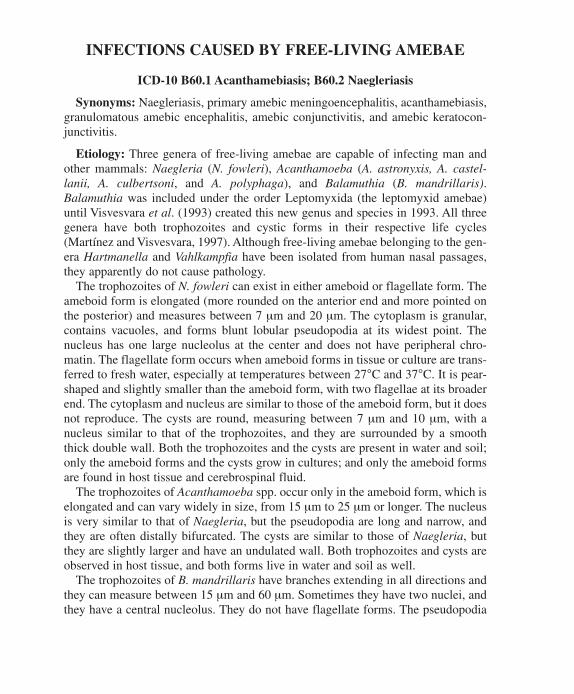
Xiao, L. Giardia infections in farm animals. Parasitol Today 10:436–438, 1994.Zajac, A.M. Giardiasis. Comp Cont Educ Vet Pract 14:604–611, 1992.
INFECTIONS CAUSED BY FREE-LIVING AMEBAE
ICD-10 B60.1 Acanthamebiasis; B60.2 Naegleriasis
Synonyms: Naegleriasis, primary amebic meningoencephalitis, acanthamebiasis,granulomatous amebic encephalitis, amebic conjunctivitis, and amebic keratocon-junctivitis.
Etiology: Three genera of free-living amebae are capable of infecting man andother mammals: Naegleria (N. fowleri), Acanthamoeba (A. astronyxis, A. castel-lanii, A. culbertsoni, and A. polyphaga), and Balamuthia (B. mandrillaris).Balamuthia was included under the order Leptomyxida (the leptomyxid amebae)until Visvesvara et al. (1993) created this new genus and species in 1993. All threegenera have both trophozoites and cystic forms in their respective life cycles(Martínez and Visvesvara, 1997). Although free-living amebae belonging to the gen-era Hartmanella and Vahlkampfia have been isolated from human nasal passages,they apparently do not cause pathology.
The trophozoites of N. fowleri can exist in either ameboid or flagellate form. Theameboid form is elongated (more rounded on the anterior end and more pointed onthe posterior) and measures between 7 µm and 20 µm. The cytoplasm is granular,contains vacuoles, and forms blunt lobular pseudopodia at its widest point. Thenucleus has one large nucleolus at the center and does not have peripheral chro-matin. The flagellate form occurs when ameboid forms in tissue or culture are trans-ferred to fresh water, especially at temperatures between 27°C and 37°C. It is pear-shaped and slightly smaller than the ameboid form, with two flagellae at its broaderend. The cytoplasm and nucleus are similar to those of the ameboid form, but it doesnot reproduce. The cysts are round, measuring between 7 µm and 10 µm, with anucleus similar to that of the trophozoites, and they are surrounded by a smooththick double wall. Both the trophozoites and the cysts are present in water and soil;only the ameboid forms and the cysts grow in cultures; and only the ameboid formsare found in host tissue and cerebrospinal fluid.
The trophozoites of Acanthamoeba spp. occur only in the ameboid form, which iselongated and can vary widely in size, from 15 µm to 25 µm or longer. The nucleusis very similar to that of Naegleria, but the pseudopodia are long and narrow, andthey are often distally bifurcated. The cysts are similar to those of Naegleria, butthey are slightly larger and have an undulated wall. Both trophozoites and cysts areobserved in host tissue, and both forms live in water and soil as well.
The trophozoites of B. mandrillaris have branches extending in all directions andthey can measure between 15 µm and 60 µm. Sometimes they have two nuclei, andthey have a central nucleolus. They do not have flagellate forms. The pseudopodia
58 PROTOZOOSES
have secondary branchings. The cysts, which can measure from 15 µm to 30 µm,also have a single nucleus, and the outer wall is undulated. Both trophozoites andcysts can be found in host tissue. Little is known about the natural reservoir ofBalamuthia.
Geographic Distribution: Free-living amebae appear to exist throughout theworld. Clinical cases have been recorded in widely distant locations, includingAustralia, Brazil, US, Europe, India, and Zambia (Strickland, 1991).
Occurrence in Man: Infections with free-living amebae have only been knownsince the 1960s. As of 1996, there had been 179 reported cases of primary amebicmeningoencephalitis caused by N. fowleri, 103 cases of granulomatous amebicencephalitis caused by Acanthamoeba spp., and 63 cases of granulomatous amebicencephalitis caused by B. mandrillaris (Martínez and Visvesvara, 1997). In addition,as of 1993, there have been 570 known cases of keratitis caused by Acanthamoebaspp. (Benenson, 1995).
Occurrence in Animals: Naegleria is capable of infecting experimentally inocu-lated mice and sheep. Acanthamoeba can infect sheep (Van der Lugt and Van derMerve, 1990) and dogs (Pearce et al., 1985) in nature, and in the laboratory, it caninfect the cornea of swine, rabbits, and mice. The invasive capacity of this protozoanappears to vary. Other researchers have found that it does not attack the cornea ofhorses, guinea pigs, rabbits, chicken, mice, rats, or cows, but that it can producesevere damage in the cornea of man, swine, and Chinese hamsters (Niederkorn etal., 1992). Balamuthia has been isolated from fatal infections in horses, gorillas,mandrills, and sheep (García and Bruckner, 1997). The range of susceptible animalsis probably greater, but there have been few reports of infection because of the dif-ficulty of diagnosing this genus and because the disease in animals receives lessattention than its human counterpart.
The Disease in Man: Naegleria mainly affects young, immunocompetent,healthy individuals. The ameba penetrates the host via the nasal cavity, where itcauses local inflammation and ulceration, and goes on to invade the olfactory nervesand ultimately the meninges, where it multiplies and produces an acute inflamma-tion with abundant neutrophils and monocytes along with hemorrhagic necroses(primary amebic meningoencephalitis). The disease is fatal. After an incubationperiod of three to seven days, the initial symptoms include sore throat, blocked nasalpassages, and intense cephalalgia, subsequently followed by fever, vomiting, andstiff neck. Mental confusion and coma develop three to four days after the firstsymptoms, and death occurs between three and four days later.
Acanthamoeba spp. preferentially attack individuals who are immunodeficient,undernourished, or weakened by other conditions. This ameba usually invades thehost through the skin, the respiratory tract, or the genitourinary tract, spreadingthrough the bloodstream until it reaches the brain and the meninges. The exactlength of incubation is unknown, but central nervous system symptoms apparentlydo not develop until weeks or even months after the primary infection. Often thereis a slow-growing cutaneous or pulmonary granulomatous lesion which tends to fol-low a subacute or chronic course (granulomatous amebic encephalitis). The pre-dominant lesions are foci of granulomatous inflammation, necroses, thromboses,and hemorrhages. The most prevalent symptoms include cutaneous papules, nod-
INFECTIONS CAUSED BY FREE-LIVING AMEBAE 59
ules, ulcers or abscesses, congestion, secretion from or ulcers in the nasal passages,sinusitis, cephalalgia, mental or motor disturbances, meningeal signs, or sensorydeficiencies. Occasionally the parasite is recovered from other organs such as theskin, kidneys, liver, or pancreas. Acanthamoeba often infects the ocular cornea,causing keratitis, uveitis, and chronic corneal ulcers, which can lead to blindness,especially in persons who wear contact lenses. Both Acanthamoeba and Naegleriaare capable of ingesting microorganisms in their environment such as Legionellaand acting as vectors of the respective infections (Tyndall and Domingue, 1982).
Less information is available about Balamuthia, which was not identified until1993. It can attack both previously healthy and weakened individuals. Although itsmechanism of penetrating the host is still unknown, it can produce a subacute orchronic illness similar to that associated with Acanthamoeba (Denney et al., 1997)and it can also cause granulomatous amebic encephalitis.
The Disease in Animals: Very little information is available about the disease inanimals, but the cases reported so far have resembled the disease in humans (Simpsonet al., 1982; Pearce et al., 1985; Niederkorn et al., 1992; Visvesvara et al., 1993).
Source of Infection and Mode of Transmission: The source of Naegleria andAcanthamoeba infections appears to be contaminated water and soil. Muñoz et al.(1993) examined 100 freshwater samples collected from different sources and foundNaegleria present in 7.6% and Acanthamoeba in 31.5%. The main source ofNaegleria infection is poorly maintained swimming pools, lakes, etc. The amebaenters the nasal passages of swimmers, especially in summer or when the water hasbeen artificially heated. This ameba is destroyed when pools are adequately chlori-nated. The flagellate trophozoite forms probably play the most important role ininfection, since they are more mobile and appear to predominate in warm water. Thecysts are capable of overwintering, and it is believed that the arrival of warm sum-mer weather causes them to break open and assume the form of flagellate tropho-zoites. Contaminated water is also the source of infection caused by Acanthamoeba,and probably by Balamuthia as well. However, the fact that some patients have hadno history of contact with suspicious water would indicate that the infection can alsobe acquired from contaminated soil through breaks in the skin, by the inhalation ofdust containing parasite cysts, or by the inhalation of aerosols containing cysts ortrophozoites. An important source of the ocular infection is the use of contact lensesthat have been poorly disinfected or kept in contaminated cases. Acanthamoeba ismore resistant to environmental agents than Naegleria, as evidenced by the fact thatit can tolerate conventional chlorination. It has been determined that 82% of allsamples of cysts survive 24 years in water at 4°C, and in vitro cultures have beenknown to retain their virulence for mice as long as eight years. The reservoir andmode of transmission of Balamuthia are unknown.
Diagnosis: Diseases caused by free-living amebae cannot be differentiated fromother etiologies on the basis of clinical manifestations alone. Under the microscopeit is difficult, though possible, to identify the parasites in tissue on the basis of theirmorphology; however, at low levels of magnification they can be easily mistaken formacrophages, leukocytes, or Entamoeba histolytica. E. histolytica has a thin nuclearmembrane and a small nucleolus that stains only faintly, unlike the free-living ame-bae, which have a very distinct nuclear membrane and a larger nucleolus that stains
60 PROTOZOOSES

brightly. In lesions caused by Naegleria, the only forms present are ameboid tropho-zoites, which are often perivascular, and polymorphonuclear cells are abundant in thereaction. On the other hand, in lesions produced by Acanthamoeba and Balamuthiathere are both trophozoites and cysts, vasculitis is present, and the reaction is char-acterized by an abundance of mononuclear cells, either with or without multinucle-ate cells (Anzil et al., 1991). The wall of Acanthamoeba cysts found in tissue turnsred with periodic acid-Schiff stain and black when methenamine silver is used. Themorphology of the amebae in cerebrospinal fluid can be observed by conventional orphase-contrast microscopy in fresh preparations or those to which Giemsa orWright’s stain has been applied. Naegleria grows on non-nutrient agar cultures in thepresence of Escherichia coli and in sodium chloride at less than 0.4% solution, whileAcanthamoeba grows in the absence of bacteria and in sodium chloride at less than0.85% solution. Because Naegleria trophozoites are destroyed at cold temperatures,the samples should never be refrigerated. The diagnosis of Balamuthia is not yet welldefined. Although the trophozoite is characterized by its branching, the cysts are verysimilar to those of Acanthamoeba; only the occasional presence of binucleateBalamuthia cysts makes it possible to use conventional microscopy to differentiateBalamuthia from Acanthamoeba. Balamuthia does not grow well on agar in the pres-ence of bacteria, but it does proliferate in mammal tissue cultures. Immunologic reac-tions with patient sera have not been successful. However, it has been possible toidentify exact genera and species using immunofluorescence with monoclonal orpolyclonal antibodies at reference centers such as the US Centers for Disease Controland Prevention. Recently, there have been encouraging results with the use of molec-ular biology techniques to identify and separate species.
Control: Infections caused by free-living amebae are not sufficiently common tojustify general control measures. Education of the public regarding appropriateswimming-pool maintenance and the importance of not swimming in suspiciouswater should reduce the risk of infection. To prevent the parasites from invading thenasal passages, those practicing aquatic sports should avoid submersing the head inwater or else use nose clips. In addition, persons who are immunodeficient or havedebilitating diseases should be careful not to let broken skin come in contact withnatural water or damp soil and avoid breathing dust or aerosols. Contact-lens wear-ers should not swim with their lenses on to avoid contamination, and lenses shouldbe disinfected either by heating them to a temperature of at least 70°C or by usinghydrogen peroxide solutions, which are more effective against Acanthamoeba thanconventional sodium chloride solutions. Control to protect against animal cases doesnot appear to be necessary. There is no evidence of human-to-human transmissionor transmission from animals to humans. These infections mainly occur in humansand in animals that transmit them from one to another.
Bibliography
Anzil, A.P., C. Rao, M.A. Wrzolek, G.S. Visvesvara, J.H. Sher, P.B. Kozlowski. Amebicmeningoencephalitis in a patient with AIDS caused by a newly recognized opportunisticpathogen, Leptomyxid ameba. Arch Pathol Lab Med 115:21–25, 1991.
Benenson, A.S., ed. Control of Communicable Diseases in Man, 16th ed. An official reportof the American Public Health Association. Washington, D.C.: APHA; 1995.
INFECTIONS CAUSED BY FREE-LIVING AMEBAE 61
Denney, C.F., V.J. Iragui, L.D. Uber-Zak, et al. Amebic meningoencephalitis caused byBalamuthia mandrillaris: Case report and review. Clin Infect Dis 25:1354–1358, 1997.
García, L.S., Bruckner, D.A. Diagnostic Medical Parasitology, 3rd ed. Washington, D.C.:ASM Press; 1997.
Martínez, A.J., G.S. Visvesvara. Free-living, amphizoic and opportunistic amebas. BrainPathol 7:583–598, 1997.
Muñoz, V., H. Reyes, B. Astorga L., E. Rugiero P., S. Del Río, P. Toche. Amebas de vidalibre en hábitats de aguas dulces de Chile. Parasitol Día 17:147–152, 1993.
Niederkorn, J.Y., J.E. Ubelaker, J.P. McCulley, et al. Susceptibility of corneas from variousanimal species to in vitro binding and invasion by Acanthamoeba castellanii. InvestOphthalmol Vis Sci 33:104–112, 1992.
Pearce, J.R., H.S. Powell, F.W. Chandler, G.S. Visvesvara. Amebic meningoencephalitiscaused by Acanthamoeba castellani in a dog. J Am Vet Assoc 187:951–952, 1985.
Simpson, C.P., E. Willaert, F.C. Neal, A.R. Stevens, M.D. Young. Experimental Naegleriafowleri meningoencephalitis in sheep: Light and electron microscopic studies. Am J Vet Res43:154–157, 1982.
Strickland, G.T., ed. Hunter’s Tropical Medicine, 7th ed. Philadelphia: Saunders; 1991.Tyndall, R.L., E.L. Domingue. Cocultivacion of Legionella pneumophila and free-living
amoebae. Appl Environ Microbiol 44:954–959, 1982.Van der Lugt, J.J., H.E. Van der Merwe. Amoebic meningoencephalitis in a sheep. J S Afr
Vet Assoc 61:33–36, 1990.Visvesvara, G.S., F.L. Schuster, A.J. Martínez. Balamuthia mandrillaris, N. G., N. Sp.,
agent of amebic meningoencephalitis in humans and other animals. J Eukaryot Microbiol40:504–514, 1993.
MALARIA IN NONHUMAN PRIMATES
ICD-10 B53.1 Malaria due to simian plasmodia
Synonyms: Monkey malaria, monkey paludism.
Etiology: Malaria is a disease caused by protozoa of the phylum Apicomplexa,genus Plasmodium. The four species that infect man are P. falciparum, P. malariae,P. ovale, and P. vivax. Some 20 species are presumed to infect nonhuman primates:the large simians are affected by 4 species of the family Pongidae, gibbons by 4species of the family Hylobatidae, Old World monkeys by 8 species of the familyCercopithecidae, New World monkeys by 2 species of the family Cebidae, andlemurs by 2 species of the family Lemuridae (Collins and Aikawa, 1977). However,the taxonomy of some species is uncertain. Table 2 shows some characteristics ofthe most common species.
Seven of the species that affect nonhuman primates have been transmitted exper-imentally to humans: P. brasilianum, P. cynomolgi, P. eylesi, P. inui, P. knowlesi, P.schwetzi, and P. simium. In addition, some species, such as P. cynomolgi, P.knowlesi, P. simium, and, possibly, P. eylesi, have been found in natural or acciden-tal infections, though this rarely occurs. Nonhuman primates can also be infected
62 PROTOZOOSES

with natural or adapted strains of the plasmodia that normally affect humans: Aotusand Saimiri monkeys with P. falciparum or P. vivax, and chimpanzees with P. ovale.
For many years, it was believed that the plasmodia of man did not have a com-mon origin (monophyletic) but rather were descended from different ancestralspecies (polyphyletic), and the relationship of some species that infect man to somespecies that infect simians was frequently the subject of speculation. Escalante et al.(1998) compared cytochrome b gene sequences from 4 species of human parasites,10 species of simian parasites, 1 species of rodent parasite, and 2 species of avianparasites. They concluded that the plasmodia of man are indeed polyphyletic. P. fal-ciparum and P. reichenowi are closely related to one another and share a commonancestor with avian malaria parasites. P. vivax forms a different but closely relatedgroup with the plasmodia of Old World monkeys, especially P. simium. P. ovale andP. malariae form a separate group, although there is very little relationship betweenthem. An interesting finding is that, contrary to widely held opinion, no inverse rela-tionship exists between virulence of the parasite and length of the plasmodium-hostassociation. Neither is there any relationship between phylogenetic proximity of theparasites and certain characteristics of malarial disease, such as virulence, periodic-ity, and occurrence of relapses. Notwithstanding the foregoing, biologic and patho-genic similarities have led to the use of certain nonhuman primate Plasmodiumspecies as the preferred models for Plasmodium species that infect man: P.cynomolgi for P. vivax, P. brasilianum for P. malariae, P. fieldi and P. simiovale forP. ovale, and P. coatneyi for P. falciparum. In addition, Lal et al. (1988) demon-strated that the immunodominant repeat domain was the same for P. brasilianum andP. malariae and for P. reichenowi and P. falciparum.
The life cycle of plasmodia of nonhuman primates, like that of the plasmodia thatinfect man, includes host mammals and insect vectors. An infected mosquito of thegenus Anopheles injects sporozoites of the parasite into a susceptible host when it
MALARIA IN NONHUMAN PRIMATES 63
TABLE 2. Plasmodium species that infect primates.
Species Host Geographic range Periodicity Relapses
P. brasilianum New World monkeys Central and South America Quartan UncertainP. coatneyi Old World monkeys Malaysia Tertian NoP. cynomolgi Old World monkeys Southeast Asia Tertian YesP. eylesi Gibbons Malaysia Tertian UncertainP. falciparum Humans Tropics Tertian NoP. fieldi Old World monkeys Malaysia Tertian YesP. gonderi Old World monkeys Central Africa Tertian NoP. hylobati Gibbons Malaysia Tertian NoP. inui Old World monkeys India and Southeast Asia Quartan NoP. knowlesi Old World monkeys Malaysia Quotidian NoP. malariae Humans Tropics and subtropics Quartan NoP. ovale Humans Asia and Africa Tertian YesP. reichenowi Chimpanzees Central Africa Tertian NoP. schwetzi Gorillas and Tropical Africa Tertian Yes
chimpanzeesP. simiovale Old World monkeys Sri Lanka Tertian YesP. simium New World monkeys Brazil Tertian UncertainP. vivax Humans Tropics and subtropics Tertian Yes

takes a blood meal. In less than an hour, the sporozoites disappear from the bloodand enter the cells of the hepatic parenchyma. There, they begin to multiply by mul-tiple fission to form thousands of filamentous parasites, the merozoites, which leavethe host cell after five days or more. In some species, there is a single generation ofhepatic merozoites, but in others, dormant forms, hypnozoites, are produced. Thehypnozoites may become active again months or years later and cause reinfection(Cogswell, 1992) (Table 2). This stage of replication in the liver is known as the exo-erythrocytic cycle. The growth and asexual division of the sporozoites to form mero-zoites is termed merogony (formerly called schizogony).
Once released, the merozoites invade the erythrocytes to form a trophozoitewithin a vacuole. The trophozoite is originally ovoid, but then it forms a ring struc-ture with a vacuole in the center. At this stage, the trophozoite begins to feed on thecytoplasm of the erythrocyte, and a dark pigment, hemozoin, is deposited into itsfood vacuoles. As the trophozoite matures, the central vacuole disappears and thenucleus begins to divide by successive mitosis, forming a multinucleate cell, themeront (formerly called the schizont). Later, the cytoplasm of the erythrocytedivides into portions that envelop each nucleus to form numerous merozoites. Themature merozoites rupture the blood cell and enter the bloodstream, where theyinvade other erythrocytes, and the same cycle is repeated. This multiplication in theerythrocytes is known as the erythrocytic cycle. Like the process that occurs in theliver, the growth and asexual division of the original parasites to form merozoites isknown as merogony. The cycle of merozoite formation in the red blood cells takes24 hours in some species (e.g., P. knowlesi) and 48 or 72 hours in others. As therecurrent fevers of malaria coincide with the mass release of merozoites from the redcells, they occur daily or every third or fourth day. Malaria is classified as quotid-ian, tertian, or quartan, respectively, according to the periodicity of these febrileattacks (Table 2).
After several rounds of asexual reproduction in the erythrocytes, some merozoitesbecome female cells, or macrogametocytes, and male cells, or microgametocytes,which are the infective forms for the vector. The process of gamete formation isknown as gametogony. When an Anopheles mosquito ingests the gametocytes dur-ing a blood meal, they mature in the insect’s alimentary tract and become macroga-metes (ova) and microgametes (sperm). A sperm fertilizes each ovum, forming amotile zygote, the ookinete, which penetrates the epithelium of the insect’s midgut,is engulfed by a membrane, and forms an oocyst in the intestinal wall. Inside theoocyst, the zygote multiplies by successive mitosis to produce an enormous numberof filamentous parasites, the sporozoites, which ultimately break out of the oocystand are distributed in the hemocele of the insect. The process of sporozoite forma-tion is known as sporogony. The sporozoites invade all of the mosquito’s tissues, andthose that reach the salivary glands may be passed to a vertebrate host with the salivaof the insect at its next blood meal.
Geographic Distribution: Although the prevailing opinion is that the plasmodiaof simians originated in Southeast Asia, Escalante et al. (1998) suggest that the ori-gin of primate malaria parasites is African and posit that from there they spread toSoutheast Asia with their host mammals and vectors. Their current geographic dis-tribution coincides with that of their preferred hosts (Table 2). In the Americas, P.brasilianum is widely distributed among the neotropical monkeys of Brazil,
64 PROTOZOOSES

Colombia, Panama, Peru, and Venezuela, while P. simium is found in the southernand eastern regions of Brazil.
Occurrence in Man: Infection of man with plasmodia of nonhuman primates isconsidered very rare. The literature records only two confirmed human casesacquired under natural conditions: one caused by P. knowlesi in Malaysia andanother by P. simium in Brazil. Two other cases have been reported but not con-firmed: one by P. knowlesi and the other by P. eylesi, both in Malaysia. All these casesoccurred prior to 1970. However, it was subsequently discovered that more than 90%of the adults in four tribes in northern Brazil had antibodies against P. brasilianum orP. malariae (these species cannot be differentiated using conventional serology),although the incidence of malaria was very low and parasitemia was below 0.02% (deArruda et al., 1989). The presence of P. brasilianum was confirmed in numerousmonkeys and in Anopheles darlingi mosquitoes in the area. This finding suggests thatP. brasilianum infection is occurring in the indigenous population.
P. knowlesi was transmitted experimentally to human volunteers through the inoc-ulation of blood or the bite of infected mosquitoes. After 170 serial passages, how-ever, the infection became so virulent that the passages were stopped (Collins andAikawa, 1977). P. cynomolgi was inoculated accidentally into humans and was thentransmitted to man and monkeys by infected mosquitos. Although the level of para-sitemia in humans was low, the disease was moderately serious. Infection by P.brasilianum, P. inui, or P. schwetzi in volunteers produced low levels of parasitemiaand mild symptoms (Collins and Aikawa, 1977). P. schwetzi was also transmittedexperimentally to man and produced a mild disease. Transmission of P. reichenowito man was attempted, but without success (Flynn, 1973). Deane (1992) reported anaccidental human infection with P. simium in southeastern Brazil.
Occurrence in Animals: P. brasilianum has been found in numerous monkeyspecies of the family Cebidae in neotropical regions. The infection rate is close to15% in howler monkeys of the genus Alouatta, spider monkeys of the genus Ateles,and capuchin or white monkeys of the genus Cebus. Natural infection by P. simiumhas been found in brown howler monkeys (Alouatta fusca) and in woolly spidermonkeys (Brachyteles arachnoides). The prevalence of malaria has been reported tobe 10% among simians in the Amazon region and 35% and 18% in the southeasternand southern regions of Brazil, respectively. Although P. brasilianum is present inall those areas, P. simium is found only along the southeastern and southern coast.Virtually all the parasites were detected in monkeys of the family Cebidae (Deane,1992). Among nonhuman primates in Asia and Africa, the prevalence of the infec-tion seems to be high in areas with large numbers of monkeys and appropriateanopheline vectors. Conversely, there are areas with sparse monkey populations inboth the New World and the Old World where the infection does not occur.
The Disease in Man: Human malaria caused by plasmodia of simian originresembles a mild and benign infection caused by human plasmodia (see Occurrencein Man). In general, the disease is of short duration, parasitemias are low, andrelapses are rare. Recuperation is spontaneous, and very few patients require treat-ment. The periodicity of malarial attacks depends on the parasite species (Table 2).
The Disease in Animals: In general, malaria in simians is a mild disease thatresolves spontaneously in the parasite’s natural hosts. However, P. knowlesi infec-
MALARIA IN NONHUMAN PRIMATES 65

tion is serious in rhesus monkeys and in baboons. P. brasilianum can cause acutedisease in American monkeys and, occasionally, may even be fatal for spider,howler, and capuchin monkeys. P. eylesi causes high parasitemia in mandrills. P.gonderi has a chronic course, P. georgesi produces a relapsing malaria, and P. petersicauses a brief infection in Cercocebus monkeys, which are the natural hosts for thesespecies (Poirriez et al., 1995). In rhesus monkeys (Macaca mulatta), P. coatneyi andP. fragile cause neurological signs similar to those that occur in humans infectedwith P. falciparum (Aikawa et al., 1992; Fujioka et al., 1994). P. cynomolgi causesplacentitis in rhesus monkeys (Saxena et al., 1993). After infection with P. ovale,monkeys of the genus Saimiri develop parasites in the liver but do not progress toerythrocytic stages (Millet et al., 1994).
Source of Infection and Mode of Transmission: Malaria of both humans andnonhuman primates is transmitted by the bite of infected anopheline mosquitoes.Which species of mosquitoes transmit malaria of nonhuman primates in the forestsof Africa, the Americas, and a large part of Asia is still not well known. In north-western Malaysia, the vector of P. cynomolgi has been shown to be Anopheles bal-abacensis balabacensis, which also transmits human malaria in that region.However, the cycles of disease transmission in humans and nonhuman primates aregenerally independent of one another because the vectors of human plasmodia feedat ground level, while those of simian plasmodia feed in the treetrops. In northernBrazil, P. brasilianum has been found in Anopheles darlingi mosquitoes. It has beendemonstrated that the distribution of P. simium and P. brasilianum in that country isgoverned by the presence of the mosquitoes A. cruzi and A. neivai, which feed in theforest canopy. This explains the rarity of human infection by plasmodia of simianorigin. Nevertheless, in some regions of Brazil, such as the mountainous andwooded coastal areas of the state of Santa Catarina, A. cruzi is the vector of humanmalaria and possibly also of simian malaria, while in other regions it is exclusivelya vector of the simian disease. In Santa Catarina, however, it was found that A. cruzifeeds both at ground level and in the treetops. In such conditions, human infectioncaused by simian plasmodia may occur naturally. In western Malaysia, a similar sit-uation exists: the vector is the same for the human and nonhuman cycles, andzoonotic infections may thus occur. However, the risk appears to be limited to thosewho live in or enter jungle areas, and it is unlikely that the infection could spread toother human communities. In tropical Africa, where chimpanzees are infected by P.malariae, P. rodhaini, and P. schwetzi, humans might become infected when theyenter the habitat of these primates. However, malariologists point out that the plas-modia of nonhuman primates pose little risk for the human population, since P.malariae from chimpanzees cannot infect Anopheles gambiae, the vector of humanmalaria, and P. schwetzi does not develop fully in that mosquito.
Diagnosis: Routine diagnosis in man and in monkeys is done by examining theparasite in thick blood films stained with Giemsa stain. Differentiation of the speciesof Plasmodium that infect nonhuman primates is based mainly on morphologic fea-tures of the parasite’s various stages of development. Another criterion is host speci-ficity. Specific diagnosis is very difficult, for example in the case of P. brasilianumand P. simium, which are similar to the human plasmodia P. malariae and P. vivax,respectively. Because the routine diagnostic techniques for differentiatingPlasmodium species are imprecise, it is possible that some human malaria cases of
66 PROTOZOOSES

simian origin have been diagnosed erroneously as being caused by human malarialagents. Another difficulty in diagnosis by microscopic examination of blood prepa-rations is the low parasitemia that occurs in nonhuman primates. To get around thisdifficulty, inoculation of blood into susceptible monkeys is recommended. Althoughserologic reactions are useful as a means of confirming malarial infection, they arerarely specific enough to identify the Plasmodium species involved.
Control: Malaria experts agree that malaria of nonhuman primates does not con-stitute an obstacle for programs to control and eradicate human malaria. The humaninfection has been eradicated from some parts of Brazil, although high rates ofinfection in monkeys persist. Given the small number of confirmed cases of humaninfection by plasmodia of simian origin and the benign nature of the clinical mani-festations, special control measures are not justified.
To prevent the disease, nonimmune persons who must go into the jungle shoulduse insect repellents on exposed body parts and on clothing. Regular use of chemo-prophylaxis would be justified only if the nonimmune person had to live in an areawhere human malaria is endemic.
Bibliography
Aikawa, M., A. Brown, C.D. Smith, et al. A primate model for human cerebral malaria:Plasmodium coatneyi-infected rhesus monkeys. Am J Trop Med Hyg 46:391–397, 1992.
Bruce-Chwatt, L.J. Malaria zoonosis in relation to malaria eradication. Trop Geogr Med20:50–87, 1968.
Coatney, G.R. Simian malarias in man: Facts, implications, and predictions. Am J Trop MedHyg 17:147–155, 1968.
Coatney, G.R. The simian malarias: Zoonoses, anthroponoses, or both? Am J Trop Med Hyg20:795–803, 1971.
Cogswell, F.B. The hypnozoite and relapse in primate malaria. Clin Microbiol Rev 5:26–35,1992.
Collins, W.E., M. Aikawa. Plasmodia of nonhuman primates. In: Kreier, J.P., ed. Vol. 3:Parasitic Protozoa. New York: Academic Press; 1977.
Deane, L.M. Epidemiology of simian malaria in the American continent. In: First Inter-American Conference on Conservation and Utilization of American Nonhuman Primates inBiomedical Research. Washington, D.C.: Pan American Health Organization; 1977.(Scientific Publication 317).
Deane, L.M. Simian malaria in Brazil. Mem Inst Oswaldo Cruz 87(Suppl 3):1–20, 1992.Deane, L.M., M.P. Deane, J. Ferreira Neto. Studies on transmission of simian malaria and
on a natural infection of man with Plasmodium simium in Brazil. Bull World Health Organ35:805–808, 1966.
de Arruda, M., E.H. Nardin, R.S. Nussenzweig, A.H. Cochrane. Sero-epidemiological stud-ies of malaria in Indian tribes of the Amazon Basin of Brazil. Am J Trop Med Hyg 41:379–385,1989.
Escalante, A.A., D.E. Freeland, W.E. Collins, A.A. Lal. The evolution of primate malariaparasites based on the gene encoding cytochrome b from the linear mitochondial genome.Proc Natl Acad Sci USA 95:8124–8129, 1998.
Flynn, R.J. Parasites of Laboratory Animals. Ames: Iowa State University Press; 1973.Fujioka, H., P. Millet, Y. Maeno, et al. A nonhuman primate model for human cerebral
malaria: Rhesus monkeys experimentally infected with Plasmodium fragile. Exp Parasitol78:371–376, 1994.
MALARIA IN NONHUMAN PRIMATES 67

Garnham, P.C. Recent research on malaria in mammals excluding man. Adv Parasit11:603–630, 1973.
Lal, A.A., V.F. de la Cruz, W.E. Collins, G.H. Campbell, P.M. Procell, T.F. McCutchan.Circumsporozoite protein gene from Plasmodium brasilianum. Animal reservoirs for humanmalaria parasites? J Biol Chem 263:5495–5498, 1988.
Millet, P., C. Nelson, G.G. Galland, et al. Plasmodium ovale: Observations on the parasitedevelopment in Saimiri monkey hepatocytes in vivo and in vitro in contrast with its inabilityto induce parasitemia. Exp Parasitol 78:394–399, 1994.
Poirriez, J., E. Dei-Cas, L. Dujardin, I. Landau. The blood-stages of Plasmodium georgesi,P. gonderi and P. petersi: Course of untreated infection in their natural hosts and additionalmorphological distinctive features. Parasitology 111:547–554, 1995.
Ruch, T.C. Diseases of Laboratory Primates. Philadelphia: Saunders; 1959.Saxena, N., V.C. Pandey, P.N. Saxena, S. Upadhyay. Hydrolytic enzymes of rhesus placenta
during Plasmodium cynomolgi infection: Ultrastructural and biochemical studies. Indian JExp Biol 31:54–56, 1993.
Warren, M. Simian and anthropoid malarias—their role in human disease. Lab Anim Care20:368–376, 1970.
World Health Organization (WHO). Parasitology of Malaria. Report of a WHO ScientificGroup. Geneva: WHO; 1969. (Technical Report Series 433).
World Health Organization (WHO). Parasitic Zoonoses. Report of a WHO Expert Commit-tee with the Participation of FAO. Geneva: WHO; 1979. (Technical Report Series 637).
MICROSPORIDIOSIS
ICD-10 B60.8 Other specified protozoal diseases
Etiology: Microsporidiosis is an emerging human infection caused by protozoaof the family Microspora. Although there are some 700 species that infect verte-brates and invertebrates, the species identified to date as parasites of man areEnterocytozoon bieneusi, Encephalitozoon intestinalis (formerly Septata intesti-nalis), Encephalitozoon hellem, Encephalitozoon cuniculi, and some species of thegenera Nosema, Pleistophora, Trachipleistophora, and Vittaforma (Scaglia et al.,1994). Enterocytozoon causes intestinal infections almost exclusively, whileEncephalitozoon may cause intestinal or systemic infections which may spread tovarious organs. Parasites of the genera Nosema, Pleistophora, Trachipleistophora,and Vittaforma are uncommon in man and do not affect the intestine (Field et al.,1996). The first known human infection was by E. bieneusi in 1985. Differentiationof genera and species requires experience and is generally based on ultrastructuralmorphology, antigenic composition, or DNA sequencing. Proof of the existence ofisolates with genetic differences exists, at least within E. bieneusi, but it is not yetknown if those differences are associated with differences in clinical or epidemio-logical conditions (Rinder et al., 1997). The genera Cryptosporidium, Isospora, andCyclospora belong to a completely different phylum: Apicomplexa (formerlyEsporozoa). However, because they are also transmitted by elements commonly
68 PROTOZOOSES

called “spores” and because they cause intestinal pathology, they are frequentlygrouped with the microsporidia under the name “intestinal spore-forming protozoa”(Goodgame, 1996).
Microsporidia are small intracellular protozoa that undergo a phase of asexual mul-tiplication—merogony—followed by a phase of sexual multiplication—sporogony—during which they produce spores, or oocysts, inside the infected cell. The spores arereleased from the host cell and are eliminated into the external environment, wherethey may infect other individuals. They are small, double-walled bodies measuring 1µm to 3 µm which contain a parasitic cell, or sporoplasm, with one or two nuclei. Attheir anterior end, they have an extrusion apparatus, the polaroplast, which everts thepolar tube or filament that is coiled around the polaroplast and sporoplasm within thespore. Infection takes place when the polar tube is extruded and penetrates the hostcell, allowing the sporoplasm to pass through it and enter the host.
Geographic Distribution: Apparently worldwide. Cases have been reported inArgentina, Australia, Botswana, Brazil, Canada, Czech Republic, Germany, France,India, Italy, Japan, New Zealand, Netherlands, Spain, Sri Lanka, Sweden,Switzerland, Thailand, Uganda, UK, USA, and Zambia (CDC, 2003).
Occurrence in Man: Microsporidiosis is one of the most frequent complicationsoccurring in immunodeficient patients, but it is rare in immunocompetent individu-als. It has also been reported in transplant patients (Rabodonirina et al., 1996). As of1994, more than 400 cases had been recognized, most in immunodeficient patients.The most frequent microsporidium is E. bieneusi, followed by E. intestinalis, whichis about 10 times less prevalent. Other species are even less frequent. In NorthAmerica, Australia, and Europe, prevalences of 12% to 50% have been reported inAIDS patients (Voglino et al., 1996). The infection generally causes chronic diarrheain immunodeficient patients. Coyle et al. (1996) found the infection in 44% of AIDSpatients with diarrhea but in only 2.3% of AIDS patients without diarrhea. InGermany, microsporidia were identified in 36% of 50 AIDS patients with diarrheaand in 4.3% of 47 AIDS patients without diarrhea. The parasites were detected in60% of patients with chronic diarrhea but in only 5.9% of patients with acute diar-rhea. In 18 of the patients, the agent was E. bieneusi and in 2, it was E. intestinalis(Sobottka et al., 1998). In Spain, Del Aguila et al. (1997) found the infection in only1.2% of HIV-positive children, whereas the percentage among HIV-positive adultswas 13.9%. In Niger, Bretagne et al. (1993) found the infection in 7% of 60 HIV-pos-itive children and in 0.8% of 990 asymptomatic HIV-negative children. In Zimbabwe,van Gool et al. (1995) found the infection in 10% of 129 adults with AIDS, but innone of 106 children with AIDS and none of 13 adults and 12 children without AIDS.
Occurrence in Animals: Microsporidiosis occurs in a great number of vertebrateand invertebrate species, but as it is not generally pathogenic for vertebrates, its dis-covery is accidental, and there are thus no reliable statistics on its frequency. OnlyE. cuniculi has been proven to be zoonotic (Deplazes et al., 1996). However, E.bieneusi has been found in macaques (Chalifoux et al., 1998) and E. intestinalis indonkeys, cows, goats, pigs, and dogs (Bornay-Llinares et al., 1998), and it is there-fore believed that these species could also be zoonotic. In addition, E. bieneusi hasbeen transmitted to macaques with AIDS and to immunodeficient pigs (Kondova etal., 1998), and E. hellem has been transmitted to mice (Snowden, 1998).
MICROSPORIDIOSIS 69

The Disease in Man: E. bieneusi infects the small intestine and, sometimes, thehepatobiliary tract in immunodeficient individuals. The clinical manifestationsinclude chronic diarrhea with passage of watery or semi-watery stools numeroustimes (2–8) a day, but without evidence of intestinal hemorrhage; malabsorptionwith atrophy of the microvilli, which is aggravated by the ingestion of food; andsubsequent progressive and irreversible weight loss. Spontaneous remissions aresometimes seen, but they are short-lived. The diarrhea can ultimately lead to dehy-dration and malnutrition. Although the causes of the intestinal disease are not wellunderstood, it is presumed that it is due to loss of microvilli and enterocytes. E.intestinalis also causes chronic diarrhea and malabsorption, and it may spread to thenasal sinuses and the kidneys (Dore et al., 1996; Moss et al., 1997). E. hellem hasbeen isolated from the corneal epithelium and the conjunctiva and has been found ingeneralized infections. As in the lower animals, the few human cases of E. cuniculihave been systemic and have affected mainly the brain and the kidneys.Trachipleistophora hominis may affect the skeletal musculature, the cornea, and theupper respiratory tract (Field et al., 1996); Vittaforma may infect the cornea.
The Disease in Animals: Most infections in vertebrates seem to be asympto-matic, except for E. cuniculi infection, which occasionally causes disease with fociof micronecrosis and formation of granulomas in the brain, kidneys, endothelia, andother organs of rabbits, rats, mice, and dogs.
Source of Infection and Mode of Transmission: The presence of microsporidiaspores in the host stools and urine suggests that the infection could be transmittedby fecal or urinary contamination of the environment, especially water. Recent stud-ies have demonstrated the presence of E. bieneusi, E. intestinalis, and Vittaformacorneae in surface and underground waters and in effluents from sewerage systems(Dowd et al., 1998), which supports that presumption. E. cuniculi can be transmit-ted by parenteral inoculation in rabbits and rodents, and it is believed that it is trans-mitted congenitally in mice.
Diagnosis: Diagnosis of microsporidiosis is difficult owing to the small size ofthe spores. Specimens are obtained, inter alia, from body fluids, feces, duodenalaspirates, urinary sediment, and corneal scrapings, and they are then stained usingmethods that facilitate microscopic examination. Fluorescence with calcofluor whiteis the most sensitive method but, as it also stains yeast cells, it may give false posi-tive results. Weber’s modified trichrome stain is almost as sensitive as calcofluorwhite, but it is more specific because it does not stain yeasts; however, it is slower.The slowest and least sensitive test is indirect immunofluorescence using polyclonalantibodies (Didier et al., 1995). In biopsies, the parasites can be detected by meansof Gram or Giemsa stains or fluorescent antibodies; however, these procedures mustbe performed by experienced personnel. Microsporidia have been grown in cell cul-tures to which stains are applied to reveal the parasitized cells (Croppo et al., 1998).Systemic immunologic reactions are of little use from a clinical standpoint becausethey do not indicate whether the infection is recent or active. Polymerase chain reac-tion has also been used successfully to identify microsporidia in feces and biopsies(Gainzarain et al., 1998). This method may also replace electron microscopy as theonly reliable procedure for differentiating species (Croppo et al., 1998).
Control: Because it is not fully understood how microsporidia are transmitted,
70 PROTOZOOSES

there is not yet an effective control protocol. However, the discovery ofmicrosporidia spores in surface and underground waters and sewage by Dowd et al.(1998) suggests that immunodeficient individuals should avoid oral exposure towater from suspicious sources such as pools, streams, and lakes, and that question-able drinking water should be boiled.
Bibliography
Bornay-Llinares, F.J., A.J. da Silva, H. Moura, et al. Immunologic, microscopic, andmolecular evidence of Encephalitozoon intestinalis (Septata intestinalis) infection in mam-mals other than humans. J Infect Dis 178:820–826, 1998.
Bretagne, S., F. Foulet, W. Alkassoum, J. Fleury-Feith, M. Develoux. Prévalence des sporesd’Enterocytozoon bieneusi dans les selles de patients sidéens et d’enfants africains non infec-tés par le VIH. Bull Soc Pathol Exot 86:351–357, 1993.
Chalifoux, L.V., J. MacKey, A. Carville, et al. Ultrastructural morphology ofEnterocytozoon bieneusi in biliary epithelium of rhesus macaques (Macaca mulatta). VetPathol 35:292–296, 1998.
Coyle, C.M., M. Wittner, D.P. Kotler, et al. Prevalence of microsporidiosis due toEnterocytozoon bieneusi and Encephalitozoon (Septata) intestinalis among patients withAIDS-related diarrhea: Determination by polymerase chain reaction to the microsporidiansmall-subunit rRNA gene. Clin Infect Dis 23:1002–1006, 1996.
Croppo, G.P., G.P Croppo, H. Moura, et al. Ultrastructure, immunofluorescence, Westernblot, and PCR analysis of eight isolates of Encephalitozoon (Septata) intestinalis establishedin culture from sputum and urine samples and duodenal aspirates of five patients with AIDS.J Clin Microbiol 36:1201–1208, 1998.
Del Aguila, C., R. Navajas, D. Gurbindo, et al. Microsporidiosis in HIV-positive childrenin Madrid (Spain). J Eukaryot Microbiol 44:84S–85S, 1997.
Deplazes, P., A. Mathis, R. Baumgartner, I. Tanner, R. Weber. Immunologic and molecu-lar characteristics of Encephalitozoon-like microsporidia isolated from humans and rabbitsindicate that Encephalitozoon cuniculi is a zoonotic parasite. Clin Infect Dis 22:557–559,1996.
Didier, E.S., J.M. Orenstein, A. Aldras, D. Bertucci, L.B. Rogers, F.A. Janney. Comparisonof three staining methods for detecting microsporidia in fluids. J Clin Microbiol33:3138–3145, 1995.
Dore, G.J., D.J. Marriott, M.C. Hing, J.L. Harkness, A.S. Field. Disseminatedmicrosporidiosis due to Septata intestinalis in nine patients infected with the human immuno-deficiency virus: Response to therapy with albendazole. Clin Infect Dis 21:70–76, 1995.
Dowd, S.E., C.P. Gerba, I.L. Pepper. Confirmation of the human-pathogenic microsporidiaEnterocytozoon bieneusi, Encephalitozoon intestinalis, and Vittaforma corneae in water. ApplEnviron Microbiol 64:3332–3335, 1998.
Gainzarain, J.C., A. Canut, M. Lozano, et al. Detection of Enterocytozoon bieneusi in twohuman immunodeficiency virus-negative patients with chronic diarrhea by polymerase chainreaction in duodenal biopsy specimens and review. Clin Infect Dis 27:394–398, 1998.
Goodgame, R.W. Understanding intestinal spore-forming protozoa: Cryptosporidia,Microsporidia, Isospora, and Cyclospora. Ann Internal Med 124:429–441, 1996.
Field, A.S., D.J. Marriott, S.T. Milliken, et al. Myositis associated with a newly describedmicrosporidian, Trachipleistophora hominis, in a patient with AIDS. J Clin Microbiol34:2803–2811, 1996.
Kondova, I., K. Mansfield, M.A. Buckholt, et al. Transmission and serial propagation ofEnterocytozoon bieneusi from humans and Rhesus macaques in gnotobiotic piglets. InfectImmun 66:5515–5519, 1998.
MICROSPORIDIOSIS 71

Levine, N.D. Veterinary Protozoology. Ames: Iowa State University Press; 1985.Moss, R.B., L.M. Beaudet, B.M. Wenig, et al. Microsporidium-associated sinusitis. Ear
Nose Throat J 76:95–101, 1997.Rabodonirina, M., M. Bertocchi, I. Desportes-Livage, et al. Enterocytozoon bieneusi as a
cause of chronic diarrhea in a heart-lung transplant recipient who was seronegative for humanimmunodeficiency virus. Clin Infect Dis 23:114–117, 1996.
Rinder, H., S. Katzwinkel-Wladarsch, T. Loscher. Evidence for the existence of geneticallydistinct strains of Enterocytozoon bieneusi. Parasitol Res 83:670–672, 1997.
Scaglia, M., L. Sacchi, S. Gatti, et al. Isolation and identification of Encephalitozoonhellem from an Italian AIDS patient with disseminated microsporidiosis. APMIS102:817–827, 1994.
Snowden, K.F., E.S. Didier, J.M. Orenstein, J.A. Shadduck. Animal models of humanmicrosporidial infections. Lab Anim Sci 48:589–592, 1998.
Sobottka, I., D.A. Schwartz, J. Schottelius, et al. Prevalence and clinical significance ofintestinal microsporidiosis in human immunodeficiency virus-infected patients with and with-out diarrhea in Germany: A prospective coprodiagnostic study. Clin Infect Dis 26:475–480,1998.
United States of America, Department of Health and Human Services, Centers for DiseaseControl and Prevention (CDC). Parasites and Health: Microsporidiosis. [web page] Availableat www.dpd.cdc.gov/dpdx/HTML/Microsporidiosis.htm. Accessed 31 January 2003.
van Gool, T., E. Luderhoff, K.J. Nathoo, C.F. Kiire, J. Dankert, P.R. Mason. High preva-lence of Enterocytozoon bieneusi infections among HIV-positive individuals with persistentdiarrhoea in Harare, Zimbabwe. Trans R Soc Trop Med Hyg 89:478–480, 1995.
Voglino, M.C., G. Donelli, P. Rossi, et al. Intestinal microsporidiosis in Italian individualswith AIDS. Ital J Gastroenterol 28:381–386, 1996.
SARCOCYSTOSIS
ICD-10 A07.8 Other specified protozoal intestinal diseases
Synonym: Sarcosporidiosis.
Etiology: Of more than a hundred species of Sarcocystis that infect mammals,only two are known to parasitize the human intestine: S. suihominis and S. hominis(also known as S. bovihominis). For many years the oocysts of these species weremistakenly assigned to the genus Isospora and referred to as Isospora hominis. Athird species appears to have been found in the intestines of five immunodeficientpatients in Egypt (el Naga et al., 1998). A presumed sarcocyst, S. lindemanni, wasobserved for the first time in human muscle in 1968; the ecologic relationshipbetween this species and man is uncertain.
Sarcocysts are coccidia belonging to the phylum Apicomplexa. Although thesecoccidia are related to Isospora, Cryptosporidium, Cyclospora, and Toxoplasma,they require both an intermediate and a definitive host. The definitive host of S.hominis and S. suihominis is man, and the intermediate hosts are cattle and swine,respectively (Markus, 1978).
72 PROTOZOOSES

The life cycle of sarcocysts appears to be similar in all the species. The definitivehost acquires them upon ingesting meat infected with the parasite. The infected stri-ated muscle contains mature, whitish-colored cysts (sarcocysts), which are usuallyoval and range in size from microscopic to clearly visible by direct observation. Thesarcocyst has a wall around it with internal septa that divide the cyst into compart-ments filled with hundreds or thousands of slowly dividing fusiform parasites, calledbradyzoites. Once the cyst is ingested, the bradyzoites are released into the intestineand invade the cells of the lamina propia, where they are immediately transformedby gametogony into sexuated parasites, which in turn fuse and form oocysts bysporogony. There is no asexual multiplication phase in the intestine of the definitivehost. The oocysts mature in the intestine, destroy the host cell, and then exit the bodyin the feces. When they are eliminated they already contain two sporocysts, eachwith four sporozoites.
The intermediate host acquires the infection upon consuming oocysts or maturesporocysts. The sporozoites are released into the intestine, penetrate the intestinalmucosa, invade the bloodstream, and multiply asexually by merogony in theendothelial cells of the small blood vessels for one or two generations. These forms,called tachyzoites, do not form cysts; instead, they multiply rapidly, invade the fibersof striated muscle, form the sarcocyst wall, and multiply asexually by merogony forseveral generations into intermediate forms known as merozoites, the forms thatgenerate the infective bradyzoites (Rommel, 1989).
Geographic Distribution: Human intestinal sarcocystosis appears to occurworldwide. Muscular sarcocystosis has been reported only in Egypt, India,Malaysia, and Thailand.
Occurrence in Man: The human intestinal infection is found in most parts of theworld, with an incidence of 6% to 10% (WHO, 1981). However, these figures areaffected by the custom of eating raw meat. For example, in the Lao People’sDemocratic Republic the prevalence of S. hominis was higher than 10% in adults,and in Tibet the rate for S. hominis was 21.8%, while that for S. suihominis rangedfrom 0.06% to 7%. About 30 cases of human muscular sarcocystosis have beenreported, most of them in Malaysia, where the prevalence of sarcocystosis in gen-eral was 21% in routine autopsies (Wong and Pathmanathan, 1992).
Occurrence in Animals: The prevalence of muscular infection caused bySarcocystis spp. in cattle and swine is very high, sometimes reaching more than90%. Cattle also harbor the species S. cruzi (alternatively referred to as S. bovica-nis) and S. hirsuta (or S. bovifelis), whose definitive hosts are dogs and cats, respec-tively, while swine are associated with S. miescheriana (or S. suicanis) and S. por-cifelis, the definitive hosts of which are dogs and cats, respectively. Since it isdifficult to differentiate species in the intermediate host, it is not known what per-centage of prevalence corresponds to the parasites that are infective for man. TheWorld Health Organization (1981) estimates that nearly half the muscular cysts incattle and swine correspond to S. hominis or S. suihominis. Indeed, in India it wasfound that the prevalence of S. suihominis in swine was 47% and that of S.miescheriana was 43% (Saleque and Bhatia, 1991). Other studies have shown thatrhesus monkeys can be infected with S. hominis and maintain an intestinal infectionfor at least a week.
SARCOCYSTOSIS 73

The Disease in Man: Intestinal sarcocystosis is usually asymptomatic.Experimentally infected volunteers experienced nausea, abdominal pain, and diar-rhea 3 to 6 hours after eating raw or undercooked beef containing S. hominis.Abdominal pain and diarrhea recurred 14 to 18 days after ingestion of the beef, coin-ciding with the maximum elimination of sporocysts in feces. Clinical symptomswere more pronounced after the subjects ate pork containing cysts of S. suihominis.Symptomatic infection is generally observed when the meat consumed contains alarge number of merozoites. In Thailand, several cases of sarcocystosis involvedacute intestinal obstruction, requiring resection of the affected segment of the smallintestine. Histopathological examination of the resected segments revealedeosinophilic or necrotizing enteritis. It is possible that a bacterial superinfection alsomay have been involved in the necrotizing enteritis (Bunyaratvej et al., 1982).
Human muscular sarcocystosis is usually discovered fortuitously during exami-nation of muscle tissue for other reasons. Although the infection is nearly alwaysasymptomatic, in some cases muscular weakness, muscular pain, myositis, periar-teritis, and subcutaneous tumefaction have been observed. However, in none of thesecases was there conclusive proof that the muscular cysts were the definite cause ofthe clinical symptoms.
The Disease in Animals: There are several species of sarcocysts in nonhumanmammals, which can occasionally cause intestinal or systemic disease. However, S.hominis does not cause disease in cattle, and S. suihominis only rarely causes severedisease in swine (Barriga, 1997). When 29 suckling pigs were given 50,000 to500,000 sporocysts of S. suihominis, severe pathologic manifestations wereobserved 12 days after inoculation, and about half the animals died.
Source of Infection and Mode of Transmission: The source of infection forhuman intestinal sarcocystosis is beef or pork containing mature sarcocysts. Onlythe bradyzoites, which appear about two and a half months after infection of theintermediate host, are infective for the definitive host. The mode of transmission isthrough the ingestion of raw or undercooked infected meat. The sources of infectionfor cattle and swine are the oocysts or sporocysts of S. hominis and S. suihominis,respectively, shed in the feces of infected individuals. The mode of transmission isthrough contamination of pastures or feedlots with feces and the eventual ingestionthereof by cattle or swine. The sarcocysts have strict host specificity, so it is clearthat other vertebrate species are not directly involved in transmission.
The epidemiology of human muscular sarcocystosis has not yet been clarified. Itmay be that S. lindemanni is a parasite of man, albeit an infrequent one, with anunknown carnivorous or omnivorous definitive host that has the opportunity to con-sume human cadavers. Some authors have suggested that the reports of human casesof sarcosporidia are based on erroneous diagnoses. Taking into account the mor-phology of the sarcocysts, Beaver (1979) and Kan and Pathmanathan (1991) havesuggested that man might be an aberrant host of sarcocysts that normally infect themusculature of monkeys. The relative frequency of the infection in areas where thereare large numbers of monkeys gives some credence to this hypothesis.
Diagnosis: Human intestinal sarcocystosis can be diagnosed by confirming thepresence of oocysts or mature sporocysts in feces starting on day 9 or 10 followingthe ingestion of infected meat. The outer wall of the oocysts is very thin and often
74 PROTOZOOSES
ruptures, which accounts for the presence of sporocysts in feces. The most efficientway to recover them from feces is by zinc sulfate flotation. S. hominis sporocystsmeasure 13 to 17 µm by 10.8 µm, and those of S. suihominis, 11.6 to 13.9 µm by 10to 10.8 µm (Frenkel et al., 1979). Both exhibit interior residue, but neither of themhas a Stieda body.
Muscular cysts in cattle and swine are found along the length of the muscle fiberand are whitish in color, often microscopic in size, and have the shape of a longcylinder. The cyst wall forms internal septa that separate the bradyzoites intobanana-shaped compartments measuring 6 to 20 µm by 4 to 9 µm (Gorman, 1984).The cysts are found most often in the cardiac muscle, esophagus, and diaphragm ofadult cattle and swine. They can be observed by trichinoscopy and, more effectively,by microscopy following tryptic digestion of the infected meat. The sarcocysts aresimilar to those in man, but they can sometimes be as long as 5 cm and are visibleto the naked eye.
Serologic tests (indirect immunofluorescence and ELISA) are not considered use-ful in diagnosing the intestinal infection (WHO, 1981), but studies are being done toassess their effectiveness in the diagnosis of muscular infection (Habeeb et al., 1996).
Control: Cattle or swine should be prevented from ingesting infected humanfeces, and humans should avoid eating raw or undercooked meat. In the first case,measures should be taken to properly dispose of human waste in rural settings wherethere are large numbers of cattle and swine. Except in areas where there are highrates of human infection, it is probably not necessary to treat infected individuals inorder to reduce contamination of the environment. The population should be edu-cated about the risk of infection when raw meat is consumed, and veterinary inspec-tion of slaughterhouses should be improved. Freezing of meat reduces the numberof viable cysts.
Bibliography
Barriga, O.O. Veterinary Parasitology for Practitioners, 2nd ed. Edina: BurgessInternational Group; 1997.
Beaver, P.C., K. Gadgil, P. Morera. Sarcocystis in man: A review and report of five cases.Am J Trop Med Hyg 28:819–844, 1979.
Bunyaratvej, S., P. Bunyawongwiroj, P. Nitiyanant. Human intestinal sarcosporidiosis:Report of six cases. Am J Trop Med Hyg 31:36–41, 1982.
el Naga, I.F., A.Y. Negm, H.N. Awadalla. Preliminary identification of an intestinal coccid-ian parasite in man. J Egypt Soc Parasitol 28:807–814, 1998.
Frenkel, J.K., A.O. Heydorn, H. Mehlhorn, M. Rommel. Sarcocystinae: Nomina dubia andavailable names. Z Parasitenkd 58:115–139, 1979.
Gorman, T. Nuevos conceptos sobre sarcosporidiosis animal. Monogr Med Vet 6:5–23,1984.
Habeeb, Y.S., M.A. Selim, M.S. Ali, L.A. Mahmoud, A.M. Abdel Hadi, A. Shafei.Serological diagnosis of extraintestinal sarcocystosis. J Egypt Soc Parasitol 26:393–400,1996.
Kan, S.P., R. Pathmanathan. Review of sarcocystosis in Malaysia. Southeast Asian J TropMed Public Health 22 Suppl:129–134, 1991.
Markus, M.B. Sarcocystis and sarcocystosis in domestic animals and man. Adv Vet SciComp Med 22:159–193, 1978.
SARCOCYSTOSIS 75

Rommel, M. Recent advances in the knowledge of the biology of cyst-forming coccidia.Angew Parasitol 30:173–183, 1989.
Saleque, A., B.B. Bhatia. Prevalence of Sarcocystis in domestic pigs in India. Vet Parasitol40:151–153, 1991.
Wong, K.T., R. Pathmanathan. High prevalence of human skeletal muscle sarcocystosis insouth-east Asia. Trans R Soc Trop Med Hyg 86:631–632, 1992.
World Health Organization (WHO). Intestinal Protozoan and Helminthic Infections. Reportof a WHO Scientific Group. Geneva: WHO; 1981. (Technical Report Series 666.)
TOXOPLASMOSIS
ICD-10 B58 Toxoplasmosis; P37.1 Congenital toxoplasmosis
Etiology: The agent of these infections is Toxoplasma gondii, a coccidiumbelonging to the phylum Apicomplexa and related to Sarcocystis. T. gondii can com-plete its evolutionary cycle in the intestine of the cat and other felines, which are thedefinitive hosts. In addition, it can, and usually does, take advantage of an interme-diate host, which may be any of some 200 species of vertebrates. The fact that manis an intermediate host makes it medically important.
When the parasites are ingested by a cat (see Source of Infection and Mode ofTransmission), they invade the feline’s intestinal cells and multiply asexually bymerogony for several generations. They then multiply sexually by gametogony andproduce immature oocysts that cause the host cells to rupture, after which they areeventually evacuated in the feces. The oocysts measure 10 µm by 12 µm and have asingle zygote inside. The cat sheds the oocytes for a period of 1 or 2 weeks, and thenthe cat develops immunity. Outside the host, the oocysts mature in 1 to 5 days,depending on the temperature and humidity of the environment, at which point theyform sporulated oocysts (11 µm by 13 µm), each containing two sporozoites (6 µmby 8 µm) without a Stieda body and each of the latter containing four more sporo-zoites (2 µm by 6–8 µm) inside it (Dubey and Beattie, 1988).
In the intermediate host, which can include man and the cat, parasites are releasedin the small intestine and invade the epithelial cells, where they multiply until theyrupture the cells. They are then disseminated by the lymphatic system or the blood-stream, either as free forms or inside macrophages or leukocytes. Although most ofthe parasites are captured by the lymph nodes, they can also be found insidemacrophages or monocytes, and in some cases they bypass the lymph nodes andspread throughout the rest of the organism. These parasites, or tachyzoites, arebanana-shaped and measure 6 µm by 2 µm. They are very active, and their cycles ofinvasion, multiplication, and cell rupture continue for one to two weeks, until thehost develops a degree of immunity. At that point they begin to be replaced in thetissues by bradyzoites (7 µm by 1.5 µm, also banana-shaped), which are more slow-moving. The bradyzoites accumulate in the cytoplasm of the parasitized cells andencase themselves in a membrane, forming a cyst. Tachyzoites, also referred to as
76 PROTOZOOSES

proliferative forms or trophozoites, are typical of the acute, or active, phase of theinfection, while bradyzoites, also known as cystic forms or cystozoites, are typicalof the latent, or chronic, infection and can persist in tissue throughout the life of thehost. These forms can parasitize any nucleated cell, but tachyzoites show a prefer-ence for macrophages and monocytes, while bradyzoites are seen more often inmuscle and nerve tissue.
Geographic Distribution: Worldwide. This is one of the most widespread of allthe zoonoses.
Occurrence in Man: The infection is very common, but the clinical disease isrelatively rare. Studies have shown that between 16% and 40% of the population inthe US and Great Britain and from 50% to 80% of the people in continental Europeand Latin America have antibodies to the parasite, indicating that they have beeninfected at some time (Barriga, 1997). Most of those with bradyzoite cysts, giventhat they often persist for the life of the host, have a latent infection. The prevalencerate is usually higher in warm, humid climates than in cold, dry ones, and it is alsohigher at lower elevations and in older persons. The seropositivity rate in humanpopulations, for whom the cause of infection is mainly the consumption of infectedmeat (see Source of Infection and Mode of Transmission), is low in children up to5 years of age, but then it begins to increase and reaches its highest levels in the pop-ulation 20 to 50 years old. In areas where the main cause of infection is the inges-tion of contaminated soil, the infection rate is also high in children because of theirinclination to get dirt on their hands.
The clinical disease usually occurs sporadically and has low levels of incidence.However, occasionally there are small epidemics attributed to the consumption ofinfected meat (Choi et al., 1997) or contaminated water (Mullens, 1996). The epi-demic reported by Mullens (1996) affected more than 110 persons and it may be thelargest one on record. In 1979, an outbreak of acute toxoplasmosis affected 39 of 98soldiers in a company that had been practicing maneuvers in the jungles of Panama.The source of infection was deemed to be the consumption of water from a streamthat may have been contaminated with the feces of wild felines (Benenson et al.,1982).
The infection or disease also occurs in recipients of organ transplants. Gallino etal. (1996) studied 121 heart transplant patients in Switzerland and found that 16 hadrecent T. gondii infections, 5 of them with clinical manifestations; 61% of the caseswere infections that had been transmitted along with the transplanted organ and 7%were reactivations of a latent infection that the recipient had had before.
The congenital infection is particularly important because of the severity of thesequelae in both the fetus and the newborn. In the US, some 3,000 babies are bornevery year with congenital toxoplasmosis and the annual cost of treating this diseaseis between US$ 31 and US$ 40 million. The frequency of toxoplasmosis has beencalculated at 1 per 4,000 births in the US, and in France the rate is 1 per 1,500. InNorway, Jenum et al. (1998) found that 10.9% of 35,940 pregnant women had beeninfected before pregnancy and that 0.17% became infected during pregnancy, mostnotably in the first trimester. Twenty-three percent of those who were infected gavebirth to infected babies: 13% of the fetuses became infected during the firsttrimester, 29% in the second trimester, and 50% in the third. In Great Britain, a studyfound the infection in 6.8% to 17.8% of pregnant women, and 0.4% of these infec-
TOXOPLASMOSIS 77

tions were recent. It is estimated that the rate of congenital infection is about 10newborns for every 10,000 deliveries. In Spain, the infection affects 56.7% of thegeneral population, but primary infection in pregnant women is only 0.056%(Rodríguez et al., 1996). In a region of Colombia, the rate of congenital infectionhas been estimated at between 30 and 120 for every 8,000 pregnancies (Gómez-Marín et al., 1997).
Toxoplasmosis is more severe in immunodeficient individuals, whose conditionappears to facilitate the infection. Bossi et al. (1998) found that 399 (24.5%) of1,628 AIDS patients had encephalitis caused by Toxoplasma, which is rare inimmunocompetent persons; 97% of the patients had antibodies to the parasite, 13%had relapses, and 84% died during the course of the study. A study in Thailandrevealed an infection rate of 13.1% in pregnant women who were HIV-negative and21.1% in women who were HIV-positive (Chintana et al., 1998).
Occurrence in Animals: The infection has been confirmed in some 200 speciesof vertebrates, including primates, ruminants, swine, equines, carnivores, rodents,marsupials, insectivores, and numerous avian species. Wild, as well as domestic,felines have been infected with Toxoplasma. In Córdoba, Argentina, when 23 spec-imens of wild cats (Oncifelis geoffroyi, Felis colocolo, or Felis eira) were studiedusing both serologic and parasitologic tests, oocysts were found in 37% of the ani-mals and positive serologic reactions in 59% (Pizzi et al., 1978). Among domesticanimals, high reactor rates have been found in cats, sheep, goats, and swine; lowerlevels in horses and dogs; and low levels in cattle. In some places, 25% to 45% ofthe cats are seropositive. For example, studies conducted in Costa Rica using eitherserologic tests or isolation of the parasite showed that 60 of 237 cats (25.3%) wereinfected. In 55 of the animals (23%) the parasite was identified by isolation fromfeces and inoculation in mice, and 82% of the isolations corresponded to cats under6 months of age. It is of interest to point out that 60% of the cats found to haveoocysts in their feces were negative in the serologic tests, which indicates that theywere suffering from a primary infection (Ruiz and Frenkel, 1980). In the US, twostudies of cats demonstrated the presence of the parasite in the brain of 24.3% and11% of the felines, respectively (Dubey, 1973). In animals, as in man, the seroposi-tivity rate increases with age.
Confirmation of the parasite’s presence in the meat of food animals is of specialinterest for public health, since undercooked meat is one of the principal sources ofinfection for man. In Europe, parasitism rates in excess of 50% have been found inthe meat of sheep and swine slaughtered in abattoirs. In Canada, the infection wasfound in 3.5% to 13.2% of pigs that underwent federal inspection, while in Japanthe rates are much lower. Cattle, on the other hand, are more resistant to the infec-tion: they have low, brief serologic titers, and parasites are isolated from them onlyrarely (Dubey and Streitel, 1976). Although there have been sporadic reports in thepast of abundant T. gondii isolations from cattle, it is now believed that the organ-ism was actually Neospora caninum, a tissue coccidium discovered in 1988, whichhas been found in ruminants, equines, and dogs (Barriga, 1997).
Most infections in animals are clinically inapparent. The clinical forms are simi-lar to those seen in man. The cases occur sporadically, with the following excep-tions: in sheep and goats the congenital infection is common, and in swine therehave been infrequent epizootic outbreaks in several parts of the world. Dogs can
78 PROTOZOOSES
develop manifestations that are mistaken for distemper, but such cases are rare. Thegreatest damage caused by toxoplasmosis in sheep and goats, and sometimes swine,is abortion and the birth of infected offspring, in which perinatal fatality can be ashigh as 50%.
The Disease in Man: Toxoplasmosis acquired postnatally is usually a mild dis-ease. Most of the infections are inapparent, and of the symptomatic infections, about90% produce mild fever, persistent lymphadenopathy in one or more lymph nodes,and asthenia. Toxoplasmosis can easily be mistaken for influenza or infectiousmononucleosis. As a rule, the patient recovers spontaneously in a few weeks ormonths. About 4% of symptomatic patients have neurological manifestations rang-ing from cephalalgia, lethargy, and facial paralysis to hemiplegia, severe reflex alter-ations, and coma. A small proportion of symptomatic patients may exhibit muscu-lar signs with myositis and weakness. There are also reports of myocarditis andpneumonitis caused by Toxoplasma, but such cases do not appear to be common.Unlike the foregoing manifestations of acute toxoplasmosis, an ocular form withsubsequent uveitis may be seen in adolescents, either as a reactivation of congenitaltoxoplasmosis or as a delayed manifestation of postnatally acquired toxoplasmosis.Encephalitis caused by T. gondii is common in immunodeficient patients but rare inimmunocompetent individuals. Ferrer et al. (1996) reviewed 63 cases and found thatthe most frequent clinical manifestations were focal neurological signs (80.9%),cephalalgia (53.3%), and fever (42.4%). Only 6% of the patients had new infections;87.3% of the cases were reactivations of an earlier infection. The average survivalwas 11.5 months. Retinitis and pneumonitis caused by Toxoplasma are also commonin AIDS patients.
Although congenital toxoplasmosis is not very frequent, it can cause severe dis-ease and sequelae. Fetal infection occurs only when the pregnant mother acquires anacute or primary infection, either symptomatic or not, that generates parasitemia andpermits transplacental transmission. Since the infection confers lifelong immunity,intrauterine transmission of the parasite does not occur in subsequent pregnanciesexcept when the mother is severely immunocompromised. Observations indicatethat the seriousness of the congenital infection depends on the duration of the fetalinfection and that the most severe cases stem from infection in the first trimester ofpregnancy (WHO, 1979). Early transmission causes few cases of fetal infection, butthe risk of severe fetal illnesses is great. Only about 13% of children with toxoplas-mosis acquired the infection during the first trimester in utero (Jenum et al., 1998),but an estimated 80% of them can be expected to suffer severe disease. Of theapproximately 29% who become infected in the second trimester, 30% will haveserious disease. Of the 50% who become infected in the third trimester, 70% to 90%are born with an inapparent infection, but they may develop ocular or neurologicalsequelae after several weeks or months. The symptoms of congenital toxoplasmosisare highly varied. Early infection can cause pre- or postnatal death or severe dam-age to the fetus. Later infection can cause generalized disease in utero, subsequentinvasion of the nervous system, and the birth of children with sequelae such ashydrocephaly, chorioretinitis, or cerebral calcifications. Even later infection mayresult in the birth of a child already in the active stage of chorioretinitis or encephali-tis. When the infection occurs shortly before delivery, the child may be born with aninapparent infection; or with fever, eruptions, hepatomegaly, splenomegaly, or pneu-
TOXOPLASMOSIS 79

monia; or with a generalized infection that compromises the hematopoietic, reticu-loendothelial, or pulmonary systems.
Ocular toxoplasmosis deserves special mention. The most common manifestationof this form is retinochoroiditis (more than 80% of the cases), but there can be otherlesions and alterations, such as strabismus, nystagmus, and microphthalmia. Ocularlesions are common in newborn infants with toxoplasmosis, and they are almostalways bilateral. Later manifestations of the lesion tend to be unilateral.
Most of the pathology of toxoplasmosis appears to involve the destruction of hostcells during the multiplication of tachyzoites. It has also been shown that the pro-duction of cytokines during the immune response to the parasite can influence thepathology.
The Disease in Animals: As in man, the infection is very common but the clini-cal disease is relatively infrequent. Its effects are particularly important in sheep andgoats because it causes abortions and disease in newborns, resulting in serious eco-nomic losses, especially in Australia, Great Britain, and New Zealand. In Tasmania,Australia, T. gondii was believed to have been the etiology in 46% of the outbreaksof abortion and neonatal mortality in sheep between 1962 and 1968 (Munday,1975). Congenitally infected lambs lack muscular coordination, they are physicallyweak, and they are unable to feed themselves. Congenital toxoplasmosis occurs inlambs only when the ewe is infected during pregnancy. When the fetus is infectedbetween days 45 and 55 of gestation, it usually dies; if the infection is acquired inthe third month of pregnancy, the lambs are born but they are sick; if it occurs after4 months, the lambs may be born with the infection but they are asymptomatic.Disease in adult sheep is rare. Some authors have defended the use of sheep ratherthan mice as animal models for the human infection, because the clinical character-istics of ovine congenital toxoplasmosis are similar to those seen in man. In swine,there have been reports of outbreaks with manifestations such as pneumonia,encephalitis, and abortion (Dubey, 1977).
Dogs have a high infection rate, and the clinical picture can resemble distemper.Cats also have a high infection rate. Both the intestinal and the systemic infectionstend to be asymptomatic in cats, but cases have been reported with generalized,intestinal, encephalic, and ocular manifestations, particularly in young animals.Artificially infected young cats have developed diarrhea, hepatitis, myocarditis,myositis, pneumonia, and encephalitis. Toxoplasmosis has also been observed inrabbits, guinea pigs, and other laboratory animals, sometimes with fatal outcome.Because toxoplasmosis is a strong trigger for helper lymphocyte type 2 immunereactions (cell-mediated immunity), the infection may interfere with experimentalresults.
Toxoplasmosis in birds can be very common, but it is rarely symptomatic. InCosta Rica T. gondii was isolated from 54% of 50 chickens even though no anti-bodies were found. In acute cases, necrotic foci have been observed in the liver,spleen, lungs, and lymph nodes.
Source of Infection and Mode of Transmission: The human infection can beacquired in utero or postnatally. In the US, it is estimated that fewer than 0.1% ofinfected adults have acquired the infection congenitally. After birth, the intermedi-ate hosts, including man, can be infected by eating raw or undercooked meat, espe-cially pork or lamb, or by ingesting mature oocysts from earth, water, or food con-
80 PROTOZOOSES
taminated with the feces of infected cats. In Thailand, a study of 1,200 pregnantwomen showed that 13% were infected. The rate was 19.5% in women who ateundercooked meat and only 9.6% in those who did not. When this factor wasexcluded, the rate was 31.8% for women who had cats in their home and 19.3% forthose who did not (Chintana et al., 1998). In Ireland, Taylor et al. (1997) found aninfection rate of 12.8% in 1,276 children between 4 and 18 years of age.Presumably, infection acquired from infected earth or food played an important role,because the rate was higher in rural areas (16.6%) than in cities (10.2%); theseresults correlated with titers for Toxocara canis, which is acquired from ingestingcontaminated earth. As in other studies, no correlation was found with the presenceof domestic cats. This result may be due to the fact that the populations studied weremainly infected through the consumption of contaminated meat, or else because catsshed oocysts for only 1 or 2 weeks; hence, the infection correlates more with theexistence of a contaminated environment that with the presence of these animals.
Cats and other felines are very important links in the epidemiology of toxoplas-mosis. Unlike man, other omnivores and carnivores can become infected by ingest-ing food, especially meat, contaminated with oocysts. Sheep, which are one of themain sources of human infection, become infected only by ingesting oocysts. Itappears that cats are a significant factor in the contamination of pastures, because asingle infected cat produces millions of oocysts, which survive in the ground foralmost a year as long as they are protected from the sun and from drying out. Theresults of studies conducted on islands near Australia lend credence to this idea: only2% of the sheep raised on the islands without cats had antibodies to T. gondii,whereas antibodies were found in 32% of the sheep on the islands with cats.Apparently, the main sources of infection for cats are rodents or birds infected withbradyzoite cysts: some experiments have shown that oocysts infect a smaller pro-portion of cats than do cysts and that most cats develop antibodies against the para-site at around the age when they begin to hunt. Although there have been reports ofcats infected with tachyzoites, these forms cannot be very efficient because they aredestroyed by gastric acid. At some point between 3 and 21 days after the initialinfection, the cat begins to shed oocysts in its feces for a period of 1 or 2 weeks, thuscontaminating the environment. The infection generates sufficient immunity to staveoff any future clinical infections for the rest of the cat’s life. However, the oocystscan remain viable for about a year in environments that are cool, humid, and shady.Even though it is difficult to diagnose clinical infection in a cat, positive serologyindicates that the animal has already had an infection, and in that case it poses norisk of contamination because it will no longer shed any oocysts.
It has been pointed out that there is a correlation between meat handling and theprevalence of seropositivity. In a serologic survey of 144 employees and workers ata slaughterhouse in Belo Horizonte, Brazil, the prevalence of positive reactors was72%, with the highest rate among meat inspectors (92%) and the lowest rate amongworkers in the corrals (60%) (Riemann et al., 1975). Higher reactor rates have alsobeen found in housewives who handle meat in the kitchen compared with the gen-eral population. Presumably, their hands become contaminated by infected meat andtransmission occurs via the oral route. Recent studies have suggested thatcoprophilic flies and cockroaches may act as transport hosts carrying cat fecaloocysts to human food, which would account for infections in vegetarians.Contamination of the soil with oocysts from wild felines would account for infec-
TOXOPLASMOSIS 81

tions contracted by indigenous peoples living along the banks of the upper XingúRiver in Brazil, who do not keep domestic cats and do not eat raw meat.
The literature also cites a few cases of transmission to man through raw milk(Riemann et al., 1975; Chiari and Neves, 1984), eggs, transfusions, and accidentalinoculations in the laboratory, but these cases are not significant from the epidemi-ologic standpoint. Congenital transmission in humans, despite its clinical signifi-cance, is also unimportant epidemiologically, both because it is relatively rare andalso because the infected person is a source of infection only for the fetus during theacute phase. Congenital transmission is infrequent in all animals except sheep andgoats. Because the latter are a source of infection for man, they are the only speciesthat are epidemiologically significant.
Diagnosis: Specific diagnosis can be made in acute-phase patients by directlyvisualizing the parasite in fluid or tissue, but this is a difficult and low-yield process.The parasite can also be isolated from organic fluid or tissue by intraperitoneal inoc-ulation in mice. In chronic cases, samples of muscle or brain tissue may be subjectedto peptic digestion before inoculation (this procedure is not recommended in acutecases because the tachyzoites are destroyed by gastric acid). During the first weekafter inoculation, tachyzoites may appear in the peritoneal exudate of the mice. At 6weeks, serologic diagnosis is performed on the surviving animals, and, if the resultis positive, the mice are sacrificed to confirm the presence of cysts in the brain.
A diagnosis may also be obtained with serologic tests. The following proceduresare generally used: Sabin-Feldman (S-F) dye test, indirect immunofluorescence(IIF), indirect hemagglutination (IHA), complement fixation (CF), direct agglutina-tion (DA), and enzyme-linked immunosorbent assay (ELISA). The S-F dye test isbased on the fact that live tachyzoites do not ordinarily stain with methylene bluebut they do stain if they have been subjected to the lethal action of antibodies andcomplement; if the patient is infected, the serum to be studied provides the anti-Toxoplasma antibodies. With membrane antigen, the S-F test, IIF, and ELISA aresensitive, specific, and often preferred over clinical tests because they give earlierresults and make it possible to diagnose active infection. However, the S-F test isbeing replaced by IIF, even though the results are equivalent, because the formerentails manipulating live parasites and breeding mice. Positive results from IHA canbe obtained later and over a longer period, but it is of little use during the acute phaseof the infection. CF is a more difficult test to perform and does not offer any advan-tages over the ones already mentioned; consequently, its use is in decline. DA withmembrane antigen is a simple, low-cost technique that diagnoses acute infections. Itwas recently introduced to test for infections in patients with AIDS. Some of theseserologic tests, particularly ELISA, have been modified for use in diagnosing infec-tion in food animals. Polymerase chain reaction has been used to confirm the pres-ence of parasite DNA in adult or fetal fluids.
Clinicians are especially interested in developing a test that can distinguishbetween the acute and chronic forms of the infection, given the importance of theformer in congenital transmission. IIF and ELISA are particularly appropriate forthis purpose because they make it possible to determine the presence of IgM anti-bodies, which appear and disappear before the IgG antibodies. In acute acquiredtoxoplasmosis, IgM antibodies peak during the first month of the disease and persistfor an average of eight months, although in some patients they may be present for
82 PROTOZOOSES
years. In the case of acute infection, it is believed that the study of IgG antibodyavidity (the total combined power of an antibody molecule and its antigen, whichdepends on the number of binding sites and the affinity of each) and the presence ofIgA antibodies give better results than merely verifying the presence of IgM anti-bodies (Rodríguez et al., 1996).
Because IgM does not cross the placenta, the presence of these antibodies in theserum of newborns is reliable evidence that the fetus developed them in utero andthat the infant was born with the infection. In the US, the Food and DrugAdministration recently studied six commercial kits to ascertain the presence of IgMantibodies for toxoplasmosis and found that they all had greater than 93% sensitiv-ity; however, the specificity of three of them was lower than 90% (Wilson et al.,1997). For diagnosing toxoplasmosis in the fetus, the most useful tests are ultra-sound, demonstration of specific IgM antibodies in the umbilical cord, and verifica-tion of parasite DNA in amniotic fluid (Beazley and Egerman, 1998).
It has also been proposed to investigate the presence of IgE antibodies forToxoplasma as an indicator of acute infection, even though they appear after theinfection and persist for only three to five months. Unfortunately, the specificity ofthe antibodies is high (98%), but their sensitivity is low (76%); hence, the absenceof IgE antibodies does not rule out acute infection (Gross et al., 1997). Another pro-cedure used for determining the presence of acute infection is the evolution of IgGantibody titers, for which purpose a quantitative serologic test is used and is repeatedafter two to four weeks. If the titers increase after more than three dilutions, it maybe speculated that the patient’s immune system is responding actively to the parasiteand therefore he or she must be in the active phase of the infection.
The toxoplasmin skin test reveals past infections and is mainly useful in epi-demiologic studies. The reaction demonstrates type IV delayed hypersensitivity. Thepositive response appears several months after the initial infection and may last forlife.
The intestinal infection in cats is diagnosed by feces flotation procedures, whichpermit observation of the small immature oocysts that are characteristic of the par-asite. However, it is difficult to find positive cats with this test because they shedoocysts for only 1 to 2 weeks starting 3 to 21 days after primary infection. In the US,it has been estimated that, even though the infection affects 15% to 40% of the felinepopulation, fewer than 1% of the cats are shedding oocysts at any given moment.Nevertheless, the infection in cats can be diagnosed remotely by serology. If a cat isserologically positive, it has already had the infection. Since feline toxoplasmosisleaves strong immunity against reinfection, the animal will not contaminate theenvironment by shedding oocysts in the future.
Control: Two circumstances facilitate human postnatal Toxoplasma infection: theingestion of bradyzoites in infected undercooked meat, and the ingestion of oocystsvia hands or food contaminated with the feces of infected cats. Hence, the controlof human toxoplasmosis consists of avoiding these circumstances. Although themeasures apply to everyone, pregnant women and immunodeficient individualsmerit special attention, the former because of the possibility of congenital infectionand the latter because of the risk of developing a severe case. Sanitary educationshould be directed particularly toward high-risk populations, and it should focus onteaching people to avoid eating raw or undercooked meat and, in the case of food
TOXOPLASMOSIS 83

handlers, to prevent their hands from becoming contaminated. Meat, particularlypork and lamb, should be cooked until there is no reddish color left. Just as it is notrecommended to use microwave ovens to kill Trichinella, the same is true forToxoplasma, because these ovens do not cook meat evenly. Alternatively, freezingthe meat for more than three days at −15°C or for more than two days at −20°C hasbeen shown to kill most of the bradyzoite cysts. Food handlers should avoid tastingraw meat, and they should wash their hands carefully after touching it because waterdestroys the tachyzoites.
Populations at risk should also be taught to prevent infection from oocysts. Peoplewho keep cats in their homes, especially young animals that are just beginning tohunt, should dispose of the cat’s fecal matter daily and rinse out the receptacles forthe feces with boiling water, thus eliminating the oocysts before they have a chanceto sporulate and become infective. These cats should be kept indoors and fedcanned, cooked, or previously frozen food to keep them from hunting and catchinginfected rodents and birds and thus becoming infected. A serologically negative catin the home of a pregnant woman should be removed from the household because itcould acquire a primary infection and contaminate the environment with oocysts. Ithas been shown in the laboratory that the addition of monensin (a carboxylicionophore produced by Streptomyces cinnamonensis) to dry cat food can suppressthe excretion of oocysts in feces (Frenkel and Smith, 1982). Pregnant women andimmunodeficient individuals should not perform tasks that expose them to poten-tially contaminated soil (for example, gardening) unless they use waterproof glovesand wash their hands carefully afterward. Fruit and vegetables that grow near theground should be washed or cooked, since they might be contaminated. Flies andcockroaches should be controlled to prevent them from serving as transport hosts forthe fecal oocysts of cats.
It would appear that an effective means of controlling infection in newborns is toidentify pregnant women with acute infection and treat them. In Switzerland, 10 of17 mothers treated during pregnancy had babies with antibodies to T. gondii andonly 1 of them was infected, while 4 of 7 untreated mothers had infected babies(Berger et al., 1995).
Preventing infection in sheep and swine requires eliminating cats and wild felinesfrom stables and pastures, which would be a major challenge. Veterinary inspectionof slaughterhouses, which has been effective in controlling trichinosis and teniasis,is not being done for toxoplasmosis.
For some years, work has been under way to develop vaccines against toxoplas-mosis for cats (Freyre et al., 1993), sheep (Wastling et al., 1995), and swine (Dubeyet al., 1998). So far, the only successful effort has been a modified live parasite vac-cine for sheep, which is administered before impregnation to prevent congenitalinfections.
Bibliography
Allain, J.P., C.R. Palmer, G. Pearson. Epidemiological study of latent and recent infection byToxoplasma gondii in pregnant women from a regional population in the U.K. J Infect Dis36:189–196, 1998.
Barriga, O.O. Veterinary Parasitology for Practitioners, 2nd ed. Edina: Burgess InternationalGroup; 1997.
84 PROTOZOOSES

Beazley, D.M., R.S. Egerman. Toxoplasmosis. Semin Perinatol 22:332–338, 1998.Benenson, M.W., E.T. Takafuji, S.M. Lemon, R.L. Greenup, A.J. Sulzer. Oocyst-transmitted
toxoplasmosis associated with ingestion of contaminated water. N Engl J Med 307:666–669,1982.
Berger, R., S. Merkel, C. Rudin. Toxoplasmose und Schwangerschaft—Erkenntnisse auseinem Nabelschnurblut-Screening von 30,000 Neugeborenen. Schweiz Med Wochenschr125:1168–1173, 1995.
Bossi, P., E. Caumes, P. Astagneau, et al. Caracteristiques epidemiologiques des toxoplas-moses cérébrales chez 399 patients infectés par le VIH suivis entre 1983 et 1994. Rev MedInterne 19:313–317, 1998.
Chiari, C. de A., D.P. Neves. Toxoplasmose adquirida através da ingestão de leite de cabra.Mem Inst Osw Cruz 79:337–340, 1984.
Chintana, T., Y. Sukthana, B. Bunyakai, A. Lekkla. Toxoplasma gondii antibody in pregnantwomen with and without HIV infection. Southeast Asian J Trop Med Public Health 29:383–386,1998.
Choi, W.Y., H.W. Nam, N.H. Kwak, et al. Foodborne outbreaks of human toxoplasmosis. JInfect Dis 175:1280–1282, 1997.
Dubey, J.P. Feline toxoplasmosis and coccidiosis: A survey of domiciled and stray cats. J AmVet Med Assoc 162:873–877, 1973.
Dubey, J.P., R.H. Streitel. Prevalence of Toxoplasma infection in cattle slaughtered at an Ohioabattoir. J Am Vet Med Assoc 169:1197–1199, 1976.
Dubey, J.P. Toxoplasma, Hammondia, Besnoitia, sarcocystis and other tissue cyst-formingcoccidia of man and animals. In: Kreier, J.P., ed. Vol. 3: Parasitic Protozoa. New York: AcademicPress; 1977.
Dubey, J.P., C.P. Beattie. Toxoplasmosis of Animals and Man. Boca Raton: CRC Press; 1988.Dubey, J.P., J.K. Lunney, S.K. Shen, O.C. Kwoc. Immunity to toxoplasmosis in pigs fed irra-
diated Toxoplasma gondii oocysts. J Parasitol 84:749–752, 1998.Ferrer, S., I. Fuentes, P. Domingo, et al. Toxoplasmosis encefálica en pacientes con infección
por el virus de la inmunodeficiencia humana (VIH). Características clínico-radiológicas y ter-apeúticas en 63 pacientes. Ann Med Interna 13:4–8, 1996.
Frenkel, J.K., K.M. Hassanein, R.S. Hassanein, E. Brown, P. Thulliez, R. Quintero-Núñez.Transmission of Toxoplasma gondii in Panama City, Panama: A five-year prospective cohortstudy of children, cats, rodents, birds, and soil. Am J Trop Med Hyg 53:458–468, 1995.
Frenkel, J.K., D.D. Smith. Inhibitory effects of monesin on shedding of Toxoplasma oocystsby cats. J Parasitol 68:851–855, 1982.
Freyre, A., L. Choromanski, J.L. Fishback, I. Popiel. Immunization of cats with tissue cysts,bradyzoites, and tachyzoites of the T-263 strain of Toxoplasma gondii. J Parasitol 79:716–719,1993.
Gallino, A., M. Maggiorini, W. Kiowski, et al. Toxoplasmosis in heart transplant recipients.Eur J Clin Microbiol Infect Dis 15:389–393, 1996.
Gómez-Marín, J.E., M.T. Montoya-de-Londoño, J.C. Castaño-Osorio. A maternal screeningprogram for congenital toxoplasmosis in Quindio, Colombia and application of mathematicalmodels to estimate incidences using age-stratified data. Am J Trop Med Hyg 57:180–186, 1997.
Gross, U., O. Keksel, M.L. Darde. Value of detecting immunoglobulin E antibodies for theserological diagnosis of Toxoplasma gondii infection. Clin Diagn Lab Immunol 4:247–251,1997.
Jenum, P.A., B. Stray-Pedersen, K.K. Melby, et al. Incidence of Toxoplasma gondii infectionin 35,940 pregnant women in Norway and pregnancy outcome for infected women. J ClinMicrobiol 36:2900–2906, 1998.
Mullens, A. “I think we have a problem in Victoria”: MDs respond quickly to toxoplasmosisoutbreak in BC. CMAJ 154:1721–1724, 1996.
Munday, B.L. Prevalence of toxoplasmosis in Tasmanian meat animals. Aust Vet J51:315–316, 1975.
TOXOPLASMOSIS 85

Pizzi, H.L., C.M. Rico, O.A.M. Pessat. Hallazgo del ciclo ontogénico selvático delToxoplasma gondii en félidos salvajes (Oncifelis geofroyi, Felis colocolo y Felis eira) de laProvincia de Córdoba. Rev Milit Vet (B Aires) 25:293–300, 1978.
Riemann, H.P., A.T. Smith, C. Stormont, et al. Equine toxoplasmosis: A survey for antibod-ies to Toxoplasma gondii in horses. Am J Vet Res 36:1797–1800, 1975.
Rodriguez, J.C., M.J. Alcantara, G. Royo. Toxoplasmosis en el embarazo: nuevas técnicasdiagnósticas. Enferm Infecc Microbiol Clin 14:145–149, 1996.
Ruiz, A., J.K. Frenkel. Toxoplasma gondii in Costa Rican cats. Am J Trop Med Hyg29:1150–1160, 1980.
Taylor, M.R., B. Lennon, C.V. Holland, M. Cafferkey. Community study of Toxoplasma anti-bodies in urban and rural schoolchildren aged 4 to 18 years. Arch Dis Child 77:406–410, 1997.
Wastling, J.M., D. Harkins, S. Maley, et al. Kinetics of the local and systemic antibodyresponse to primary and secondary infection with S48 Toxoplasma gondii in sheep. J CompPathol 112:53–62, 1995.
Wilson, M., J.S. Remington, C. Clavet, G. Varney, C. Press, D. Ware. Evaluation of six com-mercial kits for detection of human immunoglobulin M antibodies to Toxoplasma gondii. TheFDA Toxoplasmosis Ad Hoc Working Group. J Clin Microbiol 35:3112–3115, 1997.
World Health Organization (WHO). Parasitic Zoonoses: Report of a WHO Expert Committee,with the Participation of FAO. Geneva: WHO; 1979. (Technical Report Series 637.)
VISCERAL LEISHMANIASIS
ICD-10 B55.0
Synonyms: Kala-azar, black fever, Dum-dum fever, Sikari disease, Burdwanfever, Shahib’s disease, infantile splenic fever, febrile tropical splenomegaly, post-kala-azar dermal leishmaniasis.
Etiology: Although visceral leishmaniasis is generally caused by Leishmaniachagasi in the Americas and by L. donovani or L. infantum in the Old World (Wilsonand Streit, 1996), cases of visceral leishmaniasis due to L. amazonensis have beenreported (Barral et al., 1991), as have cases of cutaneous leishmaniasis due to L.infantum (Giudice et al., 1998). Moreover, L. donovani often causes cutaneouslesions in man, and some subspecies of L. braziliensis and L. major are viscerotropicin lower animals. Parasites characterized as L. tropica have also been isolated frompatients with visceral leishmaniasis in India and Israel (Lainson, 1982).
Leishmania chagasi, L. donovani, and L. infantum are considered subspecies ormembers of a principal species or species complex called L. donovani sensu lato.The leishmanias that cause the visceral form of the disease are indistinguishablemorphologically from those that cause the cutaneous and mucocutaneous forms (seeclassification and taxonomy of leishmanias in the chapter on CutaneousLeishmaniasis).
The subspecies of L. donovani sensu lato were distinguished initially by differingecological, clinical, and epidemiological characteristics. Later, serologic, enzy-
86 PROTOZOOSES

matic, and molecular biology methods began to be used to differentiate them(Minodier et al., 1997). Among the serologic techniques, the most well-known is theAdler, or Noguchi-Adler, test, which relies on the fact that leishmanias clumptogether and become immobilized when cultured in the serum of patients who havesuffered homologous infection. By means of this test, L. d. donovani can be distin-guished from L. d. infantum and from the species that cause cutaneous leishmania-sis in the Americas and in the Old World. However, L. d. infantum cannot be differ-entiated from L. d. chagasi.
In humans and other mammal reservoirs, the parasite takes the form of intracel-lular amastigotes within the macrophages. In the phlebotomine vectors and in cul-ture, it occurs as a flagellate form, or the free promastigote, which is found in theintestinal lumen and in the proboscis of the vector. Its life cycle is similar to thatdescribed for cutaneous leishmaniasis, with the difference that the parasites do notconcentrate in the subcutaneous or submucosal macrophages but rather are distrib-uted throughout the body with the circulating macrophages, and they multiply pref-erentially in the spleen, the bone marrow, and the liver.
Geographic Distribution: Ninety percent of new annual cases of visceral leish-maniasis come from five countries: Bangladesh, Brazil, India, Nepal, and Sudan(WHO, 2003). However, there are endemic areas and foci of kala-azar in severalplaces in the world. L. d. donovani occurs in Bangladesh and India and possibly inChina and Nepal. L. d. infantum is distributed across eastern, western, and centralAfrica; the former Soviet republics of Central Asia; the Mediterranean coast ofEurope and Africa; Afghanistan; Saudi Arabia; north and northwest China; Egypt;Iran; Iraq; Israel; and Yemen. L. d. chagasi is found in the northeastern region andparts of the eastern region of Brazil, although small foci have also been confirmedin the northern and center-west regions. Sporadic cases of the disease have beendiagnosed in northern Argentina, Bolivia, Colombia, Ecuador, El Salvador,Guatemala, Honduras, Mexico, Paraguay, Suriname, Venezuela, and on the islandsof Guadeloupe and Martinique. Some reports indicate that the infection could bespreading to new areas in some countries, such as Brazil, Israel (Baneth et al., 1998),and Sudan, and that the number of cases and the geographic distribution of both vis-ceral and cutaneous leishmaniasis is expanding in several countries that border theMediterranean (Gradoni et al., 1996; Harrat et al., 1996; Lagardere et al., 1992).
Occurrence in Man: In most countries, visceral leishmaniasis occurs sporadi-cally; however, it may sometimes reach epidemic proportions. In 1978, in northernBihar, India, some 50,000 cases occurred, and the infection spread to westernBengal, with 7,500 cases reported in the first eight months of 1982. The largest num-ber of cases occurred in a village in the same area in 1984 and 1985, but, with treat-ment, the number then declined steadily until 1988, when no more cases werereported (Dhiman and Sen, 1991). In China, the number of cases has declined,thanks to rigorous control efforts. Prior to 1960, as many as 600,000 cases werereported in the northeastern and northwestern parts of the country, whereas in 1979,only 48 cases were reported, most of them in the northeast. As of 1990, the figuresremained at the same level (Guan, 1991). In Iraq, 1,969 clinical cases were reportedin 1974, but in the following years, the number decreased to about 500 cases a year.In Sudan, between 3,000 and 5,000 cases a year were reported, although the preva-lence was estimated to be much higher. However, between 1984 and 1994 an epi-
VISCERAL LEISHMANIASIS 87

demic of visceral leishmaniasis broke out in a region of southern Sudan where thedisease had not previously occurred, causing 100,000 deaths in a population of280,000 inhabitants, with average mortality ranging from 38%–57% (Seaman et al.,1996). In Brazil, endemicity is highest in the states of Ceará and Bahia; 3,078 caseswere reported in that country between 1971 and 1980, 44.6% of them occurring inCeará (WHO, 1984). Between 1989 and 1991, a survey of 243 people in a villageof Bahia confirmed a new leishmaniasis focus: cutaneous tests were positive in closeto 30% of those surveyed and serologic tests indicated recent infection in 14%. Of460 dogs examined, serologic tests were positive in 6% (Cunha et al., 1995).Information for the country as a whole indicates that visceral leishmaniasis in Brazilpeaked in 1985, when 2,511 cases were reported, and that it had decreased signifi-cantly by 1991.
Although the exact incidence of visceral leishmaniasis is not known, the numberof cases occurring each year around the world is estimated in the tens of thousands.In the Americas, the western Mediterranean, and northern Africa, those mostaffected are children under 1 year of age (infantile kala-azar), while in other areas,children over the age of 5 and young adults are most affected (Marinkele, 1981).
Occurrence in Animals: Studies of the prevalence of leishmaniasis in animalsgenerally focus on dogs because they constitute the main source of infection forhumans in many areas and because they are the most frequent victims of the infec-tion in southern Europe. However, both wild canids and rodents can also be reser-voirs in specific areas.
In the state of Ceará, Brazil, a survey conducted between 1953 and 1962 foundthe infection in 1.9% of 35,272 dogs with clinical symptomatology and in 1.5% of285,592 apparently healthy dogs (Deane and Deane, 1962). In the state of Bahia,10,132 dogs were examined between 1962 and 1969 and 1.7% were positive; in theknown foci of human kala-azar, prevalence rates were as high as 25% (Sherlock andAlmeida, 1970). In another study of 1,681 dogs in the state of Bahia, 23.5% werefound to have antibodies against Leishmania and L. chagasi parasites were isolatedfrom eight dogs (Paranhos-Silva et al., 1996). Infection rates of 4% and 12% werealso found in Lycalopex vetulus foxes in Brazil. In northern Iran, 4 of 161 jackalsand 3 of 100 dogs whose viscera and skin were examined for parasites tested posi-tive, and for 6 of 48 jackals and 6 of 34 dogs results of immunofluorescence testswere seropositive (Hamidi et al., 1982). In a leishmaniasis focus in Tuscany, Italy,2.9% of 103 dogs were seropositive and 1% showed clinical symptoms. In anotherfocus, 23.9% of 250 dogs examined were seropositive, 10% had lesions, and 7% hadpositive microscopy (Gradoni et al., 1980).
The Disease in Man: The incubation period is generally two to six months, butit may range from 10 days to several years. The promastigotes inoculated by a phle-botomine into human skin are engulfed by macrophages, where they becomeamastigotes. In some patients, especially in Africa, a primary granuloma of the skin,called a leishmanioma, forms several months before systemic symptoms appear.Leishmanias multiply slowly by binary fission in the macrophages. Some para-sitized macrophages spread to the local lymph nodes. From there they enter thebloodstream and reach the viscera, particularly the spleen, the liver, and the bonemarrow, where the leishmanias then multiply rapidly in the fixed macrophages, pro-ducing reticuloendotheliosis, which ultimately destroys the macrophages.
88 PROTOZOOSES

In inhabitants of endemic areas, the disease’s onset is insidious and its course ischronic. However, in persons from areas free from the disease, onset may be abrupt.Fever is prolonged and undulating, often with two daily peaks. Some patients expe-rience cough, diarrhea, and symptoms of intercurrent infections. The disease is char-acterized by splenomegaly and, later, by hepatomegaly. Lymphadenopathy is com-mon in some regions, such as Africa and the Mediterranean. Other symptoms mayinclude anemia with leukopenia, edema, darkening of the skin, and emaciation. Theabdomen sometimes becomes distended from the splenomegaly and thehepatomegaly. Petechiae and hemorrhage of the mucous membranes are frequentand are indicative of clotting problems. Secondary infections are also common.Mortality is very high in untreated patients. L. donovani infection is not alwaysaccompanied by serious symptoms; it may occur asymptomatically or produce onlymild symptoms, depending on the host’s degree of resistance. The immune systemof some patients is able to control the infection, but the proportion of people whorecover spontaneously is not known.
In the infection foci studied in Ceará, Brazil, 67% of the patients were 0 to 4 yearsold. In the Mediterranean basin, the prevalence of infections caused by L. d. infan-tum by age group is similar. In India, in contrast, the infection is more prevalent inyoung adults.
In the Americas, cutaneous kala-azar lesions are very rarely seen, but the parasiteshave been found in macroscopically normal skin. In India, in contrast, the skin ofpatients often takes on a gray hue (the name kala-azar means “black fever”), espe-cially on feet, hands, and abdomen. In Kenya and Sudan in Africa, and in theMediterranean and China, patients may develop nodular lesions. Another type oflesion that frequently appears about a year after treatment with antimony ispost–kala-azar dermal leishmaniasis. These sequelae are common in the Old World,occurring in up to 56% of cases (Zijlstra et al., 1995); however, they are rare in theAmericas.
There is solid evidence from both experimental animals and man that the immuneresponse of the T helper 1 lymphocytes (cell-mediated immunity)—especially theproduction of gamma interferon and tumor necrosis factor alpha—protects againstleishmaniasis, and the infection may resolve spontaneously or remain asympto-matic. There is also some evidence that these reactions might contribute to tissuedamage in cutaneous leishmaniasis (Ribeiro de Jesus et al., 1998).
Visceral leishmaniasis in AIDS patients is similar to the disease in immunocom-petent individuals, but it is more severe and far more prevalent, recurs more fre-quently, and is more often resistant to antimonials (Altes et al., 1991; López-Vélezet al., 1998). In several organ transplant recipients, the disease recurred after treat-ment, resulting in the death of some patients (Berenguer et al., 1998).
The Disease in Animals: Visceral leishmaniasis in domestic dogs also occurs ingeographic foci. Frequently, but not always, the prevalence in man and dogs in thesame area is similar, although there may be areas of canine infection where nohuman infection exists. The disease causes cutaneous and systemic lesions, but theformer are more evident. The incubation period is three to seven months. The sever-ity of the disease varies. The cutaneous lesions are non-pruritic and include areas ofalopecia, desquamation, and inflammation. They occur mostly around the eyes, ears,face, and feet. Although the lesions may evolve and become nodules, ulcerations,
VISCERAL LEISHMANIASIS 89

and scabs, it is uncommon for pustules to form. The most frequent systemic mani-festations are intermittent fever, anemia, hypergammaglobulinemia, hypoalbumine-mia, lymphadenopathy, splenomegaly, lethargy, and weight loss. Episodes of diar-rhea, glomerulonephritis, and polyarthritis sometimes also occur. Antimonialtreatment is not very effective and recurrences are frequent (Barriga, 1997). Severityof clinical symptoms does not appear to be related to parasite load, as very heavilyparasitized dogs may have mild symptomatology. In Brazil, more than 30% ofinfected dogs had no apparent clinical symptoms (Hipólito et al., 1965). Infection inthe fox Lycalopex vetulus in northeast Brazil is similar to that of dogs. Some ani-mals may have clinically inapparent infections, while others manifest differentforms of the disease, including very serious and even fatal cases.
Source of Infection and Mode of Transmission: The epidemiology of the dis-ease varies from region to region and from one area to another. In the Americas, thereservoirs of visceral leishmaniasis are dogs and the wild canids. The infection isspread among canids and from these animals to man by the bite of the phlebotominefly Lutzomyia longipalpis. The epidemic significance of the man-dog link seems tovary from area to area; while some authors have found no correlation between theprevalence in humans and in dogs (Paranhos-Silva et al., 1996) or no change in thehuman prevalence after removal of infected dogs (Dietze et al., 1997), other authorsbelieve that elimination of infected dogs does reduce the prevalence of the infectionin humans (Ashford et al., 1998).
The most important endemic area in the Americas is in northeastern Brazil; themain foci are distributed across a semiarid region that is subject to prolongeddroughts. The disease is basically rural, with a few cases occurring in populations orplaces on the outskirts of cities. The distribution of kala-azar is focal. The largestconcentration of cases occurs in foothill areas or in mountain valleys, where the dis-ease is endemic with periodic epidemic outbreaks. In the flatlands, on the otherhand, cases are sporadic and occur primarily in the most humid areas, near rivers.
In Brazil, the geographic distribution of the disease coincides with that of the vec-tor. The main, and possibly the only, vector in the endemic area of northeasternBrazil is the phlebotomine L. longipalpis, an abundant insect that reaches its high-est density about two months after the heaviest rains and is resistant to drought. Thevector is found both outdoors and indoors. It feeds on dogs, wild animals, and, lessoften, man.
Dogs are an especially suitable reservoir because they offer the vector directaccess to the parasitized macrophages of their cutaneous lesions. In studies con-ducted in Ceará, Brazil, parasites were detected in the skin of 77.6% of 49 dogs withvisceral leishmaniasis but only in 16.3% of 43 human patients examined. In addi-tion, humans have been found to have a lesser number of parasites in their skin thandogs. Amastigotes are scarce in human skin and only rarely serve as a source ofinfection for the vector.
A wild host of visceral leishmaniasis in northeastern Brazil is the fox Lycalopexvetulus, which often comes near houses to hunt chickens. Amastigotes are abun-dant in the fox’s skin, and it is a great source of infection for the vector (Garnham,1971). In the tropical rain forest region of the lower Amazon, such as the state ofPará, where the number of cases in humans and domestic dogs is low, the reser-voir of the parasite is suspected to be a wild canid. In this region, L. donovani has
90 PROTOZOOSES

been isolated from the fox Cerdocyon thous (Lainson et al., 1969; Silveira et al.,1982).
In the Mediterranean basin, dogs are also the principal reservoir, while severalspecies of the genus Phlebotomus serve as vectors. In the Middle East, jackals anddogs are the hosts and the main sources of infection for phlebotomines. In India, bycontrast, no dogs or other animals have been found to be infected, and man is themain reservoir (Bhattacharya and Ghosh, 1983). Prevalence of the disease was veryhigh in the country’s large cities, but it was reduced significantly as a result of anantimalaria campaign that eliminated both mosquitoes and phlebotomines. Whenthe campaign was discontinued, Bihar experienced an epidemic resurgence of kala-azar (see Geographic Distribution and Occurrence in Man). In the absence of an ani-mal reservoir, subclinical human infections may play an important role in maintain-ing the disease (Manson and Apted, 1982). Person-to-person transmission takesplace by means of Phlebotomus argentipes, an eminently anthropophilic insectwhich feeds solely on humans. In India, the number of parasites circulating inhuman blood was found to be sufficient to infect the vector. Transmission occursinside houses, which constitute microfoci of infection (Manson and Apted, 1982). InSudan, the infection has been found in wild rodents of the species Arvicanthis niloti-cus and Acomys albigena, domestic rats Rattus rattus, and carnivores Felis philippsiand Genetta sangalensis. It is believed that rodents are the primary hosts for theagent and that carnivores are secondary reservoirs. The vector is P. orientalis.Humans develop parasitemia and, under epidemic conditions, can be a source ofinfection for the vectors.
Numerous investigators believe that visceral leishmaniasis was originally aninfection that circulated enzootically among wild animals (canids and perhapsrodents), and that later, domestic dogs were included in its cycle; eventually, the dis-ease became an infection transmitted between humans without the intervention of ananimal reservoir, as is the case of kala-azar in India. An argument in favor of thishypothesis is dogs’ low degree of adaptation to the parasite and their susceptibilityto the clinical disease, which suggests that they are a rather new host in the naturalhistory of the disease. In the Americas, it has been suggested that the fox Cerdocyonthous, which becomes infected without becoming ill, could have been the originalreservoir. However, more research is needed, especially concerning rate of infection,to confirm that this animal is the original reservoir (Lainson, 1983).
Diagnosis: Confirmation of visceral leishmaniasis is made by identifying the par-asite. In the form of visceral leishmaniasis that occurs in the Americas, the parasitecan rarely be seen in films of peripheral blood; however, this technique can yieldpositive results for kala-azar in India. The most sensitive procedure (98% positivity)is splenic aspiration, but this technique entails high risk, especially in patients withanemia and clotting problems. Aspiration of sternal or iliac bone marrow can detectthe presence of parasites in 54% to 86% of cases and lymph node aspiration candetect them in 64% of cases (WHO, 1984). In the early stages of the disease, whenparasites are scarce, culture in Novy-McNeal-Nicolle or another appropriatemedium or intraperitoneal inoculation in hamsters can be used. Often, simultaneousapplication of both methods yields better results. Polymerase chain reaction hasbegun to be used more recently. This technique is as sensitive as microscopy oflymph node and bone marrow aspirates in confirmed patients, but it offers the added
VISCERAL LEISHMANIASIS 91
advantage of confirming the disease in suspected patients who have negativemicroscopy. Although blood samples on filter paper can be used, the sensitivity ofthe test increases if lymph node or bone marrow aspirates are used (Osman et al.,1997). In dogs and other canids, the parasites can be observed or isolated by cultureor hamster inoculation, using material from cutaneous lesions or the viscera of deadanimals.
When the usual methods for detecting the parasite do not produce results or themedia needed to perform them are not available, immunologic tests are generallyused. The immunofluorescence test is useful, and although cross-reactions with T.cruzi are a drawback, these can be avoided by the use of specific antigens. The directagglutination test to detect visceral leishmaniasis has a sensitivity of over 99% anda specificity of 96% if the appropriate dilution is used (Boelaert et al., 1999). Theenzyme-linked immunosorbent assay (ELISA) for IgG antibodies has exhibited93.4% sensitivity and 93.6% specificity in Sudan (Elassad et al., 1994). In Portugal,the ELISA test showed 100% sensitivity, 90.5% specificity, and 91.4% predictabil-ity in man, with an adsorption cut-off value of 0.100; in dogs, it showed 80% sensi-tivity, 94.3% specificity, and 96.6% predictability, with a cut-off of 0.200. However,the reproducibility of the test was not entirely satisfactory (Mauricio et al., 1995).
Since in active visceral leishmaniasis there is a prevalence of T helper 2 lympho-cyte response, which includes the production of IgE, the ELISA test for IgE anti-bodies can indicate active infection. Atta et al. (1998) found these antibodies in 23patients with visceral leishmaniasis, but did not find them in persons with subclini-cal L. chagasi or T. cruzi infections or in healthy individuals. The values fellmarkedly after treatment. A dot-ELISA test showed 98.5% sensitivity and 96.7%specificity for detecting antigens of L. donovani in the circulation, and 98.5% sen-sitivity and 98.9% specificity for detecting antibodies against L. infantum.
Control: Leishmaniasis control measures are directed against the vectors andreservoirs. Application of residual insecticides such as DDT in and around dwellingsproduced excellent results when use of this insecticide was permitted. The incidenceof kala-azar in India decreased markedly in the wake of the antimalaria campaign,and the infection has virtually disappeared from the districts that were sprayed.Spraying should not be limited to dwellings, but should also be done around animaldens, stone walls, refuse dumps, and other places where the vector breeds.
In regions in which the infection is of zoonotic origin, it is considered importantto systematically eliminate infected dogs and, to the extent possible, control the foxpopulation. On the Greek island of Crete, destroying infected dogs brought down theincidence of the disease in humans significantly. However, vigorous campaigns innortheastern Brazil have not borne out the effectiveness of controlling dog popula-tions and experimental studies have shown that eliminating dogs does not reduce theincidence of human infection (Dietze et al., 1997). As no other important reservoirsof L. chagasi have been found in those areas, the human infection apparently comesfrom other humans. In regions in which the infection is of human origin, humancases should be detected and treated. Although vaccination against leishmaniasis isconsidered impractical because the infection inhibits immunity, experimental stud-ies have demonstrated that partial protection was achieved in mice injected with L.donovani promastigote antigens incorporated into liposomes (Ali and Afrin, 1997).Mathematical models suggest that the most effective method for controlling visceral
92 PROTOZOOSES

leishmaniasis is insecticide application when the vector is accessible. The next mosteffective control method is reduction of host susceptibility through improved nutri-tion for children and vaccination of people and dogs. Elimination or treatment ofdogs that serve as reservoirs is considerably less effective than either of these meth-ods (Dye, 1996).
Bibliography
Ali, N., F. Afrin. Protection of mice against visceral leishmaniasis by immunization withpromastigote antigen incorporated in liposomes. J Parasitol 83(1):70–75, 1997.
Altes, J., A. Salas, M. Riera, et al. Visceral leishmaniasis: Another HIV-associated oppor-tunistic infection? Report of eight cases and review of the literature. AIDS 5(2):201–207, 1991.
Ashford, D.A., J.R. David, M. Freire, et al. Studies on control of visceral leishmaniasis:Impact of dog control on canine and human visceral leishmaniasis in Jacobina, Bahia, Brazil.Am J Trop Med Hyg 59(1):53–57, 1998.
Atta, A.M., D. Oliveira, J. Correa, M.L. Atta, R.P. Almeida, E.M. Carvalho. Anti-leishma-nial IgE antibodies: A marker of active disease in visceral leishmaniasis. Am J Trop Med Hyg59(3):426–430, 1998.
Baneth, G., G. Dank, E. Keren-Kornblatt, et al. Emergence of visceral leishmaniasis in cen-tral Israel. Am J Trop Med Hyg 59(5):722–725, 1998.
Barral, A., D. Pedral-Sampaio, G. Grimaldi Jr., et al. Leishmaniasis in Bahia, Brazil:Evidence that Leishmania amazonensis produces a wide spectrum of clinical disease. Am JTrop Med Hyg 44(5):536–546, 1991.
Barriga, O.O. Veterinary Parasitology for Practitioners, 2nd ed. Edina: BurgessInternational Group; 1997.
Berenguer, J., F. Gómez-Campdera, B. Padilla, et al. Visceral leishmaniasis (Kala-Azar) intransplant recipients: Case report and review. Transplantation 65(10):1401–1404, 1998.
Bhattacharya, A., T.N. Ghosh. A search for leishmania in vertebrates from kala-azar-affected areas of Bihar, India. Trans R Soc Trop Med Hyg 77(6):874–875, 1983.
Boelaert, M., S. El Safi, D. Jacquet, A. de Muynck, P. van der Stuyft, D. Le Ray.Operational validation of the direct agglutination test for diagnosis of visceral leishmaniasis.Am J Trop Med Hyg 60(1):129–134, 1999.
Cunha, S., M. Freire, C. Eulalio, et al. Visceral leishmaniasis in a new ecological niche neara major metropolitan area of Brazil. Trans R Soc Trop Med Hyg 89(2):155–158, 1995.
Deane, L.M., M.P. Deane. Visceral leishmaniasis in Brazil: Geographical distribution andtransmission. Rev Inst Med Trop S Paulo 4:198–212, 1962.
del Giudice, P., P. Marty, J.P. Lacour, et al. Cutaneous leishmaniasis due to Leishmaniainfantum. Case reports and literature review. Arch Dermatol 134(2):193–198, 1998.
Dietze, R., G.B. Barros, L. Teixeira, et al. Effect of eliminating seropositive canines on thetransmission of visceral leishmaniasis in Brazil. Clin Infect Dis 25(5):1240–1242, 1997.
Dhiman, R.C., A.B. Sen. Epidemiology of kala-azar in rural Bihar (India) using village asa component unit of study. Indian J Med Res 93:155–160, 1991.
Dye, C. The logic of visceral leishmaniasis control. Am J Trop Med Hyg 55(2):125–130,1996.
Elassad, A.M., S.A. Younis, M. Siddig, J. Grayson, E. Petersen, H.W. Ghalib. The signifi-cance of blood levels of IgM, IgA, IgG and IgG subclasses in Sudanese visceral leishmania-sis patients. Clin Exp Immunol 95(2):294–299, 1994.
Garnham, P.C.C. American leishmaniasis. Bull World Health Organ 44:521–527, 1971.Gradoni, L., E. Pozio, S. Bettini, M. Gramiccia. Leishmaniasis in Tuscany (Italy). III. The
prevalence of canine leishmaniasis in two foci of Grosseto Province. Trans R Soc Trop MedHyg 74(3):421–422, 1980.
VISCERAL LEISHMANIASIS 93
Gradoni, L., R. Pizzuti, A. Scalone, et al. Recrudescence of visceral leishmaniasis unrelatedto HIV infection in the Campania region of Italy. Trans R Soc Trop Med Hyg 90(3):234–235,1996.
Guan, L.R. Current status of kala-azar and vector control in China. Bull World HealthOrgan 69(5):595–601, 1991.
Hamidi, A.N., A. Nadim, G.H. Edrissian, G. Tahvildar-Bidruni, E. Javadian. Visceral leish-maniasis of jackals and dogs in northern Iran. Trans R Soc Trop Med Hyg 76(6):756–757,1982.
Harrat, Z., F. Pratlong, S. Belazzoug, et al. Leishmania infantum and L. major in Algeria.Trans R Soc Trop Med Hyg 90(6):625–629, 1996.
Hipólito, O., M.O. Freitas, J.B. Figuereido. Doenças infeto-contagiosas dos animaisdomésticos. 4th ed. São Paulo: Melhoramentos; 1965.
Lacerda, M.M. The Brazilian Leishmaniasis Control Program. Mem Inst Oswaldo Cruz89(3):489–495, 1994.
Lagardere, B., B. Chevallier, R. Cheriet. Le kala azar. Ann Pediatr (Paris) 39(3):159–164,1992.
Lainson, R. Leishmaniasis. In: Jacobs, L., P. Arámbulo, section eds. Vol. 1: CRC HandbookSeries in Zoonoses. Boca Raton: CRC Press; 1982.
Lainson, R. The American leishmaniases: Some observations on their ecology and epi-demiology. Trans R Soc Trop Med Hyg 77:569–596, 1983.
Lainson, R., J.J. Shaw, Z.C. Lins. Leishmaniasis in Brazil. IV. The fox Cerdocyon thous (L)as a reservoir of Leishmania donovani in Para State, Brazil. Trans R Soc Trop Med Hyg63(6):741–745, 1969.
López-Vélez, R., J.A. Pérez-Molina, A. Guerrero, et al. Clinicoepidemiologic characteris-tics, prognostic factors, and survival analysis of patients coinfected with human immunodefi-ciency virus and Leishmania in an area of Madrid, Spain. Am J Trop Med Hyg 58(4):436–443,1998.
Manson, P., F.I.C. Apted. Manson’s Tropical Diseases, 18th ed. London: Ballière Tindall;1982.
Marinkele, C.J. La lutte contre les leishmanioses. Bull World Health Organ 59:189–203,1981.
Mauricio, I., L. Campino, P. Abranches. Controle de qualidade da técnica de micro-ELISAaplicada ao diagnóstico da leishmaniose visceral humana e canina. Acta Med Port8(11):607–611, 1995.
Mayrink, W., C. De A. Chiari, P.A. Magalhães, C.A. da Costa. Teste do latex no diagnós-tico do calazar americano. Rev Inst Med Trop S Paulo 14(4):273–276, 1972.
Minodier, P., R. Piarroux, F. Gambarelli, C. Joblet, H. Dumon. Rapid identification ofcausative species in patients with Old World leishmaniasis. J Clin Microbiol 35(10):2551–2555, 1997.
Osman, O.F., L. Oskam, E.E. Zijlstra, et al. Evaluation of PCR for diagnosis of visceralleishmaniasis. J Clin Microbiol 35(10):2454–2457, 1997.
Paranhos-Silva, M., L.A. Freitas, W.C. Santos, G. Grimaldi Jr., L.C. Pontes-de-Carvalho,A.J. Oliveira-dos-Santos. A cross-sectional serodiagnostic survey of canine leishmaniasis dueto Leishmania chagasi. Am J Trop Med Hyg 55(1):39–44, 1996.
Ribeiro-de-Jesus, A., R.P. Almeida, H. Lessa, O. Bacellar, E.M. Carvalho. Cytokine profileand pathology in human leishmaniasis. Braz J Med Biol Res 31(1):143–148, 1998.
Seaman, J., A.J. Mercer, E. Sondorp. The epidemic of visceral leishmaniasis in westernUpper Nile, southern Sudan: Course and impact from 1984 to 1994. Int J Epidemiol25(4):862–871, 1996.
Senaldi, G., H. Xiao-su, D.C. Hoessli, C. Bordier. Serological diagnosis of visceral leish-maniasis by a dot-enzyme immunoassay for the detection of a Leishmania donovani-relatedcirculating antigen. J Immunol Methods 193(1):9–15, 1996.
94 PROTOZOOSES

Sherlock, I.A., S.P. de Almeida. Notas sobre leishmaniose canina no Estado de Bahia. RevBras Malariol Doencas Trop 22(2):231–242, 1970.
Silveira, F.T., R. Lainson, J.J. Shaw, M.M. Povoa. Leishmaniasis in Brazil: XVIII. Furtherevidence incriminating the fox Cerdocyon thous (L) as a reservoir of Amazonian visceralleishmaniasis. Trans R Soc Trop Med Hyg 76(6):830–832, 1982.
Wilson, M.E., J.A. Streit. Visceral leishmaniasis. Gastroenterol Clin North Am 25(3):535–551, 1996.
World Health Organization (WHO). The Leishmaniases. Report of a WHO ExpertCommittee. Geneva: WHO; 1984. (Technical Report Series 701).
World Health Organization (WHO). The Leishmaniases and Leishmania/HIV Co-infec-tions. May 2000. [web page] Available at http://www.who.int/inf-fs/en/fact116.html. AccessedJanuary 2003.
Zijlstra, E.E., A.M. el-Hassan, A. Ismael. Endemic kala-azar in eastern Sudan: Post-kala-azar dermal leishmaniasis. Am J Trop Med Hyg 52(4):299–305, 1995.
VISCERAL LEISHMANIASIS 95
Section B
HELMINTHIASES
1. Trematodiases
2. Cestodiases
3. Acanthocephaliases and Nematodiases

1. Trematodiases
CERCARIAL DERMATITIS
ICD-10 B65.3
Synonyms: Swimmer’s itch or dermatitis, bather’s dermatitis, clam digger’s itch,schistosome dermatitis.
Etiology: The agents of this disease are cercariae of avian schistosomes (mainlyspecies of the genera Australobilharzia, Bilharziella, Gigantobilharzia,Microbilharzia, Ornithobilharzia, and Trichobilharzia) or of nonhuman mammals(species of the genera Heterobilharzia, Orientobilharzia, Schistosoma, andSchistosomatium). All belong to the family Schistosomatidae. Man is an aberranthost for these species and does not sustain the development of the parasite beyondits cutaneous site.
Although there are differences in the details, all schistosomes share basically thesame life cycle (see the chapter on Schistosomiasis). The eggs contain a pre-adultstage, the miracidium, when they are eliminated with the definitive host’s feces orurine. When the egg reaches the water, the miracidium is released and swims insearch of an appropriate intermediate host, which is usually a snail belonging toBulineus, Lymnaea, Nassarius, Physa, Planorbis, Stagnicola, or another genus. Themiracidia penetrate the body of the mollusk and invade the digestive gland(hepatopancreas), where they develop into another pre-adult stage, the sporocyst.Another pre-adult stage forms within the sporocyst, the redia, which, in turn, givesrise to yet another pre-adult stage, the cercaria. After several weeks, the fork-tailedcercariae mature and leave the snail, swimming in search of a definitive host. Theirinfectivity decreases quickly and they generally die if they fail to find a host within24 hours. Unlike the cercariae of other trematodes, schistosomes do not form meta-cercariae, but rather invade the definitive host’s skin directly, at which time they losetheir tails and undergo histological changes in their tegument, becoming juvenileparasites called schistosomula. The schistosomula penetrate the blood or lymph ves-sels and travel to the lungs, where they remain for several days. They then continueon to the liver, where they reach maturity and mate. From there, they migrate to theirfinal site, where they begin laying eggs.
The definitive hosts of the schistosomes that cause cercarial dermatitis in man aregenerally geese, ducks, and other waterfowl, or domestic or wild mammals, such as
99
raccoons, otters, and rodents. In man, the cercariae are usually destroyed in the skinbefore reaching the circulatory system. The species of schistosomes specific to man(Schistosoma haematobium, S. japonicum, S. mansoni, and others of limited distri-bution) also cause a cutaneous syndrome as part of the natural infection, but it isgenerally less serious. Of a group of 28 Dutch tourists who became infected withhuman schistosomes in Mali (West Africa), 10 (36%) had symptoms of cercarialdermatitis (Visser et al., 1995).
Geographic Distribution and Occurrence: Cercarial dermatitis occurs world-wide in all climates, and in all places where people, through their recreational oroccupational activities, come into contact with contaminated waters in rivers, lakes,floodlands, irrigation canals, and oceans near the coast. Swimmers, clam-diggers,washerwomen, fishermen, and rice-field workers are the groups most likely to beexposed. Although cercarial dermatitis normally occurs as isolated cases, epidemicshave been reported—for example, an outbreak among 11 children who contractedthe disease while swimming in a park in the US (Anon., 1992) and one involving 58rice-field workers in Thailand (Kullavanijaya and Wongwaisayawan, 1993).
As a precise diagnosis is difficult, many cases are probably never recognized ascercarial dermatitis. However, the disease appears to be much more common thanofficial statistics indicate: in a survey carried out in the area of Lake Michigan, inthe US, 317 human cases of cercarial dermatitis were identified in a single summer(Lindblade, 1998); in another survey conducted among 555 swimmers in LakeGeneva in Switzerland, 153 probable cases were identified (Chamot et al., 1998).
The Disease in Man: Cercarial dermatitis is basically a defense reaction to anaberrant parasite, which the host almost always successfully destroys, but whichcauses allergic sensitization.
When a person is exposed to cercariae for the first time, the symptomatology isusually mild and may pass unnoticed. Between 10 and 30 minutes after exposure,the affected person feels a transitory itching and macules appear but vanish within10 to 24 hours. After 5 to 14 days, small papules appear, accompanied by temporaryitching where the macules had been. As no immunologic reactions are expected inthe first few days of a primary infection and the cercariae are destroyed withinapproximately 30 minutes in the malpighian layer, the symptoms that occur in thefirst few days are presumed to be the result of the damage caused by the parasite andthe chemical substances it releases. The clinical manifestations that appear towardsthe end of the first week suggest an allergic reaction to the dead parasite. Baskin etal. (1997) observed that cercarial dermatitis patients produced four times more IgEthan controls without dermatitis. This finding supports the hypothesis that the causeof the disease is an early hypersensitivity reaction.
The secondary response in individuals sensitized by previous exposures is fasterand more intense than the primary reaction. The symptomatology varies somewhat,depending on the parasite species and the affected individual’s response capacity.First, red spots develop on the exposed skin, which begins to itch within 30 to 90 min-utes after infection. After 6 to 12 hours, the individual develops a macular rash andexperiences intense itching (Narain et al., 1994). This rash is replaced 10 to 20 hourslater by papules or, in some people, by marked urticaria. The papular eruption nor-mally subsides within about a week, though it may last for up to a month. Compli-cations may occur as the result of secondary bacterial infection caused by scratching.
100 HELMINTHIASES: TREMATODIASES

Experiments with ducks and mice infected with cercariae of the avian cercariaTrichobilharzia szidati (Horak et al., 1998) have shown that the antigens that causethe cutaneous reaction are present in the invasive cercariae but not in the schistoso-mula that result from their differentiation.
The Disease in Animals: Occasional cases of cercarial dermatitis in cats anddogs have been reported, mostly in association with the occurrence of the disease intheir owners. Its occurrence in domestic animals appears to be much less frequentthan in man, but this may be because animals are less able to communicate theirsymptoms and because the lesions are concealed by their fur. Moreover, it is diffi-cult to distinguish cercarial dermatitis from hookworm dermatitis caused by nema-todes of the family Ancylostomatidae.
Source of Infection and Mode of Transmission: The sources of infection forman are the banks of bodies of fresh or salt water where the snails that release thecercariae live. Epidemiologists have identified three situations in which the infectiontypically occurs. In the first, the infection originates in freshwater bodies frequentedby waterfowl (geese, ducks, etc.) or wild mammals (raccoons, otters, etc.). In thesecases, the parasites are generally species of the genera Australobilharzia,Gigantobilharzia, or Trichobilharzia, which infect fowl and develop in snails of thegenera Lymnaea, Nassarius, or Physa, or the genera Heterobilharzia orSchistosomatium, which infect mammals and develop in Lymnaea, Physa, orStagnicola snails. In the second situation, the infection is acquired on the banks ofsaltwater bodies. In these cases, the parasites generally belong to the generaAustralobilharzia, Gigantobilharzia, Microbilharzia, or Ornithobilharzia, whichinfect marine or migratory birds and develop in marine snails such as Ilyanassa. Inthe third case, the infection is acquired in rice fields and floodlands inhabited by par-asites of domestic animals and wild rodents, such as Schistosoma spindale, a speciesthat affects bovines and wild rats (Inder et al., 1997); Schistosoma bovis, a bovineschistosome; Schistosomatium douthitti, which affects rodents; and Heterobilharziaamericana, which affects dogs. Often, the intermediate hosts are snails of the fam-ily Planorbidae.
The mode of transmission is direct penetration of the cercariae into the host’s skinwithin 24 hours of its formation.
Diagnosis: Diagnosis is difficult and is based mainly on observation of thepatient’s clinical symptoms and a history of recent exposure to watercourses inwhich hosts of nonhuman schistosomes exist. As treatment is purely symptomaticand does not exclude the existence of other allergic conditions, successful treatmentdoes not help to confirm the infection. Although various serum immunologic testscan establish the diagnosis (fluorescence test, cercarial Hullen reaction, circumovalprecipitation, etc.) (Pilz et al., 1995), all require specimens of the parasite and givepositive results only 10 to 14 days after infection. Indirect immunofluorescence andenzyme-linked immunosorbent assay, employing commercially available humanschistosome antigens, have been used to diagnose the infection, but the results areless sensitive (Kolarova et al., 1994).
Control: Apart from the obvious risk posed by the presence of definitive andintermediate hosts of the agents of cercarial dermatitis, a study in the area of LakeMichigan (United States) found that the risk of infection depended on the age of the
CERCARIAL DERMATITIS 101

individual, the time of day when he/she was exposed to contaminated water, themonth in which the exposure took place, and the high algae content and shallownessof the water (Lindblade, 1998). Few of these elements are really susceptible to mod-ification. The population of snails in pools, rice fields, or irrigation canals can becontrolled with molluscicides (Kolarova et al., 1989), but their use in natural water-courses would probably cause too much ecological damage. In the case of small nat-ural ponds, clearing the vegetation from the banks will create a less favorable envi-ronment for snails and removing the mud from the bottom will eliminate them. Useof praziquantel baits has been recommended to eliminate the mature parasites offowl, but three 200 mg doses daily per duck are needed to produce a permanentreduction in the excretion of eggs. During the pre-patent period, one dose of 22.5 mgper duck daily is sufficient to prevent patency permanently (Muller et al., 1993). InJapan, rice-field workers and other individuals have been protected with copperoleate, which is applied to the skin and allowed to evaporate. Dimethyl phthalatecream can also be used for this purpose. It is recommended that swimmers dry offvigorously as soon as they emerge from the water, since the cercariae are better ableto penetrate the skin when it is allowed to air dry slowly.
Bibliography
Anonymous. Cercarial dermatitis outbreak at a state park—Delaware, 1991. MMWR MorbMortal Wkly Rep 41:225–228, 1992.
Baskin, B., K.B. Islam, B. Evengard, L. Emtestam, C.I. Smith. Direct and sequentialswitching from mu to epsilon in patients with Schistosoma mansoni infection and atopic der-matitis. Eur J Immunol 27:130–135, 1997.
Chamot, E., L. Toscani, A. Rougemont. Public health importance and risk factors for cer-carial dermatitis associated with swimming in Lake Leman at Geneva, Switzerland. EpidemiolInfect 120:305–314, 1998.
Horak P., L. Kovar, L. Kolarova, J. Nebesarova. Cercaria-schistosomulum surface transfor-mation of Trichobilharzia szidati and its putative immunological impact. Parasitology (Pt2):116:139–147, 1998.
Inder Singh, K., M. Krishnasamy, S. Ambu, R. Rasul, N.L. Chong. Studies on animal schis-tosomes in Peninsular Malaysia: Record of naturally infected animals and additional hosts ofSchistosoma spindale. Southeast Asian J Trop Med Public Health 28:303–307, 1997.
Kolarova, L., V. Gottwaldova, D. Cechoya, M. Seycoya. The occurrence of cercarial der-matitis in Central Bohemia. Zentralbl Hyg Umweltmed 189:1–13, 1989.
Kolarova, L., J. Sykora, B.A. Bah. Serodiagnosis of cercarial dermatitis with antigens ofTrichobilharzia szidati and Schistosoma mansoni. Cent Eur J Public Health 2:19–22, 1994.
Kullavanijaya, P., H. Wongwaisayawan. Outbreak of cercarial dermatitis in Thailand. Int JDermatol 32:113–115, 1993.
Lindblade, K.A. The epidemiology of cercarial dermatitis and its association with limno-logical characteristics of a northern Michigan lake. J Parasitol 84:19–23, 1998.
Muller, V., P. Kimmig, W. Frank. [The effect of praziquantel on Trichobilharzia (Digenea,Schistosomatidae), a cause of swimmer’s dermatitis in humans.] Appl Parasitol 34:187–201,1993.
Narain, K., J. Mahanta, R. Dutta, P. Dutta. Paddy field dermatitis in Assam: A cercarial der-matitis. J Commun Dis 26:26–30, 1994.
Pilz, J., S. Eisele, R. Disko. [Cercaria dermatitis (swimmer’s itch). Case report of cercariadermatitis caused by Trichobilharzia (Digena, Schistosomatidae).] Hautarzt 46:335–338,1995.
102 HELMINTHIASES: TREMATODIASES
Visser, L.G., A.M. Polderman, P.C. Stuiver. Outbreak of schistosomiasis among travelersreturning from Mali, West Africa. Clin Infect Dis 20:280–285, 1995.
CLONORCHIASIS
ICD-10 B66.1
Synonyms: Chinese liver fluke disease, oriental liver fluke disease, infection dueto Clonorchis sinensis.
Etiology: Clonorchis sinensis is a small trematode measuring 12–20 mm long and3–5 mm wide, with a reddish, translucent body. It lives in the bile ducts of humans,pigs, cats, dogs, rats, and several other species of fish-eating mammals. Someauthors place it in the genus Opisthorchis because adults of the genera Clonorchisand Opisthorchis are similar in appearance, but there are clear differences in the pre-adult stages. Moreover, the name Clonorchis has been used in the medical literaturesince 1907, so retaining it seems justified. The parasite requires two intermediatehosts to complete its life cycle. The first is any of several operculate aquatic snails,such as species of Alocinma, Bulimus, Melanoides, Parafossarulus, andSemisulcospira. Among them, P. manchouricus seems to be the most important host.The second intermediate host is any of more than 100 species of freshwater fish(often members of the family Cyprinidae), only about a dozen of which are regu-larly consumed by humans. Three species of freshwater shrimp can also serve assecond intermediate hosts.
The infected definitive host eliminates fully embryonated eggs in its feces. If theeggs reach fresh water (rivers, lakes, lagoons, reservoirs, ponds) and find appropri-ate intermediate hosts, their development continues. The snail ingests the eggs,which hatch in the intestine and release ciliated larvae, or miracidia. The miracid-ium penetrates the intestinal wall, invades the digestive gland (hepatopancreas), andbecomes a sporocyst, which produces other larvae, the rediae. After a redia leavesthe sporocyst, it produces still other pre-adult larvae, the cercariae. Multiplication oflarvae in the pre-adult stages is called pedogenesis, and is characteristic of trema-todes. The cercariae—juvenile stage larvae with a tail—emerge from the snail whenthey are mature and seek a second intermediate host, which they must find within24–48 hours or they will die. A cercaria penetrates the skin of a fish, loses its tail,and forms a resistant wall around its body. This cyst, called a metacercaria, lodgesunder the fish’s skin or in the connective tissue or underlying muscles. The meta-cercariae become infective for the definitive host in approximately one month.
When the definitive host consumes infected raw fish, the metacercariae excyst inthe host’s duodenum. The juvenile parasite penetrates the ampulla of Vater andmoves against the bile flow towards the bile ducts. After three to four weeks, the par-asite reaches sexual maturity and begins to lay eggs, and the life cycle begins anew.In rats, it has been shown that 32% of the metacercariae that enter the host reach the
CLONORCHIASIS 103
liver and that the parasite grows quickly during the first 30 days and then slowly dur-ing the following 60 days (Kim, 1995). The entire life cycle is completed in aroundthree months, but the mature parasites can live for up to 40 years.
Geographic Distribution: The endemic area of clonorchiasis is limited to China,Japan, Malaysia, Republic of Korea, Singapore, Taiwan, Vietnam, and possiblyCambodia and the Lao People’s Democratic Republic. Human cases have also beenfound in Hawaii, US, among people who had never traveled elsewhere, but thosecases may have been due to consumption of infected fish imported from endemicareas. In several countries of the world, sporadic cases have been diagnosed inimmigrants from and in people who had visited the endemic area. For example, aprevalence of 26% was found among 150 Chinese immigrants in New York City,US, and the infection rate was 15.5% among 400 immigrants examined in Montreal,Canada (Sun, 1980).
Occurrence: Human infection appears to be ancient, as eggs of the parasite havebeen found in human remains 2,600 years old. The prevalence among humans isestimated at between 7 and 30 million cases in the endemic area, with some 20 mil-lion people believed to be infected in southeastern China alone. In the Chineseprovince of Hubei, the infection was found in 5.8% of human inhabitants, 36.4% ofcats, 16.7% of pigs, 12.2% and 3.8% of two snail species, and 48.1%, 18.2%, and17.2% of three fish species (Chen et al., 1997). Although the first human case in theRepublic of Korea was not diagnosed until 1915, C. sinensis is now the most preva-lent human parasite there (Rim, 1990). In 1997, stool sample examinations in thatcountry showed a human infection rate of 11.3%, while intradermal tests detectedthe infection in 27.6% of those examined. Of 25 fish species tested, 7 were infected,with prevalences ranging from 2.8% to 30%. Nevertheless, this situation representsan improvement over that of several decades ago (Joo et al., 1997). In 1987, forexample, a study found that 80.3% of a sample of 76 people were excreting an aver-age of 27,781 eggs per gram of feces (Hong et al., 1994). In Vietnam, the infectionwas found in 13.7% of humans, 13.3% of snails, and 53.4% to 100% of farm-raisedfish, and it was determined that the largest fish were most frequently infected (Kinoet al., 1998). In some areas of Japan, 20.6% of dogs and 45.5% of cats examinedwere infected. In all the endemic areas, the infection has been found to be moreprevalent among males than females and among adults than children. These findingsare attributed to the fact that the most affected groups are those that eat raw fish mostoften.
The Disease in Man and Animals: The symptomatology of the disease dependson the number of parasites, the length of time the infection has persisted, andwhether continuous reinfections have occurred. In general, when the infection ismild and recent, there are no manifestations of disease. When the infection is moreintense and of longer duration, the patient may exhibit loss of appetite, diarrhea, asensation of intra-abdominal pressure, fever, and eosinophilia. In the heaviest andoldest infections, there may also be enlargement and tenderness of the liver, obstruc-tion of the bile ducts, and even cirrhosis, with edema and ascites. Chen et al. (1989)described three stages of the infection in cats: an acute stage of up to 5 weeks’ dura-tion, in which specific IgM, IgG, and IgA antibodies are found; a subacute stage thatlasts up to 28 weeks, during which only IgG and IgA antibodies are present; and a
104 HELMINTHIASES: TREMATODIASES

chronic stage, in which only IgG antibodies are detected. Based on these criteria, theauthors found that 3.9% of 74 patients examined were in the subacute stage and96.1% were in the chronic stage.
Light recent infections cause little harm. The principal types of damage producedby chronic clonorchiasis are hyperplasia of the mucus-secreting epithelium of thebile ducts, localized dilation of the ducts, and lymphocytic and eosinophilic inflam-mation of the periductal region, which eventually leads to fibrosis. The changes areattributed to irritation and to a 24-kD cysteine proteinase produced by the parasite(Park et al., 1995). A common complication is recurrent pyogenic cholangitis, whichresults from obstruction of the bile ducts. Mild pancreatitis is also frequent.Clonorchiasis is often cited as a predisposing factor for the formation of gallstones,but Hou et al. (1989) found no clinical evidence to substantiate this assertion.However, it was demonstrated that the combination of C. sinensis infection andexcessive alcohol consumption does predispose one to cholangiocarcinoma (Shin etal., 1996).
Source of Infection and Mode of Transmission: Studies conducted in China,where the distribution of the parasitosis is uneven, have shown that human infectionwith C. sinensis depends primarily on the presence of intermediate hosts, the exis-tence of reservoirs of the infection, and the customary consumption of undercookedfish by the population (Fang, 1994). The primary factor limiting the distribution ofthe disease is availability of the first intermediate host because only a small numberof snail species are susceptible to the parasite. Parafossarulus manchouricus is themain host in China, Japan, the Republic of Korea, and Vietnam, but some otherspecies are also susceptible. The second intermediate host is less of a limiting fac-tor, since more than 100 species of freshwater fish and several species of shrimp canharbor the developing parasite. The reservoirs of the parasite are humans, swine,cats, dogs, rats, and several other fish-eating mammals. Persistence of the infectionin nature is fostered by the presence of these reservoirs in the same ecological envi-ronment as the intermediate hosts—without which they would not have becomeinfected—and the fact that they eliminate several thousand eggs per gram of fecesevery day. The use of human feces to fertilize carp ponds, a common practice inChina, has also helped keep the infection active.
Diagnosis: Specific diagnosis of the infection is made by finding the parasite’seggs in fecal matter or by means of a duodenal probe following administration of astrong solution of magnesium sulfate to produce a reflex contraction of the gall-bladder. When the parasite burden is light, it is advisable to use egg concentrationmethods to examine stool samples. Some authors recommend the zinc sulfate flota-tion method, but many operculate eggs tend to sediment in saline solutions. The par-asite burden can be evaluated by counting the eggs in feces by means of the Stolldilution method (Rim, 1982). In humans, up to 100 eggs per gram of feces consti-tutes a light infection; between 100 and 1,000 eggs, a moderate infection; and morethan 1,000 eggs a heavy infection (Manson and Apted, 1982). The eggs of C. sinen-sis are considered quite characteristic: they are small (28–35 µm long and 12–19 µmwide), oval, operculate, and embryonated, with a thick, yellowish wall and a pro-nounced border around the operculum. However, several other trematodes inSoutheast Asia that occasionally infect man (e.g., Heterophyes heterophyes,Metagonimus yokogawai, and Opisthorchis viverrini, the first two of which are
CLONORCHIASIS 105

intestinal parasites) have eggs that are virtually indistinguishable from those of C.sinensis (Ditrich et al., 1992). Examination of the surface structure of the egg byelectronic microscopy is a more reliable way to identify the parasite but is difficultto perform in the clinical environment.
Clinical imaging studies, such as cholangiography, sonography, and computerizedtomography, may show shapes that suggest infection (Lim, 1990). However, the sen-sitivity and specificity of these techniques, at least in the case of sonography, appearto be inadequate. Using sonography, Hong et al. (1998) found 52% positivity amongpatients who were excreting eggs and 49% positivity among patients with negativestool samples.
The use of several immunodiagnostic tests has been proposed. The intradermaltest shows immediate hypersensitivity and is simple, but it does not indicate if theinfection is current. In a sample of 3,180 people examined by means of this tech-nique, 26.2% tested positive, but when stool samples from 598 of the latter groupwere examined, only 21.6% were found to be excreting eggs (Kim et al., 1990). Inone study, immunoenzymatic staining and indirect immunofluorescence usingfrozen sections of the parasite showed sensitivities of 92% and 88%, respectively,and high specificity: 2% false positives with the first technique and 4% with the sec-ond (Liu et al., 1993). Cross-reactions with cases of acute schistosomiasis, chronicschistosomiasis, and paragonimiasis were observed in 14%, 5%, and 0% of casesusing immunoenzymatic staining, and in 14%, 10%, and 0% using indirect immuno-fluorescence. In another study (Lin et al., 1995), use of enzyme-linked immunosor-bent assay (ELISA) to detect total Ig, IgG, and IgA showed specificity and sensitiv-ity of 100%, 100%, and 87%–90%, respectively. IgA antibodies were found to havedecreased significantly after a month of successful treatment, which indicates thatthis test can be used to evaluate the results of treatment.
Control: The most effective control measure is probably to refrain from eatingundercooked fish in endemic areas. Human clonorchiasis does not exist in northernChina, where people do not eat raw fish, although it is prevalent in pigs, cats, dogs,and rats in that region. However, it is very difficult to modify deeply ingrained eat-ing habits that are part of the population’s culture. Freezing or salting fish is not avery effective control measure because the metacercariae remain infective for 10 to18 days at –12°C, for 3 to 7 days at –20°C, and for 5 to 7 days in brine (Fan, 1998).The minimum doses of radiation needed to kill metacercariae are 0.05 kGy whenthey are isolated outside a host and 0.15 kGy when they are in fish (Duan et al.,1993).
Fish infections can be reduced by allowing human fecal matter to ferment for sev-eral weeks before it is used to fertilize fish-culture ponds, as the fermentationprocess kills C. sinensis eggs. Treating the population with praziquantel every sixmonths also significantly reduces the passage of eggs into the environment. Hong etal. (1998) showed that this treatment reduced the prevalence of infection from22.7% to 6.3% in 24 months among inhabitants of a community in which the dis-ease was endemic.
Use of molluscicides is not recommended because these chemicals can kill fish.However, elimination of vegetation from the edges of ponds during the spring andsummer will benefit predators that eat snail larvae, which in turn will reduce thepopulation of first intermediate hosts. A possible biological control method involves
106 HELMINTHIASES: TREMATODIASES

introducing Notocotylus attenuatus, an intestinal trematode of ducks, whose cer-cariae affect the gonads of snails and render them sterile.
Bibliography
Chen, C.Y., J.W. Shin, S.N. Chen, W.C. Hsieh. A preliminary study of clinical staging inclonorchiasis. Zhonghua Min Guo Wei Sheng Wu Ji Mian Yi Xue Za Zhi 22:193–200, 1989.
Chen, S., S. Chen, F. Wu, et al. Epidemiological survey on Clonorchiasis sinensis inYangxin County of Hubei Province of PR China. Southeast Asian J Trop Med Public Health28 Suppl:51–53, 1997.
Ditrich, O., M. Giboda, T. Scholz, S.A. Beer. Comparative morphology of eggs of theHaplorchiinae (Trematoda: Heterophyidae) and some other medically important heterophyidand opisthorchiid flukes. Folia Parasitol (Praha) 39:123–132, 1992.
Duan, Y.F., C.C. Song, G.C. Shou, et al. [Effect of gamma-irradiation on infectivity ofClonorchis sinensis metacercariae]. Zhongguo Ji Sheng Chong Xue Yu Ji Sheng Chong BingZa Zhi 11:45–49, 1993.
Fan, P.C. Viability of metacercariae of Clonorchis sinensis in frozen or salted freshwaterfish. Int J Parasitol 28:603–605, 1998.
Fang, Y.Y. Epidemiologic characteristics of Clonorchiasis sinensis in Guandong Province,China. Southeast Asian J Trop Med Public Health 25:291–295, 1994.
Hong, S.J., Y.H. Lee, M.H. Chung, D.H. Lee, H.C. Woo. Egg positive rates of Clonorchissinensis and intestinal helminths among residents in Kagye-ri, Saengbiryang-myon,Sanchong-gun, Kyongsangnam-do. Korean J Parasitol 32(4):271–273, 1994.
Hou, M.F., C.G. Ker, P.C. Sheen, E.R. Chen. The ultrasound survey of gallstone diseases ofpatients infected with Clonorchis sinensis in southern Taiwan. J Trop Med Hyg 92:108–111,1989.
Joo, C.Y., M.S. Chung, S.J. Kim, C.M. Kang. Changing patterns of Clonorchis sinensisinfections in Kyongbuk, Korea. Korean J Parasitol 35:155–164, 1997.
Kim, J. Image analytical observation on the growth and development of Clonorchis sinen-sis in rats. Korean J Parasitol 33:281–288, 1995.
Kim, S.S., M.H. Han, S.G. Park, H.S. Lim, S.T. Hong. [A survey on the epidemiologicalfactors of clonorchiasis in the Pohang industrial belt along the Hyungsan river,Kyongsangbuk-do]. Kisaengchunghak Chapchi 28:213–219, 1990.
Kino, H., H. Inaba, N. Van De, et al. Epidemiology of clonorchiasis in Ninh Binh Province,Vietnam. Southeast Asian J Trop Med Public Health 29:250–254, 1998.
Lim, J.H. Radiologic findings of clonorchiasis. AJR Am J Roentgenol 155:1001–1008,1990.
Lin, Y.L., E.R. Chen, C.M. Yen. Antibodies in serum of patients with clonorchiasis beforeand after treatment. Southeast Asian J Trop Med Public Health 26:114–119, 1995.
Liu, X.M., W.F. Hu, Z.H. Wang, D.L. Chen, F.N. Xu. [Comparative studies on the diagno-sis of clonorchiasis by IEST and IFAT]. Zhongguo Ji Sheng Chong Xue Yu Ji Sheng ChongBing Za Zhi 11:273–275, 1993.
Manson, P., F.I.C. Apted. Manson’s Tropical Diseases, 18th ed. London: Baillière Tindall;1982.
Park, H., M.Y. Ko, M.K. Paik, C.T. Soh, J.H. Seo, K.I. Im. Cytotoxicity of a cysteine pro-teinase of adult Clonorchis sinensis. Korean J Parasitol 33:211–218, 1995.
Rim, H.J. Clonorchiasis. In: Hillyer, G.V., C.E. Hopla, section eds. Vol. 3, Section C: CRCHandbook Series in Zoonoses. Boca Raton, Florida: CRC Press; 1982.
Rim, H.J. Clonorchiasis in Korea. Kisaengchunghak Chapchi 28 Suppl:63–78, 1990.Shin, H.R., C.U. Lee, H.J. Park, et al. Hepatitis B and C virus, Clonorchis sinensis for the
risk of liver cancer: A case-control study in Pusan, Korea. Int J Epidemiol 25:933–940, 1996.
CLONORCHIASIS 107

Sun, T. Clonorchiasis: A report of four cases and discussion of unusual manifestations. AmJ Trop Med Hyg 29:1223–1227, 1980.
DICROCELIASIS
ICD-10 B66.2
Synonyms: Dicroceliosis, dicrocoeliasis, lancet fluke infection, infection due toDicrocoelium dendriticum.
Etiology: The agents of this disease are Dicrocoelium dendriticum (D. lanceola-tum) and D. hospes, small, translucent, lancet-shaped trematodes, measuring 5–15mm long by 1.5–2.5 mm wide. They live in the bile ducts of sheep, goats, cattle, and,less frequently, other domestic and wild ruminants. They rarely infect pigs, dogs,rabbits, rodents, or humans.
D. dendriticum requires two intermediate hosts for its development. The first is aland snail (38 species, among them Cionella lubrica in North America, Zebrinadetrita and Helicella candidula in Europe, and Bradybaena similaris in Malaysia),and the second is an ant (12 species, among them Formica fusca in Germany andNorth America, F. cinerea and F. picea in the Russian Federation, and F. gigantisand F. rufibarbis in the Middle East).
The adult parasites deposit small oval eggs (38–45 µm x 22–30 µm), which arebrown in color, thick-walled, operculate, and embryonated at the time they areexcreted by the host. They are transported by bile and fecal matter to the exterior,where the first intermediate host ingests them. The egg, which is quite resistant todesiccation, releases the miracidium only when it is ingested by a snail. Themiracidium forms two generations of sporocysts in the digestive gland of the snail,the second of which produces numerous cercariae, which are expelled from the snailthrough the respiratory chamber after about three to four months. The cercariae thatleave the snail are stuck together in the form of a viscous mass (“slime ball”), meas-uring 1–2 mm in diameter. Infection with the parasite affects the snail’s fertility andlongevity (Schuster, 1992). Each slime ball may contain 100 to 400 cercariae,which, after being ingested by the second intermediate host, migrate into the ant’scoelom and nervous system and become metacercariae. Each ant may containbetween 38 and 76 metacercariae, depending on the species and size of the insect(Schuster, 1991).
The presence of metacercariae in the ant’s brain often alters its behavior: insteadof returning to the nest as dusk approaches and the temperature drops, an infectedant will climb to the top of a blade of grass. It will then bite down and suffer a spasmof the jaw musculature, making it impossible for the ant to let go. Thus trapped highon the vegetation, the ant is likely to be eaten by a definitive host. When herbivoresconsume infected ants while grazing, the metacercariae excyst in the duodenum andthe juvenile parasites travel against the bile flow to the bile ducts. The parasites
108 HELMINTHIASES: TREMATODIASES
mature and begin to lay eggs after 10–12 weeks. The life cycle of D. hospes, whichhas been determined more recently, is similar to that of D. dendriticum. The firstintermediate host is a pulmonate land snail of the genus Limicolaria, and the secondis an ant of the genus Camponotus, in which the metacercariae develop. The defin-itive hosts are domestic herbivores (cattle, sheep, goats) and probably also wildruminants (Frank et al., 1984).
Geographic Distribution and Occurrence: D. dendriticum is a widely distrib-uted parasite found in many parts of the world, including North Africa (Egypt),South America (Brazil and Colombia), Asia, Australia, the Caribbean islands,Europe, the Middle East, and parts of the eastern US and Canada. Prevalence ratesof 40% have been reported in domestic animals in France, 80% in Poland, 46% inSwitzerland, 100% in Yugoslavia, and 75% in goats in the Russian Federation. InGreece, parasite eggs were found in 2 of 232 dogs, but there is some doubt as towhether these were true D. dendriticum infections (Haralabidis et al., 1988). D. hos-pes is restricted to the sub-Saharan savannas of Africa. In Côte d’Ivoire and Niger,50% and 94%, respectively, of cattle and sheep have been found to be infected(Frank et al., 1984).
Human dicroceliasis also occurs worldwide. Since 1988, cases have been reportedin Saudi Arabia, the former Czechoslovakia, Spain, Kenya, Nigeria, Somalia, theformer Soviet Union, and the US, although it is likely that most cases are notreported. The presence of Dicrocoelium spp. eggs in human feces is not unusual,though it does not always indicate true infection: in Nigeria, 2 (0.4%) of 479 humanstool samples were found to contain Dicrocoelium eggs (Reinthaler et al., 1988),and in a hospital in Saudi Arabia, eggs were found in the stools of 208 patients overa three-year period (el-Shiekh Mohamed and Mummery, 1990). However, many ofthese cases are due to spurious parasites ingested with the infected liver of domes-tic animals, and the eggs merely pass through the human digestive tract. Of the 208cases in Saudi Arabia, at least 7 patients had a true infection, while 34 were spuri-ous cases.
The Disease in Man and Animals: Dicroceliasis generally produces no signs ofdisease in animals, unless the infection is very heavy or of long standing. When thisoccurs, there is a general decline in health status, the animals tend to remain pros-trate, their temperature decreases, and they exhibit some degree of malnutrition andanemia, though it is not certain that the latter is caused by the parasite infection.Theodoridis et al. (1991) found no proof of significant loss of blood or plasma pro-tein in experimentally infected sheep with parasite loads of up to 4,000. Liverenzymes and blood biochemistry are generally within normal ranges. Autopsy, how-ever, reveals hundreds or thousands of parasites in the bile ducts and gallbladder,duct inflammation and proliferation, progressive development of fibrosis in thehepatic parenchyma, and, occasionally, granulomas and abscesses (Camara et al.,1996). The disease is more severe in New World camelids (Wenker et al., 1998).Hamsters infected experimentally showed an increase in bile secretion and an accu-mulation of oxidizing molecules in the liver, which produced liver damage(Sánchez-Campos et al., 1999).
In most human cases, the main symptoms are dyspepsia and flatulence, althoughthere may occasionally be constipation alternating with diarrhea, vomiting, andabdominal pain. Peripheral eosinophilia is seen in some cases. Laboratory analysis
DICROCELIASIS 109

of blood and urine generally reveals no abnormalities. Ectopic localization of para-site eggs in the brain may cause neurological symptoms in rare instances.
Source of Infection and Mode of Transmission: The source of infection forboth animals and man is ants infected with metacercariae of the parasite. Animalsingest these insects while grazing. Humans are accidental hosts who are occasion-ally infected by nibbling on grass containing infected ants or by consuming fruits orvegetables contaminated with these insects. In at least one case, an AIDS patientbecame infected by consuming a drink contaminated with ants. The infection insnails occurs most frequently in the spring, decreases in the summer, and increasesagain in the fall. Infected ants are found only when the temperature is below 20°C(Schuster and Neumann, 1988).
Diagnosis: Diagnosis is based on the detection of the eggs of the parasite in fecesor bile from the individual suspected to have the infection. Examination of the bile ismuch more sensitive: of 49 bovines with adult D. dendriticum in their livers, eggswere found in the bile of 44 (89.8%) but in the feces of only 13 (26.5%) (Braun etal., 1995). Of the techniques available for demonstrating the presence of eggs, flota-tion of experimentally contaminated sheep feces, using a solution of mercury iodidemixed with potassium iodide with a specific density of 1.44, yielded an egg recoveryrate of 91.2±9.4%, regardless of flotation time. In contrast, solutions of zinc sulfatewith specific densities of 1.3 and 1.45, or potassium carbonate with a specific densityof 1.45 yielded an egg recovery rate of only 9.0±7.1%, 26.7±24.9%, and 13.0±11.6%,respectively, and more than 3–5 minutes of flotation time were needed to improve theyield. Sedimentation revealed 41.2±1.5% of the infections (Rehbein et al., 1999). Todistinguish genuine infections from spurious parasites, it is necessary to ensure thatthe patient does not eat animal liver for several days and then reexamine the feces foreggs during that period. Although the Dicrocoelium spp. eggs are considered distinc-tive, they cannot be differentiated by means of conventional microscopy from theeggs of Eurytrema pancreaticum, a trematode of the pancreas of ruminants and mon-keys that develops in land snails and grasshoppers. E. pancreaticum has been foundin humans in China and Japan on approximately eight occasions.
Immunologic techniques have also been used to diagnose the infection in domes-tic animals. Counterimmunoelectrophoresis, passive hemagglutination, and agar gelprecipitation detected 69.8%, 50.0%, and 23.8% of infections, respectively, in sheepand goats (Jithendran et al., 1996).
Control: Control of dicroceliasis in domestic animals is difficult owing to thevariety of intermediate host species (38 snail species and 12 ant species) and to theirdistribution over wide land areas (in contrast to trematodes with aquatic intermedi-ate hosts, which have a more localized distribution on pasturelands). Although pes-ticides may be used to control mollusks and ants, their cost and the attendant risk ofecological damage make this an impractical solution. A recommended alternative isto destroy the parasites by treating the definitive hosts, but this, too, is expensive andthere are still no drugs available that will totally eliminate the parasite. When possi-ble, cultivation of grasslands can eliminate a very high proportion of the snail pop-ulation. Similarly, it has been recommended that chickens be introduced in infectedareas because they eat snails. The low frequency of human infection probably doesnot justify mass control measures. It should be sufficient simply to educate the pop-
110 HELMINTHIASES: TREMATODIASES

ulation about the risk of eating, nibbling, or sucking on blades of grass that may becontaminated with ants.
Bibliography
Braun, U., R. Wolfensberger, H. Hertzberg. Diagnosis of liver flukes in cows—a compari-son of the findings in the liver, in the feces, and in the bile. Schweiz Arch Tierheilkd137:438–444, 1995.
Camara, L., K. Pfister, A. Aeschlimann. Analyse histopathologique de foie de bovin infestépar Dicrocoelium dendriticum. Vet Res 27:87–92, 1996.
el-Shiekh Mohamed, A.R., V. Mummery. Human dicrocoeliasis. Report on 208 cases fromSaudi Arabia. Trop Geogr Med 42:1–7, 1990.
Frank, W., R. Lucius, T. Romig. Studies on the biology, pathology, ecology and epidemiol-ogy of Dicrocoelium hospes (Looss, 1907) in West Africa (Ivory Coast). In: Markl, H., A.Bittner, eds. Recent German Research on Problems of Parasitology, Animal Health andAnimal Breeding in the Tropics and Subtropics. Selected reports on DFG supported research.Tübingen: Institute for Scientific Co-operation; 1984.
Haralabidis, S.T., M.G. Papazachariadou, A.F. Koutinas, T.S. Rallis. A survey on the preva-lence of gastrointestinal parasites of dogs in the area of Thessaloniki, Greece. J Helminthol62:45–49, 1988.
Jithendran, K.P., J. Vaid, L. Krishna. Comparative evaluation of agar gel precipitation,counterimmunoelectrophoresis and passive haemagglutination tests for the diagnosis ofDicrocoelium dendriticum infection in sheep and goats. Vet Parasitol 61:151–156, 1996.
Malek, E.A. Vol. 2: Snail-transmitted Parasitic Diseases. Boca Raton: CRC Press; 1980.Rehbein, S., S. Kokott, T. Lindner. Evaluation of techniques for the enumeration of
Dicrocoelium eggs in sheep faeces. Zentralbl Veterinarmed A 46:133–139, 1999.Reinthaler, F.F., F. Mascher, G. Klem, W. Sixl. A survey of gastrointestinal parasites in
Ogun State, southwest Nigeria. Ann Trop Med Parasitol 82:181–184, 1988.Sánchez-Campos, S., M.J. Tunón, P. González, J. González-Gallego. Oxidative stress and
changes in liver antioxidant enzymes induced by experimental dicroceliosis in hamsters.Parasitol Res 85:468–474, 1999.
Schuster, R. Factors influencing the metacercarial intensity in ants and the size ofDicrocoelium dendriticum metacercarial cysts. J Helminthol 65:275–279, 1991.
Schuster, R. Zur Beeinflussung von Helicella obvia durch Dicrocoelium-Parthenitae.Angew Parasitol 33:61–64, 1992.
Schuster, R., B. Neumann. Zum jahreszeitlichen Auftreten von Dicrocoelium dendriticumin Zwischenwirten. Angew Parasitol 29:31–36, 1988.
Theodoridis, Y., J.L. Duncan, J.M. MacLean, C.A. Himonas. Pathophysiological studies onDicrocoelium dendriticum infection in sheep. Vet Parasitol 39:61–66, 1991.
Wenker, C., J.M. Hatt, H. Hertzberg, et al. Dikrozoliose bei Neuweltkameliden. TierarztlPrax Ausg G Grosstiere Nutztiere 26:355–361, 1998.
DICROCELIASIS 111

ECHINOSTOMIASIS
ICD-10 B66.8 Other specified fluke infections
Synonym: Echinostomatidosis.
Etiology: The agents of this trematodiasis are various species of several genera ofthe family Echinostomatidae. They are trematodes of small but variable size, meas-uring 5–15 mm long, 1–3 mm wide, and 0.5–0.6 mm thick, which live in the intes-tine of mammals (generally carnivores, swine, or rats), fowl, and, occasionally,humans. The most remarkable morphologic characteristic of the mature parasite isa collar of spines surrounding the dorsal and lateral sides of the oral sucker. Thenumber, size, and position of the spines are taxonomically significant. The eggs arelarge (85–125 µm x 55–70 µm), thin-walled, and operculate, and are eliminatedbefore the embryo forms. As the nomenclature of the group is still uncertain, stud-ies are examining their nucleic acids to determine the relationships among somemembers of the family. The family Echinostomatidae comprises approximately 30genera. Some 16 species, most of the genus Echinostoma, have been recovered fromhumans (Carney, 1991). The most important causes of zoonosis are the speciesE. echinatum (E. lindoense), E. hortense, E. ilocanum, E. malayanum, E. revolutum,E. trivolvis, and Hypoderaeum conoideum. Radomyos et al. (1994) examined fecalmatter from 681 individuals in northern Thailand and found 8.3% to be infected withE. malayanum, 8.1% with E. ilocanum, and 0.8% with E. revolutum.
The life cycle differs from species to species, but in general two intermediatehosts are required. The cercariae always develop in a freshwater snail (first inter-mediate host), but they may encyst as metacercariae in another snail, a bivalve mol-lusk, a tadpole, or a freshwater fish (second intermediate host) (Table 1). The defin-itive host, including man, becomes infected by consuming raw foods (intermediatehosts) containing metacercariae (see Source of Infection and Mode ofTransmission).
Geographic Distribution and Occurrence: Human echinostome infections areconfined mainly to the Far East. However, sporadic clinical cases occur in countriessuch as the US, where more than a million immigrants from that region reside (Liuand Harinasuta, 1996).
E. malayanum is found in India, Indonesia (Sumatra), Malaysia, the Philippines,Thailand, and Singapore, where it infects dogs, cats, pigs, mongooses, rats, and,rarely, humans. E. ilocanum is distributed in the Philippines, Indonesia (Java andSulawesi), parts of southern China, India, and Thailand. In addition to man, it infectsmurid rodents, dogs, and cats. Prevalences of 1% to 50% have been found amonghumans in the Philippines, and of 14% among dogs in China. E. hortense is foundin Japan and the Republic of Korea, where the infection was detected in 3 (0.5%) of642 human stool samples on one occasion and in 11 (9.5%) of 116 samples onanother (Son et al., 1994). Their life cycle has been replicated in the laboratory usingLymnaea and Radix snails as the first intermediate hosts, tadpoles as the secondhosts, and rats as the definitive hosts (Lee et al., 1991). E. revolutum is distributedin the Far East and Europe, where it infects ducks, geese, and other fowl. Humaninfections have been diagnosed in Indonesia (Java and Sulawesi), Thailand, andTaiwan. The prevalence of human infection in Taiwan is estimated at 2.8%–6.5%. E.
112 HELMINTHIASES: TREMATODIASES

trivolvis, which for many years was confused with E. revolutum, is found in NorthAmerica, where it infects 26 fowl species and 13 mammalian species (Marquardt etal., 2000). E. echinatum is a parasite of anseriform fowl in Brazil, India, Indonesia(Java), Malaysia, and the Philippines. It used to also be quite prevalent on the islandof Sulawesi (24% to 96%), but no human cases have been detected there in recentdecades (see Control). H. conoideum is a trematode parasite of fowl that is fre-quently found in the human population of northern Thailand, where people often eatraw snails.
The Disease in Man and Animals: Most human echinostome infections seem tobe of little clinical importance. In the Republic of Korea, for example, althoughhuman stool sample examinations have revealed E. hortense prevalences of 0.4%,0.5%, and 9.5% (Lee et al., 1994; Son et al., 1994), only 75 cases had been reportedin that country as of 1994 (Huh et al., 1994). The disease’s clinical features have notbeen well studied (Huffman and Fried, 1990). In general, echinostomes are not verypathogenic, and mild and moderate infections often go unnoticed. Heavy infectionsmay cause some degree of diarrhea, flatulence, and colic pain, however. In children,anemia and edema have also been reported and, in at least one case, duodenal ulcershave been observed at the site of parasite attachment (Chai et al., 1994). Studies of
ECHINOSTOMIASIS 113
TABLE 3. Intermediate hosts and geographic distribution of the main zoonotic echinostomes.
Intermediate hosts
Species First Second Distribution
Echinostoma echinatum Planorbis snails Clams, snails Brazil, India,(E. lindoense) Indonesia (Java),
Malaysia,Philippines
E. hortense Lymnaea, Tadpoles, fish Japan, Republic ofRadix snails Korea
E. ilocanum Gyraulus, Hippeutis Snails China, India,snails Indonesia (Java and
Sulawesi),Philippines, Thailand
E. malayanum Indoplanorbis, Lymnaea, Snails, India, Indonesia and other snail species tadpoles, fish (Sumatra), Malaysia,
Philippines,Singapore, Thailand
E. revolutum Lymnaea snails Clams Indonesia (Java andSulawesi), Taiwan,Thailand
E. trivolvis Heliosoma snails Snails, clams, North America tadpoles, fish
Hypoderaeum Lymnaea, Planorbis Snails, tadpoles Thailandconoideum snails

E. hortense in rats have demonstrated that, although the parasites remain mostly inthe intestinal lumen, the activity of their suckers destroys the epithelium and causesvillus atrophy and crypt hyperplasia (Lee et al., 1990). In fowl, severe enteritis dueto E. revolutum and H. conoideum has been reported.
Source of Infection and Mode of Transmission: The first intermediate host ofthe echinostomes of zoonotic importance is always a freshwater snail (Table 1). Thesource of infection for man and other definitive hosts is the second intermediatehost, which harbors the metacercariae. In many cases, the metacercariae form insnails; in other cases, they may develop in bivalve mollusks or tadpoles and evenfreshwater fish. Humans acquire the infection by ingesting an undercooked second-ary intermediate host. Among the snails that harbor metacercariae, the genera Pilaand Viviparus are important because they are often eaten raw in the Philippines andon the island of Java. Among the bivalves, clams of the genus Corbicula are impor-tant for the same reason. A wide variety of freshwater fish have been shown to besuitable hosts for echinostome metacercariae.
From the ecological standpoint, echinostomiasis occurs in regions with an abun-dance of freshwater bodies, which allow the intermediate hosts to survive. Theendemicity of the parasitosis is due to the custom of consuming raw mollusks or fish.
Diagnosis: Diagnosis is based on confirmation of the presence of eggs in fecalmatter (see the chapter on Dicroceliasis). The size of the eggs differs, depending onthe species of equinostome, and these eggs must be distinguished from the unem-bryonated eggs of other intestinal or biliary trematodes.
Control: The relatively minor clinical importance of this parasitosis does not jus-tify the establishment of special control programs. In endemic areas, it is recom-mended that the population be educated about the risks of and warned against eat-ing raw or undercooked mollusks or fish, though changing this long-standing eatinghabit may be difficult. An interesting example of involuntary ecological control thatresulted in the disappearance of the human infection occurred in Lake Lindu, on theisland of Sulawesi. The incidence of E. echinatum in humans ranged between 24%and 96% in some communities in that region, but introduction of the fish Tilapiamossambica into the lake interfered with reproduction of the clam Corbicula lin-doensis, which was the main source of human infection. As a result, the humaninfection ceased to occur when this species of clam disappeared. However, thewildlife cycle—between rodents as definitive hosts and freshwater snails as inter-mediate hosts—persists.
Bibliography
Carney, W.P. Echinostomiasis—a snail-borne intestinal trematode zoonosis. SoutheastAsian J Trop Med Public Health 22 Suppl:206–211, 1991.
Chai, J.Y., S.T. Hong, S.H. Lee, G.C. Lee, Y.I. Min. A case of echinostomiasis with ulcer-ative lesions in the duodenum. Korean J Parasitol 32:201–204, 1994.
Huffman, J.E., B. Fried. Echinostoma and echinostomiasis. Adv Parasitol 29:215–269,1990.
Huh, S., S.U. Lee, S.C. Huh. A follow-up examination of intestinal parasitic infections ofthe Army soldiers in Whachon-gun, Korea. Korean J Parasitol 32:61–63, 1994.
114 HELMINTHIASES: TREMATODIASES

Lee, S.H., S.W. Hwang, W.M. Sohn, et al. [Experimental life history of Echinostoma hort-ense]. Kisaengchunghak Chapchi 29:161–172, 1991.
Lee, S.H., T.Y. Noh, W.M. Sohn, et al. [Chronological observation of intestinal lesions ofrats experimentally infected with Echinostoma hortense]. Kisaengchunghak Chapchi28:45–52, 1990.
Lee, S.K., B.M. Shin, N.S. Chung, J.Y. Chai, S.H. Lee. [Second report on intestinal para-sites among the patients of Seoul Paik Hospital (1984–1992)]. Korean J Parasitol 32:27–33,1994.
Liu, L.X., K.T. Harinasuta. Liver and intestinal flukes. Gastroenterol Clin North Am25:627–636, 1996.
Marquardt, W.C., R.S. Demaree, R.B. Grieve, eds. Parasitology and Vector Biology, 2nd ed.San Diego: Academic Press; 2000.
Radomyos, P., B. Radomyos, A. Tungtrongchitr. Multi-infection with helminths in adultsfrom northeast Thailand as determined by post-treatment fecal examination of adult worms.Trop Med Parasitol 45:133–135, 1994.
Son, W.Y., S. Huh, S.U. Lee, H.C. Woo, S.J. Hong. Intestinal trematode infections in thevillagers in Koje-myon, Kochang-gun, Kyongsangnam-do, Korea. Korean J Parasitol32:149–155, 1994.
FASCIOLIASIS
ICD-10 B66.3
Synonyms: Hepatic distomiasis, fasciolosis, sheep liver fluke disease.
Etiology: The agents of this disease are Fasciola hepatica and Fasciola gigantica,trematodes that live in the bile ducts of wild and domestic ruminants and other her-bivores and occasionally infect man.
F. hepatica is a flat fluke, shaped like a laurel leaf, which measures 20 to 40 mmlong by 10 to 15 mm wide. It is greenish brown in color. The adult parasite laysabout 3,000 eggs a day, which are carried to the host intestine by bile and eliminatedin feces before they become embryonated. In order to mature, the eggs need to havesuitable conditions of humidity, oxygenation, and temperature. They can survive forabout two months in feces that are moist but sufficiently compacted to keep out oxy-gen, but they will not hatch. The eggs can withstand temperatures from 0°C to 37°C,but they only develop at 10°C to 30°C. In freshwater bodies, the first juvenile stage(miracidium) develops and emerges from the egg in 10 to 12 days at temperaturesbetween 20°C and 26°C, but the process takes 60 days or longer at 10°C. Since theenergy reserves of the miracidium are limited, once it has been released it has toinvade a snail intermediate host within eight hours in order to stay alive. It is guidedto its intermediate host in part by the chemical attraction of the snail’s mucus.
The intermediate hosts are amphibious snails of the family Lymnaeidae. In thepast, most of the species in question were classified under the genus Lymnaea, buta number of these have been reassigned to other genera, including Fossaria,
FASCIOLIASIS 115

Pseudosuccinea, and Stagnicola (Barriga, 1997). Since traditional morphologicalclassification is difficult with the family Lymnaeidae, molecular methods are beingused to study phylogenetic relationships (Bargues and Mas-Coma, 1997). The mostimportant species are Fossaria bulimoides, Fossaria modicella, Pseudosuccineacolumella, S. caperata, and S. montanensi in North America; Fossaria viatrix and L.diaphana in a large part of South America; L. tomentosa in Australia and NewZealand; L. truncatula in Africa, Asia, and Europe; and L. viridis in Asia, the stateof Hawaii (USA), and Papua New Guinea (Boray, 1982). Fossaria cubensis and P.columella are the main intermediate hosts in the Caribbean, Colombia, andVenezuela (Cong et al., 1991).
The miracidia take 30 minutes to penetrate the snail using both enzymatic andmechanical means, following which they become sporocysts. Rediae (sometimestwo generations) develop within the sporocysts, and within the rediae, cercariae. Ittakes between three and seven weeks, depending on the temperature of the water, forthe sporocyst to develop inside the snail to the point of producing cercariae. Thismultiplication of preadult parasite stages inside the snail, known as pedogenesis, ischaracteristic of the trematodes and may compensate for the comparatively few eggslaid by the adults. It has been estimated that a single F. hepatica miracidium can pro-duce 320 cercariae, and an average of 418 cercariae are recovered from one infectedsnail (Barriga, 1997). The cercariae abandon the snail when it becomes more active,often when more fresh water is available following rainfall. Once they are free, thecercariae swim in the water for about two hours and then attach themselves toaquatic plants, where they secrete a protective envelope, or cyst, around them. Somecercariae may encyst in water, where they usually remain suspended, attached tobubbles. This encysted organism is called a metacercaria. It measures about 0.2 mmin diameter and becomes infective for the definitive host in two days. In order to sur-vive, the metacercaria requires a relative humidity level of under 70% and moderatetemperatures. Few of them can withstand the ice of winter, and none can survive ahot, dry summer. All of them live for 6 months at temperatures between 12°C and14°C, but only 5% live for 10 months. Their maximum survival in nature is proba-bly about one year.
The definitive hosts become infected by ingesting metacercariae along with plantsor water. The cystic envelope is digested in the small intestine of the host, and theparasite becomes active, traverses the intestinal wall, moves around in the peritonealcavity for a couple of days, and, finally, penetrates the hepatic parenchyma. The par-asite, which is only 0.3 mm long at this stage, continues to pass through the liver forthe next six or seven weeks and then invades the biliary canaliculi, by which time itmeasures 4 mm to 14 mm. The parasite matures and eggs begin to appear in fecesbetween 56 and 90 days after the initial infection. The infection lasts approximatelyfour to six years in sheep and between one and two years in cattle.
F. gigantica has a life cycle similar to that of F. hepatica. However, its intermedi-ate hosts are different aquatic snails belonging to the superspecies Lymnaea (Radix)auricularia, and which live in larger bodies of water. These snails hibernate but donot tolerate long summers. In India and Pakistan, the intermediate host is L. a.rufescens; in Malaysia, L. a. rubiginosa; in Iran, L. a. geodrosiana; and in Iraq, L.lagotis euphratica. The main intermediate host in Africa is L. a. natalensis (Malek,1980). The development cycle of F. gigantica is longer than that of F. hepatica. Incountries where both trematodes exist, such as Pakistan, their co-occurrence is deter-
116 HELMINTHIASES: TREMATODIASES

mined by the presence of appropriate intermediate hosts. F. hepatica cannot completeits larval cycle in L. auricularia, while F. gigantica cannot do so in L. truncatula. Thedefinitive hosts of F. gigantica are cattle, goats, zebras, sheep, and occasionally man.The prepatent period lasts 9 to 12 weeks. This trematode is distinguished from F.hepatica by its larger size (25–75 mm by 12 mm), smaller cephalic cone, less promi-nent shoulders, more transparent body, and slightly larger eggs (156–197 µm by90–104 µm for F. gigantica and 130–150 µm by 63–90 µm for F. hepatica).
Geographic Distribution: F. hepatica is found in almost all temperate regionswhere sheep and other ruminants are raised. Virtually all these areas have sufficienthumidity and adequate temperature conditions, at least during part of the year, tosustain a snail population. F. gigantica, on the other hand, occurs mainly in tropicalareas such as Africa, Asia Minor, Southeast Asia, southern Europe, the state ofHawaii (USA), the former USSR, and possibly southern US.
Occurrence in Man: Human fascioliasis caused by F. hepatica has been foundmainly in Australia, Bolivia, Cuba, Ecuador, England, Egypt, France, Iran, Peru, andPortugal (García and Bruckner, 1997). The frequency of the parasite in animals doesnot appear to be closely correlated with its occurrence in man. For example, althoughthe infection is common in cattle in western and southeastern US, only one humancase has been reported in that country. The situation is similar in China: although theinfection is frequently seen in animals, only 44 human cases were known to haveoccurred as of 1991 (Chen, 1991). Human fascioliasis can occur sporadically or inoutbreaks. The largest epidemics on record were in France, near Lyon in 1956–1957,with some 500 cases, and in the Lot Valley in 1957, with about 200 cases. The com-mon source of infection was watercress contaminated with metacercariae (Malek,1980). In England, the largest known outbreak affected 40 persons in 1972. In Egypt,one study revealed 40 cases in children in one year (el-Karaksy et al., 1999) andanother, a 3% infection rate in children (Curtale et al., 1998). The frequency ofhuman infection in Latin America has been underestimated in the literature. In Cuba,over 100 cases were recorded by 1944 (to which numerous subsequent reports shouldbe added), and in Chile, 82 as of 1959. In 1978, 42 clinical cases were diagnosed inthe canton of Turrialba in Costa Rica (Mora et al., 1980). A series of 31 surveys con-ducted in the Bolivian highland plateau revealed an overall prevalence of 15.4%, withlocal variations ranging from 0% to 68% (Esteban et al., 1999). In a hyperendemicarea of that Bolivian region, the prevalence was found to be 75% in children and 41%in adults—probably the highest figures in the world (Esteban et al., 1997). A studycarried out in Cuba revealed an outbreak involving 67 persons, 59 of whom hadprepatent infections and were identified initially by coproantigenic investigation.None of the prepatent cases had Fasciola antigen in the bloodstream (Espino et al.,1998). A study of 5,861 rural subjects in central Chile showed a prevalence of 0.7%in humans, 13.5% in horses, 6.1% in rabbits, and 20.6% in swine (Apt et al., 1992).There is increasing reliance on immunologic diagnosis to study epidemics and findcases in unsuspected contacts (Bechtel et al., 1992).
Occurrence in Animals: Hepatic fascioliasis is a common disease of cattle,goats, and sheep in many parts of the world. It can also affect swine, rabbits,equines, and other mammals. Morbidity and mortality rates vary from one region toanother. In endemic areas, it is not usual to find infection rates of 30%, 50%, or even
FASCIOLIASIS 117
higher. A study conducted in the central highlands of Peru revealed an infection rateof 18.6% in sheep in the foci of origin and 95.8% in the foci of dissemination. InPuerto Rico, 32% of slaughterhouse cattle were found to be infected. The lossescaused by hepatic fascioliasis are difficult to calculate, but in the US, it is estimatedthat US$ 5.5 million are lost each year because of mortality and morbidity, as wellas US$ 2.5 million on account of livers that were confiscated because they failedinspection. According to one estimate, the productive efficiency of cattle with mildinfections declines by 8% and in cattle with more serious infections, by more than20%. In the sheep-raising industry, losses in wool production alone can range from20% to 39%. Indeed, there are losses from delayed development of the animals;reduced wool, milk, and meat production; lower market prices; and the confiscationof livers. Moreover, when the parasites invade an animal’s liver, they pave the wayfor invasion by Clostridium novyi, which can cause infectious necrotic hepatitis.
F. gigantica reaches high levels of prevalence in endemic areas. In China, rates of50% in cattle, 45% in goats, and 33% in buffalo have been reported. In Iraq, rates of71% were found in buffalo, 27% in cattle, 19% in goats, and 7% in sheep. InThailand, the average prevalence of infection was 12% in cattle and buffalo, withlocal variations from 0% to 85% (Srihakim and Pholpark, 1991).
The Disease in Man: The effect of fascioliasis on human health depends on theparasite burden and the duration of the infection. The migration of young fasciolaeacross the intestinal wall and through the peritoneal cavity does not cause clinicalmanifestations, but their final journey across the hepatic parenchyma can lead totraumatic, necrotic, and inflammatory lesions, whose severity depends on the num-ber of parasites. In the bile ducts, the adult Fasciola produces pericanalicular inflam-mation and fibrosis, and adenomatous proliferation in the ductal epithelium.Massive infections can cause biliary stasis due to obstruction of the duct, atrophy ofthe liver, and periportal cirrhosis. Cholecystitis and cholelithiasis occur with somefrequency in chronic cases. The most common manifestations during acute fascio-liasis, when the young parasites migrate across the hepatic parenchyma, are abdom-inal pain, fever, hepatomegaly, eosinophilia, and mild anemia. In a study of 53patients with eosinophilia of probable parasitic origin, 30 of the cases proved to bedue to fascioliasis (el Zawawy et al., 1995). Elsewhere, F. hepatica was the parasitemost frequently associated with reduced hemoglobinemia in a group of highly par-asitized individuals (Curtale et al., 1998). This parasite was also found in 24% of187 patients with fever of unknown origin (Abdel Wahab et al., 1996).
In the chronic phase, which occurs once the parasite has become localized in thebile ducts, the common signs are biliary colic and cholangitis. The acute-phaseeosinophilia usually persists, although sometimes the chronic infection can beasymptomatic (el-Nehwihi et al., 1995). In a study of 47 patients in Chile, the mainsymptoms were abdominal pain, dyspepsia, weight loss, diarrhea, and fever. Ten ofthe 47 patients had jaundice. The eosinophil count was normal in 9 and elevated in38 cases (Faiguenbaum et al., 1962). In Spain, the most common symptoms in 6 fas-cioliasis patients were eosinophilia (100% over 1,000 cells/mm3), abdominal pain(100%), fever (83%), weight loss (83%), and generalized myalgia (67%) (deGorgolas et al., 1992).
As they pass through the peritoneal cavity, the larvae may be diverted to aberrantsites in different parts of the body. Hence it is not unusual for patients to have extra-
118 HELMINTHIASES: TREMATODIASES
hepatic abnormalities, such as pulmonary infiltrates, pleuropericarditis, meningitis,or lymphadenopathy caused by these parasites (Arjona et al., 1995).
The Disease in Animals: Fascioliasis is a disease of herbivores. Sheep are themost susceptible domestic species, followed by cattle. The disease has both an acuteand a chronic form (Soulsby, 1982).
The acute form occurs when the sheep ingests a large number of metacercariae atonce, with consequent invasion of a multitude of young parasites in the hepaticparenchyma. The migrating parasites destroy the hepatic tissue, causing hemor-rhages, hematomas, necrotic tunnels, and peripheral inflammation. In massive infec-tions, the affected sheep may die suddenly without any clinical manifestations, orthey may exhibit weakness, loss of appetite, and pain when palpated in the hepaticregion and then die a couple of days later. In less acute cases there may be weightloss and accumulation of fluid in the abdomen (ascites). Other frequent manifesta-tions are eosinophilia, anemia, hypoalbuminemia, and high alanine aminotrans-ferase (ALT) and aspartate transaminase (ASL) levels in serum. In sheep harboringC. novyi spores in the liver, the invasion of juvenile fasciolae can lead to infectiousnecrotic hepatitis with fatal outcome. Cattle rarely suffer from acute fascioliasis.
The chronic form occurs when the host ingests moderate but sustained doses ofmetacercariae. Instead of sudden, massive invasion and destruction of the liver, theparasites accumulate over time and eventually reach a pathogenic number after theyare already localized in the bile ducts. The symptoms are progressive anemia, weak-ness, loss of appetite, submandibular edema (“bottle jaw”), ascites, diarrhea, andweight loss. The symptomatology depends on the parasite burden. In sheep, 200 to700 parasites cause chronic disease and in some cases death, while 700 to 1,400cause subacute disease and certain death. In cattle, the manifestations of fascioliasisare usually constipation, diarrhea in extreme cases, weakness, and emaciation, espe-cially in young animals. Cattle are more resistant than sheep and can tolerate a largerparasite burden without having any significant clinical manifestations: about 1,400parasites will cause symptoms in 60% of the animals and a few deaths (Barriga,1997). The animals’ condition worsens when pasturage is scarce and improves whenit is abundant, but they are never cured, and the parasitosis has a cumulative effectover the years. In swine, fascioliasis is usually asymptomatic and becomes clinicallyapparent when debilitating factors, such as malnutrition or concurrent illnesses, arepresent. The parasitosis has also been described in equines and rabbits.
The pathogenesis, pathology, and symptomatology of the infection caused by F.gigantica are similar to those of the parasitosis caused by F. hepatica. Both acuteand chronic forms are seen in sheep, but cattle have only the chronic form.
Source of Infection and Mode of Transmission: The ecology of fascioliasis islinked to the presence of water, which enables the snails that serve as intermediatehosts to survive, and appropriate temperatures, which allow the parasites to com-plete their life cycle. Physiographic characteristics, soil composition, and climaticfactors determine the reproduction rate of Lymnaea and hence the epidemiologicdynamics of the disease. Specimens of Lymnaea, as well as cases of fascioliasis, canbe found in pasturelands in widely diverse settings throughout the world, from sealevel flatlands to Andean valleys at elevations of over 3,700 meters. From the eco-logic standpoint, the habitat of Lymnaea can be divided into two broad types: pri-mary foci, or reservoirs, and areas of dissemination. The primary foci are located in
FASCIOLIASIS 119

permanently wet environments such as streams, lakes, lagoons, or canals. The snailsare found near the banks, where water flows slowly. They begin to lay their eggs inspringtime when temperatures rise above 10°C and continue to do so as long as thethermometer remains above this level. At 9°C the eggs hatch in one month; at 17°Cto 19°C, in 17 to 22 days; and at 25°C, in 8 to 12 days. Since new snails begin to layeggs at 3 weeks of age, they can produce up to three generations in a single seasonas long as they have enough water. It has been calculated that a single specimen ofL. truncatula can produce up to 100,000 new snails in one season.
Many snails die during dry, hot summers, but a few of them estivate and resumetheir development when the temperature falls and moist conditions return. Many ofthem also die during very cold winters, but some go into hibernation and resumetheir development when temperatures once again rise above 10°C. The snails thatmanage to survive dry conditions, heat, and cold are the seeds for the next season’scrop of snails. Temperature above 10°C is a key factor in the epidemiology of fas-cioliasis because when it is any colder the Fasciola eggs fail to develop, the snailsdo not reproduce, the stages do not develop inside the snail, and the cercaria do notencyst. Areas of dissemination are characterized by the alternation of flooding anddroughts, and they have large concentrations of Lymnaea. Snails may reach theseareas directly from original foci carried by rising waters, or they may be reactivatedafter estivation during dry spells. Seasonal foci of this kind turn pastures intoenzootic areas in which serious outbreaks occur. Fasciola eggs transmitted byinfected animals in springtime and early summer develop inside the snails and pro-duce cercariae and metacercariae until the end of summer. The animals that ingestthem begin to show signs of the disease at the end of autumn and during winter. Theeggs transmitted by these animals infect more snails, but eggs do not develop untilsufficiently warm temperatures return in the spring. Hence the metacercariae fromthis new cycle appear at the end of spring or in early summer. When ingested by ani-mals, these metacercariae produce symptoms in summer and autumn.
The most important definitive host is the sheep. It has been estimated that a sheepwith a mild subclinical infection can contaminate a pasture with more than 500,000eggs a day, and one with a moderate infection can shed 2.5 to 3 million eggs a day.Sheep are followed in importance by cattle, but their production of Fasciola eggsdeclines rapidly. Many other species of domestic and wild herbivores, includinglagomorphs, can also serve as definitive hosts. However, studies done in Australiasuggest that some of these latter animals are only temporary hosts and cannot main-tain the cycle by themselves for any length of time. Such would be the case with rab-bits, which do not contaminate pastures to any significant extent.
Man is infected mainly by eating watercress (Nasturtium officinale) infested withmetacercariae. In France, where watercress is a popular salad ingredient (10,000tons are consumed each year), human infection is more frequent than in otherEuropean countries. Sometimes raw lettuce and other contaminated plants that areeaten raw can also be a source of infection, as can water from irrigation ditches orother receptacles. Alfalfa juice has also been implicated in places where peopledrink this beverage.
Role of Animals in the Epidemiology of the Disease: Man is an accidental host.The infection cycle in nature is maintained between animals (especially sheep andcattle) and snails of the family Lymnaeidae. Animals, therefore, serve primarily as a
120 HELMINTHIASES: TREMATODIASES

reservoir of infection for man. The epidemiological picture of human fascioliasisappears to have changed in recent years. In the last two decades, the number ofhuman cases has increased in places that are geographically unrelated to areas inwhich the animal disease is endemic. Mas-Coma et al. (1999) believe that this infec-tion has ceased to be a secondary zoonosis and propose a new classification for theepidemiology of the human infection.
Diagnosis: The disease is suspected on the basis of clinical manifestations(painful and febrile hepatomegaly coupled with eosinophilia) and is confirmed bythe finding of characteristic eggs in feces. During the acute phase, no eggs can beseen because the parasites have not yet matured, and therefore immunologic tests areoften used. However, positive reactions may not appear at such an early stage. In thisphase, it is important to distinguish fascioliasis from acute hepatitides due to othercauses. Epidemiologic antecedents (abundance of cases in the area, custom of eat-ing watercress) and the presence of peripheral eosinophilia assist in identification.In animals, the diagnosis of acute fascioliasis is often made at autopsy based onobservation of hepatic lesions and the presence of immature parasites. The mosteffective method for finding eggs in feces is sedimentation. Sometimes the parasitecan be seen using biliary endoscopy. Ultrasound does not show the migrating para-sites during the acute phase and reveals only 50% of the patent cases (Fawzy et al.,1992).
Consumption of beef or lamb’s liver may cause trematode eggs to appear in fecesand consequently give a false positive result in the coprologic examination. A cor-rect diagnosis can be made after excluding liver from the patient’s diet for severaldays. If fecal observation is negative, bile can be examined by duodenal probe. In aseries comparing the two approaches, coprologic observation revealed 68% of thecases, whereas the study of the bile aspirate identified 98% of the cases. In LatinAmerica, there have been cases of unnecessary and prolonged hospitalization ofhepatic patients, and sometimes even surgical interventions have been performed,because differential diagnosis failed to take fascioliasis into account. A number ofimmunobiologic tests have been used in an effort to diagnose the infection duringthe prepatent period, including a skin test, complement fixation, immunofluores-cence, immunoelectrophoresis, counterimmunoelectrophoresis, enzyme-linkedimmunosorbent assay (ELISA), and immunoelectrotransfer. The prepatent period offascioliasis lasts for so long (more than two months in humans), that it is one of thefew parasitic diseases in which immunology is useful for diagnosis. The search forappropriate antigens has improved the specificity and sensitivity of these tests, butthere are still cross-reactions, especially with schistosomiasis. ELISA, used on cys-teine proteinase regurgitated by the parasite, has yielded sensitivity levels of 89% to95% and specificity levels of 98% to 100% (Cordova et al., 1999). Early diagnosisof fascioliasis makes it possible to start treatment before liver damage is too faradvanced.
Control: Individuals can prevent fascioliasis by not eating raw watercress of wildor unknown origin. Watercress can be cultivated under controlled conditions thatprevent access by animals, and therefore fecal contamination, as well as infestationby snails. However, most watercress sold in markets has been gathered by personswho are unaware of the sanitary conditions under which the plant was grown.Rinsing the greens for 10 minutes in running water washes away only 50% of the
FASCIOLIASIS 121

metacercariae, but citric acid (10 ml/L), commercial vinegar (120 ml/L), liquid soap(12 ml/L), or potassium permanganate (24 mg/L) will detach or kill them all (el-Sayad et al., 1997).
A modern plan for the control of animal fascioliasis, which would ultimately fore-stall human infection, would include: a) preventing the consumption of metacer-cariae, b) strategically administering fasciolicides to the definitive hosts, and c)eliminating the intermediate hosts. Preventing the ingestion of Fasciola metacer-cariae involves fencing in contaminated areas, which is difficult, expensive, and notvery effective. The strategic administration of fasciolicides, unlike curative treat-ment, is aimed at interrupting the life cycle of the parasite by treating animalsaccording to a regimen that will prevent the initial infection, the formation of eggs,and, finally, contamination of the environment. There are now highly sophisticatedmethods for calculating the best time to administer such treatments (Yilma andMalone, 1998). Previously, some of the chemical compounds against Fasciola killedonly the adult or juvenile specimens, but now broad-spectrum treatments are avail-able. Controlling snails involves ecologic, chemical, and biologic methods. The eco-logic approach consists of modifying the environment to interrupt the life cycle ofthe snails. Drainage of the land, where this is technically and economically feasible,is the one permanent way to control or eliminate the mollusks. It is also beneficialto smooth the banks of watercourses and remove marginal vegetation to prevent theformation of backwater pools where the snails flourish. The chemical approach con-sists of applying molluscicides. Given the impressive capacity of Limnaea spp. forreproduction and recuperation, molluscicides should be applied regularly to keepdown the snail population. This approach is very costly and therefore cannot beapplied on a large scale on most livestock-raising establishments in the developingcountries. However, it can be used on small farms. In temperate climates, mollusci-cides should be applied for the first time in spring, when the snails are beginning toreproduce. Application can be repeated in mid-summer to kill the snails before thecercariae are released, and again in autumn, to reduce the population going intohibernation. In climates with only two seasons (dry and rainy), molluscicides shouldbe applied at the beginning and end of the rainy season. Many traditional mollusci-cides are inactivated by organic materials and elevated pH levels. The biologicapproach involves enlisting the natural enemies of the snails that serve as interme-diate hosts. Although there are many known competitors, predators, and parasites ofsnails, this subject has not been fully studied. Vaccination against Fasciola might bean appropriate control method, but most researchers have found that sheep do notproduce significant protective immunity against this parasite.
Bibliography
Abdel Wahab, M.F., T.A. Younis, I.A. Fahmy, I.M. el Gindy. Parasitic infections presentingas prolonged fevers. J Egypt Soc Parasitol 26:509–516, 1996.
Apt, W., X. Aguilera, F. Vega, et al. Prevalencia de fascioliasis en humanos, caballos, cer-dos y conejos silvestres, en tres provincias de Chile. Bol Oficina Sanit Panam 115:405–414,1992.
Arjona, R., J.A. Riancho, J.M. Aguado, R. Salesa, J. González-Macías. Fascioliasis indeveloped countries: A review of classic and aberrant forms of the disease. Medicine(Baltimore) 74:13–23, 1995.
122 HELMINTHIASES: TREMATODIASES

Bargues, M.D., S. Mas-Coma. Phylogenetic analysis of lymnaeid snails based on 18SrDNA sequences. Mol Biol Evol 14:569–577, 1997.
Barriga, O.O. Veterinary Parasitology for Practitioners, 2nd ed. Edina: BurgessInternational Group; 1997.
Bechtel, U., H.E. Feucht, E. Held, T. Vogl, H.D. Nothdurft. Fasciola hepatica—Infektioneiner Familie. Diagnostik und Therapie. Dtsch Med Wochenschr 117:978–982, 1992.
Boray, J.C. Fascioliasis. In: Hillyer, G.V., C.E. Hopla, section eds. Section C, vol. 3: CRCHandbook Series in Zoonoses. Boca Raton: CRC Press; 1982.
Chen, M.G. Fasciola hepatica infection in China. Southeast Asian J Trop Med PublicHealth 22 Suppl:356–360, 1991.
Cong, M.Y., G. Perera de Puga, J.R. Ferrer López. Identificación conquiológica de molus-cos huéspedes de Fasciola hepatica en Cuba. Rev Cubana Med Trop 43:202–203, 1991.
Cordova, M., L. Reategui, J.R. Espinoza. Immunodiagnosis of human fascioliasis withFasciola hepatica cysteine proteinases. Trans R Soc Trop Med Hyg 93:54–57, 1999.
Curtale, F., M. Nabil, A. el Wakeel, M.Y. Shamy. Anaemia and intestinal parasitic infectionsamong school age children in Behera Governorate, Egypt. Behera Survey Team. J TropPediatr 44:323–328, 1998.
de Gorgolas, M., R. Torres, C. Verdejo, et al. Infestación por Fasciola hepatica.Biopatología y nuevos aspectos diagnósticos y terapéuticos. Enferm Infecc Microbiol Clin10:514–519, 1992.
Esteban, J.G., A. Flores, R. Angles, W. Strauss, C. Aguirre, S. Mas-Coma. A population-based coprological study of human fascioliasis in a hyperendemic area of the BolivianAltiplano. Trop Med Int Health 2(7):695–699, 1997.
Esteban, J.G., A. Flores, R. Angles, S. Mas-Coma. High endemicity of human fascioliasisbetween Lake Titicaca and La Paz valley, Bolivia. Trans R Soc Trop Med Hyg 93:151–156,1999.
Espino, A.M., A. Díaz, A. Pérez, C.M. Finlay. Dynamics of antigenemia and coproantigensduring a human Fasciola hepatica outbreak. J Clin Microbiol 36:2723–2726, 1998.
Faiguenbaum, J., A. Feres, R. Doncaster, et al. Fascioliasis (distomatosis) hepática humana.Bol Chile Parasitol 17:7–12, 1962.
Fawzy, R.K., A.E. Salem, M.M. Osman. Ultrasonographic findings in the gall bladder inhuman fascioliasis. J Egypt Soc Parasitol 22:827–831, 1992.
García, L.S., D.A. Bruckner. Diagnostic Medical Parasitology, 3rd ed. Washington, D.C.:ASM Press; 1997.
el-Karaksy, H., B. Hassanein, S. Ocaza, B. Behairy, I. Gadallah. Human fascioliasis inEgyptian children: Successful treatment with triclabendazole. J Trop Pediatr 45:135–138,1999.
Malek, E.A. Vol. 2: Snail-transmitted Parasitic Diseases. Boca Raton: CRC Press; 1980.Mas-Coma, M.S., J.G. Esteban, M.D. Bargues. Epidemiology of human fascioliasis: A
review and proposed new classification. Bull World Health Organ 77:340–346, 1999.Mora, J. A., R. Arroyo, S. Molina, L. Troper, E. Irias. Nuevos aportes sobre el valor de la
fasciolina. Estudio en un área endémica de Costa Rica. Bol Oficina Sanit Panam 89:409–414,1980.
el-Newihi, H.M., I.A. Waked, A.A. Mihas. Biliary complications of Fasciola hepatica: Therole of endoscopic retrograde cholangiography in management. J Clin Gastroenterol21:309–311, 1995.
el-Sayad, M.H., A.F. Alam, M.M. Osman. Prevention of human fascioliasis: A study on therole of acids detergents and potassium permenganate in clearing salads from metacercariae. JEgypt Soc Parasitol 27:163–169, 1997.
Soulsby, E.J.L. Helminths, Arthropods and Protozoa of Domesticated Animals, 7th ed.Philadelphia: Lea and Febiger; 1982.
Srihakim, S., M. Pholpark. Problem of fascioliasis in animal husbandry in Thailand.Southeast Asian J Trop Med Public Health 22 Suppl:352–355, 1991.
FASCIOLIASIS 123

Yilma, J.M., J.B. Malone. A geographic information system forecast model for strategiccontrol of fasciolosis in Ethiopia. Vet Parasitol 78:103–127, 1998.
el Zawawy, L.A., S.F. el Nassery, M.Z. al Azzouni, et al. A study on patients witheosinophilia of suspected parasitic origin. J Egypt Soc Parasitol 25:245–255, 1995.
FASCIOLOPSIASIS
ICD-10 B66.5
Etiology: The agent of this infection is Fasciolopsis buski, a large, thick, reddishtrematode (up to 75 mm long, 20 mm wide, and 3 mm thick) that lives attached tothe mucosa of the duodenum and jejunum in humans and swine. The eggs elimi-nated in the feces must incubate for 16 to 18 days in still water at 30°C to form thefirst juvenile stage (miracidium) (Soulsby, 1982). Studies in China have found thatthe eggs need oxygen (they do not tolerate anaerobic conditions) and survive forthree to four months at 4°C. The miracidium emerges from the mature egg and pen-etrates small planorbid snails, mainly of the genera Gyraulus, Segmentina,Helicorbis, Hippeutis, and Polypylis. In mollusks, the miracidium passes throughthe sporocyst stage and two generations of rediae. The second generation of rediaegives rise to large numbers of cercariae, which emerge from the snails and encyst asmetacercariae on aquatic plants. The studies in China have found that almost 4% ofthe metacercariae encyst in the water. The definitive hosts, humans or swine,become infected by consuming aquatic plants or water with metacercariae. In theintestine, the metacercaria is released from its envelope, and after about threemonths, the parasite reaches maturity and reinitiates the cycle by oviposition.
Geographic Distribution and Occurrence: The infection is common in south-east Asia (Waikagul, 1991). The parasitosis occurs in Bangladesh, central and south-ern China, India, the Indochina peninsula, Indonesia, and Taiwan. Cases have alsobeen reported, many of them among immigrants, in the Philippines, Japan, and sev-eral Western countries. Some 10 million people are estimated to have this parasite.Prevalence is very variable but generally low in humans and is thought to be higherin the areas where swine are raised. In some Thai villages, the infection affects upto 70% of the population. In several areas of Chekiang and Kiangsi Provinces,China, the prevalence can be as high as 85%; in contrast, in other areas of the coun-try infection rates ranging from less than 1% to 5% are found. The prevalence of F.buski and other geohelminthiases (amebiases) has declined strikingly in this coun-try, but the prevalence of foodborne parasites (trichinosis, cysticercosis, clonorchia-sis, paragonimiasis) has increased. In a study of 5,479 randomly chosen persons inChina in 1995, 0.8% were infected. Approximately half of all human infections arebelieved to occur in China (Malek, 1980). The most affected age group is 4- to 13-year-olds. A study conducted in an endemic area of Thailand found that the preva-lence of infection in humans was similar to that of the swine population. The swine
124 HELMINTHIASES: TREMATODIASES

were also found to harbor fewer parasites, which produced fewer eggs than thoselodged in the human intestine (Manning and Ratanarat, 1970). In some areas ofChina with high rates of human infection, the parasitosis in swine has not been con-firmed. This would seem to indicate that, at least in some areas, humans are the par-asite’s preferred host.
The Disease in Man and Animals: This parasite produces few or no symptomsin most hosts. Perhaps because it is the largest trematode affecting man, traumatic,toxic, and obstructive effects have been attributed to it, with epigastric pain, nausea,diarrhea, undigested food in the feces, and edemas of the face, abdomen, and legs.Yet a clinical study of a group of mostly young persons in Thailand who were elim-inating F. buski eggs and of a control group found that both groups showed mild gas-trointestinal symptoms (Plaut et al., 1969). The severe disease described in the lit-erature seemingly corresponds to cases with a large parasite burden (Liu andHarinasuta, 1996).
Only 3 to 12 parasites are usually found in naturally infected pigs. By and large,the health of the pigs is not affected, and the symptoms of the disease occur only incases of massive parasitosis.
Source of Infection and Mode of Transmission: The source of infection forhumans and swine is aquatic plants and water containing metacercariae.Epidemiological research in China suggests that between 10% and 13% of personsand from 35% to 40% of swine are infected more from drinking water contaminatedwith metacercariae than from eating plants. Endemic areas offer the ecological con-ditions necessary for the growth of both the intermediate hosts and the edible aquaticplants. In central Thailand, these conditions occur in flooded fields, where edibleaquatic plants are cultivated near dwellings. These fields receive human excretadirectly from the houses, which are built on pillars. Human and animal excreta pro-mote the development of mollusks and plants and provide the infective material (theparasite’s eggs) for the host. The hosts are the snails Hippeutis umbilicalis andSegmentina trochoideus in Bangladesh, in addition to Polypylis hemisphaerula inChina, Thailand, and Taiwan (Gilman et al., 1982). It has also been found thatHelicorbis umbilicalis is an intermediate host in Laos (Ditrich et al., 1992). The epi-demiologically important aquatic plants, whose fruits, pods, roots, bulbs, or stems areeaten by humans, are “water chestnuts” (Eliocharis spp., Trapa spp.), the lotusNymphaea lotus, and others of the genera Eichhornia, Ipomoea, Neptunia, andZizania. Certain parts of these plants are eaten raw, and the teeth and lips are oftenused to peel the pods and bulbs. In areas where people customarily boil the plants ortheir “fruits” (water chestnuts) before eating them but give them raw to swine, theinfection rate is much higher in these animals than in humans. In general, the preva-lence of human infection is higher in areas where the aquatic plants are cultivated andlower in distant towns, since metacercariae attached to the plants are not resistant todesiccation when some time elapses between harvest and marketing. The pig is con-sidered a reservoir of the parasite that could maintain the infection in the human pop-ulation even if the sanitary elimination of human excreta were achieved. In Muslimcountries, such as Bangladesh, swine do not play any role as a reservoir; man is prac-tically the only reservoir and only source of infection for snails (Gilman et al., 1982).The infection can be imported by patients into regions where intermediate hosts exist;one study found that 3 of 93 Thai workers in Israel were infected by F. buski.
FASCIOLOPSIASIS 125
Diagnosis: The infection is suspected on the basis of symptoms and epidemio-logical conditions and is confirmed by the discovery of F. buski eggs in fecal mat-ter. The eggs are very similar to those of Fasciola gigantica and Fasciola hepatica;experts say that the eggs of F. buski (128–140 µm by 78–85 µm) cannot be distin-guished from those of F. hepatica (128–150 µm by 60–90 µm) (Zeibig, 1997). Theparasite itself can easily be identified when found in vomit or fecal matter. There areno reports on attempts at immunological diagnosis, but the parasite has shown cross-reactions in tests for Fasciola hepatica, the larva of Taenia solium, and Trichinellaspiralis.
Control: The simplest way to prevent human parasitosis is to refrain from eatingfresh or raw aquatic plants, peeling them with the teeth, or drinking water from con-taminated areas, but this recommendation requires changing a habit, which is diffi-cult to achieve. Studies conducted in China have shown that immersing contami-nated plants in boiling water for 1 to 2 minutes is sufficient to kill the parasite. Othermeasures to combat the parasitosis, in addition to health education, are to use mol-luscicides, to treat the affected population, to treat the human excreta in septic tanksor with quicklime, to prevent the fertilization of fields with human feces, and to pro-hibit swine raising in endemic areas.
Bibliography
Ditrich O., V. Nasincova, T. Scholz, M. Giboda. Larval stages of medically important flukes(Trematoda) from Vientiane province, Laos. Part II. Cercariae. Ann Parasitol Hum Comp67(3):75−81, 1992.
Gilman, R.H., G. Mondal, M. Maksud, et al. Endemic focus of Fasciolopsis buski infectionin Bangladesh. Am J Trop Med Hyg 31(4):796−802, 1982.
Liu, L.X., K.T. Harinasuta. Liver and intestinal flukes. Gastroenterol Clin North Am25(3):627−636, 1996.
Malek, E.A. Vol. 2: Snail-transmitted Parasitic Diseases. Boca Raton: CRC Press; 1980.Manning, G.S., C. Ratanarat. Fasciolopsis buski (Lankester, 1857) in Thailand. Am J Trop
Med Hyg 19(4):613−619, 1970.Plaut, A.G., C. Kampanart-Sanyakorn, G.S. Manning. A clinical study of Fasciolopsis buski
infection in Thailand. Trans R Soc Trop Med Hyg 63(4):470−478, 1969.Soulsby, E.J.L. Helminths, Arthropods and Protozoa of Domesticated Animals, 7th ed.
Philadelphia: Lea and Febiger; 1982.Waikagul, J. Intestinal fluke infections in Southeast Asia. Southeast Asian J Trop Med
Public Health 22 Suppl:158−162, 1991.Zeibig, E.A. Clinical Parasitology: A Practical Approach. Philadelphia: W.B. Saunders;
1997.
126 HELMINTHIASES: TREMATODIASES

GASTRODISCOIDIASIS
ICD-10 B66.8 Other specified fluke infections
Synonym: Amphistomiasis.
Etiology: The agent of this infection is Gastrodiscoides (Amphistomum) hominis,a bright-pink, pear-shaped trematode 5–14 mm long by 4–66 mm wide; it lives inthe cecum and ascending colon of swine and humans, although it has also beenfound in monkeys and field rats (Soulsby, 1982). The anterior part of the parasite isconical, but the posterior opens into a disc with a suction cup. The eggs leave thehost without embryonating and take 16 to 17 days, at 27°C to 34°C, to form the firstjuvenile stage (miracidium) and hatch (Neva, 1994). In experiments in India,miracidia were able to produce infection in the planorbid snail Helicorbis coenosus,which may be the natural intermediate host. Details of development in the snail arenot known, but judging from the cycle of other members of the same family, theyare presumed to form oocysts, one or two generations of rediae, and cercariae.Depending on the ambient temperature, the cercariae begin to emerge from thesnails 28 to 152 days after infection. Like those of other species of Gastrodiscidae,the cercariae are thought to encyst on aquatic plants and develop into metacercariae.The definitive hosts are infected by ingesting the metacercariae. It has been sug-gested that G. hominis in humans and swine could be different strains or varieties.
Geographic Distribution and Occurrence: This parasitosis occurs primarily inIndia (states of Assam, Bihar, Orissa, and West Bengal) and in Bangladesh, but hasalso been recorded in the Philippines, the Indochina peninsula, and in animals inIndonesia (Java), Malaysia, Myanmar, and Thailand. It has been observed in Indianimmigrants in Guyana. The geographic distribution may be wider, since the parasitewas found in a wild boar in Kazakhstan. Human infection rates vary and can be veryhigh, as in a village in Assam, India, where 41% of the population, mostly children,had the parasite’s eggs in their stools. Oddly enough, swine are not abundant in thisarea.
In a slaughterhouse in India, the parasite was found in 27% of 233 pigs examined.The infection is also found in rodents and several species of nonhuman primates inAsia: rhesus monkeys (Macaca mulatta) and cynomologus monkeys (M. fascicu-laris, M. irus, and M. philippinensis). The infection rate in 1,201 cynomologus mon-keys (M. fascicularis) was 21.4%. The infection rate in swine in India is higher inlate summer and early autumn, reaching its peak between June and September (Royand Tandon, 1992).
The Disease in Man and Animals: The infection is clinically apparent probablyonly when the parasite burden is large. In these cases, there reportedly may be alter-ations of the mucosa of the colon and cecum, colitis, and mucoid diarrhea(Strickland, 1991).
Source of Infection and Mode of Transmission: The natural definitive hostappears to be swine, in which high rates of infection have been found. The parasitehas also been found in monkeys and rodents. In general, man is considered to be asecondary definitive host. However, the true relationship between the human andanimal trematodes is not yet well known, nor has it been determined experimentally
GASTRODISCOIDIASIS 127

whether the animal parasites are transmitted to man. In some areas in India, thehuman infection occurs without infection in swine. The reverse situation has alsobeen observed (Malek, 1980). The definitive hosts acquire the infection through thedigestive tract, perhaps by ingesting aquatic plants or untreated water containingmetacercariae.
Diagnosis: Diagnosis is based on detection of the presence of eggs in feces or,more easily, on identification of the trematode following administration of an anti-helminthic to the affected person. The eggs of Gastrodiscoides (150–170 µm by60–70 µm) resemble those of Fasciolopsis buski, but are narrower and greenish.
Control: Since the lifecycle of the parasite is not known, it is difficult to recom-mend control measures. Nonetheless, for individual protection it is suggested thatpeople in endemic areas not consume aquatic plants or untreated water. For preven-tion through treatment of the animal reservoir, the best time is mid-summer, beforethe infection reaches its highest prevalence (Roy and Tandon, 1992).
Bibliography
Dutt, S.C., H.D. Srivastara. The life history of Gastrodiscoides hominis (Lewis andMcConnel, 1876) Leiper, 1913—the amphistome parasite of man and pig. J Helminthol46(1):35−46, 1972.
Faust, E.C., P.C. Beaver, R.C. Jung. Animal Agents and Vectors of Human Disease, 4th ed.Philadelphia: Lea & Febiger; 1975.
Malek, E.A. Vol. 2: Snail-transmitted Parasitic Diseases. Boca Raton: CRC Press; 1980.Neva, F.A. Basic Clinical Parasitology, 6th ed. Norwalk, Connecticut: Appleton & Lange;
1994.Roy, B., V. Tandon. Seasonal prevalence of some zoonotic trematode infections in cattle and
pigs in the north-east montane zone in India. Vet Parasitol 41(1−2):69−76, 1992.Soulsby, E.J.L. Helminths, Arthropods and Protozoa of Domesticated Animals, 7th ed.
Philadelphia: Lea & Febiger; 1982.Strickland, G.T., ed. Hunter’s Tropical Medicine, 7th ed. Philadelphia: Saunders; 1991.
HETEROPHYIASIS
ICD-10 B66.8 Other specified fluke infections
Synonyms: Heterophydiasis, heterophyes infection (small intestine).
Etiology: The agents of this infection are trematodes of the family Heterophyidae,which infect the intestine of man and other vertebrates. As of 1980, Malek (1980) hadrecognized 10 species in the world that were infective for man, the most commonbeing Heterophyes heterophyes, Heterophyes nocens, Metagonimus yokogawai, andStellantchasmus falcatus. The others on this list were Cryptocotyle (Tocotrema) lin-gua, Haplorchis calderoni, Haplorchis taichui, Haplorchis vanissima, Haplorchis
128 HELMINTHIASES: TREMATODIASES
yokogawai, and Stamnosoma armatum. In 1991, Chai and Lee (1991) added six morespecies that had infected man in the Republic of Korea: Centrocestus armatus,Heterophyes dispar, Heterophyopsis continua, M. takahashii, Pygidiopsis summa,and Stictodora fuscatum. Later Metagonimus miyatai, which for a while had beenconsidered a type of M. yokogawai, was described in patients in the Republic ofKorea and Japan (Saito et al., 1997). Genetic studies have differentiated the twospecies (Yu et al., 1997). The most important species from the medical standpoint areH. heterophyes and M. yokogawai. For example, in a study carried out in Korea, 5patients under treatment produced a total of 3,007 specimens of M. yokogawai, 120of H. nocens, and 46 of S. falcatus (Chai et al., 1998).
All the heterophyids have a similar biological cycle: the first intermediate host isan appropriate aquatic snail (Cerithidea, Cleopatra, Melania, Pironella,Semisulcospira, Tympanotomus), which ingests the mature eggs, and in which thecercariae are produced. In addition, there is a second intermediate host in which themetacercariae are produced—usually one of a large variety of fish that live in freshor brackish water. The second intermediate hosts of C. armatus, M. takahashii, andM. yokogawai are freshwater fish; those of H. continua, H. nocens, P. summa, S. fal-catus, and S. fuscatum are found in brackish water (Chai and Lee, 1991); and thoseof H. heterophyes, in estuaries and fresh or brackish water. Because of their publichealth importance, H. heterophyes and M. yokogawai are covered here in detail.
H. heterophyes is a very small pyriform trematode measuring 1–1.7 mm long by0.3–0.4 mm wide that lives in the small intestine of humans, cats, dogs, foxes, andother fish-eating mammals or birds. When observed in host feces, the eggs containa completely developed miracidium, which must be ingested by an appropriateaquatic snail (first intermediate host) in order to continue its development cycle.Once inside the snail (in Egypt, Pirenella spp.; in Japan, Cerithidea cingulata andSemisulcospira libertina), the miracidia give rise to sporocysts, which in turn giverise to one or two generations of rediae, and the last in turn to cercariae. The cer-cariae invade the second intermediate host, which may be one of about a dozenspecies of fish from fresh or brackish water that customarily spawn in brackish orsalt water. The cercariae form cysts under the scales or in the musculature of thesefish and transform into metacercariae. In Egypt, metacercariae are found primarilyin mullet (Mugil), Tilapia, and a few other species, and in Japan, in several speciesof goby belonging to the genus Acanthogobius. When man or another definitive hosteats raw fish containing metacercariae, the parasites are released from the cysticenvelope and develop inside the intestine until they turn into adult trematodes, whichstart to lay eggs in about nine days.
M. yokogawai measures 1–2.5 mm long by 0.4–0.8 mm wide and lives in thesmall intestine of humans, dogs, cats, swine, pelicans, and possibly other piscivo-rous birds. The development cycle is similar to that of H. heterophyes. The firstintermediate hosts are snails of the genera Semisulcospira, Hua, or Thiara; the sec-ond intermediate hosts are fish belonging to the salmon and trout families.
Geographic Distribution and Occurrence: H. heterophyes, and probably H. dis-par, are found in Southeast Asia, the Near and Middle East, Turkey, the Balkans, andSpain. The largest endemic focus is in the Nile Delta, where conditions are espe-cially favorable for propagation of this parasitosis: enormous numbers of Pirenellasnails live at the bottom of the delta’s brackish lagoons, mullet is abundant, the pop-
HETEROPHYIASIS 129
ulation traditionally eats raw fish, and facilities for sanitary waste disposal are inad-equate. Most of the mullet contain metacercariae, with counts as high as 6,000metacercariae per fish, and almost all the dogs and cats are infected. In one locality,it was estimated that 65% of the schoolchildren had parasites. In addition to theendemic and hyperendemic areas already mentioned, a very low prevalence of H.heterophyes has been recorded in western Africa.
A high prevalence of H. nocens was reported in Yamaguchi Prefecture in Japan.However, subsequent surveys in other prefectures showed prevalence rates of lessthan 1% (Malek, 1980). In 1994, Chai et al. (1994) found a large focus of H. nocensin the Republic of Korea: 43% of 98 persons were infected.
M. yokogawai is found primarily in the Far East and the northern provinces ofSiberia. It is seen less often in central Europe and has also been reported in Spain. Ina hospital in Seoul, Korea, a total of 52,552 fecal samples were examined between1984 and 1992, and the only heterophyid observed was M. yokogawai, which waspresent in 1.2% of the samples (Lee et al., 1994). Also, a 1991 study in Korea foundMetagonimus spp. in 12% of the males and 6% of the females examined, with moreintense infections in males. The prevalence was higher in persons over 30 years old,but there was no correlation between age and intensity. Eight species of freshwaterfish were found to be infected with metacercariae. In 1993, 465 persons and 68 fishwere studied along the Hantan River in Korea and it was determined that 3.4% of thepeople and 21% of the fish were infected with M. miyatai (Park et al., 1993). Ahn(1993) found Metagonimus infection in 7.8% of 1,067 individuals examined inKorea, with rates ranging between 3.8% and 12.8% in 5 different riverbank areas. Infish, the infection rate was 81% in the 318 specimens studied.
The range of S. falcatus includes Australia, Hawaii (USA), Indonesia, Japan, thePhilippines, Thailand, and the Middle East.
The Disease in Man and Animals: Mild infections are usually asymptomatic. Alarge parasite burden can cause irritation of the intestinal mucosa with excessivesecretion of mucus, superficial necrosis of the epithelium, chronic diarrhea, colic,and nausea. Aberrant eggs of the parasite sometimes enter the bloodstream and pro-duce granulomatous foci in various tissues and organs, including the myocardiumand brain. In the Philippines, it is believed that 15% of the cases of fatal myocardi-tis may be caused by the eggs of these parasites (García and Bruckner, 1997).Nevertheless, most human cases are benign. In M. yokogawai infections, parasiteshave been found both free-living in the lumen and encrusted in the intervillousspaces. Other observations have included massive infiltrations of lymphocytes,plasmocytes, and eosinophils in the stroma, erosion of neighboring enterocytes,depletion of globet cells, and occasionally, edema of the villi (Chi et al., 1988).
The picture is similar in animals. The infection is clinically apparent only whenthe number of parasites is large. In 1962, a metacercarial organism similar to S. fal-catus, provisionally labeled “agent SF,” was isolated in Japan from dogs in which itwas causing mild disease. It is now known that the RNA sequences of this agent are99.1% homologous with Ehrlichia risticii, the organism that causes Potomac horsefever, and 98.7% homologous with E. sennetsu, which infects humans in westernJapan (Wen et al., 1996). Its transmission would be similar to that of the canine rick-ettsia Neorickettsia helminthoeca via the trematode Nanophyetus salmincola(Soulsby, 1982).
130 HELMINTHIASES: TREMATODIASES

Source of Infection and Mode of Transmission: The source of infection forman, other mammals, and birds is fish (from fresh, brackish, or salt water) infectedwith the parasite metacercariae. The custom of eating raw or undercooked fish is themain cause of the human infection. The critical host, because of its specificity, is thesnail. The parasite is less selective regarding the second intermediate host, whichcan be one of a number of fish species found in fresh, brackish, or salt water, andeven certain shrimp. Contamination of the water with human or animal excretaensures completion of the parasite’s development cycle. The primary definitive hostsvary depending on the parasite species: for some it is piscivorous birds; for others,dogs, cats, or man. Other definitive hosts include numerous species of birds and wildanimals that feed on fish.
Diagnosis: Diagnosis is based on the microscopic observation of parasite eggs infecal matter. The eggs of H. heterophyes and M. yokogawai (Zeibig, 1997), as wellas those of Clonorchis and Opisthorchis, are all virtually indistinguishable. The sur-face structure of the eggs is a more reliable criterion than traditional morphology,but it is more difficult to visualize (Ditrich et al., 1992). The species can be identi-fied by examining the adult trematodes following anthelminthic treatment. There isno information on the diagnosis of heterophyiasis using immunologic tests, butexperimental infection has demonstrated cross-reactions: 10% with antigens ofschistosome eggs, and 35% with raw extract of Fasciola (Hassan et al., 1989).
Control: The human infection can be prevented through education aimed at pro-moting the thorough cooking of fish and the proper disposal of excreta.Metacercariae survive up to seven days in fish preserved in brine and for severaldays if they are marinated in vinegar. Dogs and cats should not be fed raw fish orscraps containing raw fish because they can become infected, contaminate the envi-ronment, and thus maintain an ongoing infection cycle.
Bibliography
Ahn, Y.K. [Intestinal flukes of genus Metagonimus and their second intermediate hosts inKangwon-do]. Korean J Parasitol 31:331–340, 1993.
Chai, J.Y., S.H. Lee. Intestinal trematodes infecting humans in Korea. Southeast Asian J TropMed Public Health 22 Suppl:163–170, 1991.
Chai, J.Y., H.K. Nam, J. Kook, S.H. Lee. The first discovery of an endemic focus ofHeterophyes nocens (Heterophyidae) infection in Korea. Korean J Parasitol 32:157–161, 1994.
Chai, J.Y., T.E. Song, E.T. Han, et al. Two endemic foci of heterophyids and other intestinalfluke infections in southern and western coastal areas in Korea. Korean J Parasitol 36:155–161,1998.
Chi, J.G., C.W. Kim, J.R. Kim, S.T. Hong, S.H. Lee. Intestinal pathology in human meta-gonimiasis with ultrastructural observations of parasites. J Korean Med Sci 3:171–177, 1988.
Ditrich, O., M. Giboda, T. Scholz, S.A. Beer. Comparative morphology of eggs of theHaplorchiinae (Trematoda: Heterophyidae) and some other medically important heterophyidand opisthorchiid flukes. Folia Parasitol (Praha) 39:123–132, 1992.
García, L.S., D.A. Bruckner. Diagnostic Medical Parasitology, 3rd ed. Washington, D.C.:ASM Press; 1997.
Hassan, M.M., A.M. Farghaly, R.L. el-Gamal, A.M. el-Ridi. Cross-reactions in immunodi-agnosis of patients infected with Schistosoma, Fasciola and Heterophyes using ELISA. J EgyptSoc Parasitol 19(2 Suppl):845–851, 1989.
HETEROPHYIASIS 131
Hong, S.J., C.K. Chung, D.H. Lee, H.C. Woo. One human case of natural infection byHeterophyopsis continua and three other species of intestinal trematodes. Korean J Parasitol34:87–89, 1996.
Lee, S.K., B.M. Shin, N.S. Chung, J.Y. Chai, S.H. Lee. [Second report on intestinal para-sites among the patients of Seoul Paik Hospital, 1984–1992]. Korean J Parasitol 32:27–33,1994.
Malek, E.A. Vol. 2: Snail-transmitted Parasitic Diseases. Boca Raton: CRC Press; 1980.Park, M.S., S.W. Kim, Y.S. Yang, et al. Intestinal parasite infections in the inhabitants along
the Hantan River, Chorwon-Gun. Korean J Parasitol 31:375–378, 1993.Saito, S., J.Y. Chai, K.H. Kim, S.H. Lee, H.J. Rim. Metagonimus miyatai sp. nov. (Digenea:
Heterophyidae), a new intestinal trematode transmitted by freshwater fish in Japan and Korea.Korean J Parasitol 35:223–232, 1997.
Soulsby, E.J.L. Helminths, Arthropods and Protozoa of Domesticated Animals, 7th ed.Philadelphia: Lea & Febiger; 1982.
Yu, J.R., J.S. Chung, J.Y Chai. Different RAPD patterns between Metagonimus yokogawaiand Metagonimus Miyata type. Korean J Parasitol 35:295–298, 1977.
Wen, B., Y. Rikihisa, S. Yamamoto, N. Kawabata, P.A. Fuerst. Characterization of the SFagent, an Ehrlichia sp. isolated from the fluke Stellantchasmus falcatus, by 16S rRNA basesequence, serological, and morphological analyses. Int J Syst Bacteriol 46:149–154, 1996.
Zeibig, E.A. Clinical Parasitology: A Practical Approach. Philadelphia: Saunders; 1997.
NANOPHYETIASIS
ICD-10 B66.8 Other specified fluke infections
Synonyms: Elokomin fluke fever, salmon poisoning (in animals).
Etiology: The agent of this disease is Nanophyetus (Troglotrema) salmincola, asmall digenetic intestinal trematode of several carnivores that also infects man. Onthe basis of several biological differences and their geographic distribution, two sub-species are recognized: N. salmincola salmincola in the northwestern US and N.salmincola schikhobalowi in Siberia, Russian Federation.
The adult trematode lives in the small intestine of coyotes, cats, lynxes, raccoons,nutrias, dogs, minks, foxes, and other carnivores. It can also infect fish-eating birdsand humans. Thirty-two species can be natural or experimental hosts of the trema-tode. The parasites are tiny (0.8–2.5 mm by 0.3–0.5 mm) and require two intermedi-ate hosts to develop. The first is a snail of the family Pleuroceridae. In the US, it hasbeen identified as Goniobasis plicifera, Juga sp., Oxytrema plicifer var. silicula, andO. silicula, but its classification still appears uncertain. In Siberia, it has been identi-fied as Semisulcospira cancellata, S. laevigata, and Juga sp. (Besprozvannykh,1994). The second host is a specimen of the salmon family (Onchorhynchus, Salmo,Salvelinus, etc.) and, less frequently, of other families (Cottidae, Cyprinidae, lam-preys, and even the Pacific giant salamander) (Soulsby, 1982). The eggs eliminatedin the feces of the definitive hosts are not embryonated and must remain in the water
132 HELMINTHIASES: TREMATODIASES
for 87 to 200 days for the miracidium to form completely. It then leaves the egg, pen-etrates a snail, and multiplies through two generations of rediae to form the cercariaethat leave the snail. These swim around and penetrate the skin of an appropriate fishand ultimately encyst in the kidneys, muscles, fins, and secondarily, any other organ.The metacercariae measure between 0.11 mm and 0.25 mm in diameter, becomeinfective to the definitive host in 10 or 11 days, and can survive up to five years inlive fish. When a definitive host ingests raw fish with metacercariae, they de-encyst,reach maturity in the intestine, and begin oviposition in five to eight days.
Geographic Distribution and Occurrence: N. s. salmincola is distributed alongthe Pacific coast of the US, mainly in Oregon. Until 1989, about a dozen humancases had been reported in that country (Fritsche et al., 1989). N. s. schikhobalowiis distributed in the northern part of Sakhalin Island and along the mountain tribu-taries of the Amur River in eastern Siberia. The rate of human infection in some ofthe villages along these tributaries can reach 98%. The distribution of nanophyetia-sis is determined by the presence of the species of the first intermediate host, thesnail. In the US, the snail is Oxytrema silicula, and in Siberia, Semisulcospira can-cellata and S. laevigata.
The Disease in Man: The infection causes clinical manifestations only whenthere are abundant parasites (Fang et al., 1991). Half of the patients studied byFritsche et al. (1989) showed only eosinophilia, but the other half complained ofgastrointestinal symptoms. The most frequent symptoms were chronic diarrhea,nausea, abdominal pain, and high peripheral eosinophilia (Harrell and Deardorff,1990).
Mild infections by N. s. schikhobalowi are asymptomatic. Patients with a parasiteburden of 500 or more flukes experience diarrhea (43%), gastric pain (32%), con-stipation (16%), and nocturnal salivation (16%).
The Disease in Animals: In canines the de-encysted parasite attaches to themucosa of the small intestine. In the US, different parasites can produce superficialenteritis that could even cause bleeding, but they commonly cause few or no symp-toms. The principal significance of the parasite in the US, however, is that in all ofits states it can harbor the agent of “salmon poisoning” or Elokomin fluke fever.
The name “salmon poisoning” is unfortunate because the disease is actually a rick-ettsiosis caused by Neorickettsia helminthoeca, not a poisoning. This rickettsia affectsonly canines. The organism is released when the parasite de-encysts and attaches to thecanine intestine, but the eggs of the trematode emerge infected and maintain the infec-tion until the metacercariae form and while they remain in the fish. In dogs, the diseasemanifests itself 5 to 7 days after infection with high fever, complete anorexia, vomiting,bloody diarrhea, thrombocytopenia, general lymphadenopathy, severe weight loss, andmortality of up to 90% in 7 to 10 days if not treated in time.
Elokomin fluke fever affects canines, ferrets, raccoons, and bears, and can occurin conjunction with “salmon poisoning.” It is caused by the Neorickettsia eloko-minica rickettsia, which is antigenically distinct from N. helminthoeca, although itis transmitted the same way. Although weight loss is also severe, adenopathy is moreprevalent than diarrhea, and the mortality among untreated cases is only 10%.
In Siberia, according to observations by Russian researchers, the infection in cats,dogs, brown rats, and badgers by N. s. schikhobalowi can cause a severe, fatal dis-
NANOPHYETIASIS 133

ease. On the other hand, it is not known whether the parasites in Siberia transmit anyother microorganism.
Fish infected with the trematode can also become sick. Different species ofsalmonids experimentally exposed to N. salmincola showed different degrees of sus-ceptibility. In general, species from the enzootic area were more resistant than thosefrom other areas. Death among fish subjected to massive infections occurred mainlyin the first 24 hours, in other words, during the penetration and migration of the cer-cariae. Although gradual infection probably does not cause as much pathology, mostresearchers agree that the parasites have pathological effects on fish, especially ifvital organs such as the heart and gills are invaded by a large number of migratingcercariae. Infected fish also show stunted development and an impairment in swim-ming ability (Millemann and Knapp, 1970).
Source of Infection and Mode of Transmission: Both humans and animals con-tract N. salmincola infection by ingesting raw or undercooked fish, especiallysalmonids, infected with metacercariae of the parasite. Yet there is at least one caseof infection from the handling of infected fish, without evidence that there wasingestion (Harrell and Deardorff, 1990).
The source of “salmon poisoning” and Elokomin fluke fever infection is fishinfected by the trematode, which in turn is infected by the respective rickettsiae. Asindicated, these infections occur only in the US. In areas in both the RussianFederation and the US where the trematode exists, a high rate of infection by meta-cercariae is found in fish, especially in salmonids.
Diagnosis: Diagnosis is confirmed by observation of the parasite eggs in humanor animal feces. The eggs measure 87–97 µm by 35–55 µm, and have a small, indis-tinct operculum and a small lobe in the opposite end. Rickettsial infections are con-firmed by a microscopic examination of biopsies of affected lymphatic ganglia,where intracellular bodies with the typical structure of rickettsiae are seen.
Control: The main prevention measure is to educate the population not to con-sume undercooked fish or give it to their dogs. Salting or pickling fish does notappear to be very effective because the metacercariae are very resistant: they cansurvive up to 165 days in fish kept at 3°C.
Bibliography
Besprozvannykh, V.V. [The epizootiological problems of trematodiases in the MaritimeTerritory]. Med Parazitol (Mosk) 3:28−31, 1994.
Fang, G., V. Araujo, R.L Guerrant. Enteric infections associated with exposure to animalsor animal products. Infect Dis Clin North Am 5:681−701, 1991.
Fritsche, T.R., R.L. Eastburn, L.H. Wiggins, C.A. Terhune, Jr. Praziquantel for treatment ofhuman Nanophyetus salmincola (Troglotrema salmincola) infection. J Infect Dis 160(5):896−899, 1989.
Harrell, L.W., T.L. Deardorff. Human nanophyetiasis: Transmission by handling naturallyinfected coho salmon (Oncorhynchus kisutch). J Infect Dis 161(1):146−148, 1990.
Malek, E.A. Vol. 2: Snail-transmitted Parasitic Diseases. Boca Raton: CRC Press; 1980.Millemann, R.E., S.E. Knapp. Biology of Nanophyetus salmincola and “salmon poisoning”
disease. Adv Parasitol 8:1−41, 1970.
134 HELMINTHIASES: TREMATODIASES

Soulsby, E.J.L. Helminths, Arthropods and Protozoa of Domesticated Animals, 7th ed.Philadelphia: Lea & Febiger; 1982.
OPISTHORCHIASIS
ICD-10 B66.0
Etiology: The agents of this disease are Opisthorchis viverrini, O. felineus, andAmphimerus pseudofelineus (Opisthorchis guayaquilensis), trematodes that lodge inthe bile ducts of humans, cats, dogs, and other animals that eat raw fish. It is oftendifficult to differentiate between the genera Opisthorchis and Clonorchis andbetween the species O. viverrini and O. felineus because the adult specimens areindistinguishable. However, there are clear differences in the excretory system dur-ing the preadult stages. The development cycle of Opisthorchis is similar to that ofClonorchis (see Clonorchiasis), requiring two intermediate hosts: aquatic snails arethe first, and various species of freshwater fish are the second.
O. viverrini measures 7–12 mm by 1.5–2.5 mm and is reddish in color when it isfresh. The eggs, each of which contains a fully formed miracidium when it leavesthe adult parasite, must be ingested by an appropriate first intermediate host, inwhich they form rediae and cercariae within four to six weeks (Adam et al., 1995).The first intermediate host may be any of four species of snails: Bithynia siamensisgoniomphalus, B. s. siamensis, B. (Digoniostoma) funniculata, or B. laevis. The cer-cariae, which average about 280 per snail, swim until they find a second intermedi-ate host and penetrate its skin. They then become encysted, mainly in subcutaneoustissues and often at the base of the fins, in the form of metacercariae. By the end ofsix weeks, they are infective for the definitive host. The role of the second interme-diate host is assumed by any of several cyprinid fish (carp), such asCyclocheilicthys, Hampala, and Puntius. The definitive hosts of this species areman, the civet Felis viverrina, dogs, domestic and wild cats, and other animals thateat fish or fish scraps. When these hosts ingest a fish containing metacercariae, theparasites excyst inside the duodenum and new juvenile parasites migrate via thecholedochus to the smaller bile ducts, where they mature and begin to lay eggswithin four weeks. They can live for up to 20 years.
O. felineus cannot be distinguished from O. viverrini in the adult stage. Its lifecycle is similar to that species, but it uses the snails Bithynia (Bulimus) leachi, B.infata, or possibly B. tentaculata as its first intermediate host. Freshwater fish of thegenera Barbus, Blicca, Leuciscus, or Tinca serve as the second intermediate host.The definitive hosts are humans, swine, cats, dogs, and foxes.
A. pseudofelineus measures 4 mm in length by 2 mm in width. The definitivehosts are the coyote (Canis latrans), dogs, and cats.
Geographical Distribution and Occurrence: O. viverrini is found in Laos,northeastern Thailand, and Viet Nam, where it affects some 8 million individuals
OPISTHORCHIASIS 135

(Khamboonruang et al., 1997). In northeastern Thailand, an estimated 3.5 millionpeople were infected with O. viverrini in 1965; 5.4 million in 1981 (Bunnag andHarinasuta, 1984); and 6–7 million in 1991 (Loaharanu and Sornmani, 1991). Insome hyperendemic regions, the infection rates have reached as high as 72%–87%of the population. In 1981, the prevalence of human opisthorchiasis in northeasternThailand was 35%; however, a decade after the establishment in 1988 of a nationalcontrol program involving diagnosis, treatment, and education, the rate had fallen to18.5%, with fluctuations ranging from 5% to 56% in different localities(Jongsuksuntigul and Imsomboon, 1997). In Laos, a study conducted in the early1990s showed that 90% of the males in the villages surveyed were infected with theadult parasite, as were 36% of the domestic or stray cats tested, while 0.5% of thesnails B. s. goniomphalus had cercariae and 7 species of carp had metacercariae.Only 0.6% of the human infections were serious, while 66% were mild (Giboda etal., 1991). In a subsequent study, it was found that 37.5% of 128 children in the vil-lages of southeastern Laos were infected (Kobayashi et al., 1996).
O. felineus is found near lakes and in river basins of the former USSR, such asthose of central Siberia, Kazakhstan, the lower Dnieper, and the Kama River. Thereare smaller foci in eastern, southern, and central Europe, the Democratic People’sRepublic of Korea, and possibly India, Japan, and the Philippines. It is estimatedthat more than 1 million people are infected with this trematode. In some hyperen-demic areas, such as Siberia, the infection rate is very high not only in the nomadicpopulation but also among people living in some urban areas. A study carried out inthe city of Tobolsk (Siberia) a few years after World War II estimated that 83% ofthe human population was infected, as were 100% of the cats and 90% of the dogstested. In Kazakhstan, 100% of the specimens of some species of fish were found tohave metacercariae. The snails that serve as the first intermediate host are very abun-dant in certain endemic regions, and their infection rate is high. A study carried outin the Ural region between 1986 and 1991 revealed infections in 10% to 30% of thehuman population tested, 0.2% of the snails of the genus Codiella, and 12% to 73%of the carp (Tsybina, 1994).
A. pseudofelineus was first found in humans and described as Opisthorchisguayaquilensis in Pedro P. Gómez Parish, Manabí Province, Ecuador. Eggs of theparasite were found in 7.3% of fecal samples from 245 persons in the area (with therate ranging from 4% in the town center to 32% in peripheral outlying locations). Inthe same parish, 3 of 100 dogs examined had parasites, whereas none of 80 swinetested had the parasite. The trematode has been found in several animal species inBrazil (Santa Catarina), Ecuador, Panama, and the US (Artigas and Pérez, 1962), butsince 1988, there have been no further reports of this species.
The Disease in Man and Animals: The infection causes hepatomegaly, and inmost cases, pericholangitis. These changes are restricted to the medium-sized andlarge bile ducts, which are the sites occupied by the parasite. The small interlobularducts do not exhibit changes. The most common damage is dilation of the ducts,with hyperplasia, desquamation, proliferation, and adenomatous transformation ofthe epithelial cells, and infiltration of the wall with connective tissue. Dilation of thegallbladder, chronic cholecystitis, and carcinomas occur only in adults (Riganti etal., 1989). The symptomatology of the disease is similar to that of the hepatic dis-tomiasis caused by Clonorchis sinensis and depends on both the parasite burden and
136 HELMINTHIASES: TREMATODIASES

the duration of the infection. In general, when only a few parasites are present, theinfection is asymptomatic, even though there may be appreciable damage to the bilecapillaries. With a parasitosis of medium intensity there is fever, diarrhea, flatulence,moderate jaundice, asthenia, cephalalgia, hepatomegaly, and passive congestion ofthe spleen. In chronic cases with a large parasite burden, there may be mechanicalobstruction and biliary stasis, as well as secondary infections with cholangitis,cholangiohepatitis, and formation of micro- and macroabscesses. When the para-sitosis is massive, there may also be invasion of the pancreas, producing catarrhalinflammation of the pancreatic ducts. Infections caused by O. felineus often produceerythematous papular eruptions. It is thought that Opisthorchis may play a role inthe development of hepatic carcinomas, especially cholangiocarcinomas. Althougha close correlation has been observed between the infection and this type of cancerin the parasite’s endemic areas, there are also areas with high prevalence of the can-cer in which the parasite is not present (Sinawat et al., 1991; Holzinger et al., 1999).In a study conducted in an endemic area, the levels of antibody to the parasite werelower in individuals who had parasite eggs in their feces than in those who were notshedding eggs. This finding was interpreted as evidence that the infection producesprotective immunity (Akai et al., 1994). However, the prevalence rate, number ofeggs in feces, and number of parasites in the liver become stabilized in adults(Sithithaworn et al., 1991) rather than decline with age, as might be expected if therewere protective immunity.
Source of Infection and Mode of Transmission: Man and other definitive hostsbecome infected by eating raw or undercooked fish containing metacercariae.Human opisthorchiasis occurs only where appropriate intermediate hosts, especiallysnails, are found, and where people customarily eat raw, lightly salted, or sun-driedfish. However, infected travelers can carry the parasite to other areas. High rates ofinfection have been found among Thai workers in other Asian countries. In the US,a 1987 study revealed a 0.6% prevalence of Opisthorchis and Clonorchis infectionsin 216,275 fecal examinations (Kappus et al., 1991). In highly endemic areas, it isthought that man is primarily responsible for maintaining the cycle, since peoplecontaminate rivers and lakes with fecal matter containing the eggs of the parasite.The main species of fish that transmit O. felineus to man, and those that also havethe highest prevalence of metacercariae, are Idus melanotus, Tinca tinca, and T. vul-garis. In Thailand, the fish most often found to be infected with O. viverrini meta-cercariae are Cyclocheilicthys siaja, Hampala dispar, and Puntius orphoides, withinfection rates ranging from 51% (in the first species) to 74% (in the second).
Animals can maintain a natural cycle independent of man. In one area in the for-mer USSR, 85% of the cats examined were found to be infected, but no cases weredetected in the human population, since the people there do not eat raw fish. Fecalmatter deposited by animals on riverbanks is washed into watercourses by rain.
Diagnosis: Laboratory diagnosis is based on demonstrating the presence of para-site eggs in feces either by sedimentation techniques or by duodenal probe.Opisthorchis eggs are rather heavy (specific gravity: 1.2814) and do not float read-ily in a saturated solution of sodium nitrate, which has a higher specific gravity (1.4)(Harnnoi et al., 1998). Of the immunologic tests, enzyme-linked immunosorbentassay (ELISA) is used most often. Assays to detect circulating antibodies for O.viverrini have shown moderately high sensitivity (91% to 92%), but specificity of
OPISTHORCHIASIS 137

only 70% to 80%. Cross-reactions have been seen in patients with a wide range ofother infections: Ascaris lumbricoides, Blastocystis hominis, Paragonimus het-erotremus, Plasmodium spp., Schistosoma spp., Strongyloides stercoralis, Taeniaspp., Trichinella spiralis, and Trichuris trichiura, as well as ancylostomes and yeasts(Sakolvaree et al., 1997). The use of monoclonal antibodies in an ELISA test todetect an O. viverrini metabolic antigen in stool samples yielded slightly greatersensitivity than the observation of eggs in feces and proved to be capable of detect-ing infections on the basis of a single specimen (Sirisinha et al., 1995).
Control: Opisthorchiasis is an infection that needs to be controlled. It is estimatedthat in Thailand, one-third of the population (6–7 million individuals) are infected,of whom 60% are 15- to 60-year-olds in the workforce. The income lost by this pop-ulation is estimated at US$ 65 million a year, and the direct cost in terms of medicalcare has been calculated at US$ 19.4 million (Loaharanu and Sornmani, 1991).
Control of opisthorchiasis entails three interrelated strategies: diagnosis and treat-ment of patients with symptomatic infections to reduce contamination of the envi-ronment; health education of the population at risk to discourage the consumptionof raw fish and unsanitary disposal of feces; and the improvement of facilities forthe adequate disposal of excreta (Jongsuksuntigul and Imsomboon, 1998). Thesestrategies have been applied in endemic areas in the northern and northeastern partsof Thailand. Although prevalence of the infections was reduced in the northeastfrom 35% in 1981 to 18.5% in 1991, with variations ranging from 5% to 56%(Jongsuksuntigul and Imsomboon, 1997), it increased considerably in the north. Theweakest link appears to be education: the regular consumption of raw fish declinedfrom 14% to 7% of the population between 1990 and 1994, but 42% continued toeat it occasionally. Another weak link is the continued lack of sanitary excreta dis-posal systems, since opisthorchiasis predominates in low-income rural areas whereit is difficult to implement this strategy.
For individual protection, the cooking of fish is effective. Temperatures of –10°Cand lower kill the metacercariae within 5 days, and saline solutions of 5%, 10%, or15% destroy them in 10 to 3 days, depending on the strength. Russian investigatorshave reported that incubation of carp in 6% acetic acid (household vinegar) for fourhours prior to salting considerably increases the capacity of salt to kill the metacer-cariae of O. felineus. It has also been proposed to irradiate fish with a radioactivedose of 0.1 kGy, which has been shown to be effective in destroying the cercariaewithout affecting the organoleptic properties of the food (Loaharanu and Sornmani,1991). On the other hand, since humans are often reinfected soon after treatment,Hinz et al. (1994) propose that therapy be administered during the month of March,when the risk of infection is minimal.
Bibliography
Adam, R., H. Arnold, E. Hinz, V. Storch. Morphology and ultrastructure of the redia andpre-emergent cercaria of Opisthorchis viverrini (Trematoda: Digenea) in the intermediate hostBithynia siamensis goniomphalus (Prosobranchia: Bithyniidae). Appl Parasitol 36:136–154,1995.
Akai, P.S., S. Pungpak, V. Kitikoon, D. Bunnag, A.D. Befus. Possible protective immunityin human opisthorchiasis. Parasite Immunol 16:279–288, 1994.
138 HELMINTHIASES: TREMATODIASES

Artigas, P. de T., M.D. Pérez. Considerações sôbre Opisthorchis pricei, Foster, 1939, O.guayaquilensis, Rodrigues, Gómez e Montalván, 1949, e O. pseudofelineus, Ward, 1901.Descrição de Amphimerus pseudofelineus minutus n. sub. sp. Mem Inst Butantan 30:157–166,1962.
Bunnag, D., T. Harinasuta. Opisthorchiasis, clonorchiasis and paragonimiasis. In: Warren,K.S., A.A.F. Mahmoud, eds. Tropical and Geographical Medicine. New York: McGraw-Hill;1984.
Giboda, M., O. Ditrich, T. Scholz, T. Viengsay, S. Bouaphanh. Current status of food-borneparasitic zoonoses in Laos. Southeast Asian J Trop Med Public Health 22 Suppl:56–61, 1991.
Harnnoi, T., A. Wijit, N. Morakote, V. Pipitgool, W. Maleewong. Specific gravity ofOpisthorchis viverrini eggs. J Helminthol 72:359–361, 1998.
Hinz, E., S. Saowakontha, V. Pipitgool. Opisthorchiasis control in northeast Thailand:Proposal for a new approach. Appl Parasitol 35:118–124, 1994.
Holzinger, F., K. Z’graggen, M.W. Buchler. Mechanisms of biliary carcinogenesis: Apathogenetic multi-stage cascade towards cholangiocarcinoma. Ann Oncol 10 Suppl4:122–126, 1999.
Jongsuksuntigul, P., T. Imsomboon. The impact of a decade long opisthorchiasis controlprogram in northeastern Thailand. Southeast Asian J Trop Med Public Health 28:551–557,1997.
Jongsuksuntigul, P., T. Imsomboon. Epidemiology of opisthorchiasis and national controlprogram in Thailand. Southeast Asian J Trop Med Public Health 29:327–332, 1998.
Kappus, K.K., D.D. Juranek, J.M. Roberts. Results of testing for intestinal parasites by statediagnostic laboratories, United States, 1987. MMWR CDC Surveill Summ 40:25–45, 1991.
Khamboonruang, C., R. Keawvichit, K. Wongworapat, et al. Application of hazard analy-sis critical control point (HACCP) as a possible control measure for Opisthorchis viverriniinfection in cultured carp (Puntius gonionotus). Southeast Asian J Trop Med Public Health 28Suppl 1:65–72, 1997.
Kobayashi, J., B. Vannachone, A. Xeutvongsa, et al. Prevalence of intestinal parasitic infec-tion among children in two villages in Lao PDR. Southeast Asian J Trop Med Public Health27:562–565, 1996.
Loaharanu, P., S. Sornmani. Preliminary estimates of economic impact of liver fluke infec-tion in Thailand and the feasibility of irradiation as a control measure. Southeast Asian J TropMed Public Health 22 Suppl:384–390, 1991.
Riganti, M., S. Pungpak, B. Punpoowong, D. Bunnag, T. Harinasuta. Human pathology ofOpisthorchis viverrini infection: A comparison of adults and children. Southeast Asian J TropMed Public Health 20:95–100, 1989.
Sakolvaree, Y., L. Ibáñez, W. Chaicumpa. Parasites elicited cross-reacting antibodies toOpisthorchis viverrini. Asian Pac J Allergy Immunol 15:115–122, 1997.
Sinawat, P., V. Hemsrichart. A histopathologic study of 61 cases of peripheral intrahepaticcholangiocarcinoma. J Med Assoc Thai 74:448–453, 1991.
Sirisinha, S., R. Chawengkirttikul, M.R. Haswell-Elkins, et al. Evaluation of a monoclonalantibody-based enzyme linked immunosorbent assay for the diagnosis of Opisthorchis viver-rini infection in an endemic area. Am J Trop Med Hyg 52:521–524, 1995.
Sithithaworn, P., S. Tesana, V. Pipitgool, et al. Quantitative post-mortem study ofOpisthorchis viverrini in man in north-east Thailand. Trans R Soc Trop Med Hyg 85:765–768,1991.
Tsybina, T.N. [The ecological-epidemiological characteristics of opisthorchiasis inSverdlovsk Province]. Med Parazitol (Mosk) 3:45–50, 1994.
OPISTHORCHIASIS 139

PARAGONIMIASIS
ICD-10 B66.4
Synonyms: Pulmonary distomiasis, endemic hemoptysis, lung fluke disease,infection due to Paragonimus species.
Etiology: This disease is caused by trematodes of the genus Paragonimus. In ahighly detailed review, Blair et al. (1999) refer to 50 species in this genus, althoughnot all of them are valid. The following nine species of Paragonimus have beenreported in humans:
1. P. africanus, in Cameroon, Côte d’Ivoire, Equatorial Guinea, and Nigeria, since1976; it also parasitizes monkeys and, experimentally, dogs and rodents.
2. P. heterotremus, in China, Lao People’s Democratic Republic, and Thailand,since 1970; it also parasitizes cats, rodents, and, experimentally, dogs and rabbits.
3. P. kellicotti, in the US, since 1986; it also parasitizes canids, felids, other car-nivores, swine, goats, and, experimentally, rodents.
4. P. mexicanus (synonyms: P. peruvianus, P. ecuadoriensis), in Colombia, CostaRica, Ecuador, El Salvador, Guatemala, Honduras, Mexico, Nicaragua, Panama,Peru, and Venezuela, since 1983; it also parasitizes marsupials, monkeys, wild car-nivores, and, experimentally, dogs and cats.
5. P. miyazakii, in Japan, since 1992; it also parasitizes wild carnivores, swine,and, experimentally, dogs, cats, and rodents.
6. P. ohirai, in Japan, since 1988; it also parasitizes wild carnivores, swine, and,experimentally, dogs, cats, rodents, and rabbits.
7. P. skrjabini, in China, since 1975; it also parasitizes monkeys, wild carnivores,and, experimentally, dogs, cats, and rodents.
8. P. uterobilateralis, in Cameroon, Côte d’Ivoire, Gabon, Guinea, Liberia, andNigeria, since 1973; it also parasitizes monkeys, dogs, wild carnivores, and, exper-imentally, dogs, cats, and rodents.
9. P. westermani (partial synonym: P. philippinensis), in China, Gabon, India,Indonesia, Japan, Lao People’s Democratic Republic, Nepal, Papua New Guinea, thePhilippines, Republic of Korea, Russian Federation, Samoa, Taiwan, and Viet Nam,since the nineteenth century; it also parasitizes macaques, wild and domestic carni-vores, swine, rodents, galliform and anseriform birds, and, experimentally, rabbits.
Most of these species were described in the 1960s, and their association with manhas been recognized since the 1970s and 1980s, and, in one case, the 1990s. Thenotable exception is P. westermani, which was recognized as a human parasite in1880. The fact that P. westermani was discovered so early reflects its greater abun-dance and also explains why most of the information on human paragonimiasisrefers to this species. Because of its wide distribution, high prevalence, and notablepathogenicity, P. westermani is considered the Paragonimus species of greatestimportance for human health. It is followed by P. heterotremus, and, in third placewith somewhat lower frequency, by P. mexicanus (García and Bruckner, 1997).However, there are still species that remain to be assessed in terms of their impor-tance. For example, a recent study in western Africa found four species ofParagonimus in humans: P. africanus, P. uterobilateralis, a specimen similar to P.westermani, and a previously unrecognized species of the genus Euparagonimus
140 HELMINTHIASES: TREMATODIASES

(Cabaret et al., 1999). Moreover, diploid and triploid forms of P. westermani havebeen observed in Asia, and it has yet not been determined whether they constitutedifferent species (Blair et al., 1999). Finally, DNA sequence analysis has revealedthat there are two groups within the species P. westermani, a northeastern one foundin China, Japan, Republic of Korea, and Taiwan, and a southern one found inMalaysia, the Philippines, and Thailand (Blair et al., 1997).
Paragonimus trematodes are reddish brown, oval parasites measuring about 4–8mm wide, 7–16 mm long, and 2–5 mm thick, which lodge in the lungs of the defini-tive hosts. Their development cycle requires two intermediate hosts: the first is a snail,and the second, an appropriate freshwater crab or crayfish. Man and other mammals,particularly carnivores, are the definitive hosts, and they harbor the parasite in theirlungs. The parasite lays 1,000–2,000 eggs a day, which are shed via expectoration, orin feces if bronchial secretions are swallowed. If the eggs reach water, they continueto develop and form a ciliated larva, or miracidium, which hatches in about threeweeks and swims around in search of a snail in which to carry on its cycle. Only cer-tain species of snails allow the cycle to continue: for P. westermani, they are speciesof the genera Semisulcospira, Brotia, and Melanopides in Southeast Asia and Juga inthe Russian Federation; for P. heterotremus, the genera Oncomelania and Neotriculain Thailand and the genus Tricula in China; and for P. mexicanus, the generaOncomelania and Aroapyrgus in Costa Rica, Ecuador, Mexico, and Peru.
Since the miracidium usually invades the snail by active means, it needs to find anintermediate host within a day or two before its energy is exhausted. Once it pene-trates an appropriate snail, the miracidium is transformed into a sac called a sporo-cyst, within which juvenile trematodes, referred to as rediae, are generated. Therediae give rise to a second generation of rediae inside the first, and from the latter,new juvenile forms, called cercariae, emerge. This multiplication of juvenile stageswithin the snail, referred to as pedogenesis, greatly increases the number of parasitesproduced by each egg, and hence its biotic potential. A large number of parasites canbe lethal for the snail. The cercariae abandon the snail after 9 to 13 weeks, depend-ing on the temperature and humidity, and seek a crustacean in which to encyst. ForP. westermani, it can be a species from the genera Cambaroides, Candidiopotamon,Ceylonthelphusa, Eriocheir, Geothelphusa, Huananpotamon, Isolapotamon, Macro-brachium, Malayapotamon, Oziothelphusa, Parapotamon, Parathelphusa, Potamis-cus, Potamon, Procambarus, Siamthelphusa, Sinopotamon, Sundathelphusa, orVaruna; for P. heterotremus, the genera Esanthelphusa, Larnaudia, Malayapotamon,Potamiscus, Potamon, Siamthelphusa, or Sinopotamon; for P. mexicanus, the generaHypolobocera, Odontothelphusa, Pseudothelphusa, Ptychophallus, or Zilchiopsis. Inrare cases, metacercariae of P. skrjabini, which is infective for dogs and cats, havebeen found in the frog Rana boulengeri in China. The cercariae can actively pene-trate the crustacean, and the crustacean can also become infected from eating infectedsnails. Once lodged in the muscles or gills of the crustacean, the parasite surroundsitself with a resistant envelope and turns into a metacercaria. It remains there for sev-eral weeks until it becomes infective for the definitive host.
The definitive host becomes infected upon eating freshwater crabs or crayfish thatcontain metacercariae. Once in the intestine, the metacercariae are released fromtheir envelope and penetrate the intestinal wall, remain in the peritoneal cavity forseveral days, and then migrate through the diaphragm into the pleural cavity. Therethey form pairs and invade the lungs, where they encyst in the conjunctival tissue of
PARAGONIMIASIS 141

the airways and begin to lay eggs 8 to 10 weeks after the initial infection. AlthoughParagonimus trematodes are morphologically hermaphroditic, functionally they areunisexual and, with the exception of the triploid forms of P. westermani, they do notself-fertilize. Juveniles that do not find a mate usually continue to move around inthe pleural cavity or the lungs and cause further damage, while the adults in the pul-monary cysts are usually found in pairs. When metacercariae are ingested by aninappropriate host—for example, a wild boar, rabbit, or rodent—the parasitesremain inside without developing further and utilize the animal as a transfer, orparatenic, host. The wild boar (Sus scrofa leucomystax) appears to serve as aparatenic host for P. westermani and P. miyazakii. On the island of Kyushu in Japan,the human infection has been attributed to the ingestion of raw wild boar meat con-taining juvenile forms of the parasite (WHO, 1979).
Geographic Distribution and Occurrence: Paragonimus is found throughout theworld. Human infections occur in Africa, the Americas, and Asia. The geographic dis-tribution of species that affect humans is indicated above in the section on etiology.The most important endemic areas are East and Southeast Asia, China, Japan, LaoPeople’s Democratic Republic, the Philippines, Republic of Korea, Thailand, Taiwan,and the maritime provinces of the former USSR, and there are also isolated foci inIndia and Viet Nam. The main etiologic agent in all these areas is P. westermani, butin some countries, other species occur either concurrently or separately. Toscano et al.(1995) have calculated that 20 million people in the world are infected, several millionof them in Asia. In the Republic of Korea, the infected population was estimated at 1to 1.5 million. A study conducted in several provinces of Thailand revealed an infec-tion rate of 6.5% in 503 persons examined. In Taiwan, the average infection rate inschoolchildren was 1.6% (Malek, 1980). A sizable endemic area was identified inVietnam, in which 44 of 155 patients (28%) with chronic pulmonary disease werefound to be infected with Paragonimus (Queuche et al., 1997). The prevalence inJapan, which rose during World War II and immediately thereafter, has declinedsharply (Nawa, 1991). In a survey conducted in an endemic region of Cameroon,examination of sputum or feces revealed P. africanus eggs in 5.6% of 900 personsexamined, with the highest rates in the population under 20 years of age (Kum andNchinda, 1982). In eastern Nigeria, cases of P. uterobilateralis infection tend to occursporadically, but the number of paragonimiasis cases increased considerably duringand after the civil war of 1967–1970, and 100 cases were diagnosed in one universityhospital (Nwokolo, 1972). Sputum specimens from 69 patients revealed that 66 hadeggs of P. uterobilateralis and 3 had eggs of P. africanus (Voelker and Nwokolo,1973). Isolated cases have been observed in Liberia and Guinea.
The main species that infects man in Latin America is P. mexicanus. Human casesof the disease have been seen in Colombia, Costa Rica, Ecuador, El Salvador,Honduras, Mexico, and Peru (in Cajamarca and along the coast north of Lima). InEcuador, between 1921 and 1969, a total of 511 cases were reported, and between1972 and 1976, there were 316 cases in four provinces of that country, most of themin the province of Manabí (Arzube and Voelker, 1978). In a study carried out innorthwestern Ecuador, 43% of the crayfish examined were found to be infected, and62% of the streams proved to be harboring infected crustaceans (Vieira, 1992).About 20 cases have been diagnosed in Cajamarca, Peru, and some have also beenreported in Mexico.
142 HELMINTHIASES: TREMATODIASES

The parasite’s range of distribution in lower mammals is much broader than thecorresponding human infection, since the spread of human infection depends on theeating habits of the population.
The Disease in Man: Paragonimus trematodes reside mainly in the lungs. A longtime elapses between the ingestion of metacercariae and the appearance of symp-toms, though the duration of this period is variable. The parasites can cause damageas they migrate toward the lungs and seek a mate in the pleural cavity, while theyare encysted in the lungs, and sometimes when they become lodged in ectopic sites.Indeed, experimental studies in dogs have shown that migration toward the lungscan produce considerable damage. During this phase, there can be abdominal pain,fever, and diarrhea. Pleural pathology, often with effusion, is common in P. wester-mani infections. The prominent symptoms of pulmonary paragonimiasis are chronicproductive cough, thoracic pain, blood-tinged viscous sputum, and sometimes fever(Im et al., 1993; Kagawa, 1997). Intense physical exercise can induce hemoptysis,which is the most notable sign. Eosinophilia is common. Small numbers of parasitesin the lungs do not significantly affect the health of the patient and do not interferewith routine activity. The triploid forms of P. westermani are larger, produce biggercysts, and cause more damage. About two-thirds of the shadows revealed by radi-ography are located in the middle and lower portions of the lungs; they are rarelyseen in the apex. The most frequent and serious ectopic localization of P. westermaniis the brain, but the parasite may also be found in the spinal cord, thoracic muscles,subcutaneous tissue, and abdominal cavity and organs. According to reports of casesin the Americas, the brain has also been parasitized by species other than P. wester-mani. In the Republic of Korea, which is a hyperendemic area, an estimated 5,000cases of cerebral paragonimiasis occur each year. The symptomatology is similar tothat of cerebral cysticercosis, with cephalalgia, convulsions, jacksonian epilepsy,hemiplegia, paresis, and visual disorders. Abdominal paragonimiasis produces a dullpain in that region, which may be accompanied by mucosanguineous diarrhea whenthe intestinal mucosa is ulcerated. In other localizations, the symptomatology variesdepending on the organ affected.
The subcutaneous nodular form, characterized by intense eosinophilia, is pre-dominant in infections caused by P. skrjabini in China and P. heterotremus inThailand. This form is clinically similar to cutaneous larva migrans. In addition tomigratory subcutaneous nodules, the most common manifestations of P. skrjabiniinfection in China are pleural, ocular, cerebral, pericardial, and hepatic lesions,while pulmonary symptoms are relatively infrequent. Cases of ectopic paragonimi-asis in the brain, liver, and perivesical and cutaneous fat have been observed in LatinAmerica. Twelve cases of cutaneous paragonimiasis occurred in the same family inEcuador; in addition, there was a single isolated case in that country and another inHonduras (Brenes et al., 1983).
The Disease in Animals: Animals parasitized by P. westermani frequently havecysts in the lungs, which pass to the respiratory tract and the pleural cavity. Thesymptoms are similar to those of human pulmonary paragonimiasis, with coughingand bloody sputum.
In the laboratory, trematodes appear in the lungs of dogs 23 to 35 days after exper-imental infection. The parasitosis begins as pneumonitis and catarrhal bronchitis,which are followed by interstitial pneumonia and the formation of cysts.
PARAGONIMIASIS 143

Source of Infection and Mode of Transmission: The source of Paragonimusinfection for man and other definitive hosts is freshwater crabs and crayfish con-taining parasite metacercariae. Transmission results from the ingestion of raw orundercooked crustaceans, raw crabs marinated in wine (“drunken crabs”), or crus-tacean juices. Paragonimiasis is a public health problem in countries where it is cus-tomary to eat raw crustaceans or use them for supposedly therapeutic purposes.However, the disease is a problem in Japan as well, even though crustaceans are wellcooked before they are eaten; in this case, the main source of infection is hands andcooking utensils contaminated during the preparation of crustaceans.
It is possible that man may also become infected by eating meat from animals thatare paratenic hosts carrying immature parasites, as evidenced by cases on the islandof Kyushu, Japan, that occurred following the consumption of raw wild boar meat.The hypothesis that there are paratenic hosts is reinforced by the fact that paragonimshave been observed in carnivores such as tigers and leopards that do not eat crus-taceans (Malek, 1980). Wars and internal conflicts that force people to relocate andcause shortages of normal protein food sources can also contribute to sharp increasesin the prevalence of the infection, as was seen in Nigeria during its civil war and inJapan during World War II (see Geographic Distribution and Occurrence).
Transmission is always cyclic—the infection cannot be transmitted directly fromone definitive host to another. The parasite must complete its natural cycle, and inorder for this to happen the two intermediate hosts must be present—appropriatespecies of both snails and crustaceans.
The reservoir of Paragonimus spp. comprises man, domestic animals, and manyspecies of wild animals (see Etiology). In endemic areas of eastern Asia, the humaninfection rate is high enough that man can maintain the infection cycle alone throughongoing contamination of freshwater bodies with human feces. In such areas, therole of animal definitive hosts may be of secondary importance. In certain areas ofJapan, mass administration of bithionol to the human population led to a consider-able reduction in the infection rate in crustaceans (WHO, 1979). This experiencebears out the importance of human infection in maintaining the endemic. In otherparts of Asia, wild animals have been parasitized by P. westermani in areas wherethere were no known human cases, which suggests that there may be a wild cycleindependent of the domestic one. On the other hand, in several parts of Africa, LatinAmerica, and Asia, wild animals are more important than man or domestic animalsin maintaining the infection cycle. For example, the main natural reservoir of P.uterobilateralis is the African civet (Viverra civetta): parasite eggs were found in 26of 28 fecal specimens examined (Sachs and Voelker, 1982).
Diagnosis: In endemic areas, paragonimiasis may be suspected if the typicalsymptoms are present and the consumption of raw or undercooked crustaceans is alocal custom. Radiographic examination is useful, but the findings may be negativeeven in symptomatic patients. Moreover, interpretation of the results can be difficultin nonendemic areas because the images may be mistaken for those of tuberculosis.Computerized tomography may give a more reliable view of the lesions (Im et al.,1993; Kagawa, 1997). Specific diagnosis of pulmonary paragonimiasis is based onthe identification of eggs in sputum, fecal matter, pleural effusions, or biopsies. Theeggs are reddish brown, operculate, and enlarged at the end opposite the operculum.Published sources report the following egg sizes: P. westermani, 85 µm by 47 µm,
144 HELMINTHIASES: TREMATODIASES

with abopercular enlargement; P. heterotremus, 86 µm by 48 µm, with no enlarge-ment; P. mexicanus, 79 µm by 48 µm, with an undulated shell; P. africanus, 92 µmby 48 µm, with an undulated shell; P. miyazakii, 75 µm by 43 µm, with no enlarge-ment; P. skrjabini, 80 µm by 48 µm, with no enlargement; and P. uterobilateralis,68 µm by 41 µm, with abopercular enlargement. It is important to differentiate theeggs of Paragonimus from those of other trematodes, as well as cestodes of the orderPseudophyllidea, such as Diphyllobothrium. The cerebral forms can be mistaken fortumors or cysticercosis, and the cutaneous forms, for other migratory larvae—hencethe interest in developing indirect tests. An intradermal test that was only weaklysensitive and of questionable specificity was widely used in the past for epidemio-logic purposes. In a province of China, a 1961 study found that 24% of the personsexamined had positive skin tests, and almost half of those cases were confirmed. In1991, following a control campaign, 9% of the cases were positive and only 0.4%of them were confirmed. Currently, the most common test is the enzyme-linkedimmunosorbent assay (ELISA) using 32 and 35 kDa specific antigens. This assaycan distinguish infections caused by different species of Paragonimus (Kong et al.,1998). In addition, the polymerase chain reaction is being used to diagnose parago-nimiasis (Maleewong, 1997).
Control: In endemic areas, control efforts should be directed at interrupting theinfection cycle by the following means: a) education of people to prevent the con-sumption of raw or undercooked crabs or crayfish; b) mass treatment of the popula-tion to reduce the reservoir of infection; c) elimination of stray dogs and cats for thesame purpose; d) sanitary disposal of sputum and fecal matter to prevent the con-tamination of rivers; and e) controlling snails with molluscicides in areas where thisapproach is feasible. For a control program to be effective, it should encompass theentire watershed area and adjacent regions.
In Latin America, where the transmission cycle appears to occur predominantlyin wildlife and where human cases are sporadic, the only practical measure is to edu-cate and warn the population about the danger of eating raw or undercooked crus-taceans. A study in China investigated the possibility of destroying metacercariae incrustaceans by irradiation with cobalt-60. No parasites could be recovered frommice infected with metacercariae irradiated at 2.5 kGy, but the fact that the micedeveloped antibodies indicates that the parasites had managed to colonize their tis-sues. Some of the metacercariae irradiated at 2 kGy excysted and survived in themice for up to 30 days. Metacercariae irradiated at 0.1 kGy did not reach the adultstage in cats (Song et al., 1992).
Bibliography
Arzube, M.E., J. Voelker. Uber das Vorkommen menschlicher Paragonimiasis in Ecuador(1972–1976). Tropenmed Parasitol 29:275–277, 1978.
Blair, D., T. Agatsuma, T. Watanobe, M. Okamoto, A. Ito. Geographical genetic structurewithin the human lung fluke, Paragonimus westermani, detected from DNA sequences.Parasitology 115 (Pt4):411–417, 1997.
Blair, D., Z.B. Xu, T. Agatsuma. Paragonimiasis and the genus Paragonimus. Adv Parasitol42:113–222, 1999.
Brenes, R.R., M.D. Little, O. Raudales, G. Muñoz, C. Ponce. Cutaneous paragonimiasis inman in Honduras. Am J Trop Med Hyg 32:376–378, 1983.
PARAGONIMIASIS 145
Cabaret, J., C. Bayssade-Dufour, G. Tami, J.L. Albaret. Identification of AfricanParagonimidae by multivariate analysis of the eggs. Acta Trop 72:79–89, 1999.
García, L.S., D.A. Bruckner. Diagnostic Medical Parasitology, 3rd ed. Washington, D.C.:ASM Press; 1997.
Im, J.G., Y. Kong, Y.M. Shin, et al. Pulmonary paragonimiasis: Clinical and experimentalstudies. Radiographics 13:575–586, 1993.
Kagawa, F.T. Pulmonary paragonimiasis. Semin Respir Infect 12:49–58, 1997.Kong, Y., A. Ito, H.J. Yang, et al. Immunoglobulin G (IgG) subclass and IgE responses in
human paragonimiases caused by three different species. Clin Diag Lab Immunol 5:474–478,1998.
Kum, P.N., T.C. Nchinda. Pulmonary paragonimiasis in Cameroon. Trans R Soc Trop MedHyg 76:768–772, 1982.
Maleewong, W. Recent advances in diagnosis of paragonimiasis. Southeast Asian J TropMed Public Health 28 Supp l:134–138, 1997.
Malek, E.A. Vol. 2: Snail-transmitted Parasitic Diseases. Boca Raton: CRC Press; 1980.Miyazaki, I. Paragonimiasis. In: Hillyer, G.V., C.E. Hopla, section eds. Section C, Vol. 3:
CRC Handbook Series in Zoonoses. Boca Raton: CRC Press; 1982.Nawa, Y. Recent trends of paragonimiasis westermani in Miyazaki Prefecture, Japan.
Southeast Asian J Trop Med Public Health 22 Suppl:342–344, 1991.Nwokolo, C. Endemic paragonimiasis in Eastern Nigeria. Clinical features and epidemiol-
ogy of the recent outbreak following the Nigerian civil war. Trop Geogr Med 24:138–147,1972.
Queuche, F., Cao Van Vien, Le Dang Ha. Un foyer de paragonimose au Viet Nam. Sante7:155–159, 1997.
Sachs, R., J. Voelker. Human paragonimiasis caused by Paragonimus uterobilateralis inLiberia and Guinea, West Africa. Tropenmed Parasitol 33:15–16, 1982.
Song, C.C., Y.F. Duan, G.C. Shou, H. Zhu. Effect of cobalt-60 irradiation on the infectivityof Paragonimus westermani metacercariae. J Parasitol 78:869–871, 1992.
Toscano, C., S.H. Yu, P. Nunn, et al. Paragonimiasis and tuberculosis, diagnostic confusion:A review of the literature. Trop Dis Bull 92:R1–R26, 1995.
Vieira, J.C., H.D. Blankespoor, P.J. Cooper, et al. Paragonimiasis in Ecuador: Prevalence andgeographical distribution of parasitisation of second intermediate hosts with Paragonimus mex-icanus in Esmeraldas province. Trop Med Parasitol 43:249–252, 1992.
Voelker, J., C. Nwokolo. Human paragonimiasis in Eastern Nigeria by Paragonimus utero-bilateralis. Z Tropenmed Parasitol 24:323–328, 1973.
World Health Organization (WHO). Parasitic Zoonoses. Report of a WHO ExpertCommittee, with the Participation of FAO. Geneva: WHO; 1979. (Technical Report Series 637).
SCHISTOSOMIASIS
ICD-10 B65
Synonyms: Bilharziasis, Katayama syndrome (acute schistosomiasis).
Etiology: The primary agents of human schistosomiasis are the small bloodtrematodes Schistosoma mansoni, S. japonicum, and S. haematobium, which meas-ure 0.5–2.5 cm in length and live in pairs inside blood vessels. In certain restricted
146 HELMINTHIASES: TREMATODIASES
geographic areas, the species S. intercalatum, S. mekongi, and S. malayensis alsoaffect humans. There have also been reports of human infections caused by thebovine parasite S. mattheei, although most of these may be caused by the hybridiza-tion of S. mattheei and S. haematobium (Kruger and Evans, 1990). S. mansoni andS. haematobium are considered strictly human species, although S. mansoni hasoccasionally been observed in rodents, monkeys, and insectivores, and S. haemato-bium has been seen in monkeys. S. japonicum can infect seven other mammalianorders, including domestic herbivores and carnivores, swine, and rats; S. mekongican infect dogs; S. malayensis affects wild rats (Greer et al., 1989); S. intercalatumis a primary parasite of ruminants; and the bovine S. mattheei also infects otherruminants and rodents. DNA comparisons have demonstrated that S. mekongi isclosely related to S. japonicum and S. malayensis, and observations in nature haveshown that S. intercalatum and S. mattheei can hybridize with S. haematobium andproduce fertile offspring (Jusot et al., 1997; Tchuem Tchuente et al., 1997). Thisphenomenon raises doubts regarding the specificity of these species (WHO, 1980).There are 19 recognized species of Schistosoma, but their phylogenetic relationshipsare complex (Rollinson et al., 1997).
Unlike the other digenic trematodes, which are hermaphrodites, the schistosomeshave both male and female forms. The males are shorter and broader than thefemales, and they have a gynecophoral canal running along the ventral surface inwhich the female, which is long and thin, is permanently accommodated. Adults livein the venous system of their definitive hosts, where they mate and lay 100 to 3,500eggs a day, depending on the species. Although all the species have a similar lifecycle, there are variations in their required intermediate hosts and in the final local-ization of the adults in the circulatory system. S. mansoni is found primarily in themesenteric veins that drain the large intestine, especially in the sigmoid branches; S.japonicum is found mainly in the mesenteric venules of the small intestine; S.haematobium, in the plexuses of the vena cava system that drains the bladder, pelvis,and uterus; and S. intercalatum and S. mekongi, the portal and mesenteric veins. Inman, S. mattheei granulomas are found in the large intestine and the liver, but theeggs may be found in both feces and urine.
The eggs are transported by the venous circulation until they form a thrombus, atwhich point they secrete enzymes that enable them to traverse the wall of the organand take up residence in the lumen. From the lumen they are eliminated in feces,urine, or other secretions or excretions of the affected organ. The eggs are depositedwith a zygote inside, and before leaving the host they develop a larva (miracidium).When they reach water, most of the eggs hatch within eight hours, stimulated bylight and water temperatures of 5°C to 36°C (Ye et al., 1997). The released miracidiaswim in search of a suitable intermediate host, but they lose their infectivity if theyfail to find one within about 10 hours. The snail intermediate hosts belong to the fol-lowing genera: Biomphalaria and Tropicorbis in the case of S. mansoni;Oncomelania for S. japonicum; Bulinus, Physopsis, and Planorbarius for S. haema-tobium (Marquardt et al., 2000), S. mattheei, and S. intercalatum; Tricula andLithoglyphopsys for S. mekongi; and Robertsiella for S. malayensis. The miracidiumpenetrates the snail and turns into a mother sporocyst, which forms daughter sporo-cysts inside it, and the latter, in turn, produce fork-tailed cercariae. The time lapsebetween penetration of the miracidium and emergence of cercariae can be as shortas 20 days, but it is usually 4 to 7 weeks. The number of cercariae produced depends
SCHISTOSOMIASIS 147
on the parasite and snail species in question, as well as the size of the snail:Biomphalaria glabrata can generate between 30,000 and 180,000 cercariae of S.mansoni; Bulinus globosus, 12,000 to 24,000 cercariae of S. haematobium; andOncomelania, 450 to 9,000 cercariae of S. japonicum (Marquardt et al., 2000).
Unlike the cercariae of other digenic trematodes, the schistosome cercariae do notform a metacercaria but instead invade the skin of the definitive host directly, oftenpenetrating via the hair follicles or sebaceous glands by enzymatic and mechanicalmeans. This process has to be completed within 36 hours or the cercaria loses its infec-tivity. Penetration takes only a matter of minutes; the cercaria drops its tail in thecourse of penetration, and within a few hours, it transforms into a juvenile schistosome(schistosomulum), which differs from the cercaria in morphology, antigenicity, andphysiology. The schistosomula travel through the bloodstream to the lungs, where theystop briefly, and then move through the circulatory and porta systems to the liver,where they reach sexual maturity and mate. About three weeks after the initial infec-tion, the parasites travel against the blood flow to the mesenteric, vesical, or pelvicvenules, depending on the species. Oviposition begins at 5 to 7 weeks (S. japonicum),7 to 8 weeks (S. mansoni), or 10 to 12 weeks (S. haematobium). The parasites live forseveral years, and there have been reports of infections lasting up to 30 years.
Geographic Distribution and Occurrence: Schistosomiasis is endemic in 74developing countries, and more than 80% of the infected persons live in sub-SaharanAfrica (WHO, 2003a). Direct mortality is relatively low, but the infection poses apublic health problem because of the chronic pathology and disability that it pro-duces. Despite control efforts in a number of countries, approximately 200 millionpeople are still infected, of whom 120 million are symptomatic and 20 million havesevere disease (WHO, 2003b).
S. mansoni has the widest geographic distribution; it is found in 52 countries ofAfrica (western and central parts of the continent, Egypt, and almost all the coun-tries south of the Sahara except for a strip on the west running from Cameroon toSouth Africa), the Eastern Mediterranean, the Caribbean, and parts of SouthAmerica. The range of S. mansoni is coextensive with that of S. haematobium inlarge areas. In Senegal, for example, there are places in which the prevalence of S.mansoni infection is nearly 100% and that of S. haematobium is 28% (De Clercq etal., 1999). There are also isolated foci in Egypt, Saudi Arabia, and Yemen. S. haema-tobium infection is found on some islands of the Lesser Antilles, the eastern coast ofBrazil (north of São Paulo), and the coast of Venezuela, and in the DominicanRepublic and Puerto Rico. It is believed that schistosomiasis was introduced to theAmericas by slaves from Africa. S. haematobium, the agent of vesical schistosomi-asis, is endemic in 53 countries of Africa as well as the Middle East, Madagascar,the southwestern Arabian peninsula, and along the Tigris and Euphrates rivers. InTanzania, it is the most abundant helminth in children (Booth et al., 1998). S. japon-icum infection is found in eight countries of Southeast Asia and the Western Pacific(Cambodia, China, Indonesia, the Philippines, and several small foci in Japan, Laos,Malaysia, and Thailand). S. intercalatum infection occurs in Cameroon, theDemocratic Republic of Congo, Gabon, and other parts of central and westernAfrica, and prevalence in different areas ranges from 2.5% to 21.2%. The parasitecauses bleeding intestinal lesions (Jusot et al., 1997; Tchuem Tchuente et al., 1997).Since its intermediate hosts (Bulinus globosus, B. forskalii) are found throughout
148 HELMINTHIASES: TREMATODIASES

Africa, this species has been tending to spread. S. mekongi occurs in northernCambodia, Laos, and Thailand, especially along the Mekong River, where in onearea the prevalence was 40% in a group of 2,391 schoolchildren and 49.3% in 1,396persons in the general population. Young persons 10 to 14 years of age were mostaffected, and the pathology was often severe (Stich et al., 1999). S. malayensisoccurs in the Malay Peninsula (Greer et al., 1989). Human infection caused by S.mattheei has been reported in South Africa. Eggs of this parasite are often found inhuman feces and urine, with infection rates as high as 40% (WHO, 1979).
In China, infection caused by S. japonicum is found in the Yangtze River valleyand to its south over an area inhabited by 100 million people, most of whom workin the rice fields. Before the current control program was undertaken, it was esti-mated that more than 10 million people were infected. In Japan, the human infec-tion is largely under control and only a few hundred carriers remain. In thePhilippines, an estimated 600,000 people are infected.
In the Americas, Brazil alone has an estimated 8 to 12 million infected individu-als. In that country, tests carried out during preparations for its schistosomiasis con-trol program revealed a positivity rate of 22.8% in 739,995 fecal samples fromschoolchildren in 6 endemic states (Machado, 1982). In some localities in north-eastern Minas Gerais, Brazil, 100% of the population was found to be infected. Inthe Caribbean area, the rates per 100,000 population in 1972 were 4.8 in theDominican Republic, 39.2 in Guadeloupe, 1.3 in Puerto Rico, and 375.7 in SaintLucia. In the US, an average of 170 imported cases were reported annually between1969 and 1972, most of them from the Caribbean islands. Mahmoud (1977) esti-mated that, even though the parasitosis is not transmitted in the US for lack of inter-mediate hosts, some 400,000 infected persons were living there. The infection hasspread in some areas because of new irrigation projects and the migration of infectedpopulations. In Brazil, schistosomiasis has spread to the states of Goiás, Maranhão,Pará, Paraná, Santa Catarina, São Paulo (where there are several isolated foci), andfrom the northeast to southern Minas Gerais (Katz and Carvalho, 1983). InParaguay, there have been no reports of infection, but more than 100 S. mansoni-infected Brazilian immigrants have been found, and the snail Biomphalariatenagophila, which could serve as an intermediate host, is present there.
Despite the fact that several countries have managed to reduce the occurrence ofschistosomiasis through vigorous control programs, its prevalence has changed lit-tle in recent decades because of the expansion of irrigation and the human migra-tions mentioned earlier. Reports published in 1999, based on research in selectedcommunities from different countries, gave the following prevalence ranges for S.mansoni: 34%–58% in Egypt, 1% in Puerto Rico (down from 21% in 1993),53%–76% in Senegal, 88% in Tanzania, 2% in Togo, 30%–84% in Uganda, and1.4% in Venezuela (down from 14% in 1943). In addition, there were an estimated2 million infected persons in Madagascar. For S. haematobium, the ranges were asfollows: 7%–11% in Egypt, 97% in Kenya, 46% in Niger, 17% in Nigeria,53%–64% in Senegal, and 25% in Togo. In addition, there are an estimated half mil-lion infected persons in Madagascar. In 1998, two surveys carried out in differentmunicipalities of São Paulo State in Brazil showed 0.4% and 43% positive serologyfor S. mansoni, but only 4.3% and 8.5% of the individuals were shedding eggs.
With regard to the infection in animals, in Brazil S. mansoni infection has beenfound in many rodents, other wild species, and cattle; in eastern Africa, in baboons,
SCHISTOSOMIASIS 149

rodents, and dogs; and in Egypt, in gerbils (genus Gerbillus) and Nile rats. Theinfection rates are often quite high. In some areas of eastern Africa infection rates ofmore than 50% have been seen in baboons (WHO, 1979). Natural infection with S.japonicum, in turn, has been found in many animal species, and in some areas therates are high (see Source of Infection and Mode of Transmission). S. haematobiumhas been known to infect nonhuman primates, rodents, and swine on rare occasions,and the prevalence has been low (WHO, 1979).
The Disease in Man: Approximately 90% of schistosome infections in humansare asymptomatic. However, some patients suffer acute respiratory abnormalitieswith radiographic signs and unspecific symptoms similar to those of influenza.There can be more significant morbidity, and even mortality, from fibrotic reactionsto parasite eggs laid in host tissue, leading especially to portal hypertension in thecase of S. mansoni or S. japonicum, and urinary tract obstruction in the case of S.haematobium (El-Garem, 1998). However, the disease’s clinical presentation haschanged over the last 10 to 15 years thanks to specific chemotherapy for schistoso-miasis and to environmental changes in many countries, and its earlier hepatosplenicand other manifestations (ascites, gastric hemorrhage, splenomegaly, cor pulmonale,glomerulopathy) are now less severe (Andrade, 1998). Between 6% and 27% ofinfected women suffer from genital lesions, but the nature and treatment of theselesions is not yet understood (Feldmeier, 1998). In less than 5% of those infectedwith S. mansoni, the obstruction of pulmonary circulation causes pulmonary hyper-tension and cor pulmonale (Morris and Knauer, 1997). Occasionally, the eggs reachthe central nervous system and produce a granulomatous reaction. When there areonly a few eggs and they are widely scattered, no signs are observed, but large gran-ulomas can cause increased intracranial pressure and focalized signs, often in thelumbosacral spinal cord (Ferrari, 1999; Pittella, 1997).
The seriousness of the disease is dictated by the parasite burden and the length oftime the patient has been infected; both factors affect the number of eggs that settlein host tissues, which is the main determinant of chronic pathology. School-age chil-dren and occupational groups that spend time frequently and for long periods inwater, such as fishermen and rice growers, have more intense infections because ofthe accumulation of parasites from repeated infections. However, there is a limit tothis accumulation because the schistosomes generate concomitant immunity; inother words, the adult forms of the parasite partially protect against new infectionsby schistosomula.
The symptomatology of schistosomiasis may be divided into four phases, accord-ing to the evolution of the parasitosis. The first phase corresponds to the penetrationof cercariae. It is commonly manifested by a cutaneous allergy to the parasite’sproducts, which occurs with greater frequency and intensity in reinfections. At firstthere are petechiae with edema and pruritus; these are followed by urticaria, whichcan become vesicular and last from 36 hours to 10 days. Unlike birds, humans donot always have cutaneous manifestations. The second phase occurs when the schis-tosomula invade the pulmonary capillaries. In most cases there are no clinical man-ifestations, although massive infections can produce pneumonitis with coughing andasthma-like crises, along with eosinophilic infiltration. The third phase developswhen the parasite matures inside the liver and oviposition begins to take place in thecorresponding venules. This phase does not usually produce damage to the tissues
150 HELMINTHIASES: TREMATODIASES

or clinical manifestations, but in the case of massive infections there may be fever,diarrhea, abdominal pain, urticaria, and prostration. It is believed that these symp-toms represent an acute immune response to antigens released by the eggs, with theformation of abundant cytokines. The fourth, or chronic or granulomatous phase,reflects the tissue response to the deposition of eggs. The antigens of the eggs thatare retained in the tissues generate a cell-mediated immune response that formsgranulomas around the eggs. When the granulomas become abundant in a tissue,they converge and can invade an important part of the organ. Prior stimulation of thepatient by antigens of the adult parasite and the intervention of tumor necrosis fac-tor alpha seem to play an important role in the formation of granulomas (Leptak andMcKerrow, 1997).
In S. mansoni infection, the main lesions are found in the intestinal wall. Overtime, they spread to the liver and produce interlobular fibrosis and portal hyperten-sion, ascites, and splenomegaly. In advanced stages, there may be pulmonary lesionsand respiratory symptoms. In the chronic phase, the following clinical forms can bedistinguished: intestinal, hepatointestinal, hepatosplenic, and pulmonary. In a studyof S. mansoni morbidity in three rural villages on an island off Tanzania, prevalencewas 86% and the average parasite burden was 176 eggs per gram of feces, while80% of those infected had abdominal pain, 43% had melena, and 35% had diarrhea.The disease was more serious in children and adolescents. Ultrasound revealedhepatomegaly in 35% of the infected individuals and splenomegaly in 80%, both ofwhich were associated with a high parasite burden and were less notable in thosewho had already been treated with praziquantel. Mild periportal fibrosis was com-mon, and signs of portal hypertension were observed in 2% of the subjects. Serumprocollagen-IV-peptide levels were elevated in patients with severe periportal fibro-sis, suggesting that this might be a marker of hepatic schistosomiasis (Kardorff etal., 1997). The most indicative signs of acute S. mansoni infection are fever, diar-rhea, abdominal pain, weight loss, and eosinophilia. The signs of chronic disease areusually persistent diarrhea and abdominal pain with hepatomegaly or splenomegaly.
The symptoms of disease caused by S. japonicum are similar to those caused byS. mansoni, but they are usually more severe, the period of incubation is shorter, andthe early lesions are typically located in the small intestine rather than the colon.Intestinal and hepatic fibrosis develop much more rapidly because S. japonicum laysmore eggs.
In infections caused by S. haematobium, the lesions and symptoms correspondprimarily to the urogenital tract, and, to a lesser degree, the intestine. Papillomatousfolds, pseudoabscesses, and miliary pseudotubercles develop in the wall of the blad-der, and sometimes there is total fibrosis of the organ. Obstruction of the urethra andthe ureters is common. The main symptoms are painful and frequent urination, ter-minal hematuria, suprapubic pain, and recurrent urinary infections. Thehepatopathies are less serious than those seen in S. mansoni infections. The eggsmay also travel to the intestine, especially the venules that drain the rectum, and theymay be eliminated in the feces. Evidence suggests that vesical schistosomiasis maybe a predisposing condition for malignant tumors because of the continuous irrita-tion produced by the eggs. In a survey of more than 1,000 persons infected with S.haematobium in two hyperendemic localities in Mali, half the subjects did not haveclinical manifestations and only 30% had pathologic lesions. Both infection andmorbidity rates were higher in children aged 7 to 14 years old. Use of the dipstick
SCHISTOSOMIASIS 151

test to measure microhematuria proved to be more sensitive in detecting the diseasethan the infection. Treatment with praziquantel resolved more than 80% of the uri-nary tract lesions within a year (Traore, 1998).
In human infections caused by S. mattheei or S. intercalatum, the lesions and symp-toms are usually mild. As a rule, S. mattheei is found in persons simultaneouslyinfected by S. mansoni or S. haematobium. Infection caused by S. intercalatum occursprimarily in young people and tends to gradually disappear in older age groups as thepopulation acquires resistance to the parasite. About 90% of patients infected by S.intercalatum complain of intestinal disorders, and some 70% have bloody feces.Hepatomegaly occurs in approximately 50% of the cases, but portal hypertension isnot seen. Other species of nonhuman schistosomes, such as S. bovis and S. rodhaini,produce an abortive infection in man because the parasite does not reach maturity.
There is also an acute form of schistosomiasis, often referred to as Katayamafever, which develops four to six weeks after a massive primary infection with S.japonicum, and sometimes, S. mansoni. The clinical manifestations are similar insome respects to those of serum disease: fever, eosinophilia, lymphadenopathy,hepatosplenomegaly, and sometimes dysentery. Because of the clinical manifesta-tions and the fact that the disease occurs at the beginning of oviposition, it isbelieved that this syndrome is caused by the formation of antigen-antibody com-plexes in the bloodstream.
The Disease in Animals: Schistosomiasis can be quite common in animals.Prevalence rates in cattle have been found to be as high as 62% (Bangladesh), 90%(Sudan), and 92% (Zimbabwe). The species that are most pathogenic for domesticruminants are S. bovis and S. japonicum. S. mattheei and S. spindale are less patho-genic, and sometimes the former is eliminated spontaneously over time. As in man,schistosomiasis in cattle has an acute phase, caused when recently matured parasitesrelease large quantities of eggs in the intestinal mucosa, and a chronic phase, duringwhich the damage is caused by the reaction to antigens produced by eggs trappedinside tissues. The former, referred to as the intestinal syndrome, occurs seven tonine weeks after a massive initial infection and causes severe hemorrhagic lesionsin the intestinal mucosa, with infiltration of eosinophils, lymphocytes, macrophages,and plasmocytes, along with profuse diarrhea or dysentery, dehydration, anorexia,anemia, hypoalbuminemia, weight loss, and retarded development. The duration ofthe disease varies depending on the parasite burden, and recovery is spontaneous.The chronic phase, or hepatic syndrome, is a cell-mediated immune response toantigens from the trapped eggs. As in man, the reaction leads to the formation ofinflammatory foci, granulomas, fibroses, and ultimately, the obstruction of portalirrigation. The chronic disease occurs in animals that have been repeatedly exposedto infections with large numbers of cercariae, and the principal manifestations areemaciation, anemia, eosinophilia, and hypoalbuminemia (Soulsby, 1982). Unlikeman, animals do not appear to be susceptible to splenomegaly or esophageal varices,but the presence of dead parasites can cause them to develop enlarged follicles orlymph nodes, as well as venous thromboses, with infarct of the organ. In addition tothe liver, the schistosome eggs can settle in the intestinal wall, lungs, kidneys, blad-der, and other organs, where they cause damage and symptoms in proportion to theparasite burden. Cattle have also been reported to have obstructive phlebitis causedby the presence of adult parasites in the veins.
152 HELMINTHIASES: TREMATODIASES

Source of Infection and Mode of Transmission: Schistosomiasis is one of themain human parasitoses and is very important to public health because of its debil-itating effect on people throughout large areas of the world. Geographic distributionof the infection appears to be undergoing a change. While some areas are makingprogress through vigorous control campaigns, the infection is spreading to others inthe wake of new irrigation projects or carried by individuals. Moreover, the geo-graphic range of the intermediate hosts is greater than that of the human infection.The Aswan Dam in Egypt provides an example of how environmental change canimpact on the disease. Although construction of the dam has resulted in importanteconomic benefits for the country, it has also brought about profound ecologicalchanges in the region and created favorable conditions for the survival of the mol-lusks that act as intermediate hosts of S. mansoni, but not for those of S. haemato-bium. Before construction of the dam, S. mansoni schistosomiasis was common inthe Nile Delta but infrequent in the region from Cairo to Khartoum, Sudan. The damreduced the flow rate of the Lower Nile and held back the alluvial sediment, therebyfavoring penetration of the mollusks by the miracidia and also facilitating humancontact with the cercariae that emerge from them. At the same time, there was anincrease in human activities, such as fishing and washing clothes and utensils, alongthe Nile River. All these factors contributed to an increase in the prevalence of S.mansoni infection in Upper Egypt. The ecology of Lower Egypt (the Nile Delta)also underwent changes favorable to the vectors of this parasitosis. The absence ofalluvial sediment promoted the growth and spread of aquatic plants as well as themicroflora on which the mollusks feed, with a consequent increase in their popula-tion and greater possibility of transmission of the parasite to the human host (Malek,1975). The situation in Egypt, which has been repeated in several other countries ofAfrica, the Americas, and Asia, shows that knowledge of ecological conditions isessential to understanding the variability of the human infection. The growing rateat which dams are being constructed in the developing countries, sometimes with-out prior ecologic and epidemiologic studies to serve as a basis for implementingdisease prevention measures, is helping to bring about the spread and intensificationof schistosomiasis.
The primary intermediate hosts of S. mansoni (Biomphalaria) and S. haemato-bium (Bulinus) are aquatic snails that flourish in irrigation canals, lagoons, riverbackwaters, and small shaded natural pools of water under 2 meters deep with a flowrate of less than 15 m per minute. Bulinus, unlike Biomphalaria, can survive in mudafter the water has dried up. The intermediate host of S. japonicum (Oncomelania),on the other hand, is an amphibious snail that can live for several months in a rela-tively dry environment, maintaining the larval stages of the parasite. These mollusksbecome infected when their water becomes contaminated with fecal matter fromdefinitive hosts, especially humans, or urine in the case of S. haematobium.
Man acquires the infection by the cutaneous route by entering water that containsmollusks infected with the parasite. For this reason, schistosomiasis is essentially arural infection. Studies in endemic areas have shown that the prevalence of infectionin the snails concerned is generally lower than 5% and that the density of free-liv-ing cercariae is extremely low because they are dispersed over a large volume ofwater. Moreover, the latter are infective for only a few hours. These low rates sug-gest that the intense infections needed to cause disease require relatively prolongedexposure to contaminated water. Thus, it follows that the population likely to have
SCHISTOSOMIASIS 153
the highest prevalence rates and parasite burdens (and consequently more severe dis-ease) is children and young adults aged 5 to 25 years, who spend the most time inwater. In some regions, schistosomiasis is also an occupational disease of farmlaborers who work in irrigated fields (rice, sugarcane) and fisherman who work infish culture ponds and rivers. Another highly exposed group is the village womenwho wash clothing and utensils along the banks of lakes and streams. The infectioncan also be contracted while bathing, swimming, or playing in the water.
Man is the main definitive host of S. mansoni. Patent S. mansoni infections havealso been seen in rodents, monkeys, and insectivores. Studies in the Americas haveshown that rodents alone cannot maintain prolonged environmental contamination, butperhaps baboons (Papio spp.) in Africa can do so. Nevertheless, for epidemiologicpurposes, S. mansoni is regarded as an exclusively human species. S. haematobium isalso an exclusively human parasite; observations of infection in monkeys have beenscarce and epidemiologically insignificant. However, S. japonicum is an entirely dif-ferent matter: at least 31 mammalian species belonging to seven orders, including vir-tually all domestic animals, are capable of having patent infections caused by this par-asite. These species play an important epidemiologic role because they contaminatethe water, enabling man to become infected. In Taiwan, there is a strain of S. japon-icum that is widespread in rodents and domestic animals, but it causes only an abortiveinfection in man because the parasite does not reach maturity. In addition, S. mekongican infect dogs, and S. malayensis is a parasite of wild rats (Greer et al., 1989), but theprecise influence of the animal reservoir on human infection is unknown in bothinstances. S. intercalatum and S. mattheei are animal parasites that secondarily infectman (the former is a parasite, inter alia, of sheep, goats, and rats, and the latter, of cat-tle, sheep, goats, equines, and rodents). They are usually found in mixed infectionsalong with S. mansoni or S. haematobium, so that the true importance of these speciesfor human health is unclear. DNA comparisons have shown that S. mekongi is closelyrelated to S. japonicum and S. malayensis, and observations in nature have shown thatS. intercalatum and S. mattheei can hybridize with S. haematobium and produce fer-tile offspring (Jusot et al., 1997; Tchuem Tchuente et al., 1997).
It has been observed that persons infected with abortive animal schistosomes orthose that have little pathogenicity for man develop a degree of cross-resistance thatprotects them against subsequent human schistosome infections. It is even thoughtthat resistance produced by abortive infections of the zoonotic strain S. japonicumin Taiwan has spared the island from being invaded by the human strain. In light ofthis heterologous or cross-immunity, some researchers have proposed vaccinatinghumans with the antigens or parasites of animal species (zooprophylaxis).
The influence of factors involving the parasite, host, and environment on the per-sistence of schistosomiasis has been studied using S. mansoni in rats (Morand et al.,1999).
Diagnosis: Schistosomiasis is suspected when the characteristic symptoms occurin an epidemiologic environment that facilitates its transmission. Specific diagnosisis based on demonstrating the presence of S. mansoni or S. japonicum eggs in fecesand those of S. haematobium in either feces or urine. The eggs of all the humanschistosome species are long and nonoperculate. Those of S. mansoni are brownish-yellow, measure 110–180 µ long by 40–70 µ wide, and have a characteristic lateralspine. Those of S. haematobium are about the same size and have a very pronounced
154 HELMINTHIASES: TREMATODIASES

terminal spine, while those of S. japonicum are smaller and have a rudimentary sub-terminal spine. S. intercalatum eggs are difficult to differentiate from those of S.haematobium (Almeda et al., 1996). The eggs may begin to appear five weeks afterthe initial infection. The ease with which their presence is confirmed depends on theintensity and duration of the infection; mild and long-standing infections producefew eggs. Whenever schistosomiasis is suspected, samples should be examined overa period of several days, since the passage of eggs is not continuous. Direct, unen-riched examination of the samples is not a very sensitive method. The Kato-Katzthick smear technique offers a good balance between simplicity and sensitivity, andit is commonly used in the field (Borel et al., 1999).
Among the feces concentration techniques, formalin-ether sedimentation is con-sidered one of the most efficient. In chronic cases with scant passage of eggs, therectal mucosa can be biopsied for high-pressure microscopy. Also, the eclosion test,in which the feces are diluted in unchlorinated water and incubated for about fourhours in a centrifuge tube lined with dark paper, can be used. At the end of this time,the upper part of the tube is illuminated in order to concentrate the miracidia, whichcan be observed with a magnifying glass. In addition to the mere presence of eggs,it is important to determine whether or not the miracidia are alive (which can be seenfrom the movement of the miracidium or its cilia) because the immune response thatleads to fibrosis is triggered by antigens produced by the miracidium. In cases ofprepatent, mild, or long-standing infection, the presence of eggs is difficult todemonstrate, and diagnosis therefore usually relies on finding specific antigens orantibodies (Tsang and Wilkins, 1997). However, searching for parasite antigens isnot a very efficient approach when the live parasite burden is low. The circumovalprecipitation, cercarien-Hullen reaction, miracidial immobilization, and cercarialfluorescent antibody tests are reasonably sensitive and specific, but they are rarelyused because they require live parasites. The enzyme-linked immunosorbent assay(ELISA) and immunotransference (Western blot) tests are now preferred. A recom-binant protein of S. mansoni (Sm22.3) has been produced that recognizes antibod-ies to S. mansoni or S. haematobium with 80% sensitivity and 95% specificity, butit still cross-reacts with serum from malaria infections (Hancock et al., 1997). IgMand IgG antibodies from all acute patients recognized the S. mansoni SM31/32 anti-gen, but only 10% of the IgM antibodies from chronic patients reacted. Hence, thereaction of this antigen to IgM antibodies may be a marker of acute disease (Valli etal., 1999).
A questionnaire administered to students and teachers from schools in urinaryschistosomiasis endemic areas revealed a surprisingly large number of S. haemato-bium infections (Partnership for Child Development, 1999). In many cases, cen-trifugation and examination of the urine sediment is sufficient to find eggs, althoughfiltration in microporous membranes is more sensitive. Examination of the urinesediment for eosinophils reveals more than 80% of all infections. The use of stripsdipped in urine to detect blood or proteins also reveals a high number of infections,even though the test is nonspecific. Also, there are now strips impregnated with spe-cific antibodies that reveal the presence of S. haematobium antigens when dipped ina urine specimen (Bosompem et al., 1997). Although this method is less sensitivethan the ELISA test, it is easy to use in mass studies. Searching for antibodies orantigens in serum was substantially more sensitive than looking for eggs in urine(Al-Sherbiny et al., 1999).
SCHISTOSOMIASIS 155

Control: The measures available for controlling schistosomiasis are: 1) diagnosisand treatment of patients; 2) selective or mass chemotherapy; 3) health education;4) control of the intermediate hosts; 5) adequate water supply and sanitary excretadisposal systems; and 6) modification of the environment.
Chemotherapy of infected individuals is not only curative but also preventive inthat it halts the production of eggs that contaminate the environment. Clinical trialswith praziquantel to treat S. mansoni infection in Brazil, S. haematobium in Zambia,and S. japonicum in the Philippines and Japan have given excellent results in termsof both parasitologic cure and the reduction of eggs being eliminated (WHO, 1980).In a three-year study carried out in Madagascar, 289 individuals from a village inwhich S. mansoni was hyperendemic were treated systematically with praziquanteland prevalence declined from 66%, with an average of 202 eggs per gram of feces,to 19% and 27 eggs per gram (Boisier et al., 1998). In most cases, it is not recom-mended to treat the entire community; a more effective approach is to perform par-asitologic examinations and treat only the infected individuals. When the intensityof infection declines in a given population, it may be necessary to resort to serologicdiagnosis, which is more sensitive. In communities that have a high prevalence ofinfection but limited economic resources, treatment can be restricted to the groupswith the highest parasite burdens, such as children between 7 and 14 years old. Inthe case of schistosomiasis caused by S. japonicum, however, Olds et al. (1996) rec-ommend mass treatment or treatment aimed at large, high-risk groups because thereare many animal reservoirs, parasitologic diagnosis is not very sensitive, the diseaseis more serious, and there is no apparent correlation between severity of the diseaseand parasite burden.
Health education consists essentially in teaching people to avoid contact with con-taminated water and not to contaminate water with their own excreta. However,many of the populations most affected by schistosomiasis are communities with lowlevels of schooling and such limited resources that they often have no alternative butto use contaminated water or to contaminate the environment with their excreta. Theintermediate hosts have been controlled in a number of areas by draining or fillingin swampland, removing vegetation from water bodies, and improving irrigationsystems. In Japan, excellent results were achieved by lining irrigation canals withconcrete. The use of molluscicides, though expensive, is a rapid and effective meansof reducing transmission if it is combined with other prevention measures, espe-cially chemotherapy. The cost-benefit ratio is more favorable where the volume ofwater to be treated is small, and for rivers or lakes where transmission is focal (lim-ited to a relatively small habitat). Selection of the molluscicide to be used shouldtake into account the nature of the snail’s habitat, the cost of the chemical com-pound, and any harmful effects it might have on fish and other forms of aquatic life.The introduction of snails that compete with the intermediate hosts of the schisto-some has been successful in some areas. In Puerto Rico, for example, introductionof the snail Marisa cornuarietis, coupled with chemical control, has eliminated B.glabrata almost entirely from the nearby island of Vieques. However, M. cornuari-etis is not effective in ecosystems with dense vegetation or in swamps or rivers(WHO, 1980). In Saint Lucia, B. glabrata was apparently eliminated from swampyareas and streams between 6 and 22 months after the introduction of Tiara grani-fera, a snail from Southeast Asia. Unfortunately, this snail can serve as the interme-diate host of Paragonimus westermani (Prentice, 1983). Environmental sanitation
156 HELMINTHIASES: TREMATODIASES

(especially the provision of potable water and sanitary waste elimination systems)in rural areas is costly and therefore difficult to implement in the short term and onthe needed scale. Moreover, changing the environment entails an improved standardof living for the population, more education, and healthier surroundings—objectivesthat are difficult to achieve.
The measures described above are useful when they are incorporated realisticallywithin the framework of a control program. In Venezuela, the SchistosomiasisControl Program was launched in 1945 and prevalence of the infection has fallenfrom 14% in 1943 to 1.4% in 1996. Up until 1982, active cases were diagnosed byfecal examination, which was then followed by treatment, but starting that year,serologic surveys were added because many infections were too mild to be diag-nosed by parasitology. Nevertheless, the true prevalence is believed to be underesti-mated. Given that 80% of infected individuals pass fewer than 100 eggs per gram offeces, it is possible that these people maintain foci of infection, thereby undermin-ing control efforts. Biological control using snails that compete with the intermedi-ate hosts has not been totally successful, since B. glabrata has managed to reinfectsome areas and increase the prevalence of infection in others. Indeed, infected snailshave been found over an expanse of approximately 15,000 km2 in which the infec-tion was believed to have been eradicated several years ago. As a result, the entireschistosomiasis control strategy in Venezuela has been revised (Alarcón de Noya etal., 1999). In Brazil, chemotherapy has been a very important tool for reducing mor-bidity, incidence, and prevalence in endemic areas, but the provision of potablewater, sanitary disposal of excreta, and health education still remain the essentialrequirements for definitive and permanent control (Katz, 1998).
Although chemotherapy has been very successful in controlling schistosomiasis,reinfection makes it necessary for people to take the treatment often, sometimesannually. Hence the search for a vaccine. Despite reasonable success in domesticand laboratory animals, vaccines for human use are still far from being effective,both because man does not respond to vaccination the same way that animals do andbecause the methods used with animals (such as infection with irradiated cercariae)are not directly applicable to man. Indeed, vaccination was not considered a viablealternative for the control of schistosomiasis until recently, when the identificationof certain protective antigens and the possibility of producing them as recombinantmolecules raised hopes for success in this endeavor (Bergquist, 1998).
Bibliography
Alarcón de Noya, B., C. Balzán, C. Arteaga, I. Cesari, O. Noya. The last fifteen years of schis-tosomiasis in Venezuela: Features and evolution. Mem Inst Oswaldo Cruz 94:139–146, 1999.
Almeda, J., C. Ascaso, G.A. Marcal, et al. Morphometric variability of Schistosoma inter-calatum eggs: A diagnostic dilemma. J Helminthol 70:97–102, 1996.
Al-Sherbiny, M.M., A.M. Osman, K. Hancock, A.M. Deelder, V.C. Tsang. Application ofimmunodiagnostic assays: Detection of antibodies and circulating antigens in human schisto-somiasis and correlation with clinical findings. Am J Trop Med Hyg 60:960–966, 1999.
Andrade, Z.A. The situation of hepatosplenic schistosomiasis in Brazil today. Mem InstOswaldo Cruz 93 Suppl 1:313–316, 1998.
Bergquist, N.R. Schistosomiasis vaccine development: Progress and prospects. Mem InstOswaldo Cruz 93 Suppl 1:95–101, 1998.
SCHISTOSOMIASIS 157

Boisier, P., C.E. Ramarokoto, V.E. Ravaoalimalala, et al. Reversibility of Schistosoma man-soni-associated morbidity after yearly mass praziquantel therapy: Ultrasonographic assess-ment. Trans R Soc Trop Med Hyg 92:451–453, 1998.
Booth, M., C. Mayombana, P. Kilima. The population biology and epidemiology of schis-tosome and geohelminth infections among school children in Tanzania. Trans R Soc Trop MedHyg 92:491–495, 1998.
Borel, E., J.F. Etard, A. Addo, M. Diakite. Comparison of a digestion-sedimentation tech-nique with the Kato-Katz technique in the detection and quantification of S. mansoni eggs inlight to moderate infections. Parasite 6:175–178, 1999.
Bosompem K.M., I. Ayi, W.K. Anyan, et al. A monoclonal antibody-based dipstick assayfor diagnosis of urinary schistosomiasis. Trans R Soc Trop Med Hyg 91:554–556, 1997.
De Clercq, D., J. Vercruysse, M. Picquet, et al. The epidemiology of a recent focus of mixedSchistosoma haematobium and Schistosoma mansoni infections around the ‘Lac de Guiers’ inthe Senegal River Basin, Senegal. Trop Med Int Health 4:544–550, 1999.
El-Garem, A.A. Schistosomiasis. Digestion 59:589–605, 1998.Ferrari, T.C. Spinal cord schistosomiasis. A report of 2 cases and review emphasizing clin-
ical aspects. Medicine (Baltimore) 78:176–190, 1999.Feldmeier, H., R.C. Daccal, M.J. Martins, V. Soares, R. Martins. Genital manifestations of
schistosomiasis mansoni in women: Important but neglected. Mem Inst Oswaldo Cruz 93Suppl 1:127–133, 1998.
Greer, G.J., D.T. Dennis, P.F. Lai, H. Anuar. Malaysian schistosomiasis: Description of apopulation at risk. J Trop Med Hyg 2:203–208, 1989.
Hancock, K., Y.B. Mohamed, X. Haichou, et al. A recombinant protein from Schistosomamansoni useful for the detection of S. mansoni and Schistosoma haematobium antibodies. JParasitol 83:612–618, 1997.
Jusot, J.F., P.P. Simarro, A. De Muynck. La bilharziose à Schistosoma intercalatum: con-siderations cliniques et epidemiologiques. Med Trop (Mars) 57:280–288, 1997.
Kardorff, R., R.M. Gabone, C. Mugashe, et al. Schistosoma mansoni-related morbidity onUkerewe Island, Tanzania: Clinical, ultrasonographical and biochemical parameters. TropMed Int Health 2:230–239, 1997.
Katz, N. Schistosomiasis control in Brazil. Mem Inst Oswaldo Cruz 93 Suppl 1:33–35,1998.
Katz, N., O.S. Carvalho. Introdução recente de esquistossomose mansoni no Sul do Estadode Minas Gerais, Brasil. Mem Inst Oswaldo Cruz 78:281–284, 1983.
Kruger, F.J., A.C. Evans. Do all human urinary infections with Schistosoma mattheei rep-resent hybridization between S. haematobium and S. mattheei? J Helminthol 64:330–332,1990.
Leptak, C.L., J.H. McKerrow. Schistosome egg granulomas and hepatic expression of TNF-alpha are dependent on immune priming during parasite maturation. J Immunol 158:301–307,1997.
Machado, P.H. The Brazilian program for schistosomiasis control, 1975–1979. Am J TropMed Hyg 31:76–86, 1982.
Mahmoud, A.A. Schistosomiasis. New Engl J Med 297(24):1329–1331, 1977.Mahmoud, A.A. Schistosomiasis. In: Warren, K.S., A.A. Mahmoud, eds. Tropical and
Geographical Medicine. New York: McGraw-Hill; 1984.Malek, E.A. Effect of the Aswan high dam on prevalence of schistosomiasis in Egypt. Trop
Geogr Med 27:359–364, 1975.Marquardt, W.C., R.S. Demaree, Jr., R.B. Grieve, eds. Parasitology and Vector Biology, 2nd
ed. San Diego: Academic Press; 2000.Morand, S., J.P. Pointier, A. Theron. Population biology of Schistosoma mansoni in the
black rat: Host regulation and basic transmission rate. Int J Parasitol 29:673–684, 1999.Morris, W., C.M. Knauer. Cardiopulmonary manifestations of schistosomiasis. Semin
Respir Infect 12:159–170, 1997.
158 HELMINTHIASES: TREMATODIASES

Ndamba, J., O. Makura, P.R. Gwatirisa, N. Makaza, K.C. Kaondera. A cost effective twostep rapid diagnosis of urinary schistosomiasis in Zimbabwe. Cent Afr J Med 44:167–171,1998.
Olds, G.R., R. Olveda, G. Wu, et al. Immunity and morbidity in schistosomiasis japonicuminfection. Am J Trop Med Hyg 55(Suppl 5):121–126, 1996.
Partnership for Child Development. Self-diagnosis as a possible basis for treating urinaryschistosomiasis: A study of schoolchildren in a rural area of the United Republic of Tanzania.Bull World Health Organ 77:477–483, 1999.
Pittella, J.E. Neuroschistosomiasis. Brain Patol 7:649–662, 1997.Prentice, M.A. Displacement of Biomphalaria glabrata by the snail Thiara granifera in
field habitats in St. Lucia, West Indies. Ann Trop Med Parasitol 77:51–59, 1983.Rollinson, D., A. Kaukas, D.A. Johnston, A.J. Simpson, M. Tanaka. Some molecular
insights into schistosome evolution. Int J Parasitol 27:11–28, 1997.Soulsby, E.J.L. Helminths, Arthropods and Protozoa of Domesticated Animals, 7th ed.
Philadelphia: Lea and Febiger; 1982.Stich, A.H., S. Biays, P. Odermatt, et al. Foci of Schistosomiasis mekongi, Northern
Cambodia: II. Distribution of infection and morbidity. Trop Med Int Health 674–685, 1999.Tchuem Tchuente, L.A., V.R. Southgate, J. Vercruysse, et al. Epidemiological and genetic
observations on human schistosomiasis in Kinshasa, Zaire. Trans R Soc Trop Med Hyg91:263–269, 1997.
Traore, M., H.A. Traore, R. Kardorff, et al. The public health significance of urinary schis-tosomiasis as a cause of morbidity in two districts in Mali. Am J Trop Med Hyg 59:407–413,1998.
Tsang, V.C., P.P. Wilkins. Immunodiagnosis of schistosomiasis. Immunol Invest26:175–188, 1997.
Valli, L.C., H.Y. Kanamura, R.M. Da Silva, R. Ribeiro Rodríguez, R. Dietze.Schistosomiasis mansoni: Immunoblot analysis to diagnose and differentiate recent andchronic infection. Am J Trop Med Hyg 61:302–307, 1999.
Williams, S.A., D.A. Johnston. Helminth genome analysis: The current status of the filar-ial and schistosome genome projects. Parasitology 118 Suppl:S19–38, 1999.
World Health Organization (WHO). Parasitic Zoonoses. Report of a WHO ExpertCommittee, with the Participation of FAO. Geneva: WHO; 1979. (Technical Report Series637).
World Health Organization (WHO). Epidemiology and Control of Schistosomiasis. Reportof a WHO Expert Committee. Geneva: WHO; 1980. (Technical Report Series 643).
World Health Organization (WHO). Schistosomiasis. Disease information [Web page].Available at www.who.int/tdr/diseases/schisto/diseaseinfo.htm. Accessed 14 February 2003.
World Health Organization (WHO). Schistosomiasis. [Web page]. Available atwww.who.int/ctd/schisto/index.html. Accessed 14 February 2003.
Ye, X.P., Y.L. Fu, Z.X. Wu, R.M. Anderson, A. Agnew. The effects of temperature, light andwater upon the hatching of the ova of Schistosoma japonicum. Southeast Asian J Trop MedPublic Health 8:575–580, 1997.
SCHISTOSOMIASIS 159

2. Cestodiases
BERTIELLIASIS
ICD-10 B71.9 Cestode infection, unspecified
Etiology: Bertiella studeri (B. satyri) and B. mucronata are anoplocephalid ces-todes whose natural definitive hosts are nonhuman primates. Differentiation of thetwo species is based on the size of the glandular portion of the vagina, the eggs andtheir pyriform apparatus, and the number of testes. Some specialists think that thesedifferences are not sufficient to separate B. studeri from B. mucronata, and acceptonly the former species name. Others accept geographic and host segregation asadditional valid criteria (Denegri et al., 1998). Adult cestodes are 10 to 30 cm longand 1 cm wide. The gravid proglottids (segments) are much wider than they arelong, detach in groups of about 20, and are eliminated in the feces of the primates.The intermediate hosts are oribatid mites of the genera Dometorina, Achipteria,Galumna, Scheloribates, and Scutovertex. These mites are about 0.5 mm long, livein the soil and humus, and, since they feed on organic matter, can become infectedby ingesting cestode eggs found in soil contaminated by the fecal matter of infectedmonkeys. The embryo travels to the mites’ body cavity and forms a larva known ascysticercoid. When a monkey ingests an infected mite with its food, digestion of themite releases the cysticercoids, which mature into adult cestodes in the host’s intes-tine.
Geographic Distribution and Occurrence: This parasitosis is rare in man. Upto 1999, 56 cases in humans had been reported: 45 cases of infection by B. studeri,7 by B. mucronata, and 4 by Bertiella sp. (Ando et al., 1996; Denegri and Perez-Serrano, 1997). The cases caused by B. studeri occurred in east Africa, Gabon,India, Indonesia, the island of Mauritius, the Philippines, the Russian Federation,Saint Kitts in the Lesser Antilles, Singapore, Spain, Thailand, the US (state ofMinnesota), and Yemen. At least two of the three cases caused by B. studeri in theNew World seem to be associated with Old World monkeys: the monkeys on SaintKitts are of African origin, and the case in Spain was apparently acquired in Kenya.Three cases caused by B. mucronata were reported in Argentina, two in Brazil, onein Cuba, and one in Paraguay. The cases caused by Bertiella sp. occurred in theDemocratic Republic of the Congo, Great Britain, India, and Saudi Arabia. The nat-ural hosts of B. studeri belong to the genera Simya, Anthropithecus, Hylobates,
160

BERTIELLIASIS 161
Cercopithecus, Troglodytes, Macaca, Pan, and Papio; the natural hosts of B.mucronata belong to the genera Allouata, Callicebus, Cebus, and Callithrix.Infection in monkeys is common. Prevalences of 3.6% to 14.0% have been reportedin rhesus monkeys, 1.4% to 5.3% in cynomolgus monkeys, 7.1% in Japanesemacaques, and 7.7% in baboons (Flynn, 1973).
The Disease in Man and Animals: The infection causes neither symptoms norlesions in monkeys (Owen, 1992). It is generally also asymptomatic in man, butsome cases with abdominal pain, intermittent diarrhea, anorexia, constipation, andweight loss have been reported. These symptoms seem to be more common in chil-dren. In rare cases, severe abdominal pain and intermittent vomiting have beendescribed.
Source of Infection and Mode of Transmission: Nonhuman primates, whichconstitute the natural reservoir of the cestode, acquire the parasitosis by ingestinginfected oribatid mites with their food. Man can become infected by accidentalingestion of food contaminated with soil containing infected mites. This occurswhen people are in close contact with monkeys kept at home or in zoos, or whenthere are large numbers of monkeys in the peridomestic environment.
Diagnosis: Preliminary diagnosis is based on observation of the proglottids elim-inated in the feces and is subsequently confirmed by microscopic examination of theeggs obtained from the proglottids. The eggs are slightly oval and thin-shelled, andthe embryo is encased in a capsule or pyriform apparatus with two blunt horns. Theeggs of B. studeri are 49–60 by 40–46 µm and the eggs of B. mucronata are 40–46by 36–40 µm.
Control: Since human infection is accidental and infrequent, its prevention is dif-ficult. Ingestion of food contaminated with soil from environments where monkeysare numerous should be avoided.
Bibliography
Ando, K., T. Ito, K. Miura, H. Matsuoka, Y. Chinzei. Infection of an adult in Mie Prefecture,Japan by Bertiella studeri. Southeast Asian J Trop Med Public Health 27:200–201, 1996.
Denegri, G.M., J. Perez-Serrano. Bertiellosis in man: A review of cases. Rev Inst Med TropSao Paulo 39:123–127, 1997.
Denegri, G.M., W. Bernadina, J. Perez-Serrano, F. Rodriguez-Caabeiro. Anoplocephalidcestodes of veterinary and medical significance: A review. Folia Parasitol (Praha) 45:1–8,1998.
Flynn, R.J. Parasites of Laboratory Animals. Ames: Iowa State University Press; 1973.Owen, D.G. Parasites of Laboratory Animals. London: Royal Society of Medicine
Services; 1992.Turner, J.A. Other cestode infections. In: Hubbert, W.T., W.F. McCulloch, P.R.
Schnurrenberger, eds. Diseases Transmitted from Animals to Man, 6th ed. Springfield:Thomas; 1975.

COENUROSIS
ICD-10 B71.9 Cestode infection, unspecified
Synonyms: Coenuriasis, vertigo, gid, sturdy.
Etiology: Coenurus cerebralis, C. serialis, and C. brauni are larval stages of thecestodes Taenia multiceps, T. serialis, and T. brauni, respectively. While thosenames do not correspond to the species of the parasite, and therefore should not bewritten in italics or in Latin with the first letter capitalized, the custom goes back tothe time when the relationship between the larval and adult stages of cestodes wasnot known. Those species used to be assigned to the genus Multiceps, whose iden-tifying characteristic is that the larval stage is a coenurus. Since this property is notevident when the adult cestodes are examined, and since they are morphologicallyindistinguishable from cestodes of the genus Taenia, they are currently assigned tothe genus Taenia. However, certain authors still reserve the subgenus Multiceps forthose larval stages (Barriga, 1997). Parasitologists disagree about how to differenti-ate these species: some attribute the morphological differences observed, especiallyin the larvae, to factors inherent in the host. For example, Lachberg et al. (1990)developed a C. serialis that seemed to be a racemose cysticercus in an immunodefi-cient mouse; Bohrmann (1990) found muscular coenuri in a gazelle, despite thebelief that coenuri in ruminants are forms of T. multiceps and are almost alwaysfound in the central nervous system. Currently, new molecular biology techniquesare being used to study cestodes, and these questions will probably be resolved inthe near future (Gasser and Chilton, 1995).
The definitive hosts are domestic dogs or wild canids such as coyotes, foxes, andjackals, which harbor the tapeworms in their small intestines. The intermediate hostsof T. multiceps are domestic herbivores, mainly sheep. The larval stage, C. cere-bralis, is found in the central nervous system of these animals, in particular the brainand spinal cord. It is believed that goats can develop this coenurus in the subcuta-neous or intermuscular tissue or in other organs. In the past, this parasite was iden-tified in goats as T. gaigeri, but the taxonomy of cestodes that form coenuri is toocomplicated to allow for the acceptance of new species without strong arguments.The intermediate hosts of T. serialis are lagomorphs and rodents, especially thedomestic rabbit and the hare. The C. serialis larva develops in the subcutaneous andintermuscular connective tissue. However, at least one case of fatal infection causedby the T. serialis larva in the brain of a cat has been described (Huss et al., 1994).The intermediate hosts of T. brauni are wild rodents. The C. brauni coenuri alsodevelop in the subcutaneous connective tissue.
The life cycle starts with the expulsion of gravid proglottids or eggs with the fecesof the definitive host. Intermediate hosts are infected by ingesting the eggs depositedin grass or in water. The oncospheres (embryos) penetrate the wall of the small intes-tine and, through the blood vessels, are distributed to different tissues and organs. C.cerebralis reaches maturity only in the central nervous system; the coenuri of theother two species develop in connective tissue. The only morphological differencebetween C. cerebralis and C. serialis is that the former has 500 to 700 scolices dis-tributed in non-linear groups and the latter has 400 to 500 scolices distributed inradial lines (Barriga, 1997). The coenurus reaches full development in the brain in
162 HELMINTHIASES: CESTODIASES

six to eight months and can grow to a size of 5 cm or more; it forms a cyst that con-tains a considerable amount of liquid and has a germinal membrane with severalhundred scolices. The cycle is completed when a dog or wild canid ingests tissue oran organ containing coenuri. Each coenurus can give rise to numerous tapeworms,which develop in the small intestine of the canids. There is no reliable morphologi-cal criterion for distinguishing the species in the adult stage.
Geographic Distribution and Occurrence: T. multiceps and its larval stage, C.cerebralis, are cosmopolitan in cattle-raising areas but occur primarily in temperateclimates. While up until 1950 just five cases of infection with the larva were recog-nized in man, by 1990, some 55 human cases of cerebral coenurosis had beenrecorded in the world, most in Africa or South America. There were also a few casesin the US and in the sheep raising areas of western Europe (Pau et al., 1990). Up until1998, six cases were recorded in the US (Ing et al., 1998). A study carried out inEthiopia found that 37 of 37 sheep (100%) that were apparently sick with coenurosisand 5 of 183 sheep (2.7%) that were apparently healthy had T. multiceps larvae withdiameters ranging from 0.8 cm to 6.5 cm. In 96% of the cases, the larvae were in thebrain, and in the rest of the cases they were in the cerebellum. Prediction of the local-ization of the coenurus based on the direction of the parasite’s circular movements orgid or the deviation of the head were accurate in just 62% of the cases. A retrospec-tive study showed that the local prevalence of coenurosis in sheep was 2.3% to 4.5%and that the prevalence of taeniasis in autopsied stray dogs was 47%. Seventy-twopercent of infections in sheep occurred when the animals were between the ages of 6and 24 months (Achenef et al., 1999). In Iran, 738 of 7,992 sheep (9.8%) studiedwere infected with T. multiceps larvae (Oryan et al., 1994). In Great Britain, T. mul-ticeps was found in 4 (0.5%) and T. serialis was found in 5 (0.6%) of 875 foxhounds,T. multiceps was found in 15 (1.7%) and T. serialis was found in 3 (0.3%) of 882 farmdogs, and T. serialis was found in 1 of 197 foxes (0.5%) (Jones and Walter, 1992). InGermany, T. multiceps was found in 3.3% of 397 foxes (Ballek et al., 1992), and inPeru, it was found in 20% of 20 foxes (Moro et al., 1998).
T. serialis and its larval stage, C. serialis, are also cosmopolitan. Human infectionis rare; about 10 cases have been recognized, mostly in Africa (Faust et al., 1974).In man, the coenuri may invade the connective tissue and central nervous system.The frequency of coenurosis in leporids is not known.
T. brauni and its larval stage, C. brauni, occur in tropical Africa (central and east-ern) and also in South Africa. In central Africa, where this is the only species found,about 25 human cases of coenurosis in connective tissue have been described, aswell as one case with ocular localization. The frequency of coenurosis in wildrodents is not known.
The Disease in Man: Most human infections are located in the brain, less fre-quently, they are subcutaneous, and, in rare cases, they are ocular or peritoneal. Thecerebral form is the most serious (Ing et al., 1998). Several years may pass betweeninfection and the appearance of symptoms, and the symptomatology varies with theneuroanatomic localization of the coenurus: cerebral coenurosis is manifested bysigns of intracranial hypertension, and the disease is very difficult to distinguishclinically from neurocysticercosis or cerebral hydatidosis. Symptoms that may beobserved consist of headache, vomiting, paraplegia, hemiplegia, aphasia, and epilep-tiform seizures. Papilloedema is a sign of increased intracranial pressure. The
COENUROSIS 163
coenurus can also develop in the vitreous humor and may affect the retina andchoroid. The degree of damage to vision depends on the size of the coenurus and theextent of the choroidoretinal lesion. The prognosis for coenurosis of the nervous tis-sue is always serious and the only treatment is surgery, although recently, the test-ing of treatment with praziquantel or albendazole has begun.
Coenurosis of the connective tissue caused by C. brauni, which is seen primarilyin tropical Africa, is the most benign form. The subcutaneous cysts resemble lipo-mas or sebaceous cysts.
Interestingly, researchers have discovered that coenuri produce certain compo-nents that interfere with the host’s immunity and may be responsible for the host’srelative tolerance of the larva (Rakha et al., 1997).
The Disease in Animals: Cerebral coenurosis occurs primarily in sheep, althoughit may also occur in goats, cattle, and horses. Two phases can be distinguished in thesymptomatology of cerebral coenurosis in sheep. The first phase is associated withthe invasion and migration of the parasite. Massive numbers of larva can migratesimultaneously and cause meningoencephalitis and the death of the animal. Thisacute form is not frequent and occurs mainly in lambs. The second phase corre-sponds to the establishment of the coenurus in the cerebral tissue. In general, symp-toms are not observed until the parasite reaches a certain size and begins to exertpressure on the nervous tissue. Symptoms vary with the location of the parasite andmay include circular movements or gid, incoordination, paralysis, convulsions,excitability, and prostration. Mortality is high. Softening of the cranial wall wasobserved in 42% of 62 sheep with coenurosis. This change occurs most often inyoung animals and when the coenuri are situated on the surface of the brain.
Source of Infection and Mode of Transmission: The transmission cycle ofinfection by T. multiceps takes place between dogs and domestic herbivores. Man isan accidental host and does not play any role in the epidemiology of the disease. Themain factor in maintaining the parasitosis in nature is access by dogs to the brainsof dead or slaughtered domestic herbivores that were infected with coenuri. The lifecycle of the other two species of Taenia that form coenuri depends on predation bydogs on leporids and rodents.
Taenia eggs expelled in the feces of infected dogs or other canids are the sourceof infection for man and for the other intermediate hosts. In general, the eggs areeliminated by the definitive host in the proglottids. Since these dry out rapidly andare destroyed outside the host, the eggs are released and dispersed by the wind, rain,irrigation, and waterways.
Diagnosis: Diagnosis in the definitive hosts can be made only by recovery andexamination of the parasite; even so, identification of the species is doubtful. Neitherthe proglottids nor the eggs are distinguishable from those of other species ofTaenia. Diagnosis in the intermediate hosts can be made only by recovery and exam-ination of the parasite. The morphological differences between C. cerebralis and C.serialis are explained above. The presumptive diagnosis in man is generally madeby establishing the existence of a lesion that occupies space; however, sincecoenurosis is much less common than hydatidosis, coenurosis is rarely consideredbefore the parasite is recovered (Pierre et al., 1998). Because of the relative infre-quency of human coenurosis, there has been no incentive to develop immunological
164 HELMINTHIASES: CESTODIASES

COENUROSIS 165
diagnostic techniques. However, the tests that are currently available indicate thatcross-reactions with other cestodes are common (Dyson and Linklater, 1979).
Control: For man, individual prophylaxis consists of avoiding the ingestion ofraw food or water that may be contaminated with dog feces. General preventivemeasures for cestodiases consist of preventing infection in the definitive host so thatit cannot contaminate the environment, preventing infection in the intermediate hostso that it cannot infect the definitive host, or changing the environment so that bothactors are not found in nature. Some details on the application of these measures forthe control of coenurosis can be found in the chapter on Hydatidosis, which has asimilar epidemiology.
Bibliography
Achenef, M., T. Markos, G. Feseha, A. Hibret, S. Tembely. Coenurus cerebralis infectionin Ethiopian highland sheep: Incidence and observations on pathogenesis and clinical signs.Trop Anim Health Prod 31:15–24, 1999.
Ballek, D., M. Takla, S. Ising-Volmer, M. Stoye. [The helminth fauna of red foxes (Vulpesvulpes Linnaeus 1758) in north Hesse and east Westphalia. 1. Cestodes]. Dtsch TierarztlWochenschr 99:362–365, 1992.
Barriga, O.O. Veterinary Parasitology for Practitioners, 2nd ed. Edina: BurgessInternational Group; 1997.
Bohrmann, R. Coenurus in the muscles of a gemsbok (Oryx gazella). Vet Parasitol36:353–356, 1990.
Dyson, D.A., K.A. Linklater. Problems in the diagnosis of acute coenurosis in sheep. VetRec 104:528–529, 1979.
Faust, E.C., P.F. Russell, R.C. Jung. Craig and Faust’s Clinical Parasitology, 8th ed.Philadelphia: Lea & Febiger; 1970.
Gasser, R.B., N.B. Chilton. Characterisation of taeniid cestode species by PCR-RFLP ofITS2 ribosomal DNA. Acta Trop 59:31–40, 1995.
Huss, B.T., M.A. Miller, R.M. Corwin, E.P. Hoberg, D.P. O’Brien. Fatal cerebral coenuro-sis in a cat. J Am Vet Med Assoc 205:69–71, 1994.
Ing, M.B., P.M. Schantz, J.A. Turner. Human coenurosis in North America: Case reportsand review. Clinic Infect Dis 27:519–523, 1998.
Jones, A., T.M. Walters. A survey of taeniid cestodes in farm dogs in mid-Wales. Ann TropMed Parasitol 86:137–142, 1992.
Lachberg, S., R.C. Thompson, A.J. Lymbery. A contribution to the etiology of racemosecysticercosis. J Parasitol 76:592–594, 1990.
Moro, P.L., J. Ballarta, R.H. Gilman, G. Leguia, M. Rojas, G. Montes. Intestinal parasitesof the grey fox (Pseudalopex culpaeus) in the central Peruvian Andes. J Helminthol 72:87–89,1998.
Oryan, A., N. Moghaddar, S.N. Gaur. Metacestodes of sheep with special reference to theirepidemiological status, pathogenesis and economic implications in Fars Province, Iran. VetParasitol 51:231–240, 1994.
Pau, A., C. Perria, S. Turtas, et al. Long-term follow-up of the surgical treatment of intracra-nial coenurosis. Br J Neurosurg 4:39–43, 1990.
Pierre, C., M. Civatte, A. Chevalier, J.P. Terrier, P. Gros, E. Carloz. Le diagnostic deshelminthes en anatomie pathologique. Med Trop (Mars) 58:85–97, 1998.
Rakha, N.K., J.B. Dixon, S.D. Carter. Immunological activities of a lymphocyte mitogenisolated from coenurus fluid of Taenia multiceps (Cestoda). Parasite 4:9–16, 1997.

CYSTICERCOSIS
ICD-10 B69
Etiology: The agent of this disease is the larval form or cysticercus of Taeniasolium and T. crassiceps. Observation of the T. solium larva in man was reported asfar back as the sixteenth century; the T. crassiceps larva was reported just five times.The T. saginata larva does not seem to occur in man (see below). Before the rela-tionship between taeniae and their cysticerci was understood, the larval stages weredescribed with their own scientific names, as if they were separate species. Thus, theT. solium larva was called Cysticercus cellulosae, the T. crassiceps larva was C.longicollis, and the T. saginata larva was C. bovis. This unfortunate situation stillexists today.
The definitive host of the T. solium and T. saginata cysticerci is man; the defini-tive hosts of T. crassiceps are the fox and other wild canids. The natural intermedi-ate hosts of the T. solium cysticercus are the domestic pig and the wild boar. Thesecysticerci are also occasionally found in dogs, cats, sheep, deer, camels, monkeys,and humans. The intermediate hosts of the T. crassiceps cysticerci are wild rodents,but the cysticercus has also been found in man and in one dog. The natural hosts ofthe adult larvae of T. saginata are domesticated cattle and, rarely, wild artiodactyls.In the past, cases of human infection by cysticerci with unarmed rostella—identifiedas T. saginata (C. bovis)—have been described. Currently, this is not considered avalid criterion for identifying the species because the hooks can detach due to thehost’s reaction. Current expert opinion holds that there is no reliable proof of humanparasitism caused by the larval stages of T. saginata. Accordingly, cases of humancysticercosis caused by C. bovis have not been mentioned in the literature during thelast decade. Individual cases of human infection with the cysticercus of T. ovis havebeen reported in the spinal cord of man in the former Soviet Union and with the cys-ticercus of T. taeniaeformis in the liver of a boy in Argentina; a case of infection withthe cysticercus of T. hydatigena in the liver has also been reported.
The adult stage of T. solium lives in the small intestine of man and regularly elim-inates gravid proglottids, which are generally expelled to the external environmentwith the feces; there they dry out and release the eggs. The eggs remain near thedroppings or are disseminated by the wind, rain, or other climatic phenomena, con-taminating water or food which may be consumed by pigs or man (for furtherdetails, see the chapter on Taeniasis). The eggs are infective from the time they leavethe intestine. When a pig or a person ingests them, the hexacanth embryo is activatedinside the egg then released from it; it then penetrates the intestinal mucosa, and isspread via the bloodstream. Once lodged in its preferred tissue, the embryo is trans-formed into a cysticercus which looks like an ovoid vesicle approximately 5 mm by8–10 mm and contains the scolex of the invaginated adult taenia. The scolex of thecysticercus, like that of the adult taenia, has four suckers and two different-sizedrows of hooks. This larva becomes infective for a new definitive host in 60 to 70days. In pigs, the cysticerci preferentially locate in striated or cardiac muscle; inman, the majority of cysticerci found are located in the nervous system or subcuta-neous tissue, although they have also been found in the eye socket, musculature,heart, liver, lungs, abdominal cavity, and almost any other area. The diversity ofhuman localizations may be due more to the ease of detecting the cysticercus in the
166 HELMINTHIASES: CESTODIASES
infected individual than to an actual tropism. Rarely, a multilobular larva that resem-bles a bunch of grapes has been found, but with vesicles that have no scolices, at thebase of the infected person’s brain; it has been designated Cysticercus racemosus.The histology of the parasite indicates that it is a taenia larva, and most authorsbelieve it is a degenerative state of C. cellulosae, perhaps due to the localization.However, others have posited that it may be a form of coenurus (see the chapter onCoenurosis).
T. crassiceps is a taenia found in wild animals. Its cysticerci are found in foxesand can affect other wild canids, such as coyotes. The adult stage of T. crassicepshas been identified in hamsters and mice treated with corticoids. The cysticerci arefound in the subcutaneous tissue or the peritoneal or pleural cavities of wild rodentsand, very rarely, in man. One case of the adult larva of T. crassiceps has beendescribed in a dog.
T. crassiceps seems to be so closely related to T. solium that the antigens of theformer are used for serologic diagnosis of and vaccination against the latter.
Geographic Distribution: Distribution of the T. solium cysticercus is worldwideand coincides with the distribution of infection with the adult taenia (see the chap-ter on Taeniasis). Human cysticercosis caused by T. crassiceps larvae has beenreported only in Austria, Canada, France, and Germany, but infection with the adultparasite—and, therefore, the opportunity for human infection with the larva—occurs where foxes are present.
Occurrence in Man: Human cysticercosis occurs worldwide, but is especiallyimportant in the rural areas of developing countries, including those of LatinAmerica. Obviously, its prevalence is parallel to that of the adult T. solium parasite(see the chapter on Taeniasis). In some areas, the prevalence is very high; for exam-ple, cysticercus antibodies were found in 14.9% of 222 blood donors inMozambique (Vilhena et al., 1999), 17% and 10% of the populations of two com-munities in Guatemala (García-Noval et al., 1996), 22% of 41 rural residents and16% of 363 urban residents in Honduras (Sánchez et al., 1998), 9% of 9,254 indi-viduals in Ecuador (Escalante et al., 1995), 3.2% of 2,180 people in five Braziliancounties (Lonardoni et al., 1996), and 9%, 4.5%, and 2% of 438 inhabitants of threeBolivian settlements (Jafri et al., 1998). A recent study conducted in Cuzco, Peru,showed a prevalence of 13% in 365 people and 43% in 89 pigs with the inmuno-electrotransfer test (Western blot) (García et al., 1999). Another study carried out inHonduras in 1991 showed 30% positive serology for porcine cysticercosis and 2%of human feces positive for taenia. Four years later, the prevalence of porcine cys-ticercosis was 35% and that of taeniasis was 1.5% (Sánchez et al., 1997). A studycarried out in Brazil found that the clinical prevalence of human cysticercosis rangedfrom 0.1% to 9% and that the serologic prevalence fluctuated between 0.7% and5.2% (Agapejev, 1996).
Neurocysticercosis, the most serious form of the disease, has been observed in 17Latin American countries. A 0.43% rate of neurocysticercosis was found in thecourse of 123,826 autopsies in nine countries that account for two-thirds of the pop-ulation of Latin America. It has been estimated that out of every 100,000 inhabitants,100 suffer from neurocysticercosis and as many as 30 from ocular or periocular cys-ticercosis. The highest morbidity rates are found in Brazil, Chile, El Salvador,Guatemala, Mexico, and Peru (WHO, 1979). The prevalence of neurocysticercosis
CYSTICERCOSIS 167
seems to be especially high in Central America and Mexico. It was estimated thatcysticercosis was the cause of 1% of all deaths in the general hospitals of MexicoCity and 25% of the intracranial tumors. Autopsies carried out from 1946 to 1979on 21,597 individuals who died in general hospitals in Mexico found cerebral cys-ticercosis in 2.9%, leading to the conclusion that about 3% of the general populationwas affected by this parasitosis (Mateos, 1982). In the Triângulo Mineiro, MinasGerais, Brazil, a rate of 2.4% of cysticercosis was found in 2,306 autopsies; of these,66% were cases of neurocysticercosis, 26.8% had cardiac localization, 25% hadmusculoskeletal localization, and 7.1% had cutaneous localization (Gobbi et al.,1980).
In India, cerebral cysticercosis is second in importance, after tuberculosis, as acause of expansive diseases of the skull, and is one of the principal causes ofepilepsy. Neurocysticercosis is also common in Indonesia. On the other hand,human cysticercosis has disappeared in western and central Europe; it is also disap-pearing in eastern and southern Europe.
Just four cases of human cysticercosis caused by T. crassiceps larvae have beenreported since 1992: one in the anterior chamber of the eye of an apparently healthygirl in Austria (Arocker-Mettinger et al., 1992), another in the subcutaneous tissueof an AIDS patient in Germany (Klinker et al., 1992), and twice in France(Chermette et al., 1995; François et al., 1998), both in AIDS patients. Apparently anintraocular case was reported earlier in Canada.
Occurrence in Animals: Information on swine cysticercosis comes from veteri-nary inspection records at slaughterhouses and packing plants. However, it must beborne in mind that usual inspection methods, which consist of cutting the meat atsites where the parasite preferentially locates, reveal only a portion of infected ani-mals. It is also important to point out that swine raised on small family farms, wherethey have a greater opportunity to ingest human feces, are generally slaughtered bytheir owners without veterinary inspection or are sold without restrictions in localmarkets.
For obvious reasons, in all areas where human taeniasis exists, animal cysticerco-sis is also found, with variations in prevalence from region to region. In theAmericas, only some countries and islands in the Caribbean have not recorded thisparasitosis. In Brazil, which accounts for more than 65% of the total swine popula-tion in Latin America, 0.83% of 12 million pigs slaughtered in 10 states during1970–1972 were found to be infected with C. cellulosae. Similar rates have beenobserved in Mexico and several South American countries, such as Chile (0.7%) andColombia. In a survey conducted in Mexico, 17 of 75 (23%) swine examined werefound to be positive for cysticercosis by palpation of the tongue and 26 (35%) byserology (Rodríguez-Canul et al., 1999). In Cuzco, Peru, a prevalence of 43% wasfound in 89 pigs by immunoelectrotransfer (García et al., 1999). Another surveyconducted in Honduras showed 30% positive serology for porcine cysticercosis(Sánchez et al., 1997).
In South Africa, the only African country with more than a million swine, theinfection rate in slaughterhouses was under 1.5%. In the Democratic Republic of theCongo, the rate ranges from 0.1% to 8.1%, depending on the region. In Asia, infor-mation on the prevalence of animal infection is scarce. Porcine cysticercosis is dis-appearing in Europe. In the former Soviet Union, the rate of cysticerci in swine was
168 HELMINTHIASES: CESTODIASES

0.14% in 1962 and only 0.004% in 1970. Similar figures have been reported fromHungary and other countries of eastern Europe. At present, very few endemic fociare found on that continent, as a consequence of modernized swine-raising prac-tices. Economic losses due to the confiscation of bovine and swine carcassesinfected by cysticercosis can be significant. In 1963, swine cysticercosis was thereason for 68% of all confiscations in six slaughterhouses in Central America, caus-ing an estimated loss of one-half million dollars. During 1980, 264,000 swine car-casses were confiscated in Mexico, and total losses due to swine cysticercosis wereestimated at more than US$ 43 million. Losses due to bovine cysticercosis in LatinAmerica are possibly even greater than those due to swine cysticercosis. The eco-nomic impact consists of not only the losses caused by the animal parasitosis, butalso the cost of treating human neurocysticercosis, which involves significantexpenses for surgery, hospitalization, and work days lost. The cost of medical carein Mexico for a patient with neurocysticercosis has been estimated at more than US$2,000.
No information is available on the prevalence of the T. crassiceps cysticercus inrodents. In the past decade, the adult parasite was found in 18% to 29% of severalthousand red foxes examined in France, Germany, and Spain; cysticercus was alsofound in Greece, and reported in less than 10% of arctic foxes examined in the stateof Alaska, US.
The Disease in Man: Cysticercosis is a disease which varies in severity accord-ing to the localization of the parasite. Man can harbor from one to several hundredcysticerci in various tissues and organs. The localization that most often prompts amedical consultation is the central nervous system (neurocysticercosis), followed bythe eye and its surrounding tissues (ocular and periocular cysticercosis).Localization in muscles and subcutaneous connective tissue is generally not clini-cally apparent unless large numbers of cysticerci are involved, causing muscularpain, cramps, and fatigue.
The symptomatology of neurocysticercosis varies with the number of cysticerci,their stage of development (young, mature, intact, degenerate), morphology (vesic-ular or racemose), location in the central nervous system, and the reaction of thepatient. The cysticerci locate most frequently in the meninges, cerebral cortex, andventricles, and less frequently in the parenchyma. The symptoms generally appearseveral years after the infection, when the death of the larva causes inflammatoryreactions. The symptoms are often not well defined and may resemble those of acerebral tumor, basal meningitis, encephalitis, intracranial hypertension, and hyste-ria. In a study of 119 cases, Sousa et al. (1998) found that 64% of the patients con-sulted a doctor because of epileptiform attacks and 22% because of headaches; twopatients had an altered mental state. Computerized tomography showed that 44% ofthe patients had more than five cysticerci and that the parietal lobe was the site mostoften affected. However, there was no relationship between the severity of the symp-toms and the radiographic findings. Of 54 patients under the age of 17 studied inEcuador (del Brutto, 1999), 89% had convulsions and just 3 had increased intracra-nial pressure. Computerized tomography revealed parenchymatous cysticerci in 52patients, 19 (36%) with a single cysticercus. In 122 children in Mexico, the mainsymptoms were convulsions, intracranial hypertension, and learning difficulties(Ruíz-García et al., 1997). A study in Brazil found that the most common clinical
CYSTICERCOSIS 169
characteristics were epileptic syndrome (22.92%), intracranial hypertension (19% to89%), and psychiatric symptoms (9% to 23%); 6% of the clinical patients and 48.5%of the autopsies represented asymptomatic cases (Agapejev, 1996). The presence ofcysticerci in the central nervous system does not always give rise to clinical symp-toms. Rodríguez-Canul et al. (1999) identified five asymptomatic cases serologi-cally. Chimelli et al. (1998) reviewed 2,522 autopsies in Brazil and found 38 (1.5%)cases of cysticercosis. Of these, 22 (58%) had not been previously diagnosed, and21 (55%) had been asymptomatic. Data from several Latin American countries showthat in 46.8% of the cases in which cysticerci were found in the central nervous sys-tem during autopsy, the individual had had no clinical manifestations of the para-sitosis during his life.
Ocular and periocular cysticercosis is less frequent, accounting for some 20% ofcases. The cysticerci locate primarily in the vitreous humor, subretinal tissue, andthe anterior chamber of the eye. The parasitosis may cause uveitis, iritis, and retini-tis, as well as palpebral conjunctivitis, and may affect the motor muscles of the eye.
Until recently, no effective chemotherapy was available for cysticercosis. Surgerywas the only treatment, and it presented serious risks in the case of neurocysticer-cosis and was often only palliative. It has been estimated that more than 30% of suchpatients die during the operation or in the postoperative period. The advent of newdrugs, especially praziquantel, in recent years, has resulted in up to a 68% rate ofcure or clinical improvement with medical treatment (Robles et al., 1997).
The clinical manifestations of cysticercosis are determined by a strong inflamma-tory reaction that seems to occur only during and after the death of the parasite.While the cysticercus generates significant immune responses, the inflammationaround viable cysticerci is quite moderate. It is now known that the live cysticercusproduces taeniaestatin and paramyosin, which inhibit complement activation, andsulfated polysaccharides, which activate complement at sites distant from the para-site and may inhibit the proliferation of lymphocytes and macrophages (White et al.,1997). These actions probably limit the inflammatory reaction while the parasite isalive.
Cases of human cysticercosis caused by T. crassiceps are so scarce that there isno general picture of their symptomatology. The François et al. case (1998) was asubcutaneous and intermuscular tumor of the arm, fluctuating and painful, in anAIDS patient. The lesion contained cysticerci that resemble small vesicles sur-rounded by a granulomatous reaction with fibrocollagenous tissue contained in acaseous material. The Klinker et al. case (1992) involved a paravertebral pseudohe-matoma in an AIDS patient that spread to most of the back in the following weeksand caused a deficiency of clotting factor V and local bleeding that required trans-fusions. The lesion ruptured spontaneously, releasing blood and spherules 2 to 3 mmin diameter, which were identified as cysticerci of the parasite. Treatment with acombination of mebendazole and praziquantel reduced the lesion and coagulationreturned to normal, but the patient suffered a relapse four months later. In this case,there appears to have been asexual multiplication of the cysticerci, as was describedin rodents, which are natural intermediate hosts.
The Disease in Animals: Cysticercosis in swine does not usually manifest itselfclinically. In isolated cases, infected swine may experience hypersensitivity of thesnout, paralysis of the tongue, and epileptiform convulsions, but the useful life of
170 HELMINTHIASES: CESTODIASES

swine is usually too short for neurologic manifestations to appear. Dogs that ingesthuman feces and become infected with the eggs of T. solium sometimes show symp-toms of cerebral cysticercosis which may be confused with those of rabies.Experimental infection of cattle with a high dose of T. saginata eggs can producefever, weakness, sialorrhea, anorexia, and muscular stiffness. Death may occur as aresult of degenerative myocarditis.
Source of Infection and Mode of Transmission: Man acquires cysticercosisthrough Cysticercus cellulosae infestation by consuming water or food (e.g., veg-etables or fruits) contaminated with T. solium eggs that were released into the envi-ronment by gravid proglottids eliminated by an infected person. Such contaminationalso allows for the infection of swine by coprophagy. A person’s hands may be con-taminated by contact with contaminated soil or water or from eggs from his ownfeces, and thus transmitted to others. T. solium eggs only survive for a few weeks ormonths outside the host, but, since the taeniae can live for many years in the humanintestine and eliminate several hundred thousand eggs every day, environmentalcontamination can continue for a long time. The risk is particularly high in the ruralareas of developing countries, where the lack of adequate excreta disposal systemspromotes outdoor defecation and consequent contamination of peridomestic areas.Moreover, taeniae eggs can be spread by rain, wind, and, possibly, by coprophagousinsects, and transported over long distances by watercourses and, possibly, also inthe intestines of gulls and other birds. Such dispersion facilitates the contaminationof produce from the family garden, either through contamination of the area aroundthe house or through irrigation with water that was contaminated farther upstream.In addition, the lack of potable water supply hinders the effective washing of handsand foods. This situation often generates a cycle of autoinfection in the family: it hasbeen shown that the most important risk factor for cysticercosis is the presence of afamily member who is infected with taenia. A study of soldiers and their relatives inMexico, for example, showed that 12.2% of the soldiers had cysticercosis and 0.5%had taeniasis, and that 12% of the soldiers’ relatives with cysticercosis had elimi-nated proglottids in the past. Just 3.7% of the relatives in an uninfected controlgroup had eliminated proglottids in the past (García-García et al., 1999).
It is also common for poor peasants to raise some swine under very primitive con-ditions and sell them locally or slaughter them for big celebrations. Those animalshave many opportunities to become infected through human feces and, since theyare consumed without veterinary inspection, they are often the source of taeniasisinfection in the community. T. solium cysticerci can survive for several years in theswine and for more than a month in the carcass.
Food handlers can be of vital importance in transmission: thus, in a Peruvian vil-lage, it was found that 3% of the general population was infected with taeniasis and24% was infected with cysticercosis, while 8.6% of the vendors of a locally-pre-pared pork dish had taeniasis and 23.3% had cysticercosis (García et al., 1998). Asurvey in Honduras found a 16% to 22% prevalence of human cysticercosis, and itwas determined that the most important risk factors were hog-raising in the areaaround the house, lack of potable water, lack of sanitary excreta disposal, existenceof a dirt floor in the house, general lack of education, and ignorance of the parasite’sbiology (Sánchez et al., 1998). In a study in Mexico, the main human risk factorsfor acquiring cysticercosis were the consumption of cysticercosis-infected swine
CYSTICERCOSIS 171
and proximity to a taeniasis-infected person. A study on Reunion Island in theIndian Ocean showed that household swine raising—a common practice there—canmaintain significant levels of human cysticercosis even if levels of taeniasis are low:14 of 993 (1.4%) individuals examined serologically for cysticercosis were positive,even though just 0.02% had taeniasis and no swine with cysticercosis were found inthe island’s slaughterhouses (Chamouillet et al., 1997). In addition to these risk fac-tors, a study in China determined that the risk factors for human cysticercosis alsoincluded: poor personal hygiene, lack of knowledge about the infection in swine,poor swine breeding practices, and a history of taeniasis.
For swine, the greatest risk of acquiring cysticercosis comes from livestock-rais-ing practices that allow the animals to roam freely and expose them to human feces.Animals confined in corrals had a much lower risk of acquiring the infection thanfree-roaming swine (Rodríguez-Canul et al., 1999).
Likewise, it has been suggested that the gravid proglottids of the taenia could becarried by reversed peristalsis to the stomach, where the eggs could be activated, andfrom there, once again be carried to the intestine, where the oncosphere would beliberated and give rise to cysticercosis. Despite the fact that most authors rejectedthat possibility, the recent finding of the oral expulsion of a T. saginata in a patient(Gupta et al., 1997) necessitates review of this opinion.
Diagnosis: Apart from subcutaneous and intraocular cysticercosis and some cys-ticercoses of the central nervous system, most cysticercus infections are clinicallyinapparent. Diagnosis of subcutaneous cysticercosis can be made by biopsy of thenodules or by radiography. Ocular cysticerci may be discovered by ophthalmoscopy.Neurological imaging, and especially computerized tomography, are very useful inthe diagnosis of neurocysticercosis because this procedure allows lesions of variousdensities to be distinguished and absorption coefficients of different tissues to bequantified (Carpio et al., 1998). In a study carried out in Ecuador, that procedure dis-covered 8 cases in 46 subjects examined (17%) in a rural population and 35 cases in147 subjects examined (24%) in an urban population. In contrast, inmunoelectro-transfer discovered 6 of 42 cases (14%) in the rural population and 28 of 124 cases(23%) in the urban population (Cruz et al., 1999). Since the course of treatment ofcysticercosis depends on the interpretation of the clinical manifestations, the find-ings on imaging, and the immunological results, del Brutto et al. (1996) developedprotocols for diagnosing individual cases as possible, probable, or definite.
The cerebrospinal fluid of those affected by neurocysticercosis shows an increasein the level of proteins, especially the gammaglobulin fraction, and a marked cellu-lar reaction with a high percentage of plasmocytes and eosinophils. Eosinophilia isalso generally present.
Serologic tests can be valuable when used in conjunction with other diagnosticprocedures. Although some laboratories still recommend the hemagglutination reac-tion test, the enzyme-linked immunosorbent assay (ELISA) and the immunoelec-trotransfer test are more sensitive, particularly with selected antigens. In a study ofsera that were positive with ELISA, just 85.4% were positive with hemagglutination(Ferreira et al., 1997). Immunoelectrotransfer with 8 kDa and 26 kDa antigens isconsidered sensitive and specific (Rodríguez-Canul et al., 1999). Using the ELISAwith recombinant antigens, 96.3% sensitivity and 91.5% specificity have beenobtained (Hubert et al., 1999). However, serology is not the preferred method of
172 HELMINTHIASES: CESTODIASES
clinical diagnosis because it indicates contact with the parasite but not necessarilyan active infection. In fact, most seropositive individuals are asymptomatic. T.solium and T. crassiceps share antigens, to the point that the use of T. crassicepsantigens to diagnose or vaccinate against T. solium is under study.
Diagnosis of swine cysticercosis can be made antemortem by palpation of thetongue, where the cysticerci are felt in cases of intense infection. More often, it ismade by study of the cysticerci during postmortem examination in slaughterhousesand packing plants. This method, which only examines certain muscles where thecysticercus commonly locates, is a compromise between cost and efficiency, andmany cases of mild infection are not detected. While there is not much incentive fordeveloping serologic methods of diagnosing the swine infection, Rodríguez-Canulet al. (1999) used immunoelectrotransfer of 8 kDa and 26 kDa antigens with theswine infection and achieved 93% sensitivity and 100% specificity.
Control: Health education for at-risk populations is the foundation of cysticerco-sis prevention. A study in China (see Source of Infection and Mode of Transmission)established that control of human cysticercosis required a combination of healtheducation and treatment of taeniases (Cao et al., 1997). A study in Mexico evaluatedthe effects of health education about the disease by measuring the change in knowl-edge and habits and in the prevalence of swine cysticercosis before and after an edu-cation program that promoted knowledge about transmission of the parasite and theappropriate hygiene practices for preventing transmission. After program execution,there were significant changes in knowledge of the parasite’s biology, but behavioralchanges were short-lived and less impressive. The incidence of human taeniasisdecreased from 5.2% to 1.2% (Sarti et al., 1997).
In addition to individual protective measures for humans, control measures forcysticercosis consist of interrupting the chain of transmission of the parasite at anyof the following intervention points: the production of eggs by an infected person,the dissemination of eggs to the environment, the ingestion of eggs by the interme-diate host, the development of the cysticercus in the intermediate host, and the dis-semination of the cysticerci to the definitive host (Barriga, 1997). For more detailson control, see the chapter on Taeniasis.
Recently, successful attempts have been made to provide mass treatment to ahuman population with human taeniasis: in an area of Guatemala where T. solium isendemic, the prevalence of human taeniasis decreased from 3.5% to 1%, and that ofswine cysticercosis decreased from 55% to 7% after 10 months of treatment (Allanet al., 1997). García et al. (1999) found that cases of human and swine cysticercosistend to be associated in geographic groups and that their serologies were positivelycorrelated. Swine serology, therefore, is a suitable indicator of environmental con-tamination by T. solium and should be used to estimate the risk of human infection.
Bibliography
Agapejev, S. Epidemiology of neurocysticercosis in Brazil. Rev Inst Med Trop Sao Paulo38:207–216, 1996.
Allan, J.C., M. Velásquez-Tohom, C. Fletes, et al. Mass chemotherapy for intestinal Taeniasolium infection: Effect on prevalence in humans and pigs. Trans R Soc Trop Med Hyg91:595–598, 1997.
CYSTICERCOSIS 173
Arocker-Mettinger, E., V. Huber-Spitzy, H. Auer, G. Grabner, M. Stur. Taenia crassiceps inder Vorderkammer des menschlichen Auges. Ein Fallbericht. Klin Monatsbl Augenheilkd201:34–37, 1992.
Barriga, O.O. Veterinary Parasitology for Practitioners, 2nd ed. Edina: BurgessInternational Group; 1997.
Cao, W., C.P. van der Ploeg, J. Xu, C. Gao, L. Ge, J.D. Habbema. Risk factors for humancysticercosis morbidity: A population-based case-control study. Epidemiol Infect119:231–235, 1997.
Carpio, A., A. Escobar, W.A. Hauser. Cysticercosis and epilepsy: A critical review.Epilepsia 39:1025–1040, 1998.
Chamouillet, H., B. Bouteille, H. Isautier, A. Begue, M. Lecadieu. Seroprévalence de lacysticercose, taeniasis et ladrerie porcine, à La Réunion en 1992. Med Trop (Mars) 57:41–46,1997.
Chermette, R., J. Bussieras, J. Marionneau, et al. Cysticercose envahissante à Taenia cras-siceps chez un patient atteint de sida. Bull Acad Natl Med 179:777–780, 1995.
Chimelli, L., A.F. Lovalho, O.M. Takayanagui. Neurocysticercie. Contribuição da necrop-sia na consolidação da notificação compulsória em Ribeirão Preto-SP. Arq Neuropsiquiatr56:577–584, 1998.
Cruz, M.E., P.M. Preux, C. Debrock, et al. Épidémiologie de la cysticercose cérébrale dansune communauté des Andes en Equateur. Bull Soc Pathol Exot 92:38–41, 1999.
Cruz, M.E., P.M. Schantz, I. Cruz, et al. Epilepsy and neurocysticercosis in an Andean com-munity. Internat J Epidemiol 28:799–803, 1999.
del Brutto, O.H. Neurocysticercosis en niños: análisis clínico, radiológico y de factorespronósticos en 54 pacientes. Rev Neurol 25:1681–1684, 1997.
del Brutto, O.H., N.H. Wadia, M. Dumas, M. Cruz, V.C. Tsang, P.M. Schantz. Proposal ofdiagnostic criteria for human cysticercosis and neurocysticercosis. J Neurol Sci 142:6, 1996.
Eom, K.S., H.J. Rim. Morphologic descriptions of Taenia asiatica sp. n. Korean J Parasitol31:1–6, 1993.
Escalante, L., E.C. Rowland, M.R. Powell. Prevalence of anti-Taenia solium antibodies insera from outpatients in an Andean region of Ecuador. Mem Inst Oswaldo Cruz 90:715–719,1995.
Ferreira, A.P., A.J. Vaz, P.M. Nakamura, A.T. Sasaki, A.W. Ferreira, J.A. Livramento.Hemagglutination test for the diagnosis of human neurocysticercosis: Development of a sta-ble reagent using homologous and heterologous antigens. Rev Inst Med Trop Sao Paulo39:29–33, 1997.
Flisser, A., J.P. Williams, C. Laclette, et al., eds. Cysticercosis: Present state of knowledgeand perspectives. Proceedings of an International Workshop on Cysticercosis held in SanMiguel de Allende, Guanajuato, Mexico, on November 16–18, 1981. New York: AcademicPress; 1982.
François, A., L. Favennec, C. Cambon-Michot, et al. Taenia crassiceps invasive cysticer-cosis: A new human pathogen in acquired immunodeficiency syndrome? Am J Surg Pathol22:488–492, 1998.
García, H.H., R. Araoz, R.H. Gilman, et al. Increased prevalence of cysticercosis and tae-niasis among professional fried pork vendors and the general population of a village in thePeruvian highlands. Cysticercosis Working Group in Peru. Am J Trop Med Hyg 59:902–905,1998.
García, H.H., R. Gilman, A.E. González, R. Pacheco, M. Verastegui, V.C. Tsang. Humanand porcine Taenia solium infection in a village in the highlands of Cusco, Peru. TheCysticercosis Working Group in Peru. Acta Trop 3:31–36, 1999.
García-García, M.L., M. Torres, D. Correa, et al. Prevalence and risk of cysticercosis andtaeniasis in an urban population of soldiers and their relatives. Am J Trop Med Hyg61:386–389, 1999.
174 HELMINTHIASES: CESTODIASES

CYSTICERCOSIS 175
García-Noval, J., J.C. Allan, C. Fletes, et al. Epidemiology of Taenia solium taeniasis andcysticercosis in two rural Guatemalan communities. Am J Trop Med Hyg 55:282–289, 1996.
Gobbi, H., S.J. Adad, R.R. Neves, et al. Ocorrência de cisticercose (Cysticercus cellulosae)em Uberaba, M.G. Rev Pat Trop (Goiana, Brazil) 9:51–59, 1980.
Gupta, R.L., V. Agrawal, S. Kumar, Monika. Oral expulsion of Taenia saginata. Indian JGastroenterol 16:70–71, 1997.
Hubert, K., A. Andriantsimahavandy, A. Michault, M. Frosch, F.A. Muhlschlegel.Serological diagnosis of human cysticercosis by use of recombinant antigens from Taeniasolium cysticerci. Clin Diagn Lab Immunol 6:479–482, 1999.
Jafri, H.S., F. Torrico, J.C. Noh, et al. Application of the enzyme-linked immunoelectro-transfer blot to filter paper blood spots to estimate seroprevalence of cysticercosis in Bolivia.Am J Trop Med Hyg 58:313–315, 1998.
Klinker, H., K. Tintelnot, R. Joeres, et al. Taenia-crassiceps-Infektion bei AIDS. Dtsch MedWochenschr 117:133–138, 1992.
Lonardoni, M.V., D.A. Bertolini, T.G. Silveira, et al. Frequência de anticorpos anti-Cysticercus cellulosae em indivíduos de cinco munícipios da região Norte do Estado doParaná-Brazil. Rev Saude Publica 30:273–279, 1996.
Mateos Gómez, J. H. La cysticercosis cerebral en Mexico. II. Frecuencia. Gac Med Mex118:2–4, 1982.
Robles, C., N. Vargas-Tentori, A.M. Sedano. Quimioterapia de la cysticercosis. Resultadosde 10 años o más después del seguimiento. Gac Med Mex 133:127–139, 1997.
Rodríguez-Canul, R., A. Fraser, J.C. Allan, J.L. Domínguez-Alpizar, F. Argaez-Rodríguez,P.S. Craig. Epidemiological study of Taenia solium taeniasis/cysticercosis in a rural village inYucatan state, Mexico. Ann Trop Med Parasitol 93:57–67, 1999.
Ruíz-García, M., A. González-Astiazaran, F. Rueda-Franco. Neurocysticercosis in children.Clinical experience in 122 patients. Childs Nerv Syst 13:608–612, 1997.
Sánchez, A.L., O. Gómez, P. Allebeck, H. Cosenza, L. Ljungstrom. Epidemiological studyof Taenia solium infections in a rural village in Honduras. Ann Trop Med Parasitol91:163–171, 1997.
Sánchez, A.L., M.T. Medina, I. Ljungstrom. Prevalence of taeniasis and cysticercosis in apopulation of urban residence in Honduras. Acta Trop 69:141–149, 1998.
Sarti, E., A. Flisser, P.M. Schantz, et al. Development and evaluation of a health educationintervention against Taenia solium in a rural community in Mexico. Am J Trop Med Hyg56:127–132, 1997.
Sousa, A.Q., H.L. Sa, T.R. Queiroz, W.G. Horta, R.D. Pearson. Neurocysticercosis in CearáState, northeastern Brazil: A review of 119 cases. Am J Trop Med Hyg 58:759–762, 1998.
Vilhena, M., M. Santos, J. Torgal. Seroprevalence of human cysticercosis in Maputo,Mozambique. Am J Trop Med Hyg 61:59–62, 1999.
White, A.C., Jr., P. Robinson, R. Kuhn. Taenia solium cysticercosis: Host-parasite interac-tions and the immune response. Chem Immunol 66:209–230, 1997.
World Health Organization (WHO). Parasitic Zoonoses. Report of a WHO ExpertCommittee, with the Participation of FAO. Geneva: WHO; 1979. (Technical Report Series637.)

DIPHYLLOBOTHRIASIS
ICD-10 B70.0
Synonyms: Bothriocephaliasis, bothriocephalosis, dibothriocephaliasis, broadtapeworm infection, fish tapeworm infection.
Etiology: The agent of this disease is a cestode of various species of the genusDiphyllobothrium (synonyms Bothriocephalus, Dibothriocephalus). Nomenclaturewithin the genus is still imprecise because the limits of intraspecific morphologicvariation and the factors associated with that variation are not known. The typespecies, and most important one, is D. latum. Some of the species currently consid-ered valid are: a dwarf form of Diphyllobothrium, described as D. parvum in 1908and subsequently confirmed as D. latum, D. parvum type in two human cases in theRepublic of Korea; D. nihonkaiense, one of the most common parasites from fish inJapan (Ohnishi and Murata, 1993); D. klebanovskii, a highly prevalent species(0.07%) in the northeastern part of the former Soviet Union (Muratov et al., 1992);and D. yonagoense, which has been reported in human cases in the Republic ofKorea. The following species have been described in human cases found in arcticand subarctic communities: D. dendriticum, which is found farther north than D.latum; D. ursi in northern Canada and in Alaska, US; D. dalliae in Alaska andSiberia; and D. klebanovskii in Siberia (Curtis and Bylund, 1991). D. pacificum hasbeen described in Chile and Peru, and D. dendriticum has been described inArgentina and Chile.
In this section, D. latum is used to describe the life cycle of the cestode. The par-asite requires two intermediate hosts: the first of these is a copepod (small, plank-tonic crustacean); the second, a freshwater fish from one of several species. Theadult or strobilar form of the parasite lives in the small intestine of man, dogs, cats,bears, and other wild animals; it has a scolex without hooks or suckers with twosucking grooves or bothria, measures 3 to 12 m long and 10 to 20 mm at its widestpart, and may have 3,000 to 4,000 proglottids. The gravid proglottids expel eggsfrom the intestine through a uterine pore, along with chains of proglottids that areempty or contain just a few eggs, which detach and are eliminated with the feces. Asingle parasite can shed up to a million eggs per day. The eggs eliminated in thehost’s feces contain an immature embryo which, after incubating in fresh water for10 to 15 days at 15–25 °C, forms a ciliated embryo called a coracidium. Thecoracidium, some 50–100 µm in diameter, emerges from the egg and remains in thewater until it is ingested by the first intermediate host, a copepod crustacean.Ingestion must occur within 24 hours of eclosion because the coracidium loses itsinfectiveness rapidly; however, the embryo of the species that use marine fish asintermediate hosts can tolerate the semi-brackish water of estuaries or briny seawater. This embryo lodges in the coelomic cavity of the crustacean and, in 10 to 20days, turns into a procercoid, a solid, elongated larva 6 to 10 mm long with a circu-lar caudal appendage. When the crustacean and larva are ingested by the secondintermediate host, any one of a variety of fish, the procercoid migrates to the mus-cles and other organs of the fish and becomes a plerocercoid or sparganum in abouta month. If the first fish is eaten by a larger fish, the transport or paratenic host, theplerocercoid simply migrates from one fish to the other. A large fish can harbor up
176 HELMINTHIASES: CESTODIASES

to 1,000 plerocercoids. When the infected fish is eaten by a definitive host, the ple-rocercoid lodges in the small intestine and starts to grow until it matures, and itbegins to release eggs after 25 to 30 days. Human infections have been known to lastup to 30 years (Marquardt et al., 2000).
The first intermediate host is an almost-microscopic copepod crustacean of thegenera Diaptomus (the Americas), Eudiaptomus (Asia and Europe),Acanthodiaptomus (Alpine region, the Carpathians, Scandinavia, Tibet, andTurkestan), Arctodiaptomus (Ural Mountains region), Eurytemora (North America),Boeckella (Australia), or Cyclops (Africa, Asia, and Europe) (von Bonsdorff, 1977).The most important fish that act as second intermediate hosts in the transmission ofD. latum to man are the pike (Esox spp.); the perches (Perca spp. and Stizostedionspp.); the burbot (Lota spp.); and the acerina (Acerina cernua). In Chile the agents ofD. latum transmission are the salmonids introduced from Europe: rainbow trout(Salmo gairdneri) and brown trout (Salmo trutta), as well as certain autochthonousfish species. Salmon of the genus Oncorhynchus are a source of infection in the US,Eurasia, and Japan. The larvae of D. dendriticum are found mainly in salmonid fishand have never been found in pike or perch. In contrast, D. latum is rarely found insalmonids. D. ursi and D. klebanovskii predominate in Pacific salmon. D. kle-banovskii is found in the eastern part of the former Soviet Union, the Sea of Japan,and the Bering Sea. The usual definitive hosts are carnivores and the intermediatehosts are fish of the genera Oncorhynchus and Salvelinus (Muratov, 1990). D. dal-liae has an affinity for local fish, such as the Alaska blackfish (Curtis and Bylund,1991). In southern Argentina, Revenga (1993) found that 9% of brook trout are hoststo D. latum and 27% are hosts to D. dendriticum; rainbow trout are also hosts to bothspecies, but perch are host only to D. latum (19%), and the mackerel were notinfected with any species.
D. latum seems to be a primary human parasite, because it is in humans where theparasite reaches its greatest size and the infections are the most protracted; thisappears to be an ancient illness, since eggs thought to be from D. latum have beenfound in Paleolithic mummies. But it also infects other fish-eating mammals, suchas dogs, cats, swine, bears, and wild carnivores. The other diphyllobothrids seem tobe predominantly zoophilic, because infections in man generally persist a fewmonths and the cestode is expelled by itself. While the most important definitivehosts for D. dendriticum are gulls, this role can also be played by other birds andmammals, such as dogs, cats, rats, or man. D. pacificum is found on the Pacific coastof the Americas. Its natural definitive hosts are pinnipeds such as the sea lion Otariabyronia (O. flavescens) on the Peruvian coast. The intermediate hosts, as yet uniden-tified, would be planktonic copepods and marine fish. The species has also beenfound in other pinnipeds of the family Otariidae along the northern Pacific coast andin fur seals (Arctocephalus australis) on San Juan Fernández Island, Chile.Plerocercoid larvae of Diphyllobothrium spp. have been found off the coast of Peruin the following species of marine fish: croakers (Sciaena deliciosa), cocos(Paralonchurus peruanus), Trachinotus paitensis, and others (Tantaleán, 1975). Theintermediate hosts definitive for D. ursi in Alaska, US, and British Columbia,Canada, are the bears Ursus arctos and U. americanus and, occasionally, man. Thesecond intermediate host is the salmon Oncorhynchus. Other human cases in Alaskaand northeastern Siberia are attributed to D. dalliae, a diphyllobothrid of dogs,foxes, and gulls, whose plerocercoids are found in the blackfish Dallia pectoralis.
DIPHYLLOBOTHRIASIS 177

Geographic Distribution and Occurrence: D. latum is a cosmopolitan speciesfound in the temperate zones, between the subarctic and subtropical, particularly inlacustrine regions. The most appropriate biotopes are lakes, river banks, and reser-voirs, where the cestode finds the intermediate hosts it needs to continue its lifecycle; but for humans to become infected they must eat raw or undercooked fish.The areas of greatest prevalence of this parasitosis are eastern and northeasternFinland, northern Norway, and northern Sweden. The prevalence of infection hasdecreased notably in almost all Eurasian countries. In Finland, where the prevalencewas about 20% in the 1940s, a rate of 1.8% was found in the period 1969–1972.Notwithstanding, it was estimated that in 1973 more than 9 million persons wereinfected worldwide (5 million in Europe, 4 million in Asia, and 0.1 million in theAmericas) (von Bonsdorff, 1977; WHO, 1979). Another endemic area is Karelia,Estonia, and St. Petersburg, Russian Federation, where lakes are abundant; impor-tant foci are also found in Siberia. In Hawaii, US, where the parasite does not exist,a case of human infection was found in a boy who had never been off the Islandsand who apparently contracted it from imported fish (Hutchinson et al., 1997). Inthe Republic of Korea, 37 cases of diphyllobothriasis were reported in 1997, in addi-tion to 21 cases in which the eggs were found in feces (Chung et al., 1997).Examination of the feces of 52,552 patients between 1984 and 1992 in a hospital inSeoul, Republic of Korea, revealed that 0.004% were infected with D. latum (Lee etal., 1994). In Australia, the cestode has been found only in European immigrantsand, apparently, the parasite does not occur naturally in that country.
D. latum appears to have been introduced into North and South America byEuropean immigrants. In North America, the highest prevalence of diphyllobothri-asis is found among Eskimos, with rates between 30% and 80% in some localities.The infection is probably caused by several species of Diphyllobothrium.Plerocercoids have been found in several species of fish in the Great Lakes in NorthAmerica, but the infection does not seem to exist in the area. The infection inhumans has been described in several areas of the US and in Montreal and Toronto,Canada. The first confirmed case in Cuba was described in 1990 (Bouza Suárez etal., 1990). In Peru, the human infection is caused by D. pacificum; it was found in136 of 314 patients with cestodiasis examined between 1962 and 1976. Semenasand Úbeda (1997) reported on 13 cases diagnosed in Patagonia, Argentina between1986 and 1995. In northern Chile, 13 cases of infection by D. pacificum were diag-nosed; in addition, plerocercoids of D. dendriticum were found in fish, and theadult parasite in gulls in the lake region of the south (Torres, 1982). In the ValdiviaRiver Basin in southern Chile, Torres et al. (1989) found a prevalence of 1.2% in1,295 individuals. All the parasites recovered after treatment were identified as D.latum. A study of 1,450 fish revealed plerocercoids of D. latum or D. dendriticumin two species of imported fish (Salmo gairdneri and Salmo trutta) and in a fewautochthonous species. A retrospective study of 10,758 patients (over a 10-yearperiod) also found, in the Valdivia River area, 11 cases of diphyllobothriasis (Kurteet al., 1990).
With respect to animals, in Alaska, US, cestodes of the genus Diphyllobothriumspp. were found in 57 of 97 autopsied dogs. Torres et al. (1989) found D. latum in5.3% to 9.8% of dogs, but none in cats or swine. They subsequently examined thefeces of 159 people, 17 dogs, 19 swine, and 4 cats, and found just one infected cat.Abo-Shehada and Ziyadeh (1991) found D. latum in 1.5% of 756 dogs in Jordan. In
178 HELMINTHIASES: CESTODIASES

1992, Muratov reported the presence of D. klebanovskii in 47% of brown bears, 1 of2 black bears, 1.7% of wolves, 1.6% of dogs, 0.3% of otters, and 2.8% of swine inthe northeastern Russian Federation; he believes that the natural definitive hosts arebrown and black bears.
The Disease in Man: While humans generally host just a single specimen, mul-tiple parasitism is not uncommon. D. latum attaches itself to the mucosa of the ileumand less frequently to that of the jejunum. In most cases, the parasitosis is asympto-matic. When symptoms occur, they generally consist of diarrhea, epigastric pain,nausea, and vomiting (Curtis and Bylund, 1991). Some patients who harbor a largenumber of parasites may suffer mechanical obstruction of the intestine. The mostserious complication of diphyllobothriasis is megaloblastic anemia; in the Balticcountries it occurs in less than 2% of persons with D. latum parasites, mainly inindividuals with parasites localized in the jejunum. The symptomatology is similarto that of pernicious anemia. It stems from the parasites blocking and competing forthe absorption of vitamin B12. The parasite interferes with that vitamin’s combina-tion with the intrinsic factor (a normal component of the gastric juice), thus result-ing in vitamin B12 deficiency. Patients frequently manifest slight jaundice, fever,glossitis, edema, hemorrhage, debility, and paresthesia in the legs. The illness occursmainly in persons 20 to 40 years of age. Megaloblastic anemia seems to be rareamong individuals with diphyllobothriasis in Latin America. For example,Frisancho et al. (1994) failed to find the parasite in 45 patients with megaloblasticanemia of unknown etiology studied in Peru. There are no reports of cases of ane-mia due to diphyllobothriasis other than those caused by D. latum.
The Disease in Animals: Infection by Diphyllobothrium is not clinically appar-ent in dogs and cats. Several epizootics in trout have been described in Great Britainand Ireland; they were caused by infection with a large number of diphyllobothridplerocercoids that may not have been D. latum. In general, infection with a smallnumber of larvae causes no major damage, but invasion by a large number of larvaemay cause death.
Source of Infection and Mode of Transmission: The cycle of infection is main-tained in nature by the contamination of rivers, lakes, and reservoirs with the fecesof humans and other fish-eating mammals. Contamination of water with feces con-taining D. latum eggs allows the initial infection of copepods and subsequent infec-tion of fish. Humans become infected by eating fish or its roe or liver raw, lightlysalted, or smoked without sufficient heat. An example of the relationship betweeneating habits and prevalence of the parasite is provided in Finland. While humandiphyllobothriasis is common in eastern Finland, where consuming raw fish is anancestral habit, in western Finland this practice is not followed and infection isinfrequent in spite of the existence of similar ecologic conditions (von Bonsdorff,1977). Ceviche, a popular dish made of fish with lemon juice, salt, and hot peppers,which is consumed in several Latin American countries, can be a source of infectionfor man. The multiple cases of D. pacificum infection in Peru are attributed toceviche prepared with marine fish. The Japanese dish sushi, which is also made withraw fish, gave rise to four cases of diphyllobothriasis in California (CDC, 1981).Human infection is not limited to the endemic areas, but can be extended by trans-port and consumption of refrigerated infected fish. One case of human infection was
DIPHYLLOBOTHRIASIS 179

180 HELMINTHIASES: CESTODIASES
found in Hawaii, US, where the autochthonous parasite does not exist. The infectionwas apparently contracted through imported fish (Hutchinson et al., 1997).
Humans are the principal definitive host for D. latum, but in their absence otherfish-eating mammals can maintain the cycle by a similar transmission mechanism.
There are indications that anadromous fish (which migrate annually from theocean to fresh water) could serve as a common source of infection by plerocercoidsof various species of Diphyllobothrium for both land and marine mammals. In thisway, freshwater fish that feed on anadromous fish could acquire larvae of marineorigin, and land mammals could become infected by eating these fish raw.
Diagnosis: Specific diagnosis is carried out by identifying the eggs of the cestode(55–75 by 40–55 µm, operculate, unembryonated, and with a small lobe on theabopercular end) in the fecal matter. It is not possible to distinguish the species byexamining the eggs, but attempts can be made to differentiate them by studying theproglottids passed spontaneously or after treatment. While formalin-ether sedimen-tation gives the best results in connection with the concentration of eggs in fecalmatter, the number of eggs the parasite produces is so high that it is rarely necessaryto concentrate them.
Control: Prevention of the infection in humans is based on the following: a) edu-cating the population to abstain from eating raw or undercooked fish and contami-nating the lakes with their feces; b) treating cestode carriers to prevent contamina-tion of the environment; c) in endemic areas, cooking fish to 56 °C for 5 minutes orfreezing it to –10 °C for 48 hours or to –18 °C for 24 hours to kill the plerocercoids;and d) taking steps to control fecal contamination of lakes and rivers, which is oftendifficult because of economic conditions in the affected areas. Treatment of domes-tic dogs in the area of lakes or rivers where fishing occurs may be useful, as mayrefraining from feeding dogs or cats scraps of raw fish. Most tests show, however,that humans are the principal reservoir of D. latum for other humans.
Bibliography
Abo-Shehada, M.N., Y. Ziyadeh. Prevalence of endoparasites in dog faecal deposits inJordan. J Helminthol 65:313–314, 1991.
Bouza Suárez, M., G. Hormilla Manso, B. Dumenigo Ripoll, R. Quintana Olmos, R.Cordovi Prado. Primer caso de certeza de Diphyllobothrium latum en Cuba. Rev Cubana MedTrop 42:9–12, 1990.
Chung, P.R., W.M. Sohn, Y. Jung, S.H. Pai, M.S. Nam. [Five human cases ofDiphyllobothrium latum infection through eating raw flesh of redlip mullet, Liza haema-tocheila]. Korean J Parasitol 35:283–289, 1997.
Curtis, M.A., G. Bylund. Diphyllobothriasis: Fish tapeworm disease in the circumpolarnorth. Arctic Med Res 50:18–24, 1991.
Frisancho, O., V. Ulloa, W. Ruíz, et al. Anemia megaloblástica asociada a diarrea crónica.Estudio prospectivo y multicéntrico en Lima. Rev Gastroenterol Peru 14:189–195, 1994.
Hutchinson, J.W., J.W. Bass, D.M. Demers, G.B. Myers. Diphyllobothriasis after eatingraw salmon. Hawaii Med J 56:176–177, 1997.
Kurte, C., M. Silva, E. Gajardo, P. Torres. Nuevos casos de difilobotriasis humana enPanguipulli, Chile. Bol Chil Parasitol 45:59–61, 1990.
Lee, S.K., B.M. Shin, N.S. Chung, J.Y. Chai, S.H. Lee. [Second report on intestinal parasitesamong the patients of Seoul Paik Hospital (1984–1992)]. Korean J Parasitol 32:27–33, 1994.
Marquardt, W.C., R.S. Demaree, R.B. Grieve. Parasitology and Vector Biology, 2nd ed. SanDiego: Academic Press; 2000.
Muratov, I.V. [Diphyllobothriasis in the Far East of the USSR]. Med Parazitol (Mosk)(6):54–58, 1990.
Muratov, I.V., P.S. Posokhov, N.A. Romanenko, A.S. Zimin, G.F. Glazyrina. [The epi-demiological characteristics of diphyllobothriasis caused by Diphyllobothrium klebanovskii inthe Amur River basin]. Med Parazitol (Mosk) 3:46–47, 1992.
Ohnishi, K., M. Murata. Single dose treatment with praziquantel for humanDiphyllobothrium nihonkaiense infections. Trans R Soc Trop Med Hyg 87:482–483, 1993.
Revenga, J.E. Diphyllobothrium dendriticum and Diphyllobothrium latum in fishes fromsouthern Argentina: Association, abundance, distribution, pathological effects, and risk ofhuman infection. J Parasitol 79:379–383, 1993.
Semenas, L., C. Úbeda. Difilobotriasis humana en la Patagonia, Argentina. Rev SaudePublica 31:302–307, 1997.
Tantaleán, N. Hallazgo de larvas plerocercoides de Diphyllobothriidae luhe, 1910(Cestoda) en peces del mar peruano. Bol Chile Parasit 30:18–20, 1975.
Torres, P. Estado actual de la investigación sobre cestodos del género Diphyllobothriumcobbold en Chile. Rev Med Chil 110:463–470, 1982.
Torres, P., R. Franjola, J. Pérez, et al. Epidemiología de la difilobotriasis en la cuenca delRío Valdivia, Chile. Rev Saude Publica 23:45–57, 1989.
United States of America, Department of Health and Human Services, Centers for DiseaseControl and Prevention (CDC). Diphyllobothriasis associated with salmon—United States.MMWR Morb Mortal Wkly Rep 30:331–337, 1981.
von Bonsdorff, B. Diphyllobothriasis in Man. London, New York: Academic Press; 1977.World Health Organization. Parasitic Zoonoses. Report of a WHO Expert Committee, with
the Participation of FAO. Geneva: WHO; 1979. (Technical Report Series 637).
DIPYLIDIASIS
ICD-10 B71.1
Synonyms: Dipylidiosis, dog cestode infection, dog tapeworm infection.
Etiology: Dipylidium caninum is a cestode 10 to 70 cm long and 3 mm at itswidest part, with 60 to 175 proglottids; its definitive hosts are the dog, cat, and somewild felids and canids. The intermediate hosts are mainly dog fleas(Ctenocephalides canis) and cat fleas (C. felis). The human flea (Pulex irritans) andthe dog louse (Trichodectes canis) can occasionally serve as intermediate hosts. Thegravid proglottids detach singly or in groups from the strobila or chain of segmentsor proglottids that make up the body of the cestode; they are mobile and pass to theexterior on their own or with the feces. The proglottids disintegrate in the environ-ment, releasing the eggs, which must be ingested by the flea larvae to continue theirdevelopment to adulthood. The eggs hatch in the intestine of the flea larva, and theembryos (oncospheres) penetrate the celomic cavity; there they turn into cysticer-coids. During this development of the parasite, the flea larva continues its own
DIPYLIDIASIS 181

development until it becomes an adult insect. Hinaidy (1991) conducted a study inAustria of 9,134 fleas from 198 cats and 182 dogs, and found that 98.5% of the catfleas and 77.5% of the dog fleas were C. felis, and that 2.3% of the cat fleas and1.6% of the dog fleas contained, on average, not more than two or three cysticer-coids per flea. When a dog or cat ingests an infected flea, the cysticercoid is releasedin the small intestine through digestion, establishes itself in the mucosa, andbecomes an adult parasite in about 20 days. The longest survival time recorded forthe parasite in cats is three years.
Geographic Distribution: The parasite exists wherever there are dogs and fleas.
Occurrence in Man: There are fewer than 150 cases of human infection reportedin the literature; most are in young children, especially in the US and Europe. InLatin America, the infection has been observed in Chile (17 cases), Argentina,Uruguay, Brazil, Venezuela, Guatemala, Mexico, and Puerto Rico. The infection isso rare in humans that, when single cases occur, they are reported in almost all coun-tries (Wijesundera and Ranaweera, 1989; Raitiere, 1992; Reid et al., 1992; Neafieand Marty, 1993; Brandstetter and Auer, 1994).
The Disease in Animals: D. caninum is the most common dog cestode in urbanareas due to the almost universal presence of the intermediate host, the flea. Theprevalence of infection with D. caninum in dogs is high, but varies worldwide.Prevalences of 45% were reported in 156 dogs autopsied in Nairobi (Wachira et al.,1993); 19.8% in 756 samples of dog droppings in Jordan (Abo-Shehada and Ziyadeh,1991); 13.2% in 303 rural dogs in Uruguay (Cabrera et al., 1996); 9.2% in 315 ruraldogs in Great Britain (Jones and Walters, 1992); and 1.1% in 3,329 dogs in Germany(Epe et al., 1993); also, it was found sporadically in 371 stray dogs in Switzerland(Deplazes et al., 1995). Several surveys of foxes conducted in Europe found a preva-lence of 0.2% to 3.8%. The infection in cats is as prevalent as or more prevalent thanin dogs, but is also variable. Prevalences of 23.8% were found in samples of the drop-pings of 52 cats in Nigeria (Umeche and Ima, 1988); 23% in 1,502 cats autopsied inSouth Africa (Baker et al., 1989); 20.7% in 58 cats autopsied in Spain (Calvete et al.,1998); and 1.4% in fecal samples of 1,147 cats in Germany (Epe et al., 1993).
The Disease in Man: Because of its epidemiological characteristics, human dipy-lidiasis affects mainly infants and young children. The symptomatology consists ofdigestive disorders, such as diarrhea and colic, irritability, erratic appetite, andinsomnia; the infection is often asymptomatic. In a series of patients studied in Chile(Belmar, 1963), abdominal distension was almost always seen. Elimination ofmotile proglottids is the sign usually noticed by the patients’ parents, and is some-times the only manifestation of the infection. In about 25% of the cases, more thanone parasite has been found.
The Disease in Animals: Dipylidiasis, like other cestodiases of dogs and cats,rarely has clinical manifestations. During an epidemiological survey, Barriga (1997)collected 1.1 kg of D. caninum upon purging an asymptomatic 13 kg dog witharecoline. Anal irritation or itching has often been attributed to the movement ofgravid proglottids in the anal area, because some infected animals rub themselves onthe ground as if trying to scratch themselves; however, the presence of inflamed analsacs, which also causes similar symptoms, has not been confirmed.
182 HELMINTHIASES: CESTODIASES
DIPYLIDIASIS 183
Source of Infection and Mode of Transmission: Dogs and cats generally defendthemselves against fleas by biting them and, frequently, by ingesting them. Thisbehavior ensures continuation of the parasite’s life cycle. Man is also infected byingesting fleas infected with cysticercoids of D. caninum. Almost all cases of humaninfection are in very young children who live in homes with infected dogs or cats.The child accidentally eats the flea when kissing or biting the pet, or when the fleadrops into his food or attaches itself to a wet pacifier.
Diagnosis: In humans and animals, diagnosis is based on microscopic observa-tion of the gravid proglottids. These cestodes have, as a unique characteristic, twogenital pores, one on each side of the proglottid. No other human cestode has thischaracteristic. D. caninum is whitish and resembles a melon seed (when expanded)or a grain of boiled rice (when contracted); it is highly motile and can often be seencrawling on the coat of infected animals or on the skin, diapers, or feces of aninfected baby. Each proglottid contains a large number of eggs typical of cestodes,but arranged in groups of 5 to 20 in sacs known as oviferous capsules. For observ-ing proglottids or eggs, better results are achieved by examining material collectedfrom the perianal region than by examining the fecal matter.
Control: Prophylactic measures consist of eliminating fleas from the home andcestodes from pets. While recommended, watching small children to keep themfrom ingesting fleas is difficult.
Bibliography
Abo-Shehada, M.N., Y. Ziyadeh. Prevalence of endoparasites in dog faecal deposits inJordan. J Helminthol 65:313–314, 1991.
Baker, M.K., L. Lange, A. Verster, S. van der Plaat. A survey of helminths in domestic catsin the Pretoria area of Transvaal, Republic of South Africa. Part 1: The prevalence and com-parison of burdens of helminths in adult and juvenile cats. J S Afr Vet Assoc 60:139–142, 1989.
Barriga, O.O. Veterinary Parasitology for Practitioners, 2nd ed. Edina: BurgessInternational Group; 1997.
Belmar, R. Dipylidium caninum en niños. Comunicación de 13 casos y tratamiento con underivado de salicilamida. Bol Chile Parasitol 18:63–67, 1963.
Brandstetter, W., H. Auer. Dipylidium caninum, ein seltener Parasit des Menschen.[Dipylidium caninum, a rare parasite in man]. Wien Klin Wochenschr 106:115–116, 1994.
Cabrera, P.A., S. Parietti, G. Haran, et al. Rates of reinfection with Echinococcus granulo-sus, Taenia hydatigena, taenia ovis and other cestodes in a rural dog population in Uruguay.Int J Parasitol 26:79–83, 1996.
Calvete, C., J. Lucientes, J.A. Castillo, et al. Gastrointestinal helminth parasites in straycats from the mid-Ebro Valley, Spain. Vet Parasitol 75:235–240, 1998.
Deplazes, P., F. Guscetti, E. Wunderlin, H. Bucklar, J. Skaggs, K. Wolff.Endoparasitenbefall bei Findel- und Verzicht-Hunden in der Sudschweiz. [Endoparasite infec-tion in stray and abandoned dogs in southern Switzerland]. Schweiz Arch Tierheilkd137:172–179, 1995.
Epe, C., S. Ising-Volmer, M. Stoye. Ergebnisse parasitologischer Kotuntersuchungen vonEquiden, Hunden, Katzen und Igeln der Jahre 1984–1991. [Parasitological fecal studies ofequids, dogs, cats and hedgehogs during the years 1984–1991]. Dtsch Tierarztl Wochenschr100:426–428, 1993.
Jones, A., T.M. Walters. A survey of taeniid cestodes in farm dogs in mid-Wales. Ann TropMed Parasitol 86:137–142, 1992.

Hinaidy, H.K. Beitrag zur Biologie des Dipylidium caninum. 2. Mitteilung. [The biologyof Dipylidium caninum. Part 2]. Zentralbl Veterinarmed [B] 38:329–336, 1991.
Marx, M.B. Parasites, pets, and people. Prim Care 18:153–165, 1991.Neafie, R.C., A.M. Marty. Unusual infections in humans. Clin Microbiol Rev 6:34–56,
1993.Raitiere, C.R. Dog tapeworm (Dipylidium caninum) infestation in a 6-month-old infant. J
Fam Pract 34:101–102, 1992.Reid, C.J., F.M. Perry, N. Evans. Dipylidium caninum in an infant. Europ J Pediatr
151:502–503, 1992.Tanowitz, H.B., L.M. Weiss, M. Wittner. Diagnosis and treatment of intestinal helminths.
Common intestinal cestodes. Gastroenterologist 1:265–273, 1993. Umeche, N., A.E. Ima. Intestinal helminthic infections of cats in Calabar, Nigeria. Folia
Parasitol (Praha) 35:165–168, 1988.Wachira, T.M., M. Sattran, E. Zeyhle, M.K. Njenga. Intestinal helminths of public health
importance in dogs in Nairobi. East Afr Med J 70:617–619, 1993.Wijesundera, M.D., R.L. Ranaweera. Case reports of Dipylidium caninum; a pet associated
infection. Ceylon Med J 34:27–30, 1989.
HYDATIDOSIS
ICD-10 B67 Echinococcosis
Synonyms: Echinococciasis, hydatid disease, hydatid cyst.
Etiology: The agent of this disease is the hydatid or larval stage of the cestodesEchinococcus granulosus, Echinococcus multilocularis, Echinococcus oligarthrus,and Echinococcus vogeli. While other species and subspecies of Echinococcus haveoccasionally appeared in the literature, their taxonomic status is doubtful or uncer-tain. The definitive hosts of E. granulosus are domestic dogs and some wild canids.The adult cestode lives attached deep inside the mucosal crypts of the definitivehost’s small intestine and is 3 to 6 mm long; it has 22 large hooks and 18 small hookson the scolex and usually has just 3 proglottids, of which only the last is gravid. Thegravid proglottid, containing several hundred eggs, detaches from the strobila, isexpelled with the feces, and disintegrates in the environment. Each egg contains anembryo (oncosphere) with six hooks (hexacanth), which must be ingested by anintermediate host to continue its development. Intermediate hosts are sheep,bovines, swine, goats, equines, camelids (Asian and American), cervids, and man.The oncosphere is released in the small intestine of the intermediate host, passesthrough the intestinal wall, and is carried by the bloodstream to various organs,where it undifferentiates and then differentiates again to develop the larval stage,called the hydatid. After three weeks the hydatid measures 250 µm in diameter andhas a central cavity. Around the fifth month, it measures approximately 1 cm and itis apparent that its wall consists of two layers: an external, cuticular or laminar layer,formed by numerous thin nacreous lamina that resemble the cross-section of an
184 HELMINTHIASES: CESTODIASES
onion, and another, internal layer, germinative or proligerous, which is a delicatecellular syncytium. The larval form of E. granulosus typically consists of a singlecavity (is unilocular). The interior of the hydatid is filled with liquid. During thesame period, brood capsules bud off from the germinative layer, and invaginatedprotoscolices, which constitute the infective agent of the parasite, develop withinthem. These capsules either adhere to the wall by means of a peduncle or float freelyin the hydatid fluid. The capsules and the protoscolices that float freely in thehydatid fluid are known as “hydatid sand.” Some hydatids do not form capsules, andsometimes the capsules do not form protoscolices: these are sterile larvae. In con-trast, daughter hydatids with a two-layer wall like that of the mother sometimes forminside the hydatid. As the larva develops and the tissues of the host are compressed,the host responds with a fibrotic reaction, surrounding the larva with dense connec-tive tissue, the adventitial layer. The hydatid surrounded by this connective tissue isthe hydatid cyst. The most common localizations of these cysts are the liver (inabout two-thirds of the cases) and the lungs (in about a fourth of the cases); on rareoccasions they may become situated in some other organ, such as the kidneys,spleen, bones, and brain. The cycle is completed when a dog or other canid ingeststhe viscera of an intermediate host in which there are fertile hydatid cysts. Thescolex attaches to the wall of the dog’s small intestine and develops into an adultcestode that begins to produce infective eggs 47 to 61 days after infection. A singlecyst can give rise to thousands of adult cestodes because of the large number of sco-lices. The species E. granulosus is polytypic, and morphological, biochemical, andbiological variants have been found in different parts of the world. For example, inGreat Britain, two strains occur: an equine strain whose development cycle involveshorses and dogs, and an ovine strain that circulates between sheep and dogs. In addi-tion to the differences in morphology and development in the different intermediatehosts, the two strains also differ in biochemical and physiological characteristics.Even though dogs are definitive hosts for both, it seems that the equine strain is nottransmitted to sheep and vice versa. Doubts also exist about the equine strain’s infec-tivity for man. In Latin America, except around Santa María, Rio Grande do Sul,Brazil, horses are rarely affected by the larval form of E. granulosus, in spite of theirclose contact with dogs. In the boreal region of the northern hemisphere, the strainof E. granulosus that circulates between wolves and the large deer Rangifer andAlces is transmitted with difficulty to domestic ungulates (Schantz, 1982). InAustralia, three strains are distinguished; one circulates between the dingo andmacropodid marsupials (wallabies, kangaroos), and the other two (one continentaland the other from Tasmania) circulate between dogs and sheep but differ in somebiochemical, morphological, and biological properties (Thompson andKumaratilake, 1982). Studies in the former Soviet Union have shown that the straincirculating between dogs and sheep is not infective for swine, and the strain circu-lating between dogs and swine is not transmitted to sheep. Recent molecular biol-ogy studies have confirmed the presence of four genotypes in Argentina: the ovine,circulating between sheep and humans; the ovine from Tasmania, circulating insheep and humans; the porcine in swine; and the camelid in humans (Rozenzvit etal., 1999). Similar work is being carried out on the other species (Rinder et al.,1997).
The adult form of E. multilocularis, sometimes identified as Echinococcus alveo-laris or Echinococcus sibiricensis, measuring 1.2 to 3.7 mm, is somewhat smaller
HYDATIDOSIS 185

than that of E. granulosus. It has 27 large hooks and 23 small hooks on the scolex.The species are distinguished by subtle characteristics of the mature proglottid andby the number and shape of the hooks on the scolex. The natural definitive hosts arefoxes, chiefly the arctic fox (Alopex lagopus) and the red fox (Vulpes vulpes). Theintermediate hosts are wild rodents, primarily species of the genera Microtus,Clethrionomys, and Lemmus. Domestic dogs and cats may also serve as definitivehosts when they enter the cycle by feeding on infected wild rodents. The rodentsdevelop the hydatid in the liver after ingesting eggs deposited with the fecal matterof definitive hosts; in about 60 days, the hydatid contains infective protoscolices.Unlike the larva of E. granulosus, this hydatid has a weak cuticular layer thatenables it to constantly form small exogenous brood capsules that invade anddestroy the surrounding tissue. The vesicles are filled with a gelatinous liquid andgenerally lack protoscolices in humans. Due to its spongelike morphology, the cystof E. multilocularis is commonly called a “multilocular” or “alveolar” cyst. Theabsence of protoscolices seems to indicate that man is not a satisfactory hostbecause, when a cyst is transplanted from man to a suitable rodent, the cyst beginsto produce them. When a fox, dog, or cat ingests an infected rodent, the protoscol-ices give rise to the development of adult cestodes, which begin producing infectiveeggs that are eliminated in the fecal matter in about 33 days.
The adult form of E. oligarthrus is about 2 to 3 mm long and consists of a scolexwith 33 large hooks and 26 small hooks, an immature proglottid, a mature proglot-tid, and a terminal gravid proglottid. The definitive hosts are wild felids such aspumas, jaguars, jaguarundis, and lynxes. The intermediate hosts are wild rodentssuch as the agouti Dasyprocta and possibly other rodents as well. The hydatid of E.oligarthrus also forms daughter larvae, which are external, larger, and filled withliquid, with abundant protoscolices in man, and not invasive like those of E. multi-locularis. This noninvasive cyst, which has multiple external compartments andabundant protoscolices, is generally called “polycystic.”
The adult form of E. vogeli is 3.9 to 5.6 mm long, has 42 large hooks and 33 smallhooks, and has been found in a wild canid (Speothos venaticus), which ranges fromPanama to northern Argentina. The intermediate host is the local rodent Cuniculuspaca, known as paca. The hydatid of E. vogeli is also polycystic and is differenti-ated from the hydatid of E. oligarthrus by the number and shape of the hooks on theprotoscolices.
Geographic Distribution: E. granulosus is the most widespread of the species,with areas of high endemicity in southern South America (Argentina, southernBrazil, Chile, Peru, and Uruguay); the Mediterranean coast, especially Bulgaria,Cyprus, southern France, Greece, Italy, Portugal, Romania, Spain, and Yugoslavia;the southern part of the former Soviet Union; the Middle East; southwestern Asia(Iran, Iraq, and Turkey); northern Africa (Algeria, Morocco, and Tunisia); Australia;New Zealand; Kenya; and Uganda. In some of these countries, the incidence hasrecently diminished notably because of control programs.
The distribution of E. multilocularis is limited to the northern hemisphere. Theparasitosis occurs in central and eastern Europe, the former Soviet Union, Turkey,Iraq, northern India, central China, some islands of Japan, several provinces ofCanada, Alaska, and several north central states of the US. The most importantendemic areas are the northern tundras of Europe and Asia and their American
186 HELMINTHIASES: CESTODIASES

extension, as well as central Siberia, the central Asian republics of the former SovietUnion, and central China. In Europe, infection with E. multilocularis occurs inAustria, Belgium, France, Germany, Liechtenstein, Luxembourg, Poland, andSwitzerland (Eckert, 1996). Infections caused by E. granulosus and E. multilocu-laris may occur together in the same areas, as happens, for example, in some partsof the former Soviet Union, Alaska (US), and Canada.
E. oligarthrus and E. vogeli are present only in South and Central America.Although the areas of infection coincide, since the definitive host of E. vogeli existsonly from Panama to northern Argentina, cases of polycystic hydatidosis outside thisarea are probably imported or due to E. oligarthrus. Moreover, this species wasidentified recently in northeastern Mexico (Salinas-López et al., 1996), and this isthe first available report on it in North America.
Occurrence in Man: The prevalence of classic unilocular hydatidosis caused byE. granulosus varies considerably between geographic areas. The highest infectionrates are recorded in countries with livestock industries, especially sheep raising, inrural areas, and among people of limited economic and cultural means. Informationon the prevalence of human hydatidosis is often based on doctors’ reports. But stud-ies from Chile (Serra Canales et al., 1999) have shown that doctors’ reports couldrepresent just a quarter of the cases found by studying hospital admission or surgeryrecords. Consequently, the published figures should be used advisedly. Moreover, itis necessary to distinguish between the infection, which may be asymptomatic, andthe disease, which, by definition, is symptomatic. The most reliable sources of infor-mation on the incidence of the disease are the hospital records of surgical opera-tions. In Latin America, the highest concentration of cases occurs in the SouthernCone of South America (Argentina, southern Brazil, the mountains of Peru, andUruguay) (Arámbulo, 1997). In the 1960s, the annual incidence of surgical cases per100,000 inhabitants was 1.0 in Peru, 2.0 in Argentina, 7.8 to 7.9 in Chile, andapproximately 20 in Uruguay. However, these data paint an unrealistic picture,because prevalence refers to the total population of the country and not the ruralpopulation, which is the population at real risk for the infection. The prevalence ofinfection caused by E. granulosus in an endemic area of the Peruvian Andes was9.1% in 407 individuals examined by imaging and immunoelectrotransfer (Westernblot), 87% in 117 slaughterhouse sheep, and 32% in 104 dogs. The prevalence ofhuman cases was five times higher than that reported in 1980, when a control pro-gram was suspended (Moro et al., 1997). In the IX Region of Chile, in the vicinityof the 38th south parallel, between 18 and 48 per 100,000 inhabitants have thehuman infection, and the prevalence in local slaughterhouses is about 40% for sheepand cattle and 15% for swine. The cost of treatment alone comes to US$ 300,000 ayear (Gutierrez et al., 1992). According to official sources, the incidence of hydati-dosis in Chile has declined in recent years, but a critical study of hospital casesfound that the actual incidence in the period 1985–1994 fluctuated between 6.5 and11.4 per 100,000 inhabitants. In other words, it was four times higher than the offi-cial figures reported (Serra Canales et al., 1999). The authors believe the apparentdecrease is the result of problems in the reporting system. In southern Argentina, a1994 survey found 16 cases (26.7 per 100,000), with a human serological prevalenceof 1.3% and a parasitological prevalence of 2.3% in dogs (Larrieu et al., 1996). Insouthern Uruguay, 156 cases (1.6%) of cystic hydatidosis were recently diagnosed
HYDATIDOSIS 187
in 9,515 individuals examined by ultrasound and serology (Carmona et al., 1998).The prevalence of infection in the general population can be determined by variousdiagnostic methods. In Chile, a series of 115,819 autopsies performed between 1947and 1970 uncovered 359 cases of human hydatidosis (310 per 100,000), and 108(204 per 100,000) in 53,014 autopsies of individuals who died violent deaths. Thesefigures on the prevalence of the infection are 25 to 40 times higher than the esti-mated prevalence of the disease for the same period. In the other Latin Americancountries, hydatidosis is not a health problem; some countries have sporadic casesand others have not reported the disease in humans. There are endemic pockets inthe US among inhabitants of Basque origin who breed sheep in the northernCalifornia area, among Eskimos in Alaska, and among the indigenous peoples ofArizona and New Mexico. However, a significant percentage of the cases inCalifornia may be imported; Donovan et al. (1995) found that 25 of 28 patients(89%) in Los Angeles had been born abroad and 19 were immigrants from the NearEast or central Asia. The Mediterranean coast of Europe constitutes one of the areasof highest prevalence, comparable only to the Southern Cone of South America. InAsia, the highest prevalences of infection are found in the southwest (Iraq andTurkey), in the southern republics of the former Soviet Union, and in China andJapan. In six provinces of China, 26,065 surgical cases of cystic hydatidosis werereported between 1951 and 1990, the majority after 1980. An extensive survey inagricultural areas found rates of infection of 0.5% to 4.5% in humans by imaging,3.3% to 90% in sheep, and 7% to 71% in dogs (Chai, 1995). A serologic study innorthwestern Mongolia found a 5.2% rate of infection in 334 seminomadic shep-herds (Watson-Jones et al., 1997). In Africa, the areas with the highest rates of infec-tion are in Kenya and in the northwestern part of the continent. A recent survey car-ried out in Libya with ultrasound techniques found 339 abdominal infections in20,220 individuals (1.7%); 233 (69%) of them were also positive with the enzyme-linked immunosorbent assay (ELISA) (Shambesh et al., 1999). Oceania is anotherarea of high prevalence; the morbidity rate in humans in Australia is estimated at 1.2per 100,000 inhabitants and 2.3 per 100,000 inhabitants in New Zealand before con-trol programs were set up.
Occurrence of the human infection caused by E. multilocularis was thought to besporadic, with low endemicity. From 1970 to 1980, 91 cases were diagnosed inFrance, equaling a prevalence rate comparable to the prevalences in Germany andSwitzerland. The only region with a high prevalence (1% of the population) wasRebun Island, Japan, where effective control measures were established. However,since 1990 there has been a significant increase in the prevalence of human infec-tion caused by this parasite in the northern part of Eurasia (Romig et al., 1999). InEurope, the prevalence of E. multilocularis in foxes is 1% to 50%. The infection indogs and cats is rare (<1%), and the prevalence in humans is 0.02 to 1.4 cases per100,000 persons in most cases. Although it is not a very common infection, it is con-sidered very important because mortality is higher than 90% without treatment, andtreatment is very expensive (Eckert, 1996). In 1990, a study of 606 individualsdrawn from the general population of the province of Gansu, China, found 8.8%with positive serology for the parasite. A study using ultrasonography and serologyconducted the following year confirmed the infection in 65 of 1,312 people (5%).Examination of domestic dogs found E. multilocularis in 10% (Graig et al., 1992).Subsequently, 584 cases were diagnosed in seven provinces of China; it is estimated
188 HELMINTHIASES: CESTODIASES

that the prevalence ranges from 2.8% to 19.2%, and morbidity ranges from 2.4% to5% (Jiang, 1998).
Up to 1998, 86 cases of human polycystic hydatidosis had been diagnosed inLatin America, in the region between Nicaragua and Argentina; 32 were attributedto E. vogeli, 3 to E. oligarthrus (2 orbital cases in Suriname and Venezuela, and 1cardiac in Brazil), and 51 whose causal agent could not be determined because thehooks of the protoscolices were not found (Basset et al., 1998). The cases of humanpolycystic hydatidosis reported in Argentina, Chile, Costa Rica, Nicaragua, andUruguay are probably caused by E. oligarthrus or are imported cases of E. vogeli,because the definitive host of the latter species does not exist in those countries(D’Alessandro, 1997).
Occurrence in Animals: In all areas where the prevalence of human infection byE. granulosus is high, a high rate of parasitism in animals, both the intermediate anddefinitive hosts, is to be expected. Some specific examples were mentioned in theprevious section. In dogs in endemic areas, infection rates greater than 30% arecommonly found. In sheep, the most important intermediate host in many parts ofthe world, rates of infection are also high. The rate of hydatid cysts found in slaugh-terhouses in hyperendemic areas of Latin America varies from 20% to 95% of sac-rificed animals. The highest rates are found in rural slaughterhouses, where olderanimals are slaughtered. High prevalence rates are also found in cattle, swine, andgoats. In Argentina and Uruguay, hydatid cysts have not been found in horses; inChile, the prevalence is low (0.29%), while in an area of Rio Grande do Sul, Brazil,it is about 20%. According to some parasitologists, the strain that parasitizes horsesis a special biotype of E. granulosus that has adapted itself to this animal species(see Etiology). In other parts of the world, such as the Middle East, in addition tohigh rates in sheep, a high prevalence is found in camels, which are intermediatehosts, and in dogs, jackals, and wolves, which are definitive hosts. Buffaloes areimportant intermediate hosts in some countries.
The prevalence of the infection caused by E. multilocularis in the natural defini-tive host, the fox, can reach high percentages in some localities; in dogs in Alaska,US, it is almost 6%. In rodents, the intermediate hosts of E. multilocularis, the infec-tion rate is relatively low and varies from 2% to 10%.
Little is known of the prevalence of E. oligarthrus infection in wild felids (thedefinitive hosts) and rodents (the intermediate hosts). Infection by E. vogeli wasfound in 96 of 425 pacas (Cuniculus paca), the main intermediate host, caught inColombia (Rausch et al., 1981).
The Disease in Man: The hexacanth embryo of E. granulosus generally travelsin the bloodstream until it colonizes a part of the liver or lung, and remains there foryears, growing slowly and silently, without causing major tissular reactions or clin-ical signs. The symptoms generally appear when the larva grows large enough tocompress or erode the neighboring tissues or ducts and interfere with their function.Absorption of parasitic antigens by the host often sensitizes the individual and maycause hypersensitivity phenomena. Because the E. granulosus cyst generally has asingle compartment, as opposed to those of the other species, human infection bythis parasite is usually called “cystic” or “unilocular” hydatidosis. Many cysts areasymptomatic throughout the infected individual’s life and are discovered only atautopsy, during surgery, or in radiographs, all related to other causes. A review of
HYDATIDOSIS 189

almost 190,000 autopsies performed in Chile from 1947 to 1984 found that 363 of568 cases of cerebral hydatidosis (64%) and 79 of 116 cases of cerebral cysticerco-sis (68%) had been discovered during these autopsies. From this it is clear that thesymptomatology of unilocular or cystic hydatidosis depends on the location of thecyst and its size. The most common location is the liver (65% to 70% of cases), fol-lowed by the lungs (about 25% of cases). There are indications that the localizationof the hydatids may depend on the strain of E. granulosus. Thus, in the case of wildE. granulosus of the boreal region, which circulates between wild cervids andwolves, the lung localization predominates in humans, and the disease is generallymore benign than that caused by the E. granulosus strain of the domestic cycle. Ina small percentage of patients, the cysts localize in other organs or tissue. In loca-tions where growth of the cyst is not restricted by anatomical structures, it can reacha very large size and contain several liters of fluid. For example, rupture of the cystby external trauma in hypersensitive patients can result in anaphylactic shock andpulmonary edema caused by rapid absorption of the antigen through the peritonealor pleural serosa. Another serious consequence of cyst rupture is hydatid seedingwithin the abdominal or pleural cavity, and the formation of many new cysts in theserosa. Rupture of a cyst can also cause arterial embolisms in the lungs and some-times in other organs. Early diagnosis in man is important for prevention of com-plications and rupture of the cyst, with its consequent seeding in multiple locations.For inoperable cases, treatment with mebendazole for several years is used, result-ing in reduction of the cysts in several cases.
In hepatic hydatidosis, most cysts (approximately 75%) are located in the right lobe;they may be situated either deep in the parenchyma or superficially, below Glisson’scapsule. The intraparenchymatous cysts cause atrophy of the surrounding tissue and,through pressure on the veins and biliary passages, provoke congestion and biliary sta-sis, which may be complicated by a secondary infection. A subcapsular cyst may growupward (anterosuperior cyst) and adhere to the diaphragm, and the cyst may even crossthe diaphragm and open into the thoracic cavity, or it may grow toward the peritonealcavity, where it can adhere to and empty into the hollow abdominal viscera. In a studyof 677 patients who had surgery for hepatic hydatid cysts, Hernando et al. (1996)found that the most common clinical manifestations were dyspepsic symptoms (60%),hepatomegaly or a palpable mass in the right hypochondrium (58%), and pain (46%).Most cysts were solitary (66%) and in the right lobe (65%). The most common com-plication of surgery was a biliary fistula; the average period of hospitalization was 25days and the mortality rate was 1.6%. The average age of the patients was about 39and the prevalence was the same in both sexes.
The second most common location is the lungs. The cyst is generally located inthe lower lobe, and more frequently in the right lung than in the left. In the lung, asin the liver, a cyst’s presence may be asymptomatic, or it may be manifested bysymptoms such as pain in the affected side of the chest (especially if the cyst isperipheral), dry cough, hemoptysis, vomiting if the cyst ruptures, and sometimesdeformation of the thorax. Expectoration of the cyst (hydatid vomica) occurs withsome frequency in pulmonary hydatidosis and may be followed by recovery. HuetoPérez de Heredia et al. (1999) studied the clinical and epidemiological characteris-tics of 40 patients with thoracic hydatidosis, 32 of whom had pulmonary cysts.
Bone hydatidosis causes destruction of the trabeculae, necrosis, and spontaneousfracture. This localization is estimated to occur in 1% of the cases. Hydatidosis of
190 HELMINTHIASES: CESTODIASES

vital organs, such as the central nervous system, heart, and kidneys, has a graveprognosis. The latency period of cerebral hydatidosis is relatively short, about eightmonths in the general population and four months in children. In Spain (Jiménez-Mejías et al., 1991), most patients (74%) had a solitary cyst, 74% in the right lobe,and in half the cases the cyst was intraparenchymatous.
The disease caused by E. multilocularis, or alveolar hydatidosis, is progressiveand malignant. In the vast majority of cases, the multilocular cyst is located in theliver and rarely in other organs. In general, the cyst starts as a small vesicle, which,by exogenous and endogenous proliferation of the germinative membrane, formsmultiple vesicles in all directions, producing its multilocular appearance. After atime, the center necroses and the cyst becomes a spongy mass consisting of smallirregular cavities filled with a gelatinous substance. Metastasis can occur, giving riseto secondary cysts in different organs. The symptomatology is similar to that of aslowly developing mucinoid carcinoma of the liver. Alveolar hydatidosis is afebrileif there is no secondary infection, but causes hepatomegaly and often splenomegaly.In more advanced stages, ascites and jaundice appear as a consequence of intrahep-atic portal hypertension. The course of the disease is always slow, and signs andsymptoms appear after many years. The average age in a group of 33 cases inAlaskan (US) Eskimos was 53 years, and the investigators (Wilson and Rausch,1980) estimate that 30 years had passed from the time of infection to the appearanceof symptoms. The most common objective signs were hepatomegaly and a palpableabdominal mass derived from the liver. By the time symptoms were apparent, themajority of the patients could not be operated on. The disease is usually fatal with-out an organ transplant.
In a study of 72 human cases of E. vogeli or E. oligarthrus polycystic hydatidosis,D’Alessandro (1997) found that in 80% of the cases the lesions were limited to theliver or other organs. The most frequent signs were palpable, hard, round masses inthe liver, hepatomegaly, bulging abdomen, pain, significant weight loss, and fever. Allthe cases were fatal, and in 25% there were signs of portal hypertension; 10% of thecases were asymptomatic. In a study of seven human cases of polycystic hydatidosiscaused by E. vogeli, Meneghelli et al. (1992) found that the most common signs wereabdominal pain, hepatomegaly, jaundice, weight loss, anemia, fever, hemoptysis, pal-pable abdominal masses, and portal hypertension. In four cases, hepatic calcificationswere observed. The most frequent localizations were the liver (six cases), the lungs(two), the mesentery (two), the spleen (one), and the pancreas (one).
To appreciate the importance of hydatidosis in public health, it should be remem-bered that the principal treatment is surgery, and hospitalization is lengthy; about60% of those operated on cannot return to work until about four months after leav-ing the hospital, and approximately 40% are incapacitated for six or more months.
The Disease in Animals: Clinical symptoms are not seen in dogs parasitized bythe adult form of E. granulosus. Barriga and Al-Khalidi (1986) obtained more than5,000 parasites from the intestine of an asymptomatic 8.5-kg dog. Infection with alarge number of parasites probably causes enteritis. In the domestic intermediatehosts of E. granulosus, no definite clinical symptomatology has been found, even incases of multiple cysts in the liver and lungs. In contrast, some studies indicate thatparasitized sheep become fatter, which would make them more attractive to preda-tors and hinder their escape.
HYDATIDOSIS 191

The confiscation of viscera with hydatid cysts, especially livers, accounts for sig-nificant economic losses. This procedure results in the loss of an estimated1,500,000 pounds of viscera annually in New Zealand. In Uruguay, approximately60% of all beef livers are confiscated because of hydatidosis and fascioliasis. It hasbeen estimated that the viscera of 2 million cattle and 3.5 million sheep are confis-cated every year in the Southern Cone, causing losses estimated at US$ 6.3 millionin Argentina and US$ 2.5 million in Chile. The costs of medical and surgical care ofhuman patients must be added to the losses suffered by the livestock economy.Hospitalization is usually lengthy (about seven weeks). The cost of hospitalizationfor a surgical case of hydatidosis, without complications, is from US$ 1,500 to$2,000 in Argentina and Chile.
Foxes infected by E. multilocularis do not manifest clinical symptoms, even whenharboring an enormous number of parasites in their intestines. On the other hand,infection by the larval form in arvicoline rodents is often fatal when the cystic bur-den is large (Schantz, 1982).
Source of Infection and Mode of Transmission: The dog-sheep-dog cycle is themost important cycle for maintenance of the parasitism in the endemic areas of thesouthern part of South America and many other areas of the world. Sheep are themost important intermediate hosts of unilocular hydatidosis caused by E. granulo-sus for several reasons: the infection rate is generally high among these animals,90% or more of their cysts are fertile, they live in close association with dogs, and,since they are often sacrificed for household consumption on ranches, the viscera arecustomarily fed to dogs. Also the Southern Cone of South America is a region witha high concentration of sheep: approximately 50% of the total sheep population liveson 10% of the total land area of the continent. Finally, the number of dogs on sheepranches is high.
Sheep and other intermediate hosts contract hydatidosis by grazing on pasturescontaminated with dog feces containing eggs of the cestode. Those eggs aredeposited directly on the grazing land or are carried by rain or wind. The dogs inturn are infected by eating viscera that contain fertile cysts (with viable protoscol-ices). Man is an intermediate host and plays no role in the transmission of the para-site, unless he is eaten by a carnivore. Nevertheless, his sanitary habits make him themain agent responsible for perpetuating the infection by feeding dogs viscera thatcontain hydatid cysts. The adult cestode of E. granulosus can live in a dog’s intes-tine for about a year, but it remains fertile for just 6 to 10 months. Therefore, theo-retically the infection would die out if man ceased reinfecting dogs by feeding themraw viscera. Domestic animals that serve as secondary hosts could still becomeinfected for a time, since the eggs of Echinococcus are resistant to environmentalfactors, but the infection cycle would be halted if dogs were prevented access to theinfected viscera.
A gravid proglottid of E. granulosus contains a very small number of eggs (from200 to 800) compared with those of other tapeworms, which contain many thou-sands. It is estimated that only one segment of E. granulosus is eliminated every twoweeks (Lawson and Gemmell, 1983). This low biotic potential of E. granulosus iscompensated for by the high rate and intensity of infection in the definitive host andby the asexual multiplication of the larva in the intermediate host. The survival timeand dispersion of the eggs are of great epidemiological interest. The eggs have little
192 HELMINTHIASES: CESTODIASES

resistance to desiccation and extreme temperatures. In the laboratory, the eggs of E.granulosus can survive in water or damp sand for three weeks at 30°C, 225 days at6°C, and 32 days at 10–21°C (Lawson and Gemmell, 1983). After 10 days, radialdispersion up to 80 m from the place the feces were deposited has been confirmedfor eggs of other taeniids; they may be able to disperse even greater distances withthe aid of mechanical vectors such as carrion birds and arthropods. The physicalcomposition of the soil, its porosity, and the kind of vegetation cover also help deter-mine the length of time that the eggs survive.
As we have said, man is an accidental host, and his direct contact with dogs isimportant. The gravid proglottids are found primarily on the surface of fecal matter,and they can accumulate in the perianal region, where they disintegrate and releasethe eggs. The dog carries the eggs on its tongue and snout to different parts of itsbody, and a person’s hands can become contaminated by touching the animal. Closecontact with dogs and deficient personal hygiene practices, such as failure to washthe hands before eating, are important factors in the transmission of the infectionfrom dogs to humans. Another important source of human infection can be vegeta-bles and water contaminated with infected dog feces. Coprophagic flies may alsoserve as mechanical vectors of the eggs.
Although hydatidosis is usually an infection of the rural population, infected dogsand human cases of the disease occur in urban areas. The difference in infectionrates between religious and ethnic groups is merely a reflection of their relationshipwith dogs. In Lebanon, for example, a higher prevalence of hydatidosis has beenobserved among Christians than among Moslems because the Koran asserts thatdogs are “dirty” animals. Long-standing cultural and religious habits account for thehigh and unusual incidence of hydatidosis among the members of the Turkana tribeof northwestern Kenya. This pastoral tribe, which includes about 150,000 persons,has attracted the attention of researchers. A large number of dogs live with the mem-bers of this tribe, and the dogs have a high rate of infection. The Turkana use dogfeces as a lubricant and as medicine, and they either do not bury dead persons orcover them only with a thin layer of earth, making it possible for the dogs to eat thecadavers (Macpherson, 1983). More than 1,500 Turkana with hydatidosis wereoperated on between 1965 and 1980; annual incidence, based on hospitalized casesof the disease, varies from 220 per 100,000 inhabitants in the northern part of thedistrict to 18 per 100,000 in the southern part (French and Nelson, 1982). In contrastto findings in 111 patients with pulmonary hydatidosis in Uruguay (Yarzábal andCapron, 1971), whose cysts did not contain protoscolices, 60% of 154 cysts inTurkana patients were fertile.
In the Holarctic region of North America—Alaska (US) and Canada—unilocularhydatidosis exists in a wild cycle that develops between the wolf (Canis lupus) andseveral cervid species. Another wild cycle independent of the domestic cycle hasbeen described in Australia between dingoes and marsupials such as wallabies andkangaroos. The strobilar form of E. granulosus has been found in Argentina in threespecies of foxes of the genus Dusicyon, and the larval form has been found in theEuropean hare. In contrast to what is occurring in the northern region of theAmericas, wildlife infection in Argentina appears to derive from the domestic cycle.
Alveolar hydatidosis (E. multilocularis) has many natural foci in the northernhemisphere; the parasite circulates between foxes of the genera Alopex and Vulpes,the definitive hosts, and arvicoline and microtine rodents, the intermediate hosts.
HYDATIDOSIS 193

Man can come into accidental contact with the eggs of the cestode when handlingdead foxes or drinking water from streams contaminated with the feces of infectedfoxes. Domestic dogs and cats can carry the infection into the home when they huntwild rodents. A community in which arvicoline rodents and dogs abound canbecome a hyperendemic focus, as has happened in some Eskimo villages of theNorth American boreal tundra. Coprophagous flies can act as mechanical transferhosts of the eggs. A study carried out in Auvergne, France, found the infection in2.4% of 943 captured Arvicola terrestris scherman rodents. In the area where theywere captured, the prevalence varied from 0% to 4.6%, which suggests that the dis-tribution is focal. Only 2 of the 23 animals (8.7%) had fertile larvae. In the sameregion, 8.5% of 70 foxes (Vulpes vulpes) harbored the strobilar form, and five humancases occurred in 10 years (Pétavy and Deblock, 1983).
The cycles of E. vogeli and E. oligarthrus are exclusively wild. Man probablybecomes infected accidentally by eggs of E. vogeli through the feces of dogs that arefed the viscera of the paca or with eggs of E. oligarthrus from the feces of cats thateat infested rodents.
Diagnosis: A diagnosis of human hydatidosis is suspected based on the clinicalsymptoms and epidemiological circumstances. Imaging methods such as radiogra-phy, computerized tomography, ultrasonography, and scintigraphy are used. Whilethey do not confirm the diagnosis, they are very helpful to the specialist.Ultrasonography is the first choice because it is economical, noninvasive, simple,and accurate and reveals developing cysts that generally cannot be found with X-rays (Suwan, 1995). Numerous immunobiologic tests have been used in the diagno-sis of human hydatidosis by E. granulosus, among them Casoni’s intradermal test,complement fixation, indirect hemagglutination, latex agglutination, inmunoelec-trophoresis, electrosyneresis, and double diffusion to detect antibodies against thearc 5 antigen. Practically all have been displaced by ELISA and the immunoelec-trotransfer or Western blot test. Casoni’s intradermal test is not very sensitive and isnonspecific for the diagnosis. While it was once used for epidemiological surveys,the collection of drops of blood on filter paper now makes it possible to use sero-logic techniques that are much more sensitive and specific on a large scale. Thecomplement fixation, indirect hemagglutination, and latex agglutination tests haveno operational advantage over ELISA and are much less specific or sensitive. Thetechniques based on observation of arc 5 were abandoned when it was found that therespective antigen was specific not for Echinococcus but for many cestodes.Navarrete et al. (1995) found that ELISA diagnosed 96.6% of hydatidosis patientsbut cross-reacted with taeniasis and ascariasis; indirect hemagglutination diagnosed86% of patients but also gave cross-reactions, and the double diffusion test for arc 5diagnosed 79% of patients but did not give false positives. Only ELISA gave falsepositives. Moreover, the test with selected antigens is not only highly sensitive andspecific but can also distinguish among infections caused by different species ofEchinococcus. ELISA for E. multilocularis, for example, showed a sensitivity of93% and a specificity of 97%, in contrast to another ELISA for E. granulosus thatshowed a sensitivity of 89% and a specificity of 99% (Helbig et al., 1993). But thereseem to be wide variations in the sensitivity and specificity of the test among dif-ferent laboratories. For example, Navarrete et al. (1995) found, in Valdivia, Chile,that 28 of 29 patients (96.5%) with hydatidosis confirmed by surgery showed posi-
194 HELMINTHIASES: CESTODIASES

tive reactions to ELISA, and taeniasis and ascariasis patients showed false positives;but Arienti et al. (1996) reported that ELISA was positive in just 62 of 176 surgerypatients (35.2%) in Córdoba, Argentina, and they found no false positives. The dif-ferences do not seem to be due to a variation in the methods or composition of theantigenic extracts used (Coltorti and Cammarieri, 1993). More recent reports com-pared ELISA with antigen electrotransfer and attributed an 82% specificity toELISA and a 94% to 97% specificity to the transfer test (Poretti et al., 1999). Morerecently, the polymerase chain reaction has also been used to detect nucleic acidsfrom the parasite in patients’ bloodstreams (Kern et al., 1995).
Results of all the tests vary according to the location of the cyst and its physio-logical state. The immunodiagnostic tests seem to be less sensitive for detecting pul-monary than hepatic hydatidosis. Several investigators are looking for antigens char-acteristic of fertile or live cysts, since these cysts are the only ones that can causesecondary hydatidosis. Knowledge of whether a cyst is sterile or dead enables thedoctor to be more conservative in treatment.
Even though there is no reason why the immunological methods for diagnosingcysts cannot be adapted to domestic animals, there apparently has been no incentiveto do so. The traditional method of diagnosing hydatidosis in these species is post-mortem examination in slaughterhouses or packing plants.
Intestinal echinococcosis in the definitive hosts is traditionally diagnosed byadministering a strong purgative, generally arecoline hydrobromide, and searchingfor the parasite in the feces. The maximum effectiveness of this technique is about65% if both the feces and the vomit are examined. Besides being slow and tedious,this method is dangerous because the eggs of Echinococcus are infective when theyare eliminated. There is evidence that intradermic reactions may be positive ininfected dogs (Barriga and Al-Khalidi, 1986), but the discovery, using ELISA, ofcirculating antibodies has a sensitivity of just 61% (Gasser et al., 1994). In recentyears, investigators have attempted to find antigens to the parasite in droppings(coproantigens) by using ELISA with monoclonal antibodies and through poly-merase chain reactions. The specificity and sensitivity of the former test were 95%to 99% and 80% to 93%, respectively. The specificity and sensitivity of the latterwere 100% and 94%, respectively (Deplazes and Eckert, 1996).
Control: At present, conventional control measures consist of: 1) educating therural population about hydatidosis and its control; 2) centralizing the slaughtering ofanimals for food in units with veterinary control; 3) ensuring sanitary conditions forslaughtering done on ranches and preventing dogs’ access to raw viscera; 4) reduc-ing the number of dogs on the ranches and treating them for Echinococcus on a reg-ular basis. A fifth measure has recently been added: looking for human hydatidosisduring primary health care visits. This has made it possible to diagnose many unsus-pected cases and interest the population in the control campaign. Recently, joint andcoordinated implementation of these health measures, both medical and veterinary,has resulted in noteworthy improvement in the results of the control campaigns.
One of the first examples of organized control was the campaign on Cyprus,which was carried out only in the area controlled by the Government of Cyprus; cer-tain areas of the island remained uncontrolled. The activity was initiated in 1971with a vigorous attack phase directed essentially at dogs: in two years, two-thirds ofthe estimated 45,000 dogs were sacrificed, and the rest were treated 3 or 4 times a
HYDATIDOSIS 195

year. The parasite disappeared, and the campaign was suspended in 1985. Studiescarried out in the period 1993–1996 showed that the parasite had returned in 20% ofthe communities checked. A consolidation campaign was then initiated, this timeemphasizing both control of the intermediate hosts and treatment of dogs. The cam-paign carried out in Tasmania, Australia, reduced the rate of infection in dogs from12.6% in 1965–1966 to 0.09% in 1981–1982, and the rate in sheep from 52.2% in1966–1967 to 0.7% in 1981–1982. New cases of human hydatidosis fell from 19 in1966 to 4 in 1982; in practice, the disease was no longer found in young people(Australia, 1973). In 1991, however, hydatid cysts were found in cattle in the north-ern part of the state, where the parasitism was thought to have been eradicated. InIceland, health education and a highly motivated population were the main factorsin the success of the campaign to eradicate the infection. The main objective of theprogram was to develop an understanding of the problem and a sense of responsi-bility in the people. The campaign that resulted in the eradication of E. granulosusin New Zealand has been described by Gemmell (1990). Campaigns for control onislands, such as Cyprus, Iceland, New Zealand, and Tasmania, in Australia, haveshown that the area under control must remain totally closed to the introduction ofnew definitive or intermediate hosts; otherwise, the initial phase of attacking theproblem must be followed by a permanent, indefinite consolidation phase(Economides et al., 1998). Observations in Bulgaria also indicate that, even if com-plete eradication is achieved, control activities should continue to ensure that theinfection does not recur. The annual incidence of human hydatidosis in Bulgaria in1950–1962 was 6.5 per 1,000 inhabitants; that provided the impetus for a controlcampaign from 1971 to 1982, which decreased the figure to 2 per 100,000.Administrative and economic problems between 1983 and 1995 necessitated sus-pension of the control measures, and the incidence returned to the previous levels(Todorov and Boeva, 1999). In Peru, suspension of the control programs in a hyper-endemic area was associated with a five-fold increase in the incidence of the humaninfection (Moro et al., 1997).
In Latin America and other developing areas where socioeconomic and culturalconditions differ from those in Iceland, New Zealand, and Tasmania (Australia), therelative effect of each known control procedure must be evaluated to adapt them tothe environment, or new procedures must be found. Regional programs for the con-trol of hydatidosis are being carried out in four Latin American countries (Argentina,Chile, Peru, and Uruguay). Programs have been organized in several Argentineprovinces. For example, in the control program being carried out in Río Negro, in thesouthern part of the country, the canine population is subject to diagnostic treatmentor deparasitization, the infection in sheep is being detected and controlled in theslaughterhouses, classes are being taught in the schools, community health educationis being promoted through the media, and human cases are being sought out,reported, and treated. Between 1979 and 1992, canine echinococcosis was reducedfrom 41.5% to 4.2%, ovine hydatidosis from 61% to 13%, and the human infectionin children age 10 and younger from 64 per 100,000 to 4.5 per 100,000 (Larrieu etal., 1994). China officially initiated a national program for the control of hydatic dis-ease between 1992 and 1995, based on education, improvement of sanitation inslaughtering livestock, and deparasitization of dogs (Chai, 1995).
While the control of hydatidosis does not include the benefit of a system of immu-nization of the hosts, a vaccine against development of the larva in intermediate
196 HELMINTHIASES: CESTODIASES

hosts is in the final stages of evaluation (Lightowlers et al., 1996). This vaccine ishighly effective, but marketing problems have created a roadblock to its widespreaduse.
With regard to individual human protection, the following are recommended: avoid-ing close contact with dogs that may carry the eggs of the parasite on their tongues orcoats and avoiding ingestion of raw vegetables and water that may have been contam-inated with the feces of infected dogs. This is particularly important in the householdgardens of sheep ranches where local dogs roam and sometimes defecate.
Bibliography
Arámbulo, P., III. Public health importance of cystic echinococcosis in Latin America. ActaTrop 67:113–124, 1997.
Arienti, H.M., S.I. Guignard, D.E. Rinaldi, O.C. Elbarcha. Comparación de dos métodosserológicos para el diagnóstico de hidatidosis. Bol Of Sanit Panam 121:221–227, 1996.
Australia, Department of Health Services, Department of Agriculture. Tasmanian HydatidDisease Newsletter. Nov. 1973.
Barriga, O.O., N.W. Al-Kalidi. Humoral immunity in the prepatent primary infection ofdogs with Echinococcus granulosus. Vet Immunol Immunopathol 11:375–389, 1986.
Basset, D., C. Girou, I.P. Nozais, et al. Neotropical echinococcosis in Suriname:Echinococcus oligarthrus in the orbit and Echinococcus vogeli in the abdomen. Am J TropMed Hyg 59:787–790, 1998.
Carmona, C., R. Perdomo, A. Carbo, et al. Risk factors associated with human cysticechinococcosis in Florida, Uruguay: Results of a mass screening study using ultrasound andserology. Am J Trop Med Hyg 58:599–605, 1998.
Chai, J.J. Epidemiological studies on cystic echinococcosis in China—a review. BiomedEnviron Sci 8:122–136, 1995.
Coltorti, E., G. Cammarieri. Inmunodiagnóstico de hidatidosis: evaluación de antígenos delíquido hidatidíco y de líquido vesicular de cisticerco de Taenia crassiceps. Rev Inst Med TropSao Paulo 35:155–162, 1993.
Craig, P.S., L. Deshan, C.N. Macpherson, et al. A large focus of alveolar echinococcosis incentral China. Lancet 340:826–831, 1992.
D’Alessandro, A. Polycystic echinococcosis in tropical America: Echinococcus vogeli andE. oligarthrus. Acta Trop 67:43–65, 1997.
Deplazes, P., J. Eckert. Diagnosis of the Echinococcus multilocularis infection in finalhosts. Appl Parasitol 37:245–252, 1996.
Donovan, S.M., N. Mickiewicz, R.D. Meyer, C.B. Panosian. Imported echinococcosis insouthern California. Am J Trop Med Hyg 53:668–671, 1995.
Eckert, J. [The “dangerous fox tapeworm” (Echinococcus multilocularis) and alveolarechinococcosis of humans in central Europe]. Berl Munch Tierarztl Wochenschr 109:202–210,1996.
Economides, P., G. Christofi, M.A. Gemmell. Control of Echinococcus granulosus inCyprus and comparison with other island models. Vet Parasitol 79:151–163, 1998.
French, C.M., G.S. Nelson. Hydatid disease in the Turkane District of Kenya. II. A study inmedical geography. Ann Trop Med Parasitol 76:439–457, 1982.
Gasser, R.B., L. Parada, A. Acuña, et al. Immunological assessment of exposure toEchinococcus granulosus in a rural dog population in Uruguay. Acta Trop 58:179–185, 1994.
Gemmell, M.A. Australasian contributions to an understanding of the epidemiology andcontrol of hydatid disease caused by Echinococcus granulosus—past, present and future. IntJ Parasitol 20:431–456, 1990.
HYDATIDOSIS 197

198 HELMINTHIASES: CESTODIASES
Gutierrez, R., J. Inostroza, C. Oberg, et al. Hidatidosis en la IX Region de Chile. Un prob-lema y desafío regional. Rev Med Chile 120:311–316, 1992.
Helbig, M., P. Frosch, P. Kern, M. Frosh. Serological differentiation between cystic and alve-olar echinococcosis by use of recombinant larval antigens. J Clin Microbiol 31:3211–3215,1993.
Hernando, E., J.L. García Calleja, E. Córdoba, L. Lahuerta, F. del Río, V. Ferreira.Hidatidosis hepática. Revisión de una serie de 677 pacientes intervenidos quirúrgicamente.Gastroenterol Hepatol 19:140–145, 1996.
Hueto Pérez de Heredia, J., M. Pérez de las Casas, J. Domínguez del Valle, et al. Hidatidosistorácica. Nuestra experiencia en los últimos quince años. Rev Clin Española 199:13–17, 1999.
Jiang, C. Alveolar echinococcosis in China. Chin Med J (Engl) 111:470–475, 1998.Jiménez-Mejías, M.E., E. Castillo-Ojeda, J.A. Cuello Contreras, et al. Hidatidosis cerebral.
Análisis de una serie de 23 casos. Med Clin Barc 97:125–132, 1991.Kern, P., P. Frosch, M. Helbig, et al. Diagnosis of Echinococcus multilocularis infection by
reverse-transcription polymerase chain reaction. Gastroenterology 109:596–600, 1995.Larrieu, E., M.T. Costa, G. Cantoni, et al. Control de la hidatidosis en la provincia de Río
Negro, Argentina: evaluación de actividades de atención veterinaria (1). Rev Sanid Hig Publica68:197–202, 1994.
Larrieu, E., R. Lamberti, J. Casaza, et al. Hidatidosis/equinococosis en el área de GeneralAcha, Provincia de la Pampa, Argentina. Bol Chil Parasitol 51:95–97, 1996.
Lawson, J.R., M.A. Gemmell. Hydatidosis and cysticercosis: the dynamics of transmission.Adv Parasitol 22:261–308, 1983.
Lightowlers, M.W., S.B. Lawrence, C.G. Gauci, et al. Vaccination against hydatidosis usinga defined recombinant antigen. Parasite Immunol 18:457–462, 1996.
Macpherson, C.N. An active intermediate host role for man in the life cycle of Echinococcusgranulosus in Turkane, Kenya. Am J Trop Med Hyg 32:397–404, 1983.
Meneghelli, U.G., A.L. Martinelli, M.A. Llorach Velludo, A.D. Bellucci, J.E. Magro, M.L.Barbo. Polycystic hydatid disease (Echinococcus vogeli). Clinical, laboratory and morpholog-ical findings in nine Brazilian patients. J Hepatol 14:203–210, 1992.
Moro, P.L., J. McDonald, R.H. Gilman, et al. Epidemiology of Echinococcus granulosusinfection in the central Peruvian Andes. Bull World Health Organ 75:553–561, 1997.
Navarrete, N., M.I. Jersic, R. Denis. Comparación de tres técnicas en el diagnósticoserológico de la hidatidosis humana. Bol Chil Parasitol 50:97–100, 1995.
Pétavy, A.F., S. Deblock. [The Auvergnan focus of alveolar echinococcosis. Research on theintermediate host, description of the lesions]. Ann Parasitol Hum Comp 58:439–453, 1983.
Poretti, D., E. Felleisen, F. Grimm, et al. Differential immunodiagnosis between cystichydatid disease and other cross-reactive pathologies. Am J Trop Med Hyg 60:193–198, 1999.
Rausch, R.L., A. D’Alessandro, V.R. Rausch. Characteristics of the larval Echinococcusvogeli Rausch and Bernstein, 1972 in the natural intermediate host, the paca, Cuniculus pacaL. (Rodentia: Dasyproctidae). Am J Trop Med Hyg 30:1043–1052, 1981.
Rinder, H., R.L. Rausch, K. Takahashi, H. Kopp, A. Thomschke, T. Loscher. Limited rangeof genetic variation in Echinococcus multilocularis. J Parasitol 83:1045–1050, 1997.
Romig, T., B. Bilger, U. Mackenstedt. [Current spread and epidemiology of Echinococcusmultilocularis]. Dtsch Tierarztl Wochenschr 106:352–357, 1999.
Rozenzvit, M.C., L.H. Zhang, L. Kamenetzky, S.G. Canova, E.A. Guarnera, D.P. McManus.Genetic variation and epidemiology of Echinococcus granulosus in Argentina. Parasitology118 (Pt 5):523–530, 1999.
Salinas-López, N., F. Jiménez-Guzman, A. Cruz-Reyes. Presence of Echinococcus oli-garthrus (Diesing, 1863) Luhe, 1910 in Lynx rufus texensis Allen, 1895 from San Fernando,Tamaulipas state, in north-east Mexico. Int J Parasitol 26:793–796, 1996.
Sánchez, G.A., C. De Bernard, O.E. Sousa. Hidatidosis poliquística hepática. Informeclínico e histopatológico del segundo caso de equinococosis autóctono en la Republica dePanama. Rev Med Panama 17:3–11, 1992.

Schantz, P.M. Echinococcosis. In: Jacobs, L., P. Arámbulo III, section eds. Section C, vol.1: CRC Handbook Series in Zoonoses. Boca Raton: CRC Press; 1982.
Serra Canales, I., V. García L., A. Pizarro G., A. Luzoro V., G. Cavada C., J. López C. Unmétodo universal para corregir la subnotificación en enfermedades transmisibles. Incidenciareal de la hidatidosis humana en Chile: 1985–1994. Rev Med Chile 127:485–492, 1999.
Shambesh, M.A., P.S. Craig, C.N. Macpherson, M.T. Rogan, A.M. Gusbi, E.F. Echtuish. Anextensive ultrasound and serologic study to investigate the prevalence of human cysticechinococcosis in northern Libya. Am Trop Med Hyg 60:462–468, 1999.
Suwan, Z. Sonographic findings in hydatid disease of the liver: Comparison with otherimaging methods. Ann Trop Med Parasitol 89:261–269, 1995.
Thompson, R.C., L.M. Kumaratilake. Intraspecific variation in Echinococcus granulosus:the Australian situation and perspectives for the future. Trans R Soc Trop Med Hyg 76:13–16,1982.
Todorov, T., V. Boeva. Human echinococcosis in Bulgaria: A comparative epidemiologicalanalysis. Bull World Health Organ 77:110–118, 1999.
Watson-Jones, D.L., P.S. Craig, D. Badamochir, M.T. Rogan, H. Wen, B. Hind. A pilot,serological survey for cystic echinococcosis in north-western Mongolia. Ann Trop MedParasitol 91:173–177, 1997.
Wilson, J.F., R.L. Rausch. Alveolar hydatid disease. A review of clinical features of 33indigenous cases of Echinococcus multilocularis. Infection in Alaskan Eskimos. Am J TropMed Hyg 29:1340–1355, 1980.
Yarzábal, L.A., A. Capron. Aportes de la inmunoelectroforesis al diagnóstico inmunológicode la hidatidosis. Torax 20:168–174, 1971.
HYMENOLEPIASIS
ICD-10 B71.0
Etiology: The agents of this disease are the cestodes Hymenolepis nana andHymenolepis diminuta. Divergent opinions exist among parasitologists with respectto the nomenclature of H. nana, which infects man as well as rodents, particularlymice. Some consider H. nana in mice a subspecies and assign it the name H. nanavar. fraterna, and H. nana var. nana in humans; others maintain that both parasitesare biological strains of a single species, physiologically adapted to particular hostsbut capable of causing cross-infections. Experimental and epidemiological observa-tions support a noteworthy host specificity in both the human and murine parasite.Although children have been infected experimentally with the parasite of rodent ori-gin and rodents have been infected with the parasite of human origin, human infec-tion always occurs more easily with the human cestode. From the epidemiologicalstandpoint, most human infections with H. nana come from other people. In fact,there does not seem to be strong evidence of transmission of the infection fromrodents to man in nature. Moreover, there is no correlation between the rates ofhuman and rodent infection in the same region. Over the last decade, the names H.nana var. fraterna and H. fraterna have hardly appeared in the literature.
HYMENOLEPIASIS 199
The cycle of H. nana is direct in most human infections, not requiring the inter-vention of an intermediate host. In these cases, man acts as both definitive andintermediate host. The adult parasite is small, very narrow, 2.5 to 4 cm long by 1mm wide, and translucent, so it is difficult to see. The scolex has hooks (is“armed”), and the body is composed of some 200 proglottids wider than they arelong. The terminal gravid proglottids contain 80 to 180 eggs each. These proglot-tids disintegrate in the intestine of the host, and the eggs, now infective, are carriedwith the feces to the external environment. When another human host ingests theembryonated eggs, the oncosphere (hexacanth embryo) is released in the upper partof the small intestine, penetrates the villi, and, in about four days, changes into acysticercoid larva. The larva has an invaginated scolex like the cysticercus, but it ismicroscopic and solid, not vesicular like the cysticercus. The cysticercoid rupturesthe villus, travels to the lumen of the intestine, and attaches itself to the upperileum, where it reaches the adult phase about 30 days after infection. It then beginsto release eggs, reinitiating the cycle. The size of the adult parasites is partly deter-mined by the number of parasites coexisting in the intestine: the more parasites, thesmaller is each individual. This is attributed to competition for essential nutrientsand is known as the crowding effect. While the adult larva of H. nana lives just afew weeks, the infection can continue because the cestode is replaced with newinfections or by autoinfection. Endogenous autoinfection is believed to be a com-mon mode of re-infection for man: some of the cestode’s eggs hatch inside theintestine, producing cysticercoids which give rise to new adults; the entire cycleoccurs without the larva leaving the host. But the actual occurrence of autoinfec-tion in man requires study because it does not occur in rodents (see below). Whenthe eggs eliminated with the feces of a definitive host are eaten by the larvae offleas or cereal or flour beetles, the cysticercoid develops in the intestine of thosearthropods. These intermediate hosts continue their development with the cestodelarva, which is infective for the person or rodent that eats it. H. nana in rodentsgrows especially well in mice, with more difficulty in rats, and has the same lifecycle described above.
H. diminuta is a cestode of rodents, in particular rats, and, rarely, man. The adultis found in the small intestine of rats and, more rarely, mice. It has a hookless scolex(is unarmed) and approximately 1,000 proglottids, wider than they are long, andmeasures 20 to 60 mm long and 4 mm wide in the distal portion. The embryonatedeggs are eliminated with rodent fecal matter and must be ingested by an intermedi-ate host for the oncosphere to develop further. The principal intermediate hosts arelarvae of fleas (Nosopsyllus and Xenopsyllus) and of cereal beetles (Tribolium andTenebrio), but different coprophilic arthropods, as well as several species ofcoleopterans, lepidopterans, myriapods, and cockroaches can support the larva’sdevelopment. The egg hatches in the intestine of these arthropods, and the oncos-phere penetrates the coelomic cavity, where it changes into a cysticercoid larva.When the infected arthropod is ingested by a rodent, the cysticercoid disinvaginatesits scolex, attaches to the mucosa of the small intestine, and develops into an adultcestode in about three weeks.
Geographic Distribution and Occurrence: The two species of Hymenolepis thatinfect man have worldwide distributions. The hymenolepiasis caused by H. nana isthe most prevalent human cestodiasis in the world. In Chile, 49.6% of 2,426 intes-
200 HELMINTHIASES: CESTODIASES
tinal cestodiases confirmed between 1961 and 1971 were caused by H. nana.However, its prevalence is highly variable. A random sample of reports obtainedfrom all over the world in the last decade yielded the following findings: 24% of theinfections in 315 rural children and 18% in 351 urban children examined inZimbabwe (Mason and Patterson, 1994), 21% in 110 preschool children in Peru(Rodríguez and Calderón, 1991), 16% in 1,800 children in Egypt (Khalil et al.,1991), 8.8% in 147 children in daycare centers in Botucatu, São Paulo, Brazil(Guimarães et al., 1995), 8.7% in 381 apparently healthy people in Bolivia (Cancriniet al., 1988), 8% in 266 rural children in Honduras (Kaminsky, 1991), 2% in 100children in orphanages in Egypt (Makhlouf et al., 1994), 2% in 146 children in ahospital in Canada (Kabani et al., 1995), 0.4% in 280 samples from the general pop-ulation in Nigeria (Agi, 1995), 0.4% in 219 school children in Chile (Navarrete andTorres, 1994), 0.4% in 216,275 fecal samples sent for parasitological examinationin the US (Kappus et al., 1991), 0.03% in 52,552 patients in a hospital in Seoul,Republic of Korea (Lee et al., 1994), and 0.008% in more than 3 million fecal sam-ples taken in Cuba between 1981 and 1995 (Suárez Hernández et al., 1998).
In general, the prevalence is higher in urban than in rural environments, and inchildren’s institutions, such as orphanages, daycare centers, boarding schools, andother schools where children are crowded together and at risk for acquiring theinfection from their companions. The prevalence in rodents can also be high in cer-tain places: in Santiago, Chile, it was found that 7.8% of 128 samples examinedwere infected, and in Bombay, India, that 14.5% were infected. But the correlationbetween the rates of murine and human infection has not been proven.
H. diminuta is a common cestode in rats, less common in mice, and infrequent inman. In the Republic of Korea, the parasite was found in 14 of 43 captured Rattusnorvegicus (33%). Most texts indicate that about 200 cases of hymenolepiasiscaused by H. diminuta in man have been reported worldwide. Between 1989 and1999, six human cases were reported in the literature, one each in India, Italy,Jamaica, Spain, the US, and Yugoslavia. During that same period, one case wasfound in Chile in more than 70,000 fecal examinations, but prevalences of 0.3%were reported in 1,050 examinations in São Tomé and Príncipe in western Africa,and 4% in 900 examinations in Minas Gerais, Brazil.
The Disease in Man and Animals: Hymenolepiasis occurs mainly in children.The parasitosis is asymptomatic in some cases, but in others it produces clinicalsigns. Of 200 infected children in Egypt, 84% had symptoms; in 62% of these cases,the patient’s body weight was below the third percentile (Khalil et al., 1991). Astudy of 325 infected children in Mexico found that the most important and consis-tent symptoms in the children infected only by H. nana were abdominal pain,decreased appetite, and irritability, but weight loss, meteorism, and flatulence werealso present. The symptoms varied very little with the parasite load. In cases of con-comitant parasitism with Giardia intestinalis, one of the most common symptoms isdiarrhea (Romero-Cabello et al., 1991). In 250 infected children in Cuba, the majorsymptoms were abdominal pain, diarrhea, and anorexia (Suárez Hernández et al.,1998). Anxiety, restless sleep, and anal or nasal pruritus are frequently attributed toH. nana infections; eosinophilia greater than 5% has been observed in about 30% ofthe cases. The prepatent period lasts from 2 to 4 weeks.
In human infections, H. diminuta appears to be less pathogenic than H. nana. The
HYMENOLEPIASIS 201
parasitosis does not seem to markedly affect the health of rodents. A large numberof parasites may cause catarrhal enteritis.
Source of Infection and Mode of Transmission: The most important risk factorsfound in infected children were contamination of the environment with human feces,lack of drinking water (Kaminsky, 1991), poor environmental hygiene, and the pres-ence of another infected person in the home (Mason and Patterson, 1994). The reser-voir of H. nana for human infection is man himself, and transmission betweenhumans occurs by the fecal-oral route. The infection is more common in childrenbecause of their deficient hygiene habits, particularly in overcrowded conditions, asin orphanages, boarding schools, and other schools. Autoinfection is believed to becommon in man, although studies with rodents do not support this opinion (seeControl). The role played by rodents in the epidemiology of the human parasitosis isnot well known, but it is thought that, under natural conditions, they play a very lim-ited role. While it has been shown experimentally that animal strains can infect manand vice versa, there is no correlation between the prevalence of human and murineinfections in the same area; also, the higher risks of human infection point to infec-tion acquired from another person (see above). Nevertheless, rodents may play a rolein fecal contamination of food. Accidental ingestion of arthropods infected with cys-ticercoids (for example, cereal and flour beetles such as Tenebrio and Tribolium) is apossible, but probably very rare, mechanism of infection.
H. nana is transmitted among rodents by the fecal-oral route, as it is in man.Coprophagia contributes significantly to the spread of the parasitosis. Ingestion ofinfected arthropods is probably a more important mechanism among rodents thanamong human beings.
The natural reservoirs of H. diminuta are rodents, mainly rats. Man is infectedonly accidentally by ingesting insects infected with the cysticercoid, particularlyinsects that contaminate precooked cereals. Since the parasite’s eggs are infectiveonly for arthropods, interhuman transmission of the cestode does not occur. Thisexplains the rarity of human infection. The parasitosis can become established inlaboratory rodent colonies, which may create great difficulties in experimentation.
Diagnosis: Infection is suspected on the basis of the symptomatology and the epi-demiological circumstances. Specific diagnosis is made by detecting the character-istic eggs in the feces. A single fecal examination is not conclusive and, in the eventof a negative result, the examination should be repeated up to three times, with sam-ples taken on alternate days. The eggs of H. nana are clear, thin-shelled, oval orround, and 38 µm to 45 µm in diameter; the embryo has small projections, with fil-aments, on each end. The eggs of H. diminuta are 60 µm to 79 µm in diameter andhave no filaments. The space between the shell and the embryo is empty and resem-bles the white of a fried egg; the embryo resembles the yolk.
Inasmuch as fecal examination is simple and unequivocally demonstrates thepresence of the parasite, there has been no interest in developing immunologicaldiagnostic tests. However, immunological diagnoses would be possible because thestudy of antigens of H. nana and other cestodes has shown that the parasite has anti-gens in common with the other helminths as well as exclusive antigens (Montenegroet al., 1994). In fact, Castillo et al. (1991) showed that the enzyme-linkedimmunosorbent assay (ELISA) for H. nana antibodies in the serum has a sensitivityof 79% and a specificity of 83%. But cross-reactions were quite common: the most
202 HELMINTHIASES: CESTODIASES

frequent occurred with the sera of cysticercosis (28%) or hydatidosis (35%) patients,and the antibodies disappeared 90 days after successful treatment of the infection.ELISAs to detect Taenia spp. antigens in the feces (coproantigens) did not showcross-reactivity with the feces of rodents infected with certain species of murineHymenolepis, including H. diminuta, but reacted at low dilutions with the feces ofpatients with H. nana (Allan et al., 1990).
Control: Since hymenolepiasis caused by H. nana is basically an infection due tocontamination of the environment with human feces, prevention of the infectionconsists of avoiding contamination of the environment and, secondarily, avoidingcontact of individuals with eggs from the contaminated environment. Kosoff et al.(1989) showed, in Costa Rica, that the infection was significantly more prevalent inpoor communities without access to sewer systems than in communities with easyaccess to them. Mason and Patterson (1994) studied the epidemiological character-istics of groups of patients in urban and rural areas of Zimbabwe and found that,while all indicators suggested that the infection was intrafamiliar in urban patients,the same phenomenon was not present in rural patients.
One dose of praziquantel probably cures 75% to 80% of infections. This level ofefficacy, along with the fact that the parasite’s eggs survive for just a short time (lessthan two weeks) in the external environment, indicates that treatment of infectedpersons would have a strong effect on preventing new infections. However, the peri-odic treatment of school children with effective anthelminthics has decreased theprevalence of other parasites, but has not definitively reduced the rates of infectionwith H. nana or G. intestinalis in the respective groups. Most H. nana infections areprobably produced through the anus-hand-hand-mouth cycle, as occurs withEnterobius vermicularis, so the infective eggs remain in the external environmentfor a short time. Under these circumstances, consistent hand washing before eatingcan be of great importance. While there is no information on the role of mechanicalvectors in the dissemination of H. nana eggs, protection of food from flies, cock-roaches, and other arthropods is a good general hygiene practice. Protection of foodand water for human consumption to prevent access by rodents is probably moreimportant in the case of H. diminuta than in the case of H. nana.
Although attempts to vaccinate people at risk for H. nana have not been pub-lished, this seems to be a possibility, since studies in murines have shown that infec-tion with eggs produces strong, rapid immunity against homologous infections andless rapid immunity against cysticercoid infection. That immunity prevents autoin-fection in immunocompetent rodents (Ito, 1997) and raises questions about theoccurrence of autoinfection in man.
Bibliography
Agi, P.I. Pattern of infection of intestinal parasites in Sagbama community of the NigerDelta, Nigeria. W Afr J Med 14:39–42, 1995.
Allan, J.C., G. Ávila, J. García Noval, A. Flisser, P.S. Craig. Immunodiagnosis of taeniasisby coproantigen detection. Parasitology 101 Pt 3:473–477, 1990.
Cancrini, G., A. Bartoloni, L. Núñez, F. Paradisi. Intestinal parasites in the Camiri,Gutierrez and Boyuibe areas, Santa Cruz Department, Bolivia. Parasitologia 30:263–269,1988.
HYMENOLEPIASIS 203
204 HELMINTHIASES: CESTODIASES
Castillo, R.M., P. Grados, C. Carcamo, et al. Effect of treatment on serum antibody toHymenolepis nana detected by enzyme-linked immunosorbent assay. J Clin Microbiol29:413–414, 1991.
Ferreti, O., F. Gabriele, C. Palmas. Development of human and mouse strain ofHymenolepis nana in mice. Int J Parasitol 11:425–430, 1981.
Guimarães, S., M.I. Sogayar. Occurrence of Giardia lamblia in children of municipal day-care centers from Botucatu, São Paulo State, Brazil. Rev Inst Med Trop Sao Paulo 37:501–506,1995.
Ito, A. Basic and applied immunology in cestode infections: from Hymenolepis to Taeniaand Echinococcus. Int J Parasitol 27:1203–1211, 1997.
Kabani, A., G. Cadrain, C. Trevenen, T. Jadavji, D.L. Church. Practice guidelines for order-ing stool ova and parasite testing in a pediatric population. The Alberta Children’s Hospital.Am J Clin Pathol 104:272–278, 1995.
Kaminsky, R.G. Parasitism and diarrhoea in children from two rural communities and mar-ginal barrio in Honduras. Trans R Soc Trop Med Hyg 85:70–73, 1991.
Kappus, K.K., D.D. Juranek, J.M. Roberts. Results of testing for intestinal parasites by statediagnostic laboratories, United States, 1987. Morb Mortal Wkly Rep CDC Surveill Summ40:25–45, 1991.
Khalil, H.M., S. el Shimi, M.A. Sarwat, A.F. Fawzy, A.O. el Sorougy. Recent study ofHymenolepis nana infection in Egyptian children. J Egyp Soc Parasitol 21:293–300, 1991.
Kosoff, P., F. Hernandez, V. Pardo, M. Visconti, M. Zimmerman. Urban helminthiasis intwo socioeconomically distinct Costa Rican communities. Rev Biol Trop 37:181–186, 1989.
Lee, S.K., B.M. Shin, N.S. Chung, J.Y. Chai, S.H. Lee. [Second report on intestinal para-sites among the patients of Seoul Paik Hospital, 1984–1992]. Korean J Parasitol 32:27–33,1994.
Makhlouf, S.A., M.A. Sarwat, D.M. Mahmoud, A.A. Mohamad. Parasitic infection amongchildren living in two orphanages in Cairo. J Egyp Soc Parasitol 24:137–145, 1994.
Mason, P.R., B.A. Patterson. Epidemiology of Hymenolepis nana infections in primaryschool children in urban and rural communities in Zimbabwe. J Parasitol 80:245–250, 1994.
Montenegro, T., R.H. Gilman, R. Castillo, et al. The diagnostic importance of species spe-cific and cross-reactive components of Taenia solium, Echinococcus granulosus, andHymenolepis nana. Rev Inst Med Trop Sao Paulo 36:327–334, 1994.
Navarrete, N., P. Torres. Prevalencia de infección por protozoos y helmintos intestinales enescolares de un sector costero de la provincia de Valdivia, Chile. Bol Chil Parasitol 49:79–80,1994.
Rodríguez, J., J. Calderón. Parasitosis intestinal en pre-escolares de Tarapoto. RevGastroenterol Peru 11:153–160, 1991.
Romero-Cabello, R., L. Godínez-Hana, M. Gutiérrez-Quiroz. Aspectos clínicos de lahimenolepiasis en pediatría. Bol Med Hosp Infant Mex 48:101–105, 1991.
Suárez Hernández, M., E. Bonet Couce, M. Díaz González, I. Ocampo Ruiz, I. VidalGarcía. Estudio epidemiológico de la infección por Hymenolepis nana en la provincia deCiego de Ávila, Cuba. Bol Chil Parasitol 53:31–34, 1998.

INERMICAPSIFERIASIS 205
INERMICAPSIFERIASIS
ICD-10 B71.9 Cestode infection, unspecified
Etiology: The agent of this infection is Inermicapsifer madagascariensis (syn-onyms: Inermicapsifer cubensis and Inermicapsifer arvicanthidis). The cestode is27 cm to 42 cm long and its maximum width is 2.3 mm; it has 350 proglottids.Inermicapsifer is distinguished from Raillietina (see the chapter on Raillietiniasis)by its hookless (unarmed) scolex and sucker. The gravid segments, in which eggcapsules take the place of a uterus, are longer than they are wide (in contrast to thenongravid segments, which are wider than they are long). Each gravid segmentencloses 150 to 175 capsules 49 µm to 53 µm in diameter, each containing six ormore eggs. Its life cycle is unknown, but by analogy to related parasites of the genusRaillietina, it is believed that an arthropod acts as intermediate host. Kourí et al.(1963) discuss this rare parasite in detail.
Geographic Distribution and Occurrence: I. madagascariensis is a parasite ofrodents (Arvicanthis) in eastern Africa, where it very occasionally affects man.Outside Africa, it may be exclusively a human parasite. Human cases have beenrecorded in the Democratic Republic of the Congo, Kenya, Madagascar, Mauritius,Philippines, Puerto Rico, Thailand, and Venezuela. The highest number of cases(more than 100 up until 1949) has been recorded in Cuba, mainly in children 1 to 2years old. Since 1989, two more cases have been reported in a Havana hospital(González Núñez et al., 1996).
The Disease and Diagnosis: The parasitosis is generally unaccompanied by clin-ical symptoms. Specific diagnosis is based on microscopic examination of theproglottids. To differentiate Inermicapsifer from Raillietina, the scolex of the ces-tode, which may be expelled spontaneously or following treatment, must be exam-ined.
Source of Infection and Mode of Transmission: The intermediate host is notknown, but, by extension of what occurs in related genera, is probably an arthropod.The larval stage would develop in an arthropod that ingests cestode eggs depositedwith the fecal matter of the definitive host (rodent or man). The cycle would be com-pleted when the definitive host ingests an intermediate host infected with the larva.In Africa, the transmission cycle would be rodent-arthropod-rodent, and, rarely,rodent-arthropod-man. Outside the African continent, transmission would occurfrom human to arthropod to human.
Control: Since the life cycle of the parasite and consequently the mode of trans-mission are unknown, the only preventive measures that can be recommended con-sist of rodent control and personal and environmental hygiene.
Bibliography
Belding, D.L. Textbook of Clinical Parasitology, 3rd ed. New York: Appleton-Century-Crofts; 1965.
Faust, E.C., P.F. Russell, R.C. Jung. Craig and Faust’s Clinical Parasitology, 8th ed.Philadelphia: Lea & Febiger; 1970.

González Núñez, I., M. Díaz Jidy, F. Núñez Fernández. Infección por Inermicapsifermadagascariensis (Davaine, 1870); Baer, 1956. Presentación de 2 casos. Rev Cubana MedTrop 48:224–226, 1996.
Kourí, P., J.G. Basnuevo, F. Sotolongo. Manual de parasitología. Tomo I: Helmintologíahumana. Havana; 1963.
MESOCESTOIDIASIS
ICD-10 B71.9 Cestode infection, unspecified
Etiology: The agents of mesocestoidiasis are the cestodes Mesocestoides lineatusand Mesocestoides variabilis. Their life cycle is not well known. The adult parasitesmeasure 40 cm or longer and not more than 2 mm wide, with proglottids shaped likemelon seeds, similar to those of Dipylidium caninum, but with each one having asingle set of reproductive organs. The nomenclature of the genus is uncertainbecause there is a great deal of variation and the morphological characteristics arenot well established. The definitive hosts are foxes, dogs, cats, and different speciesof wild carnivores. The first intermediate host may be a coprophagous arthropod thatingests the eggs of the gravid proglottids eliminated by the definitive host. Oribatidarthropods have been experimentally infected and have developed cysticercoids.The second intermediate hosts harbor a larval form known as tetrathyridium in theperitoneal or pleural cavities, liver, or lungs. The tetrathyridium is similar to a ple-rocercoid, thin with variable length, but the scolex has four acetabula or invaginatedsuckers on the thicker end, instead of the plerocercoid’s two bothria (grooved suck-ers). Moreover, the tetrathyridium can multiply asexually in the host by dividinglengthwise. The intermediate hosts are mainly rodents, but also dogs, cats, birds,amphibians, and reptiles. Some mammals, such as cats and dogs, can harbor boththe adult cestode and the tetrathyridium. When a definitive host ingests the meat ofan animal infected with the larval form, the larval form develops into an adult ces-tode in the host’s intestine in two to four weeks.
Geographic Distribution and Occurrence: M. variabilis occurs in Central andNorth America; M. lineatus is found in Africa, Asia, and Europe. Mesocestoidiasis israre in man: about 20 cases have been described, including 7 in Japan, 2 in the US, 2in Rwanda and Burundi, 1 in Greenland, and 1 in the Republic of Korea. Just twohuman cases have been reported since 1989: one in the Republic of Korea (Eom etal., 1992) and the other in the US (Schultz et al., 1992). The limited space devoted toit by textbooks on veterinary medicine notwithstanding, infection caused by adultMesocestoides in carnivores, especially red foxes, seems to be common. In Malawi,Mesocestoides spp. was found in 34% of 120 native dogs at autopsy (Fitzsimmons,1967). Between 1997 and 1999, Mesocestoides sp. was found in the intestines of 73%of 342 red foxes in Greece, 54% of 1,300 in Germany, 24% of 68 in the Netherlands,and 23% of 201 in Spain. In eight localities in Alaska, US, infection rates of 0% to
206 HELMINTHIASES: CESTODIASES
MESOCESTOIDIASIS 207
58% were found in 254 arctic foxes; in Spain, 37% of 8 lynxes were found to beinfected at autopsy, as were 14% of 58 autopsied stray cats. In endemic areas, peri-toneal infection caused by tetrathyridia is common in domestic animals (Crosbie etal., 1998) as well as snakes and frogs. Massive infection can cause illness.
The Disease and Diagnosis: In man, the main symptoms are digestive distur-bances, abdominal pain, diarrhea, and a massive discharge of small proglottids, aconstant reminder to the patient that he has a foreign living being inside him (Eomet al., 1992). The adult parasite does not produce symptoms in dogs and cats.Diagnosis is based on microscopic examination of the gravid proglottids. These seg-ments are barrel-shaped, like those of Dipylidium caninum, but with a single set ofreproductive organs, and they contain eggs with a double membrane grouped in acentral, thick-walled parauterine organ. A large number of larval forms in the serouscavities can cause peritonitis and edema in cats and dogs. The clinical symptoms ofthe peritoneal infections in 11 dogs were recently published (Crosbie et al., 1998).The animals had distended abdomens and dysuria; while lesions were not foundwith radiography, ultrasonography did show abnormal structures; microscopicexamination of the abdominal fluid showed structures compatible with thetetrathyridium, and polymerase chain reaction confirmed the diagnosis.
Source of Infection and Mode of Transmission: Dogs, cats, and wild carnivorescontract the parasitosis by eating birds, amphibians, reptiles, and small mammalsinfected with the tetrathyridium. Man is occasionally infected by the same mecha-nism when he eats the meat of insufficiently cooked intermediate hosts. In Japan,several cases were caused by eating the raw livers of snakes, to which popular beliefattributes curative powers. The human case that occurred in Africa was probably dueto ingestion of raw partridge meat. In the same locality, tetrathyridium infection wasfound in chickens, guinea fowl, and partridge; the case that occurred in the Republicof Korea was probably due to the ingestion of chicken viscera.
Control: Human infection is so infrequent that large-scale control measures arenot a consideration. Individual control of human infection in endemic areas consistsof not eating the raw or insufficiently cooked meat of wild animals. Tetrathyridiuminfections should be eradicated as quickly as possible to prevent multiplication inthe tissues.
Bibliography
Crosbie, P.R., W.M. Boyce, E.G. Platzer, S.A. Nadler, C. Kerner. Diagnostic procedures andtreatment of eleven dogs with peritoneal infections caused by Mesocestoides spp. J Am VetMed Assoc 213:1578–1583, 1998.
Eom, K.S., S.H. Kim, H.J. Rim. Second case of human infection with Mesocestoides lin-eatus in Korea. Kisaengchunghak Chapchi 30:147–150, 1992.
Fitzsimmons, W.P. A survey of the parasites of native dogs in Southern Malawi withremarks on their medical and veterinary importance. J Helminthol 41:15–18, 1967.
Schultz, L.V., R.R. Roberto, G.W. Rutherford III, B. Hummert, I. Lubell. Mesocestoides(Cestoda) infection in a California child. Pediatr Infect Dis J 11:332–334, 1992.

RAILLIETINIASIS
ICD-10 B71.9 Cestode infection, unspecified
Etiology: Raillietina celebensis and Raillietina demerariensis are the mainspecies described as agents of the disease in man. The other species of the genusRaillietina found in man, such as R. asiatica, R. formosana, R. garrisoni, R. mada-gascariensis, and R. siriraji, are thought to be identical to one of these two. The nat-ural definitive hosts of R. celebensis are rodents, especially rats. It measures up to40 cm in length by 2.5 mm wide, at a maximum, and has more than 500 proglottids.The suckers are not spinose, which is uncommon in this genus. The gravid proglot-tids contain 300 to 400 egg capsules with up to 4 eggs each. R. demerariensis hasbeen found in both rodents and howling monkeys. The original specimen describedin the first human case (1895, in Guyana) measured 23 cm and had 320 proglottids.The specimens mentioned most often in the literature are those recovered in 1925 inEcuador: they measured up to 12 m and had up to 5,000 proglottids. The gravidproglottids are shaped like grains of rice; they contain 75 to 250 egg capsules with7 to 9, and sometimes up to 12, eggs each. The length of this parasite is unusual forthe genus Raillietina. The biological cycle of the species that affect man is notknown, but the intermediate host is assumed to be an arthropod, probably an ant orbeetle, as it is for other species of the genus. About 225 species of Raillietina para-sitize birds and mammals. The intermediate hosts of the species for which the lifecycle is known are beetles, flies, and ants. When these insects ingest the Raillietinaeggs, they develop into cysticercoids in their tissues and generate new adult wormswhen a suitable definitive host eats the insect.
Geographic Distribution and Occurrence: R. celebensis has been recoveredfrom children in southeastern Africa, Australia, Iran, Japan, Mauritius, thePhilippines, Taiwan, Thailand, and the Turkestan region of Asia. Some 20 humancases have been reported in the Philippines, and 11 in Thailand. The infection iscommon in rodents: 54% of Rattus norvegicus and 9% of Rattus rattus in Taiwanwere found to be infected, as were 5% of R. rattus and 7% of Bandicota bengalen-sis in Bombay, India. The situation does not seem to have changed in recent years;37% of rats in Thailand were infected in 1997.
R. demerariensis is a neotropical species that has been found in human infectionsin Cuba, Ecuador, Guyana, and Honduras. Raillietina quitensis, Raillietina equato-riensis, Raillietina leoni, and Raillietina luisaleoni are considered to be synony-mous with this species. The largest endemic focus is found in the parish ofTumbaco, near Quito, Ecuador, where the infection rate in school-age children var-ied from 4% to 12.5% during the period 1933 to 1961. The parasitosis was diag-nosed in 0.14% of 8,148 children in the Children’s Homes in Quito and in 0.08%of the patients in another hospital in the same city. Outside of Ecuador, humaninfection is very rare.
The Disease in Man: The infection occurs primarily in children. In Ecuador, thesymptomatology attributed to this parasitosis consists of digestive upsets (nausea,vomiting, diarrhea, colic), nervous disorders (headaches, personality changes, con-vulsions), circulatory problems (tachycardia, arrhythmia, lipothymia), and generaldisorders (weight loss and retarded growth). Observations in the Philippines indi-
208 HELMINTHIASES: CESTODIASES

RAILLIETINIASIS 209
cated that the human infection is usually asymptomatic and the parasite is expelledspontaneously by the infected individual.
Source of Infection and Mode of Transmission: Rodents are the reservoirs ofthe infection. By analogy with infections caused by Raillietina in other animalspecies, it is thought that man becomes infected by accidentally ingesting food con-taminated with an arthropod infected with cysticercoids.
Diagnosis: Proglottids can be observed in the fecal matter; they resemble grainsof rice and are frequently mistaken for such. The gravid proglottids of R. celebensishave 300 to 400 egg capsules, each containing 1 to 4 oval eggs measuring 99 µm by46 µm in diameter. The gravid proglottids of R. demerariensis have 75 to 250 eggcapsules, each with 7 to 12 subspherical eggs measuring 25 µm to 40 µm in diame-ter. Free capsules can be found in the feces as a result of disintegration of theproglottid. The proglottids of Raillietina are similar to those of Inermicapsifer. Thetwo genera are easily differentiated on the basis of the scolex: the scolex ofRaillietina has hooks, while the scolex of Inermicapsifer is unarmed.
Control: The human infection is so infrequent that large-scale control actions arenot warranted. However, it has been shown that burning and annual treatment offields where the cotton rat (Sigmodon hispidus) lives can significantly reduce theprevalence and intensity of infection with Raillietina sp. in rodents. Individual con-trol measures should include hygienic handling of food, in particular, to prevent itscontamination by infected insects.
Bibliography
Belding, D.L. Textbook of Parasitology, 3rd ed. New York: Appleton-Century-Crofts; 1965.Boggs, J.F., S.T. McMurry, D.M. Leslie, Jr., D.M. Engle, R.L. Lochmiller. Influence of
habitat modification on the community of gastrointestinal helminths of cotton rats. J Wildl Dis27:584–593, 1991.
Faust, E.C., P.R. Russell. Craig and Faust’s Clinical Parasitology, 7th ed. Philadelphia: Lea& Febiger; 1964.
Jueco, N.L. Raillietina infection. In: Jacobs, L., P. Arámbulo III, eds. Section C, Vol. 1:CRC Handbook Series in Zoonoses. Boca Raton: CRC Press; 1982.
León, L.A. Un foco endémico de raillietiniasis observado a través de treinta años. RevMedicina (México) 44:342–348, 1964.
Namue, C., C. Wongsawad. A survey of helminth infection in rats (Rattus spp.) fromChiang Mai Moat. Southeast Asian J Trop Med Public Health 28 Suppl(1):179–183, 1997.
Niphadkar, S.M., S.R. Rao. On the occurrence of Raillietina (R) celebensis (Jericki, 1902)in rats of Bombay with special reference to its zoonotic importance. Ind Vet J 46:816–818,1969.

SPARGANOSIS
ICD-10 B70.1
Synonyms: Larval diphyllobothriasis, spirometrosis, plerocercoid infection.
Etiology: The agent of this zoonosis is the second larval stage (plerocercoid orsparganum) of the pseudophyllidean cestode of the genus Spirometra(Diphyllobothrium, Lueheela). Several species of medical interest have been described:Spirometra mansoni, Spirometra mansonoides, Spirometra erinaceieuropaei, andSpirometra proliferum. But, because taxonomic recognition of plerocercoids in manis extraordinarily difficult (Rego and Schaffer, 1992), there is uncertainty as towhether these names actually correspond to different species. There has been a ten-dency recently to identify the parasites occurring in the Far East as S. mansoni, thoseof the Americas as S. mansonoides, and those of Australia as S. erinaceieuropaei.Studies of nucleic acids have made it possible to differentiate S. erinacei from S.mansonoides (Lee et al., 1997). S. proliferum is a rare form of branched plerocer-coid whose definitive host is unknown; it produces lateral buds and multiplies in thehost’s tissues. Serologically, it seems to be related to S. erinacei (Nakamura et al.,1990). Noya et al. (1992) have studied its structure.
The definitive hosts of sparganum are domestic and wild canids and felids. Thedevelopment cycle requires two intermediate hosts: the first is a copepod (planktoniccrustacean) of the genus Cyclops, which ingests coracidia (free, ciliated embryos)that develop from Spirometra eggs when they reach the water with the feces of thedefinitive host. In the tissues of the copepod, the coracidium turns into the first larva,or procercoid. When a second intermediate host ingests an infected crustacean, theprocercoid develops into a second larval form, the plerocercoid or sparganum.According to some researchers, the natural second intermediate hosts would beamphibians, although they may also be other vertebrates, including reptiles, birds,small mammals (rodents and insectivores), swine, nonhuman primates, and man.Fish are not satisfactory hosts for plerocercoids. Numerous species of vertebratesbecome infected with plerocercoids by feeding on amphibians, but they may alsodevelop plerocercoids after ingesting water with copepods infected by procercoids.Several animal species that are not generally definitive hosts function as paratenicor transport hosts, since the larvae they acquire by feeding on animals infected withplerocercoids encyst again, after passing through the intestinal wall and migrating toother tissues, waiting for a definitive host. This transfer process is undoubtedlyimportant in the life cycle, but the fact that many species that act as secondary hostscan be infected directly by ingestion of copepods containing procercoids is proba-bly no less important. When the sparganum reaches the intestine of the definitivehost, it attaches to the mucosa; in 10 to 30 days, it matures into an adult cestode andbegins to produce eggs.
The adult parasite reaches about 25 cm in length in the intestine of the definitivehosts: cats, dogs, and wild carnivores. The sparganum varies from 4 to 10 cm longin tissues of the secondary intermediate hosts and the paratenic hosts, includingman.
Geographic Distribution and Occurrence: Worldwide. Human cases have beendescribed in Argentina, Australia, Belize, Brazil, China, Colombia, Ecuador,
210 HELMINTHIASES: CESTODIASES

Guyana, India, Japan, Malaysia, Mexico, Paraguay, Puerto Rico, the Republic ofKorea, Sri Lanka, Taiwan, the US, Venezuela, and Viet Nam. But human infectionis rare: probably fewer than 500 cases have been reported, mostly in Southeast Asia,China, and the Republic of Korea. Up to 1996, 62 cases had been reported in the US(Griffin et al., 1996). Approximately 30 cases have been diagnosed in Africa.
Infections caused by the adult cestode and by plerocercoid larvae are frequent insome areas. Some time ago, surveys in Japan indicated that 95% of the cats and 20%of the dogs were infected with Spirometra in some areas; a recent study of 916 dogsover eight years showed that just 0.7% were infected. In Australia, it was found that15% of the cats were infected. In Maracay, Venezuela, about 3% of the cats werefound to be infected, and in other Latin American countries, the adult parasite hasbeen recognized in domestic animals and several wild species, such as foxes, felids,and marsupials.
Sparganosis (infection by the plerocercoid) can be found in a great variety of ani-mal species. In some localities of Florida (US), infection with plerocercoids wasfound in 50% to 90% of the water snakes. On the outskirts of Brisbane, Australia,25% of the frogs (Hyla coeruela) were found to be infected. In that country, duringthe period 1971–1972, 100% of the wild pigs captured and fattened for human con-sumption in a slaughterhouse in New South Wales were confiscated because theycontained spargana. Moreover, the infection has been found in crocodile meat forhuman consumption. A high prevalence of sparganosis has also been found inYugoslavia. Spargana were found in 49% of 37 Leptodactylus ocellatus frogs and infive of six Philodryas patagoniense snakes in Uruguay. In Asian countries whereparasitological studies were conducted, high rates of infection were found in frogsand snakes.
The Disease in Man: The incubation period, determined in a study of 10 patientswho ate raw frog meat, lasts from 20 days to 14 months (Bi et al., 1983). The local-izations of the sparganum in man include the brain, spinal cord, subcutaneous tis-sue, breast, scrotum, urinary bladder, abdominal cavity, eye, and intestinal wall. Themost common localization seems to be the subcutaneous connective tissue andsuperficial muscles, where the initial lesion is nodular, develops slowly, and can befound on any part of the body. The main symptom is pruritus and, sometimes,urticaria. The lesion is painful when there is inflammation. The patient may feel dis-comfort when the larva migrates from one location to another. In a recent clinicalstudy of 22 cases of sparganosis in the province of Hunan, China, half the patientssuffered from migratory subcutaneous nodules, which disappeared and reappearedas the sparganum migrated (Bi et al., 1983). The subcutaneous lesion resembles alipoma, fibroma, or sebaceous cyst (Tsou and Huang, 1993). Ocular sparganosisoccurs mainly in Thailand, Viet Nam, and parts of China. Its main symptoms con-sist of a painful edema of the eyelids, with lacrimation and pruritus. A nodule meas-uring 1 to 3 cm forms after three to five months, usually on the upper eyelid.
Migration of the sparganum to internal organs can give rise to the visceral formof the disease. The preferred localizations are the intestinal wall, perirenal fat, andthe mesentery; vital organs are rarely affected. When the plerocercoid invades thelymphatic system, it produces a clinical picture similar to that of elephantiasis.Eosinophils are abundant in the areas near the parasite; examination of blood sam-ples reveals mild leukocytosis and increased eosinophilia.
SPARGANOSIS 211

An infrequent but serious form is proliferative sparganosis caused by S. pro-liferum. The sparganum of S. proliferum is pleomorphic, with irregular branches andproliferative buds that detach from the larva and migrate to different tissues in thehost, where they repeat the process and invade other organs. The life cycle of S. pro-liferum is not known. Nine confirmed and three suspected cases of this clinical formhave been described: seven in Japan (Nakamura et al., 1990), one in the US, and onein Venezuela; in the three suspected cases the larvae were too undifferentiated forpositive identification.
The cerebral form is reported with some degree of frequency in the Republic ofKorea. It is especially prevalent in inhabitants of rural areas who have eaten frogs orsnakes, it is chronic, and the most common symptoms are convulsions, hemiparesis,and headache (Chang et al., 1992; Kim et al., 1996).
The Disease in Animals: The adult cestode, which lodges in the intestine of thedefinitive host, generally does not affect the health of the animal. In cats, however,it may produce weight loss, irritability, and emaciation, together with an abnormalor exaggerated appetite. Infection by the larvae or spargana can be clinically appar-ent when their number is large and especially when they invade vital organs. In theintermediate host, the disease is almost always asymptomatic if the number of par-asites is relatively small. It has often been noted that rats infected with sparganabecome extremely fat. This is because the plerocercoid produces a growth factorthat, while not equivalent to the mammalian growth hormone, combines with thathormone’s receptors and imitates its effect (Phares, 1996).
Source of Infection and Mode of Transmission: Sparganosis is maintained innature primarily by contamination of natural or artificial bodies of water (lagoons,marshes, lakes, and so forth) with feces from felids and canids infected withSpirometra spp. Contamination of water with eggs of Spirometra spp. leads to theinfection of copepods and of the second intermediate hosts that ingest these crus-taceans. An important means of infection is transfer of the second larva (sparganum,plerocercoid) from one secondary host to another, which increases the number ofanimal species and individuals infected. The infection is acquired through the inges-tion of infected meat; various mammal and bird species become infected by feedingon parasitized frogs or snakes. The high rate of infection in wild pigs in Australiamay be due to this mechanism, although it may also stem from ingesting copepodsin the drinking water from lagoons. In any case, contamination of the water isassured by wild canids that share the habitat.
The infection rate in man is low compared to the rate in other animals. Manacquires sparganosis mainly by ingesting larvae contained in the raw or under-cooked meat of animals infected with spargana, such as amphibians, reptiles, birds,and wild mammals. Another mode of infection, also by larval transfer, is by contact.In Thailand and Viet Nam, frogs are popularly believed to have an antiphlogisticeffect, and they are applied as poultices. This custom is responsible for ocularsparganosis. It is also probable that man can acquire sparganosis via drinking water,by ingesting copepods infected with procercoids (first larvae).
Man is an accidental host and does not play a role in the life cycle of the parasite.However, under certain ecologic conditions, such as those in some regions of cen-tral Africa, it is suspected that man may act as an intermediate host in the epidemi-ological chain. In those regions, hyenas are the definitive hosts of Spirometra and
212 HELMINTHIASES: CESTODIASES

man is apparently the only host infected with spargana. In these circumstances, theinfection cycle is maintained as a result of a tribal custom of letting hyenas devourhuman corpses.
Diagnosis: Diagnosis is confirmed through the symptoms of the infection and theepidemiological history of the patient. Although magnetic resonance imaging is bet-ter than computerized tomography for the clinical study of sparganosis, neither ofthese techniques is diagnostic (Chang and Han, 1998). Nishiyama et al. (1994)showed that enzyme-linked immunosorbent assay (ELISA) serologic studies havehigh sensitivity and specificity for sparganosis mansoni in humans; however, spe-cific diagnosis can be made only by removing the lesion and confirming the pres-ence of the plerocercoid. Sparganum looks like a bright white ribbon with the undu-lating movement typical of a pseudosegmented cestode and with an invagination atthe oral end. Attempts have been made to identify the species of Spirometra byinfecting dogs and cats via the digestive route, but most of those attempts have notproduced adult parasites. Diagnosis in definitive hosts infected with adult cestodescan be made by coprologic examination or autopsy.
Control: Human sparganosis can be prevented by: 1) avoiding ingestion of watercontaminated with copepods that may be infected, unless it is first boiled or filtered;2) making sure that meat that may contain spargana is sufficiently cooked; and 3)avoiding compresses, poultices, or dressings prepared with the meat of frogs,snakes, or other poikilotherms that may be infected.
Bibliography
Bi, W.T., et al. [A report of 22 cases of Sparganosis mansoni in Hunan Province]. Chin JPediatr 21:355, 1983.
Chang, K.H., M.H. Han. MRI of CNS parasitic diseases. J Magn Reson Imaging8:297–307, 1998.
Chang, K.H., J.G. Chi, S.Y. Cho, M.H. Han, D.H. Han, M.C. Han. Cerebral sparganosis:Analysis of 34 cases with emphasis on CT features. Neuroradiology 34:1–8, 1992.
Griffin, M.P., K.J. Tompkins, M.T. Ryan. Cutaneous sparganosis. Am J Dermatopathol18:70–72, 1996.
Kim, D.G., S.H. Paek, K.H. Chang, et al. Cerebral sparganosis: Clinical manifestations,treatment, and outcome. J Neurosurg 85:1066–1071, 1996.
Lee, S.U., S. Huh, C.K. Phares. Genetic comparison between Spirometra erinacei and S.mansonoides using PCR-RFLP analysis. Korean J Parasitol 35:277–282, 1997.
Mueller, J.F., O.M. Froes, T. Fernández. On the occurrence of Spirometra mansonoides inSouth America. J Parasitol 61:774–775, 1975.
Nakamura, T., M. Hara, M. Matsuoka, M. Kawabata, M. Tsuji. Human proliferativesparganosis. A new Japanese case. Am J Clin Pathol 94:224–228, 1990.
Nishiyama, T., T. Ide, S. R. Himes, Jr., S. Ishizaka, T. Araki. Immunodiagnosis of humansparganosis mansoni by micro-chemiluminescence enzyme-linked immunosorbent assay.Trans R Soc Trop Med Hyg 88:663–665, 1994.
Noya, O., B. Alarcón de Noya, H. Arrechedera, J. Torres, C. Arguello. Sparganum pro-liferum: An overview of its structure and ultrastructure. Int J Parasitol 22:631–640, 1992.
Phares, K. An unusual host-parasite relationship: The growth hormone-like factor from ple-rocercoids of spirometrid tapeworms. Int J Parasitol 26:575–588, 1996.
Rego, A.A., G.V. Schaffer. Esparganose em alguns vertebrados do Brasil. Dificuldades na
SPARGANOSIS 213
identificação das espécies de Luheella (Spirometra). Mem Inst Oswaldo Cruz 87(Suppl1):213–216, 1992.
Tsou, M.H., T.W. Huang. Pathology of subcutaneous sparganosis: Report of two cases. JFormos Med Assoc 92:649–653, 1993.
TAENIASIS
ICD-10 B68.0 Taenia solium taeniasis; B68.1 Taenia saginata taeniasis
Etiology: The cestodes Taenia solium, T. saginata, and T. asiatica. T. solium andT. saginata have been known as human parasites for more than 300 years (Shulman,1982). T. asiatica, a species closely resembling T. saginata, was described as a newspecies in 1993 (Eom and Rim, 1993), although most authors consider T. asiatica tobe a subspecies of T. saginata rather than a new species, and call it T. saginata asi-atica. This section will use the specific name only to differentiate it from T. saginatasaginata.
The definitive host of these taeniae is man, in whose small intestine the adult stagelodges. The natural intermediate hosts of T. solium are the domestic pig and the wildboar, although dogs, cats, sheep, deer, camels, monkeys, and individuals infectedwith the larva or cysticercus have occasionally been found (human cysticercus infec-tion is presented in the chapter on Cysticercosis). The natural intermediate hosts ofT. saginata are bovines of the family Bovidae and some members of the familyCervidae. The natural intermediate hosts of T. asiatica are domestic and wild swine(Fan et al., 1990a; Fan et al., 1990b).
T. solium, or swine taenia, measures 2 to 4 m in length and is made up of 800 to1,000 proglottids or segments. The gravid proglottids detach from the strobila ingroups of 5 or 6, are somewhat motile, are expelled with the feces, and contain from30,000 to 50,000 eggs. Pigs, because of their coprophagic habits, may ingest a largenumber of eggs, both those contained in the proglottids and those existing free infecal matter. The embryos (oncospheres) are released from the egg in the pig’s intes-tine, penetrate the intestinal wall, and within 24 to 72 hours, spread via the circula-tory system to different tissues and organs of the body. Complete development of thelarva or cysticercus (which was called Cysticercus cellulosae when it was thoughtto be a parasite different from the adult taenia) takes place in 9 to 10 weeks. It is8–15 by 5 by 10 mm in size, and resembles a fluid-filled bladder; it holds the invagi-nated scolex equipped with the suckers and hooks of the adult taenia. When a humanconsumes raw or undercooked pork that contains cysticerci, the larva is releasedfrom the surrounding tissue, the scolex is disinvaginated and attaches to the wall ofthe small intestine, usually in the jejunum, and begins to develop strobila. The firstproglottids are expelled in the feces 62 to 72 days after infection. T. solium can sur-vive in the human intestine for a long time; cases have been observed in which the
214 HELMINTHIASES: CESTODIASES

cestode persisted for 25 years. Some authors have observed differences in the sizeof the hooks on the scolices of cysticerci found in humans, swine, cats, dogs, andbaboons, and proposed the existence of different strains or subspecies. A multilob-ular cysticercus without a scolex has frequently been observed in human cysticer-cosis in Mexico; it has been designated Cysticercus racemosus. Most investigatorsare inclined to believe that it is a degenerative state of T. solium (see the chapter onCysticercosis). The significance of T. solium for public health is that humans canbecome infected with the eggs of the taenia and develop cysticerci in their tissues.
T. saginata, or bovine taenia, is longer than T. solium; it is composed of 1,000 to2,000 proglottids and is 4 to 10 m long. The gravid proglottids, which can containmore than 100,000 eggs, detach from the strobila one by one; they are motile andoften exit actively through the anus. The eggs are either expelled from the proglot-tid or released when it disintegrates, contaminating the environment. Inside bovines,the viable eggs ingested by grazing cattle develop into cysticerci (still calledCysticercus bovis) in a manner similar to the eggs of T. solium in swine.Development takes 60 to 75 days. Cysticerci begin to degenerate in a few weeks, andafter nine months, many of them are dead and calcified. Humans are infected byingesting undercooked beef containing viable cysticerci. The adult taenia developsin the intestine in 10 to 12 weeks. Human cysticercosis caused by ingestion of T.saginata eggs either does not occur or is extremely rare (see the chapter onCysticercosis). T. saginata eggs can survive several weeks or months in wastewater,bodies of water, or on grass, in moderate climatic conditions.
Geographic Distribution: T. solium and T. saginata are distributed worldwide. T.solium is much more common in developing countries, particularly in LatinAmerica, eastern Europe, northern China, India, and eastern Africa. T. saginata ismore universally distributed, particularly in eastern and western Africa (where bothtaenia coexist), North and South America, and Europe. T. saginata is approximately10 times more prevalent than T. solium. T. asiatica, originally discovered in Taiwan,has since been identified in Ethiopia, Indonesia, Madagascar, the Republic of Korea,and Thailand (Fan et al., 1990a; Fan et al., 1990b).
Occurrence in Man: It was estimated in 1947 that nearly 39 million people inthe world were infected by T. saginata and 2.5 million by T. solium. A 1973 esti-mate attributed 45 million cases to T. saginata and 3 million to T. solium (Strickland,1991). Although the local prevalences of T. asiatica are known, there are no world-wide figures. Taeniases are not notifiable diseases, and the available information isbased on isolated studies of specific sectors of the population, such as schoolchild-ren, recruits, and others. Also, since many studies of prevalence are based on thefinding of eggs in feces, and the eggs of T. solium, T. saginata, and T. asiatica can-not be distinguished by conventional methods, the best-known prevalences do notestablish differences among the species. A local report in Poland analyzed 736 casesof cestodiasis diagnosed in 1997: 634 were caused by T. saginata, 6 by T. solium,and 63 by Taenia sp. On a college campus in Chile, the 11 cases of taeniasis diag-nosed at the species level between 1985 and 1994 were caused by T. saginata. Incontrast, in Bali, Indonesia, one of every three cases of taeniasis was caused by T.solium and two were due to T. saginata (Sutisna et al., 1999). T. solium parasiteswere identified in 98% of 56 cases of taeniasis in Guatemala (Allan et al., 1996).The relative frequency of these species is strongly influenced by local customs. For
TAENIASIS 215

example, T. solium is absent from Moslem and Jewish population groups that adhereto religious precepts prohibiting the consumption of pork.
In 216,275 fecal samples sent to state diagnostic laboratories in the US in 1987,0.1% were found to contain Taenia sp. eggs (Kappus et al., 1991). Based on thesefindings, any prevalence exceeding 1% in the general population should probably beconsidered very high. Hinz (1991) estimated that there were 900,000 infections inGermany, for a prevalence of 1.5%. Recent reports on high prevalence indicated a12.4% infection rate in 1,008 people in a hamlet in Laos (Giboda et al., 1991);10.4% in 300 children in Sudan (Karrar and Rahim, 1995); 8.1% in the residents of19 Ethiopian communities (Birrie et al., 1994); and 2.9% in 171 adults in the gen-eral population of Thailand (Supanaranond et al., 1990). The countries that histori-cally have the highest prevalences of T. saginata (10% of the population) areEthiopia, Kenya, and the Democratic Republic of the Congo. The endemic areas arethe Caucasus region, the former Soviet republics in south and central Asia, and cer-tain countries on the Mediterranean, such as Lebanon, Syria, and the formerYugoslavia. Up to 65% of the children were found to be infected in parts of the for-mer Yugoslavia. South America, southwestern Asia, Europe, and Japan have moder-ate prevalences, while Australia, Canada, the US, and some countries in the westernPacific have low prevalences.
Infection by T. solium is endemic in southern Africa (especially among theBantu), Latin America, and the non-Islamic countries of Southeast Asia. Little infor-mation is available on the prevalence of taeniasis in the Americas. Some studiesrecorded the following rates of infection by T. saginata: 0.6% in Argentina, 1%–2%in Brazil, 1.6% in Chile, 0.1% in Cuba, and 1.7% in Guatemala.
The prevalence of T. asiatica seems to be very high in the endemic areas. Fan(1997) reported a prevalence of 11% in the mountainous zones of Taiwan, 6% onCheju Island in the Republic of Korea, and 21% on Samosir Island in Indonesia. Butthe natives of these areas engage in food and hygiene practices that greatly encour-age the spread of parasites between man and swine (Depary and Kossman, 1991).
Occurrence in Animals: Animals are resistant to infection with the adult para-sites. Animal cysticercosis is discussed in the chapter on Cysticercosis.
The Disease in Man: Taeniasis by T. saginata is often subclinical and is onlyrevealed by fecal examination or when the infected person consults a physician afterfeeling the crawling movement of the proglottids in the anal region. In clinical cases,the most common symptomatology consists of abdominal pain, nausea, debility,weight loss, flatulence, and diarrhea or constipation. While a patient may have oneor several of these symptoms, experience in Chile showed that only about a third ofpatients have any of these symptoms before becoming aware of the infection. Thegravid proglottids of T. saginata sometimes travel to different organs (appendix,uterus, bile ducts, nasopharyngeal passages), causing disorders related to the site inwhich they settle. In rare cases, there may be intestinal obstruction and even perfo-ration of the colon (Demiriz et al., 1995). A high percentage of patients experiencea decrease in gastric secretion. Individual reactions to the infection differ and maybe influenced by psychogenic factors, since patients often notice symptoms onlyafter they see the proglottids (Pawlowski, 1983).
Taeniasis caused by T. solium is more rarely clinically apparent than that causedby T. saginata and is usually benign and mild, possibly because its proglottids are
216 HELMINTHIASES: CESTODIASES

less active and, therefore, less noticeable to the patient. In addition, complicationssuch as appendicitis and cholangitis have not been recorded.
In a survey of 1,661 patients with T. asiatica in Taiwan, Fan et al. (1992) foundthat 78% had signs or symptoms. The most common signs were movement ofproglottids (95% of patients), going on for years in some of them; anal pruritus in77%; nausea in 46%; abdominal pain in 45%; dizziness in 42%; increased appetitein 42%; and headache in 26%.
Source of Infection and Mode of Transmission: In contrast to their role in otherzoonotic infections, humans constitute an essential link in the epidemiology of tae-niasis. Humans are the exclusive definitive host of the three species of Taenia; theirfeces contaminate cow pastures and areas where home-bred swine may eat. Taeniaecan live for many years in the human small intestine, and can eliminate hundreds ofthousands of eggs in a single day in the gravid proglottids. Consequently, the con-tamination can be extensive and intense. Sometimes, just one human carrier of T.saginata defecating in the grain silos or water reservoirs can infect several hundredcattle in a feedlot. Epizootic outbreaks of the cysticercosis caused by T. saginatahave been described in Canada, the former Czechoslovakia, and the US. Survival ofthe eggs in pastures depends on the ambient temperature and humidity; in summer,T. saginata can survive for about two months in the environmental conditions foundin Europe, while they may survive more than five months in winter. In the highlandsof Kenya, eggs of T. saginata have been found to remain viable for up to a year. T.solium eggs seem to be a little less resistant to environmental factors.
In developing countries, where peasants on poor farms or large ranches oftendefecate in open fields, both swine and cattle have access to taenia eggs. The use ofsewer water for irrigation or of contaminated water from rivers or other sources forwatering animals contributes to the spread of cysticercosis. Another factor that hasacted to raise the incidence of taeniasis in recent years is the increasing use of deter-gents that impede the natural destruction of the parasite’s eggs in sewer systems.Taenia eggs can be carried several kilometers by river water, and they may be trans-ported over long distances by gulls and other birds. An important role in the dis-semination of taeniae eggs is also attributed to coprophagous insects. The cysticerciof T. saginata remain viable in live cattle for nine months, and in the tissues of thedead animal for two weeks; those of T. solium survive for several years in livingswine, and nearly 60% remain viable if the carcass is stored at 4°C for 26 to 30 days(Fan et al., 1998).
The distribution and prevalence rates of the human taeniases vary considerably indifferent geographic areas of the world. Several socioeconomic and cultural factorsinfluence transmission. Taeniasis caused by T. solium is much more prevalent in thedeveloping countries than in the industrialized ones. Taeniasis caused by T. saginatais prevalent in both developing and developed countries. It has been said that, whiletaeniasis caused by T. solium is especially prevalent in poor populations, taeniasiscaused by T. saginata is “prevalent in wealthy nations because of their wealth andin the poor nations because of their poverty.” Humans acquire T. solium taeniasis byeating raw or undercooked pork infected with cysticerci. The infection has almostdisappeared from the more industrialized countries, where modern intensive swine-raising practices do not permit access to human feces. In developing countries, onthe other hand, the breeding of small numbers of swine by households is still a com-
TAENIASIS 217

mon activity among impoverished rural inhabitants. Moreover, since this populationgroup often does not have the benefit of drinking water and sewer systems, the swinehave a much higher risk of infection by human feces. Finally, a high percentage ofthese swine are slaughtered at home for household or local consumption and, there-fore, the animals are not subject to veterinary inspection.
In contrast, humans acquire T. saginata taeniasis by eating raw or undercookedbeef infected with cysticerci. Human infection is closely related to the habit of eat-ing dishes prepared with raw beef or beef cut into thick pieces that are not thor-oughly cooked. The infection can also be contracted by tasting meat dishes duringtheir preparation, before the meat is completely cooked. The risk of contracting theinfection is five times greater in a family in which there is a carrier of T. saginata,which demonstrates the importance of the food handler in transmission of the dis-ease. The risk is 14 times greater among workers involved in processing and mar-keting raw meat, probably due to their access to meat that is not subject to veteri-nary inspection or that is discarded during inspection. T. saginata taeniasis iswidespread among the upper classes because beef costs more than pork and, there-fore, is consumed more by the well-to-do, particularly in thick, undercooked por-tions (with the center still pink) or in sophisticated dishes intended to be eaten raw.However, as far as the poorer classes are concerned, the systems for supplyingpotable water, excreta removal, and veterinary inspection of slaughterhouses areoften deficient, which facilitates the infection of cattle and, subsequently, of man.Man becomes infected with T. asiatica by eating the undercooked livers of infectedswine.
There is some question about whether man can contract cysticercosis throughregurgitation of distal portions of a T. solium from his own intestine, followed byactivation of the eggs by his own gastric juices. While the majority of authors usedto believe that the regurgitation of gravid proglottids from the jejunum or the ileumwould be most unusual, the discovery of the oral expulsion of a T. saginata in apatient (Gupta et al., 1997) requires another look at that opinion.
Diagnosis: T. saginata proglottids crawl the length of the intestine in order to exit,and T. solium proglottids adhere to the fecal matter. Thus, there is little opportunityfor the eggs to be released in the intestine; parasite eggs are found in the feces of justone quarter of patients. Moreover, the various species of the genus Taenia cannot bedistinguished by microscopic examination of the eggs. This is a major disadvantagebecause T. solium eggs pose a clear risk of cysticercosis to humans. For these rea-sons, diagnosis of human intestinal taeniasis is generally made by identifying gravidproglottids in the feces. In the case of infection by T. saginata, anal swabs should beused rather than direct examination of fecal samples. Proglottids are not eliminatedon a daily basis, so the examination must be repeated if results are negative.Differential diagnosis between T. saginata and T. solium is based on the number ofprimary lateral branches of the uterus of the gravid proglottids, 16 to 30 in the for-mer species and 7 to 12 in the latter. Differentiation of proglottids with 12 to 16 pri-mary branches is unreliable. When the scolices are expelled (spontaneously orbecause of treatment), T. saginata can be identified by microscopy, since its scolexlacks hooks, but that of T. solium has them. Although Taenia spp. eggs are indistin-guishable by conventional microscopy, techniques have been developed that differ-entiate the eggs of T. solium from those of T. saginata and other cestodes by the
218 HELMINTHIASES: CESTODIASES

enzyme-linked immunosorbent assay (ELISA) (Montenegro et al., 1996) and thoseof T. saginata with the polymerase chain reaction (Gottstein et al., 1991). An ELISAthat uses patients’ feces to reveal T. solium coproantigens has also been developed(Allan et al., 1996). This test is 2.5 times more sensitive than microscopic examina-tion. A survey of 475 persons in a community endemic for T. solium in Mexicofound no infection using parasitological methods but found 10 by looking for thecoproantigen; 7 of these were confirmed by the subsequent discovery of proglottids(Rodríguez-Canul et al., 1999).
Control: Human taeniases are not just a threat to public health, but also a factorin economic loss. According to estimates by Fan (1997), taenia infections resultedin an annual loss of US$ 11,327,423 in the mountainous areas of Taiwan, US$13,641,021 on Cheju Island in the Republic of Korea, and US$ 2,425,500 onSamosir Island in Indonesia. Almost all actions to control this zoonosis are based onappropriate health education of the at-risk population. Barriga (1997) proposes sev-eral control measures that consist of interrupting the epidemiological chain of theparasite at any of the following points of intervention:
1. The production of eggs and the consequent contamination of the environment.These are prevented by early diagnosis and effective treatment of infected persons,since man is the only definitive host. In the former Soviet Union, rates of T. sagi-nata infection were reduced through health education of the public and the masstreatment of the population in endemic areas: between 1964 and 1972, the rate ofinfected bovines fell from 1.09% to 0.38%.
2. Dispersion of the eggs in the environment. This is prevented through an appro-priate excreta disposal system, consisting not just of a traditional sewer system, butalso well-built and utilized septic tanks and education of the population in theirproper use. Unfortunately, the economic and cultural conditions of the rural popula-tions in developing countries often preclude these actions. Also, traditional sewersystems can decrease the viability of taenia eggs up to approximately 8%, but thefinal solids can still contain significant numbers of viable eggs (Barbier et al., 1990).
3. Ingestion of eggs by the natural intermediate host. This is avoided by prevent-ing breeding swine and bovines access to food or drink contaminated with humanfeces. This is the rule on modern, large farms. However, poor peasants customarilybreed a few swine for their own consumption or sale on the local market and,because of ignorance or lack of the means to implement hygienic breeding stan-dards, the animals have easy access to places that have been contaminated withhuman feces, and they acquire cysticercosis.
4. Development of the cysticercus in the intermediate host. This can be preventedby treating the animals—which is too expensive, insufficiently effective, and notpreventive of subsequent infections—or by vaccination. Studies of vaccination ofthe intermediate hosts of cestodiasis are very far advanced; in the case of bovine cys-ticercosis, there are just a few practical marketing problems to be resolved before itsroutine use can be initiated (Lightowlers, 1996). Attempts to vaccinate againstporcine cysticercosis in Peru fared less well (Evans et al., 1997).
5. The dissemination of cysticerci to the definitive host. This can be prevented bygood veterinary inspection in slaughterhouses and educating the population againstavoidance of inspection. Household slaughtering of swine and the consumption ofpork that has not been subject to veterinary inspection is still very prevalent in eco-
TAENIASIS 219

nomically depressed agricultural communities and plays a large part in sustainingthe human intestinal infection.
6. Personal human protection. This entails cooking pork and beef well to kill anycysticerci, and taking food hygiene measures such as washing food and washing thehands before eating to avoid ingesting T. solium eggs.
Bibliography
Allan, J.C., M. Velásquez-Tohom, R. Torres-Álvarez, P. Yurrita, J. García-Noval. Field trialof the coproantigen-based diagnosis of Taenia solium taeniasis by enzyme-linked immunosor-bent assay. Am J Trop Med Hyg 54(4):352−356, 1996.
Barbier, D., D. Perrine, C. Duhamel, R. Doublet, P. Georges. Parasitic hazard with sewagesludge applied to land. Appl Environ Microbiol 56(5):1420−1422, 1990.
Barriga, O.O. Veterinary Parasitology for Practitioners, 2nd ed. Edina: BurgessInternational Group; 1997.
Birrie, H., B. Erko, S. Tedla. Intestinal helminthic infections in the southern Rift Valley ofEthiopia with special reference to schistosomiasis. East Afr Med J 71(7):447−452, 1994.
Demiriz, M., O. Gunhan, B. Celasun, E. Aydin, R. Finci. Colonic perforation caused by tae-niasis. Trop Geogr Med 47(4):180−182, 1995.
Depary, A.A., M.L. Kosman. Taeniasis in Indonesia with special reference to SamosirIsland, north Sumatra. Southeast Asian J Trop Med Public Health 22 Suppl:239−241, 1991.
Eom, K.S., H.J. Rim. Experimental human infection with Asian Taenia saginata metaces-todes obtained from naturally infected Korean domestic pigs. Kisaengchunghak Chapchi30(1):21−24, 1992.
Eom, K.S., H.J. Rim. Morphologic descriptions of Taenia asiatica sp. n. Korean J Parasitol31(1):1−6, 1993.
Evans, C.A., A.E. González, R.H. Gilman, et al. Immunotherapy for porcine cysticercosis:Implications for prevention of human disease. Cysticercosis Working Group in Peru. Am JTrop Med Hyg 56(1):33−37, 1997.
Fall, E.H., S. Geerts, V. Kumar, T. Vervoort, R. De Deken, K.S. Eom. Failure of experi-mental infection of baboons (Papio hamadryas) with the eggs of Asian Taenia. J Helminthol69(4):367−368, 1995.
Fan, P.C. Annual economic loss caused by Taenia saginata asiatica taeniasis in threeendemic areas of East Asia. Southeast Asian J Trop Med Public Health 28 Suppl 1:217−221,1997.
Fan, P.C., W.C. Chung, C.Y. Lin, C.C. Wu. Experimental infection of Thailand Taenia(Chiengmai strain) in domestic animals. Int J Parasitol 20(1):121−123, 1990a.
Fan, P.C., W.C. Chung, C.T. Lo, C.Y. Lin. The pig as an experimental host of Taenia sagi-nata (Ethiopia and Madagascar strains). Ann Trop Med Parasitol 84(1):93−95, 1990b.
Fan, P.C., W.C. Chung, C.Y. Lin, C.H. Chan. Clinical manifestations of taeniasis in Taiwanaborigines. J Helminthol 66(2):118−123, 1992.
Fan, P.C., C.Y. Lin, C.C. Chen, W.C. Chung. Morphological description of Taenia saginataasiatica (Cyclophyllidea: Taeniidae) from man in Asia. J Helminthol 69(4):299−303, 1995.
Fan, P.C., Y.X. Ma, C.H. Kuo, W.C. Chung. Survival of Taenia solium cysticerci in car-casses of pigs kept at 4°C. J Parasitol 84(1):174−175, 1998.
Gemmell, M., Z. Matyas, Z. Pawlowski, et al., eds. Guidelines for Surveillance, Preventionand Control of Taeniasis/Cysticercosis. Geneva: World Health Organization; 1983.(VPH/83.49).
Giboda, M., O. Ditrich, T. Scholz, T. Viengsay, S. Bouaphanh. Current status of food-borneparasitic zoonoses in Laos. Southeast Asian J Trop Med Public Health 22 Suppl:56−61, 1991.
Gottstein, B., P. Deplazes, I. Tanner, J.S. Skaggs. Diagnostic identification of Taenia sagi-nata with the polymerase chain reaction. Trans R Soc Trop Med Hyg 85(2):248−249, 1991.
220 HELMINTHIASES: CESTODIASES
TAENIASIS 221
Gupta, R.L., V. Agrawal, S. Kumar, Monika. Oral expulsion of Taenia saginata. Indian JGastroenterol 16(2):70−71, 1997.
Hinz, E. Current status of food-borne parasitic zoonoses in West Germany. Southeast AsianJ Trop Med Public Health 22 Suppl:78−84, 1991.
Kappus, K.K., D.D. Juranek, J.M. Roberts. Results of testing for intestinal parasites by statediagnostic laboratories, United States, 1987. Morb Mortal Wkly Rep CDC Surveill Summ40(4):25−45, 1991.
Karrar, Z.A., F.A. Rahim. Prevalence and risk factors of parasitic infections among under-five Sudanese children: A community based study. East Afr Med J 72(2):103−109, 1995.
Lightowlers, M.W. Vaccination against cestode parasites. Int J Parasitol 26(8-9):819−824,1996.
Montenegro, T.C., E.A. Miranda, R. Gilman. Production of monoclonal antibodies for theidentification of the eggs of Taenia solium. Ann Trop Med Parasitol 90(2):145−155, 1996.
Pawlowski, Z.S. Clinical expression of Taenia saginata infection in man. In: Prokopic, J.E.,ed. The First International Symposium of Human Taeniasis and Cattle Cysticercosis; 20-24September 1982, Ceske Budejovice. Proceedings. Prague: Academia; 1983.
Rodríguez-Canul, R., A. Fraser, J.C. Allan, et al. Epidemiological study of Taenia soliumtaeniasis/cysticercosis in a rural village in Yucatan state, Mexico. Ann Trop Med Parasitol93(1):57–67, 1999.
Shulman, Y.S. Biology and taxonomy of Taenia saginata and Taenia solium. In: Lysenko,A., ed. Vol. 2: Zoonoses Control. Moscow: Centre of International Projects OKNT; 1982.
Strickland, G.T., ed. Hunter’s Tropical Medicine, 7th ed. Philadelphia: Saunders; 1991.Supanaranond, W., S. Migasena, P. Pitisuttitham, P. Suntharasamai. Health status of Thai
volunteers in a cholera vaccine trial. J Med Assoc Thai 73(10):548−551, 1990.Sutisna, I.P., A. Fraser, I.N. Kapti, et al. Community prevalence study of taeniasis and cys-
ticercosis in Bali, Indonesia. Trop Med Int Health 4(4):288−294, 1999.
3. Acanthocephaliases and Nematodiases
ACANTHOCEPHALIASIS
ICD-10 B83.8 Other specified helminthiases
Synonym: Macracanthorhynchosis.
Etiology: The agents of this disease are the acanthocephalans, or thorn-headedhelminths Macracanthorhynchus hirudinaceus (synonyms Gigantorhynchus hirud-inaceus, G. gigas, Echinorhynchus gigas), Moniliformis moniliformis,Acanthocephalus rauschi, A. bufonis (A. sinensis), Corynosoma strumosum, andBolbosoma sp. The first two are rare in man and the others are uncommon.
The definitive hosts of M. hirudinaceus are swine, wild boars, and, occasionally,bovines, rodents, dogs, monkeys, or man, in whose small intestine the parasite lives. Theparasites are milky white or slightly pink, cylindrical, and somewhat flattened; femalesmeasure 35 cm or more in length by 4–10 mm in width, and males are about 10 cm longby 3–5 mm wide. On the surface, they resemble a wrinkled ascarid, but are easily dis-tinguished from it because acanthocephalans have a retractile oral proboscis with five orsix rows of curved spines. The eggs are ovoid and about 70–110 µm long; they arealready embryonated when expelled with the feces of the definitive host. To continuetheir development, the eggs must be ingested by a beetle, usually a dung beetle of thefamily Scarabaeidae. Once inside these intermediate hosts, the eggs hatch in the midgutand the freed larvae penetrate the body cavity of the insect, where they continue theirdevelopment and encyst. When a swine or another definitive host (peccary, squirrel,muskrat, or man) ingests a parasitized coleopteran, the larva sheds its cystic envelopeand, after two to three months, reaches maturity and begins oviposition. A female canproduce more than 250,000 eggs per day for approximately 10 months. The eggs arevery resistant to environmental factors and can survive in the soil for several years.
Definitive hosts of M. moniliformis are several species of rats and other smallrodents. Intermediate hosts are beetles and cockroaches. The vertebrate hosts ofCorynosoma strumosum are the arctic fox (Alopex lagopus), dog, sea otter (Enhydralutris), and several species of cetaceans and pinnipeds. The intermediate host isprobably an amphipod crustacean (Pontoporeia affinis). Many species of fish serveas paratenic hosts. The intermediate hosts of Acanthocephalus are crustaceans.Bolbosoma is a parasite of cetaceans whose juvenile state has been found in fish.
222

ACANTHOCEPHALIASIS 223
Geographic Distribution and Occurrence: M. hirudinaceus is found in swinethroughout much of the world; western Europe seems to be free of the infection. Insome areas, the infection is common in swine and can reach high rates: in Belarus, 17%to 32% of the herds were found to be infected, and prevalence rates ranged from 0.9%to 5%, and occasionally up to 23% (Soulsby, 1982). Prevalence rates in China variedfrom 3% to 7.4% in one province, and from 50% to 60% in another (Leng et al., 1983).Human infection was said to be common during the last century in the region of theVolga in the former Soviet Union, owing to the consumption of raw Melolontha bee-tles; however, other studies have not confirmed human cases (Leng et al., 1983), withthe exception of one case of a 5-year-old child recorded in 1958 (Faust et al., 1974).Radomyos et al. (1989) reported on the ninth known human case in Thailand; isolatedcases have also been described in Brazil, Bulgaria, the former Czechoslovakia, andMadagascar. Since 1970, human infection has necessitated emergency surgery on chil-dren in three provinces in northern China and one in southern China. A study of hospi-tal records demonstrated that in Liaoning province, more than 200 surgical interven-tions were required for intestinal perforations, and that 115 cases of abdominal coliccaused by macracanthorhynchosis were treated in another hospital (Leng et al., 1983).
Isolated cases of human infection by M. moniliformis have been described inIsrael, Italy, Indonesia (island of Java), and Sudan. In 1989, the first autochthonouscase in the US was reported, in a 15-month-old child (Neafie and Marty, 1993). InNigeria, a case of M. moniliformis was reported in a man (Ikeh et al., 1992), and theinfection was found to affect 39% of rats (Rattus rattus) (Mafiana et al., 1997). Acase of human infection by Acanthocephalus bufonis was described in Indonesia,one by Corynosoma strumosum in Alaska, US, and one by A. rauschi, also in Alaska(Schmidt, 1971). Prociv et al. (1990) reported two cases of unidentified acantho-cephalans in two children from Australia.
The Disease in Man: The pathologic effect and symptomatology of the humaninfection have not been well studied. The case histories recorded in China, which arethe most numerous, refer to extreme cases with acute abdominal colic and perfora-tion of the intestine. The two most recent cases in children required resection of apart of the jejunum, which had multiple perforations (Leng et al., 1983). In an exper-imental autoinfection by M. moniliformis, a researcher experienced acute gastroin-testinal pain, diarrhea, somnolence, and general debility. The patient reported on byIkeh et al. (1992) complained of weakness, occasional dizziness, and an intermittentburning sensation in the area of the navel. Other cases have been asymptomatic.
The Disease in Animals: M. hirudinaceus attaches with its proboscis to the wallof the swine’s jejunum, duodenum, and ileum. The parasite produces an inflamma-tory reaction that can progress to necrosis and the formation of small, sometimescaseous nodules. Clinical manifestations depend on the intensity of infection, thedegree of penetration of the parasite into the intestinal wall, and, especially, the pres-ence of a secondary bacterial infection. The most severe cases are due to perforationof the intestine, leading to peritonitis and death. Generally, clinical symptoms arenot apparent. In mink, which are accidental hosts, C. strumosum has caused bloodydiarrhea and anemia.
Source of Infection and Mode of Transmission: The development of the para-site requires an intermediate host. Although swine and wild boars are the reservoirs

and main hosts of M. hirudinaceus, the species specificity of the parasite is not strictand it can infect more than 12 different species of vertebrates, including man (DeGiusti, 1971). Swine are infected by ingesting scarabaeid coleopterans, which serveas intermediate hosts. In China, besides these scarabaeids, members of the familyCarambycidae were found infected with the larvae of the last immature stage of theacanthocephalus (cystacanth) (Leng et al., 1983). Man becomes infected in a man-ner similar to swine, by accidental or deliberate ingestion of coleopterans. Mostinfections occur in children from rural areas, who catch beetles for play, and some-times eat them lightly toasted but insufficiently cooked to kill the larvae. In south-ern China, some peasants believe that coleopterans are effective against nocturia andadminister them to children for that reason.
Diagnosis: Diagnosis can be made by confirming the presence in the feces ofthick-shelled eggs containing the first larval stage (acanthor). The eggs are easier tosee after centrifugal concentration. The adult parasite can be examined after thepatient is treated with piperazine citrate and expels it. In many cases, diagnosis ismade after emergency surgery.
Control: Human infection can be prevented by avoiding the ingestion ofcoleopterans. To control the parasitosis in swine, the animals should be kept underhygienic conditions and provided with abundant food to discourage rooting andingestion of coleopterans.
Bibliography
De Giusti, D.L. Acantocephala. In: Davis, J.W., R.C. Anderson. Parasitic Diseases of WildMammals. Ames: Iowa State University Press; 1971.
Faust, E.C, P.F. Russell, R.C. Jung. Craig and Faust’s Clinical Parasitology, 8th ed.Philadelphia: Lea & Febiger; 1970.
Ikeh, E.I., J.C. Anosike, E. Okon. Acanthocephalan infection in man in northern Nigeria. JHelminthol 66(3):241–242, 1992.
Leng, Y.J., W.D. Huang, P.N. Liang. Human infection with Macracanthorhynchus hirudi-naceus Travassos, 1916 in Guangdong Province, with notes on its prevalence in China. AnnTrop Med Parasitol 77(1):107–109, 1983.
Mafiana, C.F., M.B. Osho, S. Sam-Wobo. Gastrointestinal helminth parasites of the blackrat (Rattus rattus) in Abeokuta, southwest Nigeria. J Helminthol 71(3):217–220, 1997.
Neafie, R.C., A.M. Marty. Unusual infections in humans. Clin Microbiol Rev 6(1):34–56,1993.
Prociv, P., J. Walker, L.J. Crompton, S.G. Tristram. First record of human acanthocephalaninfections in Australia. Med J Aust 152(4):215–216, 1990.
Radomyos, P., A. Chobchuanchom, A. Tungtrongchitr. Intestinal perforation due toMacracanthorhynchus hirudinaceus infection in Thailand. Trop Med Parasitol40(4):476–477, 1989.
Soulsby, E.J.L. Helminths, Arthropods and Protozoa of Domesticated Animals, 7th ed.Philadelphia: Lea & Febiger; 1982.
Schmidt, G.D. Acanthocephalan infections of man, with two new records. J Parasitol57(3):582–584, 1971.
224 HELMINTHIASES: ACANTHOCEPHALIASES AND NEMATODIASES
ANGIOSTRONGYLIASIS
ICD-10 B81.3 Intestinal angiostrongyliasis;B83.2 Angiostrongyliasis (Parastrongylus cantonensis)
Synonyms: Angiostrongylosis, eosinophilic meningitis, eosinophilic meningoen-cephalitis (A. cantonensis), abdominal angiostrongylosis (A. costaricensis).
Etiology: The agents of this disease are the metastrongylid nematodesAngiostrongylus (Morerastrongylus) costaricensis, A. cantonensis, and A.malaysiensis. Some authors prefer to place them in the genus Parastrongylus. Thefirst of these nematodes was recognized as a parasite of man in Taiwan in 1944; thesecond was described in Costa Rica in 1971, although the human disease had beenknown since 1952; the third was identified in Japan in 1990 and was subsequentlydiagnosed in aborigines in Malaysia. The first species is responsible for abdominalangiostrongyliasis; the second for eosinophilic meningitis or meningoencephalitis;and the third, A. malaysiensis, has not been associated with any pathological picture.The definitive hosts of all three species are rodents; man is an accidental host. Allthree require mollusks as intermediate hosts.
A. costaricensis is a filiform nematode measuring 14–35 mm long by 0.3 mm indiameter which lives in the mesenteric arteries (and their arterioles) of the cecum ofthe cotton rat (Sigmodon hispidus). Some 12 other rat species have been found to beinfected; coatis (Nasua arica), monkeys (Saguinus mystax), and dogs can be exper-imentally infected. The female lays eggs in those arteries; the eggs are then carriedby the bloodstream and form emboli in the arterioles and capillaries of the intestinalwall. The eggs mature and form a first-stage larva which hatches, penetrates theintestinal wall to the lumen, and is carried with the fecal matter to the exterior, whereit begins to appear around the twenty-fourth day of the prepatent period of the infec-tion. In order to continue their development, the first-stage larvae have to activelypenetrate the foot of a slug of the family Veronicellidae (particularly Vaginulus ple-beius) or be ingested by it. In Brazil, four species of Veronicellidae slug were foundto be infected: Phyllocaulis variegatus, Bradybaena similaris, Belocaulusangustipes, and Phyllocaulis soleiformis (Rambo et al., 1997). In the slug, the lar-vae mature and change successively into second- and third-stage larvae in approxi-mately 18 days. The third-stage larva, which is infective for the definitive host, iseliminated with the slug’s mucous or slime, and contaminates the soil and plantsaround it (Mojon, 1994). When the definitive host ingests the infective larva in thefree state or inside the mollusk, the larva migrates to the ileocecal region, penetratesthe intestinal wall, and invades the lymphatic vessels. In this location the larvaeundergo two molts before migrating to their final habitat: the mesenteric arteries ofthe cecal region. The parasite can complete the life cycle in man, an accidental host,reaching sexual maturity and producing eggs, but the eggs usually degenerate, caus-ing a granulomatous reaction in the intestinal wall of the host.
A. cantonensis is a small, thin nematode, 17–25 mm long and 0.3 mm in diame-ter, that lives in the pulmonary arteries of rodents of the genera Rattus andBandicota. The intermediate hosts are various species of land, amphibian, or aquaticgastropods, e.g., Vaginulus, Laevicaulus, Achatina, and Bradybaena. Five species ofOncomelania snails have been experimentally infected. The development cycle is
ANGIOSTRONGYLIASIS 225
similar to that of A. costaricensis. The definitive hosts can become infected byingesting the infective third-stage larvae, either with infected mollusks or withplants or water contaminated with the larvae that abandon the mollusk. In addition,infection can occur as a result of consuming transfer hosts (paratenic hosts), such ascrustaceans, fish, amphibians, and reptiles, which in turn have eaten infected mol-lusks or free larvae. When a definitive host ingests an infected mollusk or infectivelarvae, the larvae penetrate the intestine and are carried by the bloodstream to thebrain, where they undergo two additional changes to become juvenile parasites 2mm long. From the cerebral parenchyma, they migrate to the surface of the organ,where they remain for a time in the subarachnoid space and later migrate to the pul-monary arteries, where they reach sexual maturity and begin oviposition. The eggshatch in the pulmonary arterioles or their branches, releasing the first-stage larva,which penetrates the pulmonary alveoli and migrates through the airways to thepharynx; there it is swallowed and is eliminated with the feces starting six weeksafter infection. In man, who is an accidental host, the larvae and young adults of A.cantonensis generally die in the brain, meninges, or medulla oblongata. The nema-tode can occasionally be found in the eyes and, more rarely, the lungs. Snails orslugs, which are the intermediate hosts, become infected when they ingest the fecesof infected rodents. The third-stage infective larva forms in the mollusk in 17 or 18days and can remain there for some time or be expelled and contaminate the envi-ronment. A large number of paratenic or transport hosts, such as crustaceans, fish,amphibians, or reptiles, may become infected with these larvae and, in turn, infectrats or human beings.
A. malaysiensis is a nematode resembling A. cantonensis; it was isolated fromRattus norvegicus in Japan in 1990. This nematode showed biologic and isoenzy-matic differences when compared to A. cantonensis (Sawabe and Makiya, 1995).Subsequently, the infection was diagnosed in 23% of 108 aborigines in Malaysia byenzyme-linked immunosorbent assay (ELISA) with monoclonal antibodies (Ambuet al., 1997). The adult parasite has been found in rats, and its larva infects the snailBiomphalaria glabrata, although not as easily as it infects A. cantonensis.
Geographic Distribution and Occurrence: Abdominal angiostrongyliasiscaused by A. costaricensis is primarily a disease of the Americas. It has been iden-tified in children in Costa Rica since 1952, and more than 130 human cases had beendiagnosed when Morera and Cespedes described the parasite in 1971. Morera(1991) indicated that about 300 cases a year were diagnosed in Costa Rica alone. In1992, two cases were discovered in children on the French island of Guadeloupe inthe Caribbean (Juminer et al., 1993). Neafie and Marty (1993) described the firsthuman case in the US. The first known epidemic occurred in 1994–1995 inGuatemala and affected 22 persons (Kramer et al., 1998). The human disease hasalso been confirmed in Brazil, El Salvador, and Honduras. Based on epidemiologi-cal studies, Graeff-Teixeira et al. (1997) found that the human prevalence in twoendemic areas of southern Brazil was 30% and 66%, respectively. Suspected clini-cal cases have occurred in Nicaragua and Venezuela. With respect to the animaldefinitive hosts, 15% of Rattus norvegicus and 6% of R. rattus on the island ofGuadeloupe were found to be infected (Juminer et al., 1993). In Panama, the adultparasite was found in five species of rodents belonging to three different families.Parasites were also found in several specimens of Sigmodon hispidus in the US state
226 HELMINTHIASES: ACANTHOCEPHALIASES AND NEMATODIASES

of Texas, Oryzomys caliginosus in Colombia, and slugs (Vaginulus spp.) inGuayaquil, Ecuador. It is highly probable that the parasitosis is much more wide-spread than is currently recognized. Morera (1991) mentions that a case wasreported in Africa.
Human cases of angiostrongyliasis by A. cantonensis have occurred in Australia,Cambodia, the Philippines, Indonesia, Japan, Thailand, Taiwan, Viet Nam, and sev-eral Pacific islands. In 1992, 27 cases had been reported in Japan, the majority in theprefecture of Okinawa. The geographic distribution of A. cantonensis was oncebelieved to be limited to Africa, Asia, Australia, and the Pacific islands. However, itspresence has been confirmed in Cuba, where infected rats (R. norvegicus) and mol-lusks have been found (Aguiar et al., 1981). Likewise, five human cases with menin-goencephalitis have been attributed to A. cantonensis (Pascual et al., 1981). It isbelieved that the parasite was introduced to the island some years ago by rats froma ship from Asia. A review published in a local journal in Japan in 1992 describeshow A. cantonensis spread, after the Second World War, from South and SoutheastAsia to the islands of the western Pacific and from there, east and south throughMicronesia and Australia to Polynesia. Since 1950, cases have been identified inIndonesia (island of Sumatra), Philippines, Taiwan, and even Tahiti. Later, in the1960s, there were cases in Cambodia, Thailand, Viet Nam, and even in the US stateof Hawaii. It subsequently appeared in Australia, mainland China, India, and Japan(Okinawa). In the 1970s and 1980s, the parasite was found in rats in Cuba, Egypt,Puerto Rico, and the city of New Orleans, US; it has been found in man in the Côted’Ivoire, Cuba, and the French island of Reunion. There seems to be an autochtho-nous focus in the city of New Orleans, Louisiana, US, since infections were foundin a primate in a zoo and in rats (García and Bruckner, 1997). In a study carried outon rat species (R. norvegicus, R. rattus, and R. exulans) on the Hawaiian Islands, US,and the Society Islands, French Polynesia, the parasite was found in more than 40%of the specimens captured. In Egypt, 32.7% of 55 specimens of R. norvegicus har-bored the parasite. In the province of Havana, Cuba, 12 of 20 captured R. norvegi-cus were infected (Aguiar et al., 1981). The confirmed cases of eosinophilic menin-gitis caused by A. cantonensis number in the hundreds, and thousands have beendiagnosed clinically.
A. malaysiensis has been found in rats in Japan (Sawabe and Makiya, 1995) andin aborigines in Malaysia (Ambu et al., 1997).
The Disease in Man: The clinical manifestations of abdominal angiostrongylia-sis caused by A. costaricensis are moderate but prolonged fever, abdominal pain onthe right side, and, frequently, anorexia, diarrhea, and vomiting. Leukocytosis ischaracteristic (20,000 to 50,000 per mm3), with marked eosinophilia (11% to 82%).Palpation sometimes reveals tumoral masses or abscesses. Rectal exploration ispainful and a tumor can occasionally be palpated. Lesions are located primarily inthe ileocecal region, the ascending colon, appendix, and regional ganglia.Granulomatous inflammation of the intestinal wall can cause partial or completeobstruction. Out of 116 children with intestinal eosinophilic granulomas studiedfrom 1966 to 1975 in the National Children’s Hospital in Costa Rica, 90 had surgery(appendectomy, ileocolonic resection, and hemicolectomy). Appendicitis was thepreoperative diagnosis in 34 cases. All but two of the children survived and recov-ered. The highest prevalence (53%) was found in children 6 to 13 years old, and
ANGIOSTRONGYLIASIS 227

twice as many boys as girls were affected (Loría-Cortés and Lobo-Sanahuja, 1980).Ectopic localizations may occur, such as those found in the livers of Costa Ricanpatients with visceral larva migrans-like syndrome (Morera et al., 1982).
In Taiwan, the disease occurs mainly in children, but in other endemic areas itoccurs in adults. A study of 82 children found that the incubation period was 13days, shorter than the average of 16.5 days in adults; meningoencephalitis was thepredominant clinical form in 30% of the children, as opposed to the 5% observed inadults in Thailand, and the most common symptom was fever (91.5% of patients),followed by vomiting and headache. Cranial nerves VI and VII showed alterationsin 19.5% and 11% of the cases, respectively, and papilledema was found in 25% ofthe children but in just 12% of the adults (Hwang and Chen, 1991). The symptoma-tology of meningitis and eosinophilic meningoencephalitis was studied in 1968 and1969 in 125 patients from southern Taiwan. Most patients had a mild or moderatesymptomatology, and only a few suffered serious manifestations; four of the patientsdied and another three had permanent sequelae. Young specimens of A. cantonensiswere found in the cerebrospinal fluid of eight patients, and the parasite was foundduring autopsy in another. In 78% of the patients, the disease had a sudden onset,with intense headache, vomiting, and moderate intermittent fever. More than 50% ofthe patients experienced coughing, anorexia, malaise, constipation, and somnolence,and less than half had stiffness in the neck. Pleocytosis in the cerebrospinal fluid wasparticularly pronounced in the second and third weeks of the disease. The percent-age of eosinophils was generally high and was directly related to the number ofleukocytes in the cerebrospinal fluid. Leukocytosis and eosinophilia in the bloodwere also high. Legrand and Angibaud (1998) found that the most common signswere moderate meningeal irritation, paresthesia, and abnormalities in cranial nervesII, III, IV, and VII. While there are no effective anthelminthic and the headaches and weakness can last a few weeks, as a general rule the patient recovers withoutsequelae.
Angiostrongyliasis caused by A. cantonensis is generally expressed aseosinophilic meningitis, but there have also been isolated outbreaks in which thespinal cord, spinal nerves, and brain were extensively affected. The reason for thedifferent clinical pictures is not known, but the severe cases may be due to the highernumber of parasites present (intensity of infection). Eosinophilic meningitis usuallyoccurs after the ingestion of paratenic hosts or contaminated vegetables containingfew larvae; the most serious forms of the disease are due to direct consumption ofhighly infected intermediate hosts (Kliks et al., 1982). In American Samoa, an out-break of radiculomyeloencephalitis was described in 16 fishermen who had con-sumed raw or undercooked Achatina fulica (giant African snail), an intermediatehost of A. cantonensis, considered a delicacy by many Asians. The incubation periodwas 1 to 6 days, and the disease lasted 10 weeks. In addition to eosinophilia in thespinal fluid and the blood, the disease was characterized by acute abdominal pain,generalized pruritus, and later by pain, weakness, and paresthesia in the legs, anddysfunction of the bladder (urinary retention or incontinence) and the intestine. Halfof the patients suffered transitory hypertension or lethargy; three entered a coma andone died. Of the 12 hospitalized patients, 10 had to use wheelchairs (Kliks et al.,1982). Serologic surveys carried out in Australia, in human populations living inlocalities where the infection occurs in rats and those living in other places where itdoes not, indicate that many human infections are asymptomatic.
228 HELMINTHIASES: ACANTHOCEPHALIASES AND NEMATODIASES
The Disease in Animals: In rodents, A. costaricensis lesions are located prima-rily in the cecum, with focal or diffuse edema of the subserosa, a reduction ofmesenteric fat, and swelling of the regional ganglia. In highly parasitized animals,eggs and larvae may be found in various viscera of the body. No significant differ-ence in weight between parasitized and nonparasitized animals has been confirmed.
Rats infected by A. cantonensis may have coughing, sneezing, dyspnea, and fibro-sis in the lungs. However, the physical appearance of the animals does not reflect thedegree of pathologic changes.
For both parasites, the prevalence of the infection is greater in adult than in youngrodents, which suggests that rodents do not develop resistance to the infection.
Source of Infection and Mode of Transmission: Several species of rodentsserve as definitive hosts of A. costaricensis. In a study carried out in Panama (Teshet al., 1973), the highest prevalence of the infection was found in the cotton rat (S.hispidus), which was also the most abundant rodent in the six localities studied. Thecotton rat inhabits areas close to dwellings in both tropical and temperate zones,feeding on both plants and small vertebrate and invertebrate animals, includingslugs. All these facts suggest that this rat is a prime reservoir and that it plays animportant role in the epidemiology of the parasitosis. Rodents are infected by ingest-ing food or water contaminated with the infective larvae in the mollusk secretions(slime) or by eating the infected mollusks. Man may acquire the infection in thesame way, for example, by eating poorly-washed vegetables containing small slugsor their secretions. A study in Guatemala showed that the consumption of mintleaves, alone or as a seasoning in traditional uncooked dishes, correlated directlywith the presence of the infection in man (Kramer, 1998). It is believed that childrencan become infected while playing in areas where slugs are abundant by transferringmollusk secretions found on vegetation to their mouths. An increase in cases in chil-dren occurs in Costa Rica during the rainy season, when slugs are plentiful.Humidity is an important factor in the survival of both the first- and third-stage lar-vae, since they are susceptible to desiccation.
A. cantonensis has been found in a dozen species of the genus Rattus and inBandicota indica and Melomys littoralis. These rodents, natural definitive hosts, areinfected by consuming mollusks or paratenic hosts that harbor third-stage larvae.The infection rates of mollusks as intermediate hosts are usually high; both theprevalence and the number of larvae an individual mollusk can harbor vary accord-ing to the species. Man, who is an accidental host, is infected by consuming rawmollusks or paratenic hosts such as crustaceans or fish.
The ecology of angiostrongyliasis is closely related to the plant community inwhich the mollusks and rodents live. The frequency of the human parasitosisdepends on the abundance of these hosts and the degree to which they are infected,and, also, in the case of A. cantonensis, frequency is connected with eating habits(consumption of raw mollusks, crustaceans, and fish).
Diagnosis: Diagnosis of the human infection caused by A. costaricensis can bemade by examining biopsied or surgical specimens and confirming the presence ofthe parasites or their eggs. Graeff-Teixeira et al. (1991) established histopathologi-cal patterns for diagnosis. Also, an enzyme-linked immunosorbent assay (ELISA)was developed that demonstrated a sensitivity of 86% and a specificity of 83% whenused with sera adsorbed with Ascaris suum antigens (Graeff-Teixeira et al., 1997).
ANGIOSTRONGYLIASIS 229
In endemic areas, meningitis or meningoencephalitis caused by A. cantonensis issuspected in the presence of the characteristic signs of eosinophilia in the blood andeosinophilic pleocytosis of the cerebrospinal fluid. In places such as Thailand,where infection of the central nervous system caused by Gnathostoma spinigerumhas a high prevalence, the two diseases must be differentiated. Punyagupta et al.(1990) indicate that gnathostomiasis causes sharp pain in the nerve roots, signs ofcerebral and spinal disease, and yellowish or bloody cerebrospinal fluid. Althoughmost reports indicate that only in a few cases can the parasite be found in patients’cerebrospinal fluid or eyes, Hwang and Chen (1991) reported having recovered it bylumbar puncture in 41.5% of 84 pediatric cases. Serologic tests are useful for con-firming the presumptive diagnosis (Legrand and Angibaud, 1998). Two varieties ofELISA have shown a specificity of 100%, but sensitivity of just 50% to 60%(Eamsobhana et al., 1997).
Control: While human angiostrongyliasis is not very prevalent, except in a fewareas of high endemicity, prophylaxis is important because there is no known ther-apeutic treatment for the infection. Theoretically, angiostrongyliasis could be con-trolled by reducing rodent and mollusk populations, though practical applicationseems doubtful. Preventive measures for individuals consist of thoroughly washingvegetables, and hands after garden or field work; not eating raw or undercookedmollusks and crustaceans; and not drinking water that may be unhygienic.Experiments have shown that incubation of infective A. costaricensis larvae for 12hours at 5°C in 1.5% sodium hypochlorite kills all the larvae. Incubation in saturatedsodium chloride or in commercial vinegar reduced the number of larvae but failedto prevent the infection in mice (Zanini and Graeff-Teixeira, 1995).
Bibliography
Aguiar, P.H., P. Morera, J. Pascual. First record of Angiostrongylus cantonensis in Cuba.Am J Trop Med Hyg 30(5):963–965, 1981.
Ambu, S., A.N. Rain, J.W. Mak, D. Maslah, S. Maidah. Detection of Angiostrongylusmalaysiensis circulating antigen using monoclonal antibody-based enzyme-linkedimmunosorbent assay (MAb-ELISA). Southeast Asian J Trop Med Public Health 28Suppl:143–147, 1997.
Eamsobhana, P., H.S. Yong, J.W. Mak, D. Wattanakulpanich. Detection of circulating anti-gens of Parastrongylus cantonensis in human sera by dot-blot ELISA and sandwich ELISAusing monoclonal antibody. Southeast Asian J Trop Med Public Health 28(3):624–628, 1997.
García, L.S., D.A. Bruckner. Diagnostic Medical Parasitology, 3rd ed. Washington, D.C.:ASM Press; 1997.
Graeff-Teixeira, C., L. Camillo-Coura, H.L. Lenzi. Histopathological criteria for the diag-nosis of abdominal angiostrongyliasis. Parasitol Res 77(7):606–611, 1991.
Graeff-Teixeira, C., A.A. Agostini, L. Camillo-Coura, M.F. Ferreira-da-Cruz.Seroepidemiology of abdominal angiostrongyliasis: The standardization of an immunoenzy-matic assay and prevalence of antibodies in two localities in southern Brazil. Trop Med IntHealth 2(3):254–260, 1997.
Hwang, K.P., E.R. Chen. Clinical studies on angiostrongyliasis cantonensis among childrenin Taiwan. Southeast Asian J Trop Med Public Health 22 Suppl:194–199, 1991.
Juminer, B., G. Borel, H. Mauleon, et al. L’infestation murine naturelle par Angiostrongylus
230 HELMINTHIASES: ACANTHOCEPHALIASES AND NEMATODIASES

costaricensis Morera et Cespedes, 1971 à la Guadeloupe. Bull Soc Pathol Exot 86(5 Pt2):502–505, 1993.
Kliks, M.M., K. Kroenke, J.M. Hardman. Eosinophilic radiculomyeloencephalitis: Anangiostrongyliasis outbreak in American Samoa related to ingestion of Achatina fulica snails.Am J Trop Med Hyg 31(6):1114–1122, 1982.
Kramer, M.H., G.J. Greer, J.F. Quiñonez, et al. First reported outbreak of abdominalangiostrongyliasis. Clin Infect Dis 26(2):365–372, 1998.
Legrand, G., G. Angibaud. La meningite à eosinophiles due à Angiostrongylus cantonensis.Rev Neurol (Paris) 154(3):236–242, 1998.
Loría-Cortés, R., J.F. Lobo-Sanahuja. Clinical abdominal angiostrongylosis. A study of 116children with intestinal eosinophilic granuloma caused by Angiostrongylus costaricensis. AmJ Trop Med Hyg 29(4):538–544, 1980.
Mojon, M. Angiostrongylose humaine à Angiostrongylus costaricensis. Bull Acad Natl Med178(4):625–631, 1994.
Morera, P. Abdominal angiostrongyliasis. In: Strickland, G.T., ed. Hunter’s TropicalMedicine, 7th ed. Philadelphia: Saunders; 1991.
Morera, P., F. Pérez, F. Mora. Visceral larva migrans-like syndrome caused byAngiostrongylus costaricensis. Am J Trop Med Hyg 31(1):67–70, 1982.
Neafie, R.C., A.M. Marty. Unusual infections in humans. Clin Microbiol Rev 6(1):34–56, 1993.Pascual, J.E., R.P. Bouli, H. Aguiar. Eosinophilic meningoencephalitis in Cuba, caused by
Angiostrongylus cantonensis. Am J Trop Med Hyg 30(5):960–962, 1981.Punyagupta, S., T. Bunnag, P. Juttijudata. Eosinophilic meningitis in Thailand. Clinical and
epidemiological characteristics of 162 patients with myeloencephalitis probably caused byGnathostoma spinigerum. J Neurol Sci 96(2–3):241–256, 1990.
Rambo, P.R., A.A. Agostini, C. Graeff-Teixeira. Abdominal angiostrongylosis in southernBrazil—prevalence and parasitic burden in mollusc intermediate hosts from eighteen endemicfoci. Mem Inst Oswaldo Cruz 92(1):9–14, 1997.
Sawabe, K., K. Makiya. Comparative infectivity and survival of first-stage larvae ofAngiostrongylus cantonensis and Angiostrongylus malaysiensis. J Parasitol 81(2):228–233, 1995.
Tesh, R.B., L.J. Ackerman, W.H. Dietz, et al. Angiostrongylus costaricensis in Panama.Prevalence and pathologic finding in wild rodents infected with the parasite. Am J Trop MedHyg 22(3):348–356, 1973.
Zanini, G.M., C. Graeff-Teixeira. Angiostrongilose abdominal: Profilaxia pela destruiçãodas larvas infectantes em alimentos tratados com sal, vinagre ou hipoclorito de sodio. Rev SocBras Med Trop 28(4):389–392, 1995.
ANISAKIASIS
ICD-10 B81.0
Synonyms: Anisakiosis, anisakidosis, herring worm disease, cod worm disease.
Etiology: The agent of this parasitosis is the larval stage of nematodes of the gen-era Anisakis, Pseudoterranova (synonyms Porrocaecum, Terranova, Phocanema),or Contracaecum. These parasites belong to the order Ascaridida, family
ANISAKIASIS 231

Anisakidae, which some authors call “marine ascarids.” The species mentioned mostoften in the literature as parasites of man are Anisakis simplex and Pseudoterranovadecipiens. Before identification techniques were refined, the Japanese literaturereferred to the third-stage larva of P. decipiens, frequently found in man, as type Aor type 1 Anisakidae larva.
The adult stage Anisakis and Pseudoterranova parasites lodge in the stomach orsmall intestine of piscivorous marine mammals such as dolphins, porpoises, whales,and seals; Contracaecum lodges in the digestive tract of fish, where it lays eggswhich are expelled, unembryonated, in the feces of the definitive host. While float-ing in the water, the eggs form a second-stage larva and are ingested by a variety ofsmall crustaceans that act as intermediate hosts, inside which the third-stage larvaforms. Many fish ingest these parasitized crustaceans and act as transfer (paratenic)hosts; there the third-stage larvae accumulate and encyst, waiting for definitivehosts. These fish may be ingested by larger fish or by man, in which case the wormjust transfers from one to the other, or by definitive hosts, in which case the wormmatures, mates, and begins oviposition (Mehlhorn and Walldorf, 1988).
Man is an aberrant host in whom the larva ingested with raw fish or squid doesnot reach maturity. There are two exceptions in which juvenile P. decipiens wererecovered from human hosts.
Geographic Distribution and Occurrence: Parasites of the genus Anisakis arefound in most oceans and seas, but some species have a more restricted distribution.Human infection occurs in countries where marine fish are eaten raw, lightly salted,or smoked. From 1955, when human infection was described for the first time, to1968, 160 cases occurred in the Netherlands. Since 1969, when freezing fish for 24hours before marketing became mandatory, only a few cases have occurred. Thecountry with the highest prevalence of human anisakiasis is Japan, where 487 casesoccurred up to 1976. In both Japan and the Netherlands, the prevalence was foundto be higher in men than in women. In Japan, the highest rate of infection is in the20- to 50-year-old age group. In the Republic of South Korea, 107 cases were diag-nosed between June 1989 and June 1992 in a parasitological laboratory in Seoul.Most of the cases were due to A. simplex, and the rest were due to P. decipiens. InFrance, about 80 cases had been described up to 1995, and isolated cases have beenreported in Belgium, Denmark, England, and Germany. When 244 patients wereexamined in a hospital in Indonesia, 11% were found to have antibodies againstAnisakis spp. (Uga et al., 1996). Of 1,008 apparently healthy people examined foranisakiasis in Spain by enzyme-linked immunosorbent assay (ELISA), 47 showedtiters 1.5 to 2 times higher than the controls, and 14 showed titers more than 2 timeshigher than the controls (García-Palacios et al., 1996). Oliveira et al. (1999) identi-fied seven clinical cases in three months in a Madrid hospital. In the WesternHemisphere, up to 1997, 23 cases had been recorded in North America, 16 of whichoccurred in the US (11 in the state of California and 5 in the state of Alaska) (Kliks,1983), and 3 in Chile. Most of the cases in the US were due to P. decipiens, and theothers were caused by Anisakis spp. The number of cases in the US increased dur-ing the 1990s; in 1991, four cases were described in the state of Hawaii, bringingthe total number of cases for the Hawaiian Islands to seven.
Many species of fish have been found to be naturally infected. The prevalence ofinfection in fish can be very high. A study of Baltic herring found that up to 95% of
232 HELMINTHIASES: ACANTHOCEPHALIASES AND NEMATODIASES

the fish were infected at certain times of the year, with an average of 14 larvae each.In Peru, larvae of Anisakis spp. have been found in three species of marine fishcaught close to the port of Callao: 48.6% of 222 specimens of jacks (Trachurus mur-phyi); 1.5% of 381 croakers (Sciaena deliciosa); 1.6% of 180 “cocos” (Polyclemusperuanus); and none of 250 “cojinobas” (Seriolella violacea). The highest rates ofinfection were found between December and March. In Chile, 27% of 311 jacks(Trachurus murphyi) harbored larvae of Anisakis spp.; likewise, infection by otherAnisakidae has been confirmed in Merluccius gayi (hake), Cilus montti (corvina),and Thyrsites atun (sierra), all fish that are consumed regularly (Torres et al., 1978).P. decipiens larvae have been found in cod caught in the Atlantic Ocean near theSouth Pole.
The Disease in Man: Anisakiasis can occur clinically in several forms (Ishikuraet al., 1993). The larvae may remain in the cavity of the stomach or intestine with-out penetrating the tissues, causing an infection that is often asymptomatic. In gen-eral, asymptomatic or mild cases are caused by Pseudoterranova spp. These infec-tions are discovered when live larvae are expelled by means of coughing, vomiting,or defecating. In laboratory examinations of two cases recently infected byPseudoterranova spp., only mild and transitory eosinophilia was found. In the inva-sive forms, the larvae penetrate the gastric or intestinal submucosa, causing edema,erosion, ulcers, and bleeding. In Japan, 56 A. simplex larvae were recovered from awoman (Kagei et al., 1992). In the anatomicopathological examination of cases ofinvasive anisakiasis, ulcerations and hemorrhagic foci are found in the mucosa, andlocalized or diffuse tumors are found in the intestinal or stomach wall. An intenseeosinophilic infiltration is observed in the histopathological sections, with edema,histiocytes, lymphocytes, neutrophils, plasmocytes, and sometimes, giant cells sug-gestive of an allergic reaction. Allergy symptoms have also been found in manypatients suffering from anisakiasis caused by A. simplex, many with symptoms ofacute urticaria (Mendizabal-Basagoiti, 1999), anaphylaxis, and, occasionally, gas-tric symptoms which are often attributed to fish or shellfish allergies (Moreno-Ancillo et al., 1997). The considerable edema in the large gastric curvature observedby endoscopy and leukocytosis also suggest an allergic origin for the gastric pathol-ogy (Kakizoe et al., 1995).
In gastric anisakiasis, the symptoms appear 12 to 24 hours after the consumptionof raw fish, and consist of sudden epigastric pain, often with nausea and vomiting.Eosinophilia is present in about half of the patients, but not leukocytosis. The gas-tric form of the disease is seldom diagnosed correctly; it can become chronic, last-ing more than a year. In Japanese patients, in whom gastric anisakiasis is moreprevalent than intestinal anisakiasis, occult blood has been found in the gastric juice,as well as hypoacidity or anacidity. The clinical picture of gastric anisakiasis is sim-ilar to and has been confused with that of peptic ulcer, gastric tumor, acute gastritis,cholecystitis, and other gastrointestinal pathologies.
Intestinal anisakiasis has an incubation period of about seven days and manifestsas severe pain in the lower abdomen, nausea, vomiting, fever, diarrhea, and occultblood in the feces. There is leukocytosis, but seldom eosinophilia (Smith andWootten, 1978). Intestinal anisakiasis can be confused with appendicitis and peri-tonitis. Sometimes the parasites perforate the intestinal wall and lodge in the mesen-teric veins and various organs. In these invasive forms, the larvae are found in
ANISAKIASIS 233

eosinophilic granulomas, phlegmons, or abscesses. The clinical picture of mesen-teric anisakiasis varies with the organ affected. There have been two reported casesof pulmonary infection with high fever, dyspnea, and pleural effusion after eatingraw fish (Matsuoka et al., 1994).
In a clinicopathologic study of 92 cases in Japan, anisakiasis was localized in thestomach of 65% of patients and in the intestine (large or small) of 30%. In theNetherlands, intestinal anisakiasis was more prevalent than gastric anisakiasis. Mostcases in the US were due to a transitory, noninvasive anisakiasis caused by larvae ofPseudoterranova spp. located in the lumen of the digestive tract. The main symp-toms consisted of mild epigastric pain and nausea beginning when the infected fishwas ingested and lasting up to 20 hours; in about 2 weeks, the parasite was expelledby coughing or vomiting or was found in the mouth (Kliks, 1983).
The Disease in Animals: The larvae of anisakids can cause pathologic changesin many species of marine fish. The parasitosis can affect various organs, and thenumber of larvae may reach several hundred per fish. The most commonly affectedorgan is the liver, and atrophy is the most frequent change. A cod parasitized byContracaecum spp. weighs less than a normal fish, and if the number of larvae islarge, the fat content of its liver may be significantly reduced. In young fish,Contracaecum can cause death when they invade the cardiac region. In addition tothe liver, anisakid larvae can encapsulate in other organs, causing perforations of thestomach wall, visceral adhesions, and muscle damage. In spite of these observationsby several researchers, the pathologic effects on fish are not clear (Smith andWootten, 1978).
In marine mammals, the parasites are deeply embedded in tumors of the gastricmucosa. It can thus be assumed that parasitic invasion affects the health of these ani-mals. Lesions are usually observed when the parasite burden is large, and especiallywhen large numbers of nematodes are inserted in one spot of the gastric mucosa orsubmucosa. More than 500 parasites have been recovered from a sea lion. The par-asites that are free in the lumen of the digestive tract do not cause any apparentpathology.
In 1993, the infection of cats’ intestines with anisakid larvae was reported inKorea.
Source of Infection and Mode of Transmission: The main source of infectionfor man is marine fish, many species of which are highly parasitized. Human casesare caused by consuming raw, lightly salted, or smoked fish, whether or not it hasbeen refrigerated. In the Netherlands, the occurrence of the disease is due to thehabit of consuming raw or lightly salted herring (“green herring”). Although thehabit persists, the incidence of human anisakiasis has been drastically reduced bythe requirement that fish be frozen before it is sent to market. The highest incidenceof the disease has been recorded in Japan, where various fish dishes are eaten rawor pickled in vinegar. In the US, at least two cases were caused by eating ceviche (adish consisting of pieces of raw fish seasoned in lemon juice for 24 hours), and oth-ers by eating Japanese raw fish dishes. The conditions necessary for transmission tohumans exist on the Pacific coast of Latin American countries. In Peru and Chile,anisakid larvae have been found in the stomach wall, intestinal wall, and mesentery,and on the surface of the gonads of several species of commercial marine fish. Inaddition, in Peru and other countries along the Pacific coast, ceviche is a very pop-
234 HELMINTHIASES: ACANTHOCEPHALIASES AND NEMATODIASES
ular dish. According to Japanese parasitologists, anisakid larvae found incephalopods such as cuttlefish and octopus are third-stage larvae and so would beinfective for man (and for the natural definitive hosts) when the cephalopods areconsumed raw or undercooked. Marine fish can become infected second intermedi-ate hosts by eating invertebrates; they can also become paratenic hosts by ingestingthe infective third-stage larvae of other fish.
Diagnosis: Direct diagnosis by examination of the parasite is the preferredmethod, but in 50% to 70% of gastric cases, the parasite can be visualized and recov-ered by endoscopy (Deardorff et al., 1991). In colonic anisakiasis, it is difficult tosee the parasite by endoscopy, but the lesions and X-rays are very useful for diag-nosis. In fact, the parasites were visible on X-ray in four out of six cases (Matsumotoet al., 1992). The presence of ascites, dilation of the small intestine, and edema ofthe Kerckring’s folds found using sonography in patients with acute abdomen whohave eaten fish or shellfish recently are indications of intestinal anisakiasis (Ido etal., 1998). Serologic tests, particularly the enzyme-linked immunosorbent assay(ELISA) and Western blot, are very useful for clinical evaluation; but cross-reac-tions with Ascaris have been reported (Petithory et al., 1991).
Control: Human infection can be prevented by not eating raw fish. Most species ofanisakids that are dangerous for humans die when exposed to temperatures of –20°Cfor 24 hours or 60°C for one minute. Since these are the temperatures to which the larvamust be exposed, and since there are a few species that are more resistant, it is recom-mended that the fish be cooked at 70°C or frozen to –20°C for 72 hours in order to havea margin of safety. The freezer unit of a good home refrigerator can generally achievetemperatures of –20°C. The requirement that fish be subjected to low temperaturesbefore being sent to market has drastically decreased the infection in the Netherlands.Salting is also effective when concentrated salt solutions that reach all parts of the fishare used. Prohibiting the sale of fish that has not undergone these processes is the mosteffective measure for controlling anisakiasis in the community. It is also important toeviscerate fish immediately after they are caught to prevent the Anisakis larvae frompassing from the intestine to the muscle. Apparently, salmon farming prevents theirinfection with anisakids; Deardorff and Kent (1989) found that all the wild salmon theycaught in the state of Washington, US, were infected with A. simplex, but none of thosebred in commercial pens had the parasite.
Bibliography
Deardorff, T.L., S.G. Kayes, T. Fukumura. Human anisakiasis transmitted by marine foodproducts. Hawaii Med J 50(1):9–16, 1991.
Deardorff, T.L., M.L. Kent. Prevalence of larval Anisakis simplex in pen-reared and wild-caught salmon (Salmonidae) from Puget Sound, Washington. J Wildl Dis 25(3):416–419, 1989.
García-Palacios, L., M.L. González, M.I. Esteban, E. Mirabent, M.J. Perteguer, C. Cuéllar.Enzyme-linked immunosorbent assay, immunoblot analysis and RAST fluoroimmunoassayanalysis of serum responses against crude larval antigens of Anisakis simplex in a Spanish ran-dom population. J Helminthol 70(4):281–289, 1996.
Ido, K., H. Yuasa, M. Ide, K. Kimura, K. Toshimitsu, T. Suzuki. Sonographic diagnosis ofsmall intestinal anisakiasis. J Clin Ultrasound 26(3):125–130, 1998.
Ishikura, H., K. Kikuchi, K. Nagasawa, et al. Anisakidae and anisakidosis. Prog ClinParasitol 3:43–102, 1993.
ANISAKIASIS 235

Kagei, N., H. Isogaki. A case of abdominal syndrome caused by the presence of a largenumber of Anisakis larvae. Int J Parasitol 22(2):251–253, 1992.
Kakizoe, S., H. Kakizoe, K. Kakizoe, et al. Endoscopic findings and clinical manifestationof gastric anisakiasis. Am J Gastroenterol 90(5):761–763, 1995.
Kliks, M.M. Anisakiasis in the western United States: Four new case reports fromCalifornia. Am J Trop Med Hyg 32(3):526–532, 1983.
Matsumoto, T., M. Iida, Y. Kimura, K. Tanaka, T. Kitada, M. Fujishima. Anisakiasis of thecolon: Radiologic and endoscopic features in six patients. Radiology 183(1):97–99, 1992.
Matsuoka, H., T. Nakama, H. Kisanuki, et al. A case report of serologically diagnosed pul-monary anisakiasis with pleural effusion and multiple lesions. Am J Trop Med Hyg51(6):819–822, 1994.
McClelland, G. Phocanema decipiens: Pathology in seals. Exp Parasitol 49(3):405–419,1980.
Mehlhorn, H., V. Walldorf. Life cycles. In: Mehlhorn, H., ed. Parasitology in Focus: Factsand Trends. Berlin: Springer-Verlag; 1988.
Mendizabal-Basagoiti, L. Hypersensibilité à l’Anisakis simplex: À propos de 36 cas. AllergImmunol (Paris) 31(1):15–17, 1999.
Moreno-Ancillo, A., M.T. Caballero, R. Cabanas, et al. Allergic reactions to Anisakis sim-plex parasitizing seafood. Ann Allergy Asthma Immunol 79(3):246–250, 1997.
Oliveira, A., S. Sánchez Rancano, P. Conde Gacho, A. Moreno, A. Martínez, C. Comas.Anisakiasis gastrointestinal. Siete casos en tres meses. Rev Esp Enferm Dig 91:70–72, 1999.
Petithory, J.C., M. Rousseau, F. Siodlak. Données seroepidémiologiques sur l’anisakiase.Consequences prophylactiques pour les produits de la pêche. Ann Gastroenterol Hepatol(Paris) 27(6):285–287, 1991.
Smith, J.W., R. Wootten. Anisakis and anisakiasis. Adv Parasitol 16:93–163, 1978.Torres, P., G. Pequeño, L. Figueroa. Nota preliminar sobre Anisakidae (Railliet and Henry,
1912, Skrjabin and Korokhin, 1945), en algunos peces de consumo habitual por la poblaciónhumana de Valdivia (Chile). Bol Chil Parasitol 33(1):39–46, 1978.
Uga, S., K. Ono, N. Kataoka, H. Hasan. Seroepidemiology of five major zoonotic parasiteinfections in inhabitants of Sidoarjo, East Java, Indonesia. Southeast Asian J Trop Med PublicHealth 27(3):556–561, 1996.
ASCARIASIS
ICD-10 B77
Synonyms: Ascaridiasis, ascaridiosis.
Etiology: The agents of human ascariasis are the nematode of humans, Ascarislumbricoides, and occasionally, the nematode of swine, A. suum. The two speciesare closely related and show only slight morphologic and physiologic differences(Barriga, 1982). Both species can occasionally infect the heterologous host andreach a certain degree of development inside it. The literature mentions that A. lum-bricoides infects chimpanzees, gorillas, rhesus monkeys, and swine.Experimentally, it has been possible to infect suckling pigs with A. lumbricoides,and the nematodes have reached maturity and produced eggs. In addition to swine,
236 HELMINTHIASES: ACANTHOCEPHALIASES AND NEMATODIASES

ASCARIASIS 237
A. suum infects goats, bovines, sheep, and humans, but it rarely reaches maturity inthe latter; in general, the nematode does not go beyond the larval stages in the lungand only rarely advances to the intestinal phase.
Ascarides are large nematodes: the female is 20–35 cm long and 3–6 mm in diam-eter; the males are smaller and have a curved distal portion. The life cycles of bothseem to be similar. The eggs of A. suum, which are eliminated with the feces, con-tain just a single cell. Under ideal conditions of humidity, temperature, shade, andavailability of oxygen, a third-stage infective larva develops within the egg in 15 to20 days; under adverse conditions, this process can take much longer. Once a newhost ingests the eggs with food or drinking water, the infective larvae emerge fromthe egg in the intestine and invade the mucosa of the cecum and colon in a few hours,remain there approximately 12 hours, and migrate to the liver via the portal circula-tion (Murrell et al., 1997). The larvae are then carried in the bloodstream from theliver to the heart, and from there to the lungs. After a period of time, they break outof the pulmonary capillaries, enter the alveoli, and migrate through the bronchialtubes and trachea to the pharynx, from whence they are swallowed and carried to theintestine. In the intestine, they complete their maturation and develop into male andfemale adults. In man, the prepatent period of A. lumbricoides—from the onset ofinfection until the eggs appear in the feces—is 60 to 75 days; in swine, the prepatentperiod of A. suum is 50 to 60 days.
Geographic Distribution and Occurrence: Ascariasis is one of the most wide-spread parasitoses, and both A. lumbricoides and A. suum are found worldwide. Ithas been estimated that between 644 million and more than 1 billion persons areinfected, 42 million of whom are in Central and South America. The estimatedworldwide mortality due to ascariasis is 20,000 per year due to intestinal complica-tions; annual morbidity is a million cases, mainly due to pulmonary disorders andmalnutrition (Walsh and Warren, 1979). The parasitosis is most prevalent in ruralareas, where contamination of the soil and contact between hands or food and lar-vae are more common, and in hot, humid areas, which favor maturation of the eggs.The highest rate of infection is found in children, probably because of their lesshygienic habits, but also because an immune resistance is acquired along with theinfection. Prevalence rates vary considerably according to differences in environ-mental sanitation, health education of the population, personal and food hygiene,type of soil and climate, and other factors.
A. suum is found wherever swine are raised. Studies carried out in slaughter-houses have shown that the prevalence rate is high, ranging from 20% to 70% ormore. The highest rate is found in piglets 2 to 5 months old; it declines with agethereafter. Since swine have the same contact with the soil at any age, the differenceis believed to represent some level of acquired immunity against the infection. It isnot known to what extent A. suum is involved in human infection, but it is probablynot very important. Suckling pigs have been infected experimentally with embry-onated eggs of A. lumbricoides, resulting in a patent infection, with adult, egg-lay-ing parasites. Cases caused by accidental ingestion of A. suum eggs have been seenin a laboratory worker and some students, and one case occurred in a child whoingested swine feces. Intestinal infection was verified in 7 of 17 volunteers aftereach one was administered 25 eggs of A. suum containing infective larvae. Thesefacts indicate that human intestinal ascariasis by A. suum can occur, but is probablyseldom recognized.

A World Health Organization (WHO) Expert Committee on the Control ofAscariasis said, “When infective eggs are ingested the larvae of A. suum unques-tionably develop in the intestine, and migrate to the lungs in man as they do in manyother mammals. It is a reasonable assumption that a significant proportion of respi-ratory illnesses observed in people having contact with pigs is caused by A. suum aswell as by A. lumbricoides” (WHO, 1967). In developing countries where humansand swine are in close contact and personal and environmental hygiene are deficient,it could be anticipated that the larval phase of A. suum might participate togetherwith A. lumbricoides in the pulmonary alterations caused by the parasite’s migra-tion, and that a small fraction of human intestinal ascariases might be due to theporcine parasite.
The Disease in Man and Animals: The course of the disease and the sympto-matology are similar in both humans and swine. Children and suckling pigs are mostaffected. In the early age group, not only is the rate of infection higher, but parasiteburden is larger. Two phases of the disease are distinguished: the initial phase, pro-duced by migrating larvae, and the latter phase, caused by adult parasites.
Invasion of the liver of swine and turkeys by the ascarid larvae produces traumaticmicrofoci which become inflamed and heal with connective tissue. These microle-sions are more serious and show allergic components in reinfections, but rarely resultin clinical signs (Barriga, 1997). In man, there is generally no hepatic component inthe migration, although it has been shown that the excreta and secretions of A. lum-bricoides cause liver damage in hamsters (Mazumder et al., 1992). The pulmonaryphase is characterized by respiratory symptoms attributable to the damage producedby the larvae during pulmonary migration. In intense and repeated larval invasions, thesymptomatology consists of fever, irregular and asthmatic breathing, and spasmodiccoughing. Aberrant larvae located in the brain, eyes, and kidneys are rare, but can giverise to serious symptoms. Recently, studies conducted principally in Japan have con-firmed several human cases of visceral larva migrans in patients with serologic reac-tivity against A. suum. These cases have been attributed to infections with the swineascarides (Inatomi et al., 1999). The same situation has occurred in France (Petithoryet al., 1994). Ascariasis caused by A. lumbricoides was once prevalent in Japan, but itsincidence has been reduced to less than 0.01%. That notwithstanding, between 1994and 1995, 14 human cases with high peripheral eosinophilia, elevated titers againstAscaris, and absence of Ascaris eggs in the feces were found. Most of the patientswere asymptomatic, but laboratory tests showed liver dysfunction in seven and pul-monary infiltration in five. All lived in an area with many pig farms. Based on this evi-dence, the investigators believe that it was an epidemic of ascariasis by A. suum(Maruyama et al., 1997). Japanese investigators also described an eosinophilic gas-troenteritis caused by A. suum in man, similar to the one caused by Ancylostoma can-inum and described by the Australians (Takeyama et al., 1997).
In the intestinal phase with adult ascarides, the symptomatology also depends onthe number of parasites. Mild infections are generally asymptomatic; but when theparasite burden is larger, there may be vague abdominal discomfort, colic, diarrhea,and vomiting. The most serious complications in children include intestinal obstruc-tion by a large mass of parasites, obstruction of the pancreatic choledoch or duct,and complications resulting from the aberrant migration of adult parasites to variousorgans. Large numbers of ascarides in the intestines can cause diarrhea and stunted
238 HELMINTHIASES: ACANTHOCEPHALIASES AND NEMATODIASES

development in swine. Food conversion is affected and susceptibility to viral respi-ratory infections is increased in infected swine, but there are no other clinical man-ifestations (Barriga, 1997).
No information is available on the frequency and seriousness of the diseasecaused by the larval phase of A. suum in humans. Four students who ingested a largenumber of A. suum eggs with their food manifested, after 10 to 14 days, pulmonaryinfiltration, eosinophilia, asthmatiform symptoms, and an increase in circulatingIgE, indicating the allergic nature of the disease. The adult larvae of A. suum remainin the human intestine a relatively short time—approximately 10 months—judgingfrom the experimental infections induced in volunteers.
Source of Infection and Mode of Transmission: Humans are the reservoir of A.lumbricoides, as swine are for A. suum. The sources of infection include soil (geo-helminthiasis), edible plants, or drinking water contaminated with fecal matter con-taining eggs of Ascaris. Transmission to man can occur directly from the soil orindirectly, by means of dust, water, vegetables, or objects to which the parasite’seggs have adhered. The infection is almost always acquired by ingestion, but thereare unconfirmed reports that, in some areas, it may occur by inhalation of eggs. Themain factor in maintaining human ascariasis is fecal contamination of the soilaround dwellings, particularly in family gardens, and contamination of sources ofwater for drinking or irrigation. Clay soils are particularly suited to the survival ofAscaris eggs because they retain moisture. To have some idea of the degree of soilcontamination possible, it should be borne in mind that a single female Ascaris lum-bricoides can produce 200,000 or more eggs per day, and a female A. suum can pro-duce 1 to 2 million. It is not uncommon to find 100 eggs per gram in a child’s fecesand 2,000 eggs per gram in swine feces. The higher rates of infection in preschoolchildren are explained by their more frequent contact with soil and their lack of per-sonal hygiene. The epidemiology of swine ascariasis is similar to that of humanascariasis, although the swine are in permanent and close contact with the soil.
Role of Animals in the Epidemiology of the Disease: The role played by swinein the epidemiology of human ascariasis is not well defined. It has been confirmedexperimentally that cross-infections can occur between swine and humans orbetween humans and swine. However, the frequency of heterologous infections isunknown, given the difficulty of distinguishing between the two agents. In man, A.suum rarely achieves oviposition because it stays a relatively short time in the intes-tine. However, there is no doubt that intestinal infections by A. suum occur inhumans, as illustrated by the case of a child in Great Britain who ingested dirt froma garden that had been fertilized with swine excreta. When the parasite was expelled,study showed it to be A. suum (Crewe and Smith, 1971). Later, a case of intestinalobstruction by multiple specimens of A. suum was described in a 9-year-old girl inZimbabwe (Davies and Goldsmid, 1978).
An investigation into the role of swine in the epidemiology of A. lumbricoidesascariasis was carried out in a village in southwestern Nigeria where the inhabitantslived in close contact with swine. The study identified the intestinal infection causedby A. lumbricoides in both swine and the human population. However, an effort toexperimentally infect pigs with eggs of A. lumbricoides was unsuccessful (Kofie andDipeolu, 1983). Other studies have indicated that repeated exposure to small doses,as occurs in nature, is more effective than infection with a large number of eggs.
ASCARIASIS 239
Occasionally, A. lumbricoides has been found in the intestine of nonhuman pri-mates, and its larvae have been found in the lungs of several other species of ani-mals. A. suum can infect cattle, sheep, and goats and can reach sexual maturity inthese animals. However, in some described cases, doubt exists about the identity ofthe parasite. While A. lumbricoides and A. suum are distinguished by studying thedenticles on the lips, which are different in the two species, it is now known that theshape of the denticles of the swine parasite changes over time.
Diagnosis: In the hepatic or pulmonary migration phase of the larvae, it is difficultor impossible to confirm the diagnosis by means of laboratory tests. Sometimes larvaecan be found in the bronchial secretions of both humans and suckling pigs. Hepaticand pulmonary migrations produce antibodies that can be detected using variousimmunological tests. However, while cross-reactivity is rare with other superfamiliesof nematodes, Anisakis simplex, A. suum, A. lumbricoides, and Toxocara canis sharecommon somatic and excretory antigens (Kennedy et al., 1988).
In the intestinal phase, the characteristic eggs are found in the feces.
Control: Human ascariasis is a public health problem, especially in areas with alow economic level, deficient environmental sanitation, and low standards of per-sonal hygiene. In several industrialized countries, the prevalence rate of the para-sitosis has been significantly reduced as a result of an improved standard of living,without the adoption of specific control measures. The principal measures thatshould be included in a control program consist of massive and periodic treatmentof the human population to prevent environmental contamination, sanitary excretadisposal, provision of potable water, and health education for the purpose of instill-ing personal hygiene habits in the population. In some countries (Korea, Israel, andJapan), human ascariasis has been practically eradicated.
It is important to remember that ascaris eggs are extremely resistant to environ-mental factors. In experiments with A. lumbricoides, contamination of the soil witheggs has persisted for up to five years. Treatment of solid sewer waste in stabiliza-tion ponds is insufficient to kill the eggs of ascarides; Ayres et al. (1993) reportedthat up to 12% of A. lumbricoides eggs recovered from a pond were viable after 2.5years of operation. Treatment of sewer waste with ammonium hydroxide at 30°C, orat 40°C without the alkali, destroys them, but a temperature of 22°C, with or with-out ammonium, has no lethal effect (Ghiglietti et al., 1995). While it has not beenemployed, biological control of ascarides seems to be a possibility. Apart from theinsects that eat the eggs, at least the fungus Verticillium chlamydosporium invadesthe eggs and kills the A. lumbricoides larvae (Lysek and Sterba, 1991).
Bibliography
Ayres, R.M., D.L. Lee, D.D. Mara, S.A. Silva. The accumulation, distribution and viabilityof human parasitic nematode eggs in the sludge of a primary facultative waste stabilizationpond. Trans R Soc Trop Med Hyg 87(3):256–258, 1993.
Barriga, O.O. Ascariasis. In: Steele, J.H., section ed. Section B, Vol. 2: CRC HandbookSeries in Zoonoses. Boca Raton: CRC Press; 1982.
Barriga, O.O. Veterinary Parasitology for Practitioners, 2nd ed. Edina: BurgessInternational Group; 1997.
240 HELMINTHIASES: ACANTHOCEPHALIASES AND NEMATODIASES
Crewe, W., D.H. Smith. Human infection with pig Ascaris (A. suum). Ann Trop MedParasitol 65(1):85, 1971.
Davies, N.J., J.M. Goldsmid. Intestinal obstruction due to Ascaris suum infection. Trans RSoc Trop Med Hyg 72(1):107, 1978.
Galvin, T.J. Development of human and pig Ascaris in the pig and rabbit. J Parasitol54(6):1085–1091, 1968.
Ghiglietti, R., P. Rossi, M. Ramsan, A. Colombi. Viability of Ascaris suum, Ascaris lum-bricoides and Trichuris muris eggs to alkaline pH and different temperatures. Parassitologia37(2–3):229–232, 1995.
Inatomi, Y., T. Murakami, M. Tokunaga, K. Ishiwata, Y. Nawa, M. Uchino. Encephalopathycaused by visceral larva migrans due to Ascaris suum. J Neurol Sci 164(2):195–199, 1999.
Kennedy, M.W., J. Tierney, P. Ye, et al. The secreted and somatic antigens of the third stagelarva of Anisakis simplex, and antigenic relationship with Ascaris suum, Ascaris lumbricoides,and Toxocara canis. Mol Biochem Parasitol 31(1):35–46, 1988.
Kofie, B.A., O.O. Dipeolu. A study of human and porcine ascariasis in a rural area of south-west Nigeria. Int J Zoonoses 10(1):66–70, 1983.
Lysek, H., J. Sterba. Colonization of Ascaris lumbricoides eggs by the fungus Verticilliumchlamydosporium Goddard. Folia Parasitol 38(3):255–259, 1991.
Maruyama, H., Y. Nawa, S. Noda, T. Mimori. An outbreak of ascariasis with markedeosinophilia in the southern part of Kyushu District, Japan, caused by infection with swineascaris. Southeast Asian J Trop Med Public Health 28 Suppl 1:194–196, 1997.
Mazumder, D.N., A. Santra, G. Dutta, N. Ghosh, M.K. Chowdhury. Hepatic lesions causedby excretory and secretory products of Ascaris lumbricoides in golden hamster. Indian JGastroenterol 11(3):117–120, 1992.
Murrell, K.D., L. Eriksen, P. Nansen, H.C. Slotved, T. Rasmussen. Ascaris suum: A revi-sion of its early migratory path and implications for human ascariasis. J Parasitol83(2):255–260, 1997.
Petithory, J.C., A. Beddok, M. Quedoc. [Ascaridiases zoonoses: Visceral larva migrans syn-dromes.] Bull Acad Natl Med 178(4):635–645, 1994.
Phills, J.A, A.J. Harrold, G.V. Whiteman, L. Perelmutter. Pulmonary infiltrates, asthma andeosinophilia due to Ascaris suum infestation in man. N Engl J Med 286(18):965–970, 1972.
Takeyama, Y., S. Kamimura, J. Suzumiya, et al. Case report: Eosinophilic colitis with highantibody titre against Ascaris suum. J Gastroenterol Hepatol 12(3):204–206, 1997.
Walsh, J.A., K.S. Warren. Selective primary health care. An interim strategy for diseasecontrol in developing countries. N Engl J Med 301(18):967–974, 1979.
World Health Organization (WHO). Control of Ascariasis. Report of a WHO ExpertCommittee. Geneva: WHO; 1967. (Technical Report Series 379).
BAYLISASCARIASIS
Etiology: The agents of this infection are larvae of Baylisascaris procyonis, anascarid found in the small intestine of raccoons. Natural patent infections have beenfound in two dogs, and rats, squirrels, and opossums have developed some speci-mens of adult ascarids in experimental infections. Other species of Baylisascaris arefound in skunks, badgers, sables, and bears; no human infections by these other
BAYLISASCARIASIS 241

242 HELMINTHIASES: ACANTHOCEPHALIASES AND NEMATODIASES
species have been reported, although the larvae migrate in mice and experimentalinfections have been produced in mice and chickens with B. transfuga.
B. procyonis is a typical ascarid; the female measures about 23 cm and the male,about 12 cm. The females lay eggs in the small intestine; these are expelled with thefeces and, in three to four weeks, develop into infective larvae. These eggs may beeaten by the raccoons themselves or by intermediate hosts such as rodents, rabbits, orbirds. Larvae that seem to belong to B. procyonis have been found in 19 species ofmammals—mainly rodents and lagomorphs—and in 13 species of birds. These appearto be intermediate rather than paratenic hosts (see below). Young raccoons can becomeinfected by ingesting infective eggs, but adult raccoons become infected only byingesting the parasites in intermediate hosts. In young raccoons, the larvae developfirst in the intestinal mucosa and then in the lumen; the eggs start to appear in the feces50 to 76 days after infection. In adult raccoons, the larvae develop in the intestinallumen and the eggs start to appear 32 to 38 days after infection. There is no extra-intes-tinal migration; transmission through the uterus or milk has not been studied.
Geographic Distribution and Occurrence: The infection is presumed to occurin areas where raccoons live. The prevalence of the infection in these animals can bevery high, particularly in the northern and northeastern parts of the US; the infectionin young animals has been 92% and 94%, respectively, in these areas. Seventy-twopercent of 1,425 raccoons studied in the state of Indiana, 82% of 310 raccoons in thestate of Illinois, and 70% of 33 raccoons in the state of Texas were found to beinfected (Kerr et al., 1997). Prevalence of the infection is low in the western US andvery low to nonexistent in the southeast.
The first human infection was reported in 1984 (Huff et al., 1984). Up until 1989,there were just two confirmed and two suspected cases of cerebral baylisascariasisand two cases of ocular baylisascariasis. Between 1989 and 2000, there were reportsof one case of subacute diffuse unilateral neuroretinitis (Goldberg et al., 1993), onecase of meningoencephalitis in a 13-month-old child (Cunningham et al., 1994), andone cardiac case in a 10-year-old child (Boschetti et al., 1995).
The Disease in Man: Man is an intermediate rather than paratenic host. Thehuman infection seems to be identical to that found in laboratory animals, in whichit has been shown that the B. procyonis larvae continue to migrate, and that they moltand grow from 300 to 1,900 µm until they develop into eosinophilic granulomas.
B. procyonis causes visceral, ocular, and cerebrospinal syndromes in man. Theseverity of the disease depends on the number, location, and activity of the larvae. Amild infection with a small number of larvae, which mostly encapsulate in the con-nective and muscular tissue, will probably not produce clinical manifestations. Amore intense infection can cause the typical signs of visceral larva migrans: fever,leukocytosis, eosinophilia, hepatomegaly, and pneumonitis. Due to the size andmotility of the larvae, any infection that causes symptoms of visceral larva migranscan probably also cause nervous symptoms. The symptoms appear two to fourweeks after infection, and include lethargy, lack of muscular coordination, torticol-lis, ataxia, and nystagmus, which progress to stupor, coma, and death. Ocular casesoccur when the larvae invade the eye; the symptoms include unilateral vision loss,photophobia, and retinitis. Tunnels have been observed in the retina at seven dayspostinfection in experiments in monkeys.

The Disease in Animals: Infected raccoons are asymptomatic. In endemic areas,adult animals harbor 12 to 14 parasites and young animals harbor 48 to 62. Severeinfections in young animals produce intestinal obstruction. There have been cases ofsymptomatic systemic or fatal infection caused by Baylisascaris larvae in puppies(Rudmann et al., 1996), a gibbon in a zoo (Ball et al., 1998), and a newborn lamb(Anderson, 1999), and other cases have been reported in monkeys, rodents, lago-morphs, and birds.
Source of Infection and Mode of Transmission: The source of infection isinfected raccoons, which can eliminate millions of eggs a day. The eggs can survivein the soil for months or years. Man is thought to become infected accidentally byingesting food or water, or through hands contaminated with the feces of infectedraccoons.
Diagnosis: The human infection is suspected when symptoms of visceral larvamigrans are accompanied by signs of alteration of the central nervous system, highperipheral eosinophilia, eosinophilia of the cerebrospinal fluid, and a history ofexposure to raccoons. There are immunological tests for baylisascariasis, in partic-ular, enzyme immunoassay and immunoelectrotransfer (Cunningham et al., 1994).Baylisascaris is antigenically closer to Ascaris than to Toxocara. The four humancases reported since 1994 were positive for Baylisascaris and negative for Toxocara,but one was positive for Ascaris. In raccoons, diagnosis of the infection is made bya finding of eggs in the feces or parasites in the feces or vomit. The eggs are similarto those of T. canis, but smaller. Baylisascaris eggs measure 62–70 µm by 52–58µm, while T. canis eggs measure 85–90 by 73–77 µm.
Control: According to the available information, human baylisascariasis is veryrare, but its control is important because people tend to keep raccoons as pets and thedisease has no treatment. Pet raccoons should be examined for the parasite’s eggs. Ifthe examinations are positive, they should be treated with a medication effectiveagainst ascarids. Also, it should be borne in mind that the eggs can appear in the fecesup to two and a half months after infection. In areas where raccoons are present, chim-neys and other openings through which these animals can enter a dwelling should besealed. If nests are found, the feces should be burned. Like those of the other ascarids,Baylisascaris eggs are highly resistant to external environmental factors and disinfec-tants. If an area needs to be decontaminated, it is best to treat it with fire. Dryness andsunlight will kill the eggs, but it is not known how long this takes.
Bibliography
Anderson, B.C. Congenital Baylisascaris sp. larval migrans in a newborn lamb. J Parasitol85(1):128–129, 1999.
Ball, R.L., M. Dryden, S. Wilson, J. Veatch. Cerebrospinal nematodiasis in a white-handedgibbon (Hylobates lar) due to Baylisascaris sp. J Zoo Wildl Med 29(2):221–224, 1998.
Boschetti, A., J. Kasznica. Visceral larva migrans induced eosinophilic cardiac pseudotu-mor: A cause of sudden death in a child. J Forensic Sci 40(6):1097–1099, 1995.
Cunningham, C.K., K.R. Kazacos, J.A. McMillan, et al. Diagnosis and management ofBaylisascaris procyonis infection in an infant with nonfatal meningoencephalitis. Clin InfectDis 18(6):868–872, 1994.
BAYLISASCARIASIS 243

Goldberg, M.A., K.R. Kazacos, W.M. Boyce, E. Ai, B. Katz. Diffuse unilateral subacuteneuroretinitis. Morphometric, serologic, and epidemiologic support for Baylisascaris as acausative agent. Ophthalmology 100(11):1695–1701, 1993.
Huff, D.S., R.C. Neafie, M.J. Binder, G.A. De Leon, L.W. Brown, K.R. Kazacos. The firstfatal Baylisascaris infection in humans: An infant with eosinophilic meningoencephalitis.Pediatr Pathol 2(3):345–352, 1984.
Kazacos, K.R., W.M. Boyce. Baylisascaris larva migrans. J Am Vet Med Assoc195(7):894–903, 1989.
Kerr, C.L., S.E. Henke, D.B. Pence. Baylisascariasis in raccoons from southern coastalTexas. J Wildl Dis 33(3):653–655, 1997.
Rudmann, D.G., K.R. Kazacos, S.T. Storandt, D.L. Harris, E.B. Janovitz. Baylisascarisprocyonis larva migrans in a puppy: A case report and update for the veterinarian. J Am AnimHosp Assoc 32(1):73–76, 1996.
CAPILLARIASIS
ICD-10 B81.1 Intestinal capillariasis;B83.8 Other specified intestinal helminthiases
Synonym: Capillariosis.
Etiology: The agents of intestinal, hepatic, and pulmonary capillariasis are thenematodes Capillaria philippinensis, C. hepatica, and C. aerophila, respectively.The three species have different development cycles.
C. philippinensis is a filiform nematode. The female measures 2.5–5 mm long, andthe male 1.5–4 mm. Its anterior extremity lodges in the mucosa of the small intestinein humans, particularly in the jejunum. The females can produce either larvae or eggs.The eggs are barrel-shaped and have opercula at both ends, very similar to those ofTrichuris; they are eliminated with the feces, and when they enter fresh or contami-nated bodies of water, they embryonate in 10 to 14 days and are ingested by a fish, inwhose intestine they form infective larvae in approximately three weeks. If a larva iseaten by an appropriate host (man or a bird), it continues to develop, reaches the adultstage in about two weeks, and begins to lay larvae. These larvae do not leave the host’sintestine. They develop to maturity and lay eggs, which will begin the external infec-tion cycle anew. However, some females continue laying eggs, which mature in thehost intestine without leaving it. In most cases, there is an overlap of oviparous andlarviparous females, and as a result there is a combination of eggs, larvae, and adultsin the host’s feces (Neva and Brown, 1994). Although man is the only known host, itis thought that piscivorous birds are the natural hosts and that man is merely an acci-dental host who becomes infected by eating infected fish, which are the intermediatehosts (Cross and Basaca-Sevilla, 1991). In addition, experimental infections have beenproduced, using fish larvae, in monkeys and gerbils.
C. hepatica is also a filiform nematode, but it is longer than C. philippinensis; thefemales measure 5–8 cm long, and the males about half that. This is a common par-asite of rodents and, occasionally, many other mammals. It lodges in the hepatic
244 HELMINTHIASES: ACANTHOCEPHALIASES AND NEMATODIASES

CAPILLARIASIS 245
parenchyma, where it lays eggs, which remain trapped in the organ but do notdevelop to the infective stage. In order for C. hepatica to continue its development,the infected rodent must be eaten by a carnivore, which digests and releases the eggsenclosed in the hepatic tissue and eliminates them with the feces into the externalenvironment, where they disseminate. To become infective, the eggs require a one-to two-month incubation period under favorable conditions of temperature, shade,aeration, and moisture. When the infective eggs are again eaten by a rodent, the lar-vae are released in the intestine, enter the intestinal wall, and are carried through thebloodstream to the liver, where they mature in approximately a month. C. hepaticais a helminth that is transmitted via the soil; therefore, hepatic capillariasis is a geo-helminthiasis. In moist soils, the eggs can remain viable for many months.
C. aerophila is a filiform parasite some 2–3 cm long. Its anterior extremity lodgesin the mucosa of the trachea and bronchi of foxes, dogs, coyotes, and more rarely,other wild animals or cats. Human infection is rare. The eggs enter through the air-ways, are carried by the cilia and by coughing to the pharynx, are swallowed, andare eliminated with the feces. They develop an infective larva in five to seven weeks.When an appropriate host, such as a fox or dog, ingests the eggs, the larvae arereleased into the intestine and migrate through the bloodstream to the lungs in 7 to10 days. Around 40 days after infection they reach maturity and begin oviposition.
Geographic Distribution and Occurrence: Intestinal capillariasis caused by C.philippinensis was first recognized in 1963 on Luzon Island in the Philippines.During the next five years, more than 1,500 cases were reported, with a 6% fatalityrate. However, the prevalence of the infection seems relatively low, as eggs of theparasite were found in the feces of less than 3% of the 4,000 inhabitants of theendemic area examined during the epidemic outbreak in 1967 (Banzón, 1982).Aside from the Philippines, the most affected country seems to be Thailand, where17 reported cases were reviewed (Peng et al., 1993). From 1989 to 2000, 41 caseswere reported throughout the world: 3 in Egypt, 1 in the United Arab Emirates, 2 inSpain, 1 in Greece, 1 in India, 1 in Indonesia, 3 in the Republic of Korea, 20 inThailand, and 9 in Taiwan. One of the cases diagnosed in Spain involved a citizenof Colombia (Dronda et al., 1993).
C. hepatica is found on all continents among synanthropic and wild rodents, with aprevalence rate that ranges from 0.7% to more than 85%. In Marseilles, France, the par-asite was found in 44% of 82 rats (Davoust et al., 1997), and in Thailand, in 8% of 76rats (Namue and Wongsawad, 1997). Besides rodents, the parasite has occasionallybeen found in other species of domestic and wild mammals. Infection in man is veryrare; up until 1985, 11 cases of hepatic infection had been confirmed in Europe (9 inthe former Czechoslovakia and 2 in Italy) and 14 others in the rest of the world (amongthem, 1 in Brazil, 5 in the US, 1 in Mexico, and 3 in South Africa). From 1989 to 2000,10 other cases were reported: 1 in Germany, 1 in Japan, 3 in Mexico, 1 in the Republicof Korea, 3 in Switzerland, and 1 in Yugoslavia. In 1997, the worldwide prevalence wasestimated at some 30 cases (Davoust et al., 1997).
C. aerophila has been identified in animals in North America, Europe, the formerSoviet Union, Australia, Chile, and Uruguay. In most animals, the prevalence isunder 5% and often below 1%. Prevalences as high as 38% have been reported. Upuntil 1977, there were only nine known cases of human infection: one in Iran, onein Morocco, and seven in the former Soviet Union (Aftandelians et al., 1977).

The Disease in Man: Intestinal capillariasis caused by C. philippinensis is a seri-ous and fatal disease if not treated in time. Most patients are 20–45 years of age,with males predominating. The disease begins with insignificant symptoms such asborborygmus and vague abdominal pains. Intermittent diarrhea, which becomes per-sistent as the disease progresses, begins in two or three weeks, along with markedweight loss and cachexia. Gastrointestinal function is seriously affected; in addition,malabsorption and the loss of large quantities of protein, fat, and minerals have beenconfirmed. Death occurs as a result of heart failure or an intercurrent infection a fewweeks or months after the onset of symptoms (Cross, 1992).
Clinical cases of hepatic capillariasis are due to a massive invasion of the liver by C.hepatica, which reaches maturity and begins to produce eggs in that organ. The diseaseis serious and frequently fatal. A prominent sign is hepatomegaly; other very commonsymptoms are high morning fever, nausea, vomiting, diarrhea or constipation, abdom-inal distension, edema of the extremities, splenomegaly, and sometimes pneumonia. Alarge part of the symptomatology is due to secondary infections in weakened patients,most of them children. In a case in an adult from Nigeria, the most prominent patho-logical feature was severe hepatic fibrosis and functional disorders related thereto(Attah et al., 1983). Laboratory examinations find hyperleukocytosis with eosinophiliaand hypochromic anemia, with abnormal values in liver function tests. Autopsy revealsthe presence of grayish-white nodules on the surface of the liver. Histologically, theprincipal lesions consist of necrotic foci and granulomas. The adult parasites and eggsare found in the necrotic masses. Subclinical human infections undoubtedly occur, asattested to by solitary hepatic granulomas found in nine individuals autopsied during astudy in the former Czechoslovakia. In seven of the nine cases, only one parasite larvawas found in the lesions (Slais, 1973).
Pulmonary capillariasis caused by C. aerophila causes asthmatiform symptomswith coughing, mucoid or sometimes blood-tinged expectoration, fever, dyspnea,and moderate eosinophilia. Biopsy reveals granulomatous lesions with cellular reac-tion to a foreign body (Aftandelians et al., 1977).
The Disease in Animals: C. philippinensis has not been found in land animals,but fish-eating birds are believed to be the natural hosts, though it is not knownwhether it causes symptoms in them. Experimental infection in primates of thegenus Macaca or in wild rats is asymptomatic. In gerbils, on the other hand, theinfection is manifested by a symptomatology similar to that in man (Banzón, 1982).
C. hepatica infections in rodents cause damage proportional to the parasite bur-den: mild infections may be subclinical; intense infections can cause hepatitis,splenomegaly, ascites, and eosinophilia; and massive infections can eventually causehepatic necrosis. Although hepatic capillariasis does not have a high mortality rate,it could contribute to the control of rodent populations (McCallum, 1993). Theinfection was also found in one dog (Brander et al., 1990).
C. aerophila infections are most severe in foxes, particularly in young animals.Intense infections can cause rhinitis, tracheitis, and bronchitis, which may end inbronchopneumonia caused by a secondary bacterial infection. Massive infectionsare often fatal.
Source of Infection and Mode of Transmission: Man is the only known definitivehost of C. philippinensis. There are epidemiological reasons to suspect that the defini-tive natural hosts are piscivorous birds and that the intermediate hosts are fish in clean
246 HELMINTHIASES: ACANTHOCEPHALIASES AND NEMATODIASES

or contaminated waters. The main source of infection for humans seems to be infectedfish, and the manner of infection is the ingestion of undercooked fish. Contaminationof bodies of water with the excreta of humans or the birds that serve as hosts ensuresperpetuation of the cycle. Given that the infection can be transmitted experimentallyfrom one gerbil to another, with the parasite at different intestinal stages of develop-ment, direct person-to-person transmission may also occur (Banzón, 1982).
The main reservoir of C. hepatica is rodents. The infection is transmitted byingestion of embryonated eggs that have been released from the liver of rodents anddisseminated through the external environment by carnivores. In the peridomesticenvironment, the disseminating agents can be cats and dogs that hunt rodents. Theeggs can also be released by cannibalism among rodents or by death and decompo-sition of their cadavers. For man, the source of direct infection is the soil, and thesource of indirect infection is contaminated hands, food, or water. There are morethan 30 described cases of spurious infections due to the ingestion of raw liver ofrodents or other mammals, such as squirrels, monkeys, and wild boars, infected withunembryonated eggs. In such cases, the eggs of the parasite pass through the humandigestive tract and are eliminated with the feces without causing true infection.
The source of C. aerophila infection for man and animals is the soil, where theeggs deposited with the feces of animals continue their incubation and the larvaereach the infective stage. Larvae can remain viable inside the eggs for a year ormore. Children probably acquire the infection by ingesting dirt or water and foodcontaminated with eggs.
Diagnosis: A diagnosis of intestinal capillariasis caused by C. philippinensis issuspected in endemic zones when prolonged diarrhea with borborygmus andabdominal pain is observed in individuals who eat raw fish. Coprologic examinationconfirms the diagnosis, though a series of them may be necessary.
A specific diagnosis of hepatic capillariasis is suspected from the presence offever, hepatomegaly, and eosinophilia in a patient in an endemic area. Confirmationcan be obtained only from liver biopsy and identification of the parasite or its eggs.The discovery of C. hepatica eggs in human feces does not signify infection, butrather the passage of eggs through the intestine after ingestion of the liver of aninfected animal.
Diagnosis of pulmonary capillariasis can be obtained by confirmation of the pres-ence of eosinophils or the typical eggs in the sputum, or by biopsy of pulmonary tis-sue in which larvae or aspirated eggs can be found.
Control: In endemic areas, intestinal capillariasis can be prevented by refrainingfrom eating raw or undercooked fish. Patients should be treated with thiabendazole,both for therapeutic reasons and to decrease the dissemination of parasite eggs.Hygienic elimination of human excreta is very important.
Hepatic capillariasis is a geohelminthiasis in which the eggs develop to the infec-tive stage in the soil; they then penetrate the host orally through contaminated foodor water or, in the case of man, via contaminated hands that are brought to the mouthor handle food. Consequently, individual prevention consists of carefully washingsuspected foods and avoiding eating them raw; boiling both water and suspectedfoods; and washing hands carefully before eating. Since the infection is common inyoung children, who often eat dirt, and in homes in which rats abound, supervisionof children’s hygiene and rodent control can be important.
CAPILLARIASIS 247

To prevent pulmonary capillariasis in animals and personnel on fox breedingfarms, the animals must be kept in clean, well-ventilated, and sunny facilities to pro-mote the destruction of the eggs. Young animals, which are the most susceptible andhave the largest parasite burden, must be separated from adults. Any infection mustbe treated as soon as possible to prevent contamination of the environment with theeggs. Individuals can avoid infection by following strict hygiene rules to preventinfections with geohelminths.
Bibliography
Aftandelians, R., F. Raafat, M. Taffazoli, P.C. Beaver. Pulmonary capillariasis in a child inIran. Am J Trop Med Hyg 26(1):64–71, 1977.
Attah, E.B., S. Nagarajan, E.N. Obineche, S.C. Gera. Hepatic capillariasis. Am J ClinPathol 79(1):127–130, 1983.
Banzón, T. Human intestinal capillariasis (Capillaria philippinensis). In: Schultz, M.O.,section ed. Section B, Vol. 2: CRC Handbook Series in Zoonoses. Boca Raton: CRC Press;1982.
Brander, P., T. Denzler, M. Henzi. Capillaria hepatica bei einem Hund und einem Igel.Schweiz Arch Tierheilkd 132(7):365–370, 1990.
Cross, J.H. Intestinal capillariasis. Clin Microbiol Rev 5(2):120–129, 1992.Cross, J.H., V. Basaca-Sevilla. Capillariasis philippinensis: A fish-borne parasitic zoonosis.
Southeast Asian J Trop Med Public Health 22 Suppl:153–157, 1991.Davoust, B., M. Boni, D. Branquet, J. Ducos de Lahitte, G. Martet. Recherche de trois
infestations parasitaires chez des rats capturés à Marseille: Évaluation du risque zoonosique.Bull Acad Natl Med 181(5):887–895, 1997.
Dronda, F., F. Chaves, A. Sanz, R. López-Vélez. Human intestinal capillariasis in an areaof nonendemicity: Case report and review. Clin Inf Dis 17(15):909–912, 1993.
McCallum, H.I. Evaluation of a nematode (Capillaria hepatica Bancroft, 1893) as a con-trol agent for populations of house mice (Mus musculus domesticus Schwartz and Schwartz,1943). Rev Sci Tech 12(1):83–93, 1993.
Namue, C., C. Wongsawad. A survey of helminth infection in rats (Rattus spp.) fromChiang Mai Moat. Southeast Asian J Trop Med Public Health 28 Suppl 1:179–183, 1997.
Neva, F.A., H.W. Brown. Basic Clinical Parasitology, 6th ed. Norwalk: Appleton & Lange;1994.
Peng, H.W., H.L. Chao, P.C. Fan. Imported Opisthorchis viverrini and parasite infectionsfrom Thai labourers in Taiwan. J Helminthol 67(2):102–106, 1993.
Slais, J. The finding and identification of solitary Capillaria hepatica (Bancroft, 1893) inman in Europe. Folia Parasitol (Praha) 20(2):149–161, 1973.
248 HELMINTHIASES: ACANTHOCEPHALIASES AND NEMATODIASES
CUTANEOUS LARVA MIGRANS
ICD-10 B76.9 Hookworm disease, unspecified
Synonyms: Creeping verminous dermatitis, serpiginous eruption, larva currens(infection caused by the larvae of Strongyloides spp.).
Etiology: Cutaneous larva migrans is a clinical description more than an etiologicdiagnosis. The principal etiologic agent is the infective larva of Ancylostomabraziliense, an ancylostomid of dogs, cats, and other carnivores. Experimental infec-tions have been produced in human subjects with other animal ancylostomids, suchas A. caninum of dogs, Uncinaria stenocephala of dogs and cats, and Bunostomumphlebotomum of cattle. Since cases of cutaneous larva migrans have been seen occa-sionally in areas where these latter parasites are prevalent, it is assumed that they canalso infect man in nature. However, the larvae of A. braziliense produce much morehyaluronidase (the substance that makes it possible to break down intercellularcement and invade tissue) than do those of the other ancylostomids (Hotez et al.,1992), which may explain the greater incidence of this species. Cutaneous infectioncaused by the larvae of Strongyloides stercoralis, which progresses more rapidlythan that caused by the larvae of ancylostomids, is currently called “larva currens,”but it is also known as cutaneous larva migrans. In addition, some authors extend thevalidity of this term to gnathostomiasis (Díaz-Camacho et al., 1998). Also, a case ofinvasion of human skin by Pelodera strongyloides, a free-living soil nematoderelated to S. stercoralis (the third known case in the world), was reported as cuta-neous larva migrans (Jones et al., 1991). The name “cutaneous larva migrans” haseven been applied to the larvae of some arthropods that can colonize human skin,such as Gasterophylus and Hypoderma (Cypess, 1982). In individuals who have suf-fered previous infections, the human ancylostomids A. duodenale and Necatoramericanus can cause a picture of cutaneous allergy similar to that of cutaneouslarva migrans. Here consideration is given only to the canine ancylostomes, withparticular focus on A. braziliense.
Man is an aberrant host, in which the infective larvae cannot complete their devel-opment cycle and become adults. A. braziliense is a small species of Ancylostoma;the female measures about 1 cm long by 0.37 mm wide. Its life cycle is similar tothat of the other ancylostomes (see the chapter on Zoonotic Ancylostomiasis).
Geographic Distribution and Occurrence: A. braziliense occurs in tropical andsubtropical areas; A. caninum and B. phlebotomum, in temperate climates; and U.stenocephala, in colder parts of temperate regions (Barriga, 1997). Human cuta-neous larva migrans occurs more frequently in tropical and subtropical areas. Thedisease has been reported in Argentina, Australia, southern Brazil, the Caribbeanislands, France, Germany, India, Israel, Mexico (especially along the Gulf Coast),the Philippines, South Africa, Spain, southeastern US, and Uruguay, among otherplaces. The prevalence of human infection is unknown. The fact that cases appearonly sporadically in the literature suggests that it is a relatively infrequent condition.Nevertheless, a hospital in Paris, France, recorded 269 cases in a two-year period(Caumes et al., 1995), and a hospital in Munich, Germany, registered 98 cases infour years (Jelinek et al., 1994), most of them in travelers who had acquired theinfection outside the country.
CUTANEOUS LARVA MIGRANS 249

Infections caused by A. braziliense and other ancylostomes in dogs and cats canreach high prevalence rates: Malgor et al. (1996) found A. braziliense in 49% andA. caninum in 96% of 80 dogs autopsied in Uruguay, while Saleh et al. (1988) foundAncylostoma sp. in 68% of dogs examined in the Netherlands Antilles.
The Disease in Man: The infective larva produces a pruriginous papule upon pene-trating the skin. In the days that follow, the larva travels around in the germinal layerand produces sinuous tunnels, advancing a few millimeters to several centimeters a dayand forming vesicles along the tunnels on the outer surface of the skin. The migrationof the larvae and the corresponding tissue reaction cause intense pruritus, especially atnight, and may keep the patient awake. Secondary bacterial infections are commonbecause the pruritus induces the patient to scratch. The lesion, which can be single ormultiple, is most often located on the lower extremities (73% of the cases) and less fre-quently on the trunk and upper extremities (7% of the cases), but it can occur on anypart of the skin exposed to contaminated soil. Lesions on the palm of the hand or thesole of the foot are particularly painful. The larvae usually remain alive and travel in theskin for two to eight weeks, at the end of which the disease is cured spontaneously.However, there have been patients in whom the infection persisted for as long as 18 to55 months (Richey et al., 1996). In a few cases, the levels of IgE and peripheraleosinophilia are elevated (Jelinek et al., 1994). In one-third of the cases, the larvae man-age to invade the lungs. Some patients suffer a transitory pneumonitis with eosinophilia(Loeffler syndrome), and in such cases larvae may be found in the sputum.Ancylostoma larvae have also been found in the cornea. This finding confirms thehypothesis that the larvae of animal ancylostomids can sometimes produce visceralinfections in man. When the cause of larva currens is S. stercoralis, the lesion is lessclearly defined than in cutaneous larva migrans and is characterized by intense ery-thema as well as by its rapid progression and quick disappearance. Oral albendazoleand ivermectin have given excellent therapeutic results.
The Disease in Animals: The disease caused by ancylostomes in carnivores ismainly intestinal and is manifested by diarrhea, anemia, and malabsorption.Invasion of the skin by the larvae of B. phlebotomum in cattle or U. stenocephala indogs can cause an allergic dermatitis, especially in repeated infections, which is gen-erally short lived. The lesions are limited to the interdigital spaces, and the mostprominent signs are erythema, pruritus, and papules that disappear about five daysafter the initial infection. Occasionally, the reactions are quite severe, promptingself-mutilation.
Source of Infection and Mode of Transmission: The source of infection isinfective ancylostomid larvae found in the soil. The larvae develop from eggs thatare shed in the feces of infected dogs or cats and land in a favorable environment—i.e., with warm temperatures and high humidity and sheltered from direct sunlight.Moist and sandy soils are the most propitious for development of the larvae. Incountries with a temperate climate, the human infections occur in summer, whereasin tropical climates, they occur during the rainy season. Man is infected by contactwith contaminated soil. The groups most exposed to the infection are children whoplay in the sand; workers who have close contact with the soil, such as gardeners,farmers, construction workers, and miners; and people who spend time at the beach.A study of contamination of children’s sandboxes showed that Toxocara species
250 HELMINTHIASES: ACANTHOCEPHALIASES AND NEMATODIASES

were much more prevalent than Ancylostoma, perhaps because Toxocara eggs aremore resistant to environmental conditions (Barriga, 1997).
Diagnosis: Clinical diagnosis is based on the nature and symptomatology of thelesions—i.e., serpiginous inflammations and intense pruritus. Although clinical detec-tion can be challenging, in a series of 269 patients presenting at a tropical disease unit,cutaneous larva migrans was the most frequent diagnosis (25%), compared with pyo-derma (18%), pruritic arthropod-reactive dermatitis (10%), myiasis (9%), tungiasis(6%), and urticaria (5%) (Caumes et al., 1995). Diagnosis can be confirmed by biopsyof the affected skin to confirm the presence of larvae, but this method is only about 25%efficient. It is also difficult to identify the parasite in histological section, and becauseof this difficulty, it has not been possible to determine the percentage of cases due to A.braziliense compared with other species. Differential diagnosis should take intoaccount the other parasites mentioned at the beginning of this chapter.
Control: The principal control measures are regular treatment of dogs and catsand the elimination of stray animals to reduce contamination of the soil. Dogs andcats should not be allowed on beaches or in places where children play in the sand.Whenever possible, areas susceptible to contamination should be kept dry, clean,and free of vegetation. The larvae of Ancylostoma live for almost a month in moistand grassy soils, but only one or two days on terrain that is bare, dry, and in directsunlight (Barriga, 1997). Since the infective larvae develop in about four to five daysat optimum temperatures, the removal of canine feces twice a week also reducescontamination.
Bibliography
Barriga, O.O. Veterinary Parasitology for Practitioners, 2nd ed. Edina: BurgessInternational Group; 1997.
Caumes, E., J. Carriere, G. Guermonprez, F. Bricaire, M. Danis, M. Gentilini. Dermatosesassociated with travel to tropical countries: A prospective study of the diagnosis and manage-ment of 269 patients presenting to a tropical disease unit. Clin Infect Dis 20(3):542–548, 1995.
Cypess, R.H. Cutaneous larva migrans. In: Steele, J.H., section ed. Section C, Vol. 2: CRCHandbook Series in Zoonoses. Boca Raton: CRC Press; 1982.
Díaz-Camacho, S.P., M. Zazueta Ramos, E. Ponce Torrecillas, et al. Clinical manifestationsand immunodiagnosis of gnathostomiasis in Culiacán, Mexico. Am J Trop Med Hyg59(6):908–915, 1998.
Hotez, P.J., S. Narasimhan, J. Haggerty, et al. Hyaluronidase from infective Ancylostomahookworm larvae and its possible function as a virulence factor in tissue invasion and in cuta-neous larva migrans. Infect Immun 60(3):1018–1023, 1992.
Jelinek, T., H. Maiwald, H.D. Nothdurft, T. Loscher. Cutaneous larva migrans in travelers:Synopsis of histories, symptoms, and treatment of 98 patients. Clin Infect Dis19(6):1062–1066, 1994.
Jones, C.C., T. Rosen, C. Greenberg. Cutaneous larva migrans due to Pelodera strongy-loides. Cutis 48(2):123–126, 1991.
Malgor, R., Y. Oku, R. Gallardo, I. Yarzabal. High prevalence of Ancylostoma spp. infec-tion in dogs, associated with endemic focus of human cutaneous larva migrans, inTacuarembó, Uruguay. Parasite 3(2):131–134, 1996.
Richey, T.K., R.H. Gentry, J.E. Fitzpatrick, A.M. Morgan. Persistent cutaneous larvamigrans due to Ancylostoma species. South Med J 89(6):609–611, 1996.
CUTANEOUS LARVA MIGRANS 251

Saleh, F.C., C.E. Kirkpatrick, O. De Haseth, J.B. Lok. Occurrence of some blood and intes-tinal parasites in dogs in Curaçao, Netherlands Antilles. Trop Geogr Med 40(4):318–321,1988.
DIOCTOPHYMOSIS
ICD-10 B83.8 Other specified helminthiases
Synonym: Dioctophymiasis.
Etiology: Dioctophyma (Dioctophyme) renale is a large, blood-red nematode thatin the adult stage lodges in the kidneys of minks, occasionally other mustelids, andat times, wild and domestic canids. In dogs, the adult female of the parasite canreach up to 1 m long and 5–12 mm wide and is therefore known as the “giant kid-ney worm.” The male is much smaller. The size of the parasite depends on the hostspecies; for example, in minks it is not more than a few centimeters long.
The definitive host eliminates the eggs of the parasite via the urine. The eggsdevelop in water and, depending on the temperature, form a first-stage larva in 15 to102 days. The larval eggs must be ingested by the free-living aquatic oligochaeteannelid Lumbriculus variegatus, in whose intestine they hatch quickly and theninvade the coelomatic cavity. There, the larva undergoes two molts and becomes aninfective, third-stage larva in 70 to 120 days or more. Several fish, such as Ictalurusnebulosus and Esox lucius in North America or Idus spp. in Europe, or frogs such asRana pipiens, R. clamitans, and R. septentrionalis, can ingest the infected worm. Inthat case, the infective larva encysts in the mesentery or liver without continuing itsdevelopment to the adult stage. These animals are paratenic or transport hosts. If amink or other suitable host ingests an infected worm or paratenic host, the larva isreleased by digestion of the tissues, penetrates the mammal’s stomach wall, molts inthe submucosa, migrates to the liver, passes into the peritoneal cavity, and reachesthe kidney. The juvenile nematodes, which are already several centimeters long,penetrate the renal pelvis, mature, and begin laying eggs five or six months afterinfection. In dogs, some specimens remain in the peritoneal cavity, near the kidney,but never really invade it (Barriga, 1982).
Geographic Distribution and Occurrence: With the possible exception ofAfrica and Oceania, the parasite is distributed worldwide and has been found inmany species of carnivores. The most commonly reported form is canine diocto-phymosis. In the Americas, the animal parasitosis has been described in Argentina,Brazil, Canada, Paraguay, Uruguay, and the US, and in other countries as well.Prevalences of between 18% and 48% have been found in minks, 2% in otters, and1.5% in weasels. Although prevalences of 37% in dogs and 35% in jackals haveoccasionally been reported, in most cases the infection rate in dogs is under 1%. D.renale is, in fact, rarely discovered in veterinary practice; 60% of D. renale in dogs
252 HELMINTHIASES: ACANTHOCEPHALIASES AND NEMATODIASES

are not located in the liver and are therefore not patent and can go unnoticed. Until1969, only 204 cases of canine dioctophymosis had been reported in the world lit-erature. It is very infrequently reported in bovines, equines, and swine. These num-bers, the fact that the parasite is almost always found in the kidney of minks, fromwhich it can eliminate its eggs to the outside, and the fact that the parasite is foundless than half the time in the kidney of dogs, indicate that mustelids, particularlyminks, are the definitive natural hosts of the parasite. The infection is very rare inman. Until 1982, the literature described just 13 well-documented cases of infec-tions in the human kidney (Barriga, 1982). There are also three human cases inwhich larvae of D. renale were found in ectopic locations (Gutiérrez et al., 1989).
The Disease in Man and Animals: In humans and dogs, the nematode usuallylocates in just one kidney, most often the right one, and in most cases, only one par-asite is found. As it grows, Dioctophyma destroys the renal parenchyma and, inextreme cases, leaves only the capsule of the organ. The most prominent symptomsinclude renal colic and hematuria or pyuria. In some cases, the parasite migrates tothe ureter or urethra and blocks the flow of urine. In dogs, cases in which the para-site remains in the peritoneum are usually asymptomatic, though this localizationcan occasionally cause peritonitis. The healthy organ compensates for the loss ofrenal function and generally hypertrophies.
Source of Infection and Mode of Transmission: Minks seem to be the mainreservoirs. The definitive wild hosts are infected when they ingest the infected inter-mediate hosts (worms) or the paratenic hosts (frogs or fish). Humans and, very prob-ably, dogs are accidental hosts that almost always harbor only one parasite. Both areprobably infected by eating undercooked fish or frogs. The rarity of human infec-tion is explained by the fact that the larvae are located in the mesentery or liver offish or frogs, organs that man generally does not consume.
Diagnosis: When the parasite infecting a human or dog is a female that is in con-tact with the urinary tract, the parasitosis can be diagnosed by observing its eggs inurinary sediment. Renal infections caused by a male parasite or located in the peri-toneum can be diagnosed only by laparotomy or at autopsy.
Control: The infection can be prevented, both in humans and dogs, by avoidingthe consumption of raw or undercooked frogs and fish.
Bibliography
Barriga, O.O. Dioctophymiasis. In: Schultz, M.O., section ed. Section C, Vol. 2: CRCHandbook Series in Zoonoses. Boca Raton: CRC Press; 1982.
Fyvie, A. Dioctophyma renale. In: Davis, J.W., R.C. Anderson, eds. Parasitic Diseases ofWild Mammals. Ames: Iowa State University Press; 1971.
Gutiérrez Y., M. Cohen, C.N. Machicao. Dioctophyme larva in the subcutaneous tissues ofa woman in Ohio. Am J Surg Pathol 13(9):800–802, 1989.
DIOCTOPHYMOSIS 253

DRACUNCULIASIS
ICD-10 B72
Synonyms: Dracontiasis, dracunculosis, guinea-worm disease.
Etiology: The agent of this infection is Dracunculus medinensis, one of the longestnematodes known, despite its variable size. The female measures 50–120 cm long and1–2 mm wide, while the male is much smaller, measuring 12–29 mm long and 0.4 mmwide. The male is rarely seen in patients because it dies soon after copulation. In itsadult stage, D. medinensis parasitizes man and a variety of domestic and wild animals,including monkeys, carnivores, cattle, and equines. The species D. insignis, found inNorth America, has a life cycle similar to that of D. medinensis, but it infects carni-vores and rodents, especially those with semiaquatic habits such as raccoons, dogs,skunks, weasels, nutrias, and muskrats. The females of the two species are indistin-guishable, but D. insignis has not been observed in human infections.
The first-stage larvae of D. medinensis are expelled through the skin by the gravidfemales. In order to continue its development, the larva must be ingested within oneto three weeks by an intermediate host, which is a copepod microcrustacean of thegenus Cyclops. About 15 different species of Cyclops are known to serve as interme-diate hosts. Once the larva is ingested by an appropriate species of copepod, it willcontinue its development in the coelomic cavity of the intermediate host for three tosix weeks, until it becomes an infective third-stage larva. When the copepod, acting asintermediate host, is ingested in turn by a definitive host, the larva is released in theintestine of the latter, traverses the intestinal wall, and, probably migrating through thelymphatic system, finds a site in deep subcutaneous or retroperitoneal conjunctive tis-sue, where it becomes embedded. The worms mature in about three to four months.They then copulate, after which the male dies and the female penetrates deeply intothe tissue, remaining there for months until her uterus is filled with first-stage larvae.Ten to 14 months after the initial infection, the parasite migrates to the surface of thebody, especially the legs, feet, ankles, knees, and wrists, and occasionally other parts,and positions its anterior end in close contact with the inner surface of the skin. There,it produces an irritation which at first forms a hard papule on the skin. The papule soonturns into a vesicle, and eventually, an ulcer. When this part of the skin is immersed inwater, the parasite starts to have uterine contractions that rupture the vesicle (if it hasnot yet ulcerated), and releases about 500,000 first-stage larvae into the external envi-ronment. Subsequent contacts with water repeat the phenomenon, but the number oflarvae released is smaller. In general, the females live for 12 to 18 months, althoughmany of them die and are expelled spontaneously. Sometimes additional live larvae areextracted from patients.
Geographic Distribution and Occurrence: Dracunculiasis is restricted to tropi-cal and subtropical regions of Africa and Asia, probably because the D. medinensislarva develops best at 25°C to 30°C and cannot grow at temperatures lower than19°C (Muller, 1979). The infection is endemic in several regions of western andeastern Africa, as well as western India and Pakistan. In Africa, it is found within atriangle formed by Côte d’Ivoire, the border between Ethiopia and Kenya, and Mali.The countries most affected are Benin, Ghana, Nigeria, and Togo. In the past, therehave been minor endemic foci in parts of Asia, such as Iran, Yemen, Saudi Arabia,
254 HELMINTHIASES: ACANTHOCEPHALIASES AND NEMATODIASES

and possibly Iraq, but these foci seem to have disappeared. In 1947, Stoll estimatedthat there were 43 million infections worldwide, but this figure would appear to bequite exaggerated. The World Health Organization (WHO) calculated the worldwideprevalence in 1976 at about 10 million. However, in 1978, only 26,980 cases werereported. This disease is obviously underregistered. A study carried out in Togo in1977 found that less than 4% of the cases observed had been reported to the publichealth authorities (WHO, 1982). In Ghana and Nigeria, the countries with the high-est prevalences of dracunculiasis, incidence of the infection in 1991 was down 33%relative to 1990 and 57% relative to 1989 (CDC, 1992). Although in 1992 there werestill 3 million people infected and some 100 million at risk for the infection in India,Pakistan, and 17 African countries, these figures represented a dramatic improve-ment over the situation that existed a decade earlier (Hopkins and Ruiz-Tiben,1992). In 2000, only 75,223 cases were reported to WHO, all of them from sub-Saharan Africa (WHO, 2003a).
In some endemic foci, a high proportion of the population is infected. In southernTogo, for example, in 1989 the prevalence of infection was estimated at 80% and theincidence at 50% (Petit et al., 1989). A study of 1,200 individuals in Nigerian vil-lages revealed that 982 (82%) were infected (Okoye et al., 1995). In some villagesof Ghana and southern India, 50% of the people have been found to be infected. Theage group most affected was 20- to 40-year-olds, and reinfection was common(Johnson and Joshi, 1982).
In the Western Hemisphere, there have been foci in some parts of the Antilles,Brazil (Bahia), French Guiana, and Guyana, all of which have disappeared sponta-neously. It is believed that the infection was brought from Africa along with theslave trade. In addition, there have been imported cases of dracunculiasis outside theknown endemic areas. For example, since 1995 there have been two cases in the US,both of them imported from Sudan (CDC, 1998). In the eastern US, some sporadiccases of human dracunculiasis were attributed to D. insignis (WHO, 1979).
Dracunculus medinensis occurs naturally in monkeys, wild and domestic carni-vores, cattle, and equines. In northern Argentina, four cases of Dracunculus infec-tion were reported, but the species were not identified (Hoyos et al., 1995).
The Disease in Man: The prepatent period, from initial infection until emergenceof the parasite in the skin, lasts about a year and does not produce any symptoms inthe host. Indeed, the first sign of the infection is usually the papule or vesicle thatappears prior to larviposition by the parasite, approximately a year after the initialinfection. It may be that allergic symptomatology is absent during this periodbecause the parasite covers itself with host proteins that hide it from the immunesystem (Bloch et al., 1999). Symptoms appear when the parasite initiates its finalmigration to the skin surface. Shortly before or at the same time the vesicle isformed, some of the following allergic manifestations begin to develop: urticaria,pruritus, dyspnea, vomiting, mild fever, and sometimes fainting. Once the vesicle isformed and before the parasite emerges, the patient feels a strong burning sensation,which he may try to alleviate by immersing the affected part in cold water. Thesymptoms disappear when the vesicle ruptures and the parasite emerges. The vesi-cle and subsequent ulcer usually appear on the skin of the feet, ankles, legs, knees,wrists, and, less often, the upper part of the body. The ulcer forms a scar about amonth after the patient is rid of the parasite.
DRACUNCULIASIS 255

The most serious complications stem from secondary bacterial infections thatgain entry through the open lesion and can propagate along the length of the tunnelexcavated by the parasite. These infections often occur as a result of failed attemptsto extract the parasite. If it ruptures in the process, larvae may remain trapped in thesubcutaneous tissue and give rise to cellulitis and abscesses. Chronic ulcers, arthri-tis, and tendon contractions are other common sequelae. Although the parasite trig-gers antibody reactions, it does not appear to induce protective immunity (Bloch andSimonsen, 1998).
Even when there are no complications, many patients remain incapacitated forseveral weeks or months. According to a study conducted in the district of Ibadan,Nigeria, patients remained disabled for an average of 100 days. The degree of inca-pacity was related to the number of parasites and their localization: sites in the ankleand foot were the most serious (Kale, 1977). A study of 1,200 persons in Nigerianvillages showed that 982 (82%) were infected. Of these, 206 (21%) were totallyincapacitated; 193 (20%) were seriously incapacitated; 431 (44%), moderately inca-pacitated; and 152 (16%) were unaffected (Okoye et al., 1995).
The Disease in Animals: The course and clinical manifestations of dracunculia-sis in animals are very similar to those seen in man. In dogs, there have been clini-cal cases of purulent fistulated skin nodules caused by D. insignis (Beyer et al.,1999).
Source of Infection and Mode of Transmission: The disease is found in ruralareas and is directly linked to the lack of potable water in poor tropical and sub-tropical regions, an arid climate, or prolonged dry seasons. Transmission is moreintense during the dry season, when lagoons, ponds, and other water bodies are atlow levels and the density of infected copepods increases. In desert climates, how-ever, transmission of the infection is more frequent during the rainy season. Themain sources of infection for man are shallow lagoons, ponds, wells dug in dry riverbeds, cisterns, and wells that are accessed via steps and that people enter to obtainwater. The infective element is the copepod harboring third-stage larva, which canonly live in still water. Frogs and tadpoles are paratenic hosts for D. insignis(Eberhard and Brandt, 1995). It is not known if there are paratenic hosts for D. medi-nensis.
Infected humans contaminate the water with larvae escaping from their cutaneousparasitic ulcers, and the larvae, in turn, infect other humans when they drink watercontaining infected copepods. The infection is distinctly seasonal in nature becauseof two factors: a) climatic changes that affect the various sources of water, and b)the development cycle of the parasite itself (Muller, 1979). The transmission periodpeaks at different times depending on the particular endemic area and on ecologicalconditions. In the Sahel region of Africa, where annual precipitation is less than 75cm3, infection occurs during the rainy season and for a few months thereafter, untilthe lagoons dry up. On the other hand, in the desert foci of southern Iran, where rain-water is collected in large protected cisterns that are rarely empty, the incidence ishigher during the dry season, when the density of copepods is greater. In eachendemic area, one or two species of Cyclops—usually the largest and most carnivo-rous—serve as intermediate hosts. In an endemic region of Nigeria, it has been esti-mated that each inhabitant ingests some 75 infected copepods a year.
Man is undoubtedly the main definitive host and reservoir of the parasite. The role
256 HELMINTHIASES: ACANTHOCEPHALIASES AND NEMATODIASES

of animals in the epidemiology of human dracunculiasis is not yet clear and has beenthe subject of debate. Domestic animals, especially dogs, can be an additional reser-voir of secondary importance in areas with high rates of human infection. Eventhough there are indications that these animals alone can maintain the infection innature, the proportion of these hosts that may be infected by D. medinensis relativeto other species of Dracunculus is still unknown. Indeed, D. medinensis occurs insome places where the human infection has not been recorded, such as Malaysia andTanzania. In Kazakhstan, for example, after an endemic focus of human dracuncu-liasis was eradicated, a study found that 11.7% of 213 dogs examined were para-sitized. However, the animal infection does not appear to have interfered withnumerous successful campaigns to eradicate the human infection.
Diagnosis: Diagnosis presents no difficulties once the cephalic end of the para-site has emerged. If necessary, the infection can be confirmed by pouring a little coldwater on the ulcer and then examining a drop of the exudate for the presence of first-stage larvae. Radiologic examination reveals dead and calcified parasites. Severalimmunologic tests have been used for diagnosing this parasite. The enzyme-linkedimmunosorbent assay (ELISA) used with the antigen of first-stage larvae to detectIgG4 antibodies was 83% sensitive and 97% specific. Moreover, it was possible toincrease sensitivity to 97% by refining the antigen and measuring various types ofantibody at the same time (Bloch and Simonsen, 1998). An attempt was made todiagnose the disease on the basis of parasite antigen in the bloodstream, but nonecould be found (Bloch et al., 1998).
Control: In 1980, the US Centers for Disease Control initiated a global campaignto eradicate dracunculiasis, and WHO considers that it can be eradicated success-fully (WHO, 2003b). The most important preventive measure is to provide popula-tions with a regular supply of potable water. In Nigeria, the provision of piped waterto a city of 30,000 inhabitants reduced incidence from 60% to 0% in the course oftwo years. When economic conditions in an area are inadequate to provide potablewater, prevention consists of educating the population and identifying subterraneanwater sources. Individuals can boil or filter surface water, treat their drinking waterto kill the intermediate hosts, and take precautions to avoid contaminating watersources.
Public health education is of the utmost importance in the control of dracunculia-sis because patients in hyperendemic areas do not look upon the parasite as an agentof infection; they see it as a normal condition of the human body, and hence they donot associate it with the ingestion of contaminated water (Bierlich, 1995). Moreover,two-thirds of the population consider that boiling or filtering water is inconvenientand impractical (Ilegbodu et al., 1991). Digging wells to extract subterranean waterwith hand pumps appears to be a very effective solution. When this approach wastried in Ghana’s Upper Region, it protected between 88% and 96% of the popula-tion there (Hunter, 1997). Treatment of drinking water with temephos to kill thecrustaceans that are intermediate hosts is simple and effective. Also, providing thepopulation with nylon mesh strainers to filter out copepods has yielded excellentresults (Kaul et al., 1992). A study conducted in Pakistan showed that the filterswere adequate to remove the copepods even after 12 to 15 months of use (Imtiaz etal., 1990). Filters with 200-micron holes capture the large copepods, which are theones that harbor Dracunculus larvae. Finally, in Tashkent and Samarkand,
DRACUNCULIASIS 257

Uzbekistan, the disease was eradicated more than half a century ago by the simplestrategy of closing all the stepped wells and replacing them with curbed wells, sothat people could no longer go inside and contaminate the water.
Bibliography
Beyer, T.A., R.D. Pinckney, A.J. Cooley. Massive Dracunculus insignis infection in a dog.J Am Vet Med Assoc 214:366–368, 1999.
Bierlich, B. Notions and treatment of guinea worm in northern Ghana. Soc Sci Med41:501–509, 1995.
Bloch, P., P.E. Simonsen. Studies on immunodiagnosis of dracunculiasis. I. Detection ofspecific serum antibodies. Acta Trop 70:73–86, 1998.
Bloch, P., B.J. Vennervald, P.E. Simonsen. Studies on immunodiagnosis of dracunculiasis.II. Search for circulating antigens. Acta Trop 70:303–315, 1998.
Bloch, P., M. Lund, B.J. Vennervald, P.E. Simonsen. Human serum albumin andimmunoglobulin on Dracunculus medinensis. Acta Trop 73:135–141, 1999.
Eberhard, M.L., F.H. Brandt. The role of tadpoles and frogs as paratenic hosts in the lifecycle of Dracunculus insignis (Nematoda: Dracunculoidea). J Parasitol 81:792–793, 1995.
Hopkins, D.R., E. Ruiz-Tiben. Surveillance for dracunculiasis, 1981–1991. MMWR CDCSurveill Summ 41:1–13, 1992.
Hoyos, C.B., G.A. Jara, C.M. Monzon. Reporte de un caso de dracunculosis en un caninoen la provincia de Formosa—Argentina. Rev Inst Med Trop Sao Paulo 37:273–275, 1995.
Hunter, J.M. Bore holes and the vanishing of guinea worm disease in Ghana’s upper region.Soc Sci Med 45:71–89, 1997.
Ilegbodu, V.A., A.E. Ilegbodu, R.A. Wise, B.L. Christensen, O.O. Kale. Clinical manifes-tations, disability and use of folk medicine in Dracunculus infection in Nigeria. J Trop MedHyg 94:35–41, 1991.
Imtiaz, R., J.D. Anderson, E.G. Long, J.J. Sullivan, B.L. Cline. Monofilament nylon filtersfor preventing dracunculiasis: Durability and copepod retention after long term field use inPakistan. Trop Med Parasitol 41:251–253, 1990.
Johnson, S., V. Joshi. Dracontiasis in western Rajasthan, India. Trans R Soc Trop Med Hyg76:36–40, 1982.
Kale, O.O. The clinico-epidemiological profile of guinea-worm in the Ibadan District ofNigeria. Am J Trop Med Hyg 26:208–214, 1977.
Kaul, S.M., R.S. Sharma, T. Verghese. Monitoring the efficacy of temephos application anduse of fine mesh nylon strainers by examination of drinking water containers in guineawormendemic villages. J Commun Dis 24:159–163, 1992.
Muller, R. Guinea-worm disease: Epidemiology, control, and treatment. Bull World HealthOrgan 57:683–689, 1979.
Okoye, S.N., C.O. Onwuliri, J.C. Anosike. A survey of predilection sites and degree of dis-ability associated with guineaworm (Dracunculus medinensis). Int J Parasitol 25:1127–1129,1995.
Petit, M.M., M. Deniau, C. Tourte-Schaefer, K. Amegbo. Étude epidémiologique longitu-dinale de la dracunculose dans le sud du Togo. Bull Soc Pathol Exot Filiales 82:520–530,1989.
United States of America, Department of Health and Human Services, Centers for DiseaseControl and Prevention (CDC). Update: dracunculiasis eradication—Ghana and Nigeria,1991. MMWR Morb Mortal Wkly Rep 41:397–399, 1992.
United States of America, Department of Health and Human Services, Centers for DiseaseControl and Prevention (CDC). Imported dracunculiasis—United States, 1995 and 1997.MMWR Morb Mortal Wkly Rep 47:209–211, 1998.
258 HELMINTHIASES: ACANTHOCEPHALIASES AND NEMATODIASES

World Health Organization (WHO). Parasitic Zoonoses. Report of a WHO ExpertCommittee, with the Participation of FAO. Geneva: WHO; 1979. (Technical Report Series637).
World Health Organization (WHO). Dracunculiasis surveillance. Wkly Epidemiol Rec57:65–72, 1982.
World Health Organization (WHO). Dracunculiasis [web page]. Available atwww.who.int/ctd/dracun/progress.htm. Accessed 11 March 2003. 2003a.
World Health Organization (WHO). Dracunculiasis Erradication [web page]. Available atwww.who.int/ctd/dracun/index.html. Accessed 11 March 2003. 2003b.
ESOPHAGOSTOMIASIS AND TERNIDENSIASIS
ICD-10 B81.8 Other specified intestinal helminthiases
Synonyms: Helminthoma, helminthic abscesses, nodular worm infection.
Etiology: The agents of these diseases are strongylid nematodes of the speciesOesophagostomum bifurcum, O. stephanostomum, O. aculeatum (O. apiostomum),and Ternidens deminutus. They live in the intestine of nonhuman primates andsometimes humans, causing the formation of nodules in the intestinal wall. The tax-onomy of the esophagostomes in primates is still not fully understood. Levine(1980) has suggested that O. bifurcum is at least partially homologous with O.stephanostomum, O. apiostomum, and other species. Apparently most recent authorsagree with this view, because O. bifurcum is the only human esophagostome men-tioned in the literature since 1989.
The life cycles of the species of Oesophagostomum that occur in primates havenot been fully elucidated, but it is assumed that they follow patterns similar to thoseof other species of the genus, which are common parasites of domestic animals.Adult females measure 8–13 mm long and live in the large intestine. The eggs areshed with feces, mature, and release a first-stage larva. In five to seven days at ambi-ent temperature, the first-stage larva develops into a third-stage larva, which isencysted within the cuticle of the second-stage larva and is infective. Primatesacquire the infection by ingesting third-stage larvae. In the stomach and small intes-tine of the host, the larva frees itself from its cuticular sheath, penetrates the intes-tinal mucosa, and transforms into the next stage. Growth of the fourth-stage larva inthe mucosa, especially of the large intestine, produces nodules 1–3 mm in diameter,known as a “nodular worms.” When the larva emerges from the intestinal lumen, itleaves an ulcer several millimeters in diameter, and the nodule fills with pus(Barriga, 1997). The larva continues to mature until it reaches the adult stage andmates. At 30 to 40 days after the initial infection, the female begins to lay eggs.However, most of the parasites found in man are immature or nongravid.
The females of T. deminutus measure 12–16 mm long and 0.6 mm wide. The par-asite localizes mainly in the large intestine, but sometimes it has been found in the
ESOPHAGOSTOMIASIS AND TERNIDENSIASIS 259

260 HELMINTHIASES: ACANTHOCEPHALIASES AND NEMATODIASES
small intestine. Its life cycle is still not fully understood. From the time its eggs areshed in feces until it transforms into a third-stage larva in soil, its evolution is simi-lar to that of the esophagostomes, but what happens to the parasite from that pointon is not known. Attempts to infect human volunteers and baboons with third-stagelarvae have failed. Consequently, some authors suspect that T. deminutus mayrequire an intermediate host for its subsequent development, which would beunusual for this taxonomic group. The eggs of Oesophagostomum spp. and T.deminutus are indistinguishable from those of the ancylostomids.
Geographic Distribution and Occurrence: The esophagostomes that infect manare natural parasites of monkeys and apes. Human infection is accidental and relativelyinfrequent: as of 1989, about 70 cases had been reported, almost all of them in Africa(Ross et al., 1989). There have also been cases of human esophagostomiasis attributedto various species in Brazil, Indonesia, and Nigeria, where it is said that 4% of a prisonpopulation was infected. The first human case in Malaysia was reported in 1992 (Karimand Yang, 1992). In West Africa, O. bifurcum is common in northern Ghana and Togo,where human prevalence can be as high as 59% in small isolated villages and the infec-tion usually occurs in association with ancylostomids. The human infection begins toappear in children 3 to 5 years old, and prevalence stabilizes at the age of 10 (Krepel etal., 1992). Oesophagostomum infection is common in nonhuman primates. Amongimported monkeys in the US, the infection rate of O. bifurcum has been as high as 53%,and that of O. apiostomum, 70% (Flynn, 1973).
T. deminutus is found in nature among monkeys and apes of Africa, India, andIndonesia. It is infrequent in laboratory primates, but prevalence has been as highas 76% among monkeys in South Africa (Flynn, 1973). The human infection hasbeen observed in the southern half of Africa in Malawi, Mauritius, Mozambique,Democratic Republic of Congo, Tanzania, South Africa, Uganda, Zambia, andZimbabwe, as well as in Comoros (Goldsmid, 1982). In Zimbabwe, infection rateshave been as high as 87%. A coprologic survey of 5,545 patients in a Zimbabwehospital found that T. deminutus was the second most frequent parasite (3.75%versus 5.75% for ancylostomids), but the intensity of infection was almost alwayslow.
The Disease in Man and Animals: Pages et al. (1988) reviewed 28 cases ofintestinal pseudotumors caused by esophagostomes. The lesions consist of nodulesin the intestinal wall, primarily the large intestine, each of which contains a larvasurrounded by purulent or necrotic matter. These nodules can produce abscesses, fis-tulas, and tumors in the intestinal wall. Mild human infections caused byOesophagostomum spp. go unnoticed. In clinical cases, the symptoms range fromvague abdominal pain to intestinal obstruction associated with tumors. The diseasecan be mistaken for ameboma, carcinoma of the colon, appendicitis, or ileocecaltuberculosis. A subcutaneous nodule caused by one of these species has beenreported in a human patient (Ross et al., 1989).
Heavily parasitized monkeys develop dysenteric diarrhea. Several authors thinkthat Oesophagostomum spp. are important pathogenic agents in nonhuman primatesthat can sometimes cause fatal disease. However, the available descriptions areinsufficient to determine whether the parasitosis was in fact the main cause of death.
The larvae of T. deminutus form nodules and even ulcers in the intestine. Despitethe fact that the adult larvae ingest blood, the infections do not cause significant
symptoms; many of them are asymptomatic, and the rest pass with only mild diar-rhea and vague abdominal pain.
Source of Infection and Mode of Transmission: Nonhuman primates are themain reservoir of the infection. In esophagostomiasis, the source of infection is thesoil, where the infective larvae are found. The infection is produced by the ingestionof larvae in food or water or from contaminated hands, and it occurs almost exclu-sively during the rainy season (Krepel et al., 1995). Man is an accidental host inwhom the parasite seldom reaches maturity and oviposition. The epidemiology of T.deminutus infection has not yet been clarified. Some investigators admit the possi-bility that, in addition to the cycle between monkeys and humans, there may be aperson-to-person cycle as well, and they also suspect the intervention of an inter-mediate host (Goldsmid, 1982).
Diagnosis: Human esophagostomiasis is difficult to diagnose because thesymptoms are not specific and, in most cases, the parasites do not reach maturityand do not lay eggs. In such cases, diagnosis is confirmed by histologic examina-tion of biopsies or surgical material. When eggs are observed, they should be dif-ferentiated from other species. T. deminutus infection is diagnosed by examiningeggs in feces. The eggs of ancylostomids, T. deminutus, Oesophagostomum,Strongyloides, and Trichostrongylus are very similar, and it is therefore necessaryto culture them and study the third-stage larvae in order to differentiate thespecies. Goldsmid (1982) has published useful criteria for identifying the eggs andthird-stage larvae of these species. It has been calculated that each female O.bifurcum lays an average of 33.7 eggs per gram of feces (Krepel and Polderman,1992), but this figure is relatively unimportant because most of the damage pro-duced by the esophagostomes results from the activity of larvae and not the adultparasites. Several immunologic tests have been tried for detecting esophagosto-miasis, but most of them are not sufficiently specific. Nevertheless, up to 95%specificity has been attained with an enzyme-linked immunosorbent assay(ELISA) designed to detect IgG4 antibody (Polderman et al., 1993). In addition,there are differences in rDNA between O. bifurcum and Necator americanus,which suggests that the two species could be differentiated using polymerasechain reaction (Romstad et al., 1997).
Control: Esophagostomiasis, and probably ternidensiasis, are geohelminthiasesin which the eggs reach the infective stage in soil and penetrate the host via the oralroute through contaminated food, water, or hands. Therefore, protective measuresfor individuals consist of carefully washing or boiling suspicious foods, boilingwater, and washing hands carefully before eating. The infections are not sufficientlyfrequent to justify community prevention campaigns.
Bibliography
Barriga, O.O. Veterinary Parasitology for Practitioners, 2nd ed. Edina: BurgessInternational Group; 1997.
Flynn, R.J. Parasites of Laboratory Animals. Ames: Iowa State University Press; 1973.Goldsmid, J.M. Ternidens infection. In: Steele, J.H., section ed. Section C, Vol. 2: CRC
Handbook Series in Zoonoses. Boca Raton: CRC Press; 1982.
ESOPHAGOSTOMIASIS AND TERNIDENSIASIS 261

Karim, N., C.O. Yang. Oesophagostomiasis in man: Report of the first Malaysian case withemphasis on its pathology. Malays J Pathol 14:19–24, 1992.
Krepel, H.P., A.M. Polderman. Egg production of Oesophagostomum bifurcum, a locallycommon parasite of humans in Togo. Am J Trop Med Hyg 46:469–472, 1992.
Krepel, H.P., S. Baeta, A.M. Polderman. Human Oesophagostomum infection in northernTogo and Ghana: Epidemiological aspects. Ann Trop Med Parasitol 86:289–300, 1992.
Krepel, H.P., S. Baeta, C. Kootstra, A.M. Polderman. Reinfection patterns ofOesophagostomum bifurcum after anthelmintic treatment. Trop Geogr Med 47:160–163,1995.
Levine, N.D. Nematode Parasites of Domestic Animals and of Man, 2nd ed. Minneapolis:Burgess; 1980.
Pages, A., K. Kpodzro, S. Baeta, K. Akpo-Allavo. La “tumeur” de Dapaong. Helminthiaseà Oesophagostome. Ann Pathol 8:332–335, 1988.
Polderman, A.M., H.P. Krepel, J.J. Verweij, S. Baeta, J.P. Rotmans. Serological diagnosisof Oesophagostomum infections. Trans R Soc Trop Med Hyg 87:433–435, 1993.
Romstad, A., R.B. Gasser, P. Nansen, A.M. Polderman, J.R. Monti, N.B. Chilton.Characterization of Oesophagostomum bifurcum and Necator americanus by PCR-RFLP ofrDNA. J Parasitol 83:963–966, 1997.
Ross, R.A., D.I. Gibson, E.A. Harris. Cutaneous oesophagostomiasis in man. J Helminthol63:261–265, 1989.
GNATHOSTOMIASIS
ICD-10 B83.1
Synonyms: Gnathostomosis, larva migrans caused by Gnathostoma, wanderingswelling.
Etiology: The agents of this infection are larvae of Gnathostoma spinigerum, G.hispidum, G. doloresi, and G. nipponicum. G. spinigerum is a spiruriod nematodeparasite of dogs and domestic and wild felines. It has been known since 1890 that itcan infect man. G. hispidum is a parasite of swine and wild boars, and has beenknown as a parasite of humans since 1924. Only in 1989 was it recognized that G.doloresi, a parasite of swine and wild boars, also infects man (Nawa et al., 1989).Around the same time, it was found that G. nipponicum, a parasite of weasels, canalso occasionally infect man. The larvae of the various species are differentiated bythe number of hooks on the head bulb (see below) and the structure of the intestinalcanal section (Akahane et al., 1998). For example, the infective larvae of G. nip-ponicum have three rows of hooks with an average of 34.5, 36.7, and 39.7 hooks inthe first, second, and third rows, respectively.
G. spinigerum is a reddish worm that lives in the stomach wall of its definitivehosts. The cuticle forms a globose ring (the head bulb) behind the lips (characteris-tic of the genus), and it has eight transversal rows of small spines. The female par-asite measures 2.5–5 cm, and the male, about half that. The eggs are eliminated with
262 HELMINTHIASES: ACANTHOCEPHALIASES AND NEMATODIASES

the feces of the definitive host; they hatch in water after one to three weeks of incu-bation and release a first-stage larva. This larva actively penetrates a copepod of thegenus Cyclops, invades its hemocele and, in about 10 days, changes into a second-stage larva with a spiny head bulb. When an appropriate freshwater fish ingests theinfected copepod, the larva continues its development; it passes from the fish’s intes-tine to the musculature where, after a month, it transforms into a mature third-stagelarva and encysts. This infective larva measures about 4 mm, has four rows of spineson the head bulb, and more than 200 rows on the body, and is coiled in a spiral insidea fibrous cyst about 1 mm in diameter. When one of these hosts eats another,infected, host, the larva transfers from the first to the second without developing, sothe second host acts as a transport or paratenic host. Cats, dogs, and all other natu-ral definitive hosts are infected by consuming fish or paratenic hosts that contain theinfective larvae. In the stomach of the definitive hosts, the larvae are released fromtheir cysts, penetrate the stomach wall, migrate to the liver, and from there, go toother organs and tissues (muscular and connective). Then, from the peritoneal cav-ity they again penetrate the stomach and lodge in the mucosa. After about sixmonths, they mature into adults and begin oviposition.
Studies with G. nipponicum indicate that the larva which invades the copepodwould be an early third-stage larva, which develops into the mature third-stage larvawhen it is ingested by a fish (Ando et al., 1992). Experimental infection has shownthat about 36 species of freshwater fish, amphibians, reptiles, crustaceans, birds, androdents can serve as second intermediate hosts. In Thailand, certain freshwater fish,ducks, and chickens are particularly important as sources of infection for man. Manyanimal species, such as snakes, birds, and some mammals, can serve as transporthosts.
G. doloresi and G. hispidum parasitize the stomach mucosa of pigs and wildboars. The development cycle of G. doloresi also requires two intermediate hosts:the first are copepods and the second are salamanders and serpents. G. hispidumrequires only one intermediate host: the larvae released by the eggs are ingested byCyclops copepods and the infective larvae develop in their coelom in one or twoweeks. Fish, frogs, and reptiles can serve as paratenic hosts. When the salamandersor snakes, in the case of G. doloresi, or the copepods, in the case of G. hispidum, areingested by a pig, the larva develops into the adult stage in a manner similar to G.spinigerum.
G. nipponicum is a parasite of the esophageal mucosa of the weasel Mustela sibir-ica itatsi. This species also requires two intermediate hosts: the first are copepodsand the second are fish and small snakes. Fish, salamanders, frogs, mice, and ratshave been infected experimentally with immature larvae obtained from copepods,but small snakes, birds, or weasels have not. In other words, the infested speciesshould be considered second intermediate hosts. However, it was possible to infectfrogs, snakes, birds, and rats with mature larvae obtained from fish. Since the larvaedo not develop into adults in these hosts, but remain in the larval state, they shouldbe considered paratenic hosts. Weasels infected with mature larvae obtained fromfish began to produce eggs 69 to 90 days after infection (Ando et al., 1992).
Geographic Distribution and Occurrence: The most common gnathostomiasisis caused by G. spinigerum, which is endemic in the countries of Asia, especiallyChina, Japan, and Thailand (Rusnak and Lucey, 1993). It is common in Thailand,
GNATHOSTOMIASIS 263
which made it possible to carry out treatment studies with 98 patients in a Bangkokhospital in 1998. Gnathostomiasis caused by G. spinigerum seems to be an emerg-ing disease in Latin America: the first two human cases there were recognized inMexico in 1970, but 300 additional cases were identified between 1992 and 1995 inCuliacán, in northern Mexico (Díaz Camacho et al., 1998), and between 1993 and1997, there were 98 cases in Acapulco, in southern Mexico (Rojas-Molina et al.,1999). Cases of human infection also have been described in Argentina and Ecuador(Ollague et al., 1988). The highest concentration of human cases has been inThailand and Japan, where hundreds of patients are reported every year. The humaninfection is infrequent or rare in China, India, Indochina, Indonesia, and Malaysia.Sporadic cases have also been registered in Australia, Israel, and the state ofCalifornia, US. G. spinigerum in animals is much more widely distributed than inhumans. In an endemic area in southern Japan, 35% of cats and 4% of dogs had G.spinigerum parasites, and 60% to 100% of freshwater fish (Ophiocephalus argus)contained larvae. In the markets of Thailand, larvae were found in 37% of fish, 80%of eels, and 90% of frogs.
G. hispidum has been found in humans in China, Korea, and Taiwan. Urban casesof gnathostomiasis in Japan are the result of G. hispidum, introduced by fishimported from China, Korea, or Taiwan. The infection is relatively common in pigsin Asia, Australia, and Europe. A study of 3,478 pigs carried out in China in 1991found the infection in 15% of them. Of 38 species of animals that serve as inter-mediate or paratenic hosts, 23 are shared with G. spinigerum. G. nipponicumoccurs in China, Japan, and Korea, due to imported fish. G. doloresi has been foundin man only in southern Japan. The first case was reported in 1989, and 25 caseshad been reported by 1997: 23 cutaneous, 1 pulmonary, and 1 colonic (Nawa et al.,1997).
The Disease in Man: Man is an aberrant host in which the parasite only excep-tionally reaches sexual maturity: the larva continuously migrates and does notbecome established in the human stomach. In most cases, a single larva is responsi-ble for the clinical picture. The most common symptoms are localized, intermittent,and sometimes migratory swelling of the skin, often accompanied by pain, pruritis,and erythema. It can also affect the internal organs (Rusnak and Lucey, 1993). Thefirst symptoms appear one or two days after the ingestion of raw fish or the meat ofparatenic hosts, such as chickens and ducks. The symptoms include nausea, saliva-tion, urticaria, pruritis, and stomach discomfort; mild leukocytosis and very markedeosinophilia are common. Later, the symptoms are due to the migration of the larvainto the liver and other organs. The movements of the larva inside the abdominal orthoracic organs can cause acute pain of limited duration. The symptoms resemblecholecystitis, appendicitis, cystitis, or other diseases, depending on the organaffected by the larvae (internal or visceral gnathostomiasis). Approximately onemonth after the infective food is eaten, the larva locates in the subcutaneous tissue,usually of the abdomen, extremities, head, and chest. This is the beginning of thechronic phase, in which the organic symptoms abate or disappear and eosinophiliagradually decreases. The most prominent symptom is an intermittent subcutaneousedema that changes location each time the larva moves. The edema is pruriginousbut not painful, and initially lasts a week or more; its duration then becomes pro-gressively shorter. In older infections, the edemas recur at longer intervals. The larva
264 HELMINTHIASES: ACANTHOCEPHALIASES AND NEMATODIASES

can survive in the human body for a long time; and one case lasting 16 years hasbeen recorded.
In its erratic migration, the larva can affect a variety of different organs and tis-sues. When it penetrates the skin, it can cause a clinical picture similar to that ofcutaneous larva migrans (see the chapter on that disease). The most serious local-izations, fortunately rare, are in the brain and eyes. In 300 cases of G. spinigerum inMexico, the lesions occurred mostly on the face, neck, arms, and legs. There wasjust one ocular case, and 75% of the patients developed peripheral eosinophilia. Skinbiopsies obtained from 35 patients showed larvae in just 12 of them. The infectionin 93 individuals was identified by enzyme-linked immunosorbent assay (ELISA)with extracts of G. doloresi (Díaz Camacho et al., 1998).
In a case of G. doloresi studied in Japan, the patient had epigastric pain three daysafter eating fish (Oncorhynchus masou masou) and developed a scaly rash on thetrunk three days later. The rash spread and he sought medical attention 18 days aftereating the fish. Biopsies were negative, but two days later blisters appeared on thelower abdomen, and a nematode was obtained from one of them. The next day hehad swelling of the jaw that lasted for a week. All the lesions began to shrink on the25th day and had disappeared by day 30 (Akahane et al., 1998).
Punyagupta et al. (1990) believe that G. spinigerum is one of the main causes ofmeningitis and eosinophilic meningoencephalitis in Thailand and that it can be clin-ically distinguished from the similar disease caused by Angiostrongylus cantonen-sis, even though it is very difficult to recover the parasite for purposes of a defini-tive diagnosis. Intraocular gnathostomiasis is rare and should be differentiated fromthat caused by filariae or Angiostrongylus; up until 1994, just 12 cases had beenfound (Biswas et al., 1994).
The Disease in Animals: G. spinigerum larvae can cause necrotic tunnels duringtheir migration—before reaching the stomach—in the liver, pancreas, and otherabdominal tissues of the natural definitive hosts (cats and dogs). In the adult stage,the parasite lodges in the stomach wall, where it produces intense inflammation,with the formation of cavities full of serosanguineous fluid that become fibrouscysts. These cavities develop fistules that are connected to the lumen of the stomachto discharge the parasite’s eggs. When the fistules open onto the peritoneum, theycan cause severe peritonitis (Barriga, 1997). G. hispidum and G. doloresi can causesimilar damage to the abdominal organs and stomach ulcers in pigs. The disease isinfrequent but, when it occurs, it manifests with anorexia and weight loss. G. nip-ponicum produces nodules in the esophagi of weasels that can interfere with swal-lowing.
Source of Infection and Mode of Transmission: The reservoirs of the parasiteare cats, dogs, pigs, weasels, and several species of wild mammals that can act asparatenic hosts. The definitive hosts and humans become infected by consuminginfected fish or paratenic hosts. The habit of eating fish or fowl raw or only seasonedwith vinegar is the essential factor in the occurrence of the human disease and itsendemicity in Japan and Thailand. The parasitosis in animals is much more wide-spread than the human infection, since it occurs even in places where people do noteat raw fish or fowl. In Japan, very high rates of infection were found in two speciesof fish, Ophiocephalus argus and O. tadianus; each fish can contain hundreds of lar-vae. In Thailand, besides several species of Ophiocephalus, sources of infection
GNATHOSTOMIASIS 265

266 HELMINTHIASES: ACANTHOCEPHALIASES AND NEMATODIASES
include catfish (Clarias batrachus), eels, frogs, freshwater snakes, chickens, andducks (Daengsvang, 1982).
Diagnosis: In endemic areas, migratory and recurrent subcutaneous edemasaccompanied by leukocytosis and high eosinophilia can be considered pathognomic.Since the parasites do not develop to the adult stage in man, eggs are not found inthe feces. Specific diagnosis in man can be made by identifying the larva in surgi-cally obtained specimens. The immunobiological tests include an intradermal reac-tion of questionable specificity. ELISA with G. doloresi antigens is widely used toidentify infection by any species of Gnathostoma, despite the fact that it cross-reactsin patients with Toxocara canis, Anisakis sp., Paragonimus westermani, andFasciola sp. The antigens obtained from infective larvae of G. spinigerum are morespecific for the species (Anantaphruti, 1989). In patients with cerebral gnathostomi-asis by G. spinigerum, attempts have been made to confirm the infection by lookingfor antigens, immune complexes, or antibodies in the cerebrospinal fluid; of 11patients, just one had antigens and another had immune complexes, but nine hadantibodies (Tuntipopipat et al., 1989).
In dogs and cats, diagnosis can be made by detecting eggs in the feces, but it mustbe borne in mind that the eggs are sometimes few in number or are eliminated irreg-ularly.
Control: In enzootic areas, the best way to prevent disease is by abstaining fromeating raw or undercooked fish and fowl. According to García and Bruckner (1997),cooking or immersing raw meat in strong vinegar for five hours kills the larvae, butlemon juice or chilling at 4ºC for a month does not kill them.
Bibliography
Akahane, H., M. Sano, M. Kobayashi. Three cases of human gnathostomiasis caused byGnathostoma hispidum, with particular reference to the identification of parasitic larvae.Southeast Asian J Trop Med Public Health 29(3):611–614, 1998.
Akahane, H., K. Shibue, A. Shimizu, S. Toshitani. Human gnathostomiasis caused byGnathostoma doloresi, with particular reference to the parasitological investigation of thecausative agent. Ann Trop Med Parasitol 92(16):721–726, 1998.
Anantaphruti, M.T. ELISA for diagnosis of gnathostomiasis using antigens fromGnathostoma doloresi and G. spinigerum. Southeast Asian J Trop Med Public Health20(2):297–304, 1989.
Ando, K., H. Tokura, H. Matsuoka, D. Taylor, Y. Chinzei. Life cycle of Gnathostoma nip-ponicum Yamaguti, 1941. J Helminthol 66(1):53–61, 1992.
Barriga, O.O. Veterinary Parasitology for Practitioners, 2nd ed. Edina: BurgessInternational Group; 1997.
Biswas, J., L. Gopal, T. Sharma, S.S. Badrinath. Intraocular Gnathostoma spinigerum.Clinicopathologic study of two cases with review of literature. Retina 14(5):438–444, 1994.
Daengsvang, S. Gnathostomiasis. In: Schultz, M.O., section ed. Section C, Vol. 2: CRCHandbook Series in Zoonoses. Boca Raton: CRC Press; 1982.
Díaz Camacho, S.P., M. Zazueta Ramos, E. Ponce Torrecillas, et al. Clinical manifestationsand immunodiagnosis of gnathostomiasis in Culiacán, Mexico. Am J Trop Med Hyg59(6):908–915, 1998.
García, L.S., Bruckner, D.A. Diagnostic Medical Parasitology, 3rd ed. Washington, D.C.:ASM Press; 1997.

GONGYLONEMIASIS 267
Nawa, Y. Historical review and current status of gnathostomiasis in Asia. Southeast Asian JTrop Med Public Health 22 Suppl:217–219, 1991.
Nawa, Y., J. Imai, K. Ogata, K. Otsuka. The first record of a confirmed human case ofGnathostoma doloresi infection. J Parasitol 75(1):166–169, 1989.
Nawa, Y., H. Maruyama, K. Ogata. Current status of gnathostomiasis dorolesi in MiyazakiPrefecture, Japan. Southeast Asian J Trop Med Public Health 28 Suppl 1:11–13, 1997.
Ollague, W., E. Gómez, M. Briones. Gnathostoma spinigerum y su dinámica de transmisiónal hombre. Primer reporte en Ecuador y América. Med Cutan Ibero Lat Am 16:291–294, 1988.
Punyagupta, S., T. Bunnag, P. Juttijudata. Eosinophilic meningitis in Thailand. Clinical andepidemiological characteristics of 162 patients with myeloencephalitis probably caused byGnathostoma spinigerum. J Neurol Sci 96(2–3):241–256, 1990.
Rojas-Molina, N., S. Pedraza-Sánchez, B. Torres-Bibiano, H. Meza-Martínez, A. Escobar-Gutiérrez. Gnathostomosis, an emerging foodborne zoonotic disease in Acapulco, Mexico.Emerg Infect Dis 5(2):264–266, 1999.
Rusnak, J.M., D.R. Lucey. Clinical gnathostomiasis: Case report and review of the English-language literature. Clin Infect Dis 16(1):33–50, 1993.
Tuntipopipat, S., R. Chawengkiattikul, R. Witoonpanich, S. Chiemchanya, S. Sirisinha.Antigens, antibodies and immune complexes in cerebrospinal fluid of patients with cerebralgnathostomiasis. Southeast Asian J Trop Med Public Health 20(3):439–446, 1989.
GONGYLONEMIASIS
ICD-10 B83.8 Other specified helminthiases
Synonym: Gongylonematosis.
Etiology: The agent of this disease is Gongylonema pulchrum, a spiruroid nema-tode of the family Thelaziidae, whose main hosts are ruminants, swine, and wildboars. It is also found in horses, carnivores, monkeys, rodents, and other animals(Cappucci et al., 1982). Moreover, it has been found in macaques in Japan and squir-rels (Sciurus niger) in the state of Florida, US (Coyner et al., 1996).
The adult parasite lives in the esophageal mucosa and submucosa of the definitivehosts, but can also be found in the rumen and oral cavity. It is filiform and its sizevaries according to the host. In ruminants, the male can reach approximately 62 mmin length and 0.15–0.3 mm in diameter, and the females, up to 145 mm by 0.2–0.5mm. The parasite is smaller in humans and swine.
The females of G. pulchrum lay embryonated eggs in the esophagus or rumen ofthe definitive host. The eggs are eliminated to the exterior with the feces, and mustbe ingested by an intermediate host for the life cycle to continue. These hosts areseveral species of coprophilic beetles of the genera Aphodius, Blaps, Ontophagus,and others. Experimental infection of the cockroach Blatella germanica was alsopossible. The egg hatches in the insect’s intestine, and the larva penetrates its hemo-cele where, in about a month, it develops into the third (infective) stage and encysts.Ruminants acquire the parasitosis upon ingesting the small beetles with grass orother infested food, and swine become infected by coprophagia. The migration route
of the larva in the definitive host is not well known, but experimental infections inguinea pigs provide evidence that the larva frees itself from the coleopteran in thestomach and migrates through the stomach wall to the esophagus, where it maturesin about two months and reinitiates the cycle with oviposition.
Geographic Distribution and Occurrence: Human infection by G. pulchrum israre: just 46 cases were recorded between 1864 and 1982, 2 more were reported by1994, and an additional 2 by the year 2000. Of these, one was in the US (Eberhardand Busillo, 1999) and the other was in Germany, but originated in Hungary (Jelinekand Loscher, 1994). The human infection has been diagnosed in China, Bulgaria,Germany (in a Greek immigrant), Hungary, Italy, Morocco, New Zealand, the for-mer Soviet Union, Sri Lanka, Turkey, the US, and the former Yugoslavia.
G. pulchrum is widely distributed geographically in animals. It has been found inAsia, the US, Europe, and the Russian Federation. The prevalence of the infection indomestic ruminants varies with the area. In surveys carried out in the US, the parasitewas found in 5.9% of 1,518 pigs, with a range of 0% to 21% depending on geographicorigin; in 10% of 29 bovines in the state of Georgia; and in 5% of 20 bovines in thestate of Florida. In slaughterhouses in Ukraine, the parasite was found in 32% to 94%of adult cattle, 39% to 95% of sheep, and 0% to 37% of swine. In a slaughterhouse inTeheran, Iran, G. pulchrum was found in the esophagi of 49.7% of the cattle examined.
The Disease in Man: The lesions caused by the parasite are mainly irritative, dueto its movement through the mucosa and submucosa; parasites have been foundactively moving in the submucosa of lips, gums, hard palate, soft palate, and tonsils.Pharyngitis and stomatitis have sometimes been confirmed. Two cases described inChina included bloody sialorrhea and eroded and bleeding patches on theesophageal mucosa.
The Disease in Animals: In ruminants, G. pulchrum is found mainly in themucosa and submucosa of the esophagus, but the mature parasite can move in dif-ferent directions and invade the pharynx, oral cavity, and rumen. In swine, it is foundin the stratified squamous epithelium of the tongue mucosa. According to observa-tions in Iran, there were no lesions that would indicate that the infection produced apathologic condition. Histologic examination of swine tongues in the US revealed amild and chronic inflammatory process. On the other hand, in the former SovietUnion, lesions, sometimes important, of the esophagi of infected bovines have beenfound, with hyperemia, edema, and deformations of the organ. Likewise, the infec-tion is blamed for occlusions of the esophagus due to a reflex reaction caused by irri-tation of the nerve receptors.
Source of Infection and Mode of Transmission: Ruminants and other animalsbecome infected by ingesting coleopterans containing third-stage larvae. Man is anaccidental host who does not play any role in the maintenance of the parasite innature and probably is infected by the same mechanism. Salads and raw vegetablesare thought to be the vehicles by means of which man ingests the small beetles. Ithas also been suggested that the species of Aphodius, because of its size (4–6 mm)and capacity for flight, could be accidentally inhaled and then swallowed.
The maintenance of the parasite in nature is assured by its broad diffusion and preva-lence among herbivores, swine, and other animals (definitive hosts), and the large num-ber of susceptible species of beetles (intermediate hosts). In Ukraine, 60% to 90% of
268 HELMINTHIASES: ACANTHOCEPHALIASES AND NEMATODIASES

LAGOCHILASCARIASIS 269
the beetles were found to be infected. The highest rates corresponded to several speciesof Aphodius and Geotrupes; the number of larvae ranged between 1 and 193.
Diagnosis: Most of the human cases were diagnosed because the patient feltsomething moving in the submucosa of the oral cavity or observed the parasiteemerging from the mouth. Specific diagnosis is done by extracting the parasite andidentifying it under the microscope.
Diagnosis in live animals is rarely achieved. The eggs are not always found byfecal examination, even when flotation or sedimentation methods are used. The par-asites can be detected by postmortem examination of the esophagus (ruminants) orthe tongue (swine).
Control: Because of the rarity and mildness of human infection, special controlmeasures are not justified. Individual protection can be obtained by observing therules of personal, food, and environmental hygiene. With a few exceptions,helminthologists agree that G. pulchrum does not cause major damage to animals.Moreover, it would not be feasible to adopt measures aimed at protecting animals atpasture from ingestion of beetles.
Bibliography
Cappucci, D.T., J.K. Augsburg, P.C. Klinck. Gongylonemiasis. In: Steele, J.H., section ed.Section C, Vol. 2: CRC Handbook Series in Zoonoses. Boca Raton: CRC Press; 1982.
Coyner, D.F., J.B. Wooding, D.J. Forrester. A comparison of parasitic helminths and arthro-pods from two subspecies of fox squirrels (Sciurus niger) in Florida. J Wildl Dis 3(3)2:492–497, 1996.
Eberhard, M.L., C. Busillo. Human Gongylonema infection in a resident of New York City.Am J Trop Med Hyg 61(1):51–52, 1999.
Jelinek, T., T. Loscher. Human infection with Gongylonema pulchrum: A case report. TropMed Parasitol 45(4):329–330, 1994.
LAGOCHILASCARIASIS
ICD-10 B83.9 Helminthiasis, unspecified
Etiology: The vector of this disease is Lagochilascaris minor, a small ascarid.The female measures 6–20 mm long by 0.20–0.80 mm wide; the male is smaller. Ithas been identified in man, but parasites that seem to belong to the same specieshave been found in wild carnivores and in the agouti. Eggs, larvae, and adults of theascarid are continually found in the abscesses produced by the parasite in man, sug-gesting ongoing reproduction in the lesion (Moraes et al., 1983). While the para-site’s natural life cycle is not known, laboratory mice have been infected with larvaefrom eggs obtained from human beings; infections have been produced with adultparasites in cats infected by those mice. In mice, the larvae encysted in the muscu-

lar and subcutaneous tissue. In cats, the larvae were released in the stomach andmigrated through the esophagus, pharynx, trachea, otorhinopharynx, and cervicallymph nodes, to mature into adults in any of these organs 9 to 20 days after infec-tion (Campos et al., 1992).
Geographic Distribution and Occurrence: The disease occurs in Latin Americaand the Caribbean. It is very rare: only 19 human cases were known up to 1982 (7in Brazil, 1 in Costa Rica, 5 in Suriname, 5 in Trinidad and Tobago, and 1 inVenezuela) (Volcan et al., 1982; Moraes et al., 1983). Between 1982 and 2000, 7more cases were described (1 in Bolivia, 5 in Brazil, and 1 in Mexico).
The Disease in Man: The disease begins with a tumor in the neck, mastoidapophysis, tonsils, maxillae, or paranasal sinuses. Eventually, it opens to the surfaceof the skin, releasing pus, in which adult parasites, larvae, and eggs are intermit-tently found. Fistulas form, and may open in the nasopharynx, in which case puru-lent material and parasites are eliminated through the nose and mouth. The processis chronic and may last for years. The case of a girl in Mexico began with a hard,lobulate tumor in the neck, measuring 3 cm by 5 cm, with a purulent central pustulethat contained parasites and that had been developing for six months. Neitherrepeated treatment with thiabenzadole nor surgical removal improved the picture(Vargas-Ocampo and Alvarado-Alemán, 1997). Three Brazilian patients had fistu-lous abscesses in the area of the neck and ear, and a mastoid process containing par-asites; two of them had central nervous system involvement. Treatment withanthelmintics and surgical removal of the abscesses produced temporary improve-ment, but there were relapses in two of the cases (Veloso et al., 1992). Treatmentwith ivermectin, a veterinary anthelmintic, was successful in the other case (Bentoet al., 1993).
The Disease in Animals: Just two cases have been described, both in Brazil, offistulated abscesses in cats (Amato and Pimentel-Neto, 1990). The parasite has alsobeen discovered in the trachea of a bush dog (Speothos venaticus) (Volcan andMedrano, 1991).
Source of Infection and Mode of Transmission: The natural reservoir isunknown. The rarity of the human infection would indicate that man is an acciden-tal host and is unable by himself to maintain the parasite in nature. It is not knownhow humans become infected. In a review of the genus Lagochilascaris, the possi-bility was suggested that man is infected by ingesting embryonated eggs (possiblyeliminated by another animal species), and that the third-stage larva ascends to thetrachea, but rather than being swallowed, as occurs with the larva of Ascaris lum-bricoides, it would become established in the retropharyngeal region. The findingsof Campos et al. (1992) provide some support for this theory.
Diagnosis: Specific diagnosis is made by identifying the parasite found in lesions.The eggs are also characteristic and resemble those of Toxocara cati or A. lumbri-coides. In a case described in Venezuela, eggs of L. minor were found in the fecesof the patient (an occurrence that had not been observed before) and were at firstconfused with A. lumbricoides (Volcan et al., 1982).
Control: Lack of knowledge about the transmission cycle of this parasite to manprevents determination of effective control measures.
270 HELMINTHIASES: ACANTHOCEPHALIASES AND NEMATODIASES

MAMMOMONOGAMIASIS 271
Bibliography
Amato, J.F., L. Grisi, M. Pimentel-Neto. Two cases of fistulated abscesses caused byLagochilascaris major in the domestic cat. Mem Inst Oswaldo Cruz 85(4):471–473, 1990.
Bento, R.F., C. do C. Mazza, E.F. Motti, Y.T. Chan, J.R. Guimaraes, A. Miniti. Humanlagochilascariasis treated successfully with ivermectin: A case report. Rev Inst Med Trop SaoPaulo 35(4):373–375, 1993.
Campos, D.M., L.G. Freire Filha, M.A. Vieira, J.M. Paco, M.A. Maia. Experimental lifecycle of Lagochilascaris minor Leiper, 1909. Rev Inst Med Trop Sao Paulo 34(4):277–287,1992.
Moraes, M.A., M.V. Arnaud, P.E. de Lima. Novos casos de infecção humana porLagochilascaris minor Leiper, 1909, encontrados no estado do Para, Brasil. Rev Inst Med TropSao Paulo 25(3):139–146, 1983.
Vargas-Ocampo, F., F.J. Alvarado-Alemán. Infestation from Lagochilascaris minor inMexico. Int J Dermatol 36(6):56–58, 1997.
Veloso, M.G., M.C. Faria, J.D. de Freitas, M.A. Moraes, D.F. Gorini, J.L. de Mendonca.Lagoquilascariase humana. Sobre tres casos encontrados no Distrito Federal, Brasil. Rev InstMed Trop Sao Paulo 34:587–591, 1992.
Volcan, G.S., C.E. Medrano. Infección natural de Speothos venaticus (Carnivora: Canidae)por estadios adultos de Lagochilascaris sp. Rev Inst Med Trop Sao Paulo 33(6):451–458, 1991.
MAMMOMONOGAMIASIS
ICD-10 B83.3 Syngamiasis
Synonym: Syngamosis.
Etiology: The agents of this disease are the nematodes Mammomonogamus(Syngamus) laryngeus and M. nasicola of the family Syngamidae. The former is aparasite of the laryngotracheal region, and the latter is a parasite of the nasal fossaeof bovines, bubalines, and occasionally sheep, goats, and deer. Some helmintholo-gists consider M. nasicola and M. laryngeus to be homologous.
The nematodes are red; the female measures about 10 mm by 0.5 mm and themale measures 3 mm by 0.3 mm. Since they remain in permanent union and thefemale has the vulva near the anterior end, they look like the letter Y. The develop-ment cycle of syngamids in mammals is not well known; it is believed to be similarto that of the fowl parasite Syngamus trachea. The eggs deposited by the parasite inthe tracheal mucus are swallowed and eliminated with the feces. In the external envi-ronment, the infective larvae (third stage) can develop within or outside of the egg.Herbivores are infected by ingesting the infective larva, inside or outside of the egg,when they consume contaminated fodder or water. The infection can also probablybe produced by ingestion of paratenic hosts, such as earthworms, snails, and severaltypes of arthropods, as happens with avian S. trachea. In a herbivore’s digestivetract, the larvae are released from their protective membranes, cross the intestinalwall to the mesenteric veins, and migrate to their final localization (tracheolaryngeal

or nasal), where the two sexes couple and remain in permanent union. The cycle isreinitiated with oviposition.
Geographic Distribution and Occurrence: M. laryngeus is found in ruminantsin tropical America and in the Philippines, India, Malaysia, and Viet Nam. M. nasi-cola occurs in Africa, Brazil, the Caribbean, and the eastern region of the formerSoviet Union.
In a slaughterhouse in the state of São Paulo, Brazil, 27 (45%) of 60 slaughteredcows were found to be infected (Santos and Fukuda, 1977), as were 18 (37.5%) of48 young bulls in the state of Rio de Janeiro (Freire and Biachin, 1979). InHonduras, only 2.8% of 70 bovines examined were parasitized (Secretariat ofNatural Resources of Honduras, 1980). In the Philippines, 23% of 597 bovines wereparasitized with M. laryngeus (Van Aken et al., 1996).
Human infection is rare. Only 79 cases had been reported up to 1988 (Cunnac etal., 1988)—51 of them in inhabitants of or visitors to Martinique (Mornex et al.,1980)—and about 100 up to 1995 (5 of them in North America) (Nosanchuk et al.,1995). Nine more cases were reported between 1988 and 2000. With the exceptionof three cases in Asia—one each in the Philippines, Thailand (Pipitgool et al., 1992),and Korea (Kim et al., 1998)—all the cases occurred in the Caribbean and Brazil.
The Disease in Man and Animals: In man, the symptomatology consists of tra-cheolaryngeal irritation with persistent cough but without fever. Some patients expe-rience hemoptysis. A case was reported (Birrel, 1977) in an Australian woman wholived in Guyana for 10 months; she had respiratory symptoms consisting of achronic cough and hemoptysis, and experienced loss of weight. In April 1977, shewas admitted to Brisbane Hospital, Queensland, Australia, where bronchoscopyrevealed larvae of a parasite that was identified as M. laryngeus. Extraction of theparasite resulted in disappearance of the symptoms. A similar case was described inthe US (Gardiner and Schantz, 1983).
The animal infection is rarely symptomatic, and large numbers of M. laryngeusare required to produce an afebrile laryngitis or tracheitis. No symptoms have beenobserved in nasal infections caused by M. nasicola.
Source of Infection and Mode of Transmission: The reservoirs of M. laryngeusand M. nasicola are ruminants. Man is infected only accidentally. The sources ofinfection for man are probably raw plant foods and water contaminated with eggs orfree larvae of the parasite. The sources of infection for ruminants are soil, pasture,and water. It is thought that the exogenous development of these parasites is similarto that of Syngamus trachea of fowl. In this parasite, the paratenic hosts are veryimportant, since the third stage infective larva encysts in the coelom and can survivea year or more.
Diagnosis: The eggs of the parasite can be observed in feces and, more rarely, insputum. Coughing fits may expel these parasites, which are easy to identify. Animalmammomonogamiasis is most often found on autopsy. Diagnosis in humans is usu-ally effected by bronchoscopy and detection of the parasite.
Control: Prevention consists of observing the rules of food hygiene: wash rawfood very well, boil suspicious drinking water, and wash hands well before eating.
272 HELMINTHIASES: ACANTHOCEPHALIASES AND NEMATODIASES

Bibliography
Cunnac, M., J.F. Magnaval, D. Cayarci, P. Leophonte. À propos de 3 cas de syngamosehumaine en Guadeloupe. Rev Pneumol Clin 44:140–142, 1988.
Freire, N.M.S., I. Biachin. Prevalência de Mammomonogamus laryngeus (Raillet, 1899) embovinos no Rio de Janeiro. Arq Esc Vet UEMG (Minas Gerais) 31:23–24, 1979.
Gardiner, C.H., P.M. Schantz. Mammomonogamus infection in a human. Report of a case.Am J Trop Med Hyg 32(5):995–997, 1983.
Honduras, Secretaría de Recursos Naturales. Muestreo patológico de Honduras.Tegucigalpa: Secretaría de Recursos Naturales; 1980.
Kim, H.Y., S.M. Lee, J.E. Joo, M.J. Na, M.H. Ahn, D.Y. Min. Human syngamosis: The firstcase in Korea. Thorax 53:717–718, 1998.
Macko, J.K., V. Birova, R. Flores. Deliberations on the problems of Mammomonogamusspecies (Nametoda, Syngamidae) in ruminants. Folia Parasit (Praha) 28(1):43–49, 1981.
Mornex, J.F., J. Magdeleine, J. de Thore. La syngamose humaine (Mammomonogamusnasicola) cause de toux chronique en Martinique. 37 observations recentes. Nouv Presse Med9(47):3628, 1980.
Nosanchuk, J.S., S.E. Wade, M. Landolf. Case report of and description of parasite inMammomonogamus laryngeus (human syngamosis) infection. J Clin Microbiol33(4):998–1000, 1995.
Pipitgool, V., K. Chaisiri, P. Visetsupakarn, V. Srigan, W. Maleewong. Mammonogamus(Syngamus) laryngeus infection: A first case report in Thailand. Southeast Asian J Trop MedPublic Health 23(2):336–337, 1992.
Santos, I.F., R.F. Fukuda. Ocorrência de Syngamus laryngeus em bovinos do Município deNovo Horizonte. S P Científica (S Paulo) 5:391–393, 1977.
Van Aken, D., J.T. Lagapa, A.P. Dargantes, J. Vercruysse. Mammomonogamus laryngeus(Railliet, 1899) infections in cattle in Mindanao, Philippines. Vet Parasitol 64(4):329, 1996.
MICRONEMIASIS
ICD-10 B83.8 Other specified helminthiases
Etiology: Micronema deletrix, a very small, free-living nematode with a rhabditi-form esophagus. The female measures barely 250–445 µm in length. It was originallydescribed as Halicephalobus gingivalis by Stefanski in 1954. Anderson et al. (1998)reviewed the taxonomy of M. deletrix and concluded that both parasites were the same;consequently, M. deletrix is a synonym for H. gingivalis, although the latter name haspriority. Other authors, such as Teifke et al. (1998), call it H. deletrix. We will use thename Micronema deletrix because it is the most widely known by health professionals.
The parasite lives as a saprophyte in soil rich in decomposing organic matter. Alldevelopmental stages of the nematode are found in that natural environment: eggs,larvae, and the female and male adult forms (Shadduck et al., 1979). M. deletrix isa facultative parasite of man and equines. Eggs, larvae, and mature females, but notmales, have been found in animal tissue; therefore, it has been deduced that the par-
MICRONEMIASIS 273

asitic females are parthenogenic, like Strongyloides stercoralis. It has been sug-gested that it may be erroneous to attribute all the cases of infection to M. deletrixwithout having more information on the different species of the genus Micronema(Gardiner et al., 1981). At least one case of granulomatous verminous mastitis in amare, which could have been confused with micronemiasis, was due to another free-living nematode of the genus Cephalobus (Greiner et al., 1991).
Geographic Distribution and Occurrence: The distribution of the nematode inits natural habitat has been studied very little; presumably it is distributed world-wide. Only three human cases are known, and all of them were fatal: one in Canada(Hoogstraten and Young, 1975) and two in the US (Shadduck et al., 1979; Gardineret al., 1981). No cases were described between 1988 and 2000.
Cases of micronemiasis in equines have been diagnosed in North America,Europe, and in Egypt. Its occurrence is rare: only 7 cases were reported up to 1985,and 12 more were reported between 1988 and 2000 (2 in Germany, 1 in Canada, 8in the US, and 1 in Great Britain). However, the infection may occur more fre-quently in equines and go undiagnosed. For example, a study carried out in Egyptfound M. deletrix in 2 of 28 dead equines that had shown symptoms of encephalitis(Ferris et al., 1972).
The Disease in Man and Animals: The three known human cases died aftermanifesting symptoms of meningoencephalitis. In two patients, the lesions andnematodes were limited to the brain; in the third, micronemes were also found in theliver and heart.
The disease in equines can take several forms, depending on the localization ofthe parasites. Chorioretinitis, gingivitis, rhinitis, sinusitis, encephalomyelitis, pneu-monitis, nephritis, osteoarthritis, and osteomyelitis have been described. A nasaltumor was described in one horse, and in another, granulomas in the maxillae andthe respective sinuses. In this last case, up to 87,500 parasites per gram of granulo-matous mass were extracted. In two cases, other organs were affected in addition tothe brain (Alstad et al., 1979). In the forms that affect the central nervous system,the symptomatology is similar to that of viral encephalitides, with lethargy, ataxia,incoordination, lateral or sternal decubitus, and kicking; these often end in death.
In both humans and equines, the lesions consist of numerous foci of granulo-matosis or encephalomalacia when they occur in the brain, especially in areas adja-cent to the larger blood vessels. The nematodes are found in the walls of the ves-sels and the perivascular spaces, and are abundant in the lesions (Shadduck et al.,1979).
Source of Infection and Mode of Transmission: The source of infection is soilrich in humus and decomposing organic matter, which is the natural habitat of M.deletrix. Neither the mode of transmission nor the route of penetration of the nem-atode into the animal body is known. In the case of a Canadian child, the nematodeprobably entered through the multiple lacerations the child received in an accidentthat became contaminated with equine feces. In another case, it is suspected that thenematode penetrated through decubitus ulcers. In equines, the cases of gingivitismay have been acquired by ingestion of the parasite; the nasal and pneumonic formsmay have been acquired by inhalation, and the rest of the systemic cases may havebeen acquired by penetration of the parasite through pre-existing wounds.
274 HELMINTHIASES: ACANTHOCEPHALIASES AND NEMATODIASES

STRONGYLOIDIASIS 275
Diagnosis: Diagnosis can be made by biopsy and histopathologic examination ofthe affected tissues and identification of the nematode. In all of the human cases andseveral of the equine cases, diagnosis was made postmortem.
Control: Because of the rareness of the disease, special control measures are notjustified.
Bibliography
Alstad, A.D., I.E. Berg, C. Samuel. Disseminated Micronema deletrix infection in thehorse. J Am Vet Med Assoc 174(3):264–266, 1979.
Anderson, R.C., K.E. Linder, A.S. Peregrine. Halicephalobus gingivalis (Stefanski, 1954)from a fatal infection in a horse in Ontario, Canada with comments on the validity of H.deletrix and a review of the genus. Parasite 5(3):255–261, 1998.
Ferris, D.H., N.D. Levine, P.D. Beamer. Micronema deletrix in equine brain. Am J Vet Res33(1):33–38, 1972.
Gardiner, C.H., D.S. Koh, T.A. Cardella. Micronema in man: Third fatal infection. Am JTrop Med Hyg 30(3):586–589, 1981.
Greiner, E.C., M.B. Mays, G.C. Smart, Jr., S.E. Weisbrode. Verminous mastitis in a marecaused by a free-living nematode. J Parasitol 77(2):320–322, 1991.
Hoogstraten, J., W.G. Young. Meningo-encephalomyelitis due to the saprophagous nema-tode, Micronema deletrix. Canad J Neurol Sci 2(2):121–126, 1975.
Shadduck, J.A., J. Ubelaker, V.Q. Telford. Micronema deletrix meningoencephalitis in anadult man. Am J Clin Pathol 72(4):640–643, 1979.
Teifke, J.P., E. Schmidt, C.M. Traenckner, C. Bauer. Halicephalobus (Syn. Micronema)deletrix als Ursache einer granulomatosen Gingivitis und Osteomyelitis bei einem Pferd.Tierarztl Prax Ausg G Grosstiere Nutztiere 26(3):157–161, 1998.
STRONGYLOIDIASIS
ICD-10 B78
Synonym: Strongyloidosis.
Etiology: The agents of this disease are the nematodes Strongyloides stercoralisand S. fuelleborni. Although man can be infected experimentally with the swine par-asite S. ransomi, this latter infection does not appear to occur in humans sponta-neously in nature. A prominent characteristic of these nematodes is that free-livinggenerations alternate with parasitic ones.
The adult female of S. stercoralis is filariform, measures about 2.2 mm in lengthand 50 microns in diameter, and lives in the mucosa of the duodenum and jejunumof man, other primates, and dogs. Cats have been infected experimentally.Reproduction is parthenogenetic; males are never observed during the parasiticphase of the nematode’s life cycle. Oviposition takes place in the epithelium or even

in the submucosa. The eggs are transformed into first-stage larvae with a rhabditi-form esophagus and migrate to the intestinal lumen. These larvae are shed with fecesand may follow either of two courses of development: a direct (homogonic) cycle,or an indirect (heterogonic) cycle. In the direct cycle, the larva undergoes two suc-cessive molts and is transformed into a third-stage larva with a filariform esophagus,which is the infective element for the host. In the indirect cycle, the rhabditiformlarvae undergo four successive molts, and within two to five days they turn into free-living adult males and females. Like all free-living adult nematodes, these adultshave a rhabditiform esophagus. The males and females mate, and the fertilizedfemales lay eggs in the soil. The eggs develop in a few hours and turn into first-stagefree-living rhabditiform larvae. The larvae undergo a second stage, and finally theydevelop into third-stage filariform larvae, which are infective for the host. Hence theindirect (heterogonic) cycle introduces a generation of free-living worms betweenthe generations of parasitic worms. There is evidence that the free-living parasitesgive rise to only one generation of free-living larvae and that the next generation isalways parasitic. The parthenogenetic female apparently produces three types ofeggs: haploid, which generate free-living males; diploid, which generate free-livingfemales; and triploid, which generate female parasites. Although all eggs develop tothe point of becoming first-stage larvae, only those that can tolerate the prevailingenvironmental conditions continue to evolve. Adverse conditions (acid or wet soils,temperatures under 20°C or over 37°C, shortage of food) inhibit development of thelarvae that will turn into free-living worms, but they favor the formation of infectivelarvae. Favorable conditions, on the other hand, inhibit development of the infectivelarvae but stimulate development of the free-living cycle (Barriga, 1997).
The filariform larvae produced by either cycle penetrate the skin of the host withthe assistance of enzymes and travel via the bloodstream and lymphatic system tothe heart and the lungs, where they settle within 24 hours after the initial infection.Once there, they rupture the capillaries and pulmonary alveoli, crawl through therespiratory tract to the pharynx, are swallowed, and reach the intestine, where theyare transformed into parthenogenetic females. The larvae of this generation appearin the feces of man between two and four weeks after the initial infection, and indogs, after 8 to 16 days.
In dogs, three other routes of infection have been observed: oral, transmammary,and uterine. In all three instances, the larvae settle in the intestine, where theymature into adults, and do not migrate to the lungs. The only difference occurs whenthe ingested larvae gain entry via blood vessels in the oral mucosa instead of beingswallowed. In this case, they follow the same migration pattern as in transcutaneouspenetration.
In man, there are two forms of superinfection (acquisition of a new infection ontop of a previous one): hyperinfection and autoinfection. In hyperinfection, the rhab-ditiform larvae turn into infective filariform larvae in the upper part of the intestine;penetrate the mucosa in the lower part of the ileum or the colon; migrate to thelungs, trachea, and esophagus; and, finally, are carried by the bloodstream back tothe intestine, where they mature. In autoinfection, some of the filariform larvae shedwith the feces remain in the perianal or perineal region long enough to repenetratethe skin of the same host. In both cases, the ultimate effect is that, unlike any othernematodes of man, S. stercoralis is capable of reproducing itself in the host withouthaving to abandon it, and thus causes prolonged, very intense infections. It is not
276 HELMINTHIASES: ACANTHOCEPHALIASES AND NEMATODIASES

known if these forms of superinfection occur in dogs, but persistent strongyloidiaseshave been observed that could have been the result of self-infection. Nearly one-third of experimentally exposed dogs are unable to eliminate the infection sponta-neously, which is somewhat similar to the situation in man. Persistence of thechronic infection in man is illustrated by the fact that 30% of the US veterans whowere prisoners of war in Southeast Asia were still infected 35 years later (Grove andNorthern, 1982).
Although the canine nematode S. stercoralis is similar to the S. stercoralis thatinfects man in terms of both morphology and physiology, animals vary in their sus-ceptibility to different biotypes and geographic strains. Trials by several researchershave shown that dogs were susceptible to human strains of S. stercoralis comingfrom one region of the world but not from another (Grove and Northern, 1982).However, studies elsewhere have made it possible to document molecular differ-ences between the human and canine strains of S. stercoralis, and hence it is possi-ble that the two strains are really different species or subspecies.
S. fuelleborni inhabits the intestine of man and nonhuman primates in Africa andAsia. Its development cycle is similar to that of S. stercoralis, except that the eggshatch in the external environment rather than in the intestine. Hence eggs, ratherthan larvae, appear in fresh feces. Other animal species of Strongyloides can alsoinfect man, but they remain as larval forms in the skin and cause symptoms similarto those of cutaneous larva migrans (WHO, 1979).
Geographic Distribution and Occurrence: S. stercoralis occurs worldwide, butit is more common in tropical and subtropical climates than in temperate regions.Not much is known about the prevalence of the infection. In 1947, it was estimatedthat nearly 34 million people throughout the world were parasitized, distributed asfollows: 21 million in Asia, 8.6 million in Africa, 4 million in tropical regions of theAmericas, 400,000 in North America, and 100,000 in the Pacific islands. A subse-quent estimate in 2000 increased the number of human infections throughout theworld to 200 million (Marquardt et al., 2000). The infection has been observed inMexico, all the countries of Central America, and parts of South America. Between1965 and 1985, the following rates were cited: Argentina, 7.6%; Colombia, 16%;French Guiana, 23.6%; Panama, 20%; and Uruguay, 4.3%. In Iquitos, Peru, the ratewas 60%; in Brazil, prevalence ranged from 4% to 58% depending on the area of thecountry; and in Chile, there have been only occasional cases in man or dogs. InBrazil, in an indigenous group of the Amazon region, the prevalence was 5.6% in126 individuals in 1992 and subsequently dropped to 0% in 174 subjects from thesame locality in 1995. Other studies in Brazil showed a prevalence of less than 1%in 264 food handlers in the state of Minas Gerais; 10.8% in 37,621 parasitologicexaminations performed at a São Paulo hospital in 1993; 10.4% in 222 individualsfrom São Paulo in 1995 and 11.3% in 432 persons from the same locality in 1997;5.8% in 485 persons from Pernambuco; and 15.2% in 99 AIDS patients in Rio deJaneiro. In Argentina during 1989–1999, the prevalence rate was 2% in 207 childrenfrom Corrientes and 83.3% in 36 children hospitalized in Salta. In Peru, the rate was16% in 110 children and 2.4% in 1,511 hospitalized patients. During that sameperiod, the infection was found in 20% of 241 Sudanese refugees and in 33% of 275children in southern Sudan; 4% of 70 children in Kenya; 6.4% of 800 children inGuinea; 2.2% of 137 children in the Lao People’s Democratic Republic; 10.1% of
STRONGYLOIDIASIS 277

2,008 inhabitants and 25.1% of 2,462 people from two communities in Nigeria; and0.4% of 216,275 coprologic examinations performed by state laboratories in the US.The infection rate can reach as high as 85% in poor socioeconomic groups living inwarm, humid regions of the tropics and in institutions such as hospitals for the men-tally ill, where there are frequent opportunities for fecal contamination. On the otherhand, in hot, semiarid areas the infection rate rarely exceeds 3%.
Strongyloidiasis in dogs appears to be distributed worldwide, but its prevalence ismoderate. It was found in 6.3% of dogs and 4.8% of cats in Malaysia; in 2% and1.5% of dogs in Canada and the US, respectively; and in only 2 of 646 dogs exam-ined in Australia.
S. fuelleborni is a common parasite of nonhuman primates in Africa and Asia. Itis frequently found in these animals both in the wild and in colonies. At a primatecenter in California, US, the parasite was detected in 50% of the imported monkeysand 75% of those born in captivity (Flynn, 1973). In man, it is more prevalent thanS. stercoralis in the humid jungle regions of Central Africa—for example,Cameroon, Ethiopia, and the Central African Republic. Human S. fuelleborni infec-tion is also prevalent in the African savannah. For example, Hira and Patel (1977)found that 9.9% of the strongyloidiasis cases in Zambia were caused by S. fuelle-borni. In a study conducted in a small town in the Democratic Republic of Congo,the prevalence was 34% in 76 children examined and 48% in 185 individuals fromthe general population (Brown and Girardeau, 1977). In a jungle area of southernCameroon, S. fuelleborni was found 31% of 154 Pygmies examined, while the ratefor S. stercoralis was only 1%. In another area, the infection rates were 7% and 2%,respectively, for the two species.
The Disease in Man: In a high proportion of human patients, S. stercoralis infec-tion can be of very long duration. The evidence suggests that, even though hostimmunity inhibits the development and pathogenicity of larvae, it does not terminatethe infection. These hypobiotic larvae can remain in the patient’s tissues for years asan asymptomatic and overlooked infection, until a breakdown of immunity enablesthem to resume their development and become pathogenic once again. Mild infec-tions are usually well tolerated in immunocompetent individuals and produce nosymptoms at all, or at most only vague and variable intestinal complaints. However,in persons with large parasite burdens or lowered immunity, the clinical picture canbe cutaneous, pulmonary, or digestive, depending on the localization of the parasite,and the seriousness of the infection can range from mild to fatal (Liu and Weller,1993).
The cutaneous symptoms that develop when the larva penetrates the skin may bethe only manifestation of the infection apart from peripheral eosinophilia. The firstsign is a small erythematous papule at the invasion site, which may be associatedwith intense pruritus, urticaria, and petechiae in patients who have been sensitizedby previous exposure. After that, a linear, serpiginous, urticarial inflammationappears, known as larva currens, which is virtually pathognomonic of the infection;a similar lesion can be caused by the larvae of nonhuman ancylostomids such asAncylostoma braziliense and A. caninum (Chabasse et al., 1995). Some patientsexperience periodic urticaria, maculopapular exanthema, and pruritus, coincidingwith attacks of diarrhea and the reappearance of larvae in feces. Skin lesions can becaused by other species of Strongyloides in addition to S. stercoralis. Based on
278 HELMINTHIASES: ACANTHOCEPHALIASES AND NEMATODIASES

experimental infections in a volunteer, it is suspected that cases of dermatitis withserpiginous eruptions among hunters in swampy areas of Louisiana, US, werecaused by S. procyonis, a parasite of raccoons, or S. myopotami, of otters.
During the larvae’s pulmonary migration phase, symptoms may range from an irri-tating cough to full-blown pneumonitis or bronchopneumonia, sometimes witheosinophilic pleural effusion (Emad, 1999). A review of patients with severe pul-monary manifestations revealed that most of them had had some risk factor forstrongyloidiasis, such as corticosteroid use, age over 65 years, chronic pulmonarydisease, use of antihistamines, or some chronic debilitating disease. Almost all thepatients were experiencing cough, dyspnea, panting, and hemoptysis; in addition,90% had pulmonary infiltrates, 75% had peripheral eosinophilia, 60% were suffer-ing from secondary infections, 45% had adult respiratory distress syndrome, 15%had bacterial lung abscesses, and 30% of the patients died (Woodring et al., 1996).In most cases, the bronchopulmonary manifestations are discrete and disappearwithin a few days. The serious pulmonary symptoms are usually associated withautoinfection.
Intestinal symptoms are predominant in the clinical picture. The intestine of para-sitized individuals shows villous atrophy and cryptal hyperplasia (Coutinho et al.,1996). Depending on the severity of the lesions caused by the parasites in the intes-tinal mucosa, the symptoms may correspond to an edematous catarrhal enteritis withthickening of the intestinal wall or an ulcerative enteritis. Among the other symp-toms, epigastric pain, diarrhea, dyspepsia, nausea, and vomiting are common. Bothabdominal pain and diarrhea occur intermittently. Leukocytosis and peripheraleosinophilia are common. Although 50% or more of infected individuals do notpresent symptoms, it should be kept in mind that asymptomatics can suddenlydevelop serious clinical disease if their immune resistance is lowered. This aggrava-tion of a preexisting infection may come from a rapid rise in the parasite burden dueto an endogenous hyperinfection triggered by the renewed development of hypobi-otic larvae following the breakdown of immunity. A disruption of this kind in theequilibrium of the host-parasite relationship can occur in individuals weakened byconcurrent illnesses, malnutrition, treatment with immunosuppressive drugs, orimmunodeficiency diseases.
Several fatal cases of strongyloidiases have occurred in patients treated with cor-ticosteroid or cytotoxic drugs. Most of these patients did not have symptoms of theinfection and were not shedding larvae until the treatment was initiated. The clini-cal picture consists of ulcerative enteritis with abdominal pain, intense diarrhea,vomiting, malabsorption, dehydration, hypoproteinemia, and hypokalemia, and itcan sometimes lead to death. In immunocompromised individuals such as patientswith AIDS, strongyloidiasis becomes a disseminated infection, often with hyperin-fection, which can affect any organ and be very serious. In most of these cases, thepredominant symptoms are respiratory and pulmonary (Celedón et al., 1994) andmay include asthma, cavitation, opacities, consolidation, and infiltrates. Often, sec-ondary bacterial infections can develop, such as bacteremia, peritonitis, meningitis,endocarditis, and abscesses at various sites. It is believed that the filariform larvaespread bacteria from the intestine to different parts of the body (Ramos et al., 1984).There have also been reports of purulent meningitis (Foucan et al., 1997) andnephrotic syndrome (Wong et al., 1998) caused by the parasitosis. In addition, caseshave been described of strongyloidiasis transmitted through an organ transplant
STRONGYLOIDIASIS 279
obtained from a donor with a hypobiotic infection. The parasite does not seem toaffect the organ recipient as long as he or she is receiving cyclosporin but can appearwhen the drug is suspended, perhaps because cyclosporin also has an inhibitoryeffect on the nematode (Palau and Pankey, 1997).
The pathogenicity of S. fuelleborni has been studied very little. Because simulta-neous parasitoses occur so frequently in the tropics, it is difficult to link a particularsymptom to a specific parasite. The most common complaints associated with thisagent are abdominal pain and occasional diarrhea, as was observed in patients inZambia and also in an experimentally infected volunteer (Hira and Patel, 1977). Ingeneral, infections caused by S. fuelleborni are not sufficiently intense to cause ill-ness, and no superinfections have been reported.
The Disease in Animals: In dogs, the age of the host is an important factor.Clinical manifestations of S. stercoralis infection are seen only in young animals.Dogs and cats that have gotten rid of the parasite, either spontaneously or with treat-ment, are resistant to reinfection for more than six months. Unlike the human infec-tion, which generally lasts for a long time if left untreated, the parasitosis in animalsis of limited duration. The infection can be subclinical or symptomatic. In sympto-matic cases, the first signs to appear in puppies are loss of appetite, purulent con-junctivitis, cough, and sometimes bronchopneumonia. The larval penetration phasecan produce violent pruritus, erythema, and alopecia. The intestinal phase begins aweek to 10 days later, with diarrhea, abdominal pain, and vomiting. Serious casesmay include dehydration, emaciation, bloody diarrhea, and anemia, and they caneven lead to death. In experimental infections, it has been observed that strongy-loidiasis can become chronic in some adult dogs, but in veterinary practice the dis-ease is limited to puppies.
In nonhuman primates infected with S. fuelleborni, the predominant symptom isdiarrhea, which can vary from mild and benign to intense and hemorrhagic. In mas-sive infections, the disease can be severe in weakened or very young animals.
Source of Infection and Mode of Transmission: Man is the principal reservoirof S. stercoralis. For both man and animals, the main source of infection is feces thatcontaminate the soil. The parasite usually enters by the cutaneous—rarely the oral—route, when the host comes in contact with third-stage or filariform larvae. Warm,moist soil is propitious for exogenic development of the heterogonic (indirect) cycle,which produces the free-living nematodes, because it allows for rapid multiplicationof the infective larvae. For this reason, the infection is more common in tropical thanin subtropical regions.
The role of dogs and cats in the epidemiology of strongyloidiasis has not yet beenfully clarified. The susceptibility of dogs to certain biotypes or geographic strainswould suggest that, at least in some parts of the world, these animals may contributeto human infection by contaminating the soil. However, the literature has recordedonly one case (Georgi and Sprinkle, 1974) in which the source of human infectionwas attributed to canine feces. It is difficult to determine the frequency of human-animal cross-infections because there are no characteristics that distinguish theadults or larvae of S. stercoralis in man from those that occur in animals, but thereare molecular differences, as mentioned earlier, that would suggest the human andcanine parasites are different species or subspecies.
The reservoirs of S. fuelleborni are African and Asian simians. The source of
280 HELMINTHIASES: ACANTHOCEPHALIASES AND NEMATODIASES

infection is primate feces (nonhuman and human). Originally, the infection waszoonotic (from nonhuman to human primates), but there is growing evidence that S.fuelleborni in various parts of Africa is transmitted from human to human. Studiescarried out in Zambia have confirmed that the parasitosis occurs among populationsin periurban and urban areas, settings in which nonhuman primates are not usuallyfound, and also in very young children (34% of 76 infants under 200 days old) (Hiraand Patel, 1980; Brown and Girardeau, 1977). In such cases, it is undoubtedly manwho maintains the cycle in nature. Likewise, the high prevalence of infection insome communities, such as the Pygmies, would suggest that the parasite has a ten-dency to adapt to the human species.
Other animal species of Strongyloides rarely succeed in completing their life cyclein man. In a volunteer experimentally infected with infective S. procyonis larvaefrom raccoons, very few specimens reached maturity and oviposition. Most animalspecies of Strongyloides are capable of invading human skin and causing transitorydermatitis.
Role of Animals in the Epidemiology of the Disease: Strongyloidiasis caused byS. stercoralis is a common disease and apparently intercommunicable between manand dogs. It has been thought that, in some areas, the infection can be transmittedfrom one species to another by means of contaminated soil, but the evidence oftransmission from dog to man and vice versa is scant and circumstantial.
Strongyloidiasis caused by S. fuelleborni is an infection of both zoonotic and inter-human transmission. Dermatitis in man caused by other species of Stongyloides iszoonotic.
Diagnosis: Laboratory confirmation of the infection consists of finding rhabditi-form S. stercoralis larvae or S. fuelleborni eggs in host feces. With strongyloidiasis,there are various means of improving the results of the coprologic examination. DeKaminsky (1993) studied 427 fecal samples to compare direct smear, the modifiedBaermann technique, and agar-plate culture: the smear revealed 9 infections; theBaermann technique, 42; and the culture, 70. However, the smear is the most eco-nomical; the Baermann technique was 4 times more expensive, and the culture, 15times more costly. The larvae or eggs can be shed intermittently, and the tests shouldbe repeated on three different days. Duodenal aspiration varies in its effectiveness,and this method is best used as a complement to the coprologic examination. Larvaecan occasionally be observed in sputum. An enzyme-linked immunosorbent assay(ELISA) has been introduced which has presented some problems of cross-reactiv-ity with other nematodes. However, adsorption of problem sera with Onchocercagutturosa extract and the use of selected antigens has made it possible to achieve94% to 97% specificity and 100% sensitivity (Lindo et al., 1994). The conventionalELISA test is only 13% sensitive in immunodeficient patients, but it improves to100% with the use of biotinylated conjugates or avidin-peroxidase (Abdul-Fattah etal., 1995). Also, indirect immunofluorescence reaction has been reported to produce92% to 94% sensitivity and 94% to 97% specificity (Costa-Cruz et al., 1997).
Control: The most important community control measure is reduction of thesource of infection through the sanitary disposal of human feces. It is important totreat all infected persons, even if they are asymptomatic, in order to reduce the pos-sibility of contaminating the environment. As with ancylostomiasis, wearing shoes
STRONGYLOIDIASIS 281

provides good protection in endemic areas because it prevents larvae from penetrat-ing the skin of the foot. Since strongyloidiasis can be acquired by the oral route,good personal hygiene habits, such as washing one’s hands before eating, are alsoimportant.
Prior to the initiation of any immunosuppressive treatment, it is recommended thatthe patient be tested for S. stercoralis. In the event that the results are positive, thisinfection should be treated first to prevent the possibility of hyperinfection.
Although the importance of domestic pets in transmission of the infection to manhas not been determined, it is recommended to take basic precautions, such as treat-ing infected dogs or cats.
Bibliography
Abdul-Fattah, M.M., M.E. Nasr, S.M. Yousef, M.I. Ibraheem, S.E. Abdul-Wahhab, H.M.Soliman. Efficacy of ELISA in diagnosis of strongyloidiasis among the immune-compro-mised patients. J Egypt Soc Parasitol 25:491–498, 1995.
Barriga, O.O. Veterinary Parasitology for Practitioners, 2nd ed. Edina: BurgessInternational Group; 1997.
Brown, R.C., M.H. Girardeau. Transmammary passage of Strongyloides sp. larvae in thehuman host. Am J Trop Med Hyg 26:215–219, 1977.
Celedón, J.C., U. Mathur-Wagh, J. Fox, R. García, P.M. Wiest. Systemic strongyloidiasis inpatients infected with the human immunodeficiency virus. A report of 3 cases and review ofthe literature. Medicine (Baltimore) 73:256–263, 1994.
Chabasse, D., C. Le Clec’h, L. de Gentile, J.L. Verret. Le larbish. Sante 5:341–345, 1995.Costa-Cruz, J.M., C.B. Bullamah, M.R. Gonçalves-Pires, D.M. Campos, M.A. Vieira.
Cryo-microtome sections of coproculture larvae of Strongyloides stercoralis andStrongyloides ratti as antigen sources for the immunodiagnosis of human strongyloidiasis. RevInst Med Trop Sao Paulo 39:313–317, 1997.
Coutinho, H.B., T.I. Robalinho, V.B. Coutinho, et al. Immunocytochemistry of mucosalchanges in patients infected with the intestinal nematode Strongyloides stercoralis. J ClinPathol 49:717–720, 1996.
de Kaminsky, R.G. Evaluation of three methods for laboratory diagnosis of Strongyloidesstercoralis infection. J Parasitol 79:277–280, 1993.
Emad, A. Exudative eosinophilic pleural effusion due to Strongyloides stercoralis in a dia-betic man. South Med J 92:58–60, 1999.
Flynn, R.J. Parasites of Laboratory Animals. Ames: Iowa State University Press; 1973.Foucan, L., I. Genevier, I. Lamaury, M. Strobel. Meningite purulente aseptique chez deux
patients co-infectés par HTLV-1 et Strongyloides stercoralis. Med Trop (Mars) 57:262–264, 1997.Georgi, J.R., C.L. Sprinkle. A case of human strongyloidiasis apparently contracted from
asymptomatic colony dogs. Am J Trop Med Hyg 23:899–901, 1974.Grove, D.I., C. Northern. Infection and immunity in dogs infected with a human strain of
Strongyloides stercoralis. Trans R Soc Trop Med Hyg 76:833–838, 1982.Hira, P.R., B.G. Patel. Strongyloides fuelleborni infections in man in Zambia. Am J Trop
Med Hyg 26:640–643, 1977.Hira, P.R., B.G. Patel. Human strongyloidiasis due to the primate species Strongyloides
fuelleborni. Trop Geogr Med 32:23–29, 1980.Lindo, J.F., D.J. Conway, N.S. Atkins, A.E. Bianco, R.D. Robinson, D.A. Bundy.
Prospective evaluation of enzyme-linked immunosorbent assay and immunoblot methods forthe diagnosis of endemic Strongyloides stercoralis infection. Am J Trop Med Hyg 51:175–179,1994.
282 HELMINTHIASES: ACANTHOCEPHALIASES AND NEMATODIASES

THELAZIASIS 283
Liu, L.X., P.F. Weller. Strongyloidiasis and other intestinal nematode infections. Infect DisClin North Am 7:655–682, 1993.
Marquardt, W.C., R.S. Demaree, Jr., R.B. Grieve, eds. Parasitology and Vector Biology, 2nded. San Diego: Academic Press; 2000.
Palau, L.A., G.A. Pankey. Strongyloides hyperinfection in a renal transplant recipientreceiving cyclosporine: Possible Strongyloides stercoralis transmission by kidney transplant.Am J Trop Med Hyg 57:413–415, 1997.
Ramachandran, S., A.A. Gam, F.A. Neva. Molecular differences between several species ofStrongyloides and comparison of selected isolates of S. stercoralis using a polymerase chainreaction-linked restriction fragment length polymorphism approach. Am J Trop Med Hyg56:61–65, 1997.
Ramos, M.C., R.J. Pedro, L.J. Silva, M.L. Branchinni, F.L. Gonçales Jr. Estrongiloidiasemaciça. A propósito de quatro casos. Rev Inst Med Trop Sao Paulo 26:218–221, 1984.
Wong, T.Y., C.C. Szeto, F.F. Lai, C.K. Mak, P.K. Li. Nephrotic syndrome in strongyloidia-sis: Remission after eradication with anthelmintic agents. Nephron 79:333–336, 1998.
Woodring, J.H., H. Halfhill II, R. Berger, J.C. Reed, N. Moser. Clinical and imaging fea-tures of pulmonary strongyloidiasis. South Med J 89:10–19, 1996.
World Health Organization (WHO). Parasitic Zoonoses. Report of a WHO Expert Commit-tee, with the Participation of FAO. Geneva: WHO; 1979. (Technical Report Series 637).
THELAZIASIS
ICD-10 B83.8 Other specified helminthiases
Synonyms: Conjunctival spirurosis, thelaziosis, eyeworm.
Etiology: The agents of this disease are Thelazia callipaeda, T. californiensis, andT. rhodesii. These parasites are nematodes of the superfamily Thelazioidea whoseadult stage lodges in the conjunctival sac and conjunctiva of domestic and wildmammals and, occasionally, of man. The other species of the genus Thelazia havenot been found in humans; the correct identification is doubtful in the only humancase attributed to T. rhodesii.
T. callipaeda is a parasite of dogs and other canids. The female measures 7 mmto 17 mm and the male measures 7 mm to 11.5 mm. The female lays embryonatedeggs in the conjunctival sac, and the first-stage larvae are released and deposited onthe conjunctiva. To continue their development, Thelazia spp. require a fly as anintermediate host. The flies, by sucking conjunctival secretions, ingest the larvae (orthe eggs containing them). These larvae develop inside the insect for several weeks,until they become infective third-stage larvae. The infective larvae migrate to theproboscis of the fly and infect new conjunctiva when the arthropods resume suckingconjunctival secretions. In 2 to 6 weeks, the third-stage larva matures into an adultand begins to produce eggs. The intermediate host of T. callipaeda is not wellknown. In the Russian Far East, larvae of the parasite have been found in the flyPhortina variegata, and it is believed that this species could be the vector. Theintermediate host of T. californiensis is the fly Fannia thelaziae, a member of the F.

benjamini complex (Weinmann, 1982). The intermediate hosts of T. rhodesii arevarious species of Musca (M. autumnalis, M. convexifrons, M. larvipara), Morelliasimplex, and Stomoxys calcitrans.
Geographic Distribution and Occurrence: T. callipaeda is found in dogs andwild canids in the Far East. Up until 1985, more than 20 human cases had beenreported in China, Korea, Japan, India, Thailand, and the eastern region of the for-mer Soviet Union. Up until 2000, 9 more cases were reported: 1 in China; 4 inKorea, bringing that country to a total of 24 cases (Hong et al., 1995); 1 of an unde-termined species in India; 1 in Indonesia; 1 in Thailand; and 1 in Taiwan.
T. californiensis occurs in the western US, where it parasitizes the black-tailedjackrabbit (Lepus californicus), deer, coyotes, foxes, raccoons, bears, dogs, and lessfrequently, cats and sheep. Up until 1985, approximately 10 human cases had beenreported, and 3 more had been reported by 2000. All the cases occurred inCalifornia, US, or in neighboring states.
T. rhodesii parasitizes bovines, goats, sheep, bubalines, and deer in North Africa,Europe, and the Middle East. One human case was described in Spain, but the iden-tification of the etiologic agent has been questioned (Weinmann, 1982).
The Disease in Man and Animals: In man, Thelazia sp. lodge in the conjuncti-val sac, where they cause irritation, lacrimation, conjunctivitis, and sometimes,corneal scarring and opacity (Cheung et al., 1998; Doezie et al., 1996). Some infec-tions manifested only as a bothersome sensation of a foreign body in the affectedeye.
In animals, the parasite is found under the nictitating membrane. The symptoma-tology is similar to that of human thelaziasis. Conjunctivitis is often aggravated bypruritis, which causes the animal to rub against various objects. Corneal lesions aremore common in animals than in humans, but it has not been well establishedwhether they are due to the parasites or to other, concurrent causes. The intensity ofsymptoms is quite variable and may depend on the species of Thelazia affecting theanimal; T. rhodesii is considered to be the most pathogenic, but it may not be infec-tive for humans.
Source of Infection and Mode of Transmission: The reservoirs are several speciesof domestic and wild mammals. In a village in Thailand, where one human casecaused by T. callipaeda occurred, five of seven dogs examined were infected. Theinfection is transmitted from one animal to another or from animal to man by variousspecies of flies. Some species of Thelazia are very particular about their intermediatehosts and the first-stage larva develops only in certain species. This particularitylargely determines the geographic distribution of both T. californiensis and T. calli-paeda. The predilection of the different vectors for feeding on particular animalspecies is important in the epidemiology and is a factor that limits the number ofhuman cases. Transmission is seasonal and occurs when vector flies are abundant.
Diagnosis: After a local anesthetic is administered, the parasites are seen as whitethreads in the conjunctiva or conjunctival sac, and are extracted with ophthalmic for-ceps and identified.
Control: Special prevention measures are not justified because human infectionis so rare.
284 HELMINTHIASES: ACANTHOCEPHALIASES AND NEMATODIASES
Bibliography
Bhaibulaya, M., S. Prasertsilpa, S. Vajrasthira. Thelazia callipaeda (Raillet and Henry,1910), in man and dog in Thailand. Am J Trop Med Hyg 19(3):476–479, 1970.
Cheung, W.K., H.J. Lu, C.H. Liang, M.L. Peng, H.H. Lee. Conjunctivitis caused byThelazia callipaeda infestation in a woman. J Formos Med Assoc 97(6):425–427, 1998.
Doezie, A.M., R.W. Lucius, W. Aldeen, D.V. Hale, D.R. Smith, N. Mamalis. Thelazia cal-iforniensis conjunctival infestation. Ophthalmic Surg Lasers 27(8):716–719, 1996.
Hong, S.T., Y.K. Park, S.K. Lee, et al. Two human cases of Thelazia callipaeda infection inKorea. Korean J Parasitol 33(2):139–144, 1995.
Smith, T.A., M.I. Knudsen. Eye worms of the genus Thelazia in man, with a selected bib-liography. Calif Vect News 17:85–94, 1970.
Weinmann, C.J. Thelaziasis. In: Steele, J.H., section ed. Section C, Vol. 2: CRC HandbookSeries in Zoonoses. Boca Raton: CRC Press; 1982.
TRICHINOSIS
ICD-10 B75 Trichinellosis
Synonyms: Trichiniasis, trichinelliasis.
Etiology: The agents of this disease are nematodes of the genus Trichinella, par-ticularly T. spiralis. This species is a small nematode of the intestine of predatorymammals and the muscles of mammals preyed upon by other animals. In the intes-tine, the adults measure 1–3 mm; in the muscles, the larvae measure less than 1 mm.T. spiralis was described by Owen in 1835 and was thought to be a single speciesuntil, in 1972, Soviet researchers determined that there were several species. Thetaxonomic category, species, subspecies, strains, or varieties of the new entities weredebated for a long time. After the detailed review by Pozio et al. (1992), mostauthors seem to have accepted their categorization as comparatively new species.Differentiation of those species by polymerase chain reaction restriction fragmentlength polymorphism (Wu et al., 1999) supports this opinion. Bessonov (1998)holds the opposite opinion, but his writings are less widely read, being in Russian.Apart from three phenotypes of T. spiralis of uncertain taxonomic category, the gen-erally accepted species (Barriga, 1997) are T. spiralis, T. nativa, T. nelsoni, T. pseu-dospiralis, and T. britovi. Most of our knowledge about the parasite, the infection,and the disease results from studies of the classic species, T. spiralis.
T. spiralis is adapted to temperate zones where swine are raised; it is found indomestic, peridomestic, and wild epidemiological cycles. It is highly infective tomice, rats, guinea pigs, rabbits, and swine, and moderately infective to hamsters. Itdoes not infect birds. The species is highly pathogenic to mice and rats and moder-ately pathogenic to humans. In the muscle, the larva does not survive more than 10or 20 days at –15°C.
T. nativa is adapted to the northern circumpolar regions, above the 40th parallel,
TRICHINOSIS 285

where it circulates among wild carnivores, such as bears and foxes, and their prey.The species is highly infective to mice and slightly infective to rats, hamsters, guineapigs, rabbits, and swine. It does not infect birds. The larva survives in the muscle formore than 12 months at temperatures of –15°C.
T. nelsoni is adapted to the tropical and semitropical areas of Africa, Asia, andEurope, and areas near the Mediterranean Sea; it circulates among carnivores suchas foxes, panthers, leopards, lions, hyenas, and wild boar. The species is slightlyinfective to mice, rats, hamsters, and swine, moderately pathogenic to mice, slightlypathogenic to rats, and less pathogenic than T. spiralis to humans. It does not infectbirds. The larva survives in the muscle for 6 or more months at –12°C or –17°C andis resistant to high temperatures.
T. pseudospiralis is distributed in North America, India, and the former SovietUnion; it is thought to circulate among predators of birds and their prey. The speciesis highly infective to hamsters, slightly infective to rats, and less pathogenic than T.spiralis to monkeys and, presumably, to humans. Unlike the other species, it doesnot encyst, and it infects birds. The larva in the muscle dies in three days at –12°Cor –17°C.
T. britovi is adapted to the temperate and subarctic regions of Eurasia; it circulatesamong wild carnivores, mainly foxes, but also wolves and mustelids, and their prey,the wild boar. The species is slightly infective to mice, rats, and swine, less patho-genic than T. spiralis to humans, and less resistant to freezing than T. nativa.
When a carnivore or omnivore ingests meat with infective first-stage Trichinellalarvae, the larvae are released in the small intestine, penetrate the mucosa, gothrough four rapid molts, return to the lumen, and mature into adults in just twodays. The adult parasites mate, the female invades the intestinal mucosa again, andbegins to release live larvae on the fifth day of infection, with the highest numberbeing released on the ninth day. The period of larviposition and the number of lar-vae produced are limited by the immune response of the host. In a primary infection,larviposition lasts 10 to 20 days in mice and rats, and about 6 weeks in man; eachfemale produces between 200 and 1,700 larvae. The larvae are disseminated throughthe organism by the circulatory system and, in a few hours, penetrate the striatedmuscle fibers, where they coil up and grow for the next 10 days. The parasite prefersthe most active muscle groups, especially the jaw, lingual, ocular, back, and lumbarmuscles, and the pillars of the diaphragm. The larva quickly takes control of themuscle cell’s function and converts it to a nurse cell which satisfies the metabolicneeds of the parasite. The larvae that penetrate tissues other than the striated mus-cles do not continue to develop. The larva becomes infective for the next hostapproximately 16 days after invading the muscle. At around day 10, a collagen cystformed by the host starts to surround the larva. The cyst is completely formed inabout three months, is shaped like a lemon measuring approximately 1 mm, and, inmost cases, contains a single larva. The cysts generally begin to calcify at around sixmonths, but the parasites can live inside them for up to three years or more.Ingestion of those infected muscles by another host reinitiates the cycle. It is wor-thy of note that the same host acts first as the definitive host and then as the inter-mediate host.
Geographic Distribution: T. spiralis is widely distributed in the temperate coun-tries. Steele (1982) undertook a detailed review of its distribution up until approxi-
286 HELMINTHIASES: ACANTHOCEPHALIASES AND NEMATODIASES

mately 1980. Its presence has not been confirmed in Australia or in several tropicalor semitropical countries in Africa, Latin America, and Asia. However, it should beborne in mind that research has been limited for the most part to the domestic cyclein swine, rats, and man. Thus, the possibility exists that the infection may occur inwild or synanthropic animals without human cases having been reported. For infor-mation on the geographic distribution of the other species, see Etiology. In severalcountries there is more than one species. For example, T. spiralis and T. britovi havebeen found in China and Spain; T. spiralis and T. nelsoni in France; and T. spiralis,T. britovi, and T. nativa in Estonia. T. spiralis is found in domestic or peridomesticenvironments, and the other species in wild environments (Pozio et al., 1998).
Occurrence in Man: There is a great difference between the real prevalence oftrichinosis and that diagnosed or reported. During the time when approximately 2%of the US population was infected, there were less than two hundred known clinicalcases. This may be due to the fact that trichinosis is easy to confuse with influenza,which results in many erroneous diagnoses.
In the Americas, the disease has occurred in Argentina, Canada, Chile, Mexico,the US, Uruguay, and Venezuela. In some other countries or territories, isolatedcases have been recorded, but it is not clear whether they were autochthonous orimported cases. Few outbreaks of trichinosis have been recorded in Canada. In 1974,1975, and 1976, there were 49, 3, and 31 clinical cases, respectively. Examinationof the diaphragms of persons who died from other causes revealed percentages ofinfection ranging from 1.5% in Toronto to 4%–6% in British Columbia. Infectionamong indigenous peoples is frequent in northern Canada, but clinical cases are spo-radic or affect only small groups. In that region the source of infection is wild mam-mals, both terrestrial and marine, and the etiologic agent is probably T. nativa. Whilethe parasite is virtually nonexistent in swine, and human cases originating in swinehave not occurred for nearly 20 years, cases still occur because of the consumptionof wild boar. The 1993 outbreak in Ontario affected 24 people (Greenbloom et al.,1997). In the US, 1,428 cases were recorded in the 10 years from 1972 to 1981, foran annual average of 143 cases, 7 deaths, and a fatality rate of 0.49%. Distributionof the disease was unequal: of the 188 cases that occurred in 1981, 81.3% originatedin five northeastern states and Alaska. The rate per million inhabitants was 0.8 forthe whole country, 36.7 for Rhode Island, and 33.9 for Alaska (CDC, 1982).
The reduction in the incidence and intensity of the disease is noteworthy: in theperiod 1947–1956, an average of 358 cases were recorded each year, with 84 deathsand a fatality rate of 2.3%; between 1984 and 1988, just 44 cases per year wererecorded on average. The reduction is due essentially to the decline of the infectionin swine, but the importance of the infection from game animals has increased:Dworkin et al. (1996) reported on an epidemic of 15 cases in Idaho caused bycougar meat infected with T. nativa. While the rates are low, the infection still existsin swine in the US and can affect humans who eat the raw pork: 90 refugees fromSoutheast Asia were infected in Iowa in 1990 by eating raw sausages prepared withstore-bought pork (McAuley et al., 1992). The real prevalence of the infection hasalso declined, as confirmed by autopsies. Between 1936 and 1941, an estimated12% of the population was infected, while in 1970 the adjusted rate of infection was2.2%. In 1940, 7.3% of inhabitants had live trichinae in their diaphragms—suggest-ing relatively recent infection—while in 1970, the rate was 0.7%. In Mexico, stud-
TRICHINOSIS 287

ies carried out between 1939 and 1953 discovered trichinae in 4% to 15% of autop-sies; a study in 1972–1973 found the larvae in 4.2% of cadavers. In 1975, just threecases were diagnosed in the country. However, in Zacatecas, there were 17 out-breaks with a total of 108 cases between 1978 and 1983 (Fragoso et al., 1984).
Outbreaks of trichinosis occur periodically in Argentina and Chile, which are theonly South American countries where the disease is important from the public healthstandpoint. In 1976, the rate per 100,000 population was 0.1 for Argentina and 0.5for Chile. In 1982, in Santiago, Chile, a 2.8% rate of infection was found in thecadavers of people who died in accidents or by other violent means. The percentageis similar to that reported in studies carried out in 1966–1967 and 1972; however, in1982, all the larvae were calcified and the prevalence shifted to older age groups,which could be interpreted as a decrease in new infections. The epidemic reportedin Argentina in 1991 affected 18 people in southern Buenos Aires (Venturiello et al.,1993), and the epidemic reported in Chile in 1992 affected 36 people in the south-ern part of that country (Zamorano et al., 1994).
The morbidity rate has also declined in Europe. For example, in Poland, where pre-viously more than 500 cases occurred per year, the incidence has diminished notablyand no major outbreaks were reported in the last years of the twentieth century. In theformer Soviet Union, the endemic area with the highest prevalence is found in Belarus,where 90% of all cases have occurred. The sporadic cases recorded in the northern andcentral Asian regions of the former Soviet Union resulted from the consumption of wildanimal meat. An outbreak in Italy in 1975, which affected 89 people who ate horse meatimported from eastern Europe, was attributed to T. nelsoni. A similar outbreak occurredin France (Bellani et al., 1978). Later, an outbreak of trichinosis was discovered inParis, France, with more than 250 human cases caused by the consumption of raw orundercooked horse meat imported from the US (Ancelle and Dupouy-Camet, 1985). In1993, there was another outbreak in France, affecting 554 people, attributed to the con-sumption of horse meat (Dupouy-Camet et al., 1994). Infection caused by horse meatis surprising because strictly herbivorous animals such as horses would not have theopportunity to become infected. It has been hypothesized that these animals may haveinadvertently eaten infected rodents with their fodder, or that the horses becameinfected by eating necrophagous insects in their pasture. Trichina larvae have beenproven to survive five to eight days in the intestines of these insects and, since they mul-tiply in the host’s intestine, a few larvae ingested in this manner could cause a signifi-cant infection in a horse (Barriga, 1997).
In Asia, human trichinosis was not considered important until the 1960s and1970s. In Thailand, the first outbreak occurred in 1962 in the northern part of thecountry, and from then until 1973, 975 cases and 58 deaths were recorded. The firstoutbreak in Japan occurred in 1974, and a total of three outbreaks had been reportedby 1991: the first affected 15 people in 1974; the second affected 12 people in 1980;and the third affected 60 people in 1981. All the infections were due to consumptionof bear meat (Yamaguchi, 1991). A domestic cycle does not seem to exist in thatcountry. In Lebanon, an epidemic probably affected more than 1,000 people in 1982,and another affected 44 people in 1995 (Haim et al., 1997). Two epidemics reportedin China occurred in an endemic area in the center of the country: one affected 54people in the 1980s and the other affected 291 in the period 1995–1996 (Cui et al.,1997). In China, larvae have been found in swine, dog, sheep, and bear meat.
The situation in Africa is peculiar. In countries of northern Africa bordering on the
288 HELMINTHIASES: ACANTHOCEPHALIASES AND NEMATODIASES

Mediterranean, some outbreaks of human trichinosis were known in Algeria and inEgypt among Copts and tourists, but it was believed that the disease did not existsouth of the Sahara. The first outbreak in Kenya, due to T. nelsoni, was diagnosed in1959. The investigation demonstrated that the human infection originated as a resultof consumption of meat from a bush pig (Potamochoerus porcus). Later researchdiscovered that the infection is widely distributed in the wild fauna of Africa, includ-ing warthogs (Phacochoerus aethiopicus), hyenas, jackals, and some felids.
The infection is frequent in the Arctic regions and is mainly due to the consump-tion of bear meat. Cases linked to walrus meat were first described in Greenland andthen in the northern part of Alaska. The species that circulates at these latitudes is T.nativa, which is characterized by greater resistance to freezing temperatures.
Human cases are unknown in Australia. The Hawaiian Islands are the onlyendemic area in the Pacific; a survey conducted in 1964 found the parasite in 7.4%of cadavers autopsied. In New Zealand, the first human case was diagnosed in 1964,and the first human infection attributed to T. pseudospiralis was identified in 1994;the species of the parasite was confirmed by molecular biology techniques(Andrews et al., 1995). In 1994–1995, an epidemic in Thailand caused by raw meatfrom an infected wild pig affected 59 people and caused one death (Jongwutiwes etal., 1998).
In general, human trichinosis is still widespread in many parts of the world, butmorbidity rates are low and declining.
Occurrence in Animals: T. spiralis has a wide range of hosts among domesticand wild animals. The infection has been confirmed in 150 species of mammals,from primates to marsupials, including cetaceans and pinnipeds. Of special interestamong domestic animals are swine, whose meat and by-products are the mainsource of infection for man. The infection rate in swine depends on how they aremanaged and, in particular, how they are fed. There is a marked difference in therates of infection in grain-fed swine and those fed raw waste from either the homeor from slaughterhouses. In the US, in 1950, the prevalence of trichinosis in swinefed waste was 11%, while it was only 0.63% in those fed grain. When mandatorycooking of waste intended for swine food was established in order to prevent viralinfections, the prevalence decreased rapidly to 2.2% between 1954 and 1959, to0.5% in the 1970s, and to even lower rates later on, when less than 1% of swine inthe US were fed waste. The source of the infection is not clear, especially because,on several occasions, the role of rats was discounted. In a serologic examination of4,078 swine on 156 farms in the region of New England and the state of New Jersey,US, 15 positive swine were found on 10 farms, for an individual prevalence of0.37% and a herd prevalence of 6.4%. The most important risk factors were accessof swine to live wildlife and wildlife carcasses on the farm. However, since therewas no association between the infection and the consumption of scraps of humanfood, the recycling of infected pork is no longer an important factor in that area(Gamble et al., 1999).
In many European countries, the parasitosis is no longer found in swine; the high-est frequency is 0.1%, usually on small farms. In 1976 in Germany, only oneinfected pig was found out of 32 million examined by trichinoscopy (observation oflarvae by pressing a muscle sample between two slides and viewing it under amicroscope). In the Netherlands, not a single infected pig was discovered by
TRICHINOSIS 289
trichinoscopic examination between 1926 and 1962. However, use of the digestionmethod (digestion of muscle samples and observation of larvae in the sediment)demonstrated that some pigs had very low intensity infections, with 0.025 larvae pergram of meat (Ruitenberg et al., 1983).
In Brazil, Colombia, Ecuador, Paraguay, and Venezuela, the parasite has not beenfound by trichinoscopic examination. In Argentina and Chile, trichinoscopy recordsindicate a general frequency of 0.14% and 0.33%, respectively. Of course, the preva-lence is much higher in selected samples, such as pigs that roam around garbagedumps or pigs from small farms that are fed kitchen waste, and it is these animalsthat frequently give rise to epidemic outbreaks in South America.
Dogs and cats have ample opportunity to become infected both in the domesticcycle, with raw meat provided by their owners, and in the wild cycle, through thehunting of omnivorous rodents. For this reason, the prevalence in these animals isgenerally higher than in pigs. Studies of street dogs in Santiago, Chile (Letonja andErnst, 1974) found rates ranging from 1.2% to 4%, while 72% of 36 dogs capturedin 1955 in the municipal slaughterhouse were infected. In a later study in ValdiviaProvince, in southern Chile, 30 urban dogs and 30 rural dogs were examined, and6.6% and 16.6%, respectively, were found to be infected (Oberg et al., 1979). InMexico City, 3.3% of 150 dogs examined had trichinellosis, while in Maracay,Venezuela, all 600 animals examined were free of infection. Infection rates of 45%to 60% have been found in dogs in Alaska, Greenland, and Siberia. The parasite wasdiscovered in 7 of 12 cats examined in San Luis, Argentina; in 2% of 50 cats inSantiago, Chile; and in 25% of 300 cats studied in Mexico. By contrast, in Maracay,Venezuela, none of the 120 cats examined gave a positive result. In the US, Europe,and the former Soviet Union, the infection in dogs and cats is relatively frequent,with prevalence rates higher than those in pigs.
Rats also participate in the synanthropic or peridomestic cycle. In the US, ruralrats are not infected, but a high rate of infection has been found among rats living ingarbage dumps (5.3% of 1,268). In the former Soviet Union, 1.6% of 8,037 ratswere found to be infected. High rates of infection have been found in Lebanon (36%in a survey in 1952) and in British Columbia, Canada (25% in 1951). Studies inCosta Rica, Ecuador, Panama, Puerto Rico, and Venezuela, and more recently inSantos and São Paulo, Brazil (Paim and Cortes, 1979), yielded negative results.Almost all of these studies employed trichinoscopy, which is not very sensitive fordetection of the parasite, so very low levels of infection cannot be discounted.Numerous surveys have been done in Chile, where an important role in the epi-zootiology is attributed to rats. Of rats captured in garbage dumps in Santiago andAntofagasta, 8% and 28.6%, respectively, were found to be infected. Surveys con-ducted in 1951 and 1967 in the municipal slaughterhouse of Santiago revealed infec-tion rates in Rattus norvegicus of 10% and 25%, respectively. A high rate of infec-tion (86%) was also found in rats captured in 1983 in several sectors of the city ofConcepción. In the epidemiologic investigation of an outbreak that affected 60 per-sons, 12.3% of swine and 30.7% of R. norvegicus were found to be infected.
The main reservoirs of trichina in nature, however, seem to be the wild carnivores.The fox (Vulpes vulpes) is important in Europe because of its abundance and highinfection rates. Trichinosis is also frequent among Old World badgers (Meles meles),wolves (Canis lupus), lynxes (Felis lynx) and wild boar (Sus scrofa). In the state ofAlaska and other areas of the Arctic and Subarctic, high rates of infection have been
290 HELMINTHIASES: ACANTHOCEPHALIASES AND NEMATODIASES

found in the polar bear (Thalarctos maritimus), with an average of 45% parasitized,as well as in other ursids, Arctic and red foxes, and several species of mustelids.Among marine mammals, the infection has been confirmed in walruses (Odobenusrosmarus), with a prevalence of 0.6% to 9%, and low rates have been found in otherpinnipeds and cetaceans. In the US, the parasite was discovered in 5% of minks and6.4% of foxes in Iowa. Low-intensity infection was found in wild rodents (Microtuspennsylvanicus, Sigmodon hispidus, and others) in Virginia (Holliman and Meade,1980). There is enough evidence to assume that the wild cycle of trichinosis is self-sustainable. However, on at least one occasion, it seems that a coyote becameinfected through infected swine (Minchella et al., 1989).
In sub-Saharan Africa, only the wild cycle is known. The parasite is widely dis-tributed among wild carnivores. The infection has been confirmed in hyenas, jack-als, leopards, lions, servals (Felis serval), and wild pigs. Hyenas (Crocuta crocutaand Hyaena hyaena) seem to be the main reservoirs; 10 of 23 C. crocuta tested werepositive.
Except in Argentina and Chile, studies have not been done on the wild fauna ofLatin America. In central Chile, 2,063 wild animals were examined, of which 301were carnivores (usually very parasitized) and 1,762 were rodents (generally notvery parasitized), and the infection was not found in any of them. Out of 20 animalsexamined in Argentina, a fox (Pseudalopex gracilis), an armadillo (Chaetophractusvillosus), and a rodent (Graomis griseoflavus) were found to be infected.
The Disease in Man: Only a small proportion of infections—those that areintense—are manifested clinically. It is thought that man needs 10 to 100 parasitesper gram of muscle in order to show symptoms. Many sporadic cases pass unnoticedor are confused with other diseases.
In classic trichinosis caused by T. spiralis, the incubation period lasts about 10days, but can vary greatly—from 1 to 43 days—and seems to be directly related tothe number of larvae ingested. Three phases of the disease are described: intestinal,larval migration, and convalescence. The intestinal phase is uncommon and occursin about 15% of patients; it is expressed as a nonspecific gastroenteritis, withanorexia, nausea, vomiting, abdominal pain, and diarrhea.
Seven to 11 days after ingestion of the infective food, the signs of the larvalmigration phase begin, with fever, myalgias (which may be pronounced and indiverse locations), edema of the upper eyelids (a very common and prominent sign),cephalalgia, sweating, and chills. In a small proportion of patients with severe dis-ease there may be urticaria or scarlatiniform eruptions, and respiratory and neuro-logic symptoms. The vast majority of patients have leukocytosis and eosinophilia.In 95.9% of 47 Chilean patients, eosinophilia with values above 6% was found. Thedisease lasts about 10 days in moderate infections, but may persist a month or morein massive infections. In the convalescent phase, muscular pains can sometimes per-sist for several months. In cases of infection caused by T. spiralis in Italy, Pozio etal. (1993) found eosinophilia in 100% of the patients, specific IgG antibodies in100%, high levels of creatine phosphokinase in 90%, fever in 60%, myalgia in 50%,diarrhea in 40%, and antibodies against the newborn larva in 30%. In epidemic out-breaks, mortality is usually under 1%.
In a study of 150 patients suffering from trichinosis due to T. nelsoni in an out-break in Italy, myalgia was found in 88%, myositis in 62%, muscular weakness in
TRICHINOSIS 291
60%, and arthralgia in 20%. The degree of myositis was directly related to thedegree of hypereosinophilia, and the muscle damage observed microscopically wasoften related to eosinophilic infiltration of the muscle. The arthralgia was closelyrelated to the myalgias/myositis. There was no relationship between the clinicalmanifestations and the IgG or IgE antibodies. There was no vascularitis or involve-ment of the nervous system or heart. T. nelsoni seems to mainly affect the muscularsystem, with a favorable prognosis (Ferraccioli et al., 1988).
In T. britovi infections, Italian authors have observed that the patients presentmilder gastrointestinal symptoms, lower levels of creatine phosphokinase, and lesspersistence of specific IgG antibodies in comparison with patients suffering fromclassic T. spiralis (Pozio et al., 1993).
In an outbreak caused by T. pseudospiralis in Thailand, the most striking clinicalmanifestations were muscular swelling, myalgia, and asthenia of more than fourmonths’ duration (Jongwutiwes et al., 1998).
The Disease in Animals: Trichinosis does not cause clinical manifestations inanimals at the level of infection found in nature. However, massive experimentalinfections cause illness or death in rats, dogs, cats, and swine; the infected animalsexhibit peripheral eosinophilia, fever, anorexia, emaciation, and muscle pain.
Source of Infection and Mode of Transmission: Trichinosis in nature is aninfection of wild animals. The parasite circulates between predatory carnivores andomnivorous or necrophagous animals. The former become infected by hunting andconsuming the latter, and the latter become infected by eating the carcasses of theformer. From the epidemiological standpoint, the parasite’s resistance to putrefac-tion is important; live, often infective, larvae have been found in badly decayed fleshfor up to four months, which facilitates the infection of carrion eaters. A domestic,peridomestic, or synanthropic cycle derives from this wild cycle when synanthropicanimals such as rats, dogs, cats, and swine become infected by eating infected wildanimals and carry the infection to the domestic environment. In an extensive study,Gamble et al. (1999) found that the risk factors for the infection in swine are accessto live wildlife or wildlife carcasses on the farm. The consumption of garbage didnot constitute a risk for infection. In places where modern technology is applied toswine breeding, such as Japan and Switzerland, the wild cycle can exist withoutextending to the domestic environment (Gotstein et al., 1997; Yamaguchi, 1991).There is some evidence that the infection can also extend from the domestic to thewild environment: Minchella et al. (1989) found a coyote infected with swine trichi-nae.
It is assumed that, once in the domestic environment, the parasite circulatesamong pigs, dogs, cats, and rats. The parasite is transmitted from pig to pig mainlyby the ingestion of food scraps containing raw pork. The incidence of trichinosis inswine fed raw waste from kitchens, restaurants, or slaughterhouses is 20 timeshigher than that in grain-fed swine. Another source of infection for swine may bedead infected animals, including rats, but also dogs, cats, or wild animals, which aresometimes found in garbage dumps. One theory is that the consumption of infectedrats explains the swine infections which, in turn, cause outbreaks of the infection inman. While it is true that an association between high rates of infection in rats andswine has sometimes been found, there is also solid research that casts doubt on thisassociation (Campbell, 1983). It has also been shown that the pig can acquire the
292 HELMINTHIASES: ACANTHOCEPHALIASES AND NEMATODIASES

infection from another pig by coprophagia, since some trichina larvae are eliminatedin the feces of swine that ingest infected meat for up to five days after ingestion, andthey can infect another swine that eats them. Infection of swine by chewing the tailsof other (infected) swine has also been described. These last two transmission mech-anisms are of little practical importance.
Dogs and cats probably become infected when they eat scraps of infected rawpork provided by their owners or by hunting infected rats or ingesting infected deaddomestic, peridomestic, or wild animals. Sled dogs in the Arctic are infected by eat-ing wild animal meat fed to them by man or by consuming carrion they find in theirhabitat. This explains the extremely high rates (50% or more) found among dogs inthat region. In turn, dog and cat carcasses transmit the infection to other carrioneaters, rats, and swine.
Rats become infected by eating infected domestic or wild animals and by canni-balism. The role of the rat in the epidemiology of trichinosis, considered central fora long time, has not been objectively proven. In the opinion of most modern inves-tigators, its epidemiological role seems to be secondary.
Man is an accidental host in whom the parasite finds a dead end, except in unusualcircumstances, such as in eastern Africa, where some tribes abandon the dead ordying to the hyenas. The human infection occurs mainly as a result of consumingraw or undercooked pork or pork by-products, but also as a result of eating wildgame. It is estimated that the meat of a single parasitized pig weighing 100 kg canbe a potential source of infection for 360 persons. Since pork is frequently added tobeef in the manufacture of sausage, the potential risk is even greater. In Argentinaand Chile, outbreaks most commonly occur in rural areas, with the source of infec-tion being a pig killed by its owner and thus not subjected to veterinary inspection.The sources of infection are almost always pigs fed waste from kitchens, restaurants,or local slaughterhouses and, in small towns, animals kept at garbage dumps.However, even pigs inspected in slaughterhouses can give rise to infections, albeitprobably mild infections, since trichinoscopy cannot detect low-level parasitoses(fewer than 1–3 larvae per gram of muscle). The US is one of the few developedcountries in which trichinosis is still a public health problem, although on a muchsmaller scale than it used to be. Of 947 human cases for which the source of infec-tion could be ascertained, 79.1% were attributable to pork products, 6% were due toground beef (probably contaminated with pork), and 13.9% were linked to wild ani-mal meat, especially bear. In Alaska, half the cases were due to bear meat and theother half to walrus meat. In Japan, all the cases were due to the consumption ofwild animals. In contrast to the epidemiological pattern in Latin America, where theinfection often results from slaughter of animals and preparation of sausages athome, 81% of the tainted pork products in the US were acquired in supermarkets,butcher shops, or similar outlets. Products acquired directly from farms caused only13.8% of the cases recorded between 1975 and 1981 (Schantz, 1983). This is due tothe fact that inspection for Trichinella is mandatory in slaughterhouses in most ofthe Latin American countries where the parasite occurs, but not in the US.
In man, as in animals, the frequency of the infection and its intensity increase withage, as a result of longer opportunity for infection and reinfection. In the US,between 1966 and 1970, the average intensity for people who died under the age of45 was 2.4 trichinae per gram of diaphragm material, while the same study foundthat for older persons, it was 12.2 per gram (Zimmermann and Zinter, 1971).
TRICHINOSIS 293
Religion and ethnic origin have a great influence on the prevalence of the infection.The prevalence of trichinosis is very low among Muslims, Jews, and Seventh DayAdventists, whose religious beliefs prohibit the consumption of pork. In the MiddleEast, the disease occurs in Lebanon, where the Christian population is large, but isvery rare in the predominantly Muslim countries. On the other hand, prevalencerates in the US are higher in some ethnic groups, such as Italians, Germans, andPoles, because of their preference for pork products processed at a temperatureinsufficient to destroy the larvae. In the former Soviet Union, the habit of consum-ing raw salt pork (which contains muscle fibers) explains why this product is one ofthe main sources of infection.
Food preservation technology and the peculiarities of the different variants ofTrichinella also influence the occurrence and prevalence of trichinellosis. Thereduction in the incidence and intensity of human infection observed in the US inthe last decades of the twentieth century is due in large part to the generalizedpractice of freezing pork products, both commercially and at home. Freezing is aneffective means of killing the T. spiralis larvae found in pork or pork products.Regulations in Canada and the US establish that pork products less than 15 cmthick must be frozen at –15°C for 20 days or –30°C for six days. These tempera-tures are sufficient to kill the T. spiralis larvae, but not the T. nativa larvae, whichare found in terrestrial and marine mammals of the Arctic region. For example,viable larvae have been found in bear meat frozen at an ambient temperature of–32°C for several weeks, and in walrus meat kept in a home freezer at –12°C fora month.
Most outbreaks in Argentina and Chile occur in winter or early spring when homeslaughter of pigs is more frequent. Neighbors usually participate in sausage-makingand eat the recently made products at community meals.
In some parts of the world, such as the Arctic and Subarctic and eastern Africa,the meat of wild animals constitutes the main source of human infection. In Africa,three outbreaks are known to have been caused by consumption of bush pig(Potamochoerus porcus) meat. Although the immediate source of human infectionwas the meat of wild swine, the main reservoirs seem to be wild canids, especiallyhyenas. Outbreaks in the Arctic region generally affect only a few persons.Nevertheless, an epidemic was recorded in Greenland in 1947 that caused 300 casesand 33 deaths. The origin of that epidemic was not discovered, but in a later out-break, the source of infection was found to be walrus meat. Two more outbreakswere subsequently described in Alaska due to the consumption of walrus meat(Margolis et al., 1979). The relative rarity of clinical cases at those latitudes isexplained by the low intensity of the parasitosis in wild animals. Outside the Arcticregion, cases of human trichinosis whose source of infection was bear meat haveoccurred. Between 1967 and 1981, 5% of the human cases in the US resulted fromthe ingestion of such meat (Schantz, 1983). In several European countries, infectiondue to bear or wild boar meat is playing an increasing role in the epidemiology ofthe disease, and outbreaks of this nature have been described in the formerCzechoslovakia and the former Soviet Union (Ruitenberg et al., 1983). There werealso 58 cases of trichinosis in China due to consumption of bear meat (Wang andLuo, 1981) and 87 in Japan (Yamaguchi, 1991).
Role of Animals in the Epidemiology of the Disease: Trichinosis is an infection
294 HELMINTHIASES: ACANTHOCEPHALIASES AND NEMATODIASES

of wild and domestic animals that is accidentally transmitted to man by the inges-tion of raw or undercooked meat or meat products. It is a food-originated zoonosis.
Diagnosis: The clinical diagnosis of trichinosis is difficult due to its nonspecificsymptomatology and its similarity to common infectious diseases such as influenza.Individual or sporadic cases are often confused with other diseases, but the diagno-sis can be supported by the epidemiological circumstances (such as the recent con-sumption of pork or bear meat and the concurrent occurrence of other, similar cases)and with confirmation of peripheral eosinophilia, increased enzymes that indicatemuscle damage, and increased erythrosedimentation. Specific diagnosis can bemade by muscle biopsy and observation of the larvae. This technique is rarely usedin man because it is painful and of limited utility. It is justified only for ruling outcollagen diseases with which trichinosis may be confused.
Very precise immunobiologic and molecular biology tests are available (Ko,1997). The preferred tests for diagnosis of the human infection, because of their sen-sitivity and specificity, are indirect immunofluorescence, enzyme-linkedimmunosorbent assay (ELISA), immunoelectrotransfer, and polymerase chain reac-tion. Some authors still recommend the use of undefined mixtures as antigens(Sandoval et al., 1995), but there is strong evidence that the antigen used is crucialto the sensitivity and specificity of the tests and how early they will detect the dis-ease (Homan et al., 1992). Consequently, modern authors prefer to use well-definedantigens. Ben et al. (1997) compared indirect immunofluorescence with the enzy-matic immunohistochemical technique and found a high correlation between them;the latter showed sensitivity of 100% and specificity of 93%. ELISA is consideredto be sensitive and versatile because it detects different classes of immunoglobulins.In a study during an outbreak caused by bear meat in which 58 persons wereaffected (92% confirmed by muscle biopsy), ELISA detected IgG specific antibod-ies in 100% of the cases in the first month of the disease and IgM antibodies in 86%of the cases. In a high percentage of cases, these antibodies persisted up to 11months after the study. It was also possible to detect IgA antibodies, which were pre-sumed to have been of intestinal origin, in 62% of the patients in the first month ofthe disease; their detection is important, since patients can be treated withanthelmintics at that stage. The indirect immunofluorescence test was somewhatless sensitive (95%), but became negative faster (van Knapen et al., 1982). A prob-lem with immunobiologic reactions is that they take about three weeks to appear andlast months or years. This hinders early diagnosis and the ability to distinguish cur-rent infections from long-standing ones. To resolve these problems, ELISA tech-niques have been designed to detect the parasite antigens, rather than the antibodies,in the patient’s blood. In experimentally infected rats, the antigen is found startingon the fourth day of infection and, in a third of human patients, at the end of the thirdweek of infection (Dzbenski et al., 1994). In these patients, the sensitivity was 100%and the specificity was 96.8%; there were cross reactions with capillariasis, gnatho-stomiasis, opisthorchiasis, and strongyloidiasis (Mahannop et al., 1995). As withother diseases, two blood samples should be taken two weeks apart to observe thechange in the antibody titers, which can indicate an active infection.
To diagnose the infection in animals, direct methods are used, such astrichinoscopy and artificial digestion; immunobiologic methods such as indirectimmunofluorescence, ELISA, and Western blot; and molecular biology techniques
TRICHINOSIS 295
such as PCR and random amplified polymorphic DNA analysis (RAPD). Unlike thehuman infection, in which early diagnosis is needed, only a sensitive diagnosis isneeded in swine because the larvae do not become infective until after the 16th dayof infection. Trichinoscopy is used in the veterinary inspection of pork in slaughter-houses and meat-packing facilities in many countries. It is a rapid process, but it isnot very sensitive and does not reveal light infections. In Sweden in 1961 and inGermany in 1967, epidemic outbreaks involving several hundred cases occurred fol-lowing consumption of pork and pork products that had passed trichinoscopic exam-ination. Some experts estimate that trichinoscopy can detect the infection only whenthere are three or more larvae per gram of muscle; according to others, the figure is10 or more larvae per gram. The artificial digestion method is much more efficientand cheaper, but it is slow and does not lend itself to the rhythm of hog processingin large slaughterhouses and industrial packing plants. Its sensitivity is primarilyattributed to the use of a sample that is 50 to 100 times larger than that used intrichinoscopy. A practical modification of this method has been proposed, whichconsists of mixing samples of the diaphragmatic pillars of 20 to 25 hogs from thesame source. If trichinae are found in the composite sample, a 50–100 g sample ofdiaphragm muscle tissue from each individual pig is examined. Venturiello et al.(1998) compared trichinoscopy, artificial digestion, indirect immunofluorescence,and ELISA in 116 swine and found that the direct parasitology techniques weremuch less sensitive than the indirect techniques, and that ELISA was less sensitivethan immunofluorescence when the intensity of the infection was low. ELISA hasbeen automated for use in slaughterhouses, with substantial savings of resources andtime. One of the drawbacks of this test was the high proportion of false positives(about 15%). This drawback has been surmounted by the use of purified antigens(Gamble and Graham, 1984). RAPD has shown a sensitivity of 100% and a speci-ficity of 88% to 100% for detection of a single larva of the parasite (Pozio et al.,1999).
Control: The purpose of a control program should be to reduce and eventuallyeradicate the infection in swine, whose meat is the main source of human infection.The requirement that kitchen or slaughterhouse waste intended for pigs be heat-treated (100°C), introduced as part of the campaign to eradicate vesicular exanthemain swine and hog cholera, has proven beneficial in controlling trichinosis in the US.However, compliance with this regulation is very difficult to ensure, and, therefore,the results are not always satisfactory.
The trichinosis problem in some Latin American countries centers on the smallrural farms raising a few pigs fed with household or restaurant scraps. These farmsare very difficult to supervise, and pigs are slaughtered by the farmers without vet-erinary inspection. Continuous education of the population could at least partiallyremedy the situation. Another source of human infection is swine kept in town orvillage garbage dumps. In these cases, municipal and health authorities should pro-hibit this practice.
Trichinoscopy, which is practiced in slaughterhouses in Argentina, Chile, andother countries, has been shown to be effective in protecting the population.Although its sensitivity and cost leave much to be desired, when correctly executed,it protects the consumer against massive infections. The digestion method is muchmore efficient and cheaper in large slaughterhouses, but is costly for small plants in
296 HELMINTHIASES: ACANTHOCEPHALIASES AND NEMATODIASES

developing countries. Hopes are founded on implementing automated immunologicor molecular biology tests.
At the individual level, humans can avoid the infection by abstaining from eatingpork or pork products of dubious origin, without veterinary inspection. Pork or porkproducts that have not been inspected can be submitted to several processes todestroy the trichinae. Cooking at 57°C is more than sufficient to inactivate the par-asites. This temperature turns the raw pork, which is pink and semi-translucent,whitish and opaque. Special care should be taken with rib roasts, pork chops, andpork sausages, which are not always sufficiently cooked, particularly close to thebone. The use of microwave ovens is not recommended because they heat unevenlyand they may leave live parasites in portions of the meat. Trichinae are alsodestroyed by freezing the meat at –15°C for 20 days or at –30°C for 6 days, as longas the piece is not thicker than 15 cm. Smoking, salting, or drying of pork are notsure methods of killing larvae. The meat of wild animals should be cooked; this isthe only sure method of destroying the larvae in the Arctic.
Bibliography
Andrews, J.R., C. Bandi, E. Pozio, M.A. Gómez Morales, R. Ainsworth, D. Abernethy.Identification of Trichinella pseudospiralis from a human case using random amplified poly-morphic DNA. Am J Trop Med Hyg 53(2):185–188, 1995.
Barriga, O.O. Veterinary Parasitology for Practitioners, 2nd ed. Edina: BurgessInternational Group; 1997.
Bellani, L., A. Mantovani, S. Pampilione. Observations on an outbreak of human trichinel-losis in Northern Italy. In: Kim, C.W., Z.S. Pawlowski, eds. Trichinellosis. Proceedings of theFourth International Conference on Trichinellosis, August 26–28, 1976, Poznan, Poland.Hanover, New Hampshire: University Press of New England; 1978.
Ben, G.J., S.L. Malmassari, G.G. Núñez, S.N. Costantino, S.M. Venturiello. Evaluation ofan enzymatic immunohistochemical technique in human trichinellosis. J Helminthol71:299–303, 1997.
Bessonov, A.S. [The taxonomic position of nematodes in the genus Trichinella Railliet,1895]. Med Parazitol (Mosk) Jan–Mar(1):3–6, 1998.
Campbell, W.C. Epidemiology. I. Modes of transmission. In: Campbell, W.C., ed.Trichinella and Trichinosis. New York: Plenum Press; 1983.
Cui, J., Z.Q. Wang, F. Wu, X.X. Jin. Epidemiological and clinical studies on an outbreak oftrichinosis in central China. Ann Trop Med Parasitol 91(5):481–488, 1997.
Dupouy-Camet, J., C. Soule, T. Ancelle. Recent news on trichinellosis: Another outbreakdue to horsemeat consumption in France in 1993. Parasite 1(2):99–103, 1994.
Dworkin, M.S., H.R. Gamble, D.S. Zarlenga, P.O. Tennican. Outbreak of trichinellosisassociated with eating cougar jerky. J Infect Dis 174(3):663–666, 1996.
Dzbenski, T.H., E. Bitkowska, W. Plonka. Detection of a circulating parasitic antigen inacute infections with Trichinella spiralis: Diagnostic significance of findings. ZentralblBakteriol 281(4):519–525, 1994.
Ferraccioli, G.F., M. Mercadanti, F. Salaffi, F. Bruschi, M. Melissari, E. Pozio. Prospectiverheumatological study of muscle and joint symptoms during Trichinella nelsoni infection. QJ Med 69(260):973–984, 1988.
Fragoso, R., P. Tavizón, H. Villacaña. Universidad Autónoma de Zacatecas, 1984. (Personalcommunication).
Gamble, H.R., C.E. Graham. Monoclonal antibody-purified antigen for immunodiagnosisof trichinosis. Am J Vet Res 45(1):67–74, 1984.
TRICHINOSIS 297

Gamble, H.R., R.C. Brady, L.L. Bulaga, et al. Prevalence and risk association forTrichinella infection in domestic pigs in the northeastern United States. Vet Parasitol82(1):59–69, 1999.
Gottstein, B., E. Pozio, B. Connolly, H.R. Gamble, J. Eckert, H.P. Jakob. Epidemiologicalinvestigation of trichinellosis in Switzerland. Vet Parasitol 72(2):201–207, 1997.
Greenbloom, S.L., P. Martin-Smith, S. Isaacs, et al. Outbreak of trichinosis in Ontario sec-ondary to the ingestion of wild boar meat. Can J Public Health 88:52–56, 1997.
Haim, M., M. Efrat, M. Wilson, P.M. Schantz, D. Cohen, J. Shemer. An outbreak ofTrichinella spiralis infection in southern Lebanon. Epidemiol Infect 119(3):357–362,1997.
Holliman, R.B., B.J. Meade. Native trichinosis in wild rodents in Henrico County, Virginia.J Wildl Dis 16(2):205–207, 1980.
Homan, W.L., A.C. Derksen, F. van Knapen. Identification of diagnostic antigens fromTrichinella spiralis. Parasitol Res 78(2):112–119, 1992.
Jongwutiwes, S., N. Chantachum, P. Kraivichian, et al. First outbreak of human trichinel-losis caused by Trichinella pseudospiralis. Clin Infect Dis 26(1):111–115, 1998.
Ko, R.C. A brief update on the diagnosis of trichinellosis. Southeast Asian J Trop MedPublic Health 28 Suppl 1:91–98, 1997.
Letonja, T., S. Ernst. Triquinosis en perros de Santiago, Chile. Bol Chil Parasitol29(1–2):51, 1974.
Mahannop, P., P. Setasuban, N. Morakote, P. Tapchaisri, W. Chaicumpa. Immunodiagnosisof human trichinellosis and identification of specific antigen for Trichinella spiralis. Int JParasitol 25(1):87–94, 1995.
Margolis, H.S., J.P. Middaugh, R.D. Burgess. Arctic trichinosis: Two Alaskan outbreaksfrom walrus meat. J Infect Dis 139(1):102–105, 1979.
McAuley, J.B., M.K. Michelson, A.W. Hightower, S. Engeran, L.A. Wintermeyer, P.M.Schantz. A trichinosis outbreak among Southeast Asian refugees. Am J Epidemiol135(12):1404–1410, 1992.
Minchella, D.J., B.A. Branstetter, K.R. Kazacos. Molecular characterization of sylvatic iso-lates of Trichinella spiralis. J Parasitol 75(3):388–392, 1989.
Oberg, C., S. Ernst, P. Linfati, P. Martin. Triquinosis en perros de la comuna de Máfil,Provincia de Valdivia, Chile. Bol Chil Parasit 34(1–2):46–47, 1979.
Pozio, E., G. La Rosa, K.D. Murrell, J.R. Lichtenfels. Taxonomic revision of the genusTrichinella. J Parasitol 78(4):654–659, 1992.
Pozio, E., P. Varese, M.A. Morales, G.P. Croppo, D. Pelliccia, F. Bruschi. Comparison ofhuman trichinellosis caused by Trichinella spiralis and by Trichinella britovi. Am J Trop MedHyg 48(4):568–575, 1993.
Pozio, E., I. Miller, T. Jarvis, C.M. Kapel, G. La Rosa. Distribution of sylvatic species ofTrichinella in Estonia according to climate zones. J Parasitol 84(1):193–195, 1998.
Pozio, E., C.M. Kapel, H.R. Gamble. Specificity and sensitivity of random amplified poly-morphic DNA analysis for the identification of single larvae of Trichinella after experimentalinfection of pigs. Parasitol Res 85(6):504–506, 1999.
Ruitenberg, E.J., F. van Knapen, A. Elgersma. Incidence and control of Trichinella spiralisthroughout the world. Food Technology 37:98–100, 1983.
Sandoval, L., P. Salinas, E. Rugiero, M.C. Contreras. Valor diagnóstico de la ELISA IgGpara triquinosis, usando antígeno de Melcher. Bol Chil Parasitol 50(3–4):92–96, 1995.
Schantz, P.M. Trichinosis in the United States, 1947–1981. Food Technology 37:83–86,1983.
Steele, J.H. Trichinosis. In: Steele, J.H., section ed. Section C, Vol. 2: CRC HandbookSeries in Zoonoses. Boca Raton: CRC Press; 1982.
Steele, J.H., P. Arámbulo III. Trichinosis. A world problem with extensive sylvatic reser-voirs. Int J Zoonoses 2(2):55–75, 1975.
298 HELMINTHIASES: ACANTHOCEPHALIASES AND NEMATODIASES
United States of America, Department of Health and Human Services, Centers for DiseaseControl and Prevention (CDC). Trichinosis surveillance annual summary 1981. MMWR MorbMortal Wkly Rep CDC Surveill Summ, 1982.
van Knapen, F., J.H. Franchimont, A.R. Verdonk, J. Stumpf, K. Undeutsch. Detection ofspecific immunoglobulins (IgG, IgM, IgA, IgE) and total IgE levels in human trichinosis bymeans of the enzyme-linked immunosorbent assay (ELISA). Am J Trop Med Hyg31(5):973–976, 1982.
Venturiello, S.M., R.A. Caminoa, R. Veneroni, et al. Aspectos serológicos, clínicos y epi-demiológicos de un brote de triquinosis en Azul, Provincia de Buenos Aires. Medicina (BAires) 53(1):1–5, 1993.
Venturiello, S.M., G.J. Ben, S.N. Costantino, et al. Diagnosis of porcine trichinellosis:Parasitological and immunoserological tests in pigs from endemic areas of Argentina. VetParasitol 74(2–4):215–228, 1998.
Wu, Z., I. Nagano, E. Pozio, Y. Takahashi. Polymerase chain reaction-restriction fragmentlength polymorphism (PCR-RFLP) for the identification of Trichinella isolates. Parasitology118 (Pt 2):211–218, 1999.
Yamaguchi, T. Present status of trichinellosis in Japan. Southeast Asian J Trop Med PublicHealth 22 Suppl:295–301, 1991.
Zamorano, C.G., M.C. Contreras, A. Espinoza, et al. Brote de triquinosis en la Comuna dePurranque, Región X, Chile, octubre-noviembre, 1992. Bol Chil Parasitol 49(1–2):38–42,1994.
Zimmermann, W.J., D.E. Zinter. Trichiniasis in the US population, 1966–1970. Prevalenceand epidemiologic factors. Health Serv Rep 88:606–623, 1971.
TRICHOSTRONGYLIASIS
ICD-10 B81.2
Synonyms: Trichostrongylosis, trichostrongylidosis.
Etiology: The agents are several species of the genus Trichostrongylus (nema-tode) that inhabit the small intestine and stomach of sheep, goats, and bovines, andsometimes infect other domestic and wild animals or man. The following have beenidentified in humans: T. axei, T. colubriformis, T. orientalis, T. skrjabini, T. vitrinus,T. probolurus, T. capricola, T. brevis, T. affinis, and T. calcaratus. The species aredifficult to differentiate, and human case histories often indicate only the genus andnot the species. Other trichostrongylids have occasionally been found in humans.Among these are three cases caused by Haemonchus contortus in Australia, one inBrazil, and one in Iran; two cases caused by Ostertagia ostertagi in Iran and one inAzerbaijan; and one case caused by O. circumcincta, also in Azerbaijan.
Trichostrongylids are short parasites, measuring 1 cm or less in length, and are asslender as an eyelash, and therefore, difficult to see. The mouth is a simple orificeand the males have a well-developed copulatory sac. The development cycle isdirect. The eggs of the parasite are eliminated with the feces of the host, and, underfavorable conditions of temperature, humidity, shade, and aeration, they release the
TRICHOSTRONGYLIASIS 299
first-stage larva in one or two days. This is a free-living worm that makes its homein the soil and feeds on organic waste or small organisms; it quickly molts into a sec-ond-stage larva, which is also free-living; then it molts into a third-stage larva,which is infective to the host. The infective larva can develop in just a week; wheningested by a host, it matures into the adult stage in close contact with the intestinalor gastric mucosa, mates, and begins to produce eggs during the fourth week ofinfection.
Geographic Distribution and Occurrence: Trichostrongylids are very commonparasites of domestic ruminants and their distribution is worldwide. Human tri-chostrongyliasis occurs sporadically. In general, the prevalence is very low, butwhere people live in close contact with ruminants and food hygiene conditions areinadequate—as in nomadic communities—high rates of infection can occur. Humanprevalence rates found in Iran were 7.5% in the northern part of the country and 69%to 85% in Isfahan. In southern Sudan, 2.5% of 275 children were found to beinfected (Magambo et al., 1998). In 1993, a 1% rate of infection withTrichostrongylus sp. was found in 99 Thai workers in Israel. In southern Ethiopia,19 communities were studied, and 0.3% of those examined were found to beinfected (Birrie et al., 1994). In a total of 52,552 stool samples examined in a hos-pital in Seoul, Republic of Korea, 0.1% were found to contain T. orientalis eggs (Leeet al., 1994). In Australia, 5 cases were found out of 46,000 coprologic examinations(Boreham et al., 1995).
Endemic areas are dispersed; in particular, they cover southern Asia from theMediterranean to the Pacific, and the Asian areas of the former Soviet Union, wherenomadic tribes are still found. In some localities in Iraq, up to 25% of the popula-tion has been found to be infected. The infection is very common in some areas ofKorea and Japan, as well as in parts of Africa, such as the Democratic Republic ofthe Congo and Zimbabwe.
Human infection has also been described in Germany, Australia, and Hungary. Inthe Americas, the infection has been confirmed in Brazil, Chile, Peru, the US, andUruguay. In Brazil, 75 cases of infection by Trichostrongylus spp. were found in46,951 persons examined. In Chile, 45 cases were diagnosed between 1938 and1967, and 17 cases were found among 3,712 persons examined in the province ofValdivia between 1966 and 1971.
The Disease in Man: The parasites lodge in the duodenum and jejunum.Infections are usually asymptomatic or mild and are discovered in coprologic exam-inations carried out to diagnose other parasitoses. In acute infections, with severalhundred parasites, there may be transitory eosinophilia and digestive disorders, suchas diarrhea, abdominal pain, and weight loss; sometimes, slight anemia is observed.The infection can last several years if left untreated. The clinical picture in man hasnot been studied very much and is difficult to define, since other species of parasitesare generally found in an individual infected with trichostrongylids.
The Disease in Animals: The different species of Trichostrongylus, together withgastrointestinal parasites of other genera, constitute the etiologic complex of para-sitic or verminous gastroenteritis of ruminants, an important disease in terms of itseconomic impact, because it causes major losses in meat, milk, and wool produc-tion, and occasionally causes death (Barriga, 1997).
300 HELMINTHIASES: ACANTHOCEPHALIASES AND NEMATODIASES

In ruminants, trichostrongylids cause accelerated reproduction of the cells of theintestinal epithelium, which alters the structure of the epithelium and permits the fil-tration of plasma proteins to the lumen (Hoste et al., 1995). This does not seem tooccur in man, probably because of the small number of parasites he harbors. In ani-mals, peripheral eosinophilia is also uncommon.
Source of Infection and Mode of Transmission: The reservoirs of tri-chostrongylids are domestic and wild ruminants. However, T. orientalis is a parasiteof man and only occasionally of sheep. This species occurs in Asia and is transmit-ted between humans, especially in areas where human fecal matter is used as fertil-izer in agriculture. T. orientalis is the predominant species in human infections. T.brevis is another human species that has been described in Japan. The species of ani-mal origin produce rather sporadic cases in man, although areas of high prevalenceare known. The number of species of Trichostrongylus that infect man varies in dif-ferent areas. In Isfahan, Iran, seven different species have been found in the ruralinhabitants of the region.
The source of infection is the soil where infected ruminants deposit the eggs whenthey defecate. Man and animals are infected orally by consuming contaminated foodor water. Man acquires the infection mainly by consuming raw vegetables. The rainsthat wash the feces of infected ruminants out of the soil and carry them to bodies ofwater can contaminate sources of drinking water. A lack of food hygiene, and closecontact with ruminants, which is common among rural populations at a low socio-economic level in endemic areas, facilitate transmission. The use of manure as fer-tilizer or fuel can also facilitate transmission.
Diagnosis: The infection can go unnoticed because patients are asymptomatic;sometimes they present only peripheral eosinophilia or mild gastrointestinal distur-bances (Boreham et al., 1995). Diagnosis often occurs accidentally while lookingfor another parasite. Parasitological confirmation is established by identifying theeggs in the feces. The eggs of Trichostrongylus are quite similar to those of six orseven other genera, including ancylostomids found in man. Therefore, it may benecessary to cultivate the eggs to produce third-stage larvae and study their mor-phology in order to determine the genus. In the case of human ancylostomids, theeggs are much smaller than those of Trichostrongylus (56–75 µm by 36–45 µm ver-sus 73–95 µm by 40–50 µm).
Parasitic gastroenteritis in ruminants can be diagnosed by finding and countingthe eggs in the feces, but autopsy is more effective for determining the number andspecies of infective parasites.
Control: Preventive measures for the human infection consist of improved food,environmental, and personal hygiene. In endemic areas, it is prudent to avoid eatingvegetables or other raw foods that could be contaminated with the larvae of the par-asite and to boil suspicious drinking water.
In animals, control measures are directed toward keeping both pasture contaminationand animal infections at low levels. To achieve this objective, the animals must be keptwell nourished. Anthelmintics should be administered at the appropriate times of theyear to prevent the accumulation of parasites in animals and pastures.
TRICHOSTRONGYLIASIS 301

Bibliography
Barriga, O.O. Veterinary Parasitology for Practitioners, 2nd ed. Edina: BurgessInternational Group; 1997.
Birrie, H., B. Erko, S. Tedla. Intestinal helminthic infections in the southern Rift Valley ofEthiopia with special reference to schistosomiasis. East Afr Med J 71(7):447–452, 1994.
Boreham, R.E., M.J. McCowan, A.E. Ryan, A.M. Allworth, J.M. Robson. Human tri-chostrongyliasis in Queensland. Pathology 27(2):182–185, 1995.
Ghadirian, E., F. Arfaa. First report of human infection with Haemonchus contortus,Ostertagia ostergagi, and Marshallagia marshalli (Family Trichostrongylidae) in Iran. JParasitol 59(6):1144–1145, 1973.
Ghadirian, E., F. Arfaa, A. Sadighian. Human infection with Trichostrongylus capricola inIran. Am J Trop Med Hyg 23(5):1002–1003, 1974.
Hoste, H., J.L. Nano, S. Mallet, F. Huby, S. Fournel, P. Rampal. Stimulation of HT29-D4cell growth by excretory/secretory products of the parasite nematode Trichostrongylus colu-briformis. Epithelial Cell Biol 4(2):87–92, 1995.
Lee, S.K., B.M. Shin, N.S. Chung, J.Y. Chai, S.H. Lee. [Second report on intestinal para-sites among the patients of Seoul Paik Hospital (1984–1992)]. Korean J Parasitol32(1):27–33, 1994.
Magambo, J.K., E. Zeyhle, T.M. Wachira. Prevalence of intestinal parasites among childrenin southern Sudan. East Afr Med J 75(5):288–290, 1998.
Tongston, M.S., S.L. Eduardo. Trichostrongylidiosis. In: Steele, J.H., section ed. Section C,Vol. 2: CRC Handbook Series in Zoonoses. Boca Raton: CRC Press; 1982.
TRICHURIASIS OF ANIMAL ORIGIN
ICD-10 B79 Trichuriasis
Synonyms: Trichocephaliasis, trichocephalosis.
Etiology: The agent of trichuriasis is Trichuris vulpis of canids and, secondarily,T. suis of swine. Trichuris trichiura is a species that parasitizes man and that has beenfound in chimpanzees, monkeys, and lemurs. However, there is no proof that its trans-mission is zoonotic, except in unusual circumstances. Despite the fact that the nameTrichuris means “tail as thin as a hair,” the thin portion of the parasite’s body is actu-ally the head. For this reason, various authors prefer the term Trichocephalus, which ismorphologically correct. While it should be noted that the name Trichuris has priority,some authors incorrectly use Trichocephalus as the taxonomic denomination.
T. vulpis lives in the cecum and in neighboring portions of the large intestine ofdomestic and wild canids. It measures 4.5 cm to 7.5 cm long, and the posterior twofifths are much thicker than the anterior portion. This is typical of the genus and isthe reason the English literature refers to it as whipworm. The male has a very longspicule, 8 mm to 11 mm, with a sheath that is also very long. The females produceeggs which, as in all species of Trichuris, resemble lemons: they are oval, thick-shelled, and have two polar plugs; they measure 72–90 µm by 32–40 µm.
302 HELMINTHIASES: ACANTHOCEPHALIASES AND NEMATODIASES

T. suis lives in the cecum and in neighboring portions of the large intestine ofdomestic pigs and wild boars. It measures 30 cm to 50 cm in length; the male’sspicules measure 2–2.3 mm and the female’s eggs measure 50–56 µm by 21–25 µm.
T. trichiura lives in the cecum and in neighboring portions of the large intestineof humans and some lower primates. The worms, spicules, and eggs are the samesize as those of T. suis (Barriga, 1997). These parasites must belong to differentspecies because T. suis has six chromosomes and T. trichiura has just four, and itsability to infect heterologous hosts is deficient. That notwithstanding, the authorswho compared the two species insist that they cannot be differentiated on morpho-logical bases (Barriga, 1982).
The development cycle is similar in all species of Trichuris: the female lays eggsthat are eliminated to the exterior with the feces. Under favorable conditions ofhumidity, temperature, shade, and aeration, in two weeks or more the zygote devel-ops inside the egg into the infective first-stage larva. When the host ingests thoseeggs, the larvae are released in the small intestine, lodge in the crypts for about 10to 14 days, return to the lumen, and move to the large intestine, where they matureand begin oviposition in about three months. The prepatent period of T. vulpis is 70to 90 days in dogs; that of T. suis is 41 to 45 days in swine; and that of T. trichiurais 1 to 3 months in humans. T. vulpis lives approximately 16 months in dogs, and T.suis, approximately 4 to 5 months in swine.
Geographic Distribution and Occurrence: The two zoonotic species and T.trichiura occur all over the world and, in general, their distribution is similar to thatof the ascarids transmitted through the soil, such as Toxascaris leonina (dogs andcats), Ascaris suum (swine), and A. lumbricoides (man). This is because Trichurisspp. and Ascaris spp. need very similar environmental conditions in order for theirinfective larvae to develop, and the mechanisms of transmission to the host arealmost identical. Both are highly prevalent in warm, humid climates, less prevalentin moderate humidity or temperatures, and scarce or nonexistent in arid and hot orvery cold climates.
T. vulpis is very common in dogs. The prevalence of the infection in dogs broughtto veterinary clinics is generally between 10% and 20%, and in stray dogs, approx-imately 40%. For example, in the state of New Jersey, US, 38% of 2,737 dogs exam-ined were infected; in New York, the prevalence was 31%, and in Detroit, 52%. Thefirst case of human infection caused by T. vulpis was reported in 1956; Barriga(1982) compiled 40 more reports, 34 of which were from Viet Nam, by 1980. By2000, 8 more cases were reported: 1 in an autopsy in the US (Kenney and Yermakov,1980), 5 in a survey in India (Singh et al., 1993), and 2 clinical cases involving chil-dren in India (Mirdha et al., 1998). It is interesting that three cases prior to 1980were found on fecal examination of 1,710 patients in the state of New York; the 34cases in Viet Nam were found in 276 individuals examined, and the 5 cases reportedby Singh et al. (1993) were found in 83 individuals studied. In these examples, theprevalences were 0.2%, 12.3%, and 6%, respectively. However, it must be taken intoconsideration that most diagnoses of T. vulpis in humans were made by measuringeggs in the feces, which might not be completely reliable. Moreover, only a partic-ularly discerning technician would note that the eggs he or she is observing arelarger than usual, so many cases of human infection caused by T. vulpis may goundetected.
TRICHURIASIS OF ANIMAL ORIGIN 303
In general, T. suis is common in swine, with prevalences of 2% to 5% in adultanimals and 15% to 40% in suckling pigs. In 1938 and 1940, unsuccessful attemptswere made to infect humans experimentally with swine parasites. In the 1970s, twohuman volunteers were infected, and later an accidental infection in a laboratoryworker was studied. The three subjects passed a few eggs of low fertility in 11 to 84days (Barriga, 1982). While these studies documented the possibility of humaninfection with swine parasites, their practical importance is not known. Moreover,experiments involving the infection of swine with T. trichiura failed to producepatent infections.
The Disease in Man and Animals: Trichuriasis is very similar in humans andcanines. The infection is much more common than the disease and much moreprevalent in young individuals. In infections with a large number of parasites, theremay be abdominal pain and distension as well as diarrhea, which is sometimesbloody. In very heavy infections in children (hundreds or thousands of parasites),there can be strong tenesmus and rectal prolapse. Massive parasitoses occur mainlyin tropical regions, in children 2 to 5 years old who are usually malnourished andoften infected by other intestinal parasites and microorganisms. Geophagy and ane-mia are common signs among these children. Most cases of human infection withzoonotic Trichuris have been asymptomatic or the patients have complained only ofvague intestinal disturbances and moderate diarrhea.
Source of Infection and Mode of Transmission: The reservoirs of zoonoticspecies of Trichuris are dogs and other wild canids and, possibly, the swine. Thesources of infection are soil or water contaminated with eggs of the parasite. The modeof transmission is, as in other geohelminthiases, the ingestion of eggs in the food orwater, or hands contaminated with infective eggs. As indicated earlier, Trichuris eggshave the same climatic requirements as Ascaris eggs and, therefore, occur in the sameregions. However, Trichuris eggs are considerably more sensitive to climatic condi-tions. With constant temperatures of 22°C, the infective larva forms in 54 days; withtemperatures fluctuating between 6°C and 24°C, the process takes 210 days. It is alsoless resistant to drought, heat, and chemical disinfectants. Even in a moist environ-ment, few eggs survive more than two weeks. Soil contamination studies carried outin Switzerland showed that 16% of samples of dog feces had Toxocara canis eggs, butfewer than 1% had T. vulpis eggs (Tost et al., 1998). In Nigeria, it was found that 10%to 20% of soil samples from playgrounds were contaminated with Ascaris lumbri-coides eggs, 8% with T. canis, and 4% with T. vulpis (Umeche, 1989).
Therefore, infection by Trichuris occurs more often when there is a constantsource of environmental contamination, such as infected small children who defe-cate on the ground. The role of dogs or swine does not seem to be important inhuman trichuriasis.
Diagnosis: Diagnosis is based on confirmation of the presence in the feces of thetypical eggs. The eggs of T. vulpis (72–90 µm by 32–40 µm) can be distinguishedfrom those of T. suis (50–56 µm by 21–25 µm) or T. trichiura by their size, althoughthe reliability of this characteristic is not known. The eggs of T. suis are indistin-guishable from those of T. trichiura. The females of these species can be distin-guished by the size of the eggs inside them. T. vulpis males can be distinguishedfrom T. suis or T. trichiura males by the size of the spicules.
304 HELMINTHIASES: ACANTHOCEPHALIASES AND NEMATODIASES

Control: As with all geohelminthiases, prevention of human trichuriasis requiresimprovement of environmental hygiene through the adequate disposal of excreta toavoid contamination of the soil, personal hygiene, the washing of raw food and ofhands, and the boiling or filtering of suspicious water. For obvious reasons, the ade-quate disposal of excreta is difficult in the case of zoonotic diseases and, while theinfected animals can be treated to prevent them from contaminating the environ-ment, zoonotic trichuriasis is so rare that mass methods of control are not justifiedexcept under highly unusual circumstances.
Bibliography
Barriga, O.O. Trichuriasis. In: Steele, J.H., section ed. Section C, Vol. 2: CRC HandbookSeries in Zoonoses. Boca Raton: CRC Press; 1982.
Barriga, O.O. Veterinary Parasitology for Practitioners, 2nd ed. Edina: BurgessInternational Group; 1997.
Kenney, M., V. Yermakov. Infection of man with Trichuris vulpis, the whipworm of dogs.Am J Trop Med Hyg 29(6):1205–1208, 1980.
Mirdha, B.R., Y.G. Singh, J.C. Samantray, B. Mishra. Trichuris vulpis infection in slumchildren. Indian J Gastroenterol 17(4):154, 1998.
Singh, S., J.C. Samantaray, N. Singh, G.B. Das, I.C. Verma. Trichuris vulpis infection in anIndian tribal population. J Parasitol 79(3):457–458, 1993.
Tost, F., A. Hellmann, G. Ockert. Toxocara canis-Infektion. Umweltparasitologische undepidemiologische Untersuchungen. Ophthalmologe 95(7):486–489, 1998.
Umeche, N. Helminth ova in soil from children’s playgrounds in Calabar, Nigeria. CentrAfr J Med 35(7):432–434, 1989.
World Health Organization (WHO). Intestinal Protozoan and Helminthic Infections. Reportof a WHO Scientific Group. Geneva: WHO; 1981. (Technical Report Series 666).
VISCERAL LARVA MIGRANS AND TOXOCARIASIS
ICD-10 B83.0 Visceral larva migrans
Synonym: Larval granulomatosis.
Etiology: Visceral larva migrans refers to the presence of parasite larvae thattravel in the systemic tissues of man but not in the skin. The use of the qualifier “vis-ceral” should be discontinued because it corresponds to only one of the four clini-cal forms of the disease. There are several helminths whose larvae can cause thiscondition: for example, species of Baylisascaris, Gnathostoma, Gongynolema,Lagochilascaris, Dirofilaria, and Angiostrongylus. However, the term visceral larvamigrans is usually reserved for extraintestinal visceral infections caused by nema-todes of the genus Toxocara, especially Toxocara canis, and to a lesser extent, T. cati(T. mystax), which will be covered in this chapter.
VISCERAL LARVA MIGRANS AND TOXOCARIASIS 305

T. canis is an ascarid which, in its adult stage, lives in the small intestine of dogsand several wild canids. The female measures 9–18 cm long and the male, 4–10 cm.One of the characteristics of the genus is that the males have a caudal terminalappendage, which is digitiform. The eggs contain a zygote and they are shed in thehost feces. These eggs are very resistant to environmental conditions, and they canremain viable for several years in moist, shaded soils when temperatures are cool.Under favorable environmental conditions of humidity, temperature, shade, and aer-ation, a third-stage infective larva forms inside the egg in about 10 days at 24°C and90% relative humidity, or in about 15 days at 19°C (Araujo, 1972; Maung, 1978).When a puppy under 4 or 5 weeks old ingests eggs containing infective larvae, theparasites emerge in the intestine, pass through the intestinal wall, and enter thebloodstream, which carries them to the liver and then to the lungs. There they rup-ture capillaries and pulmonary alveoli and migrate through bronchioles, bronchi,and the trachea to the pharynx, where they are swallowed. Once again the parasitereaches the intestine, and this time it develops into the adult stage. The first eggsbegin to appear in feces between four to five weeks after the initial infection. Theaverage lifespan of T. canis in the intestine is about four months, and most of theparasites are expelled six months after the onset of infection (Schantz and Glickman,1983).
In puppies older than 5 weeks, the ingested larvae initiate the migration describedabove, but increasingly larger proportions go into hypobiosis in different systemictissues, and they do not reach the airway or the intestine. In those 3 months of ageand older, almost none of the parasites reach the intestine; some settle in the liver,others in the hepatic parenchyma, and the rest bypass the lungs and lodge in mus-cle, the kidneys, etc. (Barriga, 1997). This migration that bypasses the lungs isreferred to as somatic migration. Since the larvae lapse into hypobiosis within a fewdays, they become very resistant to anthelmintics (Carrillo and Barriga, 1987). Ingravid females, the parasites remain resistant until the final third of pregnancy. Inaddition to the age factor, the ultimate destination of the larvae (whether by trachealor somatic migration) is determined by the infective dose. Dubey (1978) demon-strated experimentally that puppies infected orally with 10,000 eggs did not exhibitpatent parasitosis—with the elimination of eggs in their feces—but did do so whenthey received 1,000 eggs. Patent infection was observed in 3 of 6 adult dogs thatwere infected with 100 eggs. It may be speculated that a large parasite burden stim-ulates immunologic mechanisms that prevent maturation of the parasite (Barriga,1998). When bitches harboring hypobiotic larvae reach the final third of their preg-nancy (starting at approximately day 42), the larvae reactivate and resume theirmigration, many of them traveling to the liver of the fetuses and, after the birth ofthe pups, migrating to the trachea and appearing in their feces when the animals areabout 21 days old.
Almost all puppies born of infected mothers are infected, which indicates thattransplacental infection is a highly important mode of transmission for the parasite.Other reactivated larvae pass into the intestine of the mother and mature. Starting atday 25 postpartum, the adult parasites lay eggs for three and a half months. One-third to half of all bitches shed eggs after they deliver a litter. Finally, some of thelarvae in their bloodstream also pass to their pups through their milk for up to fiveweeks (Barriga, 1991).
When man and other noncanid hosts, such as rodents, swine, and lambs, ingest
306 HELMINTHIASES: ACANTHOCEPHALIASES AND NEMATODIASES

infective eggs, the larvae are released into the intestine, where they initiate somaticmigration and remain in the tissues as hypobiotic larvae. These species can act asparatenic, or transport, hosts.
T. cati is a somewhat smaller ascarid than T. canis. Its natural hosts are cats andwild felids. Although the life cycle of T. cati is similar to that of T. canis, there area few important differences: the cat develops patent infection with eggs ingested atany age; it does not experience prenatal infection; and transmammary infectionappears to be common. Eberhard and Alfano (1998) reported the presence of adultor subadult T. cati in the intestine of four children who did not have symptoms orantibodies corresponding to the infection. The authors consider it more likely thatthe children became infected from ingesting subadult parasites passed on by catsthan that they ingested infective eggs. The role of T. cati in the production of humanlarva migrans is still being debated (see below).
Geographic Distribution and Occurrence: T. canis and T. cati are found in dogsand cats throughout the world. Data collected by Barriga (1988) from around theworld indicate that the infection is present in 99.4% of all newborn pups, about 40%of dogs of both sexes under 6 months of age, and 20% of males and 5% of femalesover 6 months old. In studies of apparently healthy humans using the enzyme-linkedimmunosorbent assay (ELISA), 6.7% of 1,150 sera in the US, 4.7% of 358 sera inCanada, and 3.6% of 1,321 sera in Great Britain had antibodies to the parasite. In1981 alone, 675 cases of ocular toxocariasis were diagnosed in the US. The clinicaldisease has been diagnosed in 48 different countries, and more than 1,900 humancases were reviewed by Ehrhard and Kernbaum (1979). Of 780 well-documentedcases, 56% were in children under 4 years old. Most of the clinical cases have beenreported in industrialized countries because they have better diagnostic facilities, butthe data collected by Barriga (1988) indicate that the infection is actually moreprevalent in the developing countries.
Intestinal infection with adult parasites is very rare in man. Two cases have beenattributed to T. canis and a somewhat larger number due to T. cati have beendescribed, but the accuracy of these diagnoses has been questioned in severalreports.
The Disease in Man: Toxocariasis is caused by the presence of T. canis or T. catilarvae in various human tissues. These larvae produce small tunnels of traumatic,inflammatory, and necrotic lesions in the course of their migration, followed by agranulomatous reaction with an abundance of eosinophils, and sometimesabscesses, once the larvae settle in a particular site. Toxocariasis is basically an aller-gic disorder. Originally two forms were described (visceral and ocular), but laterfour clinical forms were recognized: visceral (perhaps better referred to as sys-temic), ocular, neurological, and covert.
The visceral, or systemic, form occurs when most of the larvae are lodged in theliver or lungs, the first organs they travel through in the course of their migration.Clinical manifestations depend on the number of larvae and their anatomic local-ization. Usually, the infections are mild and asymptomatic, with the exception ofpersistent eosinophilia. In symptomatic cases, the seriousness of the clinical picturevaries, but cases with mild symptomatology are predominant. The most notable signis chronic eosinophilia. Eosinophils can represent more than 50% of the total leuko-cyte count. Hepatomegaly and pneumonitis with hypergammaglobulinemia are
VISCERAL LARVA MIGRANS AND TOXOCARIASIS 307

common during the early stages of the disease. In the cases reviewed by Ehrhard andKernbaum (1979), 56% of the patients were under 3 years old and 18% were adults.The most frequent manifestations in children were hepatomegaly (79%), respiratorysigns (72%), and fever (69%); in adults, the most common signs were fever (71%),asthenia (63%), and digestive symptoms (60%). Reinfections often affect the liverand lungs at the same time, weakening the patient considerably. Older children andadolescents frequently have fever, coughing spells, nausea, vomiting, and dyspneaduring the first week, and the symptoms may recur for several months. The diseasecan be more severe in younger children, with asthmatic attacks, high fever, anorexia,arthralgia, myalgia, nausea, vomiting, hepatomegaly, lymphadenopathy, and some-times urticaria and angioneurotic edema. Four of eight patients with systemic toxo-plasmosis studied by Rugiero et al. (1995) had cardiac symptoms, three had pul-monary symptoms, and two were affected in various organs, while all of them hadeosinophilia (35% to 90%) and seven had leukocytosis (14.5 to 160 million per mL).All were positive in the ELISA test, with titers ranging from 64 to 1,000. The car-diac cases responded only moderately to treatment; the patients suffered frequentdecompensation, and one of them died. Eosinophilia has been known to last for upto 20 years, which suggests how long the larvae can survive.
The ocular form occurs in older children and sometimes adults. It is seldom pre-ceded by or concurrent with the visceral form. The presence of larvae in the eye cancause progressive loss of vision and sudden blindness. Strabismus is common. Theinfection is unilateral and generally without systemic symptoms or eosinophilia. Thesingle granulomatous lesion is located near the optic disc and the macula retinae.Endophthalmias caused by Toxocara larvae have often been mistaken for retinoblas-tomas, resulting in enucleation of the affected eyeball. Apart from the fact that themigrating larvae induce a granulomatous response in the host, the mechanism bywhich they cause damage is still not understood. It has been found that in visceraland optical cases the symptoms correlate with the presence of antigen-antibodycomplexes and with levels of IgE, suggesting that the pathogenic mechanismincludes type I and III hypersensitivities (Obwaller et al., 1998). Eosinophils havealso been mentioned as possible agents of pulmonary damage.
The neurological form occurs when the larvae settle in the central nervous system.There, they can give rise to meningoencephalitis (Barra et al., 1996) or other neu-rological manifestations. This form appears to be more common than was oncebelieved: when irritability and minor behavioral disorders are excluded, one-fourthof 233 patients reviewed by Ehrhard and Kernbaum (1979) exhibited neurologicalsymptoms, consisting mainly of convulsions and motor deficiencies, and 15 cases ofencephalitis or meningitis were reported, some of them fatal. Several authors havefound a correlation between this infection and epileptic symptoms, although othershave not been able to verify such a connection.
The covert form is considered more common than any of the other forms. It isdescribed as a disorder found in patients with positive serology for Toxocara and afew systemic or localized symptoms, mainly abdominal pain, which do not corre-spond to the syndrome of the visceral, ocular, or neurological form of the disease.One-fourth of these patients did not have peripheral eosinophilia, and in some cases,the symptoms lasted for months or even years (Nathwani et al., 1992).
Regardless of the form of the disease, fatal cases of visceral larva migrans arerare.
308 HELMINTHIASES: ACANTHOCEPHALIASES AND NEMATODIASES

The Disease in Animals: Adult dogs and cats with larva migrans do not appearto suffer. Both species can maintain a large number of larvae in their tissues.Otherwise, uterine and lacteal transmission of T. canis and lacteal transmission of T.cati would not be possible. However, veterinarians in small animal practice do notsee clinical signs attributable to the larvae of these nematodes. Intestinal infectionwith adult parasites can cause symptoms in puppies and kittens a few weeks old,especially digestive disorders, diarrhea, vomiting, flatulence, and loss of vitality.Puppies infected prenatally with a large number of parasites can die at the age of 2or 3 weeks. Sudden death is often due to obstruction and rupture of the small intes-tine and consequent peritonitis. Prenatally infected puppies sometimes exhibit signsof pneumonia immediately after birth because their lungs have been invaded by alarge number of larvae passed on by the mother. Intestinal infections with few par-asites tend to be asymptomatic, as is often the case in adult animals as well. Dogsand cats that survive the critical period of infection recover fully and expel the par-asites from their intestine during the first six months of life.
Source of Infection and Mode of Transmission: The reservoir of larva migransfor man is infected dogs. The source of infection is soil contaminated with infectiveeggs, and the mechanism of transmission is the ingestion of these eggs in contami-nated food or water, or via contaminated hands.
For a long time, there was speculation about whether the feline T. cati is equallydangerous for man. Using the Western blot technique, Petihory et al. (1994) foundthat twice as many patients responded to T. canis antigen as they did to T. cati, andother authors have published similar results. In Iceland, however, the virtual elimi-nation of dogs (but not cats) nearly eradicated the visceral form of larva migrans,suggesting that the parasite of cats plays an insignificant role as an etiologic agent.
A mild T. canis infection produces 10,000 eggs per gram of feces, and a dog shedsan average of 136 grams of feces daily; hence every mildly infected dog contami-nates the environment with nearly 1.4 million T. canis eggs in a single day (Barriga,1988). The eggs of T. canis are highly resistant to physical and chemical factors inthe environment. Since the eggs can survive for years in a place that is cool, humid,and shady, once the environment is contaminated it remains so for a long time. Onthe other hand, since the eggs take 10 days to become infective, direct contact withdogs is less significant than contact with soil contaminated with their feces. The dogitself becomes a risk when it picks up infective eggs in the environment (Overgaauw,1997). A review of reports from around the world as of 1986 regarding contamina-tion of the soil with T. canis eggs revealed that usually 2% to 25% of the sampleswere contaminated, and in some places, the percentages were much higher (Barriga1988). In Japan, Shimizu (1993) found that 68% of 144 puppies were infected andthat 87.5% of the soil samples from parks and children’s playgrounds were contam-inated. Cases of human infection usually occur individually, but small outbreaks ofup to seven people have been described (Bratt and Tikasingh, 1992).
Dogs are infected by transplacental and transmammary transmission, by ingestionof paratenic hosts, or by ingestion of infective eggs. The transplacental route is themost important: five experiments with a total of 669 newborn puppies found that99.4% were born with the infection (Barriga, 1988). Cats can be infected by trans-mammary transmission, by ingestion of paratenic hosts, or by ingestion of infectiveeggs.
VISCERAL LARVA MIGRANS AND TOXOCARIASIS 309

Since children have more contact with the soil and tend to be more lax about per-sonal hygiene, they are more exposed and have the highest rates of prevalence.Moreover, geophagy is not uncommon in children and plays an important role intransmission of the infection. Adults can acquire the infection if they do not followthe basic rules of personal hygiene: dirty hands are almost always the vehicle for theparasite’s eggs.
Diagnosis: Human larval toxocariasis is suspected mainly when there is leukocy-tosis, persistent eosinophilia, hypergammaglobulinemia, and hepatomegaly. Otherfactors to be considered in the diagnosis are age under 4 years and a history ofgeophagy or exposure to soil contaminated with canine feces. In the case of oculartoxocariasis, the diagnosis is confirmed by ophthalmoscopic examination, and byhistopathologic examination of the eyeball if it has been enucleated. Histopathologyis also used with biopsies of the liver. Identification of the larvae in tissue is apainstaking procedure that requires serial sections from the pathologic specimen.Even with an organ as small as the eyeball, it is sometimes necessary to study morethan 100 sections before finding any larvae. In several extraocular cases, definitivediagnosis was obtained by laparotomy and resection of a visible granuloma on thesurface of the liver. Differential diagnosis between ocular larva migrans andretinoblastoma is especially important. In the case of ocular larva migrans, exami-nation of the aqueous humor usually reveals numerous eosinophils.
The difficulty of basing the diagnosis on clinical signs and the uncertainty of thediagnosis has stimulated the development of immunobiologic tests. In particular,ELISA has been used with larval excretory-secretory antigen and sera adsorbed withextracts of Ascaris lumbricoides to eliminate cross-reacting antibodies. It is esti-mated that this test is 78% sensitive and 92% specific in the visceral form and 73%sensitive and 95% specific in the ocular form (Schantz and Glickman, 1983). Amodified ELISA test to show T. canis antigen in circulating blood was positive in68% of 28 acute patients, 10% of 10 patients with inactive infection, and 28% of 7with ocular infection; at the same time, however, 25% of patients with schistosomi-asis or filariasis had false positive reactions (Gillespie et al., 1993). Since larvamigrans does not cause pathology in animals, no immunologic tests have beendeveloped for diagnosis, although the tests used for human infection should servethe purpose. Diagnosis of intestinal infection with adult parasites is made by observ-ing the parasite’s eggs in feces.
Control: The primary control measure consists of deworming dogs and cats.Since a high proportion of dogs are born infected, newborn pups are especiallyimportant in prophylaxis (Barriga, 1991). It is recommended to treat 2-week-oldpuppies with any anthelmintic that is effective against ascarids and repeat the med-ication at 4, 6, and 8 weeks of age (Barriga, 1991). This measure eliminates the par-asites before they have time to pass on eggs and contaminate the environment. Themothers should be treated at the same time.
Although most adult T. canis larvae are eliminated spontaneously from the intestinewhen dogs reach puberty (about 8 to 10 months of age), between 5% and 30% of adultdogs are infected with the parasite. Therefore, adult dogs should be treated twice a year,or else examined regularly for eggs in feces and treated if they are infected. Althoughhypobiotic larvae in the bitch are resistant to anthelmintics, treatment can kill the para-sites when they renew their migration before they are passed on to the fetuses. For this
310 HELMINTHIASES: ACANTHOCEPHALIASES AND NEMATODIASES
purpose, the mother should be given a daily dose of 50 mg/kg of fenbendazole, or 0.3mg/kg from day 40 of pregnancy to day 14 postpartum (Barriga, 1997).
Since even the best treatment has not been shown to be more than 50% effective(Barriga, 1991), other complementary measures should be used at the same time.One of these is to reduce the population of stray dogs and require all other dogs tohave a socially responsible owner. Dogs should not be allowed to run free in publicparks, especially where there are sandboxes for children. Owners can walk theirdogs on a leash and pick up their feces in a plastic bag; the feces should then beburned or disposed of in the trash at home. Flushing the feces down the toilet is notas effective because some T. canis eggs can be resistant to wastewater treatment.Finally, the most important measure is to educate the public about the transmissionof toxocariasis and the importance of washing hands and raw food before eating.
Bibliography
Araujo, P. Observações pertinentes as primeiras ecdises de larvas de Ascaris lumbricoides,A. suum e Toxocara canis. Rev Inst Med Trop Sao Paulo 14(2):83–90, 1972.
Barra, L.A., W.F. dos Santos, P.P. Chieffi, et al. Larva migrans visceral: forma mista deapresentação em adulto. Aspectos clínicos e laboratoriais. Rev Soc Bras Med Trop29(4):373–376, 1996.
Barriga, O.O. A critical look at the importance, prevalence, and control of toxocariasis, andthe possibilities of immunological control. Vet Parasitol 29(2–3):195–234, 1988.
Barriga, O.O. Rational control of canine toxocariasis by the veterinary practitioner. J AmVet Med Assoc 198(2):216–221, 1991.
Barriga, O.O. Veterinary Parasitology for Practitioners, 2nd ed. Edina: BurgessInternational Group; 1997.
Barriga, O.O. La inmunobiología de las larvas migratorias de nematodos (con énfasis enToxocara spp.). Parasitol al Día 22(número especial):44–54, 1998.
Bratt, D.E., E.S. Tikasingh. Visceral larva migrans in seven members of one family inTrinidad. Trop Geogr Med 44(1–2):109–112, 1992.
Carrillo, M., O.O. Barriga. Anthelmintic effect of levamisole hydrochloride or ivermectinon tissue toxocariasis of mice. Am J Vet Res 48(2):281–283, 1987.
Dubey, J.P. Patent Toxocara canis infection in ascarid-naive dogs. J Parasitol64(6):1021–1023, 1978.
Ehrhard, T., S. Kernbaum. Toxocara canis et toxocarose humaine. Bull Inst Pasteur77:225–227, 1979.
Eberhard, M.L., E. Alfano. Adult Toxocara cati infections in U.S. children: Report of fourcases. Am J Trop Med Hyg 59(3):404–406, 1998.
Gillespie, S.H., D. Bidwell, A. Voller, B.D. Robertson, R.M. Maizels. Diagnosis of humantoxocariasis by antigen capture enzyme linked immunosorbent assay. J Clin Pathol46(6):551–554, 1993.
Kayes, S.G. Human toxocariasis and the visceral larva migrans syndrome: Correlativeimmunopathology. Chem Immunol 66:99–124, 1997.
Maung, M. The ocurrence of the second moult of Ascaris lumbricoides and Ascaris suum.Int J Parasitol 8(5):371–378, 1978.
Nathwani, D., R.B. Laing, P.F. Currie. Covert toxocariasis—a cause of recurrent abdomi-nal pain in childhood. Br J Clin Pract 46(4):271, 1992.
Obwaller, A., E. Jensen-Jarolim, H. Auer, A. Huber, D. Kraft, H. Aspock. Toxocara infes-tations in humans: Symptomatic course of toxocarosis correlates significantly with levels ofIgE/anti-IgE immune complexes. Parasite Immunol 20(7):311–317, 1998.
VISCERAL LARVA MIGRANS AND TOXOCARIASIS 311

Overgaauw, P.A. Aspects of Toxocara epidemiology: Human toxocarosis. Crit RevMicrobiol 23(3):215–231, 1997.
Petithory, J.C., A. Beddok, M. Quedoc. Zoonoses d’origine ascaridienne: les syndromes deLarva migrans visceral. Bull Acad Natl Med 178(4):635–645, 1994.
Rugiero, E., M.E. Cabrera, G. Ducach, I. Noemi, A. Viovy. Toxocariasis sistémica en elpaciente adulto. Rev Med Chil 123(5):612–616, 1995.
Schantz, P.M., L.T. Glickman. Ascáridos de perros y gatos: un problema de salud pública yde medicina veterinaria. Bol Oficina Sanit Panam 94(6):571–586, 1983.
Schottler, G. Studie zum Vorkommen von Wurmeiern—insbesondere von Eiern desHundespulwurmes (Larva migrans visceralis-Syndrom) im Strandsand von Warnemunde1997. Gesundheitswesen 60(12):766–767, 1998.
Shimizu, T. Prevalence of Toxocara eggs in sandpits in Tokushima city and its outskirts. JVet Med Sci 55(5):807–811, 1993.
ZOONOTIC ANCYLOSTOMIASIS
ICD-10 B76.0 Ancylostomiasis; B76.8 Other hookworm diseases
Synonyms: Ankylostomiasis, hookworm disease, necatoriasis, uncinariasis.
Etiology: The agents of these diseases are the nematodes Ancylostoma caninum(of dogs) and A. ceylanicum (of cats). Up until 1982, just six cases of human infec-tion caused by A. caninum, which affected the intestine, had been reported (Barriga,1982). However, based on reports from Australia in the 1990s, it is now known thatthe parasitosis is common in that region. Human intestinal infection with A. ceylan-icum has been described, but it is uncommon. In the 1950s, several cases of humanintestinal infection due to A. braziliense were reported before the difference betweenA. braziliense and A. ceylanicum was understood. Since that difference becamewidely accepted, just one case has been reported (in Portugal in 1970). It is likely,therefore, that the previous reports confused the two species. However, A.braziliense seems to be a common agent of cutaneous larva migrans (Cypess, 1982),which is discussed in the respective chapter.
A. malayanum in Argentina and Brazil, A. japonica in Japan, Necator suillis inMalaysia, and N. argentinus in Trinidad and Brazil were once reported as causinginfection in the human intestine (Barriga, 1982). Since these species have not beenconfirmed, their identity is questionable and they will not be addressed here.Ancylostoma duodenale and Necator americanus are exclusively human parasites,although the former infects dogs and cats under experimental conditions (el-Naggaret al., 1994); N. americanus infects hamsters (Rose and Behnke, 1990) and mice(Wilkinson et al., 1990). The nematode A. duodenale was identified in 83% of 1,000swine in Nigeria, but only by examination of the eggs (Salifu et al., 1990). The old-est literature mentions that A. duodenale is occasionally found in Old and NewWorld monkeys, swine, and domestic and wild felids; N. americanus has been foundin Old and New World monkeys, rhinoceri, an African rodent, domestic carnivores,
312 HELMINTHIASES: ACANTHOCEPHALIASES AND NEMATODIASES

and a rabbit (Barriga, 1982). Albonico and Savioli (1997) believe that there are prob-ably 1.3 billion people infected with ancylostomes worldwide, and that perhaps 96million have symptoms.
The life cycles of A. caninum and A. ceylanicum are essentially identical; there-fore, only the former will be described here. The adult parasites are grayish-white toreddish-white, although they may also be dark red. They measure from 11–20 mmin length and 0.3–0.6 mm in diameter (the females are larger) and have three pairsof teeth on the ventral rim of the buccal capsule. The morphology of the bursa cop-ulatrix in males is an aid to identification. They live in the small intestine of the host,and each female lays some 16,000 eggs per day, which are eliminated to the exteriorwith the fecal matter. Under favorable environmental conditions (humidity above90%, temperature between 23°C and 30°C, shade, availability of oxygen, andabsence of predators), embryogeny is rapid, and the first-stage larva, which has arhabditiform esophagus, can hatch from the egg in 24 to 48 hours. These larvae arenot resistant to low temperatures or a dry environment. In the course of a week, thelarva undergoes two molts and develops into a third-stage larva, which is infectivefor the host. In this stage, the larva has a filiform esophagus, is encysted in the cutic-ular envelope of the second-stage larva, does not feed, and can survive in the soil forapproximately three weeks.
Hosts can become infected through the skin or orally, in the latter case by inges-tion of milk from infected mothers or consumption of paratenic hosts. Both meth-ods occur in the case of A. caninum, for which dogs are natural hosts. Transmissionof this species through the placenta is considered an exceptional situation (Barriga,1997). When the infection route is through the skin, the infective larvae lodge in thehost, attracted by the temperature and chemical substances (Ashton et al., 1999),penetrate the skin by means of mechanical and enzymatic phenomena, probablywith the aid of a hyaluronidase (Hotez et al., 1994), and are carried by the blood-stream to the lungs. Once there, they pass through the capillary and alveolar wallsand advance up the tracheobronchial tree to the pharynx, molt into the fourth stage44 to 48 hours after infection, and are swallowed. The larvae develop into juvenilenematodes in the small intestine prior to the sixth day of infection. Subsequently,they reach maturity and the females begin to lay eggs 14 days after infection. Ininfections via the oral route, a few larvae may penetrate the digestive mucosa andfollow a systemic cycle similar to that of the transcutaneous infection, but most pen-etrate the gastric or intestinal mucosa and mature there without leaving the gas-trointestinal tract. The discovery of adult ancylostomes in human infants suggeststhe possibility of either transplacental or transmammary transmission. The persist-ence of infective ancylostome larvae for days or months in rodents, rabbits, or chick-ens as transport hosts suggests that transmission in man can occur through paratenichosts.
A. ceylanicum is smaller than A. caninum and has just two pairs of teeth. The nat-ural hosts of A. caninum are dogs and other wild canids; the natural hosts of A. cey-lanicum are cats and other wild felids.
Geographic Distribution and Occurrence: The human intestinal infection is veryrare almost everywhere in the world. The literature mentions just six cases up to 1982(Barriga, 1982). However, an epidemic of 93 human cases was reported in northeasternAustralia in 1990, with an eosinophilic enteritis that seemed to be caused by this para-
ZOONOTIC ANCYLOSTOMIASIS 313

site (Prociv and Croese, 1990). Six years of research confirmed A. caninum as the cul-prit, and a few cases were found in other parts of Australia (Prociv and Croese, 1996).There seems to be no reason why the infection cannot be found in other parts of theworld, especially since A. caninum is very common in dogs, with prevalences of 20%to 60%. Of 80 stray dogs autopsied in Uruguay, 99% were infected with A. caninumand 49% were infected with A. braziliense (Malgor et al., 1996).
The geographic distribution of A. ceylanicum is difficult to determine because ofthe long-standing confusion with A. braziliense. The locations reported since 1967,when the difference from A. braziliense was already well known, include easternAsia, Brazil, the Philippines, Guyana, India, Indonesia (Sumatra), Japan, Liberia,Madagascar, Malaysia, Sri Lanka, Suriname, Thailand, Taiwan, and Zimbabwe.Between 1968 and 1982, 1 human case in Japan and 1 in the Philippines werereported; A. ceylanicum was reported in 5 of 140 people examined in Taiwan, 7 of45 people in Thailand, 2 of 15 soldiers returning to the Netherlands from Suriname,and 16 of 183 ancylostomiasis patients in India (Barriga, 1982). A. ceylanicuminfection occurs sporadically and, in general, with few specimens: 29 infected indi-viduals had an average of 2.6 specimens of A. ceylanicum, with a maximum of 23specimens. For the most part, the patients are also infected with a large number ofhuman ancylostomes: a study of 16 ancylostomiasis patients found a ratio of 1:25:54for A. ceylanicum, A. duodenale, and N. americanus.
In India, Japan, Malaysia, and Taiwan, high rates of infection due to A. ceylanicumwere found in dogs and cats. In Suriname, A. ceylanicum was found in 80% of 102 dogsand in 60% of 50 stray cats that were autopsied. In South Africa, autopsies of 1,502 catsfound 41% with Ancylostoma tubaeforme, 25% with A. braziliense, 3.3% with A. can-inum, and 1.4% with A. ceylanicum (Baker et al., 1989).
The Disease in Man: The most important signs of nonzoonotic ancylostomiasisare anemia caused by an anticoagulant peptide which inhibits the coagulation factorXa (Cappello et al., 1995) and atrophy of the intestinal villi. These signs are not seenin the zoonotic ancylostomiases because of the limited number of parasites in man.Human infection with A. caninum is probably asymptomatic in a large proportion ofcases, but it causes eosinophilic enteritis in some. The most common clinical mani-festation is abdominal pain, sometimes very intense, with or without eosinophilia.In no case has more than one parasite been found, always juvenile larvae, so theinfections did not become patent. The lesions associated with the infection are focalor diffuse eosinophilic inflammation, probably caused by reaction to the parasite’santigens, and aphthous ulcers of the terminal ileum, cecum, or colon, visible onendoscopy. These lesions were found in 5% of patients in northeastern Australia.The clinical manifestations and pathology of this infection are similar to those ofanisakiasis (Prociv and Croese, 1996).
In the few confirmed cases of intense infection by A. ceylanicum, the symptoma-tology was similar to that caused by human ancylostomes, and anemia was the mainsign. Eight volunteers who received 50 to 150 A. ceylanicum larvae via the percuta-neous route developed papules at the inoculation site; 15 to 20 days later they com-plained of epigastric discomfort, headache, fatigue, and eosinophilia. The prepatentperiod lasted three to five weeks. The early symptoms described were similar tothose observed in volunteers who received the human ancylostome N. americanusor A. duodenale (Wijers et al., 1966).
314 HELMINTHIASES: ACANTHOCEPHALIASES AND NEMATODIASES

The Disease in Animals: Animal ancylostomiasis can manifest itself clinically onthe skin due to the entry of the parasites, in the lungs due to the migration of the lar-vae, or in the intestine due to the activity of the adults. The intensity of the infectiondepends on several factors, such as the number of parasites, nutritional state of theanimal, age, or previous infections by these nematodes. Young animals are the mostaffected. Entry of larvae through the skin in a first infection causes microscopicwounds that heal quickly. Subsequent infections can cause allergic inflammationwith extensive pruritus, which can lead to further tissue damage due to scratchingand rubbing. The signs are more acute in infection by Uncinaria stenocephala thanA. caninum. In general, migration of the larvae in the respiratory system is asymp-tomatic. Extensive infections can cause petechiae and foci of traumatic inflamma-tion, and the subsequent infections can cause more intense allergic inflammations,but these rarely have clinical manifestations. In intense infections, enteritis (some-times with hemorrhagic diarrhea), atrophy of the intestinal villi, and deficiencies inintestinal absorption are frequent. Loss of blood caused by suction and the subse-quent bleeding, associated with malnutrition caused by diarrhea and malabsorption,leads to hypochromic microcytic anemia. Eosinophilia, generally 10% to 15%, islower than in human patients. Mild infections are generally asymptomatic.
It has been confirmed that some A. caninum larvae can remain hypobiotic in femaledogs and, in gravid animals, become active again toward the end of pregnancy and bepassed on to the newborn puppies in the mother’s milk (Barriga, 1997).
Source of Infection and Mode of Transmission: There is epidemiological evi-dence that human infection with A. caninum is acquired from infected dogs (Croeseet al., 1994). The sources of infection for humans are soil and vegetables contami-nated with the feces of infected dogs or cats. Soils that retain moisture are the mostfavorable for the larvae because they prevent desiccation. While the larvae do notdevelop at temperatures below 12°C, temperatures close to that favor the survival ofinfective larvae because they do not accelerate the consumption of food reserves.While human ancylostomiasis can be acquired through the transcutaneous or diges-tive route, infection with A. caninum in its normal host occurs more efficiently bythe oral route, and there is some evidence that infection of man by A. ceylanicum isalso more efficient by the oral route.
Diagnosis: As was shown in Australia, infection by A. caninum cannot be diag-nosed by the presence of eggs in the feces because the nematode does not reach sex-ual maturity in man. The observation of aphthous ulcers of the terminal ileum,cecum, or colon, associated with the clinical manifestations, can be an aid to diag-nosis. The parasite itself is observed in just 10% to 15% of endoscopies. Enzyme-linked immunosorbent assays (ELISA) with secretory and excretory products of theparasite have revealed specific IgG and IgE antibodies. The Western blot techniquewith a 68 kDa antigen appears to be more sensitive and specific, even though a sim-ilar antigen seems to be present in human ancylostomes (Prociv and Croese, 1996).
Infection with A. ceylanicum can be confirmed by discovery of the eggs in fecalmatter, but it is not possible to distinguish them from the eggs of A. duodenale, withwhich it is frequently associated. Counting the number of eggs (Stoll or Kato-Katzmethod) indicates the intensity of the infection: less than 2,000 eggs per gram offeces in man corresponds to less than 50 parasites and a subclinical infection; 5,000eggs per gram of feces are found in infections with clinical significance; and more
ZOONOTIC ANCYLOSTOMIASIS 315

than 11,000 eggs per gram are found in cases of frank anemia. However, it is notknown whether these figures are valid for A. ceylanicum. For specific diagnosis, thepatient should be given an anthelmintic (bephenium hydroxynaphthoate, pyrantelpamoate, mebendazole, or thiabendazole), and the expelled parasites identified.
Control: Zoonotic human ancylostomiasis is so infrequent as compared to thenonzoonotic variety that specific control measures are not justified, unless they alsohelp reduce human infection with ancylostomes or other, more prevalent parasites.Since both zoonotic ancylostomes are prevalent in areas in which the nonzoonoticinfection also occurs, the recommendations to avoid walking barefoot in areas thatmay be contaminated with ancylostomes, boil untreated water, avoid eating suspi-cious foods, and wash the hands before eating can help prevent both types of infec-tion. Seventy years of research have brought about important advances in the devel-opment of vaccines against ancylostomiasis (Hotez et al., 1996), but it is not knownwhether a single vaccine can protect against all species of ancylostomes. Mechanicalvectors may play a role in ancylostome infection: a study in Nigeria of 5,000 domes-tic flies found 2.6% to have Ancylostoma caninum eggs and 6.2% to have A. can-inum larvae on their external surface and their digestive system (Umeche andMandah, 1989). Health education regarding the role of pets in human infectionwould be the most effective method of controlling this and other zoonoses.
Bibliography
Albonico, M., L. Savioli. Hookworm infection and disease: Advances for control. Ann IstSuper Sanita 33(4):567–579, 1997.
Ashton, F.T., J. Li, G.A. Schad. Chemo- and thermosensory neurons: Structure and func-tion in animal parasitic nematodes. Vet Parasitol 84(3–4):297–316, 1999.
Baker, M.K., L. Lange, A. Verster, S. van der Plaat. A survey of helminths in domestic catsin the Pretoria area of Transvaal, Republic of South Africa. Part 1: The prevalence and com-parison of burdens of helminths in adult and juvenile cats. J S Afr Vet Assoc 60(3):139–142,1989.
Barriga, O.O. Ancylostomiasis. In: Schultz, M.O., section ed. Section C, Vol. 2: CRCHandbook Series in Zoonoses. Boca Raton: CRC Press; 1982.
Barriga, O.O. Veterinary Parasitology for Practitioners, 2nd ed. Edina: BurgessInternational Group; 1997.
Cappello, M., G.P. Vlasuk, P.W. Bergum, S. Huang, P.J. Hotez. Ancylostoma caninum anti-coagulant peptide: A hookworm-derived inhibitor of human coagulation factor Xa. Proc NatlAcad Sci USA 92(13):6152–6156, 1995.
Croese, J., A. Loukas, J. Opdebeeck, S. Fairley, P. Prociv. Human enteric infection withcanine hookworms. Ann Intern Med 120(5):369–374, 1994.
Cypess, R.H. Cutaneous larva migrans. In: Schultz, M.O., section ed. Section C, Vol. 2:CRC Handbook Series in Zoonoses. Boca Raton: CRC Press; 1982.
el-Naggar, H.M., A.M. el-Shazly, M. el-Mahdy. Immune response in dogs infected withAncylostoma duodenale. J Egypt Soc Parasitol 24(1):77–83, 1994.
Harvey, J.B., J.M. Roberts, P.M. Schantz. Survey of veterinarians’ recommendations fortreatment and control of intestinal parasites in dogs: Public health implications. J Am Vet MedAssoc 199(6):702–707, 1991.
Hotez, P., M. Cappello, J. Hawdon, C. Beckers, J. Sakanari. Hyaluronidases of the gas-trointestinal invasive nematodes Ancylostoma caninum and Anisakis simplex: Possible func-tions in the pathogenesis of human zoonoses. J Infect Dis 170(4):918–926, 1994.
316 HELMINTHIASES: ACANTHOCEPHALIASES AND NEMATODIASES

Hotez, P.J., J.M. Hawdon, M. Cappello, et al. Molecular approaches to vaccinating againsthookworm disease. Pediatr Res 40(4):515–521, 1996.
Malgor, R., Y. Oku, R. Gallardo, I. Yarzabal. High prevalence of Ancylostoma spp. infec-tion in dogs, associated with endemic focus of human cutaneous larva migrans, inTacuarembó, Uruguay. Parasite 3(2):131–134, 1996.
Prociv, P., J. Croese. Human eosinophilic enteritis caused by dog hookworm Ancylostomacaninum. Lancet 335(8701):1299–1302, 1990.
Prociv, P., J. Croese. Human enteric infection with Ancylostoma caninum: Hookwormsreappraised in the light of a “new” zoonosis. Acta Trop 62(1):23–44, 1996.
Rose, R.A., J.M. Behnke. Necator americanus in the DSN hamster: Density-dependentexpulsion of adult worms during primary infection. Parasitology 100 Pt 3:469–478, 1990.
Salifu, D.A., T.B. Manga, I.O. Onyali. A survey of gastrointestinal parasites in pigs of thePlateau and Rivers States, Nigeria. Rev Elev Med Vet Pays Trop 43(2):193–196, 1990.
Umeche, N., L.E. Mandah. Musca domestica as a carrier of intestinal helminths in Calabar,Nigeria. East Afr Med J 66(5):349–352, 1989.
Wijers, D.J.B., A.M. Smith. Early symptoms after experimental infection of Ancylostomabraziliense var. ceylanicum. Trop Geogr Med 18:48–52, 1966.
Wilkinson, M.J., C. Wells, J.M. Behnke. Necator americanus in the mouse:Histopathological changes associated with the passage of larvae through the lungs of miceexposed to primary and secondary infection. Parasitol Res 76(5):386–392, 1990.
World Health Organization (WHO). Intestinal Protozoan and Helminthic Infections. Reportof a WHO Scientific Group. Geneva: WHO; 1981. (Technical Report Series 666.)
ZOONOTIC FILARIASES
ICD-10 B74.1 Filariasis due to Brugia malayi; B74.8 Other filariases
Etiology: The agents of these infections are the zoonotic filariae Brugia malayi(subperiodic form), possibly B. pahangi and B. leporis, Dirofilaria immitis, D.(Nochtiella) tenuis, D. (Nochtiella) repens, Loaina sp., Meningonema sp.,Onchocerca sp., and several species of unidentified animal filariae, including somethat manifest themselves in man and that have been identified only as Microfilariasemiclarum and M. bolivarensis (Beaver et al., 1984; Orihel and Eberhard, 1998).
The subperiodic form of B. malayi infects man as well as monkeys, cats, and dogsin the Orient. While B. pahangi infects the same animals as subperiodic B. malayiand has been experimentally transmitted to man (Nutman, 1991), its presence inman has not been reported under natural conditions. Eberhard et al. (1991) found aBrugia microfilaria similar to that of B. leporis in 60% of the rabbits on the islandof Nantucket, in the state of Massachusetts, US. The authors believe that this micro-filaria could be the agent of the 21 cases of human brugiasis reported in the north-eastern US. Two years earlier, Orihel and Beaver (1989) had described nine cases ofhuman brugiasis, eight acquired in the US and one in Brazil, and reclassified as bru-giasis three other cases whose etiologic agents had been identified as “similar to”Dirofilaria, Dipetalonema, or Brugia. D. immitis is a filaria of the heart and great
ZOONOTIC FILARIASES 317
vessels of dogs, wild canids, and, less frequently, cats. It has also been described inalmost 30 other wild species, mainly carnivores, mustelids, and primates (Barriga,1982). On rare occasions, the dead parasites are found in the lungs of man. The sub-genus Nochtiella is a dirofilaria of the subcutaneous tissue; it is characterized by finetransversal striations and prominent longitudinal ridges along the cuticulae. D.tenuis is a filaria of the subcutaneous tissue in raccoons and man; it is found in thesouthern US. D. repens is a filaria of the subcutaneous tissue of dogs and cats inAfrica, Asia, and Europe; it is also found occasionally in man. Onchocerca (possi-bly O. cervicalis in horses or O. gutturosa in cattle) is a filaria that has been foundjust six times anywhere in the world in the form of subcutaneous nodules; on theseventh occasion, it was found embedded in the cornea (Burr et al., 1998). Loainais a filaria that has been found at least once in the human eye (Beaver, 1989).Meningonema (possibly M. peruzzii) is a filaria of the nervous system ofCercopithecus monkeys; it has been found in man in Cameroon and may also occurin Zimbabwe (Boussinesq et al., 1995).
Animals do not participate to a significant extent in the epidemiology of humanfilariases caused by Wuchereria bancrofti, B. malayi (periodic form), B. timori,Onchocerca volvulus, Loa loa, Mansonella ozzardi, Tetrapetalonema(Dipetalonema) perstans, or T. (Dipetalonema) streptocerca, which are all consid-ered to be parasites specific to humans (Dissanaike, 1979). Some findings in animalsare so limited that zoonotic classification is not practicable. O. volvulus has onlybeen found in one gorilla in the Democratic Republic of the Congo and in one spi-der monkey (Ateles geoffroyi) in Mexico (Dissanaike, 1979). The mandrill worm,similar to L. loa in man, is considered a different subspecies, L. loa papionis, whichis transmitted by a different vector, Chrysops. Parasites similar to M. ozzardi havebeen observed in neotropical monkeys, but there is no certainty that this is the samespecies that infects man (Dissanaike, 1979). Also, T. perstans and T. streptocercahave been found in anthropoid monkeys, but there is not enough information avail-able about their biology to determine their epidemiological importance (WHO,1979).
One of the prominent features in the biology and epidemiology of filariae is thattheir life cycle requires an arthropod host. The adult parasites are long, thin nema-todes that live in the host’s tissues or body cavities. The females are viviparous,incubating their eggs in utero and releasing embryos called microfilariae, which livein the blood or lymph, or, sometimes, in the skin. The presence or absence of asheath (the stretched shell of the egg) around the microfilariae is an important fac-tor in diagnosis. The microfilariae are ingested by an arthropod during feeding andcontinue their development into a third-stage larva inside the host; then they migrateto the invertebrate host’s mouthparts. When the arthropod feeds again, it releases theinfective larvae, which enter the body of a vertebrate host and continue their devel-opment, reaching sexual maturity and producing microfilariae.
The microfilariae of some species appear in the blood with a marked nocturnal ordiurnal periodicity. Those that do not display this phenomenon to a high degree arecalled subperiodic. B. malayi has a nocturnal periodic form in which the microfilar-iae disappear or are very rare during the day, and a subperiodic form with maximumfilaremia during the night, but with filariae also present during the day. D. repens isof diurnal periodicity, while D. immitis has a nocturnal subperiodicity, withfilaremia 5 to 10 times greater in the afternoon or at night than in the morning or at
318 HELMINTHIASES: ACANTHOCEPHALIASES AND NEMATODIASES

midday. This phenomenon, which is interpreted as an adaptation of the filariae to thefeeding habits of the vectors, is important in the epidemiology and diagnosis.
Geographic Distribution and Occurrence: Periodic B. malayi is the cause ofmost of the human cases of brugiasis malayi that occur in Southeast Asia, China,Korea, and India, but it is a parasite exclusive to man that is transmitted only exper-imentally to cats and monkeys. The subperiodic form is limited to wooded andswampy regions of Indonesia, peninsular Malaysia, Thailand, southern Viet Nam,and three foci in the Philippines. Transmission occurs between jungle animals andman by means of mosquitoes, primarily those of the genus Mansonia. The parasitehas been found in several species of nonhuman primates, domestic cats, wild felids,and pangolins (Manis javanica). B. pahangi, whose microfilariae are not easily dis-tinguished from those of B. malayi, is a parasite of dogs, cats, wild felids, and lessfrequently, primates. Experimentally, the infection was transmitted from cat to man,but it is not known if the human infection occurs naturally, given the difficulty indistinguishing the two species. Its vectors are Armigeres subalbatus and Mansoniaspp. mosquitoes; its area of distribution in Malaysia coincides with that of B. malayi.In the US, up until the year 2000, 22 cases of human infection by Brugia spp. of ani-mal origin were described, but it was not possible to determine the species of filar-iae. B. beaveri, of raccoons, and B. leporis, of rabbits, are found in that country.Judging by their geographic distribution, the human cases could be due to B. leporis(Eberhard et al., 1991). Two cases of a human infection by a zoonotic Brugia ofunknown species have been described in Colombia (Kozek et al., 1984).
D. immitis is widespread among dogs throughout the world, although the preva-lence varies greatly in different areas. In the endemic areas, the prevalence is gener-ally 40% to 70% in dogs and 1% to 4% in cats. Up until 1982, just 44 human caseshad been reported (Barriga, 1982), but then Rodrigues-Silva et al. (1995) mentioned229, and Echeverri et al. (1999), 150. Of the cases reported between 1995 and 2000,10 were in Japan, 6 in Germany, 4 in the US, 3 in Italy, and 1 each in Argentina,Brazil, Puerto Rico, and Thailand. Some 87 cases had been reported in the US by1992 (Asimacopoulos et al., 1992), and 5 more were reported by 2000. The humaninfection was rare in Japan, with just 2 cases reported up to 1968, but an additional118 cases had been reported by 1995 (Makiya, 1997) and 10 more by the year 2000.Subcutaneous human dirofilariasis is usually due to D. tenuis, a parasite of raccoons(Procyon lotor), in the US, and to D. repens, a parasite of dogs and felids, in othercountries. Many cases of human subcutaneous dirofilariasis have been diagnosed innumerous countries of Africa, the Americas (Argentina, Brazil, Canada, and theUS), Asia, and Europe. The greatest numbers of cases have been recorded in Italy,Sri Lanka, and the former Soviet Union (Dissanaike, 1979).
D. repens occurs in Africa, Asia, and Europe. In Europe, it is known to exist inFrance, Greece, Italy, Spain, and the former Yugoslavia. In the endemic areas, theprevalence in dogs generally ranges from 5% to 20%. Up to 1995, 397 cases ofhuman infection by D. repens had been reported worldwide. The highest number ofcases, 168, occurred in Italy (Pampiglione et al., 1995). There were just 4 humancases in Spain up to 1998. There were about 60 cases in France up to 1996, but onlyabout 30 were well documented (Marty, 1997). While a prevalence rate of 1.4% forD. repens was found in 5,000 dogs (Marty, 1997), in some populations of militarydogs the prevalence exceeded 20% (Chauve, 1997). In Greece, 20 human cases were
ZOONOTIC FILARIASES 319

recognized up to 1990, but 20 more cases were identified by 1997 (12 unpublished);4 cases were ocular and the rest were subcutaneous. There has been just one knownhuman case of D. repens in Japan (Makiya, 1997).
Four species are recognized in dogs: D. repens, D. immitis, D. reconditum, and D.grassii. The overall prevalence rate is 12% to 37% (Vakalis and Himonas, 1997).The infection is common in Sri Lanka: up until 1997, there were 70 human cases,and the prevalence in dogs was 30% to 60% (Dissanaike et al., 1997).
The few cases of zoonotic onchocerciasis reported have been diagnosed inCanada, the US, Japan, Switzerland, and the former Soviet Union (Burr et al.,1998). Human cases of cutaneous or ocular infection by filariae “similar toDipetalonema” have been diagnosed in Costa Rica (one case) and in the US (fourcases: three in Oregon and one in Alabama) (Beaver et al., 1984).
The Disease in Man: The main symptomatology of filariases due to B. malayi,both periodic and subperiodic (zoonotic), consists of lymphadenopathies, lymphan-gitis, and high eosinophilia. Attacks of lymphadenopathy lasting several days occurat irregular intervals, with fever, malaise, cephalalgia, nausea, swelling of one leg,and sterile abscesses. In advanced cases, elephantiasis of the lower extremities mayoccur due to obstruction of the lymphatic circulation. Elephantiasis of the scrotum,such as is seen in Bancroft’s filariasis (Wuchereria bancrofti), is rare in brugiasis.Many infections among the natives of endemic regions occur asymptomatically inspite of the presence of filaremia. In the cases of human infection by Brugia spp. ofanimal origin in the US, the parasite was unexpectedly found in infarcted ganglia inpatients who had no other symptomatology related to this infection (Gutiérrez andPetras, 1982). The two Colombian cases were also characterized by lymphadenopa-thy (Kozek et al., 1984).
The dirofilariae that infect man (D. immitis, D. repens, and D. tenuis) often causepulmonary or cutaneous symptoms. D. immitis is transmitted by a variety of mos-quitoes. In man, it appears that the parasite begins its cycle from the subcutaneoustissue, reaches the heart and dies, and is carried in the bloodstream to the lung,where it forms a thrombus. The parasites are usually found dead, forming a 1–4 cmnodule in the lung. In general, the parasite is a juvenile specimen; mature femaleshave been found on a few occasions, and parasitemia was observed only in the caseof a girl who received immunosuppressant therapy (Barriga, 1982). The X-rayimage is known as a coin lesion (Echeverri et al., 1999). In 39 patients, 22 (56%)were asymptomatic and the infection was discovered during routine examination(Flieder and Moran, 1999). However, the parasite is often removed unnecessarilywhen it is suspected that it is a neoplasm (Rodrigues-Silva et al., 1995). In the symp-tomatic cases, cough and thoracic pain lasting a month or more have been reported,along with occasional hemoptysis, fever, malaise, chills, and myalgia. X-rays showthe coin lesion, and eosinophilia is rarely confirmed.
Subcutaneous dirofilariasis and, frequently, subconjunctival dirofilariasis is due toD. tenuis in the US and D. repens in Africa, Asia, Europe, and South America.Other, unidentified species of animal dirofilariae may also be involved. Up until1995, 397 cases of human dirofilariasis by D. repens had been reported worldwide.Italy had the highest number of cases (168). The lesion is generally a subcutaneousnodule or submucosal swelling which may or may not be nodular. The nodules andswelling may or may not be painful, and some are migratory. The most frequent
320 HELMINTHIASES: ACANTHOCEPHALIASES AND NEMATODIASES

localizations are the head, chest wall, upper extremities, and, occasionally, under theconjunctiva. Sometimes it is internal, mainly in the lung (Pampiglione et al., 1995).In general, a single parasite is responsible for the lesion, and on some occasions, ithas been retrieved alive. In a few cases, microfilariae have been observed in theuterus of the parasite, and in just one case, in the patient’s blood (Marty, 1997). Thelesion is inflammatory, with accompanying histiocytes, plasmocytes, lymphocytes,and abundant eosinophils. Blood eosinophilia is unusual (Marty, 1997).
D. tenuis seems to have a greater affinity for subconjunctival localization. It wasoriginally identified as D. conjunctivae because of the frequency with which itaffected the eyelids. Periorbital dirofilariasis caused by D. tenuis is suspected in thepresence of migratory swelling in patients who have lived or traveled in the south-eastern US. The infection must be differentiated from sarcoidosis, ruptured dermoidcyst, infectious abscesses, neoplasms, and idiopathic pseudotumors (Kersten et al.,1994). Some 56 cases of human intraocular filariasis in which the parasite was aspecimen of a variety of species, predominantly nonzoonotic worms such as L. loaand W. bancrofti, have been described (Beaver, 1989). In Oregon, US, there werethree cases with actively motile filariae in the anterior chamber of the eye. Thecausal agent was classified as Dipetalonema spp., with morphology similar to thatof D. arbuta of porcupines (Erethizon dorsatum) or D. sprenti of beavers (Castorcanadensis).
The cases of zoonotic onchocerciasis in North America were manifested asfibrotic nodules on the wrist tendon and, in one case, the nodule was embedded inthe cornea (Burr et al., 1998).
The Disease in Animals: Dogs and cats do not seem to suffer symptoms of infec-tion due to subperiodic B. malayi or B. pahangi, but in the laboratory, both species—especially B. pahangi—can cause changes in the lymphatic circulation, with edemain the hind legs of infected animals. Dogs develop lymphangitis with fibrotic lym-phadenopathy similar to that of man (Snowden and Hammerberg, 1989). The infec-tion in domestic carnivores is probably underdiagnosed.
D. immitis lives in the pulmonary artery of dogs and, secondarily, in the right ven-tricle; it almost always forms a mass that includes numerous parasites. When thenumber of parasites is small, the infection may be asymptomatic. In cases of moreintense or protracted infections, the living or dead filariae cause stenosis of the pul-monary vessels, obstructing the flow of blood. Over time, this causes failure of theright ventricle (Barriga, 1997). The most prominent signs are chronic cough, loss ofvitality, and, in serious forms, right cardiac insufficiency. Chronic passive conges-tion can develop in several organs and produce ascites; thromboses caused by deadparasites can lead to pulmonary infarctions, resulting in sudden death. The acutehepatic syndrome consists of obstruction of the vena cava inferior by a large num-ber of adult parasites that matured simultaneously, with consequent acute conges-tion of the liver and kidneys, hemoglobinuria, and death in 24 to 72 hours.
D. tenuis and D. repens do not cause illness in the animal; they may occasionallycause pruritis and eczema, leading to hair loss and scab formation.
Source of Infection and Mode of Transmission: The reservoirs of subperiodicbrugiasis, which occurs in the wooded and swampy regions of Southeast Asia, aremonkeys, cats, and wild carnivores. High rates of infection have been found in themonkeys Presbytis obscurus and Macaca irus. The relative importance of wild and
ZOONOTIC FILARIASES 321

domestic animals as reservoirs is unknown, but it is likely that the latter serve morefrequently as a source of infection for man (Denham and McGreevy, 1977). Theinfection is transmitted by mosquitoes of the genus Mansonia from animal to ani-mal, from animal to human, and from human to human. The maximum concentra-tion of microfilariae in the blood occurs at night to coincide with the nocturnal feed-ing habits of the vectors. Although Mansonia mosquitoes usually feed outsidehouses, they have also been found inside them, as is demonstrated by the fact thatthe infection occurs in children. In other zoonotic human infections by Brugia spp.,Onchocerca spp., and Dipetalonema spp. (or similar species), the source of infec-tion is wild animals of undetermined species. The main reservoir of D. immitis is thedog, and the disease is transmitted by a variety of mosquitoes; man is infected onlyaccidentally. The reservoir of D. repens is the dog, and that of D. tenuis is the rac-coon. Man is an accidental host of zoonotic filariae (with the exception of subperi-odic B. malayi) and does not play any role in the epidemiology.
Role of Animals in the Epidemiology of the Disease: Of the large number offilariae species that exist in nature, only eight have fully adapted to man, and theirtransmission is exclusively or mainly person to person (see Etiology). The otherspecies of filariae are parasites of animals, affecting man only occasionally and thusnot constituting a public health problem. One exception is subperiodic Brugiamalayi, which is an important pathogen for man.
Diagnosis: Of all the filariases presented in this section, only subperiodic B.malayi infection can be diagnosed in man when microfilariae are detected in thepatient’s blood; the others are not evident in man. The most common techniques arethe blood smear stained with Giemsa stain, the Knott concentration, and Milliporefilter concentration. Since microfilaremia takes many months to appear after infec-tion, ganglion biopsy can be useful for early diagnosis.
In man, diagnosis of pulmonary or subcutaneous dirofilariasis is made by mor-phologic examination of parasites obtained through biopsy or surgery. In dogs andcats, diagnosis is made by identifying microfilariae in the blood, using a smear, themodified Knott method, or Millipore filters. In the case of D. immitis, a blood sam-ple should preferably be obtained during the nocturnal hours, when the microfi-laremia reaches its maximum level. Microfilaremia by D. immitis appears in dogssix months after infection; microfilaremia is not detectable (occult dirofilariasis) inapproximately 15% of dogs infected by D. immitis. It is also necessary to differen-tiate the microfilariae of D. immitis from those of D. repens and Dipetalonemareconditum. D. reconditum is a nonpathogenic filaria of dogs, but its microfilariaecan be confused with those of D. immitis. This need has been the impetus for thedevelopment of indirect diagnostic tests. The detection of an antigen of female D.immitis in the bloodstream of infected dogs by enzyme-linked immunosorbent assay(ELISA) makes a very sensitive and specific diagnosis possible. In man, it has beenfound that both D. immitis and D. repens cause the formation of antibodies and thatboth parasites have specific antigens. Consequently, it is possible to differentiate therespective infections serologically (Simon et al., 1997). The polymerase chain reac-tion has also been used successfully to differentiate infections caused by D. immitisand D. repens (Favia et al., 1997). However, the polymerase chain reaction and theWestern blot technique seem to be more sensitive than ELISA in detecting infectionsby D. repens (Cancrini et al., 1999).
322 HELMINTHIASES: ACANTHOCEPHALIASES AND NEMATODIASES

Control: Human filariasis is combated by controlling the vector arthropods,mainly with insecticides. Mass therapeutic treatment of human communities hasalso been successfully used to decrease the source of infection for the vectors.Control of subperiodic brugiasis is more difficult because of the ecologic character-istics of the endemic area and because of the abundance of wildlife reservoirs. InIndia and Sri Lanka, population levels of the intermediate host and vector of subpe-riodic B. malayi (Mansonia mosquitoes) were reduced by eliminating severalspecies of floating aquatic plants to which the mosquito larvae attached. In highlyenzootic areas, D. immitis infection in dogs can be prevented by periodic adminis-tration of an appropriate oral anthelmintic to kill the infective larvae when they areintroduced by the mosquito. The drug should not be given to dogs with microfi-laremia, as it can destroy the microfilariae and produce anaphylactic shock in sensi-tized animals. While this preventive treatment is not used against D. repens becausethe infection in dogs is asymptomatic, it could be used to decrease the reservoir ofvectors in areas of high human endemicity. The other human zoonotic filariases arevery rare, so individual protective measures against vectors are sufficient.
Bibliography
Asimacopoulos, P.J., A. Katras, B. Christie. Pulmonary dirofilariasis. The largest single-hospital experience. Chest 102(3):851–855, 1992.
Barriga, O.O. Dirofilariasis. In: Steele, J.H., section ed. Section C, Vol. 2: CRC HandbookSeries in Zoonoses. Boca Raton: CRC Press; 1982:93–109.
Barriga, O.O. Veterinary Parasitology for Practitioners, 2nd ed. Edina: BurgessInternational Group; 1997.
Beaver, P.C. Intraocular filariasis: A brief review. Am J Trop Med Hyg 40(1):40–45, 1989.Beaver, P.C., R. Brenes, G. Vargas Solano. Zoonotic filaria in a subcutaneous artery of a
child in Costa Rica. Am J Trop Med Hyg 33(4):583–585, 1984.Boussinesq, M., O. Bain, A.G. Chabaud, N. Gardon-Wendel, J. Kamgno, J.P. Chippaux. A
new zoonosis of the cerebrospinal fluid of man probably caused by Meningonema peruzzii, afilaria of the central nervous system of Cercopithecidae. Parasite 2(2):173–176, 1995.
Burr, W.E., Jr., M.F. Brown, M.L. Eberhard. Zoonotic Onchocerca (Nematoda: Filarioidea)in the cornea of a Colorado resident. Ophthalmology 105(8):1494–1497, 1998.
Cancrini, G., G. Prieto, G. Favia, et al. Serological assays on eight cases of human dirofi-lariasis identified by morphology and DNA diagnostics. Ann Trop Med Parasitol 93(2):147–152, 1999.
Chauve, C.M. Importance in France of the infestation by Dirofilaria (Nochtiella) repens indogs. Parassitologia 39(4):393–395, 1997.
Dissanaike, A.S. Zoonotic aspects of filarial infections in man. Bull World Health Organ57(3):349–357, 1979.
Dissanaike, A.S., W. Abeyewickreme, M.D. Wijesundera, M.V. Weerasooriya, M.M. Ismail.Human dirofilariasis caused by Dirofilaria (Nochtiella) repens in Sri Lanka. Parassitologia39(4):375–382, 1997.
Eberhard, M.L., S.R. Telford III, A. Spielman. A Brugia species infecting rabbits in thenortheastern United States. J Parasitol 77(5):796–798, 1991.
Echeverri, A., R.F. Long, W. Check, C.M. Burnett. Pulmonary dirofilariasis. Ann ThoracSurg 67(1):201–202, 1999.
Favia, G., A. Lanfrancotti, A. della Torre, G. Cancrini, M. Coluzzi. Advances in the identi-fication of Dirofilaria repens and Dirofilaria immitis by a PCR-based approach.Parassitologia 39(4):401–402, 1997.
ZOONOTIC FILARIASES 323

Flieder, D.B., C.A. Moran. Pulmonary dirofilariasis: A clinicopathologic study of 41lesions in 39 patients. Hum Pathol 30(3):251–256, 1999.
Gutiérrez, Y., R.E. Petras. Brugia infection in northern Ohio. Am J Trop Med Hyg31(6):1128–1130, 1982.
Kersten, R.C., A.J. Locastro, M.L. Eberhard, A.G. Spaulding, D.R. Kulwin. Periorbitaldirofilariasis. Ophthal Plast Reconstr Surg 10(4):293–296, 1994.
Kozek, W.J., M.A. Reyes, J. Ehrman, F. Garrido, M. Nieto. Enzootic Brugia infection in atwo-year old Colombian girl. Am J Trop Med Hyg 33(1):65–69, 1984.
Makiya, K. Recent increase of human infections with dog heart worm Dirofilaria immitisin Japan. Parassitologia 39(4):387–388, 1997.
Marty, P. Human dirofilariasis due to Dirofilaria repens in France. A review of reportedcases. Parassitologia 39(4):383–386, 1997.
Nutman, T.B. Experimental infection of humans with filariae. Rev Infect Dis 13(5):1018–1022, 1991.
Orihel, T.C., P.C. Beaver. Zoonotic Brugia infections in North and South America. Am JTrop Med Hyg 40(6):638–647, 1989.
Orihel, T.C., M.L. Eberhard. Zoonotic filariasis. Clin Microbiol Rev 11(2):366–381, 1998.Pampiglione, S., G. Canestri Trotti, F. Rivasi. Human dirofilariasis due to Dirofilaria
(Nochtiella) repens: A review of world literature. Parassitologia 37(2–3):149–193, 1995.Rodrigues-Silva, R., H. Moura, G. Dreyer, L. Rey. Human pulmonary dirofilariasis: A
review. Rev Inst Med Trop Sao Paulo 37(6):523–530, 1995.Simon, F., G. Prieto, A. Muro, G. Cancrini, M. Cordero, C. Genchi. Human humoral
immune response to Dirofilaria species. Parassitologia 39(4):397–400, 1997.Snowden, K.F., B. Hammerberg. The lymphatic pathology of chronic Brugia pahangi
infection in the dog. Trans R Soc Trop Med Hyg 83(5):670–678, 1989.Vakalis, N.C., C.A. Himonas. Human and canine dirofilariasis in Greece. Parassitologia
39(4):389–391, 1997.World Health Organization (WHO). Parasitic Zoonoses. Report of a WHO Expert
Committee, with the Participation of FAO. Geneva: WHO; 1979. (Technical Report Series637).
324 HELMINTHIASES: ACANTHOCEPHALIASES AND NEMATODIASES

Section C
ARTHROPODS

DERMATITIS CAUSED BY MITES OF ANIMAL ORIGIN
ICD-10 B88.0 Other ascariasis
Etiology: In addition to the mite that causes sarcoptic scabies, or mange (see thechapter on Zoonotic Scabies) and ticks (see the chapter on Tick Infestations), there areother acarid parasites that can infest the skin of man and cause a temporary dermati-tis, although they are incapable of becoming established on this aberrant host. Theseparasites belong to the families Cheyletiellidae, Dermanyssidae, and Macronyssidae.
In the family Cheyletiellidae, only the genus Cheyletiella is of importance forpresent purposes. The members of this genus are obligate ectoparasites of lago-morphs, dogs, cats, wild animals, and, occasionally, man. The species transmissi-ble to man are C. parasitovorax, a parasite of rabbits; C. yasguri, associated withdogs; and C. blakei, found on cats. The mites of this genus are grayish white andmeasure approximately 0.4 mm by 0.3 mm. Each palp has a claw directed towardthe mouth, and at the end of the legs is a double row of hairs instead of suckers.The entire life cycle takes place on the host and is completed in about 35 days. Thefemale attaches her eggs (0.2 mm by 0.1 mm) to the animal’s hair about 2 mm or3 mm from the skin. The hexapod larvae develop within the egg and then gothrough two nymphal stages before becoming adults. They are superficial para-sites of the skin and fur and do not dig galleries into the host. They feed on kera-tinized skin cells and occasionally suck lymph. Off the host, the adult female andthe eggs can survive up to 10 days in a cool place, but the larvae, nymphs, andadult males are less resistant and die in about 2 days in the open environment.Because of their appearance and the way they move, they are popularly referredto as “walking dandruff.”
The family Dermanyssidae includes hematophagous ectoparasitic mites of birdsand mammals. They measure approximately 0.8–1.0 mm in length and are grayishwhite when fasting and reddish when engorged. The zoonotic species areDermanyssus gallinae, a parasite of chickens, turkeys, pigeons, canaries, and wildfowl, and Liponyssoides (Allodermanyssus) sanguineus, found on small rodents. D.gallinae live in hens’ nests and nearby cracks, where the female lays her eggs. Atnight, they come out of their hiding places and feed on the birds. The females initi-ate oviposition 12 to 24 hours after feeding. The eggs can hatch in two to three days,releasing a six-legged larva that goes through two nymphal stages before becomingan adult. Under favorable environmental conditions, the entire life cycle can be com-pleted in a week. The adults can live up to 34 weeks without feeding, and hence their
327
spontaneous elimination is difficult. L. sanguineus has a similar cycle in rodents, butit takes 18 to 23 days, and the female can survive up to 51 days without feeding.
The family Macronyssidae includes hematophagous ectoparasites of birds, mam-mals, and reptiles. The potentially zoonotic species are Ornithonyssus bacoti, whichparasitizes rodents and small marsupials, and O. bursa and O. sylviarum, found onbirds. The genus Ornithonyssus has undergone several name changes, and its speciesare sometimes considered to belong to the genera Liponyssus or Bdellonyssus. O.bacoti lays its eggs in the burrows or nests of rodents, and in the case of laboratoryanimals such as mice, rats, and hamsters, in the cracks and corners of the cages.Under ideal conditions, the parasite can complete its life cycle in only 11 to 16 days,going from egg to larva and then through two nymphal stages, the adult stage, andfinally, oviposition. Hence, it can generate large populations in a short time. O.bursa infests chickens, turkeys, pigeons, sparrows, and other fowl. It lives mostly inthe birds’ nests, where it goes through its life cycle, but it spends more time on fowlthan do D. gallinae, and less time than O. sylviarum. It does not survive more than10 days in the absence of an avian host. Unlike the other species, O. sylviarum is forthe most part a permanent ectoparasite which lays its eggs, develops, and spendsmost of its life on its host. It can complete its life cycle in only seven days and sur-vive for three to four weeks off the host.
Geographic Distribution and Occurrence: Mites of the genus Cheyletiella and thespecies Dermanyssus gallinae are distributed worldwide. Liponyssoides sanguineus isfound in northern Africa, Asia, Europe, and the US. Ornithonyssus bacoti is foundthroughout the world, especially in association with the black rat, Rattus rattus. Itappears to be common in the Russian Federation because the local literature reports that36 foci were identified and eradicated in Moscow between 1990 and 1991. O. bursa isfound mainly in tropical and subtropical regions, and O. sylviarum, in temperateregions of the northern hemisphere and also in Australia and New Zealand. The preva-lence in man is difficult to determine accurately because these infestations occur onlyin special circumstances that enable the arthropod to transfer from its usual host to man.
C. yasguri has been found in dog kennels and, occasionally, in veterinary prac-tices. Many cases of C. blakei infestations on cats have been detected because theirowners were also affected and had sought medical care (Paradis, 1998). Sometimesthe infestation is discovered in a coprologic examination, as happened in a labora-tory in the US when very large eggs (0.23 mm by 0.11 mm) were found in the fecesof a cat. This finding prompted examination of another 41 cats from the same sup-plier, and 10 of them were found to be infested; at the same time, 28 cats from twoother suppliers were negative (McKeevar and Allen, 1979). C. parasitovorax, foundon rabbits, can invade laboratory colonies and affect a large number of animals. D.gallinae is rarely present in modern establishments where fowl are raised in cages;the species is more commonly seen on rural poultry farms and rustic hen houses thatafford suitable hiding places for the arthropod. Human homes can be invaded bymites from nearby hen houses or pigeon cotes, especially when the birds leave theirnests and the mites have to look for a new source of food. In Rotterdam,Netherlands, 23 individuals from 8 families were found to be infested. L. sanguineusis an abundant parasite of mice (Mus musculus), but when it needs food, it can read-ily feed on other rodents or man. O. bacoti is frequent wherever the black rat existsin large numbers, and it can be quite common in poorly maintained laboratory
328 ARTHROPODS

rodent colonies. It tends to invade human dwellings when campaigns to eliminaterats have not included treatment to suppress the arthropods. Like D. gallinae, O.bursa is not often seen on modern farms, which do not provide suitable hidingplaces. Humans experience only a passing infestation because the mite cannot sur-vive more than 10 days without feeding on its natural host. O. sylviarum is morecommon, even on modern farms, because it lives mainly on the fowl and does notneed nests or cracks to hide in.
The Disease in Man: Human infestation with Cheyletiella spp. results from closecontact with infested animals. The disease consists of an unspecific papular, prurig-inous dermatitis on the arms, thorax, waist, and thighs. In man, the infestation istransitory and disappears spontaneously once the reservoir animals in the house-hold—the source of infestation and reinfestation—are treated. The bite of D. galli-nae is painful, and the infestation usually causes a papular, pruriginous urticaria,although sometimes the only manifestation is persistent pruritus. L. sanguineusinfestation is similar, but this mite can also transmit Rickettsia akari, the agent ofvesicular rickettsiosis in man. O. bacoti causes a similar condition, with painfulbites and sometimes allergic dermatitis. O. sylviarum attacks man in the absence ofits natural hosts, and it can sometimes cause immediate irritation, followed by ery-thema, edema, and pruritus. The other avian mite, O. bursa, frequently attacks manas well, causing a mild skin irritation. In the laboratory, several of these mites havebeen infected with organisms that are pathogenic for man, but none except R. akariis known to transmit pathogens to man in nature.
The Disease in Animals: The symptoms of animal infestation with Cheyletiellaspp. are variable. In dogs, an exfoliative and a crusted form have been described. Inthe former, there is abundant formation of dandruff on the back, which is morenoticeable in the fur than as a scaly condition of the skin. There is pruritus to vary-ing degrees, alopecia, and inflammation, which is mainly the result of scratching. Inthe crusted form, the noticeable manifestation is multiple circular areas of alopeciaon the back and sides of the trunk, crusted with no inflammation underneath, whichbear a resemblance to tinea. In cats, the infection is often asymptomatic, and whenit is manifested, it usually assumes a crusted form very similar to tinea, except thatit appears on the trunk and neck instead of the face and paws.
D. gallinae does not appear to cause dermal lesions that can be detected, but inbirds it produces anemia and irritation. When the infestation is very intense, it cancause lowered egg production and even an interruption in oviposition, and bloodloss can be so severe that the birds die of anemia. In Australia, D. gallinae is a vec-tor of Borrelia anserina, the agent of fowl spirochetosis. L. sanguineus is likely tocause irritation, anemia, and debility in mice. It is the vector of R. akari, the agentof human vesicular rickettsiosis, for which the reservoirs are mice and rats. Anintense infestation of O. bursa in chickens, and even more so, of O. sylviarum, givesthe plumage a dirty appearance because of the presence of mites, their eggs, andtheir excrement. A large concentration of mites around the cloaca can cause the skinto crack and form scabs. O. bacoti infestation in laboratory rodents can result indebility, anemia, reduced reproduction, and even death (Flynn, 1973).
Source of Infestation and Mode of Transmission: Man is an accidental host. Thesemites do not colonize permanently in the human skin and, in fact, they stay there for
DERMATITIS CAUSED BY MITES OF ANIMAL ORIGIN 329

only a short time. Man becomes infested with Cheyletiella spp. as a result of close con-tact with cats, dogs, or rabbits carrying the mites. The infestation usually results fromhandling infested animals, but it can also be acquired indirectly because the females cansurvive off the animal’s body for about 10 days. It has been pointed out that this lattermode of transmission could occur when infested cats are allowed to sleep on people’sbeds. Cheyletiella females have been found stuck to fleas and louse flies(Hippoboscidae), and it is believed that this may also be a transmission mechanism incertain hosts. D. gallinae and the other avian mites can also cause human infestation asa result of contact with infested birds. D. sylviarum is found abundantly on birds’ eggs,and the handling of them can often cause infestation. Both avian mites and those ofrodents may invade human habitations simply because of the proximity of the animals’nests on roofs, between floors, underneath the dwelling, or in the vicinity. The situationbecomes worse when the birds leave their nests or the rodents are eliminated, leavingthe arthropods to search for new sources of food.
Diagnosis: In the absence of the parasite itself or epidemiologic background, diag-nosis of the infestation in man is very difficult because the condition can be mistakenfor pediculosis, scabies, or a flea infestation (Engel et al., 1998). For a definitive diag-nosis, it is necessary to find the arthropod that caused the lesion. This is importantbecause, even though human dermatitis due to zoonotic mites does not require treat-ment, it is often recurrent if the source of infestation is not eliminated. Dermatologistsrecommend that zoonotic mites be taken into account in the differential diagnosis ofany cutaneous eruption of unexplained etiology (Blankenship, 1990).
Mites of the genus Cheyletiella are too small to be seen by the naked eye, but theycan be detected on animals by microscopic examination of impressions, comb residue,or skin scrapings, or by coprologic examination, since they are often ingested.Impressions are made using strips of transparent cellophane tape: the tape is pressedagainst the animal’s skin to capture dandruff and any mites that may be present, and itis then examined under a microscope. Dandruff and mites may be collected by comb-ing or superficial scraping and then studied microscopically. These methods are not aseffective in man because the skin has no fur, frequent bathing dislodges the mites(Miller, 1983), and their numbers are limited since they do not reproduce on the humanskin. D. gallinae and other avian and rodent mites can be seen with the naked eye as ared dot on the skin when they are feeding, or as rapidly running dots in the cracks wherethey hide. To determine whether they are present in a dwelling, the dust in the home canbe vacuumed up, especially in the areas where pets sleep or where birds might enterfrom outdoors, and examined by flotation: the mites will rise to the surface because theyhave numerous hairs that trap the air and allow them to float easily in water. Taxonomicdifferentiation of the species is easy as long as sufficient clues are present.
Control: To prevent human infestation with Cheyletiella, pets such as dogs, cats,and rabbits that are suspected of being infested should be treated with appropriateacaricides. In cases of intense infestation, it is necessary to vacuum and apply pow-dered acaricides in the areas they frequent; however, a veterinarian should be con-sulted because many of these compounds can be toxic for both man and the pets. Toavoid infestations with avian or rodent mites, contact with these animals should beavoided. Repellants should be used on visits to rural areas, or else clothing shouldprotect the body and leave no openings by which the mites could enter. When homesare infested with unwanted birds or rodents, these animals and their nests should be
330 ARTHROPODS
eliminated, and insecticides or acaricides should be applied as part of the treatmentto destroy any surviving arthropods.
Bibliography
Blankenship, M.L. Mite dermatitis other than scabies. Dermatol Clin 8(2):265–275, 1990.Engel, P.M., J. Welzel, M. Maass, U. Schrammi, H.H. Wolff. Tropical rat mite dermatitis:
Case report and review. Clin Infect Dis 27:1465–1469, 1998.Flynn, R.J. Parasites of Laboratory Animals. Ames: Iowa State University Press; 1973.McKeevar, P.J., S.K. Allen. Dermatitis associated with Cheyletiella infestation in cats. J Am
Vet Med Assoc 174(7):718–720, 1979.Miller, W.H. Cheyletiella infestation. In: Parish, L.C., W.B. Nutting, R.M. Schwartzman,
eds. Cutaneous Infestations of Man and Animal. New York: Praeger; 1983.Paradis, M. Mite dermatitis caused by Cheyletiella blakei. J Am Acad Dermatol 38(6 Pt
1):1014–1015, 1998.Pecheur, M., H. Wissocq. Un cas de gale due à Cheyletiella parasitovorax chez un chat. Ann
Med Vet 125:191–192, 1981.
MYIASES
ICD-10 B87 Myiasis
Myiases are conditions caused by the infestation of animal tissues or open cavitiesby dipteran larvae. The flies that produce myiases are classified as: a) obligate or spe-cific parasites, when the larvae require a host in order to develop; b) facultative orsemispecific parasites, when the larvae normally develop in dead tissue (human or ani-mal remains) or decomposing animal or vegetal matter but can also develop in necrotictissue of live animals (these flies are usually secondary invaders, attracted by fetidodors of purulent or contaminated wounds); and c) accidental parasites, when the lar-vae normally develop in excrement, decomposing organic matter, or food, and the fliesonly accidentally invade wounds, the gastrointestinal system, or the urinary tract oflive animals or humans. Numerous species of flies can cause myiases, but only themost important ones will be covered in this chapter, namely: Cochliomyia hominivo-rax, Chrysomya bezziana, Cordylobia anthropophaga, Dermatobia hominis,Cuterebra spp., Gasterophilus spp., Hypoderma spp., Oestrus ovis, Rhinoestrus pur-pureus, and Wohlfahrtia spp. In addition, reference will be made to some of the semi-specific and accidental parasites that cause myiases.
1. Myiasis Caused by Larvae of Cochliomyia hominivorax
Synonym: Screwworm.Cochliomyia hominivorax (synonyms Cochliomyia americana or Callitroga
americana), of the family Calliphoridae, is a bluish green fly with three dark bands
MYIASES 331

on its back that measures about 10–15 mm in length. It is found almost exclusivelyin the Americas. In 1988, a focus of myiasis appeared in Libya, but the fly was elim-inated in May 1991 thanks to an eradication program based on an approach similarto the sterile insect technique used in the US (Barriga, 1997). C. macellaria, aspecies which is morphologically very similar, is not a parasite. Differentiation ofthese two species used to be a problem, but now they can be distinguished rapidlyusing low-cost molecular biology techniques (Taylor et al., 1996).
The larva of this fly (screwworm) is an obligate parasite that can invade the tis-sues of any warm-blooded animal. It is one of the main agents of myiasis over anarea ranging from the southern US to northern Argentina. This species andDermatobia hominis appear to be the principal obligate agents of myiases in LatinAmerica. C. hominivorax causes the largest number and most serious cases ofhuman myiases in the Americas. Animal myiases cause heavy economic losses interms of cattle, sheep, goats, and equines. Prior to an eradication campaign in south-ern and southeastern US, annual losses in that country due to animal myiases wereestimated at between US$ 50 million and US$ 100 million.
The females of C. hominivorax mate only once, and they deposit packets of 12 to 400eggs, overlapped like roof tiles, on the skin of the host. A single female can produce upto 4,000 eggs. The larvae emerge after 11 to 21 hours, penetrate any preexisting wound,and begin to feed on the surrounding tissue. Between four and eight days later, they fallto the ground, bury themselves about 2 cm deep, and turn into pupae. The adult fliesemerge a little less than a week afterwards if the weather is warm and humid, or longerif the climate is cooler. The flies mate within three or four days, and in a few more days,the females begin to lay eggs. In summer the entire development cycle can be com-pleted in just over three weeks, so that several generations of flies can be born in a sin-gle season. Adult flies live for about two weeks and feed on plant juices. Females cantravel about 50 km from their birthplace under their own power, and they are also car-ried considerable distances by automobiles on which they alight. These facts suggestthat eradication programs based on the sterile insect technique need to cover extensiveareas in order to achieve a lasting effect.
In the temperate zones, myiases occur in the hot season, from the end of spring tothe beginning of autumn, whereas in the tropical zones, they occur all year long andare more frequent in summer (Amarante et al., 1992). The larvae, which are screw-shaped and measure about 12 mm in length, destroy the tissue in which they lodgeand become covered by exudate from the wound. The profuse reddish brown exu-date from the wound stains the skin, fur, or wool, and attracts other flies of both thesame and other species, which deposit more eggs or larvae. The larvae of C.hominivorax can invade and produce myiases in all types of accidental woundsregardless of their size, surgical incisions (from castration, dehorning, docking, orother procedures), shearing cuts, umbilical wounds, and even skin abrasions and tickbites. It has been demonstrated that the larvae can penetrate the intact skin of rab-bits and guinea pigs. Secondary bacterial infections are common in wounds invadedby the larvae of C. hominivorax and aggravate the clinical picture, both by their ownaction and by the attraction of other flies that are semispecific parasites, which inturn deposit eggs and larvae in the lesion. Larval invasions are not limited to tegu-mentary wounds; they can also occur in open cavities of the body such as the nos-trils, mouth, eye socket, outer ear, and vagina.
The clinical manifestations are severe pain in the affected region and intense pru-
332 ARTHROPODS

ritus obliging the animal or person to scratch. If the animals are not treated, contin-uous tissue destruction produces pain and restlessness, which interferes with graz-ing and leads to weight loss. Sometimes prostration and death ensue, and when theflies are very abundant, the fatality rate can be as high as 20%. The most seriousclinical pictures are usually seen in sheep, goats, and equines, which often developsecondary infections.
Human myiases occur in rural populations, especially in areas and at the timeswhen there is an abundance of C. hominivorax, which depend mainly on domesticanimal hosts in order to reproduce. When myiases are abundant in animals, manycases can occur in man. Human myiasis is clinically similar to that of animals. Inaddition to the invasion of wounds and ulcers (varicose ulcers of the legs), myiasisalso occurs in a furuncular form characterized by a nonmigratory cutaneous nodule.Most of the myiases seen in natural cavities are also due to the larvae of C.hominivorax. Invasion of the nasal fossae (rhinomyiasis) is most frequent, and itusually occurs as a complication of ozena. The larvae of C. hominivorax oftendestroy the cartilage and palatine vault, and they can penetrate the nasal sinuses andeven the cranial cavity. The ocular form can destroy the eye (Chodosh and Clarridge,1992).
To prevent this myiasis, care should be taken to keep domestic animals from giv-ing birth during the season when the flies are abundant. The navel of animals bornduring the hot season should be treated with fly repellents. In addition, castrations,dehorning, docking, branding, and other interventions that leave tegumentarylesions should be avoided during this season. All accidental wounds, whetheraffected by myiasis or not, should be cleaned and properly treated as soon as possi-ble and covered with an effective repellent or insecticide.
Regional eradication programs based on the sterile insect technique have yieldedgood results. The technique consists of releasing large numbers of artificially bredmales sterilized with gamma rays, which are intended to compete with fertile malesin the natural population and mate with the females. Since the females mate onlyonce, they do not become fertilized if they copulate with the sterile males. The firstpilot program was initiated in Curacao in 1954 and was successful in eradicating theinfestation. The island remained free of the fly until 1975, but then, in the first ninemonths of that year, there were 261 cases of myiasis, including 14 in humans. Theanimals most affected were dogs, with 179 cases. The southeastern US was freed ofC. hominivorax in 1959, as were Puerto Rico and the Virgin Islands in 1974. On theother hand, despite application of this measure, the southwestern US continued toremain infested, especially because of the steady introduction of fertile males fromthe Mexican side of the border. For this reason, a joint initiative was undertaken withMexico in 1972 to establish a barrier of sterile males across the isthmus ofTehuantepec. This program has led to eradication of the screwworm in the US andMexico, and it is expected to extend its impact southward. In 1991, C. hominivoraxwas declared eradicated in Mexico, and campaigns were intensified or initiated inCentral America. In 1988, the technique proved to be effective in Africa, when thefly that had appeared in Libya was eradicated (Krafsur and Lindquist, 1996).
The sterile insect campaign is not a universal panacea, and it is very expensive:for example, in the US-Mexico border campaign, some 200 million sterile maleswere released each week. The campaign must be kept up continuously to prevent theintroduction of fertile males or the reproduction of residual local males; otherwise,
MYIASES 333

the epizootic often reappears. The technique only works with species that can bebred in the laboratory in massive numbers and are still able to mate despite sterili-zation, and it is only effective when the density of fertile males is low (Reichard,1999). For this reason, an additional measure has been introduced whereby insecti-cide-saturated bait is distributed to reduce the natural population of C. hominivoraxbefore releasing the sterile males.
2. Myiasis Caused by Larvae of Chrysomya bezziana
The fly Chrysomya bezziana is a species similar to C. hominivorax and producessimilar lesions. It is found in the tropical regions of Africa, Asia (India, Indonesia,Philippines, Taiwan), the Pacific islands, and Papua New Guinea. Moreover, cli-matic simulation studies indicate that it could possibly spread to Australia and theAmericas (Sutherst et al., 1989). The animals most often attacked are cattle, but italso infests sheep, goats, buffalo, equines, swine, and dogs. The human myiasis ismore frequent in India and other parts of Asia than in Africa. Like C. hominivorax,this fly deposits its eggs near wounds, ulcers, and natural openings (genitals, nose,labial commissure, eyes). The lesions produced on the face can be deforming, andthey are fetid and frequently subject to secondary infections. Invasion of the eye isuncommon, but when it occurs, it can destroy the eyeball in two days (Sachdev etal., 1990).
3. Furuncular Myiasis Caused by Larvae of Cordylobia anthropophaga
Cordylobia anthropophaga (“tumbu fly” or “mango fly”) is another fly belongingto the family Calliphoridae. It is found in sub-Saharan Africa, and a few human caseswere reported in Saudi Arabia which were believed to have been autochthonous (Omarand Abdalla, 1992). Imported cases have been seen in persons and dogs from Europeand the US who visited Africa (Jelinek et al., 1995). The larvae mature in about oneweek and abandon the host in order to pupate for three to four weeks and give birth tothe adult fly. The dog is the domestic animal most affected, but this fly can infest manyother domestic and wild species. Cordylobia rodhaini, a species of African fly thatattacks man less often, produces more intense and more severe infestations. A casewas reported in which 150 C. rodhaini larvae were recovered from a single individual.Its main hosts are the antelope and the giant rat (Soulsby, 1982).
4. Furuncular Myiasis Caused by Larvae of Dermatobia hominis
Synonyms: Torsalo (Central America), moyocuil (Mexico), berne (Brazil),mucha (Colombia), mirunta (Peru), ura (Argentina, Paraguay, and Uruguay).
Dermatobia hominis is a large fly, about 12–18 mm long, which belongs to thefamily Cuterebridae. It has an opaque, dark blue, hairy thorax that contrasts with itsbright blue abdomen. D. hominis is widely distributed in tropical America, fromMexico to Paraguay and northeastern Argentina. Several dozen imported cases havebeen described in Canada and the US (Sampson et al., 2001). It attacks a wide vari-ety of mammals, both domestic and wild, as well as several avian species, andcauses heavy economic losses, especially in the case of cattle.
334 ARTHROPODS

The fly lives in moist forest and underbrush. Its life cycle begins when the femalelays its eggs on the abdomen of a hematophagous insect (any of some 50 zoophilicspecies), which it captures in flight. This nonparasitic transport relationship is knownas phoresy. From 15 to 20 eggs are deposited in this manner, and the incubation periodlasts 7 to 10 days. When the insect transporting the incubated eggs comes into contactwith an animal, the larvae hatch, penetrate its skin (often via the lesion created by thebite of the carrier insect, but they can also penetrate healthy skin), and a few minuteslater reach subcutaneous tissue, where they produce a furuncular lesion with an orificeon the top through which to breathe. This outlet to the exterior facilitates the develop-ment of secondary infections. The larvae do not migrate; they live in the animal for aperiod of 4 to 18 weeks, at the end of which they abandon their furuncles early in themorning and fall to the ground in order to pupate. The pupae remain in the ground for28 to 77 days before developing into adult flies. They mate 24 hours after emerging,and the female lives only 1 to 9 days, during which it does not eat because it has onlya vestigial mouth. As a rule, each lesion contains a single larva, but there may be sev-eral furuncles, depending on the number of larvae deposited. In cattle, the preferredsites are the forequarters and the back. In man, the lesions are found most often onexposed parts of the body, such as the scalp, legs, arms, hands, face, and neck. Besidescausing myiasis in the skin, the larvae of D. hominis can invade the eyelids, eye sock-ets, and mouth. Such cavity myiases are seen primarily in children.
Cattle and dogs can be afflicted by large numbers of parasitic furuncles. Often,these nodules are invaded by the larvae of other flies or bacteria, giving rise toabscesses. The hides of heavily parasitized animals lose much of their value. It hasbeen estimated that Brazil loses about US$ 200 million a year from lowered meat,milk, and hide production as a result of this myiasis.
In man, pain in the affected areas is intermittent; it is especially intense in furun-cular myiasis of the scalp. In some regions, human myiases caused by D. hominiscan be quite common. In Brazil, for example, 41.3% of 363 people living on a euca-lyptus plantation had parasitic nodules. In Venezuela, 104 cases of myiasis causedby this fly have been described. In Panama, several palpebral cases were reported,as well as one cerebral case in which larvae on the scalp penetrated through thefontanelle of a child. The number of larvae invading a given person is variable.
The primary objective of a control program should be to prevent myiases in domes-tic animals by applying insecticides or repellents. In the event of an infestation, careshould be taken to keep the larvae from falling to the ground, where they can then trans-form into pupae. Larvae on animals can be destroyed by systemic insecticides.However, since the flies cover a large range of territory, a control program needs tocover an extensive area in order to be effective. The success of a program also entailsthe collaboration of cattlemen and control of animal movement. Use of the sterile insecttechnique to control and eradicate the fly has been tested in large-scale breeding stud-ies, but this approach has a disadvantage in the case of Dermatobia because, unlike C.hominivorax, the female mates several times in the course of her life.
5. Furuncular Myiasis Caused by the Larvae of Cuterebra spp.
Flies of the genus Cuterebra resemble bees and measure 20 mm or longer. Theirlarvae are obligate parasites of rodents and lagomorphs in North America.
The adult females deposit eggs on vegetation in the habitat of their hosts. The lar-
MYIASES 335
vae are born at intervals, invade the host via natural cavities or penetrate intact skin,and apparently travel extensively throughout the body. They finally appear as third-stage larvae under the skin, where they form subcutaneous furunculoid nodules.Approximately one month after the initial infestation, these nodules rupture, and thelarvae fall to the ground and begin to pupate.
The larvae of Cuterebra spp. cause subcutaneous cysts in rodents and lagomorphs.C. emasculator often parasitizes the scrotum of mice and chipmunks, destroying theirtesticles. In addition to their natural hosts, the larvae of Cuterebra spp. occasionallyinvade humans, cats, dogs, and domestic rabbits. In cats, the larvae may be found insubcutaneous pruriginous lesions, frequently in the nape of the neck or the sub-mandibular region. In addition, there have been serious or fatal cases in which the par-asite was found in the eyeball or surrounding tissue, trachea, or central nervous sys-tem of cats (Glass et al., 1998).
In 1989, Baird et al. (1989) reviewed 54 human cases in the US, plus an additional8 cases that had been reported as of mid-2001. Most of these cases involved second-or third-stage larvae that had formed furuncular lesions in the neck, chest, or back,and they occurred at the end of summer or in early autumn. It is unusual to recoverfirst-stage larvae; when this happens, the parasite is found in the vitreous humor orthe upper respiratory tract, and the lesions appear at the end of spring or in earlysummer. The same pattern has been reported in cats. The times of the year when thefirst-, second-, and third-stage larvae of Cuterebra appear would suggest that theparasite migrates through the lungs and the head before maturing in subcutaneoustissue.
6. Furuncular Myiasis Caused by Larvae of Hypoderma spp.
This myiasis is caused by the larvae of two fly species, Hypoderma lineatum and H.bovis, which belong to the family Oestridae. Both are parasites of cattle and are foundin the Northern Hemisphere (Canada, the US, Europe, and certain parts of Asia andnorthern Africa). Parasitized cattle have occasionally been introduced in Australia,South Africa, and several South American countries, but the species did not becomepermanently established.
The flies resemble bees. In cattle, they lay their eggs on hairs on the lower part ofthe body, preferentially the feet. H. lineatum deposits a row of eggs, and H. bovislays isolated eggs. The larvae are born after two to six days and invade the subcuta-neous connective tissue, from which they migrate to the rest of the body. The first-stage larvae of H. bovis migrate along the nerves and settle in the epidural fat of thespinal canal. The larvae of H. lineatum settle in the submucosa of the esophagus. Inboth cases, the larvae remain for a while in their respective sites, and then in winter(January and February), they finally migrate to the subcutaneous tissue of the dor-solumbar region, where they arrive as second-stage larvae and mature into third-stage larvae within 10 to 11 weeks. During this time, they form cysts about 3 cm indiameter, with a pore through which to breathe. The larvae spend about 10 monthsof their 11- to 12-month development cycle inside the animal’s body. In their finalstage, the larvae emerge through the hole in the cyst, fall to the ground, and pupate.The pupal stage lasts from one to three months, depending on climatic conditions.Once the adult flies emerge, they mate very quickly and begin to lay eggs. The adultflies live only eight days at most.
336 ARTHROPODS

The animals most affected are calves. Adult animals suffer less, since some resist-ance is acquired with age. The number of furuncles on a given animal can rangefrom one to hundreds. Secondary infection usually leads to the formation ofabscesses. The migration of H. bovis larvae in the epidural fat along the spinal canalcan produce inflammation and necrosis of the adipose tissue of the periosteum, aswell as neurologic alterations. The larvae of H. lineatum, in turn, can produceinflammation of the tissue underlying the esophageal mucosa and stenosis of theesophageal tract. An abundance of adult flies causes restlessness in cattle and canprovoke stampedes and interfere with their feeding. In some regions, the larvae ofHypoderma take a heavy economic toll. Annual losses attributable to this myiasis incattle have been estimated at US$ 35 million in France, US$ 192 million in the US(in 1956), and UK£ 13 million in Great Britain (Soulsby, 1982). These losses are dueto delayed growth, lowered milk and meat production, and damage to the hides.
Man is an accidental and aberrant host of the larvae of H. bovis, H. lineatum, and,less often, H. diana (whose larvae parasitize European deer). Development of the par-asite in humans is usually arrested in the first larval stage and rarely reaches the third,or mature, stage. A serologic study of more than 100 cases in France led to the conclu-sion that the species that most frequently affects man is H. bovis (Doby and Deunff,1982). The myiasis it causes is subcutaneous and only occasionally conjunctival orpalpebral-conjunctival. Endophthalmias are rare. The cutaneous forms can be mani-fested as a serpiginous myiasis, similar to cutaneous larva migrans, or as a subcuta-neous myiasis with moving furuncles that appear and disappear. The parasitosis cancause pruritus, restlessness, pain, and stomach upset. Children are affected more fre-quently than adults. The cutaneous myiasis associated with Hypoderma spp. wouldappear to be less common in humans than those caused by other species; Jelinek et al.(1995) found only 1 case in a series of 13 myiases reviewed. Authors have describedseveral cases of eosinophilic syndrome with fever and muscle pain, as well as respira-tory, muscular, cardiac, dermal, or neurologic symptoms, in patients who turned out tohave myiasis caused by H. lineatum. In several of these cases, the diagnosis was madewhen furuncular lesions appeared, usually in the scalp, and the symptoms disappearedspontaneously after they were excised (Navajar et al., 1998; Starr et al., 2000). In addi-tion, two cases of cerebral H. bovis invasion were observed in human patients(Kalelioglu et al., 1989). It is possible that the human parasitosis is more common thanhas been believed in the past, but that it goes unnoticed.
The use of insecticides or repellents in animals at risk can be successful if they areapplied at the appropriate time of year, since the season for adult infestation is relativelybrief. Most of the development of Hypoderma takes place inside the animal (10 monthsto a year), and hence the larval phase is a good point at which to attack the fly. Controlconsists of treating cattle with larvicides at the beginning of autumn to prevent the lar-vae from completing their development cycle and becoming established under the skin.Treatment at this point interrupts the life cycle of the fly and at the same time avoidsdamage to the hide. To prevent neurological damage to the animals, the larvicide shouldnot be applied in late autumn, when H. bovis larvae have reached the spinal canal. Inanimals being raised for food, the application of insecticides should take into accountthe time lapse required between administration of the insecticide and use of the meat ormilk. Also, delayed treatment can be given in the spring when the subcutaneous larvaeare first noticed; in this case, topical insecticides are used, reaching the larvae throughthe furuncular orifices.
MYIASES 337

Several European countries—Cyprus, Denmark, Germany (Bavaria), theNetherlands, and Sweden—have succeeded in eradicating the infestation. Promisingresults have also been obtained in Ireland, where the infestation rate has beenreduced to very low levels.
7. Myiasis Caused by Larvae of Oestrus ovis and Rhinoestrus purpureus
The adult fly of Oestrus ovis is gray and measures 10–12 mm in length. It islarviparous and deposits its larvae in the nostrils of sheep, goats, and, occasionally,man. Its distribution is worldwide, and it is found wherever sheep are raised.Rhinoestrus purpureus is similar to O. ovis in its morphology and developmentcycle. The larval forms are obligate parasites of equines—in whose nostrils and lar-ynx they develop—found in Africa, Asia, and Europe.
The first-stage larvae enter the nasal fossae, where they feed on mucus anddesquamated cells, and they then move on to the frontal or maxillary sinuses, wherethey mature. After 2 to 10 months, the mature larvae return to the nasal fossae,where they are expelled by sneezing, fall to the ground, and pupate for four to fiveweeks. The fly that emerges from the pupa lives for 2 to 28 days. The adult flies areannoying to the animals, and when they are very abundant, they cause the animalsto become restless. The larvae cause chronic rhinitis and sinusitis. The morbidityrate in a flock may be very high, but mortality is nil. The most prominent symptomis a mucopurulent nasal discharge. Breathing is sometimes difficult because ofswelling of the nasal mucosae. The pathology of this condition has been attributedto the mechanical effect of the size of the larvae and the irritation caused by theirspines on the mucosae of the nose, pharynx, and sinuses. Some findings indicate thathypersensitivity, probably IgE-mediated, plays an important role. The examinationof human cases, however, has not demonstrated the presence of hypersensitivity inman (Dorchies, 1997).
Human cases of myiasis caused by O. ovis have been described in several coun-tries of the world, including Chile, Ecuador, Uruguay, and the US. The parasitosisoccurs most often in sheep herders and is also seen in urban dwellers who keepsheep in residential areas (Dar et al., 1980). Man is an accidental and aberrant host,but apparently not an uncommon one. The form for which people most often seektreatment is invasion of the conjunctiva, evidenced by lacrimation and the sensationof a foreign body in the eye. Of 112 sheep herders interviewed in Italy, 80% statedthat they had had the infestation at some time, and 54% reported that more than onesite had been infected at the same time. The sites where larvae were found most fre-quently were the larynx (77 times), the conjunctiva (56 times), and the nasal fossae(32 times). They were also found in the ears (1 time). The most common sign waspain, sometimes accompanied by fever and general malaise (Pampiglione et al.,1997). In Benghazi, Libya, 80 cases of external ocular myiasis were diagnosed overa two-year period, representing an estimated incidence of 10 per 100,000 population(Dar et al., 1980). In a case in Thailand, eight larvae were recovered from the palpe-bral conjunctiva (Nacapunchai et al., 1998). Human oestriasis is usually a benigncondition that lasts only a few days because the larvae cannot develop beyond thefirst stage in man. Serious cases, with destruction of the eye and perforation of theorbital walls, are rare. In Africa, cases of oral and nasal myiases have been describedin which the larvae have entered the nasal fossae and frontal sinuses, causing local-
338 ARTHROPODS

ized pain, frontal cephalalgia, and insomnia over a period of 3 to 10 days. Therehave also been reports of invasion of the outer ear (otomyiasis). R. purpureusappears to be an uncommon parasite, having been described only once in burros(Zayed, 1992) and once in man (Rastegaev, 1980).
Treatment with modern systemic insecticides is effective against all the larvalphases, and, if it is applied annually, it can greatly reduce the populations of theseflies at livestock-raising establishments.
8. Myiasis Caused by Larvae of Gasterophilus spp.
The three most important species of this genus are Gasterophilus intestinalis, G.nasalis, and G. haemorrhoidalis, which are widely distributed on all continents. Thespecies G. inermis, G. pecorum, and G. nigricornis are found only in the Old World.The normal hosts of these flies are horses and other equines, in which the larvaelodge in the stomach. Larval infestation rates are high in equines in many parts ofthe world. G. intestinalis, which is the most common, deposits its eggs primarily onthe lower part of the animals’ forelegs; G. nasalis, on the submaxillary region; andG. haemorrhoidalis, on the lip hairs. The first-stage larvae hatch in two to sevendays and are carried to the mouth when the animal licks itself, or they can travelthere on their own. From there they invade the oral or lingual mucosa, where theydevelop for three to four weeks until they become second-stage larvae. They are thenswallowed, travel to the lumen, and attach themselves to the mucosa of the stomach,where they remain for 8 to 10 months. It has been estimated that fewer than 1% ofthe larvae lodge in the glandular portion of the stomach. Finally, when they mature,these larvae are shed in the animal’s feces, pupate in the ground for about onemonth, and give rise to adult flies, which reinitiate the cycle. The adult flies live foronly a few days, but since the flies do not all emerge from the pupal stage at thesame time, populations of Gasterophilus spp. can be present from mid-spring untilmid-summer.
Little is known about the effect of stomach infestation by Gasterophilus larvae onthe health of equines. According to some observations, it can cause anorexia andmild colic. Ulceration of the nonglandular portion of the stomach was the most fre-quent lesion. Abscesses, rupture of the stomach, and peritonitis can occasionallyoccur (Soulsby, 1982).
Adult flies frighten and disturb the animals.Human infestation is apparently rare; only a single case was reported during the
period from 1989 to 2001 (Royce et al., 1999). The larvae rarely develop beyond thefirst stage, and only exceptionally do they reach the stomach. The common clinicalform is a dermal affliction similar to cutaneous larva migrans, with superficial ser-piginous tunnels within the tegument that look like red stripes on the surface of theskin. The lesion is characterized by intense itching. The species that most oftenattacks man is G. intestinalis, and the lesions are almost always located on theextremities. The persons most affected are those who are in close contact withequines.
Since the larvae remain for a long time in the horse’s stomach, this stage poses agood point of attack for interrupting the life cycle and reducing the population ofGasterophilus spp., but it is difficult to determine the optimum time to administertreatment. For example, in Kentucky, US, it has been found that the second-stage
MYIASES 339
larvae of G. intestinalis continue to arrive in the stomach until April, while the third-stage larvae abandon the host to pupate starting in August. Accordingly, the besttime for treatment is May, June, or July (the end of spring and beginning of sum-mer) because two generations can be eliminated at once. However, the third-stagelarvae of G. nasalis become detached between March and August, while the newsecond-stage larvae reach the stomach in July. In this case, the treatment needs to beapplied in February (winter) to eliminate the previous generation and in August(summer) to eliminate the new generation.
9. Furuncular Myiasis Caused by Larvae of Wohlfahrtia spp.
Specific myiases are also caused by the larvae of Wohlfahrtia vigil and W. mag-nifica, flies belonging to the family Sarcophagidae. Both species are larviparous.
W. vigil is found in Canada and the US, where its larvae parasitize rodents, lago-morphs, foxes, mink, dogs, and other carnivores, and occasionally man. The adultflies feed on plant nectar. The larvae are deposited in packets, either on the animalsor in their vicinity, and they then penetrate intact skin and produce a furuncularlesion. The larvae mature in 7 to 9 days, abandon the animal, and pupate for 10 to12 days. When the adults emerge 11 to 17 days later, the females lay their eggs, andthus the cycle is completed. W. vigil is a pest of mink and fox farms in Canada andnorthern US. Newborn and young animals are the most vulnerable. In rodents, thisfly can cause severe tissue destruction. In humans, the infestation is found only inchildren who spend time outdoors, in whom it causes small subcutaneous abscesses,irritability, fever, and dehydration.
W. magnifica is found in China, the African and European areas of theMediterranean, the Middle East, and the Russian Federation. The fly is attracted byskin wounds, where it deposits its larvae, but it also does so in natural orifices ofhumans, sheep, bovine cattle, and other domestic animals, including fowl (espe-cially geese). The myiasis caused by W. magnifica is an important disease of sheepin the southern part of the Russian Federation. Human infestation does not appearto be frequent; during the 1990s, only five cases were reported. The following siteswere involved: the eye (one case), vulva (in an elderly woman), orotracheal region(in an elderly intubated man), ear (one case), and scalp (in a child) (Ciftcioglu et al.,1997; Delir et al., 1999; Iori et al., 1999).
Facultative or Semispecific Myiases
A large variety of dipterans can be facultative parasites of animal and human tis-sue. These flies, which normally lay their eggs or larvae on decomposing meat oranimal or human remains, can sometimes invade the necrotic tissue of wounds inlive animals. The larvae of these dipterans do not penetrate healthy skin and rarelyinvade recent wounds that have been kept clean. Their medical importance lies in thefact that the larvae of some species do not always restrict themselves to feeding onnecrotic tissue but can occasionally penetrate deeply and damage healthy tissue.One such species is Lucilia (Phaenicia) sericata, whose larvae do not usually causeserious damage but can sometimes destroy healthy tissue surrounding wounds andcan also invade the human nasal fossae in large numbers.
340 ARTHROPODS

Most of the dipterans that cause semispecific myiases in man belong to the fami-lies Sarcophagidae (Sarcophaga and Parasarcophaga) and Calliphoridae (Lucilia,Phormia, and Paraphormia). The larvae of the latter are agents of “calliphorinemyiasis” (“blowfly” or “fleece-fly strike” in Australia), which can cause heavy eco-nomic losses in sheep in certain areas. The most susceptible breed is the merino, andthe highest incidence rates are in Australia, Great Britain, and South Africa. In hot,humid summers, when the population of calliphorine flies is at its peak, this myia-sis often affects the development of sheep and causes losses in both wool and meatproduction. Invasions by the larvae of these flies can also lead to high mortality. InAustralia, the most important flies are Lucilia cuprina, L. sericata, and severalspecies of the genus Calliphora, while in Canada and the US, the prominent speciesare Phormia regina and Protophormia terraenovae. The most common site of larvalinvasion is the ano-vulvar or ano-preputial region, where the skin often becomesexcoriated from soft feces and urine, the smell of which attracts the flies. Any acci-dental or surgical wound can be the site of calliphorine myiasis. According to someauthors, a lesion is not required in order for invasion to occur; during hot summerswith abundant rain followed by sunshine, the matted wool can become rotten andattract swarms of flies. When the density of calliphorine flies is low, their larvaebreed in carcasses or garbage containing scraps of meat. The situation changes whenclimatic conditions favor a rapid increase in the fly population, at which point thelarvae also invade contaminated wounds and damp, dirty wool. The developmentcycle of these flies can be completed in a few weeks, and, under highly favorableconditions, within a single week. As a result, many generations of flies are born inthe course of one season. In areas where calliphorine flies are a problem for sheep,all wounds should be treated immediately and the animals should be protected withlarvicides or repellents.
According to reports published in different parts of the world between 1989 and2001, the most common larvae that produce facultative human myiases belong to thegenera Lucilia, Sarcophaga, Parasarcophaga, Phormia, and Paraphormia. Lucilialarvae appear to be the most frequent: of 14 human myiases reported over approxi-mately two years in Brisbane, Australia, 10 were caused by L. cuprina and 2 byParasarcophaga crassipalpis, while 2 were unidentified (Lukin, 1989). These myi-ases, because of their nature, affect wounded, bedridden, or otherwise debilitatedpeople who are unable to take care of themselves. There have been a number ofreports of nosocomial infestations caused by L. sericata: in the formerCzechoslovakia, one case in the tumor of a dying 87-year-old woman, and two casesin the mouth and nose of patients with multiple traumas (Daniel et al., 1994); inKorea, one case in the nasopharyngeal tube of a paraplegic patient; in Israel, onecase in an extreme premature newborn; and in Spain, one case in the cutaneous ori-fice of an osteoplastic tibial extension (Mateos et al., 1990). Cases have also beenreported in apparently healthy individuals, such as a cattle-rancher in Korea who hadfive larvae in the auditory canal which did not appear to be bothering him, and anurban case acquired in Spain.
The larvae of Sarcophaga also appear to be a frequent cause of facultative humanmyiases. Two nosocomial infestations were described in Spain: one in a 77-year-oldwoman with radionecrotic wounds and another in an 87-year-old man with demen-tia (Merino et al., 2000). One infestation was reported in Japan, with nine larvae inthe eye of a debilitated patient. Also, in Spain, one case of vulvar myiasis in an 86-
MYIASES 341
year-old woman living in a home for the elderly was reported. In India, 64 cases ofmyiases in the nasal cavity, hands, and toes of leprosy patients were reported, fromwhom the larvae of Sarcophaga haemorrhoidalis, Chrysomya bezziana, Callitrogaamericana, and Musca domestica were recovered (Husain et al., 1993). Also inIndia, a case of cutaneous myiasis with Sarcophaga sp. and Chrysomya bezzianalarvae was observed in a drug addict. In Japan, two cases of intestinal myiasescaused by S. peregrina were reported: one was in an 8-month-old girl with hemor-rhagic mucous feces, and the other was asymptomatic. In Morocco, a case of intes-tinal myiasis caused by S. haemorrhoidalis in a 15-year-old girl produced abdomi-nal pain, hematemesis, and vomiting of the larvae (Abkari et al., 1999). In Israel,larvae from the same fly were found in the auditory canal of four children, resultingin pain, pruritus, and secretions. A case of Parasarcophaga argyrostoma larvae inthe gangrenous toe of an elderly man was described in England, and in Japan, anintestinal myiasis caused by Parasarcophaga crassipalpis was reported. Phormiaregina was identified in a case in Pennsylvania and another in Florida, US.
In ancient times, the larvae of L. sericata, L. illustris, or P. regina were used to getrid of necrotic tissue in wounds. The technique is still being applied successfullyusing sterilized L. sericata larvae with infected wounds that are resistant to antibi-otics and difficult to treat with surgery (Fleischman et al., 1999). In forensic medi-cine, the larvae of Phormia regina, L. sericata, Eucalliphora latifrons, Lucilia illus-tris, Calliphora vicina, and other flies found on cadavers help to determine the timeelapsed since death. Since the identification of some of these larvae is difficult, poly-merase chain reaction techniques have been developed for this purpose (Vincent etal., 2000).
Accidental Myiases
Accidental myiases are caused by numerous species of flies that normally laytheir eggs or larvae on decomposing organic matter and accidentally deposit themon the food or wounds of humans or animals, giving rise to intestinal or cutaneousmyiases. Most eggs or larvae ingested in this way are destroyed in the digestivetract, but some of them survive and continue their larval development. The larvaeof Musca domestica, Fannia canicularis, F. scalaris, and Muscina stabulans, aswell as several species of Calliphoridae and Sarcophagidae, can produce intestinalmyiases.
Often, the ingested larvae are eliminated in feces without causing any damage orsymptoms. In other cases, however, there can be abdominal pain and nausea, and, invery intense infestations, damage to the intestinal mucosa and bloody diarrhea.Myiases have been described in the urinary tract (cystomyiasis), but they are rare. InIndia, Gupta et al. (1983) reported that a bedridden hospital patient had developeda case of urethral myiasis (associated with necrosis of part of the glans) from the lar-vae of a domestic fly. Cases of urogenital myiasis have also been seen in other partsof the world. These infestations have occurred mainly in immobilized elderlypatients suffering from incontinence. In rural areas, there have been cases caused bythe larvae of Fannia spp., which are frequently found in the vicinity of latrines. Insome cases of urogenital myiasis, the larvae do not go beyond the first stage—inother words, they cannot feed or develop (“pseudomyiasis”). In other cases, how-ever, second- and third-stage larvae have been found in the bladder. A urinary myi-
342 ARTHROPODS

asis caused by Fannia canicularis was identified in France. Finally, two cases ofcutaneous myiasis caused by Musca domestica were described in England, as was athird case, attributed to the same species, in a leprosy patient in India.
Role of Animals in the Epidemiology of the Disease: Animals play an essentialrole in the epidemiology of the flies that cause obligate myiases; without their ani-mal hosts, the flies could not exist. Man is only an accidental host of these larvae,and in some myiases, such as those caused by O. ovis and Gasterophilus spp., anaberrant host in which the larvae cannot complete their development. The obligatemyiases occur in humans when there is a high incidence of animal myiases, typi-cally in the spring or summer. The victims are usually people who live in rural areaswhere both the flies and the natural hosts of their larvae are abundant. With the fac-ultative myiases, the role of animals is much less significant—indeed, debatable: itcould be argued that most of the flies that produce facultative myiases normallydevelop in the feces of domestic animals, so that the humans living in the proximityof domestic animals increase their risk of becoming infested with facultative myi-ases.
Control: Human obligate myiases are controlled by eliminating or reducinginfestations in the animal reservoirs. The primary means of achieving this goal ispreventive treatment of the animals at risk with insecticides or repellents to keepthem from becoming infested, or, once they are infested, curative treatment withinsecticides to eliminate the larvae before they abandon the host and begin to pupate.With both animals and humans in areas where myiases are common, any woundshould be treated as soon as possible and watched closely until it forms a scar.Particular care should be taken with bedridden patients who have wounds and can-not protect themselves from flies. To prevent the disease in man, both personal andenvironmental hygiene should be observed, including steps to eliminate the breed-ing sites of flies.
Bibliography
Abkari, A., Z. Jouhadi, A. Hamdani, N. Mikou, N. Guessous, H.H. Khalifa. La myiase gas-tro-intestinale. À propos d’une observation marocaine. Bull Soc Pathol Exot 92(1):20–22,1999.
Amarante, A.F., M.A. Barbosa, T.C. Oliveira-Sequeira, S. Fernandes. Epidemiology ofsheep myiases in São Paulo State, Brazil. Trop Anim Health Prod 24(1):36–39, 1992.
Barriga, O.O. Veterinary Parasitology for Practitioners, 2nd ed. Edina: BurgessInternational Group; 1997.
Baird, J.K., C.R. Baird, C.W. Sabrosky. North American cuterebrid myiasis. Report of sev-enteen new infections of human beings and review of disease. J Am Acad Dermatol 21 (4 Pt1):763–772, 1989.
Chodosh, J., J. Clarridge. Ophthalmomyiasis: A review with special reference toCochliomyia hominivorax. Clin Infect Dis 14(2):444–449, 1992.
Ciftcioglu, N., K. Altintas, M. Haberal. A case of human orotracheal myiasis caused byWohlfahrtia magnifica. Parasitol Res 83(1):34–36, 1997.
Daniel, M., H. Sramova, E. Zalabska. Lucilia sericata (Diptera: Calliphoridae) causinghospital-acquired myiasis of a traumatic wound. J Hosp Infect 28(2):149–152, 1994.
Dar, M.S., M.B. Amer, F.K. Dar, V. Papazotos. Ophthalmomyiasis caused by sheep nasal
MYIASES 343
bot, Oestrus ovis (Oestridae) larvae, in Benghazi area of eastern Libya. Trans R Soc Trop MedHyg 74(3):303–306, 1980.
Delir, S., F. Handjani, M. Emad, S. Ardehali. Vulvar myiasis due to Wohlfahrtia magnifica.Clin Exp Dermatol 24(4):279–280, 1999.
Doby, J.M., J. Deunff. Considérations sur la fréquence respective des espèces d’hypoder-mes (Insecta Diptera Oestroidea) à l’origine des cas humains d’hypodermose en France. AnnParasitol Hum Comp 57(5):497–505, 1982.
Dorchies, P. Physiopathologie comparée de la myiase à Oestrus ovis (Linne 1761) chezl’homme et chez les animaux. Bull Acad Natl Med 181:673–683, 1997.
Fleischmann, W., M. Russ, D. Moch, C. Marquardt. Biochirurgie-Sind Fliegenmaden wirk-lich die besseren Chirurgen? Chirurg 70(11):1340–1346, 1999.
Glass, E.N., A.M. Cornetta, A. deLahunta, S.A. Center, M. Kent. Clinical and clinico-pathologic features in 11 cats with Cuterebra larvae myiasis of the central nervous system. JVet Intern Med 12(5):365–368, 1998.
Gupta, S.C., S. Kumar, A. Srivastava. Urethral myasis. Trop Geogr Med 35:73–74,1983.
Husain, A., S. Husain, G.N. Malaviya, R.R. Bahadur. Myiasis in leprosy. Acta Leprol8(3):137–141, 1993.
Iori, A., B. Zechini, L. Cordier, E. Luongo, G. Pontuale, S. Persichino. A case of myiasis inman due to Wohlfahrtia magnifica (Schiner) recorded near Rome. Parassitologia41(4):583–585, 1999.
Jelinek, T., H.D. Nothdurft, N. Rieder, T. Loscher. Cutaneous myiasis: Review of 13 casesin travelers returning from tropical countries. Int J Dermatol 34(9):624–626, 1995.
Kalelioglu, M., G. Akturk, F. Akturk, et al. Intracerebral myiasis from Hypoderma bovislarva in a child. Case report. J Neurosurg 71(6):929–931, 1989.
Krafsur, E.S., D.A. Lindquist. Did the sterile insect technique or weather eradicate screw-worms (Diptera: Calliphoridae) from Libya? J Med Entomol 33(6):877–887, 1996.
Lukin, L.G. Human cutaneous myiasis in Brisbane: A prospective study. Med J Aust150(5):237–240, 1989.
Mateos, M., A. León, P. González Herranz, J. Burgos, J.A. López Mondejar, F. Baquero.Infestación por Lucilia sericata de los orificios cutáneos de osteotaxo en alargamiento de tib-ias: a propósito de un caso. Enferm Infecc Microbiol Clin 8(6):365–367, 1990.
Merino, F.J., A. Campos, T. Nebreda, C. Canovas, F. Cuezva. Miasis cutánea porSarcophaga sp. Enferm Infecc Microbiol Clin 18(1):19–21, 2000.
Nacapunchai, D., C. Lamon, N. Sukprasert. A first record from Thailand of human exter-nal ophthalmomyiasis due to Oestrus ovis. Southeast Asian J Trop Med Public Health29(1):133–136, 1998.
Navajas, A., I. Cardenal, M.A. Pinan, A. Ortiz, I. Astigarraga, A. Fdez-Teijeiro.Hypereosinophilia due to myiasis. Acta Haematol 99(1):27–30, 1998.
Omar, M.S., R.E. Abdalla. Cutaneous myiasis caused by tumbu fly larvae, Cordylobiaanthropophaga in southwestern Saudi Arabia. Trop Med Parasitol 43(2):128–129, 1992.
Pampiglione, S., S. Giannetto, A. Virga. Persistence of human myiasis by Oestrus ovis L.(Diptera: Oestridae) among shepherds of the Etnean area (Sicily) for over 150 years.Parassitologia 39(4):415–418, 1997.
Powers, N.R., M.L. Yorgensen, P.D. Rumm, W. Souffront. Myiasis in humans: An overviewand a report of two cases in the Republic of Panama. Mil Med 161(8):495–497, 1996.
Rastegaev, IuM. [Myiasis in man caused by larvae of the horse botfly, Rhinoestrus pur-pureus Br.]. Med Parazitol (Mosk) 49(5):86–88, 1980.
Reichard, R. Case studies of emergency management of screwworm. Rev Sci Tech18:145–163, 1999.
Reunala, T., L.J. Laine, O. Saksela, T. Pitkanen, K. Lounatmaa. Furuncular myiasis. ActaDerm Venereol 70(2):167–170, 1990.
Rodríguez Diego, J.G., T. Blandino, M. Alonso, E. Mendoza, G. Seoane, N. Fregel.
344 ARTHROPODS

[Presence in Cuba of screw worms (Cochliomyia hominivorax) in the livestock]. Rev Elev MedVet Pay Trop 49:223–225, 1996.
Royce, L.A., P.A. Rossignol, M.L. Kubitz, F.R. Burton. Recovery of a second instarGasterophilus larva in a human infant: A case report. Am J Trop Med Hyg 60(3):403–404,1999.
Sachdev, M.S., H. Kumar, S. Roop, A.K. Jain, R. Arora, V.K. Dada. Destructive ocular myi-asis in a noncompromised host. Indian J Ophthalmol 38(4):184–186, 1990.
Sampson, C.E., J. MaGuire, E. Eriksson. Botfly myiasis: Case report and brief review. AnnPlast Surg 46(2):150–152, 2001.
Soulsby, E.J.L. Helminths, Arthropods and Protozoa of Domesticated Animals, 7th ed.Philadelphia: Lea & Febiger; 1982.
Starr, J., J.H. Pruett, J.W. Yunginger, G.J. Gleich. Myiasis due to Hypoderma lineatuminfection mimicking the hypereosinophilic syndrome. Mayo Clin Proc 75(7):755–759, 2000.
Sutherst, R.W., J.P. Spradbery, G.F. Maywald. The potential geographical distribution of theOld World screw-worm fly, Chrysomya bezziana. Med Vet Entomol 3(3):273–280, 1989.
Taylor, D.B., A.L. Szalanski, R.D. Peterson II. Identification of screwworm species bypolymerase chain reaction-restriction fragment length polymorphism. Med Vet Entomol10(1):63–70, 1996.
Ugwu, B.T., P.O. Nwadiaro. Cordylobia anthropophaga mastitis mimicking breast cancer:Case report. East Afr Med J 76(2):115–116, 1999.
Vincent, S., J.M. Vian, M.P. Carlotti. Partial sequencing of the cytochrome oxydase b sub-unit gene I: A tool for the identification of European species of blow flies for postmorteminterval estimation. J Forensic Sci 45(4):820–823, 2000.
Zayed, A.A. Studies on Rhinoestrus purpureus (Diptera: Oestridae) larvae infesting don-keys (Equus asinus) in Egypt. III. Pupal duration under controlled conditions.Vet Parasitol 44(3–4):285–290, 1992.
PENTASTOMIASES
ICD-10 B88.8 Other specified infestations
Synonyms: Tongue worm infection, porocephalosis, porocephaliasis.
Etiology: There are two genera of pentastomids that are of medical interest:Linguatula and Armillifer, both of the family Porocephalidae. On rare occasions,Porocephalus (a snake parasite, with rodents as intermediate hosts), Leiperia (a croc-odile parasite, with fish as intermediate hosts), and Raillietiella (a lizard parasite, withcockroaches as intermediate hosts) have been mentioned as human parasites.
Owing to the morphological and biological peculiarities of the pentastomids, theirtaxonomy and phylogenetic status are not yet well defined. On the basis of ultra-structural, embryologic, and genetic studies, they can be considered a class relatedto the arthropods (Self, 1982). Interestingly, almost all the adult parasites infest ahost higher on the phylogenetic scale than the hosts of the larval forms, which sug-gests that the parasite evolved along with the host. The fact that the reverse is truein the case of certain pentastomids is difficult to explain, however.
PENTASTOMIASES 345

In their adult stage, pentastomids are commonly parasites of the respiratory sys-tem of reptiles or carnivores, and in their larval stages, of herbivores. However,although their specific hosts seem to be limited, infections have been found in manyanimals. Except for a few epidemiological studies, human infection by pentastomidsis infrequent: only eight cases had been reported in the United States up to 1991(Guardia et al., 1991), and nine cases had been reported worldwide from 1989 tomid-2001.
1. Infection due to Linguatula serrata
Synonyms: Linguatuliasis, linguatulosis.
Etiology: Linguatula serrata is a linguiform parasite with discreet transverse seg-mentation. The adult female measures about 10 cm long, and the male barely 2 cm.In its adult form, L. serrata lodges in the nasal passages, frontal sinuses, and tym-panic cavity of dogs, other canids, and felids, where it ingests mucus and blood. Fewcases of adult specimens have been found in man.
The development cycle of the parasite requires herbivorous intermediate hosts,mainly sheep, goats, and lagomorphs. Bovines, deer, equines, swine, and variousother mammals can also serve as intermediate hosts. Man is an accidental and aber-rant intermediate host. Linguatula lays its eggs in the upper respiratory passages ofthe host, and they are then expelled into the environment by sneezing or splitting, orif swallowed, with the feces. The eggs ingested by the intermediate host with foodor water release the first-stage larvae in the intestine; they possess four clawed feetand an apparatus that enables them to perforate the intestinal wall. The larvaemigrate through the blood to the internal organs and encyst in the lymph glands, theliver, spleen, lungs, and other organs, where they form small pentastomid nodulesthat are discovered during the veterinary inspection of meat. Between 250 and 300days after infection and after some 12 molts within the cyst, the larva reaches thenymph, or infective stage. It is about 5 mm and resembles the adult parasite. Thenymph can break the cystic envelope, migrate through the peritoneal cavity, andpenetrate different tissues. If a carnivore consumes the tissues or organs of aninfected intermediate host, the infective nymph migrates through the stomach andesophagus to the nasopharynx, where after several molts it reaches maturity andbegins oviposition.
Geographic Distribution and Occurrence: L. serrata is widely distributedthroughout the world, but human infection is infrequent. Most cases have beenreported in several countries of North Africa, Europe, and the Middle East. In theAmericas, human linguatuliasis has been diagnosed in Brazil, Canada, Chile,Colombia, Cuba, Panama, and the US. From 1989 to mid-2001, only one ocularcase, in Ecuador, was reported worldwide (Lazo et al., 1999).
The infection rate in dogs is very high in some areas. L. serrata was found in43.3% of stray dogs in Beirut, Lebanon, in 38% in parts of India, and in a high per-centage in Mexico City. The highest rates are seen in areas where dogs are fed rawviscera from sheep and goats. Infected dogs have been found in the Midwest and inGeorgia, US, but the prevalence rate obtained by coprologic examination was verylow (Ehrenford and Newberne, 1981). Data on the frequency of nymphal infectionin domestic herbivores are not available. In Lebanon, 4 of 10 goat livers acquired in
346 ARTHROPODS
randomly selected butcher shops had larvae in the hepatic lymph nodes, and 2 of 10sheep livers were parasitized. In the US, the principal intermediate hosts seem to bewild rabbits, which have been found infected in several southern and southeasternstates (Gardner et al., 1984). A study conducted in eight southeastern states foundthat 2% of 260 Sylvilagus floridanus rabbits had nymphs of L. serrata, but the infec-tions were mild (Andrews and Davidson, 1980).
The Disease in Man: Man can become infected by ingesting either eggs ornymphs. When the infection occurs from the ingestion of eggs, the larvae becomeencapsulated in various organs, where they can survive up to two years. When thelarvae die, they are absorbed or the cyst can calcify. The larvae locate mainly in theliver, either below Glisson’s capsule or in the parenchyma and, to a lesser extent, inthe mesentery and intestinal wall. The encysted nymphs do not produce clinicalsymptoms, and the infection is almost always discovered during surgery, radiologi-cal examination, or autopsy. Clinical cases of prostatitis, ocular infection (anteriorchamber of the eye), and acute abdomen have been described; their origin is a par-asitized, inflamed lymph node adhering to the intestinal wall. The “halzoun” and“marrara” syndromes (infection of the human nasopharynx) are attributed to infec-tion caused by the nymph of L. serrata ingested with the raw or undercooked liveror lymph glands of infested goats and sheep. Halzoun occurs in Greece, Lebanon,and Turkey, and marrara in Sudan. The symptoms appear a few minutes to a half-hour after the infective food is eaten. The variation in the incubation period proba-bly depends on the place where the nymphs are released from their cysts, since theones that are swallowed require more time to migrate to the tonsils and nasopha-ryngeal mucosa than the ones that become free in the mouth. The most prominentsymptoms are throat irritation and pain. Sometimes there is congestion and intenseedema of the region, which may extend to the larynx, eustachian tube, conjunctiva,nose, and lips. Lacrimation and nasal discharge are common. At times, there is alsodyspnea, dysphagia, vomiting, headaches, photophobia, and exophthalmia. Themost serious symptomatology is believed to occur in persons sensitized by visceralinfections with L. serrata. The course of the disease is rapid and benign. About halfof the patients recover in less than one day; in others the illness may last one to twoweeks.
The Disease in Animals: The adult parasite causes a mucopurulent nasal catarrh,with sneezing, copious nasal discharge, and sometimes epistaxis in dogs. However,in mild infections no lesion is found in the nasal conchae. Larval infection in domes-tic herbivores and omnivores (intermediate hosts) is asymptomatic. Only heavy par-asite burdens can damage the affected organs.
Source of Infection and Mode of Transmission: The natural reservoirs are wildand domestic canids and, rarely, felids. Carnivores acquire the infection by ingest-ing viscera and tissues of infected intermediate hosts. In endemic areas, the cyclesbetween dogs and goats and between dogs and sheep are of special interest. Huntingdogs become infected when they catch infected lagomorphs. In the wild cycle, theinfection circulates between wild herbivores and their carnivore predators.Herbivores become infected by ingesting pasture contaminated with feces or nasalsecretions of the canids.
Man acquires the visceral form by consuming vegetables or water contaminated
PENTASTOMIASES 347

with parasite eggs shed with the fecal matter, saliva, or nasal discharge of dogs orother definitive hosts. Man contracts halzoun or marrara by consuming raw liver orlymph nodes from sheep, goats, or other infected domestic herbivores.
Diagnosis: The visceral form (small pentastomid nodules) caused by nymphs israrely diagnosed in living persons or domestic animals, except during surgery. X-rays showing calcified cysts may arouse suspicion of the infection’s presence.Specific diagnosis is effected by identification of the nymph in a biopsy specimen.Histopathological examination reveals a granulomatous reaction with multipleeosinophilic abscesses, at the center of which degenerated nymphs are found. Invery old cases, there may not be pathological findings around the calcified cysts. Incases of halzoun or marrara, the nymph should be obtained for identification. Indogs with suspicious nasal catarrh, diagnosis can be confirmed by detecting eggs inthe nasal secretion or feces.
Control: Visceral infection from ingestion of the eggs can be prevented by guard-ing against contamination of untreated water or raw food with carnivore depositionsand washing hands carefully before eating. Halzoun and marrara or nasal infectionwith the adult parasite can be prevented by not consuming raw or undercooked vis-cera. Likewise, dogs must not be fed the raw viscera of goats, sheep, or other herbi-vores.
2. Infection due to Armillifer spp.
Etiology: Armillifer armillatus is the agent of this infection. Human infection byA. moniliformis (three cases in China, the Philippines, and the Indonesian island ofJava) and by A. grandis has been reported rarely. In 1996, a local Chinese journaldescribed the first human case of infection with larvae of A. agkistrodontis. Thesepentastomids have a cylindrical body and well defined segmentation. In the adultstage, they live in the respiratory tract of snakes. The pre-adult stages are found inrodents, livestock, and many other animals, including man.
The life cycle of Armillifer is similar to that of Linguatula, but the definitive hostsare snakes and the intermediate hosts are rodents and other wild mammals. Thefemale of Armillifer deposits eggs in the respiratory cavities of snakes, and the eggsare expectorated or swallowed and then eliminated with the feces. In the cases thatare known, the life cycle of the other species is similar (for example, Porocephaluscrotali in the rattlesnake).
Geographic Distribution and Occurrence: A. armillatus and A. grandis areAfrican species; A. moniliformis is found in Asia, and A. agkistrodontis has beendescribed in China. Armilliferiasis occurs mainly in West Africa (Nigeria,Democratic Republic of Congo) and South and Southeast Asia; it seems to be infre-quent in eastern and southern Africa, and no cases have been diagnosed in theAmericas. Encysted larvae were found in 22.5% of adults autopsied in a hospital inKinshasa, Democratic Republic of Congo, in 8% of the autopsies in Cameroon, andin 1.4% of the radiographs taken in a university hospital in Ibadan, Nigeria. In addi-tion, autopsies found a 45.4% infection rate in Senoi natives in Malaysia. The mostfrequent localizations are the liver and lungs. Between 1989 and mid-2001, eightcases were described in the world: one in Benin during a diagnostic laparoscopy, one
348 ARTHROPODS

fatal case in France, three cases of abdominal calcification and one fatal case inNigeria, one case in the autopsy of a Nigerian in Canada, and one case of A. ag-kistrodontis in China. The three cases of calcification in Nigeria were found duringradiographic examination of 214 patients, thus revealing a prevalence of 1.4% in thispopulation (Nzeh et al., 1996).
The Disease in Man: Man is infected only with the larval forms; no cases ofinfection caused by the adult are known. The infection is similar to the visceral formof linguatuliasis and generally asymptomatic. The infected person usually harborsfew nymphs (from 1 to 12). Severe infections can give rise to serious illness, espe-cially when the larvae lodge in vital organs where they can produce multifocalabscesses, tumors, or obstruction of ducts. In the case in China, high fever, abdom-inal pain, diarrhea, moderate anemia, eosinophilia, hepatosplenomegaly, and polypsin the colon were observed. In the ocular case, the patient complained of pain, con-junctivitis, and vision problems. The autopsy of the Nigerian in Canada, in whichdeath was due to a longstanding infection, found nodules in the liver, lungs, pleura,and peritoneum, but there was no inflammatory or degenerative reaction around thenodules. In the case of an 18-year-old woman in Nigeria, the patient suffered fromfever, dizziness, weakness, jaundice, hypotension, and a confused mental state. Shedied shortly after being admitted, and the autopsy revealed disseminated infectionencompassing the thoracic and abdominal serous membranes and internal organs(Obafunwa et al., 1989). The diagnostic laparoscopy of a woman from Benin whohad abdominal pain for 10 years found hundreds of calcified masses 1 to 2 cm indiameter in the abdominal cavity. Microscopy of the nodules revealed questionableremains of parasites, but the X-ray showed crescent- or horseshoe-shaped calcifica-tions that were attributed to Armillifer (Mulder, 1989).
The Disease in Animals: Nonhuman primates are also accidental hosts of theinfection. The larvae of Armillifer spp. are found mainly in Old World primates andless commonly in those of the Americas. The infection is usually asymptomatic.
Source of Infection and Mode of Transmission: The reservoirs and definitivehosts of Armillifer spp. are snakes, probably including all members of the familiesBoidae and Viperidae (Self, 1982). Snakes become infected when they ingest wildmammals infected with the nymph.
Man contracts the infection by consuming water or vegetables contaminated witheggs eliminated in the feces or saliva of infected snakes, by consuming raw or under-cooked snake meat, or by placing hands to the mouth after handling contaminatedsnake meat. The other intermediate hosts also become infected by ingesting the par-asite eggs. Armillifer eggs are very resistant to environmental factors.
Diagnosis: Some cases can be diagnosed by radiographic examination, whichreveals the calcified, half-moon-shaped larvae. In the overwhelming majority ofcases, however, the encapsulated nymphs of the pentastomids are found duringautopsies or laparotomies performed for other reasons. Jones and Riley (1991) iden-tified a protein of Porocephalus crotali that combined with rat immune serum in theWestern blot test; an enzyme-linked immunosorbent assay can thus presumably bedesigned for the diagnosis of pentastomiasis.
Control: Preventive measures for humans consist of observing the rules of food
PENTASTOMIASES 349
hygiene: not consuming suspect water or raw vegetables, and washing the handsafter handling suspect meat and before eating.
Bibliography
Andrews, C.L., W.R. Davidson. Endoparasites of selected populations of cottontail rabbits(Sylvilagus floridanus) in the southeastern United States. J Wildl Dis 16(3):395–401, 1980.
Ehrenford, F.A., J.W. Newberne. An aid to clinical diagnosis of tongue worms (Linguatulaserrata) in dogs. Lab Anim Sci 31(1):74–76, 1981.
Gardiner, C.H., J.W. Dyke, S.F. Shirley. Hepatic granuloma due to a nymph of Linguatulaserrata in a woman from Michigan: A case report and review of the literature. Am J Trop MedHyg 33(1):187–189, 1984.
Guardia, S.N., H. Sepp, T. Scholten, I. Morava-Protzner. Pentastomiasis in Canada. ArchPathol Lab Med 115(5):515–517, 1991.
Jones, D.A., J. Riley. An ELISA for the detection of pentastomid infections in the rat.Parasitology 103(Pt 3):331–337, 1991.
Lazo, R.F., E. Hidalgo, J.E. Lazo, et al. Ocular linguatuliasis in Ecuador: Case report andmorphometric study of the larva of Linguatula serrata. Am J Trop Med Hyg 60(3):405–409,1999.
Mulder, K. Porocephalosis. Dtsch Med Wochenschr 114(49):1921–1923, 1989.Nzeh, D.A., J.K. Akinlemibola, G.C. Nzeh. Incidence of Armillifer armillatus (pentastome)
calcification in the abdomen. Cent Afr J Med 42(1):29–31, 1996.Obafunwa, J.O., A. Busuttil, E.J. Nwana. Sudden death due to disseminated porocephalo-
sis—a case history. Int J Legal Med 105(1):43–46, 1992.Self, J.T. Pentastomiasis. In: Hillyer, G.V., C.E. Hopla, section ed. Section C, Vol. 3: CRC
Handbook Series in Zoonoses. Boca Raton: CRC Press; 1982.
TICK INFESTATIONS
ICD-10 B88.8 Other specified infestations
Etiology: The agents of these infestations are various species of the genera Argas,Amblyomma, Boophilus, Dermacentor, Haemaphysalis, Hyalomma, Ixodes,Ornithodoros, and Rhipicephalus. Man is not affected by specific ticks, but canoccasionally be infested by ticks of other vertebrates that transmit various infections(Table 4). Ticks are divided into two groups: the family Argasidae, comprised of softticks whose bodies are covered by a coriaceous tegument, with the mouthpartslocated on the ventral surface, and the family Ixodidae, comprised of ticks whichhave an enlargement of the shield-shaped cuticle on their backs, and mouthparts onthe anterior end. That shield covers the entire back in the males, but just the anteriorhalf of the back in females, to permit their bodies to engorge while feeding.
The only soft ticks that are important in human medicine are those of the genus
350 ARTHROPODS

TICK INFESTATIONS 351
TABLE 4. Ticks that infect man, and organisms and infections they transmit.
Tick Transmission area Organism transmitted
Amblyomma americanum Southern US and Mexico Francisella tularensisRickettsia rickettsiiEhrlichia spp.
Amblyomma cajennense Texas (US) to tropical Rickettsia rickettsiiSouth America
Amblyomma hebraeum South Africa Rickettsia conoriiAmblyomma triguttatum Australia Coxiella burnetiiAmblyomma variegatum Caribbean Rickettsia africaeBoophilus decoloratus South Africa and Rickettsia conorii
tropical Africa Bunyaviruses of thetick-bornehemorrhagic fevers
Dermacentor spp. Asia Rickettsia sibiricaDermacentor andersoni Western Canada and US Francisella tularensis
Rickettsia rickettsiiDermacentor marginatus Siberia Flaviviruses of the tick-
borne arboviralencephalitidesa
Dermacentor reticulatus Siberia Flaviviruses of the tick-borne arboviralencephalitidesa
Dermacentor variabilis Western Canada, US, Francisella tularensisand Mexico Rickettsia rickettsii
Haemaphysalis spp. Asia Rickettsia sibiricaHaemaphysalis bispinosis Southern China Borrelia burgdorferiHaemaphysalis leachi South Africa Rickettsia conoriiHaemaphysalis spinigera India Flaviviruses of the tick-
borne arboviral encephalitidesa
Hyalomma aegyptium South Africa Rickettsia conoriiHyalomma anatolicum Eurasia and South Africa Bunyaviruses of the tick-
borne hemorrhagic fevers
Hyalomma excavatum Somalia Bunyaviruses of the tick-borne hemorrhagic fevers
Hyalomma impeltatum Somalia Bunyaviruses of the tick-borne hemorrhagic fevers
Hyalomma marginatum Eurasia and South Africa Bunyaviruses of the tick-borne hemorrhagic fevers
Ixodes cookei Eastern Canada and US Flaviviruses of the tick-borne arboviral encephalitides

Ornithodoros, which transmit the relapsing fevers in man caused by strains ofBorrelia recurrentis, and several species of Argas, in particular those of chickens,pigeons, and other birds that attack man when they cannot find their natural host.The species of Ornithodoros that infest man live hidden in the ground, in tools andequipment, and in the cracks of shack or cabin walls, and emerge at night to suckblood from people or chickens that take shelter there. The females measure 7–8 mmin length before feeding and up to 11 mm immediately thereafter; they producegroups of 20 to 100 eggs on alternate days, for a total of 500 to 2,000 in a lifetime.After approximately eight days at 30°C, the eggs hatch and hexapodal larvae, whichdo not feed, emerge and molt into nymphs in four days. The nymphs that go throughfour stages molt into adult males; those that go through five stages molt into adultfemales. Each nymph has one blood meal lasting 20–25 minutes. Adult specimensemerge about four months after oviposition. The female mates and produces eggs 10
352 ARTHROPODS
TABLE 4. Continued.
Tick Transmission area Organism transmitted
Ixodes granulatus Southern China Borrelia burgdorferiIxodes holocyclus Australia Rickettsia australisIxodes ovatus Japan Virus of tick-borne Asian
encephalitisIxodes pacificus Western US Borrelia burgdorferiIxodes persulcatus Asia, northern China, Borrelia burgdorferi
eastern Russian Federation Ehrlichia of the Phagocytophila group
Flaviviruses of the tick-borne arboviral encephalitides
Ixodes ricinus Europe Babesia divergensBorrelia burgdorferiEhrlichia of the
Phagocytophila group Flaviviruses of the tick-
borne arboviral encephalitides
Ixodes scapularis (I. dammini) Central and eastern US Babesia microtiBorrelia burgdorferiEhrlichia spp.a
Ornithodoros hermsi US Borrelia recurrentisOrnithodoros hispanica Africa Borrelia recurrentisOrnithodoros moubata Africa Borrelia recurrentisOrnithodoros rudis Latin America Borrelia recurrentisOrnithodoros talaje Latin America Borrelia recurrentisOrnithodoros tholozani Middle East Borrelia recurrentisOrnithodoros turicata US Borrelia recurrentisRhipicephalus appendiculatus South Africa Rickettsia conoriiRhipicephalus sanguineus Mediterranean and Ehrlichia spp.
South Africa Rickettsia conoriia Confirmation needed.

to 15 days after each blood meal, and can mate up to 40 times before dying. Also,more than half of the females can survive between 9 and 56 months without feed-ing. The cycle of Argas is similar to that of Ornithodoros, but the larvae feed by dayas well as by night and can remain attached to the host’s skin, sucking blood, forseveral days.
Among the hard ticks, the species of the genera Amblyomma, Boophilus,Dermacentor, Haemaphysalis, Hyalomma, Ixodes, and Rhipicephalus are importantin human medicine. The life cycle of all these ticks is similar, with small variationsamong the genera. The female produces several thousand eggs at a time for a fewdays, and then dies. Hexapodal larvae emerge from the eggs; they measure about 1mm in length, feed on blood for a few days, and molt into nymphs a few days there-after. Similarly, the nymphs feed on blood and molt into adults. The adults mate, thefemale sucks blood in amounts that can exceed 10 times her body weight for severaldays—an engorged hard tick is the size of a pea—and falls to the ground, seeks outa protected place, and begins to produce eggs. Hard one-host ticks remain with ahost from the larval stage until adulthood; two-host ticks remain with one host dur-ing the larval and nymph stages, but molt on the ground and the adults have to seekout another host; three-host ticks molt on the ground and need a different host ineach stage—larva, nymph, and adult. These differences are important in the spreadof disease and the design of tick control plans.
Geographic Distribution and Occurrence: The transmission areas of tick-borneinfections are shown in Table 4. The distribution of the ticks themselves is diverse;those of the genus Amblyomma are mainly parasites of small and large mammals dis-tributed throughout the tropical and subtropical areas of the Americas and sub-Saharan Africa. Ticks of the genus Boophilus are parasites of cattle, and, exception-ally, of other herbivores, and are distributed in tropical to temperate zones throughoutthe world. Human infection is exceptional. Ticks of the genus Dermacentor are par-asites of rodents and large mammals ranging from the tropical zone of Latin Americato Canada. Ticks of the genus Haemaphysalis are parasites of small mammals andbirds and are found throughout the world. Those of the genus Hyalomma are mainlyparasites of domestic animals found in the Old World below the 45th parallel North.Ticks of the genus Ixodes are parasites of birds as well as large and small mammalsand are distributed worldwide. Rhipicephalus are ticks of a variety of African andEurasian animals; only Rhipicephalus sanguineus is distributed worldwide.
Humans can be infested by 12 species of Argasidae (Argas and Ornithodoros) and22 species of Ixodidae (4 of the genus Amblyomma, 7 of Dermacentor, 3 ofHaemaphysalis, 2 of Hyalomma, and 6 of Ixodes) (Estrada-Pena and Jongejan,1999). In the US, 44 species of ticks have been found on humans: 11 species of softticks and 33 of hard ticks. But four of the former were acquired outside the countryand are not species native to the US. The most common were Amblyomma ameri-canum in the south and near the Atlantic Ocean; Dermacentor variabilis and Ixodesscapularis in the east; Dermacentor andersoni in the west; Ixodes pacificus near thePacific; and Ornithodoros spp. mainly in the west (Merten and Durden, 2000).North Carolina reported human infestations with Otobius megnini, Amblyommamaculatum, Haemaphysalis leporispalustris, Ixodes cookei, Ixodes dentatus, and R.sanguineus. O. megnini was the first specimen of this species found in NorthCarolina in over 50 years (Harrison et al., 1997).
TICK INFESTATIONS 353

Since tick infestations in man are occasional, their frequency is difficult to assess.In one locality in Italy, during 1995 and 1996, 240 infested individuals were found,with an average of 1.3 ticks per person; 89% were infested with Ixodes ricinus in allstages; 10% with R. sanguineus nymphs and adults; and 1% with Dermacentor mar-ginatus adults. Eleven percent of the cases occurred in children, 26% in students,22% in workers, and 24% in retired persons. During the period studied, the preva-lence of bites was 5 per 1,000 residents (Manfredi et al., 1999). A report from amedical school in the state of Georgia, US, indicates that 521 infestations wererecorded in two and a half years, with an average of 1.3 ticks per person (Felz andDurden, 1999). In Chile, 2.2% of 1,384 patients referred to a university clinic for“spider bites” between 1955 and 1995 really had tick bites.
The Disease in Man: Ticks cause damage directly by biting and by suckingblood, since they cause allergic reactions by injecting toxins and transmit infections.It has also been found that ticks cause a depressed immune response (Barriga, 1999),but the importance of that is probably minimal. It may be that the direct damageticks cause is slight in human beings because the majority of infestations are due toa single arthropod and the patient does not notice it. The case of Amblyomma testu-dinarium of Japan is noteworthy, since it caused infestations with more than 100 lar-vae (Nakamura-Uchiyama et al., 2000). The mouthparts that remain in the woundwhen the tick is removed can cause a granuloma that looks like a pustule and lastsfor several weeks. Despite the fact that O. megnini, the ear tick of many animals, isexceptional in man, cases of otocariasis in man are described with some frequency(Indudharan et al., 1999).
Ticks are generally not included among the arthropods that cause allergies; how-ever, there are reports of severe allergic reactions. For example, symptoms have beenreported ranging from erythematous reactions to ulcerative lesions caused by Argasreflexus (pigeon tick) (Veraldi et al., 1998), very extensive urticarias, caused partic-ularly by Argas (Basset-Stheme et al., 1999), cases of anaphylaxia caused by thegenera Argas and Ixodes (Lavaud et al., 1999), and even cases of anaphylactic shockcaused by I. ricinus (Moneret-Vautrin et al., 1998).
A paralysis caused by the female of certain ticks feeding on their hosts has beendescribed in both animals and humans; approximately 20 species have been iden-tified: D. andersoni and D. variabilis in Canada and the US; Haemaphysalis,Hyalomma, and Ixodes in Europe; Ixodes and Rhipicephalus in South Africa;Ixodes holocyclus in Australia; and Argas persicus in chickens in many countries(Barriga, 1997). While it is suspected that the paralysis is due to a toxin, it hasbeen identified only in the case of the Australian tick I. holocyclus. Grattan-Smithet al. (1997) described six cases of paralysis caused by ticks in Australian chil-dren. The patients experienced an ascending symmetrical flaccid paralysis thatcauses respiratory paralysis after about a week; the illness ends when the arthro-pod is removed, but recovery is slow. In the state of Washington, US, a review wasconducted of the 33 cases (with two deaths) reported between 1946 and 1996(Dworkin et al., 1999).
Transmission of infection is the most serious concern in connection with tickinfestation of humans. Walker (1998) conducted a review of the problem in the US,and Benenson (1995) conducted a worldwide review. Table 4 presents a summary ofthe state of knowledge up to the year 2000.
354 ARTHROPODS

The Disease in Animals: The disease in animals has the same four pathogeniccomponents as the disease in man. Since the number of ticks that attack a single ani-mal can be very high, inflammation, pain, and pruritis are intense, due either to thetrauma or hypersensitivity, and distract the cattle from feeding, in addition to caus-ing weight loss. Also, the wounds caused by the ticks can ruin the skins for indus-trial use and attract fly attacks that result in myiasis. The sucking of blood can besignificant when the infestation is intense and can also promote weight loss, sincethe cattle have to expend energy to replace the blood loss. The combined effect ofthese factors is often called “tick worry.” Paralysis caused by ticks is no longer awidespread problem in cattle, as it was 70 years ago, but cases are still reported reg-ularly in the scientific publications. With respect to the transmission of disease, ticksplay a role as important for animals as mosquitoes play for humans. Some of themost severe cattle diseases are tick-borne, such as babesiosis (see chapter onBabesiosis), theileriosis, cowdriosis (hydropericardium), and anaplasmosis(Uilenberg, 1997).
Source of Infestation: The source of infestation is the environment contaminatedwith ticks; in the case of hard ticks, the vegetation where the hungry larvae are foundin large numbers; in the case of soft ticks, the dwellings with cracks where they canfind shelter during the day. While infested animals are the source of contaminationof the environment, they are rarely a direct source of infection for man or other ani-mals.
Diagnosis: Diagnosis is made by removing and studying the tick. Studying themshould not be difficult because even the tick larvae measure more than 1 mm, andthey are red or dark after feeding. However, the tick is often located on parts of thebody where the infested person cannot see it, including behind the ears, where eventhe doctor can miss it if he or she is not specifically looking for it. When removinga tick, it is important to extract the mouthparts from the skin to prevent the forma-tion of granulomas; to ensure this, the body must be pulled continuously for oneminute, without excessive force, in a direction perpendicular to the patient’s skin,until the grip is loosened. It is advisable to remove the tick with tweezers or a plas-tic sheet to avoid contact with its blood if it should explode, since the fluid may con-tain pathogenic organisms. Taxonomic identification is rarely necessary but, if it isdesired, the specimens should be packaged in 70% alcohol and sent to theDepartment of Agriculture or university veterinary services.
Control: Control of animal ticks is based essentially on the periodic applicationof acaricides to animals at risk for infestation. An inevitable consequence of thismethod is the development of strains of ticks resistant to the acaricide. This situa-tion is common in cattle-raising countries with high rates of tick infestation, such asBrazil and South Africa. Modification of the environment to make it unsuitable forthe proliferation of ticks is complicated, and not enough is known about their ecol-ogy to ensure success. A large number of biological control experiments have beencarried out employing the natural enemies of ticks (Samish and Rehacek, 1999), butpractical solutions have not been found. Many experiments have also been con-ducted in an attempt to develop breeds of cattle with a natural resistance to thesearthropods, but, despite encouraging results, a meaningful solution has not beenfound. In an attempt to increase the host’s resistance, Australian investigators devel-
TICK INFESTATIONS 355

oped a vaccine against the Boophilus microplus tick; this inhibits by about 75% thefertility of the ticks that feed on vaccinated animals. However, in the short term, theticks continue to bite and transmit infections; over the long term, that reduction infertility could be insufficient to decrease the proliferation of arthropods in the pas-turelands. The European Union supports a project for the integrated control of ticksand tick-borne disease, with the objective of increasing livestock productivitythrough the control of ticks, vaccination, and the comprehensive diagnosis of thediseases (Jongejan, 1999). Also, techniques involving remote sensors and geo-graphic information systems are starting to be used to help control these pests(Thomson and Connor, 2000).
From the standpoint of human infestations, attempts are rarely made to eliminateticks from an entire area—although it could be tried in the endemic area for Lymedisease in the US. Rather, efforts are directed at protecting hunters and tourists whoenter areas populated with ticks. For this, it is sufficient to wear clothing that coversthe body completely, including high boots with pants legs closed around the boottops. The use of repellents is also recommended. DEET (N, N-diethyl-m-toluamide)is an excellent insect repellent, although less effective against ticks than permethrin.The concomitant use of both is particularly effective (Mafong and Kaplan, 1997).The US Army recommends this combination for soldiers on maneuvers.
Bibliography
Barriga, O.O. Evidence and mechanisms of immunosuppression in tick infestations. GenetAnal 15:139–142, 1999.
Barriga, O.O. Veterinary Parasitology for Practitioners, 2nd ed. Edina: BurgessInternational Group; 1997.
Basset-Stheme, D., P. Couturier, J. Sainte-Laudy. Urticaire géante par piqure d’Argasreflexus: à propos d’un cas. Allerg Immunol (Paris) 31:61–62, 1999.
Benenson, A.S., ed. Control of Communicable Diseases in Man, 16th ed. An official reportof the American Public Health Association. Washington, D.C.: American Public HealthAssociation; 1995.
Blankenship, M.L. Mite dermatitis other than scabies. Dermatol Clin 8:265–275, 1990.Dworkin, M.S., P.C. Shoemaker, D.E. Anderson. Tick paralysis: 33 human cases in
Washington State, 1946–1996. Clin Infect Dis 29:1435–1439, 1999.Engel, P.M., J. Welzel, M. Maass, U. Schramm, H.H. Wolff. Tropical rat mite dermatitis:
Case report and review. Clin Infect Dis 27:1465–1469, 1998.Estrada-Pena, A., F. Jongejan. Ticks feeding on humans: A review of records on human-biting
Ixodoidea with special reference to pathogen transmission. Exp Appl Acarol 23:685–715, 1999.Felz, M.W., L.A. Durden. Attachment sites of four tick species (Acari: Ixodidae) parasitiz-
ing humans in Georgia and South Carolina. J Med Entomol 36:361–364, 1999.Flynn, R.J. Parasites of Laboratory Animals. Ames: Iowa State University Press; 1973.Grattan-Smith, P.J., J.G. Morris, H.M. Johnston, et al. Clinical and neurophysiological fea-
tures of tick paralysis. Brain 120(Pt 11):1975–1987, 1997.Harrison, B.A., B.R. Engber, C.S. Apperson. Ticks (Acari: Ixodida) uncommonly found bit-
ing humans in North Carolina. J Vector Ecol 22:6–12, 1997.Indudharan, R., M. Ahamad, T.M. Ho, R. Salim, Y.N. Htun. Human otoacariasis. Ann Trop
Med Parasitol 93:163–167, 1999.Jongejan, F. Integrated control of ticks and tick-borne diseases. Parassitologia 41(Suppl
1):57–58, 1999.
356 ARTHROPODS

Lavaud, F., F. Bouchet, P.M. Mertes, S. Kochman. Allergie aux piqures d’insecteshematophages: manifestations cliniques. Allerg Immunol (Paris) 31:311–316, 1999.
Manfredi, M.T., V. Dini, S. Piacenza, C. Genchi. Tick species parasitizing people in an areaendemic for tick-borne diseases in north-western Italy. Parassitologia 41:555–560, 1999.
Mafong, E.A., L.A. Kaplan. Insect repellents. What really works? Postgrad Med 102:63,68–69, 74, 1997.
McKeever, P.J., S.K. Allen. Dermatitis associated with Cheyletiella infestation in cats. J AmVet Med Assoc 147:718–720, 1979.
Merten, H.A., L.A. Durden. A state-by-state survey of ticks recorded from humans in theUnited States. J Vector Ecol 25:102–113, 2000.
Miller, W.H. Cheyletiella infestation. In: Parish, L.C., W.B. Nutting, R.M. Schwartzman,eds. Cutaneous Infestations of Man and Animal. New York: Praeger; 1983.
Moneret-Vautrin, D.A., E. Beaudouin, G. Kanny, L. Guerin, J.F. Roche. Anaphylacticshock caused by ticks (Ixodes ricinus). J Allergy Clin Immunol 101(1 Pt 1):144–145, 1998.
Nakamura-Uchiyama, F., Y. Komuro, A. Yoshii, Y. Nawa. Amblyomma testudinarium tickbite: One case of engorged adult and a case of extraordinary number of larval tick infestation.J Dermatol 27:774–777, 2000.
Paradis, M. Mite dermatitis caused by Cheyletiella blakei. J Am Acad Dermatol 38(6 Pt1):1014–1015, 1998.
Pecheur, M., H. Wissocq. Un cas de gale due à Cheyletiella parasitovorax chez un chat. AnnMed Vet 125:191–192, 1981.
Samish, M., J. Rehacek. Pathogens and predators of ticks and their potential in biologicalcontrol. Annu Rev Entomol 44:159–182, 1999.
Thomson, M.C., S.J. Connor. Environmental information systems for the control of arthro-pod vectors of disease. Med Vet Entomol 14:227–244, 2000.
Uilenberg, G. General review of tick-borne diseases of sheep and goats world-wide.Parassitologia 39:161–165, 1997.
Veraldi, S., M. Barbareschi, R. Zerboni, G. Scarabelli. Skin manifestations caused bypigeon ticks (Argas reflexus). Cutis 61:38–40, 1998.
Walker, D.H. Tick-borne infectious diseases in the United States. Annu Rev Public Health19:237–269, 1998.
TUNGIASIS
ICD-10 B88.1 Tungiasis [sandflea infestation]
Synonyms: Chigoe, jigger flea, burrowing flea, sand flea, dermatophiliasis.
Etiology: The agent of this infestation is Tunga (Sarcopsylla) penetrans, a smallflea. The ovigerous female is an obligate parasite of warm-blooded animals, includ-ing swine, man, nonhuman primates, and dogs. It is easy to identify because it issmall (about 1 mm long), it does not have pronotal or genal combs, and it has anangular head.
The fertilized female becomes encrusted in the skin of the host, where she feedscontinuously. Her abdominal segments gradually enlarge over a period of about two
TUNGIASIS 357

weeks until she reaches the size of a pea (approximately 5 mm in diameter). As sheincreases in size, the host epidermis surrounds and encloses her in an excrescencesimilar to a wart that encloses inflammatory cells. This formation usually ulceratesand can develop secondary infections. Meanwhile, the female expels her eggsthrough an orifice on top of the excrescence. If the eggs fall on sandy soil, the lar-vae are born in three or four days. These larvae molt twice within 10 to 14 days andare transformed into pupae that bury themselves in the soil for another 10 to 14 days.At the end of the pupal stage, the adult fleas emerge. The female lays about 200 eggsand then dies. Both the young males and females feed on the blood of animals. Aftermating, the male dies and the female penetrates the skin of an animal and reinitiatesthe cycle with oviposition.
Geographic Distribution and Occurrence: T. penetrans is believed to be nativeto the tropical and subtropical regions of Central America, the Caribbean, and SouthAmerica. It was first reported in the American tropics in 1526 and in Africa in 1732.It was probably carried from America to Africa in the seventeenth century and rein-troduced in 1872 by a British ship that arrived from South America and unloaded itssand ballast on the beaches of Angola. Whether by this means or because somemembers of the crew were infested with T. penetrans, the flea was introduced intoAngola. From there it spread through the entire western coast of Africa and ulti-mately reached eastern Africa and Madagascar. Outside Africa and the Americas, T.penetrans is present in western India and Pakistan, where it was probably introducedby workers returning home from Africa (Connor, 1976).
Thus, infestations occur in Central and South America, the Caribbean, tropicalAfrica, India, and Pakistan (Lowry et al., 1996). To cite examples from studies car-ried out near the end of the twentieth century, infestations were reported in 11 (25%)of 44 children examined in the Republic of the Congo (Obengui, 1989); 49 (22.5%)of 280 in Nigeria (Nte and Eke, 1995); 32 (31.4%) of 102 in the West Indies(Chadee, 1994); and, in a subsequent report, 267 (20.4%) of 1,307 in the West Indies(Chadee, 1998). By contrast, in the rest of the world the infestation is so rare thatindividual cases are worthy of publication. Between 1989 and mid-2001, 1 case eachwas reported in Australia, Brazil, Chile, Denmark, France, Germany, New Zealand,and Switzerland; 2 cases were reported in Great Britain, 2 in Israel, 5 in Italy, 4 inMexico, 2 in the Netherlands, and 6 in the US (in addition to 14 reported previously)(Sanusi et al., 1989). In addition, Caumes et al. (1995) found 16 cases in Frenchnationals who had traveled abroad, and Matías (1989) reported an epidemic ofunknown magnitude in Rio Grande do Sul, Brazil. In all these cases except Braziland Mexico, the infestation was contracted outside the country. In Mexico, the lastcases of human tungiasis prior to those mentioned above were reported in 1948;authors believe that those 4 new cases are an indication that the parasite is reap-pearing in that country.
In some regions of Africa, human T. penetrans infestation can reach very highprevalence rates. In a village in the state of Lagos, Nigeria, 41.5% of 373 children 6to 14 years old were found to be harboring the fleas between their toes. Prevalencedeclines with age, probably because the skin is thicker and also because footwear isused more often (Ade-Serrano and Ejezie, 1981).
Little information is available about the frequency of infestation in animals.Outbreaks have been described in Tanzania and the Democratic Republic of Congo
358 ARTHROPODS

among swine (Cooper, 1967; Verhulst, 1976), and in French Guiana, among dogs(Rietschel, 1989).
The Disease in Man and Animals: The flea usually penetrates the human epi-dermis on the sole of the foot, the toes, under the edge of the toenails, and in theinterdigital spaces, but it can lodge in any exposed part of the body. Upon penetra-tion, the insect produces a mild but persistent pruritus and later, as it increases insize, a chronic proliferating inflammation that completely surrounds the site, exceptfor a small orifice on the top. Ulceration and secondary infections are common.When the flea finally lays its eggs, its body collapses and is expelled by tissue reac-tion, usually in the form of a draining abscess, leaving behind a crateriform ulcera-tion. At first, the lesion looks like a black spot on a taut area of skin, but later itassumes the appearance of a wart, then an ulcer, and finally it turns into a small ooz-ing abscess. The lesions originated by Tunga sp. offer favorable conditions for sec-ondary infections. A study conducted in the West Indies found 7 different bacteria(Streptococcus pyogenes, non-group A beta-hemolytic Streptococcus, Klebsiellaaerogenes, Enterobacter agglomerans, Staphylococcus aureus, Escherichia coli,and Bacillus sp.) in infections associated with Tunga lesions (Chadee, 1998). InSenegal, 11 cases of tetanus infection were found in 44 cases of tungiasis (Obengui,1989).
In Nigeria, the most common symptoms seen in 49 children with tungiasis werepruritus and ulceration. In all cases, the infestation was in the feet, but no case hadbeen considered serious enough to take the child to a clinic (Nte and Eke, 1995). Thepain is particularly intense when the flea penetrates under a nail. Usually only oneor two lesions are found on a single individual, but sometimes there can be hun-dreds. In a series of 102 patients, the highest prevalence of infestation was found inthe groups 5 to 9 years of age, 10 to 14, and over 55, with averages of 9, 5–6, and12 fleas per person, respectively (Chafee, 1994).
In the outbreak among swine in Tanzania, infestations were observed on the scro-tum, feet, snout, and teats, but they had not caused any marked inflammation, pru-ritus, or pain (Cooper, 1967). The outbreak in the Democratic Republic of Congowas characterized especially by agalactia in the sows and consequent death of thesuckling pigs, which could not feed because the intense concentration of T. pene-trans in the maternal nipples compressed or obstructed the lactiferous ducts(Verhulst, 1976).
Source of Infestation and Mode of Transmission: T. penetrans is found prima-rily in dry, sandy places, inside and around precarious human dwellings, and inpigsties, stables, and hen houses. Humans contract tungiasis by walking barefoot insoil containing fleas that originated from infested dogs or swine. Dogs, and some-times swine, can carry the infestation inside huts with earthen floors. Conversely,man can introduce the flea into the animal environment.
Diagnosis: In areas where T. penetrans is common, diagnosis can be based on thefinding of the characteristic lesions. Specific diagnosis can be made by extractingthe flea from the skin and examining it microscopically.
Control: The application of pesticides (insecticides, development regulators, hor-monal analogs, etc.) in contaminated environments can eliminate the source ofinfestation. The application of pesticides on infested animals can eliminate the
TUNGIASIS 359

source of the infestation. Flea control has been greatly facilitated by the develop-ment of new insecticides and chitin formation inhibitors, which are now being usedsystemically in domestic animals. Humans can protect themselves individually bywearing shoes. However, this simple preventive measure is difficult to apply becauseof the low economic level of the population and the tropical climate in affectedregions. Indeed, it has been recommended for the control of the ancylostomiases formore than 70 years, so far with very little effect.
Tungiasis by itself is a mild condition; the risk lies in secondary infections. Forthat reason, the flea should be extracted and the wound should be treated with dis-infectants and kept clean until a scar forms.
Bibliography
Ade-Serrano, M.A., G.C. Ejezie. The prevalence of tungiasis in Oto-Ijanikin village,Badagry, Lagos State. Nigeria. Ann Trop Med Parasitol 75(4):471–472, 1981.
Caumes, E., J. Carriere, G. Guermonprez, F. Bricaire, M. Danis, M. Gentilini. Dermatosesassociated with travel to tropical countries: A prospective study of the diagnosis and manage-ment of 269 patients presenting to a tropical disease unit. Clin Infect Dis 20(3):542–548, 1995.
Chadee, D.D. Distribution patterns of Tunga penetrans within a community in Trinidad,West Indies. J Trop Med Hyg 97(3):167–170, 1994.
Chadee, D.D. Tungiasis among five communities in south-western Trinidad, West Indies.Ann Trop Med Parasitol 92(1):107–113, 1998.
Connor, D.H. Tungiasis. In: Binford, C.H., D.H. Connor, eds. Vol. 2: Pathology of Tropicaland Extraordinary Diseases. Washington, D.C.: Armed Forces Institute of Pathology; 1976.
Cooper, J.E. An outbreak of Tunga penetrans in a pig herd. Vet Rec 80(11):365–366, 1967.Lowry, M.A., J.L. Ownbey, P.L. McEvoy. A case of tungiasis. Mil Med 161(2):128–129,
1996.Matias, R.S. Epidemia de tungiase no Rio Grande do Sul. Rev Soc Bras Med Trop
22(3):137–142, 1989.Nte, A.R., F.U. Eke. Jigger infestation in children in a rural area of Rivers State of Nigeria.
West Afr J Med 14(1):56–58, 1995.Obengui. La tungose et le tetanos au C.H.U. de Brazzaville. Dakar Med 34(1):44–48, 1989.Rietschel, W. Beobachtungen zum Sandfloh (Tunga penetrans) bei Mensch und Hund in
Franzosisch Guayana. Tierarztl Prax 17(2):189–193, 1989.Sanusi, I.D., E.B. Brown, T.G. Shepard, W.D. Grafton. Tungiasis: Report of one case and
review of the 14 reported cases in the United States. J Am Acad Dermatol 20(5 Pt 2):941–944,1989.
Verhulst, A. Tunga penetrans (Sarcopsylla penetrans) as a cause of agalactia in sows in theRepublic of Zaire. Vet Rec 98(19):384, 1976.
360 ARTHROPODS

ZOONOTIC SCABIES
ICD-10 B86 Scabies
Synonyms: Scabiosis, mange, sarcoptic acariasis, sarcoptic itch, seven-year itch.
Etiology: The agent of human scabies is Sarcoptes scabiei var. hominis, an ovalmite. The female measures up to 450 µm by 350 µm and the male, up to 240 µm by200 µm. This species has varieties that infest some 40 species of mammals, fromprimates to marsupials (Elgart, 1990). By and large, each variety is strongly host-specific, although some can infest other species and cause temporary illness. Sincethe varieties on the different hosts are morphologically indistinguishable, untilrecently their identification was based solely on empirical testing. However, Lee andCho (1995) proposed that Sarcoptes in humans and swine belonged to different vari-eties but that the dog mite was a different species. Serological (Arlian et al., 1996)and genetic (Walton et al., 1999) differences that have been found, at least in somevarieties, could provide more solid grounds for differentiating them.
Other mites that cause zoonotic scabies in man are Notoedres cati (also of theSarcoptidae family), which produces head scabies in cats, and Cheyletiella, the dog,cat, and rabbit mite (see the chapter on Dermatitis Caused by Mites of AnimalOrigin). In contrast, Otodectes cynotis (family Psoroptidae), which causes dog earscabies, does not seem to affect man (Park et al., 1996).
The mites of sarcoptic scabies lodge in furrows that they excavate in the epider-mis of the host and lay their eggs there. The six-legged larvae emerge from the eggsafter two days and dig lateral tunnels to migrate to the surface; there they hide underthe epidermic scales or in hair follicles. Two to three days later, the larvae give riseto eight-legged, first-stage nymphs, or protonymphs, which transform intotritonymphs; lastly, they reach the adult stage. The adults mate on the surface, andthe females begin building tunnels (from 0.5 to 5 mm a day), where they lay theireggs. The entire life cycle can take 10 to 14 days.
The life cycle of Notoedres is similar to that of Sarcoptes, although a bit slower; thecycle from egg to adult usually takes about 17 days. Unlike Sarcoptes, the larvae andnymphs of Notoedres move about freely on the skin of the host. Notoedric scabiesaffects the head of cats and occasionally causes temporary dermatitis in humans.
Sarcoptic scabies affects humans and a large number of domestic and wild ani-mals. Specific names used to be assigned to the mites of each animal species, suchas S. scabiei for the human parasite, S. equi for the horse parasite, and S. ovis for thesheep parasite. Now only one species—S. scabiei—is recognized, and the mite ofeach animal species is regarded as a subspecies.
Geographic Distribution and Occurrence: Sarcoptes is distributed worldwide.Human scabies is prevalent primarily among socioeconomic classes whose mem-bers are poor and often, malnourished, and who have inadequate hygiene; over-crowding promotes the spread of the mite and poor hygiene is conducive to its per-sistence. However, in the US and Europe, there has been a wave of humaninfestations unrelated to socioeconomic status, hygiene level, age, sex, or race.Epidemiologists have observed that epidemics of human scabies occur every 30years and have speculated that a considerable portion of the human population isprotected by a certain level of immunity during periods between epidemics.
ZOONOTIC SCABIES 361

All animals raised by man for food or transportation are susceptible to S. scabiei.Among pets and laboratory animals, the mite is found in dogs, rabbits, hamsters, andsome nonhuman primates. The infestation is also seen in zoo animals.
Man is affected by sarcoptic scabies of dogs, cattle, goats, swine, and horses, bynotoedric scabies of cats, and by cheyletiellosis of dogs, cats, and rabbits (Beck,1996; Mitra et al., 1993; Parish and Schwartzmann, 1993). Skerratt and Beveridge(1999) reported that man can also acquire the scabies of the Australian wombat. Forfurther information on Cheyletiella spp., see the chapter on Dermatitis Caused byMites of Animal Origin.
Sarcoptes of goats seems not to be very host-specific, inasmuch as there was areport of one epidemic in goats that then spread to cattle, sheep, and dogs, and even-tually affected 42 persons. Nineteen goats and one cow died, but the infestation wasself-limiting in some human cases (Mitra et al., 1993). The sarcoptic scabies ofswine also seems to be transmitted easily to man. Of 48 individuals working withswine infested by Sarcoptes in India, 30 (65%) had signs of scabies, and mites wererecovered on 20 persons (67%) (Chakrabarti, 1990). Dog scabies is also often trans-ferred to man. In most cases, the symptoms in humans disappear when the animalsare treated and contagion ceases to be constant (Fontaine, 2000). Owing to the dif-ficulty of identifying the origin of the mites, the frequency of zoonotic scabies inman is not known. Nevertheless, Normaznah et al. (1996) found that 25% of 312aborigines in Malaysia had antibodies to S. scabiei var. canis. Arlian et al. (1996)found that Sarcoptes of man, dogs, and swine had both common and exclusive anti-bodies. Consequently, it may be possible to differentiate them by serology.
The Disease in Man: The disease caused by the homologous (human) variety ofS. scabiei is characterized by tunnels in the corneous layer of the skin that arebetween a few millimeters and 2 cm long. The furrows are very thin and sinuous andare difficult to observe without the aid of a magnifying glass; they are generally notvery abundant and are situated primarily in the interdigital spaces, back of the hand,elbows, axillae, torso, inguinal region, chest, penis, and navel. Fiminani et al. (1997)conducted a detailed morphological study of dermal pathology caused by Sarcoptes.
The most prominent symptom is itching, which is especially intense at night, forc-ing patients to scratch themselves. Such scratching can cause lesions, new foci ofscabies and, often, purulent secondary infections. Irritation and pruritis are mani-fested one or two weeks after infestion and are due primarily to a type I allergic reac-tion. Scabies can persist for a long time if not treated; in fact, homologous humanscabies is unlikely to heal by itself.
It is believed that animal mites do not generally excavate tunnels in human skinand that the infestation is more superficial. This does not, however, explain thesometimes intense itching that zoonotic infestations cause. A researcher who exper-imentally infested herself with canine Sarcoptes was able to confirm by histopatho-logic examination the existence of mite tunnels in her skin (Kummel, cited inSchwartzmann, 1983). The lesion can vary from a pruriginous papular eruption,which is the most common form, to an intense allergic sensitization with the appear-ance of vesicles. Excoriations from scratching are also frequent. The location oflesions in 22 patients infested by S. scabiei var. canis corresponded to the placesmost exposed to infested dogs, such as forearms, hands, torso, and thighs (Smith andClaypole, 1967). In 35 patients who were in contact with water buffaloes infested
362 ARTHROPODS
with S. scabiei var. bubalis, the lesions were distributed on the face, fingers, hands,thighs, and legs (Chakrabarti et al., 1981). In 30 persons infested with swineSarcoptes, the lesions occurred on hands and legs (Chakrabarti, 1990). Zoonotic sca-bies is unlikely to affect the interdigital folds and external genital organs, which areoften affected by homologous scabies. In addition, zoonotic scabies heals by itselfand does not last more than one to three weeks. Spontaneous healing is attributed tothe fact that the parasites do not multiply or only reproduce for a short time on theheterologous host. An infestation that lasts longer is usually due to ongoing expo-sure and permanent superinfestation. Treatment of the animal species originating thescabies is usually sufficient to eliminate human zoonotic scabies without treatmentin a couple of weeks.
The Disease in Animals: Sarcoptic scabies in animals generally starts on thehead and on areas of the body with delicate skin (Davis and Moon, 1990). Inequines, the lesions are observed on the head and neck; in dogs, on the ear flaps,snout, and elbows. As with the human parasites, the animal mites produce an aller-gic sensitization with intense itching and the formation of papules and vesicles.Vigorous scratching by the affected animals causes the vesicles to open and becomecovered with scales and then scabby plaques, which often ooze a serous liquid. Overtime, there is a proliferation of the connective tissue and hyperkeratinization, caus-ing the skin to thicken and form creases. Hair loss in the affected areas as a result ofscratching is also frequent. Scabies limited to a small area does not particularlyaffect an animal’s health, but when it spreads to large areas of the body it can havean adverse impact and even cause death.
Source of Infestation and Mode of Transmission: Sarcoptes is transmittedmainly by recently inseminated females before they begin to build their tunnels.Notoedres, in contrast, is transmitted by larvae or nymphs. In both cases, the skin ofthe susceptible individual must be in close contact with the skin of the infested indi-vidual. In the case of interhuman transmission of Sarcoptes, the mite has been foundon fomites, and thus contagion through contaminated objects seems possible. Sincethe parasite can survive for several days off an animal’s body on clothing, towels,bedclothes, animal bedding, harnesses, and horse blankets, these objects can serveas sources of infestation.
Each animal species is a reservoir of the mite that attacks its own kind, but cross-transmission between species occasionally occurs. Human scabies is transmittedprimarily from person to person, but several animals, such as horses, dogs, cattle,bubalids, sheep, goats, swine, camels, and zoo animals, can occasionally transmit itto man. One of the most common sources of zoonotic scabies is the dog. Infestationby S. scabiei var. canis occurs through close contact with scabietic dogs and canappear in several members of the family at the same time. It has been estimated thatalmost 1% of the dogs in the UK have scabies; a study of 65 persons who had beenin contact with 28 scabietic dogs confirmed the presence of scabietic lesions in 34of them. In the US Army dermatology clinic at Fort Benning, Georgia, 20 cases ofscabies acquired from dogs were observed in the course of one year (Smith andClaypole, 1967). The University of Pennsylvania found that around 33% of dogswith scabies caused infestations in members of their owners’ families(Schwartzmann, 1983). The School of Veterinary Medicine of São Paulo, Braziltracked human infestation among 143 individuals who were exposed to 27 dogs with
ZOONOTIC SCABIES 363

sarcoptic scabies; cutaneous lesions compatible with scabiosis were found in 58(40.6%) of them (Larsson, 1978). When scabies was still common in domestic ani-mals in the Netherlands, around 25% of the veterinarians in rural areas were infestedwith Sarcoptes of zoonotic origin. Scabies of animal origin is also observed in ruralinhabitants. Transmission of sarcoptic scabies (S. scabiei var. bubalis) from waterbuffaloes to man was described in India. Of 52 persons who had been in contact withscabietic buffaloes, 35 (67.3%) had symptoms of scabies, and the presence of themite was confirmed in 22 (42.3%). All of the persons who contracted the infestation(people in charge of handling and milking the buffaloes) had intense pruritus a fewhours after initial contact with the affected animals (Chakrabarti et al., 1981).
Zoonotic scabies is not an important public health problem because it resolvesspontaneously and is not transmitted between humans.
Diagnosis: The presence of sarcoptic scabies is suspected because of intense pru-ritus and typically located lesions. For the specific diagnosis of homologous infes-tations in man and animals, the recommendation is to cover the papules and a scalpelblade with a thin layer of mineral oil so that the mites and skin scales stick to it,scrape five to seven times until a small amount of blood is drawn, and examine thescrapings under a microscope to detect the presence of mites. A solution of 10% to15% sodium or potassium hydroxide can be added to the microscope slide, whichcan then be heated slightly for 5 minutes to clarify the cornified cells that hinderobservation. Since this procedure is painful for patients, in human cases the recom-mendation is to try and remove the mite from the furrows with a needle. However,the sensitivity of this method is so low that it is almost not worth attempting. Directexamination of the skin with an epifluorescent microscope is also recommended,since this method is quick, painless, and sensitive to the diagnosis of scabies(Argenziano et al., 1997).
Zoonotic scabies in humans is more difficult to diagnose because the mites aremuch less numerous and most seem to move about on the skin. Transparent cello-phane tape can be applied to the skin and then examined under a microscope in thehope that a mite has been picked up, but this is not very effective because there isno precise method of determining where the mites are.
Control: In order to prevent human scabies of zoonotic origin, infestation of ani-mals must be prevented or contact with animals suspected of being infested must beavoided. It is easier and more effective to treat pets with acaricides in order to erad-icate the infestation. When there is professional contact with animals that may beinfested (handling of swine, goats, etc.), gloves and high boots of a material thatmites cannot penetrate should be used.
Bibliography
Argenziano, G., G. Fabbrocini, M. Delfino. Epiluminescence microscopy. A new approachto in vivo detection of Sarcoptes scabiei. Arch Dermatol 133(6):751–753, 1997.
Arlian, L.G., M.S. Morgan, J.J. Arends. Immunologic cross-reactivity among variousstrains of Sarcoptes scabiei. J Parasitol 82(1):66–72, 1996.
Beck, W. [Animal mite-induced epizoonoses and their significance in dermatology].Hautarzt 47(10):744–748, 1996.
364 ARTHROPODS

Chakrabarti, A. Some epidemiological aspects of animal scabies in human population. IntJ Zoonoses 12(1):39–52, 1985.
Chakrabarti, A. Pig handler’s itch. Int J Dermatol 29(3):205–206, 1990.Chakrabarti, A., A. Chatterjee, K. Chakrabarti, D.N. Sengupta. Human scabies from con-
tact with water buffaloes infested with Sarcoptes scabiei var. bubalis. Ann Trop Med Parasitol75(3):353–357, 1981.
Davis, D.P., R.D. Moon. Dynamics of swine mange: A critical review of the literature. JMed Entomol 27(5):727–737, 1990.
Elgart, M.L. Scabies. Dermatol Clin 8(2):253–263, 1990.Fimiani, M., C. Mazzatenta, C. Alessandrini, E. Paccagnini, L. Andreassi. The behaviour of
Sarcoptes scabiei var. hominis in human skin: An ultrastructural study. J Submicrosc CytolPathol 29(1):105–113, 1997.
Fontaine, J. Zoonoses et dermatoses: le point de vue du veterinaire. Rev Med Brux21(4):A247–250, 2000.
Larsson, M.H. Evidências epidemiológicas da ocorrência de escabiose em humanos, cau-sada pelo Sarcoptes scabiei (Degeer, 1778) var. canis (Botirguignon, 1853). Rev SaudePublica 12(3):333–339, 1978.
Lee, W.K., B.K. Cho. [Taxonomical approach to scabies mites of human and animals andtheir prevalence in Korea]. Korean J Parasitol 33(2):85–94, 1995.
Mitra, M., S.K. Mahanta, S. Sen, C. Ghosh, A.K. Hati. Sarcoptes scabiei in animals spread-ing to man. Trop Geogr Med 45(3):142–143, 1993.
Normaznah, Y., K. Saniah, M. Nazma, J.W. Mak, M. Krishnasamy, S.L. Hakin.Seroprevalence of Sarcoptes scabiei var. canis antibodies among aborigines in peninsularMalaysia. Southeast Asian J Trop Med Public Health 27(1):53–56, 1996.
Parish, L.C., R.M. Schwartzman. Zoonoses of dermatological interest. Semin Dermatol12(1):57–64, 1993.
Park, G.S., J.S. Park, B.K. Cho, W.K. Lee, J.H. Cho. [Mite infestation rate of pet dogs withear dermatoses]. Korean J Parasitol 34(2):143–150, 1996.
Skerratt, L.F., I. Beveridge. Human scabies of wombat origin. Aust Vet J 77(9):607, 1999.Schwartzman, R.M. Scabies in animals. In: Parish, L.C., W.B. Nutting, R.M. Schwartzman,
eds. Cutaneous Infestations of Man and Animal. New York: Praeger; 1983.Smith, E.B., T.F. Claypoole. Canine scabies in dogs and in humans. JAMA 199(2):59–64,
1967.Walton, S.F., J.L. Choy, A. Bonson, et al. Genetically distinct dog-derived and human-
derived Sarcoptes scabiei in scabies-endemic communities in northern Australia. Am J TropMed Hyg 61(4):542–547, 1999.
ZOONOTIC SCABIES 365

367
INDEX
A
Abscesshelminthic (see Esophagostomiasis
and ternidensiasis)liver, amebic (see Amebiasis)
Acanthamebiasis (see Infections causedby free-living amebae)
Acanthamoeba, 58–61astronyxis, 58castellanii, 58culbertsoni, 58polyphaga, 58
Acanthocephaliasis, 222–224Acanthocephalus, 222, 224
bufonis, 222–223rauschi, 222–223sinensis, 222
Acanthodiaptomus, 177Acanthogobius, 129Acariasis, sarcoptic (see Zoonotic
scabies)Acerina cernua, 177Achatina fulica, 228Achipteria, 160Acomys albigena, 91African trypanosomiasis, 3–9, 24, 46Agoutis
hydatidosis, 186lagochilascariasis, 269
Alces, 185Allodermanyssus sanguineus, 327Alocinma, 103Alopex, 193
lagopus, 186, 222Alouatta fusca, 65Amblyomma, 350, 353
americanum, 351cajennense, 351hebraeum, 351maculatum, 353testudinarium, 354triguttatum, 351variegatum, 351
Amebaeamebiasis, 11–14, 22infections caused by free-living,
58–61leptomyxid, 58
Amebiasis, 11–14, 22
Amebiosis (see Amebiasis)Amphibians
angiostrongyliasis, 225–226giardiasis, 52gnathostomiasis, 263mesocestoidiasis, 206–207sparganosis, 210, 212
Amphimerus pseudofelineus, 135Amphistomum hominis (see
Gastrodiscoides hominis)Amphistomyiases (see
Gastrodiscoidiasis)Anaplasmosis, 355Ancylostoma, 250–251
braziliense, 249–251, 278, 312,314
caninum, 238, 278, 312, 314, 316ceylanicum, 312–316duodenale, 249, 312, 314–315japonica, 312malayanum, 312tubaeforme, 314
Ancylostomatidae, 101Ancylostomiasis (see Zoonotic
ancylostomiasis)Ancylostomosis (see Zoonotic
ancylostomiasis)Angiostrongyliasis, 225–230Angiostrongylosis (see
Angiostrongyliasis)Angiostrongylus
cantonensis, 225–230, 265costaricensis, 225–227, 229–230malaysiensis, 225–227
Animals, domesticancylostomiasis, zoonotic, 312, 316babesiosis, 15–19capillariasis, 245, 247Chagas’ disease, 24, 26, 29, 31coenurosis, 162, 164cryptosporidiosis, 35–36cysticercosis, 166dermatitis
caused by mites of animal origin,327–331
cercarial, 99–101dicroceliasis, 108–110dioctophymosis, 252diphyllobothriasis, 179dipylidiasis, 181–183

dracunculiasis, 254–257esophagostomiasis and ternidensiasis,
259fascioliasis, 115, 119–120filariases, zoonotic, 319–322giardiasis, 52–56gnathostomiasis, 262gongylonemiasis, 267hydatidosis, 184–186, 189, 191–195leishmaniasis
cutaneous, 41, 45, 47visceral, 89–91
mesocestoidiasis, 206–207myiases, 333–336, 340, 343opisthorchiasis, 135–136paragonimiasis, 140, 144pentastomiases, 346–348scabies, zoonotic, 361, 364schistosomiasis, 147, 152, 154, 157sparganosis, 210–211strongyloidiasis, 282taeniasis, 214thelaziasis, 283–284tick infestations, 353toxoplasmosis, 78, 81–82trichinosis, 285, 287, 289, 293, 295trichostrongyliasis, 299–301trichuriasis of animal origin, 302–303trypanosomiasis, African, 3, 6–8tungiasis, 360(see also individual species)
Animals, laboratoryamebiasis, 12babesiosis, 17baylisascariasis, 242cryptosporidiosis, 36dermatitis caused by mites of animal
origin, 328–329filariases, zoonotic, 321hymenolepiasis, 202lagochilascariasis, 269leishmaniasis, cutaneous, 46paragonimiasis, 143scabies, zoonotic, 362toxoplasmosis, 80, 82trypanosomiasis, African, 6–7(see also individual species)
Animals, wildacanthocephaliasis, 222–223ancylostomiasis, zoonotic, 313babesiosis, 17–19capillariasis, 245–247Chagas’ disease, 24, 26, 28–29, 31
coenurosis, 162–163cysticercosis, 166–167dermatitis
caused by mites of animal origin,327
cercarial, 99–101dicroceliasis, 108–109dioctophymosis, 252diphylidiasis, 181diphyllobothriasis, 176–177dracunculiasis, 254–255fascioliasis, 115, 120filariases, zoonotic, 318–319,
321–322giardiasis, 53–55gnathostomiasis, 262–263, 265gongylonemiasis, 267heterophyiasis, 131hydatidosis, 184–186, 188–190,
193–194lagochilascariasis, 269leishmaniasis
cutaneous, 41, 44–45visceral, 88, 90–91
mesocestoidiasis, 206–207opisthorchiasis, 135paragonimiasis, 140, 142, 144pentastomiases, 347–349scabies, zoonotic, 361schistosomiasis, 147, 149–150, 154sparganosis, 210–212strongyloidiasis, 278taeniasis, 214thelaziasis, 283–284toxoplasmosis, 78, 81, 84trichinosis, 286–295, 297trichostrongyliasis, 299, 301trichuriasis, of animal origin,
302–304trypanosomiasis, African, 6–8(see also individual species)
Animals, zooangiostrongyliasis, 227baylisascariasis, 243bertielliasis, 161giardiasis, 55scabies, zoonotic, 362–363(see also individual species)
Anisakiasis, 231–235, 314Anisakidae, 232–233Anisakidosis (see Anisakiasis)Anisakiosis (see Anisakiasis)
368 INDEX

INDEX 369
Anisakis, 231–233, 235, 266simplex, 232–233, 235, 240
Ankylostomiasis (see Zoonoticancylostomiasis)
Annelids, dioctophymosis, 252Anopheles, 63–64
balabacensis balabacensis, 66cruzi, 66darlingi, 65–66gambiae, 66neivai, 66
Anoplocephalid, 160Anteaters, cutaneous leishmaniasis, 41
(see also Pangolins)Antelope
giardiasis, 55myiases, 334trypanosomiasis, African, 6–8
Anthropithecus, 160Antilocapra americana, 55Ants
dicroceliasis, 108–111raillietiniasis, 208
Aotus, 63Apes, esophagostomiasis and
ternidensiasis, 260(see also Baboons; Chimpanzees;
Gorillas; Monkeys; Primates,nonhuman)
Aphodius, 267–269Apicomplexa, 34, 62, 68, 72, 76Arctocephalus australis, 177Arctodiaptomus, 177Argas, 350, 352–354
persicus, 354reflexus, 354
Argasidae, 350, 353Armadillos
Chagas’ disease, 26, 29trichinosis, 291
Armigeres subalbatus, 319Aroapyrgus, 141Arthropods
babesiosis, 16Chagas’ disease, 23filariases, zoonotic, 318, 323hydatidosis, 193hymenolepiasis, 200, 202–203inermicapsiferiasis, 205larva migrans, cutaneous, 249, 251mammomonogamiasis, 271mesocestoidiasis, 206
raillietiniasis, 208–209thelaziasis, 283
Artiodactyls, wild, cysticercosis, 166Arvicanthis, 45, 205
niloticus, 91Arvicola terrestris scherman, 194Ascariasis, 194–195, 236–240, 327Ascaridiasis (see Ascariasis)Ascaridida, 231Ascaridiosis (see Ascariasis)Ascaris, 235, 238–239, 243, 303–304
lumbricoides, 138, 236–240, 270,303–304, 310
suum, 229, 236–240, 303Ateles, 65
geoffroyi, 318Australobilharzia, 99, 101
B
Babesia, 15–19bigemina, 17bovis, 17–18divergens, 15–18, 352gibsoni, 15major, 17microti, 15–18, 352
Babesiasis (see Babesiosis)Babesiosis, 15–19, 355Baboons
bertielliasis, 161esophagostomiasis and ternidensiasis,
260malaria, 66schistosomiasis, 149–150, 154taeniasis, 215(see also Monkeys)
Bacillus, 359Badgers
baylisascariasis, 241nanophyetiasis, 133trichinosis, 290(see also Mustelids)
Balamuthia, 58–61mandrillaris, 58–59
Balantidiasis, 20–22Balantidiosis (see Balantidiasis)Balantidium, 20
coli, 20–22suis, 21
Bandicota, 225bengalensis, 208indica, 229

Barbas, 135Baylisascariasis, 241–243Baylisascaris, 243, 305
procyonis, 241–242transfuga, 242
Bdellonyssus, 328Bears
baylisascariasis, 241diphyllobothriasis, 176–177, 179nanophyetiasis, 133thelaziasis, 284trichinosis, 286, 288–289, 291,
293–295Beavers
filariases, zoonotic, 321giardiasis, 53, 55
Beetlesacanthocephaliasis, 222–224gongylonemiasis, 267–269hymenolepiasis, 200, 202raillietiniasis, 208
Belocaulus angustipes, 225Berne (see Myiases caused by larvae of
Dermatobia hominis)Bertia studeri (see Bertiella studeri)Bertiella, 160
mucronata, 160studeri, 160satyri, 160
Bertielliasis, 160–161Bilharziasis (see Schistosomiasis)Bilharziella, 99Biomphalaria, 147
glabrata, 148, 156–157, 226tenagophila, 149
Birdsbaylisascariasis, 242–243capillariasis, 244, 246–247Chagas’ disease, 24cryptosporidiosis, 36cysticercosis, 171dermatitis
caused by mites of animal origin,327–330
cercarial, 101–102diphyllobothriasis, 177echinostomiasis, 112–114giardiasis, 52gnathostomiasis, 263, 265–266heterophyiasis, 129, 131hydatidosis, 193malaria in nonhuman primates, 63mammomonogamiasis, 271–272
mesocestoidiasis, 206–207myiases, 334, 340nanophyetiasis, 132paragonimiasis, 140raillietiniasis, 208schistosomiasis, 150sparganosis, 210, 212taeniasis, 217tick infestations, 352–353toxoplasmosis, 78, 80–81, 84trichinosis, 285–286(see also individual species)
Bithynia(Bulimus) leachi, 135(Digoniostoma) funiculata, 135infata, 135laevis, 135siamensis
goniomphalus, 135–136siamensis, 135
tentaculata, 135Blaps, 267Blastocystis, 51
hominis, 138Blatella germanica, 267Blicca, 135Boars, wild
acanthocephaliasis, 222–223capillariasis, 247cysticercosis, 166gastrodiscoidiasis, 127gnathostomiasis, 262–263gongylonemiasis, 267paragonimiasis, 142, 144taeniasis, 214trichinosis, 286–287, 290, 294trichuriasis of animal origin, 303
Boeckella, 177Boil, Aleppo (see Cutaneous
leishmaniasis)Bolbosoma, 222Boophilus, 350, 353
decoloratus, 351microplus, 19, 356
Borreliaanserina, 329burgdorferi, 17, 351–352recurrentis, 352
Bothriocephaliasis (seeDiphyllobothriasis)
Bothriocephalosis (seeDiphyllobothriasis)
Bothriocephalus (see Diphyllobothrium)
370 INDEX

INDEX 371
Bovidae, 214Bovines (see Cattle)Brachyteles arachnoides, 65Bradybaena, 225
similaris, 108, 225Brotia, 141Brugia, 317, 319–320, 322
beaveri, 319leporis, 317, 319malayi, 317–323pahangi, 317, 319, 321timori, 318
Brugiasis, 317, 319–321, 323Buba (see Cutaneous leishmaniasis)Bubalines
mammomonogamiasis, 271scabies, zoonotic, 363–364thelaziasis, 284(see also Buffalo)
Buffalocyclosporiasis, 51fascioliasis, 118hydatidosis, 189myiases, 334water (see Water buffalo)
Bulimus, 103, 135Bulinus, 147, 153
forskalii, 148globosus, 148
Bunostomum phlebotomum, 249–250Burbot, diphyllobothriasis, 177
(see also Fish)Burros, myiases, 339
(see also Donkeys)
C
Calliphora, 341vicina, 342
Calliphoridae, 331, 334, 341–342Callithrix, 161Callitroga americana (see
CochliomyiahominivoraxCalves
babesiosis, 18cryptosporidiosis, 35–36giardiasis, 53–54myiases, 337(see also Cattle)
Camelidsdicroceliasis, 109hydatidosis, 184–185
scabies, zoonotic, 363(see also Camels)
Camelscysticercosis, 166hydatidosis, 189taeniasis, 214trypanosomiasis, African, 3, 6–7(see also Camelids)
Camponotus, 109Canaries, dermatitis caused by mites of
animal origin, 327Candidiopotamon, 141Canids
ancylostomiasis, zoonotic, 313coenurosis, 162–164cysticercosis, 166–167dioctophymosis, 252dipylidiasis, 181filariases, zoonotic, 318hydatidosis, 184–186larva migrans
cutaneous, 249visceral, and toxocariasis, 306
leishmaniasiscutaneous, 41visceral, 88, 90–92
nanophyetiasis, 132–134paragonimiasis, 140pentastomiases, 346–347sparganosis, 210, 212thelaziasis, 283–284trichinosis, 294trichuriasis of animal origin, 302
Caninesdioctophymosis, 252–253heterophyiasis, 130hydatidosis, 196larva migrans
cutaneous, 249, 251visceral, and toxocariasis, 310
leishmaniasisvisceral, 89
nanophyetiasis, 133scabies, zoonotic, 362strongyloidiasis, 277, 280(see also Canids; Dogs)
Canislatrans, 135lupus, 193, 290
Capillariaaerophila, 244–247hepatica, 244–247philippinensis, 244–247

Capillariasis, 244–248, 295Capillariosis (see Capillariasis)Carambycidae, 224Carnivores
ancylostomiasis, zoonotic, 312capillariasis, 245, 247cryptosporidiosis, 35–36dioctophymosis, 252diphyllobothriasis, 177dracunculiasis, 254–255echinostomiasis, 112filariases, zoonotic, 318, 321gongylonemiasis, 267hydatidosis, 192lagochilascariasis, 269larva migrans, cutaneous, 249–250leishmaniasis, visceral, 91mesocestoidiasis, 206–207myiases, 340nanophyetiasis, 132paragonimiasis, 140–141, 144pentastomiases, 346–348schistosomiasis, 147sparganosis, 210toxoplasmosis, 78, 81–84trichinosis, 286, 290–292trypanosomiasis, African, 6–7(see also individual species)
Carpclonorchiasis, 105opisthorchiasis, 135–136, 138(see also Fish)
Castor canadensis, 321Cats
amebiasis, 11–12ancylostomiasis, zoonotic, 312–315anisakiasis, 234capillariasis, 245, 247Chagas’ disease, 24, 26, 29, 31clonorchiasis, 103–106coenurosis, 162cryptosporidiosis, 36cysticercosis, 166dermatitis
caused by mites of animal origin,327–330
cercarial, 101diphyllobothriasis, 176–180dipylidiasis, 182–183echinostomiasis, 112filariases, zoonotic, 317–319,
321–322giardiasis, 53–54, 56
gnathostomiasis, 263–266heterophyiasis, 129–131hydatidosis, 186, 188, 194lagochilascariasis, 269–270larva migrans
cutaneous, 249–251visceral, and toxocariasis, 307,
309–310mesocestoidiasis, 206–207myiases, 336nanophyetiasis, 132–133opisthorchiasis, 135–137paragonimiasis, 140–141, 145sarcocystosis, 73scabies, zoonotic, 361–362sparganosis, 210–213strongyloidiasis, 275, 278, 280, 282taeniasis, 214–215thelaziasis, 284toxoplasmosis, 78, 80–84trichinosis, 289–290, 292–293trichuriasis of animal origin, 303trypanosomiasis, African, 6(see also Felines)
Cattleacanthocephaliasis, 222amebiasis, 12ascariasis, 237, 240babesiosis, 15, 17–19coenurosis, 163–164cryptosporidiosis, 34–37cysticercosis, 166, 169, 171Chagas’ disease, 26dermatitis, cercarial, 101dicroceliasis, 108–109dioctophymosis, 253dracunculiasis, 254–255fascioliasis, 116–120filariases, zoonotic, 318giardiasis, 53gongylonemiasis, 268hydatidosis, 184, 187, 189, 192, 196larva migrans, cutaneous, 249–250leishmaniasis, cutaneous, 46mammomonogamiasis, 271–272myiases, 332, 334–337, 340–341, 346pentastomiases, 346sarcocystosis, 72–75scabies, zoonotic, 362–363schistosomiasis, 147, 149, 151–152,
154taeniasis, 214–215, 217–219thelaziasis, 284
372 INDEX
INDEX 373
tick infestations, 353, 355toxoplasmosis, 78trichostrongyliasis, 299–301trypanosomiasis, African, 3, 6–8(see also individual species)
Cavia porcellus, 26Cebidae, 62, 65Cebus, 65, 161Centrocestus armatus, 129Cephalobus, 274Cephalopods (see also Squid), 235Cercarial dermatitis, 99–102Cercocebus, 66Cercopithecidae, 62Cercopithecus, 161, 318Cerdocyon thous, 91Cerithidea, 129
cingulata, 129Cervidae, 214Cervids, hydatidosis, 184, 190, 193
(see also Deer)Cestodiasis, 178, 200, 215, 219Cetaceans
acanthocephaliasis, 222trichinosis, 289, 291(see also Whales)
Ceylonthelphusa, 141Chaetophractus villosus, 291Chagas’ disease, 23–32Chagas–Mazza disease (see Chagas’
disease)Cheyletiella, 328–330, 361–362
blakei, 327–328parasitovorax, 327–328yasguri, 327–328
Cheyletiellidae, 327Chickens
ancylostomiasis, zoonotic, 313baylisascariasis, 242Chagas’ disease, 29–30cryptosporidiosis, 36cyclosporiasis, 50dermatitis caused by mites of animal
origin, 327–329dicroceliasis, 110gnathostomiasis, 263–264, 266infection, caused by free-living
amebae, 59leishmaniasis, visceral, 90mesocestoidiasis, 207tick infestations, 352, 354toxoplasmosis, 80trypanosomiasis, African, 6
Chiclero ulcer (see Cutaneous leishmaniasis)
Chigoe (see Tungiasis)Chimpanzees
amebiasis, 13ascariasis, 236malaria, 63, 66trichuriasis of animal origin, 302(see also Monkeys; Primates,
nonhuman)Chrysomya bezziana, 331, 334, 342Chrysops, 318Cilus montti, 233Cionella lubrica, 108Civets
opisthorchiasis, 135paragonimiasis, 144(see also Felines)
Clamsdermatitis, cercarial, 99–102echinostomiasis, 113–114
Clarias batrachus, 266Cleopatra, 129Clethrionomys, 186Clonorchiasis, 103–107, 124Clonorchis, 103, 131, 135, 137
sinensis, 103–106, 136Clostridium novyi, 118–119Coatis, angiostrongyliasis, 225Cochliomyia
americana (see Cochliomyiahominivorax)
hominivorax, 9, 331–335macellaria, 332
Cockroachesacanthocephaliasis, 222gongylonemiasis, 267hymenolepiasis, 200, 203pentastomiases, 345toxoplasmosis, 81, 84
Cocosanisakiasis, 233diphyllobothriasis, 177
Cod worm disease (see Anisakiasis)Codfish, anisakiasis, 233–234Codiella, 136Coenuriasis (see Coenurosis)Coenurosis, 162–165Coenurus, 163–164, 167
brauni, 162–164cerebralis, 162–164serialis, 162–164
Cojinobas, anisakiasis, 233

Coleopterans (see Beetles)Conjunctivitis, amebic (see Infection,
caused by free-living amebae)Contracaecum, 231–232, 234Copepods
diphyllobothriasis, 176–177, 179dracunculiasis, 254, 256–257gnathostomiasis, 263sparganosis, 210, 212–213
Corbicula, 114lindoensis, 114
Cordylobiaanthropophaga, 331, 334rodhaini, 334
Corvina, anisakiasis, 233Corynosoma strumosum, 222–223Cottidae, 132Cowdriosis, 355Cows
infection, caused by free-living amebae, 59
mammomonogamiasis, 272microsporidiosis, 69scabies, zoonotic, 362taeniasis, 217trypanosomiasis, African, 6(see also Cattle)
Coxiella burnetii, 351Coyotes
capillariasis, 245coenurosis, 162cysticercosis, 167giardiasis, 54nanophyetiasis, 132opisthorchiasis, 135thelaziasis, 284trichinosis, 291–292
Crayfish, paragonimiasis, 141–142,144–145
Croakers, diphyllobothriasis, 177(see also Fish)
Crocodilespentastomiases, 345sparganosis, 211
Crocuta crocuta, 291Crustaceans
acanthocephaliasis, 222angiostrongyliasis, 226, 229–230anisakiasis, 232diphyllobothriasis, 176–177dracunculiasis, 254, 257gnathostomiasis, 263paragonimiasis, 141–142, 144–145
sparganosis, 210, 212(see also Copepods; Shrimp)
Cryptocotyle lingua, 128Cryptosporidiosis, 34–37Cryptosporidium, 34–37
parvum, 34–36Ctenocephalides
canis, 181felis, 181
Cuniculus paca, 186, 189Cutaneous larva migrans, 143, 249–251Cutaneous leishmaniasis, 38–47, 86, 87,
89features of Leishmania infections,
table, 41Cuterebra, 331, 335–336
emasculator, 336Cuterebridae, 334Cyclocheilichthys, 135
siaja, 137Cyclops, 177, 210, 254, 256, 263Cyclospora, 34, 50–51, 68, 72
cayetanensis, 49Cyclosporiasis, 49–51Cyprinidae, 103, 132Cyst, hydatid (see Hydatidosis)Cysticercosis, 124, 143, 145, 166–173,
190, 203, 215–219Cysticercus
bovis, 166, 215cellulosae, 166–168, 171, 214longicollis, 166racemosus, 167, 215
Cystomyiasis, 342
D
Dalliadalliae, 176–177pectoralis, 177
Dasyprocta, 186Deer
babesiosis, 18cryptosporidiosis, 36cysticercosis, 166giardiasis, 55hydatidosis, 184–185, 190, 193mammomonogamiasis, 271myiases, 337pentastomiases, 346taeniasis, 214thelaziasis, 284
Dendrohyrax arboreus, 45
374 INDEX

INDEX 375
Dermacentor, 350–351, 353andersoni, 351, 353–354marginatus, 351, 354reticulatus, 351variabilis, 351, 353–354
Dermanyssidae, 327Dermanyssus gallinae, 327–328Dermatitis
bather’s (see Cercarial dermatitis)caused by mites of animal origin,
327–331cercarial, 99–102creeping verminous (see Cutaneous
larva migrans)schistosome (see Cercarial dermatitis)swimmer’s (see Cercarial dermatitis)trypanosomiasis, African, 5
Dermatobia hominis, 331–332, 334–335Dermatophiliasis (see Tungiasis)Diaptomus, 177Dibothriocephaliasis (see
Diphyllobothriasis)Dibothriocephalus (see
Diphyllobothrium)Dicroceliasis, 108–111Dicroceliosis (see Dicroceliasis)Dicrocoeliasis (see Dicroceliasis)Dicrocoelium, 109, 110
dendriticum, 108, 110hospes, 108–109lanceolatum (see Dicrocoelium
dendriticum)Didelphis, 26
albiventris, 26marsupialis, 23, 26, 45
Dingoes, hydatidosis, 193Dioctophyma, 253
renale, 252–253Dioctophymiasis (see Dioctophymosis)Dioctophymosis, 252–253Dipetalonema, 317, 320–322
perstans, 318reconditum, 322sprenti, 321streptocerca, 318
Diphyllobothriasis, 176–180larval (see Sparganosis)
Diphyllobothrium, 176dalliae, 176–177dendriticum, 176–178klebanovskii, 176–177, 179latum, 176–180nihonkaiense, 176
pacificum, 176–179parvum, 176ursi, 176yonagoense, 176(see also Spirometra)
Dipylidiasis, 181–183Dipylidiosis (see Dipylidiasis)Dipylidium caninum, 181–183, 206–207Dirofilaria, 305, 317–322
(Nochtiella) repens, 317conjunctivae, 321grassii, 320immitis, 317, 320–323reconditum, 320repens, 318–323tenuis, 318–323
Dirofilariasis, 319–322Distomiasis
hepatic (see Fascioliasis)pulmonary (see Paragonimiasis)
Dogsacanthocephaliasis, 222amebiasis, 11–12ancylostomiasis, zoonotic, 312–315angiostrongyliasis, 225babesiosis, 15balantidiasis, 20–21baylisascariasis, 241capillariasis, 245–247Chagas’ disease, 24, 26, 28–29, 31clonorchiasis, 103–106coenurosis, 162–165cyclosporiasis, 50cysticercosis, 166–167, 171dermatitis
caused by mites of animal origin,327–330
cercarial, 101dicroceliasis, 108–109dioctophymosis, 252–253diphyllobothriasis, 176–180dipylidiasis, 181–183dracunculiasis, 254, 256–257echinostomiasis, 112filariases, zoonotic, 317–323giardiasis, 53–56gnathostomiasis, 262–266heterophyiasis, 129–131hydatidosis, 184–189, 191–197infection, caused by free-living
amebae, 59lagochilascariasis, 270larva migrans
cutaneous, 249–251visceral, and toxocariasis,
306–307, 309–311leishmaniasis
cutaneous, 41, 44–45, 47visceral, 88–93
mesocestoidiasis, 206–207microsporidiosis, 69–70myiases, 333–336, 340nanophyetiasis, 132–134opisthorchiasis, 135–136paragonimiasis, 140–141, 143, 145pentastomiases, 346–348sarcocystosis, 73scabies, zoonotic, 361–363schistosomiasis, 147, 150, 154sparganosis, 210–211, 213strongyloidiasis, 275–278, 280–282taeniasis, 214–215thelaziasis, 283–284toxoplasmosis, 78, 80trichinosis, 288, 290, 292–293trichuriasis of animal origin, 303–
304trypanosomiasis, African, 3, 6–7tungiasis, 357, 359(see also Canines)
Dolphins, anisakiasis, 232(see also Cetaceans)
Donkeysleishmaniasis, cutaneous, 44microsporidiosis, 69trypanosomiasis, African, 3(see also Burros)
Dracontiasis (see Dracunculiasis)Dracunculiasis, 254–258Dracunculosis (see Dracunculiasis)Dracunculus, 254–255, 257
insignis, 254–256medinensis, 254–257
Drosophila, 46Ducks
clonorchiasis, 107cyclosporiasis, 50dermatitis, cercarial, 99, 101–102echinostomiasis, 112gnathostomiasis, 263–264, 266
Dusicyon, 193Dysentery
amebic (see Amebiasis)balantidial (see Balantidiasis)
E
Earthworms, mammomonogamiasis,271
Echinococciasis (see Hydatidosis)Echinococcosis (see Hydatidosis)Echinococcus, 184
alveolaris (see E. multilocularis)granulosus, 184–187, 189–194, 196multilocularis, 184–189, 191–194oligarthrus, 184, 186–187, 189, 191,
194sibiricensis (see E. multilocularis)vogeli, 184, 186–187, 189, 191,
194Echinorhynchus gigas, 222Echinostoma
echinatum, 112–114hortense, 112–114ilocanum, 112–113lindoense (see E. echinatum)malayanum, 112–113revolutum, 112–114trivolvis, 112–113
Echinostomatidae, 112Echinostomatidosis (see
Echinostomiasis)Echinostomiasis, 112–114
intermediate hosts and their distribution, table, 113
Edentates, wild, cutaneous leishmaniasis, 41
Eels, gnathostomiasis, 264, 266Eliocharis, 125Ehrlichia, 17, 351–352
risticii, 130sennetsu, 130
Eichhornia, 125Encephalitis, amebic, granulomatous
(see Infection, caused by free-living amebae)
Encephalitozoon, 68cuniculi, 68–70hellem, 68–70intestinalis, 68–70
Enhydra lutris, 222Entamebiasis (see Amebiasis)Entamoeba, 11
dispar, 11, 14histolytica, 11–14, 21, 60polecki, 11–14
Enteritis, Giardia (see Giardiasis)Enterobacter agglomerans, 359
376 INDEX

INDEX 377
Enterobius vermicularis, 203Enterocytozoon, 68
bieneusi, 68–70Equines
babesiosis, 18Chagas’ disease, 26coenurosis, 164cryptosporidiosis, 35–36dioctophymosis, 253dracunculiasis, 254–255fascioliasis, 117, 119hydatidosis, 184–185leishmaniasis, cutaneous, 44micronemiasis, 273–275myiases, 332–334, 338–339pentastomiases, 346scabies, zoonotic, 363schistosomiasis, 154toxoplasmosis, 78trypanosomiasis, African, 6–7(see also individual species)
Erethizon dorsatum, 321Eriocheir, 141Esanthelphusa, 141Escherichia coli, 61, 359Esophagostomiasis and ternidensiasis,
259–261Esox, 177
lucius, 252Espundia (see Cutaneous leishmaniasis)Eucalliphora latifrons, 342Eudiaptomus, 177Euparagonimus, 140Eurytrema pancreaticum, 110Eyeworm (see Thelaziasis)
F
Fannia, 342benjamini, 284canicularis, 342–343scalaris, 342thelaziae, 283
Fasciola, 117–118, 120, 122, 131, 266gigantica, 115–119, 126hepatica, 115–119, 126
Fascioliasis, 115–122Fasciolopsiasis, 124–126Fasciolopsis buski, 124–126, 128Fasciolosis (see Fascioliasis)Felines
ancylostomiasis, zoonotic, 213–313,319
dipylidiasis, 181filariases, zoonotic, 319gnathostomiasis, 262hydatidosis, 186, 189larva migrans, visceral, and
toxocariasis, 307, 309paragonimiasis, 140pentastomiases, 346–347scabies, zoonotic, 361–362sparganosis, 210–212toxoplasmosis, 76–78, 81, 83–84trichinosis, 289, 291(see also Cats)
Feliscolocolo, 78eira, 78lynx, 290philippsi, 91serval, 291viverrina, 135
Ferretscyclosporiasis, 50nanophyetiasis, 133
Feverblack (see Visceral leishmaniasis)Burdwan (see Visceral leishmaniasis)dum-dum (see Visceral
leishmaniasis)Elokomin fluke (see Nanophyetiasis)infantile splenic (see Visceral
leishmaniasis)tick, 351–352
FilariasisBancroft’s, 320due to Brugia malayi (see Zoonotic
filariases)zoonotic (see Zoonotic filariases)
Fishacanthocephaliasis, 222angiostrongyliasis, 226, 229anisakiasis, 232–235capillariasis, 244, 246–247clonorchiasis, 103–106cyclosporiasis, 51dioctophymosis, 252–253diphyllobothriasis, 176–180echinostomiasis, 112–114gnathostomiasis, 263–266heterophyiasis, 129–131nanophyetiasis, 132–134opisthorchiasis, 135–138pentastomiases, 345
schistosomiasis, 154, 156sparganosis, 210
Fleasburrowing (see Tungiasis)dermatitis caused by mites of animal
origin, 330dipylidiasis, 181–183hymenolepiasis, 200jigger (see Tungiasis)sand (see Tungiasis)
Fliesamebiasis, 13–14ancylostomiasis, zoonotic, 316dermatitis caused by mites of animal
origin, 330hydatidosis, 193–194hymenolepiasis, 203leishmaniasis,
cutaneous, 38, 42, 44visceral, 90
mango (see Myiases caused by larvaeof Cordylobia anthropophaga)
myiases, 331–343raillietiniasis, 208thelaziasis, 283–284tick infestations, 355toxoplasmosis, 81, 84trypanosomiasis, African, 3–5, 7–9tumbu (see Myiases caused by larvae
of Cordylobia anthropophaga)(see also individual species)
Fluke diseaseChinese liver (see Clonorchiasis)lung (see Paragonimiasis)oriental liver (see Clonorchiasis)sheep liver (see Clonorchiasis)
Foalscryptosporidiosis, 36giardiasis, 53(see also Equines; Horses)
Formicacinerea, 108fusca, 108gigantis, 108picea, 108rufibarbis, 108
Fossaria, 115bulimoides, 116cubensis, 116modicella, 116viatrix, 116
Foxesacanthocephaliasis, 222
378 INDEX
capillariasis, 245–246, 248coenurosis, 162–163cysticercosis, 166–167, 169diphyllobothriasis, 177dipylidiasis, 182heterophyiasis, 129hydatidosis, 186, 188–189, 192–194leishmaniasis, visceral, 88, 90–92mesocestoidiasis, 206–207myiases, 340nanophyetiasis, 132opisthorchiasis, 135sparganosis, 211thelaziasis, 284trichinosis, 286, 290–291
Francisella tularensis, 351Free-living amebae, infections caused
by, 58–61Frogs
dioctophymosis, 252–253dracunculiasis, 256gnathostomiasis, 263–264, 266mesocestoidiasis, 207paragonimiasis, 141sparganosis, 211–213
G
Galumna, 160Gasterophilus, 331, 339–340, 343
haemorrhoidalis, 339inermis, 339intestinalis, 339–340nasalis, 339–340nigricornis, 339pecorum, 339
Gastrodiscidae, 127Gastrodiscoides, 128
(Amphistomum) hominis, 127hominis, 127
Gastrodiscoidiasis, 127–128Gastropods, angiostrongyliasis, 225
(see also Mollusks)Gazelles, coenurosis, 162Geese
dermatitis, cercarial, 99, 101echinostomiasis, 112myiases, 340
Genetta sangalensis, 91Geothelphusa, 141Geotrupes, 269Gerbillus, 150
gerbillus, 55

INDEX 379
Gerbilscapillariasis, 244, 246–247cysticercosis, 169giardiasis, 55leishmaniasis, cutaneous, 41, 45schistosomiasis, 150(see also Rodents)
Giardia, 51–53, 55–56agilis, 52bovis, 52canis, 52caviae, 52duodenalis, 52intestinalis, 37, 52–56, 201lamblia, 52muris, 52, 54
Giardiasis, 52–56Gibbons
baylisascariasis, 243giardiasis, 54malaria, 62–63(see also Primates, nonhuman)
Gid (see Coenurosis)Gigantobilharzia, 99, 101Gigantorhynchus
gigas, 222hirudinaceus, 222
Glossina, 3fuscipes, 7morsitans, 7pallidipes, 7palpalis, 7swynnertoni, 7tachinoide, 7
Gnathostoma, 262, 266, 305doloresi, 262–266hispidum, 262–265nipponicum, 262–265spinigerum, 230, 262–266
Gnathostomiasis, 262–266Gnathostomosis (see Gnathostomiasis)Goats
amebiasis, 11ascariasis, 237, 240coenurosis, 162, 164cryptosporidiosis, 36Chagas’ disease, 26dicroceliasis, 108–110fascioliasis, 117–118hydatidosis, 184, 189mammomonogamiasis, 271microsporidiosis, 69myiases, 332–334, 338
paragonimiasis, 140pentastomiases, 346–348scabies, zoonotic, 362–364schistosomiasis, 154thelaziasis, 284toxoplasmosis, 78–80, 82trichostrongyliasis, 299trypanosomiasis, African, 3
Gongylonema pulchrum, 267–269Gongylonematosis (see
Gongylonemiasis)Gongylonemiasis, 267–269Goniobasis plicifera, 132Gorillas
ascariasis, 236filariases, zoonotic, 318infection, caused by free-living
amebae, 59malaria, 63
Granulomatosis, larval (see Viscerallarva migrans and toxocariasis)
Graomis griseoflavus, 291Grasshoppers, dicroceliasis, 110Guinea pigs
amebiasis, 12balantidiasis, 20Chagas’ disease, 26, 29giardiasis, 55gongylonemiasis, 268infection, caused by free-living
amebae, 59myiases, 332toxoplasmosis, 80trichinosis, 285–286(see also Rodents)
Guinea worm disease (seeDracunculiasis)
Gyraulus, 113, 124
H
Haemaphysalis, 350–351, 353–354bispinosis, 351leachi, 351leporispalustris, 353spinigera, 351
Haemonchus contortus, 299Hake, anisakiasis, 233
(see also Fish)Halicephalobus
deletrix (see Micronema deletrix)gingivalis (see Micronema
deletrix)

Hampala, 135dispar, 137
Hamstersamebiasis, 12ancylostomiasis, zoonotic, 312ascariasis, 238babesiosis, 16–17cyclosporiasis, 50cysticercosis, 167dermatitis caused by mites of animal
origin, 328dicroceliasis, 109giardiasis, 55infection, caused by free-living
amebae, 59leishmaniasis
cutaneous, 39–40, 44, 46visceral, 91–92
scabies, zoonotic, 362trichinosis, 285–286
Haplorchiscalderoni, 128taichui, 128vanissima, 128yokogawai, 128–129
Harescoenurosis, 162hydatidosis, 193thelaziasis, 284(see also Lagomorphs; Leporids)
Hartmanella, 58Helicella candidula, 108Helicorbis, 124
coenosus, 127umbilicalis, 125
Heliosoma, 113Helminthiasis (see Lagochilascariasis)Helminthoma (see Esophagostomiasis
and ternidensiasis)Hemoptysis, endemic (see
Paragonimiasis)Herbivores
coenurosis, 162, 164dicroceliasis, 108–109fascioliasis, 115, 119–120gongylonemiasis, 268mammomonogamiasis, 271pentastomiases, 346–348schistosomiasis, 147tick infestations, 353trichinosis, 288(see also individual species)
Herpetosoma rangeli (see Trypanosomarangeli)
Herring worm disease (see Anisakiasis)Herrings, anisakiasis, 232–234Heterobilharzia, 99, 101
americana, 101Heterohyrax brucei, 45Heterophydiasis (see Heterophyiasis)Heterophyes
dispar, 129heterophyes, 105, 128–131nocens, 128–130
Heterophyiasis, 128–131Heterophyidae, 128Heterophyopsis continua, 129Hippeutis, 113, 124
umbilicalis, 125Hippoboscidae, 330Hookworm disease (see Zoonotic
ancylostomiasis)Horses
coenurosis, 164cryptosporidiosis, 35–36fascioliasis, 117filariases, zoonotic, 318gongylonemiasis, 267heterophyiasis, 130hydatidosis, 185, 189infection, caused by free-living
amebae, 59micronemiasis, 274myiases, 339scabies, zoonotic, 361–363toxoplasmosis, 78trichinosis, 288trypanosomiasis, African, 3, 7(see also Equines)
Hua, 129Huananpotamon, 141Hyaena hyaena, 291Hyalomma, 350, 353–354
aegyptium, 351anatolicum, 351excavatum, 351impeltatum, 351marginatum, 351
Hydatid disease (see Hydatidoisis)Hydatidosis, 163–164, 184–197, 203
canine echinococcosis, 196intestinal echinococcosis, 195
Hyenassparganosis, 212–213trichinosis, 286, 289, 291, 293–294
380 INDEX

INDEX 381
trypanosomiasis, African, 6–7(see also Felines)
Hyla coeruela, 211Hylobates, 160Hylobatidae, 62Hymenolepiasis, 199–203Hymenolepis, 200, 203
diminuta, 199, 202nana, 199–203
fraterna, 199nana, 199
Hypoderaeum conoideum, 112–113Hypoderma, 249, 331, 336–337
bovis, 336–337diana, 337lineatum, 336–337
Hypolobocera, 141Hyraxes, cutaneous leishmaniasis, 41,
45
I
Idus melanotus, 137Ilyanassa, 101Indoplanorbis, 113Inermicapsifer, 205, 209
arvicanthidis (see I. madagascariensis)
cubensis (see I. madagascariensis)madagascariensis, 205
Inermicapsiferiasis, 205Infection
broad tapeworm (seeDiphyllobothriasis)
caused by free-living amebae, 58–61cestode,
dog (see Dipylidiasis)unspecified, 160–161, 162–165,
205, 206–207, 208–209Clonorchis sinensis (see
Clonorchiasis)Cryptosporidium (see
Cryptosporidiosis)Dicrocoelium dendriticum (see
Dicroceliasis)heterophyes (see Heterophyiasis)lancet fluke (see Dicroceliasis)nodular worm (see
Esophagostomiasis and ternidensiasis)
Paragonimus sp. (see Paragonimiasis)
plerocercoid (see Sparganosis)
tapewormdog (see Dipylidiasis)fish (see Diphyllobothriasis)
tongue worm (see Pentastomiases)Trypanosoma
brucei gambiense (see African trypanosomiasis)
brucei rhodesiense (see African trypanosomiasis)
cruzi (see Chagas’ disease)Insectivores
schistosomiasis, 147, 154sparganosis, 210toxoplasmosis, 78
Invertebratesangiostrongyliasis, 229anisakiasis, 235filariases, zoonotic, 318microsporidiosis, 68–69
Ipomoea, 125Isolapotamon, 141Isospora, 34, 49, 68, 72
hominis, 72Itch
clam digger’s (see Cercarial dermatitis)
sarcoptic (see Zoonotic scabies)seven-year (see Zoonotic scabies)swimmer’s (see Cercarial dermatitis)
Ixodes, 350, 353–354cookei, 351, 353dammini, 16, 352dentatus, 353granulatus, 352holocyclus, 352, 354ovatus, 352pacificus, 352–353persulcatus, 352ricinus, 16, 18, 352, 354scapularis, 16, 18 352–353
Ixodidae, 350, 353
J
Jackalscoenurosis, 162dioctophymosis, 252hydatidosis, 189leishmaniasis, visceral, 88, 91trichinosis, 289, 291
Jaguars, hydatidosis, 186(see also Felines)

Jaguarundis, hydatidosis, 186(see also Felines)
Jerboasamebiasis, 12(see also Gerbils)
Juga, 132, 141Jurel, anisakiasis, 233–234
(see also Fish)
K
Kala-azar (see Visceral leishmaniasis)Kangaroos, hydatidosis, 185, 193Katayama syndrome (see
Schistosomiasis)Keratoconjunctivitis, amebic (see
Infection, caused by free-livingamebae)
Klebsiella aerogenes, 359
L
Laevicaulus, 225Lagochilascariasis, 269–270Lagochilascaris, 270, 305
minor, 269Lagomorphs
baylisascariasis, 242–243Chagas’ disease, 24coenurosis, 162dermatitis caused by mites of animal
origin, 327fascioliasis, 120myiases, 335–336, 340pentastomiases, 346–347(see also individual species;
Leporids)Lamblia, 52
intestinalis, 52Lambliasis (see Giardiasis)Lambs (see Sheep)Lampreys, nanophyetiasis, 132Larnaudia, 141Larva
currens (see Cutaneous larvamigrans)
migranscaused by Gnathostoma (see
Gnathostomiasis)cutaneous (see Cutaneous larva
migrans)Legionella, 60Leishmania, 38–39, 88
aethiopica, 39, 41amazonensis, 39–41, 43–44, 47, 86braziliensis, 39–41, 43–44, 46–47, 86canis, 44chagasi, 40, 41, 44, 86, 87, 88, 92donovani, 41, 86, 87guyanensis, 39–41, 43infantum, 41, 86–87, 89major, 39, 41mexicana, 39–42, 44–46pifanoi, 43panamensis, 39–41, 43peruviana, 39–41, 43–45, 47tropica, 39–41, 44–47, 86venezuelensis, 39–41, 43
Leishmaniasiscutaneous (see Cutaneous
leishmaniasis)post–kala-azar (see Visceral
leishmaniasis)visceral (see Visceral leishmaniasis)
Lemmus, 186Lemuridae, 62Lemurs
malaria, 62trichuriasis of animal origin, 302(see also Primates, nonhuman)
Leopardsparagonimiasis, 144trichinosis, 286, 291(see also Felines)
Lepidoptera, hymenolepiasis, 200Leporids, coenurosis, 163–164
(see also Lagomorphs)Leprosy, lepromatous, 43Leptodactylus ocellatus, 211Leptomyxida, 58Lepus californicus, 284Leuciscus, 135Limicolaria, 109Linguatuliasis (see Pentastomiases)Linguatulosis (see Pentastomiases)Lions
trichinosis, 286, 291trypanosomiasis, African, 6–7(see also Felines)
Liponyssoides sanguineus, 327–328Liponyssus, 328Lithoglyphopsys, 147Loa loa, 318
papionis, 318Loaina, 317–318Lota, 177
382 INDEX

INDEX 383
Louse, dog, dipylidiasis, 181Lucilia, 341
cuprina, 341illustris, 342sericata, 340
Lueheela, 210Lumbriculus variegatus, 252Lutzomyia, 38, 44
flaviscutellata, 41longipalpis, 41, 90olmeca, 41peruensis, 41trapidoi, 41umbratilis, 41verrucarum, 41wellcomei, 41
Lycalopex vetulus, 88, 90Lymnaea, 99, 101, 122–113, 115,
119–120auricularia, 116diaphana, 116lagotis, 116natalensis, 116tomentosa, 116truncatula, 116–117, 120viridis, 116
Lymnaeidae, 115–116, 120Lynxes
hydatidosis, 186mesocestoidiasis, 207nanophyetiasis, 132trichinosis, 290(see also Felines)
M
Macaca, 161, 246fascicularis, 127irus, 127, 321mulatta, 66, 127philippinensis, 127
Macracanthorhynchosis (seeAcanthocephaliasis)
Macracanthorhynchus hirudinaceus,222–224
Macrobrachium, 141Macronyssidae, 327–328Malaria in nonhuman primates, 62–67
Plasmodium species that infect primates, table 2, 63
Malayapotamon, 141Mammals
amebiasis, 11
anisakiasis, 232–234ascariasis, 238babesiosis, 15–16baylisascariasis, 242capillariasis, 244–245, 247Chagas’ disease, 23–24, 26, 28clonorchiasis, 103, 105cryptosporidiosis, 34–36dermatitis
caused by mites of animal origin,327–328
cercarial, 99, 101dioctophymosis, 252diphyllobothriasis, 177, 179–180echinostomiasis, 112–113fascioliasis, 117giardiasis, 52–53gnathostomiasis, 263, 265heterophyiasis, 129, 131infections caused by free-living
amebae, 58, 61leishmaniasis
cutaneous, 44visceral, 87
malaria in nonhuman primates, 63–64mammomonogamiasis, 271mesocestoidiasis, 206–207myiases, 334paragonimiasis, 141, 143pentastomiases, 346, 348–349raillietiniasis, 208sarcocystosis, 72, 74scabies, zoonotic, 361schistosomiasis, 147, 154sparganosis, 210, 212thelaziasis, 283–284tick infestations, 353trichinosis, 285, 287, 289, 291, 294(see also individual species)
Mammomonogamiasis, 271–272Mammomonogamus
laryngeus, 271nasicola, 271–272
Mandrillsfilariases, zoonotic, 318infection, caused by free-living
amebae, 58–59malaria, 66(see also Monkeys; Primates)
Mange (see Zoonotic scabies)Manis javanica, 319Mansonella ozzardi, 318Mansonia, 319, 322–323
Marisa cornuarietis, 156Marsupials
Chagas’ disease, 23, 26dermatitis caused by mites of animal
origin, 328hydatidosis, 185, 193leishmaniasis, cutaneous, 41, 45paragonimiasis, 140scabies, zoonotic, 361sparganosis, 211toxoplasmosis, 78trichinosis, 289(see also Opossums)
Mastomys, 45Melania, 129Melanoides, 103Melanopides, 141Meles meles, 290Melolontha, 223Melomys littoralis, 229Meningitis, eosinophilic (see
Angiostrongyliasis)Meningoencephalitis
eosinophilic (see Angiostrongyliasis)primary amebic (see Infection,
caused by free-living amebae)Meningonema, 317–318
peruzzii, 318Meriones, 45Merluccius gayi, 233Mesocestoides
lineatus, 206variabilis, 206
Mesocestoidiasis, 206–207Metagonimus, 130
miyatai, 129–130takahashii, 129yokogawai, 105, 128–131
Miceamebiasis, 12ancylostomiasis, zoonotic, 312angiostrongyliasis, 230babesiosis, 17–18baylisascariasis, 242Chagas’ disease, 30coenurosis, 162cryptosporidiosis, 34, 36cyclosporiasis, 50cysticercosis, 167dermatitis
caused by mites of animal origin,328–329
cercarial, 101
giardiasis, 55gnathostomiasis, 263hymenolepiasis, 199–201infection, caused by free-living
amebae, 59–60lagochilascariasis, 269leishmaniasis, visceral, 92microsporidiosis, 69–70myiases, 336paragonimiasis, 145toxoplasmosis, 78, 80, 82trichinosis, 285–286trypanosomiasis, African, 8–9(see also Rats; Rodents)
Microbilharzia, 99, 101Microfilaria
bolivarensis, 317semiclarum, 317
Micronema, 274deletrix, 273–274
Micronemiasis, 273–275Microspora, 68Microsporidiosis, 68–71Microtus, 186
agrestis, 17pennsylvanicus, 18, 291
Minkacanthocephaliasis, 223dioctophymosis, 252–253myiases, 340nanophyetiasis, 132trichinosis, 291
Mirunta (see Myiases, caused by larvaeof Dermatobia hominis)
Mitesbertielliasis, 160–161dermatitis caused by mites of animal
origin, 327–331scabies, zoonotic, 361–364
Mollusksangiostrongyliasis, 225–227, 229–230dermatitis, cercarial, 99dicroceliasis, 110echinostomiasis, 112, 114fascioliasis, 122fasciolopsiasis, 124–125heterophyiasis, 129–131opisthorchiasis, 135paragonimiasis, 141schistosomiasis, 153(see also individual species)
Mongooses, echinostomiasis, 112MO1, 15–16
384 INDEX
INDEX 385
Moniliformis moniliformis, 222–223Monkeys
acanthocephaliasis, 222amebiasis, 12–13ancylostomiasis, zoonotic, 312ascariasis, 236balantidiasis, 21baylisascariasis, 242–244bertielliasis, 160–161capillariasis, 246–247cyclosporiasis, 50cysticercosis, 166dicroceliasis, 110dracunculiasis, 254–255esophagostomiasis and ternidensiasis,
260–261filariases, zoonotic, 317–319, 321gastrodiscoidiasis, 127gongylonemiasis, 267malaria, 62–68microsporidiosis, 69paludism (see Monkeys, malaria)paragonimiasis, 140raillietiniasis, 208sarcocystosis, 73–74schistosomiasis, 147, 154strongyloidiasis, 278taeniasis, 214trichinosis, 286trichuriasis of animal origin, 302(see also Primates, nonhuman)
Morellia simplex, 284Morerastrongylus costaricensis (see
Angiostrongylus costaricensis)Mosquitoes
filariases, zoonotic, 319–320,322–323
leishmaniasis, visceral, 91malaria in nonhuman primates, 63–66tick infestations, 355
Moyocuil (see Myiases, caused bylarvae of Dermatobia hominis)
Mucha (see Myiases, caused by larvaeof Dermatobia hominis)
Mugil, 129Mules, African trypanosomiasis, 3Mullet, heterophyiasis, 129–130
(see also Fish)Multiceps, 162Mus musculus, 328Musca, 284
autumnalis, 284convexifrons, 284
domestica, 342–343larvipara, 284
Muscina stabulans, 342Mustela sibirica itatsi, 263Mustelids
dioctophymosis, 252–253filariases, zoonotic, 318trichinosis, 286, 291(see also Badgers)
Myiases, 331–343caused by larvae of
Chrysomya bezziana, 334Cochliomyia hominivorax, 331–334Cordylobia anthropophaga, 334Cuterebra, 335–336Dermatobia hominis, 334–335Gasterophilus, 339–340Hypoderma, 336–338Oestrus ovis, 338–339Rhinoestrus purpureus, 338–339Wohlfahrtia, 340
N
Naegleria, 58–61fowleri, 58
Naegleriasis (see Infection, caused byfree-living amebae)
Nagana, 6Nanophyetiasis, 132–134Nanophyetus salmincola, 130, 132, 134
salmincola, 132–133schikhobalowi, 132–133
Nassarius, 99, 101Nasturtium officinale, 120Nasua narica, 225Necator
americanus, 249, 261argentinus, 312suillus, 312
Necatoriasis (see Zoonoticancylostomiasis)
Negleriasis (see Infection, caused byfree-living amebae)
Neorickettsiaelokominica, 133helminthoeca, 130, 133
Neospora caninum, 78Neotricula, 141Neptunia, 125Nochtiella, 317–318Nosema, 68Nosopsyllus, 200

Notocotylus attenuatus, 107Notoedres, 361, 362
cati, 361Nutrias
dracunculiasis, 254nanophyetiasis, 132
Nymphaea lotus, 125Nyssomyia, 39–40
O
Odobaenus rosmarus, 291Odontothelphusa, 141Oesophagostomum, 259–261
aculeatum, 259apiostomum (see O. aculeatum)bifurcum, 259–261stephanostomum, 259
Oestriasis, 338Oestridae, 336Oestrus ovis, 331, 338Omnivores
pentastomiasis, 347sarcocystosis, 73toxoplasmosis, 81trichinosis, 286, 290, 292
Oncifelis geoffroyi, 78Onchocerca, 317–318, 322
cervicalis, 318gutturosa, 281, 318volvulus, 318
Oncomelania, 141, 147–148, 153, 225Oncorhynchus, 177Onchorhynchus, 132
masou masou, 265Ontophagus, 267Ophiocephalus, 265
argus, 264–265tadianus, 265
Opisthorchiasis, 135–138, 295Opisthorchis, 131
felineus, 135–138guayaquilensis, 135–136pseudofelineus (see Amphimerus
pseudofelineus)viverrini, 135–138
Opossumsbaylisascariasis, 241Chagas’ disease, 23, 26, 29leishmaniasis, cutaneous, 45
Orientobilharzia, 99Ornithodoros, 350, 352–353
hermsi, 352
hispanica, 352moubata, 352rudis, 352talaje, 352tholozani, 352turicata, 352
Ornithonyssus, 328bacoti, 328–329bursa, 328–329sylviarum, 328–329
Oryzomys caliginosus, 227Ostertagia
circumcincta, 229ostertagi, 299
Otariabyronia, 177flavescens (see O. byronia)
Otariidae, 177Otobius megnini, 353–354Otocariasis, 354Otodectes cynotis, 361Otters
acanthocephaliasis, 222dermatitis, cercarial, 100–101dioctophymosis, 252diphyllobothriasis, 179strongyloidiasis, 279
Ovines (see Sheep)Ovis
canadensis, 55musimon, 55
Oxytremasilicula, 132–133plicifer var. silicula, 135
Ozena, 333Oziothelphusa, 141
P
Pacas, hydatidosis, 189(see also Rodents)
Palpalis, 7Pan, 161Pangolins, zoonotic filariases, 319
(see also Anteaters)Panstrongylus megistus, 24, 29Panthers, trichinosis, 286
(see also Felines)Papio, 154, 161Parafossarulus, 103
manchouricus, 103, 105Paragonimiasis, 106, 124, 140–145
386 INDEX

INDEX 387
Paragonimus, 140–145africanus, 140–141, 145ecuadoriensis (see P. mexicanus)heterotremus, 138, 141, 143, 145kellicotti, 140mexicanus, 140–142, 145miyazakii, 140, 142, 145ohirai, 140peruvianus (see P. mexicanus)philippinensis, 140skrjabini, 140–141, 143, 145uterobilateralis, 140westermani, 140–144, 156, 266
Paralonchurus peruanus, 177Paraphormia, 341Parapotamon, 141Parasarcophaga, 341
argyrostoma, 342crassipalpis, 341–342
Parastrongylus cantonensis, 225Parathelphusa, 141Partridges, mesocestoidiasis, 207
(see also Birds)Peccaries, acanthocephaliasis, 222Pediculosis, 330
(see also Fleas)Pelicans, heterophyiasis, 129
(see also Birds)Pelodera strongyloides, 249Pentastomiases, 245–350Perca, 177Perch, diphyllobothriasis, 177
(see also Fish)Peromyscus leucopus, 17–18Pets
ancylostomiasis, zoonotic, 315, 316baylisascariasis, 243dermatitis caused by mites of animal
origin, 330dipylidiasis, 183scabies, zoonotic, 362, 364strongyloidiasis, 281
Phacochoerus aethiopicus, 289Phaenicia sericata (see Lucilia
sericata)Philodryas patagoniense, 211Phlebotomidae, 38Phlebotomus, 38, 45, 91
argentipes, 41, 91caucasicus, 41chinensis, 41longipes, 41major, 41
martini, 41orientalis, 41, 91papatasi, 41pedifer, 41perniciosus, 41sergenti, 41
Phocanema, 231Phormia, 341
regina, 341–342Phortina variegata, 283Phyllocaulis
soleiformis, 225variegatus, 225
Physa, 99, 101Physopsis, 147Pian-bois (see Cutaneous leishmaniasis)Pigeons
dermatitis caused by mites of animalorigin, 327–328
tick infestations, 352, 354Pigs (see Swine)Pike, diphyllobothriasis, 177
(see also Fish)Pila, 114Pinnipeds
acanthocephaliasis, 222diphyllobothriasis, 177trichinosis, 289, 291(see also Seals; Sea lions; Walruses)
Pirenella, 129Piroplasmosis (see Babesiosis)Planorbarius, 147Planorbidae, 101Planorbis, 99, 113Plasmodium, 17, 18, 62–63, 66–67,
138brasilianum, 62–66coatneyi, 63, 66cynomolgi, 63, 65–66eylesi, 62–63, 65–66falciparum, 18, 62–64fieldi, 63fragile, 66georgesi, 66gonderi, 63, 66hylobati, 63inui, 62–63, 65knowlesi, 62–65malariae, 62–66ovale, 62–63, 66petersi, 66reichenowi, 63, 65rodhaini, 66

schwetzi, 62–63, 65–66simiovale, 63simium, 62–63, 65–66vivax, 62–63, 66
Pleistophora, 68Pleuroceridae, 132Poikilotherms, sparganosis, 213Poisoning, salmon (see Nanophyetiasis)Polyclemus peruanus, 233Polypylis, 124
hemisphaerula, 125Pongidae, 62Pontoporeia affinis, 222Porcupines, zoonotic filariases, 321Porocephaliasis (see Pentastomiases)Porocephalidae, 345Porocephalosis (see Pentastomiases)Porpoises, anisakiasis, 232
(see also Birds)Porrocaecum, 231Potamiscus, 141Potamochoerus porcus, 289, 294Potamon, 141Presbytis obscurus, 321Primates
amebiasis, 11angiostrongyliasis, 227balantidiasis, 20–21capillariasis, 246Chagas’ disease, 23cryptosporidiosis, 35esophagostomiasis and ternidensiasis,
259, 260filariases, zoonotic, 318giardiasis, 54–55scabies, zoonotic, 361strongyloidiasis, 275, 277, 281toxoplasmosis, 78trichinosis, 289trichuriasis of animal origin, 303(see also Baboons; Chimpanzees;
Monkeys)Primates, nonhuman,
amebiasis, 11–13ascariasis, 240bertielliasis, 160–161esophagostomiasis and ternidensiasis,
259–261filariases, zoonotic, 319gastrodiscoidiasis, 127giardiasis, 54–55malaria, 62–67pentastomiases, 349
scabies, zoonotic, 362schistosomiasis, 150sparganosis, 210strongyloidiasis, 275, 277–278,
280–281tungiasis, 357(see also Baboons; Chimpanzees;
Monkeys)Procambarus, 141Procavia capensis, 45Procyon lotor, 55, 319Proechimys guayannensis, 44Protophormia terraenovae, 341Psammomys obesus, 45Pseudalopex gracilis, 291Pseudophyllidea, 145Pseudosuccinea, 116
columella, 116Pseudoterranova, 231–234
decipiens, 232–233Pseudothelphusa, 141Psoroptidae, 361Psychodopygus, 40
intermedius, 45Ptychophallus, 141Pulex irritans, 181Pumas
hydatidosis, 186trichinosis, 287(see also Felines)
Puntius, 135orphoides, 137
Pygidiopsis summa, 129
R
Rabbitsamebiasis, 11–12ancylostomiasis, zoonotic, 313baylisascariasis, 242Chagas’ disease, 26, 30coenurosis, 162cyclosporiasis, 50dermatitis caused by mites of animal
origin, 327–328, 330dicroceliasis, 108fascioliasis, 117, 119–120filariases, zoonotic, 317, 319infection, caused by free-living
amebae, 59microsporidiosis, 70myiases, 332, 336paragonimiasis, 140, 142
388 INDEX

INDEX 389
pentastomiases, 347scabies, zoonotic, 361–362toxoplasmosis, 80trichinosis, 285–286
Raccoonsbaylisascariasis, 241–243Chagas’ disease, 26dermatitis, cercarial, 100–101dracunculiasis, 254filariases, zoonotic, 318–319, 322giardiasis, 55nanophyetiasis, 132–133strongyloidiasis, 279, 281thelaziasis, 284
Radiculomyeloencephalitis, 228Radix, 112–113, 116Raillietina, 205, 209
asiatica, 208celebensis, 208–209demerariensis, 208–209equatoriensis (see R. demerariensis)formosana (see R. celebensis)garrisoni, 208leoni (see R. demerariensis)luisaleoni (see R. demerariensis)madagascariensis, 208quitensis (see R. demerariensis)siriraji, 208
Raillietiniasis, 208–209Rana boulengeri, 141Rangifer, 185Rats
acanthocephaliasis, 222–223amebiasis, 11–12angiostrongyliasis, 225–229balantidiasis, 20–21baylisascariasis, 241capillariasis, 245–247Chagas’ disease, 28–30clonorchiasis, 103, 105, 106cyclosporiasis, 50dermatitis
caused by mites of animal origin,328–329
cercarial, 101diphyllobothriasis, 177echinostomiasis, 112, 114gastrodiscoidiasis, 127giardiasis, 54–55gnathostomiasis, 263hymenolepiasis, 200–202infection, caused by free-living
amebae, 59
leishmaniasiscutaneous, 45visceral, 91
microsporidiosis, 70myiases, 334nanophyetiasis, 133pentastomiases, 349raillietiniasis, 208–209schistosomiasis, 147, 150, 154sparganosis, 212trichinosis, 285–287, 289–290,
292–293, 295trypanosomiasis, African, 3–4(see also Mice; Rodents)
Rattusexulans, 227norvegicus, 55, 201, 208, 226–227,
290rattus, 91, 208, 223, 226–227, 328
Reptilesangiostrongyliasis, 226dermatitis caused by mites of animal
origin, 328giardiasis, 52gnathostomiasis, 263mesocestoidiasis, 206–207pentastomiases, 346sparganosis, 210, 212(see also individual species)
Retinoblastoma, 308, 310Rhinoceroses, zoonotic ancylostomiasis,
312Rhinoestrus purpureus, 331, 338–339Rhinomyiasis, 333Rhipicephalus, 350, 353–354
appendiculatus, 352sanguineus, 352–354
Rhodnius prolixus, 24, 29Rhombomys opimus, 45Rickettsia, 130
africae, 351akari, 329australis, 352conorii, 351–352rickettsii, 351sibirica, 351
Robertsiella, 147Rodents
acanthocephaliasis, 222amebiasis, 11–12ancylostomiasis, zoonotic, 312–313angiostrongyliasis, 225–226, 229–230babesiosis, 15, 17–19

balantidiasis, 21baylisascariasis, 242–247capillariasis, 246Chagas’ disease, 24coenurosis, 162–164cryptosporidiosis, 35–36cysticercosis, 166–167, 169–170dermatitis
caused by mites of animal origin,327–330
cercarial, 101dicroceliasis, 108dracunculiasis, 254echinostomiasis, 112, 114gastrodiscoidiasis, 127giardiasis, 52, 54gnathostomiasis, 263gongylonemiasis, 267hydatidosis, 186, 189, 192–194hymenolepiasis, 199–203inermicapsiferiasis, 205larva migrans, visceral, and
toxocariasis, 306leishmaniasis
cutaneous, 41, 44–45visceral, 88, 91, 100–101
malaria in nonhuman primates, 63mesocestoidiasis, 206microsporidiosis, 70myiases, 335–336, 340paragonimiasis, 140, 142pentastomiases, 345, 348raillietiniasis, 208–209schistosomiasis, 147, 149–150, 154sparganosis, 210tick infestations, 353toxoplasmosis, 78, 81, 84trichinosis, 288, 290–291trypanosomiasis, African, 6–7(see also Mice; Rats)
Ruminantscoenurosis, 162cryptosporidiosis, 35dicroceliasis, 108–110fascioliasis, 115, 117giardiasis, 54gongylonemiasis, 267–269mammomonogamiasis, 272schistosomiasis, 147, 152toxoplasmosis, 78trichostrongyliasis, 300–301trypanosomiasis, African, 6–7(see also individual species)
S
Sable, baylisascariasis, 241Saguinus mystax, 225Saimiri, 63, 66Salamanders
gnathostomiasis, 263nanophyetiasis, 132
Salivaria, 3Salmo, 132
gairdneri, 177–178trutta, 177–178
Salmonanisakiasis, 235diphyllobothriasis, 177heterophyiasis, 129nanophyetiasis, 132–134
Salmonidsdiphyllobothriasis, 177nanophyetiasis, 134(see also individual species)
Salvelinus, 132, 177Sarcocystis, 34, 72–73, 76
bovicanis (see S. cruzi)bovifelis (see S. hirsuta)bovihominis (see S. hominis)cruzi, 73hirsuta, 73hominis, 72lindemanni, 72miescheriana, 73porcifelis, 73suicanis (see S. miescheriana)suihominis, 72–75
Sarcocystosis, 72–75Sarcophaga, 341–342
haemorrhoidalis, 342peregrina, 342
Sarcophagidae, 340, 342Sarcopsylla penetrans (see Tunga
penetrans)Sarcoptes, 361–364
equi, 361ovis, 361scabiei
var. bubalis, 363–364var. canis, 363var. hominis, 361
Sarcoptidae, 361Sarcosporidiosis (see Sarcocystosis)Scabies (see Zoonotic scabies)Scabiosis (see Zoonotic scabies)Scarabaeidae, 222
390 INDEX

INDEX 391
Scheloribates, 160Schistosoma, 99, 138, 147
bovis, 101, 152douthitti, 101haematobium, 100, 146–156intercalatum, 147–148, 152, 154–155japonicum, 100, 146–156malayensis, 147, 149, 154mansoni, 146–156mattheei, 147, 149, 152, 154mekongi, 147, 149, 154rodhaini, 152spindale, 101, 152
Schistosomatidae, 99Schistosomatium, 99, 101
douthitti, 101Schistosomiasis, 146–157Schistosomosis (see Schistosomiasis)Schizotrypanum cruzi (see Trypanosoma
cruzi)Sciaena deliciosa, 177, 233Sciurus niger, 267Screwworm (see Myiasis, caused by
larvae of Cochliomyiahominivorax)
Scutovertex, 160Sea lions
anisakiasis, 234diphyllobothriasis, 177
Seagullscysticercosis, 171diphyllobothriasis, 177–178taeniasis, 217(see also Birds)
Seals, anisakiasis, 232diphyllobothriasis, 177
Segmentina, 124trochoideus, 125
Semisulcospira, 129, 141cancellata, 132–133laevigata, 132–133libertina, 129
Septata intestinalis (seeEncephalitozoon intestinalis)
Seriolella violacea, 233Serpiginous eruption (see Cutaneous
larva migrans)Shahib’s disease (see Visceral
leishmaniasis)Sheep
ascariasis, 237, 240baylisascariasis, 243Chagas’ disease, 26
coenurosis, 162–164cryptosporidiosis, 36cysticercosis, 166dicroceliasis, 108–110fascioliasis, 115–120, 122giardiasis, 51, 53–55gongylonemiasis, 268hydatidosis, 184–185, 187–189,
191–192, 196–197infection, caused by free-living
amebae, 59larva migrans, visceral, and
toxocariasis, 306mammomonogamiasis, 271myiases, 332–334, 338, 340–341pentastomiases, 346–348scabies, zoonotic, 361–363schistosomiasis, 154taeniasis, 214thelaziasis, 284toxoplasmosis, 78–82, 84trichinosis, 288trichostrongyliasis, 299, 301trypanosomiasis, African, 3, 6–7
Shigella sonnei, 53Shrimp
clonorchiasis, 103, 105heterophyiasis, 131paragonimiasis, 142, 144–145
Siamthelphusa, 141Sierra, anisakiasis, 233
(see also Fish)Sigmodon hispidus, 209, 225–226, 229,
291Sikari disease (see Visceral
leishmaniasis)Simians
malaria, 62–67strongyloidiasis, 280
Simya, 160Sinopotamon, 141Skunks
baylisascariasis, 241dracunculiasis, 254
Sleeping sickness (see Africantrypanosomiasis)
Sloths, cutaneous leishmaniasis, 41Slugs, angiostrongyliasis, 225–227, 229Snails
angiostrongyliasis, 225–226, 228clonorchiasis, 103–107dermatitis, cercarial, 99, 101–102dicroceliasis, 108–110

echinostomiasis, 112–114fascioliasis, 115–117, 119–122fasciolopsiasis, 124–125gastrodiscoidiasis, 127heterophyiasis, 129, 131mammomonogamiasis, 271nanophyetiasis, 132–133opisthorchiasis, 135–137paragonimiasis, 141, 144–145schistosomiasis, 147–149, 153,
156–157Snakes
gnathostomiasis, 263, 266mesocestoidiasis, 207pentastomiases, 345, 348–349sparganosis, 211–213(see also individual species)
SoreBaghdad (see Cutaneous
leishmaniasis)Delhi (see Cutaneous leishmaniasis)oriental (see Cutaneous
leishmaniasis)Sparganosis, 210–213Sparrows, dermatitis caused by mites of
animal origin, 328(see also Birds)
Speothos venaticus, 186, 270Spirochetosis, fowl, 329Spirometra, 210–213
erinacei, 210erinaceieuropaei, 210mansoni, 210, 213mansonoides, 210proliferum, 210, 212
Spirometrosis (see Sparganosis)Spiruroidea, 283Spirurosis, conjunctival (see
Thelaziasis)Splenomegaly, febrile tropical (see
Visceral leishmaniasis)Sporozoa, 34Squid, anisakiasis, 232Squirrels
acanthocephaliasis, 222baylisascariasis, 241capillariasis, 247gongylonemiasis, 267
Stagnicola, 99, 101, 116caperata, 116montanensi, 116
Stamnosoma armatum, 129Staphylococcus aureus, 359
Stellantchasmus falcatus, 128–130Stictodora fuscatum, 129Stizostedion, 177Stomoxys calcitrans, 284Streptococcus, 359
beta–hemolytic, 359pyogenes, 359
Streptomyces cinnamonensis, 84Strongyloides, 249, 261, 277–278, 281
fuelleborni, 275, 277–278, 280–281myopotami, 279procyonis, 279, 281ransomi, 275stercoralis, 138, 249, 274–275
Strongyloidiasis, 275–282Strongyloidosis (see Strongyloidiasis)Sturdy (see Coenurosis)Suckling pigs
ascariasis, 236sarcocystosis, 74tungiasis, 359trichuriasis of animal origin, 304(see also Swine)
Sundathelphusa, 141Sus scrofa, 290
leucomystax, 142Swelling, wandering (see
Gnathostomiasis)Swine
acanthocephaliasis, 222–224amebiasis, 11–14ancylostomiasis, zoonotic, 312ascariasis, 236, 237–240balantidiasis, 20–22Chagas’ disease, 26clonorchiasis, 103–106cryptosporidiosis, 36cyclosporiasis, 50cysticercosis, 168–169, 170–173,
166–167dicroceliasis, 108dioctophymosis, 253diphyllobothriasis, 177–179echinostomiasis, 112fascioliasis, 117, 119fasciolopsiasis, 124–126gastrodiscoidiasis, 127–128giardiasis, 53gnathostomiasis, 262–265gongylonemiasis, 267–269heterophyiasis, 129hydatidosis, 184–185, 187, 189
392 INDEX

INDEX 393
infection, caused by free-livingamebae, 59
larva migrans, visceral, andtoxocariasis, 306
microsporidiosis, 69myiases, 332, 334opisthorchiasis, 135–136paragonimiasis, 140pentastomiases, 346sarcocystosis, 72–75scabies, zoonotic, 361–364schistosomiasis, 147, 150sparganosis, 210–212strongyloidiasis, 275taeniasis, 214–219toxoplasmosis, 78–80, 84trichinosis, 285–287, 288–294, 296trichuriasis of animal origin, 302–304trypanosomiasis, African, 6–7tungiasis, 357, 359(see also Suckling pigs)
Syngamiasis (seeMammomonogamiasis)
Syngamidae, 271Syngamosis (see
Mammomonogamiasis)Syngamus
laryngeus, 271trachea, 271–272
T
Tadpolesdracunculiasis, 256echinostomiasis, 112–114(see also Frogs)
Taenia, 138, 162, 164, 203, 215–218asiatica, 214–218brauni, 162–164crassiceps, 166–170, 173gaigeri, 162hydatigena, 166multiceps, 162–164ovis, 166saginata, 166, 171–172, 214–219
asiatica, 214saginata, 214
serialis, 162–164solium, 126, 166, 167, 171, 173,
214–220taeniaeformis, 166
Taeniasis, 163, 168, 171, 172, 173, 194,195, 214–220
caused by Taenia saginata (seeTaeniasis)
caused by Taenia solium (seeTaeniasis)
Tatera, 45Tenebrio, 200, 202Ternidens deminutus, 259–261Ternidensiasis (see Esophagostomiasis
and ternidensiasis)Terranova, 231Tetrapetalonema
(Dipetalonema) perstans, 318(Dipetalonema) streptocerca, 318perstans, 318streptocerca, 318
Thalarctos maritimus, 291Theileriosis, 355Thelazia, 283–284
californiensis, 283–284callipaeda, 283–284rhodesii, 283–284
Thelaziasis, 283–284Thelaziidae, 267Thelaziosis (see Thelaziasis)Thiara, 129
granifera, 156Thyrsites atun, 233Ticks
babesiosis, 16–19dermatitis caused by mites of animal
origin, 327infestations, 350–356
ticks that infect man, table 4,351–352
myiases, 332Tigers, paragonimiasis, 144
(see also Felines)Tilapia mossambica, 114Tinca, 135
tinca, 137vulgaris, 137
Tinea, 329Torsalo (see Myiases caused by larvae
of Dermatobia hominis)Toxascaris leonina, 303Toxocara, 305, 308
canis, 81, 240, 243, 266, 304, 305cati, 270, 305, 307, 309mystax (see T. cati)
Toxocariasis (see Visceral larva migransand toxocariasis)
Toxoplasma, 34, 72, 78–79, 82–84gondii, 76–81, 84

Toxoplasmosis, 76–84Trachinotus paitensis, 177Trachipleistophora, 68, 70
hominis, 70Trachurus murphyi, 233Trapa, 125Triatoma
braziliensis, 29infestans, 24, 29maculata, 29protracta, 29sordida, 29spinolai, 29
Triatomidae, 24Tribolium, 200, 202Trichinella, 84, 285–286, 293–294
britovi, 285–287, 292nativa, 285–287, 289, 294nelsoni, 285–289, 291–292pseudospiralis, 285–286, 289, 292spiralis, 126, 138
Trichinelliasis (see Trichinosis)Trichinellosis (see Trichinosis)Trichiniasis (see Trichinosis)Trichinosis, 84, 124, 285–297Trichobilharzia, 99, 101
szidati, 101Trichocephaliasis (see Trichuriasis, of
animal origin)Trichocephalosis (see Trichuriasis, of
animal origin)Trichocephalus, 302Trichodectes canis, 181Trichostrogylidiosis (see
Trichostrongyliasis)Trichostrongyliasis, 299–301Trichostrongylosis (see
Trichostrongyliasis)Trichostrongylus, 261
affinis, 299axei, 299brevis, 299, 301calcaratus, 299capricola, 299colubriformis, 299orientalis, 299–301probolurus, 299skrjabini, 299vitrinus, 299
Trichuriasis, of animal origin, 302–305
Trichuris, 303–304suis, 302–304
trichiura, 138, 302–304vulpis, 302–304
Tricula, 141, 147Troglodytes, 161Troglotrema salmincola (see
Nanophyetus salmincola)Tropicorbis, 147Trout
diphyllobothriasis, 177, 179heterophyiasis, 129(see also Fish)
Trypanosomabrucei, 3
gambiense, 3rhodesiense, 3
congolense, 7cruzi, 23–31, 92evansi, 7rangeli, 23, 31simiae, 7vivax, 7
Trypanosomatidae, 38Trypanosomiasis
African (see Africantrypanosomiasis)
American (see Chagas’ disease)gambiense (see African
trypanosomiasis)rhodesiense (see African
trypanosomiasis)Trypanozoon (see Trypanosoma)Tunga, 359
penetrans, 357–359(Sarcopsylla) penetrans, 357
Tungiasis, 251, 357–360Turkeys
ascariasis, 238dermatitis caused by mites of animal
origin, 327–328TW1, 15–16Tympanotomus, 129
U
Ulcer, chiclero (see Cutaneousleishmaniasis)
Uncinaria stenocephala, 315, 249–250
Uncinariasis (see Zoonoticancylostomiasis)
Ungulates, domestic, hydatidosis, 185(see also individual species)
394 INDEX

INDEX 395
Ura (see Myiases caused by larvae ofDermatobia hominis)
Ursids, trichinosis, 291(see also Bears)
Ursusamericanus, 177arctos, 177
Uta (see Cutaneous leishmaniasis)
V
Vaccinesancylostomiasis, zoonotic, 316babesiosis, 19Chagas’ disease, 32cysticercosis, 167, 173fascioliasis, 122giardiasis, 56hydatidosis, 196–197hymenolepiasis, 203leishmaniasis
cutaneous, 47visceral, 92–93
schistosomiasis, 154, 157taeniasis, 219tick infestations, 356toxoplasmosis, 84trypanosomiasis, African, 9
Vaginulus, 225, 227plebeius, 225
Vahlkampfia, 58Varuna, 141Veronicellidae, 225Vertebrates
acantocephaliasis, 222, 224angiostrongyliasis, 229babesiosis, 16balantidiasis, 20Chagas’ disease, 24filariases, zoonotic, 318heterophyiasis, 128leishmaniasis, cutaneous, 38–39malaria in nonhuman primates, 64microsporidiosis, 68–70sarcocystosis, 74sparganosis, 210tick infestations, 350toxoplasmosis, 76, 78(see also individual species)
Verticillium chlamydosporium, 240Vertigo (see Coenurosis)Vianna, 39
Visceral larva migrans and toxocariasis,305–311
Visceral leishmaniasis, 39, 42, 44,86–93
Vittaforma, 68, 70corneae, 70
Viverra civetta, 144Viviparus, 114Vulpes, 193
vulpes, 186, 194, 290
W
WA1, 15–17Wallabies, hydatidosis, 185, 193Walruses, trichinosis, 289, 291,
293–294Water buffalo, zoonotic scabies, 362,
364Weasels
dioctophymosis, 252dracunculiasis, 254gnathostomiasis, 262–263, 265
Whales, anisakiasis, 232(see also Cetaceans)
Wohlfahrtia, 331, 340magnifica, 340vigil, 340
Wolvesdiphyllobothriasis, 179hydatidosis, 184, 189–190, 193trichinosis, 286, 290
Wombats, zoonotic scabies, 362Worms, dioctophymosis, 252–253
(see also Earthworms)Wuchereria bancrofti, 318, 320–321
X
Xenopsyllus, 200
Z
Zebras, fascioliasis, 117(see also Equines)
Zebrina detrita, 108Zilchiopsis, 141Zizania, 125Zoonotic ancylostomiasis, 312–316Zoonotic filariases, 317–323Zoonotic scabies, 361–364