World Journal of - F6Publishing
16
World Journal of Gastrointestinal Pathophysiology World J Gastrointest Pathophysiol 2020 April 12; 11(2): 20-42 ISSN 2150-5330 (online) Published by Baishideng Publishing Group Inc
-
Upload
khangminh22 -
Category
Documents
-
view
1 -
download
0
Transcript of World Journal of - F6Publishing

World Journal ofGastrointestinalPathophysiologyWorld J Gastrointest Pathophysiol 2020 April 12; 11(2): 20-42
ISSN 2150-5330 (online)
Published by Baishideng Publishing Group Inc

W J G PWorld Journal ofGastrointestinalPathophysiology
Contents Irregular Volume 11 Number 2 April 12, 2020
REVIEW20 Pancreatic exocrine insufficiency after pancreaticoduodenectomy: Current evidence and management
Pathanki AM, Attard JA, Bradley E, Powell-Brett S, Dasari BVM, Isaac JR, Roberts KJ, Chatzizacharias NA
ORIGINAL ARTICLE
Observational Study
32 Outcomes of a drug shortage requiring switching in patients with ulcerative colitisvan Langenberg DR, Cheng RKY, Garg M
WJGP https://www.wjgnet.com April 12, 2020 Volume 11 Issue 2I

ContentsWorld Journal of Gastrointestinal Pathophysiology
Volume 11 Number 2 April 12, 2020
ABOUT COVER Editorial Board Member of World Journal of Gastrointestinal Pathophysiology,Tsutomu Nishida, MD, PhD, Chief Doctor, Department ofGastroenterology, Toyonaka Municipal Hospital, Toyonaka 560-8565,Osaka, Japan
AIMS AND SCOPE The primary aim of the World Journal of Gastrointestinal Pathophysiology(WJGP, World J Gastrointest Pathophysiol) is to provide scholars and readersfrom various fields of gastrointestinal pathophysiology with a platform topublish high-quality basic and clinical research articles and communicatetheir research findings online. WJGP mainly publishes articles reporting research results and findingsobtained in the field of gastrointestinal pathophysiology and covering awide range of topics including disorders of the esophagus, stomach andduodenum, small intestines, pancreas, biliary system, and liver.
INDEXING/ABSTRACTING The WJGP is now abstracted and indexed in Emerging Sources Citation Index (Web
of Science), PubMed, PubMed Central, China National Knowledge Infrastructure
(CNKI), and Superstar Journals Database.
RESPONSIBLE EDITORS FORTHIS ISSUE
Responsible Electronic Editor: Lu-Lu Qi
Proofing Production Department Director: Xiang Li
Responsible Editorial Office Director: Jia-Ping Yan
NAME OF JOURNALWorld Journal of Gastrointestinal Pathophysiology
ISSNISSN 2150-5330 (online)
LAUNCH DATEApril 15, 2010
FREQUENCYIrregular
EDITORS-IN-CHIEFSomchai Amornyotin, Kusum K Kharbanda, Tsutomu Nishida
EDITORIAL BOARD MEMBERShttps://www.wjgnet.com/2150-5330/editorialboard.htm
PUBLICATION DATEApril 12, 2020
COPYRIGHT© 2020 Baishideng Publishing Group Inc
INSTRUCTIONS TO AUTHORShttps://www.wjgnet.com/bpg/gerinfo/204
GUIDELINES FOR ETHICS DOCUMENTShttps://www.wjgnet.com/bpg/GerInfo/287
GUIDELINES FOR NON-NATIVE SPEAKERS OF ENGLISHhttps://www.wjgnet.com/bpg/gerinfo/240
PUBLICATION ETHICShttps://www.wjgnet.com/bpg/GerInfo/288
PUBLICATION MISCONDUCThttps://www.wjgnet.com/bpg/gerinfo/208
ARTICLE PROCESSING CHARGEhttps://www.wjgnet.com/bpg/gerinfo/242
STEPS FOR SUBMITTING MANUSCRIPTShttps://www.wjgnet.com/bpg/GerInfo/239
ONLINE SUBMISSIONhttps://www.f6publishing.com
© 2020 Baishideng Publishing Group Inc. All rights reserved. 7041 Koll Center Parkway, Suite 160, Pleasanton, CA 94566, USA
E-mail: [email protected] https://www.wjgnet.com
WJGP https://www.wjgnet.com April 12, 2020 Volume 11 Issue 2II

W J G PWorld Journal ofGastrointestinalPathophysiology
Submit a Manuscript: https://www.f6publishing.com World J Gastrointest Pathophysiol 2020 April 12; 11(2): 20-31
DOI: 10.4291/wjgp.v11.i2.20 ISSN 2150-5330 (online)
REVIEW
Pancreatic exocrine insufficiency after pancreaticoduodenectomy:Current evidence and management
Adithya M Pathanki, Joseph A Attard, Elizabeth Bradley, Sarah Powell-Brett, Bobby V M Dasari, John R Isaac,Keith J Roberts, Nikolaos A Chatzizacharias
ORCID number: Adithya MPathanki (0000-0002-9796-1689);Joseph A Attard(0000-0001-5770-2896); ElizabethBradley (0000-0001-6109-7502);Sarah Powell-Brett(0000-0003-4488-6862); Bobby V MDasari (0000-0003-2375-1141); JohnR Isaac (0000-0002-7946-1277); KeithJ Roberts (0000-0003-1799-9829);Nikolaos A Chatzizacharias(0000-0002-4864-189X).
Author contributions: All authorsequally contributed to this paperwith conception and design of thestudy, literature review andanalysis, drafting and criticalrevision and editing, and finalapproval of the final version.
Conflict-of-interest statement:Authors declare no conflict ofinterests for this article.
Open-Access: This article is anopen-access article that wasselected by an in-house editor andfully peer-reviewed by externalreviewers. It is distributed inaccordance with the CreativeCommons AttributionNonCommercial (CC BY-NC 4.0)license, which permits others todistribute, remix, adapt, buildupon this work non-commercially,and license their derivative workson different terms, provided theoriginal work is properly cited andthe use is non-commercial. See:http://creativecommons.org/licenses/by-nc/4.0/
Manuscript source: Invitedmanuscript
Adithya M Pathanki, Joseph A Attard, Sarah Powell-Brett, Bobby V M Dasari, John R Isaac, KeithJ Roberts, Nikolaos A Chatzizacharias, Department of HPB and Liver Transplantation, QueenElizabeth Hospital, University Hospitals Birmingham, Birmingham B15 2TH, UnitedKingdom
Elizabeth Bradley, Department of Nutrition and Dietetics, Queen Elizabeth Hospital, UniversityHospitals Birmingham, Birmingham B15 2TH, United Kingdom
Corresponding author: Nikolaos A Chatzizacharias, MD, PhD, FRCS, Consultant Surgeon,Department of HPB and liver transplantation, Queen Elizabeth Hospital, University HospitalsBirmingham, 3rd Floor, Nuffield House, Mindelsohn way, Birmingham B15 2TH, UnitedKingdom. [email protected]
AbstractPancreaticoduodenectomy (PD) is the commonest procedure performed forpancreatic cancer. Pancreatic exocrine insufficiency (PEI) may be caused orexacerbated by surgery and remains underdiagnosed and undertreated. The aimof this review was to ascertain the incidence of PEI, its consequences andmanagement in the setting of PD for indications other than chronic pancreatitis.A literature search of databases (MEDLINE, EMBASE, Cochrane and Scopus) wascarried out with the MeSH terms “pancreatic exocrine insufficiency” and“Pancreaticoduodenectomy”. Studies that analysed PEI and its complications inthe setting of PD for malignant and benign disease were included. Studiesreporting PEI in the setting of PD for chronic pancreatitis, conference abstractsand reviews were excluded. The incidence of PEI approached 100% following PDin some series. The pre-operative incidence varied depending on thecharacteristics of the patient cohort and it was higher (46%-93%) in series wherepancreatic cancer was the predominant indication for surgery. Variability wasalso recorded with regards to the method used for the diagnosis and evaluationof pancreatic function and malabsorption. Pancreatic enzyme replacementtherapy is the mainstay of the management. PEI is common and remainsundertreated after PD. Future studies are required for the identification of a well-tolerated, reliable and reproducible diagnostic test in this setting.
Key words: Pancreatic exocrine insufficiency; Pancreaticoduodenectomy; Pancreaticenzyme replacement therapy; Pancreatic cancer; Malabsorption; Steatorrhoea
WJGP https://www.wjgnet.com April 12, 2020 Volume 11 Issue 220

Received: December 2, 2019Peer-review started: December 2,2019First decision: December 23, 2019Revised: March 13, 2020Accepted: March 22, 2020Article in press: March 22, 2020Published online: April 12, 2020
P-Reviewer: Löhr JM, Wang XBS-Editor: Zhang LL-Editor: AE-Editor: Qi LL
©The Author(s) 2020. Published by Baishideng Publishing Group Inc. All rights reserved.
Core tip: Pancreatic exocrine insufficiency is highly prevalent afterpancreaticoduodenectomy and has significant implications to patients’ quality of life,nutrition, post-operative survival and cancer related outcomes. The published literaturereveals no uniform definition of pancreatic exocrine insufficiency after surgery andpatients are often under diagnosed. Pancreatic enzyme replacement therapy is effective,well tolerated and is indicated routinely in this cohort of patients. Future studies need tofocus on the identification of a well-tolerated, reliable and reproducible diagnostic test inthis setting that will facilitate a uniform definition and management approach.
Citation: Pathanki AM, Attard JA, Bradley E, Powell-Brett S, Dasari BVM, Isaac JR, RobertsKJ, Chatzizacharias NA. Pancreatic exocrine insufficiency after pancreaticoduodenectomy:Current evidence and management. World J Gastrointest Pathophysiol 2020; 11(2): 20-31URL: https://www.wjgnet.com/2150-5330/full/v11/i2/20.htmDOI: https://dx.doi.org/10.4291/wjgp.v11.i2.20
INTRODUCTIONPancreatic enzymes are an essential component of normal digestion, without whichsevere malnutrition occurs. Nonetheless, pancreatic exocrine insufficiency remainswidely under-diagnosed and undertreated. The physiological secretion of pancreaticenzymes is in response to nutritional intake in healthy individuals. The stimulationoccurs through three phases: Cephalic, gastric and the most important intestinalphase[1]. The pancreatic enzyme secretion peaks at about 30 min after the exposure ofthe duodenum to nutrients and returns to baseline after about 2-4 h. The presence ofundigested food, especially fat, in the terminal ileum exerts a robust negativefeedback mechanism[2-7].
Pancreatic exocrine insufficiency (PEI) is a common and recognized outcome afterpancreatic surgery. Multiple definitions have been used in the published literaturebased on various evaluation parameters. The “broadest” definition was presented in asystematic review by the Spanish pancreatic association and defined PEI as theinability of the pancreas to perform digestion in association with disturbed pancreaticfunction[8].
Pancreaticoduodenectomy (PD) is an operative procedure that involves resection ofthe pancreatic head in addition to the duodenum and bile duct. It is the most commonpancreatic resection performed, especially in the setting of pancreatic malignancy. Theeffect of PD on pancreatic exocrine secretion is multifactorial. The degree ofinsufficiency is influenced by the pancreatic remnant[9], preservation or resection ofthe gastric antrum and duodenum[10], the use of a roux-en-Y loop with asynchrony ofdelivery of the pancreatic enzymes[11,12] and other factors, such as the peri-operativeuse of Octreotide[13]. In the context of pancreatic surgery, PEI has been associated withprolonged hospital stay[14], increased complication rates[15], reduced survival[16], worsequality of life[8] and nutritional deficiencies[17]. Furthermore, the presence of PEI mayalso impede the progression of patients to adjuvant chemotherapy in the setting ofresections performed for malignancy.
The purpose of this paper is a comprehensive review of the current evidence on theincidence and management of PEI specifically in the setting of PD for indicationsother than chronic pancreatitis.
STUDY SELECTIONThe intention was to proceed with a systematic review of the incidence andmanagement of PEI in the setting of PD. Studies were selected in accordance withPreferred Reporting of Systematic Reviews and Meta-analyses (PRISMA) 2009guidelines[18].
A literature search of databases (MEDLINE, EMBASE, Cochrane and Scopus) wascarried out by two separate authors (AP and JA). The search was constructed by usingthe Medical Subject Heading (MeSH) terms “pancreatic exocrine insufficiency” and“Pancreaticoduodenectomy”. Studies that analysed the incidence of PEI in the settingof PD were included. Studies that focused on complications of PEI after PD were also
WJGP https://www.wjgnet.com April 12, 2020 Volume 11 Issue 2
Pathanki AM et al. PEI after PD
21

included. Case reports, reviews, consensus statements, conference abstracts andarticles in languages other than English were excluded. Studies on pancreaticresections for chronic pancreatitis were excluded, as well as studies that did not sub-classify patients according to the type of pancreatic resection and therefore data onPEI after PD could not be extracted.
The search led to a total of 746 hits. After removal of duplicates and articles inlanguages other than English, 556 articles remained. Further screening and full textreview of articles resulted in a total of 34 articles eligible for inclusion in the review.The steps of the selection process are collated in Figure 1.
APPRAISAL OF LITERATUREAn attempt at data extraction revealed that studies used different parameters todefine PEI as explained below in the results. This meant that a quantitative analysis ofthe results was not possible. Narrowing down studies further with stricter inclusioncriteria meant that a large body of evidence would be left out of the analysis therebysubjecting the review to significant bias. A recent systematic review on the samesubject, which included a total of only 9 studies, highlighted this aspect[19]. It wastherefore decided to proceed with a qualitative narrative review of the subject.
DEFINITION OF PANCREATIC EXOCRINE INSUFFICIENCYIn chronic pancreatitis, PEI is defined by the presence of steatorrhoea and commonlyassessed by the concentration of faecal elastase-1 (FE-1) in a random stool sample[20].In this setting, FE-1 is known to reflect the level of pancreatic function and waterreabsorption in the gastrointestinal tract[21,22]. It has been validated and correlates wellwith radiological findings and steatorrhoea in chronic pancreatitis[23-26]. FE-1 in thesetting of chronic pancreatitis has also been used to grade the severity of PEI (Normal-> 200 µg/g stool; mildly impaired - 100-200 µg/g stool and severe - < 100 µg/gstool)[27].
Following pancreatic surgery, however, there is no consistent definition for PEI.Furthermore, various diagnostic tests have been used in this setting, while theaccuracy of FE-1 is reduced making it an unreliable test. Table 1 highlights some ofthe most common parameters used to define PEI in patients undergoing pancreaticsurgery and especially PD[28-34].
PARAMETERS FOR THE CLINICAL ASSESSMENT OFPANCREATIC EXOCRINE INSUFFICIENCY FOLLOWINGPANCREATICODUODENECTOMYThe most characteristic clinical presentation of PEI is steatorrhoea, defined as thepresence of more than 7 g of stool fat/day[35]. However, steatorrhoea is a late sign andassociated with severe PEI (occurring after a loss of more than 90% of pancreaticfunction). Therefore, a methodical diagnostic approach is warranted, includingcomplete medical and dietetic history, physical examination and serialanthropometric measurements, supplemented by biochemical tests and in somescenarios by relevant imaging investigations[36].
Due to the low diagnostic sensitivity of steatorrhoea, other PEI-related (but also notspecific) symptomatology is important. A history of flatulence, bloating, urgency andabdominal discomfort or post-prandial abdominal pain may assist in the diagnosis ofPEI. PEI is also associated with weight loss and reduction in muscle mass[10,37]. Othersymptoms such as nausea, early satiety, vomiting, oral thrush and ulcers (secondaryto concurrent chemotherapy) may adversely affect the dietary intake contributing tomalnutrition in these patients. Dietary modifications (consciously or subconsciouslyby the patients), such as restriction of protein and/or fat intake, may result inmasking the symptomatology, including steatorrhoea, and therefore lead to late ormisdiagnosis[17,36].
A previous history of endocrine disorders (importantly diabetes mellitus), bowelconditions (such as coeliac disease, irritable bowel syndrome etc.), food intolerances oreating disorders is relevant. Previous surgery to the bowel (e.g. gastrectomy, smallbowel resection, and colectomy) can also affect the gut function and alter microbiotacausing symptoms that may aggravate or mimic PEI. Drugs like probiotics,antibiotics, laxatives, anti-diarrhoea agents also influence gut function, while others,
WJGP https://www.wjgnet.com April 12, 2020 Volume 11 Issue 2
Pathanki AM et al. PEI after PD
22

Figure 1
Figure 1 Literature review and selection process.
such as steroids and insulin, can also have an additional impact on the patient’sweight in addition to affecting gut absorption[36]. Serial anthropometric measurementsare invaluable to monitoring the nutritional status and important to assess theresponse to therapeutic interventions. Functional assessments, such as grip strength,mid arm circumference and triceps skin fold, together with weight changes, must beevaluated in the context of the patient’s symptoms and caloric intake.
BIOCHEMICAL PARAMETERS FOR THE ASSESSMENT OFPANCREATIC EXOCRINE INSUFFICIENCYRelevant laboratory investigations fall into two main categories: (1) Evaluation of thenutritional status, and (2) Evaluation of the pancreatic function (Table 2). The firstcategory includes tests such as the assessment of fat soluble vitamins, bone profile(calcium, parathyroid hormone), anaemia screen and glycaemic control. These can beused for the initial diagnosis, as well as for follow-up and evaluation of the treatmentresponse. The second category includes tests that evaluate the pancreatic function andare further broadly sub-classified into those that evaluate the exocrine function of thepancreas and tests that measure the degree of malabsorption secondary to PEI. Thelatter ones focus mainly on fat malabsorption with the limitation that they cannotdistinguish between pancreatic and extra-pancreatic causes. Currently there are notests available to diagnose nitrogen malabsorption also known to occur in PEI[38],while colonic mechanisms exist to compensate for the malabsorption ofcarbohydrates[39,40].
The 2018 ISGPS position statement considered 72 h faecal fat collection with astandard intake of fat as the gold standard test to diagnose fat malabsorption[17]. FE-1measurement is one of the most commonly used methods to evaluate andsubsequently define PEI. It is quick, non-invasive, and relatively easy to carry out inthe clinical setting (on a spot faecal sample). Additionally, it is not influenced by theintake of pancreatic enzyme supplements. However, in the setting of PD, steatorrhoeaoccurs at a much higher FE-1 level (207 µg/g in patients post PD vs 15 µg/g inpatients without a resection)[41], therefore its usefulness in this setting is questionable.Kato et al[32] detected PEI in 93% patients prior to PD (most of which with a diagnosisof pancreatic cancer) on the basis of the secretin stimulation test. The comparison of
WJGP https://www.wjgnet.com April 12, 2020 Volume 11 Issue 2
Pathanki AM et al. PEI after PD
23

Table 1 Definitions of Pancreatic Exocrine Insufficiency after pancreaticoduodenectomy
Ref. Definition of pancreatic exocrine insufficiency
Sabater et al[8] Condition wherein the amount of pancreatic secretions is not enough to maintain normal digestion
Ghaneh et al[28] Need for new pharmacological intervention for exocrine insufficiency i.e. PERT
Sikkens et al[11] Faecal elastase-1 < 0.200 mg/g of faeces
Halloran et al[29] Coefficient of fat absorption < 93%
Domínguez-Muñoz et al[30] 13C-mixed triglyceride test (Percent cumulative dose of < 5% of 13CO2 at 7 h)
Yamaguchi et al[31] BT-PABA excretion rate of < 70%
Kato et al[32] Abnormal secretin stimulation test
Perez et al[33] 72 h faecal fat estimation
Fang et al[34] Faecal chymotrypsin estimation
PERT: Pancreatic enzyme replacement therapy.
this test was with 13C- labelled trioctanoin breath assay and with parallel testing ofpara-aminobenzoic acid (PABA) and faecal chymotrypsin excretion. The sensitivitiesof both these tests were between 60% and 70% in the setting of obstructive jaundiceand PD[32]. The current review thus, reveals a lack of consensus on the parametersused to evaluate PEI after PD.
INCIDENCE OF PANCREATIC EXOCRINE INSUFFICIENCY INTHE SETTING OF PANCREATICODUODENECTOMYDue to the nature of this review, studies reporting outcomes among patientsundergoing PD for chronic pancreatitis were excluded. The reported incidence of PEIafter PD varied widely between 38% and 93% (Table 3)[42-55]. This is probablyattributed to the heterogeneity of the patient cohorts and the diagnostic tests used.
Halloran et al[29]showed an improvement in FE-1 after PD for pancreatic cancer,however, this was in the setting of a diminishing patient cohort (exclusion of patientswith mortality)introducing the possibility of bias. Additionally, FE-1 did not compareaccurately to the standard measure of PEI (Coefficient of Fat absorption). Otherstudies have consistently recorded improving pancreatic function in patients withampullary cancer post-PD[56,57]. The proposed hypothesis in these studies was the reliefof the obstruction by the ampullary tumour to the pancreatic duct draining a healthypancreas.
The correlation of pre-operative PEI to post-operative PEI is difficult to assess asFE-1 is the most frequently used marker and has been shown to underestimate PEIafter pancreatic resection[19]. Matsumoto et al[47] noted a significant post-operative dropin FE-1 levels in patients with normal pre-operative values, while FE-1 levels in thosewith pre-existing PEI remained relatively unchanged post-operatively. It is possiblethat these findings are limited not only by the use of FE-1 in post-operativeassessment, but also by the short follow-up period. This is further supported by thediagnosis of PEI in all patients at a median post-operative time of 52 mo[58].
There are several studies that have investigated possible predictors of PEI after PD,such as the presence of a dilated pancreatic duct on computerized tomography (CT)scans or endoscopic ultrasound pre-operatively[59]. One study reported that a dilatedpre-operative duct diameter (> 3 mm) was more likely to result in exocrinedysfunction at 2 mo after surgery measured by reduced PABA excretion[45]. Thisfinding was however, not corroborated by Matsumoto et al[47] who suggested that thediminishing pancreatic parenchyma was the main reason for the reduced post-operative FE-1 levels. Furthermore, post-operative parenchymal thickness on CT wasshown to be a predictor of PEI (based on the 13C-labelled mixed triglyceride test) witha sensitivity of 88.2% and specificity of 88.9% when the cut off was set at 13 mm[60].Nonetheless, the use of imaging findings to clinically predict PEI remains in usepredominantly in the setting of chronic pancreatitis[25,61,62].
WJGP https://www.wjgnet.com April 12, 2020 Volume 11 Issue 2
Pathanki AM et al. PEI after PD
24
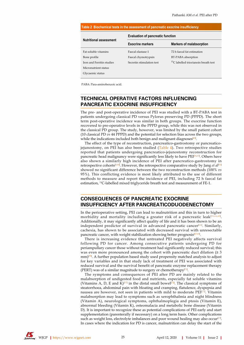
Table 2 Biochemical tests in the assessment of pancreatic exocrine insufficiency
Nutritional assessmentEvaluation of pancreatic function
Exocrine markers Markers of malabsorption
Fat soluble vitamins Faecal elastase-1 72 h faecal fat estimation
Bone profile Faecal chymotrypsin BT-PABA absorption
Iron and Ferritin studies Secretin stimulation test 13C labelled trioctanoin breath test
Micronutrient status
Glycaemic status
PABA: Para-aminobenzoic acid.
TECHNICAL OPERATIVE FACTORS INFLUENCINGPANCREATIC EXOCRINE INSUFFICIENCYThe pre- and post-operative incidence of PEI was studied with a BT-PABA test inpatients undergoing classical PD versus Pylorus preserving PD (PPPD). The shortterm post-operative incidence was similar in both groups. The exocrine functionrecovered to pre-operative levels in the PPPD group, while this was not observed inthe classical PD group. The study, however, was limited by the small patient cohort(10 classical PD vs 44 PPPD) and the potential for selection bias across the two groups,while the indications included both benign and malignant diagnoses[45].
The effect of the type of reconstruction, pancreatico-gastrostomy or pancreatico-jejunostomy, on PEI has also been studied (Table 4). Two retrospective studiesreported that patients undergoing pancreatico-jejunostomy reconstruction forpancreatic head malignancy were significantly less likely to have PEI[54,63]. Others havealso shown a similarly high incidence of PEI after pancreatico-gastrostomy inretrospective cohorts[9,44]. However, the retrospective comparative study by Jang et al[51]
showed no significant difference between the two reconstruction methods (100% vs95%). This conflicting evidence is most likely attributed to the use of differentmethods to measure and report the incidence of PEI, including 72 h faecal fatestimation, 13C-labelled mixed triglyceride breath test and measurement of FE-1.
CONSEQUENCES OF PANCREATIC EXOCRINEINSUFFICIENCY AFTER PANCREATICODUODENECTOMYIn the perioperative setting, PEI can lead to malnutrition and this in turn to highermorbidity and mortality including a greater risk of a pancreatic leak[59,64,65].Additionally, it may significantly affect quality of life and it has been shown to be anindependent predictor of survival in advanced pancreatic cancer[66]. Similarly,cachexia, has shown to be associated with decreased survival with unresectablepancreatic cancer, with weight stabilization showing better prognosis[67-69].
There is increasing evidence that untreated PEI negatively affects survivalfollowing PD for cancer. Among consecutive patients undergoing PD forperiampullary cancer those without treatment had significantly reduced survival; thiswas even more pronounced among the cohort with pancreatic duct dilation (≥ 3mm)[70]. A further population based study used propensity matched analysis to adjustfor key variables and in that study lack of treatment of PEI was associated withreduced survival and the survival benefit of pancreatic enzyme replacement therapy(PERT) was of a similar magnitude to surgery or chemotherapy[71].
The symptoms and consequences of PEI after PD are mainly related to themalabsorption of undigested food and nutrients, especially fat soluble vitamins(Vitamins A, D, E and K)[17] in the distal small bowel[72]. The classical symptoms ofsteatorrhoea, abdominal pain with bloating and cramping, flatulence, dyspepsia andnausea are however, not seen in patients with mild to moderate PEI[73]. Vitaminmalabsorption may lead to symptoms such as xerophthalmia and night blindness(Vitamin A), neurological symptoms, ophthalmoplegia and ptosis (Vitamin E),abnormal bleeding (Vitamin K), osteomalacia and metabolic bone disease (VitaminD). It is important to recognize these as potential complications of PEI early and startsupplementation (parenterally if necessary) on a long term basis. Other complicationssuch as weight loss, electrolyte imbalances and poor wound healing may also occur[17].In cases where the indication for PD is cancer, malnutrition can delay the start of the
WJGP https://www.wjgnet.com April 12, 2020 Volume 11 Issue 2
Pathanki AM et al. PEI after PD
25

Table 3 Incidence of pancreatic exocrine insufficiency before and after pancreaticoduodenectomy
Ref. Pre-operative incidence of PEI Post-operative incidence of PEI Diagnostic test
Kato et al[32] 93% 80% Secretin stimulation
Halloran et al[29] - 55% Coefficient of fat absorption
Yuasa et al[42] - 64% 13C- mixed triglyceride test
Nakamura et al[9] - 62.3%
Hirono et al[43] - 51%
Benini et al[41] - 87.5% 72 h faecal fat estimation
Lemaire et al[44] - 94%
Sato et al[45] 46% 33% BT-PABA excretion
Fujino et al[46] - 75%
Matsumoto et al[47] 68% 50% Faecal elastase-1
Van der Gaag et al[48] - 59%
Tran et al[49] - 91%
Pessaux et al[50] - 95%
Jang et al[51] - 100%
Falconi et al[52] - 24% Faecal chymotrypsin
Fang et al[34] - 33%
Bock et al[53] - 52.8% Steatorrhoea
Rault et al[54] - 42%
Van Berge Henegouwen et al[55] - 64.5%
PEI: Pancreatic exocrine insufficiency; PABA: Para-aminobenzoic acid.
recommended adjuvant chemotherapy or worse, render the patient unfit for the same.Finally, NAFLD is a rare and poorly recognized possible consequence of PEI after PD.It is believed to occur secondary to the malabsorption of essential amino acids leadingto decreased plasma levels of apoprotein B[74], which, when combined with sub-optimal insulin secretion lead to peripheral lipolysis and greater hepatic fatdeposition[75]. These changes have been shown to be reversible with the administrationof PERT and subsequent improvements in body weight[76].
MANAGEMENT OF PANCREATIC EXOCRINEINSUFFICIENCY AFTER PANCREATICODUODENECTOMYPERT is the mainstay of treatment of PEI. However, in the post-operative setting,there is a lack of consensus over the timing of initiation of PERT. While some authorsrecommend routine post-operative PERT[36,77], others advocate in favour of PERT onlyafter clinical or biochemical diagnostic evidence of PEI[8,78]. In pancreatic cancer, due tothe high incidence of PEI and obstructive jaundice, peri-operative use of PERT for allpatients has been shown to be beneficial[36] and is recommended by the UnitedKingdom National Institute of Clinical Excellence guidelines[79].
Patient education is important for the correct use of PERT. Enzymes use isadvisable with all meals, snacks and milky drinks, including various supplements.The conventional timing of administration is during or immediately after a meal inorder to achieve optimal timing for mixing with the chyme. This hypothesis however,has not been studied in the setting of pancreatic surgery and the presence ofpancreatico-biliary reconstruction and digestive asynchrony[16].
PERT is usually commenced at a dose of 50000-75000 units lipase with a meal and25000-50000 units with each snack[10,80-82]. This may be titrated to the needs of theindividual patient. In common clinical experience, patients over time learn to adjustthe dose of PERT on the basis of their symptoms and diet. Nonetheless, close follow-up is required to ensure that management remains on track in the setting of changing(recovering or deteriorating) pancreatic function and/or patient diet (as some patientsmay compromise on the nutritional value of their diet rather than the PERT dose). Theuse and effectiveness of PEI should be monitored with serial anthropometricmeasurements and nutritional blood tests[36], including measurements for glycaemiccontrol, as the use of effective PERT may result in manifestation of diabetes[83]. Inaddition to PERT, supplementation with vitamins and other micronutrients is
WJGP https://www.wjgnet.com April 12, 2020 Volume 11 Issue 2
Pathanki AM et al. PEI after PD
26

Table 4 Incidence of pancreatic exocrine insufficiency after pancreaticoduodenectomy–evidence on the role of the type of pancreaticreconstruction
Ref. Diagnostic test Incidence ofPEI–Pancreaticogastrostomy
Incidence ofPEI–Pancreaticojejunostomy
Nakamura et al[9] 13C Triglyceride breath test 62.3% -
Lemaire et al[44] Faecal Fat excretion and faecalelastase-1
100% -
Jang et al[51] Faecal elastase-1 100% (severe) 75% (severe); 20% (mild)
Roeyen et al[63] Need for PERT +/- any abnormalpancreatic function test
75% 45.7% (P < 0.001)
Rault et al[54] Steatorrhoea 70% 21.7% (P < 0.025)
PEI: Pancreatic exocrine insufficiency; PERT: Pancreatic enzyme replacement therapy.
recommended[84].The gastrointestinal environment and acidity is important for the appropriate
function of PERT. The lipase in PERT is inactivated by gastric acid activity.Consequently, commercially available PERT formulations are covered with pHsensitive, acid resistant microspheres that release the lipase at a pH of 5-6, similar towhat is present in the native duodenum. Based on studies about the optimal spheresize required to produce the best dissociation in the duodenum, most commercialpreparations have sphere size that varies from 1-2 mm[85,86]. In the post-operative PDsetting, failure of the pancreas to produce bicarbonate is hypothesized to lead to anacidic environment in the duodenum and proximal jejunum, leading to inefficientactivation of lipase[35,87]. The concurrent use of gastric acid suppression is thereforerecommended. The use of a proton pump inhibitor is known to reduce faecal fatlosses[88] and may also help reduce precipitation of bile salts[36].
PERT is generally well tolerated with minimal adverse effects. Rare reports onfibrosing colonopathy with the use of PERT are limited to paediatric patients,especially in the setting of cystic fibrosis[89-91]. There have been no such reports in theadult post-operative population. Many studies including open label PERT trials havenot found significant adverse drug reactions[92-94].
Failure of PEI to improve after escalation of PERT dosage and gastric acidsuppression must prompt further investigations for concurrent problems. The twocommonest diagnoses in this setting are bile salt malabsorption and small bowelbacterial overgrowth[30,80,82]. Bile salt malabsorption occurs due to the change in the pHin the proximal small bowel secondary to deficiency of bicarbonate secretion from thepancreas. The cholecystectomy performed during PD may also contribute to thedevelopment of this condition[36,95]. The presence of a blind loop of bowel used forreconstruction is known to occur after PD and is documented in up to 65% of patientsleading to small bowel bacterial overgrowth[36].
CONCLUSIONThis literature review confirms that PEI is prevalent after PD even for indicationsother than chronic pancreatitis and may have severe implications with respect topatients’ survival, quality of life, nutrition and subsequent management. The lack of auniform definition of PEI in this setting and the low diagnostic accuracy of theavailable tests introduce a wide variability in the reported results and suggestedmanagement. Pancreatic enzyme replacement therapy is effective, well tolerated andis indicated routinely in this cohort of patients. Future studies need to concentrate onthe identification of a well-tolerated, reliable and reproducible diagnostic test that willfacilitate a uniform definition and management approach.
REFERENCES1 Anagnostides A, Chadwick VS, Selden AC, Maton PN. Sham feeding and pancreatic secretion. Evidence
for direct vagal stimulation of enzyme output. Gastroenterology 1984; 87: 109-114 [PMID: 6724252 DOI:10.1016/0016-5085(84)90132-X]
2 Keller J, Rünzi M, Goebell H, Layer P. Duodenal and ileal nutrient deliveries regulate human intestinalmotor and pancreatic responses to a meal. Am J Physiol 1997; 272: G632-G637 [PMID: 9124585 DOI:10.1152/ajpgi.1997.272.3.G632]
WJGP https://www.wjgnet.com April 12, 2020 Volume 11 Issue 2
Pathanki AM et al. PEI after PD
27

3 Fried M, Mayer EA, Jansen JB, Lamers CB, Taylor IL, Bloom SR, Meyer JH. Temporal relationships ofcholecystokinin release, pancreatobiliary secretion, and gastric emptying of a mixed meal.Gastroenterology 1988; 95: 1344-1350 [PMID: 3169498 DOI: 10.1016/0016-5085(88)90371-X]
4 Beglinger C, Fried M, Whitehouse I, Jansen JB, Lamers CB, Gyr K. Pancreatic enzyme response to aliquid meal and to hormonal stimulation. Correlation with plasma secretin and cholecystokinin levels. JClin Invest 1985; 75: 1471-1476 [PMID: 3998145 DOI: 10.1172/JCI111850]
5 Miller LJ, Clain JE, Malagelada JR, Go VL. Control of human postprandial pancreatic exocrine secretion:a function of the gastroduodenal region. Dig Dis Sci 1979; 24: 150-154 [PMID: 107011 DOI:10.1007/BF01324743]
6 Layer P, Schlesinger T, Gröger G, Goebell H. Modulation of human periodic interdigestivegastrointestinal motor and pancreatic function by the ileum. Pancreas 1993; 8: 426-432 [PMID: 8361961DOI: 10.1097/00006676-199307000-00004]
7 Keller J, Holst JJ, Layer P. Inhibition of human pancreatic and biliary output but not intestinal motility byphysiological intraileal lipid loads. Am J Physiol Gastrointest Liver Physiol 2006; 290: G704-G709[PMID: 16322090 DOI: 10.1152/ajpgi.00411.2005]
8 Sabater L, Ausania F, Bakker OJ, Boadas J, Domínguez-Muñoz JE, Falconi M, Fernández-Cruz L,Frulloni L, González-Sánchez V, Lariño-Noia J, Lindkvist B, Lluís F, Morera-Ocón F, Martín-Pérez E,Marra-López C, Moya-Herraiz Á, Neoptolemos JP, Pascual I, Pérez-Aisa Á, Pezzilli R, Ramia JM,Sánchez B, Molero X, Ruiz-Montesinos I, Vaquero EC, de-Madaria E. Evidence-based Guidelines for theManagement of Exocrine Pancreatic Insufficiency After Pancreatic Surgery. Ann Surg 2016; 264: 949-958[PMID: 27045859 DOI: 10.1097/SLA.0000000000001732]
9 Nakamura H, Murakami Y, Uemura K, Hayashidani Y, Sudo T, Ohge H, Sueda T. Predictive factors forexocrine pancreatic insufficiency after pancreatoduodenectomy with pancreaticogastrostomy. JGastrointest Surg 2009; 13: 1321-1327 [PMID: 19415402 DOI: 10.1007/s11605-009-0896-5]
10 Tran TC, van Lanschot JJ, Bruno MJ, van Eijck CH. Functional changes after pancreatoduodenectomy:diagnosis and treatment. Pancreatology 2009; 9: 729-737 [PMID: 20090394 DOI: 10.1159/000264638]
11 Sikkens EC, Cahen DL, de Wit J, Looman CW, van Eijck C, Bruno MJ. A prospective assessment of thenatural course of the exocrine pancreatic function in patients with a pancreatic head tumor. J ClinGastroenterol 2014; 48: e43-e46 [PMID: 24717227 DOI: 10.1097/MCG.0b013e31829f56e7]
12 Bruno MJ, Haverkort EB, Tytgat GN, van Leeuwen DJ. Maldigestion associated with exocrine pancreaticinsufficiency: implications of gastrointestinal physiology and properties of enzyme preparations for acause-related and patient-tailored treatment. Am J Gastroenterol 1995; 90: 1383-1393 [PMID: 7661155]
13 Gullo L. Somatostatin analogues and exocrine pancreatic secretion. Digestion 1996; 57 Suppl 1: 93[PMID: 8813482 DOI: 10.1159/000201408]
14 van Dijk SM, Heerkens HD, Tseng DSJ, Intven M, Molenaar IQ, van Santvoort HC. Systematic review onthe impact of pancreatoduodenectomy on quality of life in patients with pancreatic cancer. HPB (Oxford)2018; 20: 204-215 [PMID: 29249649 DOI: 10.1016/j.hpb.2017.11.002]
15 Heerkens HD, van Berkel L, Tseng DSJ, Monninkhof EM, van Santvoort HC, Hagendoorn J, BorelRinkes IHM, Lips IM, Intven M, Molenaar IQ. Long-term health-related quality of life after pancreaticresection for malignancy in patients with and without severe postoperative complications. HPB (Oxford)2018; 20: 188-195 [PMID: 29092792 DOI: 10.1016/j.hpb.2017.09.003]
16 Bartel MJ, Asbun H, Stauffer J, Raimondo M. Pancreatic exocrine insufficiency in pancreatic cancer: Areview of the literature. Dig Liver Dis 2015; 47: 1013-1020 [PMID: 26211872 DOI:10.1016/j.dld.2015.06.015]
17 Gianotti L, Besselink MG, Sandini M, Hackert T, Conlon K, Gerritsen A, Griffin O, Fingerhut A, ProbstP, Abu Hilal M, Marchegiani G, Nappo G, Zerbi A, Amodio A, Perinel J, Adham M, Raimondo M, AsbunHJ, Sato A, Takaori K, Shrikhande SV, Del Chiaro M, Bockhorn M, Izbicki JR, Dervenis C, CharnleyRM, Martignoni ME, Friess H, de Pretis N, Radenkovic D, Montorsi M, Sarr MG, Vollmer CM, FrulloniL, Büchler MW, Bassi C. Nutritional support and therapy in pancreatic surgery: A position paper of theInternational Study Group on Pancreatic Surgery (ISGPS). Surgery 2018; 164: 1035-1048 [PMID:30029989 DOI: 10.1016/j.surg.2018.05.040]
18 Liberati A, Altman DG, Tetzlaff J, Mulrow C, Gøtzsche PC, Ioannidis JP, Clarke M, Devereaux PJ,Kleijnen J, Moher D. The PRISMA statement for reporting systematic reviews and meta-analyses ofstudies that evaluate health care interventions: explanation and elaboration. J Clin Epidemiol 2009; 62: e1-34 [PMID: 19631507 DOI: 10.1016/j.jclinepi.2009.06.006]
19 Tseng DS, Molenaar IQ, Besselink MG, van Eijck CH, Borel Rinkes IH, van Santvoort HC. PancreaticExocrine Insufficiency in Patients with Pancreatic or Periampullary Cancer: A Systematic Review.Pancreas 2016; 45: 325-330 [PMID: 26495777 DOI: 10.1097/MPA.0000000000000473]
20 Stein J, Jung M, Sziegoleit A, Zeuzem S, Caspary WF, Lembcke B. Immunoreactive elastase I: clinicalevaluation of a new noninvasive test of pancreatic function. Clin Chem 1996; 42: 222-226 [PMID:8595714 DOI: 10.1093/clinchem/42.2.222]
21 Löser C, Möllgaard A, Fölsch UR. Faecal elastase 1: a novel, highly sensitive, and specific tubelesspancreatic function test. Gut 1996; 39: 580-586 [PMID: 8944569 DOI: 10.1136/gut.39.4.580]
22 Dominici R, Franzini C. Fecal elastase-1 as a test for pancreatic function: a review. Clin Chem Lab Med2002; 40: 325-332 [PMID: 12059069 DOI: 10.1515/CCLM.2002.051]
23 Usküdar O, Oğuz D, Akdoğan M, Altiparmak E, Sahin B. Comparison of endoscopic retrogradecholangiopancreatography, endoscopic ultrasonography, and fecal elastase 1 in chronic pancreatitis andclinical correlation. Pancreas 2009; 38: 503-506 [PMID: 19287334 DOI:10.1097/MPA.0b013e31819f639f]
24 Symersky T, van der Zon A, Biemond I, Masclee AA. Faecal elastase-I: helpful in analysing steatorrhoea?Neth J Med 2004; 62: 286-289 [PMID: 15588069]
25 Gillams A, Pereira S, Webster G, Lees W. Correlation of MRCP quantification (MRCPQ) withconventional non-invasive pancreatic exocrine function tests. Abdom Imaging 2008; 33: 469-473 [PMID:17653788 DOI: 10.1007/s00261-007-9286-1]
26 Thomas PD, Forbes A, Green J, Howdle P, Long R, Playford R, Sheridan M, Stevens R, Valori R,Walters J, Addison GM, Hill P, Brydon G. Guidelines for the investigation of chronic diarrhoea, 2ndedition. Gut 2003; 52 Suppl 5: v1-15 [PMID: 12801941 DOI: 10.1136/gut.52.suppl_5.v1]
27 Erickson JA, Aldeen WE, Grenache DG, Ashwood ER. Evaluation of a fecal pancreatic elastase-1enzyme-linked immunosorbent assay: Assessment versus an established assay and implication inclassifying pancreatic function. Clin Chim Acta 2008; 397: 87-91 [PMID: 18706899 DOI:10.1016/j.cca.2008.07.022]
WJGP https://www.wjgnet.com April 12, 2020 Volume 11 Issue 2
Pathanki AM et al. PEI after PD
28

28 Ghaneh P, Neoptolemos JP. Exocrine pancreatic function following pancreatectomy. Ann N Y Acad Sci1999; 880: 308-318 [PMID: 10415875 DOI: 10.1111/j.1749-6632.1999.tb09534.x]
29 Halloran CM, Cox TF, Chauhan S, Raraty MG, Sutton R, Neoptolemos JP, Ghaneh P. Partial pancreaticresection for pancreatic malignancy is associated with sustained pancreatic exocrine failure and reducedquality of life: a prospective study. Pancreatology 2011; 11: 535-545 [PMID: 22094930 DOI:10.1159/000333308]
30 Domínguez-Muñoz JE. Pancreatic enzyme replacement therapy: exocrine pancreatic insufficiency aftergastrointestinal surgery. HPB (Oxford) 2009; 11 Suppl 3: 3-6 [PMID: 20495625 DOI:10.1111/j.1477-2574.2009.00132.x]
31 Yamaguchi K, Yokohata K, Nakano K, Ohtani K, Ogawa Y, Chijiiwa K, Tanaka M. Which is a lessinvasive pancreatic head resection: PD, PPPD, or DPPHR? Dig Dis Sci 2001; 46: 282-288 [PMID:11281176 DOI: 10.1023/A:1005644614104]
32 Kato H, Nakao A, Kishimoto W, Nonami T, Harada A, Hayakawa T, Takagi H. 13C-labeled trioctanoinbreath test for exocrine pancreatic function test in patients after pancreatoduodenectomy. Am JGastroenterol 1993; 88: 64-69 [PMID: 8093586]
33 Perez MM, Newcomer AD, Moertel CG, Go VL, Dimagno EP. Assessment of weight loss, food intake,fat metabolism, malabsorption, and treatment of pancreatic insufficiency in pancreatic cancer. Cancer1983; 52: 346-352 [PMID: 6305473 DOI:10.1002/1097-0142(19830715)52:2<346::aid-cncr2820520228>3.0.co;2-z]
34 Fang WL, Su CH, Shyr YM, Chen TH, Lee RC, Tai LC, Wu CW, Lui WY. Functional and morphologicalchanges in pancreatic remnant after pancreaticoduodenectomy. Pancreas 2007; 35: 361-365 [PMID:18090244 DOI: 10.1097/MPA.0b013e3180d0a8d5]
35 Ghaneh P, Neoptolemos JP. Exocrine pancreatic function following pancreatectomy. Ann N Y Acad Sci1999; 880: 308-318 [PMID: 10415875 DOI: 10.1111/j.1749-6632.1999.tb09534.x]
36 Phillips ME. Pancreatic exocrine insufficiency following pancreatic resection. Pancreatology 2015; 15:449-455 [PMID: 26145836 DOI: 10.1016/j.pan.2015.06.003]
37 Keller J, Aghdassi AA, Lerch MM, Mayerle JV, Layer P. Tests of pancreatic exocrine function - clinicalsignificance in pancreatic and non-pancreatic disorders. Best Pract Res Clin Gastroenterol 2009; 23: 425-439 [PMID: 19505669 DOI: 10.1016/j.bpg.2009.02.013]
38 Caliari S, Benini L, Sembenini C, Gregori B, Carnielli V, Vantini I. Medium-chain triglyceride absorptionin patients with pancreatic insufficiency. Scand J Gastroenterol 1996; 31: 90-94 [PMID: 8927947 DOI:10.3109/00365529609031633]
39 Owira PM, Winter TA. Colonic energy salvage in chronic pancreatic exocrine insufficiency. JPEN JParenter Enteral Nutr 2008; 32: 63-71 [PMID: 18165449 DOI: 10.1177/014860710803200163]
40 Temple SJ, Kim PT, Serrano PE, Kagedan D, Cleary SP, Moulton CA, McGilvray ID, Gallinger S, GreigPD, Wei AC. Combined pancreaticoduodenectomy and colon resection for locally advanced peri-ampullary tumours: analysis of peri-operative morbidity and mortality. HPB (Oxford) 2014; 16: 797-800[PMID: 24750414 DOI: 10.1111/hpb.12263]
41 Benini L, Amodio A, Campagnola P, Agugiaro F, Cristofori C, Micciolo R, Magro A, Gabbrielli A,Cabrini G, Moser L, Massella A, Vantini I, Frulloni L. Fecal elastase-1 is useful in the detection ofsteatorrhea in patients with pancreatic diseases but not after pancreatic resection. Pancreatology 2013; 13:38-42 [PMID: 23395568 DOI: 10.1016/j.pan.2012.11.307]
42 Yuasa Y, Murakami Y, Nakamura H, Uemura K, Ohge H, Sudo T, Hashimoto Y, Nakashima A, HiyamaE, Sueda T. Histological loss of pancreatic exocrine cells correlates with pancreatic exocrine function afterpancreatic surgery. Pancreas 2012; 41: 928-933 [PMID: 22781909 DOI:10.1097/MPA.0b013e31823d837d]
43 Hirono S, Murakami Y, Tani M, Kawai M, Okada K, Uemura K, Sudo T, Hashimoto Y, Nakagawa N,Kondo N, Yamaue H. Identification of risk factors for pancreatic exocrine insufficiency afterpancreaticoduodenectomy using a 13C-labeled mixed triglyceride breath test. World J Surg 2015; 39: 516-525 [PMID: 25318451 DOI: 10.1007/s00268-014-2832-4]
44 Lemaire E, O'Toole D, Sauvanet A, Hammel P, Belghiti J, Ruszniewski P. Functional and morphologicalchanges in the pancreatic remnant following pancreaticoduodenectomy with pancreaticogastricanastomosis. Br J Surg 2000; 87: 434-438 [PMID: 10759738 DOI: 10.1046/j.1365-2168.2000.01388.x]
45 Sato N, Yamaguchi K, Yokohata K, Shimizu S, Morisaki T, Chijiiwa K, Tanaka M. Short-term and long-term pancreatic exocrine and endocrine functions after pancreatectomy. Dig Dis Sci 1998; 43: 2616-2621[PMID: 9881491 DOI: 10.1023/a:1026686824173]
46 Fujino Y, Suzuki Y, Matsumoto I, Sakai T, Ajiki T, Ueda T, Kuroda Y. Long-term assessments afterpancreaticoduodenectomy with pancreatic duct invagination anastomosis. Surg Today 2007; 37: 860-866[PMID: 17879035 DOI: 10.1007/s00595-007-3507-7]
47 Matsumoto J, Traverso LW. Exocrine function following the whipple operation as assessed by stoolelastase. J Gastrointest Surg 2006; 10: 1225-1229 [PMID: 17114009 DOI: 10.1016/j.gassur.2006.08.001]
48 van der Gaag NA, Berkhemer OA, Sprangers MA, Busch OR, Bruno MJ, de Castro SM, van Gulik TM,Gouma DJ. Quality of life and functional outcome after resection of pancreatic cystic neoplasm. Pancreas2014; 43: 755-761 [PMID: 24743379 DOI: 10.1097/MPA.0000000000000075]
49 Tran TC, van 't Hof G, Kazemier G, Hop WC, Pek C, van Toorenenbergen AW, van Dekken H, van EijckCH. Pancreatic fibrosis correlates with exocrine pancreatic insufficiency after pancreatoduodenectomy.Dig Surg 2008; 25: 311-318 [PMID: 18818498 DOI: 10.1159/000158596]
50 Pessaux P, Aube C, Lebigot J, Tuech JJ, Regenet N, Kapel N, Caron C, Arnaud JP. Permeability andfunctionality of pancreaticogastrostomy after pancreaticoduodenectomy with dynamic magnetic resonancepancreatography after secretin stimulation. J Am Coll Surg 2002; 194: 454-462 [PMID: 11949751 DOI:10.1016/s1072-7515(02)01126-2]
51 Jang JY, Kim SW, Park SJ, Park YH. Comparison of the functional outcome after pylorus-preservingpancreatoduodenectomy: pancreatogastrostomy and pancreatojejunostomy. World J Surg 2002; 26: 366-371 [PMID: 11865376 DOI: 10.1007/s00268-001-0234-x]
52 Falconi M, Mantovani W, Crippa S, Mascetta G, Salvia R, Pederzoli P. Pancreatic insufficiency afterdifferent resections for benign tumours. Br J Surg 2008; 95: 85-91 [PMID: 18041022 DOI:10.1002/bjs.5652]
53 Bock EA, Hurtuk MG, Shoup M, Aranha GV. Late complications after pancreaticoduodenectomy withpancreaticogastrostomy. J Gastrointest Surg 2012; 16: 914-919 [PMID: 22374385 DOI:10.1007/s11605-011-1805-2]
54 Rault A, SaCunha A, Klopfenstein D, Larroudé D, Epoy FN, Collet D, Masson B. Pancreaticojejunal
WJGP https://www.wjgnet.com April 12, 2020 Volume 11 Issue 2
Pathanki AM et al. PEI after PD
29

anastomosis is preferable to pancreaticogastrostomy after pancreaticoduodenectomy for longtermoutcomes of pancreatic exocrine function. J Am Coll Surg 2005; 201: 239-244 [PMID: 16038822 DOI:10.1016/j.jamcollsurg.2005.03.026]
55 van Berge Henegouwen MI, Moojen TM, van Gulik TM, Rauws EA, Obertop H, Gouma DJ.Postoperative weight gain after standard Whipple's procedure versus pylorus-preservingpancreatoduodenectomy: the influence of tumour status. Br J Surg 1998; 85: 922-926 [PMID: 9692564DOI: 10.1046/j.1365-2168.1998.00745.x]
56 Kodama M, Tanaka T. Residual function of exocrine pancreas after operation for chronic pancreatitis byN-benzoyl-L-tyrosyl-p-aminobenzoic acid test (NBT-PABA test). Digestion 1984; 30: 41-46 [PMID:6333364 DOI: 10.1159/000199089]
57 Tanaka T, Ichiba Y, Fujii Y, Kodama O, Dohi K. Clinical and experimental study of pancreatic exocrinefunction after pancreaticoduodenectomy for periampullary carcinoma. Surg Gynecol Obstet 1988; 166:200-205 [PMID: 3257831]
58 Nordback I, Parviainen M, Piironen A, Räty S, Sand J. Obstructed pancreaticojejunostomy partly explainsexocrine insufficiency after pancreatic head resection. Scand J Gastroenterol 2007; 42: 263-270 [PMID:17327947 DOI: 10.1080/00365520600849174]
59 Domínguez-Muñoz JE, Alvarez-Castro A, Lariño-Noia J, Nieto L, Iglesias-García J. Endoscopicultrasonography of the pancreas as an indirect method to predict pancreatic exocrine insufficiency inpatients with chronic pancreatitis. Pancreas 2012; 41: 724-728 [PMID: 22228053 DOI:10.1097/MPA.0b013e31823b5978]
60 Nakamura H, Murakami Y, Uemura K, Hayashidani Y, Sudo T, Ohge H, Sueda T. Reduced pancreaticparenchymal thickness indicates exocrine pancreatic insufficiency after pancreatoduodenectomy. J SurgRes 2011; 171: 473-478 [PMID: 20605585 DOI: 10.1016/j.jss.2010.03.052]
61 Manfredi R, Perandini S, Mantovani W, Frulloni L, Faccioli N, Pozzi Mucelli R. Quantitative MRCPassessment of pancreatic exocrine reserve and its correlation with faecal elastase-1 in patients with chronicpancreatitis. Radiol Med 2012; 117: 282-292 [PMID: 22231574 DOI: 10.1007/s11547-011-0774-6]
62 Balci NC, Smith A, Momtahen AJ, Alkaade S, Fattahi R, Tariq S, Burton F. MRI and S-MRCP findings inpatients with suspected chronic pancreatitis: correlation with endoscopic pancreatic function testing(ePFT). J Magn Reson Imaging 2010; 31: 601-606 [PMID: 20187202 DOI: 10.1002/jmri.22085]
63 Roeyen G, Jansen M, Ruyssinck L, Chapelle T, Vanlander A, Bracke B, Hartman V, Ysebaert D,Berrevoet F. Pancreatic exocrine insufficiency after pancreaticoduodenectomy is more prevalent withpancreaticogastrostomy than with pancreaticojejunostomy. A retrospective multicentre observationalcohort study. HPB (Oxford) 2016; 18: 1017-1022 [PMID: 27726974 DOI: 10.1016/j.hpb.2016.09.002]
64 Crucitti F, Doglietto GB, Viola G, Frontera D, De Cosmo G, Sgadari A, Vicari D, Rizzi A. Assessment ofrisk factors for pancreatic resection for cancer. World J Surg 1998; 22: 241-247 [PMID: 9494415 DOI:10.1007/s002689900377]
65 Kanda M, Fujii T, Kodera Y, Nagai S, Takeda S, Nakao A. Nutritional predictors of postoperativeoutcome in pancreatic cancer. Br J Surg 2011; 98: 268-274 [PMID: 20960457 DOI: 10.1002/bjs.7305]
66 Partelli S, Frulloni L, Minniti C, Bassi C, Barugola G, D'Onofrio M, Crippa S, Falconi M. Faecal elastase-1 is an independent predictor of survival in advanced pancreatic cancer. Dig Liver Dis 2012; 44: 945-951[PMID: 22749648 DOI: 10.1016/j.dld.2012.05.017]
67 Davidson W, Ash S, Capra S, Bauer J; Cancer Cachexia Study Group. Weight stabilisation is associatedwith improved survival duration and quality of life in unresectable pancreatic cancer. Clin Nutr 2004; 23:239-247 [PMID: 15030964 DOI: 10.1016/j.clnu.2003.07.001]
68 Bachmann J, Ketterer K, Marsch C, Fechtner K, Krakowski-Roosen H, Büchler MW, Friess H,Martignoni ME. Pancreatic cancer related cachexia: influence on metabolism and correlation to weightloss and pulmonary function. BMC Cancer 2009; 9: 255 [PMID: 19635171 DOI:10.1186/1471-2407-9-255]
69 Heinrich S, Pestalozzi BC, Schäfer M, Weber A, Bauerfeind P, Knuth A, Clavien PA. Prospective phaseII trial of neoadjuvant chemotherapy with gemcitabine and cisplatin for resectable adenocarcinoma of thepancreatic head. J Clin Oncol 2008; 26: 2526-2531 [PMID: 18487569 DOI: 10.1200/JCO.2007.15.5556]
70 Roberts KJ, Schrem H, Hodson J, Angelico R, Dasari BVM, Coldham CA, Marudanayagam R, SutcliffeRP, Muiesan P, Isaac J, Mirza DF. Pancreas exocrine replacement therapy is associated with increasedsurvival following pancreatoduodenectomy for periampullary malignancy. HPB (Oxford) 2017; 19: 859-867 [PMID: 28711377 DOI: 10.1016/j.hpb.2017.05.009]
71 Roberts KJ, Bannister CA, Schrem H. Enzyme replacement improves survival among patients withpancreatic cancer: Results of a population based study. Pancreatology 2019; 19: 114-121 [PMID:30385188 DOI: 10.1016/j.pan.2018.10.010]
72 Whitcomb DC, Lehman GA, Vasileva G, Malecka-Panas E, Gubergrits N, Shen Y, Sander-Struckmeier S,Caras S. Pancrelipase delayed-release capsules (CREON) for exocrine pancreatic insufficiency due tochronic pancreatitis or pancreatic surgery: A double-blind randomized trial. Am J Gastroenterol 2010; 105:2276-2286 [PMID: 20502447 DOI: 10.1038/ajg.2010.201]
73 Domínguez-Muñoz JE. Pancreatic enzyme therapy for pancreatic exocrine insufficiency. GastroenterolHepatol (N Y) 2011; 7: 401-403 [PMID: 21869872]
74 Yao ZM, Vance DE. Reduction in VLDL, but not HDL, in plasma of rats deficient in choline. BiochemCell Biol 1990; 68: 552-558 [PMID: 2344402 DOI: 10.1139/o90-079]
75 Soliman AT, Alsalmi I, Asfour M. Hypoinsulinaemia has an important role in the development of oedemaand hepatomegaly during malnutrition. J Trop Pediatr 1996; 42: 297-299 [PMID: 8936962 DOI:10.1093/tropej/42.5.297]
76 Tanaka N, Horiuchi A, Yokoyama T, Kaneko G, Horigome N, Yamaura T, Nagaya T, Komatsu M, SanoK, Miyagawa S, Aoyama T, Tanaka E. Clinical characteristics of de novo nonalcoholic fatty liver diseasefollowing pancreaticoduodenectomy. J Gastroenterol 2011; 46: 758-768 [PMID: 21267748 DOI:10.1007/s00535-011-0370-5]
77 Working Party of the Australasian Pancreatic Club., Smith RC, Smith SF, Wilson J, Pearce C, WrayN, Vo R, Chen J, Ooi CY, Oliver M, Katz T, Turner R, Nikfarjam M, Rayner C, Horowitz M, HoltmannG, Talley N, Windsor J, Pirola R, Neale R. Summary and recommendations from the Australasianguidelines for the management of pancreatic exocrine insufficiency. Pancreatology 2016; 16: 164-180[PMID: 26775768 DOI: 10.1016/j.pan.2015.12.006]
78 Domínguez-Muñoz JE. Pancreatic enzyme therapy for pancreatic exocrine insufficiency. GastroenterolHepatol (N Y) 2011; 7: 401-403 [PMID: 21869872]
79 National Institute for Health and Care Excellence. Pancreatic cancer in adults: diagnosis and
WJGP https://www.wjgnet.com April 12, 2020 Volume 11 Issue 2
Pathanki AM et al. PEI after PD
30

management. NICE guideline [NG85], Recommendations. Available from: https://www.nice.org.uk/guid-ance/ng85/chapter/Recommendations#nutritional-management
80 Imrie CW, Connett G, Hall RI, Charnley RM. Review article: enzyme supplementation in cystic fibrosis,chronic pancreatitis, pancreatic and periampullary cancer. Aliment Pharmacol Ther 2010; 32 Suppl 1: 1-25[PMID: 21054452 DOI: 10.1111/j.1365-2036.2010.04437.x]
81 Seiler CM, Izbicki J, Varga-Szabó L, Czakó L, Fiók J, Sperti C, Lerch MM, Pezzilli R, Vasileva G, PapA, Varga M, Friess H. Randomised clinical trial: a 1-week, double-blind, placebo-controlled study ofpancreatin 25 000 Ph. Eur. minimicrospheres (Creon 25000 MMS) for pancreatic exocrine insufficiencyafter pancreatic surgery, with a 1-year open-label extension. Aliment Pharmacol Ther 2013; 37: 691-702[PMID: 23383603 DOI: 10.1111/apt.12236]
82 Toouli J, Biankin AV, Oliver MR, Pearce CB, Wilson JS, Wray NH; Australasian Pancreatic Club.Management of pancreatic exocrine insufficiency: Australasian Pancreatic Club recommendations. Med JAust 2010; 193: 461-467 [PMID: 20955123 DOI: 10.5694/j.1326-5377.2010.tb04000.x]
83 O'Keefe SJ, Cariem AK, Levy M. The exacerbation of pancreatic endocrine dysfunction by potentpancreatic exocrine supplements in patients with chronic pancreatitis. J Clin Gastroenterol 2001; 32: 319-323 [PMID: 11276275 DOI: 10.1097/00004836-200104000-00008]
84 Armstrong T, Strommer L, Ruiz-Jasbon F, Shek FW, Harris SF, Permert J, Johnson CD.Pancreaticoduodenectomy for peri-ampullary neoplasia leads to specific micronutrient deficiencies.Pancreatology 2007; 7: 37-44 [PMID: 17449964 DOI: 10.1159/000101876]
85 Meyer JH, Dressman J, Fink A, Amidon G. Effect of size and density on canine gastric emptying ofnondigestible solids. Gastroenterology 1985; 89: 805-813 [PMID: 4029560 DOI:10.1016/0016-5085(85)90576-1]
86 Meyer JH, Elashoff J, Porter-Fink V, Dressman J, Amidon GL. Human postprandial gastric emptying of1-3-millimeter spheres. Gastroenterology 1988; 94: 1315-1325 [PMID: 3360258 DOI:10.1016/0016-5085(88)90669-5]
87 Aloulou A, Puccinelli D, Sarles J, Laugier R, Leblond Y, Carrière F. In vitro comparative study of threepancreatic enzyme preparations: dissolution profiles, active enzyme release and acid stability. AlimentPharmacol Ther 2008; 27: 283-292 [PMID: 17973644 DOI: 10.1111/j.1365-2036.2007.03563.x]
88 Proesmans M, De Boeck K. Omeprazole, a proton pump inhibitor, improves residual steatorrhoea incystic fibrosis patients treated with high dose pancreatic enzymes. Eur J Pediatr 2003; 162: 760-763[PMID: 13680386 DOI: 10.1007/s00431-003-1309-5]
89 FitzSimmons SC, Burkhart GA, Borowitz D, Grand RJ, Hammerstrom T, Durie PR, Lloyd-Still JD,Lowenfels AB. High-dose pancreatic-enzyme supplements and fibrosing colonopathy in children withcystic fibrosis. N Engl J Med 1997; 336: 1283-1289 [PMID: 9113931 DOI:10.1056/NEJM199705013361803]
90 Smyth RL, van Velzen D, Smyth AR, Lloyd DA, Heaf DP. Strictures of ascending colon in cystic fibrosisand high-strength pancreatic enzymes. Lancet 1994; 343: 85-86 [PMID: 7903780 DOI:10.1016/S0140-6736(94)90817-6]
91 Nouisa-Arvanitakis S, Stapleton FB, Linshaw MA, Kennedy J. Therapeutic approach to pancreaticextract-induced hyperuricosuria in cystic fibrosis. J Pediatr 1977; 90: 302-305 [PMID: 830926 DOI:10.1016/S0022-3476(77)80657-4]
92 Gullo L, Pezzilli R, Gaiani S. Tolerability and safety of the long-term administration of pancreaticextracts. Pancreas 1997; 14: 210-212 [PMID: 9057197 DOI: 10.1097/00006676-199703000-00018]
93 Ramesh H, Reddy N, Bhatia S, Rajkumar JS, Bapaye A, Kini D, Kalla M, Thorat V. A 51-week, open-label clinical trial in India to assess the efficacy and safety of pancreatin 40000 enteric-coatedminimicrospheres in patients with pancreatic exocrine insufficiency due to chronic pancreatitis.Pancreatology 2013; 13: 133-139 [PMID: 23561971 DOI: 10.1016/j.pan.2013.01.009]
94 Gubergrits N, Malecka-Panas E, Lehman GA, Vasileva G, Shen Y, Sander-Struckmeier S, Caras S,Whitcomb DC. A 6-month, open-label clinical trial of pancrelipase delayed-release capsules (Creon) inpatients with exocrine pancreatic insufficiency due to chronic pancreatitis or pancreatic surgery. AlimentPharmacol Ther 2011; 33: 1152-1161 [PMID: 21418260 DOI: 10.1111/j.1365-2036.2011.04631.x]
95 Bustillo I, Larson H, Saif MW. Small intestine bacterial overgrowth: an underdiagnosed cause of diarrheain patients with pancreatic cancer. JOP 2009; 10: 576-578 [PMID: 19734643]
WJGP https://www.wjgnet.com April 12, 2020 Volume 11 Issue 2
Pathanki AM et al. PEI after PD
31

Published by Baishideng Publishing Group Inc
7041 Koll Center Parkway, Suite 160, Pleasanton, CA 94566, USA
Telephone: +1-925-3991568
E-mail: [email protected]
Help Desk:https://www.f6publishing.com/helpdesk
https://www.wjgnet.com
© 2020 Baishideng Publishing Group Inc. All rights reserved.