World Journal of Meta-Analysis - NET
116
World Journal of Meta-Analysis ISSN 2308-3840 (online) World J Meta-Anal 2020 June 28; 8(3): 173-284 Published by Baishideng Publishing Group Inc
-
Upload
khangminh22 -
Category
Documents
-
view
1 -
download
0
Transcript of World Journal of Meta-Analysis - NET

World Journal ofMeta-Analysis
ISSN 2308-3840 (online)
World J Meta-Anal 2020 June 28; 8(3): 173-284
Published by Baishideng Publishing Group Inc
WJMA https://www.wjgnet.com I June 28, 2020 Volume 8 Issue 3
World Journal of
Meta-AnalysisW J M AContents Bimonthly Volume 8 Number 3 June 28, 2020
FIELD OF VISION
COVID-19: Off-label therapies based on mechanism of action while waiting for evidence-based medicine recommendations
173
Scotto Di Vetta M, Morrone M, Fazio S
Learning and competence development via clinical cases – what elements should be investigated to best train good medical doctors?
178
Löffler-Stastka H, Wong G
REVIEW
Immunotherapy in hepatocellular carcinoma: Combination strategies190
Jordan AC, Wu J
Combined endoscopy/laparoscopy/percutaneous transhepatic biliary drainage, hybrid techniques in gastrointestinal and biliary diseases
210
Feng YL, Li J, Ye LS, Zeng XH, Hu B
MINIREVIEWS
Thrombopoietin-receptor agonists in perioperative treatment of patients with chronic liver disease220
Qureshi K, Bonder A
Role of non-coding RNAs in pathogenesis of gastrointestinal stromal tumors233
Stefanou IK, Gazouli M, Zografos GC, Toutouzas KG
SYSTEMATIC REVIEWS
Exclusive cigar smoking in the United States and smoking-related diseases: A systematic review245
Lee PN, Hamling JS, Thornton AJ
Hydatidosis and the duodenum: A systematic review of the literature265
de la Fuente-Aguilar V, Beneitez-Mascaraque P, Bergua-Arroyo S, Fernández-Riesgo M, Camón-García I, Cruza-Aguilera I, Ugarte-Yáñez K, Ramia JM
META-ANALYSIS
Prevalence of anxiety among gestational diabetes mellitus patients: A systematic review and meta-analysis275
Lee KW, Loh HC, Chong SC, Ching SM, Devaraj NK, Tusimin M, Abdul Hamid H, Hoo FK
WJMA https://www.wjgnet.com II June 28, 2020 Volume 8 Issue 3
World Journal of Meta-AnalysisContents
Bimonthly Volume 8 Number 3 June 28, 2020
ABOUT COVER
Dr. Rakhshan is an editorial board member of World Journal of Meta-Analysis, and a former lecturer in the Dental School of Islamic Azad University, Tehran, Iran. He graduated from the same university in 2004, with a DDS thesis in which he designed and implemented an AI computer vision program that could extract radiographic landmarks from lateral cephalographs. Since then, besides clinical practice, he has taught dental anatomy and morphology, and has published about 140 peer-reviewed articles on different dentistry topics. He has also peer reviewed more than 500 articles during these years, and has been the lead guest editor of the journals Pain Research and Management, Computational Intelligence and Neuroscience, and International Journal of Dentistry, and an associate editor of Frontiers in Oral Health. He is currently a PhD candidate of cognitive neuroscience at the Institute for Cognitive Science Studies, Tehran, Iran
AIMS AND SCOPE
The primary aim of World Journal of Meta-Analysis (WJMA, World J Meta-Anal) is to provide scholars and readers from various fields of clinical medicine with a platform to publish high-quality meta-analysis and systematic review articles and communicate their research findings online. WJMA mainly publishes articles reporting research results and findings obtained through meta-analysis and systematic review in a wide range of areas, including medicine, pharmacy, preventive medicine, stomatology, nursing, medical imaging, and laboratory medicine.
INDEXING/ABSTRACTING
The WJMA is now abstracted and indexed in China National Knowledge Infrastructure (CNKI), China Science and Technology Journal Database (CSTJ), and Superstar Journals Database
RESPONSIBLE EDITORS FOR THIS ISSUE
Electronic Editor: Lu-Lu Qi; Production Department Director: Yun-Xiaojian Wu; Editorial Office Director: Jin-Lei Wang.
NAME OF JOURNAL INSTRUCTIONS TO AUTHORS
World Journal of Meta-Analysis https://www.wjgnet.com/bpg/gerinfo/204
ISSN GUIDELINES FOR ETHICS DOCUMENTS
ISSN 2308-3840 (online) https://www.wjgnet.com/bpg/GerInfo/287
LAUNCH DATE GUIDELINES FOR NON-NATIVE SPEAKERS OF ENGLISH
May 26, 2013 https://www.wjgnet.com/bpg/gerinfo/240
FREQUENCY PUBLICATION ETHICS
Bimonthly https://www.wjgnet.com/bpg/GerInfo/288
EDITORS-IN-CHIEF PUBLICATION MISCONDUCT
Saurabh Chandan https://www.wjgnet.com/bpg/gerinfo/208
EDITORIAL BOARD MEMBERS ARTICLE PROCESSING CHARGE
https://www.wjgnet.com/2308-3840/editorialboard.htm https://www.wjgnet.com/bpg/gerinfo/242
PUBLICATION DATE STEPS FOR SUBMITTING MANUSCRIPTS
June 28, 2020 https://www.wjgnet.com/bpg/GerInfo/239
COPYRIGHT ONLINE SUBMISSION
© 2020 Baishideng Publishing Group Inc https://www.f6publishing.com
© 2020 Baishideng Publishing Group Inc. All rights reserved. 7041 Koll Center Parkway, Suite 160, Pleasanton, CA 94566, USA
E-mail: [email protected] https://www.wjgnet.com

WJMA https://www.wjgnet.com 173 June 28, 2020 Volume 8 Issue 3
World Journal of
Meta-AnalysisW J M ASubmit a Manuscript: https://www.f6publishing.com World J Meta-Anal 2020 June 28; 8(3): 173-177
DOI: 10.13105/wjma.v8.i3.173 ISSN 2308-3840 (online)
FIELD OF VISION
COVID-19: Off-label therapies based on mechanism of action while waiting for evidence-based medicine recommendations
Matteo Scotto Di Vetta, Marco Morrone, Serafino Fazio
ORCID number: Matteo Scotto Di Vetta 0000-0001-7929-8441; Marco Morrone 0000-0003-3212-9223; Serafino Fazio 0000-0002-2743-9836.
Author contributions: Scotto Di Vetta M, Morrone M and Fazio S contributed to the writing of the manuscript.
Conflict-of-interest statement: No conflict of interest.
Open-Access: This article is an open-access article that was selected by an in-house editor and fully peer-reviewed by external reviewers. It is distributed in accordance with the Creative Commons Attribution NonCommercial (CC BY-NC 4.0) license, which permits others to distribute, remix, adapt, build upon this work non-commercially, and license their derivative works on different terms, provided the original work is properly cited and the use is non-commercial. See: http://creativecommons.org/licenses/by-nc/4.0/
Manuscript source: Invited manuscript
Received: May 18, 2020 Peer-review started: May 18, 2020 First decision: June 8, 2020 Revised: June 8, 2020 Accepted: June 28, 2020
Matteo Scotto Di Vetta, Department of Internal Medicine, University of Naples, Naples 80100, Italy
Marco Morrone, Farmacia, Farmacia Morrone, Casoria 80022, Italy
Serafino Fazio, Department of Internal Medicine, Cardiovascular and Immunologic Sciences, Federico II University of Naples, Napoli 80100, Italy
Corresponding author: Serafino Fazio, MD, Adjunct Professor, Associate Specialist, Department of Internal Medicine, Cardiovascular and Immunologic Sciences, Federico II University of Naples, Via S. Pansini 5, Napoli 80100, Italy. [email protected]
AbstractThe world pandemic due to coronavirus disease 2019, known as COVID-19, embodies a high rate of disease transmission that causes a critical hospitalization overload. As of May 15, 2020, the disease has been the cause of more than 4 million infections and more than 280000 deaths all over the world. At the beginning, we underestimated the disease; now, we have sufficient information and it is clear that it is not just a respiratory disease. In fact, if a prompt treatment is not initiated, the disease may evolve towards an abnormal immune response and cytokine storm with an important thrombophilic pattern. Therefore, we think that while waiting for certainties to be established by evidence-based medicine, it is not ethical to not try off-label therapies for some of the well-known drugs, as they could have some efficacy based on their mechanisms of action.
Key words: COVID-19; Evidence based medicine; Hydroxycloroquine; Azithromycine; Indomethacine; Doxycycline
©The Author(s) 2020. Published by Baishideng Publishing Group Inc. All rights reserved.
Core tip: The world pandemic due to coronavirus disease 2019, known as COVID-19, embodies a high rate of disease transmission that has caused a critical hospitalization overload. While waiting for certainties to be established by evidence-based medicine, it is not ethical to not try off-label therapies with some of the well-known drugs that could show some efficacy based on their mechanisms of action.

Scotto Di Vetta M et al. Off-label therapies for COVID-19
WJMA https://www.wjgnet.com 174 June 28, 2020 Volume 8 Issue 3
Article in press: June 28, 2020 Published online: June 28, 2020
P-Reviewer: Zutshi D S-Editor: Wang JL L-Editor: Filipodia E-Editor: Liu JH
Citation: Scotto Di Vetta M, Morrone M, Fazio S. COVID-19: Off-label therapies based on mechanism of action while waiting for evidence-based medicine recommendations. World J Meta-Anal 2020; 8(3): 173-177URL: https://www.wjgnet.com/2308-3840/full/v8/i3/173.htmDOI: https://dx.doi.org/10.13105/wjma.v8.i3.173
COMMENTARY ON HOT TOPICSAt the end of 2019, some cases of pneumonia of unknown etiology were observed in Wuhan (China). A few weeks later, this disease was discovered to be due to a virus of the coronavirus family, that was named severe acute respiratory syndrome-coronavirus-2 (SARS-Cov2), and the related coronavirus disease 2019 (COVID-19) was named accordingly.
COVID-19 embodies a high rate of disease transmission that has caused a critical hospitalization overload. By May 15, 2020, it had produced more than 4 million documented positive cases and about 280000 deaths all over the world. At the beginning, we underestimated and misunderstood COVID-19. Now, however, we have sufficient information of the disease pathophysiology, and it is clear that we are not dealing with just a respiratory disease. In fact, in many cases, if a prompt treatment is not undertaken, the infection may evolve towards a more severe disease and the occurrence of a cytokine storm with multi-organ damage[1-3].
Multi-organ damage should be investigated in patients recovered from moderate-severe COVID-19. Follow-up studies should be conducted to verify the higher risk of developing autoimmune diseases due to the uncontrolled and abnormal immune response to the virus. Finally, the possibility of developing neoplastic disease should be examined[4].
It is mandatory to promptly initiate a therapy at the onset of symptoms to stop the progression of the COVID-19 disease. This will ultimately reduce the risk of cytokine storm and, consequently, a hospitalization overload.
From the beginning of the pandemic, due to the lack of specific and approved therapies for the disease, the medical community was split among two currents of thought. The interventionists suggested that antiviral drugs, immunomodulators and low molecular weight heparin should be used off-label. In contrast, the evidence-based medicine (EBM) supporters preferred to wait for a treatment backed by scientific studies.
In only 4 mo, this pandemic has severely tested hospitals and health organizations all over the world, particularly under the absence of specific therapies. However, while waiting for EBM indications, we believe that during an emergency, even with a lack of specific approved therapies, it is not ethical to not at least try some therapies based on medical rationale.
Thus, we suggest a therapeutic scheme based on drugs that have an indication according to their mechanisms of action to treat patients in home health care at the onset of symptoms. This may allow for the avoidance of disease progression as well as hospital overload.
An important issue, to which we need to pay attention, is the great difference among the mechanisms of different antiviral drugs. Some drugs work by inhibiting the entry of the virus into the host cells and others by inhibiting the viral replication inside the host cells. The first will be efficacious if promptly administrated at the onset of symptoms, to avoid the entry of the virus, while the others are useful once the virus has penetrated. On the basis of these observations, it is not surprising that, in some studies the use of hydroxychloroquine, which is an inhibitor of virus entry, has been unsuccessful due to the late onset of the therapy.
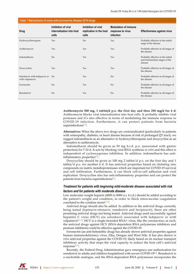
FEASIBLE AT-HOME THERAPIES FOR COVID-19At-home treatment for patients with beginning mild-moderate disease without risk factors, based on Italian experience (not published data)Hydroxychloroquine 400 mg b.i.d. p.o. on first day and 200 mg b.i.d. after for 7 d: Hydroxychloroquine blocks viral internalization into host cells and it is also effective in terms of modulating the immune response to COVID-19 infection[5-9](Table 1).
Scotto Di Vetta M et al. Off-label therapies for COVID-19
WJMA https://www.wjgnet.com 175 June 28, 2020 Volume 8 Issue 3
Table 1 Mechanisms of some anti-coronavirus disease 2019 drugs
DrugInhibition of viral internalization into host cells
Inhibition of viral replication in the host cells
Modulation of immune response to virus infection
Effectiveness against virus
Hydroxychloroquine Yes No Yes Probably effective in the initial stage of the disease
Azithromycin Yes Yes Yes Probably effective in all stages of the disease
Indomethacin No Yes Yes Probably effective in the initial and intermediate stages of the disease
Doxycycline Yes Yes Yes Probably effective in all stages of the disease
Sofosbuvir with ledipasvir or with velpatasvir
No Yes No Probably effective in all stages of the disease
Ivermectin No Yes No Probably effective in all stages of the disease
Remdesivir No Yes No Probably effective in all stages of the disease
Azithromycin 500 mg, 1 tablet/d p.o. the first day and then 250 mg/d for 4 d: Azithromycin blocks viral internalization into host cells. It probably inhibits viral proteases and it’s also effective in terms of modulating the immune response to COVID-19 infection. Furthermore, it can protect patients from bacteria superinfections[7-9].
Alternatives: When the above two drugs are contraindicated (particularly in patients with retinopathy, diabetes, or heart disease because of risk of prolonged QT tract), we suggest indomethacin as an alternative to hydroxychloroquine and doxycycline as an alternative to azithromycin.
Indomethacin should be given as 50 mg b.i.d. p.o. (associated with gastric protection) for 7-10 d. It acts by blocking viral RNA synthesis in vitro and this effect is independent of cyclooxygenase inhibition. In addition, indomethacin has anti-inflammatory properties[10].
Doxycycline should be given as 100 mg 2 tablet/d p.o. on the first day and 1 tablet/d p.o. for another 6 d. It has antiviral properties based on chelating zinc compounds on matrix metalloproteinases which are important for COVID-19 survival and cell infiltration. Furthermore, it can block cell-to-cell adhesion and viral replication. Doxycycline also has anti-inflammatory properties and can protect the patients from bacteria superinfections[11].
Treatment for patients with beginning mild-moderate disease associated with risk factors and for patients with moderate diseaseLow molecular weight heparin (4000 to 8000 u.i. b.i.d.) should be added according to the patient's weight and condition, in order to block intravascular coagulation correlated to the cytokine storm[12,13].
Antiviral drugs should also be added. In addition to the antiviral drugs currently being tested (lopinavir-ritonavir, remdesivir and favipiravir), there are other promising antiviral drugs not being tested. Antiviral drugs used successfully against hepatitis C virus (HCV) are sofosbuvir associated with ledipasvir or with velpatasvir[14-16]. HCV is a single-stranded RNA+ virus, like the coronavirus; as such, the antiviral drugs against HCV (RNA-dependent RNA polymerase inhibitors and protease inhibitors) could be effective against the COVID-19.
Ivermectin (an anti-helminthic drug) has already shown antiviral properties against human immunodeficiency virus, Zika, Dengue and West Nile. It has also shown in vitro antiviral properties against the COVID-19, likely based on its nuclear transport inhibitory activity that stops the viral capacity to reduce the host cell’s antiviral response[7,17].
Recently, the Federal Drug Administration gave emergency-use authorization for remdesivir in adults and children hospitalized with severe COVID-19[18]. Remdesivir is a nucleotide analogue, and the RNA-dependent RNA polymerase incorporates the
Scotto Di Vetta M et al. Off-label therapies for COVID-19
WJMA https://www.wjgnet.com 176 June 28, 2020 Volume 8 Issue 3
active triphosphate form of remdesivir into viral RNA. Incorporation produces termination of viral RNA synthesis and inhibits viral replication[19].
Currently, among the antiviral drugs, the most used off-label is hydro-xychloroquine, particularly in combination with azithromycin. But this combination seems controversial because of its arrhythmogenic risk. However, recently, a paper reported the results of a study in which were used data from the United States Food and Drug Administration Adverse Event Reporting System (on about 13 million total reports) and concluded that hydroxychloroquine use was not associated with safety signals, while azithromycin alone was associated with TdP/QT prolongation events and should be used with caution[20]. To avoid the problem of additive cardiotoxicity of hydroxychloroquine plus azithromycin, we can replace azithromycin with doxycycline in at-risk patients.
It is also very interesting that in some countries, such as Peru, in which hydroxychloroquine and ivermectin are largely in use, the lethality of COVID-19 is among the lowest (2.7%). Perhaps this good result could be due to their different mechanisms of action, which are empowered one with the other.
CONCLUSIONThe take-home message we want give is: while waiting a specific vaccine or EBM indications, upon which old or novel drugs will be certainly useful in the treatment of COVID-19, and on the basis of the large experience acquired in the first 4 mo of this pandemic, we think that a prompt treatment should be started at home in those patients with mild-moderate disease using off-label drugs with a medical rationale. In this way, we could avoid disease progression, hospital overload and, possibly, reduce mortality.
REFERENCES1 Wu Z, McGoogan JM. Characteristics of and Important Lessons From the Coronavirus Disease 2019
(COVID-19) Outbreak in China: Summary of a Report of 72 314 Cases From the Chinese Center for Disease Control and Prevention. JAMA 2020; 323: 1239-1242 [PMID: 32091533 DOI: 10.1001/jama.2020.2648]
2 Porcheddu R, Serra C, Kelvin D, Kelvin N, Rubino S. Similarity in Case Fatality Rates (CFR) of COVID-19/SARS-COV-2 in Italy and China. J Infect Dev Ctries 2020; 14: 125-128 [PMID: 32146445 DOI: 10.3855/jidc.12600]
3 Livingston E, Bucher K. Coronavirus Disease 2019 (COVID-19) in Italy. JAMA 2020; 323: 1335 [PMID: 32181795 DOI: 10.1001/jama.2020.4344]
4 Zhou Y, Han T, Chen J, Hou C, Hua L, He S, Guo Y, Zhang S, Wang Y, Yuan J, Zhao C, Zhang J, Jia Q, Zuo X, Li J, Wang L, Cao Q, Jia E. Clinical and Autoimmune Characteristics of Severe and Critical Cases of COVID-19. Clin Transl Sci 2020 [PMID: 32315487 DOI: 10.1111/cts.12805]
5 Keyaerts E, Vijgen L, Maes P, Neyts J, Van Ranst M. In vitro inhibition of severe acute respiratory syndrome coronavirus by chloroquine. Biochem Biophys Res Commun 2004; 323: 264-268 [PMID: 15351731 DOI: 10.1016/j.bbrc.2004.08.085]
6 Colson P, Rolain JM, Lagier JC, Brouqui P, Raoult D. Chloroquine and hydroxychloroquine as available weapons to fight COVID-19. Int J Antimicrob Agents 2020; 55: 105932 [PMID: 32145363 DOI: 10.1016/j.ijantimicag.2020.105932]
7 Choudhary R, Sharma AK, Choudhary R. Potential use of hydroxychloroquine, ivermectin and azithromycin drugs in fighting COVID-19: trends, scope and relevance. New Microbes New Infect 2020; 35: 100684 [PMID: 32322397 DOI: 10.1016/j.nmni.2020.100684]
8 Jean SS, Lee PI, Hsueh PR. Treatment options for COVID-19: The reality and challenges. J Microbiol Immunol Infect 2020; 53: 436-443 [PMID: 32307245 DOI: 10.1016/j.jmii.2020.03.034]
9 Ohe M, Shida H, Jodo S, Kusunoki Y, Seki M, Furuya K, Goudarzi H. Macrolide treatment for COVID-19: Will this be the way forward? Biosci Trends 2020; 14: 159-160 [PMID: 32249257 DOI: 10.5582/bst.2020.03058]
10 Amici C, Di Caro A, Ciucci A, Chiappa L, Castilletti C, Martella V, Decaro N, Buonavoglia C, Capobianchi MR, Santoro MG. Indomethacin has a potent antiviral activity against SARS coronavirus. Antivir Ther 2006; 11: 1021-1030 [PMID: 17302372]
11 Sodhi M, Etminan M. Therapeutic Potential for Tetracyclines in the Treatment of COVID-19. Pharmacotherapy 2020; 40: 487-488 [PMID: 32267566 DOI: 10.1002/phar.2395]
12 Thachil J. The versatile heparin in COVID-19. J Thromb Haemost 2020; 18: 1020-1022 [PMID: 32239799 DOI: 10.1111/jth.14821]
13 Tang N, Bai H, Chen X, Gong J, Li D, Sun Z. Anticoagulant treatment is associated with decreased mortality in severe coronavirus disease 2019 patients with coagulopathy. J Thromb Haemost 2020; 18: 1094-1099 [PMID: 32220112 DOI: 10.1111/jth.14817]Elfiky AA. Anti-HCV, nucleotide inhibitors, repurposing against COVID-19. Life Sci 2020; 248: 117477 [PMID: 32119961 DOI:
14

Scotto Di Vetta M et al. Off-label therapies for COVID-19
WJMA https://www.wjgnet.com 177 June 28, 2020 Volume 8 Issue 3
10.1016/j.lfs.2020.117477]15 Chen YW, Yiu CB, Wong KY. Prediction of the SARS-CoV-2 (2019-nCoV) 3C-like protease (3CL pro)
structure: virtual screening reveals velpatasvir, ledipasvir, and other drug repurposing candidates. F1000Res 2020; 9: 129 [PMID: 32194944 DOI: 10.12688/f1000research.22457.2]
16 Elfiky AA. Ribavirin, Remdesivir, Sofosbuvir, Galidesivir, and Tenofovir against SARS-CoV-2 RNA dependent RNA polymerase (RdRp): A molecular docking study. Life Sci 2020; 253: 117592 [PMID: 32222463 DOI: 10.1016/j.lfs.2020.117592]
17 Caly L, Druce JD, Catton MG, Jans DA, Wagstaff KM. The FDA-approved drug ivermectin inhibits the replication of SARS-CoV-2 in vitro. Antiviral Res 2020; 178: 104787 [PMID: 32251768 DOI: 10.1016/j.antiviral.2020.104787]
18 U.S. Food and Drug Administration. Coronavirus (COVID-19) Update: FDA Issues Emergency Use Authorization for Potential COVID-19 Treatment - May 1, 2020. Available from: https://www.fda.gov/news-events/press-announcements/coronavirus-covid-19-update-fda-issues-emergency-use-authorization-potential-covid-19-treatment
19 Gordon CJ, Tchesnokov EP, Woolner E, Perry JK, Feng JY, Porter DP, Götte M. Remdesivir is a direct-acting antiviral that inhibits RNA-dependent RNA polymerase from severe acute respiratory syndrome coronavirus 2 with high potency. J Biol Chem 2020; 295: 6785-6797 [PMID: 32284326 DOI: 10.1074/jbc.RA120.013679]
20 Sarayani A, Cicali B, Henriksen CH, Brown JD. Safety signals for QT prolongation or Torsades de Pointes associated with azithromycin with or without chloroquine or hydroxychloroquine. Res Social Adm Pharm 2020 [PMID: 32327397]
WJMA https://www.wjgnet.com 178 June 28, 2020 Volume 8 Issue 3
World Journal of
Meta-AnalysisW J M ASubmit a Manuscript: https://www.f6publishing.com World J Meta-Anal 2020 June 28; 8(3): 178-189
DOI: 10.13105/wjma.v8.i3.178 ISSN 2308-3840 (online)
FIELD OF VISION
Learning and competence development via clinical cases – what elements should be investigated to best train good medical doctors?
Henriette Löffler-Stastka, Guoruey Wong
ORCID number: Henriette Löffler-Stastka 0000-0001-8757-0435.
Author contributions: Löffler-Stastka H conceived the review and wrote the manuscript; Wong G discussed with Löffler-Stastka H international trends and proofread for English style.
Conflict-of-interest statement: No conflict of interest.
Open-Access: This article is an open-access article that was selected by an in-house editor and fully peer-reviewed by external reviewers. It is distributed in accordance with the Creative Commons Attribution NonCommercial (CC BY-NC 4.0) license, which permits others to distribute, remix, adapt, build upon this work non-commercially, and license their derivative works on different terms, provided the original work is properly cited and the use is non-commercial. See: http://creativecommons.org/licenses/by-nc/4.0/
Manuscript source: Unsolicited manuscript
Received: February 28, 2020 Peer-review started: February 28, 2020 First decision: May 31, 2020 Revised: June 15, 2020
Henriette Löffler-Stastka, Department of Psychoanalysis and Psychotherapy, and Postgraduate Unit, Teaching Center, Medical University of Vienna, Vienna 1090, Austria
Guoruey Wong, Faculté de Médecine, Université de Montréal, Montréal H3T 1J4, Quebec, Canada
Corresponding author: Henriette Löffler-Stastka, MD, Associate Professor, Dean, Department of Psychoanalysis and Psychotherapy and Postgraduate Unit, Teaching Center, Medical University of Vienna, Währinger Gürtel 18-20, Vienna 1090, Austria. [email protected]
AbstractIn European higher education, application of information technology, concentration on the learning-processes, consistent implementation, transfer learning, case-based learning, autonomous learning has been extensively studied in the last decade. Educational sciences based on neuroscientific findings use brain-based learning and teaching, including integrated thematic instructions and emotion-theory. Elements essential to this strategy, such as theory and methods for learning, competencies, attitudes, social reality, and a metadiscourse are described herein. Research on learning tends to focus on declarative knowledge, associative learning with conditional stimuli, and procedural knowledge with polythematic/crosslinking thinking. Research on competencies: In research on competencies (e.g., for clinical reasoning, decision-making), intuitive and analytical components are studied. As repeated presentation and exercising of clinical cases is crucial for an efficient learning process, the implementation of interactive scenarios including affectively involving didactics is considered. For competence-development observational methods, questionnaires/item sets or factors have to be targeted and empirically validated. Attitudes and social reality: Clinical decision-making, identification processes and attitudes (“Hidden curriculum”), as well as secondary socialization processes (integration of social norms, values, preparation of role-acquisition, occupational role) are studied via process research, conceptual research, and observational methods. With respect to social reality research, conscious and unconscious bargaining processes have to be taken into account. Methodology: Neuroscience – memory, neuronal, molecular biology, and computer science (Neurocircuits) are integrated into observational process research (e.g., affective-cognitive interface, identification processes) and conceptual research is added and studied on the meta-level, including discussion
Löffler-Stastka H et al. Competence research
WJMA https://www.wjgnet.com 179 June 28, 2020 Volume 8 Issue 3
Accepted: June 20, 2020 Article in press: June 20, 2020 Published online: June 28, 2020
P-Reviewer: Flyckt L S-Editor: Wang JL L-Editor: A E-Editor: Qi LL
of research paradigms. This discussion provides ongoing feedback to projects in a hermeneutic circle.
Key words: Social neuroscience; Case-based learning; Mixed-method design; Hidden curriculum; Socialization; Research
©The Author(s) 2020. Published by Baishideng Publishing Group Inc. All rights reserved.
Core tip: Consequent application of evidence based didactics based on social neuroscientific findings is necessary to develop good medical doctors and therapeutic professionals. An overview of the higher education history and development throughout the past decades is given. An up to date description of the current knowledge regarding higher education and research strategies to enhance the evidence-based components to optimize teaching and learning are proposed.
Citation: Löffler-Stastka H, Wong G. Learning and competence development via clinical cases – what elements should be investigated to best train good medical doctors? World J Meta-Anal 2020; 8(3): 178-189URL: https://www.wjgnet.com/2308-3840/full/v8/i3/178.htmDOI: https://dx.doi.org/10.13105/wjma.v8.i3.178
INTRODUCTIONFor a long time, educational sciences were grounded in neuroscience, whereas “Neurodidactics” were based on computer/mathematical models[1,2]. Constructivist-didactic perspectives called for revision of such archaic positions and research paradigms, stating that controlled intervention cannot be possible[2,3] nor be studied reliably, as the trajectory and outcome of learning processes are not assessable[2,4]. The next great shift in research methodology involved the expansion of the descriptive and interpretative perspectives[2,5], which was suggested in order to adequately investigate gains in acquisition of knowledge. As a result, the utility of Brain-Based Learning and Teaching[6], along with Integrated Thematic Instructions[7] (including Emotion-Theory) were researched in the context of European higher education.
In parallel with discussions on adequate research methodology, the learning context was refined, and applied research in the domain led to several new strategies in European Higher Education. First, the Declaration of Prague (2001)[8] suggested the application of Information- and Communication-Technology. Later, it was recommended that a focus on the learning-processes (Declaration of Leuven 2009)[9] should take place and that research should be implemented consistently (Declaration of Vienna-Budapest 2010)[10]. In the Lancet Report (2010)[11], a lack of research on Transfer-Learning was stated, and so as a result, Case-based learning (CBL) together with workplace-based assessment was implemented. With growing evidence for the applicability and advantages of CBL, several subsequent efforts focused on autonomous learning, leading to the LLL-Strategy (Lifelong Learning). In general, with respect to research methodology, once again the basis of Educational sciences was firmly and prominently grounded in neuroscientific findings[12].
We support the claim that brain-based learning and teaching[6] using integrated thematic instructions[7] has to include Emotion-Theory, and should not exclude the constructivist perspective of taking the learning environment into account[13].
For this strategy to work, we need to concentrate theoretical underpinnings and research in the following areas: Learning, competencies, attitudes, social reality, and inclusion of a Metadiscourse.
THEORETICAL CONSIDERATIONSLearningLearning and competence development is based on research according to the research paradigm of the educational sciences and neurosciences, including social neurosciences.
Löffler-Stastka H et al. Competence research
WJMA https://www.wjgnet.com 180 June 28, 2020 Volume 8 Issue 3
Declarative knowledge: Declarative knowledge has to be acquired concerning basic facts that include knowledge which is available and can be accessed on the conscious level, and in general is stored as unconscious knowledge in long-term memory. Teaching and learning etiological concepts to understand basic principles of specific domains is therefore bound to conscious and unconscious mechanisms.
Associative learning: In this type of learning, the temporal relationship of the two stimuli has to be recognized: The person responds to the first stimulus in anticipation of the second (a neural link/association is its foundation). The result of the reclassification of stimuli and responses are drives (conditional appetence, conditional action, conditional aversion, and/or conditioned inhibition). Associative learning is the fundamental basis of memory.
Procedural knowledge: To attain, train, foster, and confirm procedural knowledge, poly-thematic crosslinking thinking first has to take place: The ability to link information (thoughts, symbols, images, scenes) in a meaningful way and to know how to master this information has to be followed by creatively linking and combining previously seemingly unrelated areas (domains) with each other. The result is to attain the competence e.g., to write a novel instead of reading a novel. The latter is of course a less complex task.
For these three steps, evidence shows that both relational factors and feedback enhance learning outcomes, especially working alliance and motivation. Feedback over the course of the learning process enhances the effect of the teaching intervention. Personalized feedback systems fit into the unique profile of the student and monitor progress in his or her learning priorities. Conducting studies on creating a personalized feedback system adapted to the cultural diversity of the adolescent population in Europe could be an interesting task. Previous research shows that students’ establishment of personal goals and the use of visually attractive mobile interfaces enhance the adherence of students/adolescents to learning processes. Moreover, a bottom-up definition of learning tools increases its acceptability among learners and teachers. Accordingly, a participatory approach, meaning working collaboratively with learners and teachers to co-design the features, is a recent development employed in order to strengthen the learning outcome.
CompetenciesAs attaining competence in a subject is a quite differentiated and complex task, a conceptual and methodological approach has to be developed. Several studies reached the consensus that the concept of personalized reasoning consists of intuitive and analytical components[14-16]. Therefore, the methodological approach to teaching (e.g., personalized reasoning) that can be investigated in research studies on mental processes consists of providing problem descriptions or patterns that can then be stored in “frames,” “scenarios”, “semantic networks/qualifiers”, or in health professions, “illness scripts.” Repeated presentation and training of real-life situations and cases is furthermore crucial for an efficient learning process[16,17]. These considerations led to the implementation of interactive scenarios in many didactic consideration and teaching efforts[16-19]. Advanced statistical techniques that enable us to discover patterns in data and make predictions on natural phenomena and human interactions with their environment may be used. Pattern recognition, consolidation of relevant phenotypes, and development of prediction and classification algorithms for developing decision rules can, for example, be refined.
Research strategies to investigate competence development can either use observational methods, questionnaires, and / or follow the development of item sets that can be condensed and studied further as factors[14] and empirically validated.
AttitudesAttitudes influence competence development. They provide insights into how biographical experience, motivation, and personality traits have changed in response to social changes. Who we are today is in part a result of our collective responses to social and cultural change and may have potentiated a decline or an increase in prosocial traits in students. The challenges of preparing students are better understood when viewed within the broader social context.
An attitude especially in health or pedagogic professions normally contains empathic, precise, ethically sound and scientifically grounded decision making and authentic care. Identification processes play an important role in reaching this ideal attitude (“Hidden curriculum“)[20] as do secondary socialization processes (i.e.
Löffler-Stastka H et al. Competence research
WJMA https://www.wjgnet.com 181 June 28, 2020 Volume 8 Issue 3
integration of social norms, values, training of most important roles, preparation of role-acquisition, and occupational role).
In this context, research might include process research, conceptual research, as well as questionnaire/observational methods with each of their own respective research paradigms.
For research in this context, drawing upon influences from psychotherapy research and social neuroscience is helpful in order to target and understand learning processes: For example, Sharpless et al[21] measured the program performance of universities and teaching institutes in higher education by calculating prediction models for the Examination for Professional Practice (EPPP) at the end of a university program. Pass rates and program performance of universities in the United States and Canada were higher when the total amount of internships and emphasis on practical aspects were high and very prominent in the curriculum. Overall, the EPPP showed numerous advantages and better results in case the higher students had scored in the PreGraduate Record Examination in total, or the higher the percentage of Cognitive Behavioural Training Elements - CBT orientation was. Interestingly, program performance correlated positively with higher percentages of ethnic minority students in the program.
In addition to environmental factors, precise biological factors and structures may be investigated in order to understand learning processes: The encoding of subjective value is directly related to emotional regulation as well as neuro-structurally related to the ventral prefrontal cortex[22], and psychological mindedness is connected with metabolism in the right precuneus[23]. Fear influences decision-making, especially in narcissistic states[24], as links between affective and cognitive functioning may influence the sense of self-agency. These examples of results show the diverse factors that play a role in the complex puzzle of research on learning and training.
Identity, identification, and social realityAttitudes are to a great extent influenced by identification processes that start very early in life. In addition, neuro/psycho-developmental aspects also have to be considered: It is well established that psychoanalysis provides knowledge that helps us understand the development of personality. Researchers have begun to emphasize more and more the role of affect regulation in personality, development, and individualization and identification processes. Affect regulation refers to cognitive and behavioral strategies people use to maximize pleasant emotions and minimize unpleasant ones. These strategies may be explicit (coping mechanisms) or implicit (defenses). It has been proposed that feelings are mechanisms for the selection and retention of behavioral and mental responses. To the extent that particular behaviors, coping strategies, or defensive strategies become associated with regulation of aversive affective states and maximization of pleasurable ones, they will be encoded as "solutions" to affective problems. From this viewpoint, affect regulation strategies are a form of procedural knowledge and are activated under specific circumstances, such as the presence of particular affects. Affect regulation strategies can be adaptive or maladaptive. Some regulation strategies are affect-specific, whereas others can be used to regulate multiple affects of similar valence. These procedures are often activated to resolve discrepancies between perceived and desired states of self, significant others, and external circumstances. Emotions and other sensory feeling states are evolved mechanisms for channeling behavior in directions that foster adaptation. The avoidance of unpleasant states and the pursuit of pleasant ones leads to goal-directed mental and behavioral processes, including defenses and compromise formations. Affects provide a flexible motivational mechanism in humans, as they become associated with representations of perceived, feared, wished-for, or otherwise valued states through the interaction of environmental events and highly specific, naturally selected biological proclivities. The investigation of affect and its regulation also refers to the detection of coping styles. Vollrath et al[25] showed that dispositional coping styles prospectively influence change in personality. Observing affect parameters should not be left alone, as affects are activated under specific circumstances, i.e. in object-relationship. The concept of object relations has played an increasingly important role in psychoanalytic theorizing, as well as in clinical psychoanalysis, psychotherapy and medicine[26]. A short summary of a few pertinent issues will provide a context for describing what is of interest: Ogden[27] traced the contributions of Freud, Abraham, Melanie Klein, Fairbairn, Winnicott, and Bion to the conceptualization of internal object relations. The original model of all internal objects is Freud’s model of the normal development of the superego through the process of identification, as the ego assimilates aspects of the personality and functions of external objects. This newly established psychic agency acquires its own set of
Löffler-Stastka H et al. Competence research
WJMA https://www.wjgnet.com 182 June 28, 2020 Volume 8 Issue 3
motivations and actions, including object relatedness. Ogden[27] also drew on Freud’s extension of the role of splitting of the ego, beyond the formation of the superego, in the development of internal objects. For Ogden[27], another core concept is Fairbairn’s assertion that it is an aspect of the relationship with the object, rather than aspects of the object, that becomes internalized. In addition, Ogden[27] incorporated into his thesis Bion’s description of the potential for the defensive splitting of the mind into active suborganizations capable of engaging in specific forms of object relatedness. Ogden[27]
’s elaboration of these concepts indicates that splitting of the ego into new subdivisions is necessary for early interpersonal relationships to be internalized. Each sub-organization – being a component of the ego – has a dynamic capacity to semi-autonomously generate experience and leave its stamp on the quality of object relations. This psychoanalytically informed view of object relations stipulates that they are the product of intrapsychic sub-organizations of the ego (internal object representations) and not of external interpersonal relationships. However, the quality of object relations is manifested in the interpersonal situation[28]. Despite the enduring quality of object relations, these intrapsychic structures are modifiable by experiences during healthy development. It is suggested that secure attachment is the basis of the acquisition of metacognitive or mentalizing capacity. Horner cited that the concepts of internalization and of object relations are fundamental to the developmental psychology of psychoanalysis, especially in terms of therapeutic technique. The investigation of object relation styles benefits our understanding of problems that people with identity disturbances and/or problems in their mentalizing capacity have. Mentalization and reflective functioning is essential for learning. Primary socialization is the precursor for secondary occupational socialization and therefore plays a distinct role in education[29].
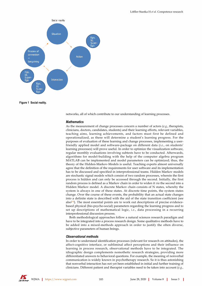
When the influence of secondary socialization on attitudes is targeted, we have to provide research data on integration of social norms and values, training of the most important roles, considerations of the preparation of role-acquisition, and definitions or possibilities for operationalization of the occupational role, which is not correlated with development or education, but more influenced by social reality (Figure 1).
Investigation of social reality can be described as a specific situation experienced by a person that leads to actions and interaction. The person experiences a situation on the basis of his/her ego-id-superego (i.e. with ego-functions, wishes, moral values, etc.). Within the person, as well as within the process of connection between two or more persons, bargaining between each other’s ego, id, and superego structure takes place. Both conscious and unconscious bargaining processes need to be taken into account.
METHODOLOGYTo investigate complex phenomena, several different research paradigms must be taken into account in order to identify as many influencing factors as possible. Which methodologies might be considered?
Neuroscience – memory (place neurons), neuronal, molecular levels, and computer sciences (Neurocircuits) have to be integrated into observational process research (e.g., affective-cognitive interface, identification processes, etc.), and conceptual research has to be added and repeatedly discussed on a meta-level, with inclusion of discussions of research paradigms. These discussions should inform ongoing projects in a hermeneutic circle. In the same way as clinical theories have been developed on the basis of clinical cases, observations, and continuous discussion among experts, the way forward is to ground these theories via empirical research work as scientific theory. Thus, an interdependence of constructivist and empirical research paradigms may be synergistically combined.
Basic sciences relevant to neurosciencesOn a molecular level, as has been investigated by Eric Kandel, the marine snail Aplysia californica was an important animal model for studying the molecular mechanisms of learning because of the very few, but very large nerve cells he uncovered. The objective was to study the gill withdrawal reflex of the animal, as it had been shown that it can be attenuated (habituation), enhanced (sensitization) or durably reinforced (conditioning). The basis of such changes is the interaction of different molecules in the nerve cells and transmitters in the synaptic cleft. These and other similar investigations on a molecular level are conducted in the neurosciences, fostering scientific knowledge with respect to brain- or memory-function (e.g., research on place-cells[30]), resting state- or DMN investigations, and e.g. research concerning the neuronal anxiety
Löffler-Stastka H et al. Competence research
WJMA https://www.wjgnet.com 183 June 28, 2020 Volume 8 Issue 3
Figure 1 Social reality.
networks, all of which contribute to our understanding of learning processes.
MathematicsAs the measurement of change processes concern a number of actors (e.g., therapists, clinicians, doctors, candidates, students) and their learning efforts, relevant variables, teaching aims, learning achievements, and factors must first be defined and operationalized, as these will determine a student’s learning progress. For the purposes of evaluation of these learning and change processes, implementing a user-friendly applied model and software-package on different data (i.e., on students’ learning processes) will prove useful. In order to optimize the visualization software, regular monthly evaluations involving subtests have to be conducted. Afterwards, algorithms for model-building with the help of the computer algebra program MATLAB can be implemented and model parameters can be optimized; thus, the theory of the Hidden-Markov-Models is useful. Teaching experts almost universally agree that the definition of the requirements for user software and its implementation has to be discussed and specified in interprofessional teams. Hidden Markov models are stochastic signal models which consist of two random processes, wherein the first process is hidden and can only be accessed through the second. Initially, the first random process is defined as a Markov chain in order to widen it via the second into a Hidden Markov model. A discrete Markov chain consists of N states, whereby the system is always in one of these states. At discrete time points, the system states change. Over the course of these events, the probability that an actual state changes into a definite state is described with the aid of the state transition coefficient (see also[31]). The most essential points are to work out descriptions of precise evidence-based physical (bio-psycho-social) parameters regarding the learning progress and to set up descriptions of mathematical logic, i.e., data processing in a recurring interprofessional discussion process.
Both methodological approaches follow a natural sciences research paradigm and have to be integrated into a process research design. Some qualitative methods have to be added into a mixed-methods approach in order to justify the often diverse, subjective parameters of human beings.
Observational methodsIn order to understand identification processes (relevant for research on attitudes), the affect-cognitive interface, or subliminal affect perceptions and their influence on learning in process research, observational methods have to be integrated. The idiographic design complements nomothetic research strategies, providing more differentiated answers to behavioral questions. For example, the meaning of nonverbal communication is widely known in psychotherapy research. So it is thus astonishing that nonverbal interaction has not yet been established in initial and further training of clinicians. Different patient and therapist variables need to be taken into account (e.g.,
Löffler-Stastka H et al. Competence research
WJMA https://www.wjgnet.com 184 June 28, 2020 Volume 8 Issue 3
via video analysis) in order to understand a number of complex aspects of the relationship. Within the scope of this research field, investigated micro-process-units are the clinicians’ and patients’ micro-expressions, as measured by the emotion facial action coding system[32]. The ability to recognize facial expressions enables novel applications in human-computer interaction and other areas. Consequently, there has been active research in this field, with several recent works utilizing convolutional neural networks (CNNs) for feature extraction and inference. Being able to recognize facial expressions is key to nonverbal communication in human beings, and the production, perception, and interpretation of facial expressions has been widely studied[33]. Advanced statistical techniques that enable us to discover patterns in data and make predictions about natural phenomena and human-environmental activity can be used. Pattern recognition, condensation of educationally relevant phenotypes, and development of prediction and classification algorithms for development of preventive decision-rules for states or problems like trainer-trainee feedback strategies may be developed.
Qualitative-quantifying research designs shed light into the ingredients relevant for change. Investigations that focused on trainers showed for instance the influence of a person/the therapist on treatment outcome when fruitful learning and change take place. Therapist effects explain up to 20% of the variance in outcomes[34-37]. For research purposes, it is necessary to study the clinician/therapist dynamic in much more detail: We know that attitudes differ, or more precisely, that therapeutic attitudes differ between potential trainees (students)[38], trainees in psychotherapy (propedeutics), and qualified psychotherapists. Attitudes are related, in that therapeutic attitudes relate to interpersonal problems and emotional reactions[39]. Therapeutic attitudes also change over the course of additional training[40]. Such research approaches of qualitative, interview-driven, quantifying designs advance operationalization of different relevant variables and parameters. Recording and transcription is another approach to be mentioned[41].
Another finding was presented via experimental case-series design[14], pointing out two different modes of thinking. Considering factors relevant for clinical reasoning, clinical decision-making, and authentic clinical care, factor analysis approaches showed three relevant factors and aspects: (1) Conscious analytic processing consisting of application of rules, conscious processing, reflecting upon reasons for procedure, meta-analytic information processing, and search for alternative; (2) Positive, holistic intuition consisting of variables like complexity, holistic processing, global rating, and clinician’s emotional arousal; and (3) Automatization. Conscious-analytic processing and automatization varies with the level of training and the years of clinical experience.
In addition to these mixed-methods designs, conceptual considerations may be added.
Conceptual researchTransformative education[42] based on disruption, action, reflection is one concept for learning – a mostly performative one. Taking unconscious/subliminal perception, fast and slow thinking[43] processes, and implicit aspects into account, learning concepts could be widened - either via empirical conceptual research[44] or via a theoretical, more rational epistemic approach.
METADISCOURSEConceptual research can be added and repeatedly discussed on a meta-level, also including discussion of research paradigms. This discussion gives feedback to ongoing projects according to a hermeneutic circle. New theoretical considerations could be implemented and investigated in a timely fashion, so that improvements can be adopted and pursued, and hindering factors can be avoided. Digitization, therefore, opens up new opportunities and avenues.
DesignsThe main concern is how to combine empirical data with constructivist objectives, which can be solved within a combination of both qualitative and quantitative research methods. Nevertheless, when it comes to training of aspects such as authentic care, decision making, training in history taking, or training of other types negotiations, we have to take into account intuitive factors[14], and/or in a broader sense, the social brain and mind. It consists of perceptions, i.e. conscious and
Löffler-Stastka H et al. Competence research
WJMA https://www.wjgnet.com 185 June 28, 2020 Volume 8 Issue 3
subliminal ones, attachment, commitment, refusal, mirror neurons, or mentalization. Findings show that perception, attachment, rejection, and mentalization network are perturbed by direct and indirect social stress. Therefore, the learning and teaching environment should be supported by didactically sound and affect-involving ingredients[45]. One possibility to affectively involve students is the training of communication skills[46] and discussion of cases in a structured and secure atmosphere. When social behavior is disrupted, a dysfunction in social perception, attachment, and rejection may be discerned. Thus, an adequate research design for learning has to take affect research into account. Case-based teaching can be assessed and studied along guidelines for case-studies[47], and simulation and virtual-reality paradigms can be used to contribute to environmental validity.
Affect research: Theory and practical applicationBased on the question of what empathy[26] and especially what affects are, the relevant metapsychological formulations in S. Freud's work are examined within the framework of recent developments in psychoanalysis and neurosciences. The theoretical structure of a psychoanalytic affect theory relates, among other things, to the results of emotion research, endocrinology, and neurophysiology on the one hand, and to cognitive psychological, behavioral, and linguistic studies on the other[48].
Recent research results from the neurosciences indicate that affects and their sequence of actions are clearly anchored in neurophysiology or require clearly, anatomically identified neuronal circuits. The facial expressions linked to affective expressions, gestures, posture, voice, as well as visceral patterns are part of subcortically integrated motor reaction patterns. Facial muscle movements and neuron firing do not reveal anything about the underlying intrapsychic dimension, which is our main interest. Affects are a central determinant of inner and outer reality.
Solms[49] shows that the question of affects forces one to recognize the inner connection between the soul and the somatic and to bring this state of affairs into line with the theoretical designs of psychoanalysis. This leads to the conclusion that affect is a primary sense modality. While sensory modalities such as seeing or hearing represent aspects of the outer, objective world, affect is that primary sense modality in which the inner, subjective world is perceived, which in principle is unconscious. The scientific task is not only to describe and classify the superficial qualities of the inner perception, but also to gain an understanding of the “real facts” that underlie the sensory phenomena of the affect according to the psychoanalytic objective (see also[32]).
Historically, Freud[50] conceptualized affects in the context of his motivation theory, namely the drive theory, and defined the affects in a specific relation to the drives. In Freud's earliest drafts of affect theory, which are primarily to be read as fear-theoretical writings, an event that takes place in external reality triggers an affective reaction that becomes meaningful due to the connection with a certain idea.
In modern affect theory designs, a connection between instinctual and affect events is assumed, but other sources of motivation appear earlier in addition to the instincts. Modern psychoanalytical affect-theoretical designs can be divided into the subject areas "affects and drives", "affects and ego development" and "affects and object relationships". According to Krause et al[51], one has to differentiate between Freud's drive theory and his biological foundation of the drive concept and psychological drive theory. Although it turned out that Freud's attempt to find a biological foundation for the concept of instinct using ideas from the stimulus physiology of the time was insufficient, it should be noted that the derivation of the concept of instinct from biology was considered necessary. In the psychological part of instinct theory, Freud differentiated between instinctual source, instinctual goal, and instinctual object, and thus tried to grasp "instincts" by their goals and not by their causes. Starting from an ethological understanding, many instinctual processes are social processes in which the characteristics of the object have the same power to trigger behavior as internal sources of stimulus. Freud's social component only comes into play through the so-called drive object. Bowlby[52] revised the classical instinct theory, taking into account the approaches of ethological instinct theory in their application to attachment motivation.
Following the discussion of Freudian concepts of affect, affects were considered from the aspect of the instinctual event, the object relationships, as well as in connection with ego functions. In their affect-theoretical discussion, contemporary psychoanalytic theories mostly address the connection between affect and ego functions as well as affects and object relationships - including the object relationship theories of M. Klein and D. Winnicott. Knapp's exemplary compilation of affect-theoretical designs from the fields of psychology, neuropsychology and psychoanalysis offers optimal guidance and understanding[48,53] for the basic building
Löffler-Stastka H et al. Competence research
WJMA https://www.wjgnet.com 186 June 28, 2020 Volume 8 Issue 3
blocks of psychic life and, above all, poses neurophysiological and psychosomatic questions.
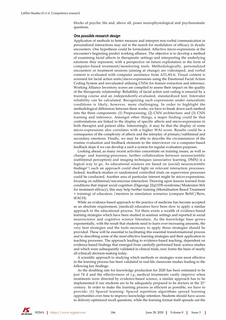
One possible research designApplication of methods to better measure and interpret non-verbal communication in personalized interactions may aid in the search for modulators of efficacy in dyadic encounters. One hypothesis could be formulated: Affective micro-expressions at the encounter's beginning predict working alliance. The objective is to develop a method of examining facial affects in therapeutic settings and interpreting the underlying emotions they represent, with a perspective on future exploitation in the form of computer-based treatment/monitoring tools. Methodologically, personalized encounters or treatment sessions (aiming at change) are videotaped, and verbal content is evaluated with computer assistance from ATLAS.ti. Visual content is screened for facial action units/micro-expressions using the Emotional Facial Action Coding System and reevaluated utilizing CNNs for feature extraction and inference. Working Alliance Inventory scores are compiled to assess their impact on the quality of the therapeutic relationship. Reliability of facial action unit coding is ensured by a training course and an independently-evaluated, standardized test. Interrater reliability can be calculated. Recognizing such expressions under naturalistic conditions is likely, however, more challenging. In order to highlight the methodological differences between these works, we have to break down each method into the three components: (1) Preprocessing; (2) CNN architecture; and (3) CNN training and inference. Amongst other things, a major finding could be that confrontations are linked to the display of specific affects and micro-expressions in both therapist and patient alike. Interestingly, it may be that the display of some micro-expressions also correlates with a higher WAI score. Results could be a consequence of the complexity of affects and the interplay of primary/subliminal and secondary emotions. Finally, we may be able to describe the circumstances of the routine evaluation and feedback elements to the interviewer via a computer-based feedback slope if we can develop a such a system for regular evaluation purposes.
Looking ahead, as many recent activities concentrate on training issues, as well as change- and learning-processes, further collaboration between neuroscientific (subliminal perception) and imaging techniques (associative learning, DMN) is a logical way to go. As educational sciences are based on (social) neuroscientific findings[12], such an approach could shed light on relevant interaction processes. Indeed, feedback-studies or randomized controlled trials on supervision processes could be conducted. Another area of particular interest might be micro-expressions, focusing on subliminal/unconscious interaction. Drawing upon lessons learned from conditions that impair social cognition (Digeorge 22q11DS-syndrome/Moderator-MA for treatment efficacy), this may help further training (Mentalisation Based Treatment + training) of educators /mentors in simulation scenarios (compare BeSiC/Bern, tEACH).
While an evidence-based approach to the practice of medicine has become accepted as an absolute requirement, (medical) educators have been slow to apply a similar approach to the educational process. Yet there exists a wealth of evidence-based learning strategies which have been studied in seminar settings and reported in social neuroscience and cognitive science literature. As the knowledge base grows exponentially, with the result that students need to learn ever-increasing amounts, the very best strategies and the tools necessary to apply those strategies should be provided. These will be essential to facilitating this essential transformational process and to describing some of the most effective learning strategies and their application to teaching processes. The approach leading to evidence-based teaching, dependent on evidence-based findings that emerged from carefully performed basic science studies and which were subsequently validated in clinical trials, now forms the basis of nearly all (clinical) decision-making today.
A scientific approach to studying which methods or strategies were most effective in the learning process has been validated in real-life classroom studies leading to the following key findings.
As the doubling rate for knowledge production for 2020 has been estimated to be just 74 d and the effectiveness of e.g., medical treatments vastly improve when treatments were directed by evidence-based science, a similar approach has to be implemented if our students are to be adequately prepared to be doctors in the 21st century. In order to make the learning process as efficient as possible, we have to provide: (1) Spaced learning: Spaced repetition algorithms spread learning opportunities over time to improve knowledge retention. Students should have access to delivery-optimized recall questions, while the learning format itself spreads out the
Löffler-Stastka H et al. Competence research
WJMA https://www.wjgnet.com 187 June 28, 2020 Volume 8 Issue 3
topics being studied so that there is space between the study of the same medical concepts; (2) Interleaving: Knowledge should be broken down into 'easily digestible' sessions. This enables students to learn more effectively by easily switching between different topics. Doing this helps students learn the similarities and differences between different medical concepts, giving them a holistic and interconnected knowledge base; (3) Didactics videos: Dual coding, which combines verbal and visual representations of the same information, should be utilized as students learn best when they have multiple representations of the same idea. Learning science also shows that shorter length representations (3-9 min) are important for increasing knowledge retention; and (4) Active retrieval: Quiz questions at the end of each learning unit should ensure learner engagement utilizing frequent low-stakes or no-stakes assessments.
Furthermore, readiness assessments should be implemented, as well as briefings and debriefings of active learning formats supported by assignments with detailed drill-down analytics that bring data-driven leaning to the forefront for every instructor. An example was shown at: https://my.ltb.io/www/#/ or https://moodle.meduniwien.ac.at/course/view.php?id=682. Data-driven solutions are the key to improving the educational process.
CONCLUSIONNeuroscience[54] defined the non-conscious and allowed some connections between biology and behavior to be made. Mathematics and computer science have put some effort into simulating the mind[55]. Considering the speed, breadth, and depth of technical/technological advances today, it is necessary to develop specialized domain knowledge, and therefore interdisciplinary working groups need to develop glossaries. The main domains could then be tested and discussed again in a hermeneutic circle to influence and see the impact on the learning process per se. Research into the subjectivity of human beings (students, trainees) may be empirical, qualitative, or inductive, but should ideally be re-evaluated by the subject in an idiographic manner.
To give an example illustrating the current dilemma as well as the need for at least two perspectives, consider that in psychoanalytic literature, countertransference is usually described in a self-report format by the therapist. For example, the most recent psychometrically sound self-report measure, the Countertransference Questionnaire[56,57] asks the therapist to report his or her feeling/reaction towards the patient by indicating whether sentences such as “I feel guilty about my feelings toward him/her.” (Item 24) are true. However, even in retrospect, these reports fall prey to the unavoidable “blind spots” of the therapist. Therefore, psychotherapy process research has searched for other, more rigourous ways to capture countertransference, which requires gauging the ongoing therapeutic process in a multimodal manner, encompassing self-report and observer-rated measurement. The most frequently applied observer-rated instruments are the Countertransference Factors Inventory (CFI[58]) and the Inventory of Countertransference Behavior (ICB[59]). The ICB can be used by supervisors to assess therapists’ positive and negative countertransference behaviour in a specific session, whereas the CFI has a broader focus on therapists’ competencies that may be important to countertransference management. The dilemma of both perspectives, self-report and observer-rated, is that the former is vulnerable to the therapist’s “blind spots”, while the latter is confined to the therapist’s observable verbal behaviour, or, as Hayes has cogently expressed: “therapists’ self-reports are limited by what they are willing and able to reveal, and raters’ observations are restricted to overt displays of countertransference”[60]. Ideally, one could triangulate further via video-based investigations into unconscious/ subliminal perceptions.
Personalized feedback systems promote change, and the more subjective perception and experience is assessed and reconsidered, the better significant change can take place. Differentiated steps may be undertaken to promote motivation, provide more security in disruptive times, and make change possible. Triangulated research designs, domain knowledge, and knowledge on data reduction might be considered in conjunction with idiographic assessment of subjective values, subliminal affect perceptions and attitudes, and values and beliefs.

Löffler-Stastka H et al. Competence research
WJMA https://www.wjgnet.com 188 June 28, 2020 Volume 8 Issue 3
REFERENCES1 Friedrich G. Allgemeine Didaktik und Neurodidaktik. Frankfurt: Lang, 20052 Psillos D, Tselfes V, Kariotoglou P. An epistemological analysis of the evolution of didactical activities in
teaching-learning sequences: the case of fluids. Int J Sci Educ 2004; 26: 555-578 [DOI: 10.1080/09500690310001614744]
3 Hoops W. Konstruktivismus. Ein neues Paradigma für Didaktisches Design? Unterrichtswissenschaft 1998; 26: 229-253
4 Siebert LL. Student and teacher beliefs about language learning. Ortesol J 2003; 21: 75 Becker N. Die neurowissenschaftliche Herausforderung der Pädagogik. Bad Heilbrunn: Julius Klinkhardt,
20066 Caine G, Caine RN. Learning About Accelerated Learning. Train Dev J 1989; 43: 65-737 Kovalik S, Olsen KD. Exceeding expectations: A user's guide to implementing brain research in the
classroom. Books for Educators Incorporated, 20108 Declaration P. Towards the European higher education area. Communiqué of the meeting of European
Ministers in charge of higher education; 2001 May 19; Prague, Czech Republic9 Declaration L, editor. The Bologna Process 2020 - The European Higher Education Area in the new
decade. Communiqué of the Conference of European Ministers Responsible for Higher Education; 2009 Apr 28-29; Leuven and Louvain-la-Neuve, Belgium
10 European Higher Education Area. Budapest-Vienna declaration on the european higher education area. Available from: http://www.ehea.info/media.ehea.info/file/2010_Budapest_Vienna/64/0/Budapest-Vienna_Declaration_598640.pdf
11 Frenk J, Chen L, Bhutta ZA, Cohen J, Crisp N, Evans T, Fineberg H, Garcia P, Ke Y, Kelley P, Kistnasamy B, Meleis A, Naylor D, Pablos-Mendez A, Reddy S, Scrimshaw S, Sepulveda J, Serwadda D, Zurayk H. Health professionals for a new century: transforming education to strengthen health systems in an interdependent world. Lancet 2010; 376: 1923-1958 [PMID: 21112623 DOI: 10.1016/S0140-6736(10)61854-5]
12 Organisation for Economic Co-operation and Development. Understanding the brain: Towards a new learning science. OECD Publishing, 2002
13 Tretter F, Löffler-Stastka H. The Human Ecological Perspective and Biopsychosocial Medicine. Int J Environ Res Public Health 2019; 16: 4230 [PMID: 31683637 DOI: 10.3390/ijerph16214230]
14 Caspar F. What goes on in a psychotherapist's mind? Psychother Res 1997; 7: 105-125 [DOI: 10.1080/10503309712331331913]
15 Croskerry P. A universal model of diagnostic reasoning. Acad Med 2009; 84: 1022-1028 [PMID: 19638766 DOI: 10.1097/ACM.0b013e3181ace703]
16 Kassirer JP. Teaching clinical reasoning: case-based and coached. Acad Med 2010; 85: 1118-1124 [PMID: 20603909 DOI: 10.1097/ACM.0b013e3181d5dd0d]
17 Norman G. Research in clinical reasoning: past history and current trends. Med Educ 2005; 39: 418-427 [PMID: 15813765 DOI: 10.1111/j.1365-2929.2005.02127.x]
18 Turk B, Ertl S, Wong G, Wadowski PP, Löffler-Stastka H. Does case-based blended-learning expedite the transfer of declarative knowledge to procedural knowledge in practice? BMC Med Educ 2019; 19: 447 [PMID: 31796049 DOI: 10.1186/s12909-019-1884-4]
19 Turk BR, Krexner R, Otto F, Wrba T, Löffler-Stastka H. Not the ghost in the machine: transforming patient data into e-learning cases within a case-based blended learning framework for medical education. Procedia Soc Behav Sci 2015; 186: 713-725 [DOI: 10.1016/j.sbspro.2015.04.106]
20 Ludwig B, Turk B, Seitz T, Klaus I, Löffler-Stastka H. The search for attitude-a hidden curriculum assessment from a central European perspective. Wien Klin Wochenschr 2018; 130: 134-140 [PMID: 29356896 DOI: 10.1007/s00508-018-1312-5]
21 Sharpless BA, Barber JP. Predictors of Program Performance on the Examination for Professional Practice in Psychology (EPPP). Prof Psychol Res Pr 2013; 44: 208-217 [DOI: 10.1037/a0031689]
22 Viviani R. Neural correlates of emotion regulation in the ventral prefrontal cortex and the encoding of subjective value and economic utility. Front Psychiatry 2014; 5: 123 [PMID: 25309459 DOI: 10.3389/fpsyt.2014.00123]
23 Roffman JL, Witte JM, Tanner AS, Ghaznavi S, Abernethy RS, Crain LD, Giulino PU, Lable I, Levy RA, Dougherty DD, Evans KC, Fava M. Neural predictors of successful brief psychodynamic psychotherapy for persistent depression. Psychother Psychosom 2014; 83: 364-370 [PMID: 25323387 DOI: 10.1159/000364906]
24 Ronningstam E, Baskin-Sommers AR. Fear and decision-making in narcissistic personality disorder-a link between psychoanalysis and neuroscience. Dialogues Clin Neurosci 2013; 15: 191-201 [PMID: 24174893]
25 Vollrath M, Alnaes R, Torgersen S. Coping styles predict change in personality disorders. J Pers Disord 1995; 9: 371-385 [DOI: 10.1521/pedi.1995.9.4.371]
26 Löffler-Stastka H, Datz F, Parth K, Preusche I, Bukowski X, Seidman C. Empathy in Psychoanalysis and Medical Education - what can we learn from each other? BMC Med Educ 2017; 17: 74 [PMID: 28464865 DOI: 10.1186/s12909-017-0907-2]
27 Ogden TH. The concept of internal object relations. Int J Psychoanal 1983; 64: 227-241 [PMID: 6347926]28 Horner A. Object relations and the developing ego in therapy. Lanham: Jason Aronson 1995; 29 Schmidsberger F, Löffler-Stastka H. Empathy is proprioceptive: the bodily fundament of empathy - a
philosophical contribution to medical education. BMC Med Educ 2018; 18: 69 [PMID: 29622015 DOI: 10.1186/s12909-018-1161-y]
30 Abbott A, Callaway E. Prize for place cells. Nature 2014; 514: 153 [PMID: 25297415 DOI: 10.1038/514153a]
31 Löffler-Stastka H, Ulrich J, Miksch R, Bruckner D. Creating tools for evaluating microprocesses in student's learning progress. Proceedings of IMCIC-ICSIT 2016; 2016 Mar 8-11; Orlando, US. International Institute of Informatics and Systemics, 2016

Löffler-Stastka H et al. Competence research
WJMA https://www.wjgnet.com 189 June 28, 2020 Volume 8 Issue 3
32 Datz F, Wong G, Löffler-Stastka H. Interpretation and Working through Contemptuous Facial Micro-Expressions Benefits the Patient-Therapist Relationship. Int J Environ Res Public Health 2019; 16: 4901 [PMID: 31817282 DOI: 10.3390/ijerph16244901]
33 Sariyanidi E, Gunes H, Cavallaro A. Automatic Analysis of Facial Affect: A Survey of Registration, Representation, and Recognition. IEEE Trans Pattern Anal Mach Intell 2015; 37: 1113-1133 [PMID: 26357337 DOI: 10.1109/TPAMI.2014.2366127]
34 Beutler L, Malik M, Alimohamed S, Harwood T, Talebi H, Noble S, Wong E. Bergin and Garfield's Handbook of psychotherapy and behavior change (5th edition). Lambert MJ, editor. New York: Wiley, 2004: 226-306
35 Lambert MJ. Bergin and Garfield's handbook of psychotherapy and behavior change (6th edition). Hoboken: John Wiley Sons, 2013
36 Luborsky L, McLellan AT, Diguer L, Woody G, Seligman DA. The psychotherapist matters: Comparison of outcomes across twenty-two therapists and seven patient samples. Clin Psycho: Science Pract 1997; 4: 53-65 [DOI: 10.1111/j.1468-2850.1997.tb00099.x]
37 Wampold BE. The great psychotherapy debate: Models, methods, and findings. Mahwah: Lawrence Erlbaum Associates Publishers, 2001: xiii, 263-xii
38 Löffler-Stastka H, Seitz T, Billeth S, Pastner B, Preusche I, Seidman C. Significance of gender in the attitude towards doctor-patient communication in medical students and physicians. Wien Klin Wochenschr 2016; 128: 663-668 [PMID: 27516078 DOI: 10.1007/s00508-016-1054-1]
39 Pastner B, Alexopoulos J, Rohm C, Preusche I, Loeffler-Stastka H. Development of therapeutic attitudes: Attitudes of trainees in training. Eur J Educ Sci 2014; 1: 110-123 [DOI: 10.19044/ejes.v1no1a12]
40 Pastner B, Schechtner C, Billeth S, Preusche I, Alexopoulos J, Loffler-Stastka H. Development of Therapeutic Attitudes: Teaching and Learning in Psychotherapy. Procedia Soc Behav Sci 2014; 116: 1170-1175 [DOI: 10.1016/j.sbspro.2014.01.364]
41 Löffler-Stastka H, Sell C, Zimmermann J, Huber D, Klug G. Is countertransference a valid source of clinical information? Investigating emotional responses to audiotaped psychotherapy sessions. Bull Menninger Clin 2019; 83: 353-375 [PMID: 31180236 DOI: 10.1521/bumc_2019_83_02]
42 Mezirow J. Perspective Transformation. Adult Educ 1978; 28: 100-110 [DOI: 10.1177/074171367802800202]
43 Kahneman D. Thinking, fast and slow. London: Macmillan, 201144 Leuzinger-Bohleber M, Fischmann T; Research Subcommittee for conceptual research of the International
psychoanalytical association. What is conceptual research in psychoanalysis? Int J Psychoanal 2006; 87: 1355-1386 [PMID: 16997730 DOI: 10.1516/73MU-E53N-D1EE-1Q8L]
45 Löffler-Stastka H. Do our medical students even want e-learning? A user rated evaluation of case based e-learning in undergraduate medical education at the medical university of Vienna. Adv Soc Sci Res J 2015; 2 [DOI: 10.14738/assrj.24.1003]
46 Himmelbauer M, Seitz T, Seidman C, Löffler-Stastka H. Standardized patients in psychiatry - the best way to learn clinical skills? BMC Med Educ 2018; 18: 72 [PMID: 29625572 DOI: 10.1186/s12909-018-1184-4]
47 Neidhart E, Löffler-Stastka H. Case Studies in Psychotherapy Training. Can the case serve as a source for scientific findings? Psychotherapie Forum 2020; 24: 3-8 [DOI: 10.1007/s00729-020-00137-2]
48 Knapp PH. Book Essay: Some Contemporary Contributions to The Study of Emotions. J Am Psychoanal Assoc 1987; 35: 205-248 [DOI: 10.1177/000306518703500112]
49 Solms M. [What are affects?]. Psyche (Stuttg) 1996; 50: 485-522 [PMID: 8767233]50 Freud S. Hemmung, Symptom und Angst - Studienausgabe. Frankfurtam: Fischer Verlag, 199751 Krause R, Haynal R. Allgemeine psychoanalytische Krankheitslehre. Psyche (Stuttg) 1997; 51: 703-70452 Bowlby J. Bindung - Eine Analyse der Mutter-Kind-Beziehung. München: Kindler, 1975: 22453 Knapp PH. Core processes in the organization of emotions. J Am Acad Psychoanal 1981; 9: 415-434
[PMID: 7263349 DOI: 10.1521/jaap.1.1981.9.3.415]54 Singer P. Ethics and intuitions. J Ethics 2005; 9: 331-352 [DOI: 10.1007/s10892-005-3508-y]55 Dietrich D, Fodor G, Zucker G, Bruckner D. Simulating the mind: A technical neuropsychoanalytical
approach: Springer, 2010 [DOI: 10.1007/978-3-211-09451-8]56 Betan E, Heim AK, Zittel Conklin C, Westen D. Countertransference phenomena and personality pathology
in clinical practice: an empirical investigation. Am J Psychiatry 2005; 162: 890-898 [PMID: 15863790 DOI: 10.1176/appi.ajp.162.5.890]
57 Zittel-Conklin C, Westen D. Countertransference Questionnaire (CTQ) manual. Atlanta: Emory University, 2005
58 Van Wagoner SL, Gelso CJ, Hayes JA, Diemer RA. Countertransference and the reputedly excellent therapist. Psychotherapy 1991; 28: 411 [DOI: 10.1037/0033-3204.28.3.411]
59 Friedman SM, Gelso CJ. The development of the inventory of countertransference behavior. J Clin Psychol 2000; 56: 1221-1235 [PMID: 10987693 DOI: 10.1002/1097-4679(200009)56:9<1221::AID-JCLP8>3.0.CO;2-W]
60 Hayes JA. The inner world of the psychotherapist: A program of research on countertransference. Psychother Res 2004; 14: 21-36 [PMID: 22011115 DOI: 10.1093/ptr/kph002]

WJMA https://www.wjgnet.com 190 June 28, 2020 Volume 8 Issue 3
World Journal of
Meta-AnalysisW J M ASubmit a Manuscript: https://www.f6publishing.com World J Meta-Anal 2020 June 28; 8(3): 190-209
DOI: 10.13105/wjma.v8.i3.190 ISSN 2308-3840 (online)
REVIEW
Immunotherapy in hepatocellular carcinoma: Combination strategies
Alexander Claudius Jordan, Jennifer Wu
ORCID number: Alexander Claudius Jordan 0000-0002-3006-7033; Jennifer Wu 0000-0002-1714-0021.
Author contributions: Jordan AC wrote and edited the manuscript; Wu J reviewed and edited the manuscript.
Conflict-of-interest statement: Authors declare no conflict of interests for this article.
Open-Access: This article is an open-access article that was selected by an in-house editor and fully peer-reviewed by external reviewers. It is distributed in accordance with the Creative Commons Attribution NonCommercial (CC BY-NC 4.0) license, which permits others to distribute, remix, adapt, build upon this work non-commercially, and license their derivative works on different terms, provided the original work is properly cited and the use is non-commercial. See: http://creativecommons.org/licenses/by-nc/4.0/
Manuscript source: Invited manuscript
Received: February 1, 2020 Peer-review started: February 1, 2020 First decision: February 24, 2020 Revised: May 1, 2020 Accepted: June 25, 2020 Article in press: June 25, 2020
Alexander Claudius Jordan, Department of Internal Medicine, New York University School of Medicine, New York, NY 10016, United States
Jennifer Wu, Division of Hematology and Oncology, Perlmutter Cancer Center, New York University Langone Medical Center, New York, NY 10016, United States
Corresponding author: Jennifer Wu, MD, Associate Professor, Division of Hematology and Oncology, Perlmutter Cancer Center, New York University Langone Medical Center, 550 1st Avenue, New York, NY 10016, United States. [email protected]
AbstractLiver cancer is one of the most common causes of cancer death globally, and its incidence in the United States is increasing. Patients with advanced hepatocellular carcinoma (HCC) who are not candidates for surgical resection, liver transplant, or locoregional therapies can be treated with systemic therapies. Multiple agents, including sorafenib, lenvatinib, and regorafenib are approved for use as either first- or second-line therapy in this patient population, but all have relatively modest survival benefits. HCC is potentially susceptible to therapy with checkpoint inhibitors, including agents such as nivolumab and pembrolizumab, which are both approved by the Food and Drug Administration for patients previously treated with sorafenib but have not demonstrated superior overall survival in phase III trials. It is clear that more effective approaches are needed to potentiate the effects of checkpoint inhibitors in patients with HCC. This review will outline and appraise the current literature on the use of checkpoint inhibitors in HCC as part of a combination treatment involving an additional mode of therapy. The list of agents that can be paired with checkpoint inhibitors includes an additional checkpoint inhibitor, vascular endothelial growth factor or vascular endothelial growth factor receptor inhibitors, tyrosine kinase inhibitors, OX-40 agonists, and PT-112 inhibitors. The main non-pharmacologic therapies currently being studied for inclusion in a combination strategy include radiation therapy, trans-arterial chemoembolization, and ablation.
Key words: Hepatocellular carcinoma; Liver neoplasms; Antineoplastic agents; Immunological; Protein kinase inhibitors; Angiogenesis inhibitors
©The Author(s) 2020. Published by Baishideng Publishing Group Inc. All rights reserved.
Core tip: Immunotherapy remains a viable option for the systemic treatment of patients
Jordan AC et al. Immunotherapy in hepatocellular carcinoma
WJMA https://www.wjgnet.com 191 June 28, 2020 Volume 8 Issue 3
Published online: June 28, 2020
P-Reviewer: Eskens F, Niu ZS, Wang GY S-Editor: Zhang L L-Editor: A E-Editor: Wang LL
with unresectable hepatocellular carcinoma, although nivolumab and pembrolizumab failed to meet their endpoints in phase III studies. Combining immunotherapy with other treatment modalities may improve treatment responses. Multiple clinical trials are evaluating the safety and efficacy of these new combination strategies, which involve pairing PD-1 or PD-L1 inhibitors with CTLA-4 inhibitors, or pairing checkpoint inhibitors with alternative agents or non-pharmacologic therapies.
Citation: Jordan AC, Wu J. Immunotherapy in hepatocellular carcinoma: Combination strategies. World J Meta-Anal 2020; 8(3): 190-209URL: https://www.wjgnet.com/2308-3840/full/v8/i3/190.htmDOI: https://dx.doi.org/10.13105/wjma.v8.i3.190
INTRODUCTIONWorldwide, liver cancer is the fourth leading cause of cancer death and the seventh most common cancer in terms of incidence[1]. The most common liver cancer subtype is hepatocellular carcinoma[1]. Over the last 40 years, hepatocellular carcinoma (HCC) incidence has risen by approximately three-fold in the United States[2]. Between 2000 and 2009, the incidence rose by approximately 4.5% per year and 0.7% per year from 2010 to 2012[3]. Between the years 2018 and 2040, global liver cancer incidence is expected to rise by approximately 62%, while the number of liver cancer deaths worldwide will rise by 64%[2]. If HCC is detected at an early stage (Barcelona Clinic Liver Cancer stage 0 or A), surgical resection or ablation can be performed in select groups of patients[4]. Approximately 70% of patients will develop evidence of recurrence following resection[4,5]. If patients are not candidates for surgical resection, liver transplantation is offered to patients who meet the Milan criteria and provides a possibility of cure[4,5]. Patients who are not eligible for surgery or liver transplantation are candidates for locoregional therapies, including trans-arterial chemoembolization (TACE) and ablation, if they have early- or intermediate-stage disease (Barcelona Clinic Liver Cancer stage 0-B), or systemic therapies if they have advanced disease (Barcelona Clinic Liver Cancer stage C)[4]. Multiple options for systemic therapy exist (Table 1). The tyrosine kinase inhibitors sorafenib and lenvatinib are approved for use as first-line therapy[6]. Ramucirumab, a monoclonal antibody directed against vascular endothelial growth factor (VEGF) receptor 2, and the tyrosine kinase inhibitors regorafenib and cabozantinib are approved for patients previously treated with sorafenib, however, candidates for ramucirumab therapy must also have an alpha-fetoprotein (AFP) level of 400 ng/mL or greater[6,7]. With the exception of lenvatinib, which produced an objective response rate (ORR) of 24.1%, the rest of the approved systemic therapies could only achieve an ORR in the range of 2 to 11%[8-12]. Sorafenib, ramucirumab, regorafenib and cabozantinib were directly compared to placebo and increased overall survival by only 1.2 to 2.8 months[8,10-12].
In hopes of discovering therapies that could produce greater responses, investigators began to utilize checkpoint inhibitors in HCC patients, given their success in other malignancies and the contribution of the PD-1/PD-L1 and CTLA4 pathways to creating an immunosuppressive tumor microenvironment[13-15](Table 2). Nivolumab demonstrated an ORR of 15% and 20% in the dose-escalation and dose-expansion phases of the CheckMate 040 trial, respectively, while pembrolizumab produced an ORR of 17% in the KEYNOTE-224 trial[13,14]. Both agents were then Food and Drug Administration (FDA)-approved for use in patients who had previously received sorafenib[16,17]. However, neither nivolumab or pembrolizumab demonstrated a statistically significant improvement in overall survival (OS) in patients with unresectable HCC when compared to sorafenib or placebo in phase III trials, respectively[18,19]. Other checkpoint inhibitors studied in HCC patients in completed phase II trials include the PD-1 inhibitors camrelizumab, the PD-L1 inhibitor durvalumab, and the CTLA4 inhibitor tremelimumab[20-22]. In a phase II multicenter study (NCT02989922) involving 217 patients from Chinese medical centers with HCC who had failed or could not tolerate prior systemic therapy who were treated with camrelizumab, the ORR was 13.8%, with a six-month OS rate of 74.7%[20]. Durvalumab demonstrated an ORR of 10.3% with a median OS of 13.2 months in a multi-center phase I/II study in a cohort of 40 patients with HCC, most of whom had received prior sorafenib[21]. In a phase II study of tremelimumab in 21 patients from Spanish medical
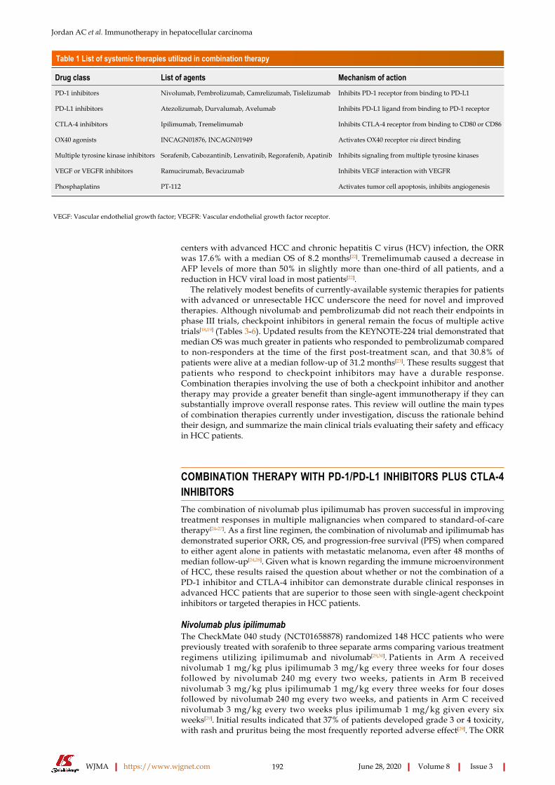
Jordan AC et al. Immunotherapy in hepatocellular carcinoma
WJMA https://www.wjgnet.com 192 June 28, 2020 Volume 8 Issue 3
Table 1 List of systemic therapies utilized in combination therapy
Drug class List of agents Mechanism of action
PD-1 inhibitors Nivolumab, Pembrolizumab, Camrelizumab, Tislelizumab Inhibits PD-1 receptor from binding to PD-L1
PD-L1 inhibitors Atezolizumab, Durvalumab, Avelumab Inhibits PD-L1 ligand from binding to PD-1 receptor
CTLA-4 inhibitors Ipilimumab, Tremelimumab Inhibits CTLA-4 receptor from binding to CD80 or CD86
OX40 agonists INCAGN01876, INCAGN01949 Activates OX40 receptor via direct binding
Multiple tyrosine kinase inhibitors Sorafenib, Cabozantinib, Lenvatinib, Regorafenib, Apatinib Inhibits signaling from multiple tyrosine kinases
VEGF or VEGFR inhibitors Ramucirumab, Bevacizumab Inhibits VEGF interaction with VEGFR
Phosphaplatins PT-112 Activates tumor cell apoptosis, inhibits angiogenesis
VEGF: Vascular endothelial growth factor; VEGFR: Vascular endothelial growth factor receptor.
centers with advanced HCC and chronic hepatitis C virus (HCV) infection, the ORR was 17.6% with a median OS of 8.2 months[22]. Tremelimumab caused a decrease in AFP levels of more than 50% in slightly more than one-third of all patients, and a reduction in HCV viral load in most patients[22].
The relatively modest benefits of currently-available systemic therapies for patients with advanced or unresectable HCC underscore the need for novel and improved therapies. Although nivolumab and pembrolizumab did not reach their endpoints in phase III trials, checkpoint inhibitors in general remain the focus of multiple active trials[18,19] (Tables 3-6). Updated results from the KEYNOTE-224 trial demonstrated that median OS was much greater in patients who responded to pembrolizumab compared to non-responders at the time of the first post-treatment scan, and that 30.8% of patients were alive at a median follow-up of 31.2 months[23]. These results suggest that patients who respond to checkpoint inhibitors may have a durable response. Combination therapies involving the use of both a checkpoint inhibitor and another therapy may provide a greater benefit than single-agent immunotherapy if they can substantially improve overall response rates. This review will outline the main types of combination therapies currently under investigation, discuss the rationale behind their design, and summarize the main clinical trials evaluating their safety and efficacy in HCC patients.
COMBINATION THERAPY WITH PD-1/PD-L1 INHIBITORS PLUS CTLA-4 INHIBITORSThe combination of nivolumab plus ipilimumab has proven successful in improving treatment responses in multiple malignancies when compared to standard-of-care therapy[24-27]. As a first line regimen, the combination of nivolumab and ipilimumab has demonstrated superior ORR, OS, and progression-free survival (PFS) when compared to either agent alone in patients with metastatic melanoma, even after 48 months of median follow-up[24,28]. Given what is known regarding the immune microenvironment of HCC, these results raised the question about whether or not the combination of a PD-1 inhibitor and CTLA-4 inhibitor can demonstrate durable clinical responses in advanced HCC patients that are superior to those seen with single-agent checkpoint inhibitors or targeted therapies in HCC patients.
Nivolumab plus ipilimumabThe CheckMate 040 study (NCT01658878) randomized 148 HCC patients who were previously treated with sorafenib to three separate arms comparing various treatment regimens utilizing ipilimumab and nivolumab[29,30]. Patients in Arm A received nivolumab 1 mg/kg plus ipilimumab 3 mg/kg every three weeks for four doses followed by nivolumab 240 mg every two weeks, patients in Arm B received nivolumab 3 mg/kg plus ipilimumab 1 mg/kg every three weeks for four doses followed by nivolumab 240 mg every two weeks, and patients in Arm C received nivolumab 3 mg/kg every two weeks plus ipilimumab 1 mg/kg given every six weeks[29]. Initial results indicated that 37% of patients developed grade 3 or 4 toxicity, with rash and pruritus being the most frequently reported adverse effect[29]. The ORR

Jordan AC et al. Immunotherapy in hepatocellular carcinoma
WJMA https://www.wjgnet.com 193 June 28, 2020 Volume 8 Issue 3
Table 2 Reported results of successful clinical trials evaluating immunotherapy in hepatocellular carcinoma patients
Therapy Comparator Therapy indication Phase Approximate data
collection period Patients enrolled Primary endpoints Trial identifier
Nivolumab None Not specified I/II November 26, 2012-August 8, 2016
214 (dose-escalation), 48 (dose-expansion)
ORR (dose-escalation phase - 15%), (dose-expansion phase – 20%) NCT01658878 (CheckMate 040)
Pembrolizumab None Second-line II June 22, 2016- February 20, 2017
104 ORR – 18.3%, median OS 13.2 mo NCT02702414 (KEYNOTE-224)
Camrelizumab Camrelizumab (regimen 1) vs camrelizumab (regimen 2)
Second-line II November 15, 2016– November 16, 2017
220 ORR 13.8%, median OS not reached NCT02989922
Durvalumab None Not specified I/II August 29, 2012-October 24, 2016
40 ORR – 10.3%, median OS 13.2 mo NCT01693562
Tremelimumab None Not specified II December 2008-May 2012 21 ORR - 17.6%, DCR 76.4% NCT01008358
Pembrolizumab Placebo Second-line III May 31, 2016-November 23, 2017
413 Median OS 13.9 vs 10.6 months – HR: 0.781; 95%CI: 0.611 to 0.998; P = 0.0238, PFS 3.0 vs 2.8 months – HR: 0.718; 95%CI, 0.570 to 0.904; P = 0.0022 (pembrolizumab vs placebo)
NCT02702401
Nivolumab plus ipilimumab
Nivolumab plus ipilimumab (regimen 1 vs regimen 2 vs regimen 3)
Second-line I/II Minimum follow-up-28 months at time of data cutoff
148 ORR – (32 - Arm A, 31- Arm B, 31 - Arm C), median DOR (17.5 - Arm A, 22.2 - Arm B, 16.6 - Arm C )
NCT01658878 (CheckMate 040)
Durvalumab plus tremelimumab
None Second-line I/II October 19, 2015-January 10, 2017
40 ORR – 15% NCT02519348
Atezolizumab plus bevacizumab
Sorafenib First-line III March 15, 2018-November 2019
501 OS HR: 0.58 (95%CI: 0.42- 0.79; P = 0.0006), PFS 6.8 vs 4.3 mo, P < 0.0001 (atezolizumab plus bevacizumab vs sorafenib)
NCT03434379(IMbrave 150)
ORR: Overall response rate; OS: Overall survival; DCR: Disease control rate; PFS: Progression-free survival; DOR: Duration of response; OS HR: Overall survival hazard rate.
was 31%, and after 24 months of follow-up, the OS rate was 40%[29]. This study was updated at the International Liver Cancer Association Conference in Sep 2019[31]. All 3 arms achieved a similar ORR (Arm A - 32%, Arm B - 31% and Arm C - 31%), while Arm A achieved the longest median OS (22.8 months vs 12.5 months for Arm B and 12.7 months for Arm C) and the highest OS rate at 30 months (44%)[31](Table 2). The study remains active[30].
The rather innovative study design allowed investigators to compare the impact of various doses of nivolumab and ipilimumab on treatment response[29-31]. The dosing schedule in Arms A and B was similar, however, the dose of ipilimumab was three times higher in Arm A, and patients in Arm C received ipilimumab less frequently than the other two arms[29]. The higher dose of ipilimumab received by patients in Arm A compared to patients in Arms B or C may have been responsible for the improved median overall survival[29]. Unsurprisingly, Arm A also had the highest number of treatment-related adverse effects, possibly due to the larger doses of ipilimumab the
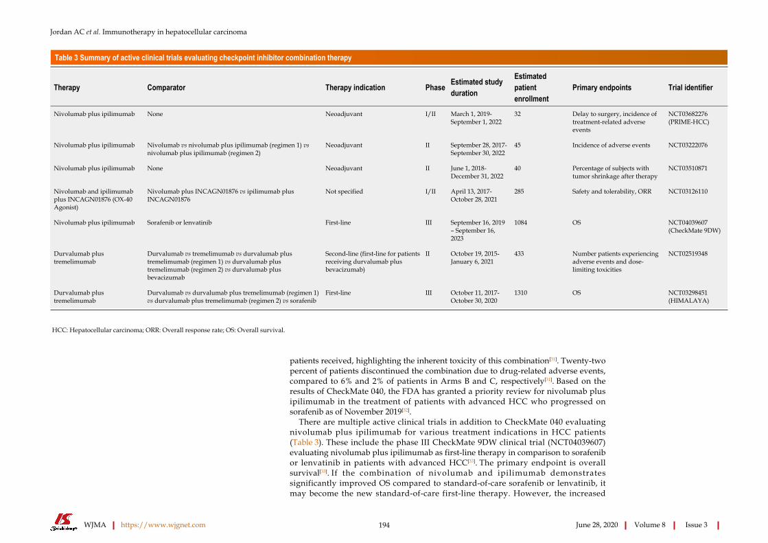
Jordan AC et al. Immunotherapy in hepatocellular carcinoma
WJMA https://www.wjgnet.com 194 June 28, 2020 Volume 8 Issue 3
Table 3 Summary of active clinical trials evaluating checkpoint inhibitor combination therapy
Therapy Comparator Therapy indication Phase Estimated study duration
Estimated patient enrollment
Primary endpoints Trial identifier
Nivolumab plus ipilimumab None Neoadjuvant I/II March 1, 2019-September 1, 2022
32 Delay to surgery, incidence of treatment-related adverse events
NCT03682276 (PRIME-HCC)
Nivolumab plus ipilimumab Nivolumab vs nivolumab plus ipilimumab (regimen 1) vs nivolumab plus ipilimumab (regimen 2)
Neoadjuvant II September 28, 2017-September 30, 2022
45 Incidence of adverse events NCT03222076
Nivolumab plus ipilimumab None Neoadjuvant II June 1, 2018-December 31, 2022
40 Percentage of subjects with tumor shrinkage after therapy
NCT03510871
Nivolumab and ipilimumab plus INCAGN01876 (OX-40 Agonist)
Nivolumab plus INCAGN01876 vs ipilimumab plus INCAGN01876
Not specified I/II April 13, 2017-October 28, 2021
285 Safety and tolerability, ORR NCT03126110
Nivolumab plus ipilimumab Sorafenib or lenvatinib First-line III September 16, 2019 – September 16, 2023
1084 OS NCT04039607 (CheckMate 9DW)
Durvalumab plus tremelimumab
Durvalumab vs tremelimumab vs durvalumab plus tremelimumab (regimen 1) vs durvalumab plus tremelimumab (regimen 2) vs durvalumab plus bevacizumab
Second-line (first-line for patients receiving durvalumab plus bevacizumab)
II October 19, 2015-January 6, 2021
433 Number patients experiencing adverse events and dose-limiting toxicities
NCT02519348
Durvalumab plus tremelimumab
Durvalumab vs durvalumab plus tremelimumab (regimen 1) vs durvalumab plus tremelimumab (regimen 2) vs sorafenib
First-line III October 11, 2017-October 30, 2020
1310 OS NCT03298451 (HIMALAYA)
HCC: Hepatocellular carcinoma; ORR: Overall response rate; OS: Overall survival.
patients received, highlighting the inherent toxicity of this combination[31]. Twenty-two percent of patients discontinued the combination due to drug-related adverse events, compared to 6% and 2% of patients in Arms B and C, respectively[31]. Based on the results of CheckMate 040, the FDA has granted a priority review for nivolumab plus ipilimumab in the treatment of patients with advanced HCC who progressed on sorafenib as of November 2019[32].
There are multiple active clinical trials in addition to CheckMate 040 evaluating nivolumab plus ipilimumab for various treatment indications in HCC patients (Table 3). These include the phase III CheckMate 9DW clinical trial (NCT04039607) evaluating nivolumab plus ipilimumab as first-line therapy in comparison to sorafenib or lenvatinib in patients with advanced HCC[33]. The primary endpoint is overall survival[33]. If the combination of nivolumab and ipilimumab demonstrates significantly improved OS compared to standard-of-care sorafenib or lenvatinib, it may become the new standard-of-care first-line therapy. However, the increased
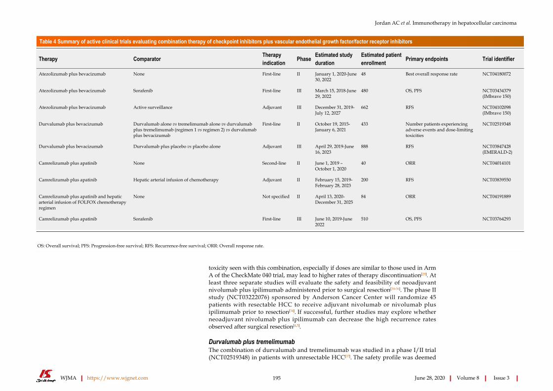
Jordan AC et al. Immunotherapy in hepatocellular carcinoma
WJMA https://www.wjgnet.com 195 June 28, 2020 Volume 8 Issue 3
Table 4 Summary of active clinical trials evaluating combination therapy of checkpoint inhibitors plus vascular endothelial growth factor/factor receptor inhibitors
Therapy Comparator Therapy indication Phase Estimated study
durationEstimated patient enrollment Primary endpoints Trial identifier
Atezolizumab plus bevacizumab None First-line II January 1, 2020-June 30, 2022
48 Best overall response rate NCT04180072
Atezolizumab plus bevacizumab Sorafenib First-line III March 15, 2018-June 29, 2022
480 OS, PFS NCT03434379 (IMbrave 150)
Atezolizumab plus bevacizumab Active surveillance Adjuvant III December 31, 2019-July 12, 2027
662 RFS NCT04102098 (IMbrave 150)
Durvalumab plus bevacizumab Durvalumab alone vs tremelimumab alone vs durvalumab plus tremelimumab (regimen 1 vs regimen 2) vs durvalumab plus bevacizumab
First-line II October 19, 2015-January 6, 2021
433 Number patients experiencing adverse events and dose-limiting toxicities
NCT02519348
Durvalumab plus bevacizumab Durvalumab plus placebo vs placebo alone Adjuvant III April 29, 2019-June 16, 2023
888 RFS NCT03847428 (EMERALD-2)
Camrelizumab plus apatinib None Second-line II June 1, 2019 – October 1, 2020
40 ORR NCT04014101
Camrelizumab plus apatinib Hepatic arterial infusion of chemotherapy Adjuvant II February 15, 2019-February 28, 2023
200 RFS NCT03839550
Camrelizumab plus apatinib and hepatic arterial infusion of FOLFOX chemotherapy regimen
None Not specified II April 13, 2020-December 31, 2025
84 ORR NCT04191889
Camrelizumab plus apatinib Sorafenib First-line III June 10, 2019-June 2022
510 OS, PFS NCT03764293
OS: Overall survival; PFS: Progression-free survival; RFS: Recurrence-free survival; ORR: Overall response rate.
toxicity seen with this combination, especially if doses are similar to those used in Arm A of the CheckMate 040 trial, may lead to higher rates of therapy discontinuation[29]. At least three separate studies will evaluate the safety and feasibility of neoadjuvant nivolumab plus ipilimumab administered prior to surgical resection[34-36]. The phase II study (NCT03222076) sponsored by Anderson Cancer Center will randomize 45 patients with resectable HCC to receive adjuvant nivolumab or nivolumab plus ipilimumab prior to resection[34]. If successful, further studies may explore whether neoadjuvant nivolumab plus ipilimumab can decrease the high recurrence rates observed after surgical resection[4,5].
Durvalumab plus tremelimumabThe combination of durvalumab and tremelimumab was studied in a phase I/II trial (NCT02519348) in patients with unresectable HCC[37]. The safety profile was deemed
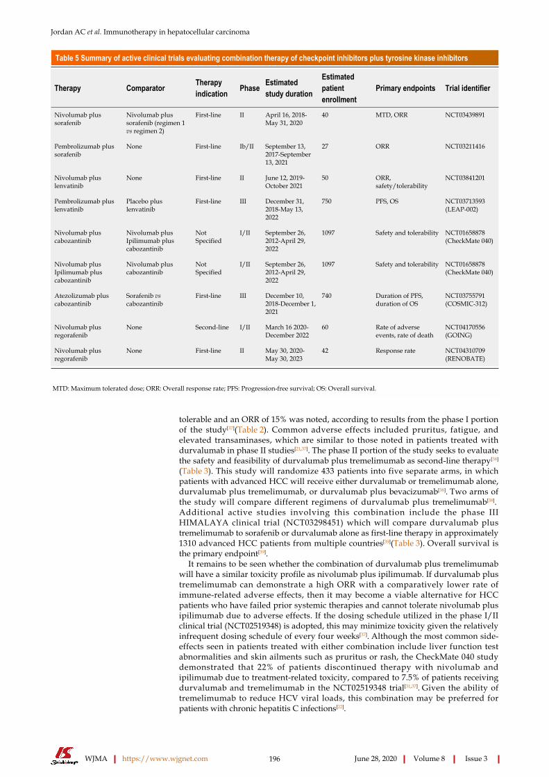
Jordan AC et al. Immunotherapy in hepatocellular carcinoma
WJMA https://www.wjgnet.com 196 June 28, 2020 Volume 8 Issue 3
Table 5 Summary of active clinical trials evaluating combination therapy of checkpoint inhibitors plus tyrosine kinase inhibitors
Therapy Comparator Therapy indication Phase Estimated
study duration
Estimated patient enrollment
Primary endpoints Trial identifier
Nivolumab plus sorafenib
Nivolumab plus sorafenib (regimen 1 vs regimen 2)
First-line II April 16, 2018-May 31, 2020
40 MTD, ORR NCT03439891
Pembrolizumab plus sorafenib
None First-line Ib/II September 13, 2017-September 13, 2021
27 ORR NCT03211416
Nivolumab plus lenvatinib
None First-line II June 12, 2019-October 2021
50 ORR, safety/tolerability
NCT03841201
Pembrolizumab plus lenvatinib
Placebo plus lenvatinib
First-line III December 31, 2018-May 13, 2022
750 PFS, OS NCT03713593 (LEAP-002)
Nivolumab plus cabozantinib
Nivolumab plus Ipilimumab plus cabozantinib
Not Specified
I/II September 26, 2012-April 29, 2022
1097 Safety and tolerability NCT01658878 (CheckMate 040)
Nivolumab plus Ipilimumab plus cabozantinib
Nivolumab plus cabozantinib
Not Specified
I/II September 26, 2012-April 29, 2022
1097 Safety and tolerability NCT01658878 (CheckMate 040)
Atezolizumab plus cabozantinib
Sorafenib vs cabozantinib
First-line III December 10, 2018-December 1, 2021
740 Duration of PFS, duration of OS
NCT03755791 (COSMIC-312)
Nivolumab plus regorafenib
None Second-line I/II March 16 2020-December 2022
60 Rate of adverse events, rate of death
NCT04170556 (GOING)
Nivolumab plus regorafenib
None First-line II May 30, 2020-May 30, 2023
42 Response rate NCT04310709 (RENOBATE)
MTD: Maximum tolerated dose; ORR: Overall response rate; PFS: Progression-free survival; OS: Overall survival.
tolerable and an ORR of 15% was noted, according to results from the phase I portion of the study[37](Table 2). Common adverse effects included pruritus, fatigue, and elevated transaminases, which are similar to those noted in patients treated with durvalumab in phase II studies[21,37]. The phase II portion of the study seeks to evaluate the safety and feasibility of durvalumab plus tremelimumab as second-line therapy[38]
(Table 3). This study will randomize 433 patients into five separate arms, in which patients with advanced HCC will receive either durvalumab or tremelimumab alone, durvalumab plus tremelimumab, or durvalumab plus bevacizumab[38]. Two arms of the study will compare different regimens of durvalumab plus tremelimumab[38]. Additional active studies involving this combination include the phase III HIMALAYA clinical trial (NCT03298451) which will compare durvalumab plus tremelimumab to sorafenib or durvalumab alone as first-line therapy in approximately 1310 advanced HCC patients from multiple countries[39](Table 3). Overall survival is the primary endpoint[39].
It remains to be seen whether the combination of durvalumab plus tremelimumab will have a similar toxicity profile as nivolumab plus ipilimumab. If durvalumab plus tremelimumab can demonstrate a high ORR with a comparatively lower rate of immune-related adverse effects, then it may become a viable alternative for HCC patients who have failed prior systemic therapies and cannot tolerate nivolumab plus ipilimumab due to adverse effects. If the dosing schedule utilized in the phase I/II clinical trial (NCT02519348) is adopted, this may minimize toxicity given the relatively infrequent dosing schedule of every four weeks[37]. Although the most common side-effects seen in patients treated with either combination include liver function test abnormalities and skin ailments such as pruritus or rash, the CheckMate 040 study demonstrated that 22% of patients discontinued therapy with nivolumab and ipilimumab due to treatment-related toxicity, compared to 7.5% of patients receiving durvalumab and tremelimumab in the NCT02519348 trial[31,37]. Given the ability of tremelimumab to reduce HCV viral loads, this combination may be preferred for patients with chronic hepatitis C infections[22].
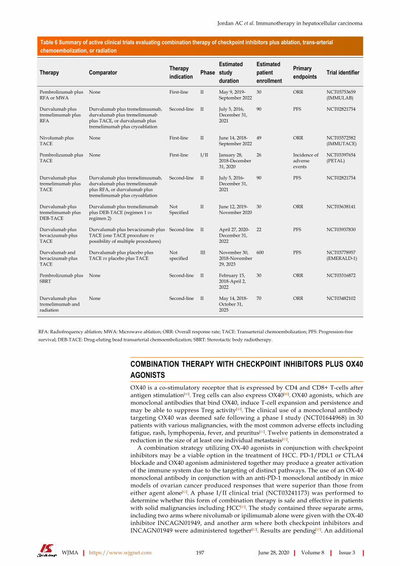
Jordan AC et al. Immunotherapy in hepatocellular carcinoma
WJMA https://www.wjgnet.com 197 June 28, 2020 Volume 8 Issue 3
Table 6 Summary of active clinical trials evaluating combination therapy of checkpoint inhibitors plus ablation, trans-arterial chemoembolization, or radiation
Therapy Comparator Therapy indication Phase
Estimated study duration
Estimated patient enrollment
Primary endpoints Trial identifier
Pembrolizumab plus RFA or MWA
None First-line II May 9, 2019-September 2022
30 ORR NCT03753659 (IMMULAB)
Durvalumab plus tremelimumab plus RFA
Durvalumab plus tremelimuumab, durvalumab plus tremelimumab plus TACE, or durvalumab plus tremelimumab plus cryoablation
Second-line II July 5, 2016, December 31, 2021
90 PFS NCT02821754
Nivolumab plus TACE
None First-line II June 14, 2018-September 2022
49 ORR NCT03572582 (IMMUTACE)
Pembrolizumab plus TACE
None First-line I/II January 28, 2018-December 31, 2020
26 Incidence of adverse events
NCT03397654 (PETAL)
Durvalumab plus tremelimumab plus TACE
Durvalumab plus tremelimuumab, durvalumab plus tremelimumab plus RFA, or durvalumab plus tremelimumab plus cryoablation
Second-line II July 5, 2016-December 31, 2021
90 PFS NCT02821754
Durvalumab plus tremelimumab plus DEB-TACE
Durvalumab plus tremelimumab plus DEB-TACE (regimen 1 vs regimen 2)
Not Specified
II June 12, 2019-November 2020
30 ORR NCT03638141
Durvalumab plus bevacizumab plus TACE
Durvalumab plus bevacizumab plus TACE (one TACE procedure vs possibility of multiple procedures)
Second-line II April 27, 2020-December 31, 2022
22 PFS NCT03937830
Durvalumab and bevacizumab plus TACE
Durvalumab plus placebo plus TACE vs placebo plus TACE
Not specified
III November 30, 2018-November 29, 2023
600 PFS NCT03778957 (EMERALD-1)
Pembrolizumab plus SBRT
None Second-line II February 15, 2018-April 2, 2022
30 ORR NCT03316872
Durvalumab plus tremelimumab and radiation
None Second-line II May 14, 2018-October 31, 2025
70 ORR NCT03482102
RFA: Radiofrequency ablation; MWA: Microwave ablation; ORR: Overall response rate; TACE: Transarterial chemoembolization; PFS: Progression-free survival; DEB-TACE: Drug-eluting bead transarterial chemoembolization; SBRT: Stereotactic body radiotherapy.
COMBINATION THERAPY WITH CHECKPOINT INHIBITORS PLUS OX40 AGONISTSOX40 is a co-stimulatory receptor that is expressed by CD4 and CD8+ T-cells after antigen stimulation[40]. Treg cells can also express OX40[40]. OX40 agonists, which are monoclonal antibodies that bind OX40, induce T-cell expansion and persistence and may be able to suppress Treg activity[40]. The clinical use of a monoclonal antibody targeting OX40 was deemed safe following a phase I study (NCT01644968) in 30 patients with various malignancies, with the most common adverse effects including fatigue, rash, lymphopenia, fever, and pruritus[41]. Twelve patients in demonstrated a reduction in the size of at least one individual metastasis[41].
A combination strategy utilizing OX-40 agonists in conjunction with checkpoint inhibitors may be a viable option in the treatment of HCC. PD-1/PDL1 or CTLA4 blockade and OX40 agonism administered together may produce a greater activation of the immune system due to the targeting of distinct pathways. The use of an OX-40 monoclonal antibody in conjunction with an anti-PD-1 monoclonal antibody in mice models of ovarian cancer produced responses that were superior than those from either agent alone[42]. A phase I/II clinical trial (NCT03241173) was performed to determine whether this form of combination therapy is safe and effective in patients with solid malignancies including HCC[43]. The study contained three separate arms, including two arms where nivolumab or ipilimumab alone were given with the OX-40 inhibitor INCAGN01949, and another arm where both checkpoint inhibitors and INCAGN01949 were administered together[43]. Results are pending[43]. An additional
Jordan AC et al. Immunotherapy in hepatocellular carcinoma
WJMA https://www.wjgnet.com 198 June 28, 2020 Volume 8 Issue 3
phase I/II trial sponsored by Incyte Biosciences (NCT03126110) is active and is similarly designed to the first trial, but is employing the OX40 agonist INCAGN01876 in patients with solid malignancies, including HCC[43,44](Table 3).
COMBINATION THERAPY WITH CHECKPOINT INHIBITORS PLUS VASCULAR ENDOTHELIAL GROWTH FACTOR OR VASCULAR ENDOTHELIAL GROWTH FACTOR RECEPTOR INHIBITORSAnti-angiogenic agents have been a focus of research in HCC due to the relatively high vascularity of HCC tumors, however, studies suggest they may also have beneficial effects on the immune system[45-47]. VEGF expression can modulate the immune system via various mechanisms, leading to immunosuppression[46,47]. VEGF molecules can inhibit leukocyte adherence to the endothelium, inhibit the development of dendritic cells, and promote Treg proliferation[46,47]. The combination of checkpoint inhibitors and VEGF or vascular endothelial growth factor receptor (VEGFR) inhibitors may cause a greater net activation of the immune system than checkpoint inhibitors alone due to the added effect of VEGF inhibition.
Atezolizumab plus bevacizumabBevacizumab, a VEGF inhibitor, is currently approved for the treatment of multiple malignancies, including metastatic colorectal cancer, non-squamous non-small cell lung cancer, and ovarian cancer[48]. Bevacizumab was studied in a phase II trial in 46 patients with unresectable HCC and treatment resulted in a median PFS of 6.9 months and an OS rate of 53% at 1 year, 28% at 2 years, and 23% at 3 years[49]. However, 11% of patients developed clinically significant bleeding, including one patient who suffered a variceal bleed that was ultimately fatal[49]. Another phase II study evaluating single-agent bevacizumab in advanced HCC patients found that 9% of patients developed gastrointestinal bleeding[45]. Atezolizumab is a humanized monoclonal antibody that targets PD-L1 and prevents its binding to the PD-1 receptor and the B7.1 molecule[50]. It has been approved either as a single agent or in combination with chemotherapy for the treatment of patients with non-small cell lung cancer, small-cell lung cancer, urothelial carcinoma, and breast cancer[51]. The combination of atezolizumab plus bevacizumab demonstrated prolonged progression-free survival in metastatic renal cell carcinoma patients in a phase III trial with an acceptable safety profile[52].
Initial data testing this combination in HCC patients originates from a phase Ib study (NCT02645531) evaluating this combination as first-line therapy in 26 patients with advanced HCC[53]. Approximately 35% of patients developed grade 3-4 toxicities with hypertension being the most frequently reported adverse event, with an ORR of 62%[53]. In late 2019, the study authors reported that patients who received the combination therapy in Arm F demonstrated significantly better median progression-free survival (5.6 vs 3.4 months, P = 0.0108) when compared to atezolizumab alone[54]. The most common side effects seen in the patients randomized to combination therapy included proteinuria, fatigue and rash[55]. Patients in Arm A had a median overall survival of 17.1 months[55]. Initial phase III data has been reported from the IMBRAVE 150 trial that randomized approximately 501 systemic treatment-naïve patients with unresectable HCC to receive atezolizumab plus bevacizumab or sorafenib alone[56]. Preliminary data published in November 2019 demonstrated an improved PFS (6.8 vs 4.3) and OS hazard ratio (0.58) with the combination vs sorafenib alone[56](Table 2). Recent quality-of-life data from IMbrave150 presented in January 2020 revealed that patients taking atezolizumab plus bevacizumab had delayed time to deterioration of quality-of-life[57]. Patients in the combination arm reported greater time to deterioration of physical functioning, diarrhea, loss of appetite, fatigue, and pain[57](Table 4).
These data suggest that atezolizumab plus bevacizumab may become an alternative regimen for patients with advanced HCC due to its relatively acceptable toxicity profile when compared to a dual checkpoint inhibitor regimen, and improved efficacy when compared to sorafenib alone. The study remains active[58].
Other active clinical trials evaluating this combination include the NCT04102098 phase III trial, a part of the IMbrave 150 study, which will randomize 662 patients with resectable HCC and a high risk of recurrence to receive atezolizumab plus bevacizumab or surveillance as adjuvant therapy[59](Table 4). Given the significant improvement in PFS seen when this combination is used as first-line therapy, it may also be successful as adjuvant therapy and reduce the high recurrence rates often seen post-resection[4-5,56,59]. A single-arm phase II trial (NCT04180072) will enroll 48 patients
Jordan AC et al. Immunotherapy in hepatocellular carcinoma
WJMA https://www.wjgnet.com 199 June 28, 2020 Volume 8 Issue 3
with advanced HCC and chronic HBV infection, allowing the investigators to determine whether HBV infection has any significant effect on the safety and effectiveness of atezolizumab plus bevacizumab[60](Table 4).
Durvalumab plus bevacizumabThe results of the studies testing atezolizumab and bevacizumab may be generalizable to other checkpoint inhibitors, such as durvalumab, if used in combination with bevacizumab or other VEGF inhibitors given similar mechanisms of action. The combination of durvalumab plus bevacizumab is being studied in multiple different trials, including the aforementioned phase II study (NCT02519348) in patients with advanced HCC as first-line therapy[38](Table 3). The phase III EMERALD-2 trial (NCT03847428) will compare durvalumab plus bevacizumab to either durvalumab alone or placebo as adjuvant therapy after either ablation or resection in HCC patients with a high risk of recurrence[61](Table 4).
Durvalumab plus ramucirumabA phase I trial (NCT02572687) is evaluating the safety of the combination of ramucirumab and durvalumab in patients with advanced gastrointestinal or thoracic malignancies including hepatocellular carcinoma[62]. Although no reported phase II or III trials are currently active, if this combination proves to be safe with a tolerable side-effect profile, further study is warranted based on the results of the REACH-2 trial to determine if this combination is most effective in patients with an AFP greater than 400[10].
Camrelizumab plus apatinibApatinib is a tyrosine kinase inhibitor of VEGFR2 that binds to its target with ten-fold more affinity than sorafenib, and is currently approved in China for use in advanced gastric cancer patients[63,64]. Apatinib has demonstrated a tolerable safety profile with evidence of anti-tumor activity in HCC patients as single-agent therapy[64]. In a murine model of lung cancer, the combination of apatinib and an anti-PD-L1 monoclonal antibody inhibited tumor growth in a synergistic fashion with a notable increase in tumor-infiltrating lymphocytes[65]. The combination of apatinib and camrelizumab was studied in a phase Ia and Ib trial (NCT02942329) that enrolled 43 Chinese patients with various gastrointestinal malignancies, including gastric cancer, esophagogastric junction cancer, and advanced hepatocellular carcinoma[63]. This combination was thought to have a tolerable safety profile, with the most common side-effects being hypertension and an elevated AST[63]. Half of all HCC patients demonstrated a partial response, a response similar in magnitude to results from the initial Phase 1b trial (NCT02715531) testing first-line atezolizumab and bevacizumab[53,63]. Multiple studies are evaluating this combination in distinct HCC patient populations[66-69](Table 4). A phase II study (NCT04014101) seeks to determine whether camrelizumab plus apatinib is safe and effective in patients with advanced HCC as second-line therapy[66]. Another phase II study (NCT03839550) will determine whether this combination is superior to hepatic arterial infusion of chemotherapy in the adjuvant setting, with recurrence-free survival as the primary endpoint[67]. The phase II TRIPLET study (NCT04191889) will evaluate the safety and efficacy of the combination of FOLFOX chemotherapy infused directly into an artery perfusing the tumors, followed by camrelizumab and apatinib in 84 patients with advanced HCC[68]. A phase III study (NCT03764293) evaluating camrelizumab and apatinib as first-line therapy in advanced HCC patients will report both OS and PFS as primary endpoints[69]. The aforementioned studies are primarily being carried out in Chinese medical centers, which will limit the external validity of the results and require additional studies before their findings can be generalized to other patient populations[66-69].
COMBINATION THERAPY WITH CHECKPOINT INHIBITORS PLUS MULTI-TARGETED TYROSINE KINASE INHIBITORSSorafenib, lenvatinib, cabozantinib, and regorafenib all have activity against VEGF receptors and may mitigate the immunosuppressive activity of VEGF[6]. Although the exact mechanisms of action are unclear, TKIs can modulate the immune system[70-72]. Sorafenib decreased the populations of Tregs and CD8+ T cells that expressed PD-1 in the tumors of mice models of HCC[70]. An in-vitro study demonstrated that sorafenib could increase effector-T cell activation and inhibit Treg suppression of effector-T cells,
Jordan AC et al. Immunotherapy in hepatocellular carcinoma
WJMA https://www.wjgnet.com 200 June 28, 2020 Volume 8 Issue 3
albeit at sub-pharmacologic doses[71]. Sorafenib has also been shown to restore the ability of dendritic cells to activate T cells in vitro[72]. In mouse models of HCC, lenvatinib demonstrated greater antitumor activity in immunocompetent mice when compared to sorafenib but not in immunodeficient mice, suggesting that the increase in activity may be related to immunomodulatory effects[73].
Given that the mechanism of action of TKIs differs from that of checkpoint inhibitors, pairing a TKI with a checkpoint inhibitor may produce responses that are either additive or synergistic. Additionally, responses to combination therapy with checkpoint inhibitors and TKIs may be more effective than responses to VEGF or VEGFR inhibitors alone because TKIs inhibit multiple distinct signaling pathways. If these combination therapies are proven effective, their safety may be of concern. Common toxicities observed with the various TKIs include diarrhea, skin rashes, fatigue, nausea, elevated aspartate aminotransferase levels, and rash[8,9,11,12]. The side-effect profiles of checkpoint inhibitors partially overlap with those of the TKIs, and it is unclear whether this may lead to greater toxicity when compared to single-agent regimens[13,14,24,31].
Sorafenib plus checkpoint inhibitorsThe safety and effectiveness of sorafenib plus pembrolizumab as first-line therapy will be evaluated in 27 patients with advanced or metastatic HCC participating in a phase Ib/II study (NCT03211416) by the Roswell Park Cancer Institute[74](Table 5). One phase II trial (NCT03439891) will study the combination of nivolumab plus sorafenib as first-line therapy in patients with advanced HCC[75](Table 5). ORR is one of the primary endpoints for both studies[74,75].
Cabozantinib plus checkpoint inhibitorsStudies testing treatment regimens involving cabozantinib plus immunotherapy include the aforementioned CheckMate 040 study (NCT01658878), which contains two separate arms in which patients are receiving nivolumab plus cabozantinib and nivolumab and ipilimumab plus cabozantinib[30](Table 2). Initial data from this portion of the CheckMate 040 study has been reported[76]. Seventy-one patients with advanced HCC who were sorafenib-naïve or who had previously been treated with sorafenib were randomized to receive nivolumab plus cabozantinib or nivolumab plus ipilimumab and cabozantinib[76]. The ORR was 26% vs 17% for those who received all three drugs or nivolumab plus cabozantinib, respectively[76](Table 2). Median PFS was 6.8 for the three-drug regimen arm and 5.5 for the two-drug regimen arm[76](Table 2). The three-drug regimen caused significantly more toxicity, with 71% of patients in that arm reporting grade 3-4 adverse effects, vs 42% of patients in the two-drug arm[76]. Approximately 20% of patients in the three-drug arm discontinued the drug secondary to toxicity, compared with 3% of the patients in the two-drug arm[76]. Although the three-drug regimen demonstrates promise based on these early results, its high rate of toxicity may prohibit widespread adoption as standard-of-care therapy. Other checkpoint inhibitors, such as atezolizumab, are being studied as part of combination regimens involving cabozantinib[76](Table 5). The phase III COSMIC-312 trial (NCT03755791) will study the combination of cabozantinib and atezolizumab vs sorafenib as first-line therapy in a multi-national group of patients with advanced HCC[77](Table 5).
Lenvatinib plus checkpoint inhibitorsA phase 1b trial (NCT03006926) in which 18 patients were given a combination of lenvatinib and pembrolizumab demonstrated an acceptable safety profile, with hypertension and poor appetite reported as the most common adverse effects[78]. One phase II study (NCT03841201) will test the combination of lenvatinib and nivolumab in a cohort of patients from a German medical center with ORR and safety and tolerability measures as the primary endpoints[79](Table 5). A phase III study (NCT03713593) sponsored by Merck will test the combination of lenvatinib plus pembrolizumab in an international cohort of approximately 750 patients with advanced HCC with PFS and OS as the primary endpoints[80]. Patients will be randomized to receive either lenvatinib plus pembrolizumab or lenvatinib plus placebo[80](Table 5).
Regorafenib plus checkpoint inhibitorsMultiple studies evaluating regorafenib in conjunction with PD-1 inhibitors are active. They include a phase Ib trial sponsored by Bayer[81] that will elucidate the safety profile of regorafenib plus pembrolizumab. Recent results were presented at the American
Jordan AC et al. Immunotherapy in hepatocellular carcinoma
WJMA https://www.wjgnet.com 201 June 28, 2020 Volume 8 Issue 3
Society of Clinical Oncology Gastrointestinal Cancers Symposium in January 2020[82]. Thirty-five patients had been treated, with 29 in the dose-defining cohort and 6 in the dose-expansion cohort[82]. Fifteen patients had discontinued treatment, with either clinical or radiologic disease progression as the most common reason[82]. Eighty-nine percent of patients experienced grade 3 or 4 treatment-emergent adverse events[82]. The multi-center phase II (GOING) trial (NCT04170556) and the single-center phase II (RENOBATE) trial from South Korea will evaluate the combination of regorafenib and nivolumab as second-line and first-line therapy, respectively[83,84](Table 5).
COMBINATION THERAPY WITH CHECKPOINT INHIBITORS PLUS LOCOREGIONAL THERAPIESLocoregional therapies can activate the immune system through various mechanisms[85-87].
Treatment with either radiofrequency ablation (RFA) or TACE can stimulate the expansion of T-cells recognizing tumor-associated antigens, while RFA can lead to natural killer cell activation[85-87]. The ability of locoregional therapies to stimulate the immune system makes them potential candidates for an effective combination strategy with checkpoint inhibitors[85-87]. The side-effect profiles of immunotherapy and these locoregional therapies differ significantly, which may lead to relatively low toxicity compared to other combination regimens previously discussed[14,15,31,88,89].
Checkpoint inhibitors plus ablationPatients who will likely be eligible for combination therapies involving ablation will have tumors 3 cm or less with early-stage, BCLC class 0 or A disease who are not candidates for surgical resection[4,5]. Although multiple types of ablation exist, RFA is the usual standard of care and adverse events may include intraperitoneal bleeding, intrahepatic abscess, grounding pad burns, bile duct injury, thermal damage to organs in close proximity to the tumor, pneumothorax, pain, and tumor seeding of tissues[4,88]. RFA is not optimal for tumors that are larger than 5 cm, numerous with 3 or more lesions, poorly visible via ultrasound, or adjacent to structures such as the biliary tree, bowel, and vital organs such as the heart[4,5,90].
Multiple studies evaluating combinations of radiofrequency ablation and immunotherapy are ongoing[91-93](Table 6). Patients with unresectable HCC underwent treatment with tremelimumab and radiofrequency ablation or chemoablation in a phase I study in which 32 patients were enrolled[91]. A partial response was noted in 5 out of 19 patients, and 12 out of 14 patients with confirmed HCV infection were noted to have a decrease in their viral load, consistent with prior studies involving tremelimumab[22,91]. The phase II clinical trial (NCT02821754) sponsored by the National Cancer Institute (NCI) includes an arm where patients with advanced HCC will receive durvalumab plus tremelimumab and RFA, and another where patients will receive cryoablation in conjunction with the checkpoint inhibitors as second-line therapy[92]. Progression-free survival is the primary outcome[92]. Studies involving pembrolizumab include the IMMULAB phase II study (NCT03753659), which will test the combination of RFA or microwave ablation plus pembrolizumab in 30 patients who had not received prior systemic therapy and report ORR as the primary outcome[93].
Checkpoint inhibitors plus TACETACE plus immunotherapy may be appropriate for patients with intermediate-stage disease, including those with BCLC class B disease based on current indications for TACE[4]. Some patients who are ineligible for RFA, such as those with larger tumors, multiple lesions, or smaller tumors which cannot be safely ablated may be candidates for TACE[4]. Potential adverse events of TACE therapy include damage to the hepatic artery, bile duct injury, acute liver failure, variceal bleeding, and cholecystitis[89,94]. Specific protocols for performing TACE may differ, and the types of chemotherapy, embolic agents, and schedule of TACE sessions may vary between institutions, among other factors[89]. This variation in how TACE is performed may lead to variation in side-effect profiles, and could affect the efficacy of combination therapies when compared across medical centers[89].
Multiple studies testing the safety of treatment combinations involving checkpoint inhibitors and TACE are active[95-99](Table 6). The phase II, single-arm IMMUTACE study from Germany (NCT03572582) will study the effectiveness and safety of
Jordan AC et al. Immunotherapy in hepatocellular carcinoma
WJMA https://www.wjgnet.com 202 June 28, 2020 Volume 8 Issue 3
nivolumab plus TACE for patients with intermediate-stage HCC who have not received prior systemic therapy or TACE, with ORR as the primary endpoint[95]. The combination of TACE and pembrolizumab will be studied as first-line therapy in the phase I/II, single-arm PETAL trial (NCT03397654), which will report the incidence of adverse effects as the primary outcome measure[96]. A phase II clinical trial (NCT03638141) will evaluate the combination of durvalumab plus tremelimumab and drug-eluting bead TACE (deb-TACE) in approximately 90 patients with advanced HCC[97]. Patients with active HCV infection will be excluded, which may hinder the ability to detect any effects of the combination therapy on HCV viral load possibly due to tremelimumab[22,97]. The previously mentioned NCT02821754 phase II trial sponsored by the NCI contains an arm in which patients will receive durvalumab plus tremelimumab and TACE[92]. The combination of durvalumab plus bevacizumab and TACE will be evaluated as second-line therapy in a phase II trial (NCT03937830) sponsored by the NCI in 22 patients with advanced HCC[98]. The combination of durvalumab, bevacizumab and TACE will be compared to TACE alone or TACE plus durvalumab in the phase III, multi-center EMERALD-1 clinical trial (NCT03778957) which will report PFS as a primary outcome measure[99]. Secondary outcome measures include overall survival[99].
COMBINATION THERAPY WITH CHECKPOINT INHIBITORS PLUS RADIATIONThe effect of checkpoint inhibitors on T-cells can be amplified by radiation[100]. Radiation therapy (RT) has been shown to induce antitumor immune responses through the formation of antitumor antibodies[100]. RT directed at one tumor site can induce responses in other tumor sites not directly targeted, a phenomenon known as the abscopal effect, which is related to immune system activation[100,102,103]. In mouse models of melanoma, radiation therapy can increase both antigen presentation to T-cells recognizing tumor antigens and infiltration of tumors by those T-cells[101]. RT can increase the expression of MHC-1 (major histocompatibility complex-1) molecules in tumors and increase T-cell and natural killer cell tumor infiltration[102,103]. Stereotactic body radiotherapy (SBRT) can be used to treat early-stage, BCLC A disease that is not amenable to treatment with RFA, and can achieve local control rates of approximately 80% or greater[90]. SBRT is a possible alternative treatment option after TACE has been unsuccessful, with local control rates of approximately 87%-99% in multiple studies[90]. Adverse events as a result of SBRT may include upper gastrointestinal bleeding from esophageal varices or gastric and duodenal ulcers, and biliary strictures[104]. SBRT generally has a more tolerable side-effect profile when compared to TACE, with rates of grade 3 or higher toxicity estimated at 6% to 27% in patients with unresectable HCC who receive SBRT[90,104]. If proven safe, SBRT plus immunotherapy could become a reasonable treatment option for HCC patients who are not candidates for RFA or who have failed TACE.
Multiple studies testing the combination of ionizing radiation and immunotherapy are ongoing in patients with unresectable HCC[105-107](Table 6). One phase I study (NCT03203304) will enroll 50 participants with advanced HCC who will first receive SBRT[105]. Patients will be randomized into two arms, one of which will receive nivolumab alone after SBRT, while the second will receive nivolumab and ipilimumab[105]. If the results from this study suggest that administering nivolumab plus ipilimumab after SBRT is safe, better responses may be seen with that combination than with nivolumab alone given after SBRT due to the synergistic effects of nivolumab plus ipilimumab[24,28,105]. A phase II study (NCT03482102) sponsored by Massachusetts General Hospital will evaluate the safety and efficacy of durvalumab and tremelimumab administered in combination with radiation as second-line therapy[106]. As with prior studies examining durvalumab plus tremelimumab, a central question of the NCT03482102 study is whether this combination will be less toxic than those utilizing nivolumab plus ipilimumab in addition to SBRT, and whether tremelimumab will lower the viral load in patients infected with HCV[22,31,106]. Another phase II study will report the overall response rate after enrolling approximately 30 patients, and treating them with pembrolizumab and SBRT in the second-line setting[107]. Both radiation dosing and the time intervals between radiation treatment and immunotherapy treatment may affect the efficacy of combination therapies, and should be considered when the results of these various studies are available[108].
Jordan AC et al. Immunotherapy in hepatocellular carcinoma
WJMA https://www.wjgnet.com 203 June 28, 2020 Volume 8 Issue 3
FUTURE DIRECTIONS
PD-1 and PT-112 inhibitorsPT-112 is a platinum-based drug of the phosphaplatin class currently under development by Phosphaplatin Therapeutics with reported anti-tumor effects both in-vivo and in-vitro[109-111]. It promotes apoptosis in tumor cells and may have anti-angiogenic effects[110]. An in-vitro study demonstrated that PT-112 treatment led to increased phosphorylation of a wide variety of targets, including VGFR1, EGFR, and CDC2, suggesting it may have activity against multiple distinct pathways[111]. Tumor cells may not be able to evade the drug’s antitumor effects through traditional drug resistance pathways because it binds to transmembrane proteins rather than DNA[110,112]. An in-vitro study in ovarian cancer cells demonstrated that cisplatin enters cells more readily than phosphaplatins, and may suggest that phosphaplatin treatment may produce fewer side-effects when compared to cisplatin therapy due to reduced intracellular accumulation[113]. A phase I clinical trial involving 62 patients receiving PT-112 (NCT2266745) demonstrated no maximum tolerated dose, with fatigue as the most frequently-reported adverse event[114]. A patient with small cell lung cancer who progressed after prior treatment with both a CTLA-4 inhibitor and PD-1 inhibitor was progression-free at 7.5 months, while a patient with non-small cell lung cancer who progressed on a PD-1 inhibitor was progression-free at 6 months, indicating the potential benefit of PT-112 therapy[114]. The ORR was approximately 10.7%[114]. A phase I/II trial testing PT-112 in patients with advanced solid tumors, including patients with advanced HCC, remains active[115]. The safety and efficacy of combining PD-1 therapy and PT-112 is being explored in a multi-center, non-randomized, phase I/II trial (NCT03409458) by Phosphaplatin Therapeutics that will administer PT-112 in combination with avelumab in patients with solid tumors, not including HCC[116]. If combination therapy involving PT-112 and immunotherapy is safe and effective, the natural properties of phosphaplatins may prevent drug resistance, leading to more durable responses. If these regimens are truly less toxic than other combination regimens due to low levels of intracellular accumulation of phosphaplatins, PT-112-based combination therapy may become an alternative option for HCC patients who cannot tolerate other regimens due to adverse effects if it is proven safe[113].
CONCLUSIONAlthough immunotherapy remains a promising strategy for HCC patients with unresectable disease, checkpoint inhibitors used as single-agent therapy produce relatively modest overall response rates without significant improvements in survival[18,19]. As we have outlined in this review, a combination treatment strategy pairing checkpoint inhibitors with additional pharmacologic agents, locoregional therapy or radiation therapy may produce greater responses than single-agent immunotherapy. Multiple active clinical trials are underway to determine which combination strategies can safely produce durable clinical responses, ideally with significant improvements in overall survival and overall response rates.
REFERENCES1 Bray F, Ferlay J, Soerjomataram I, Siegel RL, Torre LA, Jemal A. Global cancer statistics 2018:
GLOBOCAN estimates of incidence and mortality worldwide for 36 cancers in 185 countries. CA Cancer J Clin 2018; 68: 394-424 [PMID: 30207593 DOI: 10.3322/caac.21492]
2 Rawla P, Sunkara T, Muralidharan P, Raj JP. Update in global trends and aetiology of hepatocellular carcinoma. Contemp Oncol (Pozn) 2018; 22: 141-150 [PMID: 30455585 DOI: 10.5114/wo.2018.78941]
3 White DL, Thrift AP, Kanwal F, Davila J, El-Serag HB. Incidence of Hepatocellular Carcinoma in All 50 United States, From 2000 through 2012. Gastroenterology 2017; 152: 812-820.e5 [PMID: 27889576 DOI: 10.1053/j.gastro.2016.11.020]
4 Llovet JM, Zucman-Rossi J, Pikarsky E, Sangro B, Schwartz M, Sherman M, Gores G. Hepatocellular carcinoma. Nat Rev Dis Primers 2016; 2: 16018 [PMID: 27158749 DOI: 10.1038/nrdp.2016.18]
5 Forner A, Llovet JM, Bruix J. Hepatocellular carcinoma. Lancet 2012; 379: 1245-1255 [PMID: 22353262 DOI: 10.1016/S0140-6736(11)61347-0]
6 Bteich F, Di Bisceglie AM. Current and Future Systemic Therapies for Hepatocellular Carcinoma. Gastroenterol Hepatol (N Y) 2019; 15: 266-272 [PMID: 31360140]Food and Drug Administration. FDA approves ramucirumab for hepatocellular carcinoma 2019. In: U.S. Food and Drug Administration [Internet]. Available from: https://www.fda.gov/drugs/resources-
7

Jordan AC et al. Immunotherapy in hepatocellular carcinoma
WJMA https://www.wjgnet.com 204 June 28, 2020 Volume 8 Issue 3
information-approved-drugs/fda-approves-ramucirumab-hepatocellular-carcinoma8 Llovet JM, Ricci S, Mazzaferro V, Hilgard P, Gane E, Blanc JF, de Oliveira AC, Santoro A, Raoul JL,
Forner A, Schwartz M, Porta C, Zeuzem S, Bolondi L, Greten TF, Galle PR, Seitz JF, Borbath I, Häussinger D, Giannaris T, Shan M, Moscovici M, Voliotis D, Bruix J; SHARP Investigators Study Group. Sorafenib in advanced hepatocellular carcinoma. N Engl J Med 2008; 359: 378-390 [PMID: 18650514 DOI: 10.1056/NEJMoa0708857]
9 Kudo M, Finn RS, Qin S, Han KH, Ikeda K, Piscaglia F, Baron A, Park JW, Han G, Jassem J, Blanc JF, Vogel A, Komov D, Evans TRJ, Lopez C, Dutcus C, Guo M, Saito K, Kraljevic S, Tamai T, Ren M, Cheng AL. Lenvatinib versus sorafenib in first-line treatment of patients with unresectable hepatocellular carcinoma: a randomised phase 3 non-inferiority trial. Lancet 2018; 391: 1163-1173 [PMID: 29433850 DOI: 10.1016/S0140-6736(18)30207-1]
10 Zhu AX, Kang YK, Yen CJ, Finn RS, Galle PR, Llovet JM, Assenat E, Brandi G, Pracht M, Lim HY, Rau KM, Motomura K, Ohno I, Merle P, Daniele B, Shin DB, Gerken G, Borg C, Hiriart JB, Okusaka T, Morimoto M, Hsu Y, Abada PB, Kudo M; REACH-2 study investigators. Ramucirumab after sorafenib in patients with advanced hepatocellular carcinoma and increased α-fetoprotein concentrations (REACH-2): a randomised, double-blind, placebo-controlled, phase 3 trial. Lancet Oncol 2019; 20: 282-296 [PMID: 30665869 DOI: 10.1016/S1470-2045(18)30937-9]
11 Bruix J, Qin S, Merle P, Granito A, Huang YH, Bodoky G, Pracht M, Yokosuka O, Rosmorduc O, Breder V, Gerolami R, Masi G, Ross PJ, Song T, Bronowicki JP, Ollivier-Hourmand I, Kudo M, Cheng AL, Llovet JM, Finn RS, LeBerre MA, Baumhauer A, Meinhardt G, Han G; RESORCE Investigators. Regorafenib for patients with hepatocellular carcinoma who progressed on sorafenib treatment (RESORCE): a randomised, double-blind, placebo-controlled, phase 3 trial. Lancet 2017; 389: 56-66 [PMID: 27932229 DOI: 10.1016/S0140-6736(16)32453-9]
12 Abou-Alfa GK, Meyer T, Cheng AL, El-Khoueiry AB, Rimassa L, Ryoo BY, Cicin I, Merle P, Chen Y, Park JW, Blanc JF, Bolondi L, Klümpen HJ, Chan SL, Zagonel V, Pressiani T, Ryu MH, Venook AP, Hessel C, Borgman-Hagey AE, Schwab G, Kelley RK. Cabozantinib in Patients with Advanced and Progressing Hepatocellular Carcinoma. N Engl J Med 2018; 379: 54-63 [PMID: 29972759 DOI: 10.1056/NEJMoa1717002]
13 El-Khoueiry AB, Sangro B, Yau T, Crocenzi TS, Kudo M, Hsu C, Kim TY, Choo SP, Trojan J, Welling TH Rd, Meyer T, Kang YK, Yeo W, Chopra A, Anderson J, Dela Cruz C, Lang L, Neely J, Tang H, Dastani HB, Melero I. Nivolumab in patients with advanced hepatocellular carcinoma (CheckMate 040): an open-label, non-comparative, phase 1/2 dose escalation and expansion trial. Lancet 2017; 389: 2492-2502 [PMID: 28434648 DOI: 10.1016/S0140-6736(17)31046-2]
14 Zhu AX, Finn RS, Edeline J, Cattan S, Ogasawara S, Palmer D, Verslype C, Zagonel V, Fartoux L, Vogel A, Sarker D, Verset G, Chan SL, Knox J, Daniele B, Webber AL, Ebbinghaus SW, Ma J, Siegel AB, Cheng AL, Kudo M; KEYNOTE-224 investigators. Pembrolizumab in patients with advanced hepatocellular carcinoma previously treated with sorafenib (KEYNOTE-224): a non-randomised, open-label phase 2 trial. Lancet Oncol 2018; 19: 940-952 [PMID: 29875066 DOI: 10.1016/S1470-2045(18)30351-6]
15 Prieto J, Melero I, Sangro B. Immunological landscape and immunotherapy of hepatocellular carcinoma. Nat Rev Gastroenterol Hepatol 2015; 12: 681-700 [PMID: 26484443 DOI: 10.1038/nrgastro.2015.173]
16 Food and Drug Administration. FDA grants accelerated approval to nivolumab for HCC previously treated with sorafenib 2017. In: U.S. Food and Drug Administration [Internet]. Available from: https://www.fda.gov/drugs/resources-information-approved-drugs/fda-grants-accelerated-approval-nivolumab-hcc-previously-treated-sorafenib
17 HemOnc Today. FDA approves pembrolizumab for hepatocellular carcinoma, lung cancer indications 2018. In: HemOnc Today [Internet]. Available from: https://www.healio.com/hematology-oncology/lung-cancer/news/print/hemonc-today/%7B36a1e36d-6e3d-4182-a09e-4f97c889281d%7D/fda-approves-pembrolizumab-for-hepatocellular-carcinoma-lung-cancer-indications
18 Bristol-Myers Squibb. Bristol-Myers Squibb Announces Results from CheckMate -459 Study Evaluating Opdivo (nivolumab) as a First-Line Treatment for Patients with Unresectable Hepatocellular Carcinoma 2019. In: Bristol-Myers Squibb [Internet]. Available from: https://news.bms.com/press-release/bmy/bristol-myers-squibb-announces-results-checkmate-459-study-evaluating-opdivo-nivol
19 Finn RS, Ryoo BY, Merle P, Kudo M, Bouattour M, Lim HY, Breder V, Edeline J, Chao Y, Ogasawara S, Yau T, Garrido M, Chan SL, Knox J, Daniele B, Ebbinghaus SW, Chen E, Siegel AB, Zhu AX, Cheng AL; KEYNOTE-240 investigators. Pembrolizumab As Second-Line Therapy in Patients With Advanced Hepatocellular Carcinoma in KEYNOTE-240: A Randomized, Double-Blind, Phase III Trial. J Clin Oncol 2020; 38: 193-202 [PMID: 31790344 DOI: 10.1200/JCO.19.01307]
20 Qin SK, Ren ZG, Meng ZQ, Chen ZD, Chai XL, Xiong JP, Bai YX, Yang L, Zhu H, Fang WJ, Lin XY, Chen XM, Li EX, Xia Y, Zou JJ. LBA27A randomized multicentered phase II study to evaluate SHR-1210 (PD-1 antibody) in subjects with advanced hepatocellular carcinoma (HCC) who failed or intolerable to prior systemic treatment. Ann Oncol 2018; Available from: https://www.onacademic.com/detail/journal_1000040906706010_a4d9.html [DOI: 10.1093/annonc/mdy424.029]
21 Wainberg ZA, Segal NH, Jaeger D, Lee K-H, Marshall J, Antonia SJ, Butler M, Sanborn RE, Nemunaitis JJ, Carlson CA, Finn RS, Jin X, Antal J, Gupta AK, Massard C. Safety and clinical activity of durvalumab monotherapy in patients with hepatocellular carcinoma (HCC). J Clin Oncol 2017; 35: 4071 [DOI: 10.1200/JCO.2017.35.15_suppl.4071]
22 Sangro B, Gomez-Martin C, de la Mata M, Iñarrairaegui M, Garralda E, Barrera P, Riezu-Boj JI, Larrea E, Alfaro C, Sarobe P, Lasarte JJ, Pérez-Gracia JL, Melero I, Prieto J. A clinical trial of CTLA-4 blockade with tremelimumab in patients with hepatocellular carcinoma and chronic hepatitis C. J Hepatol 2013; 59: 81-88 [PMID: 23466307 DOI: 10.1016/j.jhep.2013.02.022]Kudo M, Finn RS, Edeline J, Cattan S, Ogasawara S, Palmer D, Verslype C, Zagonel V, Fartoux L, Vogal A, Sarker D, Verset G, Chan SL, Knox J, Daniele B, Gurary EB, Siegel AB, Jain L, Cheng AL, Zhu AX. Updated Efficacy and Safety of KEYNOTE-224: A Phase 2 Study of Pembrolizumab in Patients with
23

Jordan AC et al. Immunotherapy in hepatocellular carcinoma
WJMA https://www.wjgnet.com 205 June 28, 2020 Volume 8 Issue 3
Advanced Hepatocellular Carcinoma. Abstract presented at: Gastrointestinal Cancers Symposium (ASCO-GI) 2020, January 23-25, 2020; San Francisco, CA, United States [DOI: 10.1200/JCO.2020.38.4_suppl.518]
24 Larkin J, Chiarion-Sileni V, Gonzalez R, Grob JJ, Cowey CL, Lao CD, Schadendorf D, Dummer R, Smylie M, Rutkowski P, Ferrucci PF, Hill A, Wagstaff J, Carlino MS, Haanen JB, Maio M, Marquez-Rodas I, McArthur GA, Ascierto PA, Long GV, Callahan MK, Postow MA, Grossmann K, Sznol M, Dreno B, Bastholt L, Yang A, Rollin LM, Horak C, Hodi FS, Wolchok JD. Combined Nivolumab and Ipilimumab or Monotherapy in Untreated Melanoma. N Engl J Med 2015; 373: 23-34 [PMID: 26027431 DOI: 10.1056/NEJMoa1504030]
25 Motzer RJ, Tannir NM, McDermott DF, Arén Frontera O, Melichar B, Choueiri TK, Plimack ER, Barthélémy P, Porta C, George S, Powles T, Donskov F, Neiman V, Kollmannsberger CK, Salman P, Gurney H, Hawkins R, Ravaud A, Grimm MO, Bracarda S, Barrios CH, Tomita Y, Castellano D, Rini BI, Chen AC, Mekan S, McHenry MB, Wind-Rotolo M, Doan J, Sharma P, Hammers HJ, Escudier B; CheckMate 214 Investigators. Nivolumab plus Ipilimumab versus Sunitinib in Advanced Renal-Cell Carcinoma. N Engl J Med 2018; 378: 1277-1290 [PMID: 29562145 DOI: 10.1056/NEJMoa1712126]
26 Hellmann MD, Ciuleanu TE, Pluzanski A, Lee JS, Otterson GA, Audigier-Valette C, Minenza E, Linardou H, Burgers S, Salman P, Borghaei H, Ramalingam SS, Brahmer J, Reck M, O'Byrne KJ, Geese WJ, Green G, Chang H, Szustakowski J, Bhagavatheeswaran P, Healey D, Fu Y, Nathan F, Paz-Ares L. Nivolumab plus Ipilimumab in Lung Cancer with a High Tumor Mutational Burden. N Engl J Med 2018; 378: 2093-2104 [PMID: 29658845 DOI: 10.1056/NEJMoa1801946]
27 Hellmann MD, Paz-Ares L, Bernabe Caro R, Zurawski B, Kim SW, Carcereny Costa E, Park K, Alexandru A, Lupinacci L, de la Mora Jimenez E, Sakai H, Albert I, Vergnenegre A, Peters S, Syrigos K, Barlesi F, Reck M, Borghaei H, Brahmer JR, O'Byrne KJ, Geese WJ, Bhagavatheeswaran P, Rabindran SK, Kasinathan RS, Nathan FE, Ramalingam SS. Nivolumab plus Ipilimumab in Advanced Non-Small-Cell Lung Cancer. N Engl J Med 2019; 381: 2020-2031 [PMID: 31562796 DOI: 10.1056/NEJMoa1910231]
28 Hodi FS, Chiarion-Sileni V, Gonzalez R, Grob JJ, Rutkowski P, Cowey CL, Lao CD, Schadendorf D, Wagstaff J, Dummer R, Ferrucci PF, Smylie M, Hill A, Hogg D, Marquez-Rodas I, Jiang J, Rizzo J, Larkin J, Wolchok JD. Nivolumab plus ipilimumab or nivolumab alone versus ipilimumab alone in advanced melanoma (CheckMate 067): 4-year outcomes of a multicentre, randomised, phase 3 trial. Lancet Oncol 2018; 19: 1480-1492 [PMID: 30361170 DOI: 10.1016/S1470-2045(18)30700-9]
29 Yau T, Kang Y-K, Kim T-Y, El-Khoueiry AB, Santoro A, Sangro B, Melero I, Kudo M, Hou M, Matilla A, Tovoli F, Knox JJ, He AR, El-Rayes BF, Acosta-Rivera M, Neely J, Shen Y, Baccan C, Dela Cruz CM, Hsu C. Nivolumab (NIVO) + ipilimumab (IPI) combination therapy in patients (pts) with advanced hepatocellular carcinoma (aHCC): Results from CheckMate 040. J Clin Oncol 2019; 37: 4012 [DOI: 10.1200/JCO.2019.37.15_suppl.4012]
30 Bristol-Myers Squibb. An Immuno-therapy Study to Evaluate the Effectiveness, Safety and Tolerability of Nivolumab or Nivolumab in Combination with Other Agents in Patients with Advanced Liver Cancer. In: ClinicalTrials.gov [Internet]. Bethesda (MD): US National Library of Medicine. Available from: https://ClinicalTrials.gov/show/NCT01658878
31 El-Khoueiry AB, Hsu C, Kang Y, Kim T, Santoro A, Sangro B, Melero I, Kudo M, Hou M, Matilla A, Tovoli F, Knox JJ, He AR, El-Rayes B, Acosta-Rivera M, Neely J, Shen Y, Anderson J, Yau T. Safety Profile of Nivolumab Plus Ipilimumab Combination Therapy in Patients With Advanced Hepatocellular Carcinoma in the CheckMate 040 Study. In: Proceedings of the 13th International Liver Cancer Association Conference 2019; Chicago, IL, United States
32 Bristol-Myers Squibb. U.S. Food and Drug Administration Accepts for Priority Review Bristol-Myers Squibb’s Application for Opdivo (nivolumab) Plus Yervoy (ipilimumab) Combination for Patients with Previously Treated Advanced Hepatocellular Carcinoma 2019. In: Bristol-Myers Squibb [Internet]. Available at: https://news.bms.com/press-release/corporatefinancial-news/us-food-and-drug-administration-accepts-priority-review-bris-0
33 Bristol-Myers Squibb. A Study of Nivolumab in Combination with Ipilimumab in Participants with Advanced Hepatocellular Carcinoma. In: ClinicalTrials.gov [Internet]. Bethesda (MD): U.S. National Library of Medicine. Available from: https://ClinicalTrials.gov/show/NCT04039607
34 Anderson Cancer Center. Nivolumab With or Without Ipilimumab in Treating Patients with Resectable Liver Cancer. In: ClinicalTrials.gov [Internet]. Bethesda (MD): U.S. National Library of Medicine. Available from: https://ClinicalTrials.gov/show/NCT03222076
35 Imperial College London. Safety and Bioactivity of Ipilimumab and Nivolumab Combination Prior to Liver Resection in Hepatocellular Carcinoma. In: ClinicalTrials.gov [Internet]. Bethesda (MD): U.S. National Library of Medicine. Available from: https://ClinicalTrials.gov/show/NCT03682276
36 National Health Research Institutes, Taiwan. Nivolumab Plus Ipilimumab as Neoadjuvant Therapy for Hepatocellular Carcinoma (HCC). In: ClinicalTrials.gov [Internet]. Bethesda (MD): U.S. National Library of Medicine. Available from: https://ClinicalTrials.gov/show/NCT03510871
37 Kelley RK, Abou-Alfa GK, Bendell JC, Kim T, Borad MJ, Yong W, Morse M, Kang Y, Rebelatto M, Makowsky M, Xiao F, Morris SR, Sangro B. Phase I/II study of durvalumab and tremelimumab in patients with unresectable hepatocellular carcinoma (HCC): Phase I safety and efficacy analyses. J Clin Oncol 2017; 35: 4073 [DOI: 10.1200/JCO.2017.35.15_suppl.4073]
38 MedImmune LLC. A Study of Durvalumab or Tremelimumab Monotherapy, or Durvalumab in Combination with Tremelimumab or Bevacizumab in Advanced Hepatocellular Carcinoma. In: ClinicalTrials.gov [Internet]. Bethesda (MD): U.S. National Library of Medicine. Available from: https://ClinicalTrials.gov/show/NCT02519348
39 AstraZeneca. Study of Durvalumab and Tremelimumab as First-line Treatment in Patients with Advanced Hepatocellular Carcinoma. In: ClinicalTrials.gov [Internet]. Bethesda (MD): U.S. National Library of Medicine. Available from: https://ClinicalTrials.gov/show/NCT03298451
40 Linch SN, McNamara MJ, Redmond WL. OX40 Agonists and Combination Immunotherapy: Putting the Pedal to the Metal. Front Oncol 2015; 5: 34 [PMID: 25763356 DOI: 10.3389/fonc.2015.00034]Curti BD, Kovacsovics-Bankowski M, Morris N, Walker E, Chisholm L, Floyd K, Walker J, Gonzalez I, 41

Jordan AC et al. Immunotherapy in hepatocellular carcinoma
WJMA https://www.wjgnet.com 206 June 28, 2020 Volume 8 Issue 3
Meeuwsen T, Fox BA, Moudgil T, Miller W, Haley D, Coffey T, Fisher B, Delanty-Miller L, Rymarchyk N, Kelly T, Crocenzi T, Bernstein E, Sanborn R, Urba WJ, Weinberg AD. OX40 is a potent immune-stimulating target in late-stage cancer patients. Cancer Res 2013; 73: 7189-7198 [PMID: 24177180 DOI: 10.1158/0008-5472.CAN-12-4174]
42 Guo Z, Wang X, Cheng D, Xia Z, Luan M, Zhang S. PD-1 blockade and OX40 triggering synergistically protects against tumor growth in a murine model of ovarian cancer. PLoS One 2014; 9: e89350 [PMID: 24586709 DOI: 10.1371/journal.pone.0089350]
43 Incyte Biosciences International Sàrl. A Study Exploring the Safety and Efficacy of INCAGN01949 in Combination with Immune Therapies in Advanced or Metastatic Malignancies. In: ClinicalTrials.gov [Internet]. Bethesda (MD): U.S. National Library of Medicine. Available from: https://ClinicalTrials.gov/show/NCT03241173
44 Incyte Biosciences International Sàrl. Phase 1/2 Study Exploring the Safety, Tolerability, and Efficacy of INCAGN01876 Combined With Immune Therapies in Advanced or Metastatic Malignancies. In: ClinicalTrials.gov [Internet]. Bethesda (MD): U.S. National Library of Medicine. Available from: https://ClinicalTrials.gov/show/NCT03126110
45 Boige V, Malka D, Bourredjem A, Dromain C, Baey C, Jacques N, Pignon JP, Vimond N, Bouvet-Forteau N, De Baere T, Ducreux M, Farace F. Efficacy, safety, and biomarkers of single-agent bevacizumab therapy in patients with advanced hepatocellular carcinoma. Oncologist 2012; 17: 1063-1072 [PMID: 22707516 DOI: 10.1634/theoncologist.2011-0465]
46 Tromp SC, oude Egbrink MG, Dings RP, van Velzen S, Slaaf DW, Hillen HF, Tangelder GJ, Reneman RS, Griffioen AW. Tumor angiogenesis factors reduce leukocyte adhesion in vivo. Int Immunol 2000; 12: 671-676 [PMID: 10784613 DOI: 10.1093/intimm/12.5.671]
47 Terme M, Colussi O, Marcheteau E, Tanchot C, Tartour E, Taieb J. Modulation of immunity by antiangiogenic molecules in cancer. Clin Dev Immunol 2012; 2012: 492920 [PMID: 23320019 DOI: 10.1155/2012/492920]
48 DAILYMED. LABEL: AVASTIN- bevacizumab injection, solution. In: DAILYMED [Internet]. Available from: https://dailymed.nlm.nih.gov/dailymed/drugInfo.cfm?setid=939b5d1f-9fb2-4499-80ef-0607aa6b114e
49 Siegel AB, Cohen EI, Ocean A, Lehrer D, Goldenberg A, Knox JJ, Chen H, Clark-Garvey S, Weinberg A, Mandeli J, Christos P, Mazumdar M, Popa E, Brown RS Jr, Rafii S, Schwartz JD. Phase II trial evaluating the clinical and biologic effects of bevacizumab in unresectable hepatocellular carcinoma. J Clin Oncol 2008; 26: 2992-2998 [PMID: 18565886 DOI: 10.1200/JCO.2007.15.9947]
50 McDermott DF, Huseni MA, Atkins MB, Motzer RJ, Rini BI, Escudier B, Fong L, Joseph RW, Pal SK, Reeves JA, Sznol M, Hainsworth J, Rathmell WK, Stadler WM, Hutson T, Gore ME, Ravaud A, Bracarda S, Suárez C, Danielli R, Gruenwald V, Choueiri TK, Nickles D, Jhunjhunwala S, Piault-Louis E, Thobhani A, Qiu J, Chen DS, Hegde PS, Schiff C, Fine GD, Powles T. Clinical activity and molecular correlates of response to atezolizumab alone or in combination with bevacizumab versus sunitinib in renal cell carcinoma. Nat Med 2018; 24: 749-757 [PMID: 29867230 DOI: 10.1038/s41591-018-0053-3]
51 DAILYMED. LABEL: TECENTRIQ - atezolizumab injection, solution. December 11, 2019. In: DAILYMED [Internet]. Available from: https://dailymed.nlm.nih.gov/dailymed/drugInfo.cfm?setid=6fa682c9-a312-4932-9831-f286908660ee
52 Rini BI, Powles T, Atkins MB, Escudier B, McDermott DF, Suarez C, Bracarda S, Stadler WM, Donskov F, Lee JL, Hawkins R, Ravaud A, Alekseev B, Staehler M, Uemura M, De Giorgi U, Mellado B, Porta C, Melichar B, Gurney H, Bedke J, Choueiri TK, Parnis F, Khaznadar T, Thobhani A, Li S, Piault-Louis E, Frantz G, Huseni M, Schiff C, Green MC, Motzer RJ; IMmotion151 Study Group. Atezolizumab plus bevacizumab versus sunitinib in patients with previously untreated metastatic renal cell carcinoma (IMmotion151): a multicentre, open-label, phase 3, randomised controlled trial. Lancet 2019; 393: 2404-2415 [PMID: 31079938 DOI: 10.1016/S0140-6736(19)30723-8]
53 Stein S, Pishvaian MJ, Lee MS, Lee K, Hernandez S, Kwan A, Liu B, Grossman W, Iizuka K, Ryoo B. Safety and clinical activity of 1L atezolizumab + bevacizumab in a phase Ib study in hepatocellular carcinoma (HCC). J Clin Oncol 2018; 36: 4074 [DOI: 10.1200/JCO.2018.36.15_suppl.4074]
54 Lee M, Ryoo B-Y, Hsu C-H, Numata K, Stein S, Verret W, Hack S, Spahn J, Liu B, Abdullah H, He R, Lee KH. Randomised efficacy and safety results for atezolizumab (Atezo) + bevacizumab (Bev) in patients (pts) with previously untreated, unresectable hepatocellular carcinoma (HCC). Annals of Oncology 2019; 30(Supplement_5) [DOI: 10.1093/annonc/mdz394.030]
55 Targeted Oncology. Combination of Atezolizumab and Bevacizumab Shows Early Efficacy in Unresectable HCC 2019. In: Targeted Oncology [Internet]. Available from: https://www.targetedonc.com/conference/esmo-2019/combination-of-atezolizumab-and-bevacizumab-shows-early-efficacy-in-unresectable-hcc
56 Cheng A-L, Qin S, Ikeda M, Galle P, Ducreux M, Zhu A, Kim T-Y, Kudo M, Breder V, Merle P, Kaseb A, Li D, Verret W, Xu Z, Hernandez S, Liu J, Huang C, Mulla S, Lim HY, Finn R. IMbrave150: Efficacy and safety results from a ph III study evaluating atezolizumab (atezo) + bevacizumab (bev) vs sorafenib (Sor) as first treatment (tx) for patients (pts) with unresectable hepatocellular carcinoma (HCC). Annals of Oncology 2019; 30(Supplement_9) [DOI: 10.1093/annonc/mdz446.002]
57 Galle PR, Finn RS, Qin S, Ikeda M, Zhu AX, Kim T-Y, Kudo M, Breder VV, Merle P, Kaseb AO, Li D, Mulla S, Verret W, Xu D-Z, Hernandez S, Liu J, Huang C, Lim HY, Cheng A-L, Ducreux M. Patient-reported outcomes (PROs) from the Phase III IMbrave150 trial of atezolizumab (atezo) + bevacizumab (bev) vs sorafenib (sor) as first-line treatment (tx) for patients (pts) with unresectable hepatocellular carcinoma (HCC). J Clin Oncol 2020; 38: 476 [DOI: 10.1200/JCO.2020.38.4_suppl.476]
58 Hoffmann-La Roche. A Study of Atezolizumab in Combination With Bevacizumab Compared With Sorafenib in Patients With Untreated Locally Advanced or Metastatic Hepatocellular Carcinoma IMbrave150. In: ClinicalTrials.gov [Internet]. Bethesda (MD): U.S. National Library of Medicine. Available from: https://ClinicalTrials.gov/show/NCT03434379Hoffmann-La Roche. A Study of Atezolizumab Plus Bevacizumab Versus Active Surveillance as Adjuvant Therapy in Patients With Hepatocellular Carcinoma at High Risk of Recurrence After Surgical Resection or
59

Jordan AC et al. Immunotherapy in hepatocellular carcinoma
WJMA https://www.wjgnet.com 207 June 28, 2020 Volume 8 Issue 3
Ablation. In: ClinicalTrials.gov [Internet]. Bethesda (MD): U.S. National Library of Medicine. Available from: https://ClinicalTrials.gov/show/NCT04102098
60 National Health Research Institutes, Taiwan. Atezolizumab Plus Bevacizumab With HCC and HBV Infection. In: ClinicalTrials.gov [Internet]. Bethesda (MD): U.S. National Library of Medicine. Available from: https://ClinicalTrials.gov/show/NCT04180072
61 AstraZeneca. Assess Efficacy and Safety of Durvalumab Alone or Combined With Bevacizumab in High Risk of Recurrence HCC Patients After Curative Treatment. In: ClinicalTrials.gov [Internet]. Bethesda (MD): U.S. National Library of Medicine. Available from: https://ClinicalTrials.gov/show/NCT03847428
62 Eli Lilly and Company. A Study of Ramucirumab (LY3009806) Plus MEDI4736 in Participants With Advanced Gastrointestinal or Thoracic Malignancies. In: ClinicalTrials.gov [Internet]. Bethesda (MD): U.S. National Library of Medicine. Available from: https://ClinicalTrials.gov/show/NCT02572687
63 Xu J, Zhang Y, Jia R, Yue C, Chang L, Liu R, Zhang G, Zhao C, Zhang Y, Chen C, Wang Y, Yi X, Hu Z, Zou J, Wang Q. Anti-PD-1 Antibody SHR-1210 Combined with Apatinib for Advanced Hepatocellular Carcinoma, Gastric, or Esophagogastric Junction Cancer: An Open-label, Dose Escalation and Expansion Study. Clin Cancer Res 2019; 25: 515-523 [PMID: 30348638 DOI: 10.1158/1078-0432.CCR-18-2484]
64 Zhang Y, Fan W, Wang Y, Huang G, Li J. Apatinib for Patients With Sorafenib-Refractory Advanced Hepatitis B Virus Related Hepatocellular Carcinoma: Results of a Pilot Study. Cancer Control 2019; 26: 1073274819872216 [PMID: 31466465 DOI: 10.1177/1073274819872216]
65 Zhao S, Jiang T, Li X, Zhou C. OA11.07 Combining Anti-Angiogenesis and Immunotherapy Enhances Antitumor Effect by Promoting Immune Response in Lung Cancer. Journal of Thoracic Oncology 2017; 12: S288 [DOI: 10.1016/j.jtho.2016.11.293]
66 Shanghai Zhongshan Hospital. SHR-1210 Plus Apatinib in Patients With Advanced-Stage Hepatocellular Carcinoma. In: ClinicalTrials.gov [Internet]. Bethesda (MD): U.S. National Library of Medicine. Available from: https://ClinicalTrials.gov/show/NCT04014101
67 Cancer Institute and Hospital CAoMS. Combine Apatinib Mesylate With PD-1 Antibody SHR-1210 for HCC With High Risk of Recurrence After Radical Resection. In: ClinicalTrials.gov [Internet]. Bethesda (MD): U.S. National Library of Medicine. Available from: https://ClinicalTrials.gov/show/NCT03839550
68 Sun Yat-sen University. A Trial of Hepatic Arterial Infusion Combined With Apatinib and Camrelizumab for C-staged Hepatocellular Carcinoma in BCLC Classification. In: ClinicalTrials.gov [Internet]. Bethesda (MD): U.S. National Library of Medicine. Available from: https://ClinicalTrials.gov/show/NCT04191889
69 Jiangsu HengRui Medicine Co. L. A Study to Evaluate SHR-1210 in Combination with Apatinib as First-Line Therapy in Patients with Advanced HCC. In: ClinicalTrials.gov [Internet]. Bethesda (MD): U.S. National Library of Medicine. Available from: https://ClinicalTrials.gov/show/NCT03764293
70 Chen ML, Yan BS, Lu WC, Chen MH, Yu SL, Yang PC, Cheng AL. Sorafenib relieves cell-intrinsic and cell-extrinsic inhibitions of effector T cells in tumor microenvironment to augment antitumor immunity. Int J Cancer 2014; 134: 319-331 [PMID: 23818246 DOI: 10.1002/ijc.28362]
71 Cabrera R, Ararat M, Xu Y, Brusko T, Wasserfall C, Atkinson MA, Chang LJ, Liu C, Nelson DR. Immune modulation of effector CD4+ and regulatory T cell function by sorafenib in patients with hepatocellular carcinoma. Cancer Immunol Immunother 2013; 62: 737-746 [PMID: 23223899 DOI: 10.1007/s00262-012-1380-8]
72 Alfaro C, Suarez N, Gonzalez A, Solano S, Erro L, Dubrot J, Palazon A, Hervas-Stubbs S, Gurpide A, Lopez-Picazo JM, Grande-Pulido E, Melero I, Perez-Gracia JL. Influence of bevacizumab, sunitinib and sorafenib as single agents or in combination on the inhibitory effects of VEGF on human dendritic cell differentiation from monocytes. Br J Cancer 2009; 100: 1111-1119 [PMID: 19277038 DOI: 10.1038/sj.bjc.6604965]
73 Kimura T, Kato Y, Ozawa Y, Kodama K, Ito J, Ichikawa K, Yamada K, Hori Y, Tabata K, Takase K, Matsui J, Funahashi Y, Nomoto K. Immunomodulatory activity of lenvatinib contributes to antitumor activity in the Hepa1-6 hepatocellular carcinoma model. Cancer Sci 2018; 109: 3993-4002 [PMID: 30447042 DOI: 10.1111/cas.13806]
74 Roswell Park Cancer Institute. Sorafenib Tosylate and Pembrolizumab in Treating Patients with Advanced or Metastatic Liver Cancer. In: ClinicalTrials.gov [Internet]. Bethesda (MD): U.S. National Library of Medicine. Available from: https://ClinicalTrials.gov/show/NCT03211416
75 Kelley RK. Sorafenib and Nivolumab in Treating Participants with Unresectable, Locally Advanced or Metastatic Liver Cancer. In: ClinicalTrials.gov [Internet]. Bethesda (MD): U.S. National Library of Medicine. Available from: https://ClinicalTrials.gov/show/NCT03439891
76 Yau T, Zagonel V, Santoro A, Acosta-Rivera M, Choo SP, Matilla A, He AR, Gracian AC, El-Khoueiry AB, Sangro B, Eldawy T, Bruix J, Frassineti G, Vaccaro GM, Tschaika M, Scheffold C, Shen Y, Neely J, Piscaglia F. Nivolumab (NIVO) + ipilimumab (IPI) + cabozantinib (CABO) combination therapy in patients (pts) with advanced hepatocellular carcinoma (aHCC): Results from CheckMate 040. J Clin Oncol 2020; 38: 478 [DOI: 10.1200/JCO.2020.38.4_suppl.478]
77 Exelixis. Study of Cabozantinib in Combination with Atezolizumab versus Sorafenib in Subjects with Advanced HCC Who Have Not Received Previous Systemic Anticancer Therapy. In: ClinicalTrials.gov [Internet]. Bethesda (MD): U.S. National Library of Medicine. Available from: https://ClinicalTrials.gov/show/NCT03755791
78 Ikeda M, Sung MW, Kudo M, Kobayashi M, Baron AD, Finn RS, Kaneko S, Zhu AX, Kubota T, Kraljevic S, Ishikawa K, Siegel AB, Kumada H, Okusaka T. A phase 1b trial of lenvatinib (LEN) plus pembrolizumab (PEM) in patients (pts) with unresectable hepatocellular carcinoma (uHCC). J Clin Oncol 2018; 36: 4076 [DOI: 10.1200/JCO.2018.36.15_suppl.4076]
79 IKF Klinische Krebsforschung GmbH at Krankenhaus Nordwest. Immunotherapy with Nivolumab in Combination with Lenvatinib for Advanced Stage Hepatocellular Carcinoma. In: ClinicalTrials.gov [Internet]. Bethesda (MD): U.S. National Library of Medicine. Available from: https://ClinicalTrials.gov/show/NCT03841201Merck Sharp Dohme Corp. Safety and Efficacy of Lenvatinib (E7080/MK-7902) in Combination With Pembrolizumab (MK-3475) Versus Lenvatinib as First-line Therapy in Participants With Advanced
80

Jordan AC et al. Immunotherapy in hepatocellular carcinoma
WJMA https://www.wjgnet.com 208 June 28, 2020 Volume 8 Issue 3
Hepatocellular Carcinoma (MK-7902-002/E7080-G000-311/LEAP-002). In: ClinicalTrials.gov [Internet]. Bethesda (MD): U.S. National Library of Medicine. Available from: https://ClinicalTrials.gov/show/NCT03713593
81 Bayer. Regorafenib Plus Pembrolizumab in First Line Systemic Treatment of HCC. In: ClinicalTrials.gov [Internet]. Bethesda (MD): U.S. National Library of Medicine. Available from: https://ClinicalTrials.gov/show/NCT03347292
82 El-Khoueiry AB, Kim RD, Harris WP, Sung MW, Waldschmidt D, Iqbal S, Zhang A, Nakajima K, Galle PR. Phase 1b study of regorafenib plus pembrolizumab for first-line treatment of advanced hepatocellular carcinoma. Poster presented at: American Society of Clinical Oncology-Gastrointestinal Cancers Symposium 2020; San Francisco, CA, United States [DOI: 10.1200/JCO.2020.38.4_suppl.564]
83 Fundacion Clinic per a la Recerca Biomédica. Regorafenib Followed by Nivolumab in Patients with Hepatocellular Carcinoma (GOING). In: ClinicalTrials.gov [Internet]. Bethesda (MD): U.S. National Library of Medicine. Available from: https://ClinicalTrials.gov/show/NCT04170556
84 Asan Medical Center. Combination of Regorafenib and Nivolumab in Unresectable Hepatocellular Carcinoma. In: ClinicalTrials.gov [Internet]. Bethesda (MD): U.S. National Library of Medicine. Available from: https://ClinicalTrials.gov/show/NCT04310709
85 Ayaru L, Pereira SP, Alisa A, Pathan AA, Williams R, Davidson B, Burroughs AK, Meyer T, Behboudi S. Unmasking of alpha-fetoprotein-specific CD4(+) T cell responses in hepatocellular carcinoma patients undergoing embolization. J Immunol 2007; 178: 1914-1922 [PMID: 17237442 DOI: 10.4049/jimmunol.178.3.1914]
86 Zerbini A, Pilli M, Laccabue D, Pelosi G, Molinari A, Negri E, Cerioni S, Fagnoni F, Soliani P, Ferrari C, Missale G. Radiofrequency thermal ablation for hepatocellular carcinoma stimulates autologous NK-cell response. Gastroenterology 2010; 138: 1931-1942 [PMID: 20060829 DOI: 10.1053/j.gastro.2009.12.051]
87 Hiroishi K, Eguchi J, Baba T, Shimazaki T, Ishii S, Hiraide A, Sakaki M, Doi H, Uozumi S, Omori R, Matsumura T, Yanagawa T, Ito T, Imawari M. Strong CD8(+) T-cell responses against tumor-associated antigens prolong the recurrence-free interval after tumor treatment in patients with hepatocellular carcinoma. J Gastroenterol 2010; 45: 451-458 [PMID: 19936602 DOI: 10.1007/s00535-009-0155-2]
88 Rhim H. Complications of radiofrequency ablation in hepatocellular carcinoma. Abdom Imaging 2005; 30: 409-418 [PMID: 15688113 DOI: 10.1007/s00261-004-0255-7]
89 Pietrosi G, Miraglia R, Luca A, Vizzini GB, Fili' D, Riccardo V, D'Antoni A, Petridis I, Maruzzelli L, Biondo D, Gridelli B. Arterial chemoembolization/embolization and early complications after hepatocellular carcinoma treatment: a safe standardized protocol in selected patients with Child class A and B cirrhosis. J Vasc Interv Radiol 2009; 20: 896-902 [PMID: 19497762 DOI: 10.1016/j.jvir.2009.03.032]
90 Xu MJ, Feng M. Radiation Therapy in HCC: What Data Exist and What Data Do We Need to Incorporate into Guidelines? Semin Liver Dis 2019; 39: 43-52 [PMID: 30536291 DOI: 10.1055/s-0038-1676098]
91 Duffy AG, Ulahannan SV, Makorova-Rusher O, Rahma O, Wedemeyer H, Pratt D, Davis JL, Hughes MS, Heller T, ElGindi M, Uppala A, Korangy F, Kleiner DE, Figg WD, Venzon D, Steinberg SM, Venkatesan AM, Krishnasamy V, Abi-Jaoudeh N, Levy E, Wood BJ, Greten TF. Tremelimumab in combination with ablation in patients with advanced hepatocellular carcinoma. J Hepatol 2017; 66: 545-551 [PMID: 27816492 DOI: 10.1016/j.jhep.2016.10.029]
92 National Cancer Institute (NCI). A Pilot Study of Combined Immune Checkpoint Inhibition in Combination with Ablative Therapies in Subjects with Hepatocellular Carcinoma (HCC) or Biliary Tract Carcinomas (BTC). In: ClinicalTrials.gov [Internet]. Bethesda (MD): U.S. National Library of Medicine. Available from: https://ClinicalTrials.gov/show/NCT02821754
93 Institut für Klinische Krebsforschung IKF GmbH at Krankenhaus Nordwest. IMMULAB - Immunotherapy with Pembrolizumab in Combination with Local Ablation in Hepatocellular Carcinoma (HCC). In: ClinicalTrials.gov [Internet]. Bethesda (MD): U.S. National Library of Medicine. Available from: https://clinicaltrials.gov/ct2/show/NCT03753659
94 Miyayama S, Yamashiro M, Okuda M, Yoshie Y, Nakashima Y, Ikeno H, Orito N, Notsumata K, Watanabe H, Toya D, Tanaka N, Matsui O. Main bile duct stricture occurring after transcatheter arterial chemoembolization for hepatocellular carcinoma. Cardiovasc Intervent Radiol 2010; 33: 1168-1179 [PMID: 20058008 DOI: 10.1007/s00270-009-9781-6]
95 AIO-Studien-gGmbH. Transarterial Chemoembolization in Combination With Nivolumab Performed for Intermediate Stage Hepatocellular Carcinoma. In: ClinicalTrials.gov [Internet]. Bethesda (MD): U.S. National Library of Medicine. Available from: https://ClinicalTrials.gov/show/NCT03572582
96 Imperial College London. Study of Pembrolizumab Following TACE in Primary Liver Carcinoma. In: ClinicalTrials.gov [Internet]. Bethesda (MD): U.S. National Library of Medicine. Available from: https://ClinicalTrials.gov/show/NCT03397654
97 Sidney Kimmel Comprehensive Cancer Center at Johns Hopkins. CTLA-4 PD-L1 Blockade Following Transarterial Chemoembolization (DEB-TACE) in Patients with Intermediate Stage of HCC (Hepatocellular Carcinoma) Using Durvalumab and Tremelimumab. In: ClinicalTrials.gov [Internet]. Bethesda (MD): U.S. National Library of Medicine. Available from: https://ClinicalTrials.gov/show/NCT03638141
98 National Cancer Institute (NCI). Combined Treatment of Durvalumab, Bevacizumab, and Transarterial Chemoembolization (TACE) in Subjects With Hepatocellular Carcinoma. In: ClinicalTrials.gov [Internet]. Bethesda (MD): U.S. National Library of Medicine. Available from: https://ClinicalTrials.gov/show/NCT03937830
99 AstraZeneca. A Global Study to Evaluate Transarterial Chemoembolization (TACE) in Combination with Durvalumab and Bevacizumab Therapy in Patients with Locoregional Hepatocellular Carcinoma. In: ClinicalTrials.gov [Internet]. Bethesda (MD): U.S. National Library of Medicine. Available from: https://ClinicalTrials.gov/show/NCT03778957
100 Kalbasi A, June CH, Haas N, Vapiwala N. Radiation and immunotherapy: a synergistic combination. J Clin Invest 2013; 123: 2756-2763 [PMID: 23863633 DOI: 10.1172/JCI69219]Lugade AA, Moran JP, Gerber SA, Rose RC, Frelinger JG, Lord EM. Local radiation therapy of B16 melanoma tumors increases the generation of tumor antigen-specific effector cells that traffic to the tumor. J
101

Jordan AC et al. Immunotherapy in hepatocellular carcinoma
WJMA https://www.wjgnet.com 209 June 28, 2020 Volume 8 Issue 3
Immunol 2005; 174: 7516-7523 [PMID: 15944250 DOI: 10.4049/jimmunol.174.12.7516]102 Park B, Yee C, Lee KM. The effect of radiation on the immune response to cancers. Int J Mol Sci 2014; 15:
927-943 [PMID: 24434638 DOI: 10.3390/ijms15010927]103 Reits EA, Hodge JW, Herberts CA, Groothuis TA, Chakraborty M, Wansley EK, Camphausen K, Luiten
RM, de Ru AH, Neijssen J, Griekspoor A, Mesman E, Verreck FA, Spits H, Schlom J, van Veelen P, Neefjes JJ. Radiation modulates the peptide repertoire, enhances MHC class I expression, and induces successful antitumor immunotherapy. J Exp Med 2006; 203: 1259-1271 [PMID: 16636135 DOI: 10.1084/jem.20052494]
104 Sapir E, Tao Y, Schipper MJ, Bazzi L, Novelli PM, Devlin P, Owen D, Cuneo KC, Lawrence TS, Parikh ND, Feng M. Stereotactic Body Radiation Therapy as an Alternative to Transarterial Chemoembolization for Hepatocellular Carcinoma. Int J Radiat Oncol Biol Phys 2018; 100: 122-130 [PMID: 29066120 DOI: 10.1016/j.ijrobp.2017.09.001]
105 University of Chicago. Stereotactic Body Radiotherapy (SBRT) Followed by Immunotherapy in Liver Cancer. In: ClinicalTrials.gov [Internet]. Bethesda (MD): U.S. National Library of Medicine. Available from: https://ClinicalTrials.gov/show/NCT03203304. ClinicalTrials.gov Identifier: NCT03203304
106 Massachusetts General Hospital. Durvalumab (MEDI4736) and Tremelimumab and Radiation Therapy in Hepatocellular Carcinoma and Biliary Tract Cancer. In: ClinicalTrials.gov [Internet]. Bethesda (MD): U.S. National Library of Medicine. Available from: https://ClinicalTrials.gov/show/NCT03482102
107 University Health Network T. Study of Pembrolizumab and Radiotherapy in Liver Cancer. In: ClinicalTrials.gov [Internet]. Bethesda (MD): U.S. National Library of Medicine. Available from: https://ClinicalTrials.gov/show/NCT03316872
108 Demaria S, Coleman CN, Formenti SC. Radiotherapy: Changing the Game in Immunotherapy. Trends Cancer 2016; 2: 286-294 [PMID: 27774519 DOI: 10.1016/j.trecan.2016.05.002]
109 Phosplatin Therapeutics. About Phosplatin Therapeutics 2017. In: Phosplatin Therapeutics [Internet]. Available at: http://phosplatin.com/about
110 National Cancer Institute. NCI Drug Dictionary. In: National Cancer Institute [Internet]. Available at: https://www.cancer.gov/publications/dictionaries/cancer-drug/def/795706
111 Tosi D, Perez-Gracia E, Pourquier P, Ames TD, Wing RA, Jimeno J, Gongora C. Abstract 2378: A kinome analysis of the molecular pharmacodynamics of PT-112 in a human cancer cell line. Cancer Research 2017; 77: 2378 [DOI: 10.1158/1538-7445.Am2017-2378]
112 Bose RN, Moghaddas S, Belkacemi L, Tripathi S, Adams NR, Majmudar P, McCall K, Dezvareh H, Nislow C. Absence of Activation of DNA Repair Genes and Excellent Efficacy of Phosphaplatins against Human Ovarian Cancers: Implications To Treat Resistant Cancers. J Med Chem 2015; 58: 8387-8401 [PMID: 26455832 DOI: 10.1021/acs.jmedchem.5b00732]
113 Bose RN, Maurmann L, Mishur RJ, Yasui L, Gupta S, Grayburn WS, Hofstetter H, Salley T. Non-DNA-binding platinum anticancer agents: Cytotoxic activities of platinum–phosphato complexes towards human ovarian cancer cells. Proceedings of the National Academy of Sciences 2008; 105: 18314-18319 [DOI: 10.1073/pnas.0803094105]
114 Karp DD, Camidge DR, Infante JR, Ames TD, Jimeno JM, Bryce AH; PT-112: A Well-Tolerated Novel Immunogenic Cell Death (ICD) Inducer with Activity in Advanced Solid Tumors. ESMO 2018 Congress 2018. Ann Oncol 2018; 29: viii133-viii148 [DOI: 10.1093/annonc/mdy279.424]
115 SciClone Pharmaceuticals. An Open-label Phase I/II Clinical Trial of PT-112 Injection for Advanced Solid Tumors and Advanced Hepatocellular Carcinoma. In: ClinicalTrials.gov [Internet]. Bethesda (MD): U.S. National Library of Medicine. Available from: https://ClinicalTrials.gov/show/NCT03439761
116 Phosphaplatin Therapeutics. A Dose Escalation and Confirmation Study of PT-112 in Advanced Solid Tumors in Combination with Avelumab. In: ClinicalTrials.gov [Internet]. Bethesda (MD): U.S. National Library of Medicine. Available from: https://ClinicalTrials.gov/show/NCT03409458
WJMA https://www.wjgnet.com 210 June 28, 2020 Volume 8 Issue 3
World Journal of
Meta-AnalysisW J M ASubmit a Manuscript: https://www.f6publishing.com World J Meta-Anal 2020 June 28; 8(3): 210-219
DOI: 10.13105/wjma.v8.i3.210 ISSN 2308-3840 (online)
REVIEW
Combined endoscopy/laparoscopy/percutaneous transhepatic biliary drainage, hybrid techniques in gastrointestinal and biliary diseases
Yi-Long Feng, Jing Li, Lian-Song Ye, Xian-Hui Zeng, Bing Hu
ORCID number: Yi-Long Feng 0000-0003-1645-2063; Jing Li 0000-0002-6929-409X; Lian-Song Ye 0000-0001-5542-2508; Xian-Hui Zeng 0000-0002-2865-7560; Bing Hu 0000-0002-9898-8656.
Author contributions: Feng YL and Li J wrote the paper; Hu B revised the paper; Ye LS and Zeng XH collected the data.
Supported by Key Research and Development Program of Science and Technology Department of Sichuan Province, No. 2018GZ0088.
Conflict-of-interest statement: No conflict of interests.
Open-Access: This article is an open-access article that was selected by an in-house editor and fully peer-reviewed by external reviewers. It is distributed in accordance with the Creative Commons Attribution NonCommercial (CC BY-NC 4.0) license, which permits others to distribute, remix, adapt, build upon this work non-commercially, and license their derivative works on different terms, provided the original work is properly cited and the use is non-commercial. See: http://creativecommons.org/licenses/by-nc/4.0/
Yi-Long Feng, Jing Li, Lian-Song Ye, Xian-Hui Zeng, Bing Hu, Department of Gastroenterology, West China Hospital, Sichuan University, Chengdu 610041, Sichuan Province, China
Corresponding author: Bing Hu, MD, Chief Doctor, Professor, Department of Gastroenterology, West China Hospital, Sichuan University, No. 37, Guo Xue Alley, Wu Hou District, Chengdu 610041, Sichuan Province, China. [email protected]
AbstractIn recent years, a wide range of gastrointestinal endoscopy techniques have been developed, such as endoscopic submucosal dissection (ESD) and endoscopic retrograde cholangiopancreatography (ERCP). Although ESD and ERCP have an important role in gastrointestinal and biliary diseases, each technique has its limitations. Hybrid techniques that combine endoscopic and surgical procedures have emerged that have the advantages of different procedures and negate their limitations at the same time. Laparoscopic endoscopic cooperative surgery and modified laparoscopic endoscopic cooperative surgery combine ESD and laparoscopic techniques to resect submucosal tumors with minimum resection area. Air leak test by intraoperative endoscopy can effectively identify a mechanically insufficient anastomosis and decrease the complication rate. The rendezvous technique that combines percutaneous transhepatic biliary drainage and endoscopy can be performed as a rescue approach for the treatment of biliary obstruction, stenosis and bile duct injuries. For patients with simultaneous presence of stones in the gallbladder and the common bile duct, the laparo-endoscopic rendezvous technique can perform ERCP and laparoscopic cholecystectomy at the same time and reduces the risk of pancreatic injury caused by ERCP. Biliobiliary and bilioenteric anastomosis using magnetic compression anastomosis is another choice for biliary obstruction. The most common used approach to deliver the magnets is by percutaneous-peroral tract. Laparoscopic-assisted ERCP is a safe and highly effective therapy for patients who develop biliary diseases after Roux-en-Y gastric bypass surgery.
Key words: Hybrid technique; Laparoscopic and endoscopic cooperative surgery; Endoscopic retrograde cholangiopancreatography; Laparoscopic-assisted endoscopic retrograde cholangiopancreatography; Rendezvous technique; Magnetic compression anastomosis
Feng YL et al. Hybrid techniques in GI and biliary disease
WJMA https://www.wjgnet.com 211 June 28, 2020 Volume 8 Issue 3
Manuscript source: Invited manuscript
Received: February 28, 2020 Peer-review started: February 28, 2020 First decision: May 29, 2020 Revised: June 10, 2020 Accepted: June 17, 2020 Article in press: June 17, 2020 Published online: June 28, 2020
P-Reviewer: Skok P S-Editor: Wang JL L-Editor: Filipodia E-Editor: Qi LL
©The Author(s) 2020. Published by Baishideng Publishing Group Inc. All rights reserved.
Core tip: A wide range of hybrid techniques that combine two or more of endoscopy, laparoscopy and percutaneous transhepatic biliary drainage have been developed. The hybrid techniques include laparoscopic and endoscopic cooperative surgery, air leak test by intraoperative endoscopy, magnetic compression anastomosis, the rendezvous technique and laparoscopic assisted endoscopic retrograde cholangiopancreatography. This review aims to introduce these hybrid techniques and their applications for the treatment of gastrointestinal and biliary diseases.
Citation: Feng YL, Li J, Ye LS, Zeng XH, Hu B. Combined endoscopy/laparoscopy/percutaneous transhepatic biliary drainage, hybrid techniques in gastrointestinal and biliary diseases. World J Meta-Anal 2020; 8(3): 210-219URL: https://www.wjgnet.com/2308-3840/full/v8/i3/210.htmDOI: https://dx.doi.org/10.13105/wjma.v8.i3.210
INTRODUCTIONIn recent years, a wide range of gastrointestinal endoscopy techniques have been developed, such as endoscopic submucosal dissection (ESD) and endoscopic retrograde cholangiopancreatography (ERCP). ESD is now widely carried out for early neoplastic lesions of the gastrointestinal tract and has advantages of minimal invasion, low cost, patient tolerance and better quality of life of patients[1]. However, ESD is confined to incision of mucosal and submucosal layers. Laparoscopy is able to perform the full thickness resection, but sometimes laparoscopy cannot determine the precise incision line from the peritoneal cavity. ERCP has matured into an essential technique for managing biliary and pancreatic disorders, but it can be technically difficult in some situations (e.g., completely biliary obstruction and altered anatomy) where percutaneous transhepatic biliary drainage (PTBD) may get access to the biliary tree.
In brief, none of these techniques can overcome all the difficulties encountered in the clinical practice. Therefore, many hybrid techniques that combine two or more of endoscopy, laparoscopy and PTBD have been developed that have the advantages of different procedures and negate their limitations at the same time. This review aims to introduce these hybrid techniques and their applications for the treatment of gastrointestinal and biliary diseases.
COMBINATION OF ENDOSCOPY AND LAPAROSCOPYResecting the gastrointestinal tumors
Gastrointestinal submucosal tumors (SMTs) are frequently seen in patients undergoing upper gastrointestinal endoscopy[2], and gastrointestinal stromal tumor is the most common type of SMT[3]. Usually, SMTs are treated by surgical approaches. Laparoscopic wedge resection has been confirmed a feasible option for SMT < 5 cm[4]. However, localization of small and intraluminal growing SMTs is difficult from the peritoneal cavity. As a result, excessive resection is needed to ensure the negative surgical margins, which can cause the deformity of the remaining stomach and gastric malfunction.
In order to decrease the resection area as much as possible, Hiki et al[5] firstly reported the conventional laparoscopic and endoscopic cooperative surgery (LECS) where the resection is performed jointly by the endoscopy and laparoscopy. Endoscopic submucosal dissection is used in this surgery. Firstly, the periphery of the tumor is marked by coagulation. Then three-fourths of the marked areas are cut down to the submucosal layer after submucosal injection. Next, a perforation of the gastric wall is created artificially, and the tip of the ultrasonically activated device is inserted into the perforation hole. Then three-fourths of the seromusclar layer is dissected along the incision line. After the tumor is inverted into the abdominal cavity, the serosa of the unresected tumor is grasped and retracted, and finally the incision line is closed by a laparoscopic stapler (Figure 1).
After the emergence of LECS, several modified LECS were developed excessively,
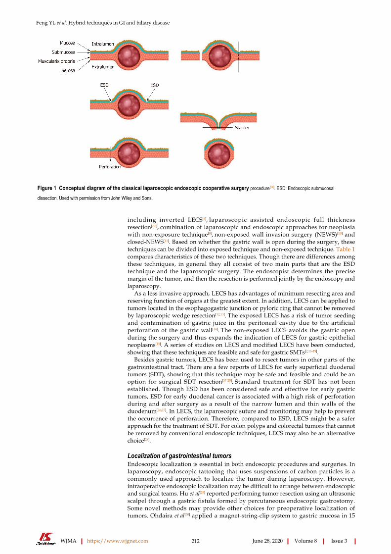
Feng YL et al. Hybrid techniques in GI and biliary disease
WJMA https://www.wjgnet.com 212 June 28, 2020 Volume 8 Issue 3
Figure 1 Conceptual diagram of the classical laparoscopic endoscopic cooperative surgery procedure[14]. ESD: Endoscopic submucosal
dissection. Used with permission from John Wiley and Sons.
including inverted LECS[6], laparoscopic assisted endoscopic full thickness resection[7,8], combination of laparoscopic and endoscopic approaches for neoplasia with non-exposure technique[9], non-exposed wall invasion surgery (NEWS)[10] and closed-NEWS[11]. Based on whether the gastric wall is open during the surgery, these techniques can be divided into exposed technique and non-exposed technique. Table 1 compares characteristics of these two techniques. Though there are differences among these techniques, in general they all consist of two main parts that are the ESD technique and the laparoscopic surgery. The endoscopist determines the precise margin of the tumor, and then the resection is performed jointly by the endoscopy and laparoscopy.
As a less invasive approach, LECS has advantages of minimum resecting area and reserving function of organs at the greatest extent. In addition, LECS can be applied to tumors located in the esophagogastric junction or pyloric ring that cannot be removed by laparoscopic wedge resection[12,13]. The exposed LECS has a risk of tumor seeding and contamination of gastric juice in the peritoneal cavity due to the artificial perforation of the gastric wall[14]. The non-exposed LECS avoids the gastric open during the surgery and thus expands the indication of LECS for gastric epithelial neoplasms[15]. A series of studies on LECS and modified LECS have been conducted, showing that these techniques are feasible and safe for gastric SMTs[2,16-19].
Besides gastric tumors, LECS has been used to resect tumors in other parts of the gastrointestinal tract. There are a few reports of LECS for early superficial duodenal tumors (SDT), showing that this technique may be safe and feasible and could be an option for surgical SDT resection[37-25]. Standard treatment for SDT has not been established. Though ESD has been considered safe and effective for early gastric tumors, ESD for early duodenal cancer is associated with a high risk of perforation during and after surgery as a result of the narrow lumen and thin walls of the duodenum[26,27]. In LECS, the laparoscopic suture and monitoring may help to prevent the occurrence of perforation. Therefore, compared to ESD, LECS might be a safer approach for the treatment of SDT. For colon polyps and colorectal tumors that cannot be removed by conventional endoscopic techniques, LECS may also be an alternative choice[28].
Localization of gastrointestinal tumorsEndoscopic localization is essential in both endoscopic procedures and surgeries. In laparoscopy, endoscopic tattooing that uses suspensions of carbon particles is a commonly used approach to localize the tumor during laparoscopy. However, intraoperative endoscopic localization may be difficult to arrange between endoscopic and surgical teams. Hu et al[29] reported performing tumor resection using an ultrasonic scalpel through a gastric fistula formed by percutaneous endoscopic gastrostomy. Some novel methods may provide other choices for preoperative localization of tumors. Ohdaira et al[30] applied a magnet-string-clip system to gastric mucosa in 15
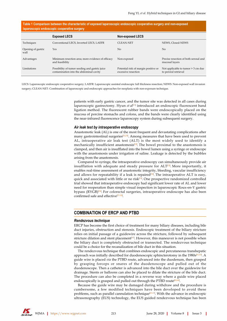
Feng YL et al. Hybrid techniques in GI and biliary disease
WJMA https://www.wjgnet.com 213 June 28, 2020 Volume 8 Issue 3
Table 1 Comparison between the characteristic of exposed laparoscopic endoscopic cooperative surgery and non-exposed laparoscopic endoscopic cooperative surgery
Exposed LECS Non-exposed LECS
Techniques Conventional LECS, Inverted LECS, LAEFR CLEAN-NET NEWS, Closed-NEWS
Opening of gastric wall
Yes No No
Advantages Minimum resection area; more evidence of efficacy and feasibility
Non-exposed Precise resection of both serosal and mucosal layers
Limitations Possibility of tumor seeding and gastric juice contamination into the abdominal cavity
Potential risk of margin positive or excessive resection
Not applicable to tumor > 3 cm due to peroral retrieval
LECS: Laparoscopic endoscopic cooperative surgery; LAEFR: Laparoscopic assisted endoscopic full thickness resection; NEWS: Non-exposed wall invasion surgery; CLEAN-NET: Combination of laparoscopic and endoscopic approaches for neoplasia with non-exposure technique.
patients with early gastric cancer, and the tumor site was detected in all cases during laparoscopic gastrectomy. Hyun et al[31] introduced an endoscopic fluorescent band ligation method. The fluorescent rubber bands were endoscopically placed on the mucosa of porcine stomachs and colons, and the bands were clearly identified using the near-infrared fluorescence laparoscopy system during subsequent surgery.
Air leak test by intraoperative endoscopyAnastomotic leak (AL) is one of the most frequent and devastating complications after many gastrointestinal surgeries[32,33]. Among measures that have been used to prevent AL, intraoperative air leak test (ALT) is the most widely used to identify a mechanically insufficient anastomosis[32]. The bowel proximal to the anastomosis is clamped, and then air is insufflated into the bowel lumen using a syringe or endoscope with the anastomosis under irrigation of saline. Leakage is detected by the bubbles arising from the anastomosis.
Compared to syringe, the intraoperative endoscopy can simultaneously provide air insufflation with adequate and steady pressure for ALT[33]. More importantly, it enables real-time assessment of anastomotic integrity, bleeding, vascular insufficiency and allows for repeatability if a leak is repaired[34]. The intraoperative ALT is easy, quick and associated with little or no risk[35]. One prospective randomized controlled trial showed that intraoperative endoscopy had significant lower rate of AL and lower need for reoperation than simple visual inspection in laparoscopic Roux-en-Y gastric bypass (RYGB)[36]. For colorectal surgeries, intraoperative endoscope has also been confirmed safe and effective[37-39].
COMBINATION OF ERCP AND PTBDRendezvous techniqueERCP has become the first choice of treatment for many biliary diseases, including bile duct injuries, obstruction and stenosis. Endoscopic treatment of the biliary stricture relies on initial passage of a guidewire across the stricture, followed by subsequent stricture dilation and stent placement[40]. However, this maneuver is not possible when the biliary duct is completely obstructed or transected. The rendezvous technique could be a choice for the recanalization of bile duct in this situation.
The rendezvous technique that combines endoscopic and percutaneous transhepatic approach was initially described for duodenoscopic sphincterotomy in the 1980s[41,42]. A guide wire is placed via the PTBD route, advanced into the duodenum, then grasped by grasping forceps or snares of the duodenoscope and pulled out of the duodenoscope. Then a catheter is advanced into the bile duct over the guidewire for drainage. Stents or balloons can also be placed to dilate the stricture of the bile duct. The procedure can also be completed in a reverse way where a guide wire placed endoscopically is grasped and pulled out through the PTBD route[43,44].
Because the guide wire may be damaged during withdraw and the procedure is cumbersome, a few modified techniques have been developed to avoid these problems, such as parallel cannulation technique[45-47]. With the advance in endoscopic ultrasonography (EUS) technology, the EUS guided rendezvous technique has been
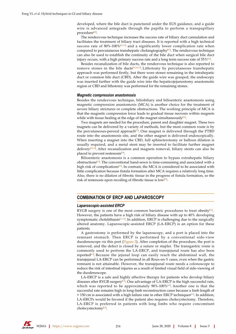
Feng YL et al. Hybrid techniques in GI and biliary disease
WJMA https://www.wjgnet.com 214 June 28, 2020 Volume 8 Issue 3
developed, where the bile duct is punctured under the EUS guidance, and a guide wire is advanced antegrade through the papilla to perform a transpapillary procedure[48].
The rendezvous technique increases the success rate of biliary duct cannulation and facilitates the treatment of biliary tract diseases. It is reported with a high technical success rate of 80%-100%[49-53] and a significantly lower complication rate when compared to percutaneous transhepatic cholangiography[53]. The rendezvous technique can also be used to establish the continuity of the bile duct when surgical bile duct injury occurs, with a high primary success rate and a long term success rate of 55%[44].
Besides recanalization of bile ducts, the rendezvous technique is also reported to remove stones in the bile ducts[43,54,55]. Lithotomy by percutaneous transhepatic approach was performed firstly, but there were stones remaining in the intrahepatic duct or common bile duct (CBD). After the guide wire was grasped, the endoscopy was inserted further with the guide wire into the hepaticojejunostomy anastomotic region or CBD and lithotomy was performed for the remaining stones.
Magnetic compression anastomosisBesides the rendezvous technique, biliobiliary and bilioenteric anastomosis using magnetic compression anastomosis (MCA) is another choice for the treatment of severe biliary strictures or complete obstructions. The working principle of MCA is that the magnetic compression force leads to gradual tissue necrosis within magnets while with tissue healing at the edge of the magnet simultaneously[56].
Two magnets are needed for the procedure, parent and daughter magnet. These two magnets can be delivered by a variety of methods, but the most common route is by the percutaneous-peroral approach[57]. One magnet is delivered through the PTBD route into the anastomosis site, and the other magnet is delivered endoscopically. When inserting a magnet into the CBD, full sphincterotomy or balloon dilation is usually required, and a metal stent may be inserted to facilitate further magnet delivery[58,59]. After recanalization and magnets removal, biliary stents can also be placed to prevent restenosis[59].
Bilioenteric anastomosis is a common operation to bypass extrahepatic biliary obstructions[60]. The conventional hand-sewn is time-consuming and associated with a high risk of complications[60]. In contrast, the MCA is considered to be associated with little complication because fistula formation after MCA requires a relatively long time. Also, there is no dilation of fibrotic tissue in the progress of fistula formation, so the risk of restenosis upon recoiling of fibrotic tissue is low[57].
COMBINATION OF ERCP AND LAPAROSCOPYLaparoscopic-assisted ERCPRYGB surgery is one of the most common bariatric procedures to treat obesity[61]. However, the patients have a high risk of biliary disease with up to 40% developing symptomatic cholelithiasis[61,62]. In addition, ERCP is challenging due to the surgically altered anatomy. Laparoscopic-assisted ERCP (LA-ERCP) is an option for these patients.
A gastrostomy is performed by the laparoscopy, and a port is placed into the remnant stomach. Then ERCP is performed by a conventional side-view duodenoscope via this port (Figure 2). After completion of the procedure, the port is removed, and the defect is closed by a suture or stapler. The transgastric route is commonly used to perform the LA-ERCP, and transjejunal route has also been reported[63]. Because the jejunal loop can easily reach the abdominal wall, the transjejunal LA-ERCP can be performed in all Roux-en-Y cases, even when the gastric remnant is not attainable. However, the transjejunal route needs a colonoscope to reduce the risk of intestinal injuries as a result of limited visual field of side-viewing of the duodenoscope.
LA-ERCP is a safe and highly effective therapy for patients who develop biliary diseases after RYGB surgery[64]. One advantage of LA-ERCP is the high successful rate, which was reported to be approximately 90%-100%[65]. Another one is that the successful rate remains high in long-limb reconstruction cases because a limb length of > 150 cm is associated with a high failure rate in other ERCP techniques[66]. In addition, LA-ERCPs would be favored if the patient also requires cholecystectomy. Therefore, LA-ERCP is preferred in patients with long limbs who require concomitant cholecystectomy[65].
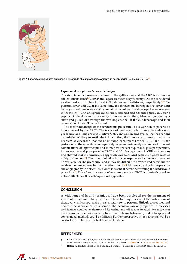
Feng YL et al. Hybrid techniques in GI and biliary disease
WJMA https://www.wjgnet.com 215 June 28, 2020 Volume 8 Issue 3
Figure 2 Laparoscopic-assisted endoscopic retrograde cholangiopancreatography in patients with Roux-en-Y anatomy[73].
Laparo-endoscopic rendezvous techniqueThe simultaneous presence of stones in the gallbladder and the CBD is a common clinical circumstance[67]. ERCP and laparoscopic cholecystectomy (LC) are considered as standard approaches to treat CBD stones and gallstones, respectively[68,69]. To perform ERCP and LC at the same time, the rendezvous intraoperative ERCP with transcystic guide-wire-assisted cannulation technique was developed as a one-stage intervention[70,71]. An antegrade guidewire is inserted and advanced through Vater’s papilla into the duodenum by a surgeon. Subsequently, the guidewire is grasped by a snare and pulled out through the working channel of the duodenoscope and then cannulation of the CBD is performed.
The major advantage of the rendezvous procedure is a lower risk of pancreatic injury caused by the ERCP. The transcystic guide wire facilitates the endoscopic procedure and thus ensures elective CBD cannulation and avoids the inadvertent cannulation of the pancreatic duct. In addition, the antegrade approach avoids the problem of discordant patient positioning encountered when ERCP and LC are performed at the same time but separately. A recent meta-analysis compared different combinations of laparoscopic and intraoperative techniques (LC plus preoperative, intraoperative and postoperative ERCP and LC plus laparoscopic CBD exploration) and showed that the rendezvous approach was associated with the highest rates of safety and success[67]. The major limitation is that an experienced endoscopist may not be available for the procedure, and it may be difficult to arrange and carry out the rendezvous procedures in the operating room[68,72]. Moreover, using intraoperative cholangiography to detect CBD stones is essential before performing the rendezvous procedure[68]. Therefore, in centers where preoperative ERCP is routinely used to detect CBD stones, this technique is not applicable.
CONCLUSIONA wide range of hybrid techniques have been developed for the treatment of gastrointestinal and biliary diseases. These techniques expand the indications of therapeutic endoscopy, make it easier and safer to perform difficult procedures and decrease the agony of patients. Some of the techniques are only reported in few cases and further detailed evaluation of feasibility and efficacy is needed. For those that have been confirmed safe and effective, how to choose between hybrid techniques and conventional methods could be difficult. Further prospective investigations should be conducted to determine the best treatment options.
REFERENCES1 Lian J, Chen S, Zhang Y, Qiu F. A meta-analysis of endoscopic submucosal dissection and EMR for early
gastric cancer. Gastrointest Endosc 2012; 76: 763-770 [PMID: 22884100 DOI: 10.1016/j.gie.2012.06.014]Hoteya S, Haruta S, Shinohara H, Yamada A, Furuhata T, Yamashita S, Kikuchi D, Mitani T, Ogawa O, 2

Feng YL et al. Hybrid techniques in GI and biliary disease
WJMA https://www.wjgnet.com 216 June 28, 2020 Volume 8 Issue 3
Matsui A, Iizuka T, Udagawa H, Kaise M. Feasibility and safety of laparoscopic and endoscopic cooperative surgery for gastric submucosal tumors, including esophagogastric junction tumors. Dig Endosc 2014; 26: 538-544 [PMID: 24355070 DOI: 10.1111/den.12215]
3 Akahoshi K, Oya M, Koga T, Shiratsuchi Y. Current clinical management of gastrointestinal stromal tumor. World J Gastroenterol 2018; 24: 2806-2817 [PMID: 30018476 DOI: 10.3748/wjg.v24.i26.2806]
4 Goh BK, Goh YC, Eng AK, Chan WH, Chow PK, Chung YF, Ong HS, Wong WK. Outcome after laparoscopic versus open wedge resection for suspected gastric gastrointestinal stromal tumors: A matched-pair case-control study. Eur J Surg Oncol 2015; 41: 905-910 [PMID: 25913060 DOI: 10.1016/j.ejso.2015.04.001]
5 Hiki N, Yamamoto Y, Fukunaga T, Yamaguchi T, Nunobe S, Tokunaga M, Miki A, Ohyama S, Seto Y. Laparoscopic and endoscopic cooperative surgery for gastrointestinal stromal tumor dissection. Surg Endosc 2008; 22: 1729-1735 [PMID: 18074180 DOI: 10.1007/s00464-007-9696-8]
6 Nunobe S, Hiki N, Gotoda T, Murao T, Haruma K, Matsumoto H, Hirai T, Tanimura S, Sano T, Yamaguchi T. Successful application of laparoscopic and endoscopic cooperative surgery (LECS) for a lateral-spreading mucosal gastric cancer. Gastric Cancer 2012; 15: 338-342 [PMID: 22350555 DOI: 10.1007/s10120-012-0146-5]
7 Abe N, Takeuchi H, Ooki A, Nagao G, Masaki T, Mori T, Sugiyama M. Recent developments in gastric endoscopic submucosal dissection: towards the era of endoscopic resection of layers deeper than the submucosa. Dig Endosc 2013; 25 Suppl 1: 64-70 [PMID: 23368096 DOI: 10.1111/j.1443-1661.2012.01387.x]
8 Abe N, Takeuchi H, Yanagida O, Masaki T, Mori T, Sugiyama M, Atomi Y. Endoscopic full-thickness resection with laparoscopic assistance as hybrid NOTES for gastric submucosal tumor. Surg Endosc 2009; 23: 1908-1913 [PMID: 19184206 DOI: 10.1007/s00464-008-0317-y]
9 Inoue H, Ikeda H, Hosoya T, Yoshida A, Onimaru M, Suzuki M, Kudo SE. Endoscopic mucosal resection, endoscopic submucosal dissection, and beyond: full-layer resection for gastric cancer with nonexposure technique (CLEAN-NET). Surg Oncol Clin N Am 2012; 21: 129-140 [PMID: 22098836 DOI: 10.1016/j.soc.2011.09.012]
10 Mitsui T, Goto O, Shimizu N, Hatao F, Wada I, Niimi K, Asada-Hirayama I, Fujishiro M, Koike K, Seto Y. Novel technique for full-thickness resection of gastric malignancy: feasibility of nonexposed endoscopic wall-inversion surgery (news) in porcine models. Surg Laparosc Endosc Percutan Tech 2013; 23: e217-e221 [PMID: 24300935 DOI: 10.1097/SLE.0b013e31828e3f94]
11 Kikuchi S, Nishizaki M, Kuroda S, Tanabe S, Noma K, Kagawa S, Shirakawa Y, Kato H, Okada H, Fujiwara T. Nonexposure laparoscopic and endoscopic cooperative surgery (closed laparoscopic and endoscopic cooperative surgery) for gastric submucosal tumor. Gastric Cancer 2017; 20: 553-557 [PMID: 27599829 DOI: 10.1007/s10120-016-0641-1]
12 Choi SM, Kim MC, Jung GJ, Kim HH, Kwon HC, Choi SR, Jang JS, Jeong JS. Laparoscopic wedge resection for gastric GIST: long-term follow-up results. Eur J Surg Oncol 2007; 33: 444-447 [PMID: 17174060 DOI: 10.1016/j.ejso.2006.11.003]
13 Hwang SH, Park DJ, Kim YH, Lee KH, Lee HS, Kim HH, Lee HJ, Yang HK, Lee KU. Laparoscopic surgery for submucosal tumors located at the esophagogastric junction and the prepylorus. Surg Endosc 2009; 23: 1980-1987 [PMID: 18470554 DOI: 10.1007/s00464-008-9955-3]
14 Hiki N, Nunobe S, Matsuda T, Hirasawa T, Yamamoto Y, Yamaguchi T. Laparoscopic endoscopic cooperative surgery. Dig Endosc 2015; 27: 197-204 [PMID: 25394216 DOI: 10.1111/den.12404]
15 Matsuda T, Nunobe S, Ohashi M, Hiki N. Laparoscopic endoscopic cooperative surgery (LECS) for the upper gastrointestinal tract. Transl Gastroenterol Hepatol 2017; 2: 40 [PMID: 28616596 DOI: 10.21037/tgh.2017.03.20]
16 Aoyama J, Goto O, Kawakubo H, Mayanagi S, Fukuda K, Irino T, Nakamura R, Wada N, Takeuchi H, Yahagi N, Kitagawa Y. Clinical outcomes of non-exposed endoscopic wall-inversion surgery for gastric submucosal tumors: long-term follow-up and functional results. Gastric Cancer 2020; 23: 154-159 [PMID: 31270624 DOI: 10.1007/s10120-019-00985-1]
17 Balde AI, Chen T, Hu Y, Redondo N JD, Liu H, Gong W, Yu J, Zhen L, Li G. Safety analysis of laparoscopic endoscopic cooperative surgery versus endoscopic submucosal dissection for selected gastric gastrointestinal stromal tumors: a propensity score-matched study. Surg Endosc 2017; 31: 843-851 [PMID: 27492430 DOI: 10.1007/s00464-016-5042-3]
18 Matsuda T, Hiki N, Nunobe S, Aikou S, Hirasawa T, Yamamoto Y, Kumagai K, Ohashi M, Sano T, Yamaguchi T. Feasibility of laparoscopic and endoscopic cooperative surgery for gastric submucosal tumors (with video). Gastrointest Endosc 2016; 84: 47-52 [PMID: 26684599 DOI: 10.1016/j.gie.2015.11.040]
19 Kanehira E, Kanehira AK, Tanida T, Takahashi K, Obana Y, Sasaki K. CLEAN-NET: a modified laparoendoscopic wedge resection of the stomach to minimize the sacrifice of innocent gastric wall. Surg Endosc 2020; 34: 290-297 [PMID: 30941549 DOI: 10.1007/s00464-019-06765-3]
20 Miyazaki Y, Takiguchi S, Kurokawa Y, Takahashi T, Fukuda Y, Yamasaki M, Makino T, Tanaka K, Motoori M, Kimura Y, Nakajima K, Mori M, Doki Y. Endoscopy-assisted laparoscopic submucosal dissection for a duodenal epithelial tumor. Asian J Endosc Surg 2019; 12: 461-464 [PMID: 30604563 DOI: 10.1111/ases.12675]
21 Tsushimi T, Mori H, Harada T, Nagase T, Iked Y, Ohnishi H. Laparoscopic and endoscopic cooperative surgery for duodenal neuroendocrine tumor (NET) G1: Report of a case. Int J Surg Case Rep 2014; 5: 1021-1024 [PMID: 25460463 DOI: 10.1016/j.ijscr.2014.10.051]
22 Nagasawa Y, Okauchi H, Kojima M, Setoyama H, Hasegawa M, Mizuta H, Tsujikawa T, Tani M, Kurumi Y. Laparoscopic-endoscopic cooperative surgery for a duodenal neuroendocrine tumor: A case report. Asian J Endosc Surg 2017; 10: 183-186 [PMID: 28093861 DOI: 10.1111/ases.12356]
23 Irino T, Nunobe S, Hiki N, Yamamoto Y, Hirasawa T, Ohashi M, Fujisaki J, Sano T, Yamaguchi T. Laparoscopic-endoscopic cooperative surgery for duodenal tumors: a unique procedure that helps ensure the safety of endoscopic submucosal dissection. Endoscopy 2015; 47: 349-351 [PMID: 25479560 DOI: 10.1055/s-0034-1390909]

Feng YL et al. Hybrid techniques in GI and biliary disease
WJMA https://www.wjgnet.com 217 June 28, 2020 Volume 8 Issue 3
24 Yorimitsu N, Oyama T, Takahashi A, Takehana T, Shiozawa S. Laparoscopy and endoscopy cooperative surgery is a safe and effective novel treatment for duodenal neuroendocrine tumor G1. Endoscopy 2020; 52: E68-E70 [PMID: 31529442 DOI: 10.1055/a-0999-5172]
25 Yanagimoto Y, Omori T, Jeong-Ho M, Shinno N, Yamamoto K, Takeuchi Y, Higashino K, Uedo N, Sugimura K, Matsunaga T, Miyata H, Ushigome H, Takahashi Y, Nishimura J, Yasui M, Asukai K, Yamada D, Tomokuni A, Wada H, Takahashi H, Ohue M, Yano M, Sakon M. Feasibility and Safety of a Novel Laparoscopic and Endoscopic Cooperative Surgery Technique for Superficial Duodenal Tumor Resection: How I Do It. J Gastrointest Surg 2019; 23: 2068-2074 [PMID: 30859426 DOI: 10.1007/s11605-019-04176-2]
26 Marques J, Baldaque-Silva F, Pereira P, Arnelo U, Yahagi N, Macedo G. Endoscopic mucosal resection and endoscopic submucosal dissection in the treatment of sporadic nonampullary duodenal adenomatous polyps. World J Gastrointest Endosc 2015; 7: 720-727 [PMID: 26140099 DOI: 10.4253/wjge.v7.i7.720]
27 Kakushima N, Kanemoto H, Tanaka M, Takizawa K, Ono H. Treatment for superficial non-ampullary duodenal epithelial tumors. World J Gastroenterol 2014; 20: 12501-12508 [PMID: 25253950 DOI: 10.3748/wjg.v20.i35.12501]
28 Suzuki S, Fukunaga Y, Tamegai Y, Akiyoshi T, Konishi T, Nagayama S, Saito S, Ueno M. The short-term outcomes of laparoscopic-endoscopic cooperative surgery for colorectal tumors (LECS-CR) in cases involving endoscopically unresectable colorectal tumors. Surg Today 2019; 49: 1051-1057 [PMID: 31250113 DOI: 10.1007/s00595-019-01840-7]
29 Yuan XL, Wang SF, Ye LS, Wu CC, Zeng XH, Khan N, Hu B. Resection of a giant gastric mass using an ultrasonic scalpel with endoscopic assistance. Endoscopy 2018; 50: E227-E228 [PMID: 29895070 DOI: 10.1055/a-0624-9277]
30 Ohdaira T, Nagai H. Intraoperative localization of early-stage upper gastrointestinal tumors using a magnetic marking clip-detecting system. Surg Endosc 2007; 21: 810-815 [PMID: 17279306 DOI: 10.1007/s00464-006-9037-3]
31 Hyun JH, Kim SK, Kim KG, Kim HR, Lee HM, Park S, Kim SC, Choi Y, Sohn DK. A novel endoscopic fluorescent band ligation method for tumor localization. Surg Endosc 2016; 30: 4659-4663 [PMID: 26895900 DOI: 10.1007/s00464-016-4785-1]
32 Wu Z, van de Haar RC, Sparreboom CL, Boersema GS, Li Z, Ji J, Jeekel J, Lange JF. Is the intraoperative air leak test effective in the prevention of colorectal anastomotic leakage? A systematic review and meta-analysis. Int J Colorectal Dis 2016; 31: 1409-1417 [PMID: 27294661 DOI: 10.1007/s00384-016-2616-4]
33 Yang SY, Han J, Han YD, Cho MS, Hur H, Lee KY, Kim NK, Min BS. Intraoperative colonoscopy for the assessment and prevention of anastomotic leakage in low anterior resection for rectal cancer. Int J Colorectal Dis 2017; 32: 709-714 [PMID: 28144745 DOI: 10.1007/s00384-017-2767-y]
34 Park JH, Jeong SH, Lee YJ, Kim TH, Kim JM, Kim DH, Kwag SJ, Kim JY, Park T, Jeong CY, Ju YT, Jung EJ, Hong SC. Safety and efficacy of post-anastomotic intraoperative endoscopy to avoid early anastomotic complications during gastrectomy for gastric cancer. Surg Endosc 2019 [PMID: 31834512 DOI: 10.1007/s00464-019-07319-3]
35 Hirst NA, Tiernan JP, Millner PA, Jayne DG. Systematic review of methods to predict and detect anastomotic leakage in colorectal surgery. Colorectal Dis 2014; 16: 95-109 [PMID: 23992097 DOI: 10.1111/codi.12411]
36 Valenzuela-Salazar C, Rojano-Rodríguez ME, Romero-Loera S, Trejo-Ávila ME, Bañuelos-Mancilla J, Delano-Alonso R, Moreno-Portillo M. Intraoperative endoscopy prevents technical defect related leaks in laparoscopic Roux-en-Y gastric bypass: A randomized control trial. Int J Surg 2018; 50: 17-21 [PMID: 29278753 DOI: 10.1016/j.ijsu.2017.12.024]
37 Beard JD, Nicholson ML, Sayers RD, Lloyd D, Everson NW. Intraoperative air testing of colorectal anastomoses: a prospective, randomized trial. Br J Surg 1990; 77: 1095-1097 [PMID: 2136198 DOI: 10.1002/bjs.1800771006]
38 Sasaki K, Ishihara S, Nozawa H, Kawai K, Hata K, Kiyomatsu T, Tanaka T, Nishikawa T, Otani K, Yasuda K, Murono K, Watanabe T. Successful Management of a Positive Air Leak Test during Laparoscopic Colorectal Surgery. Dig Surg 2018; 35: 266-270 [PMID: 28934741 DOI: 10.1159/000480157]
39 Allaix ME, Lena A, Degiuli M, Arezzo A, Passera R, Mistrangelo M, Morino M. Intraoperative air leak test reduces the rate of postoperative anastomotic leak: analysis of 777 laparoscopic left-sided colon resections. Surg Endosc 2019; 33: 1592-1599 [PMID: 30203203 DOI: 10.1007/s00464-018-6421-8]
40 Bukhari MA, Haito-Chavez Y, Ngamruengphong S, Brewer Gutierrez O, Chen YI, Khashab MA. Rendezvous Biliary Recanalization of Complete Biliary Obstruction With Direct Peroral and Percutaneous Transhepatic Cholangioscopy. Gastroenterology 2018; 154: 23-25 [PMID: 29102615 DOI: 10.1053/j.gastro.2017.09.050]
41 Shorvon PJ, Cotton PB, Mason RR, Siegel JH, Hatfield AR. Percutaneous transhepatic assistance for duodenoscopic sphincterotomy. Gut 1985; 26: 1373-1376 [PMID: 4085912 DOI: 10.1136/gut.26.12.1373]
42 Mason RR, Cotton PB. Combined duodenoscopic and transhepatic approach to stenosis of the papilla of Vater. Br J Radiol 1981; 54: 678-679 [PMID: 7260526 DOI: 10.1259/0007-1285-54-644-678]
43 Kimura K, Kudo K, Kurihara T, Yoshiya S, Mano Y, Takeishi K, Itoh S, Harada N, Ikegami T, Yoshizumi T, Ikeda T. Rendezvous Technique Using Double Balloon Endoscope for Removal of Multiple Intrahepatic Bile Duct Stones in Hepaticojejunostomy After Living Donor Liver Transplant: A Case Report. Transplant Proc 2019; 51: 579-584 [PMID: 30879594 DOI: 10.1016/j.transproceed.2018.12.005]
44 Schreuder AM, Booij KAC, de Reuver PR, van Delden OM, van Lienden KP, Besselink MG, Busch OR, Gouma DJ, Rauws EAJ, van Gulik TM. Percutaneous-endoscopic rendezvous procedure for the management of bile duct injuries after cholecystectomy: short- and long-term outcomes. Endoscopy 2018; 50: 577-587 [PMID: 29351705 DOI: 10.1055/s-0043-123935]
45 Mönkemüller KE, Linder JD, Fry LC. Modified rendezvous technique for biliary cannulation. Endoscopy 2002; 34: 936 [PMID: 12430084 DOI: 10.1055/s-2002-35304]Lee TH, Park SH, Lee SH, Lee CK, Lee SH, Chung IK, Kim HS, Kim SJ. Modified rendezvous intrahepatic bile duct cannulation technique to pass a PTBD catheter in ERCP. World J Gastroenterol 2010; 16: 5388-
46

Feng YL et al. Hybrid techniques in GI and biliary disease
WJMA https://www.wjgnet.com 218 June 28, 2020 Volume 8 Issue 3
5390 [PMID: 21072905 DOI: 10.3748/wjg.v16.i42.5388]47 Dickey W. Parallel cannulation technique at ERCP rendezvous. Gastrointest Endosc 2006; 63: 686-687
[PMID: 16564873 DOI: 10.1016/j.gie.2005.10.029]48 Tsuchiya T, Itoi T, Sofuni A, Tonozuka R, Mukai S. Endoscopic ultrasonography-guided rendezvous
technique. Dig Endosc 2016; 28 Suppl 1: 96-101 [PMID: 26786389 DOI: 10.1111/den.12611]49 Tomizawa Y, Di Giorgio J, Santos E, McCluskey KM, Gelrud A. Combined interventional radiology
followed by endoscopic therapy as a single procedure for patients with failed initial endoscopic biliary access. Dig Dis Sci 2014; 59: 451-458 [PMID: 24271117 DOI: 10.1007/s10620-013-2913-5]
50 Neal CP, Thomasset SC, Bools D, Sutton CD, Garcea G, Mann CD, Rees Y, Newland C, Robinson RJ, Dennison AR, Berry DP. Combined percutaneous-endoscopic stenting of malignant biliary obstruction: results from 106 consecutive procedures and identification of factors associated with adverse outcome. Surg Endosc 2010; 24: 423-431 [PMID: 19565296 DOI: 10.1007/s00464-009-0586-0]
51 Chang JH, Lee IS, Chun HJ, Choi JY, Yoon SK, Kim DG, You YK, Choi MG, Choi KY, Chung IS. Usefulness of the rendezvous technique for biliary stricture after adult right-lobe living-donor liver transplantation with duct-to-duct anastomosis. Gut Liver 2010; 4: 68-75 [PMID: 20479915 DOI: 10.5009/gnl.2010.4.1.68]
52 Yang MJ, Kim JH, Hwang JC, Yoo BM, Kim SS, Lim SG, Won JH. Usefulness of combined percutaneous-endoscopic rendezvous techniques after failed therapeutic endoscopic retrograde cholangiography in the era of endoscopic ultrasound guided rendezvous. Medicine (Baltimore) 2017; 96: e8991 [PMID: 29310413 DOI: 10.1097/MD.0000000000008991]
53 Bokemeyer A, Müller F, Niesert H, Brückner M, Bettenworth D, Nowacki T, Beyna T, Ullerich H, Lenze F. Percutaneous-transhepatic-endoscopic rendezvous procedures are effective and safe in patients with refractory bile duct obstruction. United European Gastroenterol J 2019; 7: 397-404 [PMID: 31019708 DOI: 10.1177/2050640619825949]
54 Kimura K, Kudo K, Yoshizumi T, Kurihara T, Yoshiya S, Mano Y, Takeishi K, Itoh S, Harada N, Ikegami T, Ikeda T. Electrohydraulic lithotripsy and rendezvous nasal endoscopic cholangiography for common bile duct stone: A case report. World J Clin Cases 2019; 7: 1149-1154 [PMID: 31183346 DOI: 10.12998/wjcc.v7.i10.1149]
55 Itoi T, Ishii K, Sofuni A, Itokawa F, Kurihara T, Tsuchiya T, Tsuji S, Umeda J, Moriyasu F. Single Balloon Enteroscopy-Assisted ERCP Using Rendezvous Technique for Sharp Angulation of Roux-en-Y Limb in a Patient with Bile Duct Stones. Diagn Ther Endosc 2009; 2009: 154084 [PMID: 20169091 DOI: 10.1155/2009/154084]
56 Fan C, Zhang H, Yan X, Ma J, Wang C, Lv Y. Advanced Roux-en-Y hepaticojejunostomy with magnetic compressive anastomats in obstructive jaundice dog models. Surg Endosc 2018; 32: 779-789 [PMID: 28779259 DOI: 10.1007/s00464-017-5740-5]
57 Jang SI, Choi J, Lee DK. Magnetic compression anastomosis for treatment of benign biliary stricture. Dig Endosc 2015; 27: 239-249 [PMID: 24905938 DOI: 10.1111/den.12319]
58 Parlak E, Koksal AS, Kucukay F, Eminler AT, Toka B, Uslan MI. A novel technique for the endoscopic treatment of complete biliary anastomosis obstructions after liver transplantation: through-the-scope magnetic compression anastomosis. Gastrointest Endosc 2017; 85: 841-847 [PMID: 27566054 DOI: 10.1016/j.gie.2016.07.068]
59 Jang SI, Lee KH, Yoon HJ, Lee DK. Treatment of completely obstructed benign biliary strictures with magnetic compression anastomosis: follow-up results after recanalization. Gastrointest Endosc 2017; 85: 1057-1066 [PMID: 27619787 DOI: 10.1016/j.gie.2016.08.047]
60 Fan C, Yan XP, Liu SQ, Wang CB, Li JH, Yu L, Wu Z, Lv Y. Roux-en-Y choledochojejunostomy using novel magnetic compressive anastomats in canine model of obstructive jaundice. Hepatobiliary Pancreat Dis Int 2012; 11: 81-88 [PMID: 22251474 DOI: 10.1016/s1499-3872(11)60129-x]
61 Mohammad B, Richard MN, Pandit A, Zuccala K, Brandwein S. Outcomes of laparoscopic-assisted ERCP in gastric bypass patients at a community hospital center. Surg Endosc 2019 [PMID: 31823046 DOI: 10.1007/s00464-019-07310-y]
62 Paranandi B, Joshi D, Mohammadi B, Jenkinson A, Adamo M, Read S, Johnson GJ, Chapman MH, Pereira SP, Webster GJ. Laparoscopy-assisted ERCP (LA-ERCP) following bariatric gastric bypass surgery: initial experience of a single UK centre. Frontline Gastroenterol 2016; 7: 54-59 [PMID: 28839834 DOI: 10.1136/flgastro-2015-100556]
63 Dalmonte G, Valente M, Bosi S, Gnocchi A, Marchesi F. Transjejunal Laparoscopic-Assisted ERCP: a Technique to Deal with Choledocholithiasis After Roux-En-Y Reconstruction. Obes Surg 2019; 29: 2005-2006 [PMID: 30972636 DOI: 10.1007/s11695-019-03882-9]
64 Bowman E, Greenberg J, Garren M, Guda N, Rajca B, Benson M, Pfau P, Soni A, Walker A, Gopal D. Laparoscopic-assisted ERCP and EUS in patients with prior Roux-en-Y gastric bypass surgery: a dual-center case series experience. Surg Endosc 2016; 30: 4647-4652 [PMID: 26823057 DOI: 10.1007/s00464-016-4746-8]
65 Krutsri C, Kida M, Yamauchi H, Iwai T, Imaizumi H, Koizumi W. Current status of endoscopic retrograde cholangiopancreatography in patients with surgically altered anatomy. World J Gastroenterol 2019; 25: 3313-3333 [PMID: 31341358 DOI: 10.3748/wjg.v25.i26.3313]
66 Schreiner MA, Chang L, Gluck M, Irani S, Gan SI, Brandabur JJ, Thirlby R, Moonka R, Kozarek RA, Ross AS. Laparoscopy-assisted versus balloon enteroscopy-assisted ERCP in bariatric post-Roux-en-Y gastric bypass patients. Gastrointest Endosc 2012; 75: 748-756 [PMID: 22301340 DOI: 10.1016/j.gie.2011.11.019]
67 Ricci C, Pagano N, Taffurelli G, Pacilio CA, Migliori M, Bazzoli F, Casadei R, Minni F. Comparison of Efficacy and Safety of 4 Combinations of Laparoscopic and Intraoperative Techniques for Management of Gallstone Disease With Biliary Duct Calculi: A Systematic Review and Network Meta-analysis. JAMA Surg 2018; 153: e181167 [PMID: 29847616 DOI: 10.1001/jamasurg.2018.1167]
68 Noel R, Arnelo U, Swahn F. Intraoperative versus postoperative rendezvous endoscopic retrograde cholangiopancreatography to treat common bile duct stones during cholecystectomy. Dig Endosc 2019; 31: 69-76 [PMID: 29947437 DOI: 10.1111/den.13222]

Feng YL et al. Hybrid techniques in GI and biliary disease
WJMA https://www.wjgnet.com 219 June 28, 2020 Volume 8 Issue 3
69 Qian Y, Xie J, Jiang P, Yin Y, Sun Q. Laparoendoscopic rendezvous versus ERCP followed by laparoscopic cholecystectomy for the management of cholecysto-choledocholithiasis: a retrospectively cohort study. Surg Endosc 2020; 34: 2483-2489 [PMID: 31428853 DOI: 10.1007/s00464-019-07051-y]
70 Swahn F, Nilsson M, Arnelo U, Löhr M, Persson G, Enochsson L. Rendezvous cannulation technique reduces post-ERCP pancreatitis: a prospective nationwide study of 12,718 ERCP procedures. Am J Gastroenterol 2013; 108: 552-559 [PMID: 23419386 DOI: 10.1038/ajg.2012.470]
71 Swahn F, Regnér S, Enochsson L, Lundell L, Permert J, Nilsson M, Thorlacius H, Arnelo U. Endoscopic retrograde cholangiopancreatography with rendezvous cannulation reduces pancreatic injury. World J Gastroenterol 2013; 19: 6026-6034 [PMID: 24106403 DOI: 10.3748/wjg.v19.i36.6026]
72 Winder JS, Juza RM, Alli VV, Rogers AM, Haluck RS, Pauli EM. Concomitant laparoscopic cholecystectomy and antegrade wire, rendezvous cannulation of the biliary tree may reduce post-ERCP pancreatitis events. Surg Endosc 2020; 34: 3216-3222 [PMID: 31489502 DOI: 10.1007/s00464-019-07074-5]
73 James HJ, James TW, Wheeler SB, Spencer JC, Baron TH. Cost-effectiveness of endoscopic ultrasound-directed transgastric ERCP compared with device-assisted and laparoscopic-assisted ERCP in patients with Roux-en-Y anatomy. Endoscopy 2019; 51: 1051-1058 [PMID: 31242509 DOI: 10.1055/a-0938-3918]
WJMA https://www.wjgnet.com 220 June 28, 2020 Volume 8 Issue 3
World Journal of
Meta-AnalysisW J M ASubmit a Manuscript: https://www.f6publishing.com World J Meta-Anal 2020 June 28; 8(3): 220-232
DOI: 10.13105/wjma.v8.i3.220 ISSN 2308-3840 (online)
MINIREVIEWS
Thrombopoietin-receptor agonists in perioperative treatment of patients with chronic liver disease
Kamran Qureshi, Alan Bonder
ORCID number: Kamran Qureshi 0000-0002-0389-7269; Alan Bonder 0000-0001-6821-0416.
Author contributions: Qureshi K and Bonder A conceived of the review, assisted in writing and revising the various drafts of the paper, and agreed on the final version.
Open-Access: This article is an open-access article that was selected by an in-house editor and fully peer-reviewed by external reviewers. It is distributed in accordance with the Creative Commons Attribution NonCommercial (CC BY-NC 4.0) license, which permits others to distribute, remix, adapt, build upon this work non-commercially, and license their derivative works on different terms, provided the original work is properly cited and the use is non-commercial. See: http://creativecommons.org/licenses/by-nc/4.0/
Manuscript source: Unsolicited manuscript
Received: March 2, 2020 Peer-review started: March 2, 2020 First decision: April 24, 2020 Revised: May 20, 2020 Accepted: June 10, 2020 Article in press: June 10, 2020 Published online: June 28, 2020
Kamran Qureshi, Department of Internal Medicine, Division of Gastroenterology and Hepatology, Saint Louis University School of Medicine, St. Louis, MO 63104, United States
Alan Bonder, Division of Gastroenterology/Liver Center, Beth Israel Deaconess Medical Center, Boston, MA 02215, United States
Corresponding author: Alan Bonder, MD, Attending Doctor, Division of Gastroenterology and Hepatology, Beth Israel Deaconess Medical Center, 110 Francis St. Suite 8E, Boston, MA 02215, United States. [email protected]
AbstractThrombocytopenia is a multifactorial disorder that is common in patients with chronic liver disease (CLD), leading to challenging perioperative planning. As thrombocytopenia in CLD is associated with thrombopoietin (TPO) deficiency, the use of TPO-receptor agonists (TPO-RAs) to increase platelet counts is a promising approach. This has led to the development of various TPO-RAs, including romiplostim, eltrombopag, avatrombopag, and lusutrombopag. Of these, only avatrombopag and lusutrombopag are approved by the United States Food and Drug Administration for the perioperative treatment of thrombocytopenia in patients with CLD. Platelet transfusion is commonly used for the clinical management of thrombocytopenia in patients with CLD undergoing invasive procedures. However, the limitations and possible risks of transfusion, including short duration of efficacy, development of antiplatelet antibodies, risk of infections and such complications as transfusion-related acute lung injury or circulatory overload, and possibility of refractoriness, limit its use. Moreover, there is no consensus among guidelines as to the platelet count at which transfusions are indicated. Results from studies using TPO-RAs perioperatively in patients with thrombocytopenia and CLD are promising and provide an alternative to platelet transfusions in the pre- and post-operative setting. These TPO-RAs are the subject of this review, with focus on their use in the perioperative setting in patients with thrombocytopenia, associated supporting clinical trials, efficacy and safety data, and their use with respect to platelet transfusions.
Key words: Chronic liver disease; Thrombocytopenia; Thrombopoietin; Receptor agonist; Avatrombopag; Lusutrombopag; Romiplostim; Perioperative
Qureshi K et al. TPO-RAs for perioperative treatment in CLD
WJMA https://www.wjgnet.com 221 June 28, 2020 Volume 8 Issue 3
P-Reviewer: Ding MX, Moretti R S-Editor: Wang JL L-Editor: A E-Editor: Qi LL
©The Author(s) 2020. Published by Baishideng Publishing Group Inc. All rights reserved.
Core tip: Thrombocytopenia in patients with chronic liver disease complicates perioperative planning. Platelet transfusions are typically used as periprocedural treatment in these patients, but their use is complicated due to risk factors such as the development of infections and refractoriness. This has led to the development of thrombopoietin-receptor agonists, such as avatrombopag and lusutrombopag, that can increase platelet counts in patients with compromised thrombopoietin production and chronic liver disease. These thrombopoietin-receptor agonists can provide physicians with a safe and effective alternative to platelet transfusions and their use in clinical practice is the focus of this review.
Citation: Qureshi K, Bonder A. Thrombopoietin-receptor agonists in perioperative treatment of patients with chronic liver disease. World J Meta-Anal 2020; 8(3): 220-232URL: https://www.wjgnet.com/2308-3840/full/v8/i3/220.htmDOI: https://dx.doi.org/10.13105/wjma.v8.i3.220
INTRODUCTIONThrombopoietin (TPO) is a hormone produced by the liver that regulates platelet production by binding to and activating TPO receptors that are present on the surface of megakaryocytes. This induces a series of signaling cascades, leading to increased platelet production[1]. As a result, TPO levels typically decrease as liver disease progresses. This impaired TPO production, along with other factors, including platelet sequestration due to hypersplenism[2], bone marrow suppression, altered TPO metabolism, and development of anti-platelet antibodies, can contribute to thrombocytopenia[3].
Recombinant TPOs, as a treatment option, were developed to counter the lowered TPO levels by stimulating the TPO receptor. These pharmacologic agents mimic the action of TPO by binding to and activating different receptors on the megakaryocytes. The first generation of TPO-receptor agonists (TPO-RAs) included recombinant human thrombopoietin (known as rhTPO) and pegylated-human recombinant megakaryocyte growth and development factor (known as Peg-rHuMGDF). These resulted in the development of neutralizing autoantibodies that cross-reacted with endogenous TPO and were therefore discontinued. The next-generation TPO-RAs developed did not have homology to endogenous TPO and did not, therefore, produce an antigenic effect. These included romiplostim (NPLATE®) and eltrombopag (Promacta®/Revolade®), and most recently, avatrombopag (Doptelet®) and lusutrombopag (Mulpleta®)[4-7]. Of these, avatrombopag and lusutrombopag are approved by the United States Food and Drug Administration for the treatment of thrombocytopenia in patients with chronic liver disease (CLD) who are scheduled to undergo a procedure. Hetrombopag is another TPO-RA that is currently undergoing Phase 2 trials.
Patients with thrombocytopenia can have a negative perioperative outcome and platelet transfusions can be used to manage platelet counts during this period. Bleeding risk associated with low platelet counts has been demonstrated in several studies; however, the literature is equivocal, as some studies do not support an association. The optimal platelet count prior to performance of surgery varies, depending on the procedure and on patient variables, and while various guidelines advocate different platelet count thresholds for different invasive procedures, there are inadequate data to support these recommendations. For instance, the British Society of Hematology proposes thresholds of 50, 80 and 100 × 109/L according to the type of invasive procedure[8].
This review will provide an overview of the currently available TPO-RAs, with a focus on their use in the perioperative setting in patients with thrombocytopenia and CLD, associated supporting clinical trials, including the efficacy and safety data, and their use with respect to platelet transfusions.
Qureshi K et al. TPO-RAs for perioperative treatment in CLD
WJMA https://www.wjgnet.com 222 June 28, 2020 Volume 8 Issue 3
ROMIPLOSTIMRomiplostim is a recombinant fusion protein (peptibody)[4] containing two identical single-chain subunits, each consisting of a human immunoglobulin IgG1 Fc domain covalently linked to a peptide, itself containing two TPO receptor binding domains. It has no sequence homology to endogenous TPO and is produced using recombinant DNA technology in Escherichia coli. Romiplostim increases platelet production in a dose-dependent manner by the binding and activation of the TPO receptor, c-Mpl. As it is given intravenously, romiplostim avoids first-pass metabolism and is cleared by its binding to c-Mpl receptors and via the renal pathway. A single subcutaneous dose of 1 mcg/kg to 10 mcg/kg in patients with immune thrombocytopenia (ITP) results in a peak platelet count 1.3-14.9 times greater than the baseline platelet count over a 2-wk to 3-wk study period[4].
Efficacy and safety in clinical studiesRomiplostim is a subcutaneously administered drug indicated in the treatment of thrombocytopenia in patients with chronic ITP who have shown an insufficient response to corticosteroids, immunoglobulins, or splenectomy. Several studies have explored the efficacy of romiplostim for the management of perioperative thrombocytopenia in patients with CLD (Table 1). However, the small sample size in these studies is a major limitation. A 3-mo, single-center, single-arm, open-label study by Moussa et al[3] evaluated the efficacy of preoperative romiplostim treatment in 35 male patients with chronic hepatitis C, liver cirrhosis, and thrombocytopenia secondary to hepatitis C virus infection over 90 d. Romiplostim administered at 2 μg/kg once a week for up to 1 mo before the scheduled surgeries resulted in an increase in platelet counts (≥ 70 × 109/L) in 33 of the 35 patients, making them eligible for the procedure. Additionally, no serious adverse events (AEs) were observed during the treatment period (up to day 30) and none of the patients experienced postoperative bleeding or a thrombotic event within 60 d of the operation[3]. The authors concluded that preoperative treatment with romiplostim could be a viable and cost-effective alternative for patients who are unresponsive to standard therapy.
A recent single-center retrospective review of 47 patients treated with romiplostim perioperatively showed that the median platelet counts improved from 47 × 109/L at the time of romiplostim initiation to 164 × 109/L at the time of surgery. Romiplostim at a dose of 3 μg/kg per week for 2 wk increased the platelet count to over 100 × 109/L in 79% of patients. Additionally, bleeding and thromboembolic events were within acceptable limits in these patients[9]. The large cohort of patients, the fact that patients in this study had a wide variety of etiologies of thrombocytopenia, and that they underwent major surgical procedures, such as open cardiac surgery (unlike previous studies where procedures were minor in nature), makes this study stand out from the rest. However, the facts that this was a retrospective study without randomization, that no uniform platelet count threshold was set prior to proceeding with the medical procedure, that romiplostim doses were not standardized, and that bone marrow evaluation was not performed limits the conclusions that can be drawn[9]. Nevertheless, this study provides evidence for the use of romiplostim perioperatively in patients with chronic thrombocytopenia; although, this remains an off-label use for romiplostim.
Romiplostim treatment was also found to cause partial portal vein thrombosis (PVT), as reported for a 50-year-old woman with Child class B liver cirrhosis and hepatitis C-associated ITP; upon discontinuation of treatment complete recanalization of the portal vein was noted[10]. Based on this case study, caution must be exercised in the use of romiplostim in patients with advanced liver cirrhosis.
ELTROMBOPAGEltrombopag is a small-molecule non-peptide TPO-RA that interacts with the transmembrane domain of the TPO receptor on megakaryocyte precursors and megakaryocytes, leading to increased platelet production[5]. Eltrombopag is metabolized via cleavage, oxidation, and conjugation with glucuronic acid, glutathione, or cysteine. The cytochrome P450 enzymes involved in the oxidation pathway include CTP1A2 and CYP2C8, whereas UGT1A1 and UFT1A3 are involved in the glucuronidation pathway[5]. Eltrombopag increases platelet production after 7 d of repeated dosing, and platelet counts decrease to baseline 1 wk to 2 wk after discontinuing treatment[1]. Eltrombopag is approved for the treatment of
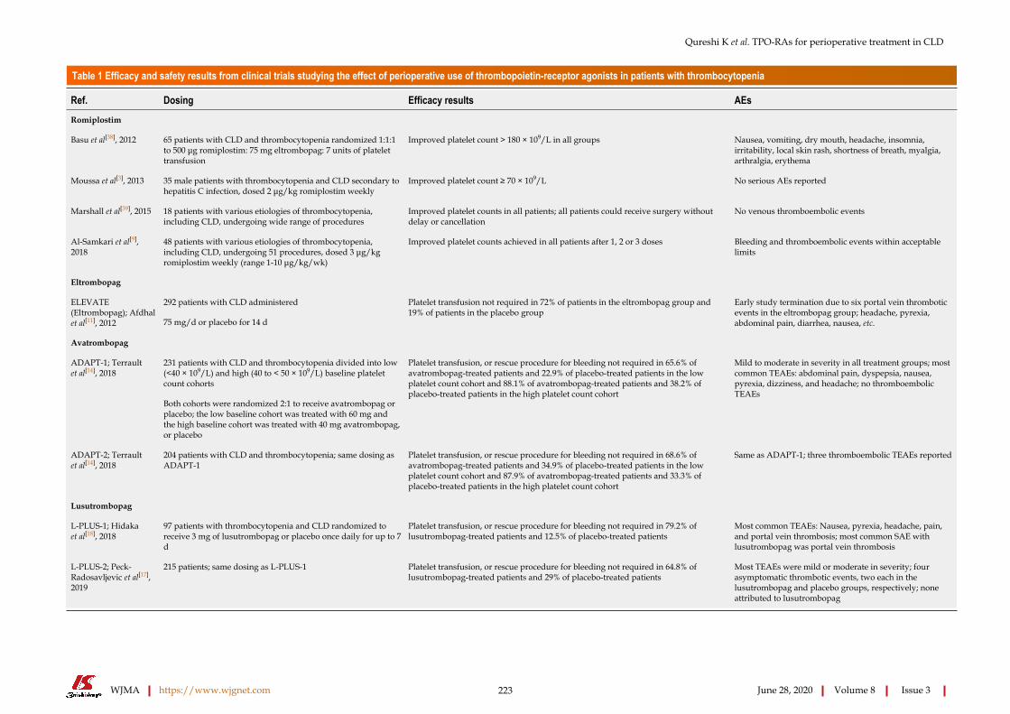
Qureshi K et al. TPO-RAs for perioperative treatment in CLD
WJMA https://www.wjgnet.com 223 June 28, 2020 Volume 8 Issue 3
Table 1 Efficacy and safety results from clinical trials studying the effect of perioperative use of thrombopoietin-receptor agonists in patients with thrombocytopenia
Ref. Dosing Efficacy results AEsRomiplostim
Basu et al[38], 2012 65 patients with CLD and thrombocytopenia randomized 1:1:1 to 500 μg romiplostim: 75 mg eltrombopag: 7 units of platelet transfusion
Improved platelet count > 180 × 109/L in all groups Nausea, vomiting, dry mouth, headache, insomnia, irritability, local skin rash, shortness of breath, myalgia, arthralgia, erythema
Moussa et al[3], 2013 35 male patients with thrombocytopenia and CLD secondary to hepatitis C infection, dosed 2 μg/kg romiplostim weekly
Improved platelet count ≥ 70 × 109/L No serious AEs reported
Marshall et al[39], 2015 18 patients with various etiologies of thrombocytopenia, including CLD, undergoing wide range of procedures
Improved platelet counts in all patients; all patients could receive surgery without delay or cancellation
No venous thromboembolic events
Al-Samkari et al[9], 2018
48 patients with various etiologies of thrombocytopenia, including CLD, undergoing 51 procedures, dosed 3 μg/kg romiplostim weekly (range 1-10 μg/kg/wk)
Improved platelet counts achieved in all patients after 1, 2 or 3 doses Bleeding and thromboembolic events within acceptable limits
Eltrombopag
292 patients with CLD administeredELEVATE (Eltrombopag); Afdhal et al[11], 2012 75 mg/d or placebo for 14 d
Platelet transfusion not required in 72% of patients in the eltrombopag group and 19% of patients in the placebo group
Early study termination due to six portal vein thrombotic events in the eltrombopag group; headache, pyrexia, abdominal pain, diarrhea, nausea, etc.
Avatrombopag
231 patients with CLD and thrombocytopenia divided into low (<40 × 109/L) and high (40 to < 50 × 109/L) baseline platelet count cohorts
ADAPT-1; Terrault et al[14], 2018
Both cohorts were randomized 2:1 to receive avatrombopag or placebo; the low baseline cohort was treated with 60 mg and the high baseline cohort was treated with 40 mg avatrombopag, or placebo
Platelet transfusion, or rescue procedure for bleeding not required in 65.6% of avatrombopag-treated patients and 22.9% of placebo-treated patients in the low platelet count cohort and 88.1% of avatrombopag-treated patients and 38.2% of placebo-treated patients in the high platelet count cohort
Mild to moderate in severity in all treatment groups; most common TEAEs: abdominal pain, dyspepsia, nausea, pyrexia, dizziness, and headache; no thromboembolic TEAEs
ADAPT-2; Terrault et al[14], 2018
204 patients with CLD and thrombocytopenia; same dosing as ADAPT-1
Platelet transfusion, or rescue procedure for bleeding not required in 68.6% of avatrombopag-treated patients and 34.9% of placebo-treated patients in the low platelet count cohort and 87.9% of avatrombopag-treated patients and 33.3% of placebo-treated patients in the high platelet count cohort
Same as ADAPT-1; three thromboembolic TEAEs reported
Lusutrombopag
L-PLUS-1; Hidaka et al[18], 2018
97 patients with thrombocytopenia and CLD randomized to receive 3 mg of lusutrombopag or placebo once daily for up to 7 d
Platelet transfusion, or rescue procedure for bleeding not required in 79.2% of lusutrombopag-treated patients and 12.5% of placebo-treated patients
Most common TEAEs: Nausea, pyrexia, headache, pain, and portal vein thrombosis; most common SAE with lusutrombopag was portal vein thrombosis
L-PLUS-2; Peck-Radosavljevic et al[17], 2019
215 patients; same dosing as L-PLUS-1 Platelet transfusion, or rescue procedure for bleeding not required in 64.8% of lusutrombopag-treated patients and 29% of placebo-treated patients
Most TEAEs were mild or moderate in severity; four asymptomatic thrombotic events, two each in the lusutrombopag and placebo groups, respectively; none attributed to lusutrombopag
Qureshi K et al. TPO-RAs for perioperative treatment in CLD
WJMA https://www.wjgnet.com 224 June 28, 2020 Volume 8 Issue 3
AE: Adverse event; CLD: Chronic liver disease; SAE: Serious adverse event; TEAE: Treatment-emergent adverse event.
thrombocytopenia in adults and children with chronic ITP who have an insufficient response to corticosteroids, immunoglobulins, or splenectomy, in patients with chronic hepatitis C virus infection to allow initiation and maintenance of interferon-based therapy, and in patients with severe aplastic anemia who have an insufficient response to immunosuppressive therapy[5].
Efficacy and safety in clinical studiesThe Eltrombopag Evaluated for Its Ability to Overcome Thrombocytopenia and Enable Procedures (referred to as “ELEVATE”) study was a double-blind, randomized, placebo-controlled, Phase 3 trial conducted in 288 patients with CLD and platelet counts below 50 × 109/L to study the ability of eltrombopag to increase platelet counts and to reduce the need for platelet transfusions in patients undergoing an elective invasive procedure[11]. Patients received either 75 mg eltrombopag once daily or placebo. The primary efficacy endpoint was the proportion of patients who did not require a platelet transfusion before, during, and up to 7 d after the elective invasive procedure, and the secondary endpoint was the proportion of patients with bleeding before, during, and up to 7 d after the procedure. A majority of patients in the eltrombopag and placebo groups (62% and 56%, respectively) underwent elective invasive procedures that were in the lowest bleeding risk category. At the end of 2 wk, 59% of patients treated with eltrombopag and 5% of those receiving placebo had an increased platelet count, and primary endpoint was achieved in 72% and 19% of patients, respectively. With the exception of thrombotic events being higher in the eltrombopag group (4% vs 1% in the placebo group), the incidence and severity of AEs were similar in both groups. Serious AEs occurred in 13% and 12% of patients in the eltrombopag and placebo groups, respectively. A total of 8 patients, comprising 6 in the eltrombopag group and 2 in the placebo group, experienced 10 thrombotic events, with 9 of those events involving the portal venous system. It must be noted that none of the 8 total patients with a thrombotic event had received both eltrombopag and a platelet/blood product transfusion, and of the 26 patients who received both eltrombopag and a transfusion, none had a thrombotic event. Additionally, 5 out of the total 8 patients who had a thrombotic event were found to have cancer. However, a separate post hoc analysis identified an association between higher platelet counts (> 200 × 109/L) and an increased risk of thrombotic events[11], and the study was terminated early due to the increased incidence of PVT in the eltrombopag arm. Key efficacy and safety data from the study are outlined in Table 1.
In the absence of better identification of risk factors for the development of thrombotic events, dose optimization, and the risk of PVT in patients with thrombocytopenia and CLD undergoing an elective procedure, eltrombopag must be used with caution and is not recommended as an alternative to platelet transfusions in
Qureshi K et al. TPO-RAs for perioperative treatment in CLD
WJMA https://www.wjgnet.com 225 June 28, 2020 Volume 8 Issue 3
this group of patients; this is an off-label use of eltrombopag.
AVATROMBOPAGAvatrombopag is an oral, small-molecule non-peptide TPO-RA that, like eltrombopag, binds to the TPO receptor to bring out a number of cellular reactions that give rise to megakaryocytic proliferation and differentiation and increased platelet production. Similar to eltrombopag and lusutrombopag[7], it does not compete with TPO for binding to the TPO receptor, and may thus have an additive effect with TPO on platelet production[6,12]. Cytochrome P450 enzymes CYP2C9 and 3A are responsible for metabolizing avatrombopag to corresponding 4-hydroxylated products, with the former enzyme playing the predominant role[13]. It is the first TPO-RA to be approved for use perioperatively to treat thrombocytopenia in adult patients with CLD scheduled to undergo a procedure[6]. Avatrombopag is an orally administered drug that is taken with food for 5 d consecutively prior to an elective procedure. Avatrombopag results in a dose- and exposure-dependent increase in platelet counts in adults within 3-5 d of treatment, with a peak effect occurring after 10-13 d and counts remaining elevated over 50 × 109/L for at least 7 d post-procedure. Baseline levels are achieved after 35 d[6].
Efficacy and safety in clinical studiesThere have been two randomized, double-blind, placebo-controlled Phase 3 trials[14] - ADAPT-1 and ADAPT-2 - with identical designs that have studied the effectiveness and safety of avatrombopag in reducing the need for platelet transfusion and rescue from bleeding in patients with CLD scheduled for a procedure. Patients were screened to exclude several criteria, including presence of arterial or venous thrombosis and a portal vein blood flow below 10 cm/s. Included patients were divided into low and high baseline platelet count cohorts and received 60 mg/d and 40 mg/d of avatrombopag, respectively. The primary endpoint of these studies was the proportion of patients who did not require a platelet transfusion or rescue procedure for bleeding after randomization and up to 7 d after a scheduled procedure. Secondary efficacy endpoints included the proportion of patients achieving the target platelet count of ≥ 50 × 109/L on the day of the procedure and the mean change in platelet count from baseline to procedure day. Patients treated with avatrombopag demonstrated increased platelet counts that were approximately 30-50 × 109/L higher on average compared with patients treated with placebo before minor invasive procedures, with a net doubling of counts on average. There were no meaningful differences in AEs of special interest among the avatrombopag-treated and placebo-treated patients, including thromboembolism, and the rates of occurrence of AEs across both the studies were comparable in the avatrombopag- and placebo-treated groups[14]. A treatment-emergent AE (TEAE) of partial PVT was observed in 1 avatrombopag-treated patient in the high baseline platelet count cohort in the ADAPT-2 trial at 13 d after the last avatrombopag dose and was assessed as nonserious and possibly related to the drug. Thromboembolic events were also observed in 2 placebo-treated patients in the ADAPT-2 trial, one being an acute myocardial infarction and the other a disseminated intravascular coagulation/ pulmonary embolus. Thromboembolic TEAEs were not noted in the ADAPT-1 trial. One non-TEAE of PVT was observed in the avatrombopag group (60 mg) at 31 d after the final dose of avatrombopag and was assessed as serious but not related. Table 1 outlines the key efficacy and safety results from these trials.
Results from a pharmacokinetic modeling study have shown that repeated dosing of avatrombopag as early as 12 d after completion of the first dosing can increase platelet counts to within safe limits[15]. Saab et al[16] have recently reported that repeated dosing of avatrombopag in 4 patients with CLD and thrombocytopenia led to a 2.3-fold mean increase in platelet count after the first dosing and 2.6-fold mean increase after the second dosing. The patients underwent procedures within 9-13 d after starting avatrombopag, with repeat dosing performed at least 30 d after completing the first dose. None of the patients required platelet transfusions or rescue therapy, or had TEAEs, and repeated dosing did not reduce the efficacy of avatrombopag[16]. Avatrombopag dosing has the ability to limit the magnitude and duration of increase in patient platelet counts and this predictable pharmacokinetic/pharmacodynamic profile could potentially be contributing to the limited risk of thromboembolic events associated with this treatment[14].
Qureshi K et al. TPO-RAs for perioperative treatment in CLD
WJMA https://www.wjgnet.com 226 June 28, 2020 Volume 8 Issue 3
LUSUTROMBOPAGLusutrombopag is an orally administered, small-molecule non-peptide TPO agonist that activates the signal transduction pathways in a manner similar to endogenous TPO (while not competing for the same binding site), leading to increased platelet production[6-7]. Lusutrombopag is metabolized via the β- and ω-oxidation pathways and glucuronidation. The cytochrome P450 enzymes are mainly responsible for its metabolism, including CYP4A11[13]. The mean maximum platelet count following treatment with 3 mg/d lusutrombopag was 86.9 × 10⁹/L and the median time to reach this was 12 d[7].
Efficacy and safety in clinical studiesData from two Phase 3 double-blind studies, L-PLUS-1 and L-PLUS-2, have shown lusutrombopag to be effective in raising platelet counts in patients with CLD and thrombocytopenia prior to undergoing an elective invasive procedure. The primary endpoint of both trials was the proportion of patients who avoided pre-procedure platelet transfusions and rescue therapy for bleeding, as assessed from randomization through 7 d post-procedure[17,18]. Key secondary endpoints included the number of patients who required no platelet transfusions during the study, the proportion of patients who achieved a platelet count of ≥ 50 × 109/L with an increase of ≥ 20 × 109/L from baseline at any time during the study, and the number of days during which the platelet count was maintained at ≥ 50 × 109/L[17]. Safety assessments included the incidence of AEs, adverse drug reactions, bleeding-related AEs, and thrombotic events. Patients treated with lusutrombopag perioperatively demonstrated improvements in platelet counts similar to those seen with avatrombopag after 7 d of treatment. In the L-PLUS-1 trial, one serious AE of PVT, possibly related to lusutrombopag treatment, was reported in the lusutrombopag-treated group and one mild incidence of superior mesenteric vein thrombosis was reported in the placebo group that was considered not related to the study drug. As neither event was associated with an extremely high platelet count, and the incidence of thrombotic events were similar in both treatment groups, the authors concluded that no further monitoring for thromboembolic events was required in lusutrombopag-treated patients. In the L-PLUS-2 trial, four TEAEs were reported, two each in the lusutrombopag- and placebo-treated groups, respectively. All events were asymptomatic and deemed to be consistent with the findings of a meta-analysis that suggested the prevalence of PVT in patients with CLD and thrombocytopenia. Table 1 outlines the key safety and efficacy results from these trials.
A recent retrospective report of 1760 cirrhotic patients with low platelet counts[2] found that 66% of patients with platelet counts below 50 × 109/L required platelet transfusions for radiofrequency ablation, 43% for transarterial chemoembolization, and 55% for endoscopic injection sclerotherapy/endoscopic variceal ligation. When 25 of these patients were administered lusutrombopag prior to the procedures, platelet counts increased significantly compared with baseline levels (82 × 109/L vs 41 × 109
/L), and of these patients, only 4 needed platelet transfusions before the procedures. The proportion of lusutrombopag-treated patients who required platelet transfusions was significantly lower (16%) compared with patients not treated with lusutrombopag (54%)[2]. Lusutrombopag was also found to be more effective in raising platelet counts in patients with baseline platelet counts above 30 × 109/L compared with patients with baseline platelet counts less than or equal to 30 × 109/L and spleen index greater than or equal to 40 cm2[2]. The authors of this retrospective study reported 1 case of portal thrombosis in the lusutrombopag group, which was revealed via a routine CT scan taken on the 12th d of treatment. The causal relationship of thrombosis with lusutrombopag treatment was unclear[2].
Sato et al[19] were the first to report the efficacy of repeated administration of lusutrombopag (3 mg daily) in a patient with hepatocellular carcinoma who had two planned invasive procedures, radiofrequency ablation and transarterial chemoembolization. In the first instance, lusutrombopag was discontinued after 5 d, as the platelet count increased to 98 × 109/L by day 5, while in the second instance, the patient required the full 7-d treatment to achieve a platelet count above 50 × 109/L. No AEs such as portal thrombus were reported during either of the treatments. More recently, Ishikawa et al[20] reported the safety and efficacy of repeated use of lusutrombopag to increase platelet counts in 8 patients with hepatocellular carcinoma and thrombocytopenia prior to repeated scheduled radiofrequency ablation procedures. Platelet counts increased from 42500 ± 5200/µL at baseline to 103100 ± 22800/µL at day 14 after first treatment, and from 43800 ± 6000/µL at baseline to 110700 ± 17800/µL at day 14 after repeat treatment, demonstrating that the repeated
Qureshi K et al. TPO-RAs for perioperative treatment in CLD
WJMA https://www.wjgnet.com 227 June 28, 2020 Volume 8 Issue 3
use of lusutrombopag does not decrease its effect on platelet counts. None of the patients developed clinical symptoms such as thrombosis or PVT[20].
HETROMBOPAGHetrombopag is a small-molecule non-peptide TPO-RA, with a mechanism of action similar to eltrombopag but with an in vivo pharmacological effect that is 8-10 times that of eltrombopag[21]. Based on animal studies, it appears that hetrombopag is primarily metabolized via glucuronidation and hydrolyzed into aglycone following excretion with bile acid[21]. A Phase 1 study demonstrated that hetrombopag is safe and well-tolerated in healthy subjects and can be a potential candidate for the treatment of patients with chronic ITP[21]. It is now in Phase 2 trials for the treatment of ITP. Currently, there are no clinical studies underway to examine the effectiveness and safety of hetrombopag as perioperative treatment of thrombocytopenia in patients with CLD.
TPO-RAs FOR OTHER INDICATION UNDER INVESTIGATIONRomiplostim and eltrombopag are currently approved for management of chronic ITP but there are studies (like the ones outlined earlier) demonstrating their efficacy in other indications. For instance, romiplostim has demonstrated efficacy in treating chemotherapy-induced thrombocytopenia and myelodysplastic syndromes[22]. Eltrombopag has been shown to be effective in the treatment of hepatitis C infection (where it raises the platelet counts enough to allow continued treatment with ribavirin and pegylated-interferon), myelodysplastic syndrome, acute myeloid leukemia, and severe aplastic anemia[23]. Both avatrombopag and lusutrombopag have been approved for use perioperatively to treat thrombocytopenia in patients with CLD. Recently, a supplemental New Drug Application was filed for avatrombopag as therapy for the treatment of chronic ITP; meanwhile, the trial for avatrombopag as therapy in chemotherapy-induced thrombocytopenia (NCT03471078) is still underway. A trial to study the safety and efficacy of hetrombopag (NCT03222843) for the treatment of chronic ITP is currently underway.
RECOMMENDATIONS ON USING PLATELET TRANSFUSIONS BY SCIENTIFIC AND GOVERNING BODIESPatients with thrombocytopenia and CLD are at an increased risk of bleeding. Platelet transfusions can be administered in these patients prophylactically, prior to scheduled medical procedures to minimize the risk of bleeding, or therapeutically to control bleeding. The goal of any prophylactic treatment is limiting hemorrhagic events in the peri- and post-operative surgical setting. However, there is uncertainty in prophylactic treatment of thrombocytopenia, as there are no effective tools that can predict bleeding risk in patients, and data on the correlation between thrombocytopenia and the risk of bleeding are equivocal[24,25]. While platelet transfusion is a primary option in these patients, the decision to undergo transfusion can be a complicated one.
Lack of consensus among guidelinesThere is a lack of consensus among guidelines related to platelet transfusion in patients, primarily due to lack of sufficient and supportive data to substantiate the recommended platelet transfusion triggers[26]. Further, there are no guidelines that provide recommendations for platelet transfusion specifically for patients with CLD prior to an elective procedure. The variations in the recommended platelet threshold values before invasive procedures often depend on the type of patient and the perceived risk of the procedure. The American Society for Gastrointestinal Endoscopy (ASGE) guidelines[27], which are not specific to gastrointestinal bleeding, state that the minimum threshold platelet count for undergoing upper gastrointestinal endoscopy has not been established, but they also point out that there are publications supporting 20 × 109/L and 50 × 109/L as the threshold for platelet counts prior to diagnostic procedures and biopsies, respectively. The American Association for the Study of Liver Diseases guidelines for transcutaneous or transvenous liver biopsy state that
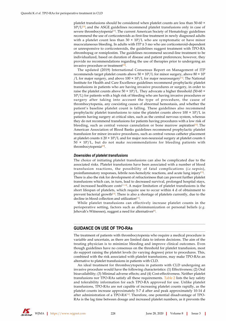
Qureshi K et al. TPO-RAs for perioperative treatment in CLD
WJMA https://www.wjgnet.com 228 June 28, 2020 Volume 8 Issue 3
platelet transfusions should be considered when platelet counts are less than 50-60 × 109/L[28], and the ASGE guidelines recommend platelet transfusions only in case of severe thrombocytopenia[29]. The current American Society of Hematology guidelines recommend the use of corticosteroids as first-line treatment in newly diagnosed adults with a platelet count less than 30 × 109/L who are symptomatic or have minor mucocutaneous bleeding. In adults with ITP ≥ 3 mo who are corticosteroid-dependent or unresponsive to corticosteroids, the guidelines suggest treatment with TPO-RA eltrombopag or romiplostim. The guidelines recommend second-line treatment to be individualized, based on duration of disease and patient preferences; however, they provide no recommendations regarding the use of therapies prior to undergoing an invasive procedure or treatment[30].
The updated (2019) International Consensus Report on Management of ITP recommends target platelet counts above 50 × 109/L for minor surgery, above 80 × 109
/L for major surgery, and above 100 × 109/L for major neurosurgery[31]. The National Institute for Health and Care Excellence guidelines recommend prophylactic platelet transfusions in patients who are having invasive procedures or surgery, in order to raise the platelet counts above 50 × 109/L. They advocate a higher threshold (50-60 × 109/L) for patients with a high risk of bleeding who are having invasive procedures or surgery after taking into account the type of procedure, the cause of thrombocytopenia, any coexisting causes of abnormal hemostasis, and whether the patient’s baseline platelet count is falling. These guidelines also recommend prophylactic platelet transfusions to raise the platelet counts above 100 × 109/L in patients having surgery at critical sites, such as the central nervous system, whereas they do not recommend transfusions for patients having procedures with a low risk of bleeding, such as central venous cannulation or bone marrow aspiration[32]. The American Association of Blood Banks guidelines recommend prophylactic platelet transfusion for minor invasive procedures, such as central venous catheter placement at platelet counts ≤ 20 × 109/L and for major non-neuraxial surgery at platelet counts ≤ 50 × 109/L, but do not make recommendations for bleeding patients with thrombocytopenia[29].
Downsides of platelet transfusionsThe choice of initiating platelet transfusions can also be complicated due to the associated risks. Platelet transfusions have been associated with a number of blood transfusion reactions, the possibility of fatal complications ( i.e. sepsis), proinflammatory responses, febrile non-hemolytic reactions, and acute lung injury[33]. There is also the risk for development of refractoriness that can prevent further platelet transfusions which can, in turn, lead to decreased survival, prolonged hospital stays, and increased healthcare costs[13,34]. A major limitation of platelet transfusions is the short lifespan of platelets, which require use to occur within 4 d of obtainment to prevent bacterial growth[33]. There is also a shortage of platelets currently, due to the decline in blood collection and utilization[35].
While platelet transfusions can effectively increase platelet counts in the perioperative setting, factors such as alloimmunization or personal beliefs (e.g. Jehovah’s Witnesses), suggest a need for alternatives[9].
GUIDANCE ON USE OF TPO-RAsThe treatment of patients with thrombocytopenia who require a medical procedure is variable and uncertain, as there are limited data to inform decisions. The aim of the treating physician is to minimize bleeding and improve clinical outcomes. Even though guidelines have no consensus on the threshold for platelet transfusion, most do support raising the platelet levels (to varying degrees) prior to procedures. This, combined with the risk associated with platelet transfusions, may make TPO-RAs an alternative to platelet transfusions in patients with CLD.
An ideal treatment for thrombocytopenia in patients with CLD undergoing an invasive procedure would have the following characteristics: (1) Effectiveness; (2) Oral bioavailability; (3) Minimal adverse effects; and (4) Cost-effectiveness. Neither platelet transfusions nor TPO-RAs satisfy all these requirements. Table 2 lists the key safety and tolerability information for each TPO-RA approved for use. Unlike platelet transfusions, TPO-RAs are not capable of increasing platelet counts rapidly, as the platelet counts increase approximately 5-7 d after and peak approximately 10-14 d after administration of a TPO-RA[36]. Therefore, one potential disadvantage of TPO-RAs is the lag time between dosage and increased platelet numbers, as it prevents the
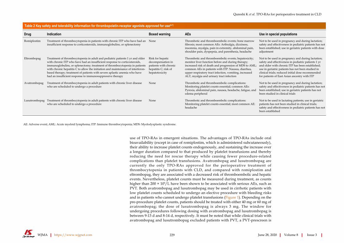
Qureshi K et al. TPO-RAs for perioperative treatment in CLD
WJMA https://www.wjgnet.com 229 June 28, 2020 Volume 8 Issue 3
Table 2 Key safety and tolerability information for thrombopoietin-receptor agonists approved for use[4-7]
Drug Indication Boxed warning AEs Use in special populations
Romiplostim Treatment of thrombocytopenia in patients with chronic ITP who have had an insufficient response to corticosteroids, immunoglobulins, or splenectomy
None Thrombotic and thromboembolic events; bone marrow fibrosis; most common AEs: Arthralgia, dizziness, insomnia, myalgia, pain in extremity, abdominal pain, shoulder pain, dyspepsia, and paresthesia, headache
Not to be used in pregnancy and during lactation; safety and effectiveness in pediatric patients has not been established; use in geriatric patients with dose adjustment
Eltrombopag Treatment of thrombocytopenia in adult and pediatric patients 1 yr and older with chronic ITP who have had an insufficient response to corticosteroids, immunoglobulins, or splenectomy; treatment of thrombocytopenia in patients with chronic hepatitis C to allow the initiation and maintenance of interferon-based therapy; treatment of patients with severe aplastic anemia who have had an insufficient response to immunosuppressive therapy
Risk for hepatic decompensation in patients with chronic hepatitis C; risk of hepatotoxicity
Thrombotic and thromboembolic events; hepatoxicity; monitor liver function before and during therapy; increased risk of death and progression of MDS to AML; common AEs in patients with ITP: Nausea, diarrhea, upper respiratory tract infection, vomiting, increased ALT, myalgia and urinary tract infection
Not to be used in pregnancy and during lactation; safety and effectiveness in pediatric patients 1 yr and older with chronic ITP has been established; use in geriatric patients has not been studied in clinical trials; reduced initial dose recommended for patients of East Asian ancestry with ITP
Avatrombopag Treatment of thrombocytopenia in adult patients with chronic liver disease who are scheduled to undergo a procedure
None Thrombotic and thromboembolic complications: Monitoring platelet counts essential; common AEs: Pyrexia, abdominal pain, nausea, headache, fatigue, and edema peripheral
Not to be used in pregnancy and during lactation; safety and effectiveness in pediatric patients has not been established; use in geriatric patients has not been studied in clinical trials
Lusutrombopag Treatment of thrombocytopenia in adult patients with chronic liver disease who are scheduled to undergo a procedure
None Thrombotic and thromboembolic complications: Monitoring platelet counts essential; most common AE: headache
Not to be used in lactating patients; use in geriatric patients has not been studied in clinical trials; safety and effectiveness in pediatric patients has not been established
AE: Adverse event; AML: Acute myeloid lymphoma; ITP: Immune thrombocytopenia; MDS: Myelodysplastic syndrome.
use of TPO-RAs in emergent situations. The advantages of TPO-RAs include oral bioavailability (except in case of romiplostim, which is administered subcutaneously), their ability to increase platelet counts endogenously, and sustaining the increase over a longer duration compared to that produced by platelet transfusions and thereby reducing the need for rescue therapy while causing fewer procedure-related complications than platelet transfusions. Avatrombopag and lusutrombopag are currently the only TPO-RAs approved for the perioperative treatment of thrombocytopenia in patients with CLD, and compared with romiplostim and eltrombopag, they are associated with a decreased risk of thromboembolic and hepatic events. Nevertheless, platelet counts must be measured during treatment, as counts higher than 200 × 109/L have been shown to be associated with serious AEs, such as PVT. Both avatrombopag and lusutrombopag may be used in cirrhotic patients with low platelet counts scheduled to undergo an elective procedure with bleeding risks and in patients who cannot undergo platelet transfusions (Figure 1). Depending on the pre-procedure platelet counts, patients should be treated with either 40 mg or 60 mg of avatrombopag; the dose of lusutrombopag is always 3 mg. The window for undergoing procedures following dosing with avatrombopag and lusutrombopag is between 9-13 d and 8-14 d, respectively. It must be noted that while clinical trials with avatrombopag and lusutrombopag excluded patients with PVT, a PVT-prescreen is
Qureshi K et al. TPO-RAs for perioperative treatment in CLD
WJMA https://www.wjgnet.com 230 June 28, 2020 Volume 8 Issue 3
Figure 1 Patient criteria and general schema for administration of oral thrombopoietin-receptor agonists in patients with chronic liver disease and thrombocytopenia scheduled to undergo a procedure. Ava: Avatrombopag; Lusu: Lusutrombopag; TPO-RAs: Thrombopoietin-receptor agonists.
not a requirement as per labeling guidelines for both these TPO-RAs. Patients with liver cirrhosis and high Child-Turcotte-Pugh scores are more likely to have severe thrombocytopenia and also require invasive diagnostic procedures. Extreme caution must be taken when using TPO-RAs in such patients, as they are at high risk of developing PVT; currently, no data on the effects of the TPO-RAs avatrombopag and lusutrombopag in this class of patients are available, as they were excluded from the clinical trials[14,16-18].
There are also pricing differences between avatrombopag and lusutrombopag; in the United States, the wholesale acquisition cost (WAC) of avatrombopag for a 5-d course is $2970 for the 40-mg dose and $4455 for the 60-mg dose. The WAC for a 7-d course of lusutrombopag is $8500[37].
CONCLUSIONRecommendations for procedure-specific minimum platelet counts prior to a scheduled procedure often have inadequate supporting evidence, and the benefit of prophylactic platelet transfusion remains unclear. Physicians typically assess the need for platelet transfusions on a patient-to-patient basis, relying on their own experience and patient comorbidities to inform their decisions. TPO-RAs are a safe and effective alternative treatment option for patients with thrombocytopenia who are undergoing a medical procedure. They are associated with increased platelet counts, decreased bleeding events, and a reduced need for rescue treatments. Recent studies also support the safe and efficacious repeated use of TPO-RAs in patients with CLD. The exact fit of TPO-RAs, as compared with platelet transfusions, in patients with thrombocytopenia prior to undergoing a medical procedure is still unclear. Comparative clinical trial data in patients with thrombocytopenia are required to assess the efficacy, safety, cost-effectiveness, and impact on patient quality of life of TPO-RA use compared with the traditional use of platelet transfusions prior to scheduling a medical procedure. In the meantime, treatment decisions must be individualized by establishing the advantages and disadvantages of both these treatment options on a case-by-case basis.
REFERENCES1 Siegal D, Crowther M, Cuker A. Thrombopoietin receptor agonists in primary immune thrombocytopenia.
Semin Hematol 2013; 50 Suppl 1: S18-S21 [PMID: 23664510 DOI: 10.1053/j.seminhematol.2013.03.005]2 Takada H, Kurosaki M, Nakanishi H, Takahashi Y, Itakura J, Tsuchiya K, Yasui Y, Tamaki N, Takaura K,
Komiyama Y, Higuchi M, Kubota Y, Wang W, Okada M, Shimizu T, Watakabe K, Enomoto N, Izumi N. Real-life experience of lusutrombopag for cirrhotic patients with low platelet counts being prepared for invasive procedures. PLoS One 2019; 14: e0211122 [PMID: 30768601 DOI: 10.1371/journal.pone.0211122]Moussa MM, Mowafy N. Preoperative use of romiplostim in thrombocytopenic patients with chronic hepatitis C and liver cirrhosis. J Gastroenterol Hepatol 2013; 28: 335-341 [PMID: 22849409 DOI:
3

Qureshi K et al. TPO-RAs for perioperative treatment in CLD
WJMA https://www.wjgnet.com 231 June 28, 2020 Volume 8 Issue 3
10.1111/j.1440-1746.2012.07246.x]4 Amgen Inc. Nplate [package insert]. Thousand Oaks: Amgen Inc., 20175 Novartis Pharmaceuticals Corporation. Promacta [package insert]. East Hanover: Novartis
Pharmaceuticals Corporation, 20186 Dova Pharmaceuticals, Inc. Doptelet [package insert]. Durham: Dova Pharmaceuticals, Inc., 20187 Shionogi, Inc. Mulpleta [package insert]. Florham Park: Shionogi, Inc., 20188 Estcourt LJ, Birchall J, Allard S, Bassey SJ, Hersey P, Kerr JP, Mumford AD, Stanworth SJ, Tinegate H;
British Committee for Standards in Haematology. Guidelines for the use of platelet transfusions. Br J Haematol 2017; 176: 365-394 [PMID: 28009056 DOI: 10.1111/bjh.14423]
9 Al-Samkari H, Marshall AL, Goodarzi K, Kuter DJ. Romiplostim for the management of perioperative thrombocytopenia. Br J Haematol 2018; 182: 106-113 [PMID: 29767837 DOI: 10.1111/bjh.15280]
10 Dultz G, Kronenberger B, Azizi A, Mihm U, Vogl TJ, Sarrazin U, Sarrazin C, Zeuzem S, Hofmann WP. Portal vein thrombosis as complication of romiplostim treatment in a cirrhotic patient with hepatitis C-associated immune thrombocytopenic purpura. J Hepatol 2011; 55: 229-232 [PMID: 21310200 DOI: 10.1016/j.jhep.2011.01.020]
11 Afdhal NH, Giannini EG, Tayyab G, Mohsin A, Lee JW, Andriulli A, Jeffers L, McHutchison J, Chen PJ, Han KH, Campbell F, Hyde D, Brainsky A, Theodore D; ELEVATE Study Group. Eltrombopag before procedures in patients with cirrhosis and thrombocytopenia. N Engl J Med 2012; 367: 716-724 [PMID: 22913681 DOI: 10.1056/NEJMoa1110709]
12 Kuter DJ. New thrombopoietic growth factors. Blood 2007; 109: 4607-4616 [PMID: 17289815 DOI: 10.1182/blood-2006-10-019315]
13 Bankston PC, Al-Horani RA. New small molecule drugs for thrombocytopenia: chemical, pharmacological, and therapeutic use considerations. Int J Mol Sci 2019; 20: 3013 [PMID: 31226783 DOI: 10.3390/ijms20123013]
14 Terrault N, Chen YC, Izumi N, Kayali Z, Mitrut P, Tak WY, Allen LF, Hassanein T. Avatrombopag before procedures reduces need for platelet transfusion in patients with chronic liver disease and thrombocytopenia. Gastroenterology 2018; 155: 705-718 [PMID: 29778606 DOI: 10.1053/j.gastro.2018.05.025]
15 Hayes S, Farrell C, Aggarwal K, Vredenburg M, Allen LF. PK simulation of avatrombopag-induced increases in platelet counts with redosing in patients with thrombocytopenia and chronic liver disease. Res Pract Thromb Hemost 2018; 2: 665 [DOI: 10.1016/j.jvir.2018.12.665]
16 Saab S, McDaniel TK, Bau SN, Patel RP. Efficacy of repeat doses of avatrombopag: a case series. Dig Med Res 2019; 2: 9 [DOI: 10.21037/dmr.2019.04.03]
17 Peck-Radosavljevic M, Simon K, Iacobellis A, Hassanein T, Kayali Z, Tran A, Makara M, Ben Ari Z, Braun M, Mitrut P, Yang SS, Akdogan M, Pirisi M, Duggal A, Ochiai T, Motomiya T, Kano T, Nagata T, Afdhal N. Lusutrombopag for the Treatment of Thrombocytopenia in Patients With Chronic Liver Disease Undergoing Invasive Procedures (L-PLUS 2). Hepatology 2019; 70: 1336-1348 [PMID: 30762895 DOI: 10.1002/hep.30561]
18 Hidaka H, Kurosaki M, Tanaka H, Kudo M, Abiru S, Igura T, Ishikawa T, Seike M, Katsube T, Ochiai T, Kimura K, Fukuhara T, Kano T, Nagata T, Tanaka K, Kurokawa M, Yamamoto K, Osaki Y, Izumi N, Imawari M. Lusutrombopag reduces need for platelet transfusion in patients with thrombocytopenia undergoing invasive procedures. Clin Gastroenterol Hepatol 2019; 17: 1192-1200 [PMID: 30502505 DOI: 10.1016/j.cgh.2018.11.047]
19 Sato S, Miyake T, Kataoka M, Isoda K, Yazaki T, Tobita H, Ishimura N, Kinoshita Y. Efficacy of repeated lusutrombopag administration for thrombocytopenia in a patient scheduled for invasive hepatocellular carcinoma treatment. Intern Med 2017; 56: 2887-2890 [PMID: 28943563 DOI: 10.2169/internalmedicine.8791-16]
20 Ishikawa T, Okoshi M, Tomiyoshi K, Kojima Y, Horigome R, Imai M, Nozawa Y, Iwanaga A, Sano T, Honma T, Yoshida T. Efficacy and safety of repeated use of lusutrombopag prior to radiofrequency ablation in patients with recurrent hepatocellular carcinoma and thrombocytopenia. Hepatol Res 2019; 49: 590-593 [PMID: 30602063 DOI: 10.1111/hepr.13305]
21 Zheng L, Liang MZ, Zeng XL, Li CZ, Zhang YF, Chen XY, Zhu X, Xiang AB. Safety, pharmacokinetics and pharmacodynamics of hetrombopag olamine, a novel TPO-R agonist, in healthy individuals. Basic Clin Pharmacol Toxicol 2017; 121: 414-422 [PMID: 28544774 DOI: 10.1111/bcpt.12815]
22 Giagounidis A, Mufti GJ, Fenaux P, Sekeres MA, Szer J, Platzbecker U, Kuendgen A, Gaidano G, Wiktor-Jedrzejczak W, Hu K, Woodard P, Yang AS, Kantarjian HM. Results of a randomized, double-blind study of romiplostim versus placebo in patients with low/intermediate-1-risk myelodysplastic syndrome and thrombocytopenia. Cancer 2014; 120: 1838-1846 [PMID: 24706489 DOI: 10.1002/cncr.28663]
23 Gill H, Wong RSM, Kwong YL. From chronic immune thrombocytopenia to severe aplastic anemia: recent insights into the evolution of eltrombopag. Ther Adv Hematol 2017; 8: 159-174 [PMID: 28473904 DOI: 10.1177/2040620717693573]
24 Seeff LB, Everson GT, Morgan TR, Curto TM, Lee WM, Ghany MG, Shiffman ML, Fontana RJ, Di Bisceglie AM, Bonkovsky HL, Dienstag JL; HALT–C Trial Group. Complication rate of percutaneous liver biopsies among persons with advanced chronic liver disease in the HALT-C trial. Clin Gastroenterol Hepatol 2010; 8: 877-883 [PMID: 20362695 DOI: 10.1016/j.cgh.2010.03.025]
25 Basili S, Raparelli V, Napoleone L, Talerico G, Corazza GR, Perticone F, Sacerdoti D, Andriulli A, Licata A, Pietrangelo A, Picardi A, Raimondo G, Violi F; PRO-LIVER Collaborators. Platelet count does not predict bleeding in cirrhotic patients: results from the PRO-LIVER study. Am J Gastroenterol 2018; 113: 368-375 [PMID: 29257146 DOI: 10.1038/ajg.2017.457]
26 Szczepiorkowski ZM, Dunbar NM. Transfusion guidelines: when to transfuse. Hematology Am Soc Hematol Educ Program 2013; 2013: 638-644 [PMID: 24319244 DOI: 10.1182/asheducation-2013.1.638]
27 ASGE Standards of Practice Committee, Ben-Menachem T, Decker GA, Early DS, Evans J, Fanelli RD, Fisher DA, Fisher L, Fukami N, Hwang JH, Ikenberry SO, Jain R, Jue TL, Khan KM, Krinsky ML, Malpas PM, Maple JT, Sharaf RN, Dominitz JA, Cash BD. Adverse events of upper GI endoscopy. Gastrointest Endosc 2012; 76: 707-718 [PMID: 22985638 DOI: 10.1016/j.gie.2012.03.252]

Qureshi K et al. TPO-RAs for perioperative treatment in CLD
WJMA https://www.wjgnet.com 232 June 28, 2020 Volume 8 Issue 3
28 Rockey DC, Caldwell SH, Goodman ZD, Nelson RC, Smith AD; American Association for the Study of Liver Diseases. Liver biopsy. Hepatology 2009; 49: 1017-1044 [PMID: 19243014 DOI: 10.1002/hep.22742]
29 Laine L. Treatment of thrombocytopenic patients with GI bleeding. Gastrointest Endosc 2018; 88: 62-65 [PMID: 29935628 DOI: 10.1016/j.gie.2018.03.003]
30 Neunert C, Terrell DR, Arnold DM, Buchanan G, Cines DB, Cooper N, Cuker A, Despotovic JM, George JN, Grace RF, Kühne T, Kuter DJ, Lim W, McCrae KR, Pruitt B, Shimanek H, Vesely SK. American Society of Hematology 2019 guidelines for immune thrombocytopenia. Blood Adv 2019; 3: 3829-3866 [PMID: 31794604 DOI: 10.1182/bloodadvances.2019000966]
31 Provan D, Arnold DM, Bussel JB, Chong BH, Cooper N, Gernsheimer T, Ghanima W, Godeau B, González-López TJ, Grainger J, Hou M, Kruse C, McDonald V, Michel M, Newland AC, Pavord S, Rodeghiero F, Scully M, Tomiyama Y, Wong RS, Zaja F, Kuter DJ. Updated international consensus report on the investigation and management of primary immune thrombocytopenia. Blood Adv 2019; 3: 3780-3817 [PMID: 31770441 DOI: 10.1182/bloodadvances.2019000812]
32 National Institute for Health and Care Exellence. Blood transfusion. Available from: https://www.nice.org.uk/guidance/ng24
33 Kurokawa T, Ohkohchi N. Platelets in liver disease, cancer and regeneration. World J Gastroenterol 2017; 23: 3228-3239 [PMID: 28566882 DOI: 10.3748/wjg.v23.i18.3228]
34 Brown RS Jr. Review article: a pharmacoeconomic analysis of thrombocytopenia in chronic liver disease. Aliment Pharmacol Ther 2007; 26 Suppl 1: 41-48 [PMID: 17958518 DOI: 10.1111/j.1365-2036.2007.03505.x]
35 Ellingson KD, Sapiano MRP, Haass KA, Savinkina AA, Baker ML, Chung KW, Henry RA, Berger JJ, Kuehnert MJ, Basavaraju SV. Continued decline in blood collection and transfusion in the United States-2015. Transfusion 2017; 57 Suppl 2: 1588-1598 [PMID: 28591469 DOI: 10.1111/trf.14165]
36 Nagrebetsky A, Al-Samkari H, Davis NM, Kuter DJ, Wiener-Kronish JP. Perioperative thrombocytopenia: evidence, evaluation, and emerging therapies. Br J Anaesth 2019; 122: 19-31 [PMID: 30579402 DOI: 10.1016/j.bja.2018.09.010]
37 Elsevier. ProspectoRx. Available from: https://www.elsevier.com/solutions/prospectorx38 Basu PP NT, Farhat S, James Shah N, Jafri M, Foustin S. Single use of romiplostim thrombopoietin
analogue in severe thrombocytopenia for outpatient percutaneous liver biopsy in patients with chronic liver disease-A randomized double blinded prospective trial. J Hepatol 2012; 56: S38 [DOI: 10.1016/S0168-8278(12)60101-9]
39 Marshall AL, Goodarzi K, Kuter DJ. Romiplostim in the management of the thrombocytopenic surgical patient. Transfusion 2015; 55: 2505-2510 [PMID: 26033367 DOI: 10.1111/trf.13181]

WJMA https://www.wjgnet.com 233 June 28, 2020 Volume 8 Issue 3
World Journal of
Meta-AnalysisW J M ASubmit a Manuscript: https://www.f6publishing.com World J Meta-Anal 2020 June 28; 8(3): 233-244
DOI: 10.13105/wjma.v8.i3.233 ISSN 2308-3840 (online)
MINIREVIEWS
Role of non-coding RNAs in pathogenesis of gastrointestinal stromal tumors
Ioannis K Stefanou, Maria Gazouli, Georgios C Zografos, Konstantinos G Toutouzas
ORCID number: Ioannis K Stefanou 0000-0002-9366-0432; Maria Gazouli 0000-0002-3295-6811; George C Zografos 0000-0002-5429-8776; Konstantinos G Toutouzas 0000-0002-7830-6945.
Author contributions: Stefanou IK, Gazouli M, Zografos GC, Toutouzas KG contributed to the writing of the manuscript.
Conflict-of-interest statement: There is no conflict of interest.
Open-Access: This article is an open-access article that was selected by an in-house editor and fully peer-reviewed by external reviewers. It is distributed in accordance with the Creative Commons Attribution NonCommercial (CC BY-NC 4.0) license, which permits others to distribute, remix, adapt, build upon this work non-commercially, and license their derivative works on different terms, provided the original work is properly cited and the use is non-commercial. See: http://creativecommons.org/licenses/by-nc/4.0/
Manuscript source: Invited manuscript
Received: May 11, 2020 Peer-review started: May 11, 2020 First decision: June 15, 2020 Revised: June 22, 2020
Ioannis K Stefanou, Department of Surgery, Hippocration Hospital Athens, Athens 11527, Greece
Maria Gazouli, Department of Basic Medical Sciences, Laboratory of Biology, National and Kapodistrian University of Athens, Athens 11527, Greece
Georgios C Zografos, Konstantinos G Toutouzas, 1st Propaedeutic Department of Surgery, National and Kapodistrian University of Athens, Athens 11527, Greece
Corresponding author: Maria Gazouli, PhD, Associate Professor, Department of Basic Medical Sciences, Laboratory of Biology, Medical School, National and Kapodistrian University of Athens, Michalakopoulou 176, Athens 11527, Greece. [email protected]
AbstractGastrointestinal stromal tumors (GISTs) are considered the model solid malignancies of targeted therapy after the discovery of imatinib effectiveness against their tyrosine kinase inhibitors. Non-coding RNAs are molecules with no protein coding capacity that play crucial role to several biological steps of normal cell proliferation and differentiation. When the expression of these molecules found to be altered it seems that they affect the process of carcinogenesis in multiple ways, such as proliferation, apoptosis, differentiation, metastasis, and drug resistance. This review aims to provide an overview of the latest research papers and summarize the current evidence about the role of non-coding RNAs in pathogenesis of GISTs, including their potential clinical applications.
Key words: Gastrointestinal stromal tumors; Non-coding RNA; MicroRNA; Transcriptomics; Biomarker; Long non-coding RNAs
©The Author(s) 2020. Published by Baishideng Publishing Group Inc. All rights reserved.
Core tip: There are several excellent reviews at the last decade contributed the role of non-coding RNAs in gastrointestinal stromal tumors (GISTs) carcinogenesis. However, until now, most of them focused only on the microRNAs characteristics. Recently there has been a substantial motion in understanding the role of other non-coding RNAs in GIST progress, like the long non-coding RNAs. This review provides an overview of both microRNAs and long non-coding RNAs role in GIST progression, their potential
Stefanou IK et al. LncRNAs and GISTs
WJMA https://www.wjgnet.com 234 June 28, 2020 Volume 8 Issue 3
Accepted: June 28, 2020 Article in press: June 28, 2020 Published online: June 28, 2020
P-Reviewer: Paul J, Zhu JM S-Editor: Wang JL L-Editor: A E-Editor: Liu JH
therapeutic use, their ability to predict drug sensitivity and many other aspects concerning GIST development.
Citation: Stefanou IK, Gazouli M, Zografos GC, Toutouzas KG. Role of non-coding RNAs in pathogenesis of gastrointestinal stromal tumors. World J Meta-Anal 2020; 8(3): 233-244URL: https://www.wjgnet.com/2308-3840/full/v8/i3/233.htmDOI: https://dx.doi.org/10.13105/wjma.v8.i3.233
INTRODUCTIONNon-coding RNAsThe discovery of transfer RNA (tRNA), and ribosomal RNA (rRNA), in the 1950s is the beginning of the history of the non-coding RNAs (ncRNAs) that play functional roles in the eukaryotic cells[1]. James Watson imagined the one gene, one ribosome and one protein hypothesis (central dogma). Therefore, RNA changed from being a just information carrying molecule, to having three flavors. rRNA, tRNA and everything else was assumed to be mRNA[2]. Later on, in the 70s Stark et al[3] published the existence of other functional RNAs like ribonuclease P and snRNAs[4]. One of the prominent examples about how huge was the surprise at this period of time, was the eventual renaming of signal recognition protein to signal recognition particle (SRP-RNA). That happened after the discovery, that it contains a 7S RNA (by Walter et al[5]). In the early 90s, other long intergenic non coding RNAs were discovered, like XIST, by Brockdorff et al[6] Nowadays, it is generally known, according to the encyclopedia of DNA elements (published by the ENCODE Project[7]) consortium that the 80% of the human genome is transcripted for RNA molecules that have no protein coding capacity[8]. In the past, it was believed that this huge amount of RNA molecules was a transcriptional noise. Contrariwise, they appear to have direct function as regulators in several endocytic molecular paths. They seem to play crucial role in differentiation, development, and apoptosis of normal cells[9], so even in the era of complete genome sequences, non-coding RNAs gene have been eventually invisible. These features of non-coding RNAs have turned them into one of the most promising fields of scientific research.
ncRNAs are classified into two big subgroups according to their size[10].
Short ncRNAs, with < 200 nucleotides (nts) in length and include: MicroRNAs (miRNAs) usually bind to a specific molecular locum at the mRNA to induce degradation or block the prosses of translation. In addition, this may be done in the context of a feedback mechanism that involves chromosome methylation.
Small interfering RNAs (siRNAs) have a similar function as miRNAs with the additional feature of inducing heterochromatin formation through RNA transcriptional silencing complex which, when bound to siRNA, promotes H3K9 methylation and chromatin condensation.
Piwi-interacting RNAs seem to interact with the piwi family proteins. They involve in chromatin regulation and suppression of transposon activity in germline and somatic cells[11].
Long ncRNAs (lncRNAs) are longer than 200nt and may comprise thousands of nucleotides[12]: This group includes the long intergenic ncRNAs (lincRNAs), the natural antisense transcript, the transcripted ultraconserved regions and non-coding pseudogenes[13]. It seems to be transcribed mostly by RNA polymerase 2 as the mRNA does but they do not undergo the standard processing steps[14]. The mechanism of their function is generally unknown, but it is suggesting that it is similar to that of HOX antisense intergenic RNA (HOTAIR) which is the most studied lncRNA. It regulates chromatin methylation of the HOXD locus through polycomb repressive complex 2. HOTAIR was recently reported to play a crucial role in metastatic disease and may be a good prognostic marker in patients with breast cancer[15].
Post-transcriptional modifications that occur in RNA molecules started being explored at the recent years and therefore led to a new field of research called epitranscriptomics. Equivalent to epigenetics, which analyzes the post-transcriptional events occurring in DNA, epitranscriptomics investigates modifications resulting from all RNA processing events, such as RNA splicing, RNA editing, or methylation[16].
Stefanou IK et al. LncRNAs and GISTs
WJMA https://www.wjgnet.com 235 June 28, 2020 Volume 8 Issue 3
Gastrointestinal stromal tumorsGastrointestinal stromal tumors (GISTs) are specific, generally c-Kit (CD117)-positive, mesenchymal tumors of the gastrointestinal tract, encompassing a majority of tumors previously considered gastrointestinal smooth muscle tumors[17]. They are believed to originate from interstitial cells of Cajal or related stem cells. Interstitial cells of Cajal and GIST cells express the hematopoietic progenitor cell marker CD34 and the growth factor receptor c-Kit. Expression of the c-Kit gene protein product, CD117, has emerged as an important defining feature of GISTs[18,19]. Using these criteria, the incidence of GISTs has been estimated to be 6 to 15 cases per million individuals per year[20]. They constitute a significant percentage ranging from 1%-2% of all the gastrointestinal neoplasms. The most common genetic alterations found in GISTs include mutations of growth factors genes such as c-Kit (70–80%) and PDGFRA (platelet-derived growth factor A, 5%-8%). Several features of GISTs have been postulated in the past to predict their clinical behavior. Nowadays, much is known about the histological, immunohistochemical and molecular aspects of GISTs especially in diagnostic purposes[21,22]. However, little is known about the clinicopathological features that can predict the biological behavior of these tumors.
At the recent years, plenty of studies have revealed the specific molecular characteristics of GISTs. Nowadays, these tumors are considered among the best genetically understood human cancers[23].
Especially after the discovery of their sensitivity to tyrosine kinase inhibitors, GISTs tend to be referred as ideal tumor for novel molecular targeted therapies. Apart from that, the fact that many studies have been published specific chromosomal changes (e.g. loss of 14q), genetic mutations (e.g. KIT, PDGFRA), gene expression profiles (e.g. ETV1, fascin1) and miRNA expression profiles, have contributed to make them one of the well-recognized tumors[24]. It is important to mention that KIT and PDGFRA mutations are almost exclusive in GISTs, which makes them specific biomarkers of these tumors. The gold standard therapy in primary localized GISTs is a R0 surgical resection[25]. First line therapy for the advanced disease is Imatinib that offers a dramatic response, in most of the cases, for about 2-3 years[26]. After long term treatment, resistance is quite common. Sunitinib and regorafenib are the second line agents in imatinib resistant GISTs with also unsatisfactory outcomes in progressive disease[27]. Therefore, further fundamental clinical studies are being conducted in order to provide improved diagnostic modalities to increase the possibility for the patients to be diagnosed in early disease, and furthermore provide novel therapeutic options for the advanced disease cases.
NcRNAs in GISTsAt the present, a clear relationship with GISTs has been reported for only a few ncRNA classes, especially miRNAs and some lncRNAs such as the ultra-conserved genes, HOTAIR, H19, MALAT1 and CCDC26[28,29]. The other types of ncRNAs it seems to participate in the genetic puzzle that gives rise to carcinogenic phenotype[13].
miRNAs are the most widely studied class of ncRNAs in GISTs and generally in human cancer. These small ncRNAs of approximately 22 nucleotides, mediate post-transcriptional gene silencing by controlling the translation of mRNA into proteins. miRNAs are estimated to regulate the translation of more than 60% of protein-coding genes[30]. They are involved in regulating many processes, including proliferation, development, differentiation, and apoptosis. Alterations of miRNAs expression profile has been reported in GISTs, and is associated with tumor location, mutation status, tumor risk, and chromosomal changes[31]. Two excellent reviews by Nannini et al[32], and Kupcinskas et al[33] have perfectly analyzed relevant miRNA profiling studies. Since then several papers came out concerning ncRNA and GISTs.
STUDIES SELECTIONThis review included all studies published in PubMed database related to the role of ncRNAs in GIST published from 2008 to 2020. The keywords we used to retrieve the papers were GIST, ncRNAs, miRNAs and lncRNAs. 82 papers selected using these keywords. According the selection criteria, only 52 of them were relevant to the topic, 32 profiling studies, 9 reviews, 11 other studies (Figure 1).
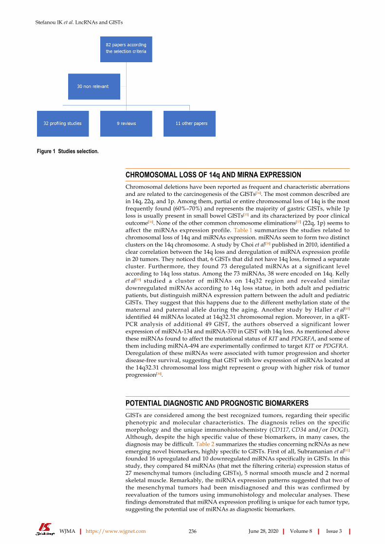
Stefanou IK et al. LncRNAs and GISTs
WJMA https://www.wjgnet.com 236 June 28, 2020 Volume 8 Issue 3
Figure 1 Studies selection.
CHROMOSOMAL LOSS OF 14q AND MIRNA EXPRESSIONChromosomal deletions have been reported as frequent and characteristic aberrations and are related to the carcinogenesis of the GISTs[34]. The most common described are in 14q, 22q, and 1p. Among them, partial or entire chromosomal loss of 14q is the most frequently found (60%–70%) and represents the majority of gastric GISTs, while 1p loss is usually present in small bowel GISTs[35] and its characterized by poor clinical outcome[36]. None of the other common chromosome eliminations[37] (22q, 1p) seems to affect the miRNAs expression profile. Table 1 summarizes the studies related to chromosomal loss of 14q and miRNAs expression. miRNAs seem to form two distinct clusters on the 14q chromosome. A study by Choi et al[38] published in 2010, identified a clear correlation between the 14q loss and deregulation of miRNA expression profile in 20 tumors. They noticed that, 6 GISTs that did not have 14q loss, formed a separate cluster. Furthermore, they found 73 deregulated miRNAs at a significant level according to 14q loss status. Among the 73 miRNAs, 38 were encoded on 14q. Kelly et al[39] studied a cluster of miRNAs on 14q32 region and revealed similar downregulated miRNAs according to 14q loss statue, in both adult and pediatric patients, but distinguish miRNA expression pattern between the adult and pediatric GISTs. They suggest that this happens due to the different methylation state of the maternal and paternal allele during the aging. Another study by Haller et al[40] identified 44 miRNAs located at 14q32.31 chromosomal region. Moreover, in a qRT-PCR analysis of additional 49 GIST, the authors observed a significant lower expression of miRNA-134 and miRNA-370 in GIST with 14q loss. As mentioned above these miRNAs found to affect the mutational status of KIT and PDGRFA, and some of them including miRNA-494 are experimentally confirmed to target KIT or PDGFRA. Deregulation of these miRNAs were associated with tumor progression and shorter disease-free survival, suggesting that GIST with low expression of miRNAs located at the 14q32.31 chromosomal loss might represent o group with higher risk of tumor progression[36].
POTENTIAL DIAGNOSTIC AND PROGNOSTIC BIOMARKERSGISTs are considered among the best recognized tumors, regarding their specific phenotypic and molecular characteristics. The diagnosis relies on the specific morphology and the unique immunohistochemistry (CD117, CD34 and/or DOG1). Although, despite the high specific value of these biomarkers, in many cases, the diagnosis may be difficult. Table 2 summarizes the studies concerning ncRNAs as new emerging novel biomarkers, highly specific to GISTs. First of all, Subramanian et al[41] founded 16 upregulated and 10 downregulated miRNAs specifically in GISTs. In this study, they compared 84 miRNAs (that met the filtering criteria) expression status of 27 mesenchymal tumors (including GISTs), 5 normal smooth muscle and 2 normal skeletal muscle. Remarkably, the miRNA expression patterns suggested that two of the mesenchymal tumors had been misdiagnosed and this was confirmed by reevaluation of the tumors using immunohistology and molecular analyses. These findings demonstrated that miRNA expression profiling is unique for each tumor type, suggesting the potential use of miRNAs as diagnostic biomarkers.
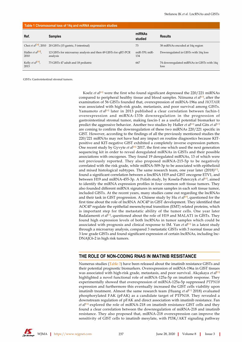
Stefanou IK et al. LncRNAs and GISTs
WJMA https://www.wjgnet.com 237 June 28, 2020 Volume 8 Issue 3
Table 1 Chromosomal loss of 14q and miRNA expression studies
Ref. Samples miRNAs studied Results
Choi et al[38], 2010 20 GISTs (15 gastric, 5 intestinal) 73 38 miRNAs encoded at 14q region
Haller et al[40], 2010
12 GISTs for microarray analysis and then 49 GISTs for qRT-PCR analysis
miR-370; miR-134
Downregulated in GISTs with 14q loss
Kelly et al[39], 2013
73 GISTs 47 adult and 18 pediatric 667 74 downregulated miRNAs in GISTs with 14q loss
GISTs: Gastrointestinal stromal tumors.
Koelz et al[42] were the first who found significant depressed the 220/221 miRNAs compared to peripheral healthy tissue and blood samples. Niinuma et al[43], after the examination of 56 GISTs founded that, overexpression of miRNA-196a and HOTAIR was associated with high-risk grade, metastasis, and poor survival among GISTs. Yamamoto et al[44] later in 2013 published a clear correlation between fachin-1 overexpression and miRNA-133b downregulation in the progression of gastrointestinal stromal tumor, making fascin-1 as a useful potential biomarker to predict the aggressive behavior. Another two studies by Haller et al[40] and Gits et al[45] are coming to confirm the downregulation of these two miRNAs 220/221 specific in GIST. However, according to the findings of all the previously mentioned studies the 220/221 miRNAs may not have had any impact on routine diagnostics because KIT-positive and KIT-negative GIST exhibited a completely inverse expression pattern. One recent study by Gyvyte et al[46] 2017, the first one which used the next generation sequencing kit in order to reveal deregulated miRNAs in GISTs and their possible associations with oncogenes. They found 19 deregulated miRNAs, 13 of which were not previously reported. They also proposed miRNA-215-5p to be negatively correlated with the risk grade, while miRNA-509-3p to be associated with epithelioid and mixed histological subtypes. The same research team, one year later (2018)[47], found a significant correlation between a lincRNA H19 and GIST oncogene ETV1, and between H19 and miRNA-455-3p. A Polish study, by Kosela-Paterczyk et al[48], aimed to identify the miRNA expression profiles in four common soft tissue tumors. They also founded different miRNA signatures in serum samples in each soft tissue tumor, included GISTs. At the recent years, many studies came out regarding the lncRNAs and their task in GIST progression. A Chinese study by Hu et al[49], questioned for the first time about the role of lncRNA AOC4P in GIST development. They identified that AOC4P regulate the epithelial mesenchymal transition (EMT) related proteins, which is important step for the metastatic ability of the tumor cells. One year later Badalamenti et al[50], questioned about the role of H19 and MALAT1 in GISTs. They found high expression levels of both lncRNAs in tumor samples which could be associated with prognosis and clinical response to IM. Yan et al[51] in a latest study through a microarray analysis, compared 3 metastatic GISTs with 3 normal tissue and 3 low grade GISTs and found significant expression of certain lncRNAs, including lnc-DNAJC6-2 in high risk tumors.
THE ROLE OF NON-CODING RNAS IN IMATINIB RESISTANCENumerus studies (Table 3) have been released about the imatinib resistance GISTs and their potential prognostic biomarkers. Overexpression of miRNA-196a in GIST tissues was associated with high-risk grade, metastasis, and poor survival. Akçakaya et al[52] highlighted a novel functional role of miRNA-125a-5p on imatinib response. They experimentally showed that overexpression of miRNA-125a-5p suppressed PTPN18 expression and furthermore this eventually increased the GIST cells viability upon imatinib treatment. Almost the same research team (Huang et al[53] 2018) evaluated phosphorylated FAK (pFAK) as a candidate target of PTPN18. They revealed a downstream regulation of pFAK and direct association with imatinib resistance. Fan et al[54] explored the role of miRNA-218 on imatinib resistance GIST cells and they found a clear correlation between the downregulation of miRNA-218 and imatinib resistance. They also proposed that, miRNA-218 overexpression can improve the sensitivity of GIST cells to imatinib mesylate, with PI3K/AKT signaling pathway
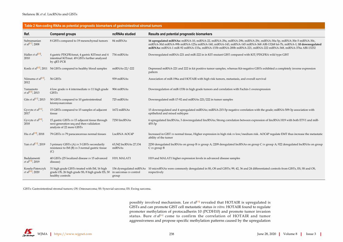
Stefanou IK et al. LncRNAs and GISTs
WJMA https://www.wjgnet.com 238 June 28, 2020 Volume 8 Issue 3
Table 2 Non-coding RNAs as potential prognostic biomarkers of gastrointestinal stromal tumors
Ref. Compared groups ncRNAs studied Results and potential prognostic biomarkers
Subramanian et al[41], 2008
8 GISTs compared to 19 mesenchymal tumors 84 miRNAs 16 upregulated miRNAs: miRNA-10, miRNA-22, miRNA-29a, miRNA-29b, miRNA-29c, miRNA-30a-5p, miRNA-30e-5 miRNA-30c, miRNA-30d miRNA-99b miRNA-125a miRNA-140, miRNA-143, miRNA-145 miRNA-368 ABI-13268 let-7b, miRNA-1; 10 downregulated miRNAs: miRNA-1 miR-92 miRNA-133a, miRNA-133b miRNA-200b miRNA-221, miRNA-222 miRNA-368, miRNA-376a ABI-13232
Haller et al[40], 2010
4 gastric PDGFRAmut, 4 gastric KITmut and 4 intestinal KITmut. 49 GISTs further analyzed by qRT-PCR
734 miRNAs Downregulated miRNA-221 and miR-222 in in KIT-mutant GIST compared with KIT/PDGFRA wild type GIST
Koelz et al[42], 2011 54 GISTs compared to healthy blood samples miRNAs-22/-222 Depressed miRNA-221 and 222 in kit positive tumor samples, whereas Kit-negative GISTs exhibited a completely inverse expression pattern
Niinuma et al[43], 2012
56 GISTs 939 miRNAs Association of miR-196a and HOTAIR with high risk tumors, metastasis, and overall survival
Yamamoto et al[44], 2013
4 low grade vs 4 intermediate vs 11 high grade GISTs
904 miRNAs Downregulation of miR-133b in high grade tumors and correlation with Fachin-1 overexpression
Gits et al[45], 2013 50 GISTs compared to 10 gastrointestinal leiomyosarcomas
725 miRNAs Downregulated miR-17-92 and miRNAs 221/222 in tumor samples
Gyvyte et al[46], 2017
15 GISTs compared to 15 samples of adjacent tissue
1672 miRNAs 15 downregulated and 4 upregulated miRNAs; miRNA-215-5p negative correlation with the grade; miRNA-509-3p association with epithelioid and mixed subtypes
Gyvyte et al[48], 2018
15 gastric GISTs vs 15 adjacent tissue through next generation seq and then validation analysis of 22 more GISTs
7250 lincRNAs 6 upregulated lincRNAs, 3 downregulated lincRNAs; Strong correlation between expression of lincRNA H19 with both ETV1 and miR-455-3p
Hu et al[49], 2018 79 GISTs vs 79 paracancerous normal tissues LncRNA AOC4P Increased in GIST vs normal tissue, Higher expression in high risk vs low/medium risk. AOC4P regulate EMT thus increase the metastatic ability of the tumor
Yan et al[51], 2019 3 primary GISTs (A) vs 3 GISTs secondarily resistance to IM (B) vs 3 normal gastric tissue (C)
63,542 lncRNAs 27,134 miRNAs
2250 deregulated lncRNAs on group B vs group A; 2209 deregulated lncRNAs on group C vs group A; 922 deregulated lncRNAs on group C vs group B
Badalamenti et al[50], 2019
40 GISTs (25 localized disease vs 15 advanced disease)
H19, MALAT1 H19 and MALAT1 higher expression levels in advanced disease samples
Kosela-Paterczyk et al[48], 2020
31 high grade GISTs treated with IM, 16 high grade OS, 26 high grade SS, 8 high grade ES, 30 healthy controls
156 dysregulated miRNAs in sarcomas vs control group
10 microRNAs were commonly deregulated in SS, OS and GISTs; 99, 42, 36 and 24 differentiated controls from GISTs, ES, SS and OS, respectively
GISTs: Gastrointestinal stromal tumors; OS: Osteosarcoma; SS: Synovial sarcoma; ES: Ewing sarcoma.
possibly involved mechanism. Lee et al[55] revealed that HOTAIR is upregulated is GISTs and can promote GIST cell metastatic status in vitro. HOTAIR found to regulate promoter methylation of protocadherin 10 (PCDH10) and promote tumor invasion status. Bure et al[56] come to confirm the correlation of HOTAIR and tumor aggressiveness and propose specific methylation patterns caused by the upregulation
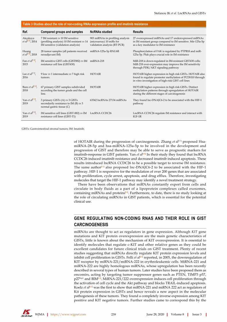
Stefanou IK et al. LncRNAs and GISTs
WJMA https://www.wjgnet.com 239 June 28, 2020 Volume 8 Issue 3
Table 3 Studies about the role of non-coding RNAs expression profile and imatinib resistance
Ref. Compared groups and samples NcRNAs studied Results
Akçakaya et al[52], 2014
7 IM resistant vs 10 IM sensitive (profiling analysis) 10 IM resistant vs 14 IM sensitive (validation analysis)
903 miRNAs in profiling analysis (microarray) 10 miRs for validation analysis (RT-PCR)
27 overexpressed miRNAs and 17 underexpressed miRNAs in IM resistant group compared to IM sensitive. Mir-125a-5p as a key modulator to IM resistance
Huang et al[53], 2018
28 tumor samples (all patients received neoadjuvant IM)
miRNA-125a-5p RNU6B Phosphorylation of FAK is regulated by PTPB18 and miR-125a-5p. Pfak plays crucial role in IM resistance
Fan et al[54], 2015
IM sensitive GIST cells (GIST882) vs IM resistance cell line (GIST430)
miRNA-218 MiR-218 is down-regulated in IM-resistant GIST430 cells; MiR-218 over-expression may improve the IM sensitivity through PI3K/AKT signaling pathway
Lee et al[55], 2016
9 low vs 1 intermediate vs 7 high risk tumors.
HOTAIR HOTAIR higher expression in high risk GISTs. HOTAIR also found to regulate promoter methylation of PCDH10 through in vitro investigation of high-risk GIST cell lines
Bure et al[56], 2018
67 primary GIST samples subdivided according the tumor grade and the cell line.
HOTAIR HOTAIR higher expression in high risk GISTs. Distinct methylation patterns through upregulation of HOTAIR during the different stages of carcinogenesis
Yan et al[51], 2019
3 primary GISTs (A) vs 3 GISTs secondarily resistance to IM (B) vs 3 normal gastric tissue (C)
63542 lncRNAs 27134 miRNAs They found lnc-DNAJC6-2 to be associated with the HIF-1 pathway
Yan et al[58], 2019
IM sensitive cell lines (GIST-882) vs IM resistance cell lines (GIST-T1)
LncRNA CCDC26 LncRNA CCDC26 regulate IM resistance and interact with IGF-1R
GISTs: Gastrointestinal stromal tumors; IM: Imatinib.
of HOTAIR during the progression of carcinogenesis. Zhang et al[57] proposed Hsa-miRNA-28-5p and hsa-miRNA-125a-5p to be involved in the development and progression of GIST and therefore may be able to serve as prognostic markers for imatinib-response in GIST patients. Yan et al[58] In their study they found that lncRNA CCDC26 induced imatinib resistance and decreased imatinib induced apoptosis. These results introduced lncRNA CCDC26 to be a possible target to reverse IM resistance. The same author[51] also proposed lnc-DNAJC6-2 to be associated with the HIF-1 pathway. HIF-1 is responsive for the modulation of over 200 genes that are associated with proliferation, cycle arrest, apoptosis, and drug efflux. Therefore, investigating molecules that target the HIF-1 pathway may identify a novel treatment strategy.
There have been observations that miRNAs constantly export from cells and circulate in body fluids as a part of a lipoprotein complexes called exosomes, containing miRNAs and proteins[59]. Furthermore, to date, there is no study looking at the role of circulating miRNAs in GIST patients, which is essential for the potential clinical use.
GENE REGULATING NON-CODING RNAS AND THEIR ROLE IN GIST CARCINOGENESISmiRNAs are thought to act as regulators in gene expression. Although KIT gene mutations and KIT protein overexpression are the main genetic characteristics of GISTs, little is known about the mechanism of KIT overexpression. It is essential to identify molecules that regulate c-KIT and other relative genes as they could be excellent candidates for future clinical trials on GIST treatment. Plenty of recent studies suggesting that miRNAs directly regulate KIT protein expression levels and inhibit cell proliferation in GISTs. Felli et al[60] reported, in 2005, the downregulation of KIT receptor by miRNA-221/miRNA-222 in erythroleukemic cells. MiRNA-221 and miRNA-222 are highly homologous miRNAs, whose upregulation has been recently described in several types of human tumors. Later studies have been proposed them as oncomirs, acting by targeting tumor suppressor genes such as PTEN, TIMP3 p57, p27Kip1 and BIM[61]. MiRNA-221/222 overexpression induces cell proliferation through the activation of cell cycle and the Akt pathway and blocks TRAIL-induced apoptosis. Koelz et al[42] was the first to show that miRNA-221 and miRNA 222 act as regulators of Kit protein expression in GISTs and hence reveals a new aspect in the molecular pathogenesis of these tumors. They found a completely inverse expression among KIT positive and KIT negative tumors. Further studies came to correspond this by the
Stefanou IK et al. LncRNAs and GISTs
WJMA https://www.wjgnet.com 240 June 28, 2020 Volume 8 Issue 3
observation that miRNA-222 and miRNA -17/20a directly target KIT and ETV1 in GISTs. MiRNA-494 is proposed as a potential KIT targeting miRNA by Kim et al[62]. This study showed that miRNA-494 is a negative regulator of KIT in GISTs and an overexpressing miRNA-494 may be a promising approach to GIST treatment. Gits et al[45] published that miRNA-17, miRNA-20a directly target KIT. They also showed that overexpression of these two miRNAs induced apoptosis and significantly inhibited cell proliferation. Interestingly they did not found correlation of miRNA494 and KIT expression like Kim et al[62] did! Lu et al[63] founded at their study, that miRNA-152 induced cell apoptosis, prevents cell proliferation and migration by repressing cathepsin L, suggesting miRNA-152 an attractive anti-tumor agent. In a latest study, Badalamenti et al[50] founded that the expression levels of MALAT1 lncRNA seem to affect the c-KIT mutational status. A recent Chinese study by Long et al[64], indicated that miRNA-374b inhibits apoptosis promotes viability of GIST cells by targeting PTEN gene through the PI3K/Akt signaling pathway. Another similar study[65] focused on the effects of neferine, an alkaloid derivative of lotus plant, in GIST development. They interestingly founded that neferine possibly upregulate miRNA-449a and then inactivate the PI3K/AKT and Notch pathways and by this mean suppress growth and migration of GIST cells. A latest paper came out from Chen et al[66]. Their results suggested that miR-4510 downregulation could promote GIST development, including growth, metastasis and invasion, through increasing APOC2 expression. Needless to say that much more scientific effort is needed in order to clarify the exact role of non-coding RNAs in GIST carcinogenesis and their interaction with tumor related genes and the respectively molecular endocytic paths.
POTENTIAL ROLE OF NON-CODING RNAS IN GIST TREATMENTThe potential role of ncRNAs as treatment tools against cancer has been explored through many studies during the recent years. The main treatment strategies aim to inhibit cell proliferation by importing exogenous ncRNAs through viral vectors (adenoviral, lentiviral and rectoviral vectors), which are mainly tumor suppressor miRNAs[67]. A recent study by Tu et al[68] suggested miR-218 loaded nanoparticle as tumor suppressor miRNA in GIST. Another study by Durso et al[69] proposed modified miRNAs 221/222 as effective inhibitors of KIT. Nowadays it is generally accepted that miRNAs can act as oncogenes or tumor suppressor genes. For this reason, it seems reasonable to manipulate those molecules against the carcinogenetic process. For example, synthesized miRNAs mimics imports into the cells and enhance endogenous miRNA function (antagomirs)[70]. Another strategy is proposed for the inhibition of over-expressed oncogenic miRNAs (oncomirs), by the use of antisense oligonucleotides[71]. This strategy includes inhibition or replacement of miRNAs through anti-miRNA oligonucleotides, antagomirs, miRNA sponges and nanoparticles. Only a few of the investigated miRNAs are currently in phase 2 stage[72]. But it must be pointed out that, up to now, although they have been shown remarkable success in in vitro models, none of these particles have been tested in GIST clinical trials.
CONCLUSIONA huge amount of preclinical data introduces non-coding RNAs as a new weapon against cancer in biomedical sciences armamentarium, although many efforts need to be done in order to understand the role of epitranscriptomics in GISTs. Especially for GISTs, numerus studies identified association patterns among specific ncRNAs with subsequent phenotypic characteristics. NcRNAs related to the tumor progression, grade, site, chromosomal eliminations, and imatinib sensitivity could probably be of importance as diagnostic or prognostic tumor biomarkers. In vitro studies revealed some of the mechanisms of action of these molecules. The endocytic paths could be served as guidance for future targeted drugs, acting as interfering or enhancing molecules. In addition, published data concerning GISTs and ncRNAs is based mainly on in vitro cell lines and fresh frozen paraffin-embedded tumor tissue blocks, thus necessitating high quality, randomized, multicentric clinical studies at a large scale of patients.
Stefanou IK et al. LncRNAs and GISTs
WJMA https://www.wjgnet.com 241 June 28, 2020 Volume 8 Issue 3
ACKNOWLEDGEMENTSThis study was supported by a non-profit organization of Greek Society of Cancer Biomarkers and Targeted Therapy.
REFERENCES1 Crick FH. On protein synthesis. Symp Soc Exp Biol 1958; 12: 138-163 [PMID: 13580867]2 Judson HF. The Eighth Day of Creation: Makers of the Revolution in Biology. New York: Cold Spring
Harbor Laboratory Press, 1996, ISBN 978-087969478-43 Stark BC, Kole R, Bowman EJ, Altman S. Ribonuclease P: an enzyme with an essential RNA component.
Proc Natl Acad Sci USA 1978; 75: 3717-3721 [PMID: 358197 DOI: 10.1073/pnas.75.8.3717]4 Yang VW, Lerner MR, Steitz JA, Flint SJ. A small nuclear ribonucleoprotein is required for splicing of
adenoviral early RNA sequences. Proc Natl Acad Sci USA 1981; 78: 1371-1375 [PMID: 6940164 DOI: 10.1073/pnas.78.3.1371]
5 Walter P, Blobel G. Signal recognition particle contains a 7S RNA essential for protein translocation across the endoplasmic reticulum. Nature 1982; 299: 691-698 [PMID: 6181418 DOI: 10.1038/299691a0]
6 Brockdorff N, Ashworth A, Kay GF, McCabe VM, Norris DP, Cooper PJ, Swift S, Rastan S. The product of the mouse Xist gene is a 15 kb inactive X-specific transcript containing no conserved ORF and located in the nucleus. Cell 1992; 71: 515-526 [PMID: 1423610 DOI: 10.1016/0092-8674(92)90519-I]
7 ENCODE Project Consortium. The ENCODE (ENCyclopedia Of DNA Elements) Project. Science 2004; 306: 636-640 [PMID: 15499007 DOI: 10.1126/science.1105136]
8 Djebali S, Davis CA, Merkel A, Dobin A, Lassmann T, Mortazavi A, Tanzer A, Lagarde J, Lin W, Schlesinger F, Xue C, Marinov GK, Khatun J, Williams BA, Zaleski C, Rozowsky J, Röder M, Kokocinski F, Abdelhamid RF, Alioto T, Antoshechkin I, Baer MT, Bar NS, Batut P, Bell K, Bell I, Chakrabortty S, Chen X, Chrast J, Curado J, Derrien T, Drenkow J, Dumais E, Dumais J, Duttagupta R, Falconnet E, Fastuca M, Fejes-Toth K, Ferreira P, Foissac S, Fullwood MJ, Gao H, Gonzalez D, Gordon A, Gunawardena H, Howald C, Jha S, Johnson R, Kapranov P, King B, Kingswood C, Luo OJ, Park E, Persaud K, Preall JB, Ribeca P, Risk B, Robyr D, Sammeth M, Schaffer L, See LH, Shahab A, Skancke J, Suzuki AM, Takahashi H, Tilgner H, Trout D, Walters N, Wang H, Wrobel J, Yu Y, Ruan X, Hayashizaki Y, Harrow J, Gerstein M, Hubbard T, Reymond A, Antonarakis SE, Hannon G, Giddings MC, Ruan Y, Wold B, Carninci P, Guigó R, Gingeras TR. Landscape of transcription in human cells. Nature 2012; 489: 101-108 [PMID: 22955620 DOI: 10.1038/nature11233]
9 Palazzo AF, Lee ES. Non-coding RNA: what is functional and what is junk? Front Genet 2015; 6: 2 [PMID: 25674102 DOI: 10.3389/fgene.2015.00002]
10 Taft RJ, Pang KC, Mercer TR, Dinger M, Mattick JS. Non-coding RNAs: regulators of disease. J Pathol 2010; 220: 126-139 [PMID: 19882673 DOI: 10.1002/path.2638]
11 Kapranov P, Drenkow J, Cheng J, Long J, Helt G, Dike S, Gingeras TR. Examples of the complex architecture of the human transcriptome revealed by RACE and high-density tiling arrays. Genome Res 2005; 15: 987-997 [PMID: 15998911 DOI: 10.1101/gr.3455305]
12 Mercer TR, Dinger ME, Mattick JS. Long non-coding RNAs: insights into functions. Nat Rev Genet 2009; 10: 155-159 [PMID: 19188922 DOI: 10.1038/nrg2521]
13 Setoyama T, Ling H, Natsugoe S, Calin GA. Non-coding RNAs for medical practice in oncology. Keio J Med 2011; 60: 106-113 [PMID: 22200634 DOI: 10.2302/kjm.60.106]
14 Motamedi MR, Verdel A, Colmenares SU, Gerber SA, Gygi SP, Moazed D. Two RNAi complexes, RITS and RDRC, physically interact and localize to noncoding centromeric RNAs. Cell 2004; 119: 789-802 [PMID: 15607976 DOI: 10.1016/j.cell.2004.11.034]
15 Gupta RA, Shah N, Wang KC, Kim J, Horlings HM, Wong DJ, Tsai MC, Hung T, Argani P, Rinn JL, Wang Y, Brzoska P, Kong B, Li R, West RB, van de Vijver MJ, Sukumar S, Chang HY. Long non-coding RNA HOTAIR reprograms chromatin state to promote cancer metastasis. Nature 2010; 464: 1071-1076 [PMID: 20393566 DOI: 10.1038/nature08975]
16 Eddy SR. Non-coding RNA genes and the modern RNA world. Nat Rev Genet 2001; 2: 919-929 [PMID: 11733745 DOI: 10.1038/35103511]
17 Koh JS, Trent J, Chen L, El-Naggar A, Hunt K, Pollock R, Zhang W. Gastrointestinal stromal tumors: overview of pathologic features, molecular biology, and therapy with imatinib mesylate. Histol Histopathol 2004; 19: 565-574 [PMID: 15024716 DOI: 10.14670/HH-19.565]
18 Tran T, Davila JA, El-Serag HB. The epidemiology of malignant gastrointestinal stromal tumors: an analysis of 1,458 cases from 1992 to 2000. Am J Gastroenterol 2005; 100: 162-168 [PMID: 15654796 DOI: 10.1111/j.1572-0241.2005.40709.x]
19 Rammohan A, Sathyanesan J, Rajendran K, Pitchaimuthu A, Perumal SK, Srinivasan U, Ramasamy R, Palaniappan R, Govindan M. A gist of gastrointestinal stromal tumors: A review. World J Gastrointest Oncol 2013; 5: 102-112 [PMID: 23847717 DOI: 10.4251/wjgo.v5.i6.102]
20 Sandvik OM, Søreide K, Kvaløy JT, Gudlaugsson E, Søreide JA. Epidemiology of gastrointestinal stromal tumours: single-institution experience and clinical presentation over three decades. Cancer Epidemiol 2011; 35: 515-520 [PMID: 21489899 DOI: 10.1016/j.canep.2011.03.002]
21 Miettinen M, Lasota J. Gastrointestinal stromal tumors: pathology and prognosis at different sites. Semin Diagn Pathol 2006; 23: 70-83 [PMID: 17193820 DOI: 10.1053/j.semdp.2006.09.001]
22 Joensuu H. Risk stratification of patients diagnosed with gastrointestinal stromal tumor. Hum Pathol 2008; 39: 1411-1419 [PMID: 18774375 DOI: 10.1016/j.humpath.2008.06.025]Hirota S, Isozaki K, Moriyama Y, Hashimoto K, Nishida T, Ishiguro S, Kawano K, Hanada M, Kurata A, Takeda M, Muhammad Tunio G, Matsuzawa Y, Kanakura Y, Shinomura Y, Kitamura Y. Gain-of-function
23

Stefanou IK et al. LncRNAs and GISTs
WJMA https://www.wjgnet.com 242 June 28, 2020 Volume 8 Issue 3
mutations of c-kit in human gastrointestinal stromal tumors. Science 1998; 279: 577-580 [PMID: 9438854 DOI: 10.1126/science.279.5350.577]
24 Heinrich MC, Rubin BP, Longley BJ, Fletcher JA. Biology and genetic aspects of gastrointestinal stromal tumors: KIT activation and cytogenetic alterations. Hum Pathol 2002; 33: 484-495 [PMID: 12094373 DOI: 10.1053/hupa.2002.124124]
25 Chaudhry UI, DeMatteo RP. Management of resectable gastrointestinal stromal tumor. Hematol Oncol Clin North Am 2009; 23: 79-96, viii [PMID: 19248972 DOI: 10.1016/j.hoc.2009.01.001]
26 Shah K, Chan KKW, Ko YJ. A systematic review and network meta-analysis of post-imatinib therapy in advanced gastrointestinal stromal tumour. Curr Oncol 2017; 24: e531-e539 [PMID: 29270063 DOI: 10.3747/co.24.3463]
27 Miettinen M, Sobin LH, Lasota J. Gastrointestinal stromal tumors of the stomach: a clinicopathologic, immunohistochemical, and molecular genetic study of 1765 cases with long-term follow-up. Am J Surg Pathol 2005; 29: 52-68 [PMID: 15613856 DOI: 10.1097/01.pas.0000146010.92933.de]
28 Kim WK, Yang HK, Kim H. MicroRNA involvement in gastrointestinal stromal tumor tumorigenesis. Curr Pharm Des 2013; 19: 1227-1235 [PMID: 23092343 DOI: 10.2174/138161213804805748]
29 Esteller M. Non-coding RNAs in human disease. Nat Rev Genet 2011; 12: 861-874 [PMID: 22094949 DOI: 10.1038/nrg3074]
30 Calin GA, Croce CM. MicroRNA signatures in human cancers. Nat Rev Cancer 2006; 6: 857-866 [PMID: 17060945 DOI: 10.1038/nrc1997]
31 Grimson A. A targeted approach to miRNA target identification. Nat Methods 2010; 7: 795-797 [PMID: 20885440 DOI: 10.1038/nmeth1010-795]
32 Nannini M, Ravegnini G, Angelini S, Astolfi A, Biasco G, Pantaleo MA. miRNA profiling in gastrointestinal stromal tumors: implication as diagnostic and prognostic markers. Epigenomics 2015; 7: 1033-1049 [PMID: 26447534 DOI: 10.2217/epi.15.52]
33 Kupcinskas J. Small Molecules in Rare Tumors: Emerging Role of MicroRNAs in GIST. Int J Mol Sci 2018; 19: 397 [PMID: 29385688 DOI: 10.3390/ijms19020397]
34 Assämäki R, Sarlomo-Rikala M, Lopez-Guerrero JA, Lasota J, Andersson LC, Llombart-Bosch A, Miettinen M, Knuutila S. Array comparative genomic hybridization analysis of chromosomal imbalances and their target genes in gastrointestinal stromal tumors. Genes Chromosomes Cancer 2007; 46: 564-576 [PMID: 17330260 DOI: 10.1002/gcc.20439]
35 Breiner JA, Meis-Kindblom J, Kindblom LG, McComb E, Liu J, Nelson M, Bridge JA. Loss of 14q and 22q in gastrointestinal stromal tumors (pacemaker cell tumors). Cancer Genet Cytogenet 2000; 120: 111-116 [PMID: 10942800 DOI: 10.1016/s0165-4608(00)00212-0]
36 Derré J, Lagacé R, Terrier P, Sastre X, Aurias A. Consistent DNA losses on the short arm of chromosome 1 in a series of malignant gastrointestinal stromal tumors. Cancer Genet Cytogenet 2001; 127: 30-33 [PMID: 11408061 DOI: 10.1016/s0165-4608(00)00409-x]
37 Lasota J, Wozniak A, Kopczynski J, Dansonka-Mieszkowska A, Wasag B, Mitsuhashi T, Sarlomo-Rikala M, Lee JR, Schneider-Stock R, Stachura J, Limon J, Miettinen M. Loss of heterozygosity on chromosome 22q in gastrointestinal stromal tumors (GISTs): a study on 50 cases. Lab Invest 2005; 85: 237-247 [PMID: 15580284 DOI: 10.1038/labinvest.3700218]
38 Choi HJ, Lee H, Kim H, Kwon JE, Kang HJ, You KT, Rhee H, Noh SH, Paik YK, Hyung WJ, Kim H. MicroRNA expression profile of gastrointestinal stromal tumors is distinguished by 14q loss and anatomic site. Int J Cancer 2010; 126: 1640-1650 [PMID: 19795448 DOI: 10.1002/ijc.24897]
39 Kelly L, Bryan K, Kim SY, Janeway KA, Killian JK, Schildhaus HU, Miettinen M, Helman L, Meltzer PS, van de Rijn M, Debiec-Rychter M, O'Sullivan M; NIH Pediatric and Wild-Type GIST Clinic. Post-transcriptional dysregulation by miRNAs is implicated in the pathogenesis of gastrointestinal stromal tumor [GIST]. PLoS One 2013; 8: e64102 [PMID: 23717541 DOI: 10.1371/journal.pone.0064102]
40 Haller F, von Heydebreck A, Zhang JD, Gunawan B, Langer C, Ramadori G, Wiemann S, Sahin O. Localization- and mutation-dependent microRNA (miRNA) expression signatures in gastrointestinal stromal tumours (GISTs), with a cluster of co-expressed miRNAs located at 14q32.31. J Pathol 2010; 220: 71-86 [PMID: 19768731 DOI: 10.1002/path.2610]
41 Subramanian S, Lui WO, Lee CH, Espinosa I, Nielsen TO, Heinrich MC, Corless CL, Fire AZ, van de Rijn M. MicroRNA expression signature of human sarcomas. Oncogene 2008; 27: 2015-2026 [PMID: 17922033 DOI: 10.1038/sj.onc.1210836]
42 Koelz M, Lense J, Wrba F, Scheffler M, Dienes HP, Odenthal M. Down-regulation of miR-221 and miR-222 correlates with pronounced Kit expression in gastrointestinal stromal tumors. Int J Oncol 2011; 38: 503-511 [PMID: 21132270 DOI: 10.3892/ijo.2010.857]
43 Niinuma T, Suzuki H, Nojima M, Nosho K, Yamamoto H, Takamaru H, Yamamoto E, Maruyama R, Nobuoka T, Miyazaki Y, Nishida T, Bamba T, Kanda T, Ajioka Y, Taguchi T, Okahara S, Takahashi H, Nishida Y, Hosokawa M, Hasegawa T, Tokino T, Hirata K, Imai K, Toyota M, Shinomura Y. Upregulation of miR-196a and HOTAIR drive malignant character in gastrointestinal stromal tumors. Cancer Res 2012; 72: 1126-1136 [PMID: 22258453 DOI: 10.1158/0008-5472.CAN-11-1803]
44 Yamamoto H, Kohashi K, Fujita A, Oda Y. Fascin-1 overexpression and miR-133b downregulation in the progression of gastrointestinal stromal tumor. Mod Pathol 2013; 26: 563-571 [PMID: 23196799 DOI: 10.1038/modpathol.2012.198]
45 Gits CM, van Kuijk PF, Jonkers MB, Boersma AW, van Ijcken WF, Wozniak A, Sciot R, Rutkowski P, Schöffski P, Taguchi T, Mathijssen RH, Verweij J, Sleijfer S, Debiec-Rychter M, Wiemer EA. MiR-17-92 and miR-221/222 cluster members target KIT and ETV1 in human gastrointestinal stromal tumours. Br J Cancer 2013; 109: 1625-1635 [PMID: 23969726 DOI: 10.1038/bjc.2013.483]
46 Gyvyte U, Juzenas S, Salteniene V, Kupcinskas J, Poskiene L, Kucinskas L, Jarmalaite S, Stuopelyte K, Steponaitiene R, Hemmrich-Stanisak G, Hübenthal M, Link A, Franke S, Franke A, Pangonyte D, Lesauskaite V, Kupcinskas L, Skieceviciene J. MiRNA profiling of gastrointestinal stromal tumors by next-generation sequencing. Oncotarget 2017; 8: 37225-37238 [PMID: 28402935 DOI: 10.18632/oncotarget.16664]

Stefanou IK et al. LncRNAs and GISTs
WJMA https://www.wjgnet.com 243 June 28, 2020 Volume 8 Issue 3
47 Gyvyte U, Kupcinskas J, Juzenas S, Inciuraite R, Poskiene L, Salteniene V, Link A, Fassan M, Franke A, Kupcinskas L, Skieceviciene J. Identification of long intergenic non-coding RNAs (lincRNAs) deregulated in gastrointestinal stromal tumors (GISTs). PLoS One 2018; 13: e0209342 [PMID: 30557328 DOI: 10.1371/journal.pone.0209342]
48 Kosela-Paterczyk H, Paziewska A, Kulecka M, Balabas A, Kluska A, Dabrowska M, Piatkowska M, Zeber-Lubecka N, Ambrozkiewicz F, Karczmarski J, Mikula M, Rutkowski P, Ostrowski J. Signatures of circulating microRNA in four sarcoma subtypes. J Cancer 2020; 11: 874-882 [PMID: 31949491 DOI: 10.7150/jca.34723]
49 Hu JC, Wang Q, Jiang LX, Cai L, Zhai HY, Yao ZW, Zhang ML, Feng Y. Effect of long non-coding RNA AOC4P on gastrointestinal stromal tumor cells. Onco Targets Ther 2018; 11: 6259-6269 [PMID: 30288061 DOI: 10.2147/OTT.S174524]
50 Badalamenti G, Barraco N, Incorvaia L, Galvano A, Fanale D, Cabibi D, Calò V, Currò G, Bazan V, Russo A. Are Long Noncoding RNAs New Potential Biomarkers in Gastrointestinal Stromal Tumors (GISTs)? The Role of H19 and MALAT1. J Oncol 2019; 2019: 5458717 [PMID: 31827510 DOI: 10.1155/2019/5458717]
51 Yan J, Chen D, Chen X, Sun X, Dong Q, Du Z, Wang T. Identification of imatinib-resistant long non-coding RNAs in gastrointestinal stromal tumors. Oncol Lett 2019; 17: 2283-2295 [PMID: 30675294 DOI: 10.3892/ol.2018.9821]
52 Akçakaya P, Caramuta S, Åhlen J, Ghaderi M, Berglund E, Östman A, Bränström R, Larsson C, Lui WO. microRNA expression signatures of gastrointestinal stromal tumours: associations with imatinib resistance and patient outcome. Br J Cancer 2014; 111: 2091-2102 [PMID: 25349971 DOI: 10.1038/bjc.2014.548]
53 Huang WK, Akçakaya P, Gangaev A, Lee L, Zeljic K, Hajeri P, Berglund E, Ghaderi M, Åhlén J, Bränström R, Larsson C, Lui WO. miR-125a-5p regulation increases phosphorylation of FAK that contributes to imatinib resistance in gastrointestinal stromal tumors. Exp Cell Res 2018; 371: 287-296 [PMID: 30149002 DOI: 10.1016/j.yexcr.2018.08.028]
54 Fan R, Zhong J, Zheng S, Wang Z, Xu Y, Li S, Zhou J, Yuan F. microRNA-218 increase the sensitivity of gastrointestinal stromal tumor to imatinib through PI3K/AKT pathway. Clin Exp Med 2015; 15: 137-144 [PMID: 24706111 DOI: 10.1007/s10238-014-0280-y]
55 Lee NK, Lee JH, Kim WK, Yun S, Youn YH, Park CH, Choi YY, Kim H, Lee SK. Promoter methylation of PCDH10 by HOTAIR regulates the progression of gastrointestinal stromal tumors. Oncotarget 2016; 7: 75307-75318 [PMID: 27659532 DOI: 10.18632/oncotarget.12171]
56 Bure I, Geer S, Knopf J, Roas M, Henze S, Ströbel P, Agaimy A, Wiemann S, Hoheisel JD, Hartmann A, Haller F, Moskalev EA. Long noncoding RNA HOTAIR is upregulated in an aggressive subgroup of gastrointestinal stromal tumors (GIST) and mediates the establishment of gene-specific DNA methylation patterns. Genes Chromosomes Cancer 2018; 57: 584-597 [PMID: 30248209 DOI: 10.1002/gcc.22672]
57 Zhang Z, Jiang NY, Guan RY, Zhu YK, Jiang FQ, Piao D. Identification of critical microRNAs in gastrointestinal stromal tumor patients treated with Imatinib. Neoplasma 2018; 65: 683-692 [PMID: 30249101 DOI: 10.4149/neo_2018_170906N575]
58 Yan J, Chen D, Chen X, Sun X, Dong Q, Hu C, Zhou F, Chen W. Downregulation of lncRNA CCDC26 contributes to imatinib resistance in human gastrointestinal stromal tumors through IGF-1R upregulation. Braz J Med Biol Res 2019; 52: e8399 [PMID: 31166382 DOI: 10.1590/1414-431x20198399]
59 Cortez MA, Bueso-Ramos C, Ferdin J, Lopez-Berestein G, Sood AK, Calin GA. MicroRNAs in body fluids--the mix of hormones and biomarkers. Nat Rev Clin Oncol 2011; 8: 467-477 [PMID: 21647195 DOI: 10.1038/nrclinonc.2011.76]
60 Felli N, Fontana L, Pelosi E, Botta R, Bonci D, Facchiano F, Liuzzi F, Lulli V, Morsilli O, Santoro S, Valtieri M, Calin GA, Liu CG, Sorrentino A, Croce CM, Peschle C. MicroRNAs 221 and 222 inhibit normal erythropoiesis and erythroleukemic cell growth via kit receptor down-modulation. Proc Natl Acad Sci USA 2005; 102: 18081-18086 [PMID: 16330772 DOI: 10.1073/pnas.0506216102]
61 Garofalo M, Quintavalle C, Romano G, Croce CM, Condorelli G. miR221/222 in cancer: their role in tumor progression and response to therapy. Curr Mol Med 2012; 12: 27-33 [PMID: 22082479 DOI: 10.2174/156652412798376170]
62 Kim WK, Park M, Kim YK, Tae YK, Yang HK, Lee JM, Kim H. MicroRNA-494 downregulates KIT and inhibits gastrointestinal stromal tumor cell proliferation. Clin Cancer Res 2011; 17: 7584-7594 [PMID: 22042971 DOI: 10.1158/1078-0432.CCR-11-0166]
63 Lu HJ, Yan J, Jin PY, Zheng GH, Qin SM, Wu DM, Lu J, Zheng YL. MicroRNA-152 inhibits tumor cell growth while inducing apoptosis via the transcriptional repression of cathepsin L in gastrointestinal stromal tumor. Cancer Biomark 2018; 21: 711-722 [PMID: 29278883 DOI: 10.3233/CBM-170809]
64 Long ZW, Wu JH, Cai-Hong, Wang YN, Zhou Y. MiR-374b Promotes Proliferation and Inhibits Apoptosis of Human GIST Cells by Inhibiting PTEN through Activation of the PI3K/Akt Pathway. Mol Cells 2018; 41: 532-544 [PMID: 29902839 DOI: 10.14348/molcells.2018.2211]
65 Xue F, Liu Z, Xu J, Xu X, Chen X, Tian F. Neferine inhibits growth and migration of gastrointestinal stromal tumor cell line GIST-T1 by up-regulation of miR-449a. Biomed Pharmacother 2019; 109: 1951-1959 [PMID: 30551450 DOI: 10.1016/j.biopha.2018.11.029]
66 Chen Y, Qin C, Cui X, Geng W, Xian G, Wang Z. miR-4510 acts as a tumor suppressor in gastrointestinal stromal tumor by targeting APOC2. J Cell Physiol 2020; 235: 5711-5721 [PMID: 31975384 DOI: 10.1002/jcp.29506]
67 Tutar L, Tutar E, Tutar Y. MicroRNAs and cancer; an overview. Curr Pharm Biotechnol 2014; 15: 430-437 [PMID: 24846068 DOI: 10.2174/1389201015666140519095304]
68 Tu L, Wang M, Zhao WY, Zhang ZZ, Tang DF, Zhang YQ, Cao H, Zhang ZG. miRNA-218-loaded carboxymethyl chitosan - Tocopherol nanoparticle to suppress the proliferation of gastrointestinal stromal tumor growth. Mater Sci Eng C Mater Biol Appl 2017; 72: 177-184 [PMID: 28024574 DOI: 10.1016/j.msec.2016.10.052]Durso M, Gaglione M, Piras L, Mercurio ME, Terreri S, Olivieri M, Marinelli L, Novellino E, Incoronato M, Grieco P, Orsini G, Tonon G, Messere A, Cimmino A. Chemical modifications in the seed region of miRNAs 221/222 increase the silencing performances in gastrointestinal stromal tumor cells. Eur J Med
69

Stefanou IK et al. LncRNAs and GISTs
WJMA https://www.wjgnet.com 244 June 28, 2020 Volume 8 Issue 3
Chem 2016; 111: 15-25 [PMID: 26854374 DOI: 10.1016/j.ejmech.2016.01.047]70 Bader AG, Brown D, Winkler M. The promise of microRNA replacement therapy. Cancer Res 2010; 70:
7027-7030 [PMID: 20807816 DOI: 10.1158/0008-5472.CAN-10-2010]71 Cheng G. Circulating miRNAs: roles in cancer diagnosis, prognosis and therapy. Adv Drug Deliv Rev 2015;
81: 75-93 [PMID: 25220354 DOI: 10.1016/j.addr.2014.09.001]72 Ji W, Sun B, Su C. Targeting MicroRNAs in Cancer Gene Therapy. Genes (Basel) 2017; 8: 21 [PMID:
28075356 DOI: 10.3390/genes8010021]
WJMA https://www.wjgnet.com 245 June 28, 2020 Volume 8 Issue 3
World Journal of
Meta-AnalysisW J M ASubmit a Manuscript: https://www.f6publishing.com World J Meta-Anal 2020 June 28; 8(3): 245-264
DOI: 10.13105/wjma.v8.i3.245 ISSN 2308-3840 (online)
SYSTEMATIC REVIEWS
Exclusive cigar smoking in the United States and smoking-related diseases: A systematic review
Peter N Lee, Janette S Hamling, Alison J Thornton
ORCID number: Peter N Lee 0000-0002-8244-1904; Jan S Hamling 0000-0001-7788-4738; Alison J Thornton 0000-0003-3689-4395.
Author contributions: Lee PN designed the study; Hamling JS carried out the searches and the analyses; Hamling JS and Thornton AJ entered the data on the databases; Lee PN, Hamling JS and Thornton AJ checked the data entered and discussed changes needed; Lee PN and Hamling JS wrote the paper; all authors read and approved the final manuscript.
Supported by the JT International SA, Assignment Schedule, No. 14.
Conflict-of-interest statement: No conflict of interests.
PRISMA 2009 Checklist statement: The manuscript was revised according to the PRISMA 2009 Checklist.
Open-Access: This article is an open-access article that was selected by an in-house editor and fully peer-reviewed by external reviewers. It is distributed in accordance with the Creative Commons Attribution NonCommercial (CC BY-NC 4.0) license, which permits others to distribute, remix, adapt, build upon this work non-commercially, and license their derivative works
Peter N Lee, Department of Statistics, P.N. Lee Statistics and Computing Ltd., Sutton, Surrey SM2 5DA, United Kingdom
Janette S Hamling, Department of Statistics, RoeLee Statistics Ltd., Sutton SM2 5DA, United Kingdom
Alison J Thornton, Independent Consultant, Okehampton, Devon EX20 1SG, United Kingdom
Corresponding author: Peter N Lee, MA, Director, Senior Statistician, Consultant Medical Statistician, Department of Statistics, P.N. Lee Statistics and Computing Ltd., 17 Cedar Road, Sutton, Surrey SM2 5DA, United Kingdom. [email protected]
AbstractBACKGROUND Little information has been published on the risks of cigar smoking. Since 1990 cigar smoking has become more prevalent in the United States.
AIM To summarise the evidence from the United States relating exclusive cigar smoking to risk of the major smoking-related diseases.
METHODS Literature searches detected studies carried out in the United States which estimated the risk of lung cancer, chronic obstructive pulmonary disease (COPD), heart disease, stroke or overall circulatory disease in exclusive cigar smokers as compared to those who had never smoked any tobacco product. Papers were identified from reviews and detailed searches on MEDLINE. For each study, data were extracted onto a study database and a linked relative risk database. Relative risks and 95%CIs were extracted, or estimated, relating to current, former or ever exclusive cigar smokers, and meta-analysed using standard methods. Sensitivity analyses were conducted including or excluding results from studies that did not quite fit the full selection criteria (for example, a paper presenting combined results from five studies, where 86% of the population were in the United States).
RESULTS The literature searches identified 17 relevant publications for lung cancer, four for COPD and 12 for heart disease, stroke and circulatory disease. These related to 11 studies for lung cancer, to four studies for COPD and to eight studies for heart
Lee PN et al. Exclusive cigar smoking and smoking-related diseases
WJMA https://www.wjgnet.com 246 June 28, 2020 Volume 8 Issue 3
on different terms, provided the original work is properly cited and the use is non-commercial. See: http://creativecommons.org/licenses/by-nc/4.0/
Manuscript source: Unsolicited manuscript
Received: February 13, 2020 Peer-review started: February 13, 2020 First decision: February 26, 2020 Revised: May 15, 2020 Accepted: June 10, 2020 Article in press: June 10, 2020 Published online: June 28, 2020
P-Reviewer: OnoT, Ju SQ S-Editor: Wang JL L-Editor: A E-Editor: Qi LL
disease, stroke or overall circulatory disease. As some studies provided results for more than one disease, the total number of studies considered was 13, with results from four of these used in sensitivity analyses. There was evidence of significant heterogeneity in some of the meta-analyses so the random-effects estimates are summarized below. As the results from the sensitivity analyses were generally very similar to those from the main analyses, and involved more data, only the sensitivity results are summarized below. For lung cancer, relative risks (95%CI) for current, former and ever smokers were respectively, 2.98 (2.08 to 4.26), 1.61 (1.23 to 2.09), and 2.22 (1.79 to 2.74) based on 6, 4 and 10 individual study estimates. For COPD, the corresponding estimates were 1.44 (1.16 to 1.77), 0.47 (0.02 to 9.88), and 0.86 (0.48 to 1.54) based on 4, 2 and 2 estimates. For ischaemic heart disease (IHD) the estimates were 1.11 (1.04 to 1.19), 1.26 (1.03 to 1.53) and 1.15 (1.08 to 1.23) based on 6, 3 and 4 estimates, while for stroke they were 1.02 (0.92 to 1.13), 1.08 (0.85 to 1.38), and 1.11 (0.95 to 1.31) based on 5, 3 and 4 estimates. For overall circulatory disease they were 1.10 (1.05 to 1.16), 1.11 (0.84 to 1.46), and 1.15 (1.06 to 1.26) based on 3, 3 and 4 estimates.
CONCLUSION Exclusive cigar smoking is associated with an increased risk of lung cancer, and less so with COPD and IHD. The increases are lower than for cigarettes.
Key words: Tobacco products; Cigar smoking; Lung neoplasms; Pulmonary disease; Chronic obstructive; Heart diseases; Stroke; Circulatory disease; Systematic review; Meta-analysis
©The Author(s) 2020. Published by Baishideng Publishing Group Inc. All rights reserved.
Core tip: Thirteen studies in the United States presented evidence relating exclusive cigar smoking to risk of lung cancer, chronic obstructive pulmonary disease (COPD) and/or circulatory disease. Compared to never smokers, current exclusive cigar smoking increased risk of lung cancer about three-fold, COPD by about 40% and heart disease by about 10% but did not increase risk of stroke. These increases are much lower than those for cigarette smoking.
Citation: Lee PN, Hamling JS, Thornton AJ. Exclusive cigar smoking in the United States and smoking-related diseases: A systematic review. World J Meta-Anal 2020; 8(3): 245-264URL: https://www.wjgnet.com/2308-3840/full/v8/i3/245.htmDOI: https://dx.doi.org/10.13105/wjma.v8.i3.245
INTRODUCTIONThere is extensive evidence on the relationship of cigarette smoking to health endpoints but far less evidence relating to cigar smoking. While some studies have reported results relating to dual use of cigars and pipes[1-4] or to cigar smoking in those who may also smoke other tobacco products[5], the health effects of exclusive cigar smoking have been less often reported. Comparing disease risk in cigar smokers who have never smoked other tobacco products with that in never smokers of any tobacco product avoids the problems of residual confounding by other smoking habits and of possible differences in cigar smoking habits (such as depth of inhalation[6]) in those who have ever smoked other tobacco products.
Exclusive cigar smoking is much less common than cigarette smoking so the population studied must be large enough to include enough exclusive cigar smokers for a useful risk assessment to be made. For this reason we have restricted attention to studies in the United States, a country not only with a large population, but one where cigar smoking is relatively common compared with other countries[7,8]. We also restrict attention to the major smoking-related diseases.

Lee PN et al. Exclusive cigar smoking and smoking-related diseases
WJMA https://www.wjgnet.com 247 June 28, 2020 Volume 8 Issue 3
MATERIALS AND METHODSStudy inclusion criteriaPublished studies were included if they were carried out in the United States and reported the risk of lung cancer or chronic obstructive pulmonary disease (COPD) or heart disease, stroke and/or overall circulatory disease, comparing exclusive cigar smokers (current, former or ever cigar smokers who never smoked other tobacco products) with never smokers of any tobacco (or a closely-related comparison group). The results considered were for overall lung cancer rather than lung cancer subtypes, and related to overall risk measures rather than dose response indices, although dose response results by amount smoked were also identified.
Literature searchesSearching for results on cigar smoking was complicated by the MEDLINE search term “cigar smoking” being available only from the start of 2018. Before then the only search term to include cigar smoking was “tobacco products”.
For lung cancer, the first step was to examine publications from a previous review relating lung cancer to various indices of smoking based on studies published during the 1900s[9]. Subsequently three different MEDLINE searches were conducted using terms such as (“cigar” or “cigars”), “United States” and “lung neoplasms”. This was followed by a fourth search that attempted to retrieve relevant papers that had not yet been indexed with MeSH terms on MEDLINE, this search not being lung cancer specific. A fifth search used wholly non-MeSH search terms, with the final stage being to look for relevant results in papers identified as relevant in the searches for COPD and for heart disease, stroke and circulatory diseases.
For COPD, the process started with three different MEDLINE searches using the terms “COPD” or “pulmonary disease, chronic obstructive” to identify the disease. The fourth search used the term “Smoking” rather than “Cigar”, while the fifth search used the MeSH terms “Smoking/mortality” or “Smoking/adverse effects”. The next step was to review the results from the fourth lung cancer search, while the final step was to look at papers identified as relevant in the searches for lung cancer and for heart disease, stroke and circulatory diseases.
For heart disease, stroke and circulatory diseases, the process started with four different MEDLINE searches using the disease terms “Heart disease”, “Stroke” or “Heart”. The next step was again to review the results from the fourth lung cancer search, while the final step was to look at relevant papers from the lung cancer and COPD searches.
Searching ended when no new data was found and all the papers referenced by reviews had been examined. Full details of the searches are given in Supplementary material.
Sorting publications into studies and avoidance of overlapThe papers identified in the searches were reviewed for the studies they reported, and multiple publications reporting the same study were identified.
The source papers identified as providing relevant estimates were then considered for overlap of reporting. Where more than one of the source papers reported on the same study, the results may have been reported in different ways or for different lengths of follow-up, or have combined results from multiple studies.
Data recordedFor each paper identified as providing relevant results details were entered onto a study database and a linked relative risk (RR) database for the relevant disease.
The study database contained a record for each study describing the following aspects: A study name based on the published study name or on the name of the first author of the paper; study title; study design; sexes considered; age range and other details of the population studied; timing and length of follow-up; details of overlaps or links with other studies; number of cases; number of controls or subjects at risk; types of controls and matching factors used in case-control studies; and confounding variables considered.
The RR database holds the detailed results, typically containing multiple records for each study. Each record is linked to the relevant study via the study name, and holds details of a specific risk estimate. It records the type of estimate, its value and confidence interval, its source and other details such as the age range included in the estimate if this is different from the overall study age range. Some estimates were taken directly from the source paper. Others were derived using the details provided
Lee PN et al. Exclusive cigar smoking and smoking-related diseases
WJMA https://www.wjgnet.com 248 June 28, 2020 Volume 8 Issue 3
in the paper.Where no RR estimate was given or a RR estimate was given without a confidence
interval, information on the sample size and the number of deaths was used to estimate these. Estimates for separate independent subsets of the population such as age groups were combined using simple meta-analysis. Non-independent RRs using a common comparison group (e.g., never smokers) were combined using the Hamling method[10]. This method was used to combine RRs by number of cigars smoked per day and to combine RRs for former and current smokers to give an estimate for ever smokers. It was also used to estimate risk for overall circulatory disease when the study provided separate estimates for cerebrovascular disease and a broad definition of coronary heart disease, and to estimate overall stroke from separate risk estimates for ischaemic and haemorrhagic stroke. The International Classification of Disease codes used to define IHD, stroke and circulatory disease can be found at https://coder.aapc.com/icd-10-codes-range/110.
Each extract was carried out by one of the authors, the entered data and any additional calculations then being reported and checked by another of the authors, with problems discussed and amendments made until both agreed that the data entered were a true representation of the study data.
Dose-response data on risk by number of cigars smoked per day were also identified and are discussed below.
Statistical analysisFor each disease considered, fixed-effect and random-effects meta-analyses were conducted using the Fleiss and Gross method[11], with heterogeneity quantified by H, the ratio of the heterogeneity to its degrees of freedom, which is directly related to the I2statistic[12] by the formula I2 = 100 (H-1)/H.
Whenever more than one paper provided equivalent results for a study, only one result was included in a meta-analysis. The selection of the result was based on four criteria: Prospective follow-up was given preference over cross-sectional analysis at baseline; the longest follow-up reported (for prospective studies); the widest age range reported; and finally the RR adjusted for the most confounding factors.
Some papers provided results for comparisons that did not exactly match our selection criteria. Where any were relevant to a meta-analysis, the analysis was performed excluding those results, and then including them in a sensitivity analysis.
The KAISER study[13] used a questionnaire that asked about the participant’s history of cigarette smoking and their current pipe and cigar smoking. Ever cigarette smokers were excluded from their analyses. It was, therefore, possible to identify participants who had never smoked cigarettes and who did not, at baseline, smoke cigars or a pipe. This is not completely equivalent to our requirement for the comparison group to be never smokers of any tobacco product. Also, the participants categorised as current cigar smokers may have included former pipe smokers. However, the study was large (1546 current cigar smokers and 16228 never cigarette smokers) and had a long follow-up (25-26 years) so justified inclusion in sensitivity analyses.
For the MALHOT study a pooled analysis of data from five large prospective studies was reported[14]. Of these studies, two (the Netherlands Cohort Study and the Melbourne Collaborative Cohort Study) were conducted outside the United States, while the other three (the VITamins And Lifestyle study, the NIH-AARP Diet and Health study and the Prostate, Lung, Colorectal and Ovarian Cancer Screening Trial) were conducted in the United States. However, the studies in the United States were larger and formed 85.7% of the total population reported, and the largest of the studies (NIH-AARP) followed up participants for a median of 15.5 years. The size of this pooled analysis, the large proportion of participants from the United States, the long follow-up in the largest study, and the lack of study-specific reports relating to cigar smoking for the studies pooled justified including the combined results in sensitivity analysis.
For the NHIS study, a cross-sectional analysis of baseline data[15] reports results for a definition of “Heart conditions” including angina, coronary heart disease, heart attack and other heart disease which is too broad for the results to be included in our main analysis of ischaemic heart disease (IHD). However, as the report considers data from four years’ surveys of this large, repeated, nationally representative study, and as no other source was found that reported IHD for this study, it was decided to include its results in sensitivity analysis.
Also for the NHIS study, an analysis of prospective findings from six years’ survey data with follow up for at least five years, the source publication[16] reports results for a broad definition of coronary heart disease which includes rheumatic fever, some hypertensive heart disease, IHD and some other heart disease. Though this definition
Lee PN et al. Exclusive cigar smoking and smoking-related diseases
WJMA https://www.wjgnet.com 249 June 28, 2020 Volume 8 Issue 3
is too broad to be included in our analyses of IHD, the paper also reports results for cerebrovascular disease (stroke), and taken together, the definitions of coronary heart disease and cerebrovascular disease were very close to our ideal definition of circulatory disease. We therefore combined the results for coronary heart disease and for cerebrovascular disease, and included the resulting estimates in the sensitivity analyses for overall circulatory disease.
The NLMS study[17] reported too broad a definition of cardiovascular disease, an ideal definition of stroke and a definition of circulatory disease that included all the relevant disease categories except diseases of the veins and other diseases of the circulatory system. The reported results for stroke were included in the main meta-analyses and the results for circulatory disease were included in sensitivity analysis only.
RESULTSLiterature searchFor lung cancer, 17 publications were identified that were relevant to the meta-analyses (including the sensitivity analyses), 13 from the previous review[9], three from additional searches, and one from reviews identified in the searches, as shown in Figure 1. Two of these[18,19] are by the same authors reporting the same study, the first giving overall study information and the second giving results by disease, so only the latter is cited in the analysis results. For COPD, four publications were identified (Figure 2), while for heart disease, stroke and circulatory disease 12 were found (Figure 3).
StudiesTable 1[13,14,17-31] (lung cancer), Table 2[13,17,23,27] (COPD), and Table 3[13,15-17,19,21,23,26-28,32,33] (heart disease, stroke and circulatory disease) present details on each study, including its name, the source publications, the study type, the years of follow-up, the study size, and the sexes and age groups considered. Results used in sensitivity analyses only are marked with an asterisk. Table 3 also includes details of the definition of heart disease, stroke and circulatory diseases.
For lung cancer, the 17 publications relate to 11 studies, though two (KAISER, MALHOT) are only used in sensitivity analyses. Eight of the studies are of prospective design and three case-controls. Except for NLMS, which considers both sexes, all provide results only for males. The studies vary widely in size, with three involving over 400000 people, and four less than 10000.
For COPD, the four publications concern separate studies, though again KAISER is only used in sensitivity analyses. All the studies are prospective, with all except NLMS considering only males. All four of these studies also provide results for lung cancer. The study size in KAISER is much lower than in the other three studies.
For heart disease, stroke and circulatory disease, the 12 publications concern eight studies: Six prospective, one case-control and one reported both as a cross-sectional analysis of baseline data and using prospective follow-up. KAISER (all results), NHIS (prospective results for circulatory disease and cross-sectional results for ischaemic heart disease) and NMLS (results for circulatory disease) were only used in the sensitivity analyses. Most results were for men only, the exceptions being those from NHIS and NLMS that were for the sexes combined. As for lung cancer, the studies varied widely in size.
Overall, as many studies provided data for more than one disease, there were 13 studies, of which two only provided results for the sensitivity analyses and two others had some results restricted to sensitivity analyses.
Meta-analysesThe individual study RR estimates used are given in Table 4[13-17,19,20,22,23,25,27-33], with the results of the meta-analyses conducted summarised in Table 5. Unless otherwise stated references to combined estimates are to random-effects estimates, with 95%CI given in parentheses.
Lung cancerFor current smokers there was highly significant heterogeneity (P < 0.001) between the five estimates, which ranged from 1.66 (1.18 to 2.34) for DORN to 5.10 (4.00 to 6.60) for CPS II. The overall estimate was 3.12 (2.11 to 4.62). Including the result from study
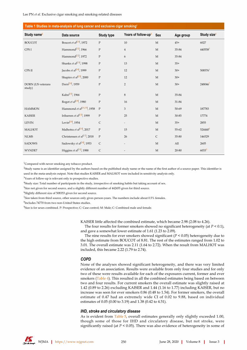
Lee PN et al. Exclusive cigar smoking and smoking-related diseases
WJMA https://www.wjgnet.com 250 June 28, 2020 Volume 8 Issue 3
Table 1 Studies in meta-analysis of lung cancer and exclusive cigar smoking1
Study name2 Data source Study type Years of follow-up3 Sex Age group Study size4
BOUCOT Boucot et al[20], 1972 P 10 M 45+ 6027
CPS I Hammond[21], 1966 P 4 M 35-84 4405585
Hammond[22], 1972 P 6 M 35-84
Shanks et al[23], 1998 P 13 M 35+
CPS II Jacobs et al[24], 1999 P 12 M 30+ 5085766
Shapiro et al[25], 2000 P 12 M 30+
DORN (US veterans study)
Dorn[26], 1959 P 2 M 30+ 2480467
Kahn[27], 1966 P 8 M 35-84
Rogot et al[28], 1980 P 16 M 31-84
HAMMON Hammond et al[18,19], 1958 P 3 M 50-69 187783
KAISER Iribarren et al[13], 1999 P 25 M 30-85 17774
LEVIN Levin[29], 1954 C - M 35+ 2855
MALHOT Malhotra et al[14], 2017 P 15 M 55-62 5244408
NLMS Christensen et al[17], 2018 P 26 C 35-80 146529
SADOWS Sadowsky et al[30], 1953 C - M All 2605
WYNDE7 Higgins et al[31], 1988 C - M 20-80 60339
1Compared with never smoking any tobacco product. 2Study name is an identifier assigned by the authors based on the published study name or the name of the first author of a source paper. This identifier is used in the meta-analysis output. Note that studies KAISER and MALHOT were included in sensitivity analysis only. 3Years of follow-up is relevant only to prospective studies.4Study size: Total number of participants in the study, irrespective of smoking habits but taking account of sex. 5Size not given for second source, and a slightly different number of 442455 given for third source. 6Slightly different size of 508353 given for second source. 7Size taken from third source, other sources only gives person-years. The numbers include about 0.5% females. 8Includes 74770 from two non-United States studies. 9Size is for sexes combined. P: Prospective; C: Case control; M: Male; C: Combined male and female.
KAISER little affected the combined estimate, which became 2.98 (2.08 to 4.26).The four results for former smokers showed no significant heterogeneity (at P < 0.1),
and gave a somewhat lower estimate of 1.61 (1.23 to 2.09).The nine results for ever smokers showed significant (P < 0.05) heterogeneity due to
the high estimate from BOUCOT of 8.81. The rest of the estimates ranged from 1.02 to 3.01. The overall estimate was 2.11 (1.64 to 2.72). When the result from MALHOT was included, this became 2.22 (1.79 to 2.74).
COPDNone of the analyses showed significant heterogeneity, and there was very limited evidence of an association. Results were available from only four studies and for only two of these were results available for each of the exposures current, former and ever smokers (Table 4). This resulted in all the combined estimates being based on between two and four results. For current smokers the overall estimate was slightly raised at 1.42 (0.89 to 2.26) excluding KAISER and 1.44 (1.16 to 1.77) including KAISER, but no increase was seen for ever smokers 0.86 (0.48 to 1.54). For former smokers, the overall estimate of 0.47 had an extremely wide CI of 0.02 to 9.88, based on individual estimates of 0.05 (0.00 to 3.19) and 1.38 (0.42 to 4.51).
IHD, stroke and circulatory diseaseAs is evident from Table 5, overall estimates generally only slightly exceeded 1.00, though some of those for IHD and circulatory disease, but not stroke, were significantly raised (at P < 0.05). There was also evidence of heterogeneity in some of
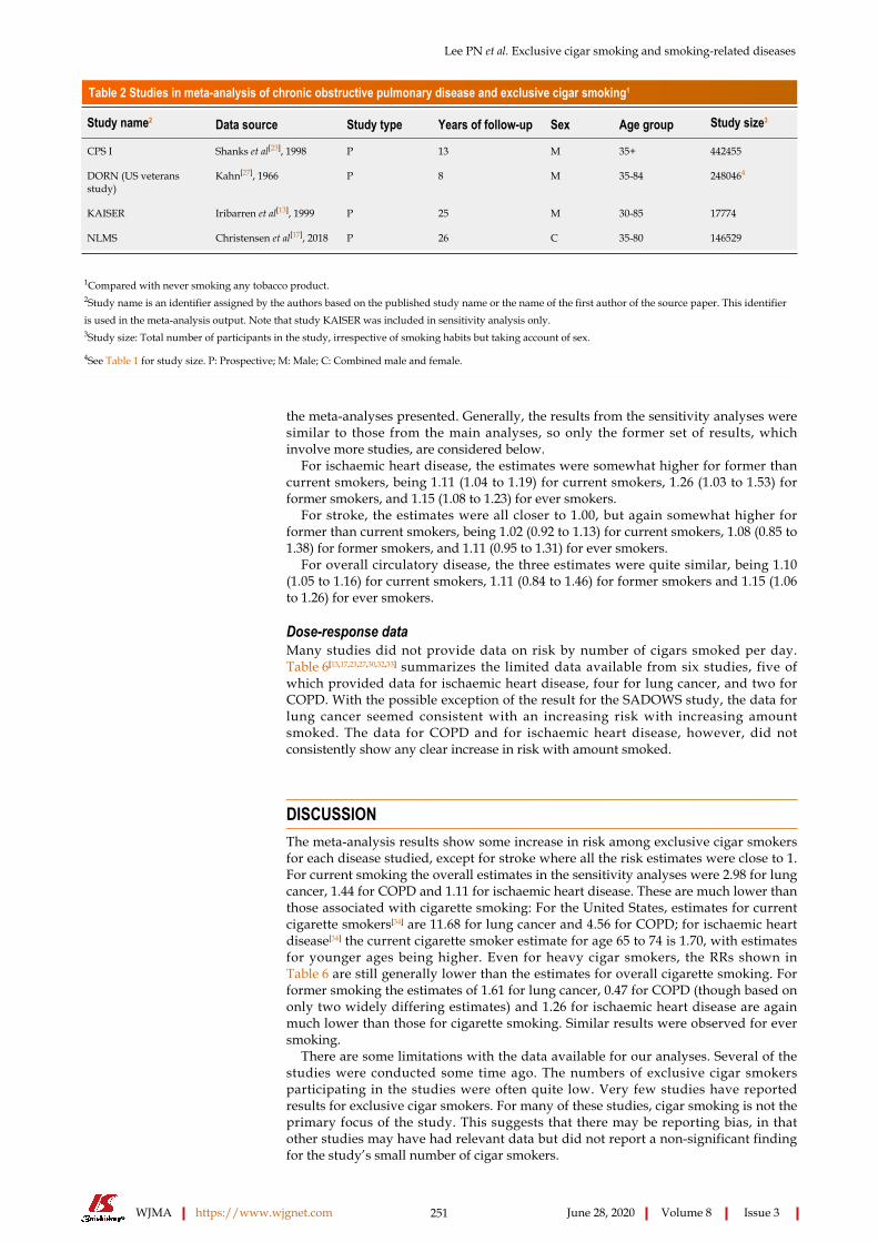
Lee PN et al. Exclusive cigar smoking and smoking-related diseases
WJMA https://www.wjgnet.com 251 June 28, 2020 Volume 8 Issue 3
Table 2 Studies in meta-analysis of chronic obstructive pulmonary disease and exclusive cigar smoking1
Study name2 Data source Study type Years of follow-up Sex Age group Study size3
CPS I Shanks et al[23], 1998 P 13 M 35+ 442455
DORN (US veterans study)
Kahn[27], 1966 P 8 M 35-84 2480464
KAISER Iribarren et al[13], 1999 P 25 M 30-85 17774
NLMS Christensen et al[17], 2018 P 26 C 35-80 146529
1Compared with never smoking any tobacco product. 2Study name is an identifier assigned by the authors based on the published study name or the name of the first author of the source paper. This identifier is used in the meta-analysis output. Note that study KAISER was included in sensitivity analysis only. 3Study size: Total number of participants in the study, irrespective of smoking habits but taking account of sex.
4See Table 1 for study size. P: Prospective; M: Male; C: Combined male and female.
the meta-analyses presented. Generally, the results from the sensitivity analyses were similar to those from the main analyses, so only the former set of results, which involve more studies, are considered below.
For ischaemic heart disease, the estimates were somewhat higher for former than current smokers, being 1.11 (1.04 to 1.19) for current smokers, 1.26 (1.03 to 1.53) for former smokers, and 1.15 (1.08 to 1.23) for ever smokers.
For stroke, the estimates were all closer to 1.00, but again somewhat higher for former than current smokers, being 1.02 (0.92 to 1.13) for current smokers, 1.08 (0.85 to 1.38) for former smokers, and 1.11 (0.95 to 1.31) for ever smokers.
For overall circulatory disease, the three estimates were quite similar, being 1.10 (1.05 to 1.16) for current smokers, 1.11 (0.84 to 1.46) for former smokers and 1.15 (1.06 to 1.26) for ever smokers.
Dose-response dataMany studies did not provide data on risk by number of cigars smoked per day. Table 6[13,17,23,27,30,32,33] summarizes the limited data available from six studies, five of which provided data for ischaemic heart disease, four for lung cancer, and two for COPD. With the possible exception of the result for the SADOWS study, the data for lung cancer seemed consistent with an increasing risk with increasing amount smoked. The data for COPD and for ischaemic heart disease, however, did not consistently show any clear increase in risk with amount smoked.
DISCUSSIONThe meta-analysis results show some increase in risk among exclusive cigar smokers for each disease studied, except for stroke where all the risk estimates were close to 1. For current smoking the overall estimates in the sensitivity analyses were 2.98 for lung cancer, 1.44 for COPD and 1.11 for ischaemic heart disease. These are much lower than those associated with cigarette smoking: For the United States, estimates for current cigarette smokers[34] are 11.68 for lung cancer and 4.56 for COPD; for ischaemic heart disease[34] the current cigarette smoker estimate for age 65 to 74 is 1.70, with estimates for younger ages being higher. Even for heavy cigar smokers, the RRs shown in Table 6 are still generally lower than the estimates for overall cigarette smoking. For former smoking the estimates of 1.61 for lung cancer, 0.47 for COPD (though based on only two widely differing estimates) and 1.26 for ischaemic heart disease are again much lower than those for cigarette smoking. Similar results were observed for ever smoking.
There are some limitations with the data available for our analyses. Several of the studies were conducted some time ago. The numbers of exclusive cigar smokers participating in the studies were often quite low. Very few studies have reported results for exclusive cigar smokers. For many of these studies, cigar smoking is not the primary focus of the study. This suggests that there may be reporting bias, in that other studies may have had relevant data but did not report a non-significant finding for the study’s small number of cigar smokers.
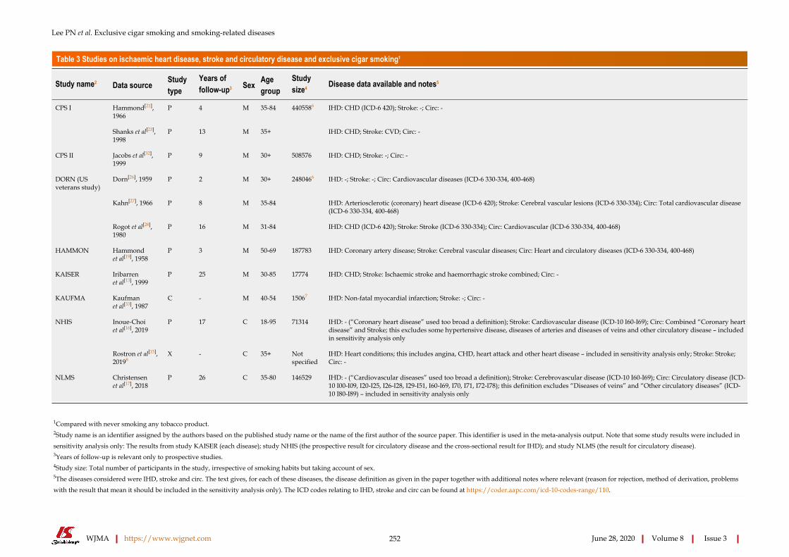
Lee PN et al. Exclusive cigar smoking and smoking-related diseases
WJMA https://www.wjgnet.com 252 June 28, 2020 Volume 8 Issue 3
Table 3 Studies on ischaemic heart disease, stroke and circulatory disease and exclusive cigar smoking1
Study name2 Data source Study type
Years of follow-up3 Sex Age
groupStudy size4 Disease data available and notes5
CPS I Hammond[21], 1966
P 4 M 35-84 4405586 IHD: CHD (ICD-6 420); Stroke: -; Circ: -
Shanks et al[23], 1998
P 13 M 35+ IHD: CHD; Stroke: CVD; Circ: -
CPS II Jacobs et al[32], 1999
P 9 M 30+ 508576 IHD: CHD; Stroke: -; Circ: -
DORN (US veterans study)
Dorn[26], 1959 P 2 M 30+ 2480466 IHD: -; Stroke: -; Circ: Cardiovascular diseases (ICD-6 330-334, 400-468)
Kahn[27], 1966 P 8 M 35-84 IHD: Arteriosclerotic (coronary) heart disease (ICD-6 420); Stroke: Cerebral vascular lesions (ICD-6 330-334); Circ: Total cardiovascular disease (ICD-6 330-334, 400-468)
Rogot et al[28], 1980
P 16 M 31-84 IHD: CHD (ICD-6 420); Stroke: Stroke (ICD-6 330-334); Circ: Cardiovascular (ICD-6 330-334, 400-468)
HAMMON Hammond et al[19], 1958
P 3 M 50-69 187783 IHD: Coronary artery disease; Stroke: Cerebral vascular diseases; Circ: Heart and circulatory diseases (ICD-6 330-334, 400-468)
KAISER Iribarren et al[13], 1999
P 25 M 30-85 17774 IHD: CHD; Stroke: Ischaemic stroke and haemorrhagic stroke combined; Circ: -
KAUFMA Kaufman et al[33], 1987
C - M 40-54 15067 IHD: Non-fatal myocardial infarction; Stroke: -; Circ: -
NHIS Inoue-Choi et al[16], 2019
P 17 C 18-95 71314 IHD: - (“Coronary heart disease” used too broad a definition); Stroke: Cardiovascular disease (ICD-10 I60-I69); Circ: Combined “Coronary heart disease” and Stroke; this excludes some hypertensive disease, diseases of arteries and diseases of veins and other circulatory disease – included in sensitivity analysis only
Rostron et al[15], 20198
X - C 35+ Not specified
IHD: Heart conditions; this includes angina, CHD, heart attack and other heart disease – included in sensitivity analysis only; Stroke: Stroke; Circ: -
NLMS Christensen et al[17], 2018
P 26 C 35-80 146529 IHD: - (“Cardiovascular diseases” used too broad a definition); Stroke: Cerebrovascular disease (ICD-10 I60-I69); Circ: Circulatory disease (ICD-10 I00-I09, I20-I25, I26-I28, I29-I51, I60-I69, I70, I71, I72-I78); this definition excludes “Diseases of veins” and “Other circulatory diseases” (ICD-10 I80-I89) – included in sensitivity analysis only
1Compared with never smoking any tobacco product. 2Study name is an identifier assigned by the authors based on the published study name or the name of the first author of the source paper. This identifier is used in the meta-analysis output. Note that some study results were included in sensitivity analysis only: The results from study KAISER (each disease); study NHIS (the prospective result for circulatory disease and the cross-sectional result for IHD); and study NLMS (the result for circulatory disease). 3Years of follow-up is relevant only to prospective studies. 4Study size: Total number of participants in the study, irrespective of smoking habits but taking account of sex. 5The diseases considered were IHD, stroke and circ. The text gives, for each of these diseases, the disease definition as given in the paper together with additional notes where relevant (reason for rejection, method of derivation, problems with the result that mean it should be included in the sensitivity analysis only). The ICD codes relating to IHD, stroke and circ can be found at https://coder.aapc.com/icd-10-codes-range/110.
Lee PN et al. Exclusive cigar smoking and smoking-related diseases
WJMA https://www.wjgnet.com 253 June 28, 2020 Volume 8 Issue 3
6See Table 1 for study size.
7Study limited to non-cigarette smokers. 8This is a cross-sectional analysis of baseline data: Results from the prospective study (above) were used in the meta-analysis when possible. IHD: Ischaemic heart disease; Circ: Stroke and circulatory disease P: Prospective; C: Case control; X: Cross-sectional; M: Male; C: Combined male and female; ICD: The International Classification of Disease.
There was a limited amount of dose-response data, and a lack of data on how risk varied by type of cigar smoked. No meta-analyses could be carried out by subgroups such as race and age and gender, as there was insufficient data. No study reported results for sex separately, so no analysis by sex could be done.
Nevertheless, the data provide fairly clear evidence that exclusive cigar smoking is associated with an increased risk of lung cancer, though less markedly than is the case for exclusive cigarette smoking. For COPD and ischaemic heart disease, the association is weaker, and is also less than that for cigarette smoking.
How do these results compare with previous estimates? It should be noted that no other review has provided meta-analysis estimates for exclusive cigar smokers in the United States, and that many of the previous reviews considered below were conducted many years ago.
The review of smoking and lung cancer[9] referred to under literature searches provided random effects meta-analysis estimates for lung cancer in current, former and ever exclusive cigar smokers of 4.67 (n = 15), 2.85 (n = 5) and 2.95 (n = 15) respectively, but these analyses were not restricted to studies in the United States. The risk estimates included in those analyses showed significant heterogeneity. A review by Wynder et al[35] considering the risk of lung cancer in pipe and cigar smokers noted that, in prospective studies in North America the mortality ratios were in the range 2 to 6. For retrospective studies, mostly conducted in Germany and Switzerland, “it appears that the risk of lung cancer is higher than that for such smokers in the United States”. This review suggested that these differences stemmed from different patterns of inhalation in the two regions. A similar review by Higgins et al[31], also considering pipe and cigar smokers, again suggested that risk estimates from prospective studies in North America are lower than those from case control studies in Europe. Smoking and Tobacco Control Monograph No. 9[23], reviewing data from CPS-I, stated that “Lung cancer mortality ratios increase with increasing number of cigars smoked per day and with increasing depth of inhalation. When depth of inhalation and number of cigars per day are examined together, depth of inhalation is more powerful in predicting lung cancer risk than number of cigars smoked per day.” The 1979 report by the Surgeon General[36] summarised the available evidence as “Several prospective epidemiological studies have demonstrated higher lung cancer mortality ratios for pipe and cigar smokers than for nonsmokers, but the risk of developing lung cancer for pipe and cigar smokers is less than for cigarette smokers”.
Smoking and Tobacco Control Monograph No. 9[23] also reported estimates for COPD risk. It concluded that “The data taken as a whole support the conclusion that
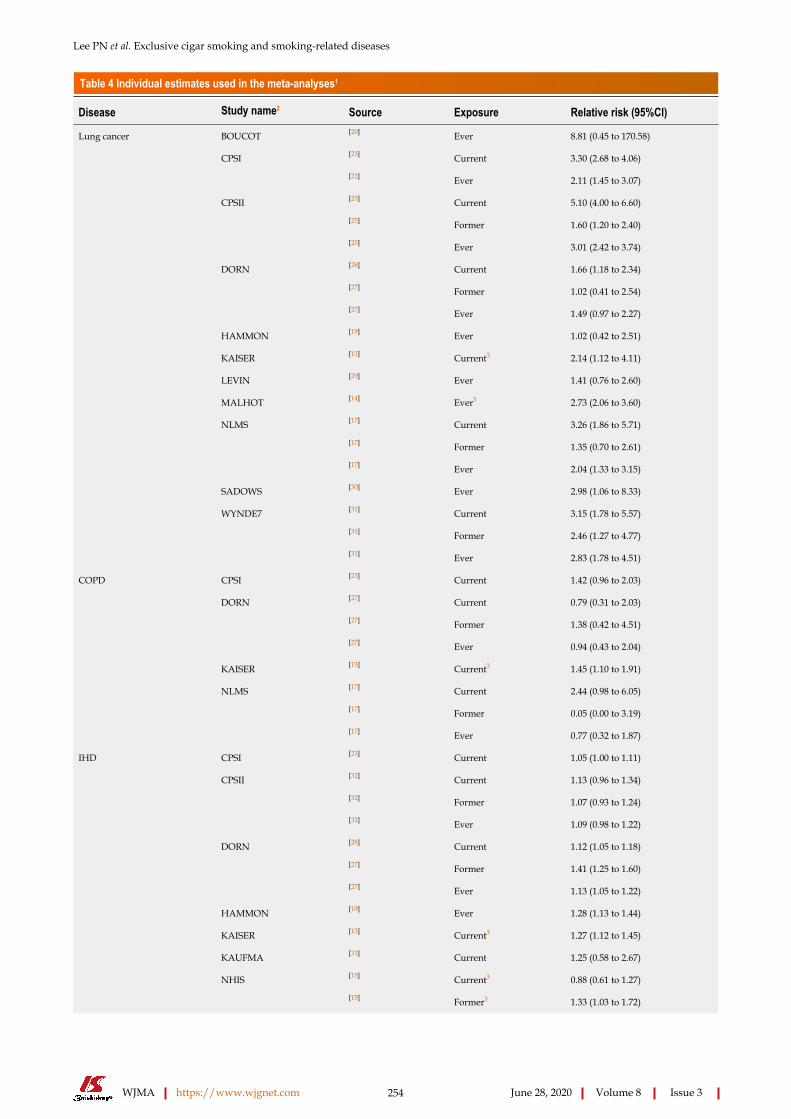
Lee PN et al. Exclusive cigar smoking and smoking-related diseases
WJMA https://www.wjgnet.com 254 June 28, 2020 Volume 8 Issue 3
Table 4 Individual estimates used in the meta-analyses1
Disease Study name2 Source Exposure Relative risk (95%CI)
Lung cancer BOUCOT [20] Ever 8.81 (0.45 to 170.58)
CPSI [23] Current 3.30 (2.68 to 4.06)
[22] Ever 2.11 (1.45 to 3.07)
CPSII [25] Current 5.10 (4.00 to 6.60)
[25] Former 1.60 (1.20 to 2.40)
[25] Ever 3.01 (2.42 to 3.74)
DORN [28] Current 1.66 (1.18 to 2.34)
[27] Former 1.02 (0.41 to 2.54)
[27] Ever 1.49 (0.97 to 2.27)
HAMMON [19] Ever 1.02 (0.42 to 2.51)
KAISER [13] Current3 2.14 (1.12 to 4.11)
LEVIN [29] Ever 1.41 (0.76 to 2.60)
MALHOT [14] Ever3 2.73 (2.06 to 3.60)
NLMS [17] Current 3.26 (1.86 to 5.71)
[17] Former 1.35 (0.70 to 2.61)
[17] Ever 2.04 (1.33 to 3.15)
SADOWS [30] Ever 2.98 (1.06 to 8.33)
WYNDE7 [31] Current 3.15 (1.78 to 5.57)
[31] Former 2.46 (1.27 to 4.77)
[31] Ever 2.83 (1.78 to 4.51)
COPD CPSI [23] Current 1.42 (0.96 to 2.03)
DORN [27] Current 0.79 (0.31 to 2.03)
[27] Former 1.38 (0.42 to 4.51)
[27] Ever 0.94 (0.43 to 2.04)
KAISER [13] Current3 1.45 (1.10 to 1.91)
NLMS [17] Current 2.44 (0.98 to 6.05)
[17] Former 0.05 (0.00 to 3.19)
[17] Ever 0.77 (0.32 to 1.87)
IHD CPSI [23] Current 1.05 (1.00 to 1.11)
CPSII [32] Current 1.13 (0.96 to 1.34)
[32] Former 1.07 (0.93 to 1.24)
[32] Ever 1.09 (0.98 to 1.22)
DORN [28] Current 1.12 (1.05 to 1.18)
[27] Former 1.41 (1.25 to 1.60)
[27] Ever 1.13 (1.05 to 1.22)
HAMMON [19] Ever 1.28 (1.13 to 1.44)
KAISER [13] Current3 1.27 (1.12 to 1.45)
KAUFMA [33] Current 1.25 (0.58 to 2.67)
NHIS [15] Current3 0.88 (0.61 to 1.27)
[15] Former3 1.33 (1.03 to 1.72)
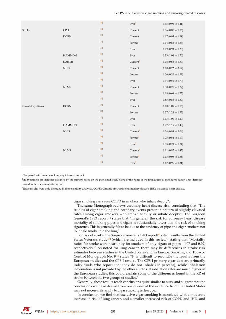
Lee PN et al. Exclusive cigar smoking and smoking-related diseases
WJMA https://www.wjgnet.com 255 June 28, 2020 Volume 8 Issue 3
[15] Ever3 1.15 (0.93 to 1.41)
Stroke CPSI [23] Current 0.96 (0.87 to 1.06)
DORN [28] Current 1.07 (0.95 to 1.21)
[27] Former 1.14 (0.85 to 1.53)
[27] Ever 1.09 (0.93 to 1.29)
HAMMON [19] Ever 1.33 (1.04 to 1.70)
KAISER [13] Current3 1.08 (0.88 to 1.33)
NHIS [16] Current 1.60 (0.72 to 3.57)
[16] Former 0.56 (0.20 to 1.57)
[16] Ever 0.94 (0.50 to 1.77)
NLMS [17] Current 0.50 (0.21 to 1.22)
[17] Former 1.08 (0.66 to 1.75)
[17] Ever 0.85 (0.55 to 1.30)
Circulatory disease DORN [28] Current 1.10 (1.05 to 1.16)
[27] Former 1.37 (1.24 to 1.52)
[27] Ever 1.13 (1.06 to 1.20)
HAMMON [19] Ever 1.27 (1.15 to 1.40)
NHIS [16] Current3 1.34 (0.88 to 2.06)
[16] Former3 0.75 (0.52 to 1.10)
[16] Ever3 0.93 (0.70 to 1.24)
NLMS [17] Current3 1.11 (0.87 to 1.42)
[17] Former3 1.13 (0.93 to 1.38)
[17] Ever3 1.12 (0.96 to 1.31)
1Compared with never smoking any tobacco product. 2Study name is an identifier assigned by the authors based on the published study name or the name of the first author of the source paper. This identifier is used in the meta-analysis output. 3These results were only included in the sensitivity analyses. COPD: Chronic obstructive pulmonary disease; IHD: Ischaemic heart disease.
cigar smoking can cause COPD in smokers who inhale deeply”.The same Monograph reviews coronary heart disease risk, concluding that “The
studies of cigar smoking and coronary events present a pattern of slightly elevated rates among cigar smokers who smoke heavily or inhale deeply”. The Surgeon General’s 1983 report[37] states that “In general, the risk for coronary heart disease mortality of smoking pipes and cigars is substantially lower than the risk of smoking cigarettes. This is generally felt to be due to the tendency of pipe and cigar smokers not to inhale smoke into the lung”.
For risk of stroke, the Surgeon General’s 1983 report[37] cited results from the United States Veterans study[28] (which are included in this review), stating that “Mortality ratios for stroke were near unity for smokers of only cigars or pipes – l.07 and 0.99, respectively.” As noted for lung cancer, there may be differences in stroke risk estimates between studies in the United States and in Europe. Smoking and Tobacco Control Monograph No. 9[23] states “It is difficult to reconcile the results from the European studies and the CPS-I results. The CPS-I primary cigar data are primarily individuals who report that they do not inhale (78 percent), while inhalation information is not provided by the other studies. If inhalation rates are much higher in the European studies, this could explain some of the differences found in the RR of stroke between the two groups of studies.”
Generally, these results reach conclusions quite similar to ours, and suggest that the conclusions we have drawn from our review of the evidence from the United States may not necessarily apply to cigar smoking in Europe.
In conclusion, we find that exclusive cigar smoking is associated with a moderate increase in risk of lung cancer, and a smaller increased risk of COPD and IHD, and
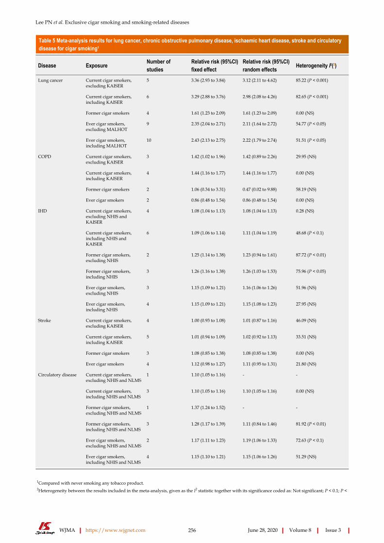
Lee PN et al. Exclusive cigar smoking and smoking-related diseases
WJMA https://www.wjgnet.com 256 June 28, 2020 Volume 8 Issue 3
Table 5 Meta-analysis results for lung cancer, chronic obstructive pulmonary disease, ischaemic heart disease, stroke and circulatory disease for cigar smoking1
Disease Exposure Number of studies
Relative risk (95%CI) fixed effect
Relative risk (95%CI) random effects Heterogeneity I2(2)
Lung cancer Current cigar smokers, excluding KAISER
5 3.36 (2.93 to 3.84) 3.12 (2.11 to 4.62) 85.22 (P < 0.001)
Current cigar smokers, including KAISER
6 3.29 (2.88 to 3.76) 2.98 (2.08 to 4.26) 82.65 (P < 0.001)
Former cigar smokers 4 1.61 (1.23 to 2.09) 1.61 (1.23 to 2.09) 0.00 (NS)
Ever cigar smokers, excluding MALHOT
9 2.35 (2.04 to 2.71) 2.11 (1.64 to 2.72) 54.77 (P < 0.05)
Ever cigar smokers, including MALHOT
10 2.43 (2.13 to 2.75) 2.22 (1.79 to 2.74) 51.51 (P < 0.05)
COPD Current cigar smokers, excluding KAISER
3 1.42 (1.02 to 1.96) 1.42 (0.89 to 2.26) 29.95 (NS)
Current cigar smokers, including KAISER
4 1.44 (1.16 to 1.77) 1.44 (1.16 to 1.77) 0.00 (NS)
Former cigar smokers 2 1.06 (0.34 to 3.31) 0.47 (0.02 to 9.88) 58.19 (NS)
Ever cigar smokers 2 0.86 (0.48 to 1.54) 0.86 (0.48 to 1.54) 0.00 (NS)
IHD Current cigar smokers, excluding NHIS and KAISER
4 1.08 (1.04 to 1.13) 1.08 (1.04 to 1.13) 0.28 (NS)
Current cigar smokers, including NHIS and KAISER
6 1.09 (1.06 to 1.14) 1.11 (1.04 to 1.19) 48.68 (P < 0.1)
Former cigar smokers, excluding NHIS
2 1.25 (1.14 to 1.38) 1.23 (0.94 to 1.61) 87.72 (P < 0.01)
Former cigar smokers, including NHIS
3 1.26 (1.16 to 1.38) 1.26 (1.03 to 1.53) 75.96 (P < 0.05)
Ever cigar smokers, excluding NHIS
3 1.15 (1.09 to 1.21) 1.16 (1.06 to 1.26) 51.96 (NS)
Ever cigar smokers, including NHIS
4 1.15 (1.09 to 1.21) 1.15 (1.08 to 1.23) 27.95 (NS)
Stroke Current cigar smokers, excluding KAISER
4 1.00 (0.93 to 1.08) 1.01 (0.87 to 1.16) 46.09 (NS)
Current cigar smokers, including KAISER
5 1.01 (0.94 to 1.09) 1.02 (0.92 to 1.13) 33.51 (NS)
Former cigar smokers 3 1.08 (0.85 to 1.38) 1.08 (0.85 to 1.38) 0.00 (NS)
Ever cigar smokers 4 1.12 (0.98 to 1.27) 1.11 (0.95 to 1.31) 21.80 (NS)
Circulatory disease Current cigar smokers, excluding NHIS and NLMS
1 1.10 (1.05 to 1.16) - -
Current cigar smokers, including NHIS and NLMS
3 1.10 (1.05 to 1.16) 1.10 (1.05 to 1.16) 0.00 (NS)
Former cigar smokers, excluding NHIS and NLMS
1 1.37 (1.24 to 1.52) - -
Former cigar smokers, including NHIS and NLMS
3 1.28 (1.17 to 1.39) 1.11 (0.84 to 1.46) 81.92 (P < 0.01)
Ever cigar smokers, excluding NHIS and NLMS
2 1.17 (1.11 to 1.23) 1.19 (1.06 to 1.33) 72.63 (P < 0.1)
Ever cigar smokers, including NHIS and NLMS
4 1.15 (1.10 to 1.21) 1.15 (1.06 to 1.26) 51.29 (NS)
1Compared with never smoking any tobacco product. 2Heterogeneity between the results included in the meta-analysis, given as the I2 statistic together with its significance coded as: Not significant; P < 0.1; P <

Lee PN et al. Exclusive cigar smoking and smoking-related diseases
WJMA https://www.wjgnet.com 257 June 28, 2020 Volume 8 Issue 3
0.05; P < 0.01; P < 0.001. The I2 statistic is calculated as 100 × (H-df)/H, where H is the heterogeneity chi-squared statistic, df is the degrees of freedom, and if the resulting value is negative I2 is taken to be 0. NS: Not significant; COPD: Chronic obstructive pulmonary disease; IHD: Ischaemic heart disease.
that these increases in risk are less than for cigarette smoking.
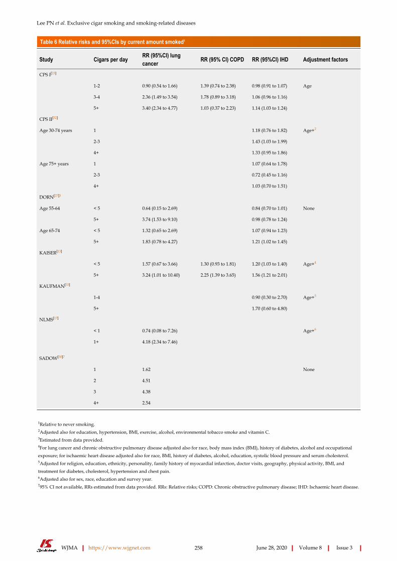
Lee PN et al. Exclusive cigar smoking and smoking-related diseases
WJMA https://www.wjgnet.com 258 June 28, 2020 Volume 8 Issue 3
Table 6 Relative risks and 95%CIs by current amount smoked1
Study Cigars per day RR (95%CI) lung cancer RR (95% CI) COPD RR (95%CI) IHD Adjustment factors
CPS I[23]
1-2 0.90 (0.54 to 1.66) 1.39 (0.74 to 2.38) 0.98 (0.91 to 1.07) Age
3-4 2.36 (1.49 to 3.54) 1.78 (0.89 to 3.18) 1.06 (0.96 to 1.16)
5+ 3.40 (2.34 to 4.77) 1.03 (0.37 to 2.23) 1.14 (1.03 to 1.24)
CPS II[32]
Age 30-74 years 1 1.18 (0.76 to 1.82) Age+2
2-3 1.43 (1.03 to 1.99)
4+ 1.33 (0.95 to 1.86)
Age 75+ years 1 1.07 (0.64 to 1.78)
2-3 0.72 (0.45 to 1.16)
4+ 1.03 (0.70 to 1.51)
DORN[27]3
Age 55-64 < 5 0.64 (0.15 to 2.69) 0.84 (0.70 to 1.01) None
5+ 3.74 (1.53 to 9.10) 0.98 (0.78 to 1.24)
Age 65-74 < 5 1.32 (0.65 to 2.69) 1.07 (0.94 to 1.23)
5+ 1.83 (0.78 to 4.27) 1.21 (1.02 to 1.45)
KAISER[13]
< 5 1.57 (0.67 to 3.66) 1.30 (0.93 to 1.81) 1.20 (1.03 to 1.40) Age+4
5+ 3.24 (1.01 to 10.40) 2.25 (1.39 to 3.65) 1.56 (1.21 to 2.01)
KAUFMAN[33]
1-4 0.90 (0.30 to 2.70) Age+5
5+ 1.70 (0.60 to 4.80)
NLMS[17]
< 1 0.74 (0.08 to 7.26) Age+6
1+ 4.18 (2.34 to 7.46)
SADOW[30]7
1 1.62 None
2 4.51
3 4.38
4+ 2.54
1Relative to never smoking.2Adjusted also for education, hypertension, BMI, exercise, alcohol, environmental tobacco smoke and vitamin C.3Estimated from data provided.4For lung cancer and chronic obstructive pulmonary disease adjusted also for race, body mass index (BMI), history of diabetes, alcohol and occupational exposure; for ischaemic heart disease adjusted also for race, BMI, history of diabetes, alcohol, education, systolic blood pressure and serum cholesterol. 5Adjusted for religion, education, ethnicity, personality, family history of myocardial infarction, doctor visits, geography, physical activity, BMI, and treatment for diabetes, cholesterol, hypertension and chest pain. 6Adjusted also for sex, race, education and survey year.795% CI not available, RRs estimated from data provided. RRs: Relative risks; COPD: Chronic obstructive pulmonary disease; IHD: Ischaemic heart disease.
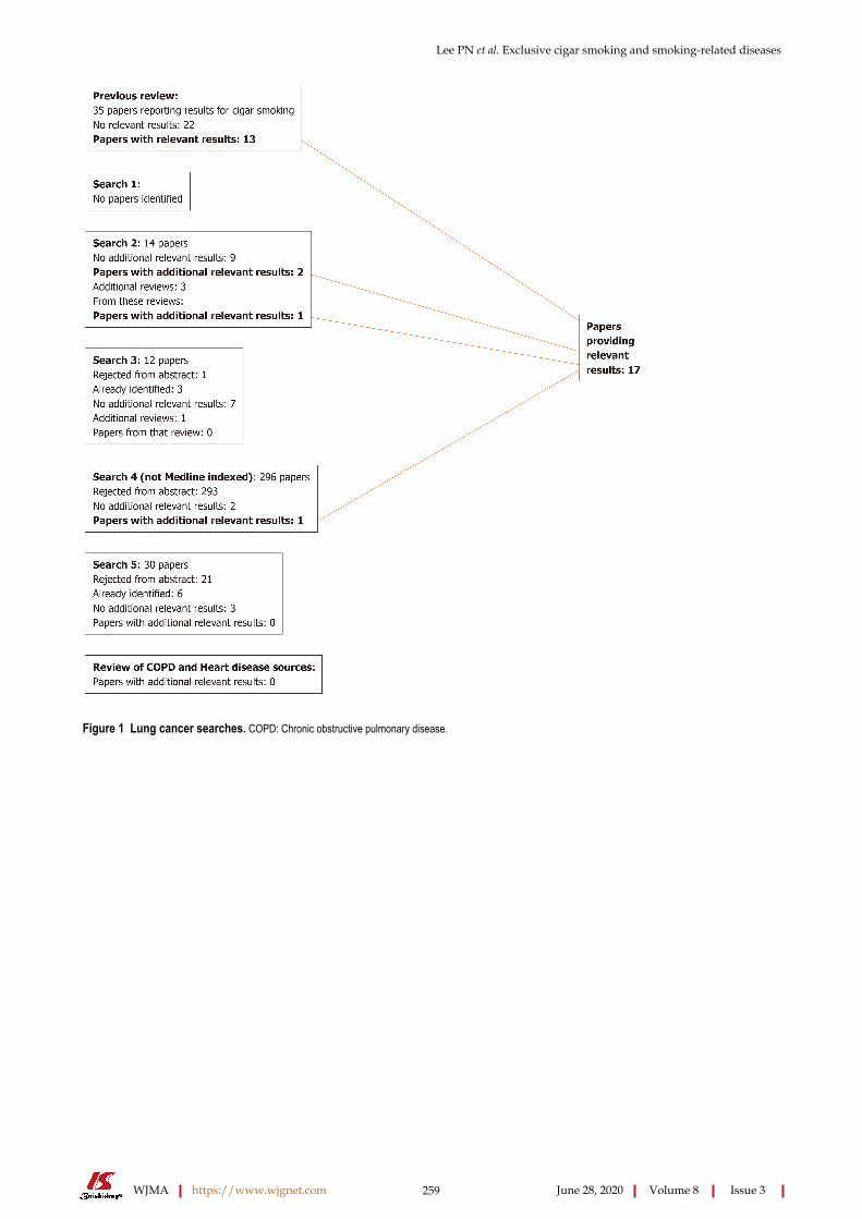
Lee PN et al. Exclusive cigar smoking and smoking-related diseases
WJMA https://www.wjgnet.com 259 June 28, 2020 Volume 8 Issue 3
Figure 1 Lung cancer searches. COPD: Chronic obstructive pulmonary disease.
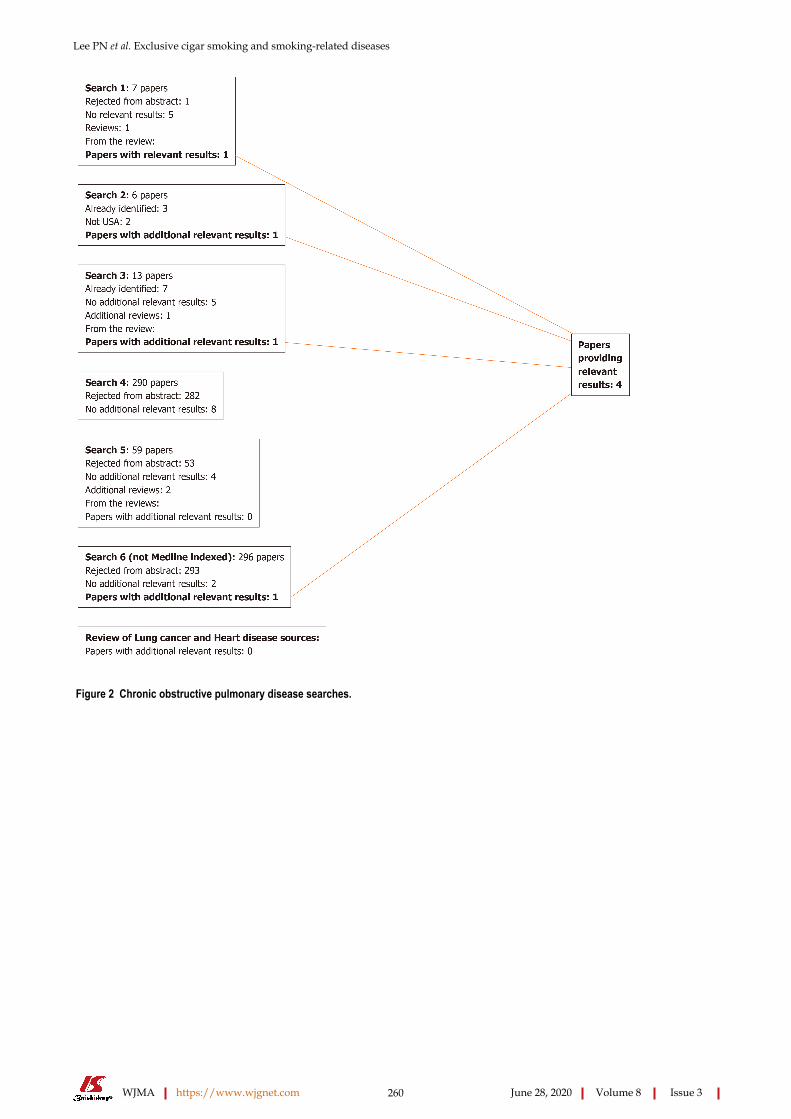
Lee PN et al. Exclusive cigar smoking and smoking-related diseases
WJMA https://www.wjgnet.com 260 June 28, 2020 Volume 8 Issue 3
Figure 2 Chronic obstructive pulmonary disease searches.
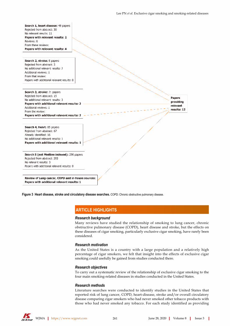
Lee PN et al. Exclusive cigar smoking and smoking-related diseases
WJMA https://www.wjgnet.com 261 June 28, 2020 Volume 8 Issue 3
Figure 3 Heart disease, stroke and circulatory disease searches. COPD: Chronic obstructive pulmonary disease.
ARTICLE HIGHLIGHTSResearch backgroundMany reviews have studied the relationship of smoking to lung cancer, chronic obstructive pulmonary disease (COPD), heart disease and stroke, but the effects on these diseases of cigar smoking, particularly exclusive cigar smoking, have rarely been considered.
Research motivationAs the United States is a country with a large population and a relatively high percentage of cigar smokers, we felt that insight into the effects of exclusive cigar smoking could usefully be gained from studies conducted there.
Research objectivesTo carry out a systematic review of the relationship of exclusive cigar smoking to the four main smoking-related diseases in studies conducted in the United States.
Research methodsLiterature searches were conducted to identify studies in the United States that reported risk of lung cancer, COPD, heart-disease, stroke and/or overall circulatory disease comparing cigar smokers who had never smoked other tobacco products with those who had never smoked any tobacco. For each study identified as providing
Lee PN et al. Exclusive cigar smoking and smoking-related diseases
WJMA https://www.wjgnet.com 262 June 28, 2020 Volume 8 Issue 3
relevant results, data were recorded on study characteristics and on the appropriate relative risks (RRs) and 95%CIs relating to overall current, former and ever exclusive cigar use, and, for current smokers, by daily cigar consumption. RRs for a given smoking group and disease were combined using fixed-effect and random-effects meta-analyses.
Research resultsData were available on lung cancer from 11 studies, on COPD from four studies and on heart disease, stroke and circulatory disease from 10 studies. As RRs tended to be heterogeneous, random-effects estimates are given below. For current smoking overall RR estimates were 2.98 (95%CI: 2.08 to 4.26, based on n = 6 estimates) for lung cancer, 1.44 (1.16 to 1.77, n = 4) for COPD, 1.11 (1.04 to 1.19, n = 6) for ischaemic heart disease, 1.02 (0.92 to 1.13, n = 5) for stroke and 1.10 (1.05 to 1.16, n = 3) for overall circulatory disease. These RRs are much lower than those reported for the United States for exclusive cigarette smokers; 11.68 for lung cancer, 4.56 for COPD and at least 1.70, depending on age, for ischaemic heart disease. Even for heavy cigar smoking, RRs are generally lower than for overall cigarette smoking. RRs for former and for ever smoking were also much lower than for cigarette smoking.
Research conclusionsAlthough our analyses were based on relatively few studies, some conducted some time ago, the results clearly show that exclusive cigar smoking is associated with an increased risk of lung cancer, though much less than is the case for exclusive cigarette smoking. For COPD and ischaemic heart disease the association is weaker, and also less than for cigarette smoking. No previous study has clarified the effects of exclusive cigarette smoking so clearly. Future research could extend results on exclusive cigar smoking to countries other than the United States, and compare risks of cigar smoking with those of using other nicotine products.
Research perspectivesWhile our results show that exclusive cigar smoking is associated with risks of smoking-related diseases that are much lower than those associated with cigarettes smoking, little of the evidence comes from studies conducted in this millenium. Further large prospective studies are needed to collect more up-to-date results, and to clarify how risk varies by type of cigar smoked.
ACKNOWLEDGEMENTSWe thank Barbara Forey for assistance with the literature searching and study selection, and comments on drafts of the paper, and also John Fry and John Hamling for assistance with conduct of the meta-analyses. We also thank Yvonne Cooper and Diana Morris for typing the various drafts of the paper.
REFERENCES1 Wynder EL, Mabuchi K, Beattie EJ Jr. The epidemiology of lung cancer. Recent trends. JAMA 1970; 213:
2221-2228 [PMID: 5468929 DOI: 10.1001/jama.1970.03170390013002]2 Falk RT, Pickle LW, Fontham ET, Greenberg SD, Jacobs HL, Correa P, Fraumeni JF Jr. Epidemiology of
bronchioloalveolar carcinoma. Cancer Epidemiol Biomarkers Prev 1992; 1: 339-344 [PMID: 1339048]3 Sandler DP, Comstock GW, Helsing KJ, Shore DL. Deaths from all causes in non-smokers who lived with
smokers. Am J Public Health 1989; 79: 163-167 [PMID: 2913834 DOI: 10.2105/AJPH.79.2.163]4 Chow WH, Schuman LM, McLaughlin JK, Bjelke E, Gridley G, Wacholder S, Chien HT, Blot WJ. A
cohort study of tobacco use, diet, occupation, and lung cancer mortality. Cancer Causes Control 1992; 3: 247-254 [PMID: 1610971 DOI: 10.1007/BF00124258]
5 Williams RR, Horm JW. Association of cancer sites with tobacco and alcohol consumption and socioeconomic status of patients: interview study from the Third National Cancer Survey. J Natl Cancer Inst 1977; 58: 525-547 [PMID: 557114]
6 National Cancer Institute. Cigars: health effects and trends. In: Shopland DR, Burns DM, Hoffmann D, Cummings KM, Amacher RH, editors. Smoking and Tobacco Control - Monograph No. 9. Bethesda: US Department of Health and Human Services, National Institutes of Health, National Cancer Institute, 1998: 232
7 Forey B, Hamling J, Lee P, Wald N. International Smoking Statistics. A collection of historical data from 30 economically developed countries. 2nd edition. London and Oxford: Wolfson Institute of Preventive Medicine and Oxford University Press, 2002: 854

Lee PN et al. Exclusive cigar smoking and smoking-related diseases
WJMA https://www.wjgnet.com 263 June 28, 2020 Volume 8 Issue 3
8 Forey B, Lee PN. New edition of International Smoking Statistics. Int J Epidemiol 2007; 36: 471-472 [PMID: 17470484 DOI: 10.1093/ije/dym037]
9 Lee PN, Forey BA, Coombs KJ. Systematic review with meta-analysis of the epidemiological evidence in the 1900s relating smoking to lung cancer. BMC Cancer 2012; 12: 385 [PMID: 22943444 DOI: 10.1186/1471-2407-12-385]
10 Hamling J, Lee P, Weitkunat R, Ambühl M. Facilitating meta-analyses by deriving relative effect and precision estimates for alternative comparisons from a set of estimates presented by exposure level or disease category. Stat Med 2008; 27: 954-970 [PMID: 17676579 DOI: 10.1002/sim.3013]
11 Fleiss JL, Gross AJ. Meta-analysis in epidemiology, with special reference to studies of the association between exposure to environmental tobacco smoke and lung cancer: a critique. J Clin Epidemiol 1991; 44: 127-139 [PMID: 1995774 DOI: 10.1016/0895-4356(91)90261-7]
12 Higgins JP, Thompson SG, Deeks JJ, Altman DG. Measuring inconsistency in meta-analyses. BMJ 2003; 327: 557-560 [PMID: 12958120 DOI: 10.1136/bmj.327.7414.557]
13 Iribarren C, Tekawa IS, Sidney S, Friedman GD. Effect of cigar smoking on the risk of cardiovascular disease, chronic obstructive pulmonary disease, and cancer in men. N Engl J Med 1999; 340: 1773-1780 [PMID: 10362820 DOI: 10.1056/NEJM199906103402301]
14 Malhotra J, Borron C, Freedman ND, Abnet CC, van den Brandt PA, White E, Milne RL, Giles GG, Boffetta P. Association between Cigar or Pipe Smoking and Cancer Risk in Men: A Pooled Analysis of Five Cohort Studies. Cancer Prev Res (Phila) 2017; 10: 704-709 [PMID: 28972007 DOI: 10.1158/1940-6207.Capr-17-0084]
15 Rostron BL, Corey CG, Gindi RM. Cigar smoking prevalence and morbidity among US adults, 2000-2015. Prev Med Rep 2019; 14: 100821 [PMID: 30815336 DOI: 10.1016/j.pmedr.2019.100821]
16 Inoue-Choi M, Shiels MS, McNeel TS, Graubard BI, Hatsukami D, Freedman ND. Contemporary Associations of Exclusive Cigarette, Cigar, Pipe, and Smokeless Tobacco Use With Overall and Cause-Specific Mortality in the United States. JNCI Cancer Spectr 2019; 3: pkz036 [PMID: 31321380 DOI: 10.1093/jncics/pkz036]
17 Christensen CH, Rostron B, Cosgrove C, Altekruse SF, Hartman AM, Gibson JT, Apelberg B, Inoue-Choi M, Freedman ND. Association of Cigarette, Cigar, and Pipe Use With Mortality Risk in the US Population. JAMA Intern Med 2018; 178: 469-476 [PMID: 29459935 DOI: 10.1001/jamainternmed.2017.8625]
18 Hammond EC, Horn D. Smoking and death rates; report on forty-four months of follow-up of 187,783 men. I. Total mortality. J Am Med Assoc 1958; 166: 1159-1172 [PMID: 13513334 DOI: 10.1001/jama.1958.02990100047009]
19 Hammond EC, Horn D. Smoking and death rates: report on forty-four months of follow-up of 187,783 men. 2. Death rates by cause. J Am Med Assoc 1958; 166: 1294-1308 [PMID: 12308037 DOI: 10.1001/jama.1958.02990110030007]
20 Boucot KR, Weiss W, Seidman H, Carnahan WJ, Cooper DA. The Philadelphia pulmonary neoplasm research project: basic risk factors of lung cancer in older men. Am J Epidemiol 1972; 95: 4-16 [PMID: 5007365 DOI: 10.1093/oxfordjournals.aje.a121368]
21 Hammond EC. Smoking in relation to the death rates of one million men and women. In: Haenszel W. Epidemiological approaches to the study of cancer and other chronic diseases. Bethesda: U.S. Department of Health, Education, and Welfare; Public Health Service National Cancer Institute, 1966: 127-204
22 Hammond EC. Smoking habits and air pollution in relation to lung cancer. In: Lee HK. Environmental factors in respiratory diseases. New York: Academic Press Inc, 1972: 177-198
23 Shanks TG, Burns D. Disease consequences of cigar smoking - Cigars: Health effects and trends. In: Shopland DR, Burns DM, Hoffmann D, Cummings KM, Amacher RH, editors. Smoking and Tobacco Control - Monograph No. 9. Bethesda, MD: US Department of Health and Human Services, National Institutes of Health, National Cancer Institute, 1998: 105-158
24 Jacobs EJ, Shapiro JA and Thun MJ. Cigar smoking in men and risk of death from tobacco-related cancers. Am J Epidemiol 1999; 149: S71-S71
25 Shapiro JA, Jacobs EJ, Thun MJ. Cigar smoking in men and risk of death from tobacco-related cancers. J Natl Cancer Inst 2000; 92: 333-337 [PMID: 10675383 DOI: 10.1093/jnci/92.4.333]
26 Dorn HF. Tobacco consumption and mortality from cancer and other diseases. Public Health Rep 1959; 74: 581-593 [PMID: 13668008]
27 Kahn HA. The Dorn study of smoking and mortality among U.S. veterans: report on eight and one-half years of observation. In: Haenszel W. Epidemiological approaches to the study of cancer and other chronic diseases. Bethesda: U.S. Department of Health, Education, and Welfare; Public Health Service National Cancer Institute, 1966: 1-125
28 Rogot E, Murray JL. Smoking and causes of death among U.S. veterans: 16 years of observation. Public Health Rep 1980; 95: 213-222 [PMID: 7384406]
29 Levin ML. Etiology of lung cancer: present status. N Y State J Med 1954; 54: 769-777 [PMID: 13144988]30 Sadowsky DA, Gilliam AG, Cornfield J. The statistical association between smoking and carcinoma of the
lung. J Natl Cancer Inst 1953; 13: 1237-1258 [PMID: 13035448]31 Higgins IT, Mahan CM, Wynder EL. Lung cancer among cigar and pipe smokers. Prev Med 1988; 17: 116-
128 [PMID: 3362797 DOI: 10.1016/0091-7435(88)90077-1]32 Jacobs EJ, Thun MJ, Apicella LF. Cigar smoking and death from coronary heart disease in a prospective
study of US men. Arch Intern Med 1999; 159: 2413-2418 [PMID: 10665889 DOI: 10.1001/archinte.159.20.2413]
33 Kaufman DW, Palmer JR, Rosenberg L, Shapiro S. Cigar and pipe smoking and myocardial infarction in young men. Br Med J (Clin Res Ed) 1987; 294: 1315-1316 [PMID: 3109633 DOI: 10.1136/bmj.294.6583.1315]
34 Lee PN, Fry JS, Hamling JF, Sponsiello-Wang Z, Baker G, Weitkunat R. Estimating the effect of differing assumptions on the population health impact of introducing a Reduced Risk Tobacco Product in the USA. Regul Toxicol Pharmacol 2017; 88: 192-213 [PMID: 28651854 DOI: 10.1016/j.yrtph.2017.06.009]Wynder EL, Mabuchi K. Lung cancer among cigar and pipe smokers. Prev Med 1972; 1: 529-542 [PMID: 35

Lee PN et al. Exclusive cigar smoking and smoking-related diseases
WJMA https://www.wjgnet.com 264 June 28, 2020 Volume 8 Issue 3
4670408 DOI: 10.1016/0091-7435(72)90035-7]36 US Surgeon General. Smoking and health - A report of the Surgeon General -DHEW Publication No.
(PHS) 79-50066. Vol Rockville: US Department of Health, Education, and Welfare; Public Health Service, 1979: 1194
37 US Surgeon General. The health consequences of smoking - Cardiovascular disease - A report of the Surgeon General - DHHS Publication No. (PHS) 84-50204. Vol Rockville: US Department of Health and Human Services, Public Health Service, 1983: 384
WJMA https://www.wjgnet.com 265 June 28, 2020 Volume 8 Issue 3
World Journal of
Meta-AnalysisW J M ASubmit a Manuscript: https://www.f6publishing.com World J Meta-Anal 2020 June 28; 8(3): 265-274
DOI: 10.13105/wjma.v8.i3.265 ISSN 2308-3840 (online)
SYSTEMATIC REVIEWS
Hydatidosis and the duodenum: A systematic review of the literature
Víctor de la Fuente-Aguilar, Patricia Beneitez-Mascaraque, Sergio Bergua-Arroyo, María Fernández-Riesgo, Irene Camón-García, Iris Cruza-Aguilera, Kennia Ugarte-Yáñez, José M Ramia
ORCID number: Victor de la Fuente-Aguilar 0000-0001-9472-8219; Patricia Beneitez-Mascaraque 0000-0002-4394-7347; Sergio Bergua-Arroyo 0000-0001-9746-2671; María Fernández-Riesgo 0000-0003-0026-8245; Irene Camón-García 0000-0001-8366-1688; Iris Cruza-Aguilera 0000-0003-2269-7155; Kennia Ugarte-Yáñez 0000-0001-8024-7182; José M Ramia 0000-0003-1186-953X.
Author contributions: Ramia JM designed the research; de la Fuente-Aguilar V, Beneitez-Mascaraque P, Bergua-Arroyo S, Fernández-Riesgo M, Camón-García I, Cruza-Aguilera I, Ugarte-Yáñez K performed the research; de la Fuente-Aguilar V, Beneitez-Mascaraque P, Bergua-Arroyo S, Fernández-Riesgo M, Camón-García I, Cruza-Aguilera I, Ugarte-Yáñez K and Ramia JM analyzed the data; and de la Fuente-Aguilar V, Beneitez-Mascaraque P, Bergua-Arroyo S, Fernández-Riesgo M, Camón-García I, Cruza-Aguilera I, Ugarte-Yáñez K and Ramia JM wrote the paper.
Conflict-of-interest statement: The authors have no conflicts of interest to declare.
PRISMA 2009 Checklist statement: The authors have read the PRISMA 2009 Checklist, and the manuscript was prepared and revised in accordance with this checklist.
Víctor de la Fuente-Aguilar, Patricia Beneitez-Mascaraque, Sergio Bergua-Arroyo, María Fernández-Riesgo, Irene Camón-García, Iris Cruza-Aguilera, Kennia Ugarte-Yáñez, José M Ramia, Faculty of Medicine, Universidad of Alcalá, Alcalá de Henares 28801, Spain
José M Ramia, Servicio de Cirugía General y del Aparato Digestivo, Hospital General Universitario de Alicante and Institute for Health and Biomedical Research (ISABIAL) Alicante 03010, Spain
Corresponding author: José M Ramia, BSc, FACS, FRCS (Gen Surg), MD, PhD, Head, Department of Surgery, Hospital General Universitario de Alicante. Calle Pintor Baeza 11, Alicante, Spain. [email protected]
AbstractBACKGROUND Injury to the duodenum (fistula formation, compression, or other complications) by a hydatid cyst (HC) is an exceptional complication.
AIM To perform a systematic review of the literature on the fistulization of HC in the duodenum.
METHODS Following PRISMA guidelines, a search for HC with duodenal involvement was carried out in the databases of PubMed, SCielo and EMBASE without time limits.
RESULTS Fourteen patients were identified, seven men and seven women, with a mean age of 53.14 years (SD = 17.65, range: 28-78). Three out of the 14 (21%) had HC relapse. The most frequent clinical manifestations were abdominal pain and nausea and/or vomiting. Various imaging studies were performed in almost all cases, the most commonly used being abdominal computed tomography (10/14, 71%). A range of surgical techniques were reported, most frequently HC drainage (41%) and enucleations (16%). Ten of the 14 patients had no complications and one patient died. The follow-up period and recurrences could not be determined.
CONCLUSION The most frequent symptoms were abdominal pain, nausea and vomiting. Computed tomography was the most used diagnostic imaging technique, and HC drainage and fistula closure via laparotomy was the most frequent treatment.
de la Fuente-Aguilar V et al. Hydatidosis and duodenum
WJMA https://www.wjgnet.com 266 June 28, 2020 Volume 8 Issue 3
Open-Access: This article is an open-access article that was selected by an in-house editor and fully peer-reviewed by external reviewers. It is distributed in accordance with the Creative Commons Attribution NonCommercial (CC BY-NC 4.0) license, which permits others to distribute, remix, adapt, build upon this work non-commercially, and license their derivative works on different terms, provided the original work is properly cited and the use is non-commercial. See: http://creativecommons.org/licenses/by-nc/4.0/
Manuscript source: Invited manuscript
Received: February 13, 2020 Peer-review started: February 13, 2020 First decision: March 26, 2020 Revised: April 15, 2020 Accepted: June 13, 2020 Article in press: June 13, 2020 Published online: June 28, 2020
P-Reviewer: Kamimura K, Nari GA S-Editor: Wang JL L-Editor: A E-Editor: Qi LL
However, all diagnostic and therapeutic options for HC fistulizing the duodenum had a low level of evidence.
Key words: Echinococcosis; Hydatidosis; Duodenum; Surgery; Review
©The Author(s) 2020. Published by Baishideng Publishing Group Inc. All rights reserved.
Core tip: Hydatidosis is a global zoonosis. The most frequent organs affected are the liver (50%-70%) and the lungs (20%-30%) The duodenum is an organ that is very rarely affected by hydatidosis, either primarily or secondarily. Possible forms of secondary duodenal involvement are compression, or, more frequently, fistulization from hydatid cysts located in neighboring organs. The hydatid cysts that most often fistulize the duodenum are those located in the liver (0.15% of instances), with cases also described from the pancreas and kidney. Its low prevalence and the few existing records mean that the choice of the best therapeutic management is a challenge. A systematic review let readers know all published cases and best management.
Citation: de la Fuente-Aguilar V, Beneitez-Mascaraque P, Bergua-Arroyo S, Fernández-Riesgo M, Camón-García I, Cruza-Aguilera I, Ugarte-Yáñez K, Ramia JM. Hydatidosis and the duodenum: A systematic review of the literature. World J Meta-Anal 2020; 8(3): 265-274URL: https://www.wjgnet.com/2308-3840/full/v8/i3/265.htmDOI: https://dx.doi.org/10.13105/wjma.v8.i3.265
INTRODUCTIONHydatidosis is a zoonosis caused by the larval form of the genus Equinococcus spp, and Equinococcus granulosus (E. granulosus) causes the formation of hydatid cysts (HC). The life cycle of E. granulosus includes different herbivores (lambs, cows, pigs, deer) and carnivores (dogs, foxes, wolves); the dog is the most frequent definitive host[1]. The distribution of E. granulosus is associated with livestock exploitation and temperate climate zones such as southern Europe, Latin America, Australia and New Zealand, Africa, and the Middle East. A prevalence of HC of 2%-6% is estimated in the population in endemic areas[2].
The human being is an intermediate host, as it is infected orally with food contaminated by eggs from the feces of the definitive host. The eggs hatch in the intestine, releasing oncospheres that penetrate the intestinal wall and invade different sites via the bloodstream, the most frequent being the liver (50%-70%) and the lungs (20%-30%). The oncospheres evolve into HC, which may remain asymptomatic until their growth causes complications derived from the mass effect, depending on their location. Rupture of the HC can spread its contents throughout the body and trigger anaphylactic shock[3].
The duodenum is an organ that is very rarely affected by HC, either primarily or secondarily. Possible forms of secondary duodenal involvement are compression, or, more frequently, fistulization from HC located in neighboring organs[4]. The HC that most often fistulize the duodenum are those located in the liver (0.15% of instances), with cases also described from the pancreas and kidney. Its low prevalence and the few existing records mean that the choice of the best therapeutic management is a challenge[4].
Here we present a systematic literature review of cases of hydatidosis with duodenal involvement published in the scientific literature.
MATERIALS AND METHODSLiterature searchFollowing the guidelines of the PRISMA methodology for systematic reviews, a search was performed in the PubMed, SCielo and EMBASE databases[5], unlimited by language or time. The terms used for the search were: (duodenum) AND [(echinococcosis, hepatic) OR (echinococcosis) OR (echinococcosis, hepatic) OR (hepatic echinococcosis) OR (hepatic echinococcosis) OR (echinococcosis, hepatic
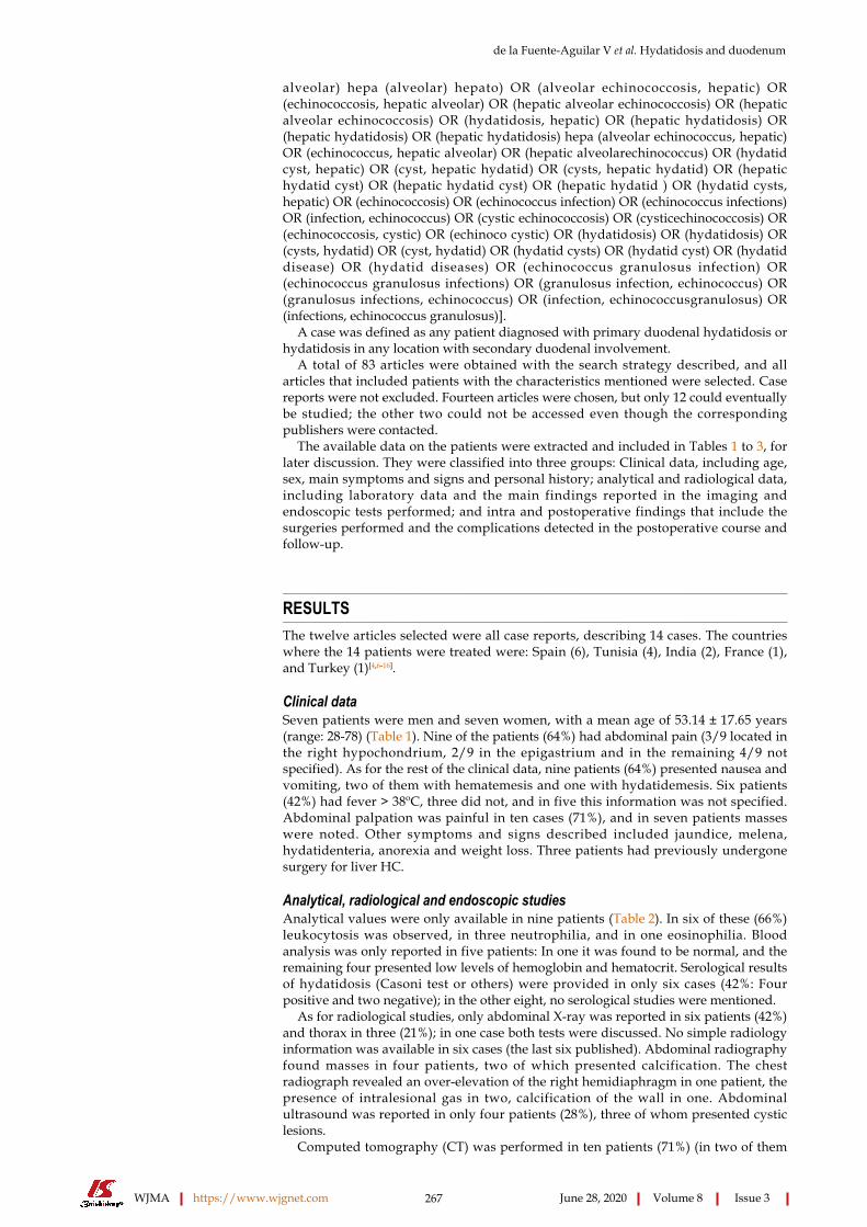
de la Fuente-Aguilar V et al. Hydatidosis and duodenum
WJMA https://www.wjgnet.com 267 June 28, 2020 Volume 8 Issue 3
alveolar) hepa (alveolar) hepato) OR (alveolar echinococcosis, hepatic) OR (echinococcosis, hepatic alveolar) OR (hepatic alveolar echinococcosis) OR (hepatic alveolar echinococcosis) OR (hydatidosis, hepatic) OR (hepatic hydatidosis) OR (hepatic hydatidosis) OR (hepatic hydatidosis) hepa (alveolar echinococcus, hepatic) OR (echinococcus, hepatic alveolar) OR (hepatic alveolarechinococcus) OR (hydatid cyst, hepatic) OR (cyst, hepatic hydatid) OR (cysts, hepatic hydatid) OR (hepatic hydatid cyst) OR (hepatic hydatid cyst) OR (hepatic hydatid ) OR (hydatid cysts, hepatic) OR (echinococcosis) OR (echinococcus infection) OR (echinococcus infections) OR (infection, echinococcus) OR (cystic echinococcosis) OR (cysticechinococcosis) OR (echinococcosis, cystic) OR (echinoco cystic) OR (hydatidosis) OR (hydatidosis) OR (cysts, hydatid) OR (cyst, hydatid) OR (hydatid cysts) OR (hydatid cyst) OR (hydatid disease) OR (hydatid diseases) OR (echinococcus granulosus infection) OR (echinococcus granulosus infections) OR (granulosus infection, echinococcus) OR (granulosus infections, echinococcus) OR (infection, echinococcusgranulosus) OR (infections, echinococcus granulosus)].
A case was defined as any patient diagnosed with primary duodenal hydatidosis or hydatidosis in any location with secondary duodenal involvement.
A total of 83 articles were obtained with the search strategy described, and all articles that included patients with the characteristics mentioned were selected. Case reports were not excluded. Fourteen articles were chosen, but only 12 could eventually be studied; the other two could not be accessed even though the corresponding publishers were contacted.
The available data on the patients were extracted and included in Tables 1 to 3, for later discussion. They were classified into three groups: Clinical data, including age, sex, main symptoms and signs and personal history; analytical and radiological data, including laboratory data and the main findings reported in the imaging and endoscopic tests performed; and intra and postoperative findings that include the surgeries performed and the complications detected in the postoperative course and follow-up.
RESULTSThe twelve articles selected were all case reports, describing 14 cases. The countries where the 14 patients were treated were: Spain (6), Tunisia (4), India (2), France (1), and Turkey (1)[4,6-16].
Clinical dataSeven patients were men and seven women, with a mean age of 53.14 ± 17.65 years (range: 28-78) (Table 1). Nine of the patients (64%) had abdominal pain (3/9 located in the right hypochondrium, 2/9 in the epigastrium and in the remaining 4/9 not specified). As for the rest of the clinical data, nine patients (64%) presented nausea and vomiting, two of them with hematemesis and one with hydatidemesis. Six patients (42%) had fever > 38ºC, three did not, and in five this information was not specified. Abdominal palpation was painful in ten cases (71%), and in seven patients masses were noted. Other symptoms and signs described included jaundice, melena, hydatidenteria, anorexia and weight loss. Three patients had previously undergone surgery for liver HC.
Analytical, radiological and endoscopic studiesAnalytical values were only available in nine patients (Table 2). In six of these (66%) leukocytosis was observed, in three neutrophilia, and in one eosinophilia. Blood analysis was only reported in five patients: In one it was found to be normal, and the remaining four presented low levels of hemoglobin and hematocrit. Serological results of hydatidosis (Casoni test or others) were provided in only six cases (42%: Four positive and two negative); in the other eight, no serological studies were mentioned.
As for radiological studies, only abdominal X-ray was reported in six patients (42%) and thorax in three (21%); in one case both tests were discussed. No simple radiology information was available in six cases (the last six published). Abdominal radiography found masses in four patients, two of which presented calcification. The chest radiograph revealed an over-elevation of the right hemidiaphragm in one patient, the presence of intralesional gas in two, calcification of the wall in one. Abdominal ultrasound was reported in only four patients (28%), three of whom presented cystic lesions.
Computed tomography (CT) was performed in ten patients (71%) (in two of them
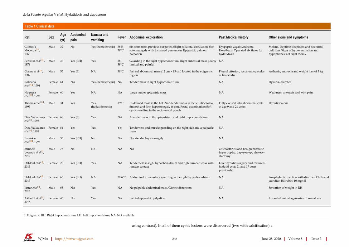
de la Fuente-Aguilar V et al. Hydatidosis and duodenum
WJMA https://www.wjgnet.com 268 June 28, 2020 Volume 8 Issue 3
Table 1 Clinical data
Ref. Sex Age (yr)
Abdominal pain
Nausea and vomiting Fever Abdominal exploration Past Medical history Other signs and symptoms
Gilmas Y Mocoroa[16], 1963
Male 32 No Yes (hematemesis) 38.5-39ºC
Six scars from previous surgeries. Slight collateral circulation. Soft splenomegaly with increased percussion. Epigastric pain on palpation
Dyspeptic vagal syndrome. Heartburn. Operated six times for hydatidosis
Melena. Daytime sleepiness and nocturnal delirium. Signs of hypoventilation and hypophonesis of right thorax
Perrotin et al[15], 1978
Male 37 Yes (RH) Yes 38-39ºC
Guarding in the right hypochondrium. Right subcostal mass poorly limited and painful
NA
Cosme et al[13], 1987
Male 55 Yes (E) NA 38ºC Painful abdominal mass (12 cm × 15 cm) located in the epigastric region
Pleural effusion, recurrent episodes of bronchitis
Asthenia, anorexia and weight loss of 3 kg
Robbana et al[12], 1991
Female 64 NA Yes (hematemesis) No Tender mass in right hypochon-drium NA Dysuria, diarrhea
Noguera et al[11], 1993
Female 60 Yes NA NA Large tender epigastric mass NA Weakness, anorexia and joint pain
Thomas et al[14], 1993
Male 31 Yes Yes (hydatidemesis)
39ºC Ill-defined mass in the LH. Non-tender mass in the left iliac fossa. Smooth and firm hepatomegaly (6 cm). Rectal examination: Soft cystic swelling in the rectovesical pouch
Fully excised intraabdominal cysts at age 9 and 21 years
Hydatidenteria
Diez Valladares et al[9], 1998
Female 68 Yes (E) Yes NA A tender mass in the epigastrium and right hypochon-drium NA
Diez Valladares et al[9], 1998
Female 84 Yes Yes Yes Tenderness and muscle guarding on the right side and a palpable mass
NA
Patankar et al[10], 1998
Male 35 Yes (RH) No No Non-tender hepatomegaly NA
Muinelo Lorenzo et al[8], 2012
Male 78 No No NA NA Osteoarthritis and benign prostatic hypertrophy. Laparoscopy cholecy-stectomy
Daldoul et al[4], 2013
Female 28 Yes (RH) Yes NA Tenderness in right hypochon-drium and right lumbar fossa with lumbar contact
Liver hydatid surgery and recurrent hydatid cysts 21 and 17 years previously
Daldoul et al[4], 2013
Female 63 Yes (EH) NA 38.6ºC Abdominal involuntary guarding in the right hypochon-drium NA Anaphylactic reaction with diarrhea Chills and jaundice: Bilirubin: 10 mg/dl
Jarrar et al[7], 2015
Male 63 NA Yes NA No palpable abdominal mass. Gastric distension NA Sensation of weight in RH
Akbulut et al[6], 2018
Female 46 No Yes No Painful epigastric palpation NA Intra-abdominal aggressive fibromatosis
E: Epigastric; RH: Right hypochondrium; LH: Left hypochondrium; NA: Not available
using contrast). In all of them cystic lesions were discovered (two with calcification) a
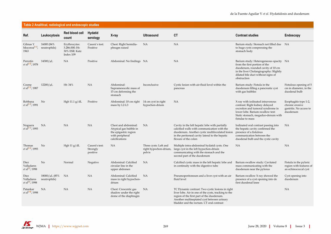
de la Fuente-Aguilar V et al. Hydatidosis and duodenum
WJMA https://www.wjgnet.com 269 June 28, 2020 Volume 8 Issue 3
Table 2 Analitical, radiological and endoscopic studies
Ref. Leukocytosis Red blood cell count
Hydatid serology X-ray Ultrasound CT Contrast studies Endoscopy
Gilmas Y Mocoroa[16], 1963
16000 (84% neutrophils)
Erythrocytes: 3.286.000; Ht: 30% ESR: Katz Index 109
Casoni´s test: Positive
Chest: Right hemidia-phragm raised
NA NA Barium study: Stomach not filled due to huge cysts compressing the stomach body
NA
Perrotin et al[15], 1978
14300/µL NA Positive Abdominal: No findings NA NA Barium study: Heterogeneous opacity from the first portion of the duodenum, rounded cavity of 10 cm in the liver Cholangiography: Slightly dilated bile duct without signs of obstruction
NA
Cosme et al[13], 1987
12200/µL Ht: 34% NA Abdominal: Supramesocolic mass of 15 cm deforming the stomach
Inconclusive Cystic lesion with air-fluid level within the pancreas
Barium study: Fistula in the duodenum filling a pancreatic cyst with gas bubbles
Fistulous opening of 5 cm in diameter, in the duodenal bulb
Robbana et al[12], 1991
No Hgb 11.1 g/dL Positive Abdominal: 10 cm right mass by L2-L3
14 cm cyst in right hypochon-drium
NA X-ray with iodinated intravenous contrast: Right kidney delayed excretion and tumoral syndrome in lower lobe. Barium swallow test: Static stomach, megaduo-denum with fistulae to mass
Esophagitis type 1-2, chronic erosive gastritis. No access to duodenum
Noguera et al[11], 1993
NA NA NA Chest and abdominal: Atypical gas bubble in the epigastric region with peripheral calcifications
NA Cavity in the left hepatic lobe with partially calcified walls with communication with the duodenum. Another cystic multiloculated lesion in the peritoneal cavity lateral to the hepatic flexure of the colon
Iodinated oral contrast passing into the hepatic cavity confirmed the presence of a fistulous communication between the duodenal bulb and the cystic cavity
NA
Thomas et al[14], 1993
No Hgb 11 g/dL Casoni's test: Strongly positive
NA Three cysts: Left and right hypochon-drium, pelvis
Multiple intra-abdominal hydatid cysts. One large cyst in the left hypochon-drium communicating with the stomach and the second part of the duodenum
NA NA
Diez Valladares et al[9], 1998
No Normal Negative Abdominal: Calcified circular line in the upper abdomen
NA Calcified cystic mass in the left hepatic lobe and in continuity with the digestive tube
Barium swallow study: Cavitated mass communicating with the duodenum near the pylorus
Fistula in the pyloric region with features of an echinococcal cyst
Diez Valladares et al[9], 1998
18000/µL (85% neutrophils)
NA NA Abdominal: Calcified mass in right hypochon-drium
NA Pneumoperitoneum and a liver cyst with an air fluid level
Barium swallow X-ray showed the presence of a cyst opening into de first duodenal knee
Cyst opening into duodenum
TC Dynamic contrast: Two cystic lesions in right liver lobe. Air in one of the cysts, tracking to the region of the first part of the duodenum. Another multiseptated cyst between urinary bladder and the rectum. CT oral contrast
Patankar et al[10], 1998
NA NA NA Chest: Crescentic gas shadow under the right dome of the diaphragm
NA NA
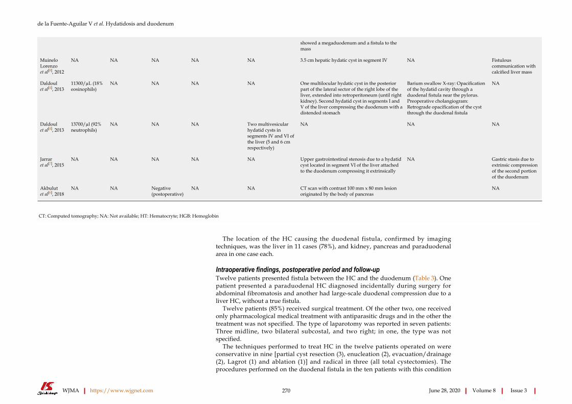
de la Fuente-Aguilar V et al. Hydatidosis and duodenum
WJMA https://www.wjgnet.com 270 June 28, 2020 Volume 8 Issue 3
showed a megaduodenum and a fistula to the mass
Muinelo Lorenzo et al[8], 2012
NA NA NA NA NA 3.5 cm hepatic hydatic cyst in segment IV NA Fistulous communication with calcified liver mass
Daldoul et al[4], 2013
11300/µL (18% eosinophils)
NA NA NA NA One multilocular hydatic cyst in the posterior part of the lateral sector of the right lobe of the liver, extended into retroperitoneum (until right kidney). Second hydatid cyst in segments I and V of the liver compressing the duodenum with a distended stomach
Barium swallow X-ray: Opacification of the hydatid cavity through a duodenal fistula near the pylorus. Preoperative cholangiogram: Retrograde opacification of the cyst through the duodenal fistula
NA
Daldoul et al[4], 2013
13700/µl (92% neutrophils)
NA NA NA Two multivesicular hydatid cysts in segments IV and VI of the liver (5 and 6 cm respectively)
NA NA NA
Jarrar et al[7], 2015
NA NA NA NA NA Upper gastrointestinal stenosis due to a hydatid cyst located in segment VI of the liver attached to the duodenum compressing it extrinsically
NA Gastric stasis due to extrinsic compression of the second portion of the duodenum
Akbulut et al[6], 2018
NA NA Negative (postoperative)
NA NA CT scan with contrast 100 mm x 80 mm lesion originated by the body of pancreas
NA
CT: Computed tomography; NA: Not available; HT: Hematocryte; HGB: Hemoglobin
The location of the HC causing the duodenal fistula, confirmed by imaging techniques, was the liver in 11 cases (78%), and kidney, pancreas and paraduodenal area in one case each.
Intraoperative findings, postoperative period and follow-upTwelve patients presented fistula between the HC and the duodenum (Table 3). One patient presented a paraduodenal HC diagnosed incidentally during surgery for abdominal fibromatosis and another had large-scale duodenal compression due to a liver HC, without a true fistula.
Twelve patients (85%) received surgical treatment. Of the other two, one received only pharmacological medical treatment with antiparasitic drugs and in the other the treatment was not specified. The type of laparotomy was reported in seven patients: Three midline, two bilateral subcostal, and two right; in one, the type was not specified.
The techniques performed to treat HC in the twelve patients operated on were conservative in nine [partial cyst resection (3), enucleation (2), evacuation/drainage (2), Lagrot (1) and ablation (1)] and radical in three (all total cystectomies). The procedures performed on the duodenal fistula in the ten patients with this condition
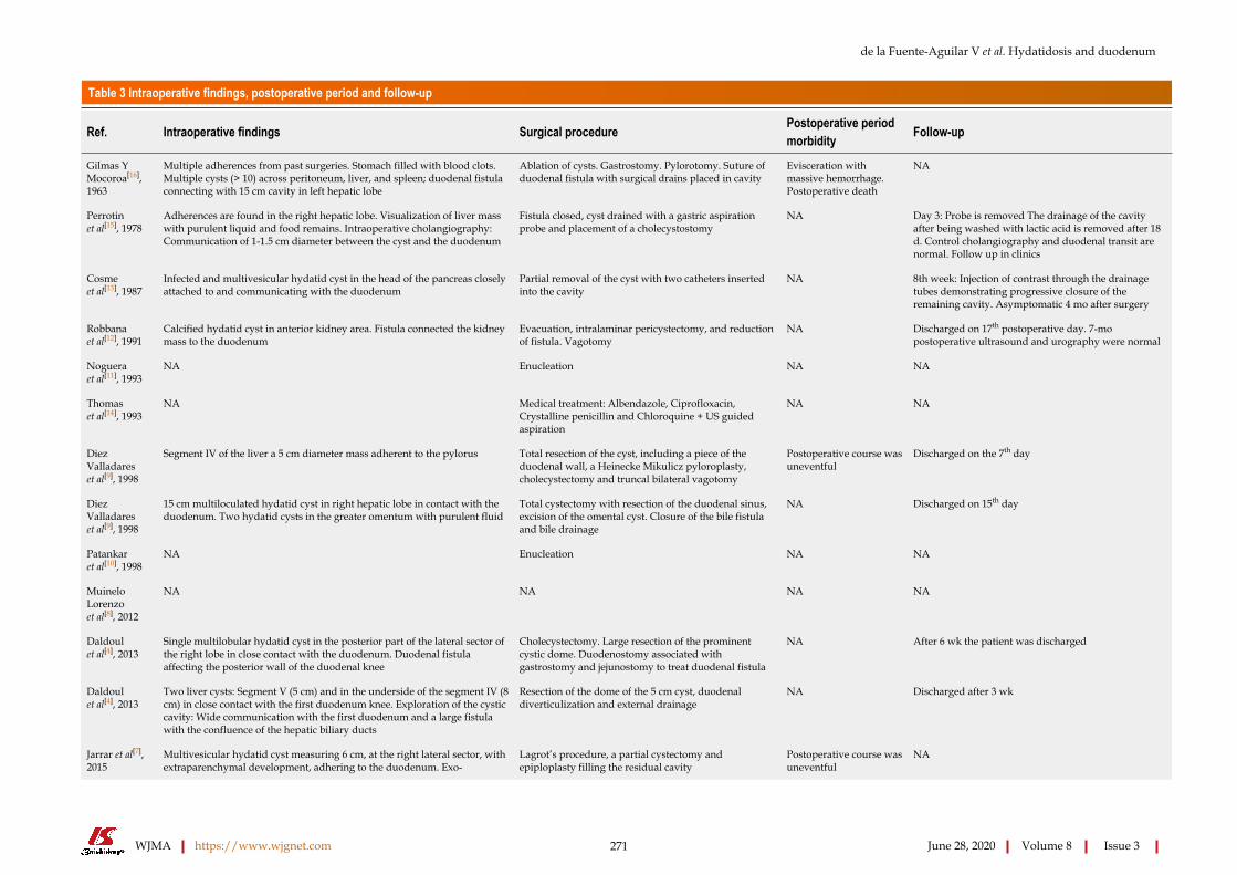
de la Fuente-Aguilar V et al. Hydatidosis and duodenum
WJMA https://www.wjgnet.com 271 June 28, 2020 Volume 8 Issue 3
Table 3 Intraoperative findings, postoperative period and follow-up
Ref. Intraoperative findings Surgical procedure Postoperative period morbidity Follow-up
Gilmas Y Mocoroa[16], 1963
Multiple adherences from past surgeries. Stomach filled with blood clots. Multiple cysts (> 10) across peritoneum, liver, and spleen; duodenal fistula connecting with 15 cm cavity in left hepatic lobe
Ablation of cysts. Gastrostomy. Pylorotomy. Suture of duodenal fistula with surgical drains placed in cavity
Evisceration with massive hemorrhage. Postoperative death
NA
Perrotin et al[15], 1978
Adherences are found in the right hepatic lobe. Visualization of liver mass with purulent liquid and food remains. Intraoperative cholangiography: Communication of 1-1.5 cm diameter between the cyst and the duodenum
Fistula closed, cyst drained with a gastric aspiration probe and placement of a cholecystostomy
NA Day 3: Probe is removed The drainage of the cavity after being washed with lactic acid is removed after 18 d. Control cholangiography and duodenal transit are normal. Follow up in clinics
Cosme et al[13], 1987
Infected and multivesicular hydatid cyst in the head of the pancreas closely attached to and communicating with the duodenum
Partial removal of the cyst with two catheters inserted into the cavity
NA 8th week: Injection of contrast through the drainage tubes demonstrating progressive closure of the remaining cavity. Asymptomatic 4 mo after surgery
Robbana et al[12], 1991
Calcified hydatid cyst in anterior kidney area. Fistula connected the kidney mass to the duodenum
Evacuation, intralaminar pericystectomy, and reduction of fistula. Vagotomy
NA Discharged on 17th postoperative day. 7-mo postoperative ultrasound and urography were normal
Noguera et al[11], 1993
NA Enucleation NA NA
Thomas et al[14], 1993
NA Medical treatment: Albendazole, Ciprofloxacin, Crystalline penicillin and Chloroquine + US guided aspiration
NA NA
Diez Valladares et al[9], 1998
Segment IV of the liver a 5 cm diameter mass adherent to the pylorus Total resection of the cyst, including a piece of the duodenal wall, a Heinecke Mikulicz pyloroplasty, cholecystectomy and truncal bilateral vagotomy
Postoperative course was uneventful
Discharged on the 7th day
Diez Valladares et al[9], 1998
15 cm multiloculated hydatid cyst in right hepatic lobe in contact with the duodenum. Two hydatid cysts in the greater omentum with purulent fluid
Total cystectomy with resection of the duodenal sinus, excision of the omental cyst. Closure of the bile fistula and bile drainage
NA Discharged on 15th day
Patankar et al[10], 1998
NA Enucleation NA NA
Muinelo Lorenzo et al[8], 2012
NA NA NA NA
Daldoul et al[4], 2013
Single multilobular hydatid cyst in the posterior part of the lateral sector of the right lobe in close contact with the duodenum. Duodenal fistula affecting the posterior wall of the duodenal knee
Cholecystectomy. Large resection of the prominent cystic dome. Duodenostomy associated with gastrostomy and jejunostomy to treat duodenal fistula
NA After 6 wk the patient was discharged
Daldoul et al[4], 2013
Two liver cysts: Segment V (5 cm) and in the underside of the segment IV (8 cm) in close contact with the first duodenum knee. Exploration of the cystic cavity: Wide communication with the first duodenum and a large fistula with the confluence of the hepatic biliary ducts
Resection of the dome of the 5 cm cyst, duodenal diverticulization and external drainage
NA Discharged after 3 wk
Multivesicular hydatid cyst measuring 6 cm, at the right lateral sector, with extraparenchymal development, adhering to the duodenum. Exo-
Jarrar et al[7], 2015
Lagrot’s procedure, a partial cystectomy and epiploplasty filling the residual cavity
Postoperative course was uneventful
NA
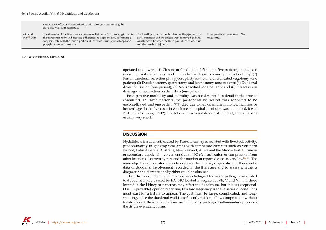
de la Fuente-Aguilar V et al. Hydatidosis and duodenum
WJMA https://www.wjgnet.com 272 June 28, 2020 Volume 8 Issue 3
vesiculation of 2 cm, communicating with the cyst, compressing the duodenal wall without fistula
Akbulut et al[6], 2018
The diameter of the fibromatous mass was 120 mm × 100 mm, originated in the pancreatic body and creating adherences to adjacent tissues forming a conglomerate with the fourth portion of the duodenum, jejunal loops and prepyloric stomach antrum
The fourth portion of the duodenum, the jejunum, the distal pancreas and the spleen were removed en bloc. Anastomosis between the third part of the duodenum and the proximal jejunum
Postoperative course was uneventful
NA
NA: Not available; US: Ultrasound.
operated upon were: (1) Closure of the duodenal fistula in five patients, in one case associated with vagotomy, and in another with gastrostomy plus pylorotomy; (2) Partial duodenal resection plus pyloroplasty and bilateral truncated vagotomy (one patient); (3) Duodenostomy, gastrostomy and jejunostomy (one patient); (4) Duodenal diverticulization (one patient); (5) Not specified (one patient); and (6) Intracavitary drainage without action on the fistula (one patient).
Postoperative morbidity and mortality was not described in detail in the articles consulted. In three patients the postoperative period was reported to be uncomplicated, and one patient (7%) died due to hemoperitoneum following massive hemorrhage. In the five cases in which mean hospital admission was mentioned, it was 20.4 ± 11.72 d (range: 7-42). The follow-up was not described in detail, though it was usually very short.
DISCUSSIONHydatidosis is a zoonosis caused by Echinococcus spp associated with livestock activity, predominantly in geographical areas with temperate climates such as Southern Europe, Latin America, Australia, New Zealand, Africa and the Middle East[2]. Primary or secondary duodenal involvement due to HC via fistulization or compression from other locations is extremely rare and the number of reported cases is very low[4,6-16]. The main objective of our study was to evaluate the clinical, diagnostic and therapeutic data of duodenal involvement recorded in the literature and to assess whether a diagnostic and therapeutic algorithm could be obtained.
The articles included do not describe any etiological factors or pathogenesis related to duodenal injury caused by HC. HC located in segments IVB, V and VI, and those located in the kidney or pancreas may affect the duodenum, but this is exceptional. Our (unprovable) opinion regarding this low frequency is that a series of conditions must exist for a fistula to appear: The cyst must be large, complicated, and long-standing, since the duodenal wall is sufficiently thick to allow compression without fistulization. If these conditions are met, after very prolonged inflammatory processes the fistula eventually forms.
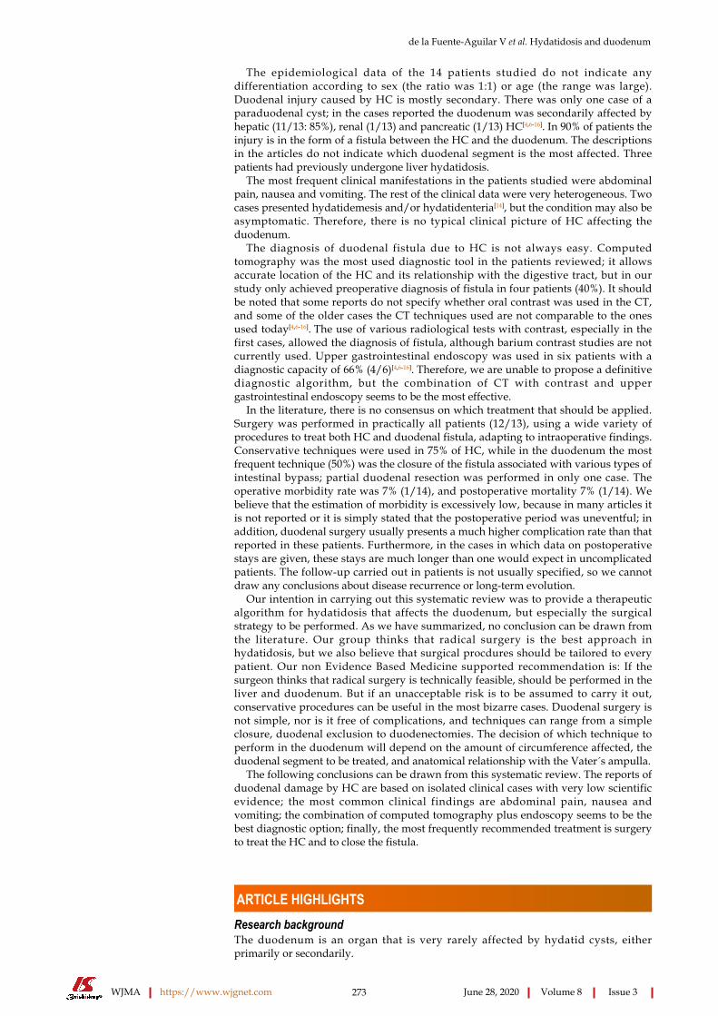
de la Fuente-Aguilar V et al. Hydatidosis and duodenum
WJMA https://www.wjgnet.com 273 June 28, 2020 Volume 8 Issue 3
The epidemiological data of the 14 patients studied do not indicate any differentiation according to sex (the ratio was 1:1) or age (the range was large). Duodenal injury caused by HC is mostly secondary. There was only one case of a paraduodenal cyst; in the cases reported the duodenum was secondarily affected by hepatic (11/13: 85%), renal (1/13) and pancreatic (1/13) HC[4,6-16]. In 90% of patients the injury is in the form of a fistula between the HC and the duodenum. The descriptions in the articles do not indicate which duodenal segment is the most affected. Three patients had previously undergone liver hydatidosis.
The most frequent clinical manifestations in the patients studied were abdominal pain, nausea and vomiting. The rest of the clinical data were very heterogeneous. Two cases presented hydatidemesis and/or hydatidenteria[14], but the condition may also be asymptomatic. Therefore, there is no typical clinical picture of HC affecting the duodenum.
The diagnosis of duodenal fistula due to HC is not always easy. Computed tomography was the most used diagnostic tool in the patients reviewed; it allows accurate location of the HC and its relationship with the digestive tract, but in our study only achieved preoperative diagnosis of fistula in four patients (40%). It should be noted that some reports do not specify whether oral contrast was used in the CT, and some of the older cases the CT techniques used are not comparable to the ones used today[4,6-16]. The use of various radiological tests with contrast, especially in the first cases, allowed the diagnosis of fistula, although barium contrast studies are not currently used. Upper gastrointestinal endoscopy was used in six patients with a diagnostic capacity of 66% (4/6)[4,6-16]. Therefore, we are unable to propose a definitive diagnostic algorithm, but the combination of CT with contrast and upper gastrointestinal endoscopy seems to be the most effective.
In the literature, there is no consensus on which treatment that should be applied. Surgery was performed in practically all patients (12/13), using a wide variety of procedures to treat both HC and duodenal fistula, adapting to intraoperative findings. Conservative techniques were used in 75% of HC, while in the duodenum the most frequent technique (50%) was the closure of the fistula associated with various types of intestinal bypass; partial duodenal resection was performed in only one case. The operative morbidity rate was 7% (1/14), and postoperative mortality 7% (1/14). We believe that the estimation of morbidity is excessively low, because in many articles it is not reported or it is simply stated that the postoperative period was uneventful; in addition, duodenal surgery usually presents a much higher complication rate than that reported in these patients. Furthermore, in the cases in which data on postoperative stays are given, these stays are much longer than one would expect in uncomplicated patients. The follow-up carried out in patients is not usually specified, so we cannot draw any conclusions about disease recurrence or long-term evolution.
Our intention in carrying out this systematic review was to provide a therapeutic algorithm for hydatidosis that affects the duodenum, but especially the surgical strategy to be performed. As we have summarized, no conclusion can be drawn from the literature. Our group thinks that radical surgery is the best approach in hydatidosis, but we also believe that surgical procdures should be tailored to every patient. Our non Evidence Based Medicine supported recommendation is: If the surgeon thinks that radical surgery is technically feasible, should be performed in the liver and duodenum. But if an unacceptable risk is to be assumed to carry it out, conservative procedures can be useful in the most bizarre cases. Duodenal surgery is not simple, nor is it free of complications, and techniques can range from a simple closure, duodenal exclusion to duodenectomies. The decision of which technique to perform in the duodenum will depend on the amount of circumference affected, the duodenal segment to be treated, and anatomical relationship with the Vater´s ampulla.
The following conclusions can be drawn from this systematic review. The reports of duodenal damage by HC are based on isolated clinical cases with very low scientific evidence; the most common clinical findings are abdominal pain, nausea and vomiting; the combination of computed tomography plus endoscopy seems to be the best diagnostic option; finally, the most frequently recommended treatment is surgery to treat the HC and to close the fistula.
ARTICLE HIGHLIGHTSResearch backgroundThe duodenum is an organ that is very rarely affected by hydatid cysts, either primarily or secondarily.
de la Fuente-Aguilar V et al. Hydatidosis and duodenum
WJMA https://www.wjgnet.com 274 June 28, 2020 Volume 8 Issue 3
Research motivationDuodenal involvement by hydatidosis is an exceptional finding and this study aimed to characterize epidemiological, clinical and diagnostic features and options of treatments.
Research objectivesOur aim is to characterize epidemiological, clinical and pathological features and options of treatment in hydatidosis with duodenal involvement.
Research methodsWe perform a system review including all patients with hydatidosis and duodenal involvement following PRISMA guidelines.
Research resultsThis review shows that the most frequent symptoms were abdominal pain, nausea and vomiting. Computed tomography was the most used diagnostic imaging technique, and hydatid cyst drainage and fistula closure via laparotomy was the most frequent treatment.
Research conclusionsLiterature on hydatidosis with duodenal involvement is scarce, only case reports, so the evidence is low.
REFERENCES1 Craig P, Mastin A, van Kesteren F, Boufana B. Echinococcus granulosus: Epidemiology and state-of-the-
art of diagnostics in animals. Vet Parasitol 2015; 213: 132-148 [PMID: 26321135 DOI: 10.1016/j.vetpar.2015.07.028]
2 Schantz PM. Progress in diagnosis, treatment and elimination of echinococcosis and cysticercosis. Parasitol Int 2006; 55 Suppl: S7-S13 [PMID: 16386944 DOI: 10.1016/j.parint.2005.11.050]
3 Agudelo Higuita NI, Brunetti E, McCloskey C. Cystic Echinococcosis. J Clin Microbiol 2016; 54: 518-523 [PMID: 26677245 DOI: 10.1128/JCM.02420-15]
4 Daldoul S, Moussi A, Zaouche A. Spontaneous fistulization of hepatic hydatid cyst into the duodenum: an exceptional complication. J Coll Physicians Surg Pak 2013; 23: 424-426 [PMID: 23763804]
5 Liberati A, Altman DG, Tetzlaff J, Mulrow C, Gøtzsche PC, Ioannidis JP, Clarke M, Devereaux PJ, Kleijnen J, Moher D. The PRISMA statement for reporting systematic reviews and meta-analyses of studies that evaluate healthcare interventions: explanation and elaboration. BMJ 2009; 339: b2700 [PMID: 19622552 DOI: 10.1136/bmj.b2700]
6 Akbulut S, Yilmaz M, Alan S, Kolu M, Karadag N. Coexistence of duodenum derived aggressive fibromatosis and paraduodenal hydatid cyst: A case report and review of literature. World J Gastrointest Surg 2018; 10: 90-94 [PMID: 30510634 DOI: 10.4240/wjgs.v10.i8.90]
7 Jarrar MS, Ben Hadj Khalifa MH, Toumi R, Ghrissi R, Elghali MA, Khenissi A, Hamila F, Letaief R. Upper digestive stenosis due to a hydatid cyst of the liver. Tunis Med 2015; 93: 129-130 [PMID: 26367398]
8 Muinelo Lorenzo M, Alvarez-Prida E, Simó Fernández V, Alcoba García LM. [Hydatid cyst with fistulisation to the duodenum]. Cir Esp 2012; 90: e23 [PMID: 21411075 DOI: 10.1016/j.ciresp.2010.10.013]
9 Diez Valladares L, Sanchez-Pernaute A, Gonzalez O, Perez-Aguirre E, Talavera P, Gutierrez del Olmo A, Torres AJ, Balibrea JL. Hydatid liver cyst perforation into the digestive tract. Hepatogastroenterology 1998; 45: 2110-2114 [PMID: 9951874]
10 Patankar T, Prasad S, Rohondia OS, Mohite JD, James P, Radhakrishnan R. Fistulization of hepatic hydatid cyst into duodenum. Indian J Gastroenterol 1998; 17: 152 [PMID: 9795506]
11 Noguera M, Alvarez-Castells A, Castella E, Gifre L, Andreu J, Quiroga S. Spontaneous duodenal fistula due to hepatic hydatid cyst. Abdom Imaging 1993; 18: 234-236 [PMID: 8508082 DOI: 10.1007/bf00198111]
12 Robbana A, Daghfous MH, Mseddi H, Gandoura, Raies H, Ben Jaafar M. [Hydatid cyst of the right kidney invading into the duodenum. Apropos of a case]. J Urol (Paris) 1991; 97: 230-233 [PMID: 1761871]
13 Cosme A, Orive V, Ojeda E, Aramburu V, Irazusta M, Arenas JI. Hydatid cyst of the head of the pancreas with spontaneous fistula to the duodenum. Am J Gastroenterol 1987; 82: 1311-1313 [PMID: 3687907]
14 Thomas S, Mishra MC, Kriplani AK, Kapur BM. Hydatidemesis: a bizarre presentation of abdominal hydatidosis. Aust N Z J Surg 1993; 63: 496-498 [PMID: 8498923]
15 Perrotin J, Bastian D, Mourot J. [Perforation of a hydatid cyst of the liver into the duodenum (author's transl)]. Chirurgie 1978; 104: 569-574 [PMID: 747967]
16 Gilmas Y Mocoroa A. [Hepatic hydatid cyst perforated into the duodenum and hemorrhagic]. Rev Esp Enferm Apar Dig Nutr 1963; 22: 118-122 [PMID: 13947972]
WJMA https://www.wjgnet.com 275 June 28, 2020 Volume 8 Issue 3
World Journal of
Meta-AnalysisW J M ASubmit a Manuscript: https://www.f6publishing.com World J Meta-Anal 2020 June 28; 8(3): 275-284
DOI: 10.13105/wjma.v8.i3.275 ISSN 2308-3840 (online)
META-ANALYSIS
Prevalence of anxiety among gestational diabetes mellitus patients: A systematic review and meta-analysis
Kai Wei Lee, Hong Chuan Loh, Seng Choi Chong, Siew Mooi Ching, Navin Kumar Devaraj, Maiza Tusimin, Habibah Abdul Hamid, Fan Kee Hoo
ORCID number: Kai Wei Lee 0000-0001-9737-8066; Hong Chuan Loh 0000-0002-2462-5828; Seng Choi Chong 0000-0002-6085-1726; Siew Mooi Ching 0000-0002-4425-7989; Navin Kumar Devaraj 0000-0001-8097-3192; Maiza Tusimin 0000-0001-9672-5804; Habibah Abdul Hamid 0000-0002-5693-617X; Fan Kee Hoo 0000-0003-1687-627X.
Author contributions: Lee KW and Loh HC conceived and designed the experiments; and performed the data extraction; Lee KW, Loh HC, Chong SC, Ching SM, Devaraj NK, Tusimin M, Abdul Hamid H, and Hoo FK analysed the data; Ching SM, Devaraj NK, Tusimin M, and Hoo FK contributed to the quality appraisal; Lee KW, Loh HC, Chong SC, Ching SM, Devaraj NK and Abdul Hamid H wrote the paper; all authors have read and approved the manuscript.
Supported by the Universiti Putra Malaysia under Putra Graduate Initiative, No UPM/700-2/1/GP-IPS/2018/9593800; and High Impact Grant, No. UPM/800-3/3/1/GPB/2018/9659600.
Conflict-of-interest statement: The authors declare that they have no competing interests.
PRISMA 2009 Checklist statement: This study was written according
Kai Wei Lee, Siew Mooi Ching, Navin Kumar Devaraj, Department of Family Medicine, Faculty of Medicine and Health Sciences, Universiti Putra Malaysia, Serdang 43400, Selangor, Malaysia
Hong Chuan Loh, Clinical Research Centre, Hospital Seberang Jaya, Ministry of Health Malaysia, Perai 13700, Penang, Malaysia
Seng Choi Chong, Department of Psychiatry, Faculty of Medicine and Health Sciences, Universiti Putra Malaysia, Serdang 43400, Selangor, Malaysia
Maiza Tusimin, Habibah Abdul Hamid, Department of Obstetrics and Gynaecology, Faculty of Medicine and Health Sciences, Universiti Putra Malaysia, Serdang 43400, Selangor, Malaysia
Fan Kee Hoo, Department of Medicine, Faculty of Medicine and Health Sciences, Universiti Putra Malaysia, Serdang 43400, Selangor, Malaysia
Corresponding author: Siew Mooi Ching, MD, MMed (FamMed), Associate Professor, Department of Family Medicine, Faculty of Medicine and Health Sciences, Universiti Putra Malaysia, Serdang 43400, Selangor, Malaysia. [email protected]
AbstractBACKGROUND A diagnosis of gestational diabetes mellitus (GDM) negatively influences maternal mental health. There is a lack of systematic review and meta-analysis on prevalence of anxiety among GDM women.
AIM To pool data from existing literature to determine the pooled estimates for the prevalence of anxiety among women diagnosed with GDM.
METHODS We searched multiple databases including MEDLINE, Cinahl, PubMed and Scopus to identify studies published up to 31 October 2019 with data on the prevalence of anxiety among women diagnosed with GDM. Data were extracted from published reports. Estimates were pooled using random-effects meta-analyses.
RESULTS We reviewed 19 abstracts, retrieved 10 articles and included three studies

Lee KW et al. Anxiety among GDM
WJMA https://www.wjgnet.com 276 June 28, 2020 Volume 8 Issue 3
to the PRISMA 2009 Checklist.
Open-Access: This article is an open-access article that was selected by an in-house editor and fully peer-reviewed by external reviewers. It is distributed in accordance with the Creative Commons Attribution NonCommercial (CC BY-NC 4.0) license, which permits others to distribute, remix, adapt, build upon this work non-commercially, and license their derivative works on different terms, provided the original work is properly cited and the use is non-commercial. See: http://creativecommons.org/licenses/by-nc/4.0/
Manuscript source: Unsolicited manuscript
Received: April 23, 2020 Peer-review started: April 23, 2020 First decision: April 30, 2020 Revised: May 4, 2020 Accepted: June 17, 2020 Article in press: June 17, 2020 Published online: June 28, 2020
P-Reviewer: Byeon H S-Editor: Wang JL L-Editor: A E-Editor: Li X
incorporating 12744 GDM women from three countries. The pooled prevalence of anxiety was 29.5% (95%CI: 6.9, 52.0) among GDM women.
CONCLUSION Prevalence of anxiety among GDM women was high. We suggest that epidemiological studies on anxiety should be conducted urgently as it merits clinical attention. In addition, it is important to identify factors associated with anxiety among women diagnosed with GDM.
Key words: Prevalence; Anxiety; Gestational diabetes; Psychiatry; Meta-analysis; Systematic review
©The Author(s) 2020. Published by Baishideng Publishing Group Inc. All rights reserved.
Core tip: This is a systematic review and meta-analysis reporting the pooled prevalence of anxiety among gestational diabetes mellitus patients which stood at 29.5%.
Citation: Lee KW, Loh HC, Chong SC, Ching SM, Devaraj NK, Tusimin M, Abdul Hamid H, Hoo FK. Prevalence of anxiety among gestational diabetes mellitus patients: A systematic review and meta-analysis. World J Meta-Anal 2020; 8(3): 275-284URL: https://www.wjgnet.com/2308-3840/full/v8/i3/275.htmDOI: https://dx.doi.org/10.13105/wjma.v8.i3.275
INTRODUCTIONThe prevalence of gestational diabetes mellitus (GDM) has been increasing over the past decades[1,2]. Globally, GDM has been reported as a leading cause of morbidity and mortality among both the infants and their mother[3,4]. Mothers with GDM are at increased risk of getting pregnancy complications such as preterm delivery, preeclampsia, abnormal birth weight, metabolic and electrolyte disorders[5]. Studies also indicated that GDM may persist after postpartum and subsequently develop into overt diabetes mellitus, and it was estimated that the risk for developing diabetes mellitus after GDM increased linearly with the duration of follow-up ranged from 19.72% at 10 years. The estimated risks for type-2 diabetes mellitus ranged from 19.7% at 10 years to 39.0% at 30 years[6]. Neonates born to GDM mothers are at higher risk of suffering from adverse neonatal outcomes such as abnormal birth weight, congenital anomalies, hypoglycaemia and longer duration in neonatal intensive care unit for further investigation[7-9].
Previous studies showed that the prevalence of depression among mothers with GDM were ranging from 25.9% to 56.7%[10,11] and the prevalence of anxiety was within a range of 4.8% to 57.7%[12,13]. Anxiety is a normal reaction to stress which involves both psychological and physical reactions. It becomes clinically significant when the anxiety grows out of proportion to the situation and causes functional impairment. Anxiety disorders are among the most common mental illnesses, and are characterized by feelings of tension, worrying thoughts and physical changes such as increased blood pressure. People with anxiety disorders usually have recurring intrusive thoughts or concerns. They may avoid certain situations out of worry. They may also have physical symptoms such as sweating, trembling, dizziness or a rapid heartbeat[14]. With a remarkable increase in lifetime prevalence, anxiety has become a public health burden worldwide, causing increased use of mental health services and loss of productivity[15]. In particular, anxiety is a common psychiatric condition that affects up to one-fifth of the pregnant mothers[16] and is significantly associated with postpartum depression (odds ratio = 2.6, 95%CI: 2.0, 3.5) and reduced odds of breastfeeding (odds ratio = 0.63, 95%CI: 0.5, 0.7)[17]. Thus, anxiety and related mental conditions could pose negative effects on child development[18]. A high state of anxiety is found in 15.8% of pregnant women, while 12.5% of women suffer high trait anxiety[19]. Similarly, pregnant mothers with GDM were more anxious than pregnant women with other medical problems or healthy pregnant women[20].
There are multiple factors associated with anxiety during pregnancy, including current or past pregnancy-related complications, previous pregnant loss and personal history of mental illness[21]. Study have also shown that women with GDM experience
Lee KW et al. Anxiety among GDM
WJMA https://www.wjgnet.com 277 June 28, 2020 Volume 8 Issue 3
significantly worse quality of life[22]. However, findings from previous studies indicate that there is a lack of data on the epidemiology of antenatal anxiety among GDM patients. Therefore, we aimed to determine the pooled prevalence of anxiety among GDM patients by conducting a meta-analysis.
MATERIALS AND METHODSThis present study was registered in the Medical Research and Ethics Committee, Ministry of Health Malaysia (registration number: NMRR-20-117-52644), and conducted according to Preferred Reporting Items for Systematic Reviews and Meta-Analyses[23]. As this work only involved secondary data retrieval and analysis, no ethical approval was sought.
Literature searchTwo investigators (Lee KW and Loh HC) independently searched MEDLINE, Cinahl, PubMed and Scopus databases for potential studies published in journals from inception to 31 October 2019. We used following search terms: (Anxiety OR anxiety symptom OR anxiety disorder OR generalized anxiety disorder OR panic disorder OR panic attack OR agoraphobia OR phobia OR specific phobia OR specific phobic disorder OR medication-induced anxiety disorder OR medical condition induced anxiety disorder OR social anxiety disorder) AND (prevalence) AND (gestational diabetes OR GDM OR gestational diabetes mellitus OR diabetes in pregnancy). The search strategies with the Boolean or phrase operators were shown in the Supplementary material 1. Studies in English, available in full-text and conducted among humans were searched. Then, we removed duplications using Endnote, after that we screened the title and abstracts for its suitability. Finally, articles with their full text were assessed for eligibility to be recruited into the quantitative analysis.
Inclusion criteriaAny studies that reported the prevalence or percentage for anxiety symptoms or anxiety disorders among GDM patients and fulfilled the inclusion criteria were analysed. The inclusion criteria were as follows: (1) Diagnosing or screening of anxiety was made according to Diagnostic and Statistical Manual of Mental Disorders/International Classification of Diseases diagnostic criteria or by any screening tools; (2) Participants in the study were diagnosed with GDM; and (3) Studies were published in English peer-reviewed journal from inception to 31 October 2019. Other related studies were also included through careful review of the reference lists of related review articles and reverse-forward citation tracking. Studies were excluded if they included only pregnant mothers with pre-existing diabetes mellitus, were of case-control design or examined anxiety prior to the diagnosis of GDM.
Study selectionAll relevant articles identified through the above databases were imported into Endnote programme X5 version. Initially, we performed de-duplication. Two investigators independently screened each title and abstract for suitability based on the search strategies mentioned above. Then, full-text articles were assessed based on the inclusion criteria mentioned above. Any disagreements between the investigators were resolved through discussions before the final consensus for quantitative analysis was reached.
Data extractionThe following data were extracted from every study: The last name of the first author, year of publication, country, sample size, study design, recruitment duration, timing of GDM diagnosis, GDM diagnosis guidelines, assessment of anxiety guidelines, and timing of anxiety assessment. The outcomes measures included the numbers of GDM patients with anxiety symptoms or disorders and total number of GDM patients. Two investigators (Lee KW and Loh HC) individually extracted the data and assessed the study quality, with differences resolved through discussion with the third and fourth investigators (Ching SM and Hoo FK).
Quality assessmentThe quality of the individual studies was determined using the checklist of Strengthening the Reporting of Observational Studies in Epidemiology (STROBE)[24].
Lee KW et al. Anxiety among GDM
WJMA https://www.wjgnet.com 278 June 28, 2020 Volume 8 Issue 3
The aim and use of STROBE were to assess the strengths and weaknesses of the studies reported in the medical literature. STROBE results also helps readers to know what was planned, done and found, as well as what is incomplete and inadequate in the reporting of the articles. The tool consists of 22 items to help assess the important components found in observational studies. In certain instances where the information provided was insufficient in order to make judgement for a certain item, that item was graded with a “0”, rendering the item as having a high risk of bias. Each article’s quality was graded as “good” if the STROBE score was ≥ 14; or graded as “poor” if the STROBE score was < 14. Two investigators (Devaraj NK and Maiza T) individually assessed the study quality, with differences resolved by discussion with the third and fourth investigators (Ching SM and Hoo FK). Studies were included in analysis regardless of STROBE score and grade.
Statistical analysisA random-effects (DerSimonian and Laird Method) meta-analysis method was employed to pool the prevalence estimated from these related studies and was reported with a 95%CI. I2 index was used to assess the studies heterogeneity (i.e. low is < 25%, moderate 25%–50%, and high > 50%) that indicated the total percent of discrepancy due to variation in the included studies[25]. For statistical analysis, Open Meta (Analyst) software was used, this software can be accessed and downloaded from http://www.cebm.brown.edu/openmeta/index.html[26]. Funnel plot was generated using The Jamovi project computer Software which can be retrieved from https://www.jamovi.org[27].
RESULTSDescription of included studiesThirty manuscripts were identified in the initial screening as shown in Figure 1. After removal of duplicate articles (n = 11), a total of 19 studies were retrieved for further assessment. After screening for its suitability through title and abstract, 10 studies fulfilled both our inclusion and exclusion criteria. After careful evaluation of the 10 articles, only three studies were eligible for quantitative analysis in this study.
Characteristics of included studiesThe main characteristics of the included studies are shown in Table 1. A total sample of 12744 women diagnosed with GDM was included in the analysis. The respondents were diagnosed using either American Diabetes Association or World Health Organization guidelines. These studies were conducted in Canada[13], Ireland[12] and Malaysia[28]. In terms of diagnosing or screening for anxiety, Beka et al[13] (2018) used the diagnostic criteria of the International Classification of Diseases- Ninth version (ICD-9) (prior to 2002) and the International Classification of Diseases- Tenth version (ICD-10) (2002 onward); while Egan et al[12] (2017) and Lee et al[28] (2019) used 21-item Depression Anxiety Stress Scale (DASS-21). For quality assessment, we assigned each study with an overall rating based on the tool derived from STROBE checklist. The overall quality of included studies appeared to be good.
Prevalence of anxietyThe overall pooled prevalence of anxiety was 29.5% (95%CI: 6.9, 52.0) (Figure 2). The pooled prevalence of anxiety using DASS-21 was higher than prevalence of anxiety using ICD-9/10 (42.4% vs 4.8%). Sensitivity analysis reveals that study by Beka et al[13] had substantial influences on the overall prevalence, which caused prevalence of anxiety to increase from 29.5% (95%CI: 6.9, 52.0) to 42.4% (95%CI: 13.2, 71.5). On the other hand, removal of studies by Egan et al[12], 2017 or Lee et al[28], 2019, it did not cause statistically significant changes to the overall prevalence of anxiety (Supplementary material 2). Indeed, funnel plot (Supplementary material 3) suggested that there was publication bias. Nevertheless, we did not exclude any studies from the meta-analysis in view there was only three studies available.
Quality assessmentWe assigned the studies with an overall rating based on STROBE checklist. All three studies received an overall “Good” quality with a score of ≥ 14 over 22 (Supplementary materials 4-6). In summarizing the results, we concluded that all studies had methodological issues such as not describing any efforts to address
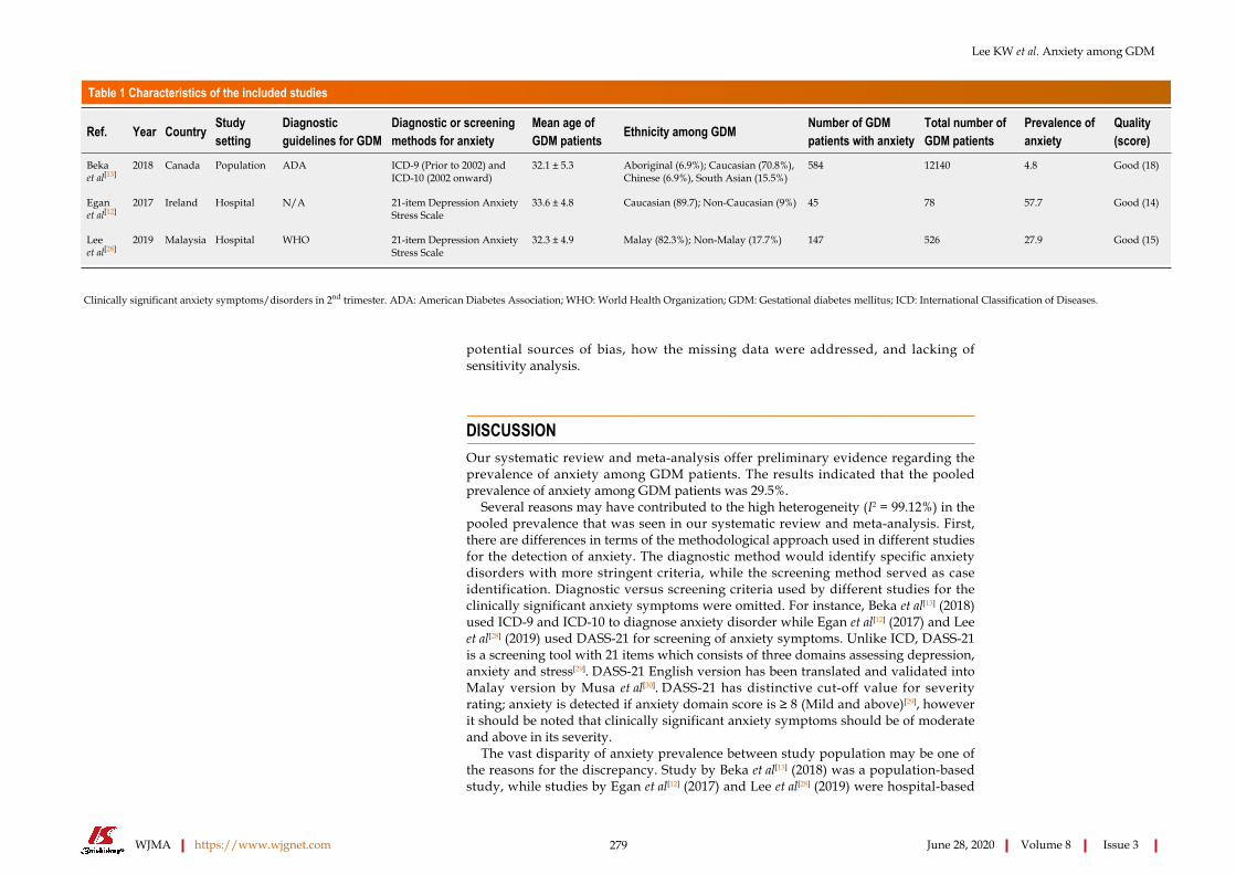
Lee KW et al. Anxiety among GDM
WJMA https://www.wjgnet.com 279 June 28, 2020 Volume 8 Issue 3
Table 1 Characteristics of the included studies
Ref. Year Country Study setting
Diagnostic guidelines for GDM
Diagnostic or screening methods for anxiety
Mean age of GDM patients Ethnicity among GDM Number of GDM
patients with anxietyTotal number of GDM patients
Prevalence of anxiety
Quality (score)
Beka et al[13]
2018 Canada Population ADA ICD-9 (Prior to 2002) and ICD-10 (2002 onward)
32.1 ± 5.3 Aboriginal (6.9%); Caucasian (70.8%), Chinese (6.9%), South Asian (15.5%)
584 12140 4.8 Good (18)
Egan et al[12]
2017 Ireland Hospital N/A 21-item Depression Anxiety Stress Scale
33.6 ± 4.8 Caucasian (89.7); Non-Caucasian (9%) 45 78 57.7 Good (14)
Lee et al[28]
2019 Malaysia Hospital WHO 21-item Depression Anxiety Stress Scale
32.3 ± 4.9 Malay (82.3%); Non-Malay (17.7%) 147 526 27.9 Good (15)
Clinically significant anxiety symptoms/disorders in 2nd trimester. ADA: American Diabetes Association; WHO: World Health Organization; GDM: Gestational diabetes mellitus; ICD: International Classification of Diseases.
potential sources of bias, how the missing data were addressed, and lacking of sensitivity analysis.
DISCUSSIONOur systematic review and meta-analysis offer preliminary evidence regarding the prevalence of anxiety among GDM patients. The results indicated that the pooled prevalence of anxiety among GDM patients was 29.5%.
Several reasons may have contributed to the high heterogeneity (I2 = 99.12%) in the pooled prevalence that was seen in our systematic review and meta-analysis. First, there are differences in terms of the methodological approach used in different studies for the detection of anxiety. The diagnostic method would identify specific anxiety disorders with more stringent criteria, while the screening method served as case identification. Diagnostic versus screening criteria used by different studies for the clinically significant anxiety symptoms were omitted. For instance, Beka et al[13] (2018) used ICD-9 and ICD-10 to diagnose anxiety disorder while Egan et al[12] (2017) and Lee et al[28] (2019) used DASS-21 for screening of anxiety symptoms. Unlike ICD, DASS-21 is a screening tool with 21 items which consists of three domains assessing depression, anxiety and stress[29]. DASS-21 English version has been translated and validated into Malay version by Musa et al[30]. DASS-21 has distinctive cut-off value for severity rating; anxiety is detected if anxiety domain score is ≥ 8 (Mild and above)[29], however it should be noted that clinically significant anxiety symptoms should be of moderate and above in its severity.
The vast disparity of anxiety prevalence between study population may be one of the reasons for the discrepancy. Study by Beka et al[13] (2018) was a population-based study, while studies by Egan et al[12] (2017) and Lee et al[28] (2019) were hospital-based
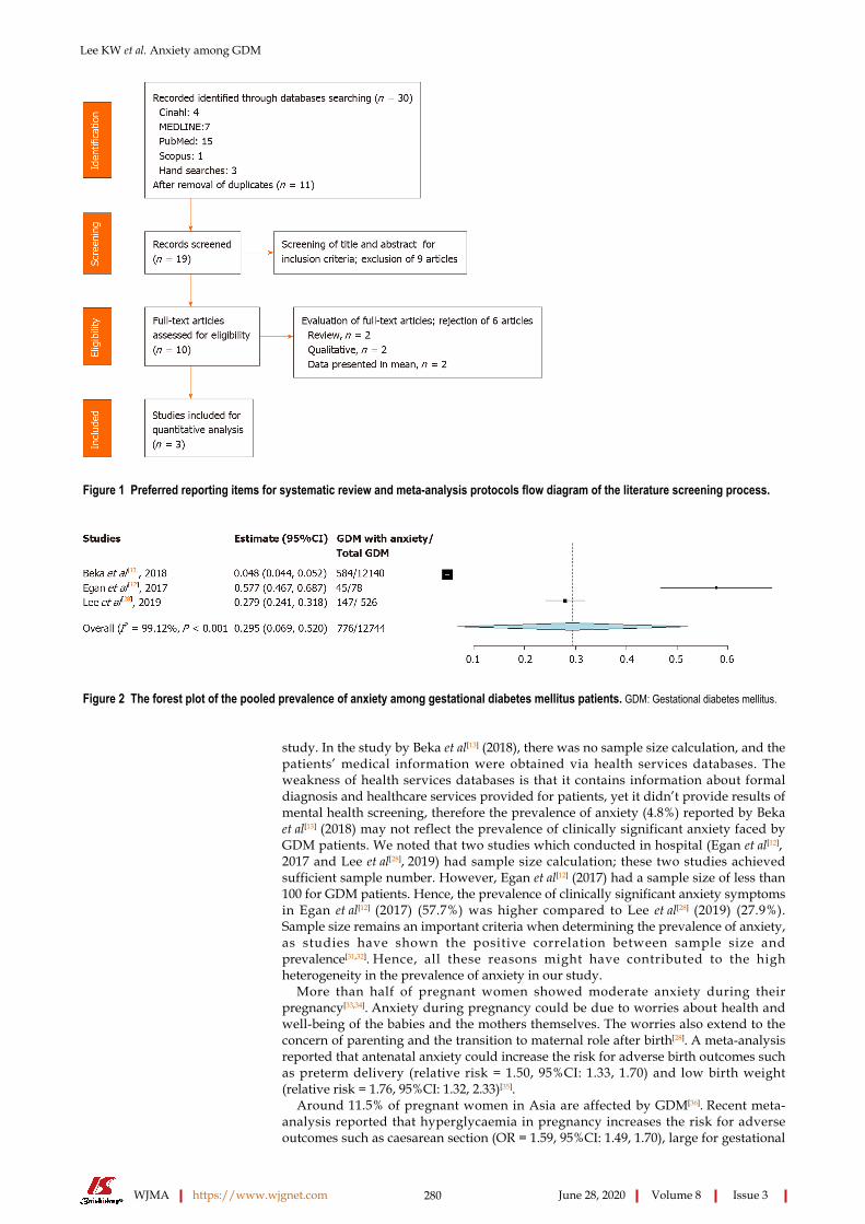
Lee KW et al. Anxiety among GDM
WJMA https://www.wjgnet.com 280 June 28, 2020 Volume 8 Issue 3
Figure 1 Preferred reporting items for systematic review and meta-analysis protocols flow diagram of the literature screening process.
Figure 2 The forest plot of the pooled prevalence of anxiety among gestational diabetes mellitus patients. GDM: Gestational diabetes mellitus.
study. In the study by Beka et al[13] (2018), there was no sample size calculation, and the patients’ medical information were obtained via health services databases. The weakness of health services databases is that it contains information about formal diagnosis and healthcare services provided for patients, yet it didn’t provide results of mental health screening, therefore the prevalence of anxiety (4.8%) reported by Beka et al[13] (2018) may not reflect the prevalence of clinically significant anxiety faced by GDM patients. We noted that two studies which conducted in hospital (Egan et al[12], 2017 and Lee et al[28], 2019) had sample size calculation; these two studies achieved sufficient sample number. However, Egan et al[12] (2017) had a sample size of less than 100 for GDM patients. Hence, the prevalence of clinically significant anxiety symptoms in Egan et al[12] (2017) (57.7%) was higher compared to Lee et al[28] (2019) (27.9%). Sample size remains an important criteria when determining the prevalence of anxiety, as studies have shown the positive correlation between sample size and prevalence[31,32]. Hence, all these reasons might have contributed to the high heterogeneity in the prevalence of anxiety in our study.
More than half of pregnant women showed moderate anxiety during their pregnancy[33,34]. Anxiety during pregnancy could be due to worries about health and well-being of the babies and the mothers themselves. The worries also extend to the concern of parenting and the transition to maternal role after birth[28]. A meta-analysis reported that antenatal anxiety could increase the risk for adverse birth outcomes such as preterm delivery (relative risk = 1.50, 95%CI: 1.33, 1.70) and low birth weight (relative risk = 1.76, 95%CI: 1.32, 2.33)[35].
Around 11.5% of pregnant women in Asia are affected by GDM[36]. Recent meta-analysis reported that hyperglycaemia in pregnancy increases the risk for adverse outcomes such as caesarean section (OR = 1.59, 95%CI: 1.49, 1.70), large for gestational
Lee KW et al. Anxiety among GDM
WJMA https://www.wjgnet.com 281 June 28, 2020 Volume 8 Issue 3
age (OR = 2.11, 95%CI: 1.73, 2.58), macrosomia (OR = 2.06, 95%CI: 1.86, 2.28), neonatal hypoglycaemia (OR = 1.37, 95%CI: 1.20, 1.57), gestational hypertension (OR = 1.91, 95%CI: 1.49, 2.43) and pre-eclampsia (OR = 2.15, 95%CI: 1.45, 3.19)[5]. GDM patients are at higher risk for experiencing anxiety as compared to pregnant women without medical complications[20]. Similarly, the adverse birth outcomes could be exacerbated if women with GDM experiences anxiety during pregnancy.
Antenatal anxiety is an evolving field, and unlike depression, only a few studies have been conducted among GDM patients. However, studies have reported that antenatal anxiety is more prevalent than antenatal depression[28,34], and this study reports anxiety symptoms are prevalent in GDM patients. In order to promote the detection of antenatal anxiety, several screening tools have been recently recommended by National Institute for Health and Care Excellence, which include Generalized Anxiety Disorder scale, GAD-2[37]. GAD-2 can be used as an ultra-brief screening scale for antenatal anxiety. Even so, other screening scales are more commonly used in clinical setting as compared to GAD-2, such as DASS-21[29], Edinburgh Postnatal Depression Scale[38], Hospital Anxiety and Depression Scale – Anxiety subscale[39], State-Trait Anxiety Inventory[40], GAD-7[41], Brief Measure of Worry Severity[42], Cambridge Worry Scale[43] and Wijma Delivery Expectancy/Experience Questionnaire –Version A[44].
Impacts of anxiety after delivery periodMental illness is a leading cause of maternal morbidity and even endangers maternal life especially in high -income countries[45]. Indirectly it also impacts new-born babies, causing perinatal morbidity and mortality as well as the impact on the long-term child development[46,47]. The National Institute for Health and Clinical Excellence (NICE) has emphasised that perinatal mental illness is one of the most important issues in women’s health that need to be highlighted, especially in the postpartum period[48,49].
The prevalence of postpartum anxiety disorders varies. Reck et al[50] (2008) and Miller et al[51] (2006) found a comparable percentage of postpartum women having anxiety disorder, at 11.1 % and 10 % respectively. Matthey et al[52] documented that 16.2% of mothers were diagnosed with a pure anxiety disorder while Wenzel et al[53] (2005) noted a prevalence rate of 8.2% for generalized anxiety disorder.
There were many reviews confined to maternal depression in postpartum period but there were scarcity of data on anxiety disorder despite of the high health risks for both mother and child associated with postpartum disorders[50]. Maternal anxiety disorder is part of a broad spectrum condition comprising of mild to severe mental illnesses such as bipolar disorder and psychotic disorder. It is common that both anxiety and depression co-exist in postpartum women[51,54].
Socio-demographic factors and socioeconomically deprived status have important impacts on maternal mental illness. The most common risk factors include age of more than 35 year old, single parent, lower educational level and low -income family[47,55]. Women with greater socioeconomic deprivation are more likely to have maternal mental illness than those with lesser degree of socioeconomic deprivation[56,57]. An early recognition of women at risk and implementation of effective intervention are essential as preventive measures to treat maternal mental illness accordingly, aiming to reduce the complications related to maternal mental illness.
Strength and limitationsTo date, this is the first systematic and meta-analysis on anxiety among patient with GDM. This study clearly indicates that anxiety is prevalent among GDM patients. The finding of this review is consistent with the previous literature pertaining to anxiety among pregnant women experiencing medical complications. However, there are several limitations. Strict inclusion/exclusion criteria and a paucity of literature on the topic of interest have resulted in the inclusion of only three papers. However, according to Valentine et al[58], 2010, the minimum number of studies needed to conduct a meta-analysis is two. On top of that, we did sensitivity analysis and funnel plot to show the publication bias. Nevertheless, due care is necessary when interpreting the results as at least 5 studies or more are needed to reasonably and consistently achieve powers from the random-effects meta-analyses that are greater than the studies that contribute to them[59]. Second, the pooled sample size is not large enough to reflect the anxiety prevalence in clinical setting, therefore limiting the generalizability of our study findings.
In conclusion, our study provides an estimation of the prevalence of anxiety among patients with GDM. Our study showed that the pooled prevalence was high at 29.5%. We recommend that more epidemiological studies on anxiety during pregnancy to be conducted in this particular population. In addition, it is important to identify factors
Lee KW et al. Anxiety among GDM
WJMA https://www.wjgnet.com 282 June 28, 2020 Volume 8 Issue 3
associated with anxiety during pregnancy so that early detection and intervention can be implemented to improve various obstetric and mental health outcomes.
ARTICLE HIGHLIGHTSResearch backgroundThere is lack of systematic review and meta-analysis on prevalence of anxiety among GDM women.
Research motivationThe systematic review and meta-analysis reporting the pooled prevalence of anxiety among GDM patients is high (29.5%).
Research objectivesAuthors aimed to pool data from existing literature to determine the pool estimates for the prevalence of anxiety among women diagnosed with GDM.
Research methodsMultiple databases including MEDLINE, Cinahl, PubMed and Scopus were searched to identify studies published up to 31 October 2019 with data on the prevalence of anxiety among women diagnosed with GDM.
Research resultsTotal 19 abstracts, retrieved 10 articles and included three studies incorporating 12744 GDM women from three countries were reviewed. The pooled prevalence of anxiety was 29.5% among GDM women.
Research conclusionsThe results suggest that epidemiological studies on anxiety should be conducted urgently as it merits clinical attention. In addition, it is important to identify factors associated with anxiety among women diagnosed with GDM.
ACKNOWLEDGEMENTSWe would like to thank the Director General of Health Malaysia for his permission to publish this article.
REFERENCES1 Bellamy L, Casas JP, Hingorani AD, Williams D. Type 2 diabetes mellitus after gestational diabetes: a
systematic review and meta-analysis. Lancet 2009; 373: 1773-1779 [PMID: 19465232 DOI: 10.1016/S0140-6736(09)60731-5]
2 Rayanagoudar G, Hashi AA, Zamora J, Khan KS, Hitman GA, Thangaratinam S. Quantification of the type 2 diabetes risk in women with gestational diabetes: a systematic review and meta-analysis of 95,750 women. Diabetologia 2016; 59: 1403-1411 [PMID: 27073002 DOI: 10.1007/s00125-016-3927-2]
3 Bener A, Saleh NM, Al-Hamaq A. Prevalence of gestational diabetes and associated maternal and neonatal complications in a fast-developing community: global comparisons. Int J Womens Health 2011; 3: 367-373 [PMID: 22140323 DOI: 10.2147/IJWH.S26094]
4 Veeraswamy S, Vijayam B, Gupta VK, Kapur A. Gestational diabetes: the public health relevance and approach. Diabetes Res Clin Pract 2012; 97: 350-358 [PMID: 22726771 DOI: 10.1016/j.diabres.2012.04.024]
5 Farrar D, Simmonds M, Bryant M, Sheldon TA, Tuffnell D, Golder S, Dunne F, Lawlor DA. Hyperglycaemia and risk of adverse perinatal outcomes: systematic review and meta-analysis. BMJ 2016; 354: i4694 [PMID: 27624087 DOI: 10.1136/bmj.i4694]
6 Li Z, Cheng Y, Wang D, Chen H, Chen H, Ming WK, Wang Z. Incidence Rate of Type 2 Diabetes Mellitus after Gestational Diabetes Mellitus: A Systematic Review and Meta-Analysis of 170,139 Women. J Diabetes Res 2020; 2020: 3076463 [PMID: 32405502 DOI: 10.1155/2020/3076463]
7 Mak JKL, Lee AH, Pham NM, Pan XF, Tang L, Binns CW, Sun X. Gestational diabetes incidence and delivery outcomes in Western China: A prospective cohort study. Birth 2019; 46: 166-172 [PMID: 30216525 DOI: 10.1111/birt.12397]Thevarajah A, Simmons D. Risk factors and outcomes for neonatal hypoglycaemia and neonatal hyperbilirubinaemia in pregnancies complicated by gestational diabetes mellitus: a single centre
8

Lee KW et al. Anxiety among GDM
WJMA https://www.wjgnet.com 283 June 28, 2020 Volume 8 Issue 3
retrospective 3-year review. Diabet Med 2019; 36: 1109-1117 [PMID: 30972790 DOI: 10.1111/dme.13962]9 Somohano-Mendiola N, Champion JD, Vatcheva K. Assessment of Gestational Diabetes Mellitus
Outcomes for Hispanic Women Living in the Rio Grande Valley. Hisp Health Care Int 2019; 17: 111-117 [PMID: 30922188 DOI: 10.1177/1540415319833996]
10 Chazotte C, Freda MC, Elovitz M, Youchah J. Maternal depressive symptoms and maternal-fetal attachment in gestational diabetes. J Womens Health 1995; 4: 375-380 [DOI: 10.1089/jwh.1995.4.375]
11 Natasha K, Hussain A, Khan AK. Prevalence of depression among subjects with and without gestational diabetes mellitus in Bangladesh: a hospital based study. J Diabetes Metab Disord 2015; 14: 64 [PMID: 26221580 DOI: 10.1186/s40200-015-0189-3]
12 Egan AM, Dunne FP, Lydon K, Conneely S, Sarma K, McGuire BE. Diabetes in pregnancy: worse medical outcomes in type 1 diabetes but worse psychological outcomes in gestational diabetes. QJM 2017; 110: 721-727 [PMID: 29024981 DOI: 10.1093/qjmed/hcx106]
13 Beka Q, Bowker S, Savu A, Kingston D, Johnson JA, Kaul P. Development of Perinatal Mental Illness in Women With Gestational Diabetes Mellitus: A Population-Based Cohort Study. Can J Diabetes 2018; 42: 350-355.e1 [PMID: 28943221 DOI: 10.1016/j.jcjd.2017.08.005]
14 Major B, Cozzarelli C, Horowitz MJ. Encyclopedia of psychology: 8 Volume set. New York and Washington: Oxford University Press and the American Psychological Association, 2000
15 Sowers KM, Dulmus CN, Linn BK. Mental Illness: Worldwide. In: Encyclopedia of Social Work. National Association of Social Workers Press and Oxford University Press, 2019 [DOI: 10.1093/acrefore/9780199975839.013.1154]
16 Soto-Balbuena C, Rodríguez MF, Escudero Gomis AI, Ferrer Barriendos FJ, Le HN, Pmb-Huca G. Incidence, prevalence and risk factors related to anxiety symptoms during pregnancy. Psicothema 2018; 30: 257-263 [PMID: 30009746 DOI: 10.7334/psicothema2017.379]
17 Grigoriadis S, Graves L, Peer M, Mamisashvili L, Tomlinson G, Vigod SN, Dennis CL, Steiner M, Brown C, Cheung A, Dawson H, Rector NA, Guenette M, Richter M. A systematic review and meta-analysis of the effects of antenatal anxiety on postpartum outcomes. Arch Womens Ment Health 2019; 22: 543-556 [PMID: 30523416 DOI: 10.1007/s00737-018-0930-2]
18 Netsi E, Pearson RM, Murray L, Cooper P, Craske MG, Stein A. Association of Persistent and Severe Postnatal Depression With Child Outcomes. JAMA Psychiatry 2018; 75: 247-253 [PMID: 29387878 DOI: 10.1001/jamapsychiatry.2017.4363]
19 Podvornik N, Globevnik Velikonja V, Praper P. Depression and Anxiety in Women During Pregnancy in Slovenia. Zdr Varst 2015; 54: 45-50 [PMID: 27646621 DOI: 10.1515/sjph-2015-0006]
20 Abrar A, Fairbrother N, Smith AP, Skoll A, Albert AYK. Anxiety among women experiencing medically complicated pregnancy: A systematic review and meta-analysis. Birth 2020; 47: 13-20 [PMID: 31222840 DOI: 10.1111/birt.12443]
21 Biaggi A, Conroy S, Pawlby S, Pariante CM. Identifying the women at risk of antenatal anxiety and depression: A systematic review. J Affect Disord 2016; 191: 62-77 [PMID: 26650969 DOI: 10.1016/j.jad.2015.11.014]
22 Marchetti D, Carrozzino D, Fraticelli F, Fulcheri M, Vitacolonna E. Quality of Life in Women with Gestational Diabetes Mellitus: A Systematic Review. J Diabetes Res 2017; 2017: 7058082 [PMID: 28326332 DOI: 10.1155/2017/7058082]
23 Moher D, Shamseer L, Clarke M, Ghersi D, Liberati A, Petticrew M, Shekelle P, Stewart LA; PRISMA-P Group. Preferred reporting items for systematic review and meta-analysis protocols (PRISMA-P) 2015 statement. Syst Rev 2015; 4: 1 [PMID: 25554246 DOI: 10.1186/2046-4053-4-1]
24 Vandenbroucke JP, von Elm E, Altman DG, Gøtzsche PC, Mulrow CD, Pocock SJ, Poole C, Schlesselman JJ, Egger M; STROBE Initiative. Strengthening the Reporting of Observational Studies in Epidemiology (STROBE): explanation and elaboration. Epidemiology 2007; 18: 805-835 [PMID: 18049195 DOI: 10.1097/EDE.0b013e3181577511]
25 Higgins JP, Thompson SG. Quantifying heterogeneity in a meta-analysis. Stat Med 2002; 21: 1539-1558 [PMID: 12111919 DOI: 10.1002/sim.1186]
26 Wallace BC, Dahabreh IJ, Trikalinos TA, Lau J, Trow P, Schmid CH. Closing the gap between methodologists and end-users: R as a computational back-end. J Stat Softw 2012; 49: 1-15 [DOI: 10.18637/jss.v049.i05]
27 Jamovi. The jamovi project (2020). Jamovi (Version 1.2). Available from: https://www.jamovi.org28 Lee KW, Ching SM, Hoo FK, Ramachandran V, Chong SC, Tusimin M, Mohd Nordin N. Prevalence and
factors associated with depressive, anxiety and stress symptoms among women with gestational diabetes mellitus in tertiary care centres in Malaysia: a cross-sectional study. BMC Pregnancy Childbirth 2019; 19: 367 [PMID: 31638930 DOI: 10.1186/s12884-019-2519-9]
29 Lovibond PF, Lovibond SH. The structure of negative emotional states: comparison of the Depression Anxiety Stress Scales (DASS) with the Beck Depression and Anxiety Inventories. Behav Res Ther 1995; 33: 335-343 [PMID: 7726811 DOI: 10.1016/0005-7967(94)00075-U]
30 Musa R, Fadzil MA, Zain Z. Translation, validation and psychometric properties of Bahasa Malaysia version of the Depression Anxiety and Stress Scales (DASS). ASEAN J Psychiatr 2007; 8: 82-89
31 Guo X, Zheng L, Zhang X, Zou L, Li J, Sun Z, Hu J, Sun Y. The prevalence and heterogeneity of prehypertension: a meta-analysis and meta-regression of published literature worldwide. Cardiovasc J Afr 2012; 23: 44-50 [PMID: 22331252 DOI: 10.5830/CVJA-2011-058]
32 Arya R, Antonisamy B, Kumar S. Sample size estimation in prevalence studies. Indian J Pediatr 2012; 79: 1482-1488 [PMID: 22562262 DOI: 10.1007/s12098-012-0763-3]
33 Faisal-Cury A, Rossi Menezes P. Prevalence of anxiety and depression during pregnancy in a private setting sample. Arch Womens Ment Health 2007; 10: 25-32 [PMID: 17187166 DOI: 10.1007/s00737-006-0164-6]
34 Lee AM, Lam SK, Sze Mun Lau SM, Chong CS, Chui HW, Fong DY. Prevalence, course, and risk factors for antenatal anxiety and depression. Obstet Gynecol 2007; 110: 1102-1112 [PMID: 17978126 DOI: 10.1097/01.AOG.0000287065.59491.70]

Lee KW et al. Anxiety among GDM
WJMA https://www.wjgnet.com 284 June 28, 2020 Volume 8 Issue 3
35 Ding XX, Wu YL, Xu SJ, Zhu RP, Jia XM, Zhang SF, Huang K, Zhu P, Hao JH, Tao FB. Maternal anxiety during pregnancy and adverse birth outcomes: a systematic review and meta-analysis of prospective cohort studies. J Affect Disord 2014; 159: 103-110 [PMID: 24679397 DOI: 10.1016/j.jad.2014.02.027]
36 Lee KW, Ching SM, Ramachandran V, Yee A, Hoo FK, Chia YC, Wan Sulaiman WA, Suppiah S, Mohamed MH, Veettil SK. Prevalence and risk factors of gestational diabetes mellitus in Asia: a systematic review and meta-analysis. BMC Pregnancy Childbirth 2018; 18: 494 [PMID: 30547769 DOI: 10.1186/s12884-018-2131-4]
37 Antenatal N. Postnatal mental health: Clinical management and service guidance (CG192). London: National Institute for Health and Care Excellence, 2014
38 Cox JL, Holden JM, Sagovsky R. Detection of postnatal depression. Development of the 10-item Edinburgh Postnatal Depression Scale. Br J Psychiatry 1987; 150: 782-786 [PMID: 3651732 DOI: 10.1192/bjp.150.6.782]
39 Zigmond AS, Snaith RP. The hospital anxiety and depression scale. Acta Psychiatr Scand 1983; 67: 361-370 [PMID: 6880820 DOI: 10.1111/j.1600-0447.1983.tb09716.x]
40 Spielberger CD, Gorsuch RL, Lushene RE. Lushene RE. State-Trait Anxiety Inventory (STAI). Test Manual for Form X: Consulting Psychologists Press, 1968
41 Spitzer RL, Kroenke K, Williams JB, Löwe B. A brief measure for assessing generalized anxiety disorder: the GAD-7. Arch Intern Med 2006; 166: 1092-1097 [PMID: 16717171 DOI: 10.1001/archinte.166.10.1092]
42 Gladstone GL, Parker GB, Mitchell PB, Malhi GS, Wilhelm KA, Austin MP. A Brief Measure of Worry Severity (BMWS): personality and clinical correlates of severe worriers. J Anxiety Disord 2005; 19: 877-892 [PMID: 16243636 DOI: 10.1016/j.janxdis.2004.11.003]
43 Green JM, Kafetsios K, Statham HE, Snowdon CM. Factor structure, validity and reliability of the Cambridge Worry Scale in a pregnant population. J Health Psychol 2003; 8: 753-764 [PMID: 14670208 DOI: 10.1177/13591053030086008]
44 Wijma K, Wijma B, Zar M. Psychometric aspects of the W-DEQ; a new questionnaire for the measurement of fear of childbirth. J Psychosom Obstet Gynaecol 1998; 19: 84-97 [PMID: 9638601 DOI: 10.3109/01674829809048501]
45 Oates M. Perinatal psychiatric disorders: a leading cause of maternal morbidity and mortality. Br Med Bull 2003; 67: 219-229 [PMID: 14711766 DOI: 10.1093/bmb/ldg011]
46 Jablensky AV, Morgan V, Zubrick SR, Bower C, Yellachich LA. Pregnancy, delivery, and neonatal complications in a population cohort of women with schizophrenia and major affective disorders. Am J Psychiatry 2005; 162: 79-91 [PMID: 15625205 DOI: 10.1176/appi.ajp.162.1.79]
47 Ban L, Gibson JE, West J, Fiaschi L, Oates MR, Tata LJ. Impact of socioeconomic deprivation on maternal perinatal mental illnesses presenting to UK general practice. Br J Gen Pract 2012; 62: e671-e678 [PMID: 23265226 DOI: 10.3399/bjgp12X656801]
48 The National Institute for Health and Care Excellence. Antenatal and postnatal mental health: the NICE guideline on clinical management and service guidance. British Psychological Society, 2007
49 Feldman R, Granat A, Pariente C, Kanety H, Kuint J, Gilboa-Schechtman E. Maternal depression and anxiety across the postpartum year and infant social engagement, fear regulation, and stress reactivity. J Am Acad Child Adolesc Psychiatry 2009; 48: 919-927 [PMID: 19625979 DOI: 10.1097/CHI.0b013e3181b21651]
50 Reck C, Struben K, Backenstrass M, Stefenelli U, Reinig K, Fuchs T, Sohn C, Mundt C. Prevalence, onset and comorbidity of postpartum anxiety and depressive disorders. Acta Psychiatr Scand 2008; 118: 459-468 [PMID: 18840256 DOI: 10.1111/j.1600-0447.2008.01264.x]
51 Miller RL, Pallant JF, Negri LM. Anxiety and stress in the postpartum: is there more to postnatal distress than depression? BMC Psychiatry 2006; 6: 12 [PMID: 16563155 DOI: 10.1186/1471-244X-6-12]
52 Matthey S, Barnett B, Howie P, Kavanagh DJ. Diagnosing postpartum depression in mothers and fathers: whatever happened to anxiety? J Affect Disord 2003; 74: 139-147 [PMID: 12706515 DOI: 10.1016/s0165-0327(02)00012-5]
53 Wenzel A, Haugen EN, Jackson LC, Brendle JR. Anxiety symptoms and disorders at eight weeks postpartum. J Anxiety Disord 2005; 19: 295-311 [PMID: 15686858 DOI: 10.1016/j.janxdis.2004.04.001]
54 Ross LE, McLean LM. Anxiety disorders during pregnancy and the postpartum period: A systematic review. J Clin Psychiatry 2006; 67: 1285-1298 [PMID: 16965210 DOI: 10.4088/jcp.v67n0818]
55 Nager A, Johansson LM, Sundquist K. Are sociodemographic factors and year of delivery associated with hospital admission for postpartum psychosis? A study of 500,000 first-time mothers. Acta Psychiatr Scand 2005; 112: 47-53 [PMID: 15952945 DOI: 10.1111/j.1600-0447.2005.00525.x]
56 Rich-Edwards JW, Kleinman K, Abrams A, Harlow BL, McLaughlin TJ, Joffe H, Gillman MW. Sociodemographic predictors of antenatal and postpartum depressive symptoms among women in a medical group practice. J Epidemiol Community Health 2006; 60: 221-227 [PMID: 16476752 DOI: 10.1136/jech.2005.039370]
57 Wall-Wieler E, Carmichael SL, Urquia ML, Liu C, Hjern A. Severe maternal morbidity and postpartum mental health-related outcomes in Sweden: a population-based matched-cohort study. Arch Womens Ment Health 2019; 22: 519-526 [PMID: 30334101 DOI: 10.1007/s00737-018-0917-z]
58 Valentine JC, Pigott TD, Rothstein HR. How many studies do you need? A primer on statistical power for meta-analysis. J Educ Behav Stat 2010; 35: 215-247 [DOI: 10.3102/1076998609346961]
59 Jackson D, Turner R. Power analysis for random-effects meta-analysis. Res Synth Methods 2017; 8: 290-302 [PMID: 28378395 DOI: 10.1002/jrsm.1240]

Published by Baishideng Publishing Group Inc
7041 Koll Center Parkway, Suite 160, Pleasanton, CA 94566, USA
Telephone: +1-925-3991568
E-mail: [email protected]
Help Desk: https://www.f6publishing.com/helpdesk
https://www.wjgnet.com
© 2020 Baishideng Publishing Group Inc. All rights reserved.