Within-host evolution and virus escape of two ... - UCL Discovery
266
1 Within-host evolution and virus escape of two important pandemic viruses Oluwadamilola Aderonke Collier UCL This thesis is submitted for the degree of Doctor of Philosophy (PhD) in the field of Virology May 2021
-
Upload
khangminh22 -
Category
Documents
-
view
3 -
download
0
Transcript of Within-host evolution and virus escape of two ... - UCL Discovery
1
Within-host evolution and virus escape of two
important pandemic viruses
Oluwadamilola Aderonke Collier
UCL
This thesis is submitted for the degree of Doctor of Philosophy (PhD) in
the field of Virology
May 2021

2
Declaration
I, Oluwadamilola Aderonke Collier confirm that the work presented in this thesis is
my own. Where information has been derived from other sources, I confirm that this
has been indicated in the thesis.
3
Abstract
Viruses are known to adapt to the constraints of the niche in which they find
themselves. This leads to within-host adaptations, detected as nucleic acid base
mutations, protein changes or epigenetic modification. These changes are driven by
selection pressures which may be due to immune responses to the virus, drug
treatment and requirement to exist in specific compartments within the host.
Cerebrospinal fluid (CSF) escape and central nervous system (CNS)
compartmentalisation are problems in HIV-1 that are not fully understood and have
implications for HIV-1 treatment and cure. The true extent of this problem in
subtype C HIV-1, which is responsible for the epidemic in Southern Africa is
unknown. The prevalence of these were determined in a cohort of participants co-
infected with HIV-1 and cryptococcal meningitis. Following this, the phenotypic
characteristics of CSF and plasma derived clones that may underly the mechanisms
by which they adapt to the CNS were investigated.
CSF escape was uncommon in this cross sectional study. Four participants
underwent in-depth characterisation of the genomes in both the CSF and plasma.
This revealed that 2 out of 4 of them had CNS compartmentalisation according to
analyses of their phylogeny. The diversity of the CSF and plasma env genomes were
different in these individuals and are most likely reflective of their unique within-
host selection pressures. All clones tested utilised the CCR5 co-receptor for viral
entry with some evidence that CSF derived clones may have a higher affinity for the
4
CCR5 coreceptor and hence better adapted to infect CNS macrophage with low
levels of CD4.
An in-depth analysis of within-host evolution of SARS-CoV-2 in a chronic infection
case was undertaken. Escape mutations were identified and their functional
relevance were tested in infection and neutralisation experiments. Additionally,
factors related to poor immune response to mRNA BNT162b2 SARS-CoV-2 vaccine
were investigated.
The findings revealed a dynamic shift in viral population in vivo that was likely
driven by pressure from treatment with remdesivir and convalescent plasma. In vitro
testing of emerging Spike mutants revealed that D796H mutation facilitated escape
from neutralising antibodies but was accompanied by a reduction in infectivity.
Meanwhile, △H69/V70 deletions had no impact on the neutralisation activity of
convalescent plasma but increased the infectivity of the pseudotyped virus.
Importantly older age i.e ≥80 years was a risk factor for lower Spike-specific
antibody binding levels, lower neutralisation responses against Spike pseudotyped
viruses and lower T cell interferon gamma (IFN𝛾) and IL-12 responses following the
first dose of mRNA BNT162b2 vaccine. However, these poor responses were
overcome by the second dose in all participants tested.
These findings will contribute to our understanding of within-host viral evolution
and have implications for patients care, vaccine and cure strategies.
5
Impact Statement
The findings of the work presented herein provides important insights into within-
host evolution of two important pandemic viruses; HIV-1 and SARS-CoV-2. It
investigates proposed hypotheses of the mechanisms underlying central nervous
system escape of HIV-1 from immune control in subtype C HIV-1 participants co-
infected with cryptococcal meningitis. Important mutations in the Spike protein of
SARS-CoV-2 that are functional adaptions, driving escape from antibody
neutralisation are described. Suboptimal vaccine-elicited immune responses could
also provide the optimal niche for selecting escape mutants, which may in the long-
term jeopardise the vaccine program’s efforts. Therefore, the findings from the study
into determinants of poor immune responses to SARS-CoV-2 mRNA vaccines will
potentially impact on public health strategies to bolster the vaccine program and
target key groups in order to prevent vaccine resistant variants from emerging.
These findings are of interest to the academic world because they ultimately teach us
something about within-host adaptation of these RNA viruses and what selection
pressures are in play in the host. They have implications for vaccine development
and cure strategies. They are also of interest to clinicians because they may inform
the use of antiviral interventions that could help manage patients better. Finally, they
have wider implications for public health as they may inform our understanding of
hotspots for emergence of variants that maybe more transmissible or pathogenic and
could help prioritise where investment in efforts to reduce transmission should be
directed.
6
Acknowledgements
Firstly, I would like to thank my supervisor Professor Ravindra Gupta for his
unwavering support and encouragement. For encouraging me to pursue academic
medicine and undertake a PhD in his lab. For investing in my research ideas,
opening doors and facilitating their execution. His mentorship has been essential to
my progress as a clinician scientist and his mix of brilliance and out of the box
thinking has taught me to approach scientific enquiry with alternative hypothesis
rather than accept scientific dogma.
I would also like to thank Dr Laura McCoy for her mentorship, counsel and training
in laboratory techniques. Without her my PhD would not have been possible. I
would like to thank Professor Rob Heyderman for facilitating access to biobanked
samples on which a large part of my thesis is based, again without him this PhD
would not have been possible.
I would like to express my gratitude to all the members of the Gupta lab past and
present, Dr Katherine Sutherland for teaching me basic lab techniques and principles
when I was fresh off the wards and in the lab. Drs Petra Mlcochova, Bo Meng,
Rawlings Datir and Isabella Ferreira for their unwavering enthusiasm for scientific
discovery, advice and for reading drafts of this thesis. I would like to thank Dr
Steven Kemp for his patience and enthusiasm in teaching me bioinformatics and
helping make the best of my genomic data.
7
I would like to thank other scientists with whom I have collaborated on some of the
work presented here- Professor Rainer Doffinger, Dr Mark Wills, Dr Eleanor Lim
and Dr Chris Illingworth.
Finally, I would like to thank my husband for his unwavering support during my
PhD and my children for constantly asking me about what I do in the lab which has
been the best training for public, scientific engagement.
8
Table of Contents
Declaration ................................................................................................................... 2
Abstract ........................................................................................................................ 3
Impact Statement .......................................................................................................... 5
Acknowledgements ...................................................................................................... 6
Table of Contents ......................................................................................................... 8
List of Tables.............................................................................................................. 16
List of Figures ............................................................................................................ 18
PREFACE .................................................................................................................. 24
List of Publications .................................................................................................... 25
CHAPTER 1: BACKGROUND AND INTRODUCTION ....................................... 27
PART 1 ...................................................................................................................... 27
1.1 The HIV-1 pandemic ................................................................................... 27
1.2 Human immunodeficiency Virus-1 Life Cycle .......................................... 30
1.2.1 HIV-1 attachment and Entry ......................................................................... 31
1.2.2 Reverse Transcriptase ................................................................................... 33
1.2.3 Integration ..................................................................................................... 38
1.2.4 Transcription and mRNA processing ............................................................ 40
1.2.5 Assembly and packaging ............................................................................... 41
1.3 Within-Host adaptations ............................................................................. 42
1.3.1 Inherent viral diversity .................................................................................. 42
1.3.2 The host environment .................................................................................... 42
1.3.3 Latent reservoirs ........................................................................................... 44
9
1.4 CNS replication and HIV-1 ......................................................................... 45
1.5 Factors associated with HIV-1 neurotropism and neurovirulence.......... 48
1.5.1 HIV-1 subtype and CNS disease ................................................................ 48
1.5.2 Cellular tropism ......................................................................................... 49
1.5.3 Coreceptor usage ........................................................................................ 49
1.5.4 Drug resistant mutations ............................................................................ 50
1.5.5 Escape from antibody neutralisation .......................................................... 51
1.5.6 CNS co-infection ........................................................................................ 52
1.6 Aims ............................................................................................................... 52
Specific objectives/questions are: .......................................................................... 53
PART 2 ...................................................................................................................... 54
2.1 Pandemic SARS-CoV-2 ............................................................................... 54
2.2 SARS-CoV-2 Life cycle ................................................................................ 60
2.3 Within-host adaptation ................................................................................ 62
2.4 Host factors that determine immune responses to SARS-CoV-2 vaccines
63
2.5 Aims ............................................................................................................... 65
CHAPTER 2: METHODS ......................................................................................... 66
2.1 Section 1: HIV-1 viral escape and CNS compartmentalisation ............... 66
2.1.1 Clinical Samples ........................................................................................ 66
2.1.2 Viral load .................................................................................................... 67
10
2.1.3 Single genome amplification (SGA) .......................................................... 67
2.1.4 Next generation sequencing (NGS) ........................................................... 69
2.1.5 Determination of compartmentalisation ..................................................... 70
2.1.6 Drug resistance ........................................................................................... 72
2.1.7 Prediction of coreceptor usage ................................................................... 73
2.1.8 Phenotypic co-receptor usage .................................................................... 73
2.1.9 Neutralisation ................................................................................................ 81
2.1.10 Statistical Methods ................................................................................. 83
2.1.11 Ethical approval ..................................................................................... 83
2.2 Section 2: SARS-COV-2 Escape from Convalescent Plasma .................. 84
2.2.1 Clinical Sample Collection ........................................................................... 84
2.2.2 Viral load/Cycle threshold (CT) ................................................................... 84
2.2.3 SARS-CoV-2 binding antibody levels .......................................................... 85
2.2.4 Convalescent plasma for antibody titres ....................................................... 85
2.2.5 Whole blood T cell and innate stimulation assay.......................................... 86
2.2.6 Next generation sequencing (NGS) .............................................................. 87
2.2.7 Single Genome Amplification (SGA) ........................................................... 90
2.2.8 Generation of Spike mutants ......................................................................... 92
2.2.9 Spike pseudotyped virus preparation ............................................................ 93
2.2.10 Normalisation of viral input by SYBR Green-based product-enhanced PCR
assay (SG-PERT) ................................................................................................... 94
2.2.11 Spike expression by Western blot ............................................................... 94
2.2.12 Serum/plasma pseudotype neutralization assay .......................................... 95
2.2.13 Structural Viewing ...................................................................................... 96
2.2.14 Ethics ........................................................................................................... 96
11
2.3 Section 3: Age-related heterogeneity of SARS-CoV-2 mRNA vaccine-elicited
responses ................................................................................................................... 97
2.3.1 Study Design ................................................................................................. 97
2.3.2 Sample size calculation ................................................................................. 98
2.3.3 Clinical samples ............................................................................................ 98
2.3.4 Generation of pseudotyped viruses ............................................................... 98
2.3.5 Neutralisation assays ..................................................................................... 99
2.3.6 SARS-CoV-2 binding antibody levels ........................................................ 100
2.3.7 CMV serology ............................................................................................. 101
2.3.8 T cell assays- IFNγ and IL2 FLUOROSPOT ............................................. 101
2.3.9 Statistical Analyses ..................................................................................... 102
2.3.10 Ethical approval ........................................................................................ 103
CHAPTER 3: RESULTS 1- HIV-1 VIRAL ESCAPE AND CNS
COMPARTMENTALISATION .............................................................................. 104
3.1 Cerebrospinal fluid (CSF) escape is uncommon in this clinical cohort ...... 106
3.2 A viral population structure consistent with CNS compartmentalisation was
shown in some participants ................................................................................... 113
3.3 There is increased diversity in compartmentalised participants ................. 116
3.4 No clinical or demographic factors were identified which are associated with
CNS compartmentalisation ................................................................................... 121
3.5 Co-receptor usage by computer predicted algorithms ........................... 122
3.6 Infectivity of pseudotyped viruses .................................................................. 124

12
3.7 Co-receptor usage in a phenotypic assay ....................................................... 125
3.8 Co-receptor inhibitors confirm co-receptor usage in phenotypic assay ..... 131
3.9 The frequency of drug resistance mutations is similar in both compartments
.................................................................................................................................. 135
3.10 Heterogeneity of CNS and plasma derived Env clones to bnAbs .............. 138
3.11 Discussion ........................................................................................................ 142
CHAPTER 4: RESULTS 2- SARS-COV-2 ESCAPE FROM POLYCLONAL
CONVALESCENT SERA ....................................................................................... 145
4.1 Clinical Case History ....................................................................................... 147
4.1.1 COVID-19 clinical case in an immunocompromised host ......................... 147
4.1.2 B cell depletions and deficient SARS-CoV-2 binding antibodies .............. 150
4.1.3 Innate and T cell response despite absence of B cell responses ................. 154
4.2 Deep sequencing of participants samples revealed diversity in the SAR-
CoV-2 viral population .......................................................................................... 155
4.3 SARS-CoV-2 evolves in vivo in response to pressure from remdesivir and
convalescent plasma ............................................................................................... 163
4.4 Spike Mutants D796H causes a decrease in infectivity which is compensated
for by acquisition of 𝚫69/70 deletion .................................................................... 175
4.5 Spike Mutants D796H and 𝚫69/70 individually or in combination does not
impact Spike incorporation ................................................................................... 177
4.6 The spike D796H evolved to escape neutralisation from polyclonal sera ... 178

13
4.7 The location of D796H and △H69/V70 Spike mutations may affect antibody
binding and neutralisation by allostery ............................................................... 186
4.8 Discussion .......................................................................................................... 188
CHAPTER 5: RESULTS 3- AGE-RELATED HETEROGENEITY OF SARS-COV-
2 MRNA VACCINE-ELICITED RESPONSES ..................................................... 190
5.1 Cohort Description and study procedure ...................................................... 192
5.2 Neutralisation activity of vaccine-elicited sera against SARS-CoV-2
following first and second dose mRNA BNT162b2 vaccine ............................... 196
5.2.1 Poor serum neutralisation of WT Spike pseudotyped virus after the first dose
of mRNA BNT162b2 is overcome by the booster/second dose .......................... 197
5.2.2 Older age is a risk factor for poor serum neutralisation of WT Spike
pseudotyped virus after the first dose of mRNA BNT162b2 ............................... 200
5.3 Binding antibody responses following first and second dose mRNA
BNT162b2 vaccination ........................................................................................... 203
5.3.1 Binding antibody levels increases in response to mRNA BNT162b2 vaccine
.............................................................................................................................. 203
5.3.2 Binding antibody levels correlate with age ................................................. 207
5.3.3 Binding antibody levels correlate with serum neutralisation of SARS-CoV-2
.............................................................................................................................. 208
5.4 T cell responses following first and second dose mRNA BNT162b2 vaccine
.................................................................................................................................. 211
5.4.1 Frequency of Spike specific T cells following vaccination ........................ 211
14
5.4.2 Correlation of Spike specific T cell response with serum neutralisation of
SARS-CoV-2 ....................................................................................................... 215
5.4 3 T cell responses to SARS-CoV-2 Spike peptide pools are specific ............ 217
5.4.4 HCMV seropositivity maybe associated with increased frequency of IFN𝛾
producing Spike specific Tcells ........................................................................... 218
5.5 Discussion .......................................................................................................... 220
CHAPTER 6: DISCUSSION ................................................................................... 222
PART 1 .................................................................................................................... 222
6.1 HIV-1 CSF escape in the context of cryptococcal meningitis ............... 222
6.2 CNS compartmentalisation and patient characteristics associated with
CSF compartmentalisation in the context of CM................................................ 223
6.3 Phylogenetic characteristics of paired CNS and plasma genomes of HIV-
1 subtype C in the context of co-infection with cryptococcal meningitis .......... 224
6.4 Phenotypic properties of HIV-1 isolated from the CSF ......................... 226
6.4.1 Co-receptor usage..................................................................................... 226
6.4.2 Drug resistant mutations (DRMs) ............................................................ 227
6.4.3 Neutralisation sensitivity to broadly neutralising antibodies (bnAbs) ..... 228
PART 2 .................................................................................................................... 229
6.5 SARS-CoV-2 escape in the context of neutralising antibodies.............. 229
6.6 Age-related heterogeneity to mRNA BNT162b2 vaccine against SARS-CoV-
2 ................................................................................................................................ 231

15
6.7 Conclusion ......................................................................................................... 233
REFERENCES ......................................................................................................... 236

16
List of Tables
TABLE 1.1 HIV ANTIRETROVIRAL DRUG CLASSES AND MECHANISM OF
ACTION. ................................................................................................................................. 29
TABLE 1.2. DIAGNOSTIC CRITERIA FOR HIV-ASSOCIATED
NEUROCOGNITIVE DISORDER. ............................................................................... 45
TABLE 1.3: CSF ESCAPE AND CSF DISCORDANCE. ................................................ 47
TABLE 2.1 THERAPEUTIC OPTIONS CURRENTLY IN USE FOR MANAGING
COVID-19. ............................................................................................................................. 58
TABLE 2.2 COVID-19 VACCINES AND THE MOLECULAR PLATFORMS
CURRENTLY IN USE. ...................................................................................................... 59
TABLE 2.1.1. PCR PRIMER USED FOR SINGLE GENOME AMPLIFICATION
OF HIV-1 ENV. ..................................................................................................................... 68
TABLE 2.2.1. PCR PRIMER USED FOR SINGLE GENOME AMPLIFICATION
IF SARS-COV-2 SPIKE. .................................................................................................... 91
TABLE 2.2.2. SITE DIRECTED MUTAGENESIS PRIMERS..................................... 92
TABLE 3.1. CLINICAL AND DEMOGRAPHIC CHARACTERISTIC OF STUDY
PARTICIPANTS. ............................................................................................................... 107
TABLE 3.2. CLINICAL AND DEMOGRAPHIC DATA OF PARTICIPANTS
WITH SUCCESSFUL SEQUENCING. ...................................................................... 109
TABLE 3.3. CLINICAL AND DEMOGRAPHIC PROPERTIES OF THE 4
PARTICIPANTS WHO SUCCESSFULLY UNDERWENT SINGLE
GENOME AMPLIFICATION. ...................................................................................... 112
TABLE 3.4. COMPARTMENTALISATION ANALYSES OF CNS AND
PLASMA DERIVED GENOMES OF 4 PARTICIPANTS................................... 116
17
TABLE 3.5: ENV VARIABLE LOOPS 2-4 DIVERSITY IN CSF AND PLASMA
COMPARTMENTS. .......................................................................................................... 117
TABLE 3.6. INHIBITORY CONCENTRATION 50 OF ENV CLONES FROM
PARTICIPANTS 002 AND 003. ................................................................................... 141
TABLE 4.1. SERUM IMMUNOGLOBULIN LEVELS PRE- AND POST-
RECEIPT OF CONVALESCENT PLASMA (CP).................................................. 151
TABLE 4.2. SAMPLING TIMEPOINT, TYPE AND SEQUENCING METHODS.
.................................................................................................................................................. 157
TABLE 4.3. PREVALENCE OF ALL NUCLEOTIDE VARIANTS AT
SEQUENTIAL TIME POINTS MEASURED BY SHORT-READ
(ILLUMINA MISEQ). ...................................................................................................... 173
TABLE 4.4: SINGLE GENOME AMPLIFICATION DATA SHOWING THE
PREVALENCE OF EACH MUTATION IN RESPIRATORY SAMPLES AT
INDICATED TIME POINTS. ........................................................................................ 174
TABLE 4.5. INHIBITORY DILUTION 50 AND FOLD CHANGES FOR D796H,
△H69/V70 AND D796H +△H69/V70 SPIKE MUTATIONS. ........................... 184
TABLE 4.6. INHIBITORY DILUTION 50 AND FOLD CHANGES FOR W64G+
P330 SPIKE MUTATIONS. ........................................................................................... 186
TABLE 5.1: CHARACTERISTICS OF STUDY PARTICIPANTS. ........................... 195
TABLE 5.2: NEUTRALISATION IN PARTICIPANTS AFTER THE FIRST DOSE
OF THE MRNA BNT162B2 VACCINE AGAINST WILD TYPE
PSEUDOTYPED VIRUSES. .......................................................................................... 202
18
List of Figures
FIGURE 1.1 HIV-1 GENOME MAP. ..................................................................................... 30
FIGURE 1.2 HIV-1 LIFE CYCLE. .......................................................................................... 32
FIGURE 1.3. HIV-1 ENVELOPE BINDING ....................................................................... 32
FIGURE 1.4. STRUCTURE OF HIV-1 REVERSE TRANSCRIPTASE. ................... 34
FIGURE 1.5. HIV-1 REVERSE TRANSCRIPTION. ........................................................ 36
FIGURE 1.6. STRUCTURE OF HIV-1 INTEGRASE STRAND TRANSFER
COMPLEX. ............................................................................................................................ 39
FIGURE 2.1 NEIGHBOUR-JOINING PHYLOGENETIC TREE BASED ON
NUCLEOTIDE SEQUENCES OF WHOLE GENOME OF
CORONAVIRUSES ............................................................................................................ 55
FIGURE 2.2 SARS-COV-2 GENOME MAP. ...................................................................... 60
FIGURE 2.3 SARS-COV-2 LIFE CYCLE. ........................................................................... 62
FIGURE 2.1.1. B41 PSVIII ENVELOPE EXPRESSION VECTOR MAP ................. 75
FIGURE 2.1.2. HIV-1 ENVELOPE GENOME MAP ....................................................... 76
FIGURE 2.1.3. HIV-1 NL4-3 ΔENV VPR LUCIFERASE REPORTER VECTOR
(PNL4-3.LUC.R-E-) MAP ................................................................................................. 78
FIGURE 2.1.4. PSG3 ΔENV VECTOR MAP ...................................................................... 82
FIGURE 2.3.1: SCHEMATIC OF THE FLUROSPOTTM ASSAY ........................... 102
FIGURE 3.1. STUDY FLOW .................................................................................................. 107
FIGURES 3.2. READ DEPTH OF PLASMA (A) AND CEREBROSPINAL FLUID
(B) IN ILLUMINA MISEQ NEXT-GENERATION SEQUENCING ............... 110
FIGURE 3.3. HIV-1 ENVELOPE GENOME MAP ......................................................... 111
19
FIGURE 3.4. MAXIMUM LIKELIHOOD PHYLOGENETIC TREES HIV-1 ENV
NUCLEIC ACID TREE INCLUDING GP120 TO GP41 TRANSMEMBRANE
DOMAIN .............................................................................................................................. 115
FIGURE 3.5. MULTIDIMENSIONAL SCALING PLOTS OF THE AVERAGE
PAIRWISE DISTANCES BETWEEN SEQUENCES ........................................... 121
FIGURE 3.6: VIRUS CO-RECEPTOR USAGE PREDICTED BY COMPUTER
PREDICTED ALGORITHMS. ...................................................................................... 124
FIGURE 3.7 A AND B. INFECTIVITY AND CO-RECEPTOR USAGE BY
PSEUDOTYPED VIRAL CLONES FROM PARTICIPANT 002 ..................... 127
FIGURE 3.8 A AND B. CORECEPTOR USAGE BY PSEUDOTYPED VIRAL
CLONES FROM PARTICIPANT 001 ........................................................................ 128
FIGURE 3.9 A AND B. CORECEPTOR USAGE BY PSEUDOTYPED VIRAL
CLONES FROM PARTICIPANT 002 ........................................................................ 129
FIGURE 3.10 A AND B. CORECEPTOR USAGE BY PSEUDOTYPED VIRAL
CLONES FROM PARTICIPANT 003 ........................................................................ 130
FIGURE 3.11. VIRUS INFECTIVITY COMPARING 5 PSEUDOTYPED
VIRUSES GENERATED BY A DOUBLE OR TRIPLE PLASMID
TRANSFECTION SYSTEM .......................................................................................... 131
FIGURE 3.12. DOSE RESPONSE OF CORECEPTOR INHIBITORS ON
INFECTION BY PSEUDOTYPED VIRAL CLONES FROM PARTICIPANT
002 ........................................................................................................................................... 133
FIGURE 3.13. DOSE RESPONSE OF CORECEPTOR INHIBITORS ON
INFECTION BY PSEUDOTYPED VIRAL CLONES FROM PARTICIPANT
003 ........................................................................................................................................... 134
20
FIGURE 3.14. DRUG RESISTANCE MUTATION IN CONSENSUS
SEQUENCES OF CSF AND PLASMA DERIVED HIV-1. ................................. 136
FIGURE 3.15. NEUTRALISATION SENSITIVITY OF ENV PSEUDOTYPED
LENTIVIRUSES FROM PARTICIPANT 002 TO BNABS ................................. 139
FIGURE 3.16. NEUTRALISATION SENSITIVITY OF ENV PSEUDOTYPED
LENTIVIRUSES FROM PARTICIPANT 003 TO BNABS ................................. 140
FIGURE 4.1. TIME COURSE OF CLINICAL EVENTS WITH DAY OF
SAMPLING AND CT VALUES. .................................................................................. 148
FIGURE 4.2. TIME COURSE OF CT CHEST RADIOLOGICAL CHANGES
FOLLOWING PCR DETECTION OF SARS-COV-2 ............................................ 149
FIGURE 4.3. TIME COURSE OF INFLAMMATORY MARKERS IN THIS
CLINICAL CASE. ............................................................................................................. 150
FIGURE 4.4. SERUM ANTIBODY LEVELS AGAINST SARS-COV-2 ................. 152
FIGURE 4.5. SARS-COV-2 ANTIBODY TITRES IN CONVALESCENT
PLASMA ............................................................................................................................... 153
FIGURE 4.6. ASSESSMENT OF T CELL AND INNATE FUNCTION .................. 155
FIGURE 4.7. ANALYSIS OF WHOLE-GENOME SEQUENCES OF SARS-COV-
2 OF THE PARTICIPANT IN THE CONTEXT OF LOCAL, UK AND
GLOBALLY DERIVED SEQUENCES...................................................................... 159
FIGURE 4.8. MAXIMUM LIKELIHOOD PHYLOGENETIC TREE OF SPIKE
AMINO ACID OF THE PARTICIPANT ................................................................... 160
FIGURE 4.9. PAIRWISE DISTANCES BETWEEN SAMPLES TO EXPLORE
WITHIN-HOST CLADAL STRUCTURE. ................................................................ 162
FIGURE 4.10. WHOLE-GENOME VARIANT FREQUENCY PLOTS. .................. 165
21
FIGURE 4.11. LONGITUDINAL MUTATION FREQUENCY PLOT OF 6 KEY
MUTATIONS IN SARS-COV-2 SPIKE PROTEIN ............................................... 165
FIGURE 4.12. SARS-COV-2 SPIKE 𝚫69/70 DELETION ............................................. 166
FIGURE 4.13. SARS-COV-2 SPIKE PSEUDOTYPED VIRUS INFECTIVITY. .. 177
FIGURE 4.14. SPIKE EXPRESSION OF WT AND MUTANT PSEUDOTYPED
VIRUSES .............................................................................................................................. 178
FIGURE 4.15. NEUTRALISATION CURVES AGAINST THE WT
PSEUDOTYPED VIRUS. ............................................................................................... 180
FIG 4.16. NEUTRALISATION SENSITIVITY OF SPIKE WT, D796H, △H69/V70
AND D796H +△H69/V70 PSEUDOTYPED LENTIVIRUSES ......................... 182
FIG 4.17. NEUTRALISATION SENSITIVITY OF SPIKE WT AND W64G+ P330
PSEUDOTYPED LENTIVIRUSES ............................................................................. 185
FIGURE 4.18. SPIKE MUTATIONS ΔH69/70 AND D796H ....................................... 187
FIGURE 5.1: STUDY FLOW DESCRIBING THE PROCEDURES FOR
PARTICIPANTS RECRUITED. ................................................................................... 194
FIGURE 5.2 SERUM NEUTRALISATION CURVES. .................................................. 197
FIGURE 5.3. NEUTRALISATION ACTIVITY BY MRNA BNT162B2 VACCINE
SERA AGAINST SARS-COV-2 IN A SPIKE LENTIVIRAL
PSEUDOTYPING ASSAY EXPRESSING WILD TYPE SPIKE ...................... 198
FIGURE 5.4. NEUTRALISATION ACTIVITY BY MRNA BNT162B2 VACCINE
SERA AGAINST SARS-COV-2 IN A SPIKE LENTIVIRAL
PSEUDOTYPING ASSAY EXPRESSING WILD TYPE SPIKE ...................... 199
FIGURE 5.5. BINDING ANTIBODY RESPONSES FOLLOWING
VACCINATION WITH MRNA BNT162B2 VACCINE ...................................... 204
22
FIGURE 5.6. ANTI-S, N, RBD IGG SUBCLASS RESPONSES POST FIRST AND
SECOND DOSE OF MRNA BNT162B2 VACCINE ............................................. 205
FIGURE 5.7. ANTI-SPIKE AND ANTI-RBD IGG- TOTAL AND SUBCLASSES
AFTER FIRST DOSE OF MRNA BNT16B2 VACCINE STRATIFIED BY
AGE < AND ≥80 YEARS OLD .................................................................................... 206
FIGURE 5.8. CORRELATION BETWEEN ANTI-SPIKE IG A AND IGG
BINDING ANTIBODY RESPONSES AND AGE .................................................. 208
FIGURE 5.9. CORRELATION BETWEEN ANTI-SPIKE IG A AND IGG
BINDING ANTIBODY RESPONSES AND SERUM NEUTRALISATION. 210
FIGURE 5.10: T CELL RESPONSES AFTER THE FIRST DOSE OF THE MRNA
BNT162B2 VACCINE ..................................................................................................... 212
FIGURE 5.11: T CELL RESPONSES AFTER THE FIRST AND SECOND DOSE
OF THE MRNA BNT162B2 VACCINE. ................................................................... 213
FIGURE 5.12: T CELL RESPONSES AFTER THE FIRST AND SECOND DOSE
OF THE MRNA BNT162B2 VACCINE ACCORDING TO AGE GROUP ... 214
FIGURE 5.13. CORRELATION BETWEEN T CELL RESPONSES AGAINST
SARS-COV-2 SPIKE AND SERUM NEUTRALISATION OF WT SPIKE
PSEUDOTYPED VIRUS FOLLOWING DOSE 1 AND 2 OF THE MRNA
BNT162B2 VACCINE ..................................................................................................... 216
FIGURE 5.14. FLUOROSPOT IFN𝛾 PBMC RESPONSES TO PEPTIDE POOL OF
CYTOMEGALOVIRUS, EPSTEIN BARR VIRUS AND INFLUENZA
VIRUS (CEF). ..................................................................................................................... 217
FIGURE 5.15. HUMAN CYTOMEGALOVIRUS SEROSTATUS, T CELL
RESPONSES AND SERUM NEUTRALISATION OF WT SPIKE

23
PSEUDOTYPED VIRUS AFTER DOSE 1 OF THE MRNA BNT162B2
VACCINE ............................................................................................................................. 219
24
PREFACE
Two viruses in recent history have managed to successfully jump species and reach
pandemic proportions – human immunodeficiency virus (HIV-1) in 1981 and severe
acute respiratory syndrome coronavirus- 2 (SARS-CoV-2) in 2019. The adaptation
of these viruses to humans and what makes them highly transmissible and
pathogenic viruses is a remarkable story. This thesis focuses on within-host
adaptations that makes these viruses successful in evading the host immune system
and establishing prolonged infection. It also examines the heterogeneity in vaccine
responses, focusing on the impact of age on the adaptive immune response to SARS-
CoV-2 vaccination with the Pfzier BNT162b2 vaccine.
Some of the research undertaken during the course of this PhD and presented in this
thesis have been published in peer-reviewed journals and are listed below.
Some of the research presented was done in collaboration with other scientists and
clinicians and where this is the case, it is acknowledged in the methods and figure
legends. However, I have only presented work where the conception, experimental
plan and interpretation of the data were my own.
25
List of Publications
• Collier DA, Ferreira IATM, Kotagiri P, Datir R, Lim E, Touizer E, et al.
Age-related immune response heterogeneity to SARS-CoV-2 vaccine
BNT162b2. Nature. 2021.
• Collier DA, De Marco A, Ferreira IATM, Meng B, Datir RP, Walls AC, et al.
Sensitivity of SARS-CoV-2 B.1.1.7 to mRNA vaccine-elicited antibodies.
Nature. 2021.
• Kemp SA, Collier DA, Datir RP, Ferreira I, Gayed S, Jahun A, et al. SARS-
CoV-2 evolution during treatment of chronic infection. Nature.
2021;592(7853):277-82.
• Collier DA, Assennato SM, Warne B, Sithole N, Sharrocks K, Ritchie A, et
al. Point of Care Nucleic Acid Testing for SARS-CoV-2 in Hospitalized
Patients: A Clinical Validation Trial and Implementation Study. Cell Rep
Med. 2020;1(5):100062.
• Mlcochova P, Collier D, Ritchie A, Assennato SM, Hosmillo M, Goel N, et
al. Combined Point-of-Care Nucleic Acid and Antibody Testing for SARS-
CoV-2 following Emergence of D614G Spike Variant. Cell Rep Med.
2020;1(6):100099.
26
• Collier DA, Monit C, Gupta RK. The Impact of HIV-1 Drug Escape on the
Global Treatment Landscape. Cell Host Microbe. 2019;26(1):48-60.
• Collier DA, Haddow L, Brijkumar J, Moosa MS, Benjamin L, Gupta RK.
HIV Cerebrospinal Fluid Escape and Neurocognitive Pathology in the Era of
Combined Antiretroviral Therapy: What Lies Beneath the Tip of the Iceberg
in Sub-Saharan Africa? Brain Sci. 2018;8(10).
• Kugathasan R, Collier DA, Haddow LJ, El Bouzidi K, Edwards SG,
Cartledge JD, et al. Diffuse White Matter Signal Abnormalities on Magnetic
Resonance Imaging Are Associated With Human Immunodeficiency Virus
Type 1 Viral Escape in the Central Nervous System Among Patients With
Neurological Symptoms. Clin Infect Dis. 2017;64(8):1059-65.
27
CHAPTER 1: BACKGROUND AND INTRODUCTION
PART 1
1.1 The HIV-1 pandemic
The first clinical cases of Acquired Immunodeficiency Syndrome (AIDS) caused by
HIV-1 were published as a case series in 1981. Affecting men who have sex with
men (MSM) who presented with opportunistic pneumocystis pneumonia (PCP)
caused by Pneumocystis jirovecii (Centers for Disease 1981). The virus was first
isolated from a lymph node of a patient with pre-AIDS by Montagnier and Barre-
Sinoussi in 1983 (Barre-Sinoussi et al. 1983). Archived samples tested for HIV-1
showed that the virus was present in the central Africa from as early as 1959-ZR59
and 1960- DRC60 (Worobey et al. 2008). HIV-1 originated as a zoonotic infection
that became a human virus. Urine and feacal samples from chimpanzees (Pan
Troglodytes troglodytes) in central Africa were tested in the 1980s and revealed the
presence of simian immunodeficiency virus (SIVcpz) (Sharp and Hahn 2011), which
is pathogenic in the natural host. Like HIV-1, it is transmitted sexually, vertically in
utero and via blood contamination. Sequencing of SIVcpz and the pandemic HIV-1
subgroup M, showed a common ancestor (D'Arc et al. 2015).
The “cut hunter” hypothesis is proposed as the index case for HIV-1 whereby a
hunter was exposed to the bodily fluids of a chimpanzee whilst hunting and
butchering it (Pepin 2011). However, others propose more complex interactions
with non-human primates that predate colonial incursions into Africa involving
28
agricultural expansion, keeping primates as pets and the extraction of natural
resources from the Sangha basin forest (Rupp et al. 2016). Colonialism, population
explosion and global development has seen the movement of large numbers of
people from rural to urban areas and will have undoubtedly accelerated the spread of
HIV-1 in Kinshasa, Democratic republic of Congo. In these urban areas, it is
hypothesised HIV-1 was spread by sexual transmission and probably unhealthy
medical practices such as using non-sterile equipment (Pepin 2011; Hogan et al.
2016). The global spread of HIV-1 was facilitated by the colonisation of Africa and
the return of foreign nationals to their home countries. For example, french speaking
doctors from Haiti arrived back to Haiti in the 1960s having been infected in DRC.
Archival samples have examined the genomes from Africa and Haiti and shown that
the Haitian sequence is the ancestor to the USA HIV-1 sequences (Worobey et al.
2016).
Currently, 38 million people are living with HIV/AIDS of which 26 million are
receiving antiretroviral treatment (ART) (UNAIDS 2020). Since the rollout of ART
the incidence of HIV-associated mortality has fallen dramatically but still, it was
estimated at 690 000 in 2019 (UNAIDS 2020). HIV-1 is treated by a combination of
ART with a view to suppressing the virus replication to the point where it cannot be
detected by standard diagnostic RT-PCR assays. The classes of drugs available to
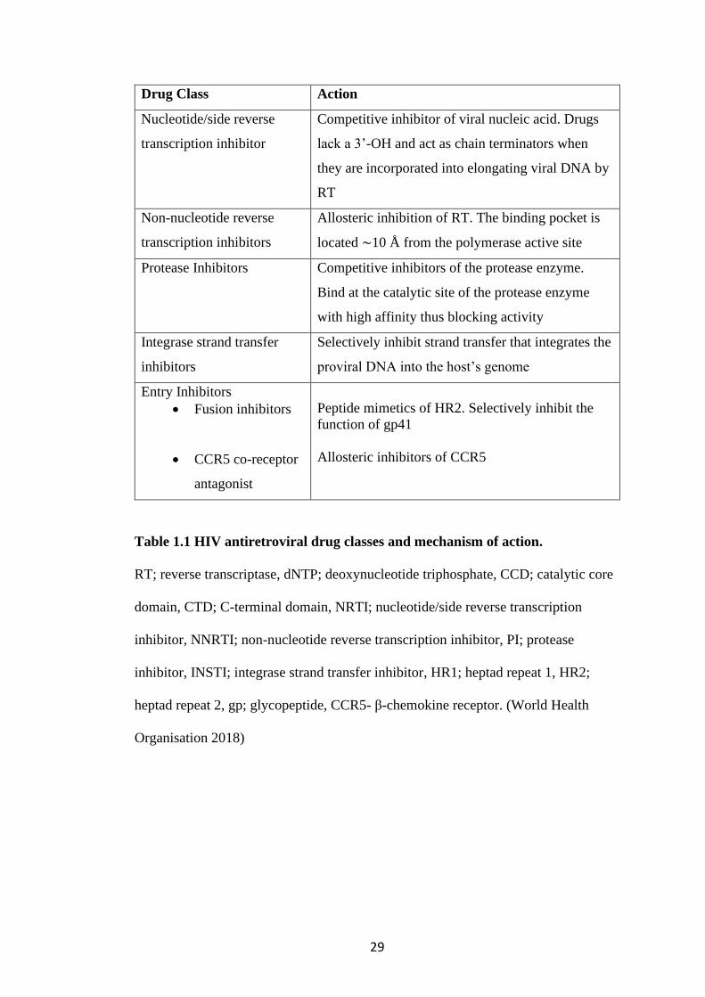
treat HIV-1 currently in clinical use are detailed in Table 1.1.
29
Drug Class Action
Nucleotide/side reverse
transcription inhibitor
Competitive inhibitor of viral nucleic acid. Drugs
lack a 3’-OH and act as chain terminators when
they are incorporated into elongating viral DNA by
RT
Non-nucleotide reverse
transcription inhibitors
Allosteric inhibition of RT. The binding pocket is
located ∼10 Å from the polymerase active site
Protease Inhibitors Competitive inhibitors of the protease enzyme.
Bind at the catalytic site of the protease enzyme
with high affinity thus blocking activity
Integrase strand transfer
inhibitors
Selectively inhibit strand transfer that integrates the
proviral DNA into the host’s genome
Entry Inhibitors
• Fusion inhibitors
• CCR5 co-receptor
antagonist
Peptide mimetics of HR2. Selectively inhibit the
function of gp41
Allosteric inhibitors of CCR5
Table 1.1 HIV antiretroviral drug classes and mechanism of action.
RT; reverse transcriptase, dNTP; deoxynucleotide triphosphate, CCD; catalytic core
domain, CTD; C-terminal domain, NRTI; nucleotide/side reverse transcription
inhibitor, NNRTI; non-nucleotide reverse transcription inhibitor, PI; protease
inhibitor, INSTI; integrase strand transfer inhibitor, HR1; heptad repeat 1, HR2;
heptad repeat 2, gp; glycopeptide, CCR5- β-chemokine receptor. (World Health
Organisation 2018)
30
1.2 Human immunodeficiency Virus-1 Life Cycle
HIV-1 is a single stranded positive RNA lentivirus, which is part of the Retroviridae
family. HIV-1 is 100nm in diameter. The HIV-1 genome consists of two identical
single-stranded RNA molecules which encodes for the Gag (Group specific antigen),
Pol (Polymerase) and Env (Envelope) structural proteins as well as non-structural
proteins including Vif (Viral infectivity factor), Vpr (Viral protein R), Vpu (Viral
protein U), Tat (Trans-activator of transcription), Rev (Regulator of virion
expression) and Nef (Negative regulatory factor) (Figure 1.1).
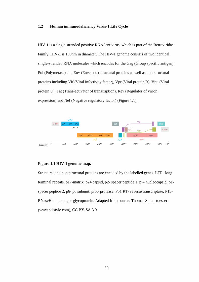
Figure 1.1 HIV-1 genome map.
Structural and non-structural proteins are encoded by the labelled genes. LTR- long
terminal repeats, p17-matrix, p24 capsid, p2- spacer peptide 1, p7- nucleocapsid, p1-
spacer peptide 2, p6- p6 subunit, prot- protease, P51 RT- reverse transcriptase, P15-
RNaseH domain, gp- glycoprotein. Adapted from source: Thomas Splettstoesser
(www.scistyle.com), CC BY-SA 3.0

31
1.2.1 HIV-1 attachment and Entry
The HIV-1 life cycle begins with the attachment of the HIV-1 particle to the host cell
receptor (Figure 1.2). The viral membrane is studded with Envelope (Env) proteins,
which interacts with T cell CD4 receptors. This is then followed by interaction with
a co-receptor, which maybe a β-chemokine receptor (CCR5) or an ⍺-chemokine
receptor (CXCR4) (Figure 1.3). HIV-1 Env protein consists of trimers gp41 and
gp120 subunits. Gp120 is responsible for virion attachment to target cells via the
CD4 receptor, while gp41 mediates fusion between the virus and host cell
membranes (Merk and Subramaniam 2013).
32
Figure 1.2 HIV-1 life cycle.
Adapted from source: Jmarchn https://commons.wikimedia.org/wiki/File:HIV-
replication-cycle-en.svg CC-BY-SA 3.0 Unported.
The trimer surface unit gp120 binds to CD4 from which a phenylalanine at position
43 protrudes which is essential for binding with gp120 (Kwong et al. 1998). Binding
of gp120 to CD4 leads to structural rearrangements that expose the co-receptor
binding sites and secondary binding to the coreceptor occurs (Dragic et al. 1996).
Next the fusion peptide (FP) in the C-Terminal domain of gp41 flips over and inserts
into the cell membrane. The protein then hairpins in order to bring the membranes
together.
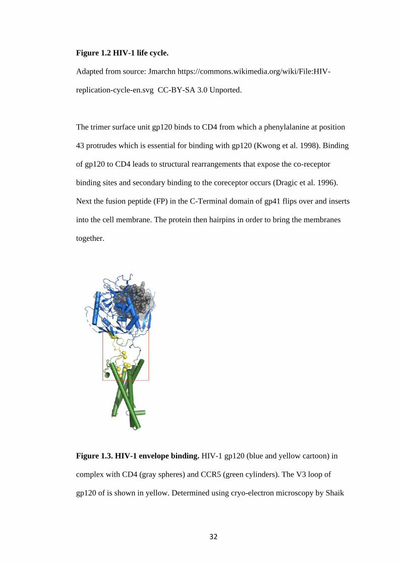
Figure 1.3. HIV-1 envelope binding. HIV-1 gp120 (blue and yellow cartoon) in
complex with CD4 (gray spheres) and CCR5 (green cylinders). The V3 loop of
gp120 of is shown in yellow. Determined using cryo-electron microscopy by Shaik
33
et al. (2019). Source: Collier et al. Brain Sci. 2018, 8, 190;
doi:10.3390/brainsci8100190.
The importance of CCR5 co-receptor in HIV-1 entry is underlined by the fact that
CCR5-delta 32 mutation protects against HIV-1 infection and two patients with
exclusively CCR5 co-receptor using HIV-1 who received a bone marrow transplant
from a donor with homozygous CCR5-delta 32 deletion following bone marrow
ablative chemotherapy and were cured of HIV-1 (Gupta et al. 2019; Hütter et al.
2009).
1.2.2 Reverse Transcription
Within the viral particle is a conical shaped capsid within which reverse transcription
of the viral RNA into DNA occurs. Following plasma fusion, the capsid is thought to
remain intact in order to protect the viral genetic material from triggering hosts toll-
like receptors (TLRs) or pattern recognition receptors (PRRs) and eliciting an innate
immune response to HIV-1 (Figure 1.2) (Jacques et al. 2016). The capsid moves
through the host cell cytoplasm and recruits microtubules to reach the nucleus
(Sodeik, Ebersold, and Helenius 1997) where it docks at a nuclear pore (Jacques et
al. 2016).
The HIV-1 particle contains its own reverse transcriptase (RT) that has two
enzymatic activities; 1) DNA polymerase enzyme which reverse transcribes viral
34
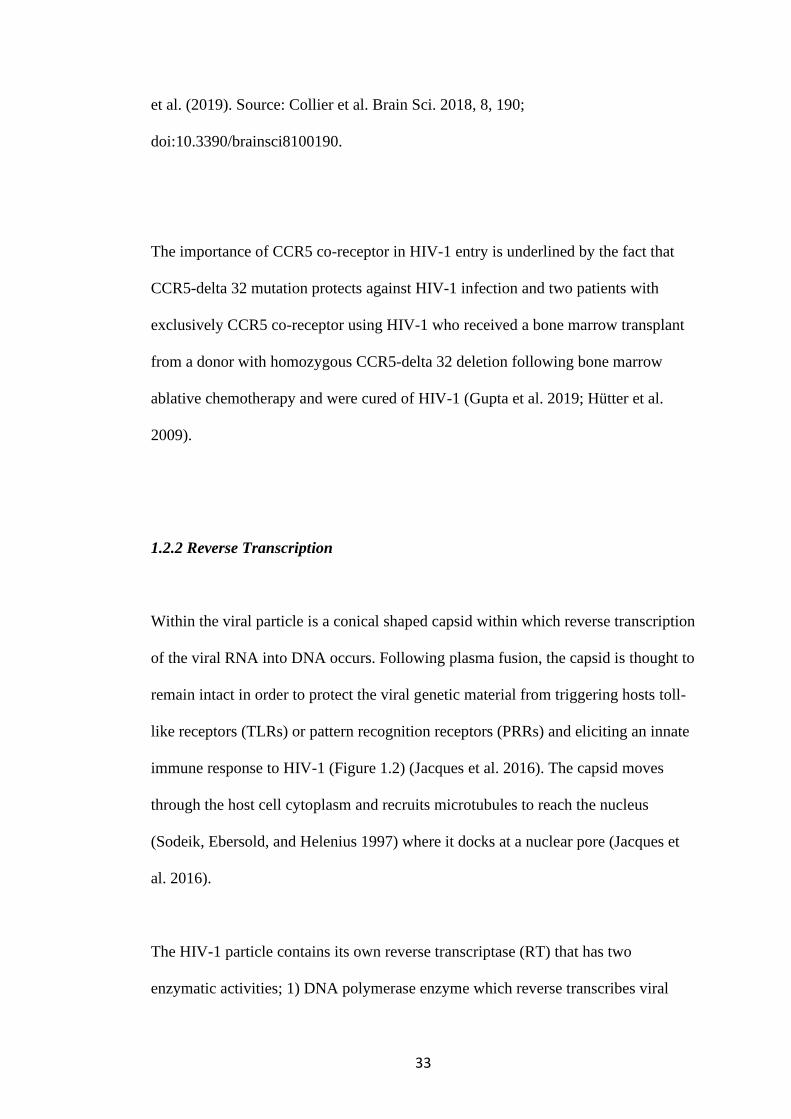
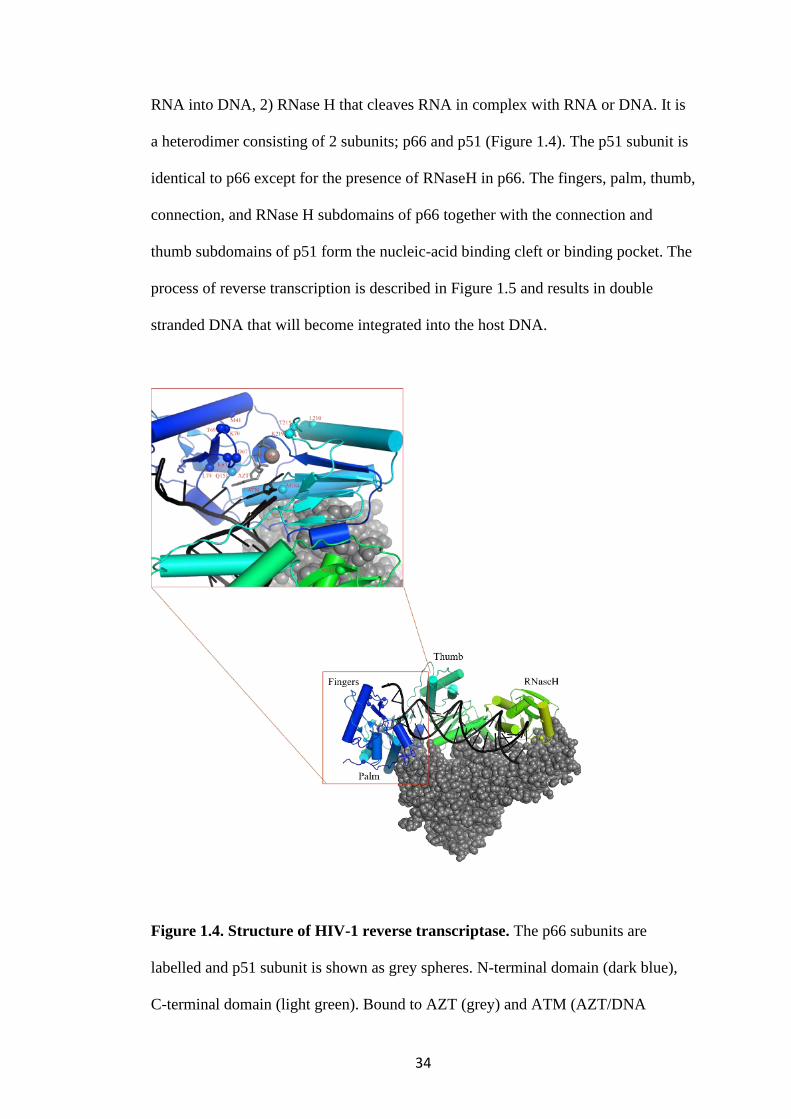
RNA into DNA, 2) RNase H that cleaves RNA in complex with RNA or DNA. It is
a heterodimer consisting of 2 subunits; p66 and p51 (Figure 1.4). The p51 subunit is
identical to p66 except for the presence of RNaseH in p66. The fingers, palm, thumb,
connection, and RNase H subdomains of p66 together with the connection and
thumb subdomains of p51 form the nucleic-acid binding cleft or binding pocket. The
process of reverse transcription is described in Figure 1.5 and results in double
stranded DNA that will become integrated into the host DNA.
Figure 1.4. Structure of HIV-1 reverse transcriptase. The p66 subunits are
labelled and p51 subunit is shown as grey spheres. N-terminal domain (dark blue),
C-terminal domain (light green). Bound to AZT (grey) and ATM (AZT/DNA
35
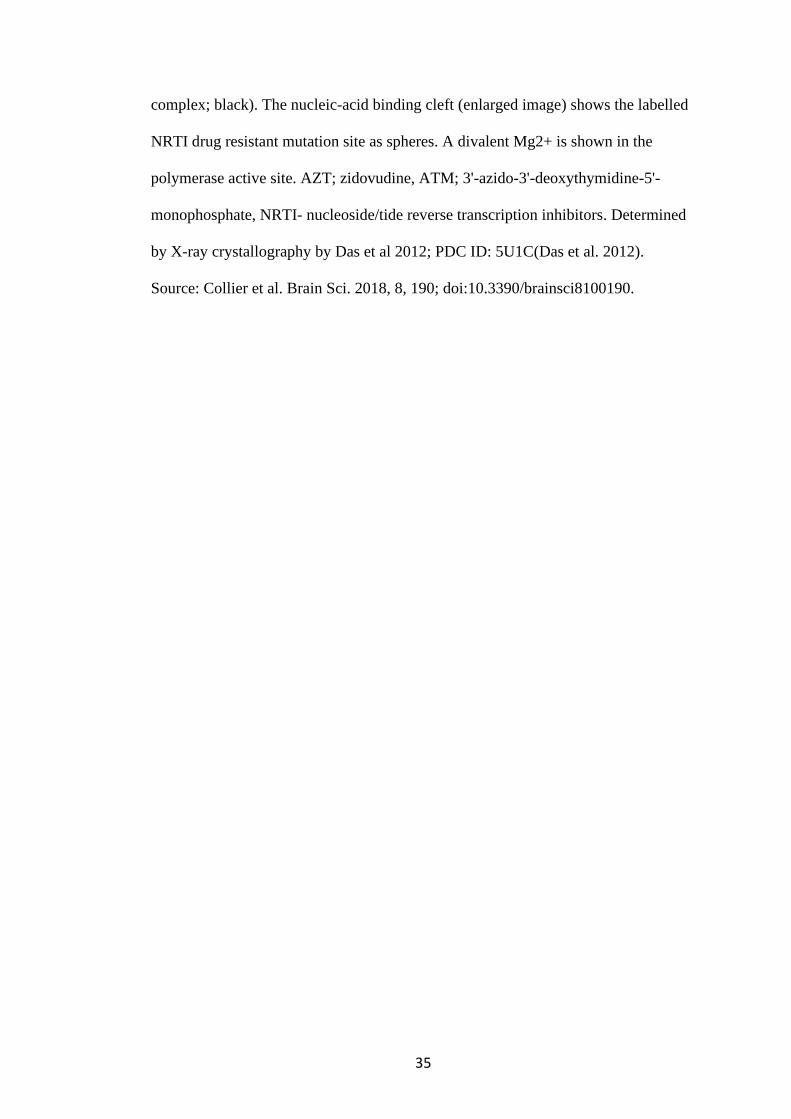
complex; black). The nucleic-acid binding cleft (enlarged image) shows the labelled
NRTI drug resistant mutation site as spheres. A divalent Mg2+ is shown in the
polymerase active site. AZT; zidovudine, ATM; 3'-azido-3'-deoxythymidine-5'-
monophosphate, NRTI- nucleoside/tide reverse transcription inhibitors. Determined
by X-ray crystallography by Das et al 2012; PDC ID: 5U1C(Das et al. 2012).
Source: Collier et al. Brain Sci. 2018, 8, 190; doi:10.3390/brainsci8100190.
36
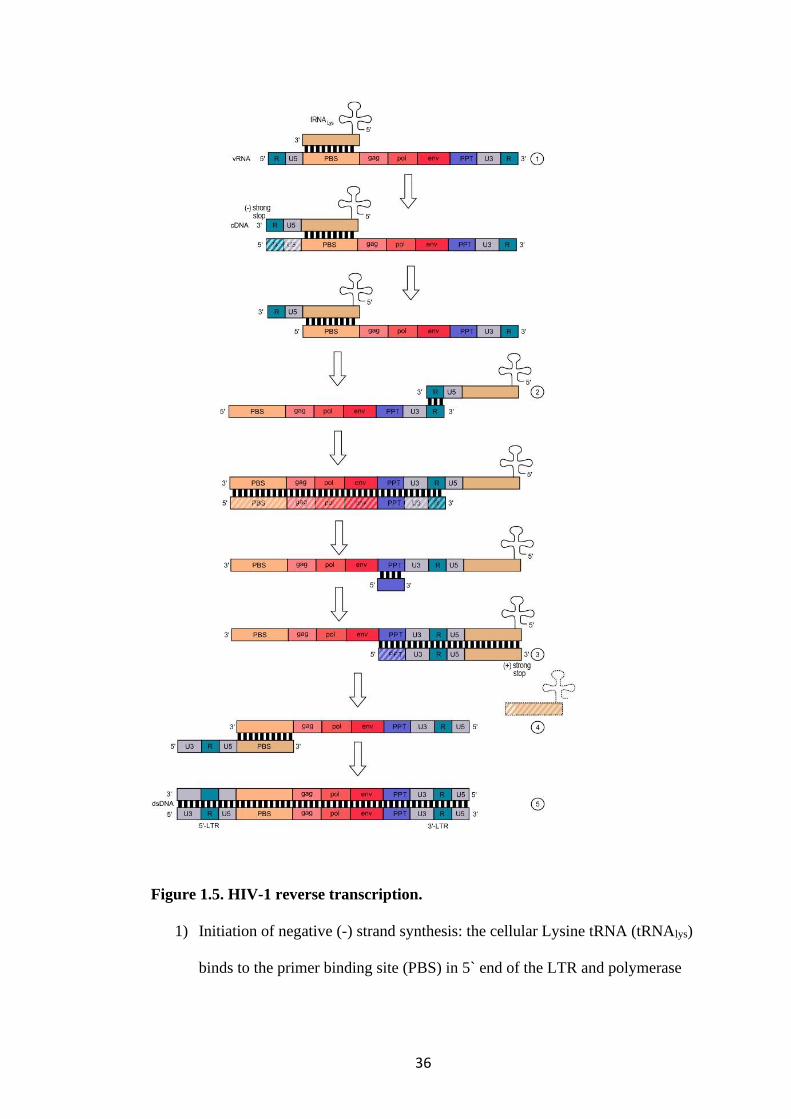
Figure 1.5. HIV-1 reverse transcription.
1) Initiation of negative (-) strand synthesis: the cellular Lysine tRNA (tRNAlys)
binds to the primer binding site (PBS) in 5` end of the LTR and polymerase
37
initiates transcription until the (-) strong stop is produced. The RNA template
that has been transcribed is broken down by RNaseH.
2) First strand transfer: the (-) strong stop contains an R sequence which is
complementary to the R on the 3` end of the RNA template and will continue
to copy the RNA in a 3` to 5` direction, including the PBS. The template
RNA apart from the polypurine tract (PPT) is degraded by RNAaseH as
polymerisation continues.
3) Positive (+) strand synthesis: complementary DNA strand synthesis is
primed at the PPT and proceeds in the 5` to 3` direction. It copies the primer
site of the bound tRNA to produce the (+) strong stop DNA, complementary
to the PBS on the (-) stand DNA. The tRNA and PPT are degraded by
RNaseH.
4) Second strand transfer: (+) strand synthesis continues from the (+) strong
stop in the 5` to 3` direction all the way to the end of the (-) strand.
5) Strand displacement synthesis: RT extends the 3` end of the (-) strand DNA
to the end of the (+) stand to include U3 at the 5` end.
Adapted from source: Alan Cann: Principles of molecular virology. Amsterdam:
Elsevier Academic Press, 2005, p. 93 ISBN 0-12-088787-8.; en:Reverse
transcription entry in
Wikipedia.https://creativecommons.org/licenses/by/3.0/deed.en
38
1.2.3 Integration
Double stranded retroviral DNA integrates into the host genome. Integrated viral
DNA is called proviral DNA. This process is catalysed by HIV-1 integrase which
has 2 enzymatic functions 1) strand transfer (ST) which integrates the proviral DNA
into the host’s DNA (Pommier, Johnson, and Marchand 2005) and 2) 3ʹ -end
processing (3ʹEP) which removes two nucleotides from the 3' at both ends of HIV-1
DNA. The enzyme is encoded by the pol gene and is cleaved from the pol
polyprotein by HIV-1 protease enzyme (Figure 1.1). It is a tetramer and has 3
domains: 1) N-terminal domain (NTD), 2) the catalytic core domain (CCD and 3)
the C-terminal domain (CTD), which is the DNA binding site (Lodi et al. 1995)
(Figure 1.6).
Integrase binds other proteins and cofactors to facilitate the integration of the HIV-1
DNA into the host genome. Lens epithelium-derived growth factor (LEDGF) binds
integrase and chromosomal DNA at nucleosomes, which tethers the HIV-1 DNA to
sites of active transcription (Ciuffi et al. 2005). The proviral DNA hijacks the host
transcription machinery to synthesize many copies of viral mRNA by RNA
polymerase II. These mRNAs are subsequently translated into viral proteins.
39
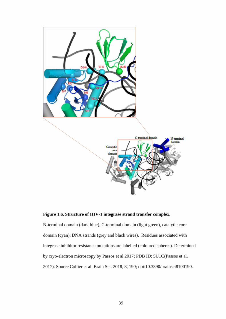
Figure 1.6. Structure of HIV-1 integrase strand transfer complex.
N-terminal domain (dark blue), C-terminal domain (light green), catalytic core
domain (cyan), DNA strands (grey and black wires). Residues associated with
integrase inhibitor resistance mutations are labelled (coloured spheres). Determined
by cryo-electron microscopy by Passos et al 2017; PDB ID: 5U1C(Passos et al.
2017). Source Collier et al. Brain Sci. 2018, 8, 190; doi:10.3390/brainsci8100190.
40
1.2.4 Transcription and mRNA processing
Following integration into the host genome at transcriptionally active sites, HIV-1
gene expression is facilitated by a highly regulated process that co-opts the host’s
replication machinery. In the LTR of the provirus is a transcriptional control region
that contains two NF𝜅B binding sites between nucleotides −104 and −180, which are
important HIV-1 transcription enhancers (Kwon et al. 1998). Also within this region
are promoters essential for both basal and Tat trans-activation. It consists of a TATA
box, which is a largely conserved region of DNA sequences across HIV-1 subtype
that binds TATA binding proteins and is important for RNA synthesis (Montano,
Nixon, and Essex 1998) and a transcription initiation site to which the trans-
activation response element (TAR) is connected. TAR is a RNA stem loop structure
to which the trans-activator protein Tat binds in order to increase the frequency of
full length transcripts (Berkhout, Silverman, and Jeang 1989). Tat also directly
interacts with the cellular cofactor positive transcription elongation factor b (P-
TEFb) which results in phosphorylation of RNA polymerase II and mRNA
elongation (Mancebo et al. 1997).
Transcription generates multiply spliced as well as unsliced transcripts which
includes the full-length HIV-1 genomic RNA. A spliced mRNA that encodes Rev, a
nuclear export protein, is exported out of the nucleus. It is translated and then re-
enters the nucleus where it binds to the rev response element (RRE) at the 3` end of
the viral genome (Rausch and Le Grice 2015). This complex is recognised by
cellular export machinery and allows export of the whole viral genome into the
41
cytoplasm where two copies of RNA dimerise and form a stable RNA structure that
will be packaged into virus particles (Moore and Hu 2009).
1.2.5 Assembly and packaging
Translation of the HIV-1 protein precursor proteins occurs on ribosomal RNAs in
the cytoplasm. Polyprotein precursors Gag and Gagpol are made in a 20:1 ratio
(Shehu-Xhilaga, Crowe, and Mak 2001). The matrix subunit of Gag has a
hydrophobic sequence that is myristoylated, forming a lipid residue that directs Gag
to the plasma membrane. The Nucleocapsid of the Gag precursor protein interacts
with Psi packaging signal that directs the two genomic RNA molecules into the
forming virus particle (Sundquist and Kräusslich 2012). As the internal structures are
assembled along the plasma membrane budding occurs simultaneously with the help
of the cellular ESCRT (Endosomal sorting complexes required for transport)
proteins by interaction between the p6 submit of Gag and proteins of the ESCRT
pathway such as ALIX (Bieniasz 2009). After release from the host cell, ordered
cleavage of gag results in mature infectious virus ready to infect another host cell
(Fun et al. 2012).

42
1.3 Within-Host adaptations
1.3.1 Inherent viral diversity
HIV-1 is highly diverse. There are 4 groups – M, N, O, P. HIV-1 main group M is
responsible for the global pandemic. It consists of 9 subtypes (A, B , C, D, F, G, H, J
and K), 6 sub-subtypes (A1-A4 and F1 and F2) and multiple circulating recombinant
forms (CRF) and unique recombinant forms (URF) (Los Alamos HIV Sequence
Database).
Evolution of the virus is driven by the need to evade host immunity. This is
facilitated by a high replication rate (1010 virions generated per day in an individual),
which is error prone, resulting in one mutation for every 105 nucleotides copied
(Mansky and Temin 1995). In addition, reverse transcription has no proof-reading
mechanism and a high recombination rate serves to increase quasispecies diversity
(Song et al. 2018). Consequently, HIV-1 has vast genetic diversity within and
between individuals (Kearney et al. 2009). Depending on the quality of ART
suppression, genetic diversity maybe broader in the presence of ART compared to
drug naïve individuals (Haddad et al. 2000).
1.3.2 The host environment
HIV-1 has mechanisms to evade the host innate immune response and avoid
induction of Interferon gamma (IFN𝛾) (Rasaiyaah et al. 2013). This has probably
43
facilitated its success as a pandemic virus. In addition, within-host evolution occurs.
There is a constant co-evolutionary race between the virus- to escape the adaptive
immune response and the host- to match the viruses antigenic evolution. The
adaptive immune response includes HIV-specific CD8+ cytotoxic T-lymphocytes
(CTLs) that clear infected T cells (Walker et al. 1987). This exerts an intense
selection pressure on the viral population, leading to CTL escape mutants (Borrow et
al. 1997). These are observed in targeted CTL Gag and Env-specific epitopes
(Borrow et al. 1997; Deng et al. 2015; Walker et al. 1987).
The viral Env protein is the target of the humoral adaptive response to HIV-1
(Burton and Mascola 2015). A changing landscape of the Env protein has been
described whereby the Env epitopes continually evolves to avoid neutralisation by
antibodies (Wu, Wang, et al. 2012). In addition, Env residues acquire N-linked
glycosylation which serve to shield the virus from antibody recognition (Wei et al.
2003).
Finally, HIV-1 may evolve in host with certain class I histocompatibility-linked
leukocyte antigen (HLA) alleles such as HLA B57 and B27, which confer protection
or a better prognosis once exposed to HIV-1 by promoting CTL mediated clearance
of infected cells (Gao et al. 2005; Kaslow et al. 1996). This has been associated with
CTL escape mutants and interestingly, these alleles are over-represented in elite
controllers (Merindol et al. 2018; Migueles et al. 2000).
44
Some have argued that these evolutionary adaptation are “short-sighted” because
although they adapt the virus to chronic infection within a host, they are a
disadvantage to onward transmission (Lythgoe et al. 2017).
1.3.3 Latent reservoirs
Cellular and anatomical compartmental reservoirs of latent HIV-1 have been
described. It is estimated that 1 in 103 resting CD4+ T cells harbours HIV-1 provirus
(Bruner et al. 2016) and there is persistence of 10% of infected cells during ART
(Besson et al. 2014). Different populations of HIV-1 have been identified in cellular
compartments including memory, naive CD4 T cells and CD14 monocytes (Delobel
et al. 2005). Long-lived cells such as neurons and macrophages in anatomical
compartments such as the CNS, male genital tract and gut, may harbour replication
competent HIV-1 (Collier et al. 2018; Ganor et al. 2019). Proviral DNA may lie
dormant in these cells and reactivate at a later time (Sigal and Baltimore 2012).
Furthermore, viruses in these anatomical reservoirs may undergo low-level
replication and continue to replenish the compartment and the peripheral blood
(Lorenzo-Redondo, Fryer, Bedford, Kim, Archer, Kosakovsky Pond, et al. 2016).
HIV-1 can also adapt to its cellular tropism to replicate in different cell types and
anatomical compartments. This evolution may involve a change in chemokine co-
receptor used i.e. CCR5 to CXCR4 shift, or a change from R5-T cell tropism to R5-
macrophage tropism (Arrildt et al. 2015). Macrophages in the central nervous system
(CNS) have lower levels of CD4 and are targeted by R5-Mac-tropic HIV-1 which
45
has adapted to infect cells with low levels of CD4 (Joseph et al. 2015). Macrophages
can become highly permissive to HIV-1 by the deactivation of the restriction factor-
SAMHD1 following G0 to G1 transition (Mlcochova et al. 2017).
The inherent diversity of HIV-1 and the dynamic virus and host co-evolution has
implications for the persistence of HIV-1 particularly in latent reservoirs such as the
CNS.
1.4 CNS replication and HIV-1
Since the rollout of ART, along with a reduction in HIV-associated mortality, the
incidence of HIV-associated central nervous system (CNS) pathology has fallen
(Sacktor et al. 2001). However, HIV-associated neurocognitive disorder (HAND)
remains a problem, with a prevalence of up to 50% in ARV treated patients in
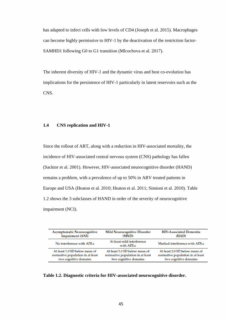
Europe and USA (Heaton et al. 2010; Heaton et al. 2011; Simioni et al. 2010). Table
1.2 shows the 3 subclasses of HAND in order of the severity of neurocognitive
impairment (NCI).
Table 1.2. Diagnostic criteria for HIV-associated neurocognitive disorder.

46
According to the Frascati diagnostic criteria. ADLs; activities of daily living. SD;
standard deviation. Source- Collier et al. Brain Sci. 2018, 8, 190;
doi:10.3390/brainsci8100190.
It is thought that HIV-1 entry to the CNS is facilitated by loss of the integrity of the
blood brain barrier (BBB) early in HIV-1 infection. HIV-1 ingresses as free particles
or within activated CD4 central memory T cell or monocytes. The CNS viral
reservoir is then maintained by clonal expansion of latently infected T cells or low-
grade replication in macrophages (Sigal and Baltimore 2012), which may lead to an
independently replicating population of HIV-1 viruses in the CNS compartment,
distinct from the population in the peripheral circulating blood. This is known as
CNS compartmentalisation. Due to inadequate ARV penetration into the CNS, the
virus has the opportunity to sequester in the CNS and multiply (Joseph et al. 2019;
Lorenzo-Redondo, Fryer, Bedford, Kim, Archer, Pond, et al. 2016). Following
treatment interruption, resistant viruses may emerge in the CNS, amplifying this
process (Canestri et al. 2010). The presence of replicating HIV-1 in the brain has
been implicated in HIV-associated dementia (HAD); the most severe form of HAND
and HIV-1 encephalitis (HIVE); a presentation of acute or subacute brain
inflammation characterised by any of fever, headache, confusion, seizures or coma
(Schnell et al. 2011; Filipowicz et al. 2016). Autopsy and living brain biopsy
specimens from patients with HAD show positive p24 immunostaining in microglia
and macrophages (Gray et al. 2013). In addition, HIV-1 isolates have been derived
from brain biopsy specimen of participants with HAD (Gorry et al. 2001). Similarly
in the animal models of HIVE and SIVE there is perivascular accumulation of gag
47
positive CD68 + and CD163+ macrophages in brain tissue (Filipowicz et al. 2016).
CSF HIV-1 viral load (VL) correlates well with brain HIV-1 RNA levels (Gelman et
al. 2013).
HIV-1 is suppressed by ART in both the blood and the CNS. However, this does not
happen in every treated individual and the occurrence of CSF escape/discordance is
now recognised (Canestri et al. 2010). It remains uncertain whether independently
replicating HIV-1 in the brain parenchyma and the proxy for this—the presence of
HIV-1 virus in the cerebrospinal fluid (CSF), is responsible for NCI in the ART era.
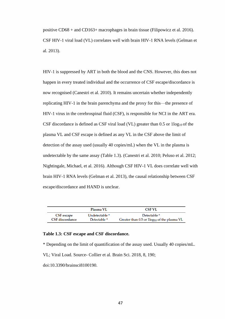
CSF discordance is defined as CSF viral load (VL) greater than 0.5 or 1log10 of the
plasma VL and CSF escape is defined as any VL in the CSF above the limit of
detection of the assay used (usually 40 copies/mL) when the VL in the plasma is
undetectable by the same assay (Table 1.3). (Canestri et al. 2010; Peluso et al. 2012;
Nightingale, Michael, et al. 2016). Although CSF HIV-1 VL does correlate well with
brain HIV-1 RNA levels (Gelman et al. 2013), the causal relationship between CSF
escape/discordance and HAND is unclear.
Table 1.3: CSF escape and CSF discordance.
* Depending on the limit of quantification of the assay used. Usually 40 copies/mL.
VL; Viral Load. Source- Collier et al. Brain Sci. 2018, 8, 190;
doi:10.3390/brainsci8100190.
48
1.5 Factors associated with HIV-1 neurotropism and neurovirulence
It has long been established that different but related strains of HIV-1 can coexist in
different tissues (Gorry et al. 2001; Koyanagi et al. 1987). These strains exhibit
genetic and phenotypic differences. CSF derived virus- JRCSF obtained from a
patient with AIDS encephalopathy was found to infect a mixture of brain derived
cells (astrocytes 70%, oligodendrocytes 30% and some fibroblasts). JRFL derived
from the frontal lobe on the other hand had cellular tropism for macrophage and
monocytes (Koyanagi et al. 1987). However, the factors that determine HIV-1
neurotropism are not well understand and there has been limited characterisation of
this in HIV-1 subtype C viruses.
1.5.1 HIV-1 subtype and CNS disease
There is evidence to suggest that HIV-1 subtype may play a role in NCI. NCI has
been associated with CRF_02AG when compared with subtype G in Nigeria (Royal
et al. 2012). A greater risk of HAD was found in participants with subtype D virus
compared to those with subtype A virus in Uganda (Sacktor et al. 2009). There is up
to 35% variation within HIV-1 env across subtypes (Araujo and Almeida 2013).
With the adaptive immune response against HIV-1 being mainly targeted at the
envelope glycoprotein, inherent differences between HIV-1 env subtypes may play a
role in HIV-1 neurotropism. It is therefore important to know if there are subtype
dependent variations in the neurotropism and neurovirulence of HIV-1.
49
1.5.2 Cellular tropism
More recent studies characterising the phenotypic properties of CSF derived viruses
have correlated macrophage tropism with neurotropism with evidence of a tropism
shift from R5-T cell tropism to R5-macrophage tropism (Arrildt et al. 2015; Gorry et
al. 2001; Schnell et al. 2011). Microglial cells and other macrophages in the CNS
have lower levels of CD4 and are the targets for R5-Macrophage-tropic HIV-1,
adapted to infect cells with low levels of CD4 (Joseph et al. 2015).
1.5.3 Coreceptor usage
The role of coreceptor tropism in viral pathogenicity is complex. The transmitted
founder viruses exclusively utilise CCR5 coreceptor. Traditionally a switch from
CCR5 coreceptor use to CXCR4 heralded the onset of advanced HIV/clinical AIDs.
This is supported by the association of dual-tropic- CXCR4/R5 viruses with lower
CD4 counts (Wilkin et al. 2007; Moyle et al. 2005). This phenomenon appears to be
subtype specific with up to 50% of subtype B HIV-1 infected individuals undergoing
co-receptor switching. This is approximately 20% in HIV-1 subtype C (Cilliers et al.
2003). A higher prevalence of CXCR4 variants is seen in subtype D compared with
subtype A infected individuals (Huang et al. 2007). The prevalence of CXCR4 only
using variants is less than 20% in most cohorts that have determined tropism using
phenotypic assays (Moyle et al. 2005; Melby et al. 2006; Wilkin et al. 2007). In
subtypes A and C progression of HIV-1 was accompanied by increased ability to
utilise low levels of CCR5 and increased macrophage tropism. Neurotropism is also
50
associated with an increased ability to use low level of CD4 and or CCR5.
Therefore, it would seem that the relative affinity of the virus to CD4/CCR5
receptors rather than a coreceptor switch is a better predictor of neurovirulence
(Gorry et al. 2001; Gorry et al. 2002).
One study examining coreceptor usage in patients co-infected with HIV-1 subtype C
and Cryptococcal meningitis (CM), found a evidence of CXCR4 usage using
computer predictive algorithms (Sojane et al. 2018). This may reflect advanced HIV-
1 infection or a higher prevalence of CXCR4 usage by CNS derived HIV-1 viruses.
1.5.4 Drug resistant mutations
CNS compartmentalisation has been shown (Liu et al. 2013; Schnell et al. 2011;
Sturdevant et al. 2015; Stam et al. 2013), with greater genetic diversity of CSF
escape viruses compared to plasma viruses according to phylogenetic analyses of
pol/RT (Liu et al. 2013). Tong et al used deep sequencing to explore the range of
minority resistance associated mutations in paired CSF and plasma and discovered
mutations in CSF that were not identified by Sanger sequencing alone (Tong et al.
2015). The evolution of resistance mutations is also seen in CSF viruses (Canestri et
al. 2010; Mukerji et al. 2017; Nightingale, Geretti, et al. 2016; Beguelin et al. 2016).
Peluso et al. found CSF viral resistance in patients in whom resistance genotyping
was conducted; 6/7 had NRTI mutations, 5/7 patients had PI mutations and 2/7
patients had NNRTI mutations (Peluso et al. 2012). In Mukherji’s study of the
pooled cohort from CHARTER, NNTC and HNRC, the CSF escape cases were
51
combined with all published CSF cases and showed that M184V/I mutations were
detected more frequently in the CSF and plasma of patients with escape; 61%
(34/56) and 30% (16/55) of samples respectively, compared to participants without
escape, where it was only detected in 7% (3/43) of both CSF and plasma samples
(Mukerji et al. 2018). APOBEC3F/G mediated hypermutation has been associated
with compartmentalisation in CSF compared with peripheral blood mononuclear
cells (PBMCs) and induced drug-resistant mutations in CSF; G73S in protease,
M184V and M230I in reverse transcriptase (Fourati et al. 2014).
1.5.5 Escape from antibody neutralisation
Broadly neutralizing antibodies (bNAbs) targets 6 epitopes on the HIV-1 Env trimer-
CD4 binding site, V2 apex, the V3 loop glycan patch, the membrane-proximal
external region (MPER), the interface between the gp120 and gp41 subunits and
‘silent face’ of gp120 (Sok and Burton 2018). HIV-1 viruses in the CNS and plasma
may evolve in response to differential selection pressures from both cellular immune
response and neutralizing antibodies (Nabs) (Pillai et al. 2006). The glycoprotein
shield associated with some of these epitopes is a target for the humoral immune
response. An evolving ‘glycan shield’ mechanism of neutralization escape has been
described by Wei et al, whereby selected changes in envelope glycosylation density
and position prevented NAbs from binding but not receptor binding (Wei et al.
2003). Furthermore, over time the population of virus sensitive to neutralisation by
autologous NAbs was replaced by neutralisation resistant virus. Viral escape through
52
mutation of the HIV-1 envelope glycoprotein (Env) maybe a means through which
an independently replicating viral population can exist in the CNS.
1.5.6 CNS co-infection
Cryptococcal meningitis (CM) is an important AIDS defining illness and a leading
cause of death in HIV-1 positive individuals in sub-Saharan Africa. It is uncertain to
what extent opportunistic CNS infections such as CM increase the likelihood of
HIV-1 CSF escape or discordance and to what extent persistent CNS HIV-1 impacts
on the outcomes of CM. There is evidence of compartmentalised immune responses
between CSF and blood in individuals with CNS infections such as TB meningitis
(Christo et al. 2007) and CM (Chang et al. 2017), which may play a role in recruiting
HIV-1 variants into the CNS, a phenomenon known as secondary CSF escape.
However, it is unclear to what degree the HIV-1 variants in the CNS are an
independently replicating/compartmentalised population or if they are intermixed
with the plasma population as maybe the case due to BBB inflammation.
1.6 Aims
CNS compartmentalisation may lead to establishment of a long-lived viral reservoir
in the CNS that may have different genotypic and phenotypic properties to the
viruses in the periphery. This has implications for HIV-1 treatment, vaccine and cure
strategies. The aim of the first part of thesis is therefore to investigate the factors that
53
determine HIV-1 subtype C CNS compartmentalisation in the context of CNS
opportunistic infection/inflammation.
Specific objectives/questions are:
1 What is the prevalence of CNS compartmentalisation and what patient
characteristics are associated with CSF compartmentalisation in the context
of CM?
2 What are the phylogenetic characteristics of paired CNS and plasma genomes
of HIV-1 subtype C in the context of co-infection with CM?
3 What are the phenotypic properties of HIV-1 clones isolated from the CNS?

54
PART 2
2.1 Pandemic SARS-CoV-2
SARS-CoV-2 emerged in 2019 as the causative agent of a severe acute pneumonia
first reported in Wuhan China (Zhou et al. 2020). This novel agent was sequenced in
January 2020 from a bronchoalveolar lavage sample and was identified as a novel
virus of the Coronaviridae family (Wu et al. 2020). Coronaviruses that affect humans
are zoonotic and many are thought to have originated in bats (Tao et al. 2017).
SARS-CoV-2 is a Betacoronavirus and one of the seven coronaviruses that infect
humans. SARS-CoV and MERS-CoV cause severe disease, whilst HKU1, NL63,
OC43 and 229E are associated with mild seasonal colds (Corman et al. 2018).
However only SARS-CoV-2 has caused a pandemic.
The novel SARS-CoV-2 has 96% nucleotide identity with a bat coronavirus
RaTG13, identified from a Rhinolophus affinis bat (Figure 2.1). However, the
receptor binding domain (RBD) is where they diverge (Zhou et al. 2020). A related
coronavirus found in Malayan pangolins (Manis javanica), is postulated to be the
intermediary host. This is because SARS-like coronavirus found in pangolins have
the 5 of the 6 amino acid changes in the RBD that are found in SARS-CoV-2 which
enhances binding to their respective ACE2 (angiotensin converting enzyme-2)
receptors (Andersen et al. 2020). Overall, SARS-CoV-2 is more closely related to
the bat coronavirus (Figure 2.1) (Zhou et al. 2020). Also unique to SARS-CoV-2 is
the insertion of a polybasic furin cleavage site between Spike subunits 1 and 2,
which has been found to increase its infectivity (Xia et al. 2020). The acquisition of a

55
furin cleavage site in Influenza heamglutinninn also heralds an increase in
infectivity, host range and pathogenicity of Influenza (Alexander and Brown 2009;
Nao et al. 2017). The furin cleavage site is also present in human coronavirus HKU1
and MERS-CoV but absent in the sampled bat and pangolin coronaviruses
(Andersen et al. 2020).
Figure 2.1 Neighbour-joining phylogenetic tree based on nucleotide sequences
of whole genome of coronaviruses. Neighbour-joining tree using HKY genetic
distance model with 1000 bootstraps. pangolin, bat and sar-cov-2. each taxon is
labelled by the genebank accession number.
Like HIV-1, the emergence of SARS-CoV-2 has been brought about by the growth
of the human population and expansion into the natural habitats of wild animals
56
(Jones et al. 2008). But unlike HIV-1 this emerging virus has impacted every single
person on this planet. As of the 30th of April 2021 over 150 million cases of COVID-
19 (coronavirus disease 2019) and 3.2 million associated deaths have been reported
globally (Dong, Du, and Gardner 2020).
As yet there is no curative treatment for COVID-19. The treatments available
continue to evolve during the pandemic as clinical trial evidence emerges. Many of
the available drugs are directed at either mitigating or preventing the overwhelming
systemic inflammation that is the hallmark of COVID-19 disease (Table 2.1).
However, rapid advances have been made in the face of this global challenge and
seven effective vaccines have been developed and are now being used globally in a
bid to tackle this pandemic (Table 2.2). They all utilise the Spike protein antigen
target. For the first time an mRNA vaccine has been approved for used to prevent a
human infection (Abbasi 2020).
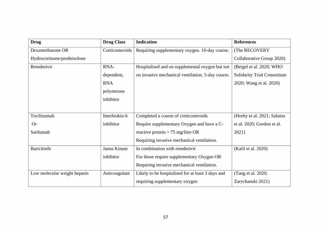
57
Drug Drug Class Indication References
Dexamethasone OR
Hydrocortisone/prednisolone
Corticosteroids Requiring supplementary oxygen. 10-day course. (The RECOVERY
Collaborative Group 2020)
Remdesivir RNA-
dependent,
RNA
polymerase
inhibitor
Hospitalised and on supplemental oxygen but not
on invasive mechanical ventilation. 5-day course.
(Beigel et al. 2020; WHO
Solidarity Trial Consortium
2020; Wang et al. 2020)
Tocilizumab
Or
Sarilumab
Interleukin-6
inhibitor
Completed a course of corticosteroids
Require supplementary Oxygen and have a C-
reactive protein > 75 mg/litre OR
Requiring invasive mechanical ventilation.
(Horby et al. 2021; Salama
et al. 2020; Gordon et al.
2021)
Baricitinib Janus Kinase
inhibitor
In combination with remdesivir
For those require supplementary Oxygen OR
Requiring invasive mechanical ventilation.
(Kalil et al. 2020)
Low molecular weight heparin
Anticoagulant Likely to be hospitalised for at least 3 days and
requiring supplementary oxygen
(Tang et al. 2020;
Zarychanski 2021)

58
14-day course or until discharge
Non-invasive or invasive ventilation base
decision of VTE vs bleeding risk adjust dose
accordingly
Bamlanivimab/etesevimab
(LY-CoV555/ LY-CoV016 - Lilly)
Casirivimab/imdevimab (REGN-
CoV2 - Regeneron)
Monoclonal
antibody
cocktails
Mild to moderate COVID-19 in high risk
individuals in order to prevent severe COVID-19
resulting in hospitalisation or death
(Gottlieb et al. 2021;
Weinreich et al. 2020)
Convalescent plasma Neutralising
antibodies
High titre CCP for hospitalised patients with
COVID-19 early in the course of hospitalisation
(Simonovich et al. 2020;
Libster et al. 2021)
Table 2.1 Therapeutic options currently in use for managing COVID-19.
RNA- ribonucleic acid; COVID-19 (coronavirus disease 2019).

59
Vaccine Platform Reference
BNT162b2 (Pfzier
BionTech)
mRNA (Polack et al. 2020)
mRNA-1273 SARS-
CoV-2 Vaccine
(Moderna)
mRNA (Baden et al. 2020)
ChAdOx1 nCoV-19
vaccine (AZD1222,
AstraZeneca)
Simian adenovirus
vectored (ChAdOx1)
(Voysey, Clemens, et al.
2021)
CoronaVac (Sinovac) Inactivated vaccines (Wu et al. 2021)
Gam-COVID-Vac
(Sputnik V)
Recombinant adenovirus
(rAd)-based vaccine
(rAd26 and rAd5)
(Logunov et al. 2021)
NVX-CoV2373
(Novovax)
Adjuvanted recombinant
protein- based
nanoparticles
(Novovax 2021)
Janssen Ad26.COV2.S
(Johnson &J)
Adenovirus vectored
vaccines
(Sadoff et al. 2021)
Table 2.2 COVID-19 vaccines and the molecular platforms currently in use.
mRNA- messenger ribonucleic acid; COVID-19 (coronavirus disease 2019), rAd-
recombinant adenovirus, ChAdOx-1- replication deficient simian adenovirus.
60
2.2 SARS-CoV-2 Life cycle
SARS-CoV-2 is a positive single strand RNA virus. It has a large 30kb genome
(Figure 2.2). Like all viruses SARS-CoV-2 is an obligate intracellular pathogen that
has to replicate by co-opting the host cellular machinery. SARS-CoV-2 gains entry
into the cell via interaction between the RBD in the Spike protein S1 subunit and the
ACE2 receptor (Figure 2.3). Proteolytic cleavage by cellular surface protease
TMPRSS2 leads to separation of the two functional units of the Spike protein and
allows the S2 subunit to mediate membrane fusion (Jaimes, Millet, and Whittaker
2020; Örd, Faustova, and Loog 2020). The virus particle may also gain entry by
receptor mediated endocytosis. Acidification of endosomes leads to endosome
activated cleavage by cathepsin, allowing insertion of the FP into the endosomal
membrane (Jaimes, Millet, and Whittaker 2020; Örd, Faustova, and Loog 2020).
Figure 2.2 SARS-CoV-2 Genome map.
Structural and non-structural proteins are encoded by the labelled genes. The
subunits and regions of the Spike gene are shown in the expanded S gene. NTD- N-
terminal domain; RBM- receptor binding motif; FP- fusion peptide; HR1- heptad
repeat 1; HR2- heptad repeat 2; TM- transmembrane region; IC- intracellular
domain.
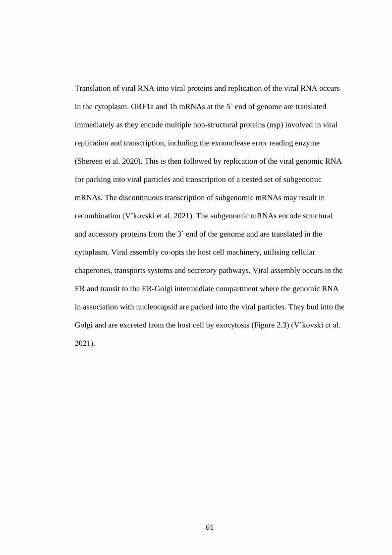
61
Translation of viral RNA into viral proteins and replication of the viral RNA occurs
in the cytoplasm. ORF1a and 1b mRNAs at the 5` end of genome are translated
immediately as they encode multiple non-structural proteins (nsp) involved in viral
replication and transcription, including the exonuclease error reading enzyme
(Shereen et al. 2020). This is then followed by replication of the viral genomic RNA
for packing into viral particles and transcription of a nested set of subgenomic
mRNAs. The discontinuous transcription of subgenomic mRNAs may result in
recombination (V’kovski et al. 2021). The subgenomic mRNAs encode structural
and accessory proteins from the 3` end of the genome and are translated in the
cytoplasm. Viral assembly co-opts the host cell machinery, utilising cellular
chaperones, transports systems and secretory pathways. Viral assembly occurs in the
ER and transit to the ER-Golgi intermediate compartment where the genomic RNA
in association with nucleocapsid are packed into the viral particles. They bud into the
Golgi and are excreted from the host cell by exocytosis (Figure 2.3) (V’kovski et al.
2021).
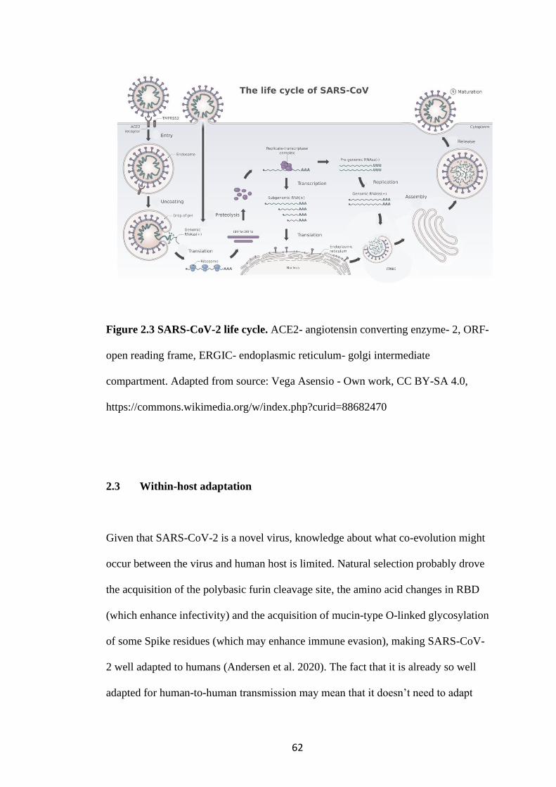
62
Figure 2.3 SARS-CoV-2 life cycle. ACE2- angiotensin converting enzyme- 2, ORF-
open reading frame, ERGIC- endoplasmic reticulum- golgi intermediate
compartment. Adapted from source: Vega Asensio - Own work, CC BY-SA 4.0,
https://commons.wikimedia.org/w/index.php?curid=88682470
2.3 Within-host adaptation
Given that SARS-CoV-2 is a novel virus, knowledge about what co-evolution might
occur between the virus and human host is limited. Natural selection probably drove
the acquisition of the polybasic furin cleavage site, the amino acid changes in RBD
(which enhance infectivity) and the acquisition of mucin-type O-linked glycosylation
of some Spike residues (which may enhance immune evasion), making SARS-CoV-
2 well adapted to humans (Andersen et al. 2020). The fact that it is already so well
adapted for human-to-human transmission may mean that it doesn’t need to adapt
63
much more. However, knowledge from other human coronaviruses has highlighted
that within-host adaptation is to be expected. The original SARS-CoV acquired
mutations between amino acids 442- 487 of the RBD that enhanced viral interactions
with both human and civet ACE2 and resulted in increased infectivity (Wu, Peng, et
al. 2012). Already, it is evident that SARS-CoV-2 is mutating and has the potential
to adapt, impacting transmission (Davies, Abbott, et al. 2021; Volz et al. 2021) and
vaccine response (Collier, De Marco, et al. 2021; Madhi et al. 2021). However, it
remains uncertain if these adaptations are impacting the severity of disease and death
rates (Davies, Jarvis, et al. 2021; Frampton et al. 2021).
Compared with HIV-1 and Influenza viruses, the rate of evolution of SARS-CoV-2
is slow, with a nucleotide substitution rate of 8.4x10⁻⁴ substitutions per site per year
(Nextstrain 2021) compared with HIV-1 which ranges from 2.7 to 6.7x10⁻³
substitutions per site per year (Leitner and Albert 1999) and Influenza A and B
which ranges from 0.5 to 2.6x10⁻³ substitutions per site per year (Nobusawa and Sato
2006). Although it is slowly evolving, any adaptation to humans will be shaped by
constraints of the human niche, which includes natural immunity as more people
become infected and develop an adaptive immune response to SARS-CoV-2 as well
as vaccine-elicited responses.
2.4 Host factors that determine immune responses to SARS-CoV-2 vaccines
Vaccines are a key tool to controlling the COVID-19 pandemic. Many vaccines have
shown promise in clinical trials and real-world setting (Table 2.2). However,
64
duration of protection is as yet unknown. mRNA vaccine had shown good efficacy
in clinical trials after two doses, separated by a three or four week gap (Baden et al.
2020; Polack et al. 2020). However, not many participants above the age of 80 years
were included in the clinical trials despite being at greatest risk of severe disease and
death (Docherty et al. 2020). Only twelve participants over 65 years were evaluated
for neutralising responses in a BNT162b2 vaccine study (Walsh et al. 2020). It is
therefore unknown what neutralising responses or efficacy will be in individual
above the age of 80 years.
Furthermore, the Joint Committee on Vaccination and Immunisation (JCVI)
increased the dosing interval to 12 weeks in order to vaccinate a greater proportion
of the population during a period of substantial transmission (Care 2021). Due to
lack of clinical trial data it is unknown if the elderly would mount adequate and
sustained immune responses in the intervening period between doses to offer
sufficient protection from SARS-CoV-2. There is evidence from studies in mice that
immune responses in ageing mice after one dose of ChAdOx1 nCoV-19 is limited
but is restored to levels comparable to young mice after the second dose (Silva-
Cayetano et al. 2021).
Ageing can impact responses in many ways including but not limited to reducing the
magnitude of antibody response to vaccines (Wilkinson et al. 2017), shortening the
persistence of antibody response (Powers and Belshe 1993), limiting the quality of
antibodies via reduced affinity and breadth, limiting induction of CD4+/8+ T cell
response (Westmeier et al. 2020) and limiting induction of effector memory
65
(Gustafson et al. 2020). It is hypothesised that there will be age-related heterogeneity
in immune response to SARS-CoV-2 vaccines.
2.5 Aims
The aims of the second part of this thesis are to:
1) Search for escape mutations in SARS-CoV-2 in a participant who had a
prolonged illness with COVID-19
2) Characterise the functional importance of these escape mutations.
3) Explore age-related heterogeneity in immune responses to the mRNA
BNT162b2 vaccine.

66
CHAPTER 2: METHODS
2.1 Section 1: HIV-1 viral escape and CNS compartmentalisation
This is a cross-sectional study design nested within a multisite, open-label, phase III
randomised control trial (RCT) called the Advancing Cryptococcal Meningitis
Treatment for Africa (ACTA) trial(Molloy et al. 2018). Participants in this cross-
sectional study were selected from the study population recruited in Malawi. The
study consists of Human immunodeficiency virus-1 (HIV-1) and Cryptococcal
meningitis (CM) co-infected adults, who were older than18 years old, with bio-
banked paired cerebrospinal fluid (CSF) and plasma specimens. Exclusion criteria
included CSF red blood cell count > 1000 cells/mm3 in order to decrease the risk of
contamination of CSF with blood that may have occurred during CSF sampling and
for those on ART, a duration of treatment of less than 6 months.
2.1.1 Clinical Samples
Samples from blood and CSF were obtained in most cases simultaneously and no
more that 14 days apart in a few cases. The samples were centrifuged at 1300 x g for
10 minutes and supernatant cryopreserved at −80 °C (Teunissen et al. 2011).

67
2.1.2 Viral load
Viral load quantification was done by the University College London Hospital
Virology Laboratory. RNA extraction was done on the Qiasymphony using the DSP
Virus/Pathogen kit as per manufacturer’s protocols (Qiagen). Viral loads were
quantified using the Panther (Hologic) and the Aptima HIV-1 viral load assay
(United States). The limit of quantification is 30 copies/ml.
2.1.3 Single genome amplification (SGA)
The population of viruses in the CSF and blood compartments were sampled using
SGA and sequencing of near full-length env. RNA extraction was done as previously
described (Methods 2.1.2) cDNA was generated by reverse transcription using
SuperScript IV (Thermofisher Scientific) and the env reverse primer- OFM19.
Template RNA was degraded with RNAse H (Thermofisher Scientific). SGA
involved serial dilution of cDNA template up to the point when less that 30% of
wells in a 96 well plate was positive (Salazar-Gonzalez et al. 2008). This ensured
that each positive well contains one amplifiable cDNA template greater than 80% of
the time (Salazar-Gonzalez et al. 2008). Near full-length env was amplified using
Platinum Taq DNA Polymerase High Fidelity (Invitrogen, Carlsbad, CA) in a
nested PCR with the following primers (Table 2.1.1) (Haaland et al. 2009; Keele et
al. 2008). All products derived from cDNA dilutions yielding less than 30% PCR
positivity were Sanger sequenced with sequencing primers covering env (Table
2.1.1) (Sanger, Nicklen, and Coulson 1977).
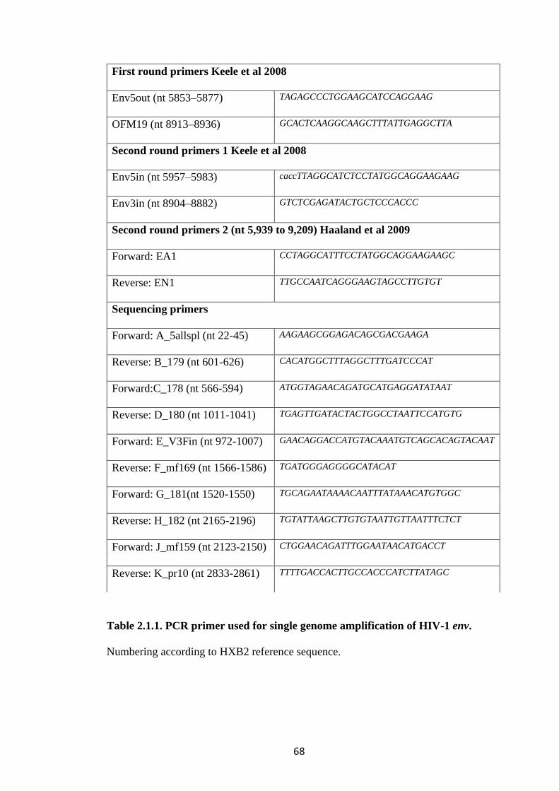
68
Table 2.1.1. PCR primer used for single genome amplification of HIV-1 env.
Numbering according to HXB2 reference sequence.
First round primers Keele et al 2008
Env5out (nt 5853–5877) TAGAGCCCTGGAAGCATCCAGGAAG
OFM19 (nt 8913–8936) GCACTCAAGGCAAGCTTTATTGAGGCTTA
Second round primers 1 Keele et al 2008
Env5in (nt 5957–5983) caccTTAGGCATCTCCTATGGCAGGAAGAAG
Env3in (nt 8904–8882) GTCTCGAGATACTGCTCCCACCC
Second round primers 2 (nt 5,939 to 9,209) Haaland et al 2009
Forward: EA1 CCTAGGCATTTCCTATGGCAGGAAGAAGC
Reverse: EN1 TTGCCAATCAGGGAAGTAGCCTTGTGT
Sequencing primers
Forward: A_5allspl (nt 22-45) AAGAAGCGGAGACAGCGACGAAGA
Reverse: B_179 (nt 601-626) CACATGGCTTTAGGCTTTGATCCCAT
Forward:C_178 (nt 566-594) ATGGTAGAACAGATGCATGAGGATATAAT
Reverse: D_180 (nt 1011-1041) TGAGTTGATACTACTGGCCTAATTCCATGTG
Forward: E_V3Fin (nt 972-1007) GAACAGGACCATGTACAAATGTCAGCACAGTACAAT
Reverse: F_mf169 (nt 1566-1586) TGATGGGAGGGGCATACAT
Forward: G_181(nt 1520-1550) TGCAGAATAAAACAATTTATAAACATGTGGC
Reverse: H_182 (nt 2165-2196) TGTATTAAGCTTGTGTAATTGTTAATTTCTCT
Forward: J_mf159 (nt 2123-2150) CTGGAACAGATTTGGAATAACATGACCT
Reverse: K_pr10 (nt 2833-2861) TTTTGACCACTTGCCACCCATCTTATAGC
69
2.1.4 Next generation sequencing (NGS)
Short-read deep sequencing by Illumina MiSeq (Illumina, San Diego, CA, USA) was
used to sequence the whole genome of viruses from CSF and plasma compartments
(Gall et al. 2012). NGS and the bioinformatics quality control was performed by the
diagnostic laboratory at University College London Hospital. As per the viral load
testing protocol, nucleic acids were extracted from 230 μL of plasma using the
Qiasymphony using the DSP Virus/Pathogen kit as per manufacturer’s protocols
(Qiagen, Hilden, Germany) and amplified using in-house HIV-1 primer (gag-pol
codons 691-3582, pol-int gag-pol codons 2696-5527, int-env [g120] gag-pol codons
5518-7374). Library preparations were generated using the Nextera XT DNA
Sample Preparation Kit (Bio Scientific, Texas, USA) and sequenced on the Illumina
MiSeq platform. Prior to assembly the reads were trimmed using Trimmomatic
(version 0.38). The following parameters were used; 10 bases were trimmed from
the leading and training ends, a sliding window of 4:30 was used and reads with less
than 50 bases were deleted. Adaptors were removed using ILLUMINACLIP 2:10:7
settings. The parameters used can be found here
https://github.com/usadellab/Trimmomatic. Kraken2 was used to align reads to a
database of genomes with viruses, bacteria and human. Everything that was non-
viral was removed (including human). Read assembly was done using the de novo
Iterative Virus Assembler (Gupta-Wright et al. 2020). Following assembly, samples
were aligned to a 99ZACM9, subtype C reference sample from South Africa
(GenBank: AF411967) (Papathanasopoulos et al. 2002) using the MAFFT program
(version 7). A consensus sequence was generated from the mapped reads.
70
2.1.5 Determination of compartmentalisation
The primary outcome is central nervous system (CNS) compartmentalisation, which
is defined as an independently replicating viral population in the CNS. This was
determined by comparing HIV-1 genomes obtained from paired CSF and plasma
samples using SGA. CNS compartmentalisation status was determined by three
methods.
1. Visual inspection: Maximum likelihood (ML) phylogenetic trees were
produced from the SGA sequences and limited to sequences with no gaps.
Env nucleic acid sequences were aligned in Geneious version 10. RaxmlGUI
was used to construct the ML tree using general time reversible model with a
gamma distribution (Edler et al. 2019). The phylogenetic reconstruction was
repeated 1000 times by resampling from the dataset to provide the degree of
confidence in the tree branches, i.e. 1000 bootstraps. This was presented as a
proportion, and a proportion greater than 70% was deemed to provide good
support for that branch. The ML trees were inspected for the presence of a
population structure consistent with an independently replicating population
in the CSF.
2. Distance based methods: Wright’s FST was used. It compares the mean
pairwise genetic distance between two sequences sampled from different
compartments to the mean distance between sequences sampled from the
same compartment. The closer the FST is to 1 the greater the likelihood of
compartmentalisation. FST <0.05 indicates very little genetic difference
71
between compartments. Values > 0.15 show a significant amount of genetic
difference and values >0.25 show a very high degree of genetic difference.
Corrections were made for unequal character frequencies, A>G, C>T and
transversional bias with the Tamura-Nei model, corrections were made for
distance and for rate variation using a gamma distribution. A run of 10,000
bootstrap replicates was done.
3. Tree-based methods: Slatkin-Maddison (SM) determines the minimum
number of migration events between the separated populations, consistent
with the structure of the reconstructed phylogenetic tree. The more migration
events needed to explain the distribution of sequences the less likely that
there is compartmentalisation. A p-value <0.05 is indicative of significance.
Simmons association index (AI) assesses the degree of population structure
in the phylogenetic tree recursively, using a bootstrap sample. A value of 1
implies that the population structures observed would be expected by chance
whilst a value of 0 implies a strict genetic structuring by
compartmentalisation. The tree-based methods were done using BaTS, which
uses a posterior distribution of phylogenies from Bayesian MCMC analysis
(Parker, Rambaut, and Pybus 2008). Given that compartmentalisation is an
intra-host phenomenon, tree-based compartmentalisation analyses require at
least 20 viral genomes in each compartment to consistently classify a
compartment at compartmentalised (Zarate et al. 2007).
The intra-host viral diversity was further explored using multidimension scaling
(MDS). This allowed mapping of the relative distances between sequences in a

72
visual plot. Pairwise distances between all assembled SGA sequences were
calculated using the dist.dna() package, with a TN93 nucleotide-nucleotide
substitution matrix (Tamura & Nei, 1993) and with pairwise deletion using the R
package Ape v.5.4 (Paradis et al., 2004). Multi-dimensional scaling (MDS) was
implemented using the cmdscale() function with pairwise deletion in R v4.0.4. This a
data reduction method akin to principle component analysis, which is intended to
simplify complex data into a more interpretable format, by reducing dimensionality
of data but retaining most of the variation. In a genomics context we can use this on
pairwise distance matrices, where each dimension is a sequence with data points of
n-1 sequences pairwise distance, converted to a vector which can be plotted on 2-
dimentional plot. This was done in collaboration with Dr Steven Kemp.
2.1.6 Drug resistance
Stanford HIV drug-resistance database was used to identify resistant mutations to
non-nucleotide reverse transcriptase inhibitor (NNRTI), nucleotide(side) reverse
transcriptase inhibitor (NRTI), protease inhibitor (PI) and integrase strand transfer
inhibitors (INSTI) drugs (Shafer 2006). Minority variants were called at the 20% and
5% levels.
73
2.1.7 Prediction of coreceptor usage
Co-receptor usage was also determined genotypically by analysing the V3 loop
sequences of each amplified virus with freely available computer predictive
algorithms (CPAs), Geno2pheno (G2P) and Web position-specific scoring matrix
(WebPSSM). G2P was trained on a set of 1100 subtype B sequences with
genotypic-phenotypic correlation and set at a false positive rate (FPR) of 2.5%
(http://coreceptor.bioinf.mpi-inf.mpg.de/). PSSM uses the net charge as well as the
amino acid at positions 11 and 25, if these are either an Arginine (R) or Lysine (K),
the virus is more likely to be CXCR4 using. There are two matrices available,
WebPSSM subtype C sinsi
(https://indra.mullins.microbiol.washington.edu/webpssm) was used, which bases
predictions on known syncytium-inducing phenotypes on the MT-2 cell line (Jensen
et al. 2006). These tools predict the major coreceptor usage of env V3 sequences but
are not optimized to discriminate between R5X4-tropic variants from X4-tropic
variants. It is therefore necessary to use a phenotypic approach such as described
above to differentiate between dual tropic R5X4/X4 and CXCR4 viruses.
2.1.8 Phenotypic co-receptor usage
2.1.8.1. Cells
HEK293T cells and HeLa TZM-bl reporter cells were grown in Dulbecco's modified
74
Eagle's medium (DMEM; Gibco) supplemented with 10% fetal calf serum (FCS;
Gibco) and 1% penicillin-streptomycin (10,000IU/ml, 10,000µg.ml) (Gibco). U87
cells expressing either CCR5 or CXCR4 co-receptors were grown in DMEM (Gibco)
supplemented with 10% FCS (Gibco), Puromycin 1ug/ml and Neomycin 100ug/ml.
2.1.8.2. Clones
HIV-1 Gp120 env from distinct viruses in the blood and CSF compartments obtained
from SGA were cloned into a B41 psvIII envelope expression vector (Figure 2.1.1).
An additional round of PCR was done on the SGA PCR products using the primers
912 AATTGTGGGTCACAGTCTATTATGGGGTAC and 930
ATATTTGTATAATTCACTTCTCCAATTGTCCCTCATATCT (6318 to 7690)
with complementarity to the Kpn I and Mfe I restriction sites respectively. The PCR
product was cloned into a B41 psvIII envelope expression vector. B41 psvIII was
restriction digested using restriction enzymes Kpn I and Mfe I (Neb biolabs). The
digested vector was run on a 1% agarose gel and gel purified using Qiaquickgel
extraction kit (Qiagen, Germany). The amplified gp120 env was cloned into the
vector using NEBuilder® HiFi DNA Assembly Master Mix (Figure 2.1.2).

75
Figure 2.1.1. B41 psvIII envelope expression vector map. Donated by Dr Laura
McCoy.

76
Figure 2.1.2. HIV-1 Envelope genome map. Nucleic acid numbering 1 to 2,579 base pairs. Indicating region in gp120 bound by primer 912
and 930 in brown that is cloned into the B41 psVIII envelope expression vector. SP- signal peptide; gp- glycopeptide; V-variable loop; FP-
fusion peptide; TM- transmembrane domain; 912 and 930- forward and reverse cloning primers with the Kpn I and Mfe I restriction sites
respectively.
77
2.1.8.3. Replication competent virus production
A double transfection system was used to generate pseudotyped virus using the
cloned env vector and the pNL4-3-Env Luc packaging vector, which contains a
luciferase reporter gene (Figure 2.1.3). This was done in the containment level 3
(CL3) laboratory. 1x10⁶ HEK 293T cells were plated in a 6 well plate in 2ml of
DMEM supplemented with 10% FCS, 1% 10,000 units penicillin and 1% 10 000
µg/mL Streptomycin. In a separate tube, 200ng of the participant derived Env
cloned into the envelope expression vector and 400ng of the packing vector were
added to 33µl of Optimen. 2µl of Fugene HD transfection reagent was added and left
to incubate for 20mins. The HEK293Ts were transfected with this plasmid mixture
and the produced pseudotyped viruses were harvested 48 hours later and stored at -
80oC. Pseudotyped viruses with the Env of dual tropic WEAU-d15.410.787, CXCR4
using HXB2 and CCR5 using JRSCF were created and used as controls. The bald
packaging vectors were used as negative controls.
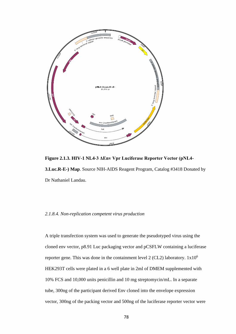
78
Figure 2.1.3. HIV-1 NL4-3 ΔEnv Vpr Luciferase Reporter Vector (pNL4-
3.Luc.R-E-) Map. Source NIH-AIDS Reagent Program, Catalog #3418 Donated by
Dr Nathaniel Landau.
2.1.8.4. Non-replication competent virus production
A triple transfection system was used to generate the pseudotyped virus using the
cloned env vector, p8.91 Luc packaging vector and pCSFLW containing a luciferase
reporter gene. This was done in the containment level 2 (CL2) laboratory. 1x10⁶
HEK293T cells were plated in a 6 well plate in 2ml of DMEM supplemented with
10% FCS and 10,000 units penicillin and 10 mg streptomycin/mL. In a separate
tube, 300ng of the participant derived Env cloned into the envelope expression
vector, 300ng of the packing vector and 500ng of the luciferase reporter vector were
79
added to 70µl of Optimen. 6µl of Fugene HD transfection reagent was added and left
to incubate for 20mins. The HEK293Ts were transfected with this plasmid mixture
and the produced pseudotyped viruses were harvested 48 hours later and stored at -
80oC. Pseudotyped viruses with the Env of dual tropic WEAU-d15.410.787, CXCR4
using HXB2 and CCR5 using JRSCF were created and used as controls. The bald
packaging vectors were used as negative controls.
2.1.8.5. Standardisation of virus input by tissue culture infectious dose (TCID)
The TCID was determined in a single round infectivity assay by titration of the
pseudotyped viruses on target cells- U87 cells expressing either CCR5 or CXCR4
co-receptor. The relative light units (RLU) of infection was determined using the
Steady-Glo Luciferase assay system (Promega). The dilution of virus that gave a
RLU of 500 000 RLU was used in further experiments.
2.1.8.6. Normalisation of viral input by SYBR Green-based product-enhanced PCR
assay (SG-PERT)
In addition, virus input was normalised for reverse transcriptase (RT) activity in the
virus supernatant as measured by qPCR. Ten-fold dilutions of virus supernatant were
lysed for 10 minutes at room temperature in a 1:1 ratio in a 2x lysis solution containing
100mM Tris-HCL buffered to pH7.4, 50mM KCl, 0.25% Triton X-100, 40% glycerol,
RNase inhibitor 0.8 U/ml. 12µl of each lysate was added to 13µl of a master mix
80
containing SYBR Green master mix (Qiagen), 0.5µM of MS2-RNA foward and
reverse primers, 3.5pmol/ml of MS2-RNA, and 0.125U/µl of Ribolock RNAse
inhibitor. Thermocycling was performed in a QuantStudio. RT activity was
determined from the amount of transcription of a fixed concentration of MS2 RNA
bacteriophage in each sample relative to a known standard of HIV-1 RT (Merck).
2.1.8.7. Co-receptor usage phenotypic assay
Co-receptor usage was determined by infecting 1x10⁴ U87 cells expressing either
CCR5 or CXCR4 co-receptors in 96 well plates with these pseudotyped viruses in
serial dilution(Gupta et al. 2019). Entry efficiency was measured 48 hours later by a
luciferase readout of RLUs of the lysed infected cells using Steady glo lysis buffer
and substrate. Pseudotyped viruses with the Env of dual tropic WEAU-d15.410.787,
X4 using HXB2 and R5 using JRSCF were created and used as controls and the bald
packaging vectors were used as negative controls.
CCR5 or CXCR4 coreceptor usage was confirmed by treating target cells in the co-
receptor assay with co-receptor antagonists AMD-3100 800nM to block CXCR4-
mediated entry and TAK-779 400nM to block CCR5-mediated entry prior to
infection. The drugs were a kind donation from Dr Clare Jolly, UCL.

81
2.1.9 Neutralisation
Compartmentalised cellular and immune responses in the CNS and plasma
compartments may exert differential pressure on viruses in either compartment,
leading to viral escape. Since epitopes on the HIV-1 envelope are the main target for
humoral immune response(Sok and Burton 2018), variation in Env can occur due to
selection pressure that render them better adapted to one compartment versus
another. The neutralisation activity of 4 HIV-1 broadly neutralising (bnAbs) were
tested against participant derived Env pseudotyped viruses. The 4 bnAbs were
PGT121 and PGT128 (targeting the high mannose patch), VRC01 (CD4 binding site
blocker) and PG9 (targeting the apex). Anti-dengue serotype 3 antibody was used as
the negative control. This assay was done in collaboration with Dr Laura McCoy,
UCL.
A single round virus neutralisation assays were performed on HeLa TZMBLs cells.
HeLa TZM-bl reporter cells were grown in Dulbecco's modified Eagle's medium
(DMEM; Gibco) supplemented with 10% fetal calf serum (FCS; Gibco) and
penicillin-streptomycin (50 μg/ml) (Gibco). 10 μg/ml DEAE-Dextran (Sigma) was
added to cell prior to infection. Participant derived Env pseudotyped viruses were
produced as previously described in a CL3 laboratory. A double transfection system
with packaging vector psG3-∆Env (Figure 2.1.4) and the participant derived Env
plasmids, pseudotyped viruses were created. Envelope pseudotyped virus do not
contain a luciferase gene but contain tat which will induce luciferase expression in
TZM-bl cells. These pseudotyped viruses were incubated with serial dilution of
bnAbs for 1h at 37˚C. Virus and cell only controls were also included. Then, freshly
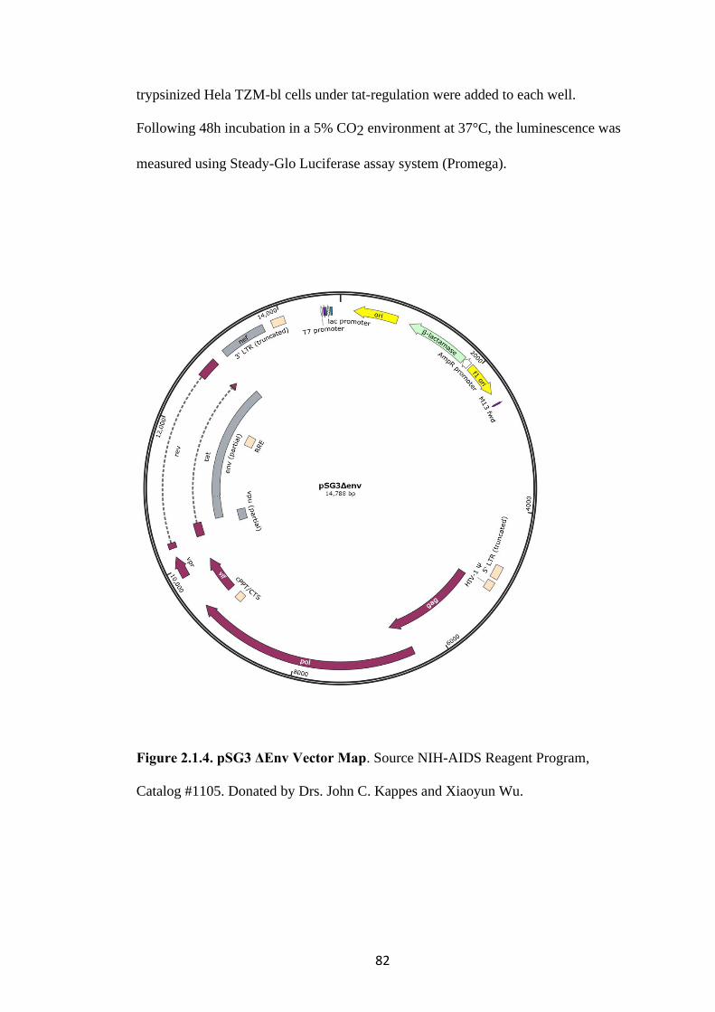
82
trypsinized Hela TZM-bl cells under tat-regulation were added to each well.
Following 48h incubation in a 5% CO2 environment at 37°C, the luminescence was
measured using Steady-Glo Luciferase assay system (Promega).
Figure 2.1.4. pSG3 ΔEnv Vector Map. Source NIH-AIDS Reagent Program,
Catalog #1105. Donated by Drs. John C. Kappes and Xiaoyun Wu.
83
2.1.10 Statistical Methods
Descriptive analyses of clinical and demographic data are presented as median and
interquartile range (IQR) when continuous and as frequency and proportion (%)
when categorical. The difference in continuous and categorical data were tested
using Wilcoxon rank sum and Chi-square test respectively. A sample size of 42 is
required to determine the prevalence of CNS compartmentalisation with a power of
80% and risk of type I error of 0.05.
Neutralisation activity was calculated relative to virus only controls. Dilution curves
were presented as a mean neutralisation with standard error of the mean (SEM). The
inhibitory concentration 50 (IC50)- the concentration of bnAbs at which 50%
infection was inhibited was calculated in Prism version 9. The IC50 within groups
were summarised as a geometric mean titre (GMT) and statistical comparison
between groups were made with Mann-Whitney for unpaired data or Wilxocon
ranked sign test for paired data. Statistical analyses were conducted using Stata
version 13 and Prism version 9.
2.1.11 Ethical approval
Ethical approval for the ACTA trial was obtained from the London School of
Hygiene and Tropical Medicine Research Ethics Committee- reference 8854 and the
University of Malawi College of Medicine Research Ethics Committee- reference
ISRCTN45035509 and all participants gave their written informed consent.

84
2.2 Section 2: SARS-CoV-2 Escape from Convalescent Plasma
This was an in-depth case-study of in vivo escape of SARS-CoV-2 from neutralising
antibodies in an immunocompromised participant with prolonged shedding of
SARS-CoV-2.
2.2.1 Clinical Sample Collection
Twenty-three samples from the upper airway- nose and throat swabs and lower
airway-endotracheal aspirates were collected at various time points over 101 days of
illness. Day 1 was the day on which the first SARS-CoV-2 PCR positive swab result
was obtained.
2.2.2 Viral load/Cycle threshold (CT)
Viral load quantification was done by the Public Health England Clinical
Microbiology. RNA extraction from 500µl of sample and was done on the easyMAG
platform (Biomerieux, Marcy-l'Étoile) as per manufacturer’s instructions. CT values
were determined in one-step RT q-PCR assay quantified using a Rotorgene™ PCR
instrument (Meredith et al. 2020). Samples with a CT values of ≤36 was considered
positive.
85
2.2.3 SARS-CoV-2 binding antibody levels
Serology was done by the Department of Clinical Biochemistry and Immunology,
Addenbrooke’s Hospital, Cambridge, UK by multiplex particle-based flow cytometry
(Luminex, Netherlands) (Xiong et al. 2020). Carboxylated beads were first activated
to generate amine-reactive forms according to manufacturer’s instructions (Themo
Fisher Scientific). They were then covalently coupled to recombinant SARS-CoV-2
Nucleocapsid (N), Spike (S) and receptor binding domain (RBD) proteins to form a
3-plex assay. The beads were washed and then blocked for non-specific binding with
a blocking buffer (containing 1% BSA, 10 mM PBS, and 0.05% NaN3). They were
then incubated with the participant’s sera at a dilution of 1 in 100 for 1 hour at room
temperature in a 96-well plate, washed 3 times with washing buffer (containing 10
mM PBS and 0.05% Tween 20) and then incubated for 30 minutes with a PE-labeled
anti–human IgG -Fc antibody (Leinco/Biotrend). The plates were read on a Luminex
analyzer (Luminex / R&D Systems) using Exponent Software V31. Specific binding
was reported as mean fluorescence intensity (MFI). Negative controls were unexposed
healthy controls with pre-pandemic biobanked sera and positive controls were PCR
positive COVID-19 cases.
2.2.4 Convalescent plasma for antibody titres
400ul sample of the 3 units of convalescent plasma (CP) administered to the
participant was obtained from the NHS Blood and Transplant Service. The titres of
anti- SARS-CoV-2 IgG was determined by NHS Blood and Transplant Service using

86
an indirect ELISA based assay- Euroimmun (Medizinische Labordiagnostika).
Diluted CP was incubated with recombinant Spike 1 (S1) protein of SARS-CoV-2.
The bound IgG antibody was detected by addition of an enzyme-labelled anti-human
IgG antibody which catalyses a colour reaction. The ratio of optical extinction of the
sample over that of a calibrator is determined. According to the manufacturer’s
instruction a ratio < 0.8 is negative, ratio ≥0.8 to <1.1 is borderline and ratio ≥1.1 is
positive (https://www.fda.gov/media/137609/download)
2.2.5 Whole blood T cell and innate stimulation assay
This was done by the Department of Clinical Biochemistry and Immunology,
Addenbrooke’s Hospital, Cambridge, UK. Whole blood cytokine levels of IFN𝛾,
TNF⍺, interleukin (IL) -1β, -2, -6, -10 and -17 were measured by multiplexed particle-
based flow cytometry (Bio-Plex, Bio-Rad, UK) using a custom kit (R&D Systems,
UK). T cell stimulation was done with phytohemagglutinin (PHA; 10 μg/ml; Sigma-
Aldrich), or co-stimulating with anti-CD3 (MEM57, Abcam, 200 ng/ml) and IL-2
(Immunotools, 1430U/ml) or Phorbol 12-myristate 13-acetate/ionomycin,
(PMA/IONO) and innate stimulation with lipopolysaccharide (LPS; 1 μg/ml, List
Biochemicals). Supernatants were taken 24 hours after stimulation. Cytokines levels
in pg/ml were measured by on a Luminex analyzer. The matched participant serum
from the same time point was a negative control, run in parallel to account for any
background cytokines released in the blood and a healthy donor samples were run in
parallel as a positive control.
87
2.2.6 Next generation sequencing (NGS)
2.2.6.1 Long-read NGS
RNA extraction was done on the easyMAG platform (Biomerieux, Marcy-l'Étoile)
as per manufacturer’s instructions. Long-read NGS was done using MinION flow
cells v.9.4.1 (Oxford Nanopore Technologies) by The COVID-19 Genomics UK
Consortium (COG-UK; https://www.cogconsortium.uk/). The ARTICnetwork V3
protocol was used (https://dx.doi.org/10.17504/protocols.io.bbmuik6w). The
bioinformatics was done using the ARTICnetwork curated reference-based assembly
pipeline with minimum coverage of 20 times across the whole genome
(https://artic.network/ncov-2019/ncov2019-bioinformatics-sop.html) (Loman, Rowe,
and Rambaut 2020).
Phylogenetic analyses were done in collaboration with Dr Steven Kemp. All available
full-genome SARS-CoV-2 sequences were downloaded from the GISAID database
(http://gisaid.org/)(Shu and McCauley 2017) on the 22nd of April 2021. Low-coverage
sequences were excluded. Sequences were re-aligned to the Wuhan-Hu-1 reference
sequence using MAFFT v7.475 using the --keeplength and --addfragments options.
Sequences were deduplicated and a random subset of 487 global sequences (from 56
countries) were obtained using seqtk (https://github.com/lh3/seqtk). To this, 239
randomly subsampled sequences were added from the UK, including England, Wales,
Scotland and Ireland. Finally, the reference sequence MN908947.3, 20 sequences
from the participant and 21 local sequences (to the hospital) were added to a multiple
sequence alignment for a total of 768 sequences. Major SARS-CoV-2 lineage was
88
determined using Phylogenetic Assignment Of Named Global Outbreak Lineages
(pangolin) (Rambaut et al. 2020).
A maximum-likelihood phylogeny was inferred using IQTREE v2.13 using the -fast
option. (Minh et al. 2020). Evolutionary model selection for trees were inferred using
ModelFinder (Kalyaanamoorthy et al. 2017) and trees were estimated using the
GTR+F+I model with 1000 bootstrap replicates (Minh, Nguyen, and von Haeseler
2013). Trees were viewed with Figtree v.1.4.4
(http://tree.bio.ed.ac.uk/software/figtree/), rooted on GenBank: MN908947.3 SARS-
CoV-2 reference sequence.
2.2.6.2 Short-read NGS
In addition, 20 of the 23 samples underwent short-read NGS using Illumina MiSeq
(Illumina, San Diego, CA, USA) and was done by the Department of Pathology,
University of Cambridge. Library preparations were generated using the KAPA Hyper
Prep kit (Roche) and sequenced on the Illumina MiSeq platform using a Miseq Nano
v2 with 2x 250 paired-end sequencing. Bioinformatics processing involved removal
of poor quality reads, removal of adaptors and trimming the reads with TrimGalore
v0.6.6 (Martin 2011). Trimmed paired-end reads were mapped to the GenBank:
MN908947.3 SARS-CoV-2 reference sequence using MiniMap2-2.17 (Li 2018).
BAM files were indexed with samtools v1.11 and PCR optical duplicates removed
using Picard (http://broadinstitute.github.io/picard). A consensus sequence was
generated with minimum whole genome coverage of at least 20 times. BCFtools using
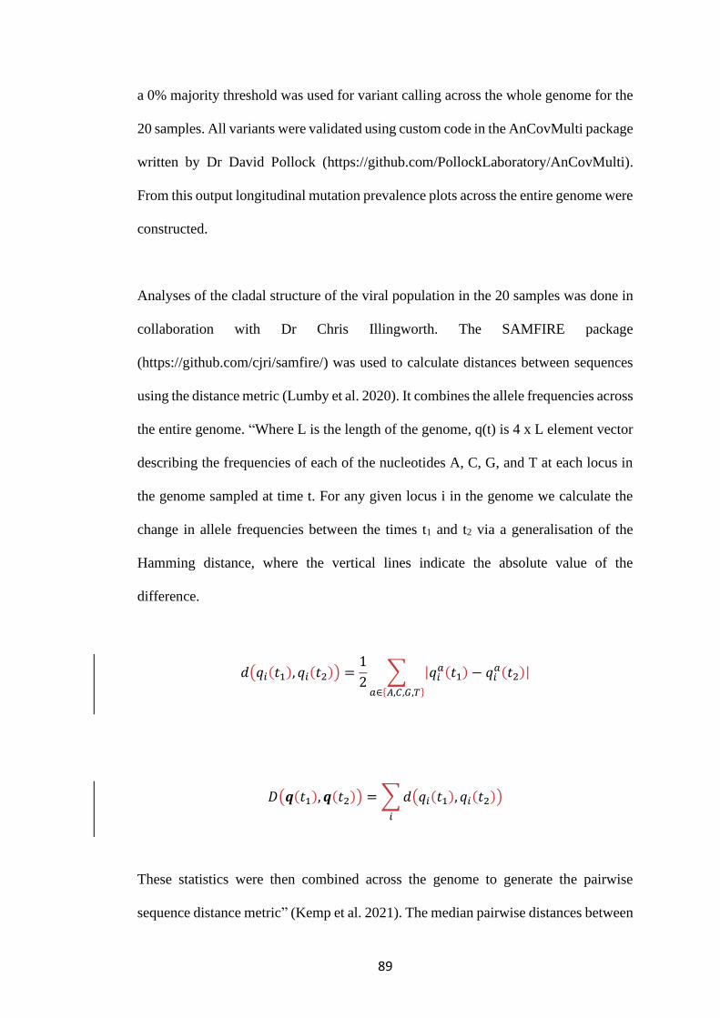
89
a 0% majority threshold was used for variant calling across the whole genome for the
20 samples. All variants were validated using custom code in the AnCovMulti package
written by Dr David Pollock (https://github.com/PollockLaboratory/AnCovMulti).
From this output longitudinal mutation prevalence plots across the entire genome were
constructed.
Analyses of the cladal structure of the viral population in the 20 samples was done in
collaboration with Dr Chris Illingworth. The SAMFIRE package
(https://github.com/cjri/samfire/) was used to calculate distances between sequences
using the distance metric (Lumby et al. 2020). It combines the allele frequencies across
the entire genome. “Where L is the length of the genome, q(t) is 4 x L element vector
describing the frequencies of each of the nucleotides A, C, G, and T at each locus in
the genome sampled at time t. For any given locus i in the genome we calculate the
change in allele frequencies between the times t1 and t2 via a generalisation of the
Hamming distance, where the vertical lines indicate the absolute value of the
difference.
𝑑(𝑞𝑖(𝑡1), 𝑞𝑖(𝑡2)) =1
2∑ |𝑞𝑖
𝑎(𝑡1) − 𝑞𝑖𝑎(𝑡2)|
𝑎∈{𝐴,𝐶,𝐺,𝑇}
𝐷(𝒒(𝑡1), 𝒒(𝑡2)) = ∑𝑑(𝑞𝑖(𝑡1), 𝑞𝑖(𝑡2))
𝑖
These statistics were then combined across the genome to generate the pairwise
sequence distance metric” (Kemp et al. 2021). The median pairwise distances between
90
samples in different clades of the phylogenetic tree were calculated and the statistically
tested with the Mann-Whitney test (Kemp et al. 2021).
Long-read sequencing data have been deposited in the NCBI SRA database-
BioProject PRJNA682013; accession codes SAMN16976824 - SAMN16976846
(https://www.ncbi.nlm.nih.gov/bioproject/PRJNA682013). Short reads sequence data
were deposited at https://github.com/Steven-Kemp/sequence_files.
2.2.7 Single Genome Amplification (SGA)
SGA of the spike gene was used as an independent method to detect mutations
observed in 3 samples at different time points. Viral RNA extracts were obtained as
previously described Methods 2.2.2) and were reverse transcribed using SuperScript
IV (Thermofisher Scientific). and an “in-house” spike specific primer- CoVOuter-R.
Template RNA was degraded with RNAse H (Thermofisher Scientific). Terminally
diluted cDNA was used to amplify partial Spike (Spike amino acids 21- 800~ 1.8 kb)
by nested PCR using Platinum Taq DNA Polymerase High Fidelity (Invitrogen,
Carlsbad, CA). ‘In-house’ designed primers were used (Table 2.2.1) based on the
participant’s consensus NGS sequences. All products derived from cDNA dilutions
yielding less than 30% PCR positivity were Sanger sequenced with 8 in-house
sequencing primers covering spike by Genewiz UK (Table 2.2.1). This meant that
there was at least an 80% chance that each well contained a single genome.
Sequences were manually curated in DNA Dynamo software (Blue Tractor Software
Ltd, UK).

91
Table 2.2.1. PCR primer used for single genome amplification if SARS-CoV-2
spike.
First round primers
CoVOuter-F AATGAAAATGGAACCATTACAGATGC
CoVOuter-R ACCCGCTAACAGTGCAGAAG
Second round primers
CoVInner-F AAGTGTACGTTGAAATCCTTCACTG
CoVInner-R AATGAGGTCTCTAGCAGCAA
Sequencing primers
Seq_F1 TCTTGTTTTATTGCCACTAGTCTCT
Seq_R1 TGTTAGACTTCTCAGTGGAAGCA
Seq _F2 ACCACAAAAACAACAAAAGTTGGA
Seq_R2 CCCACATAATAAGCTGCAGCAC
Seq_F3 GTGTACGTTGAAATCCTTCACTGT
Seq_R3 TCCAAGCTATAACGCAGCCT
Seq_F4 CTATCAGGCCGGTAGCACAC
Seq_R4 ACACTGACACCACCAAAAGAAC
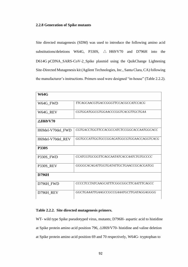
92
2.2.8 Generation of Spike mutants
Site directed mutagenesis (SDM) was used to introduce the following amino acid
substitutions/deletions W64G, P330S, △ H69/V70 and D796H into the
D614G pCDNA_SARS-CoV-2_Spike plasmid using the QuikChange Lightening
Site-Directed Mutagenesis kit (Agilent Technologies, Inc., Santa Clara, CA) following
the manufacturer’s instructions. Primers used were designed “in-house” (Table 2.2.2).
Table 2.2.2. Site directed mutagenesis primers.
WT- wild type Spike pseudotyped virus, mutants; D796H- aspartic acid to histidine
at Spike protein amino acid position 796, △H69/V70- histidine and valine deletion
at Spike protein amino acid position 69 and 70 respectively, W64G- tryptophan to
W64G
W64G_FWD TTCAGCAACGTGACCGGGTTCCACGCCATCCACG
W64G_REV CGTGGATGGCGTGGAACCCGGTCACGTTGCTGAA
△H69/V70
H69del-V70del_FWD CGTGACCTGGTTCCACGCCATCTCCGGCACCAATGGCACC
H69del-V70del_REV GGTGCCATTGGTGCCGGAGATGGCGTGGAACCAGGTCACG
P330S
P330S_FWD CCATCGTGCGGTTCAGCAATATCACCAATCTGTGCCCC
P330S_REV GGGGCACAGATTGGTGATATTGCTGAACCGCACGATGG
D796H
D796H_FWD CCCCTCCTATCAAGCATTTCGGCGGCTTCAATTTCAGCC
D796H_REV GGCTGAAATTGAAGCCGCCGAAATGCTTGATAGGAGGGG

93
glycine at Spike protein amino acid position 64, P330S- proline to serine at Spike
protein amino acid position 330.
2.2.9 Spike pseudotyped virus preparation
Wild type (WT) Spike and mutant pseudotyped virus was generated using a
lentiviral triple transfection system. WT Spike was codon optimised, based on the
Wuhan-1 sequence but bearing the aspartic acid to glycine change at amino acid
position 614 of the Spike protein (D614G). The mutant Spikes pseudotyped viruses
were D796H- aspartic acid to histidine at Spike protein amino acid position 796,
△H69/V70- histidine and valine deletions at Spike protein amino acid position 69
and 70 respectively, W64G- tryptophan to glycine at Spike protein amino acid
position 64, P330S- proline to serine at Spike protein amino acid position 330. There
were engineered singly or in pairs consisting of D796H+△H69/V70 or W64G+
P330S. Sequences were verified by Sanger sequencing.
1x10⁶ HEK 293T cells were seeded in a 10cm dish 24 hours prior. The cells were
transfected with a triple plasmids system consisting of 1µg of the WT or mutant
D614G pCDNA_SARS-CoV-2_Spike plasmid, 1ug of p8.91 lentiviral packaging
plasmid and 1.5µg pCSFLW expressing the firefly luciferase reporter gene using
11ul of Fugene HD transfection reagent (Promega). Supernatant was collected at 48
hours after transfection, filtered through 0.45um filter and cryopreserved at -80˚C.
The tissue culture infectious dose (TCID) of SARS-CoV-2 pseudotyped virus was
determined by titrating the viruses on HEK293T cells which had been transiently
94
transfected with ACE2 and TMPRSS2. Infection of the target cells were readout as
relative light units (RLU) on a luminometer using Steady-Glo Luciferase assay
system (Promega).
2.2.10 Normalisation of viral input by SYBR Green-based product-enhanced
PCR assay (SG-PERT)
Virus input was normalised for reverse transcriptase (RT) activity in the virus
supernatant as measured by qPCR using. Ten-fold dilutions of virus supernatant were
lysed for 10 minutes at room temperature in a 1:1 ratio in a 2x lysis solution containing
100mM Tris-HCL buffered to pH7.4, 50mM KCl, 0.25% Triton X-100, 40%
glycerol, RNase inhibitor 0.8 U/ml. 12µl of each lysate was added to 13µl of a master
mix containing SYBR Green master mix (Qiagen), 0.5µM of MS2-RNA foward and
reverse primers, 3.5pmol/ml of MS2-RNA, and 0.125U/µl of Ribolock RNAse
inhibitor. Thermocycling was performed in a QuantStudio. RT activity was
determined from the amount of transcription of a fixed concentration of MS2 RNA
bacteriophage in each sample relative to a known standard of HIV-1 RT (Merck).
2.2.11 Spike expression by Western blot
Forty-eight hours after transfection of HEK 293T cells with the triple plasmid
preparation, the culture supernatant was harvested and filtered through a 0.45-μm-
pore-size filter. The supernatant was centrifuged at 15,000 rpm for 120 min at 4˚C to
95
pellet virions. This was then lysed with Laemmli reducing buffer (100% glycerol, β-
mercaptoethanol, 1 M Tris-HCl [pH 6.8] SDS and bromophenol blue). The lysates
were electrophoresed on SDS– 4 to 12% bis-Tris protein gels (Thermo Fisher
Scientific) under reducing conditions. The protein bands were electroblotting onto
polyvinylidene difluoride (PVDF) membranes, which were probed with anti-Spike
(Invitrogen) and anti-p24 Gag (NIH AIDS reagents) antibodies. ChemiDoc MP
imaging system (Biorad) was used for visualisation.
2.2.12 Serum/plasma pseudotype neutralization assay
Spike pseudotype assays have been shown to have similar characteristics as
neutralisation testing using fully infectious wild type SARS-CoV-2 (Schmidt et al.
2020). Serum neutralisation activity of CP and participant’s sera at time points
preceding and following administration CP was measured in a single-round in vitro
neutralisation assay. WT, △H69/V70+ D796H, W64G+ P330 and single mutant Spike
pseudotyped virus were incubated with serial dilution of heat inactivated CP or
participant’s serum samples in duplicate for 1h at 37˚C in a 96-well plate. 5x10⁴
HEK293T cells, transiently transfected with ACE2 and TMPRSS2 were added to each
well. After 48 hours incubation in a 5% CO2 at 37°C, RLU was measured using
Steady-Glo Luciferase assay system (Promega) (Mlcochova et al. 2020). Serum
neutralisation curves were generated from which inhibitory dilution 50 (ID50) were
determined. ID50 is the dilution of serum required to inhibit 50% of infection of a
SARS-CoV-2 Spike pseudotyped virus in the assay. Human AB serum stored prior to

96
2019 was the negative control. Positive controls were sera from COVD-19 PCR
positive cases.
2.2.13 Structural Viewing
The location of the four spike mutations of interest was mapped onto an uncleaved
SARS-CoV-2 spike structure solved by cryo-electron-microscopy by Wrobel et al
(PDB: 6ZGE) (Wrobel et al. 2020). The Pymol Molecular Graphics System v2.4.0
(https://github.com/schrodinger/pymol-open-source/releases) was used.
2.2.14 Ethics
Ethics approval was granted by the East of England – Cambridge Central Research
Ethics Committee (17/EE/0025). Written informed consent was obtained. Additional
controls with COVID-19 were enrolled to the NIHR BioResource Centre Cambridge
under ethics review board (17/EE/0025).
97
2.3 Section 3: Age-related heterogeneity of SARS-CoV-2 mRNA vaccine-elicited
responses
2.3.1 Study Design
This was a cohort study of participants receiving the mRNA BNT162b2 vaccine.
Participants included members of the public receiving their vaccine at Addenbrookes
Hospital, Cambridge and health care workers receiving the first dose of the vaccine.
Participants had their first vaccine between the 14th of December 2020 to the 29th of
January 2021. Initially the second dose was given 3 weeks after the first but during the
course of the study the dosing interval was increased to 12 weeks. Participants were
consecutively recruited without exclusion. Participants were seen at least 3 weeks after
receiving their first does and then followed up until 3 weeks after their second dose.
Blood samples were collected at each visit. The exposure of interest was age,
categorised into 2 exposure levels- < 80 and ≥ 80 years. The primary outcome was
inadequate vaccine-elicited serum antibody neutralisation activity at least 3 weeks after
the first dose. This was measured as the dilution of serum required to inhibit infection
by 50% (ID50) in an in vitro neutralisation assay. An ID50 of 20 or below was deemed
as inadequate neutralisation. Secondary outcomes included Spike-specific binding
antibody responses and IFNγ and IL-2 T cell responses.
98
2.3.2 Sample size calculation
The sample size was determined by assuming a risk ratio of non-neutralisation in the
≥80 years group compared with <80 years group of 5. An alpha of 0.05 and power of
90% was used. This estimated a sample size of 50 with a 1:1 ratio was required in each
group.
2.3.3 Clinical samples
Blood samples collected included 10ml of serum sample, which was spun at 1000g for
30 minutes, aliquoted and cryopreserved at -80oC and 20ml of whole blood from which
peripheral blood mononuclear cells (PBMCs) were prepared using Lymphoprep
density gradient centrifugation and cryopreserved at -80oC.
2.3.4 Generation of pseudotyped viruses
Wild type (WT) Spike lentiviral pseudotyped virus was generated in a triple
transfection system. The WT was a codon optimised Spike, based on the Wuhan-1
sequence but bearing the aspartic acid to glycine change at amino acid position 614 of
the Spike protein (D614G).
1x10⁶ HEK293T cells were seeded in a 10cm dish 24 hours prior transfection. The
cells were transfected with a triple plasmids system consisting of 1µg of the Spike
99
expression plasmid- pCDNA_SARS-CoV-2_Spike plasmid, 1ug of p8.91 lentiviral
packaging plasmid and 1.5µg pCSFLW expressing the firefly luciferase reporter
gene. 11ul of Fugene HD transfection reagent (Promega) was used. Supernatant was
collected at 48 hours after transfection, filtered through 0.45um filter and
cryopreserved at -80˚C. The tissue culture infectious dose (TCID) of SARS-CoV-2
pseudotyped virus was determined by titrating the viruses on HEK 293T cells which
had been transiently transfected with ACE2 and TMPRSS2. Infection of the target
cells were readout as relative light units (RLU) on a luminometer using Steady-Glo
Luciferase assay system (Promega).
2.3.5 Neutralisation assays
Serum neutralisation activity of vaccine-elicited sera was measured in a single-round
in vitro neutralisation assay. WT Spike pseudotyped virus were incubated with serial
dilution of heat inactivated participant’s serum following the first and second dose of
mRNA BNT162b2 vaccine in duplicate for 1h at 37˚C in a 96- well plate. 5x10⁴ HEK
293T cells, transiently transfected with ACE2 and TMPRSS2 were added to each well.
After 48hours incubation in a 5% CO2 at 37°C, RLU was measured using Steady-Glo
Luciferase assay system (Promega) (Mlcochova et al. 2020). Serum neutralisation
curves were generated from which inhibitory dilution 50 (ID50) were determined.
ID50 is the dilution of serum required to inhibit 50% of infection of a SARS-CoV-2
Spike pseudotyped virus in the assay. Human AB serum stored prior to 2019 was the
negative control. Positive controls were sera from COVD-19 PCR positive cases. The
cut off for 50% neutralisation was set at an ID50 of 1in 20. The ID50 within groups
100
were summarised as a geometric mean titre (GMT) and statistical comparison between
groups were made with Mann-Whitney or Wilxocon ranked sign test for unpaired and
paired analyses respectively.
2.3.6 SARS-CoV-2 binding antibody levels
Serology was done by the Department of Clinical Biochemistry and Immunology,
Addenbrooke’s Hospital, Cambridge, UK by multiplex particle-based flow cytometry
(Luminex, Netherlands) (Xiong et al. 2020). Carboxylated beads were first activated
to generate amine-reactive forms according to manufacturer’s instructions (Themo
Fisher Scientific). They were then covalently coupled to recombinant SARS-CoV-2
Nucleocapsid (N), Spike (S) and receptor binding domain (RBD) proteins to form a
3-plex assay. The beads were washed and then blocked for non-specific binding with
a blocking buffer (containing 1% BSA, 10 mM PBS, and 0.05% NaN3). They were
then incubated with the participant’s sera at a dilution of 1 in 100 for 1 hour at room
temperature in a 96-well plate, washed 3 times with washing buffer (containing 10
mM PBS and 0.05% Tween 20) and then incubated with incubated for 30 min with a
PE-labeled anti–human IgG/IgA/IgM-Fc antibody (Leinco/Biotrend). The plates were
read on a Luminex analyzer (Luminex / R&D Systems) using Exponent Software V31.
Specific binding was reported as mean fluorescence intensity (MFI). Negative controls
were unexposed healthy controls with pre-pandemic biobanked sera and positive
controls were PCR positive COVID-19 cases.
101
2.3.7 CMV serology
Human cytomegalovirus (HCMV) IgG levels were determined using an IgG
enzyme-linked immunosorbent (EIA) assay, HCMV Captia (Trinity Biotech, Didcot,
UK) following manufacturer’s instructions, on plasma samples. This was done in
collaboration with Dr Mark Wills.
2.3.8 T cell assays- IFNγ and IL2 FLUOROSPOT
Frozen PBMCs were thawed and diluted into 10ml of TexMACS media (Miltenyi
Biotech), centrifuged and resuspended in 10ml of fresh media with 10U/ml DNase
(Benzonase, Merck-Millipore via Sigma-Aldrich). PBMCs were then incubated at
37°C for 1h, followed by centrifugation and resuspension in fresh media
supplemented with 5% Human AB serum (Sigma Aldrich) before being counted.
PBMCs were stained with 2ul of LIVE/DEAD Fixable Far Red Dead Cell Stain Kit
(Thermo Fisher Scientific) and live PBMC enumerated on the BD Accuri C6 flow
cytometer.
1.0 to 2.5 x 10⁵ PBMCs were incubated in pre-coated FluoroSpotFLEX plates (anti
IFNγ and IL2 capture antibodies Mabtech AB, Nacka Strand, Sweden)) in duplicate
with either peptide mixes specific for Wuhan-1(QHD43416.1) Spike SARS-CoV-2
protein (Miltenyi Biotech) or a mixture of peptides specific for Cytomegalovirus,
Epstein Barr virus and Influenza virus (CEF+) (final peptide concentration
1µg/ml/peptide, Miltenyi Biotech) in addition to an unstimulated (media only) and
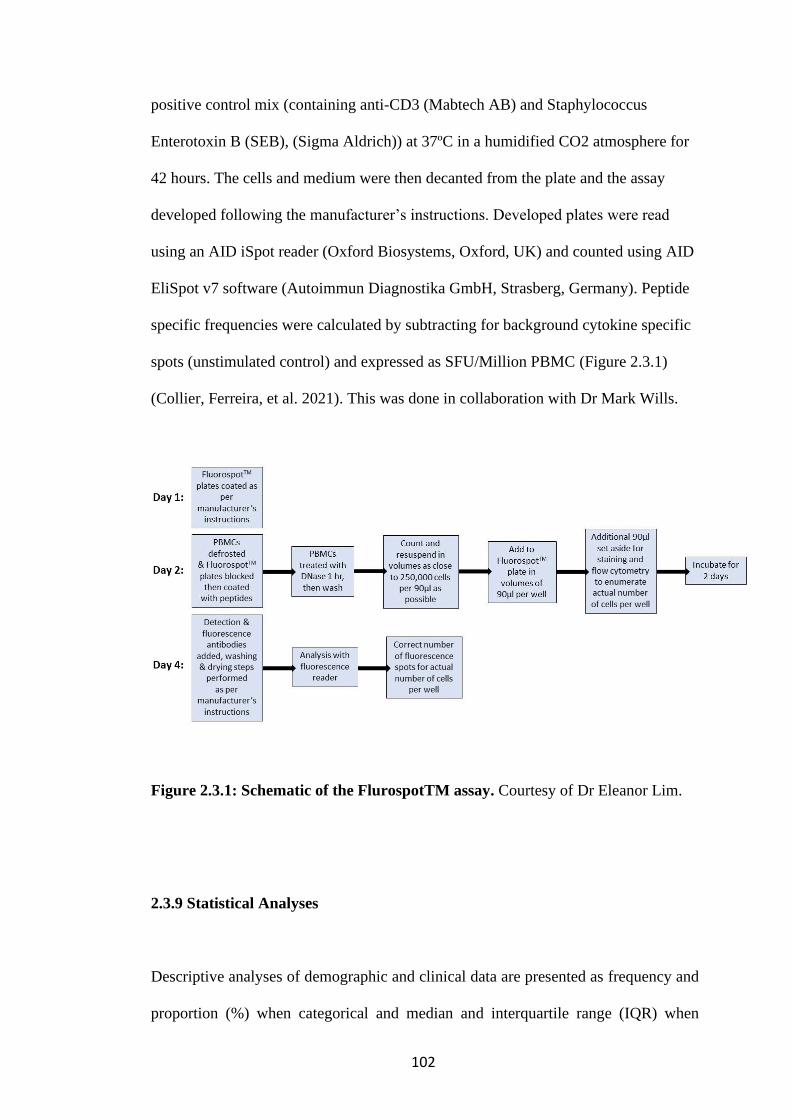
102
positive control mix (containing anti-CD3 (Mabtech AB) and Staphylococcus
Enterotoxin B (SEB), (Sigma Aldrich)) at 37ºC in a humidified CO2 atmosphere for
42 hours. The cells and medium were then decanted from the plate and the assay
developed following the manufacturer’s instructions. Developed plates were read
using an AID iSpot reader (Oxford Biosystems, Oxford, UK) and counted using AID
EliSpot v7 software (Autoimmun Diagnostika GmbH, Strasberg, Germany). Peptide
specific frequencies were calculated by subtracting for background cytokine specific
spots (unstimulated control) and expressed as SFU/Million PBMC (Figure 2.3.1)
(Collier, Ferreira, et al. 2021). This was done in collaboration with Dr Mark Wills.
Figure 2.3.1: Schematic of the FlurospotTM assay. Courtesy of Dr Eleanor Lim.
2.3.9 Statistical Analyses
Descriptive analyses of demographic and clinical data are presented as frequency and
proportion (%) when categorical and median and interquartile range (IQR) when
103
continuous. Test for significance included Wilcoxon rank sum for continuous variables
and Chi-square test for categorical variables. Logistic regression was used to test the
association between age and inadequate neutralisation by vaccine-elicited antibodies
after the first dose of the BNT162b2 vaccine. The association was adjusted for the
following confounders- sex and time interval from vaccination to sampling. Linear
regression was also used to test the association between age and log transformed ID50,
binding antibody levels, antibody subclass levels and T cell response after dose 1 and
dose 2 of the BNT162b2 vaccine. Bonferroni adjustment was made for multiple
comparisons. The Pearson correlation coefficient was reported. Statistical analyses
were done using Stata v13, Prism v9 and R (version 3.5.1).
2.3.10 Ethical approval
Ethics approval was granted by the East of England – Cambridge Central Research
Ethics Committee (17/EE/0025). PBMC from unexposed volunteers previously
recruited by the NIHR BioResource Centre Cambridge through the ARIA study
(2014-2016), with ethical approval from the Cambridge Human Biology Research
Ethics Committee (HBREC.2014.07) and currently North of Scotland Research
Ethics Committee 1 (NS/17/0110).

104
CHAPTER 3: RESULTS 1- HIV-1 VIRAL ESCAPE AND
CNS COMPARTMENTALISATION
Key Findings:
• CSF escape is uncommon in this study population.
• Visual inspection and statistical analyses of phylogenetic trees revealed the
prevalence of CNS compartmentalisation is 50%.
• No clinical or demographic factors have been identified which are associated
with CNS compartmentalisation.
• Env diversity is compartmentalised in participants with CNS
compartmentalisation.
• The founder virus of CNS compartmentalised viruses maybe a plasma
variant.
• HIV-1 CNS compartmentalised clones characterized here utilize CCR5 co-
receptors for viral entry.
• Computer predictive algorithms do not always accurately predict co-receptor
usage.
• The frequency of drug resistance mutations is similar in both compartments.
• There is heterogeneity in sensitivity of CNS and plasma derived clones to
broadly neutralizing antibodies.

105
The CNS is an immune privileged site bounded by the blood brain barrier which
allows selective entry of molecules and immune cells through from the peripheral
blood into the CNS (Rahimy et al. 2017). It is a recognised reservoir for HIV-1,
where proviral DNA may lay dormant in long-lived cells like macrophages and
neurones and be reactivated at a later date (Sigal and Baltimore 2012; Collier et al.
2018). Clonal expansion of HIV-1 or low-level replication in the CNS may lead to
CNS compartmentalisation, which is the independent replication of a population of
virus in an anatomical or cellular compartment, in this case the CNS (Adewumi et al.
2020; Bingham et al. 2011; Lorenzo-Redondo, Fryer, Bedford, Kim, Archer, Pond,
et al. 2016; Stam et al. 2013). These compartmentalised viruses are distinct from the
population in the peripheral blood. This process may also drive CSF escape (Collier
et al. 2018), which for the purposes of this work is defined as a CSF viral load (VL)
greater than 0.5log10 of the plasma VL.
Viruses in the CNS will be limited by the constraints in that niche and are known to
have acquired some features that make them adapted to the CNS such as the switch
of cellular tropism from R5 T cell to R5-Macrophage tropism (Schnell et al. 2011;
Sturdevant et al. 2015), co-receptor usage with higher affinity for CCR5 co-receptor
(Gorry et al. 2002) and acquisition of some drug resistant mutations (Mukerji et al.
2017). The extent to which CNS compartmentalised viruses evolve as a result of
differential selection pressures from both cellular and humoral immune responses is
unknown. In addition, the extent to which compartmentalised immune responses
between CNS and blood in individuals with CNS co-infections drives within-host
evolution in the CNS is not fully known (Adewumi et al. 2020). It is also unclear to
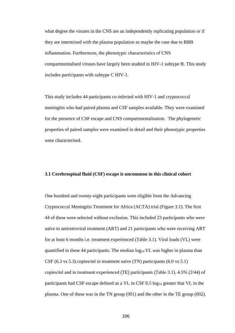
106
what degree the viruses in the CNS are an independently replicating population or if
they are intermixed with the plasma population as maybe the case due to BBB
inflammation. Furthermore, the phenotypic characteristics of CNS
compartmentalised viruses have largely been studied in HIV-1 subtype B. This study
includes participants with subtype C HIV-1.
This study includes 44 participants co-infected with HIV-1 and cryptococcal
meningitis who had paired plasma and CSF samples available. They were examined
for the presence of CSF escape and CNS compartmentalisation. The phylogenetic
properties of paired samples were examined in detail and their phenotypic properties
were characterised.
3.1 Cerebrospinal fluid (CSF) escape is uncommon in this clinical cohort
One hundred and twenty-eight participants were eligible from the Advancing
Cryptococcal Meningitis Treatment for Africa (ACTA) trial (Figure 3.1). The first
44 of these were selected without exclusion. This included 23 participants who were
naïve to antiretroviral treatment (ART) and 21 participants who were receiving ART
for at least 6 months i.e. treatment experienced (Table 3.1). Viral loads (VL) were
quantified in these 44 participants. The median log10 VL was higher in plasma than
CSF (6.3 vs 5.3) copies/ml in treatment naïve (TN) participants (6.0 vs 5.1)
copies/ml and in treatment experienced (TE) participants (Table 3.1). 4.5% (2/44) of
participants had CSF escape defined as a VL in CSF 0.5 log10 greater that VL in the
plasma. One of these was in the TN group (001) and the other in the TE group (002).
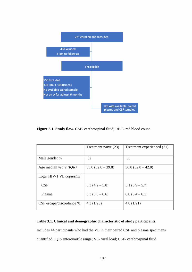
107
Figure 3.1. Study flow. CSF- cerebrospinal fluid; RBC- red blood count.
Treatment naïve (23) Treatment experienced (21)
Male gender % 62 53
Age median years (IQR) 35.0 (32.0 – 39.8) 36.0 (32.0 – 42.0)
Log10 HIV-1 VL copies/ml
CSF
Plasma
5.3 (4.2 – 5.8)
6.3 (5.8 – 6.6)
5.1 (3.9 – 5.7)
6.0 (5.4 – 6.1)
CSF escape/discordance % 4.3 (1/23) 4.8 (1/21)
Table 3.1. Clinical and demographic characteristic of study participants.
Includes 44 participants who had the VL in their paired CSF and plasma specimens
quantified. IQR- interquartile range; VL- viral load; CSF- cerebrospinal fluid.

108
Thirty-one participants successfully underwent next generation sequencing (NGS) of
viruses in both CSF and plasma compartments by Illumina Miseq (Methods 2.1.4)
and 4 of these had single genome amplification (SGA) (Methods 2.1.3) performed
on paired CSF and plasma samples. Participants with successful NGS had a
significantly higher median log10 VL in CSF and plasma compared to those without
successful NGS; 5.4 (IQR 5.0-6.3) vs 3.0 (IQR 0-3.9) copies/ml (p <0.0001) in the
CSF and 6.0 (5.7-6.4) vs 5.6 (0.0-6.2) copies/ml (p 0.04) in the plasma. Otherwise,
these two groups had similar characteristics.
The 31 participants with paired genomic data form the basis of further analyses. The
demographic and clinical data for the participants who had successful NGS done are
summarised in Table 3.2. The median age was 35 years (IQR 32-40), 64.5% were
male, 45.2% were on ARTs and 2 participants had CSF escape. The median mapped
read depth for 31 paired specimens using the HXB2 nucleic acid positions 691-7374
was 1037 (IQR 169-3292) in the plasma and 262 (IQR 23-1879) in the CSF
specimens. The lower read depth in CSF is not surprising as it is recognised that
PCR amplification from CSF samples is less successful due to the presence of
protein and cellular inhibitors, which can result in false negative CSF-PCR
(Ratnamohan, Cunningham, and Rawlinson 1998). The read depth was variable
along the length of the genome. It was at least 1000 reads in the regions spanning the
gag- p17 and p24, pol- RT and env- V1-V3 genes. (Figures 3.2A, B).
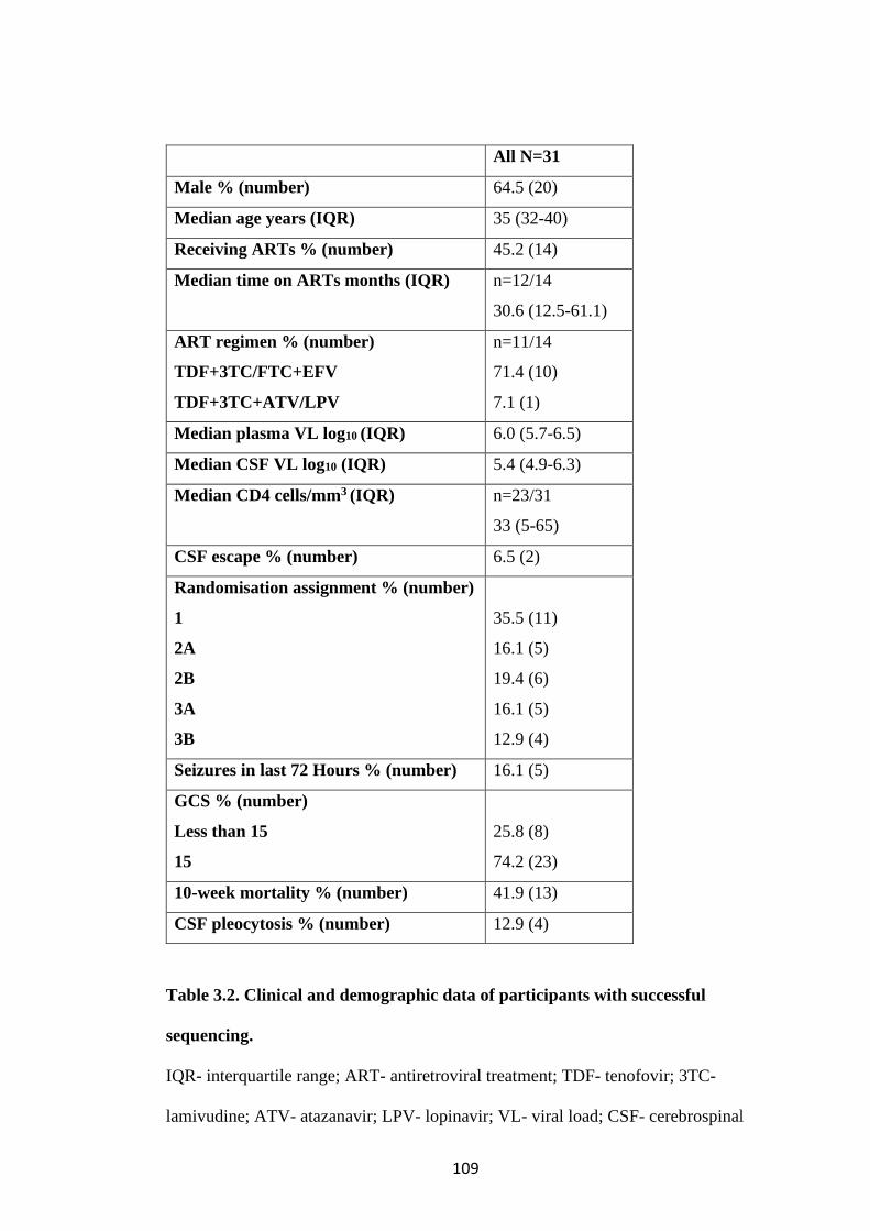
109
All N=31
Male % (number) 64.5 (20)
Median age years (IQR) 35 (32-40)
Receiving ARTs % (number) 45.2 (14)
Median time on ARTs months (IQR)
n=12/14
30.6 (12.5-61.1)
ART regimen % (number)
TDF+3TC/FTC+EFV
TDF+3TC+ATV/LPV
n=11/14
71.4 (10)
7.1 (1)
Median plasma VL log10 (IQR) 6.0 (5.7-6.5)
Median CSF VL log10 (IQR) 5.4 (4.9-6.3)
Median CD4 cells/mm3 (IQR)
n=23/31
33 (5-65)
CSF escape % (number) 6.5 (2)
Randomisation assignment % (number)
1
2A
2B
3A
3B
35.5 (11)
16.1 (5)
19.4 (6)
16.1 (5)
12.9 (4)
Seizures in last 72 Hours % (number) 16.1 (5)
GCS % (number)
Less than 15
15
25.8 (8)
74.2 (23)
10-week mortality % (number) 41.9 (13)
CSF pleocytosis % (number) 12.9 (4)
Table 3.2. Clinical and demographic data of participants with successful
sequencing.
IQR- interquartile range; ART- antiretroviral treatment; TDF- tenofovir; 3TC-
lamivudine; ATV- atazanavir; LPV- lopinavir; VL- viral load; CSF- cerebrospinal

110
fluid; GCS- Glasgow coma scale. Randomisation assignment in the ACTA study
drug regimen.
A
B
Figures 3.2. Read depth of plasma (A) and cerebrospinal fluid (B) in Illumina
MiSeq next-generation sequencing. Bp- base pair, HXB2- reference HIV-1
sequence. Median read depth (green line), Interquartile range (red lines).
In addition, 4 participants had SGA of the env gene performed on paired CSF and
plasma samples. Env sequences included amino acids 27 to 706 spanning the start of
gp120 to the transmembrane domain in gp41 (Figure 3.3). These participants had
between 21 and 35 viral genomes sequenced in each compartment. Table 3.3 shows
the clinical and demographic details of these 4 participants. They range between the
ages of 27 and 60 years. Two were male and 2 were on ART with a range of duration
of ART of 10.6 to 65.7 months. They universally had high HIV-1 VL in both plasma
and CSF compartments and low CD4 T cell counts. Two participants had CSF
escape; 001 and 002 and 2 participants did not; 003 and 004.
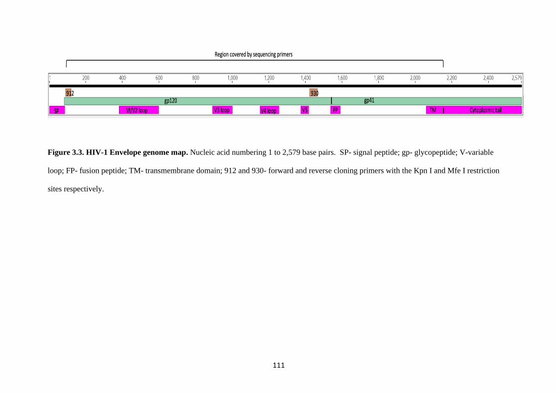
111
Figure 3.3. HIV-1 Envelope genome map. Nucleic acid numbering 1 to 2,579 base pairs. SP- signal peptide; gp- glycopeptide; V-variable
loop; FP- fusion peptide; TM- transmembrane domain; 912 and 930- forward and reverse cloning primers with the Kpn I and Mfe I restriction
sites respectively.
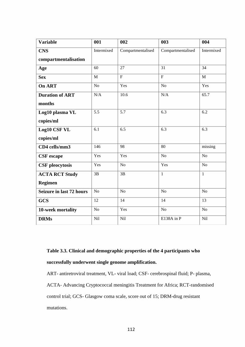
112
Table 3.3. Clinical and demographic properties of the 4 participants who
successfully underwent single genome amplification.
ART- antiretroviral treatment, VL- viral load; CSF- cerebrospinal fluid; P- plasma,
ACTA- Advancing Cryptococcal meningitis Treatment for Africa; RCT-randomised
control trial; GCS- Glasgow coma scale, score out of 15; DRM-drug resistant
mutations.
Variable 001 002 003 004
CNS
compartmentalisation
Intermixed Compartmentalised Compartmentalised Intermixed
Age 60 27 31 34
Sex M F F M
On ART No Yes No Yes
Duration of ART
months
N/A 10.6 N/A 65.7
Log10 plasma VL
copies/ml
5.5 5.7 6.3 6.2
Log10 CSF VL
copies/ml
6.1 6.5 6.3 6.3
CD4 cells/mm3 146 98 80 missing
CSF escape Yes Yes No No
CSF pleocytosis Yes No Yes No
ACTA RCT Study
Regimen
3B 3B 1 1
Seizure in last 72 hours No No No No
GCS 12 14 14 13
10-week mortality No Yes No No
DRMs Nil Nil E138A in P Nil

113
3.2 A viral population structure consistent with CNS compartmentalisation was
shown in some participants
Maximum likelihood phylogenetic trees were constructed from env nucleic acid
sequences and were limited to sequences with no gaps. General time reversible
model with a gamma distribution and 1000 bootstraps were used in RaxML
(Methods 2.1.5). Visual inspection of maximum likelihood phylogenetic trees
revealed a spatial population structure that clearly segregated the taxa of the CSF
derived genomes from those of the plasma in participants 002 and 003 (Figures
3.4A, B). However, genomes from the CSF and plasma from participants 001 and
004 showed no spatial population structure, therefore these plasma and CSF derived
viruses are intermixed (Figures 3.4C, D). The phylogenetic trees shed some light on
the sequence of evolutionary events that generated the present-day diversity of
genomes. In the participant 003 with CNS compartmentalisation, the most recent
common ancestor (MRCA) of a cluster of CSF genomes appeared to be a plasma
variant (Figure 3.4B).
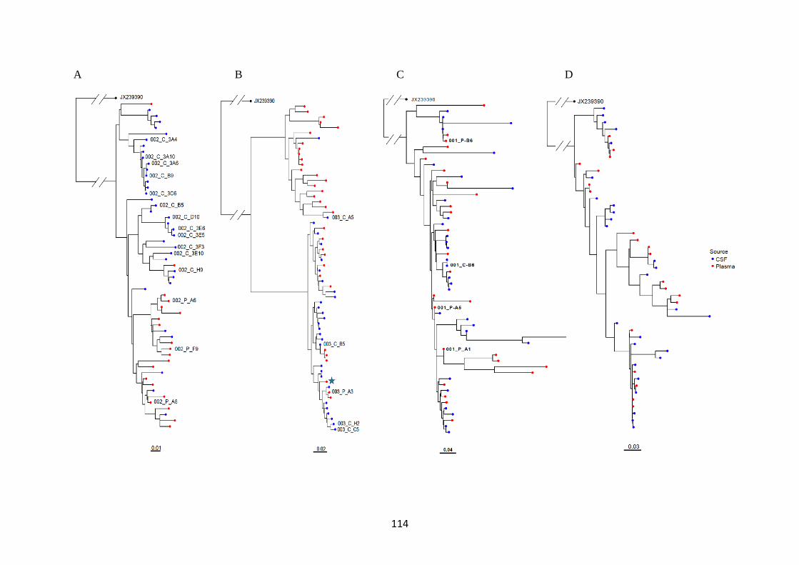
114
A B C D
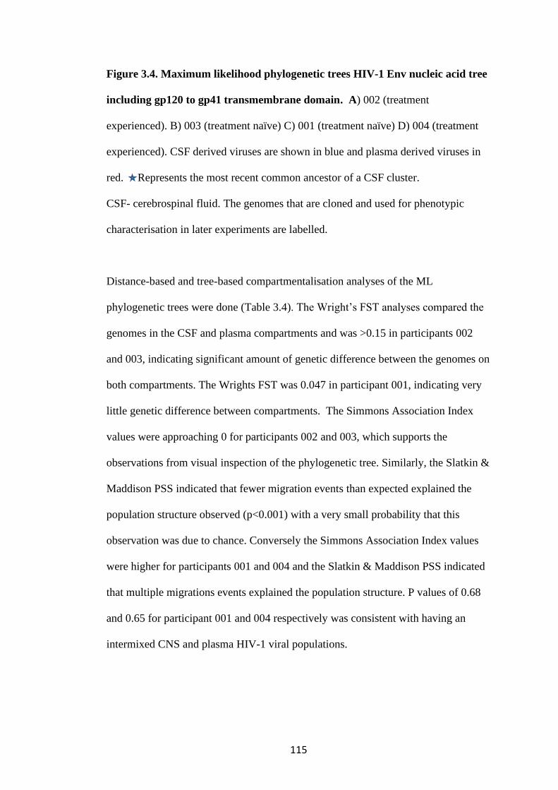
115
Figure 3.4. Maximum likelihood phylogenetic trees HIV-1 Env nucleic acid tree
including gp120 to gp41 transmembrane domain. A) 002 (treatment
experienced). B) 003 (treatment naïve) C) 001 (treatment naïve) D) 004 (treatment
experienced). CSF derived viruses are shown in blue and plasma derived viruses in
red. Represents the most recent common ancestor of a CSF cluster.
CSF- cerebrospinal fluid. The genomes that are cloned and used for phenotypic
characterisation in later experiments are labelled.
Distance-based and tree-based compartmentalisation analyses of the ML
phylogenetic trees were done (Table 3.4). The Wright’s FST analyses compared the
genomes in the CSF and plasma compartments and was >0.15 in participants 002
and 003, indicating significant amount of genetic difference between the genomes on
both compartments. The Wrights FST was 0.047 in participant 001, indicating very
little genetic difference between compartments. The Simmons Association Index
values were approaching 0 for participants 002 and 003, which supports the
observations from visual inspection of the phylogenetic tree. Similarly, the Slatkin &
Maddison PSS indicated that fewer migration events than expected explained the
population structure observed (p<0.001) with a very small probability that this
observation was due to chance. Conversely the Simmons Association Index values
were higher for participants 001 and 004 and the Slatkin & Maddison PSS indicated
that multiple migrations events explained the population structure. P values of 0.68
and 0.65 for participant 001 and 004 respectively was consistent with having an
intermixed CNS and plasma HIV-1 viral populations.
116
Visual inspection Wright’s
FST
Simmons
Association Index
Slatkin & Maddison PSS
Migration
events
P value
001 Intermixed 0.047 0.339 9.9 (9.0-11.0) 0.68
002 Compartmentalised 0.158 0.079 8.5 (8.0-9.0) <0.001
003 Compartmentalised 0.250 <0.001 3.9 (3.0- 4.0) <0.001
004 Intermixed Not done 0.34 15.4 (14.0- 16.0) 0.65
Table 3.4. Compartmentalisation analyses of CNS and plasma derived genomes
of 4 participants.
3.3 There is increased diversity in compartmentalised participants
Env diversity was explored by calculating the average pairwise distance (APD)
between env V2 to V4 in Mega X (Kumar et al. 2018). APD is the number of base
substitutions per site from averaging over all sequence pairs within each group.
Table 3.5 shows the results of analyses conducted using the Jukes-Cantor model and
replicated 100 times to provide 100 bootstrap support. The rate variation among sites
was modelled with a gamma distribution (shape parameter = 1).
APD amongst the participants with CNS compartmentalisation showed differences
between the CSF and plasma derived viruses. In participant 002 there was more

117
diversity in the CSF viruses compared with the plasma viruses (5.1% vs 3.5%). In
participant 003 there was more diversity in the plasma viruses compared with the
CSF viruses (9.1% vs 3.7%). In participants with intermixing of CSF and plasma
viruses; 001 and 004, the APD in both compartments were very similar, suggesting
less genetic diversity between CSF and plasma derived viruses in participants with
intermixed viral populations.
Visual inspection APD SE
001, treatment naive
CSF
Plasma
Intermixed
8.3
8.6
0.007
0.007
002, treatment experienced
CSF
Plasma
Compartmentalised
5.1
3.5
0.006
0.005
003, treatment naïve
CSF
Plasma
Compartmentalised
3.7
9.1
0.003
0.006
004, treatment experienced
CSF
Plasma
Intermixed
5.2
5.5
0.007
0.006
Table 3.5: Env Variable loops 2-4 diversity in CSF and plasma compartments.
Presented as APD (%) within groups. CSF- cerebrospinal fluid; APD- average
pairwise distance; SE- standard error
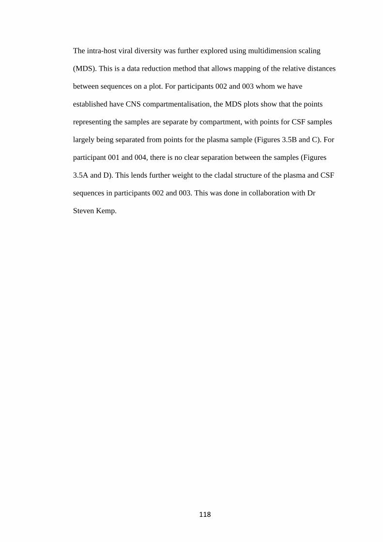
118
The intra-host viral diversity was further explored using multidimension scaling
(MDS). This is a data reduction method that allows mapping of the relative distances
between sequences on a plot. For participants 002 and 003 whom we have
established have CNS compartmentalisation, the MDS plots show that the points
representing the samples are separate by compartment, with points for CSF samples
largely being separated from points for the plasma sample (Figures 3.5B and C). For
participant 001 and 004, there is no clear separation between the samples (Figures
3.5A and D). This lends further weight to the cladal structure of the plasma and CSF
sequences in participants 002 and 003. This was done in collaboration with Dr
Steven Kemp.
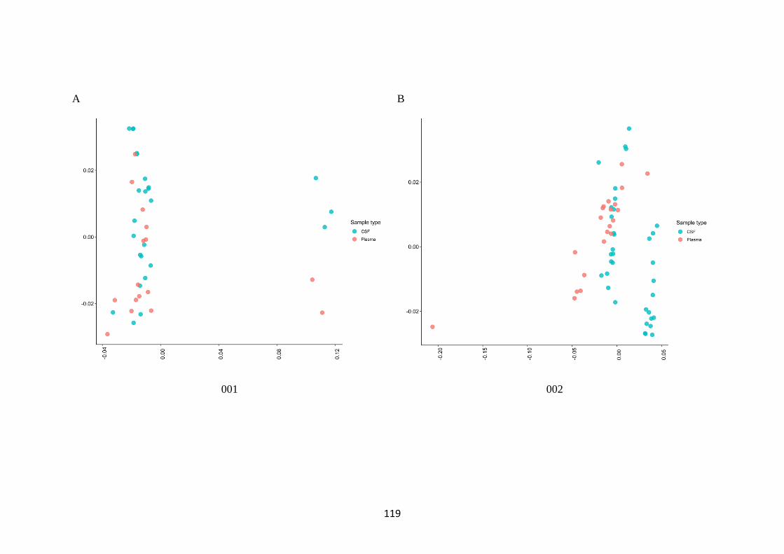
119
A B
001 002
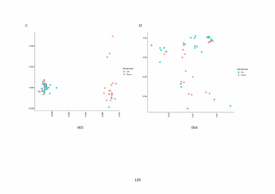
120
C D
003 004
121
Figure 3.5. Multidimensional scaling plots of the average pairwise distances
between sequences. A. Participant 001(treatments naive) B. Participant 002
(treatment experienced) C. Participant 003 (treatment naïve) D. Participant 004
(treatment experienced). Sequences are from both CSF (blue) and plasms (red)
compartments for each participant and are assembled single genome amplification
Env nucleotide sequences from gp120 to the TM region of gp41. TM-
transmembrane domain, gp- glycopeptide. Done in collaboration with Dr Steven
kemp.
3.4 No clinical or demographic factors were identified which are associated with
CNS compartmentalisation
The clinical and demographic characteristics of participants with CNS
compartmentalisation was compared to those without CNS compartmentalisation
(Table 3.3). The age range in the CNS compartmentalised group was 27 to 31years
whilst the range for the intermixed group was 34 to 60 years. The two participants in
the CNS compartmentalised group were female whilst the two participants in the
intermixed group were male. One participant in either group was on ART. The HIV-
1 VL in CSF and plasma were similar in both groups. The range of plasma VL was
5.7 to 6.3 log10 copies/ml in the CNS compartmentalised group vs 5.5 to 6.2 log10
copies/ml in the intermixed group and the range of CSF VL was 6.3 to 6.5 log10
copies/ml in the CNS compartmentalised group vs 6.1 to 6.3 log10 copies/ml in the
intermixed group. Of the two patients with CSF escape, one patient (002) had
compartmentalised CNS virus and the other (001) had intermixed virus. The same
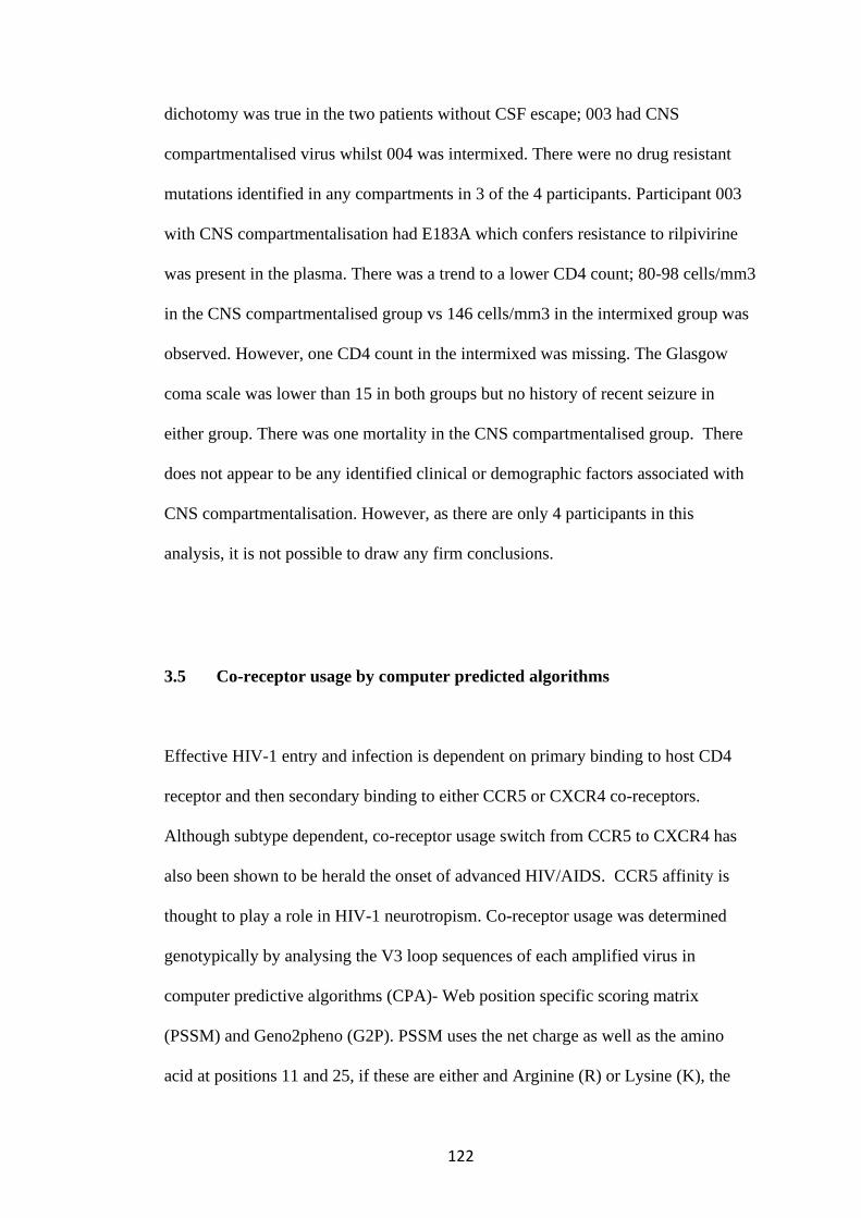
122
dichotomy was true in the two patients without CSF escape; 003 had CNS
compartmentalised virus whilst 004 was intermixed. There were no drug resistant
mutations identified in any compartments in 3 of the 4 participants. Participant 003
with CNS compartmentalisation had E183A which confers resistance to rilpivirine
was present in the plasma. There was a trend to a lower CD4 count; 80-98 cells/mm3
in the CNS compartmentalised group vs 146 cells/mm3 in the intermixed group was
observed. However, one CD4 count in the intermixed was missing. The Glasgow
coma scale was lower than 15 in both groups but no history of recent seizure in
either group. There was one mortality in the CNS compartmentalised group. There
does not appear to be any identified clinical or demographic factors associated with
CNS compartmentalisation. However, as there are only 4 participants in this
analysis, it is not possible to draw any firm conclusions.
3.5 Co-receptor usage by computer predicted algorithms
Effective HIV-1 entry and infection is dependent on primary binding to host CD4
receptor and then secondary binding to either CCR5 or CXCR4 co-receptors.
Although subtype dependent, co-receptor usage switch from CCR5 to CXCR4 has
also been shown to be herald the onset of advanced HIV/AIDS. CCR5 affinity is
thought to play a role in HIV-1 neurotropism. Co-receptor usage was determined
genotypically by analysing the V3 loop sequences of each amplified virus in
computer predictive algorithms (CPA)- Web position specific scoring matrix
(PSSM) and Geno2pheno (G2P). PSSM uses the net charge as well as the amino
acid at positions 11 and 25, if these are either and Arginine (R) or Lysine (K), the
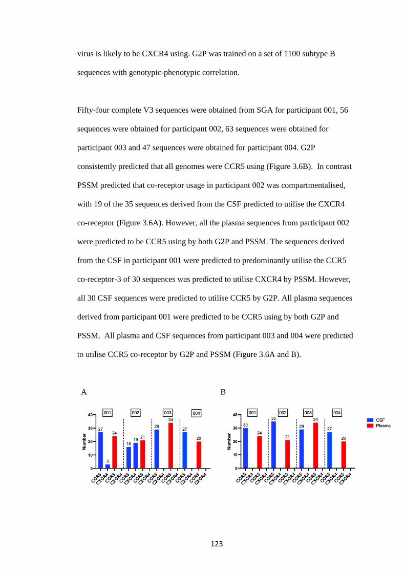
123
virus is likely to be CXCR4 using. G2P was trained on a set of 1100 subtype B
sequences with genotypic-phenotypic correlation.
Fifty-four complete V3 sequences were obtained from SGA for participant 001, 56
sequences were obtained for participant 002, 63 sequences were obtained for
participant 003 and 47 sequences were obtained for participant 004. G2P
consistently predicted that all genomes were CCR5 using (Figure 3.6B). In contrast
PSSM predicted that co-receptor usage in participant 002 was compartmentalised,
with 19 of the 35 sequences derived from the CSF predicted to utilise the CXCR4
co-receptor (Figure 3.6A). However, all the plasma sequences from participant 002
were predicted to be CCR5 using by both G2P and PSSM. The sequences derived
from the CSF in participant 001 were predicted to predominantly utilise the CCR5
co-receptor-3 of 30 sequences was predicted to utilise CXCR4 by PSSM. However,
all 30 CSF sequences were predicted to utilise CCR5 by G2P. All plasma sequences
derived from participant 001 were predicted to be CCR5 using by both G2P and
PSSM. All plasma and CSF sequences from participant 003 and 004 were predicted
to utilise CCR5 co-receptor by G2P and PSSM (Figure 3.6A and B).
A B
124
Figure 3.6: Virus co-receptor usage predicted by computer predicted
algorithms.
A) By position-specific scoring matrix (PSSM) and B) Genotopheno (G2P) and
using V3 loop sequences of CSF and plasma derived sequences. CSF derived
sequences are in blue and plasma derived sequences are in red. CSF- cerebrospinal
fluid.
3.6 Infectivity of pseudotyped viruses
Pseudotyped viruses were generated by dual transfection of the participant derived
env clone in B41 psvIII env expression plasmid with pNL4-3-Env Luc packaging
vectors (Methods 2.1.8.3). The infectivity of the pseudotyped viruses generated from
clones from participant 002 was determine by two methods. Firstly, the pseudotyped
viruses were titrated on the target cells; U87 bearing CCR5 coreceptors to determine
the TCID and expressed as RLU/µl of virus input (Methods 2.1.8.5) (Figure 3.7A
and 3.8A). In order to demonstrate equivalence between the TCID and SG-PERT
determined infectivity, one single round infection experiment for each participant
derived Env pseudotyped virus done using TCID standardised input was back
corrected for SG-PERT determined RT-activity. This showed equivalence between
both approaches (Methods 2.1.8.6) (Figure 3.7A, B). The TCID normalised input at
the dilution of virus that gave a RLU of 500 000 RLU was used in further
experiments.
125
3.7 Co-receptor usage in a phenotypic assay
The predicted co-receptor usage by CSF and plasma clones was tested in a
phenotypic co-receptor usage experiment. A subset of clones from both CSF and
plasma compartments from participants 001, 002 and 003 were tested as indicated on
the phylogenetic trees (Figure 3.5A, B, C). Clones from participant 001 (Figures
3.8A, B); P_A1, P_C5 and P_B6 from plasma and C_B6 from CSF utilised CCR5
co-receptor. They were able to infect U87 CCR5+ cells at least as well as the
positive controls WEAU and JRCSF (Figure 3.8A). However, they were not able to
infect U87 CXCR4 cells compared to the positive control WEAU (Figure 3.8B).
This corroborated the predicted co-receptor usage by the CPAs.
Clones from participant 002 (3.9A, B ); P_A6, P_A8 and P_F9 from plasma and
C_B5, C_B9, C_H9, C_D10, C_3F3, C_3A4, C_3A6, C_3E10, C_3A10, C_3E5,
C_3C6 and C_3E6 from CSF utilised CCR5 co-receptor and were able to infect U87
CCR5+ cells as well as the positive controls WEAU and JRCSF but in contrast to
the negative control HXB2 (Figure 3.9A).However these clones could not infect U87
CXCR4+ cells when compared to the WEAU positive control (Figure 3.9B). Of
interest this was inconsistent with the co-receptor usage prediction by PSSM which
predicted that majority of CSF clones tested were CXCR4 co-receptor utilising.
Clones from participant 003 (figures 3.10A B); P_A3 from plasma and C_A5, C_C5,
C_B5 and C_H2 from CSF utilised CCR5 co-receptor and were able to infect U87
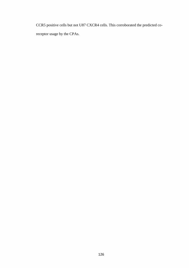
126
CCR5 positive cells but not U87 CXCR4 cells. This corroborated the predicted co-
receptor usage by the CPAs.
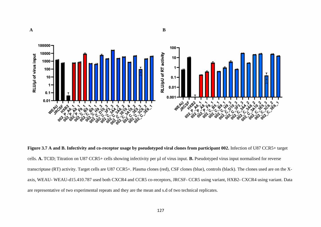
127
A B
Figure 3.7 A and B. Infectivity and co-receptor usage by pseudotyped viral clones from participant 002. Infection of U87 CCR5+ target
cells. A. TCID; Titration on U87 CCR5+ cells showing infectivity per µl of virus input. B. Pseudotyped virus input normalised for reverse
transcriptase (RT) activity. Target cells are U87 CCR5+. Plasma clones (red), CSF clones (blue), controls (black). The clones used are on the X-
axis, WEAU- WEAU-d15.410.787 used both CXCR4 and CCR5 co-receptors, JRCSF- CCR5 using variant, HXB2- CXCR4 using variant. Data
are representative of two experimental repeats and they are the mean and s.d of two technical replicates.
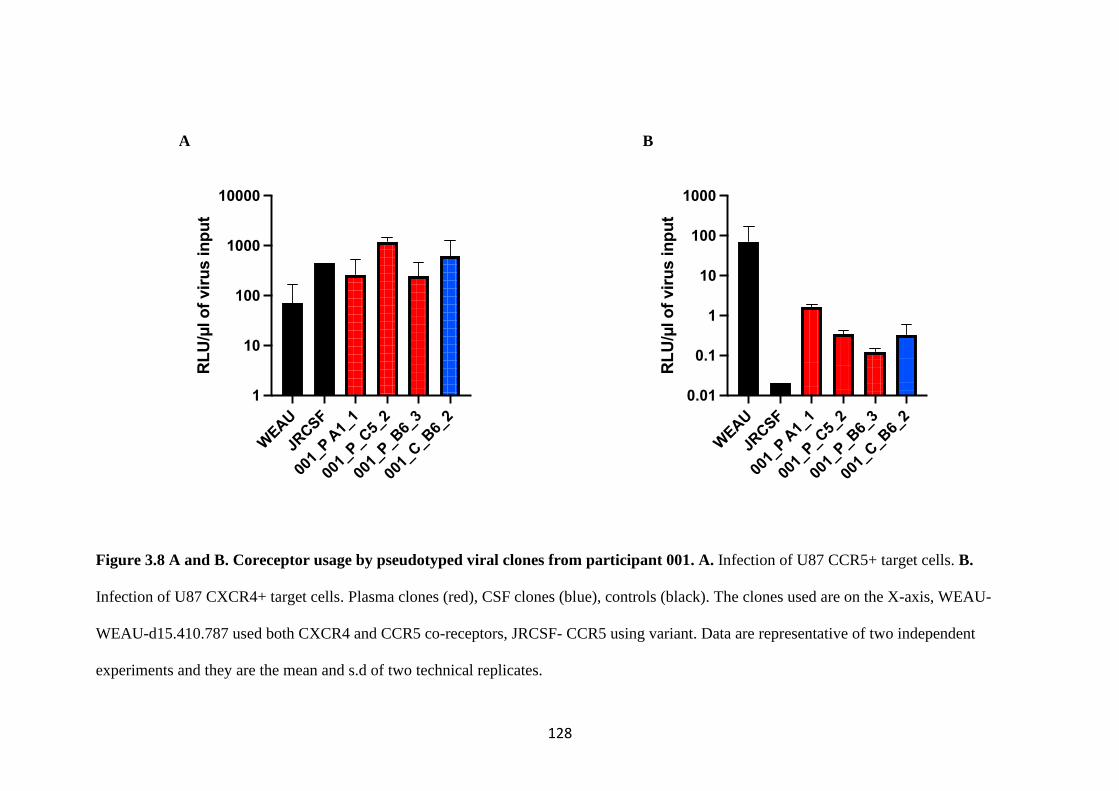
128
A B
Figure 3.8 A and B. Coreceptor usage by pseudotyped viral clones from participant 001. A. Infection of U87 CCR5+ target cells. B.
Infection of U87 CXCR4+ target cells. Plasma clones (red), CSF clones (blue), controls (black). The clones used are on the X-axis, WEAU-
WEAU-d15.410.787 used both CXCR4 and CCR5 co-receptors, JRCSF- CCR5 using variant. Data are representative of two independent
experiments and they are the mean and s.d of two technical replicates.
WEA
U
JRCSF
001_
P A1_
1
001_
P_C
5_2
001_
P_B
6_3
001_
C_B
6_2
1
10
100
1000
10000R
LU
/µl o
f vir
us in
pu
t
WEAU
JRCSF
001_
P A1_
1
001_
P_C
5_2
001_
P_B
6_3
001_
C_B
6_2
0.01
0.1
1
10
100
1000
RL
U/µ
l o
f vir
us in
pu
t
129
A B
Figure 3.9 A and B. Coreceptor usage by pseudotyped viral clones from participant 002. A. Infection of U87 CCR5+ target cells. B.
Infection of U87 CXCR4+ target cells. Plasma clones (red), CSF clones (blue), controls (black). The clones used are on the X-axis, WEAU-
WEAU-d15.410.787 used both CXCR4 and CCR5 co-receptors, JRCSF- CCR5 using variant, HXB2- CXCR4 using variant. Data are
representative of two independent experiments and they are the mean and s.d of two technical replicates.
WEAU
JRCSF
HXB
2
002_
P_A
6_1
002_
P_A
8_1
002_
P_F9_
1
002_
C_B
5_1
002_
C_B
9_1
002_
C_H
9_3
002_
C_D
10_3
002_
C_3
F3_3
002_
C_3
A4_
2
002_
C_3
A6_
2
002_
C_3
E10_4
002_
C_3
A10
_2
002_
C_3
E5_
3
002_
C_3
C6_
2
002_
C_3
E6_
1
0.01
0.1
1
10
100
1000
10000
100000R
LU
/µl o
f vir
us in
pu
t
WEAU
JRCSF
002_
P_A
6_1
002_
P_A
8_1
002_
P_F
9_1
002_
C_B
5_1
002_
C_B
9_1
002_
P_F
9_1
002_
C_H
9_3
002_
C_D
10_3
002_
C_3
F3_3
002_
C_3
A4_
2
002_
C_3
A6_
2
002_
C_3
E10
_4
002_
C_3
A10
_2
002_
C_3
E5_
3
002_
C_3
C6_
2
002_
C_3
E6_
1
0.01
0.1
1
10
100
1000
RL
U/µ
l o
f vir
us in
pu
t

130
A B
Figure 3.10 A and B. Coreceptor usage by pseudotyped viral clones from participant 003. A. Infection of U87 CCR5+ target cells B.
Infection of U87 CXCR4+ target cells. Infection of U87 CCR5+ target cells. Plasma clones (red), CSF clones (blue), controls (black). The
clones used are on the X-axis, WEAU- WEAU-d15.410.787 used both CXCR4 and CCR5 co-receptors, JRCSF- CCR5 using variant, HXB2-
CXCR4 using variant. Data are representative of two independent experiment and they are the mean and s.d of two technical replicates.
WEAU
JRCSF
HXB2
003_
P_A
3_1
003_
C_A
5_1
003_
C_C
5_1
003_
C_B
5_2
003_
C_H
2_1
0.01
0.1
1
10
100
1000
10000
100000R
LU
/µl o
f vir
us in
pu
t
WEAU
JRCSF
003_
P_A
3_1
003_
C_A
5_1
003_
C_C
5_1
003_
C_B
5_2
003_
C_H
2_1
0.1
1
10
100
RL
U/µ
l o
f vir
us in
pu
t
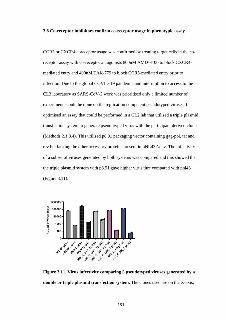
131
3.8 Co-receptor inhibitors confirm co-receptor usage in phenotypic assay
CCR5 or CXCR4 coreceptor usage was confirmed by treating target cells in the co-
receptor assay with co-receptor antagonists 800nM AMD-3100 to block CXCR4-
mediated entry and 400nM TAK-779 to block CCR5-mediated entry prior to
infection. Due to the global COVID-19 pandemic and interruption to access to the
CL3 laboratory as SARS-CoV-2 work was prioritised only a limited number of
experiments could be done on the replication competent pseudotyped viruses. I
optimised an assay that could be performed in a CL2 lab that utilised a triple plasmid
transfection system to generate pseudotyped virus with the participant derived clones
(Methods 2.1.8.4). This utilised p8.91 packaging vector containing gag-pol, tat and
rev but lacking the other accessory proteins present in pNL43△env. The infectivity
of a subset of viruses generated by both systems was compared and this showed that
the triple plasmid system with p8.91 gave higher virus titre compared with pnl43
(Figure 3.11).
Figure 3.11. Virus infectivity comparing 5 pseudotyped viruses generated by a
double or triple plasmid transfection system. The clones used are on the X-axis,
JRCSF p
8.91
JRCSF p
nl43
WEAU p
8.91
WEAU p
nl43
002_
C_D
10_3
p8.
91
002_
C_D
10_3
pnl4
3
002_
C_D
10_6
p8.
91
002_
C_D
10_6
pnl4
3
002_
C_H
9 p8.
91
002_
C_H
9_3
pnl43
10
100
1000
10000
100000
1000000
RL
U/µ
l o
f vir
us in
pu
t
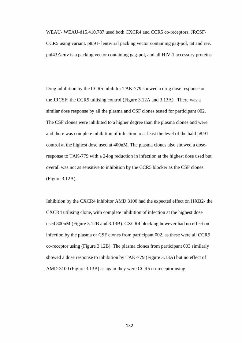
132
WEAU- WEAU-d15.410.787 used both CXCR4 and CCR5 co-receptors, JRCSF-
CCR5 using variant. p8.91- lentiviral packing vector containing gag-pol, tat and rev.
pnl43△env is a packing vector containing gag-pol, and all HIV-1 accessory proteins.
Drug inhibition by the CCR5 inhibitor TAK-779 showed a drug dose response on
the JRCSF; the CCR5 utilising control (Figure 3.12A and 3.13A). There was a
similar dose response by all the plasma and CSF clones tested for participant 002.
The CSF clones were inhibited to a higher degree than the plasma clones and were
and there was complete inhibition of infection to at least the level of the bald p8.91
control at the highest dose used at 400nM. The plasma clones also showed a dose-
response to TAK-779 with a 2-log reduction in infection at the highest dose used but
overall was not as sensitive to inhibition by the CCR5 blocker as the CSF clones
(Figure 3.12A).
Inhibition by the CXCR4 inhibitor AMD 3100 had the expected effect on HXB2- the
CXCR4 utilising clone, with complete inhibition of infection at the highest dose
used 800nM (Figure 3.12B and 3.13B). CXCR4 blocking however had no effect on
infection by the plasma or CSF clones from participant 002, as these were all CCR5
co-receptor using (Figure 3.12B). The plasma clones from participant 003 similarly
showed a dose response to inhibition by TAK-779 (Figure 3.13A) but no effect of
AMD-3100 (Figure 3.13B) as again they were CCR5 co-receptor using.
133
A B
Figure 3.12. Dose response of coreceptor inhibitors on infection by pseudotyped viral clones from participant 002. A. Infection of U87
CCR5+ target cells. B. Infection of U87 CXCR4+ target cells. TAK 779- CCR5 inhibitor, AMD 3100- CXCR4 inhibitor, RLU- relative light
units. Data are representative of two independent experiment and they are the mean and s.d of two technical replicates.

134
A B
Figure 3.13. Dose response of coreceptor inhibitors on infection by pseudotyped viral clones from participant 003. A. Infection of U87
CCR5+ target cells. B. Infection of U87 CXCR4+ target cells. TAK 779- CCR5 inhibitor, AMD 3100- CXCR4 inhibitor, RLU- relative light
units. Data are representative of two independent experiment and they are the mean and s.d of two technical replicates.
135
3.9 The frequency of drug resistance mutations is similar in both compartments
Consensus sequences of HIV-1 from paired plasma and CSF from the 31 participants
with successful next generation sequencing were generated and analysed for drug
resistance mutations (DRMs). The pol gene was examined for DRMs using the
Stanford HIV drug resistance database (https://hivdb.stanford.edu/hivdb/by-
sequences/report/). The frequency of NRTI and NNRTI DRMs encountered in CSF
and plasma compartments were largely similar, with some small differences (figures
3.14A-D). Previously reported DRMs associated with CNS compartmentalisation -
M184V and M230L were found in equal frequencies in plasma and paired CSF
samples. Thymidine analogue mutations (TAMs) were more frequent in the plasma
compared with the CSF specimens (11 in plasma vs 8 in CSF). NRTI mutations
E44D (1 in CSF vs 0 in plasma) and K70E/R (5 in CSF vs 3 in plasma) were more
frequent in CSF compared with plasma samples. However, in 3 of the 4 participants
analysed for CNS compartmentalisation, there were no drug resistant mutations
found in either compartment. In one participant-003 with CNS
compartmentalisation, E138A conferring resistance to rilpirivine was found in the
plasma (Table 3.3).
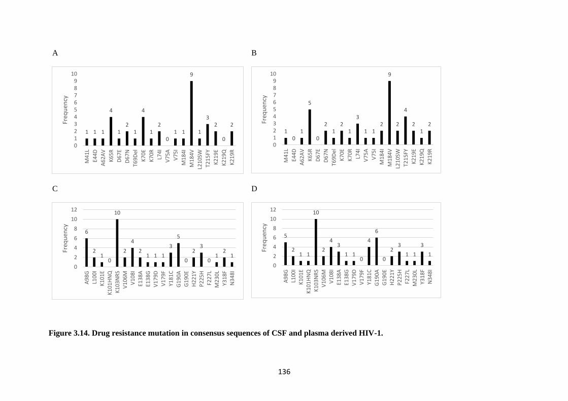
136
A
B
C
D
Figure 3.14. Drug resistance mutation in consensus sequences of CSF and plasma derived HIV-1.
1 1 1
4
12
1
4
12
01 1
9
1
32
0
2
0123456789
10
M4
1L
E44
D
A6
2A
V
K6
5R
D6
7E
D6
7N
T69
Del
K7
0E
K7
0R
L74
I
V7
5A
V7
5I
M1
84
I
M1
84
V
L21
0SW
T21
5FY
K2
19
E
K2
19
Q
K2
19
R
Freq
uen
cy
10
1
5
0
21
21
3
1 12
9
2
4
21
2
0123456789
10
M4
1L
E44
D
A6
2A
V
K6
5R
D6
7E
D6
7N
T69
Del
K7
0E
K7
0R
L74
I
V7
5A
V7
5I
M1
84
I
M1
84
V
L21
0SW
T21
5FY
K2
19
E
K2
19
Q
K2
19
R
Freq
uen
cy
6
21
0
10
2
4
21 1 1
3
5
0
23
01
21
0
2
4
6
8
10
12
A9
8G
L10
0I
K1
01
E
K1
01
HN
Q
K1
03
NR
S
V1
06
M
V1
08
I
E13
8A
E13
8G
V1
79
D
V1
79
F
Y18
1C
G1
90
A
G1
90
E
H2
21
Y
P2
25
H
F22
7L
M2
30
L
Y31
8F
N3
48
I
Freq
uen
cy
5
21 1
10
2
43
1 10
4
6
0
23
1 1
3
1
0
2
4
6
8
10
12
A9
8G
L10
0I
K1
01
EK
10
1H
NQ
K1
03
NR
SV
10
6M
V1
08
IE1
38
AE1
38
GV
17
9D
V1
79
FY1
81
CG
19
0A
G1
90
E
H2
21
YP
22
5H
F22
7L
M2
30
L
Y31
8F
N3
48
I
Freq
uen
cy
137
A. NRTI DRMs in CSF compartment B. NRTI DRMs in plasma compartment C. NNRTI DRMs in CSF compartment D. NNRTI DRMs in
plasma compartment. DRMs- drug resistant mutations; NRTI- nucleotide reverse transcriptase inhibitors; NNRTI- non-nucleotide reverse
transcriptase inhibitors; CSF-cerebrospinal fluid.

138
3.10 Heterogeneity of CNS and plasma derived Env clones to bnAbs
The neutralising activity of 4 broadly neutralising antibodies (bnAbs) against
participant derived Env pseudotyped viruses was measured in a single-round in vitro
neutralisation assay (Methods 2.1.9). Serum neutralisation curves were generated
from which inhibitory concentration 50 (IC50) were determined. IC50 is the
concentration of bnAbs required to inhibit 50% of infection of a Env pseudotyped
virus in the assay. The 4 bnAbs were PGT121 and PGT128 (targeting the high
mannose patch), VRC01 (CD4 binding site blocker) and PG9 (targeting the apex).
Anti-dengue serotype 3 antibody was used as the negative control. This was done for
the 2 participants 002 and 003 who had evidence of CNS compartmentalisation.
Clones from participant 002 were universally resistant to neutralisation by PGT121
and PGT128 (Figure 3.15, Table 3.5). Clone 002_C_B9 form the CSF was sensitive
to neutralisation by VRCO1(Figure 3.15A) and clone 002_P_F9 from the plasma was
sensitive to neutralisation by PG9 (Figure 3.15B). The CSF derived clones from
participant 003 were between 3 to 30- fold times more sensitive to neutralisation by
VRCO1 compared with the plasma clone (Figure 3.16A, Table 3.15). None of the 003
clones were neutralised by PG9 (Figure 3.16B). All the 003 clones were sensitive to
neutralisation by PG121 (figure 3.16C). The CSF clone 003_C_A5 was neutralised by
PGT128 and the plasma clone 003_P_A3 only just reached 50% neutralisation at the
highest concentration of PGT128 (Figure 3.16D).

139
A B C
Figure 3.15. Neutralisation sensitivity of Env pseudotyped lentiviruses from participant 002 to bnAbs. A. Representative neutralisation
curves for Env clones against VRCO1. B. Representative neutralisation curves for Env clones against PG9. C. Representative neutralisation
curves for Env clones against DEN-3 (negative control). bnAbs- broadly neutralising antibodies. Data are mean and s.d of two technical repeats
and are based on one experiment.
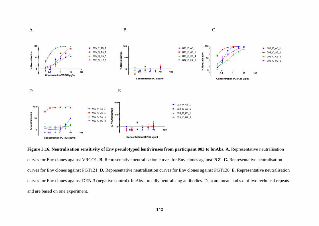
140
A B C
D E
Figure 3.16. Neutralisation sensitivity of Env pseudotyped lentiviruses from participant 003 to bnAbs. A. Representative neutralisation
curves for Env clones against VRCO1. B. Representative neutralisation curves for Env clones against PG9. C. Representative neutralisation
curves for Env clones against PGT121. D. Representative neutralisation curves for Env clones against PGT128. E. Representative neutralisation
curves for Env clones against DEN-3 (negative control). bnAbs- broadly neutralising antibodies. Data are mean and s.d of two technical repeats
and are based on one experiment.
0.1 1 10 100
0
50
100
Concentration PGT121 µg/ml
% N
eu
tralisati
on
003_P_A3_1
003_C_A5_1
003_C_C5_1
003_C_H2_3
0.1 1 10 100
0
50
100
Concentration PGT128 µg/ml
% N
eu
tralisati
on
003_P_A3_1
003_C_A5_1
003_C_C5_1
003_C_H2_3
0.1 1 10 100
0
50
100
Concentration DEN-3 µg/ml
% N
eu
tralisati
on
003_P_A3_1
003_C_A5_1
003_C_C5_1
003_C_H2_3
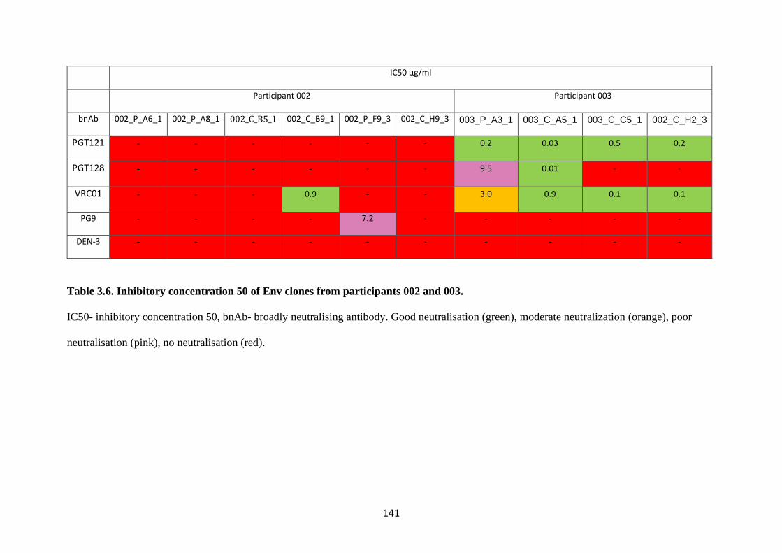
141
IC50 µg/ml
Participant 002 Participant 003
bnAb 002_P_A6_1 002_P_A8_1 002_C_B5_1 002_C_B9_1 002_P_F9_3 002_C_H9_3 003_P_A3_1 003_C_A5_1 003_C_C5_1 002_C_H2_3
PGT121 - - - - - - 0.2 0.03 0.5 0.2
PGT128 - - - - - - 9.5 0.01 - -
VRC01 - - - 0.9 - - 3.0 0.9 0.1 0.1
PG9 - - - - 7.2 - - - - -
DEN-3 - - - - - - - - - -
Table 3.6. Inhibitory concentration 50 of Env clones from participants 002 and 003.
IC50- inhibitory concentration 50, bnAb- broadly neutralising antibody. Good neutralisation (green), moderate neutralization (orange), poor
neutralisation (pink), no neutralisation (red).
142
3.11 Discussion
In this cross-sectional study CSF escape was found to be uncommon at a prevalence
of 4%. The prevalence of CSF escape is heavily influenced by the limit of detection
of the assay used to quantify viral load. On the other hand, there was evidence of
CNS compartmentalisation in 2 out of the 4 participants who underwent SGA and
sequencing. Multiple approaches including visual inspection, distance and tree-based
methods were used to determine CNS compartmentalisation, providing confidence in
the assignation of this status.
Pairwise distance analyses of sequences revealed compartment-related heterogeneity
in Env diversity amongst participants with CNS compartmentalisation. It has been
shown that there is a linear correlation between pairwise distance and recency of
HIV-1 infection (Moyo et al. 2017). It was hypothesised that if CNS
compartmentalised viruses gained entry into the CNS early in acute HIV-1 infection
and established latency in long-lived cells, then the diversity of CNS
compartmentalised viruses will be lower than that of the plasma. This was the case in
the treatment naïve participant but not the treatment experienced. It is certainly
plausible the ART may have exerted a selective sweep on the viruses in the plasma,
limiting its diversity (Feder et al. 2016b) compared to the CNS where there is poor
drug penetration (Letendre 2011). Analyses of recency of HIV-1 infection was
limited by the lack of longitudinal sampling and also it was not possible to make
inference about the evolutionary rate of the viruses in either compartment.

143
The phenotypic co-receptor inhibition assay revealed all clones were CCR5 using,
and infection was blocked by CCR5 inhibitors. If as hypothesised, that CSF clones
had adapted their cellular tropism for macrophages in the CNS with low-level CD4,
it would be compelling to have tested for greater affinity for the CCR5 co-receptor
by the CSF clones. In future work the co-receptor usage assay will be repeated with
the inclusion of Affinophile target cells which have inducible levels of CD4 and
CCR5 using doxycycline and Ponasterone A respectively (Joseph, Lee, and
Swanstrom 2014).
An interesting observation was that the CSF clones from participant 003 were 3 to
30 -fold more sensitive to neutralisation activity of VRCO1- CD4 binding site
antibody but more resistant to CCR5 inhibition. This may suggest that these CSF
clones have become adapted to binding CCR5 with higher affinity. The antibody
neutralisation data will be more meaningful if I was able to clone autologous
antibodies from each compartment in the participants in order to test neutralisation
activity of autologous antibodies and cross neutralisation against their own virus
from the plasma. This would give more insight into the antibody selection pressure
that drove evolution of the virus in that compartment.
The env coverage from Illumina MiSeq was insufficient to be validated against the
SGA data, which precluded analyses of the NGS data for compartmentalisation. In
addition, analyses of clinical and demographic factors associated with CNS
compartmentalisation was limited by the small sample size.

144
This chapter highted the importance of longitudinal sampling and hence time-solved
samples in studying within-host adaptation. The following chapter explores within-
host adaptation of SARS-CoV-2 in a case of chronic infection that had sampling
from multiple timepoints over the course of 101 days.
145
CHAPTER 4: RESULTS 2- SARS-COV-2 ESCAPE FROM
POLYCLONAL CONVALESCENT SERA
The Spike protein SARS-CoV-2 engages with of the human ACE2 receptor and is a
major antibody target. This is because it is the means by which the virus attaches and
gains entry into host cells (V’kovski et al. 2021). Natural infection by SARS-CoV-2
elicits potently neutralizing monoclonal antibodies that recognize the RBD and other
Key Findings:
• Description of in vivo evolution of SARS-CoV-2 in an
immunocompromised host with limited T cell and innate responses
and absent B cell responses.
• Dynamic shifts in viral population can occurs in a host driven by
immune pressure from convalescent plasma.
• D796H mutation enabled the virus escape serum neutralisation, co-
occurred with △H69/V70 which compensated for the loss of
infectivity.
• These mutations had no impact on Spike incorporation and are not
within the Spike RBD but may impact on antibody binding by
allosteric mechanisms.
146
epitopes of the Spike protein (Chi et al. 2020). Using mutagenesis in yeast display
vector in combination with Deep Mutational Scanning, the Bloom lab have been able
to map mutations in RBD that escape antibody binding (Greaney et al. 2021). They
provided a picture of the functional consequences of single mutations within the
RBD, most of which are deleterious to protein folding, protein expression and ACE2
binding (Starr et al. 2020). Outside of the RBD immunodominant epitopes to
polyclonal sera have been found at the fusion peptide and linker region upstream of
the heptad repeat region 2 (Garrett et al. 2020; Lu et al. 2021; Li et al. 2020).
Given the rapid spread and amplification of SARS-CoV-2, variants that can evade
the immune response are likely to arise. Already the virus has adapted with various
Variants of Concern emerging since the start of the pandemic. It is known that some
of these changes increase binding to the human ACE2 receptor such as N501Y
(Collier, De Marco, et al. 2021; Starr et al. 2020), other changes for example the
B.1.1.7 variant has have been associated with increased transmission and higher viral
loads (Frampton et al. 2021), some changes have been associated with vaccine-
elicited antibody escape for example E484K (Collier, De Marco, et al. 2021).
This was an in-depth case- study of an immunocompromised man with chronic
infection with SARS-CoV-2. The aim of this study was to characterise the dynamics,
genotypic and phenotypic properties of the virus population in this individual’s
respiratory samples over 101 days of illness, with a view to providing insights into in
vivo escape of SARS-CoV-2 from neutralising antibodies.
147
4.1 Clinical Case History
4.1.1 COVID-19 clinical case in an immunocompromised host
A man in his 70s presenting to the Emergency Room with acute shortness of breath
and dry cough. He was diagnosed with COVID-19 after testing positive for SARS-
CoV-2 RT-PCR test on a nasopharyngeal swab with a CT value of 25, 35 days prior.
The day of his first PCR positive SARS-CoV-2 test was designated day 1 (Figure
4.1). His past medical history was significant for Marginal B Cell Lymphoma
diagnosed 8 years prior and was treated with vincristine, prednisolone,
cyclophosphamide and anti-CD20 B cell depletion with rituximab, last given one
year prior. Clinical examination revealed an elevated respiratory rate of 42/min and
oxygen saturation of 92% on 6 L of oxygen. Laboratory tests on admission were
significant for a C-reactive protein (CRP) of 245 mg/L, lymphopaenia and IgG <1.4
g/l. Imaging studies on admission including a Chest x ray showing widespread
interstitial opacification and CT chest done showed widespread, basal and dependent
predominant ground glass and peri broncho vascular consolidation. He was
transferred to the intensive care unit (ITU) where he received non-invasive
ventilation.
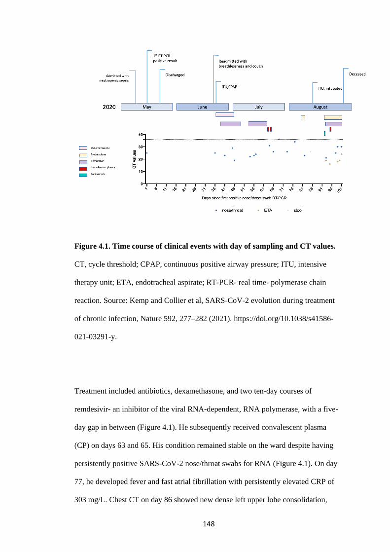
148
Figure 4.1. Time course of clinical events with day of sampling and CT values.
CT, cycle threshold; CPAP, continuous positive airway pressure; ITU, intensive
therapy unit; ETA, endotracheal aspirate; RT-PCR- real time- polymerase chain
reaction. Source: Kemp and Collier et al, SARS-CoV-2 evolution during treatment
of chronic infection, Nature 592, 277–282 (2021). https://doi.org/10.1038/s41586-
021-03291-y.
Treatment included antibiotics, dexamethasone, and two ten-day courses of
remdesivir- an inhibitor of the viral RNA-dependent, RNA polymerase, with a five-
day gap in between (Figure 4.1). He subsequently received convalescent plasma
(CP) on days 63 and 65. His condition remained stable on the ward despite having
persistently positive SARS-CoV-2 nose/throat swabs for RNA (Figure 4.1). On day
77, he developed fever and fast atrial fibrillation with persistently elevated CRP of
303 mg/L. Chest CT on day 86 showed new dense left upper lobe consolidation,
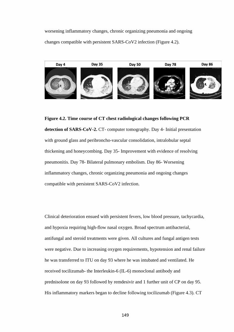
149
worsening inflammatory changes, chronic organizing pneumonia and ongoing
changes compatible with persistent SARS-CoV2 infection (Figure 4.2).
Figure 4.2. Time course of CT chest radiological changes following PCR
detection of SARS-CoV-2. CT- computer tomography. Day 4- Initial presentation
with ground glass and peribroncho-vascular consolidation, intralobular septal
thickening and honeycombing. Day 35- Improvement with evidence of resolving
pneumonitis. Day 78- Bilateral pulmonary embolism. Day 86- Worsening
inflammatory changes, chronic organizing pneumonia and ongoing changes
compatible with persistent SARS-CoV2 infection.
Clinical deterioration ensued with persistent fevers, low blood pressure, tachycardia,
and hypoxia requiring high-flow nasal oxygen. Broad spectrum antibacterial,
antifungal and steroid treatments were given. All cultures and fungal antigen tests
were negative. Due to increasing oxygen requirements, hypotension and renal failure
he was transferred to ITU on day 93 where he was intubated and ventilated. He
received tocilizumab- the Interleukin-6 (IL-6) monoclonal antibody and
prednisolone on day 93 followed by remdesivir and 1 further unit of CP on day 95.
His inflammatory markers began to decline following tocilizumab (Figure 4.3). CT

150
values on tracheal aspirate samples also showed signs of increasing following the
combination of treatments but unfortunately the individual died on day 101 (Figure
4.1).
Figure 4.3. Time course of inflammatory markers in this clinical case.
WCC- white cell count, CRP- C reactive protein.
4.1.2 B cell depletions and deficient SARS-CoV-2 binding antibodies
The participant had received repeated cycles of rituximab anti-CD20 B cell depletion
therapy and consequently he was hypogammaglobulinaemic with reduction in total
serum IgG and IgA. The serum IgM was within the normal reference range which is
either due to his underlying lymphoproliferative state or ongoing infection (Table
4.1). SARS-CoV-2 specific Spike (S), Receptor binding domain (RBD) and
Nucleocapsid (N) IgG antibodies were measured in the participant’s serum at
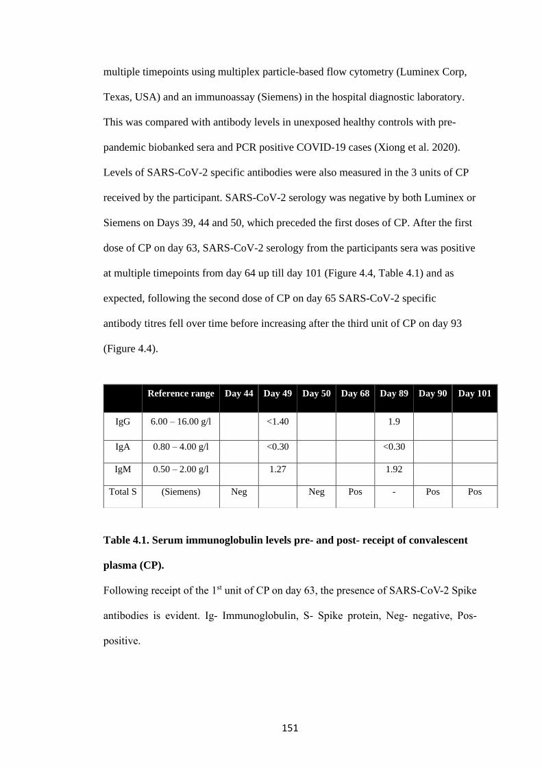
151
multiple timepoints using multiplex particle-based flow cytometry (Luminex Corp,
Texas, USA) and an immunoassay (Siemens) in the hospital diagnostic laboratory.
This was compared with antibody levels in unexposed healthy controls with pre-
pandemic biobanked sera and PCR positive COVID-19 cases (Xiong et al. 2020).
Levels of SARS-CoV-2 specific antibodies were also measured in the 3 units of CP
received by the participant. SARS-CoV-2 serology was negative by both Luminex or
Siemens on Days 39, 44 and 50, which preceded the first doses of CP. After the first
dose of CP on day 63, SARS-CoV-2 serology from the participants sera was positive
at multiple timepoints from day 64 up till day 101 (Figure 4.4, Table 4.1) and as
expected, following the second dose of CP on day 65 SARS-CoV-2 specific
antibody titres fell over time before increasing after the third unit of CP on day 93
(Figure 4.4).
Table 4.1. Serum immunoglobulin levels pre- and post- receipt of convalescent
plasma (CP).
Following receipt of the 1st unit of CP on day 63, the presence of SARS-CoV-2 Spike
antibodies is evident. Ig- Immunoglobulin, S- Spike protein, Neg- negative, Pos-
positive.
Reference range Day 44 Day 49 Day 50 Day 68 Day 89 Day 90 Day 101
IgG 6.00 – 16.00 g/l <1.40 1.9
IgA 0.80 – 4.00 g/l <0.30 <0.30
IgM 0.50 – 2.00 g/l 1.27 1.92
Total S (Siemens) Neg Neg Pos - Pos Pos
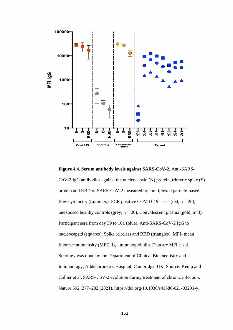
152
Figure 4.4. Serum antibody levels against SARS-CoV-2. Anti-SARS-
CoV-2 IgG antibodies against the nucleocapsid (N) protein, trimeric spike (S)
protein and RBD of SARS-CoV-2 measured by multiplexed particle-based
flow cytometry (Luminex). PCR positive COVID-19 cases (red, n = 20),
unexposed healthy controls (grey, n = 20), Convalescent plasma (gold, n=3).
Participant sera from day 39 to 101 (blue). Anti-SARS-CoV-2 IgG to
nucleocapsid (squares), Spike (circles) and RBD (triangles). MFI- mean
fluorescent intensity (MFI). Ig- immunoglobulin. Data are MFI ± s.d.
Serology was done by the Department of Clinical Biochemistry and
Immunology, Addenbrooke’s Hospital, Cambridge, UK. Source: Kemp and
Collier et al, SARS-CoV-2 evolution during treatment of chronic infection,
Nature 592, 277–282 (2021). https://doi.org/10.1038/s41586-021-03291-y.
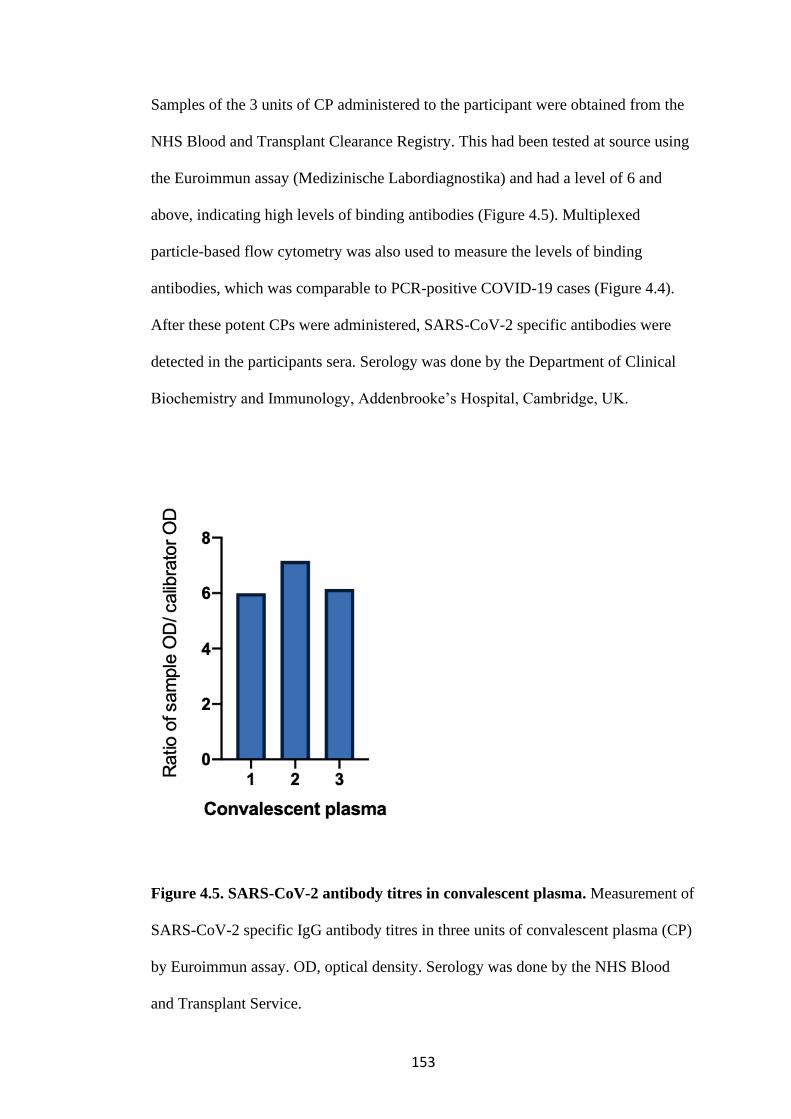
153
Samples of the 3 units of CP administered to the participant were obtained from the
NHS Blood and Transplant Clearance Registry. This had been tested at source using
the Euroimmun assay (Medizinische Labordiagnostika) and had a level of 6 and
above, indicating high levels of binding antibodies (Figure 4.5). Multiplexed
particle-based flow cytometry was also used to measure the levels of binding
antibodies, which was comparable to PCR-positive COVID-19 cases (Figure 4.4).
After these potent CPs were administered, SARS-CoV-2 specific antibodies were
detected in the participants sera. Serology was done by the Department of Clinical
Biochemistry and Immunology, Addenbrooke’s Hospital, Cambridge, UK.
Figure 4.5. SARS-CoV-2 antibody titres in convalescent plasma. Measurement of
SARS-CoV-2 specific IgG antibody titres in three units of convalescent plasma (CP)
by Euroimmun assay. OD, optical density. Serology was done by the NHS Blood
and Transplant Service.
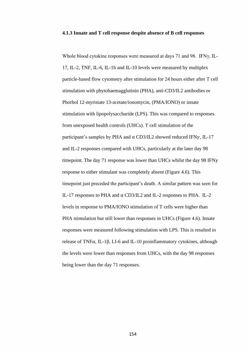
154
4.1.3 Innate and T cell response despite absence of B cell responses
Whole blood cytokine responses were measured at days 71 and 98. IFNγ, IL-
17, IL-2, TNF, IL-6, IL-1b and IL-10 levels were measured by multiplex
particle-based flow cytometry after stimulation for 24 hours either after T cell
stimulation with phytohaemagglutinin (PHA), anti-CD3/IL2 antibodies or
Phorbol 12-myristate 13-acetate/ionomycin, (PMA/IONO) or innate
stimulation with lipopolysaccharide (LPS). This was compared to responses
from unexposed health controls (UHCs). T cell stimulation of the
participant’s samples by PHA and ⍺ CD3/IL2 showed reduced IFN𝛾, IL-17
and IL-2 responses compared with UHCs, particularly at the later day 98
timepoint. The day 71 response was lower than UHCs whilst the day 98 IFN𝛾
response to either stimulant was completely absent (Figure 4.6). This
timepoint just preceded the participant’s death. A similar pattern was seen for
IL-17 responses to PHA and ⍺ CD3/IL2 and IL-2 responses to PHA. IL-2
levels in response to PMA/IONO stimulation of T cells were higher than
PHA stimulation but still lower than responses in UHCs (Figure 4.6). Innate
responses were measured following stimulation with LPS. This is resulted in
release of TNF⍺, IL-1β, LI-6 and IL-10 proinflammatory cytokines, although
the levels were lower than responses from UHCs, with the day 98 responses
being lower than the day 71 responses.
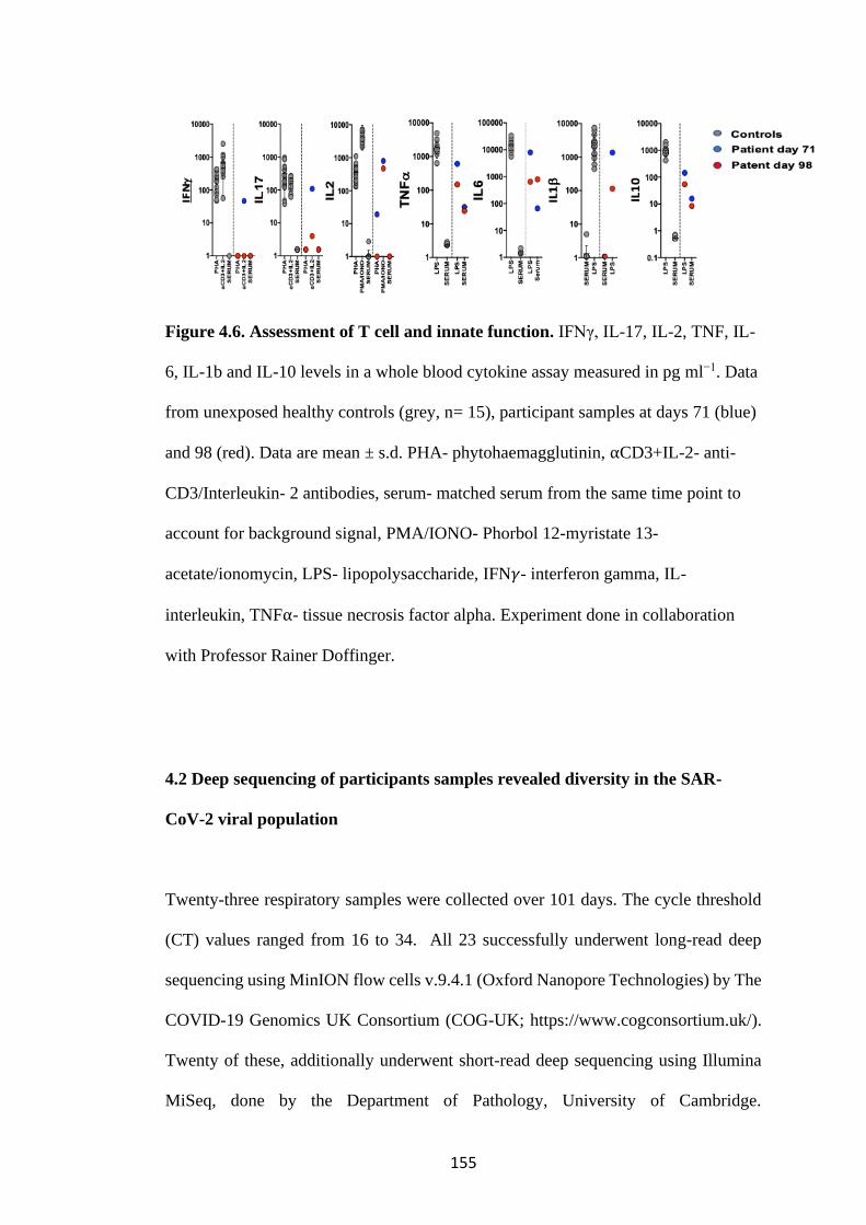
155
Figure 4.6. Assessment of T cell and innate function. IFNγ, IL-17, IL-2, TNF, IL-
6, IL-1b and IL-10 levels in a whole blood cytokine assay measured in pg ml−1. Data
from unexposed healthy controls (grey, n= 15), participant samples at days 71 (blue)
and 98 (red). Data are mean ± s.d. PHA- phytohaemagglutinin, ⍺CD3+IL-2- anti-
CD3/Interleukin- 2 antibodies, serum- matched serum from the same time point to
account for background signal, PMA/IONO- Phorbol 12-myristate 13-
acetate/ionomycin, LPS- lipopolysaccharide, IFN𝛾- interferon gamma, IL-
interleukin, TNF⍺- tissue necrosis factor alpha. Experiment done in collaboration
with Professor Rainer Doffinger.
4.2 Deep sequencing of participants samples revealed diversity in the SAR-
CoV-2 viral population
Twenty-three respiratory samples were collected over 101 days. The cycle threshold
(CT) values ranged from 16 to 34. All 23 successfully underwent long-read deep
sequencing using MinION flow cells v.9.4.1 (Oxford Nanopore Technologies) by The
COVID-19 Genomics UK Consortium (COG-UK; https://www.cogconsortium.uk/).
Twenty of these, additionally underwent short-read deep sequencing using Illumina
MiSeq, done by the Department of Pathology, University of Cambridge.

156
Independently, single genome amplification (SGA) and sequencing of the spike gene
was used to detect mutations in 3 samples at different time points (Table 4.2).
Timepoint
(Day)
Sequence Identifier CT value
Sample
type
Nanopore Illumina SGA
1 NB16_CAMB-1B19D5 25 NT Y Y Y
37 NB23_CAMB-1B4FB2 - NT Y Y Y
41 NB01_CAMB-1B5124 23 NT Y Y -
45 NB08_CAMB-1B529A 29 NT Y Y -
50 NB13_CAMB-1B54A3 - NT Y Y -
54 NB10_CAMB-1B5643 22 NT Y - -
55 NB08_CAMB-1B5467 - NT Y Y -
56 NB11_CAMB-1B5616 23 NT Y - -
57 NB14_CAMB-1B5607 24 NT Y - -
66 NB05_CAMB-1B55FB 26 NT Y Y -
82 NB06_CAMB-1B55CE 23 NT Y Y -
86 NB07_CAMB-1B5A29 - NT Y - -
89 NB01_CAMB-1B5ADE - NT Y Y -
93 NB16_CAMB-1B5BAE 21 NT Y Y -
93 NB17_CAMB-1B5B71 19 ETA Y Y -
95 NB02_CAMB-1B5BCC 16 NT Y Y -
98 NB01_CAMB-1B5BDB 25 NT Y Y Y
99 NB09_CAMB-1B5C50 30 NT Y Y -
99 NB10_CAMB-1B5C6F 18 ETA Y Y -
100 NB13_CAMB-1B5CE7 - NT Y Y -
100 NB14_CAMB-1B5CBA 19 ETA Y Y -
101 NB15_CAMB-1B5CC9 24 ETA Y Y -
101 NB16_CAMB-1B5C9C 30 NT Y Y -
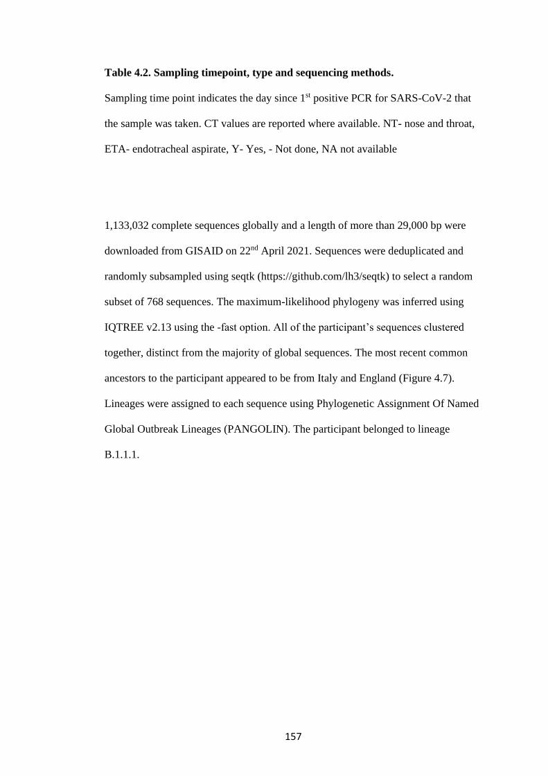
157
Table 4.2. Sampling timepoint, type and sequencing methods.
Sampling time point indicates the day since 1st positive PCR for SARS-CoV-2 that
the sample was taken. CT values are reported where available. NT- nose and throat,
ETA- endotracheal aspirate, Y- Yes, - Not done, NA not available
1,133,032 complete sequences globally and a length of more than 29,000 bp were
downloaded from GISAID on 22nd April 2021. Sequences were deduplicated and
randomly subsampled using seqtk (https://github.com/lh3/seqtk) to select a random
subset of 768 sequences. The maximum-likelihood phylogeny was inferred using
IQTREE v2.13 using the -fast option. All of the participant’s sequences clustered
together, distinct from the majority of global sequences. The most recent common
ancestors to the participant appeared to be from Italy and England (Figure 4.7).
Lineages were assigned to each sequence using Phylogenetic Assignment Of Named
Global Outbreak Lineages (PANGOLIN). The participant belonged to lineage
B.1.1.1.
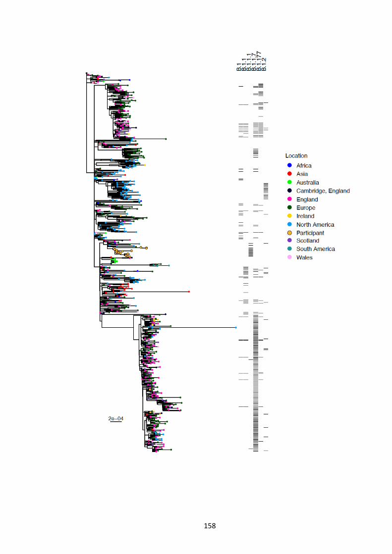
158

159
Figure 4.7. Analysis of whole-genome sequences of SARS-CoV-2 of the
participant in the context of local, UK and globally derived sequences. The
maximum-likelihood phylogenetic tree is rooted on the Wuhan-Hu-1 reference
sequence, showing a subset of total of 768 sequences of SARS-CoV-2 from GISAID.
The Illumina Miseq short read sequences were used for further analyses. Visual
inspection of the maximum likelihood phylogenetic tree of the participant’s sequences
revealed the existence of two distinct viral populations. The samples collected at days
93 and 95 bearing the W64G and P330S Spike mutations were rooted within, but
significantly divergent from the original population (Figure 4.8). The relationship of
the divergent samples to those at earlier time points argues against superinfection. All
samples were consistent with having arisen from a single underlying viral population.
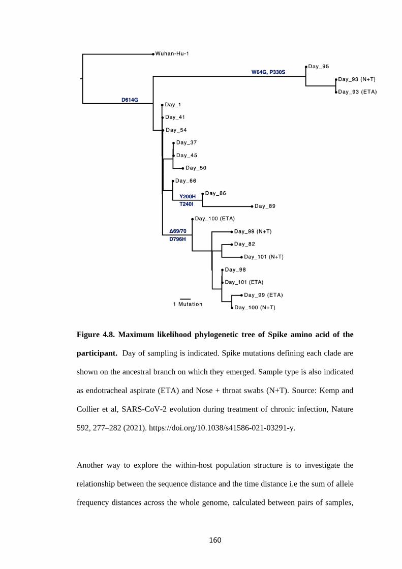
160
Figure 4.8. Maximum likelihood phylogenetic tree of Spike amino acid of the
participant. Day of sampling is indicated. Spike mutations defining each clade are
shown on the ancestral branch on which they emerged. Sample type is also indicated
as endotracheal aspirate (ETA) and Nose + throat swabs (N+T). Source: Kemp and
Collier et al, SARS-CoV-2 evolution during treatment of chronic infection, Nature
592, 277–282 (2021). https://doi.org/10.1038/s41586-021-03291-y.
Another way to explore the within-host population structure is to investigate the
relationship between the sequence distance and the time distance i.e the sum of allele
frequency distances across the whole genome, calculated between pairs of samples,

161
plotted against the difference in times at which the samples were collected (Figure
4.9A). This was done in collaboration with Dr Chris Illingworth using the distance
metric (Lumby et al. 2020). The median pairwise distance between samples in the
main clade and those collected on days 93 and 95, bearing W64G, P330S Spike
mutations was significantly different (Figure 4.9B, p<0.001). However, when the
sample collected on days 93 and 95 were removed the median pairwise distance
between samples in the main clade and those collected on days 86 and 89, bearing the
Y200H and T240I Spike mutation was not significantly different (Figure 4.9C, p
0.066). This provided further support for the cladal structure of the viral population.
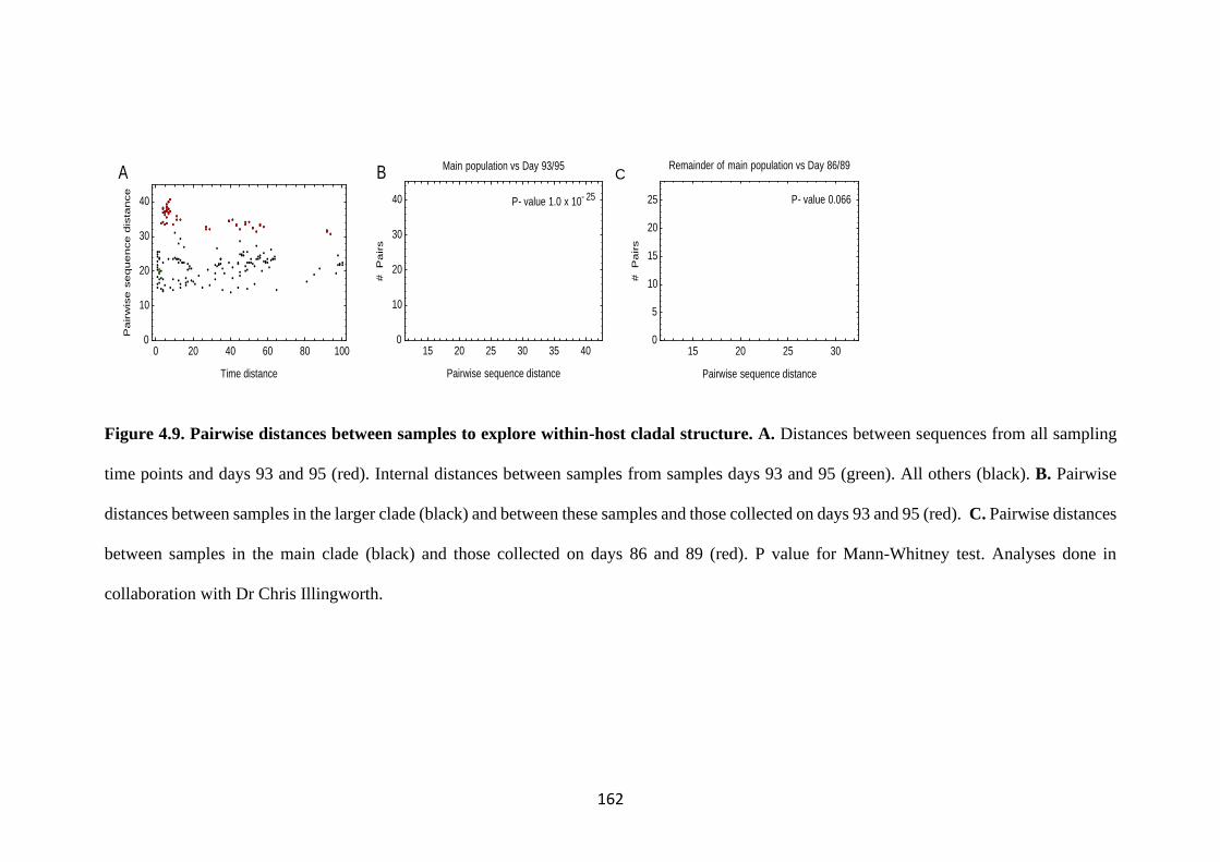
162
Figure 4.9. Pairwise distances between samples to explore within-host cladal structure. A. Distances between sequences from all sampling
time points and days 93 and 95 (red). Internal distances between samples from samples days 93 and 95 (green). All others (black). B. Pairwise
distances between samples in the larger clade (black) and between these samples and those collected on days 93 and 95 (red). C. Pairwise distances
between samples in the main clade (black) and those collected on days 86 and 89 (red). P value for Mann-Whitney test. Analyses done in
collaboration with Dr Chris Illingworth.
P- value 0.066
15 20 25 300
5
10
15
20
25
Pairwise sequence distance
#P
air
s
Remainder of main population vs Day 86/89
P- value 1.0 x 10- 25
15 20 25 30 35 400
10
20
30
40
Pairwise sequence distance#
Pa
irs
Main population vs Day 93/95
0 20 40 60 80 1000
10
20
30
40
Time distance
Pa
irw
ise
se
qu
en
ce
dis
tan
ce
Evolutionary Rate = 0.025 nucleotides / day
= 8.97 nucleotides / year
0 20 40 60 80 1000
5
10
15
20
25
30
35
Time distance
Pa
irw
ise
se
qu
en
ce
dis
tance
Primary Clade
Evolutionary Rate = 0.125 nucleotides / day
= 45.80 nucleotides / year
0 20 40 60 80 1000
5
10
15
20
25
30
35
Time distance
Pa
irw
ise
se
qu
en
ce
dis
tance
Samples from days 1, 93, and 95
A B
C D
E
P- value 0.066
15 20 25 300
5
10
15
20
25
Pairwise sequence distance
#P
air
s
Remainder of main population vs Day 86/89
P- value 1.0 x 10- 25
15 20 25 30 35 400
10
20
30
40
Pairwise sequence distance
#P
air
s
Main population vs Day 93/95
0 20 40 60 80 1000
10
20
30
40
Time distance
Pa
irw
ise
se
qu
en
ce
dis
tan
ce
Evolutionary Rate = 0.025 nucleotides / day
= 8.97 nucleotides / year
0 20 40 60 80 1000
5
10
15
20
25
30
35
Time distance
Pa
irw
ise
se
qu
en
ce
dis
tance
Primary Clade
Evolutionary Rate = 0.125 nucleotides / day
= 45.80 nucleotides / year
0 20 40 60 80 1000
5
10
15
20
25
30
35
Time distance
Pa
irw
ise
se
qu
en
ce
dis
tance
Samples from days 1, 93, and 95
A B
C D
E
C
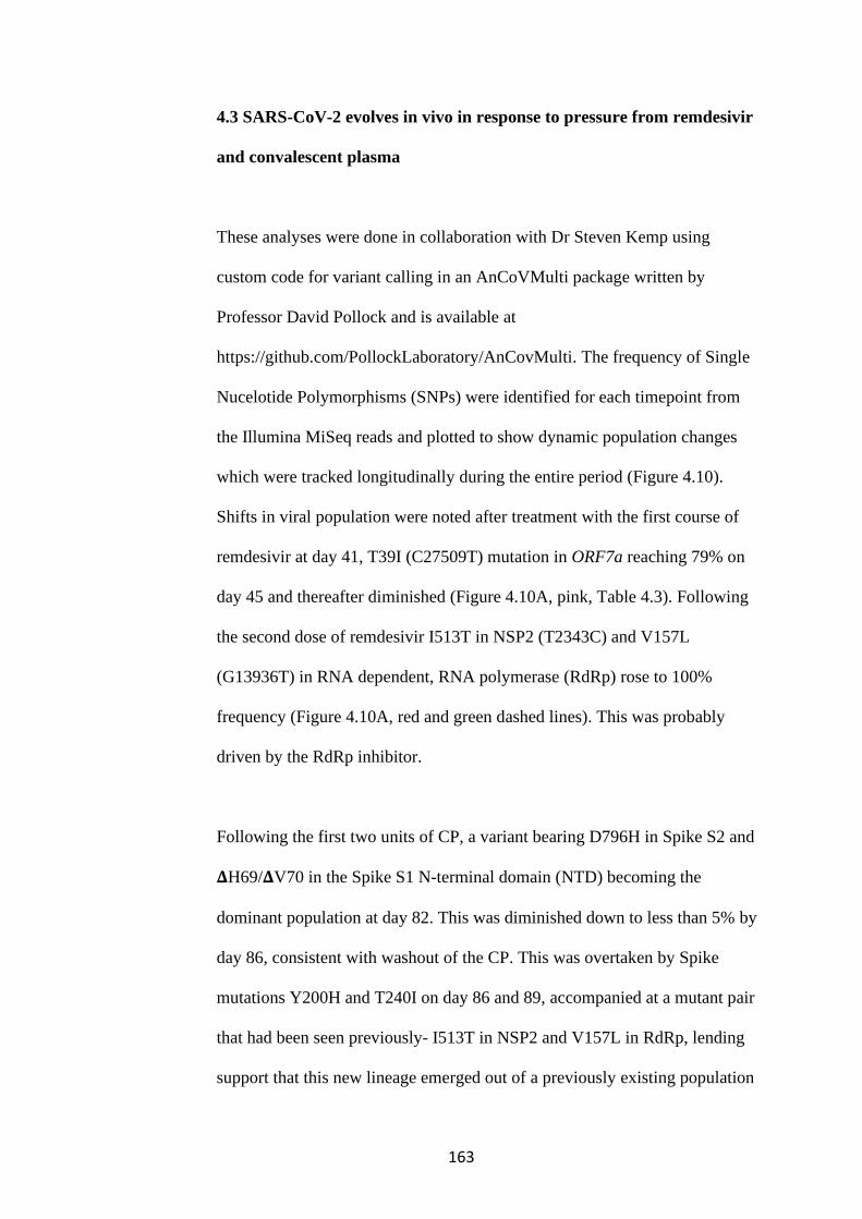
163
4.3 SARS-CoV-2 evolves in vivo in response to pressure from remdesivir
and convalescent plasma
These analyses were done in collaboration with Dr Steven Kemp using
custom code for variant calling in an AnCoVMulti package written by
Professor David Pollock and is available at
https://github.com/PollockLaboratory/AnCovMulti. The frequency of Single
Nucelotide Polymorphisms (SNPs) were identified for each timepoint from
the Illumina MiSeq reads and plotted to show dynamic population changes
which were tracked longitudinally during the entire period (Figure 4.10).
Shifts in viral population were noted after treatment with the first course of
remdesivir at day 41, T39I (C27509T) mutation in ORF7a reaching 79% on
day 45 and thereafter diminished (Figure 4.10A, pink, Table 4.3). Following
the second dose of remdesivir I513T in NSP2 (T2343C) and V157L
(G13936T) in RNA dependent, RNA polymerase (RdRp) rose to 100%
frequency (Figure 4.10A, red and green dashed lines). This was probably
driven by the RdRp inhibitor.
Following the first two units of CP, a variant bearing D796H in Spike S2 and
𝚫H69/𝚫V70 in the Spike S1 N-terminal domain (NTD) becoming the
dominant population at day 82. This was diminished down to less than 5% by
day 86, consistent with washout of the CP. This was overtaken by Spike
mutations Y200H and T240I on day 86 and 89, accompanied at a mutant pair
that had been seen previously- I513T in NSP2 and V157L in RdRp, lending
support that this new lineage emerged out of a previously existing population
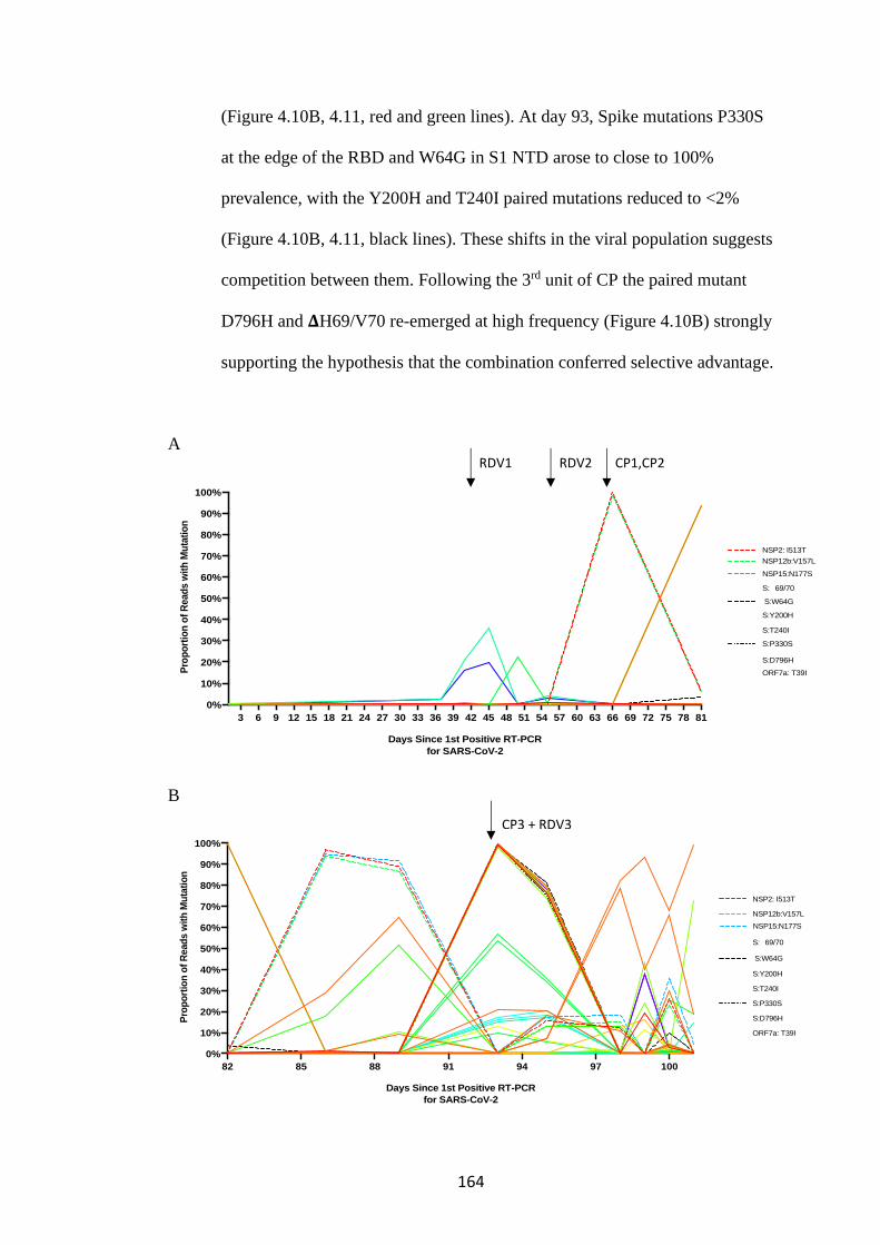
164
(Figure 4.10B, 4.11, red and green lines). At day 93, Spike mutations P330S
at the edge of the RBD and W64G in S1 NTD arose to close to 100%
prevalence, with the Y200H and T240I paired mutations reduced to <2%
(Figure 4.10B, 4.11, black lines). These shifts in the viral population suggests
competition between them. Following the 3rd unit of CP the paired mutant
D796H and 𝚫H69/V70 re-emerged at high frequency (Figure 4.10B) strongly
supporting the hypothesis that the combination conferred selective advantage.
A
B
3 6 9 12 15 18 21 24 27 30 33 36 39 42 45 48 51 54 57 60 63 66 69 72 75 78 81
0%
10%
20%
30%
40%
50%
60%
70%
80%
90%
100%
Days Since 1st Positive RT-PCR
for SARS-CoV-2
Pro
po
rtio
n o
f R
ead
s w
ith
Mu
tati
on
NSP2: I513T
NSP12b:V157L
NSP15:N177S
S:� 69/70
S:W64G
S:Y200H
S:T240I
S:P330S
S:D796H
ORF7a: T39I
82 85 88 91 94 97 100
0%
10%
20%
30%
40%
50%
60%
70%
80%
90%
100%
Days Since 1st Positive RT-PCR
for SARS-CoV-2
Pro
po
rtio
n o
f R
ead
s w
ith
Mu
tati
on
NSP2: I513T
NSP12b:V157L
NSP15:N177S
S:� 69/70
S:W64G
S:Y200H
S:T240I
S:P330S
S:D796H
ORF7a: T39I
RDV1 RDV2 CP1,CP2
CP3 + RDV3
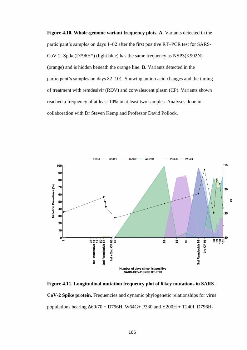
165
Figure 4.10. Whole-genome variant frequency plots. A. Variants detected in the
participant’s samples on days 1–82 after the first positive RT–PCR test for SARS-
CoV-2. Spike(D796H*) (light blue) has the same frequency as NSP3(K902N)
(orange) and is hidden beneath the orange line. B. Variants detected in the
participant’s samples on days 82–101. Showing amino acid changes and the timing
of treatment with remdesivir (RDV) and convalescent plasm (CP). Variants shown
reached a frequency of at least 10% in at least two samples. Analyses done in
collaboration with Dr Steven Kemp and Professor David Pollock.
Figure 4.11. Longitudinal mutation frequency plot of 6 key mutations in SARS-
CoV-2 Spike protein. Frequencies and dynamic phylogenetic relationships for virus
populations bearing 𝚫69/70 + D796H, W64G+ P330 and Y200H + T240I. D796H-
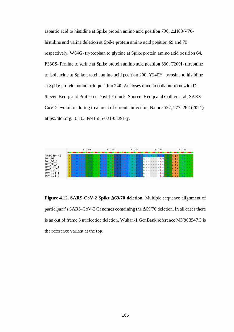
166
aspartic acid to histidine at Spike protein amino acid position 796, △H69/V70-
histidine and valine deletion at Spike protein amino acid position 69 and 70
respectively, W64G- tryptophan to glycine at Spike protein amino acid position 64,
P330S- Proline to serine at Spike protein amino acid position 330, T200I- threonine
to isoleucine at Spike protein amino acid position 200, Y240H- tyrosine to histidine
at Spike protein amino acid position 240. Analyses done in collaboration with Dr
Steven Kemp and Professor David Pollock. Source: Kemp and Collier et al, SARS-
CoV-2 evolution during treatment of chronic infection, Nature 592, 277–282 (2021).
https://doi.org/10.1038/s41586-021-03291-y.
Figure 4.12. SARS-CoV-2 Spike 𝚫69/70 deletion. Multiple sequence alignment of
participant’s SARS-CoV-2 Genomes containing the 𝚫69/70 deletion. In all cases there
is an out of frame 6 nucleotide deletion. Wuhan-1 GenBank reference MN908947.3 is
the reference variant at the top.
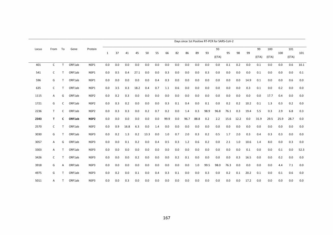
167
Locus From To Gene Protein
Days since 1st Positive RT-PCR for SARS-CoV-2
1 37 41 45 50 55 66 82 86 89 93 93
(ETA) 95 98 99
99
(ETA)
100
(ETA) 100
101
(ETA) 101
401 C T ORF1ab NSP1 0.0 0.0 0.0 0.0 0.0 0.0 0.0 0.0 0.0 0.0 0.0 0.0 0.1 0.2 0.0 0.1 0.0 0.0 0.6 10.1
541 C T ORF1ab NSP1 0.0 0.5 0.4 27.1 0.0 0.0 0.3 0.0 0.0 0.0 0.3 0.0 0.0 0.0 0.0 0.1 0.0 0.0 0.0 0.1
596 G T ORF1ab NSP1 0.0 0.0 0.0 0.0 0.0 0.4 0.3 0.0 0.0 0.0 0.0 0.0 0.0 0.0 14.9 0.1 0.0 0.0 0.6 0.0
635 C T ORF1ab NSP1 0.0 3.5 0.3 18.2 0.4 0.7 1.1 0.6 0.0 0.0 0.0 0.0 0.0 0.0 0.3 0.1 0.0 0.2 0.0 0.0
1115 A G ORF1ab NSP2 0.0 0.2 0.3 0.0 0.0 0.0 0.0 0.0 0.0 0.0 0.0 0.0 0.0 0.0 0.0 0.0 17.7 0.4 0.0 0.0
1721 G C ORF1ab NSP2 0.0 0.3 0.2 0.0 0.0 0.0 0.3 0.1 0.4 0.0 0.1 0.0 0.2 0.2 10.2 0.1 1.3 0.3 0.2 0.0
2236 T C ORF1ab NSP2 0.0 0.3 0.3 0.0 0.2 0.7 0.2 0.0 1.4 0.3 98.9 96.8 76.1 0.3 19.4 5.5 0.3 2.9 6.8 0.3
2343 T C ORF1ab NSP2 0.0 0.0 0.0 0.0 0.0 0.0 99.9 0.0 96.7 88.8 0.2 2.2 15.6 12.2 0.0 31.9 29.5 25.9 28.7 0.0
2570 C T ORF1ab NSP2 0.0 0.9 16.8 6.3 0.0 1.4 0.0 0.0 0.0 0.0 0.0 0.0 0.0 0.0 0.0 0.0 0.0 0.0 0.0 0.0
3030 G T ORF1ab NSP3 0.0 0.2 1.3 0.2 13.3 0.0 1.0 0.7 2.0 0.3 0.2 0.5 1.7 2.0 0.3 0.4 0.3 0.3 0.0 0.0
3057 A G ORF1ab NSP3 0.0 0.0 0.1 0.2 0.0 0.4 0.5 0.3 1.2 0.6 0.2 0.0 2.1 1.0 10.6 1.4 8.0 0.0 0.3 0.0
3303 A T ORF1ab NSP3 0.0 0.0 0.0 0.0 0.0 0.0 0.0 0.0 0.0 0.0 0.0 0.0 0.0 0.0 0.1 0.0 0.0 0.1 0.0 52.3
3426 C T ORF1ab NSP3 0.0 0.0 0.0 0.2 0.0 0.0 0.0 0.2 0.1 0.0 0.0 0.0 0.0 0.3 16.5 0.0 0.0 0.2 0.0 0.0
3918 G A ORF1ab NSP3 0.0 0.0 0.0 0.0 0.0 0.0 0.0 0.0 0.0 1.0 99.5 98.0 76.3 0.0 0.0 0.0 0.0 4.4 7.1 0.0
4975 G T ORF1ab NSP3 0.0 0.2 0.0 0.1 0.0 0.4 0.3 0.1 0.0 0.0 0.3 0.0 0.2 0.1 20.2 0.1 0.0 0.1 0.6 0.0
5011 A T ORF1ab NSP3 0.0 0.0 0.3 0.0 0.0 0.0 0.0 0.0 0.0 0.0 0.0 0.0 0.0 0.0 17.2 0.0 0.0 0.0 0.0 0.0

168
Locus From To Gene Protein
Days since 1st Positive RT-PCR for SARS-CoV-2
1 37 41 45 50 55 66 82 86 89 93 93
(ETA) 95 98 99
99
(ETA)
100
(ETA) 100
101
(ETA) 101
5012 G T ORF1ab NSP3 0.0 0.0 0.0 0.0 0.0 0.0 0.0 0.0 0.0 0.0 0.0 0.0 0.0 0.0 17.2 0.0 0.0 0.0 0.0 0.0
5339 C T ORF1ab NSP3 0.0 0.0 0.0 0.0 0.0 0.5 23.0 0.1 0.0 0.5 0.2 0.5 0.1 0.0 0.0 0.1 0.0 0.1 0.0 1.4
5393 T C ORF1ab NSP3 0.0 0.5 0.0 0.0 0.3 0.0 0.0 0.4 0.8 0.0 0.4 0.0 7.2 78.7 39.6 64.3 28.4 65.5 65.4 18.9
5406 T G ORF1ab NSP3 0.0 0.4 0.0 0.0 0.3 0.0 0.4 0.0 28.6 64.9 0.4 2.1 18.0 11.0 0.0 30.9 16.3 29.9 28.4 0.0
5425 G T ORF1ab NSP3 0.0 0.0 0.0 0.0 0.0 0.0 0.0 99.6 1.2 9.4 0.2 0.8 7.3 82.3 93.1 65.6 53.9 67.9 65.8 98.9
5590 T C ORF1ab NSP3 0.0 0.0 0.0 0.0 0.0 0.0 0.4 0.2 0.8 0.0 99.6 95.9 79.4 0.3 0.0 4.4 0.0 4.1 7.4 0.0
6070 C A ORF1ab NSP3 0.2 0.6 6.3 19.2 0.0 0.0 0.4 0.4 0.3 0.6 0.0 0.0 0.3 0.0 0.0 0.1 0.0 0.0 0.3 0.0
6568 C T ORF1ab NSP3 0.0 0.0 1.9 0.0 31.4 0.9 0.0 0.0 0.0 0.0 0.0 0.0 0.0 0.0 0.0 0.0 1.4 0.0 0.5 0.0
7667 G A ORF1ab NSP3 0.3 0.2 0.0 0.0 0.0 0.0 0.3 0.0 0.4 0.0 0.0 0.2 0.0 0.0 0.3 0.3 0.0 0.0 0.0 13.6
7729 T G ORF1ab NSP3 0.0 0.0 0.0 0.0 0.0 0.0 0.0 0.0 0.0 0.0 0.0 0.0 0.0 0.0 0.0 0.0 0.0 0.0 0.0 11.3
7805 G A ORF1ab NSP3 0.0 0.2 0.3 0.0 0.0 0.6 0.3 0.0 0.0 0.0 0.0 0.0 0.0 0.0 0.0 0.0 0.0 0.3 0.0 11.1
8389 C T ORF1ab NSP3 0.0 0.0 0.3 0.0 0.0 0.0 0.0 0.0 0.0 0.0 20.9 18.6 20.2 0.3 0.0 1.1 0.0 0.6 1.1 0.0
8496 G C ORF1ab NSP3 0.0 0.0 0.0 0.0 0.0 0.0 0.0 0.0 0.0 0.0 0.0 0.6 0.0 0.0 12.1 0.0 0.0 0.0 0.0 0.0
9130 G T ORF1ab NSP4 0.0 0.2 0.0 0.0 0.2 0.0 0.2 0.0 0.0 0.0 0.1 0.0 0.0 12.2 15.9 0.0 19.6 0.2 0.0 0.0
9438 C T ORF1ab NSP4 0.0 0.0 0.3 0.0 0.3 0.0 0.3 0.3 0.0 0.5 99.2 97.6 78.9 0.0 0.0 5.3 0.0 2.9 9.2 0.0
9773 G A ORF1ab NSP4 0.0 0.0 0.0 0.0 0.0 0.3 0.0 0.0 0.0 0.0 0.0 0.0 0.0 0.0 17.4 0.0 0.0 0.0 0.0 0.0
10642 G C ORF1ab NSP5 0.0 0.0 0.0 0.0 0.0 0.0 0.0 0.0 0.0 0.0 0.0 0.0 0.0 0.0 0.0 0.0 0.0 0.2 0.0 27.0

169
Locus From To Gene Protein
Days since 1st Positive RT-PCR for SARS-CoV-2
1 37 41 45 50 55 66 82 86 89 93 93
(ETA) 95 98 99
99
(ETA)
100
(ETA) 100
101
(ETA) 101
10700 G A ORF1ab NSP5 0.0 0.0 0.5 0.0 0.0 0.2 0.4 0.3 0.0 0.3 99.3 96.4 77.4 0.2 11.4 4.6 0.2 2.8 6.6 0.2
10906 T C ORF1ab NSP5 0.0 0.0 0.0 0.0 0.0 0.0 0.4 0.0 0.0 0.4 13.0 19.6 5.9 0.0 0.0 0.5 0.0 0.6 0.8 0.0
11306 G A ORF1ab NSP6 0.0 0.0 0.0 0.0 0.0 0.0 0.0 0.0 0.0 0.1 0.2 0.0 0.0 0.0 22.8 0.0 0.0 0.0 0.0 0.0
11620 C T ORF1ab NSP6 0.2 0.3 0.2 0.1 0.0 0.4 0.2 99.6 0.8 10.1 0.2 0.0 0.4 1.6 24.1 0.0 8.6 0.2 0.8 72.9
11770 A G ORF1ab NSP6 0.0 0.0 0.2 0.0 0.2 0.0 0.2 0.2 0.4 0.3 98.1 91.0 73.5 0.2 43.2 5.9 0.6 3.2 5.5 0.6
12043 C T ORF1ab NSP7 0.0 0.1 0.0 0.0 0.3 0.0 0.0 0.0 17.5 51.4 0.4 4.1 13.1 13.1 0.0 27.5 12.0 25.4 27.7 18.8
12344 A G ORF1ab NSP8 0.0 0.0 0.0 0.0 0.0 0.0 0.0 0.2 0.0 0.0 0.0 0.0 0.0 0.2 12.7 0.0 0.0 0.2 0.0 0.0
12459 C T ORF1ab NSP8 0.0 0.6 10.1 0.2 0.0 4.4 0.2 0.1 0.0 0.0 0.2 0.0 0.2 0.5 0.0 0.0 0.1 0.2 0.2 0.2
13527 T C ORF1ab NSP12b 0.0 0.0 0.0 0.0 0.0 0.0 0.3 0.2 0.9 0.7 56.8 50.2 35.5 0.0 0.0 1.1 0.0 1.1 3.4 0.0
13936 G T ORF1ab NSP12b 0.0 0.2 0.0 0.0 0.1 0.6 98.4 0.1 93.8 86.5 0.4 1.1 13.0 15.2 0.0 25.9 40.5 23.1 26.2 0.0
14068 C T ORF1ab NSP12b 0.0 0.0 0.1 0.0 0.0 0.0 0.1 0.0 0.1 0.0 0.1 0.0 0.0 0.0 0.0 0.0 0.0 0.1 0.2 11.0
14776 G T ORF1ab NSP12b 0.0 0.0 0.2 0.2 0.2 0.0 0.1 0.1 0.2 0.0 53.5 51.0 34.3 0.1 0.0 1.8 0.3 1.6 1.9 0.1
14913 C T ORF1ab NSP12b 0.0 0.0 0.9 0.0 22.6 0.4 0.0 0.0 0.0 0.3 9.5 16.9 5.5 0.0 0.0 0.5 0.0 1.1 1.3 0.0
15814 G A ORF1ab NSP12b 0.0 0.0 0.0 0.0 0.0 0.0 0.3 0.0 0.8 0.3 99.2 97.1 76.4 0.0 0.0 4.5 0.0 2.9 6.7 0.0
16733 C T ORF1ab NSP13 0.2 2.3 20.9 36.0 0.0 3.8 0.0 0.0 0.0 0.0 0.0 0.0 0.0 0.3 0.0 0.0 0.0 0.0 0.0 0.0
16901 G T ORF1ab NSP13 0.0 0.0 0.0 0.0 0.0 0.0 0.0 0.0 0.0 0.0 0.0 0.5 0.2 0.2 25.7 0.1 0.0 0.2 0.0 0.0
17207 A G ORF1ab NSP13 0.2 0.0 0.0 0.0 0.1 0.0 0.1 0.0 0.3 0.2 99.3 97.4 78.8 0.6 0.0 5.2 0.2 3.7 9.9 0.1
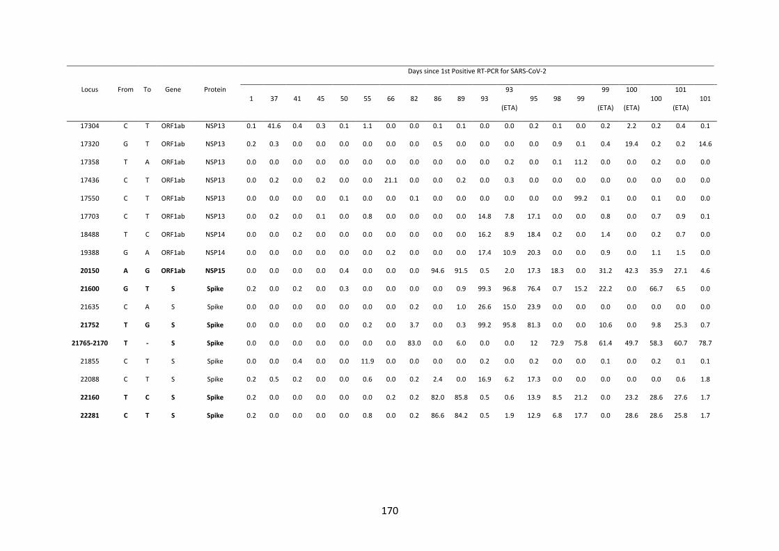
170
Locus From To Gene Protein
Days since 1st Positive RT-PCR for SARS-CoV-2
1 37 41 45 50 55 66 82 86 89 93 93
(ETA) 95 98 99
99
(ETA)
100
(ETA) 100
101
(ETA) 101
17304 C T ORF1ab NSP13 0.1 41.6 0.4 0.3 0.1 1.1 0.0 0.0 0.1 0.1 0.0 0.0 0.2 0.1 0.0 0.2 2.2 0.2 0.4 0.1
17320 G T ORF1ab NSP13 0.2 0.3 0.0 0.0 0.0 0.0 0.0 0.0 0.5 0.0 0.0 0.0 0.0 0.9 0.1 0.4 19.4 0.2 0.2 14.6
17358 T A ORF1ab NSP13 0.0 0.0 0.0 0.0 0.0 0.0 0.0 0.0 0.0 0.0 0.0 0.2 0.0 0.1 11.2 0.0 0.0 0.2 0.0 0.0
17436 C T ORF1ab NSP13 0.0 0.2 0.0 0.2 0.0 0.0 21.1 0.0 0.0 0.2 0.0 0.3 0.0 0.0 0.0 0.0 0.0 0.0 0.0 0.0
17550 C T ORF1ab NSP13 0.0 0.0 0.0 0.0 0.1 0.0 0.0 0.1 0.0 0.0 0.0 0.0 0.0 0.0 99.2 0.1 0.0 0.1 0.0 0.0
17703 C T ORF1ab NSP13 0.0 0.2 0.0 0.1 0.0 0.8 0.0 0.0 0.0 0.0 14.8 7.8 17.1 0.0 0.0 0.8 0.0 0.7 0.9 0.1
18488 T C ORF1ab NSP14 0.0 0.0 0.2 0.0 0.0 0.0 0.0 0.0 0.0 0.0 16.2 8.9 18.4 0.2 0.0 1.4 0.0 0.2 0.7 0.0
19388 G A ORF1ab NSP14 0.0 0.0 0.0 0.0 0.0 0.0 0.2 0.0 0.0 0.0 17.4 10.9 20.3 0.0 0.0 0.9 0.0 1.1 1.5 0.0
20150 A G ORF1ab NSP15 0.0 0.0 0.0 0.0 0.4 0.0 0.0 0.0 94.6 91.5 0.5 2.0 17.3 18.3 0.0 31.2 42.3 35.9 27.1 4.6
21600 G T S Spike 0.2 0.0 0.2 0.0 0.3 0.0 0.0 0.0 0.0 0.9 99.3 96.8 76.4 0.7 15.2 22.2 0.0 66.7 6.5 0.0
21635 C A S Spike 0.0 0.0 0.0 0.0 0.0 0.0 0.0 0.2 0.0 1.0 26.6 15.0 23.9 0.0 0.0 0.0 0.0 0.0 0.0 0.0
21752 T G S Spike 0.0 0.0 0.0 0.0 0.0 0.2 0.0 3.7 0.0 0.3 99.2 95.8 81.3 0.0 0.0 10.6 0.0 9.8 25.3 0.7
21765-2170 T - S Spike 0.0 0.0 0.0 0.0 0.0 0.0 0.0 83.0 0.0 6.0 0.0 0.0 12 72.9 75.8 61.4 49.7 58.3 60.7 78.7
21855 C T S Spike 0.0 0.0 0.4 0.0 0.0 11.9 0.0 0.0 0.0 0.0 0.2 0.0 0.2 0.0 0.0 0.1 0.0 0.2 0.1 0.1
22088 C T S Spike 0.2 0.5 0.2 0.0 0.0 0.6 0.0 0.2 2.4 0.0 16.9 6.2 17.3 0.0 0.0 0.0 0.0 0.0 0.6 1.8
22160 T C S Spike 0.2 0.0 0.0 0.0 0.0 0.0 0.2 0.2 82.0 85.8 0.5 0.6 13.9 8.5 21.2 0.0 23.2 28.6 27.6 1.7
22281 C T S Spike 0.2 0.0 0.0 0.0 0.0 0.8 0.0 0.2 86.6 84.2 0.5 1.9 12.9 6.8 17.7 0.0 28.6 28.6 25.8 1.7
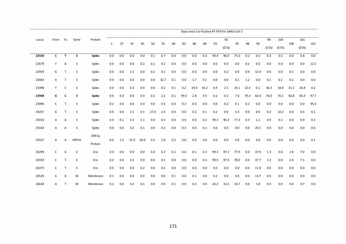
171
Locus From To Gene Protein
Days since 1st Positive RT-PCR for SARS-CoV-2
1 37 41 45 50 55 66 82 86 89 93 93
(ETA) 95 98 99
99
(ETA)
100
(ETA) 100
101
(ETA) 101
22550 C T S Spike 0.0 0.0 0.0 0.0 0.1 0.7 0.0 0.0 0.0 0.3 99.4 96.9 75.0 0.2 0.5 6.3 0.5 0.0 5.8 0.0
22679 T A S Spike 0.0 0.0 0.0 0.2 0.1 0.2 0.0 0.0 0.0 0.0 0.0 0.0 0.0 0.2 0.0 0.0 0.0 0.0 0.0 12.5
22959 G T S Spike 0.0 0.0 1.5 0.0 0.2 0.2 0.0 0.0 0.0 0.0 0.0 0.2 0.0 0.0 13.4 0.0 0.0 0.1 0.0 0.0
23063 A T S Spike 0.0 0.0 0.0 0.0 0.0 32.7 0.1 0.0 1.7 0.1 0.0 0.0 0.1 1.2 0.0 0.1 0.1 0.1 0.0 0.0
23398 T C S Spike 0.0 0.0 0.2 0.0 0.0 0.2 0.1 0.2 24.9 65.2 0.4 2.5 16.1 12.4 0.1 36.3 34.8 31.1 26.8 0.2
23948 G C S Spike 0.0 0.3 0.0 0.3 0.1 2.2 0.1 99.5 2.8 9.5 0.2 0.2 7.6 79.3 82.4 76.0 70.1 82.8 65.4 97.7
23996 C T S Spike 0.2 0.0 0.0 0.4 0.0 0.3 0.0 0.2 0.0 0.0 0.0 0.2 0.1 0.2 0.0 0.0 0.0 0.0 0.0 95.4
24257 G T S Spike 0.0 0.0 2.1 0.1 27.0 2.4 0.0 0.0 0.2 0.1 0.2 0.0 0.3 0.0 0.0 0.2 10.2 0.0 0.3 0.1
25033 G A S Spike 0.0 0.1 0.2 1.1 0.0 0.3 0.0 0.0 0.0 0.2 99.3 96.3 77.4 0.3 1.1 4.9 0.1 4.0 6.9 0.2
25334 G A S Spike 0.0 0.0 0.2 0.1 0.0 0.2 0.0 0.3 0.0 0.1 0.0 0.0 0.0 0.0 20.1 0.0 0.0 0.0 0.0 0.0
25537 G A ORF3a ORF3a
Protein 0.0 2.5 15.9 20.0 0.3 2.9 0.2 0.0 0.0 0.0 0.0 0.0 0.0 0.0 0.0 0.0 0.0 0.0 0.0 0.1
26299 C A E Env 0.0 0.0 0.0 0.0 0.0 0.2 0.1 0.0 0.1 0.3 99.3 97.3 77.9 0.0 37.9 1.3 0.0 2.8 7.0 0.0
26333 C T E Env 0.2 0.0 0.2 0.0 0.0 0.2 0.0 0.0 0.0 0.3 99.5 97.4 78.0 0.0 37.7 1.3 0.0 3.4 7.1 0.0
26373 C T E Env 0.0 0.0 0.0 0.2 0.0 0.2 0.0 0.0 0.0 0.0 0.0 0.0 0.0 0.0 11.8 0.0 0.0 0.0 0.0 0.0
26529 G A M Membrane 0.1 0.0 0.0 0.0 0.0 0.0 0.1 0.0 0.1 0.0 0.2 0.0 0.0 0.0 14.7 0.0 0.0 0.0 0.0 0.0
26634 G T M Membrane 0.2 0.0 0.2 0.1 0.0 0.0 0.1 0.0 0.2 0.0 43.2 32.4 10.7 0.0 1.8 0.3 0.0 0.0 0.7 0.0
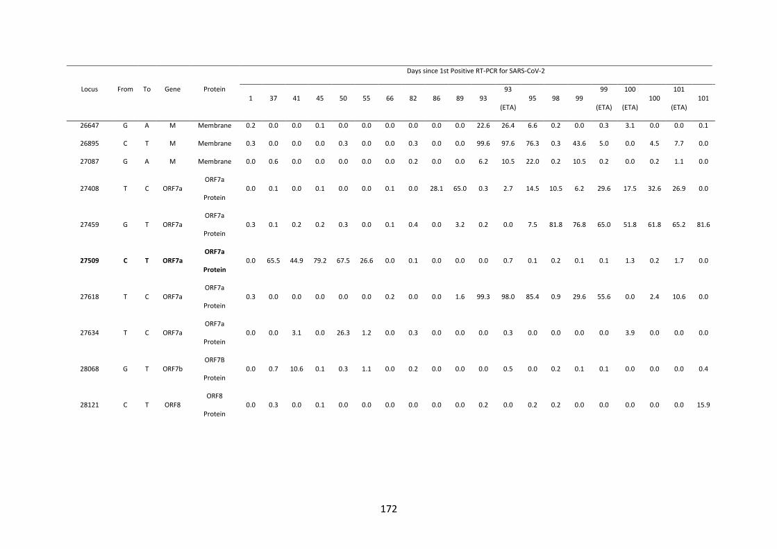
172
Locus From To Gene Protein
Days since 1st Positive RT-PCR for SARS-CoV-2
1 37 41 45 50 55 66 82 86 89 93 93
(ETA) 95 98 99
99
(ETA)
100
(ETA) 100
101
(ETA) 101
26647 G A M Membrane 0.2 0.0 0.0 0.1 0.0 0.0 0.0 0.0 0.0 0.0 22.6 26.4 6.6 0.2 0.0 0.3 3.1 0.0 0.0 0.1
26895 C T M Membrane 0.3 0.0 0.0 0.0 0.3 0.0 0.0 0.3 0.0 0.0 99.6 97.6 76.3 0.3 43.6 5.0 0.0 4.5 7.7 0.0
27087 G A M Membrane 0.0 0.6 0.0 0.0 0.0 0.0 0.0 0.2 0.0 0.0 6.2 10.5 22.0 0.2 10.5 0.2 0.0 0.2 1.1 0.0
27408 T C ORF7a ORF7a
Protein 0.0 0.1 0.0 0.1 0.0 0.0 0.1 0.0 28.1 65.0 0.3 2.7 14.5 10.5 6.2 29.6 17.5 32.6 26.9 0.0
27459 G T ORF7a ORF7a
Protein 0.3 0.1 0.2 0.2 0.3 0.0 0.1 0.4 0.0 3.2 0.2 0.0 7.5 81.8 76.8 65.0 51.8 61.8 65.2 81.6
27509 C T ORF7a ORF7a
Protein 0.0 65.5 44.9 79.2 67.5 26.6 0.0 0.1 0.0 0.0 0.0 0.7 0.1 0.2 0.1 0.1 1.3 0.2 1.7 0.0
27618 T C ORF7a ORF7a
Protein 0.3 0.0 0.0 0.0 0.0 0.0 0.2 0.0 0.0 1.6 99.3 98.0 85.4 0.9 29.6 55.6 0.0 2.4 10.6 0.0
27634 T C ORF7a ORF7a
Protein 0.0 0.0 3.1 0.0 26.3 1.2 0.0 0.3 0.0 0.0 0.0 0.3 0.0 0.0 0.0 0.0 3.9 0.0 0.0 0.0
28068 G T ORF7b ORF7B
Protein 0.0 0.7 10.6 0.1 0.3 1.1 0.0 0.2 0.0 0.0 0.0 0.5 0.0 0.2 0.1 0.1 0.0 0.0 0.0 0.4
28121 C T ORF8 ORF8
Protein 0.0 0.3 0.0 0.1 0.0 0.0 0.0 0.0 0.0 0.0 0.2 0.0 0.2 0.2 0.0 0.0 0.0 0.0 0.0 15.9
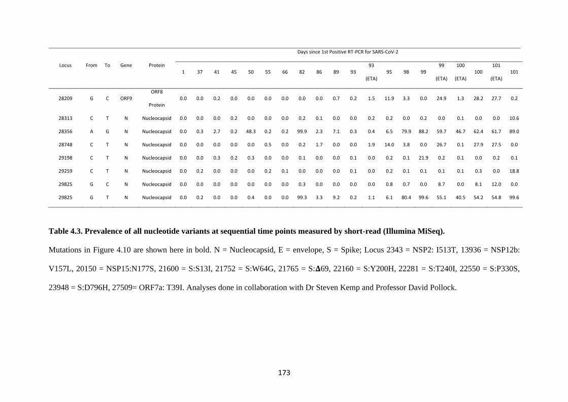
173
Locus From To Gene Protein
Days since 1st Positive RT-PCR for SARS-CoV-2
1 37 41 45 50 55 66 82 86 89 93 93
(ETA) 95 98 99
99
(ETA)
100
(ETA) 100
101
(ETA) 101
28209 G C ORF9 ORF8
Protein 0.0 0.0 0.2 0.0 0.0 0.0 0.0 0.0 0.0 0.7 0.2 1.5 11.9 3.3 0.0 24.9 1.3 28.2 27.7 0.2
28313 C T N Nucleocapsid 0.0 0.0 0.0 0.2 0.0 0.0 0.0 0.2 0.1 0.0 0.0 0.2 0.2 0.0 0.2 0.0 0.1 0.0 0.0 10.6
28356 A G N Nucleocapsid 0.0 0.3 2.7 0.2 48.3 0.2 0.2 99.9 2.3 7.1 0.3 0.4 6.5 79.9 88.2 59.7 46.7 62.4 61.7 89.0
28748 C T N Nucleocapsid 0.0 0.0 0.0 0.0 0.0 0.5 0.0 0.2 1.7 0.0 0.0 1.9 14.0 3.8 0.0 26.7 0.1 27.9 27.5 0.0
29198 C T N Nucleocapsid 0.0 0.0 0.3 0.2 0.3 0.0 0.0 0.1 0.0 0.0 0.1 0.0 0.2 0.1 21.9 0.2 0.1 0.0 0.2 0.1
29259 C T N Nucleocapsid 0.0 0.2 0.0 0.0 0.0 0.2 0.1 0.0 0.0 0.0 0.1 0.0 0.2 0.1 0.1 0.1 0.1 0.3 0.0 18.8
29825 G C N Nucleocapsid 0.0 0.0 0.0 0.0 0.0 0.0 0.0 0.3 0.0 0.0 0.0 0.0 0.8 0.7 0.0 8.7 0.0 8.1 12.0 0.0
29825 G T N Nucleocapsid 0.0 0.2 0.0 0.0 0.4 0.0 0.0 99.3 3.3 9.2 0.2 1.1 6.1 80.4 99.6 55.1 40.5 54.2 54.8 99.6
Table 4.3. Prevalence of all nucleotide variants at sequential time points measured by short-read (Illumina MiSeq).
Mutations in Figure 4.10 are shown here in bold. N = Nucleocapsid, E = envelope, S = Spike; Locus 2343 = NSP2: I513T, 13936 = NSP12b:
V157L, 20150 = NSP15:N177S, 21600 = S:S13I, 21752 = S:W64G, 21765 = S:𝚫69, 22160 = S:Y200H, 22281 = S:T240I, 22550 = S:P330S,
23948 = S:D796H, 27509= ORF7a: T39I. Analyses done in collaboration with Dr Steven Kemp and Professor David Pollock.
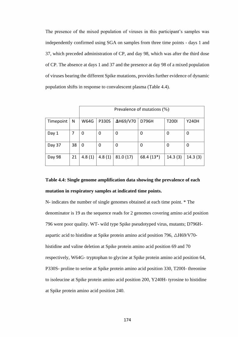
174
The presence of the mixed population of viruses in this participant’s samples was
independently confirmed using SGA on samples from three time points - days 1 and
37, which preceded administration of CP, and day 98, which was after the third dose
of CP. The absence at days 1 and 37 and the presence at day 98 of a mixed population
of viruses bearing the different Spike mutations, provides further evidence of dynamic
population shifts in response to convalescent plasma (Table 4.4).
Prevalence of mutations (%)
Timepoint N W64G P330S 𝚫H69/V70 D796H T200I Y240H
Day 1 7 0 0 0 0 0 0
Day 37 38 0 0 0 0 0 0
Day 98 21 4.8 (1) 4.8 (1) 81.0 (17) 68.4 (13*) 14.3 (3) 14.3 (3)
Table 4.4: Single genome amplification data showing the prevalence of each
mutation in respiratory samples at indicated time points.
N- indicates the number of single genomes obtained at each time point. * The
denominator is 19 as the sequence reads for 2 genomes covering amino acid position
796 were poor quality. WT- wild type Spike pseudotyped virus, mutants; D796H-
aspartic acid to histidine at Spike protein amino acid position 796, △H69/V70-
histidine and valine deletion at Spike protein amino acid position 69 and 70
respectively, W64G- tryptophan to glycine at Spike protein amino acid position 64,
P330S- proline to serine at Spike protein amino acid position 330, T200I- threonine
to isoleucine at Spike protein amino acid position 200, Y240H- tyrosine to histidine
at Spike protein amino acid position 240.
175
4.4 Spike Mutants D796H causes a decrease in infectivity which is compensated
for by acquisition of 𝚫69/70 deletion
The phenotypic properties of Spike mutant virus bearing four of the observed
mutants W64G + P330S and 𝚫H69/V70 + D796H were investigated using lentivirus
pseudotyping infectivity and neutralisation assays. These mutations were engineered
into codon optimised Spike expression pCDNA_SARS-CoV-2_S plasmid bearing
D614G by site direct mutatgenesis (SDM), either as single or double mutation as
indicated (Methods 2.2.8). Pseudotyped viruses were generated by transfection of
HEK293T cells with p8.91 lentivirus packaging plasmid, pCSFLW luciferase
expression plasmid and pCDNA_SARS-CoV-2_S Spike expression plasmid.
Pseudotyped viruses were harvested 48 hours later (Methods 2.2.9). The wild type
(WT) was the Spike pseudotyped virus bearing D614G and the pseudotyped virus
bearing W64G + P330S and 𝚫H69/V70 + D796H either individually or in pairs were
designated single or double mutants prefixed by the amino acid mutation and
position in Spike protein.
Firstly, the infectivity of these pseudotyped viruses were determined in a single
round infectivity assay (Methods 2.2.9) by titration on target cells- HEK293T cells
which had transiently been transfected with ACE2 and TMPRSS2 or HeLA
expressing ACE2 receptors. The relative light units (RLU) per µl of infectious dose
was determined using the Steady-Glo Luciferase assay system (Promega). (Figures
4.13A, B). In another experiment, infectivity was measured by normalising virus
input for reverse RT activity in the virus supernatant as measured by qPCR (Methods
2.2.10) (Figures 4.13 C, D). The normalised input was used to infect either HeLa
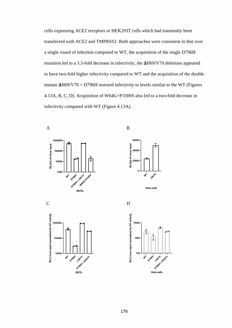
176
cells expressing ACE2 receptors or HEK293T cells which had transiently been
transfected with ACE2 and TMPRSS2. Both approaches were consistent in that over
a single round of infection compared to WT, the acquisition of the single D796H
mutation led to a 3.5-fold decrease in infectivity, the 𝚫H69/V70 deletions appeared
to have two-fold higher infectivity compared to WT and the acquisition of the double
mutant 𝚫H69/V70 + D796H restored infectivity to levels similar to the WT (Figures
4.13A, B, C, D). Acquisition of W64G+P3300S also led to a two-fold decrease in
infectivity compared with WT (Figure 4.13A).
A
B
C
D
WT
∆69/
70
0
20000
40000
60000
Hela cells
RL
U/µ
l o
f vir
us in
pu
t
177
Figure 4.13. SARS-CoV-2 Spike pseudotyped virus infectivity. A, B. Infectivity
determined by titration of the pseudotyped viruses on the indicated target cell line.
Data points are RLU/µl of input virus, are mean ± s.d. of two technical replicates and
represent two independent experiments. C, D. Infectivity determined by
quantification of reverse transcriptase activity by qPCR. Data points are RLU
normalised for RT-activity are mean ± s.d. of two technical replicates and represent
two independent experiments. WT- wild type Spike pseudotyped virus, mutants;
D796H- aspartic acid to histidine at Spike protein amino acid position 796,
△H69/V70- histidine and valine deletion at Spike protein amino acid position 69 and
70 respectively, W64G- tryptophan to glycine at Spike protein amino acid position
64, P330S- proline to serine at Spike protein amino acid position 330.
4.5 Spike Mutants D796H and 𝚫69/70 individually or in combination does not
impact Spike incorporation
Spike protein from each pseudotyped virus was detected by Western blot in pelleted
virions obtained by centrifugation of the supernatant from transfected cells (Methods
2.2.11). Equal amounts of full-length Spike protein was detected in pelleted virions
in the WT and each mutant (Figure 4.14). HIV-1 p24 antibody probing was used to
monitor levels of lentiviral particle production.
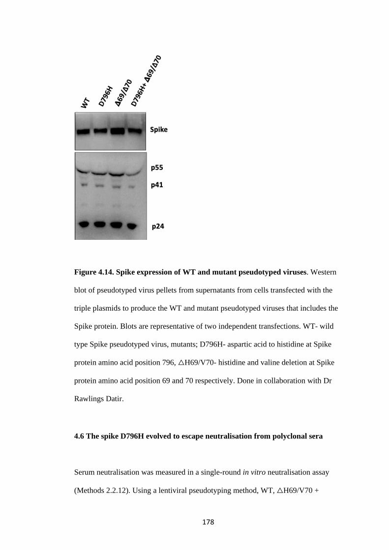
178
Figure 4.14. Spike expression of WT and mutant pseudotyped viruses. Western
blot of pseudotyped virus pellets from supernatants from cells transfected with the
triple plasmids to produce the WT and mutant pseudotyped viruses that includes the
Spike protein. Blots are representative of two independent transfections. WT- wild
type Spike pseudotyped virus, mutants; D796H- aspartic acid to histidine at Spike
protein amino acid position 796, △H69/V70- histidine and valine deletion at Spike
protein amino acid position 69 and 70 respectively. Done in collaboration with Dr
Rawlings Datir.
4.6 The spike D796H evolved to escape neutralisation from polyclonal sera
Serum neutralisation was measured in a single-round in vitro neutralisation assay
(Methods 2.2.12). Using a lentiviral pseudotyping method, WT, △H69/V70 +
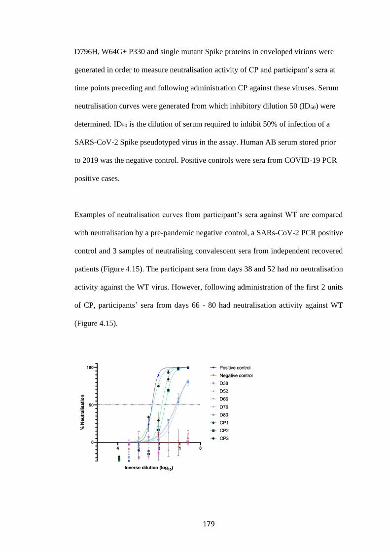
179
D796H, W64G+ P330 and single mutant Spike proteins in enveloped virions were
generated in order to measure neutralisation activity of CP and participant’s sera at
time points preceding and following administration CP against these viruses. Serum
neutralisation curves were generated from which inhibitory dilution 50 (ID50) were
determined. ID50 is the dilution of serum required to inhibit 50% of infection of a
SARS-CoV-2 Spike pseudotyped virus in the assay. Human AB serum stored prior
to 2019 was the negative control. Positive controls were sera from COVID-19 PCR
positive cases.
Examples of neutralisation curves from participant’s sera against WT are compared
with neutralisation by a pre-pandemic negative control, a SARs-CoV-2 PCR positive
control and 3 samples of neutralising convalescent sera from independent recovered
patients (Figure 4.15). The participant sera from days 38 and 52 had no neutralisation
activity against the WT virus. However, following administration of the first 2 units
of CP, participants’ sera from days 66 - 80 had neutralisation activity against WT
(Figure 4.15).

180
Figure 4.15. Neutralisation curves against the WT pseudotyped virus. Days 38
and 52 of the participant’s sera with no neutralisation activity against WT (pink).
Days 66 to 80 of the participant’s sera with neutralisation activity against WT (light
blue). CP- Convalescent plasma, units 1 to 3 with activity against the WT (green).
Negative control- pre-pandemic human AB serum (red). Positive control- serum
from a SARS-CoV-2 PCR positive case (dark blue). D- day sample was taken, day 1
being first PCR positive SARS-CoV-2 swab. WT- wild type Spike pseudotyped
virus.
D796H alone and the D796H + △H69/V70 double mutant were less sensitive to
neutralisation by all CP samples. The neutralisation curves show a shift to the right
for both these mutants (Figure 4.16A). The was at least a 2-fold decrease in ID50
against the D796H + △H69/V70 mutants by CP samples 2 and 3 and a 1.3-fold
decrease by CP 1 (Table 4.5). These mutations emerged after administration of CP
units 1 and 2, diminished and then rebounded after CP unit 3, which may explain why
the fold decrease by CP 1 was lower than that of CP 2 and 3. △H69/V70 deletion on
its own had no impact in neutralisation activity by any of the CPs (Figure 4.16A).
Patient derived serum from days 64 and 66, one day after CP1 and CP2 respectively,
similarly showed lower potency against the D796H + △H69/V70 mutants, with 1.7 to
2.6-fold decrease in ID50s (Figure 4.16B, Table 4.5).
W64G + P330S as a double mutant was not detected in the participants’ samples
until day 93 when it reached a prevalence 99%. It was already diminishing on day 95
when the 3rd unit of CP was given, probably in response to remdesivir given on day

181
93. It completely disappeared and rebounded again to 25% (W64G) and 6% (P330S)
by day 101. This double mutant was less sensitive to neutralisation by CP unit 3,
with a 3.4-fold decrease in ID50 (Fig 4.17, Table 4.6). CP unit 2 had a 1.5-fold
decrease in ID50 against the mutant whilst CP1 has equal neutralisation activity
against the mutant and the WT. (Fig 4.17, Table 4.6).
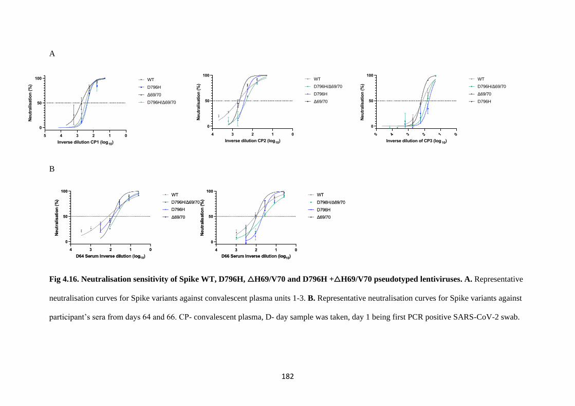
182
A
B
Fig 4.16. Neutralisation sensitivity of Spike WT, D796H, △H69/V70 and D796H +△H69/V70 pseudotyped lentiviruses. A. Representative
neutralisation curves for Spike variants against convalescent plasma units 1-3. B. Representative neutralisation curves for Spike variants against
participant’s sera from days 64 and 66. CP- convalescent plasma, D- day sample was taken, day 1 being first PCR positive SARS-CoV-2 swab.
012345
0
50
100
Inverse dilution CP1 (log10)
Neu
tralisati
on
(%
)
WT
D796H
Δ69/70
D796H/Δ69/70
01234
0
50
100
Inverse dilution CP2 (log10)
Neu
tralisati
on
(%
)
WT
D796H/Δ69/70
D796H
Δ69/70
012345
0
50
100
Inverse dilution of CP3 (log 10)
Neu
tralisati
on
(%
)
WT
D796H/Δ69/70
Δ69/70
D796H

183
WT- wild type Spike pseudotyped virus, mutants; D796H- aspartic acid to histidine
at Spike protein amino acid position 796, △H69/V70- histidine and valine deletion
at Spike protein amino acid position 69 and 70 respectively. Data points represent
mean neutralisation of technical replicates and error bars represent standard error of
the mean. Data are representative of two independent experiments.
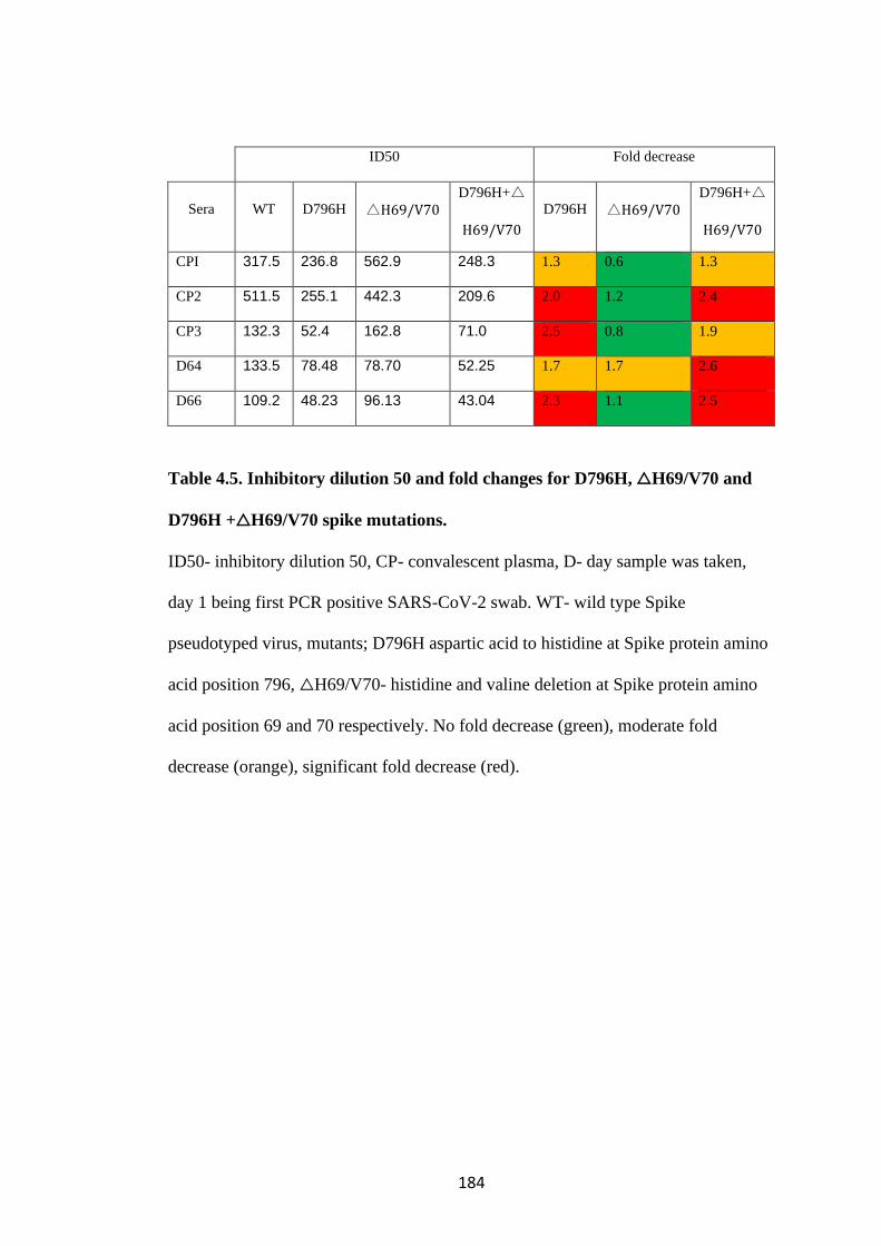
184
ID50 Fold decrease
Sera WT D796H △H69/V70 D796H+△
H69/V70
D796H △H69/V70
D796H+△
H69/V70
CPI 317.5 236.8 562.9 248.3 1.3 0.6 1.3
CP2 511.5 255.1 442.3 209.6 2.0 1.2 2.4
CP3 132.3 52.4 162.8 71.0 2.5 0.8 1.9
D64 133.5 78.48 78.70 52.25 1.7 1.7 2.6
D66 109.2 48.23 96.13 43.04 2.3 1.1 2.5
Table 4.5. Inhibitory dilution 50 and fold changes for D796H, △H69/V70 and
D796H +△H69/V70 spike mutations.
ID50- inhibitory dilution 50, CP- convalescent plasma, D- day sample was taken,
day 1 being first PCR positive SARS-CoV-2 swab. WT- wild type Spike
pseudotyped virus, mutants; D796H aspartic acid to histidine at Spike protein amino
acid position 796, △H69/V70- histidine and valine deletion at Spike protein amino
acid position 69 and 70 respectively. No fold decrease (green), moderate fold
decrease (orange), significant fold decrease (red).
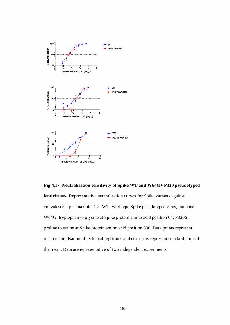
185
Fig 4.17. Neutralisation sensitivity of Spike WT and W64G+ P330 pseudotyped
lentiviruses. Representative neutralisation curves for Spike variants against
convalescent plasma units 1-3. WT- wild type Spike pseudotyped virus, mutants;
W64G- tryptophan to glycine at Spike protein amino acid position 64, P330S-
proline to serine at Spike protein amino acid position 330. Data points represent
mean neutralisation of technical replicates and error bars represent standard error of
the mean. Data are representative of two independent experiments.
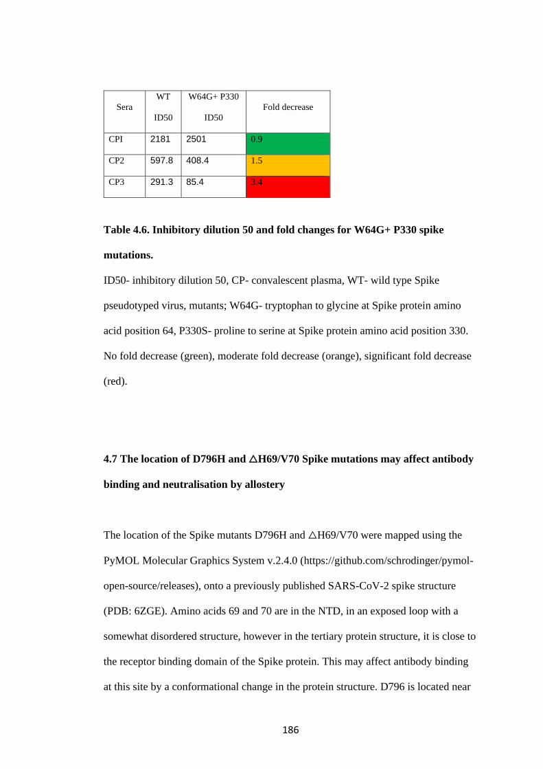
186
Sera
WT
ID50
W64G+ P330
ID50
Fold decrease
CPI 2181 2501 0.9
CP2 597.8 408.4 1.5
CP3 291.3 85.4 3.4
Table 4.6. Inhibitory dilution 50 and fold changes for W64G+ P330 spike
mutations.
ID50- inhibitory dilution 50, CP- convalescent plasma, WT- wild type Spike
pseudotyped virus, mutants; W64G- tryptophan to glycine at Spike protein amino
acid position 64, P330S- proline to serine at Spike protein amino acid position 330.
No fold decrease (green), moderate fold decrease (orange), significant fold decrease
(red).
4.7 The location of D796H and △H69/V70 Spike mutations may affect antibody
binding and neutralisation by allostery
The location of the Spike mutants D796H and △H69/V70 were mapped using the
PyMOL Molecular Graphics System v.2.4.0 (https://github.com/schrodinger/pymol-
open-source/releases), onto a previously published SARS-CoV-2 spike structure
(PDB: 6ZGE). Amino acids 69 and 70 are in the NTD, in an exposed loop with a
somewhat disordered structure, however in the tertiary protein structure, it is close to
the receptor binding domain of the Spike protein. This may affect antibody binding
at this site by a conformational change in the protein structure. D796 is located near

187
the base of Spike in the S2 subunit and is also in an external loop. This site may not
come into contact with neutralising antibodies but may mediate escape by
introduction of a charged amino acid, again, leading to a conformational change of
the protein structure (Figure 4.18).
Figure 4.18. Spike mutations ΔH69/70 and D796H. Surface protein representation
of the Spike homotrimer are coloured separately in shades of grey. In red are the
amino acid residues H69 and V70, deleted in the N-terminal domain. In orange is
D796H in the Spike S2 subunit. Close-ups are cartoon stick representation of
ΔH69/Y70 (above) and D796H (below) (PDB: 6ZGE Wrobek et al., 2020). Source:
Kemp and Collier et al, SARS-CoV-2 evolution during treatment of chronic
infection, Nature 592, 277–282 (2021). https://doi.org/10.1038/s41586-021-03291-y.
188
4.8 Discussion
This in-depth analyses of in vivo dynamic evolution and escape, demonstrates within-
host adaptation of SARS-CoV-2 to evade neutralising antibodies.
It is possible that some of the viruses sampled represent a compartmentalised
population that was previously unsampled, particularly the virus population
containing W64G + P330S, which emerged at high prevalence on a particularly long
branch at days 93 and 95. The SGA analyses suggests that although this was a
divergent cluster, it was present within the mixed population of viruses at a low level
in a sample from a later timepoint. In addition, the phylogenetic tree showed that this
population was rooted within the main population.
Ideally, the neutralisation assay would be replicated in a live virus system. However,
Spike pseudotyped viral infection assays have been shown to have similar
characteristics as assays using infectious wild type SARS-CoV-2 (Schmidt et al.
2020).
Although recent head to head analyses of long read Oxford Nanopore Technology
(ONT) and short read Illumina found that they are equally good at consensus-level
sequence determination (Bull et al. 2020), short read Illumina MiSeq data was used
for variant calling in the mutational prevalence analyses in this study. This is
because Illumina is known to have a lower sequencing error rate (Bull et al. 2020;
Kim et al. 2020). However, errors introduced in sample preparation and PCR may
result in missing some variants and patterns of intrahost diversity. Therefore, future

189
work will include using high-throughput single genome amplification from RNA,
which lends itself well to exploring variations in a limited region of the genome (Ko
et al. 2021), as well as reconciling the dynamic shifts in the virus population with
antibody responses and mapping the immunodominant epitopes targeted by
autologous antibodies to the emerging viruses detected.
Of note, no regions outside of the Spike protein were considered in this work. This is
because the Spike protein is really well characterised and it is the target of neutralising
antibodies following natural infection. Although other regions outside of the Spike
protein might contribute to immune escape, the Spike protein is most likely directly
involved in this process.
This is a study of a single case and therefore limits generalisability. The circumstances
of this participant with absent B cell, suboptimal T cell response maybe applicable to
a broad group of immunocompromised hosts for example the elderly. In the next
chapter I will explore some factors associated with immune response to Spike directed
mRNA vaccines including age and cytomegalovirus seropositivity.

190
CHAPTER 5: RESULTS 3- AGE-RELATED
HETEROGENEITY OF SARS-COV-2 MRNA VACCINE-
ELICITED RESPONSES
Key Findings:
• SARS-CoV-2 specific binding antibody levels negatively correlate
with age.
• The odds of an inadequate neutralising antibody response to a spike
pseudotyped virus after only one dose of the mRNA BNT162b2
vaccine was 3.7 in those ≥80 years old.
• Poor serum neutralisation of WT Spike pseudotyped virus after the
first dose of mRNA BNT162b2 is overcome by the booster/second
dose.
• The frequency of IFN𝛾 secreting Spike specific T cells following
only one dose of mRNA BNT162b2 vaccine is significantly lower in
the ≥80 years group compared with the <80 years group. This is
overcome by the booster/second dose.
• The frequency of IL-2 secreting Spike specific T cells following only
one dose of mRNA BNT162b2 vaccine is significantly lower in the
≥80 years group compared with the <80 years group. This however
was not increased by the booster/second dose.
• The frequency of IFN𝛾 secreting Spike specific T cells following
only one dose of mRNA BNT162b2 vaccine is higher in the ≥80 years
group with HCMV seropositivity compared to ≥80 year who are
HCMV negative.
191
From the previous body of work on virus escape from neutralising antibodies, it was
clear that the ability of the SARS-CoV-2 Spike expressing pseudotyped viruses to
escape inhibition by neutralising antibodies had implications for response to vaccine-
elicited antibodies. Vaccines are a key tool to controlling the COVID-19 pandemic.
In particular, mRNA vaccine had shown good efficacy in clinical trials after two
doses, separated by a three or four week gap (Baden et al. 2020; Polack et al. 2020).
However, the duration of protection is not known and very few participants above
the age of 80 years were included in the clinical trials despite being at greatest risk of
severe disease and death (Docherty et al. 2020). The Joint Committee on
Vaccination and Immunisation (JCVI) recommended extending the dosing interval
to 12 weeks in order to vaccinate a greater proportion of the population during a
period of substantial transmission (Care 2021). This strategy is being considered in
other countries in order to maximise first dose administration (DW. 2021; Kadire,
Wachter, and Lurie 2021).
Ageing can impact responses in many ways including but not limited to reducing the
magnitude of antibody response to vaccines (Wilkinson et al. 2017), shortening the
persistence of antibody response (Powers and Belshe 1993), limiting the quality of
antibodies via reduced affinity and breadth, limiting induction of CD4+/8+ T cell
response (Westmeier et al. 2020) and limiting induction of effector memory
(Gustafson et al. 2020). Data on vaccine responses, particularly in groups under-
represented in clinical trial are necessary to understand the efficacy of vaccination
using this regime.
192
Under the ethical framework of an ongoing large NIHR CRF trial called the
Bioresource, a cohort study was conducted to assess real world immune responses
following vaccination with mRNA-based vaccine BNT162b21 in elderly participants
from the community and younger health care workers. The study aimed to
investigate age as a risk factor for escape from mRNA BNT162b2 vaccine-elicited
antibody neutralisation. It also aimed to characterise binding antibody responses, T
cell immune responses to the vaccine by measuring SARS-CoV-2 Spike specific
IFN𝛾 and IL-2 responses in PBMCs, the B Cell repertoire, autoantibodies and
inflammatory markers.
5.1 Cohort Description and study procedure
One hundred and one participants received the first dose of the BNT162b2 vaccine
whilst twenty-one received their second dose, including one who was recruited after
their second dose of vaccine (Table 5.1). The second dose was initially given under
the manufacturer’s recommended schedule, three weeks after the first but was
changed during the study to 12 weeks following the JCVI’s recommendation.
Therefore, the remaining eighty participants are yet to receive their second dose.
Participants recruited reflected the Joint Committee on Vaccination and
Immunisation (JCVI) priority groups for immunisation and included older adults and
health care workers receiving the first dose of the BNT162b2 vaccine between the
14th of December 2020 to the 29th of January 2021. Each participant was seen at least
3 weeks after their first vaccine dose and up to 12 weeks after. Participants were then

193
seen again at 3 weeks following the second dose of the BNT162b2 vaccine and
underwent the same blood sampling procedure (Figure 5.1).
194
Figure 5.1: Study flow describing the procedures for participants recruited. The
numbers of participants with results from each type of assay is indicated. The study
was approved by the East of England – Cambridge Central Research Ethics
Committee (17/EE/0025).
The median age of the cohort was 81 years (IQR 70-84) and 39% of participants
were female. Participants were stratified by age into 2 exposure groups- < 80 years
and ≥ 80 years. 42 (41.6%) of participants were in the <80 years group, whilst 59
(58.4%) were in the ≥ 80 years (Table 5.1). These exposure levels were chosen
because the JCVI priority list included participants over the 80 years old as the
highest priority to receive the available SARS-CoV-2 vaccines. In addition, there
was considerable concern about the efficacy of only one dose of the vaccine to
provide protection for the elderly who were at greatest risk from COVID1-9 disease
during a period of high background transmission in the community. It was
hypothesised that due to aging, the ≥80 years group would mount an inadequate
immune response with only one dose of the vaccine and would require a
booster/second for an adequate protective immune response.
Previous SARS-CoV-2 infection was determined by measuring anti-Nucleocapsid
protein and anti-Spike protein IgG antibody levels in all participants. When both
were elevated, the participants were deemed to have likely had exposure to pandemic
SARS-CoV-2. This is because the mRNA vaccine contains the sequence for the
Spike protein only. Although cross-reactivity with one of the other seasonal
coronaviruses could not be excluded, it was assumed that N protein antibody
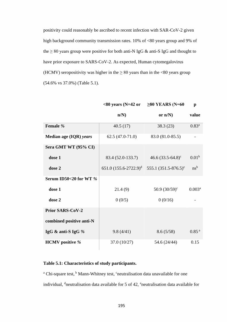
195
positivity could reasonably be ascribed to recent infection with SAR-CoV-2 given
high background community transmission rates. 10% of <80 years group and 9% of
the ≥ 80 years group were positive for both anti-N IgG & anti-S IgG and thought to
have prior exposure to SARS-CoV-2. As expected, Human cytomegalovirus
(HCMV) seropositivity was higher in the ≥ 80 years than in the <80 years group
(54.6% vs 37.0%) (Table 5.1).
<80 years (N=42 or
n/N)
≥80 YEARS (N=60
or n/N)
p
value
Female % 40.5 (17) 38.3 (23) 0.83a
Median age (IQR) years 62.5 (47.0-71.0) 83.0 (81.0-85.5) -
Sera GMT WT (95% CI)
dose 1
dose 2
83.4 (52.0-133.7)
651.0 (155.6-2722.9)d
46.6 (33.5-64.8)c
555.1 (351.5-876.5)e
0.01b
nsb
Serum ID50<20 for WT %
dose 1
dose 2
21.4 (9)
0 (0/5)
50.9 (30/59)c
0 (0/16)
0.003a
-
Prior SARS-CoV-2
combined positive anti-N
IgG & anti-S IgG %
9.8 (4/41)
8.6 (5/58)
0.85 a
HCMV positive % 37.0 (10/27) 54.6 (24/44) 0.15
Table 5.1: Characteristics of study participants.
a Chi-square test, b Mann-Whitney test, cneutralisation data unavailable for one
individual, dneutralisation data available for 5 of 42, eneutralisation data available for
196
16 of 60, GMT- geometric mean titre, WT- wild type, ID50- (Inhibitory dilution) –
the serum dilution achieving 50% neutralisation, ns- non-significant, CI-confidence
interval, N- Nucleocapsid protein, S- Spike protein, IgG- immunoglobulin.
5.2 Neutralisation activity of vaccine-elicited sera against SARS-CoV-2
following first and second dose mRNA BNT162b2 vaccine
The primary outcome was inadequate vaccine-elicited serum antibody neutralisation
activity at least 3 weeks after the first dose of vaccine, defined as an inhibitory
dilution 50 (ID50) of ≤1 in 20. This dilution was chosen because although the limit
of detection of the assay in 1 in 4, there is a degree of variability at lower dilutions
which is overcome by using a more conservative cut-off. This was measured in an in
vitro neutralisation assay and is the dilution of serum required to inhibit 50% of
infection of a Spike pseudotyped virus (Methods 2.3.4). The pseudotyped virus was
generated by transfection of HEK293T cells with p8.91 lentivirus packaging
plasmid, pCSFLW luciferase expression plasmid and pCDNA_SARS-CoV-2_S
Spike codon optimized expression plasmid D614G. Pseudotyped viruses were
harvested 48 hours later (Methods 2.3.3). The wild type (WT) was the Spike
pseudotyped virus bearing D614G mutation in Spike protein.
Serum neutralisation curves were generated from infection of 293T cells transiently
transfected with ACE2 and TMPRSS2 plasmids and infected with WT Spike
pseudotyped virus in the presence of a limiting dilution of vaccinee sera. This was
done in duplicate. ID50s were determined from these curves using Prism 9. (Figure
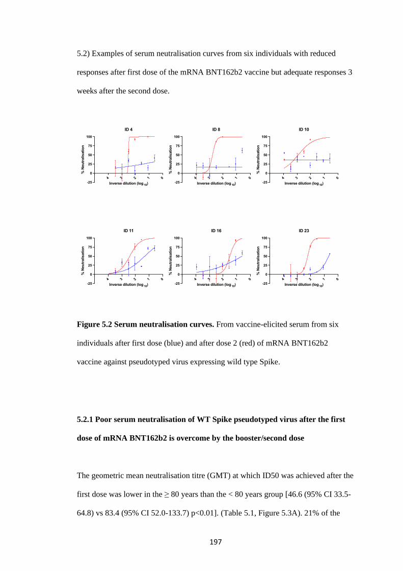
197
5.2) Examples of serum neutralisation curves from six individuals with reduced
responses after first dose of the mRNA BNT162b2 vaccine but adequate responses 3
weeks after the second dose.
Figure 5.2 Serum neutralisation curves. From vaccine-elicited serum from six
individuals after first dose (blue) and after dose 2 (red) of mRNA BNT162b2
vaccine against pseudotyped virus expressing wild type Spike.
5.2.1 Poor serum neutralisation of WT Spike pseudotyped virus after the first
dose of mRNA BNT162b2 is overcome by the booster/second dose
The geometric mean neutralisation titre (GMT) at which ID50 was achieved after the
first dose was lower in the ≥ 80 years than the < 80 years group [46.6 (95% CI 33.5-
64.8) vs 83.4 (95% CI 52.0-133.7) p<0.01]. (Table 5.1, Figure 5.3A). 21% of the
01234
-25
0
25
50
75
100
Inverse dilution (log 10)
% N
eu
tralisati
on
ID 4
01234
-25
0
25
50
75
100
Inverse dilution (log 10)
% N
eu
tralisati
on
ID 10
01234-25
0
25
50
75
100
Inverse dilution (log 10)
% N
eu
tralisati
on
ID 16
01234
-25
0
25
50
75
100
Inverse dilution (log 10)
% N
eu
tralisati
on
ID 8
01234-25
0
25
50
75
100
Inverse dilution (log 10)
% N
eu
tralisati
on
ID 11
01234-25
0
25
50
75
100
Inverse dilution (log 10)
% N
eu
tralisati
on
ID 23

198
<80 years group had inadequate neutralisation against the WT Spike pseudotyped
virus with an ID50 of <1:20 compared with 51% of the ≥ 80 years group. After the
second dose, sera from vaccinated individuals exhibited an increase in neutralising
titres. The GMT increased for the <80 years group to 651.0 (95% CI 155.6-2722.9)
and the ≥ 80 years group to 555.1 (95% CI 351.5-876.5) with no statistically
significant difference between these two groups (Table 5.1, Figure 5.3B).
A
B
Figure 5.3. Neutralisation activity by mRNA BNT162b2 vaccine sera against
SARS-CoV-2 in a Spike lentiviral pseudotyping assay expressing wild type
Spike. A. Inhibitory Dilution 50 (ID50) values for individuals after dose 1 (n=101,
blue) and after dose 2 (n=21, red) a comparing the two age groups; < 80 and ≥ 80
years. Geometric mean with s.d is shown. Each point is a mean of technical
replicates from two experiment repeats. B. ID50 for all individuals after dose 1
(n=101, blue) and after dose 2 (n=21, red). Mann-Whitney test was used for unpaired

199
comparisons and Wilcoxon matched-pairs signed rank test for paired comparisons. p-
values * <0.05, ** <0.01, **** <0.0001, ns not significant, HS – human AB serum
control.
Given that ageing is a continuum and stratification into < 80 and ≥ 80 years groups is
arbitrary but justified given public health policy at the time, age as a continuous
variable was correlated with serum neutralisation using a linear regression model.
Age showed a statistically significant negative correlation with log10 ID50 against
WT pseudotyped virus after the first but not the second dose of the BNT162b2
vaccine (Figure 5.4A, B).
A B
Figure 5.4. Neutralisation activity by mRNA BNT162b2 vaccine sera against
SARS-CoV-2 in a Spike lentiviral pseudotyping assay expressing wild type
Spike. Serum neutralisation of Spike (D614G) pseudotyped lentiviral particles
(inhibitory dilution at which 50% inhibition of infection is achieved, ID50), log10
200
transformed ID50 after dose 1 (A, n=101) and dose 2 (B, n=21). r– Pearson’s
correlation coefficient, β slope/regression coefficient, p-value.
5.2.2 Older age is a risk factor for poor serum neutralisation of WT Spike
pseudotyped virus after the first dose of mRNA BNT162b2
A logistic regression model was used to analyse the association between age and
inadequate vaccine-elicited serum antibody neutralisation against WT pseudotyped
virus after the first dose of the BNT162b2. The unadjusted odds ratio (OR) for
achieving inadequate neutralisation against WT was 4.4 (1.7-11.1), p0.002) for
participants ≥ 80 years old versus <80 years (Table 5.2). In the univariable analyses,
a longer time interval since the first vaccine dose was received was also associated
with inadequate neutralisation with an OR of 2.9 (95% CI 1.3-6.8) in those
vaccinated 9-12 weeks prior compared with those vaccinated between 3-8 weeks
prior. Sex, prior exposure to SAR-CoV-2 and HCMV serostatus were not associated
with inadequate vaccine-elicited serum antibody neutralisation. In the fully adjusted
model including age, sex, and time interval since the first dose of vaccine was
received, the association of older age with inadequate vaccine-elicited serum
antibody neutralisation was still present with an OR of 3.7 (95% CI 1.2-11.2) for
participants ≥ 80 years old versus <80 years but the association with time interval
since the first dose of vaccine was no longer present as it was a confounder for the
association between age and inadequate vaccine-elicited serum antibody
neutralisation. Other confounders such as the presence of comorbidities and a history
201
of immunosuppressive treatment could not be adjusted for as these data was not
available for the majority of participants.
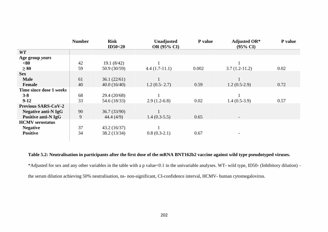
202
Number
Risk
ID50<20
Unadjusted
OR (95% CI)
P value Adjusted OR*
(95% CI)
P value
WT
Age group years
<80
≥ 80
42
59
19.1 (8/42)
50.9 (30/59)
1
4.4 (1.7-11.1)
0.002
1
3.7 (1.2-11.2)
0.02
Sex
Male
Female
61
40
36.1 (22/61)
40.0 (16/40)
1
1.2 (0.5- 2.7)
0.59
1
1.2 (0.5-2.9)
0.72
Time since dose 1 weeks
3-8
9-12
68
33
29.4 (20/68)
54.6 (18/33)
1
2.9 (1.2-6.8)
0.02
1
1.4 (0.5-3.9)
0.57
Previous SARS-CoV-2
Negative anti-N IgG
Positive anti-N IgG
90
9
36.7 (33/90)
44.4 (4/9)
1
1.4 (0.3-5.5)
0.65
-
HCMV serostatus
Negative
Positive
37
34
43.2 (16/37)
38.2 (13/34)
1
0.8 (0.3-2.1)
0.67
-
Table 5.2: Neutralisation in participants after the first dose of the mRNA BNT162b2 vaccine against wild type pseudotyped viruses.
*Adjusted for sex and any other variables in the table with a p value<0.1 in the univariable analyses. WT- wild type, ID50- (Inhibitory dilution) –
the serum dilution achieving 50% neutralisation, ns- non-significant, CI-confidence interval, HCMV- human cytomegalovirus.
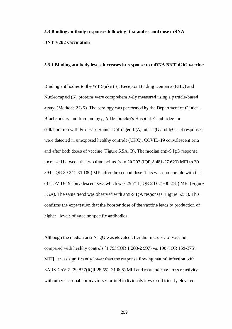
203
5.3 Binding antibody responses following first and second dose mRNA
BNT162b2 vaccination
5.3.1 Binding antibody levels increases in response to mRNA BNT162b2 vaccine
Binding antibodies to the WT Spike (S), Receptor Binding Domains (RBD) and
Nucleocapsid (N) proteins were comprehensively measured using a particle-based
assay. (Methods 2.3.5). The serology was performed by the Department of Clinical
Biochemistry and Immunology, Addenbrooke’s Hospital, Cambridge, in
collaboration with Professor Rainer Doffinger. IgA, total IgG and IgG 1-4 responses
were detected in unexposed healthy controls (UHC), COVID-19 convalescent sera
and after both doses of vaccine (Figure 5.5A, B). The median anti-S IgG response
increased between the two time points from 20 297 (IQR 8 481-27 629) MFI to 30
894 (IQR 30 341-31 180) MFI after the second dose. This was comparable with that
of COVID-19 convalescent sera which was 29 711(IQR 28 621-30 238) MFI (Figure
5.5A). The same trend was observed with anti-S IgA responses (Figure 5.5B). This
confirms the expectation that the booster dose of the vaccine leads to production of
higher levels of vaccine specific antibodies.
Although the median anti-N IgG was elevated after the first dose of vaccine
compared with healthy controls [1 793(IQR 1 283-2 997) vs. 198 (IQR 159-375)
MFI], it was significantly lower than the response flowing natural infection with
SARS-CoV-2 (29 877(IQR 28 652-31 008) MFI and may indicate cross reactivity
with other seasonal coronaviruses or in 9 individuals it was sufficiently elevated

204
along with anti-S IgG to suggest prior SARS-CoV-2 exposure (Figure 5.5A). The
same trend was observed with anti-S IgA responses (Figure 5.5B)
A
B
Figure 5.5. Binding antibody responses following vaccination with mRNA
BNT162b2 vaccine. A. Anti- S, N, RBD IgG responses post first dose (light green,
n=99) and second dose (dark green, n=21) compared to COVID-19 convalescent
patients (red, n=18) and healthy controls (grey, n=18) at serum dilutions 1 in 100. B.
Anti- S, N, RBD total IgA responses post first dose (light green, n=99) and second
dose (dark green, n=21) compared to COVID-19 convalescent patients (red, n=18)
and healthy controls (grey, n=18) at serum dilutions 1 in 100. Data presented are
MFI – mean fluorescence intensity ± s.d. S – Spike, N – nucleocapsid, RBD – Spike
receptor binding domain, Ig- immunoglobulin. Serology was done by the
Department of Clinical Biochemistry and Immunology, Addenbrooke’s Hospital,
Cambridge, UK.
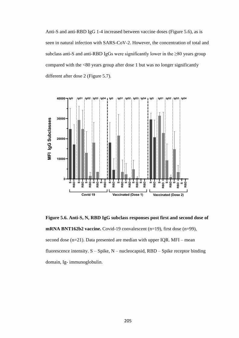
205
Anti-S and anti-RBD IgG 1-4 increased between vaccine doses (Figure 5.6), as is
seen in natural infection with SARS-CoV-2. However, the concentration of total and
subclass anti-S and anti-RBD IgGs were significantly lower in the ≥80 years group
compared with the <80 years group after dose 1 but was no longer significantly
different after dose 2 (Figure 5.7).
Figure 5.6. Anti-S, N, RBD IgG subclass responses post first and second dose of
mRNA BNT162b2 vaccine. Covid-19 convalescent (n=19), first dose (n=99),
second dose (n=21). Data presented are median with upper IQR. MFI – mean
fluorescence intensity. S – Spike, N – nucleocapsid, RBD – Spike receptor binding
domain, Ig- immunoglobulin.
S
RB
D S
RB
D S
RB
D S
RB
D S
RB
D S
RB
D S
RB
D S
RB
D S
RB
D S
RB
D S
RB
D S
RB
D S
RB
D S
RB
D
S
RB
D
0
10000
20000
30000
40000
MF
I Ig
G S
ub
cla
sses
Vaccinated (Dose 2)
IgGIgGIgG
Vaccinated (Dose 1)Covid 19
IgG1 IgG2 IgG3 IgG4 IgG1 IgG2 IgG3 IgG4 IgG1 IgG2 IgG3 IgG4
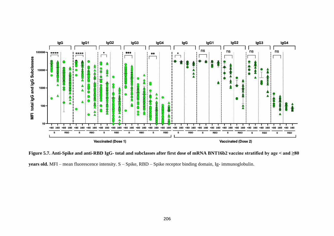
206
Figure 5.7. Anti-Spike and anti-RBD IgG- total and subclasses after first dose of mRNA BNT16b2 vaccine stratified by age < and ≥80
years old. MFI – mean fluorescence intensity. S – Spike, RBD – Spike receptor binding domain, Ig- immunoglobulin.
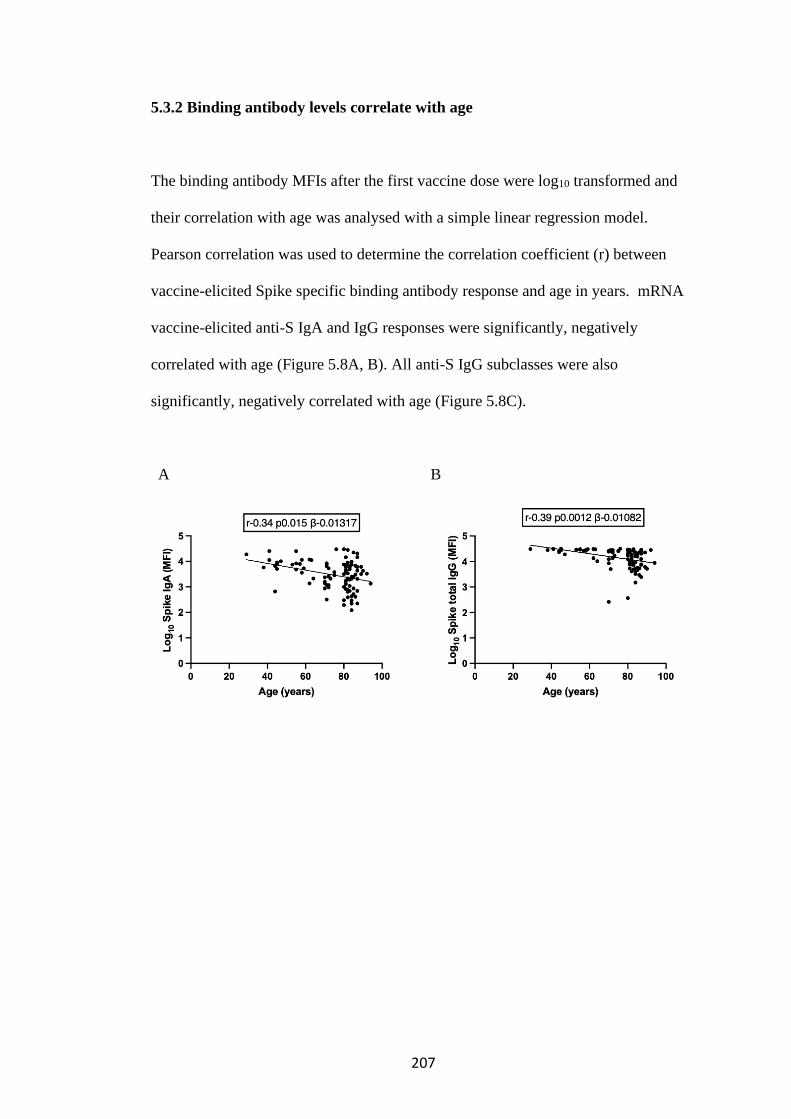
207
5.3.2 Binding antibody levels correlate with age
The binding antibody MFIs after the first vaccine dose were log10 transformed and
their correlation with age was analysed with a simple linear regression model.
Pearson correlation was used to determine the correlation coefficient (r) between
vaccine-elicited Spike specific binding antibody response and age in years. mRNA
vaccine-elicited anti-S IgA and IgG responses were significantly, negatively
correlated with age (Figure 5.8A, B). All anti-S IgG subclasses were also
significantly, negatively correlated with age (Figure 5.8C).
A B
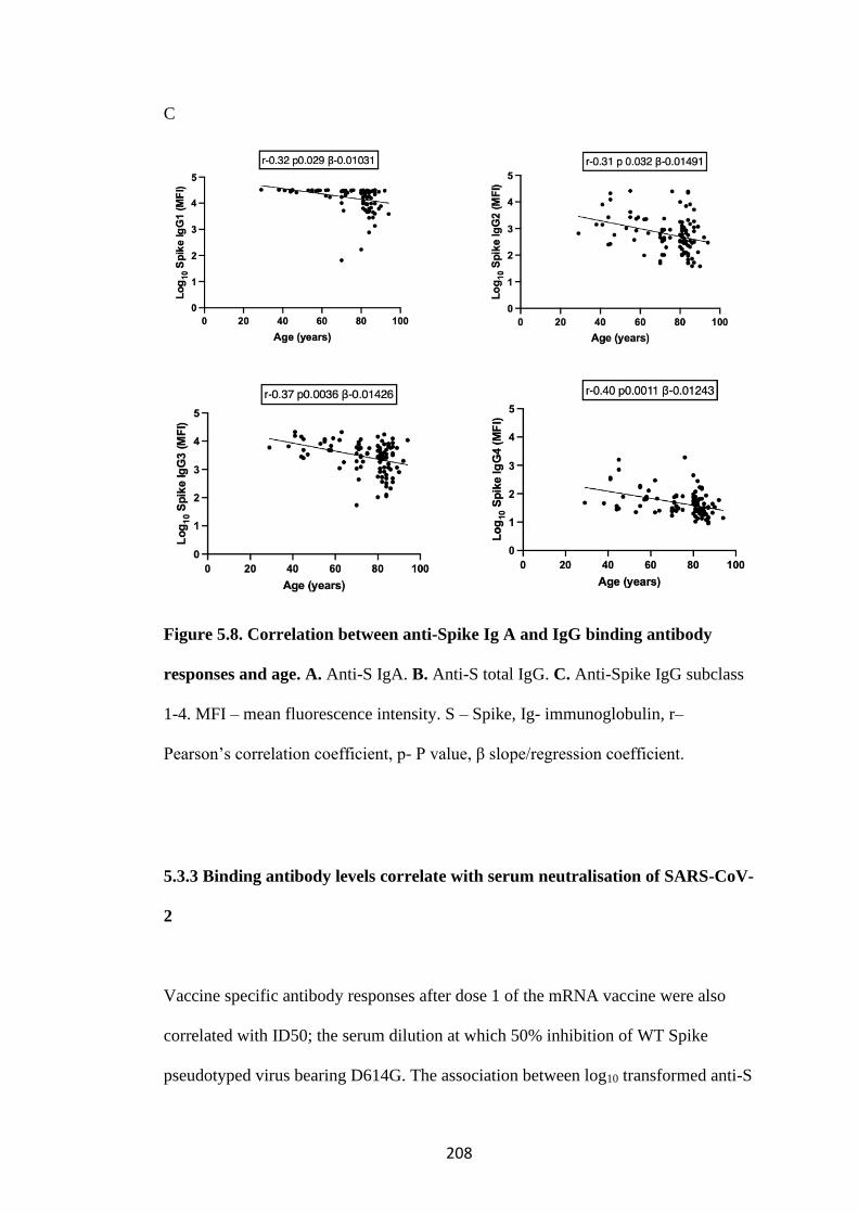
208
C
Figure 5.8. Correlation between anti-Spike Ig A and IgG binding antibody
responses and age. A. Anti-S IgA. B. Anti-S total IgG. C. Anti-Spike IgG subclass
1-4. MFI – mean fluorescence intensity. S – Spike, Ig- immunoglobulin, r–
Pearson’s correlation coefficient, p- P value, β slope/regression coefficient.
5.3.3 Binding antibody levels correlate with serum neutralisation of SARS-CoV-
2
Vaccine specific antibody responses after dose 1 of the mRNA vaccine were also
correlated with ID50; the serum dilution at which 50% inhibition of WT Spike
pseudotyped virus bearing D614G. The association between log10 transformed anti-S
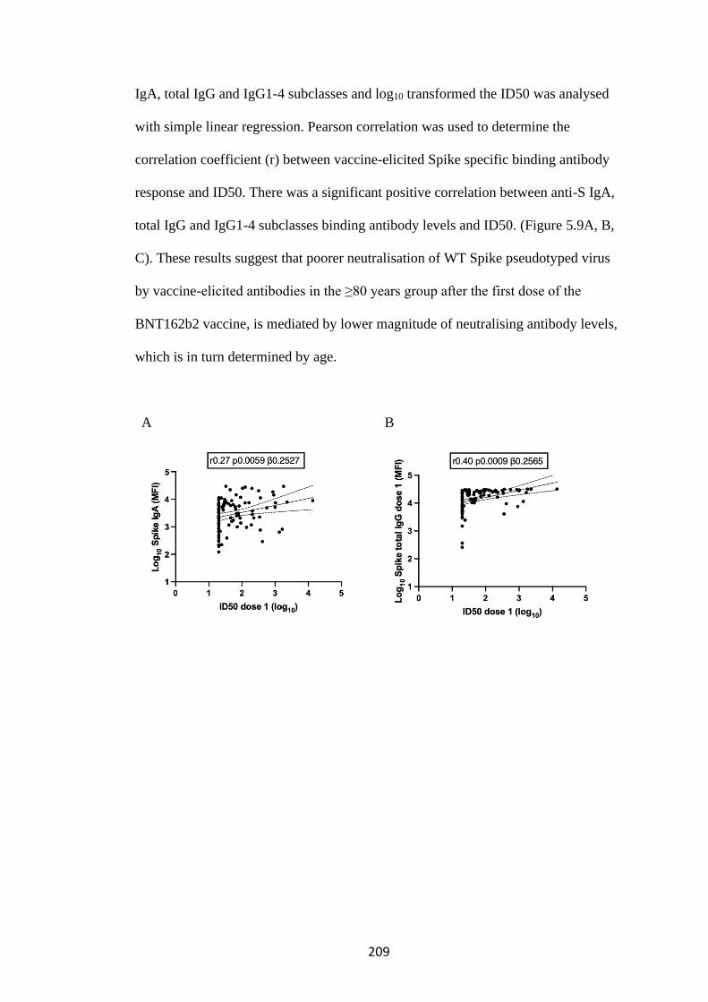
209
IgA, total IgG and IgG1-4 subclasses and log10 transformed the ID50 was analysed
with simple linear regression. Pearson correlation was used to determine the
correlation coefficient (r) between vaccine-elicited Spike specific binding antibody
response and ID50. There was a significant positive correlation between anti-S IgA,
total IgG and IgG1-4 subclasses binding antibody levels and ID50. (Figure 5.9A, B,
C). These results suggest that poorer neutralisation of WT Spike pseudotyped virus
by vaccine-elicited antibodies in the ≥80 years group after the first dose of the
BNT162b2 vaccine, is mediated by lower magnitude of neutralising antibody levels,
which is in turn determined by age.
A
B

210
C
Figure 5.9. Correlation between anti-Spike Ig A and IgG binding antibody
responses and serum neutralisation. A. Anti-S IgA. B. Anti-S total IgG. C. Anti-
Spike IgG subclass 1-4. MFI – mean fluorescence intensity. ID50- inhibitory dilution
at which 50% inhibition of infection is achieved against WT Spike pseudotyped
virus, S- Spike, Ig- immunoglobulin, r– Pearson’s correlation coefficient, p- P value,
β slope/regression coefficient.

211
5.4 T cell responses following first and second dose mRNA BNT162b2 vaccine
5.4.1 Frequency of Spike specific T cells following vaccination
Induction of memory T cells is key to T cell vaccine efficacy, therefore T cell
responses to SARS-CoV-2 spike protein following the first and second doses of the
mRNA BNT162b2 vaccine were measured. PBMCs from vaccinees were stimulated
with overlapping peptide pools to the WT SARS-CoV-2 spike or as a virus specific
control, a peptide pool including Cytomegalovirus, EBV and Flu (CEF+) specific
peptides. IFN and IL-2 FluoroSpot assay was used to count spike specific T cells.
An unexposed healthy control (UHC) population was selected from PBMC
biobanked between 2014-2016 which precedes the current SARS-CoV-2 pandemic,
in order to provide a background control for SARS-CoV-2 spike protein responses.
A positive control population was selected from biobanked PBMCs from
participants with PCR confirmed SARS-CoV-2 infection.
Following the first dose of the mRNA vaccine in the < 80 years group, the frequency
of IFNγ spike specific T cells was significantly larger than the responses seen in the
UHC population. However, in the ≥80 years group, the IFNγ spike specific T cell
responses were not different from the UHC following first dose (Figure 5.10A).
Spike specific IL-2 T cell frequencies were significantly greater than the UHCs in
both age groups but there was a difference between the <80 and ≥80 years groups,
with the ≥80s having significantly lower IL-2 responses (Figure 5.10B).
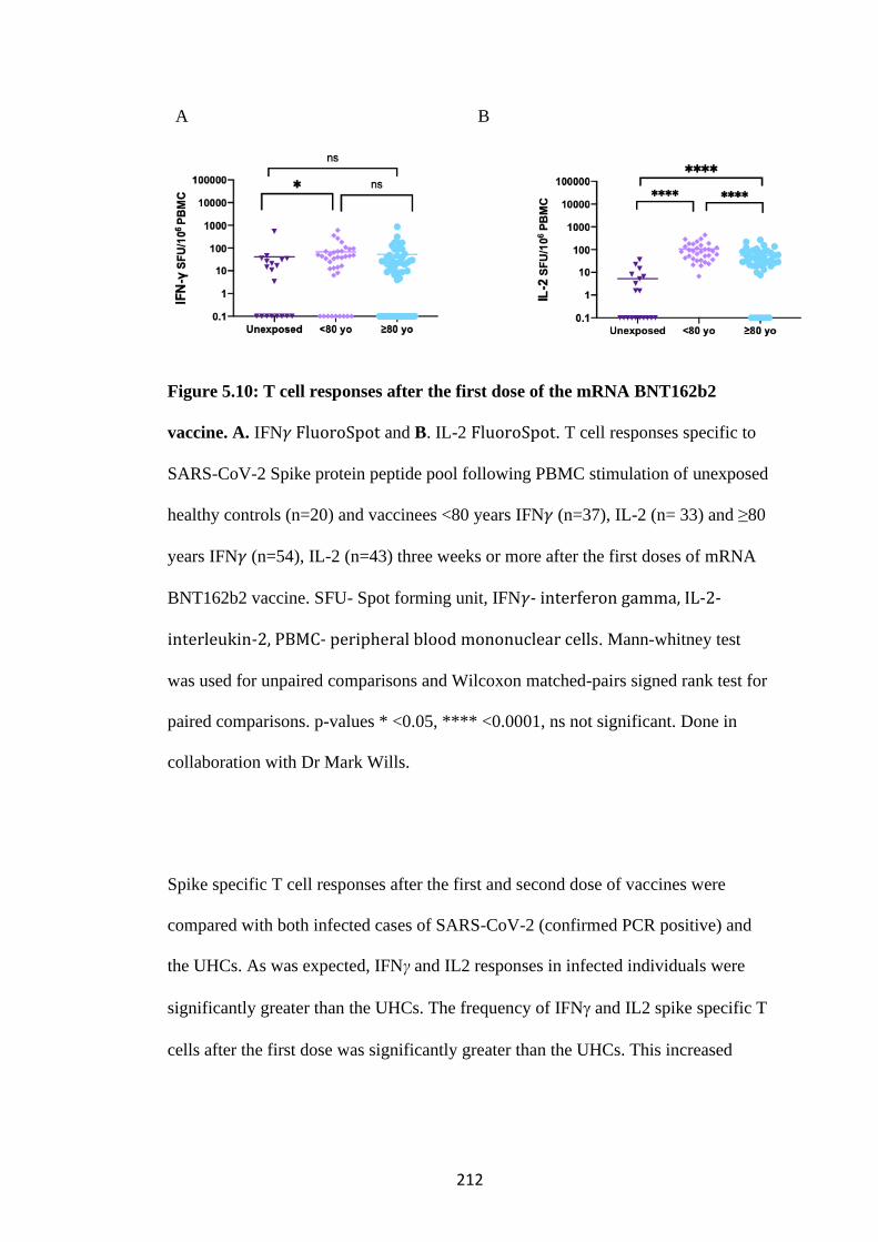
212
A B
Figure 5.10: T cell responses after the first dose of the mRNA BNT162b2
vaccine. A. IFN𝛾 FluoroSpot and B. IL-2 FluoroSpot. T cell responses specific to
SARS-CoV-2 Spike protein peptide pool following PBMC stimulation of unexposed
healthy controls (n=20) and vaccinees <80 years IFN𝛾 (n=37), IL-2 (n= 33) and ≥80
years IFN𝛾 (n=54), IL-2 (n=43) three weeks or more after the first doses of mRNA
BNT162b2 vaccine. SFU- Spot forming unit, IFN𝛾- interferon gamma, IL-2-
interleukin-2, PBMC- peripheral blood mononuclear cells. Mann-whitney test
was used for unpaired comparisons and Wilcoxon matched-pairs signed rank test for
paired comparisons. p-values * <0.05, **** <0.0001, ns not significant. Done in
collaboration with Dr Mark Wills.
Spike specific T cell responses after the first and second dose of vaccines were
compared with both infected cases of SARS-CoV-2 (confirmed PCR positive) and
the UHCs. As was expected, IFNγ and IL2 responses in infected individuals were
significantly greater than the UHCs. The frequency of IFN and IL2 spike specific T
cells after the first dose was significantly greater than the UHCs. This increased
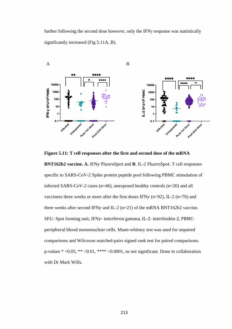
213
further following the second dose however, only the IFN response was statistically
significantly increased (Fig 5.11A, B).
A B
Figure 5.11: T cell responses after the first and second dose of the mRNA
BNT162b2 vaccine. A. IFN𝛾 FluoroSpot and B. IL-2 FluoroSpot. T cell responses
specific to SARS-CoV-2 Spike protein peptide pool following PBMC stimulation of
infected SARS-CoV-2 cases (n=46), unexposed healthy controls (n=20) and all
vaccinees three weeks or more after the first doses IFN𝛾 (n=92), IL-2 (n=76) and
three weeks after second IFN𝛾 and IL-2 (n=21) of the mRNA BNT162b2 vaccine.
SFU- Spot forming unit, IFN𝛾- interferon gamma, IL-2- interleukin-2, PBMC-
peripheral blood mononuclear cells. Mann-whitney test was used for unpaired
comparisons and Wilcoxon matched-pairs signed rank test for paired comparisons.
p-values * <0.05, ** <0.01, **** <0.0001, ns not significant. Done in collaboration
with Dr Mark Wills.
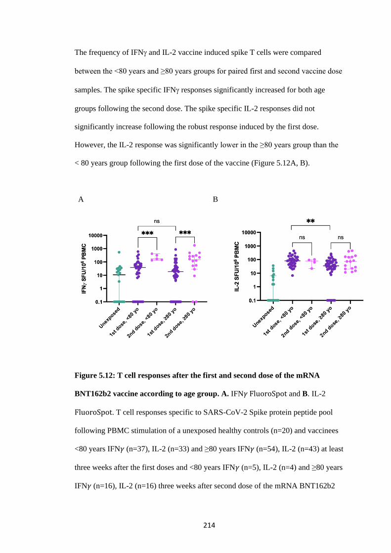
214
The frequency of IFN and IL-2 vaccine induced spike T cells were compared
between the <80 years and ≥80 years groups for paired first and second vaccine dose
samples. The spike specific IFN responses significantly increased for both age
groups following the second dose. The spike specific IL-2 responses did not
significantly increase following the robust response induced by the first dose.
However, the IL-2 response was significantly lower in the ≥80 years group than the
< 80 years group following the first dose of the vaccine (Figure 5.12A, B).
A B
Figure 5.12: T cell responses after the first and second dose of the mRNA
BNT162b2 vaccine according to age group. A. IFN𝛾 FluoroSpot and B. IL-2
FluoroSpot. T cell responses specific to SARS-CoV-2 Spike protein peptide pool
following PBMC stimulation of a unexposed healthy controls (n=20) and vaccinees
<80 years IFN𝛾 (n=37), IL-2 (n=33) and ≥80 years IFN𝛾 (n=54), IL-2 (n=43) at least
three weeks after the first doses and <80 years IFN𝛾 (n=5), IL-2 (n=4) and ≥80 years
IFN𝛾 (n=16), IL-2 (n=16) three weeks after second dose of the mRNA BNT162b2

215
vaccine. SFU- Spot forming unit, IFN𝛾- interferon gamma, IL-2- interleukin-2,
PBMC- peripheral blood mononuclear cells. Mann-whitney test was used for
unpaired comparisons and Wilcoxon matched-pairs signed rank test for paired
comparisons. p-values ** <0.01, *** <0.001, ns not significant. Done in
collaboration with Dr Mark Wills.
5.4.2 Correlation of Spike specific T cell response with serum neutralisation of
SARS-CoV-2
Given that T helper cells have a role in effecting extrafollicular and follicular B cell
responses to antigens (Papavasiliou and Schatz 2002), it was hypothesised that Spike
specific T cell response following vaccination with the mRNA vaccine may be
correlated with serum neutralisation. The association between log10 transformed
frequency of IFN and IL-2 vaccine induced Spike T cell responses with log10
transformed ID50; the serum dilution at which 50% inhibition of infection was
achieved were analysed with simple linear regression. Pearson correlation was used
to determine the correlation coefficient (r) between vaccine-elicited Spike specific
binding antibody response and ID50. IFN and IL-2 vaccine induced spike T cell
responses did not correlate with ID50 against WT Spike pseudotyped virus after first
or second dose vaccine (Figures 5.13A-D).
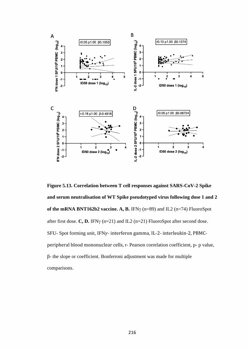
216
Figure 5.13. Correlation between T cell responses against SARS-CoV-2 Spike
and serum neutralisation of WT Spike pseudotyped virus following dose 1 and 2
of the mRNA BNT162b2 vaccine. A, B. IFN (n=89) and IL2 (n=74) FluoroSpot
after first dose. C, D. IFN (n=21) and IL2 (n=21) FluoroSpot after second dose.
SFU- Spot forming unit, IFN𝛾- interferon gamma, IL-2- interleukin-2, PBMC-
peripheral blood mononuclear cells, r- Pearson correlation coefficient, p- p value,
β- the slope or coefficient. Bonferroni adjustment was made for multiple
comparisons.
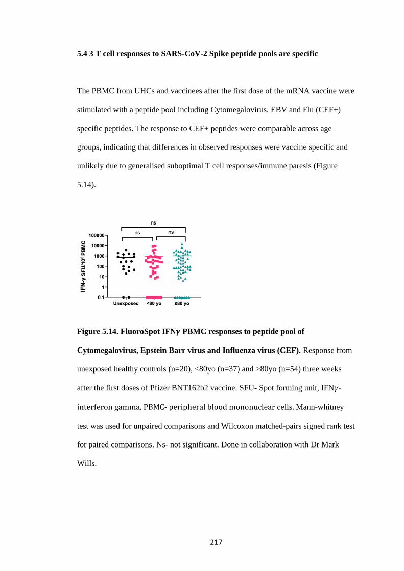
217
5.4 3 T cell responses to SARS-CoV-2 Spike peptide pools are specific
The PBMC from UHCs and vaccinees after the first dose of the mRNA vaccine were
stimulated with a peptide pool including Cytomegalovirus, EBV and Flu (CEF+)
specific peptides. The response to CEF+ peptides were comparable across age
groups, indicating that differences in observed responses were vaccine specific and
unlikely due to generalised suboptimal T cell responses/immune paresis (Figure
5.14).
Figure 5.14. FluoroSpot IFN𝛾 PBMC responses to peptide pool of
Cytomegalovirus, Epstein Barr virus and Influenza virus (CEF). Response from
unexposed healthy controls (n=20), <80yo (n=37) and >80yo (n=54) three weeks
after the first doses of Pfizer BNT162b2 vaccine. SFU- Spot forming unit, IFN𝛾-
interferon gamma, PBMC- peripheral blood mononuclear cells. Mann-whitney
test was used for unpaired comparisons and Wilcoxon matched-pairs signed rank test
for paired comparisons. Ns- not significant. Done in collaboration with Dr Mark
Wills.
218
5.4.4 HCMV seropositivity maybe associated with increased frequency of IFN𝛾
producing Spike specific Tcells
HCMV seropositivity has been associated with poor age-related responses to
vaccination (van den Berg et al. 2019). HCMV serostatus was determined for UHCs
and vaccinees. Given the demographic from which the study population was drawn,
HCMV positivity was more common in the ≥80 years compared with the <80 years
group (55% vs 37%) (Figure 5.15A). Individuals in the ≥80 years group with
positive HCMV serology had significantly higher IFN but not IL2, responses to
SARS-CoV-2 Spike peptides compared to the <80 years group (Figure 5.15B, C).
Although not significant, there was a there was a trend towards a lower ID50 in
HCMV positive individuals (Figure 5.15D). HCMV positive individuals as expected
has a greater response to CEF peptides than HCMV seronegative. The HCMV
positive group responded to the CMV peptides in the CEF pool, while the HCMV
negative group could only have responded to the EBV and Flu components. This
effect the same in both age groups (Figure 5.15E).
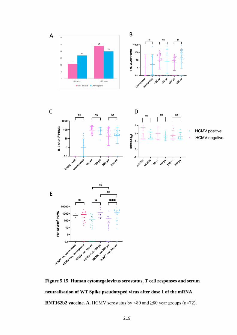
219
Figure 5.15. Human cytomegalovirus serostatus, T cell responses and serum
neutralisation of WT Spike pseudotyped virus after dose 1 of the mRNA
BNT162b2 vaccine. A. HCMV serostatus by <80 and ≥80 year groups (n=72),

220
HCMV seropositive (blue), HCMV seronegative (pink). B IFN FluoroSpot (n=72)
and C. IL-2 FluoroSpot (n=64) T cell responses to SARS-CoV-2 Spike peptides
after the first dose of BNT162b2 vaccine by HCMV serostatus and age group. D.
ID50 after the first dose of BNT162b2 vaccine by HCMV serostatus and age group.
ID50- serum dilution at which 50% inhibition of infection of WT Spike pseudotyped
virus bearing D614G is achieved. E. IFN𝛾 FluoroSpot response to CEF peptides
after the first dose of BNT162b2. after the first dose. SFU- Spot forming unit, IFN𝛾-
interferon gamma, PBMC- peripheral blood mononuclear cells. Mann-whitney
test was used for unpaired comparisons and Wilcoxon matched-pairs signed rank test
for paired comparisons. p-values * <0.05, *** <0.001, ns- not significant. Done in
collaboration with Dr Mark Wills.
5.5 Discussion
It was hypothesised that an appropriate vaccine response would include titres of
neutralising antibodies sufficient to inhibit infection in a Spike pseudotyped assay. It
is still unknown what level of neutralisation offers protection. However, an arbitrary
cut off of ID50 at a serum dilution of 1 in 20 was deemed an adequate response.
Although the limit of detection of the assay was at a dilution of 1 in 4, I found some
variability in ID50 estimates at dilutions lower than 1 in 20. In addition, vaccines
should elicit a response in the T cell arm of the adaptive immune system and it was
hypothesised that an adequate response to the mRNA BNT162b2 vaccine would
increase the frequency of Spike-specific IFN𝛾 and IL-12 secreting T cells
compared with unvaccinated individuals. The data presented shows that the odds
of an inadequate neutralising antibody response to a spike pseudotyped virus after
221
only one dose of the mRNA BNT162b2 vaccine was 3.7 in those ≥80 years old. This
correlated with anti-Spike IgA and IgG binding antibody levels. In addition,
frequency of IFN𝛾 and IL-12secreting Spike specific T cells following only one
dose of mRNA BNT162b2 vaccine was significantly lower in the ≥80 years group
compared with the <80 years group. These age-related differences were overcome
with a second dose of the vaccine.
In the analyses of the association between age group and inadequate neutralisation,
other confounders including immune suppression and comorbidities were not
adjusted for as these data were not collected. They may be other unmeasured
confounders that were not adjusted for. In other to prevent selection bias,
consecutive participants were recruited without exclusion and the sample size was
adequate.
Important correlates of vaccine response have been shown which are age dependent.
Future work would include following up these participants to look for break through
SARS-CoV-2 infection, to track the kinetics of neutralisation activity and more
definitively identify correlates of protection. It would be important to widen the
study population to include more broadly immunocompromised individuals because
as seen from the within-host evolution work presented in the previous chapter, a
suboptimal immune response may drive within-in host adaptation of the virus to
escape vaccine control. In addition, Spike specific B cell receptor sequencing,
antibody cloning and expression would be required, as well as a broader analyses of
T cell phenotype and cytokine responses would be important to further understand
the processes mediating the effects shown.
222
CHAPTER 6: DISCUSSION
PART 1
6.1 HIV-1 CSF escape in the context of cryptococcal meningitis
HIV-1 CSF escape was uncommon in this study population and occurred in only 4%
(2/44). The true prevalence of CSF escape in virologically suppressed individuals is
uncertain. Estimates have ranged from 1.5 to 10 % (Eden et al. 2010; Underwood et
al. 2018; van Zoest et al. 2017). The prevalence is highly influenced by the limit of
detection of the HIV-1 VL assay used and the volume of CSF analysed. Studies that
have done longitudinal sampling have reported that some of these events are CSF
blips and on resampling persistently detectable HIV-1 VL in the CSF is uncommon
(reduction from 36% to 3%) (Eden et al. 2016).
However, in neurosymptomatic individuals or people having a lumbar puncture for
clinical reasons, the prevalence is higher, ranging between 6 and 18% (Kugathasan
et al. 2017; Mukerji et al. 2017; Mukerji et al. 2018; Rawson et al. 2012). As yet,
there are no published prevalence studies of CSF escape/discordance from sub–
Saharan Africa, so the burden in this region remains unknown (Collier et al. 2018).
However, one study looked at the prevalence in a population with HIV-1 subtype C
in Brazil and found a combined prevalence of CSF escape/discordance of 15.3% (de
Almeida et al. 2020). Although reported asymptomatic, neuropsychological
assessment of this cohort found evidence of global neuropsychological deficits in
participants with CSF escape/discordance compared with HIV-1 negative controls.
223
Overall, CSF escape is higher in symptomatic individuals and those having a
clinically indicated LP, it was therefore surprising that the prevalence of CSF escape
in this study was not higher.
6.2 CNS compartmentalisation and patient characteristics associated with
CNS compartmentalisation in the context of CM
Several studies have shown that the presence of HIV-1 replicating independently in
the CNS is associated with clinically significant neurological disease. However, it
was unclear to what extent the HIV-1 variants in the CNS are an independently
replicating/compartmentalised population in patients with co-infection with HIV-1
subtype C and cryptococcal meningitis.
Visual inspection of the maximum likelihood phylogenetic trees revealed a spatial
population structure that clearly segregated the taxa of the CSF derived genomes
from those of the plasma in 2 out of 4 participants. This was supported by the results
from the compartmentalisation analyses. The prevalence of CNS
compartmentalisation in this preliminary analysis was determined. The prevalence of
CNS compartmentalisation was 50% (95% CI 6.8-93.2). This is small sample size
and the true prevalence maybe a lot smaller or greater as indicated by the wide
confidence intervals. Consistent with the data presented in this thesis
CNS compartmentalisation has been found in up to 40% of HIV-1/cryptococcal
meningitis co-infected participants in other studies in sub-Saharan Africa (Adewumi
et al. 2020; Lange et al. 2018). This sample size was too small to do any meaningful
224
analyses of clinical or demographic factors associated with CNS
compartmentalisation.
6.3 Phylogenetic characteristics of paired CNS and plasma genomes of HIV-
1 subtype C in the context of co-infection with cryptococcal meningitis
CNS compartmentalisation was identified by visual inspection of maximum
likelihood phylogenetic tree as detailed above. Statistical analyses using distance and
tree-based methods were used in addition and these supported the conclusions from
visual inspection. In the 2 participants with CNS compartmentalisation, the most
recent common ancestor was inferred to be a plasma variant that was in circulation
prior to emergence of the cluster of viruses detected on the CNS. This fits with the
hypothesis that the viruses in the CNS crosses the blood brain barrier earlier in the
individual’s history of HIV-1 infection and establishes an independently replicating
reservoir. In the other participants with intermixed viruses, this evolutionary process
is not evident. Although, it is possible that all the variants in each compartment have
not been sampled by SGA and the founder variant may have been missed.
The viruses in the CNS cluster are not clonal, suggesting that the virus has continued
to evolve in the CNS compartment (Stam et al. 2013). One of the participants with
CNS compartmentalisation was on ART for 10.6 months, it possible that poor
penetration of ART into the CSF may have driven this evolution under drug
pressure.
225
In addition, analyses of the diversity measured by average pairwise distance (APD)
between genomes in plasma compared with CSF found that participants with CNS
compartmentalisation had increased diversity of HIV-1 genomes in either their
plasma or CSF compartment but those participants with intermixed viruses had
similarly diverse viruses in both compartments. Increased diversity of CSF derived
HIV-1 genomes has previously been reported (Tong et al. 2015; Liu et al. 2013).
This maybe due to differential drug levels, cellular and humoral immune responses
in the CNS and plasma compartments which may exert differential pressure on
viruses in either compartment, leading to viral evolution and the observed diversity.
Participant 002 had received tenofovir/lamivudine/efavirenz combination therapy for
10 months but had no DRMs in the context of very high VLs, suggesting this
participant was non-adherent. However, if this participant had taken some drug for a
proportion of time, it is likely that the selection pressure exerted by poor ART levels
may have driven a soft selective sweep and limited the diversity of the HIV-1 plasma
population as observed by average pairwise distance (APD) (Feder et al. 2016a). In
contrast participant 003 was not on ART and had a high diversity of viruses in the
plasma according to APD.
226
6.4 Phenotypic properties of HIV-1 isolated from the CSF
6.4.1 Co-receptor usage
Effective HIV-1 entry and infection is dependent on primary binding to host CD4
receptor followed by secondary binding to either CCR5 or CXCR4 co-receptors.
Subtype dependent, co-receptor switch from CCR5 to CXCR4 has also been shown
to precede the onset of advanced HIV/AIDS. Since cryptococcal meningitis is an
AIDS defining illness, it maybe expected that patients with HIV-1/CM co-infection
may utilize the CXCR4 co-receptor. Furthermore, a study of patients with HIV-
1/CM co-infection in South Africa using computer predictive algorithms (CPAs),
identified participants with CXCR4 utilising HIV-1 env clones in plasma and CSF
(Sojane et al. 2018). All four participants V3 loop sequences were analysed using
CPAs- GenotoPheno and Web PSSM. GenotoPheno predicted that all sequences
from both compartments in all participants tested, utilised the CCR5 co-receptor. In
contrast, PSSM predicted compartmentalised CNS co-receptor usage, with more than
half of the CSF sequences from one participant and 3% of CSF sequences from
another participant utilising the CXCR4 co-receptor. Both these participants had
CNS compartmentalised viruses. However, this was not consistent with the results in
the phenotypic co-receptor analyses. All clones derived from CSF and plasma tested
phenotypically were found to utilise the CCR5 co-receptor. Furthermore, inhibition
with a CCR5 inhibitor, blocked infection and confirmed dependence of these clones
on the CCR5 co-receptor.
227
The CPAs have their drawbacks with regards their accuracy in predicting coreceptor
usage (Low et al. 2007; Sing et al. 2007). They only consider the V3 loop, whereas
regions outside the V3 loop such as the V2 and gp41 have been found to also
contribute to determination of coreceptor usage (Huang et al. 2008; Thielen et al.
2010). Although GenotoPheno accurately predicted coreceptor usage in this dataset,
PSSM did not. PSSM may have performed more poorly here because it uses the
amino acid at positions 11 and 25 and the net charge to predict co-receptor usage,
which is vulnerable to indels in the alignment.
6.4.2 Drug resistant mutations (DRMs)
Persistence of HIV-1 in tissue reservoirs such as the CNS, where there is poor drug
penetration may allow the virus to sequester and replicate (Beguelin et al. 2016;
Collier et al. 2018; Canestri et al. 2010; Peluso et al. 2012). In addition, mutations
G73S in protease, M184V and M230I in reverse transcriptase have been reported to
be significantly over-represented in patients with CSF escape and CNS
compartmentalisation (Fourati et al. 2014; Liu et al. 2013; Mukerji et al. 2018). The
RT sequence of 31 paired CSF and plasma samples were analysed and revealed no
clear evidence of compartmentalised DRMs. The frequency and type of DRMs were
largely similar in both CSF and plasma compartments. This is not surprising in 55%
of participants who were not receiving ART. Therefore, their viruses would not be
under drug pressure. In the two participants identified with CNS
compartmentalisation, one was treatment naïve whilst the other was treatment
228
experienced. The treatment experienced participant had no DRMs in either
compartment.
6.4.3 Neutralisation sensitivity to broadly neutralising antibodies (bnAbs)
The HIV-1 Envelope is the target of neutralising antibodies. It was hypothesised that
viral escape through mutation of the HIV-1 envelope glycoprotein (Env) maybe a
means through which an independently replicating viral population can exist in the
CNS. Given the chronic history of HIV-1 infection in this cohort it was expected that
clones from both plasma and CSF maybe resistant to neutralisation bnAbs. There
was heterogeneity in the neutralising effect of bnAbs on the clones of the two
participants tested. An interesting finding was that the CSF clones from participant
003 were 3 to 30 -fold more sensitive to neutralisation activity of VRCO1- CD4
binding site antibody but more resistant to CCR5 inhibition by TAK-779 compared
to their plasma clones. It is difficult to draw conclusions as to what these patterns of
neutralisation sensitivity means because each virus is constrained by the niche in
which it finds itself. However, this may indicate that these CSF clones have become
adapted to binding CCR5 with higher affinity. Clones from participant 002 were
more resistant to bnaAbs compared with participant 003. However, one CSF clone
from participant 002 was sensitive to neutralisation activity of VRCO1. In contrast to
participant 003, this clone was sensitive to inhibition by the CCR5 inhibitor.
Data on duration of HIV-1 infection were missing however it is possible that some
of these observation are driven by the duration of HIV-1 infection and viruses in

229
participants with a longer history of HIV-1 would have had more opportunity to
acquire adaptations to allow survival in its niche such as the acquisition of more N-
linked glycosylation to facilitate escape from neutralising antibodies (Wei et al.
2003).
PART 2
6.5 SARS-CoV-2 escape in the context of neutralising antibodies
It is increasingly recognised that some individuals have a protracted illness with
chronic infection with SARS-CoV-2 and persistent viral shedding. The case
presented in this thesis exemplifies this and provides a rare insight to in vivo escape
from neutralising antibodies. This case was intensively sampled over the course of
illness and gave the opportunity to map the viral response to antiviral treatment with
remdesivir and convalescent plasma (CP) in an individual with poor B and T cell
immune responses. In this case the dynamics of the viral population revealed a
shifting landscape of Spike mutations in response to treatment with CP. Two
mutations- △H69/V70 and D796H emerged after the receipt of CP, diminished after
a period of time consistent with the wash-out of the CP and remerged after receipt of
a further unit of CP. D796H was found to be 2-fold more resistant to the neutralising
activity of the third unit of CP and a combination of △H69/V70 and D796H had a
similar effect. Other pairs of mutations arose and receded during the interval
between CP administration, underlying a dynamic process of selective sweeps on the
viral population. Interestingly, in a tissue culture system, D796H had a 3.5-fold
230
reduced infectivity compared to wild type (WT) and △H69/V70 had a 2-fold
increased infectivity and when it occurred with D796H, the infectivity was restored
to levels similar to WT. This appears to be evidence of within-host adaption
following CP. It also exemplifies what is referred a “short-sighted” evolution
whereby a virus acquires “transmissibility-reducing adaptations” in response to
adaptive immune responses (Lythgoe et al. 2017) – so for example D796H is an
adaptation to CP but it reduced infectivity in tissue culture.
Two other cases of prolonged shedding that demonstrated within-host evolution have
been reported. Choi et al10, presented an immunosuppressed individual with
significant virus evolution, which including NTD deletions and RBD mutations but in
the absence of SARS-CoV-2 specific antibody therapy. Avanzato et al also reported a
case of another immunocompromised individual with significant shifts in genetic
composition of the viral population over 105 days (Avanzato et al. 2020). In this case
CP was also used.
Intriguingly, △H69/V70 is observed in the B.1.1.7 variant first reported in the UK and
it appears that we are witnessing converging evolution where this deletion in the NTD
maybe a common pathway to restoring detects in infectivity from other spike
mutations. Other deletion and mutations in the NTD have been reported, for example
△144 in B.1.1.7 and △242-4 in B.1.135 first reported in South Africa. This is of
importance because there is evidence of epitopes in the NTD targeted by strongly
neutralising monoclonal antibodies such as 4A8 (Chi et al. 2020; Collier, De Marco,
et al. 2021).
231
Divergent cluster of viruses bearing W64G and P330S were identified on days 93 and
95. This probably represents a previously unsampled compartmentalised population
but remained rooted within the original population and consistent with having arisen
from a single underlying viral population. Other studies have reported detection of
SARS-CoV-2 RNA in sites outside of the airways (Choi et al. 2020; Zhang et al.
2020).
6.6 Age-related heterogeneity to mRNA BNT162b2 vaccine against SARS-CoV-
2
Protection from infectious pathogens elicited by vaccines is driven by neutralising
antibodies binding to the pathogens and preventing attachment to host cells. The next
line of defence is the cellular immune response that targets and clears infected cells
(The Royal Society 2021). The same process applies to protection from SARS-CoV-
2 infection, where studies in rhesus macaques have shown that RBD-specific
antibodies levels, neutralisation titres and T cells responses to both vaccine and
natural infection predict protection from SARS-CoV-2 in challenge models
(Mercado et al. 2020; McMahan et al. 2021). Similarly, a broad neutralising
antibody and T cell response to the mRNA and Adenovirus vectored vaccines was
seen in human clinical trials (Anderson et al. 2020; Ewer et al. 2021; Sahin et al.
2020) and following natural infection in humans with SARS-CoV-2 (Peng et al.
2020; Zuo et al. 2021; Seow et al. 2020). Although no human challenge studies have
taken place, it stands to reason that these are likely correlates of protection.
232
B and T cells must work in concert to provide effective protection from infectious
pathogens (Papavasiliou and Schatz 2002). However, the quality and magnitude of
responses of both these arms of the adaptive immune system are affected by ageing
(Gustafson et al. 2020; Westmeier et al. 2020; Wilkinson et al. 2017). The data
presented in this thesis showed that 55% of those >80 years had a suboptimal
neutralising antibody response after vaccination with one dose of BNT162b2 and a
lower magnitude of neutralising antibody levels compared with the < 80 years (GMT
83.4 (95% CI 52.0-133.7) vs 46.6 (95% CI 33.5-64.8) p 0.01), which was restored to
similar levels after the second dose was given. Binding IgA and IgG antibody levels
were correlated with neutralisation. The finding of a lower antibody response
following vaccines was reported in Phase I/II trials of the mRNA vaccines
(Ramasamy et al. 2020; Walsh et al. 2020) and has been seen with other vaccines
(Anderson et al. 2020). The improvement in antibody levels after the second dose
was also seen in mouse studies where the ChadOx1 n-CoV-19 Adenovirus vectored
vaccine was used (Silva-Cayetano et al. 2021).
Similar to the B cell antibody responses, the data presented show that Spike specific
T cell responses to BNT162b2 were also impaired by ageing. The frequency of
Interferon gamma (IFN𝛾) secreting CD8+ T cells and interleukin-12 (IL-12)
secreting CD4+ T cells responding to stimulation by a pool of Spike peptides were
shown to be lower in the >80 years group. The IFN𝛾 secreting T cells increased
after the second dose but the IL-2 secreting T cells were not boosted further.
Human vaccine studies have shown a Th1 skewed response to vaccination (Ewer et
al. 2021; Zhu et al. 2020; Sahin et al. 2020; Anderson et al. 2020; Jackson et al.
2020). Similar to the data presented, Spike specific T cell response were poorer in
233
aged mice and improved after the second dose when the ChadOx1 n-CoV-19
Adenovirus vectored vaccine was used (Silva-Cayetano et al. 2021). In addition, the
data presented showed an expected higher prevalence of cytomegalovirus (CMV)
seropositivity in the >80 years but this surprisingly was associated with greater IFN𝛾
Spike specific responses, although some studies have linked CMV seropositive to
poorer immune response to vaccines (van den Berg et al. 2019).
The importance of the dual action of both arms of the adaptive immune response in
protecting against SARS-COV-2 is underlined in a monkey challenge study were
depletion of CD8+ T cells in the context of low neutralising antibody levels, led to
inadequate protection from infection (McMahan et al. 2021). This may offer
sufficient protection in the real-world setting as observational studies have
demonstrated some effect against severe disease and death after 1 dose of the
BNT162b2 or ChAdOx1nCoV-19 (Voysey, Costa Clemens, et al. 2021; Bernal et al.
2021; Vasileiou 2021).
6.7 Conclusion
HIV-1 and SARS-CoV-2 are highly successful viruses that have established
pandemics. They successfully jumped species, overcame transmission bottlenecks
and adapted to successful transmission in humans. HIV-1 has demonstrated that it
highly diverse and can continue to adapt to the constraints of its niche to stablish
chronic infection in its host. Similarly, we have now seen that in the absence of
234
adequate immune responses, SARS-CoV-2 can similarly adapt to the constraints of
its niche and establish chronic infection.
Compartmentalised cellular and immune responses in the CNS and plasma
compartments may exert differential pressure on HIV-1 in either compartment,
leading to viral escape. Since epitopes on the HIV-1 envelope are the main target for
humoral immune response (Sok and Burton 2018), variation in Env can occur due to
selection pressure that render them better adapted to one compartment versus
another.
CNS compartmentalisation may lead to establishment of a long-lived viral reservoir
in the CNS that may have different genotypic and phenotypic properties to the
viruses in the periphery, which has implications for HIV-1 treatment, vaccine and
cure strategies. Persistence of drug resistant HIV-1 in latent reservoirs is not only a
challenge for HIV-1 eradication but more immediately for deciding on subsequent
regimens following virological failure. An understanding of the biology of CNS
compartmentalisation may yield insights into how patients should be managed.
SARS-CoV-2 immune escape can be driven by suboptimal B and T cell effector
function, where antibodies have little support from cytotoxic T cells responses. By
understanding the within-host evolution in response to selection pressures in the
immunocompromised host we can learn how to improve patient care and adapt our
pandemic response tools to prevent transmission of such variants.
235
Immune senescence is a well-characterised (Akbar and Gilroy 2020) and impacts on
vaccine response in multiple ways, including reducing the magnitude of antibody
response to vaccines (Wilkinson et al. 2017), shortening the persistence of antibody
response (Powers and Belshe 1993), limiting the quality of antibodies via reduced
affinity and breadth, limiting induction of CD4+/8+ T cell response (Westmeier et al.
2020) and limiting induction of effector memory (Gustafson et al. 2020). Indeed
vaccines are impaired/dysregulated with age (Haq and McElhaney 2014). Although
the second dose was able to boost neutralising antibodies, it is unclear if the elderly
and other immunosuppressed people will be adequately protected in the protracted
interval between doses. Suboptimal antibody levels could also provide the optimal
niche for selecting escape mutants which may in the long term jeopardies our the
vaccine program’s efforts. It is also possible that more vaccine resistant variants
would lead to poorer outcome from a single vaccine dose (Collier, De Marco, et al.
2021).
Knowledge gained from the work presented in this thesis contributes to the field of
within-host evolution of these two pandemic viruses. It explored the mechanisms
underlying central nervous system escape of HIV-1 from immune control in subtype
C HIV-1. It also identified functional adaptions in the Spike protein of SARS-CoV-2
that drove escape from antibody neutralisation and showed the blue-print for
emergence of Variants of Concern. This work also identified the elderly as a risk
group where suboptimal vaccine-elicited immune responses could compromise the
vaccine program’s efforts. These finding have implications for the immediate choice
of therapeutic options in patients, public health and vaccination program policy,
vaccine development and cure strategies.

236
REFERENCES
Abbasi, Jennifer. 2020. 'COVID-19 and mRNA Vaccines—First Large Test for a New Approach', JAMA, 324: 1125-27.
Adewumi, O. M., E. Dukhovlinova, N. Y. Shehu, S. Zhou, O. D. Council, M. O. Akanbi, B. Taiwo, A. Ogunniyi, K. Robertson, C. Kanyama, M. C. Hosseinipour, and R. Swanstrom. 2020. 'HIV-1 Central Nervous System Compartmentalization and Cytokine Interplay in Non-Subtype B HIV-1 Infections in Nigeria and Malawi', AIDS Res Hum Retroviruses, 36: 490-500.
Akbar, Arne N., and Derek W. Gilroy. 2020. 'Aging immunity may exacerbate COVID-19', Science, 369: 256-57.
Alexander, D. J., and I. H. Brown. 2009. 'History of highly pathogenic avian influenza', Rev Sci Tech, 28: 19-38.
Andersen, K. G., A. Rambaut, W. I. Lipkin, E. C. Holmes, and R. F. Garry. 2020. 'The proximal origin of SARS-CoV-2', Nat Med, 26: 450-52.
Anderson, Evan J., Nadine G. Rouphael, Alicia T. Widge, Lisa A. Jackson, Paul C. Roberts, Mamodikoe Makhene, James D. Chappell, Mark R. Denison, Laura J. Stevens, Andrea J. Pruijssers, Adrian B. McDermott, Britta Flach, Bob C. Lin, Nicole A. Doria-Rose, Sijy O’Dell, Stephen D. Schmidt, Kizzmekia S. Corbett, Phillip A. Swanson, Marcelino Padilla, Kathy M. Neuzil, Hamilton Bennett, Brett Leav, Mat Makowski, Jim Albert, Kaitlyn Cross, Venkata Viswanadh Edara, Katharine Floyd, Mehul S. Suthar, David R. Martinez, Ralph Baric, Wendy Buchanan, Catherine J. Luke, Varun K. Phadke, Christina A. Rostad, Julie E. Ledgerwood, Barney S. Graham, and John H. Beigel. 2020. 'Safety and Immunogenicity of SARS-CoV-2 mRNA-1273 Vaccine in Older Adults', New England Journal of Medicine, 383: 2427-38.
Araujo, L. A., and S. E. Almeida. 2013. 'HIV-1 diversity in the envelope glycoproteins: implications for viral entry inhibition', Viruses, 5: 595-604.
Arrildt, K. T., C. C. LaBranche, S. B. Joseph, E. N. Dukhovlinova, W. D. Graham, L. H. Ping, G. Schnell, C. B. Sturdevant, L. P. Kincer, M. Mallewa, R. S. Heyderman, A. V. Rie, M. S. Cohen, S. Spudich, R. W. Price, D. C. Montefiori, and R. Swanstrom. 2015. 'Phenotypic Correlates of HIV-1 Macrophage Tropism', J Virol, 89: 11294-311.
Avanzato, Victoria A, M Jeremiah Matson, Stephanie N Seifert, Rhys Pryce, Brandi N Williamson, Sarah L Anzick, Kent Barbian, Seth D Judson, Elizabeth R Fischer, and Craig Martens. 2020. 'Case Study: Prolonged infectious SARS-CoV-2 shedding from an asymptomatic immunocompromised cancer patient', Cell.
Baden, L. R., H. M. El Sahly, B. Essink, K. Kotloff, S. Frey, R. Novak, D. Diemert, S. A. Spector, N. Rouphael, C. B. Creech, J. McGettigan, S. Kehtan, N. Segall, J. Solis, A. Brosz, C. Fierro, H. Schwartz, K. Neuzil, L. Corey, P. Gilbert, H. Janes, D. Follmann, M. Marovich, J. Mascola, L. Polakowski, J. Ledgerwood, B. S. Graham, H. Bennett, R. Pajon, C. Knightly, B. Leav, W. Deng, H. Zhou, S. Han, M. Ivarsson, J. Miller, T. Zaks, and Cove Study Group. 2020. 'Efficacy and Safety of the mRNA-1273 SARS-CoV-2 Vaccine', N Engl J Med.
Barre-Sinoussi, F, JC Chermann, F Rey, MT Nugeyre, S Chamaret, J Gruest, C Dauguet, C Axler-Blin, F Vezinet-Brun, C Rouzioux, W Rozenbaum, and L
237
Montagnier. 1983. 'Isolation of a T-lymphotropic retrovirus from a patient at risk for acquired immune deficiency syndrome (AIDS)', Science, 220: 868-71.
Beguelin, C., M. Vazquez, M. Bertschi, S. Yerly, D. de Jong, K. Gutbrod, A. Rauch, and A. Cusini. 2016. 'Viral Escape in the Central Nervous System with Multidrug-Resistant Human Immunodeficiency Virus-1', Open Forum Infect Dis, 3: ofv210.
Beigel, John H., Kay M. Tomashek, Lori E. Dodd, Aneesh K. Mehta, Barry S. Zingman, Andre C. Kalil, Elizabeth Hohmann, Helen Y. Chu, Annie Luetkemeyer, Susan Kline, Diego Lopez de Castilla, Robert W. Finberg, Kerry Dierberg, Victor Tapson, Lanny Hsieh, Thomas F. Patterson, Roger Paredes, Daniel A. Sweeney, William R. Short, Giota Touloumi, David Chien Lye, Norio Ohmagari, Myoung-don Oh, Guillermo M. Ruiz-Palacios, Thomas Benfield, Gerd Fätkenheuer, Mark G. Kortepeter, Robert L. Atmar, C. Buddy Creech, Jens Lundgren, Abdel G. Babiker, Sarah Pett, James D. Neaton, Timothy H. Burgess, Tyler Bonnett, Michelle Green, Mat Makowski, Anu Osinusi, Seema Nayak, and H. Clifford Lane. 2020. 'Remdesivir for the Treatment of Covid-19 — Final Report', New England Journal of Medicine, 383: 1813-26.
Berkhout, B., R. H. Silverman, and K. T. Jeang. 1989. 'Tat trans-activates the human immunodeficiency virus through a nascent RNA target', Cell, 59: 273-82.
Bernal, Jamie Lopez, Nick Andrews, Charlotte Gower, Julia Stowe, Chris Robertson, Elise Tessier, Ruth Simmons, Simon Cottrell, Richard Roberts, Mark O’Doherty, Kevin Brown, Claire Cameron, Diane Stockton, Jim McMenamin, and Mary Ramsay. 2021. 'Early effectiveness of COVID-19 vaccination with BNT162b2 mRNA vaccine and ChAdOx1 adenovirus vector vaccine on symptomatic disease, hospitalisations and mortality in older adults in England', medRxiv: 2021.03.01.21252652.
Besson, G. J., C. M. Lalama, R. J. Bosch, R. T. Gandhi, M. A. Bedison, E. Aga, S. A. Riddler, D. K. McMahon, F. Hong, and J. W. Mellors. 2014. 'HIV-1 DNA decay dynamics in blood during more than a decade of suppressive antiretroviral therapy', Clin Infect Dis, 59: 1312-21.
Bieniasz, P. D. 2009. 'The cell biology of HIV-1 virion genesis', Cell Host Microbe, 5: 550-8.
Bingham, R., N. Ahmed, P. Rangi, M. Johnson, M. Tyrer, and J. Green. 2011. 'HIV encephalitis despite suppressed viraemia: a case of compartmentalized viral escape', Int J STD AIDS, 22: 608-9.
Borrow, P., H. Lewicki, X. Wei, M. S. Horwitz, N. Peffer, H. Meyers, J. A. Nelson, J. E. Gairin, B. H. Hahn, M. B. Oldstone, and G. M. Shaw. 1997. 'Antiviral pressure exerted by HIV-1-specific cytotoxic T lymphocytes (CTLs) during primary infection demonstrated by rapid selection of CTL escape virus', Nat Med, 3: 205-11.
Bruner, K. M., A. J. Murray, R. A. Pollack, M. G. Soliman, S. B. Laskey, A. A. Capoferri, J. Lai, M. C. Strain, S. M. Lada, R. Hoh, Y. C. Ho, D. D. Richman, S. G. Deeks, J. D. Siliciano, and R. F. Siliciano. 2016. 'Defective proviruses rapidly accumulate during acute HIV-1 infection', Nat Med, 22: 1043-9.
Bull, Rowena A., Thiruni N. Adikari, James M. Ferguson, Jillian M. Hammond, Igor Stevanovski, Alicia G. Beukers, Zin Naing, Malinna Yeang, Andrey Verich, Hasindu Gamaarachchi, Ki Wook Kim, Fabio Luciani, Sacha Stelzer-Braid,
238
John-Sebastian Eden, William D. Rawlinson, Sebastiaan J. van Hal, and Ira W. Deveson. 2020. 'Analytical validity of nanopore sequencing for rapid SARS-CoV-2 genome analysis', Nature Communications, 11: 6272.
Burton, D. R., and J. R. Mascola. 2015. 'Antibody responses to envelope glycoproteins in HIV-1 infection', Nat Immunol, 16: 571-6.
Canestri, A., F. X. Lescure, S. Jaureguiberry, A. Moulignier, C. Amiel, A. G. Marcelin, G. Peytavin, R. Tubiana, G. Pialoux, and C. Katlama. 2010. 'Discordance between cerebral spinal fluid and plasma HIV replication in patients with neurological symptoms who are receiving suppressive antiretroviral therapy', Clin Infect Dis, 50: 773-8.
Care, Department of Health and Social. 2021. "Optimising the COVID-19 vaccination programme for maximum short-term impact." In.
Centers for Disease, Control. 1981. 'Pneumocystis pneumonia--Los Angeles', MMWR Morb Mortal Wkly Rep, 30: 250-2.
Chang, C. C., R. Kangethe, S. Omarjee, K. Hiramen, B. Gosnell, K. Sojane, M. S. Moosa, S. R. Lewin, M. A. French, and T. Ndung'u. 2017. 'Relationship of Human Immunodeficiency Virus Viral Load in Cerebrospinal Fluid and Plasma in Patients Co-infected With Cryptococcal Meningitis', Open Forum Infect Dis, 4: ofx032.
Chi, Xiangyang, Renhong Yan, Jun Zhang, Guanying Zhang, Yuanyuan Zhang, Meng Hao, Zhe Zhang, Pengfei Fan, Yunzhu Dong, Yilong Yang, Zhengshan Chen, Yingying Guo, Jinlong Zhang, Yaning Li, Xiaohong Song, Yi Chen, Lu Xia, Ling Fu, Lihua Hou, Junjie Xu, Changming Yu, Jianmin Li, Qiang Zhou, and Wei Chen. 2020. 'A neutralizing human antibody binds to the N-terminal domain of the Spike protein of SARS-CoV-2', Science, 369: 650-55.
Choi, B., M. C. Choudhary, J. Regan, J. A. Sparks, R. F. Padera, X. Qiu, I. H. Solomon, H. H. Kuo, J. Boucau, K. Bowman, U. D. Adhikari, M. L. Winkler, A. A. Mueller, T. Y. Hsu, M. Desjardins, L. R. Baden, B. T. Chan, B. D. Walker, M. Lichterfeld, M. Brigl, D. S. Kwon, S. Kanjilal, E. T. Richardson, A. H. Jonsson, G. Alter, A. K. Barczak, W. P. Hanage, X. G. Yu, G. D. Gaiha, M. S. Seaman, M. Cernadas, and J. Z. Li. 2020. 'Persistence and Evolution of SARS-CoV-2 in an Immunocompromised Host', N Engl J Med, 383: 2291-93.
Christo, P. P., D. B. Greco, A. W. Aleixo, and J. A. Livramento. 2007. 'Factors influencing cerebrospinal fluid and plasma HIV-1 RNA detection rate in patients with and without opportunistic neurological disease during the HAART era', BMC Infect Dis, 7: 147.
Cilliers, T., J. Nhlapo, M. Coetzer, D. Orlovic, T. Ketas, W. C. Olson, J. P. Moore, A. Trkola, and L. Morris. 2003. 'The CCR5 and CXCR4 coreceptors are both used by human immunodeficiency virus type 1 primary isolates from subtype C', J Virol, 77: 4449-56.
Ciuffi, Angela, Manuel Llano, Eric Poeschla, Christian Hoffmann, Jeremy Leipzig, Paul Shinn, Joseph R. Ecker, and Frederic Bushman. 2005. 'A role for LEDGF/p75 in targeting HIV DNA integration', Nature Medicine, 11: 1287-89.
Collier, D. A., L. Haddow, J. Brijkumar, M. S. Moosa, L. Benjamin, and R. K. Gupta. 2018. 'HIV Cerebrospinal Fluid Escape and Neurocognitive Pathology in the Era of Combined Antiretroviral Therapy: What Lies Beneath the Tip of the Iceberg in Sub-Saharan Africa?', Brain Sci, 8.
239
Collier, Dami A., Anna De Marco, Isabella A. T. M. Ferreira, Bo Meng, Rawlings P. Datir, Alexandra C. Walls, Steven A. Kemp, Jessica Bassi, Dora Pinto, Chiara Silacci-Fregni, Siro Bianchi, M. Alejandra Tortorici, John Bowen, Katja Culap, Stefano Jaconi, Elisabetta Cameroni, Gyorgy Snell, Matteo S. Pizzuto, Alessandra Franzetti Pellanda, Christian Garzoni, Agostino Riva, Stephen Baker, Gordon Dougan, Christoph Hess, Nathalie Kingston, Paul J. Lehner, Paul A. Lyons, Nicholas J. Matheson, Willem H. Owehand, Caroline Saunders, Charlotte Summers, James E. D. Thaventhiran, Mark Toshner, Michael P. Weekes, Ashlea Bucke, Jo Calder, Laura Canna, Jason Domingo, Anne Elmer, Stewart Fuller, Julie Harris, Sarah Hewitt, Jane Kennet, Sherly Jose, Jenny Kourampa, Anne Meadows, Criona O’Brien, Jane Price, Cherry Publico, Rebecca Rastall, Carla Ribeiro, Jane Rowlands, Valentina Ruffolo, Hugo Tordesillas, Ben Bullman, Benjamin J. Dunmore, Stuart Fawke, Stefan Gräf, Josh Hodgson, Christopher Huang, Kelvin Hunter, Emma Jones, Ekaterina Legchenko, Cecilia Matara, Jennifer Martin, Federica Mescia, Ciara O’Donnell, Linda Pointon, Nicole Pond, Joy Shih, Rachel Sutcliffe, Tobias Tilly, Carmen Treacy, Zhen Tong, Jennifer Wood, Marta Wylot, Laura Bergamaschi, Ariana Betancourt, Georgie Bower, Chiara Cossetti, Aloka De Sa, Madeline Epping, Richard Grenfell, Andrew Hinch, Oisin Huhn, Sarah Jackson, Isobel Jarvis, Daniel Lewis, Joe Marsden, Francesca Nice, Georgina Okecha, Ommar Omarjee, Marianne Perera, Nathan Richoz, Veronika Romashova, Natalia Savinykh Yarkoni, Rahul Sharma, Luca Stefanucci, Jonathan Stephens, Mateusz Strezlecki, Lori Turner, Eckart M. D. D. De Bie, Katherine Bunclark, Masa Josipovic, Michael Mackay, Sabrina Rossi, Mayurun Selvan, Sarah Spencer, Cissy Yong, Ali Ansaripour, Alice Michael, Lucy Mwaura, Caroline Patterson, Gary Polwarth, Petra Polgarova, Giovanni di Stefano, Codie Fahey, Rachel Michel, Sze-How Bong, Jerome D. Coudert, Elaine Holmes, John Allison, Helen Butcher, Daniela Caputo, Debbie Clapham-Riley, Eleanor Dewhurst, Anita Furlong, Barbara Graves, Jennifer Gray, Tasmin Ivers, Mary Kasanicki, Emma Le Gresley, Rachel Linger, Sarah Meloy, Francesca Muldoon, Nigel Ovington, Sofia Papadia, Isabel Phelan, Hannah Stark, Kathleen E. Stirrups, Paul Townsend, Neil Walker, Jennifer Webster, Anne Elmer, Nathalie Kingston, Barbara Graves, Laura E. McCoy, Kenneth G. C. Smith, John R. Bradley, Nigel Temperton, Lourdes Ceron-Gutierrez, Gabriela Barcenas-Morales, Samuel C. Robson, Nicholas J. Loman, Thomas R. Connor, Tanya Golubchik, Rocio T. Martinez Nunez, Catherine Ludden, Sally Corden, Ian Johnston, David Bonsall, Colin P. Smith, Ali R. Awan, Giselda Bucca, M. Estee Torok, Kordo Saeed, Jacqui A. Prieto, David K. Jackson, William L. Hamilton, Luke B. Snell, Catherine Moore, Ewan M. Harrison, Sonia Goncalves, Derek J. Fairley, Matthew W. Loose, Joanne Watkins, Rich Livett, Samuel Moses, Roberto Amato, Sam Nicholls, Matthew Bull, Darren L. Smith, Jeff Barrett, David M. Aanensen, Martin D. Curran, Surendra Parmar, Dinesh Aggarwal, James G. Shepherd, Matthew D. Parker, Sharon Glaysher, Matthew Bashton, Anthony P. Underwood, Nicole Pacchiarini, Katie F. Loveson, Alessandro M. Carabelli, Kate E. Templeton, Cordelia F. Langford, John Sillitoe, Thushan I. de Silva, Dennis Wang, Dominic Kwiatkowski, Andrew Rambaut, Justin O’Grady, Simon Cottrell,
240
Matthew T. G. Holden, Emma C. Thomson, Husam Osman, Monique Andersson, Anoop J. Chauhan, Mohammed O. Hassan-Ibrahim, Mara Lawniczak, Alex Alderton, Meera Chand, Chrystala Constantinidou, Meera Unnikrishnan, Alistair C. Darby, Julian A. Hiscox, Steve Paterson, Inigo Martincorena, David L. Robertson, Erik M. Volz, Andrew J. Page, Oliver G. Pybus, Andrew R. Bassett, Cristina V. Ariani, Michael H. Spencer Chapman, Kathy K. Li, Rajiv N. Shah, Natasha G. Jesudason, Yusri Taha, Martin P. McHugh, Rebecca Dewar, Aminu S. Jahun, Claire McMurray, Sarojini Pandey, James P. McKenna, Andrew Nelson, Gregory R. Young, Clare M. McCann, Scott Elliott, Hannah Lowe, Ben Temperton, Sunando Roy, Anna Price, Sara Rey, Matthew Wyles, Citiid-Nihr BioResource Covid-Collaboration The, investigators Principal, Crf, nurses volunteer research, logistics Sample, processing Sample, acquisition data, collection Clinical data, I. C. U. Royal Papworth Hospital, I. C. U. Addenbrooke’s Hospital, Cambridge, Trust Peterborough Foundation, Anpc, Medicine Centre for Molecular, Therapeutics Innovative, Nihr BioResource, Covid-Genomics U. K. Consortium The, leadership Funding acquisition, metadata curation project administration samples supervision, sequencing logistics, software analysis, tools analysis, visualization, leadership Funding acquisition, metadata curation project administration samples supervision, sequencing logistics, analysis, software, tools analysis, Leadership, metadata curation project administration samples supervision, sequencing logistics, software analysis, tools analysis, visualization, metadata curation samples Funding acquisition, sequencing logistics, software analysis, tools analysis, visualization, leadership Funding acquisition, metadata curation project administration supervision, samples, logistics, leadership Funding acquisition, metadata curation samples supervision, logistics, sequencing, analysis, leadership Funding acquisition, project administration samples supervision, logistics, sequencing, analysis, leadership Funding acquisition, sequencing supervision, software analysis, tools analysis, visualization, samples Funding acquisition, sequencing logistics, software analysis, tools analysis, visualization, Leadership, metadata curation project administration samples supervision, logistics, sequencing, analysis, Leadership, metadata curation project administration samples supervision, logistics, visualization, Leadership, metadata curation project administration sequencing supervision, analysis, software, tools analysis, project administration samples Metadata curation, sequencing logistics, analysis, software, tools analysis, project administration samples Metadata curation, sequencing logistics, analysis, visualization, leadership Funding acquisition, metadata curation supervision, samples, logistics, leadership Funding acquisition, project administration supervision, samples, logistics, Leadership, metadata curation project administration supervision, samples, logistics, Leadership, metadata curation samples supervision, logistics, sequencing, analysis, Leadership, metadata curation samples supervision, logistics, software, tools analysis, Leadership, metadata curation samples supervision, logistics, visualization, Leadership, metadata curation sequencing supervision, analysis, software, tools analysis, Leadership, project administration

241
samples supervision, logistics, sequencing, analysis, Leadership, sequencing supervision, software analysis, tools analysis, visualization, project administration samples Metadata curation, logistics, sequencing, analysis, project administration sequencing Metadata curation, analysis, software, tools analysis, samples Metadata curation, sequencing logistics, analysis, visualization, sequencing Metadata curation, software analysis, tools analysis, visualization, sequencing Project administration, software analysis, tools analysis, visualization, leadership Funding acquisition, supervision, curation metadata, leadership Funding acquisition, supervision, administration project, leadership Funding acquisition, supervision, sequencing, analysis, Leadership, metadata curation supervision, sequencing, analysis, Leadership, project administration supervision, samples, logistics, Leadership, project administration supervision, sequencing, analysis, Leadership, samples supervision, logistics, sequencing, analysis, Leadership, sequencing supervision, analysis, software, tools analysis, Leadership, sequencing supervision, analysis, visualization, project administration Metadata curation, samples, logistics, project administration Metadata curation, sequencing, analysis, samples Metadata curation, logistics, sequencing, analysis, samples Metadata curation, logistics, visualization, sequencing Metadata curation, analysis, software, and tools analysis. 2021. 'Sensitivity of SARS-CoV-2 B.1.1.7 to mRNA vaccine-elicited antibodies', Nature.
Collier, Dami A., Isabella A.T.M. Ferreira, Rawlings Datir, Prasanti Kotagiri, Eleanor Lim, Bo Meng, Laura Bergamaschi, Anne Elmer, Nathalie Kingston, Barbara Graves, Emma Le Gresley Laura McCoy, Kenneth GC Smith, John R. Bradley, Paul A. Lyons, Paulina Cortes-Acevedo, Lourdes Ceron-Gutierrez, Gabriela Barcenas-Morales, Eoin McKinney, Rainer Doffinger, Mark Wills, and Ravindra K. Gupta. 2021. 'Age-related heterogeneity in immune responses to SARS-CoV-2 vaccine BNT162b2', medRxiv: 2021.02.03.21251054.
Corman, V. M., D. Muth, D. Niemeyer, and C. Drosten. 2018. 'Hosts and Sources of Endemic Human Coronaviruses', Adv Virus Res, 100: 163-88.
D'Arc, M., A. Ayouba, A. Esteban, G. H. Learn, V. Boué, F. Liegeois, L. Etienne, N. Tagg, F. H. Leendertz, C. Boesch, N. F. Madinda, M. M. Robbins, M. Gray, A. Cournil, M. Ooms, M. Letko, V. A. Simon, P. M. Sharp, B. H. Hahn, E. Delaporte, E. Mpoudi Ngole, and M. Peeters. 2015. 'Origin of the HIV-1 group O epidemic in western lowland gorillas', Proc Natl Acad Sci U S A, 112: E1343-52.
Das, K., S. E. Martinez, J. D. Bauman, and E. Arnold. 2012. 'HIV-1 reverse transcriptase complex with DNA and nevirapine reveals non-nucleoside inhibition mechanism', Nat Struct Mol Biol, 19: 253-9.
Davies, Nicholas G., Sam Abbott, Rosanna C. Barnard, Christopher I. Jarvis, Adam J. Kucharski, James D. Munday, Carl A. B. Pearson, Timothy W. Russell, Damien C. Tully, Alex D. Washburne, Tom Wenseleers, Amy Gimma, William Waites, Kerry L. M. Wong, Kevin van Zandvoort, Justin D. Silverman, Karla Diaz-Ordaz, Ruth Keogh, Rosalind M. Eggo, Sebastian Funk, Mark Jit, Katherine E. Atkins, and W. John Edmunds. 2021. 'Estimated transmissibility and impact of SARS-CoV-2 lineage B.1.1.7 in England', Science, 372: eabg3055.
242
Davies, Nicholas G., Christopher I. Jarvis, Kevin van Zandvoort, Samuel Clifford, Fiona Yueqian Sun, Sebastian Funk, Graham Medley, Yalda Jafari, Sophie R. Meakin, Rachel Lowe, Matthew Quaife, Naomi R. Waterlow, Rosalind M. Eggo, Jiayao Lei, Mihaly Koltai, Fabienne Krauer, Damien C. Tully, James D. Munday, Alicia Showering, Anna M. Foss, Kiesha Prem, Stefan Flasche, Adam J. Kucharski, Sam Abbott, Billy J. Quilty, Thibaut Jombart, Alicia Rosello, Gwenan M. Knight, Mark Jit, Yang Liu, Jack Williams, Joel Hellewell, Kathleen O’Reilly, Yung-Wai Desmond Chan, Timothy W. Russell, Simon R. Procter, Akira Endo, Emily S. Nightingale, Nikos I. Bosse, C. Julian Villabona-Arenas, Frank G. Sandmann, Amy Gimma, Kaja Abbas, William Waites, Katherine E. Atkins, Rosanna C. Barnard, Petra Klepac, Hamish P. Gibbs, Carl A. B. Pearson, Oliver Brady, W. John Edmunds, Nicholas P. Jewell, Karla Diaz-Ordaz, Ruth H. Keogh, and Cmmid Covid- Working Group. 2021. 'Increased mortality in community-tested cases of SARS-CoV-2 lineage B.1.1.7', Nature.
de Almeida, Sérgio Monteiro, Indianara Rotta, Ana Paula de Pereira, Bin Tang, Anya Umlauf, Cléa Elisa Lopes Ribeiro, Scott Letendre, Ronald J. Ellis, and H. I. V. Neurobehavioral Research Center Group. 2020. 'Cerebrospinal fluid pleocytosis as a predictive factor for CSF and plasma HIV RNA discordance and escape', Journal of NeuroVirology, 26: 241-51.
Delobel, P., K. Sandres-Saune, M. Cazabat, F. E. L'Faqihi, C. Aquilina, M. Obadia, C. Pasquier, B. Marchou, P. Massip, and J. Izopet. 2005. 'Persistence of distinct HIV-1 populations in blood monocytes and naive and memory CD4 T cells during prolonged suppressive HAART', AIDS, 19: 1739-50.
Deng, K., M. Pertea, A. Rongvaux, L. Wang, C. M. Durand, G. Ghiaur, J. Lai, H. L. McHugh, H. Hao, H. Zhang, J. B. Margolick, C. Gurer, A. J. Murphy, D. M. Valenzuela, G. D. Yancopoulos, S. G. Deeks, T. Strowig, P. Kumar, J. D. Siliciano, S. L. Salzberg, R. A. Flavell, L. Shan, and R. F. Siliciano. 2015. 'Broad CTL response is required to clear latent HIV-1 due to dominance of escape mutations', Nature, 517: 381-5.
Docherty, A. B., E. M. Harrison, C. A. Green, H. E. Hardwick, R. Pius, L. Norman, K. A. Holden, J. M. Read, F. Dondelinger, G. Carson, L. Merson, J. Lee, D. Plotkin, L. Sigfrid, S. Halpin, C. Jackson, C. Gamble, P. W. Horby, J. S. Nguyen-Van-Tam, A. Ho, C. D. Russell, J. Dunning, P. J. Openshaw, J. K. Baillie, M. G. Semple, and Isaric C. investigators. 2020. 'Features of 20 133 UK patients in hospital with covid-19 using the ISARIC WHO Clinical Characterisation Protocol: prospective observational cohort study', BMJ, 369: m1985.
Dong, E., H. Du, and L. Gardner. 2020. 'An interactive web-based dashboard to track COVID-19 in real time', Lancet Infect Dis, 20: 533-34.
Dragic, T., V. Litwin, G. P. Allaway, S. R. Martin, Y. Huang, K. A. Nagashima, C. Cayanan, P. J. Maddon, R. A. Koup, J. P. Moore, and W. A. Paxton. 1996. 'HIV-1 entry into CD4+ cells is mediated by the chemokine receptor CC-CKR-5', Nature, 381: 667-73.
DW. 2021. "COVID vaccines: Will Germany follow the UK's example?" In. Eden, A., D. Fuchs, L. Hagberg, S. Nilsson, S. Spudich, B. Svennerholm, R. W. Price,
and M. Gisslen. 2010. 'HIV-1 viral escape in cerebrospinal fluid of subjects on suppressive antiretroviral treatment', J Infect Dis, 202: 1819-25.

243
Eden, A., S. Nilsson, L. Hagberg, D. Fuchs, H. Zetterberg, B. Svennerholm, and M. Gisslen. 2016. 'Asymptomatic Cerebrospinal Fluid HIV-1 Viral Blips and Viral Escape During Antiretroviral Therapy: A Longitudinal Study', J Infect Dis, 214: 1822-25.
Edler, Daniel, Johannes Klein, Alexandre Antonelli, and Daniele Silvestro. 2019. 'raxmlGUI 2.0 beta: a graphical interface and toolkit for phylogenetic analyses using RAxML', bioRxiv: 800912.
Ewer, Katie J., Jordan R. Barrett, Sandra Belij-Rammerstorfer, Hannah Sharpe, Rebecca Makinson, Richard Morter, Amy Flaxman, Daniel Wright, Duncan Bellamy, Mustapha Bittaye, Christina Dold, Nicholas M. Provine, Jeremy Aboagye, Jamie Fowler, Sarah E. Silk, Jennifer Alderson, Parvinder K. Aley, Brian Angus, Eleanor Berrie, Sagida Bibi, Paola Cicconi, Elizabeth A. Clutterbuck, Irina Chelysheva, Pedro M. Folegatti, Michelle Fuskova, Catherine M. Green, Daniel Jenkin, Simon Kerridge, Alison Lawrie, Angela M. Minassian, Maria Moore, Yama Mujadidi, Emma Plested, Ian Poulton, Maheshi N. Ramasamy, Hannah Robinson, Rinn Song, Matthew D. Snape, Richard Tarrant, Merryn Voysey, Marion E. E. Watson, Alexander D. Douglas, Adrian V. S. Hill, Sarah C. Gilbert, Andrew J. Pollard, Teresa Lambe, Aabidah Ali, Elizabeth Allen, Megan Baker, Eleanor Barnes, Nicola Borthwick, Amy Boyd, Charlie Brown-O’Sullivan, Joshua Burgoyne, Nicholas Byard, Ingrid Cabrera Puig, Federica Cappuccini, Jee-Sun Cho, Paola Cicconi, Elizabeth Clark, Wendy E. M. Crocker, Mehreen S. Datoo, Hannah Davies, Susanna Jane Dunachie, Nick J. Edwards, Sean C. Elias, Julie Furze, Ciaran Gilbride, Stephanie A. Harris, Susanne H. C. Hodgson, Mimi M. Hou, Susan Jackson, Kathryn Jones, Reshma Kailath, Lloyd King, Colin W. Larkworthy, Yuanyuan Li, Amelia M. Lias, Aline Linder, Samuel Lipworth, Raquel Lopez Ramon, Meera Madhavan, Emma Marlow, Julia L. Marshall, Alexander J. Mentzer, Hazel Morrison, Andrés Noé, Dimitra Pipini, David Pulido-Gomez, Fernando Ramos Lopez, Adam John Ritchie, Indra Rudiansyah, Helen Sanders, Adam Shea, Sarah Silk, Alexandra J. Spencer, Rachel Tanner, Yrene Themistocleous, Merin Thomas, Nguyen Tran, Adam Truby, Cheryl Turner, Nicola Turner, Marta Ulaszewska, Andrew T. Worth, Lucy Kingham-Page, Marco Polo Peralta Alvarez, Rachel Anslow, Louise Bates, Kirsten Beadon, Rebecca Beckley, Amy Beveridge, Else Margreet Bijker, Luke Blackwell, Jamie Burbage, Susana Camara, Melanie Carr, Rachel Colin-Jones, Rachel Cooper, Christina J. Cunningham, Tesfaye Demissie, Claudio Di Maso, Naomi Douglas, Rachael Drake-Brockman, Ruth Elizabeth Drury, Katherine R. W. Emary, Sally Felle, Shuo Feng, Karen J. Ford, Emma Francis, Lara Gracie, Joseph Hamlyn, Brama Hanumunthadu, Daisy Harrison, Thomas C. Hart, Sophia Hawkins, Jennifer Hill, Elizabeth Howe, Nicola Howell, Elizabeth Jones, Jade Keen, Sarah Kelly, David Kerr, Liaquat Khan, Jasmin Kinch, Stanislava Koleva, Emily A. Lees, Alice Lelliott, Xinxue Liu, Spyridoula Marinou, Joanne McEwan, Ella Morey, Gertraud Morshead, Jilly Muller, Claire Munro, Sarah Murphy, Philomena Mweu, Elizabeth Nuthall, Katie O’Brien, Daniel O’Connor, Peter John O’Reilly, Blanché Oguti, Piper Osborne, Nelly Owino, Kaye Parker, Katja Pfafferott, Samuel Provstgaard-Morys, Helen Ratcliffe, Thomas Rawlinson, Sarah Rhead, Hannah Roberts, Katherine
244
Sanders, Laura Silva-Reyes, Catherine C. Smith, David J. Smith, Anna Szigeti, Tonia M. Thomas, Amber Thompson, Susan Tonks, Rachel Varughes, Iason Vichos, Laura Walker, Caroline White, Rachel White, Xin Li Yao, Christopher P. Conlon, John Frater, Liliana Cifuentes, Ioana Baleanu, Emma Bolam, Elena Boland, Tanja Brenner, Brad E. Damratoski, Chandra Datta, Omar El Muhanna, Richard Fisher, Pablo Galian-Rubio, Gina Hodges, Frederic Jackson, Shuchang Liu, Lisa Loew, Roisin Morgans, Susan Jane Morris, Vicki Olchawski, Catarina Oliveria, Helena Parracho, Emilia Reyes Pabon, Abdessamad Tahiri-Alaoui, Keja Taylor, Paul Williams, Dalila Zizi, Edward H. Arbe-Barnes, Philip Baker, Alexander Batten, Charlotte Downing, Jonathan Drake, Marcus Rex English, John Aaron Henry, Poppy Iveson, Annabel Killen, Thomas B. King, Jessica P. J. Larwood, Gary Mallett, Kushal Mansatta, Neginsadat Mirtorabi, Maia Patrick-Smith, James Perring, Kajal Radia, Sophie Roche, Ella Schofield, Rebecca te Water Naude, James Towner, Natalie Baker, Kevin R. Bewley, Emily Brunt, Karen R. Buttigieg, Sue Charlton, Naomi S. Coombes, Michael J. Elmore, Kerry Godwin, Bassam Hallis, Daniel Knott, Lorna McInroy, Imam Shaik, Kelly Thomas, Julia A. Tree, Caitlin L. Blundell, Michelangelo Cao, Dearbhla Kelly, Donal T. Skelly, Andreas Themistocleous, Tao Dong, Samantha Field, Elizabeth Hamilton, Elizabeth Kelly, Paul Klenerman, Julian C. Knight, Yolanda Lie, Christos Petropoulos, Cynthia Sedik, Terri Wrin, Gretchen Meddaugh, Yanchun Peng, Gavin Screaton, Elizabeth Stafford, and Covid Vaccine Trial Group the Oxford. 2021. 'T cell and antibody responses induced by a single dose of ChAdOx1 nCoV-19 (AZD1222) vaccine in a phase 1/2 clinical trial', Nature Medicine, 27: 270-78.
Feder, A. F., S. Y. Rhee, S. P. Holmes, R. W. Shafer, D. A. Petrov, and P. S. Pennings. 2016a. 'More effective drugs lead to harder selective sweeps in the evolution of drug resistance in HIV-1', Elife, 5.
Feder, Alison F., Soo-Yon Rhee, Susan P. Holmes, Robert W. Shafer, Dmitri A. Petrov, and Pleuni S. Pennings. 2016b. 'More effective drugs lead to harder selective sweeps in the evolution of drug resistance in HIV-1', eLife, 5: e10670.
Filipowicz, A. R., C. M. McGary, G. E. Holder, A. A. Lindgren, E. M. Johnson, C. Sugimoto, M. J. Kuroda, and W. K. Kim. 2016. 'Proliferation of Perivascular Macrophages Contributes to the Development of Encephalitic Lesions in HIV-Infected Humans and in SIV-Infected Macaques', Sci Rep, 6: 32900.
Fourati, S., S. Lambert-Niclot, C. Soulie, M. Wirden, I. Malet, M. A. Valantin, R. Tubiana, A. Simon, C. Katlama, G. Carcelain, V. Calvez, and A. G. Marcelin. 2014. 'Differential impact of APOBEC3-driven mutagenesis on HIV evolution in diverse anatomical compartments', AIDS, 28: 487-91.
Frampton, D., T. Rampling, A. Cross, H. Bailey, J. Heaney, M. Byott, R. Scott, R. Sconza, J. Price, M. Margaritis, M. Bergstrom, M. J. Spyer, P. B. Miralhes, P. Grant, S. Kirk, C. Valerio, Z. Mangera, T. Prabhahar, J. Moreno-Cuesta, N. Arulkumaran, M. Singer, G. Y. Shin, E. Sanchez, S. M. Paraskevopoulou, D. Pillay, R. A. McKendry, M. Mirfenderesky, C. F. Houlihan, and E. Nastouli. 2021. 'Genomic characteristics and clinical effect of the emergent SARS-CoV-
245
2 B.1.1.7 lineage in London, UK: a whole-genome sequencing and hospital-based cohort study', Lancet Infect Dis.
Fun, Axel, Annemarie M. J. Wensing, Jens Verheyen, and Monique Nijhuis. 2012. 'Human Immunodeficiency Virus gag and protease: partners in resistance', Retrovirology, 9: 63.
Gall, A., B. Ferns, C. Morris, S. Watson, M. Cotten, M. Robinson, N. Berry, D. Pillay, and P. Kellam. 2012. 'Universal amplification, next-generation sequencing, and assembly of HIV-1 genomes', J Clin Microbiol, 50: 3838-44.
Ganor, Y., F. Real, A. Sennepin, C. A. Dutertre, L. Prevedel, L. Xu, D. Tudor, B. Charmeteau, A. Couedel-Courteille, S. Marion, A. R. Zenak, J. P. Jourdain, Z. Zhou, A. Schmitt, C. Capron, E. A. Eugenin, R. Cheynier, M. Revol, S. Cristofari, A. Hosmalin, and M. Bomsel. 2019. 'HIV-1 reservoirs in urethral macrophages of patients under suppressive antiretroviral therapy', Nat Microbiol, 4: 633-44.
Gao, X., A. Bashirova, A. K. Iversen, J. Phair, J. J. Goedert, S. Buchbinder, K. Hoots, D. Vlahov, M. Altfeld, S. J. O'Brien, and M. Carrington. 2005. 'AIDS restriction HLA allotypes target distinct intervals of HIV-1 pathogenesis', Nat Med, 11: 1290-2.
Garrett, M. E., H. L. Itell, K. H. D. Crawford, R. Basom, J. D. Bloom, and J. Overbaugh. 2020. 'Phage-DMS: A Comprehensive Method for Fine Mapping of Antibody Epitopes', iScience, 23: 101622.
Gelman, B. B., J. G. Lisinicchia, S. Morgello, E. Masliah, D. Commins, C. L. Achim, H. S. Fox, D. L. Kolson, I. Grant, E. Singer, C. T. Yiannoutsos, S. Sherman, G. Gensler, D. J. Moore, T. Chen, and V. M. Soukup. 2013. 'Neurovirological correlation with HIV-associated neurocognitive disorders and encephalitis in a HAART-era cohort', J Acquir Immune Defic Syndr, 62: 487-95.
Gordon, Anthony C., Paul R. Mouncey, Farah Al-Beidh, Kathryn M. Rowan, Alistair D. Nichol, Yaseen M. Arabi, Djillali Annane, Abi Beane, Wilma van Bentum-Puijk, Lindsay R. Berry, Zahra Bhimani, Marc J.M. Bonten, Charlotte A. Bradbury, Frank M. Brunkhorst, Adrian Buzgau, Allen C. Cheng, Michelle A. Detry, Eamon J. Duffy, Lise J. Estcourt, Mark Fitzgerald, Herman Goossens, Rashan Haniffa, Alisa M. Higgins, Thomas E. Hills, Christopher M. Horvat, Francois Lamontagne, Patrick R. Lawler, Helen L. Leavis, Kelsey M. Linstrum, Edward Litton, Elizabeth Lorenzi, John C. Marshall, Florian B. Mayr, Danny McAuley, Anna McGlothlin, Shay P McGuinness, Bryan J. McVerry, Stephanie K. Montgomery, Susan C. Morpeth, Srinivas Murthy, Katrina Orr, Rachael L. Parke, Jane C. Parker, Asad E. Patanwala, Ville Pettilä, Emma Rademaker, Marlene S. Santos, Christina T. Saunders, Christopher W. Seymour, Manu Shankar-Hari, Wendy I. Sligl, Alexis F. Turgeon, Anne M. Turner, Frank L. van de Veerdonk, Ryan Zarychanski, Cameron Green, Roger J. Lewis, Derek C. Angus, Colin J. McArthur, Scott Berry, Steve A. Webb, and Lennie P.G. Derde. 2021. 'Interleukin-6 Receptor Antagonists in Critically Ill Patients with Covid-19 – Preliminary report', medRxiv: 2021.01.07.21249390.
Gorry, P. R., G. Bristol, J. A. Zack, K. Ritola, R. Swanstrom, C. J. Birch, J. E. Bell, N. Bannert, K. Crawford, H. Wang, D. Schols, E. De Clercq, K. Kunstman, S. M. Wolinsky, and D. Gabuzda. 2001. 'Macrophage tropism of human immunodeficiency virus type 1 isolates from brain and lymphoid tissues

246
predicts neurotropism independent of coreceptor specificity', J Virol, 75: 10073-89.
Gorry, P. R., J. Taylor, G. H. Holm, A. Mehle, T. Morgan, M. Cayabyab, M. Farzan, H. Wang, J. E. Bell, K. Kunstman, J. P. Moore, S. M. Wolinsky, and D. Gabuzda. 2002. 'Increased CCR5 affinity and reduced CCR5/CD4 dependence of a neurovirulent primary human immunodeficiency virus type 1 isolate', J Virol, 76: 6277-92.
Gottlieb, Robert L., Ajay Nirula, Peter Chen, Joseph Boscia, Barry Heller, Jason Morris, Gregory Huhn, Jose Cardona, Bharat Mocherla, Valentina Stosor, Imad Shawa, Princy Kumar, Andrew C. Adams, Jacob Van Naarden, Kenneth L. Custer, Michael Durante, Gerard Oakley, Andrew E. Schade, Timothy R. Holzer, Philip J. Ebert, Richard E. Higgs, Nicole L. Kallewaard, Janelle Sabo, Dipak R. Patel, Paul Klekotka, Lei Shen, and Daniel M. Skovronsky. 2021. 'Effect of Bamlanivimab as Monotherapy or in Combination With Etesevimab on Viral Load in Patients With Mild to Moderate COVID-19: A Randomized Clinical Trial', JAMA, 325: 632-44.
Gray, F., F. X. Lescure, H. Adle-Biassette, M. Polivka, S. Gallien, G. Pialoux, and A. Moulignier. 2013. 'Encephalitis with infiltration by CD8+ lymphocytes in HIV patients receiving combination antiretroviral treatment', Brain Pathol, 23: 525-33.
Greaney, A. J., T. N. Starr, P. Gilchuk, S. J. Zost, E. Binshtein, A. N. Loes, S. K. Hilton, J. Huddleston, R. Eguia, K. H. D. Crawford, A. S. Dingens, R. S. Nargi, R. E. Sutton, N. Suryadevara, P. W. Rothlauf, Z. Liu, S. P. J. Whelan, R. H. Carnahan, J. E. Crowe, Jr., and J. D. Bloom. 2021. 'Complete Mapping of Mutations to the SARS-CoV-2 Spike Receptor-Binding Domain that Escape Antibody Recognition', Cell Host Microbe, 29: 44-57 e9.
Gupta, Ravindra K., Sultan Abdul-Jawad, Laura E. McCoy, Hoi Ping Mok, Dimitra Peppa, Maria Salgado, Javier Martinez-Picado, Monique Nijhuis, Annemarie M. J. Wensing, Helen Lee, Paul Grant, Eleni Nastouli, Jonathan Lambert, Matthew Pace, Fanny Salasc, Christopher Monit, Andrew J. Innes, Luke Muir, Laura Waters, John Frater, Andrew M. L. Lever, Simon G. Edwards, Ian H. Gabriel, and Eduardo Olavarria. 2019. 'HIV-1 remission following CCR5Δ32/Δ32 haematopoietic stem-cell transplantation', Nature, 568: 244-48.
Gupta-Wright, A., K. Fielding, J. J. van Oosterhout, M. Alufandika, D. J. Grint, E. Chimbayo, J. Heaney, M. Byott, E. Nastouli, H. C. Mwandumba, E. L. Corbett, and R. K. Gupta. 2020. 'Virological failure, HIV-1 drug resistance, and early mortality in adults admitted to hospital in Malawi: an observational cohort study', Lancet HIV, 7: e620-e28.
Gustafson, C. E., C. Kim, C. M. Weyand, and J. J. Goronzy. 2020. 'Influence of immune aging on vaccine responses', J Allergy Clin Immunol, 145: 1309-21.
Haaland, R. E., P. A. Hawkins, J. Salazar-Gonzalez, A. Johnson, A. Tichacek, E. Karita, O. Manigart, J. Mulenga, B. F. Keele, G. M. Shaw, B. H. Hahn, S. A. Allen, C. A. Derdeyn, and E. Hunter. 2009. 'Inflammatory genital infections mitigate a severe genetic bottleneck in heterosexual transmission of subtype A and C HIV-1', PLoS Pathog, 5: e1000274.
247
Haddad, D. N., C. Birch, T. Middleton, D. E. Dwyer, A. L. Cunningham, and N. K. Saksena. 2000. 'Evidence for late stage compartmentalization of HIV-1 resistance mutations between lymph node and peripheral blood mononuclear cells', AIDS, 14: 2273-81.
Haq, K., and J. E. McElhaney. 2014. 'Immunosenescence: Influenza vaccination and the elderly', Curr Opin Immunol, 29: 38-42.
Heaton, R. K., D. B. Clifford, D. R. Franklin, Jr., S. P. Woods, C. Ake, F. Vaida, R. J. Ellis, S. L. Letendre, T. D. Marcotte, J. H. Atkinson, M. Rivera-Mindt, O. R. Vigil, M. J. Taylor, A. C. Collier, C. M. Marra, B. B. Gelman, J. C. McArthur, S. Morgello, D. M. Simpson, J. A. McCutchan, I. Abramson, A. Gamst, C. Fennema-Notestine, T. L. Jernigan, J. Wong, I. Grant, and Charter Group. 2010. 'HIV-associated neurocognitive disorders persist in the era of potent antiretroviral therapy: CHARTER Study', Neurology, 75: 2087-96.
Heaton, R. K., D. R. Franklin, R. J. Ellis, J. A. McCutchan, S. L. Letendre, S. Leblanc, S. H. Corkran, N. A. Duarte, D. B. Clifford, S. P. Woods, A. C. Collier, C. M. Marra, S. Morgello, M. R. Mindt, M. J. Taylor, T. D. Marcotte, J. H. Atkinson, T. Wolfson, B. B. Gelman, J. C. McArthur, D. M. Simpson, I. Abramson, A. Gamst, C. Fennema-Notestine, T. L. Jernigan, J. Wong, I. Grant, Charter Group, and Hnrc Group. 2011. 'HIV-associated neurocognitive disorders before and during the era of combination antiretroviral therapy: differences in rates, nature, and predictors', J Neurovirol, 17: 3-16.
Hogan, C. A., J. Iles, E. H. Frost, G. Giroux, O. Cassar, A. Gessain, M. J. Dion, V. Ilunga, A. Rambaut, AÉ Yengo-Ki-Ngimbi, F. Behets, O. G. Pybus, and J. Pépin. 2016. 'Epidemic History and Iatrogenic Transmission of Blood-borne Viruses in Mid-20th Century Kinshasa', J Infect Dis, 214: 353-60.
Horby, Peter W, Guilherme Pessoa-Amorim, Leon Peto, Christopher E Brightling, Rahuldeb Sarkar, Koshy Thomas, Vandana Jeebun, Abdul Ashish, Redmond Tully, David Chadwick, Muhammad Sharafat, Richard Stewart, Banu Rudran, J Kenneth Baillie, Maya H Buch, Lucy C Chappell, Jeremy N Day, Saul N Furst, Thomas Jaki, Katie Jeffery, Edmund Juszczak, Wei Shen Lim, Alan Montgomery, Andrew Mumford, Kathryn Rowan, Guy Thwaites, Marion Mafham, Richard Haynes, and Martin J Landray. 2021. 'Tocilizumab in patients admitted to hospital with COVID-19 (RECOVERY): preliminary results of a randomised, controlled, open-label, platform trial', medRxiv: 2021.02.11.21249258.
Huang, W., S. H. Eshleman, J. Toma, S. Fransen, E. Stawiski, E. E. Paxinos, J. M. Whitcomb, A. M. Young, D. Donnell, F. Mmiro, P. Musoke, L. A. Guay, J. B. Jackson, N. T. Parkin, and C. J. Petropoulos. 2007. 'Coreceptor tropism in human immunodeficiency virus type 1 subtype D: high prevalence of CXCR4 tropism and heterogeneous composition of viral populations', J Virol, 81: 7885-93.
Huang, W., J. Toma, S. Fransen, E. Stawiski, J. D. Reeves, J. M. Whitcomb, N. Parkin, and C. J. Petropoulos. 2008. 'Coreceptor tropism can be influenced by amino acid substitutions in the gp41 transmembrane subunit of human immunodeficiency virus type 1 envelope protein', J Virol, 82: 5584-93.
Hütter, G., D. Nowak, M. Mossner, S. Ganepola, A. Müssig, K. Allers, T. Schneider, J. Hofmann, C. Kücherer, O. Blau, I. W. Blau, W. K. Hofmann, and E. Thiel.
248
2009. 'Long-term control of HIV by CCR5 Delta32/Delta32 stem-cell transplantation', N Engl J Med, 360: 692-8.
Jackson, Lisa A., Evan J. Anderson, Nadine G. Rouphael, Paul C. Roberts, Mamodikoe Makhene, Rhea N. Coler, Michele P. McCullough, James D. Chappell, Mark R. Denison, Laura J. Stevens, Andrea J. Pruijssers, Adrian McDermott, Britta Flach, Nicole A. Doria-Rose, Kizzmekia S. Corbett, Kaitlyn M. Morabito, Sijy O’Dell, Stephen D. Schmidt, Phillip A. Swanson, Marcelino Padilla, John R. Mascola, Kathleen M. Neuzil, Hamilton Bennett, Wellington Sun, Etza Peters, Mat Makowski, Jim Albert, Kaitlyn Cross, Wendy Buchanan, Rhonda Pikaart-Tautges, Julie E. Ledgerwood, Barney S. Graham, and John H. Beigel. 2020. 'An mRNA Vaccine against SARS-CoV-2 — Preliminary Report', New England Journal of Medicine, 383: 1920-31.
Jacques, D. A., W. A. McEwan, L. Hilditch, A. J. Price, G. J. Towers, and L. C. James. 2016. 'HIV-1 uses dynamic capsid pores to import nucleotides and fuel encapsidated DNA synthesis', Nature, 536: 349-53.
Jaimes, Javier A., Jean K. Millet, and Gary R. Whittaker. 2020. 'Proteolytic Cleavage of the SARS-CoV-2 Spike Protein and the Role of the Novel S1/S2 Site', iScience, 23: 101212.
Jensen, M. A., M. Coetzer, A. B. van 't Wout, L. Morris, and J. I. Mullins. 2006. 'A reliable phenotype predictor for human immunodeficiency virus type 1 subtype C based on envelope V3 sequences', J Virol, 80: 4698-704.
Jones, Kate E., Nikkita G. Patel, Marc A. Levy, Adam Storeygard, Deborah Balk, John L. Gittleman, and Peter Daszak. 2008. 'Global trends in emerging infectious diseases', Nature, 451: 990-93.
Joseph, S. B., K. T. Arrildt, C. B. Sturdevant, and R. Swanstrom. 2015. 'HIV-1 target cells in the CNS', J Neurovirol, 21: 276-89.
Joseph, S. B., L. P. Kincer, N. M. Bowman, C. Evans, M. J. Vinikoor, C. K. Lippincott, M. Gisslen, S. Spudich, P. Menezes, K. Robertson, N. Archin, A. Kashuba, J. J. Eron, R. W. Price, and R. Swanstrom. 2019. 'Human Immunodeficiency Virus Type 1 RNA Detected in the Central Nervous System (CNS) After Years of Suppressive Antiretroviral Therapy Can Originate from a Replicating CNS Reservoir or Clonally Expanded Cells', Clin Infect Dis, 69: 1345-52.
Joseph, S. B., B. Lee, and R. Swanstrom. 2014. 'Affinofile Assay for Identifying Macrophage-Tropic HIV-1', Bio Protoc, 4.
Kadire, Siri R., Robert M. Wachter, and Nicole Lurie. 2021. 'Delayed Second Dose versus Standard Regimen for Covid-19 Vaccination', New England Journal of Medicine, 384: e28.
Kalil, Andre C., Thomas F. Patterson, Aneesh K. Mehta, Kay M. Tomashek, Cameron R. Wolfe, Varduhi Ghazaryan, Vincent C. Marconi, Guillermo M. Ruiz-Palacios, Lanny Hsieh, Susan Kline, Victor Tapson, Nicole M. Iovine, Mamta K. Jain, Daniel A. Sweeney, Hana M. El Sahly, Angela R. Branche, Justino Regalado Pineda, David C. Lye, Uriel Sandkovsky, Anne F. Luetkemeyer, Stuart H. Cohen, Robert W. Finberg, Patrick E.H. Jackson, Babafemi Taiwo, Catharine I. Paules, Henry Arguinchona, Nathaniel Erdmann, Neera Ahuja, Maria Frank, Myoung-don Oh, Eu-Suk Kim, Seow Y. Tan, Richard A. Mularski, Henrik Nielsen, Philip O. Ponce, Barbara S. Taylor, LuAnn Larson, Nadine G. Rouphael, Youssef Saklawi, Valeria D. Cantos, Emily R. Ko, John J. Engemann,

249
Alpesh N. Amin, Miki Watanabe, Joanne Billings, Marie-Carmelle Elie, Richard T. Davey, Timothy H. Burgess, Jennifer Ferreira, Michelle Green, Mat Makowski, Anabela Cardoso, Stephanie de Bono, Tyler Bonnett, Michael Proschan, Gregory A. Deye, Walla Dempsey, Seema U. Nayak, Lori E. Dodd, and John H. Beigel. 2020. 'Baricitinib plus Remdesivir for Hospitalized Adults with Covid-19', New England Journal of Medicine, 384: 795-807.
Kalyaanamoorthy, Subha, Bui Quang Minh, Thomas K. F. Wong, Arndt von Haeseler, and Lars S. Jermiin. 2017. 'ModelFinder: fast model selection for accurate phylogenetic estimates', Nature Methods, 14: 587-89.
Kaslow, R. A., M. Carrington, R. Apple, L. Park, A. Muñoz, A. J. Saah, J. J. Goedert, C. Winkler, S. J. O'Brien, C. Rinaldo, R. Detels, W. Blattner, J. Phair, H. Erlich, and D. L. Mann. 1996. 'Influence of combinations of human major histocompatibility complex genes on the course of HIV-1 infection', Nat Med, 2: 405-11.
Kearney, M., F. Maldarelli, W. Shao, J. B. Margolick, E. S. Daar, J. W. Mellors, V. Rao, J. M. Coffin, and S. Palmer. 2009. 'Human immunodeficiency virus type 1 population genetics and adaptation in newly infected individuals', J Virol, 83: 2715-27.
Keele, B. F., E. E. Giorgi, J. F. Salazar-Gonzalez, J. M. Decker, K. T. Pham, M. G. Salazar, C. Sun, T. Grayson, S. Wang, H. Li, X. Wei, C. Jiang, J. L. Kirchherr, F. Gao, J. A. Anderson, L. H. Ping, R. Swanstrom, G. D. Tomaras, W. A. Blattner, P. A. Goepfert, J. M. Kilby, M. S. Saag, E. L. Delwart, M. P. Busch, M. S. Cohen, D. C. Montefiori, B. F. Haynes, B. Gaschen, G. S. Athreya, H. Y. Lee, N. Wood, C. Seoighe, A. S. Perelson, T. Bhattacharya, B. T. Korber, B. H. Hahn, and G. M. Shaw. 2008. 'Identification and characterization of transmitted and early founder virus envelopes in primary HIV-1 infection', Proc Natl Acad Sci U S A, 105: 7552-7.
Kemp, S. A., D. A. Collier, R. P. Datir, Iatm Ferreira, S. Gayed, A. Jahun, M. Hosmillo, C. Rees-Spear, P. Mlcochova, I. U. Lumb, D. J. Roberts, A. Chandra, N. Temperton, K. Sharrocks, E. Blane, Y. Modis, K. E. Leigh, J. A. G. Briggs, M. J. van Gils, K. G. C. Smith, J. R. Bradley, C. Smith, R. Doffinger, L. Ceron-Gutierrez, G. Barcenas-Morales, D. D. Pollock, R. A. Goldstein, A. Smielewska, J. P. Skittrall, T. Gouliouris, I. G. Goodfellow, E. Gkrania-Klotsas, C. J. R. Illingworth, L. E. McCoy, and R. K. Gupta. 2021. 'SARS-CoV-2 evolution during treatment of chronic infection', Nature, 592: 277-82.
Kim, Ki Wook, Ira W Deveson, Chi Nam I Pang, Malinna Yeang, Zin Naing, Thiruni Adikari, Jillian M Hammond, Igor Stevanovski, Alicia G Beukers, and Andrey Verich. 2020. 'Respiratory viral co-infections among SARS-CoV-2 cases confirmed by virome capture sequencing'.
Ko, Sung Hee, Elham Bayat Mokhtari, Prakriti Mudvari, Sydney Stein, Christopher D. Stringham, Danielle Wagner, Sabrina Ramelli, Marcos J. Ramos-Benitez, Jeffrey R. Strich, Richard T. Davey, Jr., Tongqing Zhou, John Misasi, Peter D. Kwong, Daniel S. Chertow, Nancy J. Sullivan, and Eli A. Boritz. 2021. 'High-throughput, single-copy sequencing reveals SARS-CoV-2 spike variants coincident with mounting humoral immunity during acute COVID-19', PLOS Pathogens, 17: e1009431.
250
Koyanagi, Y., S. Miles, R. T. Mitsuyasu, J. E. Merrill, H. V. Vinters, and I. S. Chen. 1987. 'Dual infection of the central nervous system by AIDS viruses with distinct cellular tropisms', Science, 236: 819-22.
Kugathasan, R., D. A. Collier, L. J. Haddow, K. El Bouzidi, S. G. Edwards, J. D. Cartledge, R. F. Miller, and R. K. Gupta. 2017. 'Diffuse White Matter Signal Abnormalities on Magnetic Resonance Imaging Are Associated With Human Immunodeficiency Virus Type 1 Viral Escape in the Central Nervous System Among Patients With Neurological Symptoms', Clin Infect Dis, 64: 1059-65.
Kumar, S., G. Stecher, M. Li, C. Knyaz, and K. Tamura. 2018. 'MEGA X: Molecular Evolutionary Genetics Analysis across Computing Platforms', Mol Biol Evol, 35: 1547-49.
Kwon, H., N. Pelletier, C. DeLuca, P. Genin, S. Cisternas, R. Lin, M. A. Wainberg, and J. Hiscott. 1998. 'Inducible expression of IkappaBalpha repressor mutants interferes with NF-kappaB activity and HIV-1 replication in Jurkat T cells', J Biol Chem, 273: 7431-40.
Kwong, P. D., R. Wyatt, J. Robinson, R. W. Sweet, J. Sodroski, and W. A. Hendrickson. 1998. 'Structure of an HIV gp120 envelope glycoprotein in complex with the CD4 receptor and a neutralizing human antibody', Nature, 393: 648-59.
Lange, Camille, Maura Manion, Kathy Hullsiek, Brandon Keele, Helene Highbarger, David Meya, David Boulware, and Frank Maldarelli. 2018. 'P-A12 Discordant HIV populations with discordant V3 tropism in CSF and Plasma: Implications for establishing HIV reservoirs', JAIDS Journal of Acquired Immune Deficiency Syndromes, 77: 56.
Leitner, T., and J. Albert. 1999. 'The molecular clock of HIV-1 unveiled through analysis of a known transmission history', Proc Natl Acad Sci U S A, 96: 10752-7.
Letendre, S. 2011. 'Central nervous system complications in HIV disease: HIV-associated neurocognitive disorder', Top Antivir Med, 19: 137-42.
Li, Heng. 2018. 'Minimap2: pairwise alignment for nucleotide sequences', Bioinformatics, 34: 3094-100.
Li, Y., D. Y. Lai, H. N. Zhang, H. W. Jiang, X. Tian, M. L. Ma, H. Qi, Q. F. Meng, S. J. Guo, Y. Wu, W. Wang, X. Yang, D. W. Shi, J. B. Dai, T. Ying, J. Zhou, and S. C. Tao. 2020. 'Linear epitopes of SARS-CoV-2 spike protein elicit neutralizing antibodies in COVID-19 patients', Cell Mol Immunol, 17: 1095-97.
Libster, Romina, Gonzalo Pérez Marc, Diego Wappner, Silvina Coviello, Alejandra Bianchi, Virginia Braem, Ignacio Esteban, Mauricio T. Caballero, Cristian Wood, Mabel Berrueta, Aníbal Rondan, Gabriela Lescano, Pablo Cruz, Yvonne Ritou, Valeria Fernández Viña, Damián Álvarez Paggi, Sebastián Esperante, Adrián Ferreti, Gastón Ofman, Álvaro Ciganda, Rocío Rodriguez, Jorge Lantos, Ricardo Valentini, Nicolás Itcovici, Alejandra Hintze, M. Laura Oyarvide, Candela Etchegaray, Alejandra Neira, Ivonne Name, Julieta Alfonso, Rocío López Castelo, Gisela Caruso, Sofía Rapelius, Fernando Alvez, Federico Etchenique, Federico Dimase, Darío Alvarez, Sofía S. Aranda, Clara Sánchez Yanotti, Julián De Luca, Sofía Jares Baglivo, Sofía Laudanno, Florencia Nowogrodzki, Ramiro Larrea, María Silveyra, Gabriel Leberzstein, Alejandra Debonis, Juan Molinos, Miguel González, Eduardo Perez, Nicolás
251
Kreplak, Susana Pastor Argüello, Luz Gibbons, Fernando Althabe, Eduardo Bergel, and Fernando P. Polack. 2021. 'Early High-Titer Plasma Therapy to Prevent Severe Covid-19 in Older Adults', New England Journal of Medicine, 384: 610-18.
Liu, L., Y. Zhang, F. Wei, Q. Zhao, X. Wang, L. Yuan, N. Li, and D. Chen. 2013. 'Discordant genotypic resistance and HIV-1 genetic diversity from paired plasma and cerebrospinal fluid samples in Chinese settings', J Neurovirol, 19: 131-6.
Lodi, P. J., J. A. Ernst, J. Kuszewski, A. B. Hickman, A. Engelman, R. Craigie, G. M. Clore, and A. M. Gronenborn. 1995. 'Solution structure of the DNA binding domain of HIV-1 integrase', Biochemistry, 34: 9826-33.
Logunov, D. Y., I. V. Dolzhikova, D. V. Shcheblyakov, A. I. Tukhvatulin, O. V. Zubkova, A. S. Dzharullaeva, A. V. Kovyrshina, N. L. Lubenets, D. M. Grousova, A. S. Erokhova, A. G. Botikov, F. M. Izhaeva, O. Popova, T. A. Ozharovskaya, I. B. Esmagambetov, I. A. Favorskaya, D. I. Zrelkin, D. V. Voronina, D. N. Shcherbinin, A. S. Semikhin, Y. V. Simakova, E. A. Tokarskaya, D. A. Egorova, M. M. Shmarov, N. A. Nikitenko, V. A. Gushchin, E. A. Smolyarchuk, S. K. Zyryanov, S. V. Borisevich, B. S. Naroditsky, and A. L. Gintsburg. 2021. 'Safety and efficacy of an rAd26 and rAd5 vector-based heterologous prime-boost COVID-19 vaccine: an interim analysis of a randomised controlled phase 3 trial in Russia', Lancet, 397: 671-81.
Loman, Nick, Will Rowe, and Andrew Rambaut. 2020. "nCoV-2019 novel coronavirus bioinformatics protocol." In.: v1.
Lorenzo-Redondo, R., H. R. Fryer, T. Bedford, E. Y. Kim, J. Archer, S. L. Kosakovsky Pond, Y. S. Chung, S. Penugonda, J. G. Chipman, C. V. Fletcher, T. W. Schacker, M. H. Malim, A. Rambaut, A. T. Haase, A. R. McLean, and S. M. Wolinsky. 2016. 'Persistent HIV-1 replication maintains the tissue reservoir during therapy', Nature, 530: 51-56.
Lorenzo-Redondo, R., H. R. Fryer, T. Bedford, E. Y. Kim, J. Archer, S. L. K. Pond, Y. S. Chung, S. Penugonda, J. Chipman, C. V. Fletcher, T. W. Schacker, M. H. Malim, A. Rambaut, A. T. Haase, A. R. McLean, and S. M. Wolinsky. 2016. 'Persistent HIV-1 replication maintains the tissue reservoir during therapy', Nature, 530: 51-56.
Los Alamos HIV Sequence Database. 'http://www.hiv.lanl.gov/', Accessed 4th March 2019. https://www.hiv.lanl.gov/content/sequence/HelpDocs/subtypes-more.html.
Low, A. J., W. Dong, D. Chan, T. Sing, R. Swanstrom, M. Jensen, S. Pillai, B. Good, and P. R. Harrigan. 2007. 'Current V3 genotyping algorithms are inadequate for predicting X4 co-receptor usage in clinical isolates', AIDS, 21: F17-24.
Lu, Shuai, Xi-xiu Xie, Lei Zhao, Bin Wang, Jie Zhu, Ting-rui Yang, Guang-wen Yang, Mei Ji, Cui-ping Lv, Jian Xue, Er-hei Dai, Xi-ming Fu, Dong-qun Liu, Lun Zhang, Sheng-jie Hou, Xiao-lin Yu, Yu-ling Wang, Hui-xia Gao, Xue-han Shi, Chang-wen Ke, Bi-xia Ke, Chun-guo Jiang, and Rui-tian Liu. 2021. 'The immunodominant and neutralization linear epitopes for SARS-CoV-2', Cell Reports, 34: 108666.
Lumby, C. K., L. Zhao, J. Breuer, and C. J. Illingworth. 2020. 'A large effective population size for established within-host influenza virus infection', Elife, 9.

252
Lythgoe, K. A., A. Gardner, O. G. Pybus, and J. Grove. 2017. 'Short-Sighted Virus Evolution and a Germline Hypothesis for Chronic Viral Infections', Trends Microbiol, 25: 336-48.
Madhi, Shabir A., Vicky Baillie, Clare L. Cutland, Merryn Voysey, Anthonet L. Koen, Lee Fairlie, Sherman D. Padayachee, Keertan Dheda, Shaun L. Barnabas, Qasim Ebrahim Bhorat, Carmen Briner, Gaurav Kwatra, Khatija Ahmed, Parvinder Aley, Sutika Bhikha, Jinal N. Bhiman, As’ad Ebrahim Bhorat, Jeanine du Plessis, Aliasgar Esmail, Marisa Groenewald, Elizea Horne, Shi-Hsia Hwa, Aylin Jose, Teresa Lambe, Matt Laubscher, Mookho Malahleha, Masebole Masenya, Mduduzi Masilela, Shakeel McKenzie, Kgaogelo Molapo, Andrew Moultrie, Suzette Oelofse, Faeezah Patel, Sureshnee Pillay, Sarah Rhead, Hylton Rodel, Lindie Rossouw, Carol Taoushanis, Houriiyah Tegally, Asha Thombrayil, Samuel van Eck, Constantinos Kurt Wibmer, Nicholas M. Durham, Elizabeth J Kelly, Tonya L Villafana, Sarah Gilbert, Andrew J Pollard, Tulio de Oliveira, Penny L. Moore, Alex Sigal, and Alane Izu. 2021. 'Safety and efficacy of the ChAdOx1 nCoV-19 (AZD1222) Covid-19 vaccine against the B.1.351 variant in South Africa', medRxiv: 2021.02.10.21251247.
Mancebo, H. S., G. Lee, J. Flygare, J. Tomassini, P. Luu, Y. Zhu, J. Peng, C. Blau, D. Hazuda, D. Price, and O. Flores. 1997. 'P-TEFb kinase is required for HIV Tat transcriptional activation in vivo and in vitro', Genes Dev, 11: 2633-44.
Mansky, L. M., and H. M. Temin. 1995. 'Lower in vivo mutation rate of human immunodeficiency virus type 1 than that predicted from the fidelity of purified reverse transcriptase', J Virol, 69: 5087-94.
Martin, Marcel. 2011. 'Cutadapt removes adapter sequences from high-throughput sequencing reads', EMBnet. journal, 17: 10-12.
McMahan, Katherine, Jingyou Yu, Noe B. Mercado, Carolin Loos, Lisa H. Tostanoski, Abishek Chandrashekar, Jinyan Liu, Lauren Peter, Caroline Atyeo, Alex Zhu, Esther A. Bondzie, Gabriel Dagotto, Makda S. Gebre, Catherine Jacob-Dolan, Zhenfeng Li, Felix Nampanya, Shivani Patel, Laurent Pessaint, Alex Van Ry, Kelvin Blade, Jake Yalley-Ogunro, Mehtap Cabus, Renita Brown, Anthony Cook, Elyse Teow, Hanne Andersen, Mark G. Lewis, Douglas A. Lauffenburger, Galit Alter, and Dan H. Barouch. 2021. 'Correlates of protection against SARS-CoV-2 in rhesus macaques', Nature, 590: 630-34.
Melby, T., M. Despirito, R. Demasi, G. Heilek-Snyder, M. L. Greenberg, and N. Graham. 2006. 'HIV-1 coreceptor use in triple-class treatment-experienced patients: baseline prevalence, correlates, and relationship to enfuvirtide response', J Infect Dis, 194: 238-46.
Mercado, N. B., R. Zahn, F. Wegmann, C. Loos, A. Chandrashekar, J. Yu, J. Liu, L. Peter, K. McMahan, L. H. Tostanoski, X. He, D. R. Martinez, L. Rutten, R. Bos, D. van Manen, J. Vellinga, J. Custers, J. P. Langedijk, T. Kwaks, M. J. G. Bakkers, D. Zuijdgeest, S. K. Rosendahl Huber, C. Atyeo, S. Fischinger, J. S. Burke, J. Feldman, B. M. Hauser, T. M. Caradonna, E. A. Bondzie, G. Dagotto, M. S. Gebre, E. Hoffman, C. Jacob-Dolan, M. Kirilova, Z. Li, Z. Lin, S. H. Mahrokhian, L. F. Maxfield, F. Nampanya, R. Nityanandam, J. P. Nkolola, S. Patel, J. D. Ventura, K. Verrington, H. Wan, L. Pessaint, A. Van Ry, K. Blade, A. Strasbaugh, M. Cabus, R. Brown, A. Cook, S. Zouantchangadou, E. Teow,
253
H. Andersen, M. G. Lewis, Y. Cai, B. Chen, A. G. Schmidt, R. K. Reeves, R. S. Baric, D. A. Lauffenburger, G. Alter, P. Stoffels, M. Mammen, J. Van Hoof, H. Schuitemaker, and D. H. Barouch. 2020. 'Single-shot Ad26 vaccine protects against SARS-CoV-2 in rhesus macaques', Nature, 586: 583-88.
Meredith, Luke W., William L. Hamilton, Ben Warne, Charlotte J. Houldcroft, Myra Hosmillo, Aminu S. Jahun, Martin D. Curran, Surendra Parmar, Laura G. Caller, Sarah L. Caddy, Fahad A. Khokhar, Anna Yakovleva, Grant Hall, Theresa Feltwell, Sally Forrest, Sushmita Sridhar, Michael P. Weekes, Stephen Baker, Nicholas Brown, Elinor Moore, Ashley Popay, Iain Roddick, Mark Reacher, Theodore Gouliouris, Sharon J. Peacock, Gordon Dougan, M. Estée Török, and Ian Goodfellow. 2020. 'Rapid implementation of SARS-CoV-2 sequencing to investigate cases of health-care associated COVID-19: a prospective genomic surveillance study', The Lancet Infectious Diseases, 20: 1263-72.
Merindol, Natacha, Mohamed El-Far, Mohamed Sylla, Nasser Masroori, Caroline Dufour, Jia-xin Li, Pearl Cherry, Mélodie B. Plourde, Cécile Tremblay, and Lionel Berthoux. 2018. 'HIV-1 capsids from B27/B57+ elite controllers escape Mx2 but are targeted by TRIM5α, leading to the induction of an antiviral state', PLOS Pathogens, 14: e1007398.
Merk, A., and S. Subramaniam. 2013. 'HIV-1 envelope glycoprotein structure', Curr Opin Struct Biol, 23: 268-76.
Migueles, S. A., M. S. Sabbaghian, W. L. Shupert, M. P. Bettinotti, F. M. Marincola, L. Martino, C. W. Hallahan, S. M. Selig, D. Schwartz, J. Sullivan, and M. Connors. 2000. 'HLA B*5701 is highly associated with restriction of virus replication in a subgroup of HIV-infected long term nonprogressors', Proc Natl Acad Sci U S A, 97: 2709-14.
Minh, Bui Quang, Minh Anh Thi Nguyen, and Arndt von Haeseler. 2013. 'Ultrafast Approximation for Phylogenetic Bootstrap', Molecular biology and evolution, 30: 1188-95.
Minh, Bui Quang, Heiko A Schmidt, Olga Chernomor, Dominik Schrempf, Michael D Woodhams, Arndt von Haeseler, and Robert Lanfear. 2020. 'IQ-TREE 2: New Models and Efficient Methods for Phylogenetic Inference in the Genomic Era', Molecular biology and evolution, 37: 1530-34.
Mlcochova, P., D. Collier, A. Ritchie, S. M. Assennato, M. Hosmillo, N. Goel, B. Meng, K. Chatterjee, V. Mendoza, N. Temperton, L. Kiss, L. C. James, K. A. Ciazynska, X. Xiong, J. A. Briggs, J. A. Nathan, F. Mescia, L. Bergamaschi, H. Zhang, P. Barmpounakis, N. Demeris, R. Skells, P. A. Lyons, J. Bradley, S. Baker, J. P. Allain, K. G. Smith, R. Bousfield, M. Wilson, D. Sparkes, G. Amoroso, E. Gkrania-Klotsas, S. Hardwick, A. Boyle, I. Goodfellow, R. K. Gupta, and Citiid-Nihr Covid BioResource Collaboration. 2020. 'Combined point of care nucleic acid and antibody testing for SARS-CoV-2 following emergence of D614G Spike Variant', Cell Rep Med: 100099.
Mlcochova, P., K. A. Sutherland, S. A. Watters, C. Bertoli, R. A. de Bruin, J. Rehwinkel, S. J. Neil, G. M. Lenzi, B. Kim, A. Khwaja, M. C. Gage, C. Georgiou, A. Chittka, S. Yona, M. Noursadeghi, G. J. Towers, and R. K. Gupta. 2017. 'A G1-like state allows HIV-1 to bypass SAMHD1 restriction in macrophages', EMBO J, 36: 604-16.
254
Molloy, S. F., C. Kanyama, R. S. Heyderman, A. Loyse, C. Kouanfack, D. Chanda, S. Mfinanga, E. Temfack, S. Lakhi, S. Lesikari, A. K. Chan, N. Stone, N. Kalata, N. Karunaharan, K. Gaskell, M. Peirse, J. Ellis, C. Chawinga, S. Lontsi, J. G. Ndong, P. Bright, D. Lupiya, T. Chen, J. Bradley, J. Adams, C. van der Horst, J. J. van Oosterhout, V. Sini, Y. N. Mapoure, P. Mwaba, T. Bicanic, D. G. Lalloo, D. Wang, M. C. Hosseinipour, O. Lortholary, S. Jaffar, T. S. Harrison, and Acta Trial Study Team. 2018. 'Antifungal Combinations for Treatment of Cryptococcal Meningitis in Africa', N Engl J Med, 378: 1004-17.
Montano, M. A., C. P. Nixon, and M. Essex. 1998. 'Dysregulation through the NF-kappaB enhancer and TATA box of the human immunodeficiency virus type 1 subtype E promoter', J Virol, 72: 8446-52.
Moore, M. D., and W. S. Hu. 2009. 'HIV-1 RNA dimerization: It takes two to tango', AIDS Rev, 11: 91-102.
Moyle, G. J., A. Wildfire, S. Mandalia, H. Mayer, J. Goodrich, J. Whitcomb, and B. G. Gazzard. 2005. 'Epidemiology and predictive factors for chemokine receptor use in HIV-1 infection', J Infect Dis, 191: 866-72.
Moyo, Sikhulile, Eduan Wilkinson, Alain Vandormael, Rui Wang, Jia Weng, Kenanao P. Kotokwe, Simani Gaseitsiwe, Rosemary Musonda, Joseph Makhema, Max Essex, Susan Engelbrecht, Tulio de Oliveira, and Vladimir Novitsky. 2017. 'Pairwise diversity and tMRCA as potential markers for HIV infection recency', Medicine, 96: e6041-e41.
Mukerji, S. S., V. Misra, D. Lorenz, A. M. Cervantes-Arslanian, J. Lyons, S. Chalkias, A. Wurcel, D. Burke, N. Venna, S. Morgello, I. J. Koralnik, and D. Gabuzda. 2017. 'Temporal Patterns and Drug Resistance in CSF Viral Escape Among ART-Experienced HIV-1 Infected Adults', J Acquir Immune Defic Syndr, 75: 246-55.
Mukerji, S. S., V. Misra, D. R. Lorenz, H. Uno, S. Morgello, D. Franklin, R. J. Ellis, S. Letendre, and D. Gabuzda. 2018. 'Impact of Antiretroviral Regimens on CSF Viral Escape in a Prospective Multicohort Study of ART-Experienced HIV-1 Infected Adults in the United States', Clin Infect Dis.
Nao, N., J. Yamagishi, H. Miyamoto, M. Igarashi, R. Manzoor, A. Ohnuma, Y. Tsuda, W. Furuyama, A. Shigeno, M. Kajihara, N. Kishida, R. Yoshida, and A. Takada. 2017. 'Genetic Predisposition To Acquire a Polybasic Cleavage Site for Highly Pathogenic Avian Influenza Virus Hemagglutinin', mBio, 8.
Nextstrain. 2021. 'Genomic epidemiology of novel coronavirus - Global subsampling, Global clock'.
Nightingale, S., A. M. Geretti, A. Beloukas, M. Fisher, A. Winston, L. Else, M. Nelson, S. Taylor, A. Ustianowski, J. Ainsworth, R. Gilson, L. Haddow, E. Ong, V. Watson, C. Leen, J. Minton, F. Post, M. Pirmohamed, T. Solomon, and S. Khoo. 2016. 'Discordant CSF/plasma HIV-1 RNA in patients with unexplained low-level viraemia', J Neurovirol, 22: 852-60.
Nightingale, S., B. D. Michael, M. Fisher, A. Winston, M. Nelson, S. Taylor, A. Ustianowski, J. Ainsworth, R. Gilson, L. Haddow, E. Ong, C. Leen, J. Minton, F. Post, A. Beloukas, R. Borrow, M. Pirmohamed, A. M. Geretti, S. Khoo, and T. Solomon. 2016. 'CSF/plasma HIV-1 RNA discordance even at low levels is associated with up-regulation of host inflammatory mediators in CSF', Cytokine, 83: 139-46.

255
Nobusawa, Eri, and Katsuhiko Sato. 2006. 'Comparison of the Mutation Rates of Human Influenza A and B Viruses', Journal of Virology, 80: 3675-78.
Novovax. 2021. "Novavax COVID-19 Vaccine Demonstrates 89.3% Efficacy in UK Phase 3 Trial." In.
Örd, Mihkel, Ilona Faustova, and Mart Loog. 2020. 'The sequence at Spike S1/S2 site enables cleavage by furin and phospho-regulation in SARS-CoV2 but not in SARS-CoV1 or MERS-CoV', Scientific Reports, 10: 16944.
Papathanasopoulos, M. A., T. Cilliers, L. Morris, J. L. Mokili, W. Dowling, D. L. Birx, and F. E. McCutchan. 2002. 'Full-length genome analysis of HIV-1 subtype C utilizing CXCR4 and intersubtype recombinants isolated in South Africa', AIDS Res Hum Retroviruses, 18: 879-86.
Papavasiliou, F. N., and D. G. Schatz. 2002. 'Somatic hypermutation of immunoglobulin genes: merging mechanisms for genetic diversity', Cell, 109 Suppl: S35-44.
Parker, J., A. Rambaut, and O. G. Pybus. 2008. 'Correlating viral phenotypes with phylogeny: accounting for phylogenetic uncertainty', Infect Genet Evol, 8: 239-46.
Passos, D. O., M. Li, R. Yang, S. V. Rebensburg, R. Ghirlando, Y. Jeon, N. Shkriabai, M. Kvaratskhelia, R. Craigie, and D. Lyumkis. 2017. 'Cryo-EM structures and atomic model of the HIV-1 strand transfer complex intasome', Science, 355: 89-92.
Peluso, M. J., F. Ferretti, J. Peterson, E. Lee, D. Fuchs, A. Boschini, M. Gisslen, N. Angoff, R. W. Price, P. Cinque, and S. Spudich. 2012. 'Cerebrospinal fluid HIV escape associated with progressive neurologic dysfunction in patients on antiretroviral therapy with well controlled plasma viral load', AIDS, 26: 1765-74.
Peng, Yanchun, Alexander J. Mentzer, Guihai Liu, Xuan Yao, Zixi Yin, Danning Dong, Wanwisa Dejnirattisai, Timothy Rostron, Piyada Supasa, Chang Liu, César López-Camacho, Jose Slon-Campos, Yuguang Zhao, David I. Stuart, Guido C. Paesen, Jonathan M. Grimes, Alfred A. Antson, Oliver W. Bayfield, Dorothy E. D. P. Hawkins, De-Sheng Ker, Beibei Wang, Lance Turtle, Krishanthi Subramaniam, Paul Thomson, Ping Zhang, Christina Dold, Jeremy Ratcliff, Peter Simmonds, Thushan de Silva, Paul Sopp, Dannielle Wellington, Ushani Rajapaksa, Yi-Ling Chen, Mariolina Salio, Giorgio Napolitani, Wayne Paes, Persephone Borrow, Benedikt M. Kessler, Jeremy W. Fry, Nikolai F. Schwabe, Malcolm G. Semple, J. Kenneth Baillie, Shona C. Moore, Peter J. M. Openshaw, M. Azim Ansari, Susanna Dunachie, Eleanor Barnes, John Frater, Georgina Kerr, Philip Goulder, Teresa Lockett, Robert Levin, Yonghong Zhang, Ronghua Jing, Ling-Pei Ho, Eleanor Barnes, Danning Dong, Tao Dong, Susanna Dunachie, John Frater, Philip Goulder, Georgina Kerr, Paul Klenerman, Guihai Liu, Andrew McMichael, Giorgio Napolitani, Graham Ogg, Yanchun Peng, Mariolina Salio, Xuan Yao, Zixi Yin, J. Kenneth Baillie, Paul Klenerman, Alexander J. Mentzer, Shona C. Moore, Peter J. M. Openshaw, Malcolm G. Semple, David I. Stuart, Lance Turtle, Richard J. Cornall, Christopher P. Conlon, Paul Klenerman, Gavin R. Screaton, Juthathip Mongkolsapaya, Andrew McMichael, Julian C. Knight, Graham Ogg, Tao Dong, T. cell Consortium Oxford Immunology Network Covid-19 Response,

256
and Isaric C. Investigators. 2020. 'Broad and strong memory CD4+ and CD8+ T cells induced by SARS-CoV-2 in UK convalescent individuals following COVID-19', Nature Immunology, 21: 1336-45.
Pepin, Jacques. 2011. The Origins of AIDS (Cambridge University Press: Cambridge). Pillai, S. K., S. L. Pond, Y. Liu, B. M. Good, M. C. Strain, R. J. Ellis, S. Letendre, D. M.
Smith, H. F. Gunthard, I. Grant, T. D. Marcotte, J. A. McCutchan, D. D. Richman, and J. K. Wong. 2006. 'Genetic attributes of cerebrospinal fluid-derived HIV-1 env', Brain, 129: 1872-83.
Polack, F. P., S. J. Thomas, N. Kitchin, J. Absalon, A. Gurtman, S. Lockhart, J. L. Perez, G. Perez Marc, E. D. Moreira, C. Zerbini, R. Bailey, K. A. Swanson, S. Roychoudhury, K. Koury, P. Li, W. V. Kalina, D. Cooper, R. W. Frenck, Jr., L. L. Hammitt, O. Tureci, H. Nell, A. Schaefer, S. Unal, D. B. Tresnan, S. Mather, P. R. Dormitzer, U. Sahin, K. U. Jansen, W. C. Gruber, and C. Clinical Trial Group. 2020. 'Safety and Efficacy of the BNT162b2 mRNA Covid-19 Vaccine', N Engl J Med, 383: 2603-15.
Pommier, Y., A. A. Johnson, and C. Marchand. 2005. 'Integrase inhibitors to treat HIV/AIDS', Nat Rev Drug Discov, 4: 236-48.
Powers, D. C., and R. B. Belshe. 1993. 'Effect of age on cytotoxic T lymphocyte memory as well as serum and local antibody responses elicited by inactivated influenza virus vaccine', J Infect Dis, 167: 584-92.
Rahimy, E., F. Y. Li, L. Hagberg, D. Fuchs, K. Robertson, D. J. Meyerhoff, H. Zetterberg, R. W. Price, M. Gisslen, and S. Spudich. 2017. 'Blood-Brain Barrier Disruption Is Initiated During Primary HIV Infection and Not Rapidly Altered by Antiretroviral Therapy', J Infect Dis, 215: 1132-40.
Ramasamy, Maheshi N., Angela M. Minassian, Katie J. Ewer, Amy L. Flaxman, Pedro M. Folegatti, Daniel R. Owens, Merryn Voysey, Parvinder K. Aley, Brian Angus, Gavin Babbage, Sandra Belij-Rammerstorfer, Lisa Berry, Sagida Bibi, Mustapha Bittaye, Katrina Cathie, Harry Chappell, Sue Charlton, Paola Cicconi, Elizabeth A. Clutterbuck, Rachel Colin-Jones, Christina Dold, Katherine R. W. Emary, Sofiya Fedosyuk, Michelle Fuskova, Diane Gbesemete, Catherine Green, Bassam Hallis, Mimi M. Hou, Daniel Jenkin, Carina C. D. Joe, Elizabeth J. Kelly, Simon Kerridge, Alison M. Lawrie, Alice Lelliott, May N. Lwin, Rebecca Makinson, Natalie G. Marchevsky, Yama Mujadidi, Alasdair P. S. Munro, Mihaela Pacurar, Emma Plested, Jade Rand, Thomas Rawlinson, Sarah Rhead, Hannah Robinson, Adam J. Ritchie, Amy L. Ross-Russell, Stephen Saich, Nisha Singh, Catherine C. Smith, Matthew D. Snape, Rinn Song, Richard Tarrant, Yrene Themistocleous, Kelly M. Thomas, Tonya L. Villafana, Sarah C. Warren, Marion E. E. Watson, Alexander D. Douglas, Adrian V. S. Hill, Teresa Lambe, Sarah C. Gilbert, Saul N. Faust, Andrew J. Pollard, Jeremy Aboagye, Kelly Adams, Aabidah Ali, Elizabeth R. Allen, Lauren Allen, Jennifer L. Allison, Foteini Andritsou, Rachel Anslow, Edward H. Arbe-Barnes, Megan Baker, Natalie Baker, Philip Baker, Ioana Baleanu, Debbie Barker, Eleanor Barnes, Jordan R. Barrett, Kelly Barrett, Louise Bates, Alexander Batten, Kirsten Beadon, Rebecca Beckley, Duncan Bellamy, Adam Berg, Laura Bermejo, Eleanor Berrie, Amy Beveridge, Kevin Bewley, Else M. Bijker, Geeta Birch, Luke Blackwell, Heather Bletchly, Caitlin L. Blundell, Susannah R. Blundell, Emma Bolam, Elena Boland, Daan
257
Bormans, Nicola Borthwick, Konstantinos Boukas, Thomas Bower, Francesca Bowring, Amy Boyd, Tanja Brenner, Phillip Brown, Charlie Brown-O'Sullivan, Scott Bruce, Emily Brunt, Jamie Burbage, Joshua Burgoyne, Karen R. Buttigieg, Nicholas Byard, Ingrid Cabera Puig, Susana Camara, Michelangelo Cao, Federica Cappuccini, Melanie Carr, Miles W. Carroll, Paul Cashen, Ana Cavey, Jim Chadwick, Ruth Challis, David Chapman, David Charles, Irina Chelysheva, Jee-Sun Cho, Liliana Cifuentes, Elizabeth Clark, Sarah Collins, Christopher P. Conlon, Naomi S. Coombes, Rachel Cooper, Cushla Cooper, Wendy E. M. Crocker, Sarah Crosbie, Dan Cullen, Christina Cunningham, Fiona Cuthbertson, Brad E. Datoo, Lynne Dando, Mehreen S. Datoo, Chandrabali Datta, Hannah Davies, Sarah Davies, Elizabeth J. Davis, Judith Davis, David Dearlove, Tesfaye Demissie, Stefania Di Marco, Claudio Di Maso, Danielle DiTirro, Claire Docksey, Tao Dong, Francesca R. Donnellan, Naomi Douglas, Charlotte Downing, Jonathan Drake, Rachael Drake-Brockman, Ruth E. Drury, Susanna J. Dunachie, Christopher J. Edwards, Nick J. Edwards, Omar El Muhanna, Sean C. Elias, Ryan S. Elliott, Michael J. Elmore, Marcus Rex English, Sally Felle, Shuo Feng, Carla Ferreira Da Silva, Samantha Field, Richard Fisher, Carine Fixmer, Karen J. Ford, Jamie Fowler, Emma Francis, John Frater, Julie Furze, Pablo Galian-Rubio, Celine Galloway, Harriet Garlant, Madita Gavrila, Felicity Gibbons, Karyna Gibbons, Ciaran Gilbride, Hardeep Gill, Kerry Godwin, Katherine Gordon-Quayle, Giacomo Gorini, Lyndsey Goulston, Caroline Grabau, Lara Gracie, Nichola Graham, Nicola Greenwood, Oliver Griffiths, Gaurav Gupta, Elizabeth Hamilton, Brama Hanumunthadu, Stephanie A. Harris, Tara Harris, Daisy Harrison, Thomas C. Hart, Birgit Hartnell, Louise Haskell, Sophia Hawkins, John Aaron Henry, Macarena Hermosin Herrera, David Hill, Jennifer Hill, Gina Hodges, Susanne H. C. Hodgson, Katie L. Horton, Elizabeth Howe, Nicola Howell, Jessica Howes, Ben Huang, Jonathan Humphreys, Holly E. Humphries, Poppy Iveson, Frederic Jackson, Susan Jackson, Sam Jauregui, Helen Jeffers, Bryony Jones, Christine E. Jones, Elizabeth Jones, Kathryn Jones, Amar Joshi, Reshma Kailath, Jade Keen, Dearbhla M. Kelly, Sarah Kelly, Debbie Kelly, David Kerr, Liaquat Khan, Baktash Khozoee, Annabel Killen, Jasmin Kinch, Lloyd D. W. King, Thomas B. King, Lucy Kingham, Paul Klenerman, Julian C. Knight, Daniel Knott, Stanislava Koleva, Gail Lang, Colin W. Larkworthy, Jessica P. J. Larwood, Rebecca Law, Arlene Lee, Kim Y. N. Lee, Emily A. Lees, Stephanie Leung, Yuanyuan Li, Amelia M. Lias, Aline Linder, Samuel Lipworth, Shuchang Liu, Xinxue Liu, Stephanie Lloyd, Lisa Loew, Raquel Lopez Ramon, Meera Madhavan, David O. Mainwaring, Garry Mallett, Kushal Mansatta, Spyridoula Marinou, Phedra Marius, Emma Marlow, Paula Marriott, Julia L. Marshall, Jane Martin, Shauna Masters, Joanne McEwan, Joanna L. McGlashan, Lorna McInroy, Nicky McRobert, Clare Megson, Alexander J. Mentzer, Neginsadat Mirtorabi, Celia Mitton, Maria Moore, Marni Moran, Ella Morey, Róisín Morgans, Susan J. Morris, Hazel Morrison Morrison, Gertraud Morshead, Richard Morter, Nathifa A. Moya, Ekta Mukhopadhyay, Jilly Muller, Claire Munro, Sarah Murphy, Philomena Mweu, Andrés Noé, Fay L. Nugent, Katie O'Brien, Daniel O'Connor, Blanché Oguti, Victoria Olchawski, Catarina Oliveira, Peter John O'Reilly, Piper Osborne,
258
Lydia Owen, Nelly Owino, Panagiotis Papageorgiou, Helena Parracho, Karen Parsons, Bhumika Patel, Maia Patrick-Smith, Yanchun Peng, Elizabeth J. Penn, Marco Polo Peralta-Alvarez, James Perring, Christos Petropoulos, Daniel J. Phillips, Dimitra Pipini, Samuel Pollard, Ian Poulton, Danny Pratt, Laura Presland, Pamela C. Proud, Samuel Provstgaard-Morys, Sophie Pueschel, David Pulido, Ria Rabara, Kajal Radia, Durga Rajapaska, Fernando Ramos Lopez, Helen Ratcliffe, Sara Rayhan, Byron Rees, Emilia Reyes Pabon, Hannah Roberts, Isla Robertson, Sophie Roche, Christine S. Rollier, Rossana Romani, Zoe Rose, Indra Rudiansyah, Sabeha Sabheha, Stephannie Salvador, Helen Sanders, Katherine Sanders, Iman Satti, Chloe Sayce, Annina B. Schmid, Ella Schofield, Gavin Screaton, Cynthia Sedik, Samiullah Seddiqi, Rameswara R. Segireddy, Beatrice Selby, Imam Shaik, Hannah R. Sharpe, Robert Shaw, Adam Shea, Sarah Silk, Laura Silva-Reyes, Donal T. Skelly, David J. Smith, Daniel C. Smith, Nicholas Smith, Alexandra J. Spencer, Louise Spoors, Elizabeth Stafford, Imogen Stamford, Lisa Stockdale, David Stockley, Lisa V. Stockwell, Matthew Stokes, Louise H. Strickland, Arabella Stuart, Sulaiman Sulaiman, Eloise Summerton, Zoe Swash, Anna Szigeti, Abdessamad Tahiri-Alaoui, Rachel Tanner, Iona Taylor, Keja Taylor, Ursula Taylor, Rebecca te Water Naude, Andreas Themistocleous, Merin Thomas, Tonia M. Thomas, Amber Thompson, Kevin Thompson, Viv Thornton-Jones, Lan Tinh, Adriana Tomic, Susan Tonks, James Towner, Nguyen Tran, Julian A. Tree, Adam Truby, Cheryl Turner, Richard Turner, Marta Ulaszewska, Rachel Varughese, Dennis Verbart, Marije K. Verheul, Iason Vichos, Laura Walker, Matthew E. Wand, Bridget Watkins, Jessica Welch, Alison J. West, Caroline White, Rachel White, Paul Williams, Mark Woodyer, Andrew T. Worth, Daniel Wright, Terri Wrin, Xin Li Yao, Diana-Andreea Zbarcea, and Dalila Zizi. 2020. 'Safety and immunogenicity of ChAdOx1 nCoV-19 vaccine administered in a prime-boost regimen in young and old adults (COV002): a single-blind, randomised, controlled, phase 2/3 trial', The Lancet, 396: 1979-93.
Rambaut, Andrew, Edward C. Holmes, Áine O’Toole, Verity Hill, John T. McCrone, Christopher Ruis, Louis du Plessis, and Oliver G. Pybus. 2020. 'A dynamic nomenclature proposal for SARS-CoV-2 lineages to assist genomic epidemiology', Nature Microbiology, 5: 1403-07.
Rasaiyaah, J., C. P. Tan, A. J. Fletcher, A. J. Price, C. Blondeau, L. Hilditch, D. A. Jacques, D. L. Selwood, L. C. James, M. Noursadeghi, and G. J. Towers. 2013. 'HIV-1 evades innate immune recognition through specific cofactor recruitment', Nature, 503: 402-05.
Ratnamohan, V. M., A. L. Cunningham, and W. D. Rawlinson. 1998. 'Removal of inhibitors of CSF-PCR to improve diagnosis of herpesviral encephalitis', J Virol Methods, 72: 59-65.
Rausch, J. W., and S. F. Le Grice. 2015. 'HIV Rev Assembly on the Rev Response Element (RRE): A Structural Perspective', Viruses, 7: 3053-75.
Rawson, T., D. Muir, N. E. Mackie, L. J. Garvey, A. Everitt, and A. Winston. 2012. 'Factors associated with cerebrospinal fluid HIV RNA in HIV infected subjects undergoing lumbar puncture examination in a clinical setting', J Infect, 65: 239-45.
259
Royal, W., 3rd, M. Cherner, J. Carr, A. G. Habib, A. Akomolafe, A. Abimiku, M. Charurat, J. Farley, A. Oluyemisi, I. Mamadu, J. Johnson, R. Ellis, J. A. McCutchan, I. Grant, and W. A. Blattner. 2012. 'Clinical features and preliminary studies of virological correlates of neurocognitive impairment among HIV-infected individuals in Nigeria', J Neurovirol, 18: 191-9.
Rupp, Stephanie, Philippe Ambata, Victor Narat, and Tamara Giles-Vernick. 2016. 'Beyond the Cut Hunter: A Historical Epidemiology of HIV Beginnings in Central Africa', EcoHealth, 13: 661-71.
Sacktor, N., R. H. Lyles, R. Skolasky, C. Kleeberger, O. A. Selnes, E. N. Miller, J. T. Becker, B. Cohen, J. C. McArthur, and Aids Cohort Study Multicenter. 2001. 'HIV-associated neurologic disease incidence changes:: Multicenter AIDS Cohort Study, 1990-1998', Neurology, 56: 257-60.
Sacktor, N., N. Nakasujja, R. L. Skolasky, M. Rezapour, K. Robertson, S. Musisi, E. Katabira, A. Ronald, D. B. Clifford, O. Laeyendecker, and T. C. Quinn. 2009. 'HIV subtype D is associated with dementia, compared with subtype A, in immunosuppressed individuals at risk of cognitive impairment in Kampala, Uganda', Clin Infect Dis, 49: 780-6.
Sadoff, Jerald, Glenda Gray, An Vandebosch, Vicky Cárdenas, Georgi Shukarev, Beatriz Grinsztejn, Paul A. Goepfert, Carla Truyers, Hein Fennema, Bart Spiessens, Kim Offergeld, Gert Scheper, Kimberly L. Taylor, Merlin L. Robb, John Treanor, Dan H. Barouch, Jeffrey Stoddard, Martin F. Ryser, Mary A. Marovich, Kathleen M. Neuzil, Lawrence Corey, Nancy Cauwenberghs, Tamzin Tanner, Karin Hardt, Javier Ruiz-Guiñazú, Mathieu Le Gars, Hanneke Schuitemaker, Johan Van Hoof, Frank Struyf, and Macaya Douoguih. 2021. 'Safety and Efficacy of Single-Dose Ad26.COV2.S Vaccine against Covid-19', New England Journal of Medicine.
Sahin, Ugur, Alexander Muik, Evelyna Derhovanessian, Isabel Vogler, Lena M. Kranz, Mathias Vormehr, Alina Baum, Kristen Pascal, Jasmin Quandt, Daniel Maurus, Sebastian Brachtendorf, Verena Lörks, Julian Sikorski, Rolf Hilker, Dirk Becker, Ann-Kathrin Eller, Jan Grützner, Carsten Boesler, Corinna Rosenbaum, Marie-Cristine Kühnle, Ulrich Luxemburger, Alexandra Kemmer-Brück, David Langer, Martin Bexon, Stefanie Bolte, Katalin Karikó, Tania Palanche, Boris Fischer, Armin Schultz, Pei-Yong Shi, Camila Fontes-Garfias, John L. Perez, Kena A. Swanson, Jakob Loschko, Ingrid L. Scully, Mark Cutler, Warren Kalina, Christos A. Kyratsous, David Cooper, Philip R. Dormitzer, Kathrin U. Jansen, and Özlem Türeci. 2020. 'COVID-19 vaccine BNT162b1 elicits human antibody and TH1 T cell responses', Nature, 586: 594-99.
Salama, Carlos, Jian Han, Linda Yau, William G. Reiss, Benjamin Kramer, Jeffrey D. Neidhart, Gerard J. Criner, Emma Kaplan-Lewis, Rachel Baden, Lavannya Pandit, Miriam L. Cameron, Julia Garcia-Diaz, Victoria Chávez, Martha Mekebeb-Reuter, Ferdinando Lima de Menezes, Reena Shah, Maria F. González-Lara, Beverly Assman, Jamie Freedman, and Shalini V. Mohan. 2020. 'Tocilizumab in Patients Hospitalized with Covid-19 Pneumonia', New England Journal of Medicine, 384: 20-30.
Salazar-Gonzalez, J. F., E. Bailes, K. T. Pham, M. G. Salazar, M. B. Guffey, B. F. Keele, C. A. Derdeyn, P. Farmer, E. Hunter, S. Allen, O. Manigart, J. Mulenga, J. A.
260
Anderson, R. Swanstrom, B. F. Haynes, G. S. Athreya, B. T. Korber, P. M. Sharp, G. M. Shaw, and B. H. Hahn. 2008. 'Deciphering human immunodeficiency virus type 1 transmission and early envelope diversification by single-genome amplification and sequencing', J Virol, 82: 3952-70.
Sanger, F., S. Nicklen, and A. R. Coulson. 1977. 'DNA sequencing with chain-terminating inhibitors', Proc Natl Acad Sci U S A, 74: 5463-7.
Schmidt, Fabian, Yiska Weisblum, Frauke Muecksch, Hans-Heinrich Hoffmann, Eleftherios Michailidis, Julio C. C. Lorenzi, Pilar Mendoza, Magdalena Rutkowska, Eva Bednarski, Christian Gaebler, Marianna Agudelo, Alice Cho, Zijun Wang, Anna Gazumyan, Melissa Cipolla, Marina Caskey, Davide F. Robbiani, Michel C. Nussenzweig, Charles M. Rice, Theodora Hatziioannou, and Paul D Bieniasz. 2020. 'Measuring SARS-CoV-2 neutralizing antibody activity using pseudotyped and chimeric viruses': 2020.06.08.140871.
Schnell, G., S. Joseph, S. Spudich, R. W. Price, and R. Swanstrom. 2011. 'HIV-1 replication in the central nervous system occurs in two distinct cell types', PLoS Pathog, 7: e1002286.
Seow, Jeffrey, Carl Graham, Blair Merrick, Sam Acors, Suzanne Pickering, Kathryn J. A. Steel, Oliver Hemmings, Aoife O’Byrne, Neophytos Kouphou, Rui Pedro Galao, Gilberto Betancor, Harry D. Wilson, Adrian W. Signell, Helena Winstone, Claire Kerridge, Isabella Huettner, Jose M. Jimenez-Guardeño, Maria Jose Lista, Nigel Temperton, Luke B. Snell, Karen Bisnauthsing, Amelia Moore, Adrian Green, Lauren Martinez, Brielle Stokes, Johanna Honey, Alba Izquierdo-Barras, Gill Arbane, Amita Patel, Mark Kia Ik Tan, Lorcan O’Connell, Geraldine O’Hara, Eithne MacMahon, Sam Douthwaite, Gaia Nebbia, Rahul Batra, Rocio Martinez-Nunez, Manu Shankar-Hari, Jonathan D. Edgeworth, Stuart J. D. Neil, Michael H. Malim, and Katie J. Doores. 2020. 'Longitudinal observation and decline of neutralizing antibody responses in the three months following SARS-CoV-2 infection in humans', Nature Microbiology, 5: 1598-607.
Shafer, R. W. 2006. 'Rationale and uses of a public HIV drug-resistance database', J Infect Dis, 194 Suppl 1: S51-8.
Sharp, P. M., and B. H. Hahn. 2011. 'Origins of HIV and the AIDS pandemic', Cold Spring Harb Perspect Med, 1: a006841.
Shehu-Xhilaga, M., S. M. Crowe, and J. Mak. 2001. 'Maintenance of the Gag/Gag-Pol ratio is important for human immunodeficiency virus type 1 RNA dimerization and viral infectivity', J Virol, 75: 1834-41.
Shereen, M. A., S. Khan, A. Kazmi, N. Bashir, and R. Siddique. 2020. 'COVID-19 infection: Origin, transmission, and characteristics of human coronaviruses', J Adv Res, 24: 91-98.
Shu, Yuelong, and John McCauley. 2017. 'GISAID: Global initiative on sharing all influenza data - from vision to reality', Euro Surveill, 22: 30494.
Sigal, A., and D. Baltimore. 2012. 'As good as it gets? The problem of HIV persistence despite antiretroviral drugs', Cell Host Microbe, 12: 132-8.
Silva-Cayetano, A., W. S. Foster, S. Innocentin, S. Belij-Rammerstorfer, A. J. Spencer, O. T. Burton, S. Fra-Bidó, J. Le Lee, N. Thakur, C. Conceicao, D. Wright, J. Barrett, N. Evans-Bailey, C. Noble, D. Bailey, A. Liston, S. C. Gilbert, T. Lambe,

261
and M. A. Linterman. 2021. 'A booster dose enhances immunogenicity of the COVID-19 vaccine candidate ChAdOx1 nCoV-19 in aged mice', Med (N Y), 2: 243-62.e8.
Simioni, S., M. Cavassini, J. M. Annoni, A. Rimbault Abraham, I. Bourquin, V. Schiffer, A. Calmy, J. P. Chave, E. Giacobini, B. Hirschel, and R. A. Du Pasquier. 2010. 'Cognitive dysfunction in HIV patients despite long-standing suppression of viremia', AIDS, 24: 1243-50.
Simonovich, Ventura A., Leandro D. Burgos Pratx, Paula Scibona, María V. Beruto, Marcelo G. Vallone, Carolina Vázquez, Nadia Savoy, Diego H. Giunta, Lucía G. Pérez, Marisa del L. Sánchez, Andrea Vanesa Gamarnik, Diego S. Ojeda, Diego M. Santoro, Pablo J. Camino, Sebastian Antelo, Karina Rainero, Gabriela P. Vidiella, Erica A. Miyazaki, Wanda Cornistein, Omar A. Trabadelo, Fernando M. Ross, Mariano Spotti, Gabriel Funtowicz, Walter E. Scordo, Marcelo H. Losso, Inés Ferniot, Pablo E. Pardo, Eulalia Rodriguez, Pablo Rucci, Julieta Pasquali, Nora A. Fuentes, Mariano Esperatti, Gerardo A. Speroni, Esteban C. Nannini, Alejandra Matteaccio, Hernán G. Michelangelo, Dean Follmann, H. Clifford Lane, and Waldo H. Belloso. 2020. 'A Randomized Trial of Convalescent Plasma in Covid-19 Severe Pneumonia', New England Journal of Medicine, 384: 619-29.
Sing, T., A. J. Low, N. Beerenwinkel, O. Sander, P. K. Cheung, F. S. Domingues, J. Büch, M. Däumer, R. Kaiser, T. Lengauer, and P. R. Harrigan. 2007. 'Predicting HIV coreceptor usage on the basis of genetic and clinical covariates', Antivir Ther, 12: 1097-106.
Sodeik, B., M. W. Ebersold, and A. Helenius. 1997. 'Microtubule-mediated transport of incoming herpes simplex virus 1 capsids to the nucleus', J Cell Biol, 136: 1007-21.
Sojane, K., R. T. Kangethe, C. C. Chang, M. S. Moosa, S. R. Lewin, M. A. French, and T. Ndung'u. 2018. 'Individuals with HIV-1 Subtype C Infection and Cryptococcal Meningitis Exhibit Viral Genetic Intermixing of HIV-1 Between Plasma and Cerebrospinal Fluid and a High Prevalence of CXCR4-Using Variants', AIDS Res Hum Retroviruses, 34: 607-20.
Sok, D., and D. R. Burton. 2018. 'Recent progress in broadly neutralizing antibodies to HIV', Nat Immunol, 19: 1179-88.
Song, H., E. E. Giorgi, V. V. Ganusov, F. Cai, G. Athreya, H. Yoon, O. Carja, B. Hora, P. Hraber, E. Romero-Severson, C. Jiang, X. Li, S. Wang, H. Li, J. F. Salazar-Gonzalez, M. G. Salazar, N. Goonetilleke, B. F. Keele, D. C. Montefiori, M. S. Cohen, G. M. Shaw, B. H. Hahn, A. J. McMichael, B. F. Haynes, B. Korber, T. Bhattacharya, and F. Gao. 2018. 'Tracking HIV-1 recombination to resolve its contribution to HIV-1 evolution in natural infection', Nat Commun, 9: 1928.
Stam, A. J., M. Nijhuis, W. M. van den Bergh, and A. M. Wensing. 2013. 'Differential genotypic evolution of HIV-1 quasispecies in cerebrospinal fluid and plasma: a systematic review', AIDS Rev, 15: 152-61.
Starr, T. N., A. J. Greaney, S. K. Hilton, D. Ellis, K. H. D. Crawford, A. S. Dingens, M. J. Navarro, J. E. Bowen, M. A. Tortorici, A. C. Walls, N. P. King, D. Veesler, and J. D. Bloom. 2020. 'Deep Mutational Scanning of SARS-CoV-2 Receptor Binding Domain Reveals Constraints on Folding and ACE2 Binding', Cell, 182: 1295-310 e20.
262
Sturdevant, C. B., S. B. Joseph, G. Schnell, R. W. Price, R. Swanstrom, and S. Spudich. 2015. 'Compartmentalized replication of R5 T cell-tropic HIV-1 in the central nervous system early in the course of infection', PLoS Pathog, 11: e1004720.
Sundquist, W. I., and H. G. Kräusslich. 2012. 'HIV-1 assembly, budding, and maturation', Cold Spring Harb Perspect Med, 2: a006924.
Tang, N., H. Bai, X. Chen, J. Gong, D. Li, and Z. Sun. 2020. 'Anticoagulant treatment is associated with decreased mortality in severe coronavirus disease 2019 patients with coagulopathy', J Thromb Haemost, 18: 1094-99.
Tao, Y., M. Shi, C. Chommanard, K. Queen, J. Zhang, W. Markotter, I. V. Kuzmin, E. C. Holmes, and S. Tong. 2017. 'Surveillance of Bat Coronaviruses in Kenya Identifies Relatives of Human Coronaviruses NL63 and 229E and Their Recombination History', J Virol, 91.
Teunissen, C. E., H. Tumani, J. L. Bennett, F. S. Berven, L. Brundin, M. Comabella, D. Franciotta, J. L. Federiksen, J. O. Fleming, R. Furlan, R. Q. Hintzen, S. G. Hughes, C. R. Jimenez, M. H. Johnson, J. Killestein, E. Krasulova, J. Kuhle, M. C. Magnone, A. Petzold, C. Rajda, K. Rejdak, H. K. Schmidt, V. van Pesch, E. Waubant, C. Wolf, F. Deisenhammer, G. Giovannoni, and B. Hemmer. 2011. 'Consensus Guidelines for CSF and Blood Biobanking for CNS Biomarker Studies', Mult Scler Int, 2011: 246412.
The RECOVERY Collaborative Group. 2020. 'Dexamethasone in Hospitalized Patients with Covid-19', New England Journal of Medicine, 384: 693-704.
The Royal Society. 2021. "Why we know vaccines work." In. Thielen, A., N. Sichtig, R. Kaiser, J. Lam, P. R. Harrigan, and T. Lengauer. 2010.
'Improved prediction of HIV-1 coreceptor usage with sequence information from the second hypervariable loop of gp120', J Infect Dis, 202: 1435-43.
Tong, C. Y., S. Costelloe, J. Hubb, J. Mullen, S. O'Shea, M. Marta, R. Kulasegaram, and S. Rackstraw. 2015. 'Deep Sequencing of HIV-1 in Cerebrospinal Fluid', Clin Infect Dis, 61: 1022-5.
UNAIDS. 2020. "Global HIV & AIDS statistics — 2020 fact sheet." In. Underwood, J., J. H. Cole, R. Leech, D. J. Sharp, A. Winston, and Charter group.
2018. 'Multivariate Pattern Analysis of Volumetric Neuroimaging Data and Its Relationship With Cognitive Function in Treated HIV Disease', J Acquir Immune Defic Syndr, 78: 429-36.
V’kovski, Philip, Annika Kratzel, Silvio Steiner, Hanspeter Stalder, and Volker Thiel. 2021. 'Coronavirus biology and replication: implications for SARS-CoV-2', Nature Reviews Microbiology, 19: 155-70.
van den Berg, S. P. H., K. Warmink, J. A. M. Borghans, M. J. Knol, and D. van Baarle. 2019. 'Effect of latent cytomegalovirus infection on the antibody response to influenza vaccination: a systematic review and meta-analysis', Med Microbiol Immunol, 208: 305-21.
van Zoest, R. A., J. Underwood, D. De Francesco, C. A. Sabin, J. H. Cole, F. W. Wit, M. W. A. Caan, N. A. Kootstra, D. Fuchs, H. Zetterberg, Cblm Majoie, P. Portegies, A. Winston, D. J. Sharp, M. Gisslen, P. Reiss, and Aids Collaboration Comorbidity in Relation to. 2017. 'Structural Brain Abnormalities in Successfully Treated HIV Infection: Associations With Disease and Cerebrospinal Fluid Biomarkers', J Infect Dis, 217: 69-81.
263
Vasileiou, Eleftheria & Simpson, Colin & Robertson, Chris & Shi, Ting & Kerr, Steven & Agrawal, Utkarsh & Akbari, Ashley & Bedston, Stuart & Beggs, Jillian & Bradley, Declan & Chuter, Antony & Lusignan, Simon & Docherty, Annemarie & Ford, David & Hobbs, FD & Joy, Mark & Katikireddi, Srinivasa & Marple, James & McCowan, Colin & Sheikh, Aziz. 2021. 'Effectiveness of First Dose of COVID-19 Vaccines Against Hospital Admissions in Scotland: National Prospective Cohort Study of 5.4 Million People.'.
Volz, Erik, Swapnil Mishra, Meera Chand, Jeffrey C. Barrett, Robert Johnson, Lily Geidelberg, Wes R Hinsley, Daniel J Laydon, Gavin Dabrera, Áine O’Toole, Roberto Amato, Manon Ragonnet-Cronin, Ian Harrison, Ben Jackson, Cristina V. Ariani, Olivia Boyd, Nicholas J Loman, John T McCrone, Sónia Gonçalves, David Jorgensen, Richard Myers, Verity Hill, David K. Jackson, Katy Gaythorpe, Natalie Groves, John Sillitoe, Dominic P. Kwiatkowski, Seth Flaxman, Oliver Ratmann, Samir Bhatt, Susan Hopkins, Axel Gandy, Andrew Rambaut, and Neil M Ferguson. 2021. 'Transmission of SARS-CoV-2 Lineage B.1.1.7 in England: Insights from linking epidemiological and genetic data', medRxiv: 2020.12.30.20249034.
Voysey, M., S. A. C. Clemens, S. A. Madhi, L. Y. Weckx, P. M. Folegatti, P. K. Aley, B. Angus, V. L. Baillie, S. L. Barnabas, Q. E. Bhorat, S. Bibi, C. Briner, P. Cicconi, A. M. Collins, R. Colin-Jones, C. L. Cutland, T. C. Darton, K. Dheda, C. J. A. Duncan, K. R. W. Emary, K. J. Ewer, L. Fairlie, S. N. Faust, S. Feng, D. M. Ferreira, A. Finn, A. L. Goodman, C. M. Green, C. A. Green, P. T. Heath, C. Hill, H. Hill, I. Hirsch, S. H. C. Hodgson, A. Izu, S. Jackson, D. Jenkin, C. C. D. Joe, S. Kerridge, A. Koen, G. Kwatra, R. Lazarus, A. M. Lawrie, A. Lelliott, V. Libri, P. J. Lillie, R. Mallory, A. V. A. Mendes, E. P. Milan, A. M. Minassian, A. McGregor, H. Morrison, Y. F. Mujadidi, A. Nana, P. J. O'Reilly, S. D. Padayachee, A. Pittella, E. Plested, K. M. Pollock, M. N. Ramasamy, S. Rhead, A. V. Schwarzbold, N. Singh, A. Smith, R. Song, M. D. Snape, E. Sprinz, R. K. Sutherland, R. Tarrant, E. C. Thomson, M. E. Torok, M. Toshner, D. P. J. Turner, J. Vekemans, T. L. Villafana, M. E. E. Watson, C. J. Williams, A. D. Douglas, A. V. S. Hill, T. Lambe, S. C. Gilbert, A. J. Pollard, and Covid Vaccine Trial Group Oxford. 2021. 'Safety and efficacy of the ChAdOx1 nCoV-19 vaccine (AZD1222) against SARS-CoV-2: an interim analysis of four randomised controlled trials in Brazil, South Africa, and the UK', Lancet, 397: 99-111.
Voysey, M., S. A. Costa Clemens, S. A. Madhi, L. Y. Weckx, P. M. Folegatti, P. K. Aley, B. Angus, V. L. Baillie, S. L. Barnabas, Q. E. Bhorat, S. Bibi, C. Briner, P. Cicconi, E. A. Clutterbuck, A. M. Collins, C. L. Cutland, T. C. Darton, K. Dheda, C. Dold, C. J. A. Duncan, K. R. W. Emary, K. J. Ewer, A. Flaxman, L. Fairlie, S. N. Faust, S. Feng, D. M. Ferreira, A. Finn, E. Galiza, A. L. Goodman, C. M. Green, C. A. Green, M. Greenland, C. Hill, H. C. Hill, I. Hirsch, A. Izu, D. Jenkin, C. C. D. Joe, S. Kerridge, A. Koen, G. Kwatra, R. Lazarus, V. Libri, P. J. Lillie, N. G. Marchevsky, R. P. Marshall, A. V. A. Mendes, E. P. Milan, A. M. Minassian, A. McGregor, Y. F. Mujadidi, A. Nana, S. D. Padayachee, D. J. Phillips, A. Pittella, E. Plested, K. M. Pollock, M. N. Ramasamy, A. J. Ritchie, H. Robinson, A. V. Schwarzbold, A. Smith, R. Song, M. D. Snape, E. Sprinz, R. K. Sutherland, E. C. Thomson, M. E. Torok, M. Toshner, D. P. J. Turner, J.

264
Vekemans, T. L. Villafana, T. White, C. J. Williams, A. D. Douglas, A. V. S. Hill, T. Lambe, S. C. Gilbert, A. J. Pollard, and Covid Vaccine Trial Group Oxford. 2021. 'Single-dose administration and the influence of the timing of the booster dose on immunogenicity and efficacy of ChAdOx1 nCoV-19 (AZD1222) vaccine: a pooled analysis of four randomised trials', Lancet, 397: 881-91.
Walker, B. D., S. Chakrabarti, B. Moss, T. J. Paradis, T. Flynn, A. G. Durno, R. S. Blumberg, J. C. Kaplan, M. S. Hirsch, and R. T. Schooley. 1987. 'HIV-specific cytotoxic T lymphocytes in seropositive individuals', Nature, 328: 345-8.
Walsh, Edward E., Robert W. Frenck, Ann R. Falsey, Nicholas Kitchin, Judith Absalon, Alejandra Gurtman, Stephen Lockhart, Kathleen Neuzil, Mark J. Mulligan, Ruth Bailey, Kena A. Swanson, Ping Li, Kenneth Koury, Warren Kalina, David Cooper, Camila Fontes-Garfias, Pei-Yong Shi, Özlem Türeci, Kristin R. Tompkins, Kirsten E. Lyke, Vanessa Raabe, Philip R. Dormitzer, Kathrin U. Jansen, Uğur Şahin, and William C. Gruber. 2020. 'Safety and Immunogenicity of Two RNA-Based Covid-19 Vaccine Candidates', New England Journal of Medicine, 383: 2439-50.
Wang, Y., D. Zhang, G. Du, R. Du, J. Zhao, Y. Jin, S. Fu, L. Gao, Z. Cheng, Q. Lu, Y. Hu, G. Luo, K. Wang, Y. Lu, H. Li, S. Wang, S. Ruan, C. Yang, C. Mei, Y. Wang, D. Ding, F. Wu, X. Tang, X. Ye, Y. Ye, B. Liu, J. Yang, W. Yin, A. Wang, G. Fan, F. Zhou, Z. Liu, X. Gu, J. Xu, L. Shang, Y. Zhang, L. Cao, T. Guo, Y. Wan, H. Qin, Y. Jiang, T. Jaki, F. G. Hayden, P. W. Horby, B. Cao, and C. Wang. 2020. 'Remdesivir in adults with severe COVID-19: a randomised, double-blind, placebo-controlled, multicentre trial', Lancet, 395: 1569-78.
Wei, X., J. M. Decker, S. Wang, H. Hui, J. C. Kappes, X. Wu, J. F. Salazar-Gonzalez, M. G. Salazar, J. M. Kilby, M. S. Saag, N. L. Komarova, M. A. Nowak, B. H. Hahn, P. D. Kwong, and G. M. Shaw. 2003. 'Antibody neutralization and escape by HIV-1', Nature, 422: 307-12.
Weinreich, David M., Sumathi Sivapalasingam, Thomas Norton, Shazia Ali, Haitao Gao, Rafia Bhore, Bret J. Musser, Yuhwen Soo, Diana Rofail, Joseph Im, Christina Perry, Cynthia Pan, Romana Hosain, Adnan Mahmood, John D. Davis, Kenneth C. Turner, Andrea T. Hooper, Jennifer D. Hamilton, Alina Baum, Christos A. Kyratsous, Yunji Kim, Amanda Cook, Wendy Kampman, Anita Kohli, Yessica Sachdeva, Ximena Graber, Bari Kowal, Thomas DiCioccio, Neil Stahl, Leah Lipsich, Ned Braunstein, Gary Herman, and George D. Yancopoulos. 2020. 'REGN-COV2, a Neutralizing Antibody Cocktail, in Outpatients with Covid-19', New England Journal of Medicine, 384: 238-51.
Westmeier, J., K. Paniskaki, Z. Karaköse, T. Werner, K. Sutter, S. Dolff, M. Overbeck, A. Limmer, J. Liu, X. Zheng, T. Brenner, M. M. Berger, O. Witzke, M. Trilling, M. Lu, D. Yang, N. Babel, T. Westhoff, U. Dittmer, and G. Zelinskyy. 2020. 'Impaired Cytotoxic CD8(+) T Cell Response in Elderly COVID-19 Patients', mBio, 11.
WHO Solidarity Trial Consortium. 2020. 'Repurposed Antiviral Drugs for Covid-19 — Interim WHO Solidarity Trial Results', New England Journal of Medicine, 384: 497-511.
Wilkin, T. J., Z. Su, D. R. Kuritzkes, M. Hughes, C. Flexner, R. Gross, E. Coakley, W. Greaves, C. Godfrey, P. R. Skolnik, J. Timpone, B. Rodriguez, and R. M.
265
Gulick. 2007. 'HIV type 1 chemokine coreceptor use among antiretroviral-experienced patients screened for a clinical trial of a CCR5 inhibitor: AIDS Clinical Trial Group A5211', Clin Infect Dis, 44: 591-5.
Wilkinson, K., Y. Wei, A. Szwajcer, R. Rabbani, R. Zarychanski, A. M. Abou-Setta, and S. M. Mahmud. 2017. 'Efficacy and safety of high-dose influenza vaccine in elderly adults: A systematic review and meta-analysis', Vaccine, 35: 2775-80.
World Health Organisation. 2018. "Updated recommendations on first-line and second-line antiretroviral
regimens and post-exposure prophylaxis and recommendations on early infant diagnosis of HIV:
interim guidelines. Supplement to the 2016 consolidated guidelines on the use of antiretroviral
drugs for treating and preventing HIV infection." In. Geneva: World Health Organisation
Worobey, M., M. Gemmel, D. E. Teuwen, T. Haselkorn, K. Kunstman, M. Bunce, J. J. Muyembe, J. M. Kabongo, R. M. Kalengayi, E. Van Marck, M. T. Gilbert, and S. M. Wolinsky. 2008. 'Direct evidence of extensive diversity of HIV-1 in Kinshasa by 1960', Nature, 455: 661-4.
Worobey, M., T. D. Watts, R. A. McKay, M. A. Suchard, T. Granade, D. E. Teuwen, B. A. Koblin, W. Heneine, P. Lemey, and H. W. Jaffe. 2016. '1970s and 'Patient 0' HIV-1 genomes illuminate early HIV/AIDS history in North America', Nature, 539: 98-101.
Wrobel, Antoni G., Donald J. Benton, Pengqi Xu, Chloë Roustan, Stephen R. Martin, Peter B. Rosenthal, John J. Skehel, and Steven J. Gamblin. 2020. 'SARS-CoV-2 and bat RaTG13 spike glycoprotein structures inform on virus evolution and furin-cleavage effects', Nature Structural & Molecular Biology, 27: 763-67.
Wu, F., S. Zhao, B. Yu, Y. M. Chen, W. Wang, Z. G. Song, Y. Hu, Z. W. Tao, J. H. Tian, Y. Y. Pei, M. L. Yuan, Y. L. Zhang, F. H. Dai, Y. Liu, Q. M. Wang, J. J. Zheng, L. Xu, E. C. Holmes, and Y. Z. Zhang. 2020. 'A new coronavirus associated with human respiratory disease in China', Nature, 579: 265-69.
Wu, K., G. Peng, M. Wilken, R. J. Geraghty, and F. Li. 2012. 'Mechanisms of host receptor adaptation by severe acute respiratory syndrome coronavirus', J Biol Chem, 287: 8904-11.
Wu, Xueling, Charlene Wang, Sijy O'Dell, Yuxing Li, Brandon F. Keele, Zhongjia Yang, Hiromi Imamichi, Nicole Doria-Rose, James A. Hoxie, Mark Connors, George M. Shaw, Richard T. Wyatt, and John R. Mascola. 2012. 'Selection Pressure on HIV-1 Envelope by Broadly Neutralizing Antibodies to the Conserved CD4-Binding Site', Journal of Virology, 86: 5844-56.
Wu, Z., Y. Hu, M. Xu, Z. Chen, W. Yang, Z. Jiang, M. Li, H. Jin, G. Cui, P. Chen, L. Wang, G. Zhao, Y. Ding, Y. Zhao, and W. Yin. 2021. 'Safety, tolerability, and immunogenicity of an inactivated SARS-CoV-2 vaccine (CoronaVac) in healthy adults aged 60 years and older: a randomised, double-blind, placebo-controlled, phase 1/2 clinical trial', Lancet Infect Dis.
Xia, Shuai, Qiaoshuai Lan, Shan Su, Xinling Wang, Wei Xu, Zezhong Liu, Yun Zhu, Qian Wang, Lu Lu, and Shibo Jiang. 2020. 'The role of furin cleavage site in SARS-CoV-2 spike protein-mediated membrane fusion in the presence or absence of trypsin', Signal Transduction and Targeted Therapy, 5: 92.
266
Xiong, X., K. Qu, K. A. Ciazynska, M. Hosmillo, A. P. Carter, S. Ebrahimi, Z. Ke, S. H. W. Scheres, L. Bergamaschi, G. L. Grice, Y. Zhang, Citiid-Nihr Covid- BioResource Collaboration, J. A. Nathan, S. Baker, L. C. James, H. E. Baxendale, I. Goodfellow, R. Doffinger, and J. A. G. Briggs. 2020. 'A thermostable, closed SARS-CoV-2 spike protein trimer', Nat Struct Mol Biol, 27: 934-41.
Zarate, S., S. L. Pond, P. Shapshak, and S. D. Frost. 2007. 'Comparative study of methods for detecting sequence compartmentalization in human immunodeficiency virus type 1', J Virol, 81: 6643-51.
Zarychanski, Ryan. 2021. 'Therapeutic Anticoagulation in Critically Ill Patients with Covid-19 – Preliminary Report', medRxiv: 2021.03.10.21252749.
Zhang, Yuhao, Xiuchao Geng, Yanli Tan, Qiang Li, Can Xu, Jianglong Xu, Liangchao Hao, Zhaomu Zeng, Xianpu Luo, Fulin Liu, and Hong Wang. 2020. 'New understanding of the damage of SARS-CoV-2 infection outside the respiratory system', Biomedicine & Pharmacotherapy, 127: 110195.
Zhou, Peng, Xing-Lou Yang, Xian-Guang Wang, Ben Hu, Lei Zhang, Wei Zhang, Hao-Rui Si, Yan Zhu, Bei Li, Chao-Lin Huang, Hui-Dong Chen, Jing Chen, Yun Luo, Hua Guo, Ren-Di Jiang, Mei-Qin Liu, Ying Chen, Xu-Rui Shen, Xi Wang, Xiao-Shuang Zheng, Kai Zhao, Quan-Jiao Chen, Fei Deng, Lin-Lin Liu, Bing Yan, Fa-Xian Zhan, Yan-Yi Wang, Geng-Fu Xiao, and Zheng-Li Shi. 2020. 'A pneumonia outbreak associated with a new coronavirus of probable bat origin', Nature, 579: 270-73.
Zhu, F. C., X. H. Guan, Y. H. Li, J. Y. Huang, T. Jiang, L. H. Hou, J. X. Li, B. F. Yang, L. Wang, W. J. Wang, S. P. Wu, Z. Wang, X. H. Wu, J. J. Xu, Z. Zhang, S. Y. Jia, B. S. Wang, Y. Hu, J. J. Liu, J. Zhang, X. A. Qian, Q. Li, H. X. Pan, H. D. Jiang, P. Deng, J. B. Gou, X. W. Wang, X. H. Wang, and W. Chen. 2020. 'Immunogenicity and safety of a recombinant adenovirus type-5-vectored COVID-19 vaccine in healthy adults aged 18 years or older: a randomised, double-blind, placebo-controlled, phase 2 trial', Lancet, 396: 479-88.
Zuo, Jianmin, Alexander C. Dowell, Hayden Pearce, Kriti Verma, Heather M. Long, Jusnara Begum, Felicity Aiano, Zahin Amin-Chowdhury, Bassam Hallis, Lorrain Stapley, Ray Borrow, Ezra Linley, Shazaad Ahmad, Ben Parker, Alex Horsley, Gayatri Amirthalingam, Kevin Brown, Mary E. Ramsay, Shamez Ladhani, and Paul Moss. 2021. 'Robust SARS-CoV-2-specific T cell immunity is maintained at 6 months following primary infection', Nature Immunology.