Where do we stand on universal jurisdiction? Proposed points for further reflexion and debate - 2012
Upload
khangminh22Category
view
1download
0
1
Invasive Assessment Of The Coronary Microcirculation In Reperfused STEMI Patients: Where Do We Stand?
Heerajnarain Bulluck1,2,3,4, Nicolas Foin3,4, Jack WC Tan4, Adrian F Low5,
Murat Sezer6, Derek J Hausenloy1,2,3,4
1The Hatter Cardiovascular Institute, Institute of Cardiovascular Science, University College London 2The National Institute of Health Research University College London Hospitals Biomedical Research Centre, London, UK 3Cardiovascular and Metabolic Disorders Program, Duke-National University of Singapore 4National Heart Research Institute Singapore, National Heart Centre Singapore 5 National University Heart Centre, Singapore 6Istanbul University, Istanbul Faculty of Medicine, Department of Cardiology, Çapa, Istanbul, Turkey Running title: Assessing the coronary microcirculation in STEMI Disclosures: None KEYWORDS: ST-segment elevation myocardial infarction; index of microvascular resistance; hyperemic microvascular resistance; coronary flow velocity reserve; zero flow pressure Corresponding author: Professor Derek J Hausenloy Cardiovascular & Metabolic Diseases Program Duke-NUS Graduate Medical School Singapore 8 College Road, Singapore 169857 Tel +65 65166719 Email [email protected]
2
ABSTRACT
For patients presenting with an acute ST-segment elevation myocardial infarction
(STEMI), the most effective therapy for reducing myocardial infarct (MI) size and
preserving left ventricular (LV) systolic function is primary percutaneous coronary
intervention (PPCI). However, mortality and morbidity remain significant. This is
partly attributed to the development of microvascular obstruction (MVO), which
occurs in up to 50% of STEMI patients post-PPCI, and it is associated with adverse
LV remodeling and worse clinical outcomes. Although MVO can be detected by
cardiac imaging techniques several hours post-PPCI, it may be too late to intervene
at that time. Therefore, being able to predict the development of MVO at the time of
PPCI may identify high-risk patients who might benefit from further adjuvant
intracoronary therapies such as thrombolysis, vasodilators, glycoprotein IIbIIIa
inhibitors and anti-inflammatory agents that may reduce MVO. Recent studies have
shown that invasive coronary physiology measurements performed during PPCI can
be used to assess the coronary microcirculation. In this article, we provide an
overview of the various invasive methods currently available to assess the coronary
microcirculation in the setting of STEMI, and how they could potentially be used in
the future for tailoring therapies to those most at risk.
3
INTRODUCTION
Prompt restoration of blood flow in the occluded epicardial coronary artery by
primary percutaneous coronary intervention (PPCI), following an acute ST-segment
elevation myocardial infarction (STEMI), is currently the gold-standard therapy for
reducing myocardial infarct (MI) size and preserving left ventricular (LV) systolic
function1. However, although mortality due to STEMI has declined substantially since
the introduction of PPCI, morbidity remains significant at one year.2 This has been
partly attributed to the detrimental effects of prompt restoration of coronary blood flow
to the acutely ischemic myocardium, which itself can induce coronary microvascular
injury and cardiomyocyte death, a phenomenon termed ‘Reperfusion Injury (RI)’.
There are 4 types of RI: myocardial stunning; reperfusion arrhythmias; coronary no
reflow or microvascular obstruction (MVO); and ‘lethal myocardial reperfusion injury’3
as illustrated in Figure 1. The first two are considered reversible, as they are usually
transient or easily treated. To date, there is no effective therapy to prevent or
minimize the burden of microvascular obstruction (MVO) and ‘lethal myocardial
reperfusion injury’ and both are currently considered irreversible.
The phenomenon of MVO was first described in 1966 and it refers to the
inability to reperfuse a previously ischemic myocardium in the presence of a patent
epicardial coronary artery.4 MVO occurs in up to 50% of STEMI patients following
PPCI and is associated with adverse LV remodeling and worse clinical outcomes.5
MVO can be detected at the time of PPCI by the presence of impaired myocardial
blush grade (MBG) despite a patent epicardial coronary artery. It can be assessed
non-invasively by electrocardiography, myocardial contrast echocardiography
(MCE)6, myocardial scintigraphy7 and contrast-enhanced cardiovascular magnetic
resonance (CMR)8 but these are typically performed a few hours or days post-PPCI.
4
Recent studies have shown that invasive coronary physiology measurements
acquired during PPCI may be used to assess the coronary microcirculation at the
time of PPCI. This may allow early implementation of adjuvant therapies via the
intracoronary route to reduce MVO, whilst the patient is still in the cardiac
catheterization laboratory. In this article, we provide an overview of the various
invasive coronary physiology techniques to assess the coronary microcirculation in
the setting of STEMI and explore their strengths and limitations, and how they could
potentially be applied in the clinical setting to improve outcomes.
THE DETERMINANTS AND CLINICAL SIGNIFANCE OF MVO
Five major factors have been shown to contribute to the development of MVO
namely9: pre-existing coronary microvascular dysfunction; the extent of ischemic
injury; the presence of RI; distal coronary micro-embolization; and individual
susceptibility (genetic factors such as 1976T.C polymorphism of the adenosine 2A
receptors gene predisposing certain patients to the development of MVO; the
presence of pre-infarct angina in certain patients may protect against the
development of MVO)9. The pre-existence of coronary microvascular dysfunction and
individual susceptibility are non-modifiable. The extent of ischemic injury is
dependent on the symptom onset-to-door and door-to-balloon times, the area-at-risk,
Thrombolysis in Myocardial Infarction (TIMI) flow pre-PPCI and collateral flow. Efforts
have already been made to reduce the onset-to-balloon time to a minimum since the
introduction of PPCI. Therefore, the main focus of research to minimize the burden of
MVO has been to target both RI and distal coronary micro-embolization. A number of
mechanisms have been described to contribute to the occurrence of MVO and these
include external compression of capillaries by interstitial and/or cellular edema, by
swollen cardiomyocytes and endothelial cells; the release of thrombogenic and
5
vasoactive substances; neutrophil plugging; capillary damage with extravasation of
red blood cells (leading to intramyocardial hemorrhage - IMH); impaired coronary
vasodilation; coronary micro-embolization from the atherosclerotic plaque, in-situ
thrombosis and platelet micro-thrombi as illustrated in Figure 2.10 IMH has been
shown to be a consequence of the process of reperfusion itself.11 MVO is closely
linked with the development of IMH12 and clinical studies have supported the notion
that MVO precedes the development of IMH in a subset of patients and is considered
a more severe form of microvascular injury due to RI.13, 14
The presence of MVO following PPCI as assessed by TIMI flow post-PPCI15,
a combination of ST-segment resolution and MBG16, MCE 17 and CMR18 have all
strongly been linked with worse outcomes.9 In a recent meta-analysis5 of more than
1025 STEMI patients reperfused by PPCI and with a CMR performed within the first
week, MVO was associated with the occurrence of a composite of cardiac death,
congestive heart failure, and myocardial re-infarction with a hazard ratio of 3.74 (95%
confidence interval of 2.21 to 6.34) whereas MI size was not, in a multivariate Cox
regression analysis, after adjusting for confounders.
Despite having a patent epicardial coronary artery post-PPCI, those patients
with MVO have areas of ongoing hypoperfusion at the microcirculation level and
achieving patency of the microcirculation may theoretically reduce MVO, prevent IMH
and limit MI size. Recently, high dose intra-coronary adenosine and sodium
nitroprusside failed to reduce MVO and MI size in a cohort of 247 reperfused STEMI
patients.19 However 67% of those patients had MVO by CMR and it is likely that
those drugs failed to reach the microcirculation in two third of the patients. Therefore,
another approach that would more likely improve outcomes in these patients might
be to identify those patients at risk of MVO at the time of PPCI and subject them to
low dose intracoronary thrombolysis 20 first, to achieve patency of the

6
microcirculation, and they treated with an infusion of adenosine or sodium
nitroprusside, that would then be more likely to reach the microcirculation.19
NON-INVASIVE DETECTION ON MVO
The current techniques for identifying MVO are mainly via non-invasive tests
(summarized in Table 1) and these are usually performed 3-5 days post-PPCI, when
it may be too late to intervene. Figure 3 shows an example of a patient with a left
anterior descending coronary artery occlusion, reperfused by PPCI with TIMI flow 3,
but with extensive areas of MVO (red arrows) on the delayed enhancement CMR
images performed on day 3. This imaging modality cannot be performed in most
centers immediately post-PPCI, and it may be too late to intervene by the time MVO
is detected using this modality..
The coronary microvascular circulation can also be assessed in the cardiac
catheterization laboratory with TIMI flow grade22, corrected TIMI frame count
(cTFC)23, TIMI myocardial perfusion grade (TMPG)24 and MBG22 (Table 1). However,
these indexes are semi-quantitative and can be subjective25, although automated
software are available.26 Furthermore, capillary permeability, microvascular spasm
and capillary resistance under resting conditions may influence these indices.27
Therefore, the utility and accuracy of these angiographic methods to detect MVO
after primary PCI have limited their clinical application.
INVASIVE ASSESSMENT OF THE CORONARY MICROCIRCULATION
The coronary circulation can be divided into the epicardial vessels, the
microcirculation and the venous circulation28 (Figure 2). Under normal resting
physiological conditions, coronary blood flow is maintained at near constant levels

7
over a wide range of perfusion pressures by autoregulation.29 However, disturbances
in the autoregulatory process and/or impaired microvascular vasodilatory function
due to disruption in the coronary microcirculation occurring in the presence of
prolonged ischemia and MVO can be detected by invasive measures of coronary
microcirculation. This can be divided into flow-based and resistance-based
parameters (although flow remains an important component in the derivation of the
latter parameters) as below:
1. Flow - based parameters
a. Coronary flow velocity reserve (CFVR)
b. Deceleration time of diastolic coronary flow velocity
c. Presence of systolic flow reversal
2. Resistance - based parameters
a. Index of microvascular resistance (IMR)
b. Hyperemic microvascular resistance (HMR)
c. Coronary zero flow pressure (Pzf)
Flow – based parameters
a) Coronary flow velocity reserve
CFVR is an index providing information on both the epicardial and coronary
microvascular compartment. It can be derived by using both the Doppler29 and
thermodilution30 techniques. CFVR is defined as the ratio of hyperemic to resting
coronary blood flow.29 CFVR has been used to assess the coronary microcirculation
in the absence of epicardial stenosis. CFVR can also be derived using the
thermodilution principle [CFVR = Tmn at rest/ Tmn at hyperemia]30. A ratio of ≥ 2.0 is
considered normal.8 A value of <2.0 has recently been shown to have a sensitivity of
8
79% for MVO and 80% for IMH but with a low specificity of 34% for the detection of
both MVO and IMH in a large single-center study of 283 patients.8
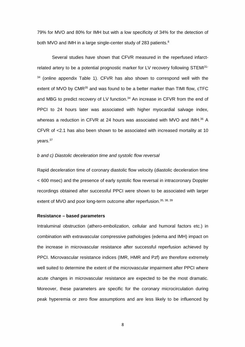
Several studies have shown that CFVR measured in the reperfused infarct-
related artery to be a potential prognostic marker for LV recovery following STEMI31-
34 (online appendix Table 1). CFVR has also shown to correspond well with the
extent of MVO by CMR35 and was found to be a better marker than TIMI flow, cTFC
and MBG to predict recovery of LV function.34 An increase in CFVR from the end of
PPCI to 24 hours later was associated with higher myocardial salvage index,
whereas a reduction in CFVR at 24 hours was associated with MVO and IMH.36 A
CFVR of <2.1 has also been shown to be associated with increased mortality at 10
years.37
b and c) Diastolic deceleration time and systolic flow reversal
Rapid deceleration time of coronary diastolic flow velocity (diastolic deceleration time
< 600 msec) and the presence of early systolic flow reversal in intracoronary Doppler
recordings obtained after successful PPCI were shown to be associated with larger
extent of MVO and poor long-term outcome after reperfusion.35, 38, 39
Resistance – based parameters
Intraluminal obstruction (athero-embolization, cellular and humoral factors etc.) in
combination with extravascular compressive pathologies (edema and IMH) impact on
the increase in microvascular resistance after successful reperfusion achieved by
PPCI. Microvascular resistance indices (IMR, HMR and Pzf) are therefore extremely
well suited to determine the extent of the microvascular impairment after PPCI where
acute changes in microvascular resistance are expected to be the most dramatic.
Moreover, these parameters are specific for the coronary microcirculation during
peak hyperemia or zero flow assumptions and are less likely to be influenced by
9
hemodynamic perturbations such as microvascular tone and resistance40, heart
rate41 and infusion of sodium nitroprusside and dobutamine.41
a) Index of microvascular resistance (IMR)
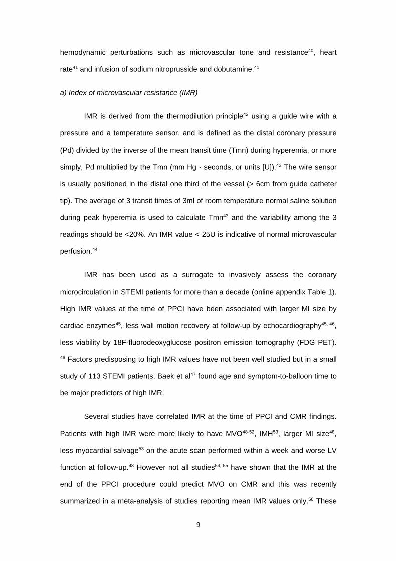
IMR is derived from the thermodilution principle42 using a guide wire with a
pressure and a temperature sensor, and is defined as the distal coronary pressure
(Pd) divided by the inverse of the mean transit time (Tmn) during hyperemia, or more
simply, Pd multiplied by the Tmn (mm Hg · seconds, or units [U]).42 The wire sensor
is usually positioned in the distal one third of the vessel (> 6cm from guide catheter
tip). The average of 3 transit times of 3ml of room temperature normal saline solution
during peak hyperemia is used to calculate Tmn43 and the variability among the 3
readings should be <20%. An IMR value < 25U is indicative of normal microvascular
perfusion.44
IMR has been used as a surrogate to invasively assess the coronary
microcirculation in STEMI patients for more than a decade (online appendix Table 1).
High IMR values at the time of PPCI have been associated with larger MI size by
cardiac enzymes45, less wall motion recovery at follow-up by echocardiography45, 46,
less viability by 18F-fluorodeoxyglucose positron emission tomography (FDG PET).
46 Factors predisposing to high IMR values have not been well studied but in a small
study of 113 STEMI patients, Baek et al47 found age and symptom-to-balloon time to
be major predictors of high IMR.
Several studies have correlated IMR at the time of PPCI and CMR findings.
Patients with high IMR were more likely to have MVO48-52, IMH53, larger MI size48,
less myocardial salvage53 on the acute scan performed within a week and worse LV
function at follow-up.48 However not all studies54, 55 have shown that the IMR at the
end of the PPCI procedure could predict MVO on CMR and this was recently
summarized in a meta-analysis of studies reporting mean IMR values only.56 These

10
studies individually were small and lacked power but after combining data from 6
studies (246 patients) reporting mean IMR values, those with MVO had significantly
higher IMR (49±33U, 99%CI 41-57U) than those without MVO (27±22U, 99%CI 22-
32U). In a recent single-center study of 283 patients by Carrick et al8, IMR was
shown to be more closely associated with MVO, IMH and adverse LV remodeling by
CMR and clinical outcomes than TMPG or CFR.
Data on serial IMR measurements post-STEMI is limited. Sezer et al57
showed that >33% improvement in IMR in the infarcted territory at 5 months was
associated with a 50% reduction in MI size assessed by single photon emission
computed tomography in a small cohort of 35 reperfused STEMI patients. Cuculi et
al54 showed that, in 30 patients with CMR data at 6 months, those with MVO had a
lower CFVR immediately post-PPCI and at 24 hours and a trend towards higher IMR.
At 6 months, there was no difference in IMR and CFVR between these 2 groups of
patients, despite a larger chronic MI size in the MVO group. However, unlike Sezer et
al57, they did not explore the reduction in MI size in those with an improvement in
IMR. Most recently, Hoole et al55 showed in 41 patients that those with an IMR <32U
pre-stenting had a significant increase in IMR post-stenting and this was attributed to
iatrogenic microvascular injury. Serial IMR measurements can improve our
understanding of the microcirculation post-STEMI, but due to its invasive nature,
getting patients back for repeat invasive measurements in the convalescent/chronic
phase is challenging as highlighted by the Cuculi et al54 (almost half of the patients
dropped out at 6 months).
Fukunaga et al52 (88 patients) found that the shape of the thermodilution-
derived temperature recovery curve following saline injection could be characterized
into three categories. Patients in the “bimodal group” had higher prevalence of MVO
on CMR and were at higher risk of death and rehospitalization for heart failure when
compared to those in the “narrow unimodal” and “wide unimodal groups”. However

11
the impact of the speed of hand injections and the inter-observer reproducibility of
these bimodal curves were not assessed and needs further validation.
Another approach explored by Park et al58 (89 patients) has been to stratify
STEMI patients according to both IMR and CFVR values. They found that those
patients with CFVR<2 and IMR>27U did not show an improvement in wall motion
score index by echocardiography. Ahn et al50 (40 patients) showed that a combined
high IMR (>36U) and low CFVR (<1.7) were highly predictive of MVO by CMR after
PPCI. However, Carrick et al59 recently showed that combining IMR >40U with CFR
≤2.0 did not add prognostic value in 283 patients. All these 3 studies used different
cut-off values for IMR and CFR and although Carrick et al59 had the largest number
of patients, it was not powered for clinical outcomes.
The effect of IMR on clinical outcomes post-PPCI has been investigated in a
large multi-center study of 253 STEMI patients. Fearon et al60 found that patients
with an IMR >40U, measured immediately after PPCI, was the only independent
predictor of death (hazard ratio 4.3, P 0.02) after a median follow-up of 2.8 years. A
recent meta-analysis56 showed that patients with an IMR >41U at the end of the
PPCI procedure were more likely to have MVO on the CMR. Most recently, in a
single-center study of 283 STEMI patients, Carrick et al59 also showed that an IMR
>40U was a multivariable associate of adverse LV remodeling by CMR at 6 months,
and was a better predictor of all-cause death or heart failure than the duration of
ischemia, ST-segment resolution, TMPG and CFR after a median follow-up of 845
days.
However IMR remains an indirect measure for the coronary microvascular
resistance and uses the inverse of transit time as a surrogate for flow. Furthermore,
the manual injections of normal saline to obtain the transit times are prone to inter
12
and intra-observer variability and not all groups54, 55 have shown IMR could
differentiate between patients with or without MVO.
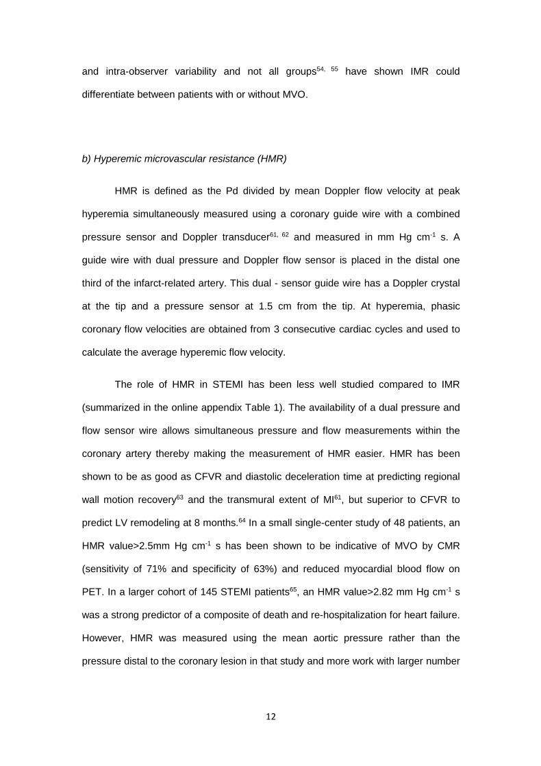
b) Hyperemic microvascular resistance (HMR)
HMR is defined as the Pd divided by mean Doppler flow velocity at peak
hyperemia simultaneously measured using a coronary guide wire with a combined
pressure sensor and Doppler transducer61, 62 and measured in mm Hg cm-1 s. A
guide wire with dual pressure and Doppler flow sensor is placed in the distal one
third of the infarct-related artery. This dual - sensor guide wire has a Doppler crystal
at the tip and a pressure sensor at 1.5 cm from the tip. At hyperemia, phasic
coronary flow velocities are obtained from 3 consecutive cardiac cycles and used to
calculate the average hyperemic flow velocity.
The role of HMR in STEMI has been less well studied compared to IMR
(summarized in the online appendix Table 1). The availability of a dual pressure and
flow sensor wire allows simultaneous pressure and flow measurements within the
coronary artery thereby making the measurement of HMR easier. HMR has been
shown to be as good as CFVR and diastolic deceleration time at predicting regional
wall motion recovery63 and the transmural extent of MI61, but superior to CFVR to
predict LV remodeling at 8 months.64 In a small single-center study of 48 patients, an
HMR value>2.5mm Hg cm-1 s has been shown to be indicative of MVO by CMR
(sensitivity of 71% and specificity of 63%) and reduced myocardial blood flow on
PET. In a larger cohort of 145 STEMI patients65, an HMR value>2.82 mm Hg cm-1 s
was a strong predictor of a composite of death and re-hospitalization for heart failure.
However, HMR was measured using the mean aortic pressure rather than the
pressure distal to the coronary lesion in that study and more work with larger number
13
of patients are required to confirm the prognostic significant of HMR immediately post
PPCI.
HMR also remains an indirect measure for the microvascular resistance and
uses half the peak Doppler-derived velocity as a surrogate for flow. Additionally,
detecting an adequate flow signal using a guide wire tipped with both a Doppler flow
and a pressure sensor can be technically difficult.
c) Zero flow pressure (Pzf)
Pzf is defined as the distal coronary pressure when hypothetically there would
be no flow in the coronary artery. Data from several cardiac cycles are used to plot
Pd against the peak velocity. There are automated algorithms that can then sample
the resultant pressure–velocity loop at the mid-diastolic period of the averaged
cardiac cycle. A regression can then be drawn automatically from the diastolic data
points, and Pzf is the pressure at which this line crosses the x-axis. This is the
extrapolated distal coronary pressure at which flow would cease in the infarct-related
artery.40 It provides comprehensive assessment of the microvascular compartment
as it assesses coronary flow over a range of pressures, irrespective of cardiac
contractility and may reflect vascular tone.66 In the context of STEMI, it also provides
information on the effect of the interstitial myocardial pressure on the coronary
microcirculation.66 Pzf is derived from pressure-velocity loop analysis67 and it informs
the operator on the effect of intra-ventricular and interstitial myocardial pressure
(external forces) over collapsible elements (capillaries) of the microcirculation.
Therefore, Pzf measured after PPCI can be expected to be dependent mainly on the
extent of external microcirculatory compression by edema and IMH. After PPCI,
microvascular impairment may be partly attributed to decrease in total cross-
sectional microvascular area by compressive effect generated by edema and/or IMH.
Additionally, in patients with STEMI, increased diastolic filling pressures due to

14
increased cardiac muscle stiffness caused by cellular and interstitial edema may also
decrease intramyocardial vascular capacitance and limits coronary flow in late
diastole. Therefore, the transmitted increase in intra-cavity and interstitial pressures
contribute to external compression of microcirculation and result in increased Pzf.
In small proof-of-concept studies, Pzf was a better predictor of viability by
FDG PET than CFVR (27 patients)67; was associated with higher left ventricular filling
pressures (68 patients)66 and adverse LV remodeling (48 patients)68; and was a
better predictor of chronic MI size by CMR than HMR and IMR (34 patients).40
However, Pzf was not found to be superior to CFVR, Pzf and diastolic deceleration
time in predicting the transmural extent of MI by CMR performed at 13 days (27
patients).61 So far, only one study has evaluated Pzf and MVO by CMR and a cut-off
value of 42mmHg for Pzf did not differentiate those with and without MVO.40
As summarized in table 2, Pzf requires off-line post-processing and the
automated algorithms for its interpretation are not widely available yet. Therefore this
index currently remains a research tool.
CURRENT APPLICATIONS
IMR has already been used as a surrogate endpoint in several proof-of-concept
studies aiming to improve the coronary microcirculation in reperfused STEMI patients
(summarized in the online appendix Table 2). The impact of strategies such as
intracoronary streptokinase administered immediately after PPCI 20, nicorandil69,
sodium nitropruside70, distal protection device71, thrombus aspiration55 and a
combination of intracoronary abciximab and aspiration thrombectomy51 on IMR has
been investigated in small proof-of-concept studies and there are several other
studies that are ongoing (summarized in the online appendix table 2). Of note, none
of the randomized studies pre-selected patients based on high IMR values.
15
Hypothetically, in an ideal study, invasive coronary measurement with a reliable
marker at the end of the PPCI procedure would identify those patients with MVO with
high sensitivity and specificity. Given that MVO is due to a combination of factors,
these patients would then be randomized to a combination therapy with intracoronary
thrombolysis (to achieve patency of the microcirculation) and an intracoronary
vasodilator with anti-inflammatory properties such as adenosine (which could be
continued intravenously on the ward) or placebo. The primary endpoint of interest
should ideally be hard clinical outcomes such as cardiovascular death and
hospitalization for heart failure. However, such a study would require a large number
of patients and endpoints such as the extent of IMH, MVO and MI size by CMR 3
days later and adverse LV remodeling at 6 months could be used as surrogates.
LIMITATIONS OF CURRENT INVASIVE MARKERS AND FUTURE DIRECTIONS
Table 2 summarizes the limitations of the current invasive markers to assess the
microcirculation discussed so far. Future studies should aim at addressing these
limitations in the first instance. Some examples would be:
To explore the possibility for an automated method (e.g. using a pump injector) to
inject the 3ml of normal saline to minimize operator-related errors and improve inter-
observer and inter-site reproducibility when performing IMR measurement.
Further validation work is required to assess the performance of HMR to detect MVO
by CMR before it can be used to assess the effectiveness of therapies. Moreover,
improvement in the delivery profile of the Doppler wire would increase the use of this
technique in future studies.
The derivation of Pzf is based on the extrapolation from the pressure-velocity loop
and the analysis techniques are time-consuming and are not available for immediate
16
read-outs of Pzf. Therefore, more work remains to be done to make the analysis of
fully automated and the Pzf read-outs to be immediately available at the time of
PPCI, before it can be widely used.
Limited data are available regarding the strength of diastolic deceleration time and
systolic flow reversal to identify MVO when compared to IMR, HMR and Pzf.
Therefore, further, adequately powered, comparative studies using these
parameters, using a multi-center approach to facilitate recruitment are needed.
These above steps would help to assess which of these markers would emerge as
the most robust surrogate marker for predicting MVO at the end of the PPCI
procedure. Early identification of these high-risk patients is important as already
described above and early adjuvant intervention could then be started in the cardiac
catheterization laboratory and administered via the intracoronary route and continued
intravenously in the ward in needed. Therapies that may be beneficial in this setting
would be glycoprotein IIb/IIIa inhibitors (e.g. abciximab21 to reduce platelet
aggregation in the microcirculation); thrombolytics (e.g. half dose alteplase for lysis of
distal embolization of thrombi); vasodilators (e.g. adenosine19, 72, nicorandil69 for
spasm of the microcirculation due to release of vasoactive substances); and anti-
inflammatory agents (e.g. methylprednisolone73 to reduce reperfusion edema and
relieve extrinsic compression of the microcirculation). Other treatments aiming to
stabilize the endothelium with angiopoietin-1 or tyrosine kinase inhibitors13 may help
to reduce extravasation of red blood cells and the development of IMH. Using this
approach would also avoid any adjuvant strategies be given to those patients who
are unlikely to have MVO, and minimize their exposure to potential adverse events.
Figure 4 shows a hypothetical approach in future studies to identify and target those
at high-risk at the end of the PPCI procedure using IMR as an example. Given that
patients with an IMR of >40U has been shown to more likely have MVO56 and worse
17
outcomes 60 59, this value could be used as a cut-off. Those patients with an IMR of
>40U at the end of PPCI could then be targeted with further adjuvant therapies
mentioned above and this approach may improve outcomes in this group of patients.
CONCLUSION
Invasive assessment of the coronary microcirculation at the time of PPCI is an
exciting field that could provide us with the opportunity to interrogate the extent of the
microvascular injury reliably in the cardiac catheterization laboratory at the end of the
PPCI procedure despite having a patent infarct-related epicardial coronary artery.
This approach would potentially identify those patients at high risk of MVO and target
them with adjuvant therapies. However, more validation work remains to be done
before one or a combination of these invasive markers described here could be used
in therapeutic trials aiming to eventually improve outcomes in these patients.
REFERENCES
1. O'Gara PT, Kushner FG, Ascheim DD, Casey DE, Jr., Chung MK, de Lemos JA, Ettinger SM, Fang JC, Fesmire FM, Franklin BA, Granger CB, Krumholz HM, Linderbaum JA, Morrow DA, Newby LK, Ornato JP, Ou N, Radford MJ, Tamis-Holland JE, Tommaso CL, Tracy CM, Woo YJ, Zhao DX, American College of Cardiology F, American Heart Association Task Force on Practice G, American College of Emergency P, Society for Cardiovascular A and Interventions. 2013 ACCF/AHA guideline for the management of ST-elevation myocardial infarction: executive summary: a report of the American College of Cardiology Foundation/American Heart Association Task Force on Practice Guidelines: developed in collaboration with the American College of Emergency Physicians and Society for Cardiovascular Angiography and Interventions. Catheterization and cardiovascular interventions : official journal of the Society for Cardiac Angiography & Interventions. 2013;82:E1-27. 2. Torabi A, Cleland JG, Khan NK, Loh PH, Clark AL, Alamgir F, Caplin JL, Rigby AS and Goode K. The timing of development and subsequent clinical course of heart failure after a myocardial infarction. European heart journal. 2008;29:859-70. 3. Yellon DM and Hausenloy DJ. Mechanisms of disease: Myocardial reperfusion injury. New England Journal of Medicine. 2007;357:1121-1135. 4. Krug A, Du Mesnil de R and Korb G. Blood supply of the myocardium after temporary coronary occlusion. Circulation research. 1966;19:57-62.
18
5. van Kranenburg M, Magro M, Thiele H, de Waha S, Eitel I, Cochet A, Cottin Y, Atar D, Buser P, Wu E, Lee D, Bodi V, Klug G, Metzler B, Delewi R, Bernhardt P, Rottbauer W, Boersma E, Zijlstra F and van Geuns RJ. Prognostic value of microvascular obstruction and infarct size, as measured by CMR in STEMI patients. JACC Cardiovascular imaging. 2014;7:930-9. 6. Ito H, Maruyama A, Iwakura K, Takiuchi S, Masuyama T, Hori M, Higashino Y, Fujii K and Minamino T. Clinical implications of the 'no reflow' phenomenon. A predictor of complications and left ventricular remodeling in reperfused anterior wall myocardial infarction. Circulation. 1996;93:223-8. 7. Schofer J, Montz R and Mathey DG. Scintigraphic evidence of the "no reflow" phenomenon in human beings after coronary thrombolysis. Journal of the American College of Cardiology. 1985;5:593-8. 8. Carrick D, Haig C, Carberry J, May VT, McCartney P, Welsh P, Ahmed N, McEntegart M, Petrie MC, Eteiba H, Lindsay M, Hood S, Watkins S, Mahrous A, Rauhalammi SM, Mordi I, Ford I, Radjenovic A, Sattar N, Oldroyd KG and Berry C. Microvascular resistance of the culprit coronary artery in acute ST-elevation myocardial infarction. JCI Insight. 2016;1:e85768. 9. Niccoli G, Scalone G, Lerman A and Crea F. Coronary microvascular obstruction in acute myocardial infarction. European heart journal. 2016;37:1024-33. 10. Niccoli G, Burzotta F, Galiuto L and Crea F. Myocardial no-reflow in humans. Journal of the American College of Cardiology. 2009;54:281-92. 11. Reffelmann T and Kloner RA. Microvascular reperfusion injury: rapid expansion of anatomic no reflow during reperfusion in the rabbit. American Journal of Physiology-Heart and Circulatory Physiology. 2002;283:H1099-H1107. 12. Driesen RB, Zalewski J, Vanden Driessche N, Vermeulen K, Bogaert J, Sipido KR, Van de Werf F and Claus P. Histological correlate of a cardiac magnetic resonance imaged microvascular obstruction in a porcine model of ischemia-reperfusion. Cardiovasc Pathol. 2012;21:129-31. 13. Betgem RP, de Waard GA, Nijveldt R, Beek AM, Escaned J and van Royen N. Intramyocardial haemorrhage after acute myocardial infarction. Nature reviews Cardiology. 2015;12:156-67. 14. Bulluck H and Hausenloy DJ. Microvascular Obstruction: The Bane of Myocardial Reperfusion. Revista espanola de cardiologia. 2015;68:919-20. 15. Morishima I, Sone T, Okumura K, Tsuboi H, Kondo J, Mukawa H, Matsui H, Toki Y, Ito T and Hayakawa T. Angiographic no-reflow phenomenon as a predictor of adverse long-term outcome in patients treated with percutaneous transluminal coronary angioplasty for first acute myocardial infarction. Journal of the American College of Cardiology. 2000;36:1202-1209. 16. Sorajja P, Gersh BJ, Costantini C, McLaughlin MG, Zimetbaum P, Cox DA, Garcia E, Tcheng JE, Mehran R, Lansky AJ, Kandzari DE, Grines CL and Stone GW. Combined prognostic utility of ST-segment recovery and myocardial blush after primary percutaneous coronary intervention in acute myocardial infarction. European heart journal. 2005 Apr;26:667-74. 17. Bolognese L, Carrabba N, Parodi G, Santoro GM, Buonamici P, Cerisano G and Antoniucci D. Impact of microvascular dysfunction on left ventricular remodeling and long-term clinical outcome after primary coronary angioplasty for acute myocardial infarction. Circulation. 2004;109:1121-6. 18. Hombach V, Grebe O, Merkle N, Waldenmaier S, Hoher M, Kochs M, Wohrle J and Kestler HA. Sequelae of acute myocardial infarction regarding cardiac structure and function and their prognostic significance as assessed by magnetic resonance imaging. European heart journal. 2005;26:549-57.

19
19. Nazir SA, McCann GP, Greenwood JP, Kunadian V, Khan JN, Mahmoud IZ, Blackman DJ, Been M, Abrams KR, Shipley L, Wilcox R, Adgey AA and Gershlick AH. Strategies to attenuate micro-vascular obstruction during P-PCI: the randomized reperfusion facilitated by local adjunctive therapy in ST-elevation myocardial infarction trial. European heart journal. 2016;37:1910-9. 20. Sezer M, Oflaz H, Goren T, Okcular I, Umman B, Nisanci Y, Bilge AK, Sanli Y, Meric M and Umman S. Intracoronary streptokinase after primary percutaneous coronary intervention. The New England journal of medicine. 2007;356:1823-34. 21. Eitel I, Wohrle J, Suenkel H, Meissner J, Kerber S, Lauer B, Pauschinger M, Birkemeyer R, Axthelm C, Zimmermann R, Neuhaus P, Brosteanu O, de Waha S, Desch S, Gutberlet M, Schuler G and Thiele H. Intracoronary compared with intravenous bolus abciximab application during primary percutaneous coronary intervention in ST-segment elevation myocardial infarction: cardiac magnetic resonance substudy of the AIDA STEMI trial. Journal of the American College of Cardiology. 2013;61:1447-54. 22. van 't Hof AW, Liem A, Suryapranata H, Hoorntje JC, de Boer MJ and Zijlstra F. Angiographic assessment of myocardial reperfusion in patients treated with primary angioplasty for acute myocardial infarction: myocardial blush grade. Zwolle Myocardial Infarction Study Group. Circulation. 1998;97:2302-6. 23. Gibson CM, Cannon CP, Daley WL, Dodge JT, Jr., Alexander B, Jr., Marble SJ, McCabe CH, Raymond L, Fortin T, Poole WK and Braunwald E. TIMI frame count: a quantitative method of assessing coronary artery flow. Circulation. 1996;93:879-88. 24. Gibson CM, Cannon CP, Murphy SA, Ryan KA, Mesley R, Marble SJ, McCabe CH, Van De Werf F and Braunwald E. Relationship of TIMI myocardial perfusion grade to mortality after administration of thrombolytic drugs. Circulation. 2000;101:125-30. 25. Porto I, Hamilton-Craig C, Brancati M, Burzotta F, Galiuto L and Crea F. Angiographic assessment of microvascular perfusion--myocardial blush in clinical practice. Am Heart J. 2010;160:1015-22. 26. Korosoglou G, Haars A, Michael G, Erbacher M, Hardt S, Giannitsis E, Kurz K, Franz-Josef N, Dickhaus H, Katus HA and Kuecherer H. Quantitative evaluation of myocardial blush to assess tissue level reperfusion in patients with acute ST-elevation myocardial infarction: incremental prognostic value compared with visual assessment. Am Heart J. 2007;153:612-20. 27. Kaul S. Coronary angiography cannot be used to assess myocardial perfusion in patients undergoing reperfusion for acute myocardial infarction. Heart. 2001;86:483-4. 28. Marcus ML, Chilian WM, Kanatsuka H, Dellsperger KC, Eastham CL and Lamping KG. Understanding the coronary circulation through studies at the microvascular level. Circulation. 1990;82:1-7. 29. Knaapen P, Camici PG, Marques KM, Nijveldt R, Bax JJ, Westerhof N, Gotte MJ, Jerosch-Herold M, Schelbert HR, Lammertsma AA and van Rossum AC. Coronary microvascular resistance: methods for its quantification in humans. Basic research in cardiology. 2009;104:485-98. 30. De Bruyne B, Pijls NH, Smith L, Wievegg M and Heyndrickx GR. Coronary thermodilution to assess flow reserve: experimental validation. Circulation. 2001;104:2003-6. 31. Takahashi T, Hiasa Y, Ohara Y, Miyazaki S, Ogura R, Miyajima H, Yuba K, Suzuki N, Hosokawa S, Kishi K and Ohtani R. Usefulness of coronary flow reserve immediately after primary coronary angioplasty for acute myocardial infarction in predicting long-term adverse cardiac events. The American journal of cardiology. 2007;100:806-11. 32. Garot P, Pascal O, Simon M, Monin JL, Teiger E, Garot J, Gueret P and Dubois-Rande JL. Impact of microvascular integrity and local viability on left ventricular remodelling after reperfused acute myocardial infarction. Heart. 2003;89:393-7.

20
33. Wakatsuki T, Nakamura M, Tsunoda T, Toma H, Degawa T, Oki T and Yamaguchi T. Coronary flow velocity immediately after primary coronary stenting as a predictor of ventricular wall motion recovery in acute myocardial infarction. Journal of the American College of Cardiology. 2000;35:1835-41. 34. Bax M, de Winter RJ, Schotborgh CE, Koch KT, Meuwissen M, Voskuil M, Adams R, Mulder KJ, Tijssen JG and Piek JJ. Short- and long-term recovery of left ventricular function predicted at the time of primary percutaneous coronary intervention in anterior myocardial infarction. Journal of the American College of Cardiology. 2004;43:534-41. 35. Hirsch A, Nijveldt R, Haeck JD, Beek AM, Koch KT, Henriques JP, van der Schaaf RJ, Vis MM, Baan J, Jr., de Winter RJ, Tijssen JG, van Rossum AC and Piek JJ. Relation between the assessment of microvascular injury by cardiovascular magnetic resonance and coronary Doppler flow velocity measurements in patients with acute anterior wall myocardial infarction. Journal of the American College of Cardiology. 2008;51:2230-8. 36. Cuculi F, Dall'Armellina E, Manlhiot C, De Caterina AR, Colyer S, Ferreira V, Morovat A, Prendergast BD, Forfar JC, Alp NJ, Choudhury RP, Neubauer S, Channon KM, Banning AP and Kharbanda RK. Early change in invasive measures of microvascular function can predict myocardial recovery following PCI for ST-elevation myocardial infarction. European heart journal. 2014;35:1971-80. 37. van de Hoef TP, Bax M, Meuwissen M, Damman P, Delewi R, de Winter RJ, Koch KT, Schotborgh C, Henriques JP, Tijssen JG and Piek JJ. Impact of coronary microvascular function on long-term cardiac mortality in patients with acute ST-segment-elevation myocardial infarction. Circulation Cardiovascular interventions. 2013;6:207-15. 38. Iwakura K, Ito H, Takiuchi S, Taniyama Y, Nakatsuchi Y, Negoro S, Higashino Y, Okamura A, Masuyama T, Hori M, Fujii K and Minamino T. Alternation in the coronary blood flow velocity pattern in patients with no reflow and reperfused acute myocardial infarction. Circulation. 1996;94:1269-75. 39. Furber AP, Prunier F, Nguyen HC, Boulet S, Delepine S and Geslin P. Coronary blood flow assessment after successful angioplasty for acute myocardial infarction predicts the risk of long-term cardiac events. Circulation. 2004;110:3527-33. 40. Patel N, Petraco R, Dall'Armellina E, Kassimis G, De Maria GL, Dawkins S, Lee R, Prendergast BD, Choudhury RP, Forfar JC, Channon KM, Davies J, Banning AP and Kharbanda RK. Zero-Flow Pressure Measured Immediately After Primary Percutaneous Coronary Intervention for ST-Segment Elevation Myocardial Infarction Provides the Best Invasive Index for Predicting the Extent of Myocardial Infarction at 6 Months: An OxAMI Study (Oxford Acute Myocardial Infarction). JACC Cardiovascular interventions. 2015;8:1410-21. 41. Ng MK, Yeung AC and Fearon WF. Invasive assessment of the coronary microcirculation: superior reproducibility and less hemodynamic dependence of index of microcirculatory resistance compared with coronary flow reserve. Circulation. 2006;113:2054-61. 42. Fearon WF, Balsam LB, Farouque HM, Caffarelli AD, Robbins RC, Fitzgerald PJ, Yock PG and Yeung AC. Novel index for invasively assessing the coronary microcirculation. Circulation. 2003;107:3129-32. 43. Pijls NH, De Bruyne B, Smith L, Aarnoudse W, Barbato E, Bartunek J, Bech GJ and Van De Vosse F. Coronary thermodilution to assess flow reserve: validation in humans. Circulation. 2002;105:2482-6. 44. Berry C, Corcoran D, Hennigan B, Watkins S, Layland J and Oldroyd KG. Fractional flow reserve-guided management in stable coronary disease and acute myocardial infarction: recent developments. European heart journal. 2015;36:3155-64. 45. Fearon WF, Shah M, Ng M, Brinton T, Wilson A, Tremmel JA, Schnittger I, Lee DP, Vagelos RH, Fitzgerald PJ, Yock PG and Yeung AC. Predictive value of the index of
21
microcirculatory resistance in patients with ST-segment elevation myocardial infarction. Journal of the American College of Cardiology. 2008;51:560-5. 46. Lim HS, Yoon MH, Tahk SJ, Yang HM, Choi BJ, Choi SY, Sheen SS, Hwang GS, Kang SJ and Shin JH. Usefulness of the index of microcirculatory resistance for invasively assessing myocardial viability immediately after primary angioplasty for anterior myocardial infarction. European heart journal. 2009;30:2854-60. 47. Baek YS, Park SD, Kim SH, Lee MJ, Shin SH, Kim DH, Kwan J, Park KS and Woo SI. Clinical and Angiographic Predictors of Microvascular Dysfunction in ST-Segment Elevation Myocardial Infarction. Yonsei Med J. 2015;56:1235-43. 48. McGeoch R, Watkins S, Berry C, Steedman T, Davie A, Byrne J, Hillis S, Lindsay M, Robb S, Dargie H and Oldroyd K. The index of microcirculatory resistance measured acutely predicts the extent and severity of myocardial infarction in patients with ST-segment elevation myocardial infarction. JACC Cardiovascular interventions. 2010;3:715-22. 49. Yoo SH, Yoo TK, Lim HS, Kim MY and Koh JH. Index of microcirculatory resistance as predictor for microvascular functional recovery in patients with anterior myocardial infarction. Journal of Korean medical science. 2012;27:1044-50. 50. Ahn SG, Hung OY, Lee JW, Lee JH, Youn YJ, Ahn MS, Kim JY, Yoo BS, Lee SH, Yoon J, Kwon W and Samady H. Combination of the Thermodilution-Derived Index of Microcirculatory Resistance and Coronary Flow Reserve Is Highly Predictive of Microvascular Obstruction on Cardiac Magnetic Resonance Imaging After ST-Segment Elevation Myocardial Infarction. JACC Cardiovascular interventions. 2016;9:793-801. 51. Ahn SG, Lee SH, Lee JH, Lee JW, Youn YJ, Ahn MS, Kim JY, Yoo BS, Yoon J, Choe KH and Tahk SJ. Efficacy of combination treatment with intracoronary abciximab and aspiration thrombectomy on myocardial perfusion in patients with ST-segment elevation myocardial infarction undergoing primary coronary stenting. Yonsei Med J. 2014;55:606-16. 52. Fukunaga M, Fujii K, Kawasaki D, Sawada H, Miki K, Tamaru H, Imanaka T, Iwasaku T, Nakata T, Shibuya M, Akahori H, Masutani M, Kobayashi K, Ohyanagi M and Masuyama T. Thermodilution-derived coronary blood flow pattern immediately after coronary intervention as a predictor of microcirculatory damage and midterm clinical outcomes in patients with ST-segment-elevation myocardial infarction. Circulation Cardiovascular interventions. 2014;7:149-55. 53. Payne AR, Berry C, Doolin O, McEntegart M, Petrie MC, Lindsay MM, Hood S, Carrick D, Tzemos N, Weale P, McComb C, Foster J, Ford I and Oldroyd KG. Microvascular Resistance Predicts Myocardial Salvage and Infarct Characteristics in ST-Elevation Myocardial Infarction. Journal of the American Heart Association. 2012;1:e002246. 54. Cuculi F, De Maria GL, Meier P, Dall'Armellina E, de Caterina AR, Channon KM, Prendergast BD, Choudhury RC, Forfar JC, Kharbanda RK and Banning AP. Impact of Microvascular Obstruction on the Assessment of Coronary Flow Reserve, Index of Microcirculatory Resistance, and Fractional Flow Reserve After ST-Segment Elevation Myocardial Infarction. Journal of the American College of Cardiology. 2014;64:1894-904. 55. Hoole SP, Jaworski C, Brown AJ, McCormick LM, Agrawal B, Clarke SC and West NE. Serial assessment of the index of microcirculatory resistance during primary percutaneous coronary intervention comparing manual aspiration catheter thrombectomy with balloon angioplasty (IMPACT study): a randomised controlled pilot study. Open Heart. 2015;2:e000238. 56. Bulluck H, Foin N, Carbrera-Fuentes HA, Yeo KK, Wong AS, Fam JM, Wong PE, Tan JW, Low AF and Hausenloy DJ. Index of Microvascular Resistance and Microvascular Obstruction in patients with Acute Myocardial Infarction. JACC Cardiovascular interventions. 2016;9:2172-8. 57. Sezer M, Aslanger EK, Cimen AO, Yormaz E, Turkmen C, Umman B, Nisanci Y, Bugra Z, Adalet K and Umman S. Concurrent microvascular and infarct remodeling after successful
22
reperfusion of ST-elevation acute myocardial infarction. Circulation Cardiovascular interventions. 2010;3:208-15. 58. Park SD, Baek YS, Lee MJ, Kwon SW, Shin SH, Woo SI, Kim DH, Kwan J and Park KS. Comprehensive assessment of microcirculation after primary percutaneous intervention in ST-segment elevation myocardial infarction: insight from thermodilution-derived index of microcirculatory resistance and coronary flow reserve. Coronary artery disease. 2016;27:34-9. 59. Carrick D, Haig C, Ahmed N, Carberry J, Teng Yue May V, McEntegart M, Petrie MC, Eteiba H, Lindsay M, Hood S, Watkins S, Davie A, Mahrous Abouzaid A, Mordi I, Ford I, Radjenovic A, Oldroyd KG and Berry C. Comparative Prognostic Utility of Indices of Microvascular Function Alone or in Combination in Patients with an Acute ST-Segment Elevation Myocardial Infarction. Circulation. 2016. 60. Fearon WF, Low AF, Yong AS, McGeoch R, Berry C, Shah MG, Ho MY, Kim HS, Loh JP and Oldroyd KG. Prognostic value of the index of microcirculatory resistance measured after primary percutaneous coronary intervention. Circulation. 2013;127:2436-41. 61. Kitabata H, Imanishi T, Kubo T, Takarada S, Kashiwagi M, Matsumoto H, Tsujioka H, Ikejima H, Arita Y, Okochi K, Kuroi A, Ueno S, Kataiwa H, Tanimoto T, Yamano T, Hirata K, Nakamura N, Tanaka A, Mizukoshi M and Akasaka T. Coronary microvascular resistance index immediately after primary percutaneous coronary intervention as a predictor of the transmural extent of infarction in patients with ST-segment elevation anterior acute myocardial infarction. JACC Cardiovascular imaging. 2009;2:263-72. 62. Teunissen PF, de Waard GA, Hollander MR, Robbers LF, Danad I, Biesbroek PS, Amier RP, Echavarria-Pinto M, Quiros A, Broyd C, Heymans MW, Nijveldt R, Lammertsma AA, Raijmakers PG, Allaart CP, Lemkes JS, Appelman YE, Marques KM, Bronzwaer JG, Horrevoets AJ, van Rossum AC, Escaned J, Beek AM, Knaapen P and van Royen N. Doppler-derived intracoronary physiology indices predict the occurrence of microvascular injury and microvascular perfusion deficits after angiographically successful primary percutaneous coronary intervention. Circulation Cardiovascular interventions. 2015;8:e001786. 63. Yoon MH, Tahk SJ, Yang HM, Woo SI, Lim HS, Kang SJ, Choi BJ, Choi SY, Hwang GS and Shin JH. Comparison of accuracy in the prediction of left ventricular wall motion changes between invasively assessed microvascular integrity indexes and fluorine-18 fluorodeoxyglucose positron emission tomography in patients with ST-elevation myocardial infarction. The American journal of cardiology. 2008;102:129-34. 64. Kitabata H, Kubo T, Ishibashi K, Komukai K, Tanimoto T, Ino Y, Kashiwagi M, Ozaki Y, Shiono Y, Shimamura K, Orii M, Hirata K, Tanaka A, Imanishi T and Akasaka T. Prognostic value of microvascular resistance index immediately after primary percutaneous coronary intervention on left ventricular remodeling in patients with reperfused anterior acute ST-segment elevation myocardial infarction. JACC Cardiovascular interventions. 2013;6:1046-54. 65. Jin X, Yoon MH, Seo KW, Tahk SJ, Lim HS, Yang HM, Choi BJ, Choi SY, Hwang GS, Shin JH and Park JS. Usefulness of Hyperemic Microvascular Resistance Index as a Predictor of Clinical Outcomes in Patients with ST-Segment Elevation Myocardial Infarction. Korean Circ J. 2015;45:194-201. 66. Van Herck PL, Carlier SG, Claeys MJ, Haine SE, Gorissen P, Miljoen H, Bosmans JM and Vrints CJ. Coronary microvascular dysfunction after myocardial infarction: increased coronary zero flow pressure both in the infarcted and in the remote myocardium is mainly related to left ventricular filling pressure. Heart. 2007;93:1231-7. 67. Shimada K, Sakanoue Y, Kobayashi Y, Ehara S, Hirose M, Nakamura Y, Fukuda D, Yamagishi H, Yoshiyama M, Takeuchi K and Yoshikawa J. Assessment of myocardial viability using coronary zero flow pressure after successful angioplasty in patients with acute anterior myocardial infarction. Heart. 2003;89:71-6.
23
68. Ito H, Terai K, Iwakura K, Kawase I and Fujii K. Hemodynamics of microvascular dysfunction in patients with anterior wall acute myocardial infarction. The American journal of cardiology. 2004;94:209-12. 69. Kostic J, Djordjevic-Dikic A, Dobric M, Milasinovic D, Nedeljkovic M, Stojkovic S, Stepanovic J, Tesic M, Trifunovic Z, Zamaklar-Tifunovic D, Radosavljevic-Radovanovic M, Ostojic M and Beleslin B. The effects of nicorandil on microvascular function in patients with ST segment elevation myocardial infarction undergoing primary PCI. Cardiovasc Ultrasound. 2015;13:26. 70. Morimoto K, Ito S, Nakasuka K, Sekimoto S, Miyata K, Inomata M, Yoshida T, Tamai N, Saeki T, Suzuki S, Murakami Y, Sato K, Morino A and Shimizu Y. Acute effect of sodium nitroprusside on microvascular dysfunction in patients who underwent percutaneous coronary intervention for acute ST-segment elevation myocardial infarction. Int Heart J. 2012;53:337-40. 71. Ito N, Nanto S, Doi Y, Kurozumi Y, Tonomura D, Natsukawa T, Sawano H, Masuda D, Yamashita S, Okada K, Hayashi Y, Kai T and Hayashi T. Distal protection during primary coronary intervention can preserve the index of microcirculatory resistance in patients with acute anterior ST-segment elevation myocardial infarction. Circulation journal : official journal of the Japanese Circulation Society. 2011;75:94-8. 72. Bulluck H, Sirker A, Loke YK, Garcia-Dorado D and Hausenloy DJ. Clinical benefit of adenosine as an adjunct to reperfusion in ST-elevation myocardial infarction patients: An updated meta-analysis of randomized controlled trials. International journal of cardiology. 2016;202:228-37. 73. Fernandez-Jimenez R, Garcia-Prieto J, Sanchez-Gonzalez J, Aguero J, Lopez-Martin GJ, Galan-Arriola C, Molina-Iracheta A, Doohan R, Fuster V and Ibanez B. Pathophysiology Underlying the Bimodal Edema Phenomenon After Myocardial Ischemia/Reperfusion. Journal of the American College of Cardiology. 2015;66:816-28. 74. Schroder R. Prognostic impact of early ST-segment resolution in acute ST-elevation myocardial infarction. Circulation. 2004;110:e506-10. 75. Giugliano RP, Sabatine MS, Gibson CM, Roe MT, Harrington RA, Murphy SA, Morrow DA, Antman EM and Braunwald E. Combined assessment of thrombolysis in myocardial infarction flow grade, myocardial perfusion grade, and ST-segment resolution to evaluate epicardial and myocardial reperfusion. The American journal of cardiology. 2004;93:1362-7, A5-6. 76. Kaul S. Myocardial contrast echocardiography: a 25-year retrospective. Circulation. 2008;118:291-308. 77. Carrick D, Haig C, Ahmed N, McEntegart M, Petrie MC, Eteiba H, Hood S, Watkins S, Lindsay MM, Davie A, Mahrous A, Mordi I, Rauhalammi S, Sattar N, Welsh P, Radjenovic A, Ford I, Oldroyd KG and Berry C. Myocardial Hemorrhage After Acute Reperfused ST-Segment-Elevation Myocardial Infarction: Relation to Microvascular Obstruction and Prognostic Significance. Circulation Cardiovascular imaging. 2016;9:e004148. 78. Ogasawara S, Mukawa H, Sone T, Tsuboi H, Morishima I, Uesugi M, Matsushita E, Morita Y, Okumura K and Murohara T. Presence of myocardial hypoenhancement on multidetector computed tomography after primary percutaneous coronary intervention in acute myocardial infarction predicts poor prognosis. International journal of cardiology. 2015;184:101-7. 79. Watabe H, Sato A, Nishina H, Hoshi T, Sugano A, Kakefuda Y, Takaiwa Y, Aihara H, Fumikura Y, Noguchi Y and Aonuma K. Enhancement patterns detected by multidetector computed tomography are associated with microvascular obstruction and left ventricular remodelling in patients with acute myocardial infarction. European heart journal. 2016;37:684-92.

24
80. Henriques JP, Zijlstra F, van 't Hof AW, de Boer MJ, Dambrink JH, Gosselink M, Hoorntje JC and Suryapranata H. Angiographic assessment of reperfusion in acute myocardial infarction by myocardial blush grade. Circulation. 2003;107:2115-9. 81. Aarnoudse W, Fearon WF, Manoharan G, Geven M, van de Vosse F, Rutten M, De Bruyne B and Pijls NH. Epicardial stenosis severity does not affect minimal microcirculatory resistance. Circulation. 2004;110:2137-42. 82. Layland J, MacIsaac AI, Burns AT, Somaratne JB, Leitl G, Whitbourn RJ and Wilson AM. When collateral supply is accounted for epicardial stenosis does not increase microvascular resistance. Circulation Cardiovascular interventions. 2012;5:97-102.
25
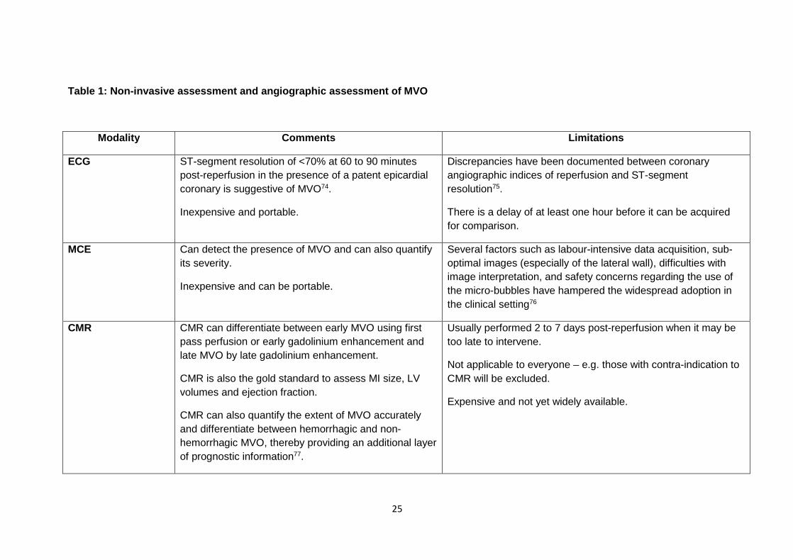
Table 1: Non-invasive assessment and angiographic assessment of MVO
Modality Comments Limitations
ECG ST-segment resolution of <70% at 60 to 90 minutes
post-reperfusion in the presence of a patent epicardial
coronary is suggestive of MVO74.
Inexpensive and portable.
Discrepancies have been documented between coronary
angiographic indices of reperfusion and ST-segment
resolution75.
There is a delay of at least one hour before it can be acquired
for comparison.
MCE Can detect the presence of MVO and can also quantify
its severity.
Inexpensive and can be portable.
Several factors such as labour-intensive data acquisition, sub-
optimal images (especially of the lateral wall), difficulties with
image interpretation, and safety concerns regarding the use of
the micro-bubbles have hampered the widespread adoption in
the clinical setting76
CMR CMR can differentiate between early MVO using first
pass perfusion or early gadolinium enhancement and
late MVO by late gadolinium enhancement.
CMR is also the gold standard to assess MI size, LV
volumes and ejection fraction.
CMR can also quantify the extent of MVO accurately
and differentiate between hemorrhagic and non-
hemorrhagic MVO, thereby providing an additional layer
of prognostic information77.
Usually performed 2 to 7 days post-reperfusion when it may be
too late to intervene.
Not applicable to everyone – e.g. those with contra-indication to
CMR will be excluded.
Expensive and not yet widely available.
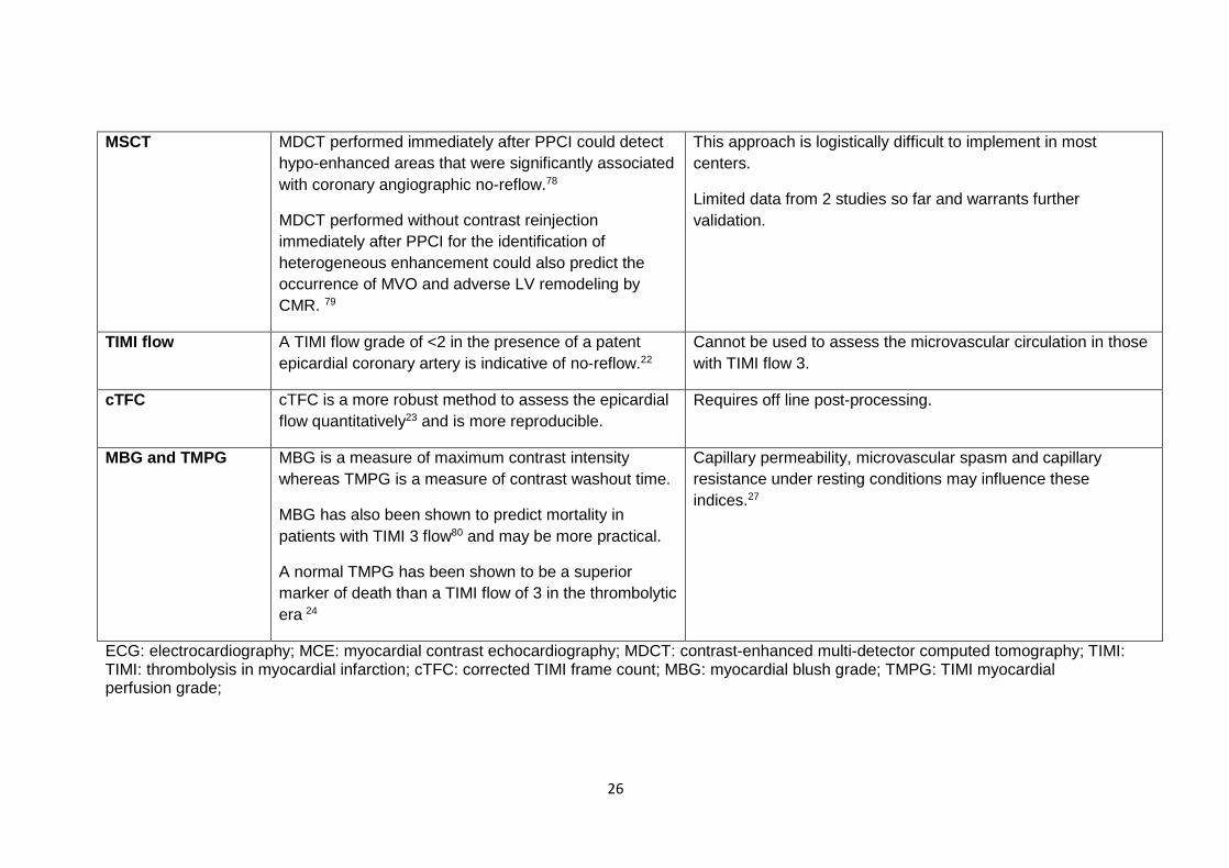
26
MSCT MDCT performed immediately after PPCI could detect
hypo-enhanced areas that were significantly associated
with coronary angiographic no-reflow.78
MDCT performed without contrast reinjection
immediately after PPCI for the identification of
heterogeneous enhancement could also predict the
occurrence of MVO and adverse LV remodeling by
CMR. 79
This approach is logistically difficult to implement in most
centers.
Limited data from 2 studies so far and warrants further
validation.
TIMI flow A TIMI flow grade of <2 in the presence of a patent
epicardial coronary artery is indicative of no-reflow.22
Cannot be used to assess the microvascular circulation in those
with TIMI flow 3.
cTFC cTFC is a more robust method to assess the epicardial
flow quantitatively23 and is more reproducible.
Requires off line post-processing.
MBG and TMPG MBG is a measure of maximum contrast intensity
whereas TMPG is a measure of contrast washout time.
MBG has also been shown to predict mortality in
patients with TIMI 3 flow80 and may be more practical.
A normal TMPG has been shown to be a superior
marker of death than a TIMI flow of 3 in the thrombolytic
era 24
Capillary permeability, microvascular spasm and capillary
resistance under resting conditions may influence these
indices.27
ECG: electrocardiography; MCE: myocardial contrast echocardiography; MDCT: contrast-enhanced multi-detector computed tomography; TIMI: TIMI: thrombolysis in myocardial infarction; cTFC: corrected TIMI frame count; MBG: myocardial blush grade; TMPG: TIMI myocardial perfusion grade;
27
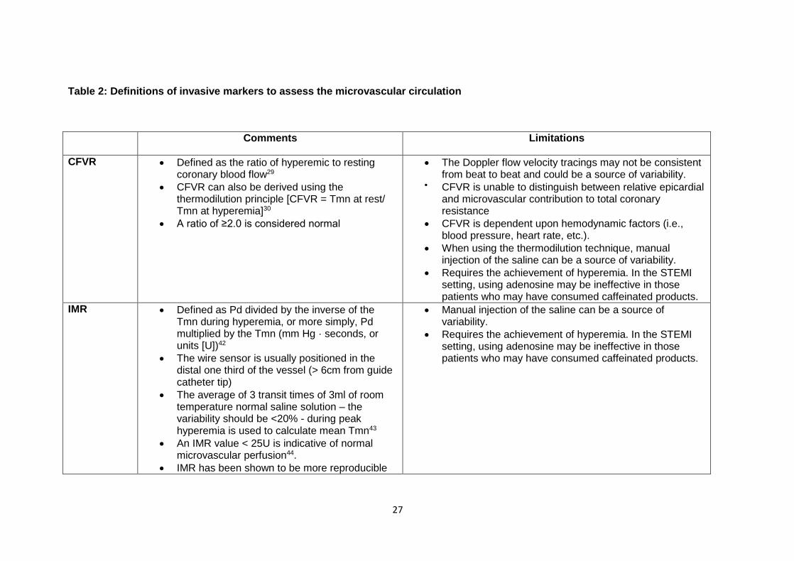
Table 2: Definitions of invasive markers to assess the microvascular circulation
Comments Limitations
CFVR Defined as the ratio of hyperemic to resting coronary blood flow29
CFVR can also be derived using the thermodilution principle [CFVR = Tmn at rest/ Tmn at hyperemia]30
A ratio of ≥2.0 is considered normal
The Doppler flow velocity tracings may not be consistent from beat to beat and could be a source of variability.
CFVR is unable to distinguish between relative epicardial and microvascular contribution to total coronary resistance
CFVR is dependent upon hemodynamic factors (i.e., blood pressure, heart rate, etc.).
When using the thermodilution technique, manual injection of the saline can be a source of variability.
Requires the achievement of hyperemia. In the STEMI setting, using adenosine may be ineffective in those patients who may have consumed caffeinated products.
IMR Defined as Pd divided by the inverse of the Tmn during hyperemia, or more simply, Pd multiplied by the Tmn (mm Hg · seconds, or units [U])42
The wire sensor is usually positioned in the distal one third of the vessel (> 6cm from guide catheter tip)
The average of 3 transit times of 3ml of room temperature normal saline solution – the variability should be <20% - during peak hyperemia is used to calculate mean Tmn43
An IMR value < 25U is indicative of normal microvascular perfusion44.
IMR has been shown to be more reproducible
Manual injection of the saline can be a source of variability.
Requires the achievement of hyperemia. In the STEMI setting, using adenosine may be ineffective in those patients who may have consumed caffeinated products.
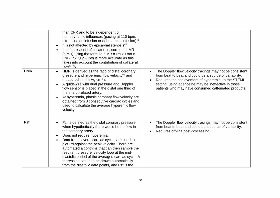
28
than CFR and to be independent of hemodynamic influences (pacing at 110 bpm, nitroprusside infusion or dobutamine infusion)41.
It is not affected by epicardial stenosis42 In the presence of collaterals, corrected IMR
(cIMR) using the formula cIMR = Pa x Tmn x (Pd - Pw)/(Pa - Pw) is more accurate as this takes into account the contribution of collateral flow81 82.
HMR HMR is derived as the ratio of distal coronary pressure and hyperemic flow velocity61 and measured in mm Hg cm-1 s
A guidewire with dual pressure and Doppler flow sensor is placed in the distal one third of the infarct-related artery.
At hyperemia, phasic coronary flow velocity are obtained from 3 consecutive cardiac cycles and used to calculate the average hyperemic flow velocity
The Doppler flow velocity tracings may not be consistent from beat to beat and could be a source of variability.
Requires the achievement of hyperemia. In the STEMI setting, using adenosine may be ineffective in those patients who may have consumed caffeinated products.
Pzf Pzf is defined as the distal coronary pressure when hypothetically there would be no flow in the coronary artery.
Does not require hyperemia. Data from several cardiac cycles are used to
plot Pd against the peak velocity. There are automated algorithms that can then sample the resultant pressure–velocity loop at the mid-diastolic period of the averaged cardiac cycle. A regression can then be drawn automatically from the diastolic data points, and Pzf is the
The Doppler flow velocity tracings may not be consistent from beat to beat and could be a source of variability.
Requires off-line post-processing.
29
pressure at which this line crosses the x-axis. This is the extrapolated distal coronary pressure at which flow would cease in the infarct-related artery.40
CFVR: coronary flow reserve; IMR: index of microvascular resistance; STEMI: ST-segment elevation myocardial infarction; Pd: distal pressure; Tmn: mean transit time; Pw: wedge pressure; Pa: aortic pressure; HMR: hyperemic microvascular resistance; Pzf: zero
30
Figures legend
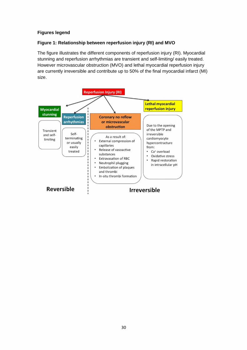
Figure 1: Relationship between reperfusion injury (RI) and MVO
The figure illustrates the different components of reperfusion injury (RI). Myocardial
stunning and reperfusion arrhythmias are transient and self-limiting/ easily treated.
However microvascular obstruction (MVO) and lethal myocardial reperfusion injury
are currently irreversible and contribute up to 50% of the final myocardial infarct (MI)
size.
31
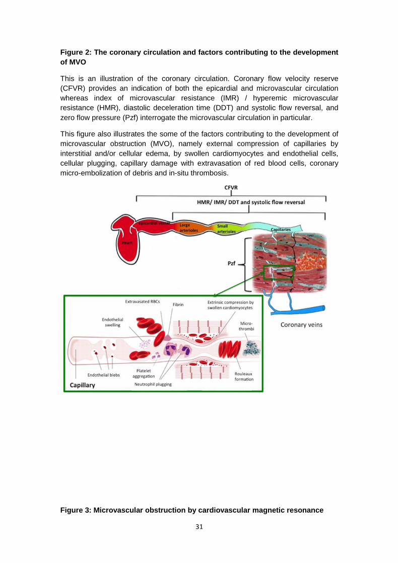
Figure 2: The coronary circulation and factors contributing to the development
of MVO
This is an illustration of the coronary circulation. Coronary flow velocity reserve
(CFVR) provides an indication of both the epicardial and microvascular circulation
whereas index of microvascular resistance (IMR) / hyperemic microvascular
resistance (HMR), diastolic deceleration time (DDT) and systolic flow reversal, and
zero flow pressure (Pzf) interrogate the microvascular circulation in particular.
This figure also illustrates the some of the factors contributing to the development of
microvascular obstruction (MVO), namely external compression of capillaries by
interstitial and/or cellular edema, by swollen cardiomyocytes and endothelial cells,
cellular plugging, capillary damage with extravasation of red blood cells, coronary
micro-embolization of debris and in-situ thrombosis.
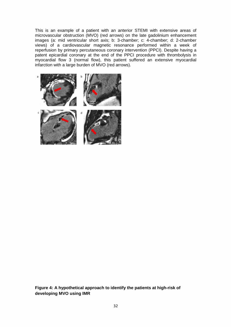
Figure 3: Microvascular obstruction by cardiovascular magnetic resonance
32
This is an example of a patient with an anterior STEMI with extensive areas of microvascular obstruction (MVO) (red arrows) on the late gadolinium enhancement images (a: mid ventricular short axis; b: 3-chamber; c: 4-chamber; d: 2-chamber views) of a cardiovascular magnetic resonance performed within a week of reperfusion by primary percutaneous coronary intervention (PPCI). Despite having a patent epicardial coronary at the end of the PPCI procedure with thrombolysis in myocardial flow 3 (normal flow), this patient suffered an extensive myocardial infarction with a large burden of MVO (red arrows).
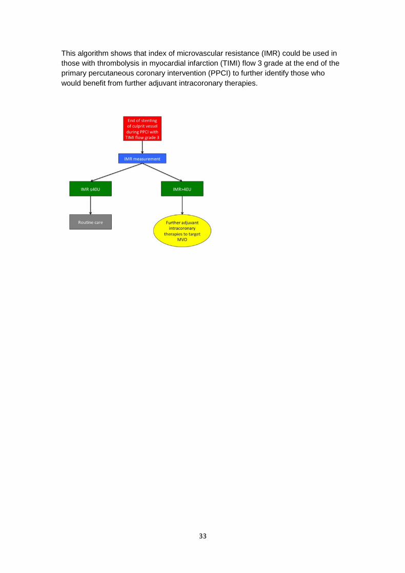
Figure 4: A hypothetical approach to identify the patients at high-risk of
developing MVO using IMR
33
This algorithm shows that index of microvascular resistance (IMR) could be used in
those with thrombolysis in myocardial infarction (TIMI) flow 3 grade at the end of the
primary percutaneous coronary intervention (PPCI) to further identify those who
would benefit from further adjuvant intracoronary therapies.
Copyright © 2022 FDOKUMEN