Water-miscible organic cosolvents enhance phosphatidylinositol-specific phospholipase C...
13
Water-miscible organic cosolvents enhance phosphatidylinositol-specific phospholipase C phosphotransferase as well as phosphodiesterase activity Hania Wehbi, Jianwen Feng, Mary F. Roberts * Department of Chemistry, E.F. Merkert Chemistry Center, Boston College, 2609 Beacon Street, Chestnut Hill, MA 02167, USA Received 12 February 2003; received in revised form 22 April 2003; accepted 30 April 2003 Abstract Phosphatidylinositol-specific phospholipase C (PI-PLC) from Bacillus thuringiensis catalyzes the hydrolysis of phosphatidylinositol (PI) in a Ca 2+ -independent two-step mechanism: (i) an intramolecular phosphotransferase reaction to form inositol 1,2-(cyclic)-phosphate (cIP), followed by (ii) a cyclic phosphodiesterase activity that converts cIP to inositol 1-phosphate (I-1-P). Moderate amounts of water-miscible organic solvents have previously been shown to dramatically enhance the cyclic phosphodiesterase activity, that is, hydrolysis of cIP. Cosolvents [isopropanol (iPrOH), dimethylsufoxide (DMSO), and dimethylformamide (DMF)] also enhance the phosphotransferase activity of PI-PLC toward PI initially presented in vesicles, monomers, or micelles. Although these water-miscible organic cosolvents caused large changes in PI particle size and distribution (monitored with pyrene-labeled PI fluorescence, 31 P NMR spectroscopy, gel filtration, and electron microscopy) that differed with the activating solvent, the change in PI substrate structure in different cosolvents was not correlated with the enhanced catalytic efficiency of PI-PLC toward its substrates. PI-PLC stability was decreased in water/organic cosolvent mixtures (e.g., the T m for PI-PLC thermal denaturation decreased linearly with added iPrOH). However, the addition of myo-inositol, a water-soluble inhibitor of PI-PLC, helped stabilize the protein. At 30% iPrOH and 4 jC (well below the T m for PI-PLC in the presence of iPrOH), cosolvent-induced changes in protein secondary structure were minimal. iPrOH and diheptanoylphosphatidylcholine, each of which activates PI-PLC for cIP hydrolysis, exhibited a synergistic effect for cIP hydrolysis that was not observed with PI as substrate. This behavior is consistent with a mechanism for cosolvent activation that involves changes in active site polarity along with small conformational changes involving the barrel rim tryptophan side chains that have little effect on protein secondary structure. D 2003 Elsevier Science B.V. All rights reserved. Keywords: Phospholipase C; Organic solvent effects; Phosphatidylinositol; Vesicle solubilization 1. Introduction Phosphatidylinositol-specific phospholipase C (PI-PLC) catalyzes the hydrolysis of phosphoester bonds of phos- phoinositides to produce lipid-soluble diacylglycerol and water-soluble myo-inositol phosphates. PI-PLC enzymes are found in a wide range of eukaryotic and prokaryotic cells [1]. In mammalian cells, intracellular PI-PLC is important in signal transduction. Hydrolysis of phosphatidylinositol 4,5- bisphosphate (PIP 2 ) catalyzed by PI-PLC produces two second messenger molecules: membrane-localized diacyl- glycerol, which allosterically activates protein kinase C, and water-soluble D-myo-inositol 1,4,5-trisphosphate which is important in intracellular calcium mobilization. In contrast to the mammalian PI-PLC, the bacterial PI-PLC enzymes are secreted and usually show low activity toward PIP 2 . They are considerably smaller than mammalian PI-PLCs but their a/h barrel structure is conserved as the catalytic domain in the mammalian enzymes [2–4]. These bacterial PI-PLC enzymes are virulence factors in pathogens like Listeria monocytogenes [5,6] and Staphylococcus aureus [7]. The Bacillus thuringiensis PI-PLC shares some kinetic features with the mammalian PI-PLC enzymes. It exhibits kinetic interfacial activation where aggregated substrates are preferred over monomeric substrates [8], a characteristic of most water-soluble phospholipases including PI-PLCy1 [1]. However, the enzyme efficiency (k cat /K m ) of the bacterial PI-PLC toward the water-soluble substrate cIP (the cyclic phosphodiesterase step) is also enhanced with the addition of a non-substrate interface (e.g., PC micelles or vesicles) 0005-2736/03/$ - see front matter D 2003 Elsevier Science B.V. All rights reserved. doi:10.1016/S0005-2736(03)00134-2 * Corresponding author. Tel.: +1-617-552-3616; fax: +1-617-552- 2705. E-mail address: [email protected] (M.F. Roberts). www.bba-direct.com Biochimica et Biophysica Acta 1613 (2003) 15 – 27
-
Upload
independent -
Category
Documents
-
view
0 -
download
0
Transcript of Water-miscible organic cosolvents enhance phosphatidylinositol-specific phospholipase C...

www.bba-direct.com
Biochimica et Biophysica Acta 1613 (2003) 15–27
Water-miscible organic cosolvents enhance phosphatidylinositol-specific
phospholipase C phosphotransferase as well as phosphodiesterase activity
Hania Wehbi, Jianwen Feng, Mary F. Roberts*
Department of Chemistry, E.F. Merkert Chemistry Center, Boston College, 2609 Beacon Street, Chestnut Hill, MA 02167, USA
Received 12 February 2003; received in revised form 22 April 2003; accepted 30 April 2003
Abstract
Phosphatidylinositol-specific phospholipase C (PI-PLC) from Bacillus thuringiensis catalyzes the hydrolysis of phosphatidylinositol (PI)
in a Ca2 +-independent two-step mechanism: (i) an intramolecular phosphotransferase reaction to form inositol 1,2-(cyclic)-phosphate (cIP),
followed by (ii) a cyclic phosphodiesterase activity that converts cIP to inositol 1-phosphate (I-1-P). Moderate amounts of water-miscible
organic solvents have previously been shown to dramatically enhance the cyclic phosphodiesterase activity, that is, hydrolysis of cIP.
Cosolvents [isopropanol (iPrOH), dimethylsufoxide (DMSO), and dimethylformamide (DMF)] also enhance the phosphotransferase activity
of PI-PLC toward PI initially presented in vesicles, monomers, or micelles. Although these water-miscible organic cosolvents caused large
changes in PI particle size and distribution (monitored with pyrene-labeled PI fluorescence, 31P NMR spectroscopy, gel filtration, and
electron microscopy) that differed with the activating solvent, the change in PI substrate structure in different cosolvents was not correlated
with the enhanced catalytic efficiency of PI-PLC toward its substrates. PI-PLC stability was decreased in water/organic cosolvent mixtures
(e.g., the Tm for PI-PLC thermal denaturation decreased linearly with added iPrOH). However, the addition of myo-inositol, a water-soluble
inhibitor of PI-PLC, helped stabilize the protein. At 30% iPrOH and 4 jC (well below the Tm for PI-PLC in the presence of iPrOH),
cosolvent-induced changes in protein secondary structure were minimal. iPrOH and diheptanoylphosphatidylcholine, each of which activates
PI-PLC for cIP hydrolysis, exhibited a synergistic effect for cIP hydrolysis that was not observed with PI as substrate. This behavior is
consistent with a mechanism for cosolvent activation that involves changes in active site polarity along with small conformational changes
involving the barrel rim tryptophan side chains that have little effect on protein secondary structure.
D 2003 Elsevier Science B.V. All rights reserved.
Keywords: Phospholipase C; Organic solvent effects; Phosphatidylinositol; Vesicle solubilization
1. Introduction important in intracellular calcium mobilization. In contrast
Phosphatidylinositol-specific phospholipase C (PI-PLC)
catalyzes the hydrolysis of phosphoester bonds of phos-
phoinositides to produce lipid-soluble diacylglycerol and
water-soluble myo-inositol phosphates. PI-PLC enzymes are
found in a wide range of eukaryotic and prokaryotic cells
[1]. In mammalian cells, intracellular PI-PLC is important in
signal transduction. Hydrolysis of phosphatidylinositol 4,5-
bisphosphate (PIP2) catalyzed by PI-PLC produces two
second messenger molecules: membrane-localized diacyl-
glycerol, which allosterically activates protein kinase C, and
water-soluble D-myo-inositol 1,4,5-trisphosphate which is
0005-2736/03/$ - see front matter D 2003 Elsevier Science B.V. All rights reserv
doi:10.1016/S0005-2736(03)00134-2
* Corresponding author. Tel.: +1-617-552-3616; fax: +1-617-552-
2705.
E-mail address: [email protected] (M.F. Roberts).
to the mammalian PI-PLC, the bacterial PI-PLC enzymes
are secreted and usually show low activity toward PIP2.
They are considerably smaller than mammalian PI-PLCs but
their a/h barrel structure is conserved as the catalytic
domain in the mammalian enzymes [2–4]. These bacterial
PI-PLC enzymes are virulence factors in pathogens like
Listeria monocytogenes [5,6] and Staphylococcus aureus
[7].
The Bacillus thuringiensis PI-PLC shares some kinetic
features with the mammalian PI-PLC enzymes. It exhibits
kinetic interfacial activation where aggregated substrates are
preferred over monomeric substrates [8], a characteristic of
most water-soluble phospholipases including PI-PLCy1 [1].
However, the enzyme efficiency (kcat/Km) of the bacterial
PI-PLC toward the water-soluble substrate cIP (the cyclic
phosphodiesterase step) is also enhanced with the addition
of a non-substrate interface (e.g., PC micelles or vesicles)
ed.

H. Wehbi et al. / Biochimica et Biophysica Acta 1613 (2003) 15–2716
[9]; a similar activation was observed toward PI in vesicles
that also contained a small amount of PC [10]. A similar
interfacial activation is exhibited by PI-PLCy1 [11] but not
by PI-PLCg [12]. The catalytic efficiencies of the second
step for both the bacterial PI-PLC and the PLCy1 isozyme
are also enhanced by addition of water-miscible organic
solvents such as isopropanol (iPrOH), dimethylsulfoxide
(DMSO), and dimethylformamide (DMF) with maximal
activity observed at 30% iPrOH, 40% DMF, and 50%
DMSO [11,13].
In this work, the effects of organic solvents on B.
thuringiensis PI-PLC phosphotransferase activity toward
long-chain PI in vesicles and short-chain PI monomers and
micelles are examined. Activation of the phosphotransferase
step by organic cosolvents parallels that observed for cIP
hydrolysis. Although cosolvents alter the distribution and
structures adopted by PI, the activation of PI-PLC by
cosolvents does not correlate with any substrate aggregate
changes. This suggests that PI-PLC activity is enhanced in
the presence of organic solvents regardless of the type of
substrate presented and regardless of the effect these cosol-
vents have on the substrate structure. The minimal solvent-
induced changes in PI-PLC secondary structure (monitored
by CD spectroscopy) are consistent with changes in the local
polarity of the active site and conformational changes of side
chains as possible causes of the enhanced enzymatic activity.
2. Materials and methods
2.1. Chemicals
L-Phosphatidylinositol and other phospholipids including
diheptanoylphosphatidylcholine (diC7PC) and 1-palmitoyl-
2-oleoyl-phosphatidylcholine (POPC) were obtained from
Avanti and used without further purification. iPrOH, DMF,
and DMSO were purchased from Aldrich and were used
without further purification. Dibutyroylphosphatidylinositol
(diC4PI) and dioctanoylphosphatidylinositol (diC8PI) were
purchased from Echelon Biochemicals. Other reagents
including carboxyfluorescein (CF), D2O (99.9%), and
Sephadex G-100 were purchased from Sigma.
2.2. Purification of PI-PLC
The recombinant Escherichia coli cell strain MM294
transfected with B. thuringiensis PI-PLC gene [14] was
obtained from Dr. Ming-Daw Tsai (Ohio State University).
The enzyme was isolated from cell lysates and was purified
by chromatography using a 1.6� 12 cm column of Q
sepharose Fast Flow (Amersham Pharmacia Biotech) and
a 1.0� 10 cm column of phenyl sepharose (Amersham
Pharmacia Biotech). The enzyme from this protocol had
less than 5% total impurities as monitored by intensity of the
35-kDa band in Coomassie-stained SDS-polyacrylamide
gels compared to other very faint bands. PI-PLC concen-
tration was determined by Bradford assay using bovine
serum albumin as a standard. Typically, 20 mg of PI-PLC
was obtained from 1 l of culture. The enzyme was stored in
50% glycerol and 20 mM Tris buffer, pH 8.5. The concen-
trated stock solutions (typicallyz 0.5 mg/ml) were further
diluted with 0.1% bovine serum albumin (Bio-Rad Protein
Assay Standard II) in 20 mM Tris, pH 8.5, before enzyme
assays. This recombinant PI-PLC had lower (and more
reproducible from batch to batch) specific activities toward
PI SUVs than PI-PLC secreted into Bacillus medium [10],
presumably because the secreted protein had lipids or other
materials bound which enhanced its activity toward PI
vesicles.
2.3. Vesicle preparation
Lipid stock solutions in chloroform were obtained from
Avanti and used without further purification. All samples
were placed in glass scintillation vials, and the chloroform
was removed with a stream of N2. The lipid film was
lyophilized overnight and then rehydrated with 25 mM
HEPES (N-(2-hydroxyethyl)piperazine-NV-2-ethanesulfonicacid), pH 7.5, for enzyme assays. SUV and LUV prepara-
tions were generated by sonication or extrusion, respec-
tively, as described previously [10]. Mixed PI/PC SUVs
were prepared by sonicating a rehydrated PI and PC
suspension in 25 mM HEPES using a Branson sonifier cell
disrupter and a microtip probe until maximum clarity was
achieved (typically 5–10 min). When vesicle preparations
were mixed with organic cosolvents, the optical density at
600 nm of solutions was measured as a rough assessment of
changes in particle sizes (i.e., increasing OD600 indicated
larger particles and decreasing OD600 indicated a bias
toward smaller particles).
2.4. Enzymatic synthesis and purification of inositol-(1,2)-
(cyclic)-phosphate (cIP)
Crude soybean PI was used for the enzymatic generation
of cIP by PI-PLC as described previously [9]. One gram of
crude PI was dissolved in 15 ml Tris buffer (pH 7.5)
containing 4% Triton X-100. Bacterial PI-PLC (20 Ag)was added and the reaction was monitored using 31P
NMR spectroscopy. After the PI was hydrolyzed to cIP
(typically 4 h at room temperature), further reaction [e.g.,
hydrolysis of cIP to inositol 1-phosphate (I-1-P)] was
quenched by the addition of chloroform. Purification of
cIP was carried out as previously described [9].
2.5. 31P NMR assays of PI-PLC activity
The buffer used in all PI-PLC activity assays was 25 mM
HEPES, pH 7.5. 31P NMR parameters were based on those
previously reported [9,16]. For fixed time point assays using
PI as substrate, the amount of enzyme added in each kinetic
run was adjusted so that less than 20% hydrolysis of
H. Wehbi et al. / Biochimica et Biophysica Acta 1613 (2003) 15–27 17
substrate occurred during the 10-min incubation at room
temperature. Since the enzyme was diluted in stocks with
0.1% BSA before kinetics, the final BSA concentration in
assay mixtures was < 0.005%. After 10 min, the reactions
were quenched using chloroform and the aqueous layer was
extracted and analyzed for product using 31P NMR spectro-
scopy (202.3 MHz on a Varian INOVA 500 spectrometer).
PI cleavage and cIP hydrolysis rates were measured from
the integrated intensity of cIP and I-1-P resonances, respec-
tively, as a function of incubation time. Specific activities
are based on assays run at a minimum in duplicate.
2.6. Fluorescence spectroscopy
Two types of fluorescence experiments were performed
to characterize the effect of cosolvents on PI vesicles: (i)
measurement of 1-myrisotyl-2-pyrenebutanoyl-glycero-3-
(D-inositol-1-phosphate), or Pyr-PI (obtained from Dr. Stew-
art Hendrickson, University of Washington), excimer and
monomer band intensities to monitor vesicle solubilization,
and (ii) measurement of leakage of entrapped CF by
increased fluorescence at 520 nm. Fluorescence studies
were done at 25 jC using a Shimadzu RF5000U fluorimeter.
The fluorescence spectra of Pyr-PI vesicles (20 AM) in 20
mM HEPES, pH 7.5, were acquired with an excitation
wavelength of 350 nm and the emission scanned between
360 and 600 nm. Excitation and emission slit widths were
set to 3 nm. The fluorescence intensities of monomer (380–
390 nm) and excimer (450 nm) peaks were measured as a
function of added organic solvents. CF was trapped inside
PI vesicles by sonicating the resuspended PI solution in the
presence of 100 mM CF. Free CF in solution was removed
by gel filtration through a 0.7� 50 cm column of Sephadex
G-10-120 obtained from Sigma. CF entrapped in vesicles is
self-quenched. However, if vesicles become leaky, the CF is
diluted into the solution and the fluorescence at 520 nm
(excitation at 490 nm) increased dramatically.
2.7. 1H NMR analyses of PI
1H (500 MHz) spectra (acquired on the same spectrom-
eter mentioned above) were used to compare the conforma-
tion and dynamics of naturally occurring PI with synthetic
short-chain PI species [17]. NOESY experiments were
carried out for 3 mM diC4PI and 3 mM diC8PI solubilized
in D2O or iPrOH-d8/D2O (30:70, v/v) and 3 mM long-chain
PI in iPrOH-d8/D2O. Buffers were not included in these
solutions (to avoid overlap of buffer peaks with PI protons);
however, the pH of the solutions in the absence of cosolvent
was typically between pH 6.5 and 7.5. Since no groups in
the PI should ionize in this range, the lack of a buffer should
not pose a problem. NOESY spectra were acquired with
0.4–0.5 s mixing times. The residual HDO resonance was
suppressed using a WET pulse sequence that applies pulse
field gradients. Gaussian weighting was used in both F1 and
F2 dimensions. 1H spin-lattice relaxation times for the same
samples, measured by inversion-recovery, were used to
assess PI dynamics in the iPrOH-d8/D2O cosolvent system.
2.8. Gel filtration of PI aggregates in water/cosolvent
mixtures
PI SUVs (5 mM) in 20 mM HEPES, pH 7.5, were
chromatographed on a Sephadex G-100 gel exclusion col-
umn (1.5� 50 cm equilibrated with 20 mM HEPES, pH
7.5); in the absence of organic solvent, all of the PI eluted in
the column void volume. The same column was used to get
a rough idea of the particle size distribution for PI vesicles
mixed with 10% and 30% iPrOH; for these experiments, the
resin was equilibrated with the same concentration iPrOH.
cIP (10 mM) was present in all samples as a marker for the
elution volume of small molecules. Column fractions were
lyophilized, redissolved in 30% iPrOH (additional iPrOH
had no effect on the 31P PI intensities) in HEPES, pH 7.5,
and quantified by 31P NMR spectroscopy to assess the
distribution of PI compared with cIP.
2.9. Characterization of PI/iPrOH aggregates using
electron microscopy
The size distribution of PI sonicated vesicles after mixing
with different percentages of iPrOH was monitored using a
CM10 Phillips Electron Microscope and uranyl acetate (2%)
to stain the PI samples. The concentration of PI in SUVs
mixed with 0% and 10% iPrOH was 1 mg/ml; 5 mg/ml PI
sonicated vesicles were incubated with 20% iPrOH and 10
mg/ml PI SUV preparation was incubated with 30% and
40% iPrOH. Although different SUV preparations were
used, the average initial size was similar as monitored by
the 31P linewidth. The 2% uranyl acetate (10 Al) was first
applied to formvar/carbon-coated 400 mesh grids followed
by application of 4 Al of the PI sample of interest. Several
pictures of PI vesicles at each percentage of iPrOH were
taken and the sizes of the vesicles were measured using
catalase crystals (spacing = 87.5 A) to calibrate sizes at
different magnifications. Typically, three to five grids were
imaged (5–10 photomicrographs taken of each grid) for each
vesicle/iPrOH combination; 20–200 particles, selected ran-
domly, were usually counted from each photomicrograph.
2.10. Circular dichroism
CD spectroscopy was used to monitor changes in secon-
dary structure of PI-PLC induced by iPrOH. CD spectra of
0.2 mg/ml PI-PLC in 10 mM borate buffer, pH 8.0, were
obtained between 245 and 190 nm in 1 mm path length cells
using an AVIV Circular Dichroism model 202 spectrometer.
A baseline scan of the cell filled with buffer (and iPrOH)
was subtracted from the spectra obtained with protein. The
percentage of secondary structure elements was estimated
using the program CDNN [18,19]. Temperature-induced
changes in the ellipticity at 222 nm (averaging time of 1

Table 1
PI-PLC phosphotransferase activity toward PI in the absence and presence
of 30% iPrOHa
Substrate
(mM)
Aggregation
state
Additive Specific
activityb
(Amol min� 1
mg� 1)
Rate
enhancement
diC4PI
(1.5)
monomer – 0.18
monomer 30% iPrOH 12 67
diC8PI
(1.25)
micelle – 0.32
c
H. Wehbi et al. / Biochimica et Biophysica Acta 1613 (2003) 15–2718
s) were monitored at 1j intervals from 10 to 60 jC for a
0.2–0.3 mg/ml protein solution in a 1-mm-path-length
quartz cell. The temperature was maintained to within
F 0.2j with 1 min equilibration time between each increase
in temperature. The thermal melting transition, Tm, of the
protein under these conditions in the absence and presence
of iPrOH and 50 mM myo-inositol (a weak inhibitor of the
enzyme that does not have any absorbance above 200 nm)
was determined by plotting the derivative of the [V222]
versus temperature scan; the Tm was taken to be the
maximum in the derivative spectrum.
– 30% iPrOH 29 91PI (8.0) LUV – 0.37
monomers/small
aggregates
30% iPrOH 820 2220
SUV – 8.4
monomers/small
aggregates
30% iPrOH 920 110
a Assay conditions include 50 mM Hepes, pH 7.5, and the amount of
enzyme added adjusted so that V 20% PI cleavage occurs in 2 h.b Errors in measuring specific activities were typically F 15% except
for very low specific activities ( < 1 Amol min� 1 mg� 1) where the error
could be as large as 25%.
3. Results
3.1. Effect of organic solvents on B. thuringiensis PI-PLC
activity toward PI vesicles
Phospholipase kinetics with their natural long-chain
substrates are complicated because the phospholipids are
presented in a two-dimensional matrix and the water-solu-
Fig. 1. Effect of iPrOH (.), DMF (E), and DMSO (n) on the specific
activity of PI-PLC toward (A) 8 mM cIP and (B) 6 mM PI SUVs. The data
in A are from Ref. [13].
c The addition of the iPrOH increases the CMC so that monomers
coexist with micelles.
ble enzyme must interact with the interface as well as an
individual substrate molecule. Not only are changes in the
protein structure important, but perturbations of the 2D-
matrix can affect the observed specific activities. For PI-
PLC, cIP serves as a water-soluble substrate that can be
used to monitor for effects of nonsubstrate interfaces on
enzyme activity. It was observed previously [13] that B.
thuringiensis PI-PLC activity toward cIP was enhanced in
the presence of organic solvents (iPrOH, DMSO, and DMF)
(Fig. 1A). As shown in Fig. 1B, the phosphotransferase
activity of the enzyme toward small unilamellar PI vesicles
was also significantly enhanced by these water-miscible
organic solvents, although the mole fraction of each solvent
needed for maximum activation was slightly different.
Higher mole fractions of cosolvents reduced the activation
of PI-PLC, but observed phosphotransferase and phospho-
diesterase activities with solvents were still much higher
than in the absence of organic solvents. Thus, similar
cosolvent effects for both steps of the PI-PLC reaction were
observed. PI LUVs are poorer substrates than SUVs for the
enzyme, presumably because of the tighter packing of the
lipids in the more planar bilayers (Table 1). However,
iPrOH increases PI-PLC activity toward both PI bilayer
systems to about the same extent. In the case of PI vesicles,
the organic solvent can have a pronounced effect on the
substrate structure and this could affect the types of inter-
actions PI has with the enzyme.
3.2. Effect of solvents on PI vesicles
To assess the importance of structural perturbations of
the long-chain PI substrate to PI-PLC activation by organic
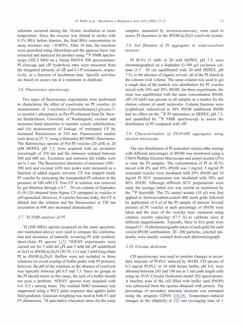
Fig. 2. Optical density (at 600 nm) of PI vesicle solution as a function of percentage of organic cosolvents: PI SUVs mixed with iPrOH (.), DMF (E), and
DMSO (n); PI/PC (1:2) SUVs mixed with iPrOH (�); PI suspensions (formed by hydrating a dry PI film with buffer and shaking) mixed with iPrOH (o).
Solid lines are for OD600 values shown on the left axis while the dotted lines are for the OD600 range shown on the right axis.
H. Wehbi et al. / Biochimica et Biophysica Acta 1613 (2003) 15–27 19
solvent, a variety of physical studies of the PI/cosolvent
systems were carried out. Both DMF and DMSO at mod-
erate mole fractions caused formation of large particles
(larger than 1000–2000 A LUVs since they sediment by
gravity) from PI SUVs (Fig. 2). Centrifugation of the
solutions containing PI and >30% DMF or DMSO removed
the large particles and produced a supernatant that had
virtually no residual PI. These aprotic polar solvents are
known to dehydrate protein surfaces [20]. It is likely these
solvents also dehydrate the inositol head group, since they
promote fusion of SUVs. Since in the absence of organic
cosolvents, PI-PLC activity toward SUVs is f 20-fold
Fig. 3. Ratio of fluorescence intensities of monomer (380–390 nm) and excimer ba
of iPrOH. The excitation wavelength was 350 nm.
higher than toward LUVs, the observation of high activity
with the addition of DMF and DMSO, where very large
vesicles (likely a mixture of multilamellar and large uni-
lamellar species) are formed, is consistent with a direct
effect of the cosolvent on the enzyme, since no significant
fraction of smaller particles was formed. In contrast to the
aprotic cosolvents, iPrOH added to the same PI SUVs
decreased the optical density of the solution; MLVs of PI
were also clarified by 30% iPrOH (in fact, PI dissolved in
30% iPrOH formed an easy optically clear assay system).
Kinetic activation of PI SUVs mixed with iPrOH, could
reflect contributions from decreasing particle sizes and
nds (450 nm) of pyrene-labeled PI (20 AM) as a function of percentage (v/v)

H. Wehbi et al. / Biochimica et Biophysica Acta 1613 (2003) 15–2720
enhanced particle curvature, or isolation of very small ag-
gregates. The generation of smaller particles could account
for the fact that the specific activity of PI-PLC toward PI in
the presence of iPrOH is relatively higher than that of the
reaction in the presence of DMF or DMSO. The fluores-
cence spectrum of Pyr-PI vesicles (20 AM) is characterized
by a strong excimer band at 450 nm and weak monomer
bands at 380–390 nm such that the ratio of intensity of
monomer at 385 nm compared to excimer is less than 0.05.
If the Pyr-PI vesicles are solubilized by iPrOH and mono-
mers are formed, the ratio of fluorescence intensity at 385
nm compared to that at 450 nm should increase. As shown
in Fig. 3, an increased proportion of Pyr-PI monomer be-
gan to appear with the addition of 20% iPrOH; in 40%
iPrOH, the ratio of Pyr-PI fluorescence at 380–390 nm
was the same as if the Pyr-PI was solubilized by a large
excess of Triton X-100 where Pyr-PI molecules are com-
pletely surrounded by detergent and few if any excimers
Fig. 4. Electron micrograph showing PI vesicle sizes (A) in buffer alone and (B) w
distribution of PI vesicles (detected by EM) incubated with different percentages of
10% iPrOH (white bars) and PI (5 mg/ml) in 20% iPrOH (hatched bars) were sta
form. Between the two amounts of iPrOH, there must exist
some type(s) of aggregated PI to produce the excimer
band.
Other techniques were also used to get a sense of changes
in average particle sizes. The 31P linewidth of PI decreased
and its integrated intensity increased with the addition of
iPrOH (data not shown), consistent with the generation of
smaller particles. The largest changes in these two param-
eters occurred between 10% and 20% iPrOH. As little as 5%
iPrOH added to PI SUVs or LUVs caused leakage of
entrapped CF with very rapid release of CF at 10% iPrOH.
For comparison, Anchordoguy et al. [21] showed that 3 M
DMSO (f 20%) added to CF entrapped in egg phosphati-
dylcholine vesicles did not induce vesicle leakiness.
The distribution of PI in large particles (vesicles) versus
small aggregates or monomers in the absence and presence
of iPrOH was also assessed with gel filtration chromatog-
raphy using Sephadex G-100. PI vesicles, regardless of size,
ith 10% iPrOH. The arrows in both figures correspond to 250 A. (C) Size
iPrOH: vesicles of PI (1 mg/ml) in 20 mM Hepes, pH 7.5 (gray bars) and in
ined using 2% uranyl acetate.

Table 2
PI-PLC activity toward 8 mM PI and cIP in the presence of activators
iPrOH and phosphatidylcholinea
Substrate IPrOH
(30%)
diC7PC PCb Specific activity
(Amol min� 1 mg� 1)c
cIP � + (8 mM) � 60
+ � � 65
+ + (8 mM) � 100
PI � � + 10
+ � � 542
+ � + 575
� + (40 mM) � 240
+ + (40 mM) � 590
a Assays were carried out in 50 mM Hepes, pH 7.5; the amounts of
enzyme added was adjusted to produce V 20% hydrolysis in 10 min at
room temperature. The enzyme stock used for the kinetics in this table was
>6 months old and had a reduced specific activity compared that
wasf 60% that newly purified recombinant PI-PLC toward PI dispersed
in 30% iPrOH.b SUVs were prepared for PI or PI/PC in the absence of iPrOH or
diC7PC. The PI/PC (8 mM:16 mM) vesicle sizes were distributed around
300 A.c Errors in specific activities are V 15%.
H. Wehbi et al. / Biochimica et Biophysica Acta 1613 (2003) 15–27 21
elute in the void volume of the column. cIP, a molecule that
has no tendency to partition into a bilayer [9], is a marker
for small molecules and elutes at the column volume. In the
elution profile of PI in 10% iPrOH (data not shown), there
was a slightly broader distribution of PI sizes with some
significantly smaller particles. However, with PI in 30%
iPrOH, the range of PI particles was biased toward much
smaller particles. Indeed, there was some overlap of frac-
tions containing PI and cIP indicating significant amounts of
monomeric PI or very small PI particles. Clearly, iPrOH
interacts differently with PI bilayers than the two aprotic
cosolvents and leads to mixtures of both smaller and larger
particles.
A more accurate description of the larger particles formed
from incubation of PI SUVs with iPrOH was provided by
electron microscopy. The distribution of PI SUVs in aque-
ous solution was used as a standard. In general, the diam-
eters of most PI SUVs in aqueous solution ranged from 100
to 400 A (Fig. 4). The addition of 10% iPrOH led to a bias
with somewhat larger particles while 20% iPrOH promoted
fusion to even larger particles (1000–3000 A diameter).
Since 30% and 40% iPrOH yielded mostly small aggregates
(which were not detected by electron microscopy), higher
concentrations of PI mixtures were applied to the grid. With
those amounts of iPrOH, the few vesicles detected had
diameters ranging from 2000 to 5000 A.
3.3. Activity of PI-PLC in the presence of both activators
iPrOH and phosphatidylcholine
Phosphatidylcholine activates both the cyclic phospho-
diesterase and phosphotransferase activities of PI-PLC
[9,10]. It has been shown that a PC monomer binds tightly
to PI-PLC [22] and increases the specific activity of PI-PLC
toward cIP, although a PC surface is much more effective
with PC micelles enhancing the enzymatic activity for cIP
cleavage 20- to 40-fold [9]. Incorporation of long-chain PC
in PI vesicles was also shown to enhance PI-PLC phospho-
transferase activity [10]. Since there appears to be a distinct
site for PC binding, an interesting question is what happens
to the enzyme when both PC and iPrOH are present
simultaneously. The presence of 8 mM diC7PC and 30%
iPrOH separately led to similar values of Km (29 and 30
mM, respectively). A comparison of PI-PLC specific activ-
ities toward 8 mM cIP with 8 mM diC7PC or 30% iPrOH is
shown in Table 2. PI-PLC specific activity was almost 2-
fold higher when both activators were present. Multiple
repeats of this experiment led to an activation of PI-PLC that
ranged from 1.6- to 2.2-fold over iPrOH (or diC7PC) alone.
Given the solubilization of long-chain PI by 30% iPrOH, it
is likely that iPrOH increases the CMC of diC7PC dramat-
ically such that mostly monomeric PC is present in solution
under these conditions (and hence one might expect reduced
activation by PC). Nonetheless, there was a synergistic
effect of both activators on PI-PLC cyclic phosphodiesterase
activity.
The simultaneous effect of PC in the PI vesicles (1:2 PI/
PC) and iPrOH on PI-PLC phosphotransferase activity was
also measured (Table 2). As observed previously for this
composition of vesicles, there was no activation of PI-PLC
compared to the enzyme specific activity toward PI SUVs
[10]. In contrast, the addition of iPrOH to SUVs of PI or PI/
PC greatly enhanced PI cleavage. With this amphiphilic
substrate, a good portion of the enhancement is likely
caused by the conversion of PI from a bilayer to the smaller
particles (Fig. 2). If PI-PLC phosphotransferase specific
activity was compared for PI in 30% iPrOH to PI/PC in
the same cosolvent, there was no synergistic effect of the
two activator molecules as there was for cIP as the substrate.
An alternate assay system is PI dispersed in diC7PC micelles
[9]. Again, PI-PLC phosphotransferase activity for PI/
diC7PC (1:5) with iPrOH is the same as that for iPrOH
alone. For both these phosphotransferase assay systems, the
increased PI-PLC activity with both activators was compa-
rable to that for iPrOH alone. A synergistic effect of diC7PC
and iPrOH was only observed for cIP hydrolysis. This
suggests that acyl chain binding of the substrate is important
for optimal PI-PLC activity and that with cIP, the missing
substrate acyl chains can be in part replaced by nonsubstrate
PC binding to the enzyme.
3.4. PI-PLC activity toward short-chain PI
The phosphotransferase activity of PI-PLC toward the
short-chain substrates diC4PI and diC8PI was also moni-
tored for any enhancement by iPrOH (Table 1). It was
previously observed that PI-PLC activity toward low con-
centrations ( < 2 mM) of diC6PI exhibited a sigmoidal
dependence on PI [8]. For 1–1.5 mM monomeric diC4PI
and micellar diC8PI (1.25 mM), the specific activity was

Fig. 5. NOESY contour plots for 3 mM diC4PI (A) and diC8PI (B)
dispersed in D2O. The dashed lines in each plot indicate the crosspeaks
between the inositol C(2)–H and one of the glycerol sn-3 CH2 protons.
H. Wehbi et al. / Biochimica et Biophysica Acta 1613 (2003) 15–2722
very low ( < 0.5 Amol min� 1 mg� 1). However, the addition
of 30% iPrOH enhanced PI-PLC activity 70- to 90-fold.
With that amount of cosolvent, diC4PI is monomeric and
Fig. 6. (A) 1H (500 MHz) spectrum of naturally occurring PI (3 mM) in 30% iP
C(2)–H with the glycerol sn-3 CH2, and inositol C(1)–H and C(3)–H (the dash
with the glycerol sn-3 CH2).
diC8PI will have an increased proportion of monomers
(water-miscible alcohols increase the CMC of most phos-
pholipids), so that this activation does not represent the
interfacial activation exhibited for a micellar versus a
monomeric substrate. The observed increase in specific
activity for PI-PLC in the presence of iPrOH toward sub-
strates that form monomers (diC4PI), micelles (diC8PI), and
vesicles (long-chain PI) indicates that PI-PLC is activated
by organic cosolvents regardless of the type of substrate.
3.5. Conformation of PI in 30% iPrOH
The conformation and dynamics of synthetic short-chain
PI in water were assessed by 1H NMR spectroscopy and
compared to previous NMR studies of diC6PI and diC7PI
species in D2O [17]. Key observations in the earlier work
were an NOE between a glycerol C(3)–H and the inositol
C(2)–H and motional constraints on several of the inositol
ring protons consistent with intramolecular hydrogen-bond-
ing. An NOE consistent with this interaction is also seen in
micellar diC8PI and monomeric diC4PI as well (Fig. 5A and
B). This NOE observed for both short-chain PIs had a
positive sign (opposite sign of the diagonal). The rest of the
NOEs were all positive in the case of diC4PI monomers and
mostly negative in the case of micellar diC8PI. This is
consistent with the difference in aggregation state of the
two different PI species in aqueous solution. The 1H spec-
trum of long-chain PI solubilized in 30% iPrOH-d8/D2O
exhibited relatively sharp resonances (Fig. 6A), although
they were not as sharp as observed for monomeric diC6PI
rOH-d8/D2O. (B) NOESY contour plot showing crosspeaks of the inositol
ed line indicates these crosspeaks and the arrow specifically the interaction

Fig. 8. CD spectra of PI-PLC (0.3 mg/ml) at 25 jC as a function of time
incubation in the presence of 30% (v/v) iPrOH in the absence (A) or
presence (B) of 50 mM myo-inositol. Successive scans are 15 min apart. (C)
Dependence of the thermal denaturation temperature (Tm) of PI-PLC as
measured by following the ellipticity at 222 nm as a function of temperature
on the volume percentage of iPrOH in the absence (.) and presence (o) of
50 mM myo-inositol. Experimental conditions included PI-PLC (0.3 mg/
ml) in 10 mM borate buffer, pH 8.0, and 1 min incubation times at each
temperature.
Fig. 7. Comparison of 1H T1 values for 3 mM long-chain PI (5) and 3 mM
diC8PI (�) in 30% iPrOH-d8/D2O, and for comparable concentrations of
diC8PI (o) and diC4PI (.) dispersed in D2O. The identification of the
protons is indicated on the plot.
H. Wehbi et al. / Biochimica et Biophysica Acta 1613 (2003) 15–27 23
in D2O [17]. This observation is consistent with small
aggregates and fast exchange of individual PI molecules
between the different species. A NOESY experiment (Fig.
6B) clearly showed inositol ring interactions (as negative
NOEs). Crosspeaks reflecting the inositol C(2)–H/C(1)–H
and C(2)–H/C(3)–H NOEs were clearly observed. The
NOE between a glycerol C(3)–H and inositol C(2)–H
was also detected in PI solubilized in the organic solvent/
water mixtures. The crosspeak in the NOESY spectrum for
the glycerol C(3)–H/inositol C(2)–H interaction in diC8PI
was reduced in 30% iPrOH-d8/D2O (data not shown).
Under these conditions, the inositol C(2)–H/C(3)–H and
C(1)–H crosspeaks were negative while that for the glyc-
erol/inositol interaction was smaller and positive. Similarly,
the glycerol C(3)–H/inositol C(2)–H crosspeak in diC4PI
solubilized in the iPrOH/D2O cosolvent mixture was nearly
nonexistent.1H spin lattice relaxation times of the long-chain PI in
iPrOH/D2O were also measured and compared with T1values for diC8PI in the absence and presence of iPrOH.
As shown in Fig. 7, the T1’s for the inositol protons of long-
chain PI in 30% iPrOH were similar to those for diC8PI in
iPrOH (as well as the diC4PI in water) but much longer than
for the inositol protons of diC8PI in water (micelles). Acyl
chain T1’s of the terminal CH3 group were longer in samples
with iPrOH, consistent with small aggregates. The more
pronounced change in the inositol T1’s was consistent with
increased flexibility of this moiety in PI dispersed in iPrOH.
Regardless of the acyl chain length, PI in iPrOH is a
flexible molecule with reduced interactions between the
inositol and the glycerol backbone. However, this cannot
be the sole reason for the enhanced PI-PLC phosphotrans-
ferase activity, since diC4PI in water also has a more flexible
inositol ring, but the same low hydrolysis rate as seen for
diC8PI (where the NOE between the glycerol backbone and
the inositol C-2 is visible). If changes in the substrate do not
correlate with cosolvent activation, the enzyme must be
altered in some way—either its conformation, aggregation
state, or microenvironment is altered by cosolvent.

H. Wehbi et al. / Biochimica et Biophysica Acta 1613 (2003) 15–2724
3.6. Effect of organic cosolvents on PI-PLC secondary
structure
Circular dichroism spectroscopy was used to assess the
effect of the cosolvent on PI-PLC secondary structure. In
general, secondary structure changes of proteins induced by
organic solvents cannot be studied because of the strong
absorbance by the organic solvents. However, iPrOH does
not interfere with the CD spectrum above 200 nm, so that
the effect of this cosolvent on PI-PLC secondary structure
could be examined. At 25 jC, PI-PLC lost secondary
structure in 30% iPrOH (Fig. 8A) as monitored by the loss
of negative ellipticity around 220 nm (the increased pos-
itive ellipticity around 200 nm is likely the result of
unfolded protein aggregation). The loss of structure was
biphasic with most of the change occurring within the first
15 min. Loss of secondary structure due to protein denatu-
ration is a common trait of proteins incubated in water/
cosolvent mixtures where the amount of organic solvent is
between 20% and 70% [23,24]. The hydrophobic interac-
tions between organic solvents and proteins that could
destabilize folded structure are known to be favored at
higher temperatures [21]. When PI-PLC was incubated
with 30% iPrOH at 4 jC, the protein secondary structure
was stable for at least 1 week. In parallel, the activities of
PI-PLC in 30% iPrOH incubated at 25 and at 4 jC for 30
min and 16 h, respectively, were determined. No activity
was observed in the first case and full activity was
preserved in the latter case. The thermostability of PI-
PLC as monitored by an effective Tm, the midpoint of
the thermal denaturation transition as measured by loss of
secondary structure, decreased linearly with increasing
iPrOH percentage as monitored by CD spectroscopy (Fig.
8C). With 30% iPrOH present, the PI-PLC Tm was
decreased from 53.6 to 30 jC. The half-width of the
transition was 7–8j regardless of the presence of iPrOH.
Fig. 9. CD spectra of PI-PLC (0.3 mg/ml in 1 mm cuvette) at 4 jC in
However, at 25 jC, some unfolding was detected. Since the
thermal unfolding of PI-PLC under these conditions was
irreversible, the protein incubated at 25 jC would be
unfolded over time as was indeed monitored by the CD
spectra (Fig. 8A).
This loss of activity of PI-PLC at room temperature when
the protein was incubated in 30% iPrOH appears contra-
dictory to 31P NMR assays where there was a linear
response for I-1-P production from cIP (or cIP production
from PI) during the 2-h time course for samples with 30%
iPrOH. A possible explanation is that PI-PLC is stable in
organic cosolvents when a molecule occupies the active site.
To test this hypothesis, the secondary structure region of the
CD spectrum of PI-PLC in 30% iPrOH was monitored in the
presence of 50 mM myo-inositol. Although myo-inositol is
not a good inhibitor for this bacterial PI-PLC[Ki f 40 mM
using PI/Triton X-100 as the substrate (C. Zhou, unpub-
lished results)], this concentration was still able to bind to
PI-PLC, retard iPrOH-induced unfolding (Fig. 8B), and
preserve enzymatic activity. With myo-inositol present, the
Tm for PI-PLC in iPrOH is higher than for PI-PLC dispersed
in iPrOH alone (Fig. 8C). If myo-inositol enhances PI-PLC
thermostability in iPrOH, it is likely that cIP (and the
amphiphilic substrate PI) and I-1-P do as well.
Since PI-PLC is stable in 30% iPrOH at 4 jC, one can
quantify the effect of organic cosolvent on PI-PLC secon-
dary structure. CD spectra of PI-PLC (0.30 mg/ml in 1 mm
cuvette) between 190 and 265 nm in the absence and
presence of 30% iPrOH were recorded at 4 jC (Fig. 9).
The spectra were nearly superimposable with only minor
changes. The wavelength dependence of molar ellipticity
was analyzed for secondary structure content using the
program CDNN 2.1 [18,19]. Clearly, any changes in back-
bone structure are quite small (at most 0.5–1% change).
Therefore, the mechanism by which iPrOH activates PI-
PLC is likely to involve small microenvironmental changes
the absence and presence of 30% iPrOH (dotted line) is shown.

H. Wehbi et al. / Biochimica et Biophysica Acta 1613 (2003) 15–27 25
or orientations of side chains in the active site and not any
significant change in protein backbone structure.
4. Discussion
4.1. Comparison of the effect of organic cosolvents on
phosphotransferase and cyclic phosphodiesterase activities
Bacterial PI-PLC has been used to synthesize inositol
phosphodiesters by addition of moderate amounts of pri-
mary alcohols initiating a reaction that competes with cIP
hydrolysis [25]. Thus, it is clear that alcohols can bind to the
active site and replace water in the hydrolysis reaction. If the
primary alcohols are replaced by a branched chain alcohol
(iPrOH) or the aprotic solvents DMSO and DMF, no new
inositol phosphodiester is formed, but rather the enzyme is
activated for both PI and cIP hydrolysis. The observation
that both phosphotransferase and cyclic phosphodiesterase
activities of PI-PLC are similarly enhanced by the presence
of organic cosolvents might suggest a common mechanism
for this kinetic effect. However, the substrates of the two
steps differ in structure: cIP is a water-soluble monomer and
naturally occurring PI is amphipathic and initially presented
in bilayers or detergent-mixed micelles. Cosolvents can
have a profound effect on vesicle structure and this could
be responsible for the PI ‘activation’ by cosolvent. The
kinetic activation coupled with the effects of cosolvents on
PI structure indicate that changes in bilayer structure,
curvature, and integrity do not contribute significantly to
observed activities.
The activation profiles of PI-PLC toward cIP and PI in
the presence of iPrOH show a maximum activation at 30%
iPrOH. The bulk of the PI in 30% iPrOH is present in small
aggregates with a substantial population of monomers and
only a few large vesicles. While PI-PLC activity is enhanced
5- to 10-fold toward PI presented in curved interfaces, it is
decreased 4- to 6-fold for monomeric PI compared to
micellar PI (the classic ‘interfacial activation’ [1,8]). Since
the enhancement by iPrOH is comparable to that achieved
by DMSO or DMF, yet the structure presentation in solution
differs radically, the substrate aggregate structure must play
only a small role in organic cosolvent activation of PI-PLC.
In support of this is the 90-fold enhancement of PI-PLC
activity toward diC8PI by 30% iPrOH which, at the assay
concentration used, is predominantly micellar in aqueous
solution but will have an increased proportion of monomers
in 30% iPrOH. Thus, PI-PLC is activated in organic
cosolvents regardless of the type of substrate and regardless
of the changes organic solvents introduce to the substrate.
4.2. Synergistic effect of diC7PC and iPrOH on cIP hydro-
lysis
The presence of PC and iPrOH along with cIP leads to
an enhanced PI-PLC activity toward substrate above what
each activator can do alone. However, this synergistic
effect is observed for cIP hydrolysis and not PI cleavage.
A major difference between PI and cIP is that the first of
these is an amphiphile with hydrophobic acyl chains while
the second is quite polar. It is possible that acyl chain
binding to PI-PLC is crucial for promoting the correct
environment for substrate hydrolysis. Organic cosolvent is
still effective in activating the enzyme toward PI, but there
would be no additive effect with PC binding since acyl
chains would already be present with PI (but not cIP) as
the substrate. Both iPrOH and diC7PC activate PI-PLC
toward cIP and PI to similar extents. However, the mech-
anism by which activation is accomplished is different: a
number of changes in the orientation of many side chains
(in the active site as well as on the surface of the protein)
might be expected upon the addition of cosolvent while
conformational changes are likely to be more localized
with diC7PC.
4.3. PI-PLC stability in organic cosolvents
Enzymes in neat organic solvents, the area of ‘‘non-
aqueous enzymology’’, have been widely studied [26,27].
These adopt a tighter structure than the one they adopt in
aqueous solution. Although the structure adopted in
nearly anhydrous organic solvents is stable, the activity
is often low. The decrease in activity is due to the lack of
the flexibility in the protein structure making the catalysis
less favorable. Proteins in water/organic cosolvent mix-
tures (typically 20–70% organic solvent) are known to
generally unfold. Protein folding is a balance between
non-covalent, hydrophobic, and electrostatic interactions.
When hydrophobic interactions are disrupted (as will
happen with cosolvents), proteins unfold [28]. CD studies
showed that PI-PLC in 30% iPrOH at room temperature
unfolds over the time. Previous work that monitored an
increase in the intrinsic fluorescence of PI-PLC at room
temperature as the media polarity was decreased by
inclusion of cosolvent (iPrOH or DMSO), is likely to
reflect protein unfolding [13]. However, at low temper-
ature (4 jC), the secondary structure and enzymatic
activity of PI-PLC in 30% iPrOH were preserved for at
least a week. The observation that myo-inositol inhibited
the cosolvent-induced unfolding indicates that a molecule
(even a hydrophilic one) bound in the active site of PI-
PLC can stabilize it in iPrOH/H2O mixtures. A combina-
tion of lower temperature and presence of myo-inositol (or
other substrate and product analogs) could constitute the
conditions for future structural studies of PI-PLC in 30%
iPrOH.
4.4. Effect of organic cosolvent on the structure of PI-PLC
If cosolvent effects on the substrate do not correlate
with the observed rate enhancement for PI-PLC, then
the major mechanism for cosolvent activation involves
H. Wehbi et al. / Biochimica et Biophysica Acta 1613 (2003) 15–2726
changes in the enzyme. It has already been observed that a
correlation exists between the activation of PI-PLC and
two important physical properties, solution polarity and
hydrophobicity, of the different cosolvents [13]. Organic
cosolvent might (i) change local or global polarity of the
enzyme stabilizing the surface binding site for substrate, or
(ii) it may remove or change the distribution of water
molecules in the active site (particularly critical for the
cyclic phosphodiesterase activity) that in turn might affect
kcat. There is a large difference in the surface dehydrating
ability of the two aprotic solvents DMSO and DMF versus
iPrOH. The stronger the dehydrating character, the lower
the PI-PLC Km for cIP. Comparable amounts of DMSO and
DMF are more dehydrating than iPrOH, yet all three
solvents mixed with water activate the enzyme for both
phosphotransferase as well as phosphodiesterase [13] che-
mistry.
Previous studies showed that PC activation of both cIP
hydrolysis and PI cleavage is inhibited when the trypto-
phans at the rim of the active site, Trp47 in helix B and
Trp242 in the 232–244 residue loop, are replaced by
alanine [15]. Replacement of these two tryptophans also
had a profound effect on PI-PLC binding to PC surfaces.
This in turn translates to lower specific activities than the
native enzyme. Helix B and the rim loop (residues 237–
243) are not themselves part of the catalytic site. They
flank the opening of an imperfect ah-barrel that is fairly
open and exposed to solvent. If organic cosolvent merely
affected active site polarity, these mutants should show
high phosphotransferase activity in iPrOH, yet they
showed significant inhibition compared to native protein
[15]. In fact, the double mutant (W47A/W242A) showed
cleavage rates for PI in iPrOH considerably less that for
PI/diC7PC mixed micelles (and not much higher than cIP
hydrolysis with diC7PC present [15]). Thus, the iPrOH
results with the rim tryptophan mutants compared to
native protein suggest that either the orientation or dynam-
ics of these segments of the protein are altered by
cosolvent and it is this, rather than just the polarity around
active site residues, that leads to activation of the enzyme.
As observed by CD spectroscopy, addition of iPrOH to
PI-PLC at 4 jC (well below the Tm even in the presence
of iPrOH) introduces at most a very slight change in
secondary structure elements indicating no significant
conformational change of the protein backbone. Changes
in the orientation of helix B with respect to the 237–243
loop (whose conformation could change significantly
without it altering the CD spectrum), in particular in the
region involving tryptophans, is likely the target of water-
miscible organic cosolvent activation of this bacterial PI-
PLC.
Acknowledgement
This work has been supported by NIH GM60418.
References
[1] M.F. Roberts, in: A. Sitaramayya (Ed.), Signal Transduction, Bir-
khauser, Boston, 1999, pp. 89–146.
[2] D.W. Heinz, M. Ryan, T.L. Bullock, O.H. Griffith, Crystal struc-
ture of the phosphatidylinositol-specific phospholipase C from Ba-
cillus cereus in complex with myo-inositol, EMBO J. 14 (1995)
3855–3863.
[3] L.O. Essen, O. Perisic, R. Cheung, M. Katan, R.L. Williams, Struc-
ture of a mammalian phosphoinositide-specific phospholipase Cy,Nature 380 (1996) 595–602.
[4] D.W. Heinz, L.O. Essen, R.L. Williams, Structural and mechanistic
comparison of prokaryotic and eukaryotic phosphoinositide-specific
phospholipases C, J. Mol. Biol. 275 (1998) 635–650.
[5] A. Camilli, H. Goldfine, D.A. Pottnoy, Listeria monocytogenes mu-
tants lacking phosphatidylinositol-specific phospholipase C are avir-
ulent, J. Exp. Med. 173 (1991) 751–754.
[6] J. Mengaud, C. Braum-bretou, P. Cossart, Identification of phospha-
tidylinositol-specific phospholipase C activity in Listeria monocyto-
genes: a novel type of virulence factor? Mol. Microbiol. 5 (1991)
367–372.
[7] M.B. Marques, P.F. Weller, J. Parsonnet, B.J. Ransil, A. Nicholson-
Weller, Phosphatidylinositol-specific phospholipase C, a possible vir-
ulence factor of Staphylococcus aureus, J. Clin. Microbiol. 27 (1989)
2451–2454.
[8] K.A. Lewis, V. Garigapati, C. Zhou, M.F. Roberts, Substrate require-
ments of bacterial phosphatidylinositol-specific phospholipase C, Bi-
ochemistry 32 (1993) 8836–8841.
[9] C. Zhou, Y. Wu, M.F. Roberts, Activation of phosphatidylinositol-
specific phospholipase C towards inositol 1,2-(cyclic)-phosphate,
Biochemistry 36 (1997) 347–355.
[10] X. Qian, C. Zhou, M.F. Roberts, Phosphatidylcholine activation of
bacterial phosphatidylinositol-specific phospholipase C towards PI
vesicles, Biochemistry 37 (1998) 6513–6522.
[11] Y. Wu, O. Perisic, R.L. Williams, M. Katan, M.F. Roberts, Phospha-
tidylinositol-specific phospholipase Cy1 activity towards micellar
substrates, inositol 1,2-cyclic phosphate and other water soluble sub-
strates: a sequential mechanism and allosteric activation, Biochemis-
try 36 (1997) 11223–11233.
[12] C. Zhou, D. Horstman, G. Carpenter, M.F. Roberts, Action of phos-
phatidylinositol-specific phospholipase Cg1 on soluble and micellar
substrates: separating effects on catalysis from modulation of the sur-
face, J. Biol. Chem. 274 (1999) 2786–2793.
[13] Y. Wu, M.F. Roberts, Phosphatidylinositol-specific phospholipase C
cyclic phosphodiesterase activity depends on solvent polarity, Bio-
chemistry 36 (1997) 8514–8521.
[14] R.J. Hondal, S.R. Riddle, A.V. Kravchuk, Z. Zhao, H. Liao, K.S.
Bruzik, M.D. Tsai, Interaction between arginine-69 and the phos-
phate group of phosphatidylinositol, Biochemistry 36 (1997)
6633–6642.
[15] J. Feng, H. Wehbi, M.F. Roberts, Role of tryptophan residues in
interfacial binding of phosphatidylinositol-specific phospholipase C,
J. Biol. Chem. 277 (2002) 19867–19875.
[16] J.J. Volwerk, M.S. Shashidhar, A. Luppe, O.H. Griffith, Phosphatidy-
linositol-specific phospholipase C from Bacillus cereus combines in-
trinsic phosphotransferase and cyclic phosphodiesterase activities: a
phosphorus-31 NMR study, Biochemistry 29 (1990) 8056–8062.
[17] C. Zhou, V. Garigapati, M.F. Roberts, Short-chain phosphatidylinosi-
tol conformation and its relevance to phosphatidylinositol-specific
phospholipase, Biochemistry 36 (1997) 15925–15931.
[18] G. Bohm, R. Muhr, R. Jaenicke, Quantitative analysis of protein far
UV circular dichroism spectra by neural networks, Prot. Eng. 5 (1992)
191–195.
[19] M.A. Andrade, P. Chacon, J.J. Merelo, F. Moran, Evaluation of
secondary structure of proteins from UV circular dichroism spectra
using an unsupervised learning neural network, Prot. Eng. 6 (1993)
383–390.

H. Wehbi et al. / Biochimica et Biophysica Acta 1613 (2003) 15–27 27
[20] Y.-J. Zheng, R.L. Ornstein, A molecular dynamics and quantum me-
chanics analysis of the effect of DMSO on enzyme structure and
dynamics: subtilisin, J. Am. Chem. Soc. 118 (1996) 4175–4180.
[21] T.J. Anchordoguy, J.F. Carpenter, C.A. Cecchini, J.H. Crowe, L.M.
Crowe, Effects of protein perturbants on phospholipid bilayers, Arch.
Biochem. Biophys. 283 (1990) 356–361.
[22] C. Zhou, X. Qian, M.F. Roberts, Allosteric activation of phosphati-
dylinositol-specific phospholipase C: phospholipid binding anchors
the enzyme to the interface, Biochemistry 36 (1998) 10089–10097.
[23] K. Griebenow, M. Vidal, C. Baez, A.M. Santos, G.A.M. Barletta,
Nativelike enzyme properties are important for optimum activity in
neat organic solvents, J. Am. Chem. Soc. 123 (2001) 5380–5381.
[24] L.M. Simon, M. Kotorman, G. Garab, I. Laczko, Structure and activ-
ity of a-chymotrypsin and trypsin in aqueous organic media, Bio-
chem. Biophys. Res. Commun. 280 (2001) 1367–1371.
[25] K.S. Bruzik, Z. Guan, S. Riddle, M.D. Tsai, Synthesis of inositol
phosphodiesters by phospholipase C-catalyzed transesterification,
J. Am. Chem. Soc. 118 (1996) 7679–7688.
[26] A.M.P. Koskinen, A.M. Klibanov (Eds.), Enzymatic Reactions in
Organic Media, Blackie, London, 1996.
[27] G. Carrea, S. Riva, Properties and synthetic applications of en-
zymes in organic solvents, Angew. Chem., Int. Ed. Engl. 39 (2000)
2226–2254.
[28] A.K. Gladilin, A.V. Levashov, Enzyme stability in systems with or-
ganic solvents, Biochemistry (Moscow) 63 (1998) 345–356.