Structural basis for catalysis in a CDP-alcohol phosphotransferase
Upload
independentCategory
view
5download
0
EUKARYOTIC CELL, Dec. 2003, p. 1304–1314 Vol. 2, No. 61535-9778/03/$08.00�0 DOI: 10.1128/EC.2.6.1304–1314.2003Copyright © 2003, American Society for Microbiology. All Rights Reserved.
Saccharomyces cerevisiae Histidine Phosphotransferase Ypd1p Shuttlesbetween the Nucleus and Cytoplasm for SLN1-Dependent
Phosphorylation of Ssk1p and Skn7pJade Mei-Yeh Lu, Robert J. Deschenes, and Jan S. Fassler*
Departments of Biological Sciences and Biochemistry, University of Iowa, Iowa City, Iowa 52242
Received 23 June 2003/Accepted 10 September 2003
Sln1p is a plasma membrane-localized two-component histidine kinase that functions as an osmotic stresssensor in Saccharomyces cerevisiae. Changes in osmotic pressure modulate Sln1p kinase activity, which,together with Ypd1p, a phosphorelay intermediate, changes the phosphorylation status of two responseregulators, Ssk1p and Skn7p. Ssk1p controls the activity of the HOG1 mitogen-activated protein kinasepathway. Skn7p is a nuclearly localized transcription factor that regulates genes involved in cell wall integrityand other processes. Subcellular compartmentalization may therefore play an important role in eukaryotictwo-component pathway regulation. We have studied the subcellular localization of SLN1 pathway componentsand find that Ypd1p is a dynamic protein with a role in shuttling the osmotic stress signal from Sln1p to Ssk1pin the cytosol and to Skn7p in the nucleus. The need to translocate the signal into different intracellularcompartments contributes a spatial dimension to eukaryotic two-component pathways compared to the pro-totypical two-component pathways of prokaryotes.
An important aspect of regulation of many, if not all, eu-karyotic signal transduction pathways is the subcellular local-ization or compartmentalization of signaling molecules. InSaccharomyces cerevisiae, the osmotic stress-regulated mito-gen-activated protein (MAP) kinase Hog1p is predominantlycytoplasmic in unstressed cells but rapidly concentrates withinthe nucleus in response to hyperosmotic conditions (7). Nu-clear accumulation depends on Hog1p phosphorylation by thecytoplasmic MAP kinase kinase Pbs2p (7, 31), interaction withthe Ptp2 protein (which has been proposed to be a nuclearanchor [22]), and the presence in the nucleus of the Msn2p andMsn4p transcription factors, which are involved in the stressresponse (31). The nuclear localization of Msn2p and Msn4p isregulated by some of the more global consequences of expo-sure of cells to stress, such as changes in cyclic AMP levels (10).Like Hog1p, Msn2p and Msn4p lack an apparent nuclear lo-calization sequence (NLS), and a localization mechanismbased on cytoplasmic anchoring has been proposed (10). An-other example of a transcription factor that relocates to thenucleus in response to stress is Yap1p. Nuclear accumulationof Yap1p is accomplished via modification of the Yap1p cys-teine-rich domains in the presence of an oxidant, which per-turbs the Yap1p interaction with the exportin Crm1p as well asother interactions that favor export (16, 38).
The HOG1 pathway is regulated by a two-component phos-phorelay pathway that consists of a dimeric sensor-kinase,Sln1p; the histidine phosphotransfer (Hpt) molecule, Ypd1p;and two response regulators, Ssk1p and Skn7p (14, 18, 21, 28,29). Sln1p autokinase activity is modulated in response tochanges in osmotic pressure. The phosphoryl group is trans-ferred from a histidine residue near the kinase domain of
Sln1p to a conserved aspartate within the Sln1p receiver do-main, from there to a histidine on Ypd1p, and finally to anaspartate residue of one of two response regulators, Ssk1p andSkn7p (Fig. 1A) (18, 29). Since activation of the HOG1 MAPkinase pathway requires unphosphorylated Ssk1p, the pathwayis inactive under normal osmotic conditions by virtue of thepresence of a basal level of phosphorylated Ssk1p. Hypertonicconditions reduce Sln1p pathway activity, allowing Ssk1p toaccumulate in the unphosphorylated form and thus triggeringthe activity of the HOG1 pathway (21, 29). In contrast, hypo-tonic conditions and certain mutations (e.g., fps1 or sln1*)stimulate Sln1p pathway activity and cause hyperphosphoryla-tion of both the Ssk1p and Skn7p response regulators (5, 18,36). Skn7p is a DNA binding protein, and phospho-Skn7pactivates expression of the mannosyltransferase gene OCH1and other genes, including but not limited to those involved incell wall integrity and cell cycle progression (19; J. S. Fassler etal., unpublished data). Skn7p is also involved in the activationof oxidative stress response genes; however, Skn7p aspartylphosphorylation is not required (18, 24).
The Ssk1p response regulator is expected to localize to thecytoplasm, since molecules in the HOG1 pathway, includingHOG1, are cytoplasmic in unstressed cells. The Skn7p re-sponse regulator, in contrast, is nuclear (4, 30). Hence, a de-tailed molecular understanding of yeast two-component regu-lation must account for the subcellular localization of themolecules in the pathway. Phosphotransfer to the nuclearSkn7p response regulator requires at least the transient pres-ence of other two-component molecules in the nucleus ormovement of the Skn7 protein out of the nucleus. In this reportwe show that there are no apparent changes in Ssk1p or Skn7plocalization in response to different types of stress. In contrast,we find that the histidine phosphotransferase, Ypd1p, is nor-mally present in both cytosol and nucleus, suggesting thatYpd1p may be responsible for delivering the phosphoryl group
* Corresponding author. Mailing address: Department of BiologicalSciences, University of Iowa, Iowa City, IA 52242. Phone: (319) 335-1542. Fax: (319) 335-1069. E-mail: [email protected].
1304
on Decem
ber 8, 2014 by guesthttp://ec.asm
.org/D
ownloaded from
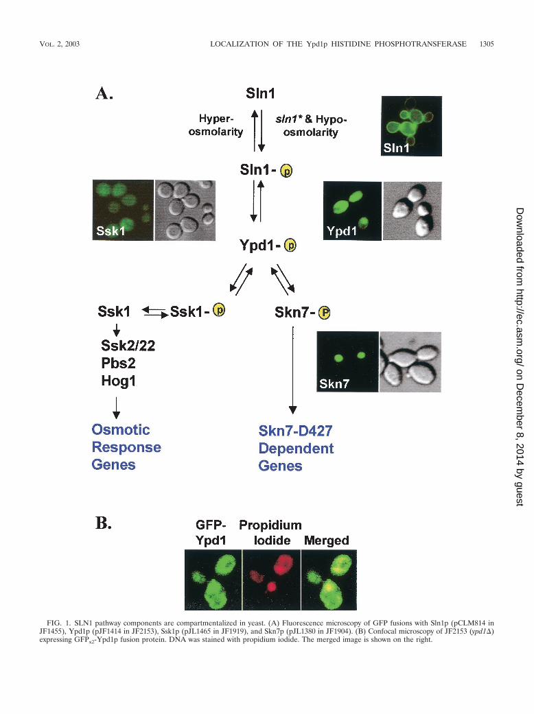
FIG. 1. SLN1 pathway components are compartmentalized in yeast. (A) Fluorescence microscopy of GFP fusions with Sln1p (pCLM814 inJF1455), Ypd1p (pJF1414 in JF2153), Ssk1p (pJL1465 in JF1919), and Skn7p (pJL1380 in JF1904). (B) Confocal microscopy of JF2153 (ypd1�)expressing GFPx2-Ypd1p fusion protein. DNA was stained with propidium iodide. The merged image is shown on the right.
VOL. 2, 2003 LOCALIZATION OF THE Ypd1p HISTIDINE PHOSPHOTRANSFERASE 1305
on Decem
ber 8, 2014 by guesthttp://ec.asm
.org/D
ownloaded from
to Ssk1p in the cytosol and to Skn7p in the nucleus. Thepresence of Ypd1p in both the cytosol and the nucleus mayreflect the need for simultaneous phosphorylation of the Ssk1pand Skn7p response regulators under normal growth condi-tions.
MATERIALS AND METHODS
Strains. All strains used are isogenic and derivatives of S288C. Strains wereconstructed for these experiments or are from the Fassler laboratory collection(Table 1). Disruption of YPD1 was achieved by transforming yeast with a PCRfragment containing a kanamycin resistance gene amplified with disruption oli-gonucleotides YPD-KAN-F (5�-TTGAATACCGGAGATATAATTCGTTTATGCCACATAATCATCAATACATCATAGGCCACTAGT-3�) and YPD-KAN-R (5�-ATCTACACCTTAACCAATAATGAGTTATAGAGGAGCAATGCAAAATCTAGCCAGCTGAAGCTT-3�), corresponding to the YPD1 regionfrom position �291 to �714. Disruptions were confirmed by genomic PCR andsubsequent restriction analysis of the amplified fragment. ypd1�::kan transfor-mants of JF2148 bearing pRS426-PTP2 were screened by sensitivity to fluoro-orotic acid (FOA) before PCR and restriction analyses. skn7�::TRP1 disruptionwas carried out as described previously (18). Deletion of the SSK1 gene wasachieved by one-step replacement with the PstI-XbaI fragment from plasmidpDSS14 from H. Saito (28). To disrupt HOG1, JF1565 was transformed with adisruption fragment linearized from pGY150 (39) by BamHI-ClaI digestion.Transformants were screened by sensitivity to 0.9 M NaCl before Southernhybridization analysis. JF2123 (fps1�::LEU2) is a plasmid-free derivative ofJF1732 (36).
Media. Solid and liquid media were prepared as described by Sherman et al.(32) and included synthetic complete (SC) medium lacking one or more specificamino acids and rich medium (yeast extract-peptone-dextrose [YPD]). Yeasttransformation was performed by a modified lithium acetate method (9, 13).Yeast strains were grown to log phase and streaked or spotted onto variousmedia after serial dilution. The viability of ypd1� strains carrying pRS426-PTP2was assayed on SC medium containing 0.1% 5-FOA to test functional comple-mentation by various YPD1 plasmids. All plate assays were carried out at 30°C.
Stress treatment. All localization studies were carried out with log-phasecultures grown at 28°C. Green fluorescent protein (GFP) fusion plasmids wereintroduced into strains with deletions in the corresponding gene.
(i) Osmotic stress. Hyperosmolarity was achieved by adding sorbitol to 1 M orNaCl to 0.5 or 0.9 M to log-phase cultures. Samples were taken and fixed at 5, 15,30, 60, and 90 min after addition of the osmoticum. A HOG1-GFP construct (7)
was used as a control. Hypo-osmolarity was generated by using the fps1 mutation(strain JF2123), which prevents glycerol efflux and causes accumulation of intra-cellular glycerol (20). We previously showed that the fps1 mutation activates theSLN1-SKN7 pathway (36), and we conclude, based on these studies, that Sln1pkinase activity is increased under hypotonic conditions.
(ii) Heat stress. Log-phase cells expressing GFP-SKN7 or GFP-YPD1 wereshifted to elevated temperature (37 and 42°C), and samples were taken at 5, 15,30, 60, 120, and 180 min.
(iii) Oxidative stress. Log-phase cells expressing GFP-SKN7 were treated with0.6 or 1 mM tert-butylhydroperoxide (Sigma) or 0.05% hydrogen peroxide(Sigma), and samples were taken at 5, 15, 30, 60, and 90 min. Activation of theoxidative response gene TRX2 was monitored in parallel to confirm the treat-ment conditions.
Plasmids. The plasmids used in this study are summarized in Table 2. Con-struction schemes are described below. The nucleotide positions are referred as� if downstream of the ATG start codon and as � if upstream. All PCRfragments were amplified by using the high-fidelity Pfu or Turbo Pfu DNApolymerase (Stratagene).
(i) Reporters. The OCH1 reporter plasmid was created by subcloning a 3-kbfragment containing UASOCH1 (�336 to �26)-lacZ from pZL1320 (19) intopRS314 by using EcoRI and SalI sites to create pJL1416.
(ii) GFP fusions. The SLN1-GFP plasmid pCLM814 was constructed by in-sertion of a NotI fragment containing the GFP open reading frame (ORF) at thestop codon of YEplac112-SLN1 (B812). The GFP fragment was amplified byPCR with oligonucleotides B568 (5�-TCAAGTCGCGGCCGCATGTCTAAAGGTGAAGAATTATTC-3�) and B569 (5�-ATGACAGTGCGGCCGCTTATTTGTACAATTCATCCATACC-3�) and GFP mut3 (S65G S72A, GenBank acces-sion number U73901) as a template. The pCLM814 plasmid was shown to befunctional by complementation of the 2:2 inviability phenotype in tetrads fromthe sln1�/SLN1� heterozygote, JF1455, into which it was transformed.
The 2 �m GFP-SKN7 plasmid pJL1380 was constructed by insertion of theSKN7 ORF downstream of UASSKN7-GFP (F64L S65T) cassette previouslycloned into pRS425 (pJL1363). The pJF1380 plasmid was shown to be functionalby complementation of the oxidative stress and hygromycin B sensitivities of askn7� strain.
To construct the GFP-YPD1 fusion, a BamHI-NheI polylinker was engineeredby PCR (QuickChange site-directed mutagenesis kit [Stratagene]) beyond thetranslation start site of YPD1 in pCLM669 (a pRS316-YPD1 plasmid) by usingoligonucleotides YPD1 � 3 BamHI-NheI-F (5�-GCGGATCCAGCTAGCGTCTACTATTCCCTCAGAAA-3�) and YPD1 � 6 BamHI-NheI-R (5�-CGTGCTAGCTGGATCCCAGACATTATTGTGTGTAT-3�). A GFP PCR fragment
TABLE 1. Yeast strains used in this study
Straina Relevant genotype Derivation (reference)
JF1455 MATa/� his4-917 lys2-128� trp1�1 ura3-52 leu2 sln1�::LEU2/SLN1 Diploid strainJF1565 MAT� his3�200 leu2�1 ura3-52 trp1�63 lys2�201 canR cyhR CanR CyhR derivative of FY834 (37)JF1592 MATa kar1-1 ade2-101 his4 leu2 trp1�1 or -62 ura3-52JF1904 MAT� skn7�::TRP1 his3�200 leu2�1 ura3-52 trp1�63 lys2�201
canR cyhRskn7�::TRP1 derivative of JF1565; one-step replacement
JF1910b MAT� sln1-22 his3�200 leu2�1 ura3-52 trp1�63 lys2�201 canR
cyhRsln1-22 derivative of JF1565; two-step replacement (36);
plasmid freeJF1919 MAT� ssk1�::LEU2 his3�200 leu2�1 ura3-52 trp1�63 lys2�201 ssk1�::LEU2 derivative of FY834; one-step replacementJF1920b MAT� sln1-22 ssk1�::LEU2 his3�200 leu2�1 ura3-52 trp1�63
lys2�201 canR cyhRssk1�::LEU2 derivative of JF1910; one-step replacement
JF1974 MAT� hog1�::TRP1 his3�200 leu2�1 ura3-52 trp1�63 lys2�201canR cyhR
hog1�::TRP1 derivative of JF1565; one-step replacement
JF2123 MAT� fps1�::LEU2 his3�200 leu2�1 ura3-52 trp1�63 lys2�201canR cyhR
Plasmid-free version of JF1732 (36)
JF2148 MAT� his3�200 leu2�1 ura3-52 trp1�63 lys2�201 canR cyhR;pRS426-PTP2
JF1565 carrying pRS426-PTP2
JF2150 MAT� ypd1�::kan ssk1�::LEU2 his3�200 leu2�1 ura3-52 trp1�63lys2�201
ypd1�::kan derivative of JF1919; one-step replacement
JF2153 MAT� ypd1�::kan his3�200 leu2�1 ura3-52 trp1�63 lys2�201 canR
cyhR; pRS426-PTP2ypd1�::kan derivative of JF2148; one-step replacement
JF2219b MAT� sln1-22 ypd1�::kan ssk1�::LEU2 his3�200 leu2�1 ura3-52trp1�63 lys2�201 canR cyhR
ypd1�::kan derivative of JF1920; one-step replacement
a All strains used in this study except JF1455 were created by transformation of FY834 (37) or its canR cyhR derivative JF1565, both of which are isogenic to S288C.The JF1455 strain is congenic with S288C.
b sln1-22 is one of several activating alleles of SLN1 collectively referred to as sln1* alleles. The sln1-22 mutation causes a change of proline 1148 to serine (5).
1306 LU ET AL. EUKARYOT. CELL
on Decem
ber 8, 2014 by guesthttp://ec.asm
.org/D
ownloaded from
amplified by using primers GFPBamHI BglII-F (5�-GGGATCCTGAGATCTATGTCTAAAGGTGAAGAATT-3�) and GFP NheI-R (5�-CCAGCTAGCGATATCTTGTACAATTCATCCATACC-3�) was then digested with BamHI andNheI and cloned to generate pJL1356, in which GFP is fused to the N terminusof the YPD1 ORF. To create GFPx2-YPD1, a 1.8-kb fragment containing theGFPx1-YPD1 (�3 to �1072) sequence was fused via synthetic BglII and SalI sitesdownstream of GFP, thus replacing the YPD1 ORF with GFP-YPD1 in pJL1356and generating a pRS316-GFPx2-YPD1 plasmid called pJL1414. PlasmidpJL1414 was shown to be functional by complementation of the sln1* activationdefect in JF2219 (sln1* ssk1� ypd1�) and the inviability phenotype of JF2153(ypd1�, pRS426-PTP2) on 5-FOA plates selecting against the presence of thePTP2 plasmid.
The GFPx2-SSK1 plasmid pJL1465 was constructed by replacing the YPD1ORF and downstream sequence in pJL1414 with a PCR fragment containingSSK1 (�3 to �2632) via NheI and SalI sites.
The nonphosphorylatable (H64Q) YPD1 allele was made by two-step PCRmutagenesis based on the method of Landt et al. (17). In round one, YPD1sequences from position �178 to �1072 were amplified with a forward primerencoding the H64Q mutation (YPD1 � 178F-H/Q) (5�-GACAATCTGGGCCAGTTTTTAAAGGGTTCT-3�) and the reverse primer YPD1 � 1072SalI-R(5�-GAAGGATTCTGTCGACTTTGTTGGTAC-3�). The PCR product wasused as the reverse primer for round two with forward primer YPD1-786 SpeI-F(5�-GAACATTTAAACTAGTGTGATTCAGG-3�) and pCLM669 (pRS316-YPD1) as the template. The 1.8-kb PCR product was purified, digested with SpeIand SalI, and cloned into pRS416. The GFP-ypd1H64Q fusion was constructedby first amplifying the region encompassing the YPD1 coding sequence (positions�3 to 1072) from pJL1429 with oligonucleotides YPD1 � 3BamHI-NheI-F andYPD1 � 1072SalI-R and then cloning the fragment into pRS315 at BamHI andSalI sites to create a promoterless plasmid, in which the H64Q mutation wasconfirmed by sequencing. Finally, an SpeI-NheI fragment containing UASYPD1-GFPx2 was inserted upstream of the YPD1 coding region to create pJL1433, amutant counterpart to pJL1414. GFPx2-ypd1H64Q was also subcloned intopRS316 via NotI and SalI sites to create pJL1415.
CaaX and SaaX fusions of YPD1 were created by mutagenizing the stop codonof YPD1 to create MluI and XbaI sites by using oligonucleotides YPD1 �501MluI XbaI-R (5�-GCTCTAGACGCGTAGGTTTGTGTTGTAATATTTAG-3� and YPD1-786 SpeI-F. The 3.2-kb fragment was digested with SpeI andXbaI and ligated into XbaI site of pRS416, resulting in pJL1436, in which theupstream XbaI site was destroyed upon fusion with the SpeI site and the down-stream MluI-XbaI sites were intact for subsequent ligation. A fragment consistingof the last 43 amino acids of Ras2p, including the CaaX box (CCIIS) motif, wasPCR amplified from plasmid pB701 (2) by using oligonucleotides RAS2 � 844MluI-F (5�-GGGACGCGTGAATAATAATAGTAAGGCCGGTC-3�) and
RAS2 � 969 XbaI-R (5�-GCTCTAGACTTAACTTATAATACAACAGCCACCCG-3�). The 126-bp PCR product was digested with MluI and XbaI and fusedbehind the YPD1 coding sequence in pJL1436, creating the GFPx2-YPD1-CaaXconstruct pJL1437. Similarly a SaaX-containing fragment in which the CCIISSsequence at the C terminus of Ras2 was mutated into SSIIS was amplifiedby using oligonucleotides RAS2 � 844 MluI-F and RAS2m � 950 XbaI-R(5�-GCTCTAGACTTAACTTATAATAGACGAGCCACCCGATCCGCTCT-3�) (mutated bases are underlined). The PCR fragment was inserted intopJL1436 via MluI and XbaI sites to create pJL1441. The NotI-SalI fragments ofpJL1414, pJL1437, and pJL1441 were subcloned into pRS315 to obtain pJL1419,pJL1440, and pJL1447, respectively.
The NLS-YPD1 construct pJL1453 was made by inserting a 0.24-kb BamHI-BglII fragment from pGPKI-NLS (a gift from S. Green) (3) containing twotandem copies of the simian virus 40 NLS into the BglII site between the twocopies of GFP in pJL1414. In the NES-YPD1 fusion, the nuclear export sequence(NES) from PKI (33) was engineered between the second copy of GFP and theN terminus of YPD1 in two steps. The GFPx2NES cassette was amplified by usingYPD1-786 SpeI-F and GFP-NES-NheI-R (5�-CCTGCTAGCGTAAGCTTTGTCTTGTTGATATCGAGGCCTGCTAGTTTCAGCGCTAATTCATTTTTGTACAATTCATCCATGCC-3�) (encoding GMDELYKNELALKLAGLDINKTKLTLA). The 2.3-kb PCR product (UASYPD1-GFPx2NES) was fused upstream ofthe YPD1 coding sequence in pJL1414 via SpeI and NheI sites to create pJL1456(GFPx2-NES-YPD1). The NLS- and NES-tagged GFP-YPD1 fusions were sub-cloned into pRS315 to create pJL1454 and pJL1461, respectively.
Galactose-inducible GFP constructs. The GFP-NLSx2-GFP-YPD1 fragmentwas released from pJF1454 by use of BamHI and SalI and fused in frame at theBamHI site downstream of the GAL promoter in pRS313-UASGAL10 (5) tocreate pJL1514.
The control plasmid, GFP-HTB1 (pJL1532) was created in two steps. AnHTB1 PCR fragment was generated by using forward primer HTB1 � 4 Bam-BglII (5�-TGGATCCTGAGATCTGCTAAAGCCGAAAAGAAACC-3�) andreverse primer HTB1 � 1007 SalI (5�-GATCAGAGCTCGTCGACAAGGAATACTGAAGTGCA-3�) and genomic DNA as the template. The PCR fragmentwas inserted behind GFP in an intermediate plasmid. The GFP-HTB1 fragmentwas then subcloned into a derivative of pRS313-UASGAL10 (5).
�-galactosidase assays. Yeast protein extracts were prepared by glass beadlysis from cultures grown at 30°C and harvested at a density of 107 cells/ml.Extracts were cleared by centrifugation in all cases. Activities were normalized toprotein concentrations (23) and are the averages for at least four differenttransformants.
Yeast protein extracts and immunoblot analysis. Yeast cultures were grown tolog phase in selective media. Cells were pelleted and resuspended in 10 mMTris-50 mM EDTA, washed once in lysis buffer (50 mM Tris [pH 7.6], 140 mMNaCl, 0.1% Triton X-100, 5 mM EDTA, 1 mM dithiothreitol, 1 mM phenyl-methylsulfonyl fluoride, and protease inhibitor cocktail [Sigma]), pelleted, andfrozen at �80°C. The pellets were resuspended in lysis buffer and broken byvigorous vortexing in the presence of 425- to 600-�m-diameter glass beads(Sigma), and lysates were cleared by centrifugation and stored at �20°C. Theprotein concentration was determined by using the Bio-Rad Microassay. GFPprotein levels were examined by using rabbit anti-GFP antibody and horseradishperoxidase-conjugated goat anti-rabbit immunoglobulin G antiserum (Sigma).Immune complexes were visualized by using the ECL enhanced chemilumines-cence kit (Amersham Corp.).
Fluorescence microscopy. Log-phase yeast cultures expressing GFP fusionswere fixed in 70% ethanol, washed and resuspended in phosphate-buffered saline(PBS), and stained with 0.5 �g of 4�,6�-diamidino-2-phenylindole (DAPI)(Sigma) per ml to visualize nuclei. Cells were observed with a Leica DM RBEmicroscope and a Leica 100� PL Fluotar 1.3 NA objective. Images were cap-tured with a Photonic Science digital charge-coupled device camera system.Images were processed by using IP-LAB Spectrum software and edited in AdobePhotoshop.
For confocal microscopy, log-phase cells were fixed in cold 75% methanol,washed twice in PBS and three times in 2� SSC (0.3 M NaCl, 0.03 M sodiumcitrate [pH 7.0]), and treated with DNase-free RNase A (100 �g/ml) (Sigma) for20 min at 37°C. Samples were then rinsed several times in 2� SSC and incubatedwith 500 nM propidium iodide (Molecular Probes) prepared in 2� SSC for 5 minat room temperature. Samples were washed five times and resuspended in 2�SSC prior to microscopy with an MRC-600 laser scanning confocal microscope(Bio-Rad).
Heterokaryon assay. The heterokaryon assay was modified from publishedprotocols (8, 27). JF2153 (ypd1�kan) carrying a pRS426-PTP2 plasmid as well aspJL1514 (pRS313-UASGAL10-GFP-NLSx2-GFP-YPD1) was grown to late logphase in synthetic medium (2% glucose) before dilution into medium containing
TABLE 2. Plasmids used in this study
Plasmid Descriptiona
pCLM814......................YEplac112-UASSLN1-SLN1-GFPHOG1-GFP ..................pRS416-HOG1-GFP (7)pJL1363.........................pRS425-UASSKN7-GFPpJL1380.........................pRS425-UASSKN7-GFP-SKN7pJL1414.........................pRS316-UASYPD1-GFPx2-YPD1PJL1415.........................PRS316-UASYPD1-GFPx2-ypd1H64QpJL1416.........................pRS314-OCH1 (�336 to �26)-lacZpJL1419.........................pRS315-UASYPD1-GFPx2-YPD1PJL1429.........................PRS416-UASYPD1-GFPx2-ypd1H64QpJL1433.........................pRS315-UASYPD1-GFPx2-YPD1 H64QpJL1437.........................pRS416-UASYPD1-GFPx2-YPD1-CaaX(CCIIS)pJL1440.........................pRS315-UASYPD1-GFPx2-YPD1-CaaX(CCIIS)pJL1441.........................pRS416-UASYPD1-GFPx2-YPD1-SaaX(SSIIS)pJL1447.........................pRS315-GFPx2-YPD1-SaaX(SSIIS)pJL1453.........................pRS316-UASYPD1-GFP-NLSx2-GFP-YPD1pJL1454.........................pRS315-UASYPD1-GFP-NLSx2GFP-YPD1pJL1456.........................pRS316-UASYPD1-GFPx2-NES-YPD1pJL1461.........................pRS315-UASYPD1-GFPx2-NES-YPD1pJL1465.........................pRS316-UASYPD1-GFPx2-SSK1pJL1514.........................pRS313-UASGAL10-GFP-NLSx2-GFP-YPD1pJL1532.........................pRS313-UASGAL10-GFP-HTB1
a All plasmids except HOG1-GFP (7) were constructed for this study. SeeMaterials and Methods for construction details. Plasmids used solely as inter-mediates in various construction schemes are not listed here.
VOL. 2, 2003 LOCALIZATION OF THE Ypd1p HISTIDINE PHOSPHOTRANSFERASE 1307
on Decem
ber 8, 2014 by guesthttp://ec.asm
.org/D
ownloaded from
2% raffinose as the sole carbon source. The culture was grown overnight to 106
cells/ml, and galactose was added to a final concentration of 2%. The culture wasthen grown for 3 h, washed three times in YPD (2% glucose), and resuspendedin YPD for an additional 2 h of growth. Cells (2 � 107) were mixed with an equalnumber of JF1592 (kar1-1) cells from a YPD-grown log-phase culture. The cellmixture was concentrated on a 25-mm-diameter, 0.45-�m-pore-size nitrocellu-lose filter and incubated at 28°C for 2 h on solid YPD. Cells were washed fromfilters by gentle vortexing in a microcentrifuge tube containing 0.5 ml of YPD,collected by brief centrifugation, fixed in cold 80% methanol for 30 min, washedtwice in PBS, and resuspended in DAPI (0.5 �g/ml in PBS). The nonshuttlingcontrol, JF1565 (wild-type strain) bearing pJL1532 (pRS313-UASGAL10-GFP-HTB1), was mated to kar1-1 cells in the same manner.
RNA analysis. RNA samples were prepared by using a hot acidic-phenolmethod (1). Electrophoresis and blotting were performed as described previously(39). Hybridization was performed with PerfectHyb hybridization buffer (Sigma)as directed by the manufacturer. 32P-labeled probes were prepared by usingrandom primers with Prime-It random priming labeling kit (Stratagene) withfragments isolated from plasmids. The GFP ORF was derived as a 0.75-kbBamHI-BglII fragment from pJL1414, and ACT1 was isolated as a 3-kb EcoRIfragment from pYIP5-ACT1 (39). Blots were exposed to X-ray film, and radio-labeled bands were visualized by autoradiography. The GFP probe was removedfrom the blot by washing in 0.2� SSC–1% sodium dodecyl sulfate at 75°C for 3 hfollowed by a 0.1% sodium dodecyl sulfate–10 mM Tris (pH 7.5) rinse at roomtemperature prior to hybridization with the ACT1 probe.
RESULTS
Ypd1p resides in both nuclear and cytosolic compartments.The subcellular localization of Sln1p, Ypd1, Ssk1p, and Skn7pwas examined by using GFP fusions on high-copy (Sln1p andSkn7p) or low-copy (Ssk1p and Ypd1p) yeast expression plas-mids under the control of the native promoters (with the ex-ception of Ssk1p, which was expressed by using the strongerYPD1 promoter). In all cases, expression of the GFP fusionsfrom these plasmids was able to complement the phenotypes ofthe corresponding mutants (data not shown). Sln1p-GFP lo-calized to the rim of the cell, consistent with the expectedplasma membrane localization of the sensor kinase (Fig. 1A).In contrast, GFP-Skn7p was present exclusively in the nucleus.GFP-Ssk1p appears to be localized to the cytoplasm (Fig. 1A);however, the relatively weak intensity of the GFP-Ssk1p signalprecludes our ruling out its presence in the nucleus. Theseobservations suggest that phosphotransfer from Ypd1p to thetwo response regulators occurs in distinct intracellular com-partments and that the localization of Ypd1p could be a reg-ulated step in the pathway, as has been previously reported forplants (12).
Analysis of GFPx2-Ypd1p revealed that the Ypd1p protein isdistributed throughout the cytoplasm and in the nucleus undernormal growth conditions. There is no evidence of plasmamembrane localization, suggesting that its interaction with theplasma membrane-localized Sln1p is transient. Nuclear Ypd1pis seen as a more intense spot of fluorescence that coincideswith the DAPI-staining material (Fig. 1A and 2) within thecytoplasm. The presence of Ypd1p in the nucleus was con-firmed by confocal microscopy (Fig. 1B). Since two copies ofGFP were fused in frame with Ypd1p, the resulting fusionprotein was 74 kDa in size, which is too large for passive importinto the nucleus. We therefore conclude that the presence ofYpd1p in the nucleus could be due to active transport.
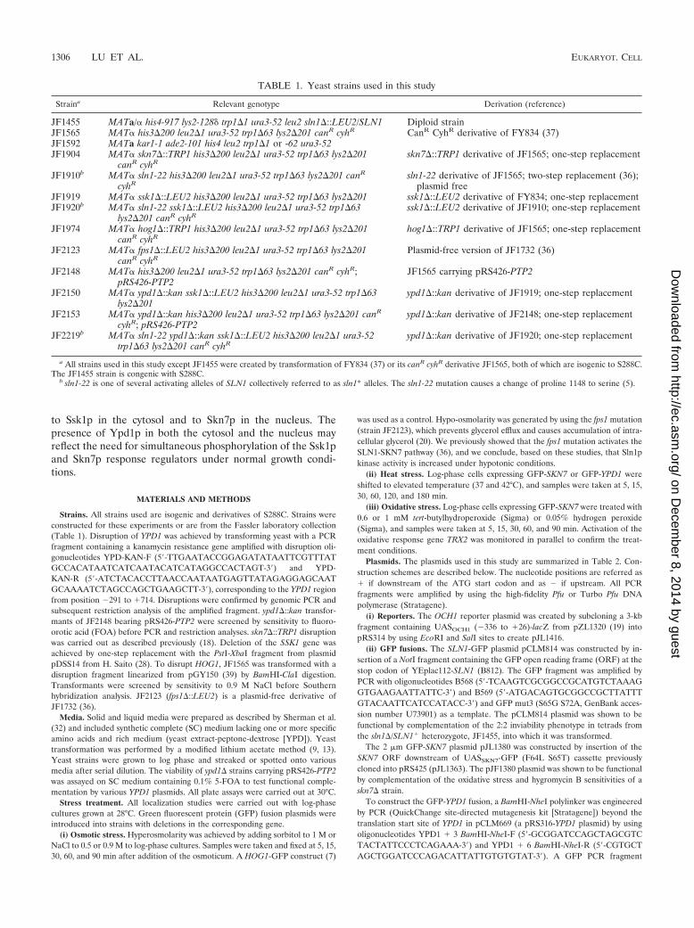
Localization of Ypd1p and Skn7p is unchanged in responseto osmotic stress. To determine whether localization of Ypd1por Skn7p changes in response to fluctuations in osmotic pres-sure, cells carrying GFPx2-SKN7 and GFPx2-YPD1 were shifted
to hyperosmotic medium. A HOG1-GFP construct was used asa control, since the rapid translocation of Hog1p to the nucleusupon exposure to a hyperosmotic stimulus has been well stud-ied (7). As previously reported, Hog1p-GFP rapidly moved tothe nucleus following salt addition; however, neither the GFP-Skn7p nor GFP-Ypd1p localization profile was altered follow-ing exposure of the cells to hyperosmotic conditions (Fig. 2A).
Skn7p undergoes Sln1p-dependent phosphorylation whencells are exposed to hypo-osmotic stress (19, 36). To examinethe effect of a hypo-osmotic environment on localization, we
FIG. 2. Subcellular localization of Ypd1p (pJF1414), Ssk1(pJL1465), and Skn7p (pJL1380) in response to changes in osmoticpressure. (A) Effects of one set of hyperosmotic conditions (0.9 MNaCl for 30 min). GFP-Hog1p (pRS416-GFP-HOG1, a gift from P.Silver) is shown as a control for osmotic stress-regulated nuclear lo-calization. (B) The effect of hypo-osmotic stress in FPS1� (JF1565)and fps1� (JF2123) strains was compared.
1308 LU ET AL. EUKARYOT. CELL
on Decem
ber 8, 2014 by guesthttp://ec.asm
.org/D
ownloaded from
introduced a mutation in the FPS1 gene, which encodes themajor glycerol channel in yeast. Reduced glycerol efflux in thefps1 mutant results in the accumulation of intracellular glycerolunder normal growth conditions, which is interpreted by cellsas a hypo-osmotic signal and increases activity of the SLN1-SKN7 pathway (20, 36). However, neither Hog1p, Skn7p, norYpd1p localization was affected in the fps1 mutant (Fig. 2B).The effects of heat shock and oxidative stress, two conditionsthat activate Skn7p in an SLN1-independent fashion (15, 24,30), were also examined. Again, no apparent changes in local-ization of Skn7p or Ypd1p were observed under these condi-tions (data not shown).
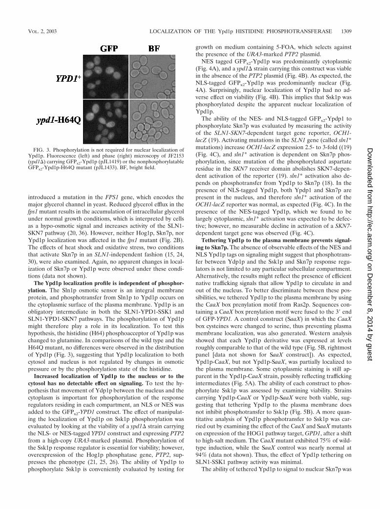
The Ypd1p localization profile is independent of phosphor-ylation. The Sln1p osmotic sensor is an integral membraneprotein, and phosphotransfer from Sln1p to Ypd1p occurs onthe cytoplasmic surface of the plasma membrane. Ypd1p is anobligatory intermediate in both the SLN1-YPD1-SSK1 andSLN1-YPD1-SKN7 pathways. The phosphorylation of Ypd1pmight therefore play a role in its localization. To test thishypothesis, the histidine (H64) phosphoacceptor of Ypd1p waschanged to glutamine. In comparisons of the wild type and theH64Q mutant, no differences were observed in the distributionof Ypd1p (Fig. 3), suggesting that Ypd1p localization to bothcytosol and nucleus is not regulated by changes in osmoticpressure or by the phosphorylation state of the histidine.
Increased localization of Ypd1p to the nucleus or to thecytosol has no detectable effect on signaling. To test the hy-pothesis that movement of Ydp1p between the nucleus and thecytoplasm is important for phosphorylation of the responseregulators residing in each compartment, an NLS or NES wasadded to the GFPx2-YPD1 construct. The effect of manipulat-ing the localization of Ypd1p on Ssk1p phosphorylation wasevaluated by looking at the viability of a ypd1� strain carryingthe NLS- or NES-tagged YPD1 construct and expressing PTP2from a high-copy URA3-marked plasmid. Phosphorylation ofthe Ssk1p response regulator is essential for viability; however,overexpression of the Hog1p phosphatase gene, PTP2, sup-presses the phenotype (21, 25, 26). The ability of Ypd1p tophosphorylate Ssk1p is conveniently evaluated by testing for
growth on medium containing 5-FOA, which selects againstthe presence of the URA3-marked PTP2 plasmid.
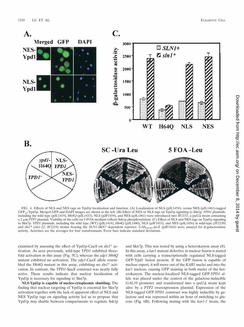
NES tagged GFPx2-Ypd1p was predominantly cytoplasmic(Fig. 4A), and a ypd1� strain carrying this construct was viablein the absence of the PTP2 plasmid (Fig. 4B). As expected, theNLS-tagged GFPx2-Ypd1p was predominantly nuclear (Fig.4A). Surprisingly, nuclear localization of Ypd1p had no ad-verse effect on viability (Fig. 4B). This implies that Ssk1p wasphosphorylated despite the apparent nuclear localization ofYpd1p.
The ability of the NES- and NLS-tagged GFPx2-Ypdp1 tophosphorylate Skn7p was evaluated by measuring the activityof the SLN1-SKN7-dependent target gene reporter, OCH1-lacZ (19). Activating mutations in the SLN1 gene (called sln1*mutations) increase OCH1-lacZ expression 2.5- to 3-fold ((19)(Fig. 4C), and sln1* activation is dependent on Skn7p phos-phorylation, since mutation of the phosphorylated aspartateresidue in the SKN7 receiver domain abolishes SKN7-depen-dent activation of the reporter (19). sln1* activation also de-pends on phosphotransfer from Ypd1p to Skn7p (18). In thepresence of NLS-tagged Ypd1p, both Ypdp1 and Skn7p arepresent in the nucleus, and therefore sln1* activation of theOCH1-lacZ reporter was normal, as expected (Fig. 4C). In thepresence of the NES-tagged Ypd1p, which we found to belargely cytoplasmic, sln1* activation was expected to be defec-tive; however, no measurable decline in activation of a SKN7-dependent target gene was observed (Fig. 4C).
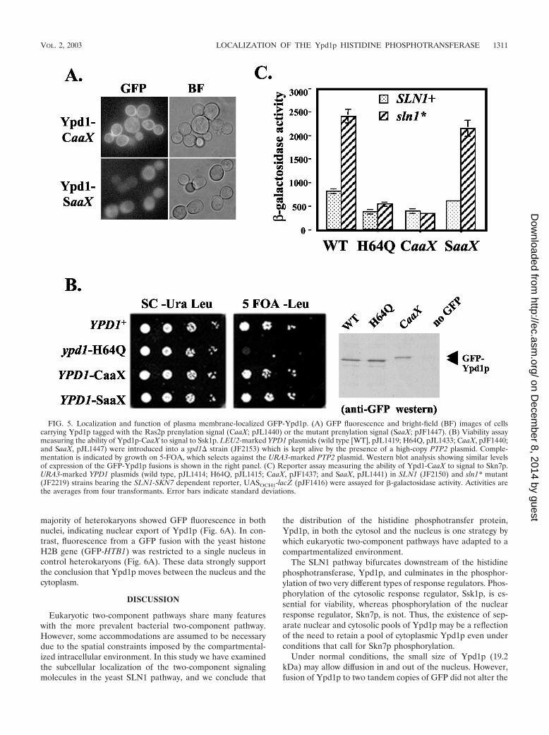
Tethering Ypd1p to the plasma membrane prevents signal-ing to Skn7p. The absence of observable effects of the NES andNLS Ypd1p tags on signaling might suggest that phosphotrans-fer between Ydp1p and the Ssk1p and Skn7p response regu-lators is not limited to any particular subcellular compartment.Alternatively, the results might reflect the presence of efficientnative trafficking signals that allow Ypd1p to circulate in andout of the nucleus. To better discriminate between these pos-sibilities, we tethered Ypd1p to the plasma membrane by usingthe CaaX box prenylation motif from Ras2p. Sequences con-taining a CaaX box prenylation motif were fused to the 3� endof GFP-YPD1. A control construct (SaaX) in which the CaaXbox cysteines were changed to serine, thus preventing plasmamembrane localization, was also generated. Western analysisshowed that each Ypd1p derivative was expressed at levelsroughly comparable to that of the wild type (Fig. 5B, rightmostpanel [data not shown for SaaX construct]). As expected,Ypd1p-CaaX, but not Ypd1p-SaaX, was partially localized tothe plasma membrane. Some cytoplasmic staining is still ap-parent in the Ypd1p-CaaX strain, possibly reflecting traffickingintermediates (Fig. 5A). The ability of each construct to phos-phorylate Ssk1p was assessed by examining viability. Strainscarrying Ypd1p-CaaX or Ypd1p-SaaX were both viable, sug-gesting that tethering Ypd1p to the plasma membrane doesnot inhibit phosphotransfer to Ssk1p (Fig. 5B). A more quan-titative analysis of Ypd1p phosphotransfer to Ssk1p was car-ried out by examining the effect of the CaaX and SaaX mutantson expression of the HOG1 pathway target, GPD1, after a shiftto high-salt medium. The CaaX mutant exhibited 75% of wild-type induction, while the SaaX control was nearly normal at94% (data not shown). Thus, the effect of Ypd1p tethering onSLN1-SSK1 pathway activity was minimal.
The ability of tethered Ypd1p to signal to nuclear Skn7p was
FIG. 3. Phosphorylation is not required for nuclear localization ofYpd1p. Fluorescence (left) and phase (right) microscopy of JF2153(ypd1�) carrying GFPx2-Ypd1p (pJL1419) or the nonphosphorylatableGFPx2-Ypd1p-H64Q mutant (pJL1433). BF, bright field.
VOL. 2, 2003 LOCALIZATION OF THE Ypd1p HISTIDINE PHOSPHOTRANSFERASE 1309
on Decem
ber 8, 2014 by guesthttp://ec.asm
.org/D
ownloaded from
examined by assessing the effect of Ypd1p-CaaX on sln1* ac-tivation. As seen previously, wild-type YPD1 exhibited three-fold activation in this assay (Fig. 5C), whereas the ydp1 H64Qmutant exhibited no activation. The ydp1-CaaX allele resem-bled the H64Q mutant in this assay, exhibiting no sln1* acti-vation. In contrast, the YPD1-SaaX construct was nearly fullyactive. These results indicate that nuclear localization ofYpd1p is necessary for signaling to Skn7p.
NLS-Ypd1p is capable of nucleo-cytoplasmic shuttling. Thefinding that nuclear targeting of Ypd1p is essential for Skn7pactivation together with the lack of apparent effect of NLS andNES Ypd1p tags on signaling activity led us to propose thatYpd1p may shuttle between compartments to regulate Ssk1p
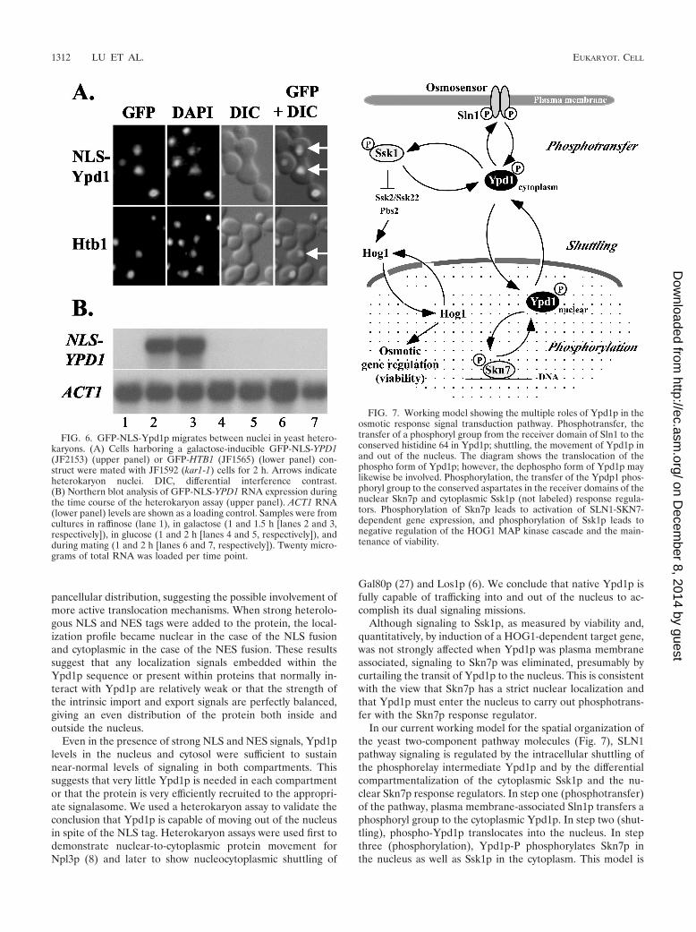
and Skn7p. This was tested by using a heterokaryon assay (8).In this assay, a kar1 mutant defective in nuclear fusion is matedwith cells carrying a transcriptionally regulated NLS-taggedGFP-Ypd1 fusion protein. If the GFP fusion is capable ofnuclear export, it will move out of the KAR1 nuclei and into thekar1 nucleus, causing GFP staining in both nuclei of the het-erokaryon. The nuclear-localized NLS-tagged GFP-YPD1 al-lele was placed under the control of the galactose-inducibleGAL10 promoter and transformed into a ypd1� strain keptalive by a PTP2 overexpression plasmid. Expression of theNLS-tagged GFP-YPD1 construct was highly inducible by ga-lactose and was repressed within an hour of switching to glu-cose (Fig. 6B). Following mating with the kar1-1 strain, the
FIG. 4. Effects of NLS and NES tags on Ypd1p localization and function. (A) Localization of NLS (pJL1454)- versus NES (pJL1461)-taggedGFPx2-Ypd1p. Merged GFP and DAPI images are shown at the left. (B) Effect of NES or NLS tags on Ypd1p signaling to Ssk1p. YPD1 plasmids,including the wild type (pJL1419), H64Q (pJL1433), NLS (pJF1454), and NES (pJL1461) were introduced into JF2153, a ypd1� strain containinga 2 �m PTP2 plasmid. Viability of the cells on 5-FOA medium reflects Ssk1p phosphorylation. (C) Effect of NLS and NES tags on Ypd1p signalingto Skn7p. YPD1 plasmids, including the wild type (WT) (pJL1414), H64Q (pJL1480), NLS (pJF1453), and NES (pJL1456) in wild-type (JF2150)and sln1* (sln1-22; JF2219) strains bearing the SLN1-SKN7 dependent reporter, UASOCH1-lacZ (pJF1416) were assayed for -galactosidaseactivity. Activities are the averages for four transformants. Error bars indicate standard deviations.
1310 LU ET AL. EUKARYOT. CELL
on Decem
ber 8, 2014 by guesthttp://ec.asm
.org/D
ownloaded from
majority of heterokaryons showed GFP fluorescence in bothnuclei, indicating nuclear export of Ypd1p (Fig. 6A). In con-trast, fluorescence from a GFP fusion with the yeast histoneH2B gene (GFP-HTB1) was restricted to a single nucleus incontrol heterokaryons (Fig. 6A). These data strongly supportthe conclusion that Ypd1p moves between the nucleus and thecytoplasm.
DISCUSSION
Eukaryotic two-component pathways share many featureswith the more prevalent bacterial two-component pathway.However, some accommodations are assumed to be necessarydue to the spatial constraints imposed by the compartmental-ized intracellular environment. In this study we have examinedthe subcellular localization of the two-component signalingmolecules in the yeast SLN1 pathway, and we conclude that
the distribution of the histidine phosphotransfer protein,Ypd1p, in both the cytosol and the nucleus is one strategy bywhich eukaryotic two-component pathways have adapted to acompartmentalized environment.
The SLN1 pathway bifurcates downstream of the histidinephosphotransferase, Ypd1p, and culminates in the phosphor-ylation of two very different types of response regulators. Phos-phorylation of the cytosolic response regulator, Ssk1p, is es-sential for viability, whereas phosphorylation of the nuclearresponse regulator, Skn7p, is not. Thus, the existence of sep-arate nuclear and cytosolic pools of Ypd1p may be a reflectionof the need to retain a pool of cytoplasmic Ypd1p even underconditions that call for Skn7p phosphorylation.
Under normal conditions, the small size of Ypd1p (19.2kDa) may allow diffusion in and out of the nucleus. However,fusion of Ypd1p to two tandem copies of GFP did not alter the
FIG. 5. Localization and function of plasma membrane-localized GFP-Ypd1p. (A) GFP fluorescence and bright-field (BF) images of cellscarrying Ypd1p tagged with the Ras2p prenylation signal (CaaX; pJL1440) or the mutant prenylation signal (SaaX; pJF1447). (B) Viability assaymeasuring the ability of Ypd1p-CaaX to signal to Ssk1p. LEU2-marked YPD1 plasmids (wild type [WT], pJL1419; H64Q, pJL1433; CaaX, pJF1440;and SaaX, pJL1447) were introduced into a ypd1� strain (JF2153) which is kept alive by the presence of a high-copy PTP2 plasmid. Comple-mentation is indicated by growth on 5-FOA, which selects against the URA3-marked PTP2 plasmid. Western blot analysis showing similar levelsof expression of the GFP-Ypd1p fusions is shown in the right panel. (C) Reporter assay measuring the ability of Ypd1-CaaX to signal to Skn7p.URA3-marked YPD1 plasmids (wild type, pJL1414; H64Q, pJL1415; CaaX, pJF1437; and SaaX, pJL1441) in SLN1 (JF2150) and sln1* mutant(JF2219) strains bearing the SLN1-SKN7 dependent reporter, UASOCH1-lacZ (pJF1416) were assayed for -galactosidase activity. Activities arethe averages from four transformants. Error bars indicate standard deviations.
VOL. 2, 2003 LOCALIZATION OF THE Ypd1p HISTIDINE PHOSPHOTRANSFERASE 1311
on Decem
ber 8, 2014 by guesthttp://ec.asm
.org/D
ownloaded from
pancellular distribution, suggesting the possible involvement ofmore active translocation mechanisms. When strong heterolo-gous NLS and NES tags were added to the protein, the local-ization profile became nuclear in the case of the NLS fusionand cytoplasmic in the case of the NES fusion. These resultssuggest that any localization signals embedded within theYpd1p sequence or present within proteins that normally in-teract with Ypd1p are relatively weak or that the strength ofthe intrinsic import and export signals are perfectly balanced,giving an even distribution of the protein both inside andoutside the nucleus.
Even in the presence of strong NLS and NES signals, Ypd1plevels in the nucleus and cytosol were sufficient to sustainnear-normal levels of signaling in both compartments. Thissuggests that very little Ypd1p is needed in each compartmentor that the protein is very efficiently recruited to the appropri-ate signalasome. We used a heterokaryon assay to validate theconclusion that Ypd1p is capable of moving out of the nucleusin spite of the NLS tag. Heterokaryon assays were used first todemonstrate nuclear-to-cytoplasmic protein movement forNpl3p (8) and later to show nucleocytoplasmic shuttling of
Gal80p (27) and Los1p (6). We conclude that native Ypd1p isfully capable of trafficking into and out of the nucleus to ac-complish its dual signaling missions.
Although signaling to Ssk1p, as measured by viability and,quantitatively, by induction of a HOG1-dependent target gene,was not strongly affected when Ypd1p was plasma membraneassociated, signaling to Skn7p was eliminated, presumably bycurtailing the transit of Ypd1p to the nucleus. This is consistentwith the view that Skn7p has a strict nuclear localization andthat Ypd1p must enter the nucleus to carry out phosphotrans-fer with the Skn7p response regulator.
In our current working model for the spatial organization ofthe yeast two-component pathway molecules (Fig. 7), SLN1pathway signaling is regulated by the intracellular shuttling ofthe phosphorelay intermediate Ypd1p and by the differentialcompartmentalization of the cytoplasmic Ssk1p and the nu-clear Skn7p response regulators. In step one (phosphotransfer)of the pathway, plasma membrane-associated Sln1p transfers aphosphoryl group to the cytoplasmic Ypd1p. In step two (shut-tling), phospho-Ypd1p translocates into the nucleus. In stepthree (phosphorylation), Ypd1p-P phosphorylates Skn7p inthe nucleus as well as Ssk1p in the cytoplasm. This model is
FIG. 6. GFP-NLS-Ypd1p migrates between nuclei in yeast hetero-karyons. (A) Cells harboring a galactose-inducible GFP-NLS-YPD1(JF2153) (upper panel) or GFP-HTB1 (JF1565) (lower panel) con-struct were mated with JF1592 (kar1-1) cells for 2 h. Arrows indicateheterokaryon nuclei. DIC, differential interference contrast.(B) Northern blot analysis of GFP-NLS-YPD1 RNA expression duringthe time course of the heterokaryon assay (upper panel). ACT1 RNA(lower panel) levels are shown as a loading control. Samples were fromcultures in raffinose (lane 1), in galactose (1 and 1.5 h [lanes 2 and 3,respectively]), in glucose (1 and 2 h [lanes 4 and 5, respectively]), andduring mating (1 and 2 h [lanes 6 and 7, respectively]). Twenty micro-grams of total RNA was loaded per time point.
FIG. 7. Working model showing the multiple roles of Ypd1p in theosmotic response signal transduction pathway. Phosphotransfer, thetransfer of a phosphoryl group from the receiver domain of Sln1 to theconserved histidine 64 in Ypd1p; shuttling, the movement of Ypd1p inand out of the nucleus. The diagram shows the translocation of thephospho form of Ypd1p; however, the dephospho form of Ypd1p maylikewise be involved. Phosphorylation, the transfer of the Ypdp1 phos-phoryl group to the conserved aspartates in the receiver domains of thenuclear Skn7p and cytoplasmic Ssk1p (not labeled) response regula-tors. Phosphorylation of Skn7p leads to activation of SLN1-SKN7-dependent gene expression, and phosphorylation of Ssk1p leads tonegative regulation of the HOG1 MAP kinase cascade and the main-tenance of viability.
1312 LU ET AL. EUKARYOT. CELL
on Decem
ber 8, 2014 by guesthttp://ec.asm
.org/D
ownloaded from
supported by several lines of evidence. First, localization stud-ies show the presence of Ypd1p in both the cytoplasm and thenucleus. Second, the fact that alteration of Ypd1p levels ineither compartment by addition of NES or NLS tags had aminimal impact on signaling to Ssk1p or Skn7p suggests thateven under these conditions Ypd1p is not confined to onecompartment. Third, heterokaryon assays show that NLS-tagged Ypd1, and presumably native Ypd1p, is indeed capableof nuclear export. Finally, since tethering of Ypd1p to theplasma membrane via a CaaX box caused a severe impairmentin Skn7p signaling, we conclude that Skn7p is restricted to thenucleus and that its activation requires nuclear targeting ofYpd1p. The temporal relationship of these events is not clear.Since the intracellular distribution of the nonphosphorylatableYpd1p H64Q is indistinguishable from that of normal Ypd1p,it is possible that both phospho and dephospho forms ofYpd1p move in and out the nucleus. However, there is noknown role for dephospho-Ypd1p in the nucleus.
An alternative model in which Ssk1p is phosphorylated in-side the nucleus before moving out to the cytoplasm to regu-late the HOG1 pathway seems unlikely, since Ssk1p appears tobe cytoplasmically localized under all osmotic conditionstested. This localization pattern is consistent with the knowncytoplasmic localization of the MAP kinase kinase, Pbs2p (7),which is activated by interaction with Ssk1p, and with theobservation that the MAP kinase, Hog1p, translocates to thenucleus upon osmotic treatment. In addition, both the NES-Ypd1p and plasma membrane-tethered Ypd1-CaaX exhibitednear-normal signaling to Ssk1p, indicating that nuclear Ypd1pis not required for Ssk1p signaling.
Recruitment of cytoplasmic signaling proteins into the nu-cleus is an essential step in the activation of gene expression inresponse to extracellular signals. In MAP kinase pathways, it isfrequently the MAP kinase that transits to the nucleus. Ineukaryotic two-component signaling, it appears that indepen-dent histidine phosphotransferase (Hpt) proteins such asYpd1p may have evolved for this purpose. In Arabidopsis thali-ana, AHP1 to -5 encode homologs of Ypd1p that function inthe cytokinin signal transduction pathway (12, 34, 35). A subsetof AHP molecules are translocated into the nucleus in a cyto-kinin-dependent manner, where they can signal to a nuclearfamily of response regulators (11). The regulated transport ofthe AHPs in Arabidopsis and the constitutive cycling of yeastYpd1p into and out of the nucleus appear to represent distinctmechanisms for two-component signaling in compartmental-ized cells. The basis for the difference between the two systemsmay lie with the tolerance and/or requirement of the cell forexpression of pathway targets. For example, expression of cy-tokinin genes in the absence of cytokinins may be harmful,whereas modest expression of SLN1-SKN7 target genes suchas OCH1, encoding a mannosyltransferase, is likely to be im-portant even in the absence of stress. It will be of interest toexamine the localization of molecules in other eukaryotic two-component pathways to determine whether Hpt-based trans-location is a general principle in eukaryotic two-componentsignaling pathways.
ACKNOWLEDGMENTS
This work was supported by Public Health Service grant GM-56719from the National Institutes of Health to J.S.F. and R.J.D. and by a
postdoctoral fellowship from the American Heart Association to J.M.-Y.L.
Our appreciation goes to Pam Silver for the Hog1-GFP construct, toSteven Green for the GPKI-NLS plasmid, to W. Scott Moye-Rowleyfor the GFP antibody, to Kevin Campbell and members of his lab foruse of the confocal microscope, to Michael Dailey for assistance withvarious nuclear staining protocols, and to Dan Weeks for help withXenopus-based assays for examining localization.
REFERENCES
1. Ausubel, F. M., R. Brent, R. E. Kingston, D. E. Moore, J. G. Seidman, J. A.Smith, and K. Struhl. 1989. Current protocols in molecular biology. JohnWiley and Sons, New York, N.Y.
2. Bartels, D. J., D. A. Mitchell, X. Dong, and R. J. Deschenes. 1999. Erf2, anovel gene product that affects the localization and palmitoylation of Ras2 inSaccharomyces cerevisiae. Mol. Cell. Biol. 19:6775–6787.
3. Bok, J., X. M. Zha, Y. S. Cho, and S. H. Green. 2003. An extranuclear locusof cAMP-dependent protein kinase action is necessary and sufficient forpromotion of spiral ganglion neuronal survival by cAMP. J. Neurosci. 23:777–787.
4. Brown, J. L., H. Bussey, and R. C. Stewart. 1994. Yeast Skn7p functions ina eukaryotic two-component regulatory pathway. EMBO J. 13:5186–5194.
5. Fassler, J. S., W. M. Gray, C. L. Malone, W. Tao, H. Lin, and R. J. De-schenes. 1997. Activated alleles of yeast SLN1 increase Mcm1-dependentreporter gene expression and diminish signaling through the Hog1 osmo-sensing pathway. J. Biol. Chem. 272:13365–13371.
6. Feng, W., and A. K. Hopper. 2002. A Los1p-independent pathway for nuclearexport of intronless tRNAs in Saccharomyces cerevisiae. Proc. Natl. Acad.Sci. USA 99:5412–5417.
7. Ferrigno, P., F. Posas, D. Koepp, H. Saito, and P. A. Silver. 1998. Regulatednucleo/cytoplasmic exchange of HOG1 MAPK requires the importin betahomologs NMD5 and XPO1 EMBO J. 17:5606–5614.
8. Flach, J., M. Bossie, J. Vogel, A. Corbett, T. Jinks, D. A. Willins, and P. A.Silver. 1994. A yeast RNA-binding protein shuttles between the nucleus andthe cytoplasm. Mol. Cell. Biol. 14:8399–8407.
9. Gietz, R. D., and R. A. Woods. 2002. Transformation of yeast by lithiumacetate/single-stranded carrier DNA/polyethylene glycol method. MethodsEnzymol. 350:87–96.
10. Gorner, W., E. Durchschlag, M. T. Martinez-Pastor, F. Estruch, G. Am-merer, B. Hamilton, H. Ruis, and C. Schuller. 1998. Nuclear localization ofthe C2H2 zinc finger protein Msn2p is regulated by stress and protein kinaseA activity. Genes Dev. 12:586–597.
11. Hwang, I., and J. Sheen. 2001. Two-component circuitry in Arabidopsiscytokinin signal transduction. Nature 413:383–389.
12. Imamura, A., Y. Yoshino, and T. Mizuno. 2001. Cellular localization of thesignaling components of Arabidopsis His-to-Asp phosphorelay. Biosci. Bio-technol. Biochem. 65:2113–2117.
13. Ito, H., Y. Fukuda, K. Murata, and A. Kimura. 1983. Transformation ofintact yeast cells treated with alkali cations. J. Bacteriol. 153:163–168.
14. Ketela, T., J. L. Brown, R. C. Stewart, and H. Bussey. 1998. Yeast Skn7pactivity is modulated by the Sln1p-Ypd1p osmosensor and contributes toregulation of the HOG pathway. Mol. Gen. Genet. 259:372–378.
15. Krems, B., C. Charizanis, and K.-D. Entian. 1996. The response regulator-like protein Pos9/Skn7 of Saccharomyces cerevisiae is involved in oxidativestress resistance. Curr. Genet. 29:327–334.
16. Kuge, S., N. Jones, and A. Nomoto. 1997. Regulation of yAP-1 nuclearlocalization in response to oxidative stress. EMBO J. 16:1710–1720.
17. Landt, O., H. P. Grunert, and U. Hahn. 1990. A general method for rapidsite-directed mutagenesis using the polymerase chain reaction. Gene 96:125–128.
18. Li, S., A. Ault, C. L. Malone, D. Raitt, S. Dean, L. H. Johnston, R. J.Deschenes, and J. S. Fassler. 1998. The yeast histidine protein kinase, Sln1p,mediates phosphotransfer to two response regulators, Ssk1p and Skn7p.EMBO J. 17:6952–6962.
19. Li, S., S. Dean, Z. Li, J. Horecka, R. J. Deschenes, and J. S. Fassler. 2002.The eukaryotic two-component histidine kinase Sln1p regulates OCH1 viathe transcription factor, Skn7p. Mol. Biol. Cell 13:412–424.
20. Luyten, K., J. Albertyn, W. F. Skibbe, B. A. Prior, J. Ramos, J. M. Thevelein,and S. Hohmann. 1995. Fps1, a yeast member of the MIP family of channelproteins, is a facilitator for glycerol uptake and efflux and is inactive underosmotic stress. EMBO J. 14:1360–1371.
21. Maeda, T., S. Wurgler-Murphy, and H. Saito. 1994. A two-component sys-tem that regulates an osmosensing MAP kinase cascade in yeast. Nature369:242–245.
22. Mattison, C. P., and I. M. Ota. 2000. Two protein tyrosine phosphatases,Ptp2 and Ptp3, modulate the subcellular localization of the Hog1 MAPkinase in yeast. Genes Dev. 14:1229–1235.
23. Miller, J. H. 1972. Experiments in molecular genetics. Cold Spring HarborLaboratory, Cold Spring Harbor, N.Y.
24. Morgan, B. A., G. R. Banks, W. M. Toone, D. Raitt, S. Kuge, and L. H.Johnston. 1997. The Skn7 response regulator controls gene expression in the
VOL. 2, 2003 LOCALIZATION OF THE Ypd1p HISTIDINE PHOSPHOTRANSFERASE 1313
on Decem
ber 8, 2014 by guesthttp://ec.asm
.org/D
ownloaded from
oxidative stress response of the budding yeast Saccharomyces cerevisiae.EMBO J. 16:1035–1044.
25. Ota, I. M., and A. Varshavsky. 1992. A gene encoding a putative tyrosinephosphatase suppresses lethality of an N-end rule dependent mutant. Proc.Natl. Acad. Sci. USA 89:2355–2359.
26. Ota, I. M., and A. Varshavsky. 1993. A yeast protein similar to bacterialtwo-component regulators. Science 262:566–568.
27. Peng, G., and J. E. Hopper. 2000. Evidence for Gal3p’s cytoplasmic locationand Gal80p’s dual cytoplasmic-nuclear location implicates new mechanismsfor controlling Gal4p activity in Saccharomyces cerevisiae. Mol. Cell. Biol.20:5140–5148.
28. Posas, F., and H. Saito. 1998. Activation of the yeast SSK2 MAP kinasekinase kinase by the SSK1 two-component response regulator. EMBO J.17:1385–1394.
29. Posas, F., S. M. Wurgler-Murphy, T. Maeda, E. A. Witten, T. C. Thai, andH. Saito. 1996. Yeast Hog1 MAP kinase cascade is regulated by a multistepphosphorelay mechanism in the Sln1-Ypd1-Ssk1 “two component” osmo-sensor. Cell 86:865–875.
30. Raitt, D. C., A. L. Johnson, A. M. Erkine, K. Makino, B. Morgan, D. S.Gross, and L. H. Johnston. 2000. The Skn7 response regulator of Saccha-romyces cerevisiae interacts with Hsf1 in vivo and is required for the inductionof heat shock genes by oxidative stress. Mol. Biol. Cell 11:2335–2347.
31. Reiser, V., H. Ruis, and G. Ammerer. 1999. Kinase activity-dependent nu-clear export opposes stress-induced nuclear accumulation and retention of
Hog1 mitogen-activated protein kinase in the budding yeast Saccharomycescerevisiae. Mol. Biol. Cell 10:1147–1161.
32. Sherman, F., G. R. Fink, and J. B. Hicks. 1986. Methods in yeast genetics, p.163–167. Cold Spring Harbor Laboratory, Cold Spring Harbor, N.Y.
33. Stade, K., C. S. Ford, C. Guthrie, and K. Weis. 1997. Exportin 1 (Crm1p) isan essential nuclear export factor. Cell 90:1041–1050.
34. Suzuki, T., A. Imamura, C. Ueguchi, and T. Mizuno. 1998. Histidine-con-taining phosphotransfer (HPt) signal transducers implicated in His-to-Aspphosphorelay in Arabidopsis. Plant Cell Physiol. 39:1258–1268.
35. Suzuki, T., K. Sakurai, A. Imamura, A. Nakamura, C. Ueguchi, and T.Mizuno. 2000. Compilation and characterization of histidine-containingphosphotransmitters implicated in His-to-Asp phosphorelay in plants: AHPsignal transducers of Arabidopsis thaliana. Biosci. Biotechnol. Biochem. 64:2486–2489.
36. Tao, W., R. J. Deschenes, and J. S. Fassler. 1999. Intracellular glycerol levelsmodulate the activity of Sln1, an S. cerevisiae two-component regulator.J. Biol. Chem. 274:360–367.
37. Winston, F., C. Dollard, and S. L. Ricupero-Hovasse. 1995. Construction ofa set of convenient Saccharomyces cerevisiae strains that are isogenic toS288C. Yeast 11:53–55.
38. Yan, C., L. H. Lee, and L. I. Davis. 1998. Crm1p mediates regulated nuclearexport of a yeast AP-1-like transcription factor. EMBO J. 17:7416–7429.
39. Yu, G., R. J. Deschenes, and J. S. Fassler. 1995. The essential transcriptionfactor, Mcm1 is a downstream target of Sln1, a yeast “two-component”regulator. J. Biol. Chem. 270:8739–8743.
1314 LU ET AL. EUKARYOT. CELL
on Decem
ber 8, 2014 by guesthttp://ec.asm
.org/D
ownloaded from
Copyright © 2022 FDOKUMEN