Ninein Is Expressed in the Cytoplasm of Angiogenic Tip-Cells and Regulates Tubular Morphogenesis of...
9
Charlotte Wikner, Gordon Chan, Lena Claesson-Welsh and Anna Dimberg Taro Matsumoto, Petter Schiller, Lothar C. Dieterich, Fuad Bahram, Yuji Iribe, Ulf Hellman, Morphogenesis of Endothelial Cells Ninein Is Expressed in the Cytoplasm of Angiogenic Tip-Cells and Regulates Tubular Print ISSN: 1079-5642. Online ISSN: 1524-4636 Copyright © 2008 American Heart Association, Inc. All rights reserved. Greenville Avenue, Dallas, TX 75231 is published by the American Heart Association, 7272 Arteriosclerosis, Thrombosis, and Vascular Biology doi: 10.1161/ATVBAHA.108.169128 2008; 2008;28:2123-2130; originally published online September 4, Arterioscler Thromb Vasc Biol. http://atvb.ahajournals.org/content/28/12/2123 World Wide Web at: The online version of this article, along with updated information and services, is located on the http://atvb.ahajournals.org//subscriptions/ at: is online Arteriosclerosis, Thrombosis, and Vascular Biology Information about subscribing to Subscriptions: http://www.lww.com/reprints Information about reprints can be found online at: Reprints: document. Question and Answer Permissions and Rights page under Services. Further information about this process is available in the which permission is being requested is located, click Request Permissions in the middle column of the Web Copyright Clearance Center, not the Editorial Office. Once the online version of the published article for can be obtained via RightsLink, a service of the Arteriosclerosis, Thrombosis, and Vascular Biology in Requests for permissions to reproduce figures, tables, or portions of articles originally published Permissions: by guest on February 17, 2014 http://atvb.ahajournals.org/ Downloaded from by guest on February 17, 2014 http://atvb.ahajournals.org/ Downloaded from
Transcript of Ninein Is Expressed in the Cytoplasm of Angiogenic Tip-Cells and Regulates Tubular Morphogenesis of...
Charlotte Wikner, Gordon Chan, Lena Claesson-Welsh and Anna DimbergTaro Matsumoto, Petter Schiller, Lothar C. Dieterich, Fuad Bahram, Yuji Iribe, Ulf Hellman,
Morphogenesis of Endothelial CellsNinein Is Expressed in the Cytoplasm of Angiogenic Tip-Cells and Regulates Tubular
Print ISSN: 1079-5642. Online ISSN: 1524-4636 Copyright © 2008 American Heart Association, Inc. All rights reserved.
Greenville Avenue, Dallas, TX 75231is published by the American Heart Association, 7272Arteriosclerosis, Thrombosis, and Vascular Biology
doi: 10.1161/ATVBAHA.108.1691282008;
2008;28:2123-2130; originally published online September 4,Arterioscler Thromb Vasc Biol.
http://atvb.ahajournals.org/content/28/12/2123World Wide Web at:
The online version of this article, along with updated information and services, is located on the
http://atvb.ahajournals.org//subscriptions/
at: is onlineArteriosclerosis, Thrombosis, and Vascular Biology Information about subscribing to Subscriptions:
http://www.lww.com/reprints
Information about reprints can be found online at: Reprints:
document. Question and AnswerPermissions and Rightspage under Services. Further information about this process is available in the
which permission is being requested is located, click Request Permissions in the middle column of the WebCopyright Clearance Center, not the Editorial Office. Once the online version of the published article for
can be obtained via RightsLink, a service of theArteriosclerosis, Thrombosis, and Vascular Biologyin Requests for permissions to reproduce figures, tables, or portions of articles originally publishedPermissions:
by guest on February 17, 2014http://atvb.ahajournals.org/Downloaded from by guest on February 17, 2014http://atvb.ahajournals.org/Downloaded from
Ninein Is Expressed in the Cytoplasm of AngiogenicTip-Cells and Regulates Tubular Morphogenesis of
Endothelial CellsTaro Matsumoto, Petter Schiller, Lothar C. Dieterich, Fuad Bahram, Yuji Iribe, Ulf Hellman,
Charlotte Wikner, Gordon Chan, Lena Claesson-Welsh, Anna Dimberg
Objective—Angiogenesis is an integral part of many physiological processes but may also aggravate pathologicalconditions such as cancer. Development of effective angiogenesis inhibitors requires a thorough understanding of themolecular mechanisms regulating vessel formation. The aim of this project was to identify proteins that regulate tubularmorphogenesis of endothelial cells.
Methods and Results—Phosphotyrosine-dependent affinity-purification and mass spectrometry showed tyrosine phos-phorylation of ninein during tubular morphogenesis of endothelial cells. Ninein was recently identified as a centrosomalmicrotubule-anchoring protein. Our results show that ninein is localized in the cytoplasm in endothelial cells, and thatit is highly expressed in the vasculature in normal and pathological human tissues. Using embryoid bodies as a modelof vascular development, we found that ninein is abundantly expressed in the cytoplasm of endothelial cells duringsprouting angiogenesis, in particular in the sprouting tip-cell. In accordance, siRNA-dependent silencing of ninein inendothelial cells inhibited tubular morphogenesis.
Conclusions—In this study, we show that ninein is expressed in developing vessels and in endothelial tip cells, and thatninein is critical for formation of the vascular tube. These data strongly implicate ninein as an important new regulatorof angiogenesis. (Arterioscler Thromb Vasc Biol. 2008;28:2123-2130.)
Key Words: ninein � angiogenesis � tubular morphogenesis � microtubule � endothelial
There is a well-established relationship between excessangiogenesis and progression of cancer and other patho-
logical conditions, as well as impaired vascularization inischemic conditions. This implies that anti- or proangiogenictreatments may be beneficial as a complement to existingtherapies for a wide range of diseases. Development ofefficient drugs that target the vasculature requires delineationof mechanisms that regulate blood vessel formation. Thecritical importance of growth factors and signal transductionby their cognate receptors, including vascular endothelialgrowth factors (VEGFs) and fibroblast growth factors (FGFs)in regulation of angiogenesis is widely recognized.1,2 How-ever, we still need to identify their downstream moleculartargets that regulate different aspects of the fine-tuned re-sponse resulting in formation of new vessels.
See accompanying article on page 2094In the adult, new blood vessels form through distinct and
overlapping mechanisms, depending on the microenviron-ment and the molecular context.3–5 Sprouting angiogenesis is
initiated when endothelial cells are stimulated by angiogenicgrowth factors eg, VEGF. The endothelial cells degrade thebasement membrane of preexisting vessels, migrate into thesurrounding matrix, proliferate, and finally differentiate tonew, lumen containing vessels. Circulating endothelial cellsor progenitors may contribute to a variable extent, formingnew vessels through a process that largely resembles embry-onic vasculogenesis. Finally, the newly formed vessel depos-its a vascular basement membrane, and recruits stabilizingpericytes.
To define the proteome regulating endothelial differentia-tion to lumen-containing vessels, we used an in vitro model oftubular morphogenesis and screened for proteins that weretyrosine phosphorylated during this process. One of theidentified phosphoproteins was ninein, a protein that has beenshown to be involved in the minus-end anchoring of micro-tubules in the centrosome.6–8 In this study, we have analyzedexpression of ninein in several types of endothelial cells invitro and in vivo, and used advanced cell culture models toevaluate the role of ninein in blood vessel formation.
Original received April 23, 2008; final version accepted August 26, 2008.From the Department of Genetics and Pathology (T.M., P.S., L.C.D., F.B., C.W., L.C.-W., A.D.), Uppsala University, Rudbeck Laboratory, Sweden;
the Division of Cell Regeneration and Transplantation, Advanced Medical Research Center (T.M., Y.I.), Nihon University School of Medicine, Tokyo,Japan; the Ludwig Institute for Cancer Research, Uppsala Branch, Biomedical Center (U.H.), Uppsala, Sweden; and the Department of Oncology (G.C.),University of Alberta, Experimental Oncology, Cross Cancer Institute, Edmonton, Alberta, Canada.
Correspondence to Anna Dimberg, Department of Genetics and Pathology, Rudbeck Laboratory, S-751 85 Uppsala, Sweden. [email protected]
© 2008 American Heart Association, Inc.
Arterioscler Thromb Vasc Biol is available at http://atvb.ahajournals.org DOI: 10.1161/ATVBAHA.108.169128
2123 by guest on February 17, 2014http://atvb.ahajournals.org/Downloaded from
MethodsCell Culture and In Vitro Angiogenesis AssaysBovine capillary endothelial (BCE) cells from calf adrenal cortexwere maintained in DMEM (Life Technologies), 10% newborn calfserum (NCS), and 2 ng/mL fibroblast growth factor-2 (FGF-2,Boehringer). Tubular morphogenesis was induced in a three-dimensional (3D) collagen matrix in DMEM containing 10% NCSand 10 ng/mL FGF-2, as described.9 Telomerase-immortalizedhuman microvascular endothelial (TIME) cells10 were cultured inEBM MV2 medium with supplements (5 ng/mL EGF, 0.2 �g/mLhydrocortisone, 0.5 ng/mL VEGF-A, 10 ng/mL FGF-2, 20 ng/mLIGF-1; Promocell). Tube formation was induced in 3D collagen gelsby 50 ng/mL VEGF-A (PeproTech) in EBM MV2 medium, 2% fetalbovine serum (FBS) as described.11 Mouse ES cell line R112 wasmaintained on growth-arrested mouse embryonic fibroblasts inDMEM-Glutamax medium supplemented with 15% FBS,25 mmol/L HEPES pH 7.4, 1.2 mmol/L sodium pyruvate, 0.12%monothiolglycerol and 1000 U/mL leukemia inhibitory factor. Em-bryoid bodies were induced as described13 in the presence ofVEGF-A (30 ng/mL, PeproTech), seeded in 8-well tissue-cultureslides or in a 3D-collagen matrix to induce formation of endothelialsprouts and analyzed at day 8 or 12.13 Porcine aortic endothelial(PAE) cells and human 293T cells were maintained in DMEM with10%FBS, and human U2OS cells were maintained in RPMI with10%FBS.
Cell Lysates and Western Blot AnalysisCells were washed in PBS and lysed in boiling SDS sample buffer orin a modified RIPA buffer (1% Triton X-100, 40 mmol/L Tris-HCl,pH 8, 0.1% SDS) with complete protease inhibitor (Roche AppliedScience). Extracts were fractionated on Novex NuPAGE (3% to 8%Tris-acetate) precast gels (Invitrogen). The membrane (Hybond-Cextra, GE Healthcare) was blocked in 5% dry milk in 0.1% Tween,Tris-buffered saline (TTBS), and Western blot analysis was done asdescribed.9 Primary antibodies used were: antiphospho-tyrosineantibody 4G10 (Upstate Biotechnology), anti-�-catenin (Transduc-tion Laboratories), antininein produced in-house,14 and antininein(Biolegend).
Purification and Identification of PhosphotyrosylProteins in Differentiating BCE CellsBriefly, serum-starved BCE cells were cultured on collagen gels orgelatinized dishes and incubated in DMEM containing 10% NCS and10 ng/mL FGF-2 for 24 hours at 37°C. Cells were lysed andphophotyrosine-containing proteines were immunoprecipitated usingan antiphosphotyrosine (4G10)-agarose conjugate (Upstate Biotech-nology). Proteins where separated by SDS-PAGE, the gel was silverstained, and protein bands where extracted by in-gel digestion.Identification of proteins was done using a Bruker Biflex IIIMALDI-TOF-MS (Bruker Daltonics). For a full description ofmethods involved, please see http://atvb.ahajournals.org.
ImmunofluorescenceBCE cells, TIME cells, or embryoid bodies were washed in TBS,fixed for 30 minutes at rt or at 4°C overnight in zinc fix (0.1 mol/LTris-HCl, pH 7.5, 3 mmol/L calcium acetate, 23 mmol/L zincacetate, 37 mmol/L zinc chloride) containing 0.2% Triton X-100,blocked in 3% BSA in TBS for 1 hour at rt, and incubated withprimary antibodies in blocking solution for 2 hours at rt. Sampleswere washed in TBS and incubated with Alexa-conjugated second-ary antibodies (Molecular Probes) and/or phalloidin-Texas Red(Molecular Probes) in blocking solution for 1 hour at rt, followed byHoechst 33342 nuclear staining.
Fully anonymized tissue samples were used in accordance with theSwedish biobank legislation. The use of human tissue was approvedby the Ethical Review Board in Uppsala (No. Ups 03-412/2003).Frozen sections (6 �m) were obtained from the Fresh TissueBiobank, Uppsala University Hospital; renal carcinoma (n�5) andnormal kidney (n�4). Methanol-fixed sections were blocked in 3%
BSA and then incubated with primary antibodies in blocking solutionfor 2 hours at rt. The sections were washed in PBS and incubatedwith Alexa-conjugated secondary antibodies (Molecular Probes) orFluorescein Ulex Europeus Agglutinin-I (fluorescein isothiocyanate[FITC]-UEA-1, Vector Laboratories), followed by nuclear stainingwith Hoechst 33342. Primary antibodies used were: antininein,14
anti–�-tubulin (Molecular Probes), anti–�-tubulin (Sigma), anti-CD31 (BD Biosciences) and antinerve glia 2 (NG2) (Chemicon).
Ninein-GFP cDNA TransfectionPAE and 293T cells were transfected with vectors encoding greenfluorescent protein (GFP; pMaxGFP, Amaxa, Cologne, Germany) orninein fused to GFP (ninein-GFP,15 a kind gift from Dr MichelBornens, Institute Curie, Paris, France) using Lipofectamine (In-vitrogen) according to the manufacturer’s instructions.
In Situ Proximity LigationTIME cells were plated on gelatinized 8-well culture slides andcultured in EBM MV2 medium with supplements for 48 hours andfixed in in 2% paraformaldehyde, 15 minutes on ice. Tyrosinephosphorylated ninein was detected by the Proximity Ligation Assay(OLINK), through oligonucleotide-conjugated secondary antibodies.Close proximity of the secondary antibodies allows a rolling-circleamplification, detected by use of Texas Red-labeled probe (seewww.olink.com). Primary antibodies used were rabbit-antininein(Abcam), combined with either mouse monoclonal antiphosphoty-rosine 4G10 (Upstate Biotechnology) or mouse monoclonal an-tiphosphotyrosine p-Tyr-100 (Cell Signaling). As a negative control,a rabbit–anti-HA (human influenza hemagglutinin) antibody (SantaCruz Biotechnology) was used in combination with a mouse anti-ninein antibody.14
Ninein siRNA TransfectionTIME cells were seeded in 6-well tissue culture dishes 24 hoursbefore transfection with siRNA targeting ninein (Nin-1, Nin-2) orcontrol siRNA (Medium GC-content Stealth RNAi Negative control,Invitrogen) using Lipofectamine RNAiMAX Reagent (Invitrogen).At 48 hours posttransfection, cells were seeded on collagen matricesin 12-well plates for tube formation as described above. After 24hours, tubular structures were fixed using zinc-fix containing 0.2%Triton X-100 and incubated with Texas Red-phalloidin (MolecularProbes) and Hoechst 33342. Samples were examined using a NikonEclipse E1000 microscope (Nikon). Images from 10� or 4� opticalfields spanning the tube-forming area of each well were analyzedusing Easy Image Analysis 2000 software (Rainfall). Total area oftubular structures were calculated, and the values of Nin-1 and Nin-2treated cells were compared to nonsilencing control siRNA-treatedcells.
Ninein targeting Stealth RNAi Duplexes (Invitrogen) used were:Nin-1: Sense: ACAAGAAGACAUUACUAACCCUGGCAntisense: GCCAGGGUUAGUAAUGUCUUCUUGUNin-2: Sense: UUUAACUUCAGAGAGCUCCGCCUCCAntisense: GGAGGCGGAGCUCUCUGAAGUUAAAImmunohistochemical and immunofluorescence microscopySamples were mounted using Fluoromount-G (Southern Biotech)
and analyzed using a Nikon Eclipse E100 microscope (Nikon; Figure4A), a Nikon TE300 Eclipse inverted fluorescence microscope(Nikon; Figure 1A, 6D upper panel), or an LSM 510 META confocallaser-scanning inverted microscope (Carl Zeiss International,Oberkochen, Germany; Figures 2A through 2E, 3, 4A through 4D,5B through 4E, and 6D, lower panel). The following objectives wereused: Nikon Plan Apochromat 4�/0.2, 10�/0.45, 20�/0.75; NikonTE300 Eclipse Plan Fluor 10�/0.3; Zeiss confocal Plan Neofluar20�/0.75 UV Plan Apochromat 63�/1.4 oil immersion 100�/1.45oil immersion. Microphotographs were captured using a NikonEclipse DXM 1200 camera or a Spot camera (Diagnostic Instru-ments, Inc).
2124 Arterioscler Thromb Vasc Biol December 2008
by guest on February 17, 2014http://atvb.ahajournals.org/Downloaded from
Statistical ExaminationStatistical examination was performed on all data using ANOVA orunpaired Student t test using the Statview software. We considered aprobability value less than 0.05 to be significant.
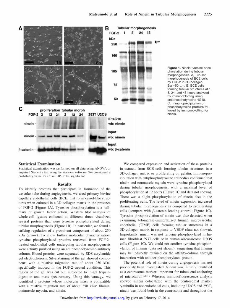
ResultsTo identify proteins that participate in formation of thevascular tube during angiogenesis, we used primary bovinecapillary endothelial cells (BCE) that form vessel-like struc-tures when cultured in a 3D-collagen matrix in the presenceof FGF-2 (Figure 1A). Tyrosine phosphorylation is a hall-mark of growth factor action. Western blot analysis ofwhole-cell lysates collected at different times visualizedseveral proteins that were tyrosine phosphorylated duringtubular morphogenesis (Figure 1B). In particular, we found astriking regulation of a prominent component of about 250kDa (arrow). To allow further molecular characterization,tyrosine phosphorylated proteins retrieved from FGF-2–treated endothelial cells undergoing tubular morphogenesiswere affinity purified using an antiphosphotyrosine-antibodycolumn. Eluted proteins were separated by SDS-acrylamidegel electrophoresis. Silverstaining of the gel showed compo-nents with a relative migration rate of about 250 kDa,specifically induced in the FGF-2–treated condition. Thisregion of the gel was cut out, subjected to in-gel trypsin-digestion and mass spectrometry. Using this strategy, weidentified 3 proteins whose molecular mass is compatiblewith a relative migration rate of about 250 kDa: filamin,nonmuscle myosin, and ninein.
We compared expression and activation of these proteinsin extracts from BCE cells forming tubular structures in a3D-collagen matrix or proliferating on gelatin. Immunopre-cipitation with antiphosphotyrosine antibodies confirmed thatninein and nonmuscle myosin were tyrosine phosphorylatedduring tubular morphogenesis, with a maximal level ofphosphorylation at 12 hours (Figure 1C and data not shown).There was a slight phosphorylation of ninein also in theproliferating cells. The level of ninein expression increasedduring tubular morphogenesis as compared to proliferatingcells (compare with �-catenin loading control; Figure 1C).Tyrosine phosphorylation of ninein was also detected whenexamining telomerase-immortalized human microvascularendothelial (TIME) cells forming tubular structures in a3D-collagen matrix in response to VEGF (data not shown).Importantly, ninein was not tyrosine phosphorylated in hu-man fibroblast 293T cells or in human osteosarcoma U2OScells (Figure 1C). We could not confirm tyrosine phosphor-ylation of filamin (data not shown), suggesting that filaminmay be indirectly retained on the affinity-column throughinteraction with another phosphorylated protein.
The potential role of ninein during angiogenesis has notpreviously been investigated. Ninein was initially identifiedas a centrosome-marker, important for minus-end anchoringof microtubuli.6,8,16 Whereas immunofluorescence analysisshowed ninein colocalized with the centrosomal marker�-tubulin in nonendothelial cells, including U2OS and 293T,ninein was found both in the centrosome and throughout the
Figure 1. Ninein tyrosine phos-phorylation during tubularmorphogenesis. A, Tubularmorphogenesis of BCE cellsby FGF-2 in 3D-collagen.Bar�50 �m. B, BCE cellsforming tubular structures at 1,8, 24, and 48 hours analyzedby immunoblotting usingantiphosphotyrosine 4G10.C, Immunoprecipitation ofphosphotyrosine-proteins fol-lowed by immunoblotting forninein.
Matsumoto et al Role of Ninein in Tubular Morphogenesis 2125
by guest on February 17, 2014http://atvb.ahajournals.org/Downloaded from
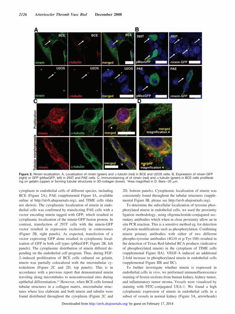
cytoplasm in endothelial cells of different species, includingBCE (Figure 2A), PAE (supplemental Figure IA, availableonline at http://atvb.ahajournals.org), and TIME cells (datanot shown). The cytoplasmic localization of ninein in endo-thelial cells was confirmed by transfecting PAE cells with avector encoding ninein tagged with GFP, which resulted incytoplasmic localization of the ninein-GFP fusion protein. Incontrast, transfection of 293T cells with the ninein-GFPvector resulted in expression exclusively in centrosomes(Figure 2B, right panels). As expected, transfection of avector expressing GFP alone resulted in cytoplasmic local-ization of GFP in both cell types (pMaxGFP; Figure 2B, leftpanels). The cytoplasmic distribution of ninein differed de-pending on the endothelial cell program. Thus, during FGF-2–induced proliferation of BCE cells cultured on gelatin,ninein was partially colocalized with the microtubular cy-toskeleton (Figure 2C and 2D, top panels). This is inaccordance with a previous report that demonstrated nineintraveling along microtubules to noncentrosomal sites duringepithelial differentiation.17 However, when BCE cells formedtubular structures in a collagen matrix, microtubular struc-tures where less elaborate and both ninein and tubulin werefound distributed throughout the cytoplasm (Figure 2C and
2D, bottom panels). Cytoplasmic localization of ninein wasconsistently found throughout the tubular structures (supple-mental Figure IB, please see http://atvb.ahajournals.org).
To determine the subcellular localization of tyrosine phos-phorylated ninein in endothelial cells, we used the proximityligation methodology, using oligonucleotide-conjugated sec-ondary antibodies which when in close proximity allow an insitu PCR reaction. This is a sensitive method eg, for detectionof protein modifications such as phosphorylation. Combiningninein primary antibodies with either of two differentphospho-tyrosine antibodies (4G10 or p-Tyr-100) resulted inthe detection of Texas Red-labeled RCA products (indicativeof phosphorylated ninein) in the cytoplasm of TIME cells(supplemental Figure IIA). VEGF-A induced an additional2-fold increase in phosphorylated ninein in endothelial cells(supplemental Figure IIB and IIC).
To further investigate whether ninein is expressed inendothelial cells in vivo, we performed immunofluorescencestaining of frozen sections from human kidney, kidney tumor,and inflammatory tumor stroma. Vessels were visualized bystaining with FITC-conjugated UEA-1. We found a highcytoplasmic expression of ninein in endothelial cells in asubset of vessels in normal kidney (Figure 3A, arrowheads)
Figure 2. Ninein localization. A, Localization of ninein (green) and �-tubulin (red) in BCE and U2OS cells. B, Expression of ninein-GFP(right) or GFP (pMaxGFP, left) in 293T and PAE cells. C, Immunostaining of of ninein (red) and �-tubulin (green) in BCE cells proliferat-ing on gelatin (upper) or forming tubular structures in 3D-collagen (lower). *Area magnified in D. Bars�20 �m.
2126 Arterioscler Thromb Vasc Biol December 2008
by guest on February 17, 2014http://atvb.ahajournals.org/Downloaded from
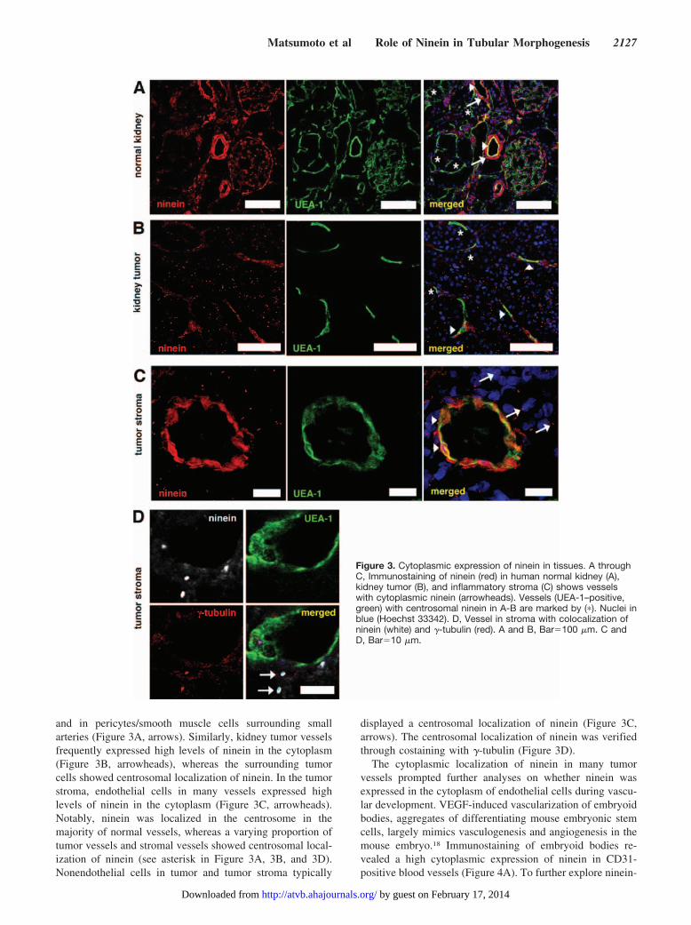
and in pericytes/smooth muscle cells surrounding smallarteries (Figure 3A, arrows). Similarly, kidney tumor vesselsfrequently expressed high levels of ninein in the cytoplasm(Figure 3B, arrowheads), whereas the surrounding tumorcells showed centrosomal localization of ninein. In the tumorstroma, endothelial cells in many vessels expressed highlevels of ninein in the cytoplasm (Figure 3C, arrowheads).Notably, ninein was localized in the centrosome in themajority of normal vessels, whereas a varying proportion oftumor vessels and stromal vessels showed centrosomal local-ization of ninein (see asterisk in Figure 3A, 3B, and 3D).Nonendothelial cells in tumor and tumor stroma typically
displayed a centrosomal localization of ninein (Figure 3C,arrows). The centrosomal localization of ninein was verifiedthrough costaining with �-tubulin (Figure 3D).
The cytoplasmic localization of ninein in many tumorvessels prompted further analyses on whether ninein wasexpressed in the cytoplasm of endothelial cells during vascu-lar development. VEGF-induced vascularization of embryoidbodies, aggregates of differentiating mouse embryonic stemcells, largely mimics vasculogenesis and angiogenesis in themouse embryo.18 Immunostaining of embryoid bodies re-vealed a high cytoplasmic expression of ninein in CD31-positive blood vessels (Figure 4A). To further explore ninein-
Figure 3. Cytoplasmic expression of ninein in tissues. A throughC, Immunostaining of ninein (red) in human normal kidney (A),kidney tumor (B), and inflammatory stroma (C) shows vesselswith cytoplasmic ninein (arrowheads). Vessels (UEA-1–positive,green) with centrosomal ninein in A-B are marked by (�). Nuclei inblue (Hoechst 33342). D, Vessel in stroma with colocalization ofninein (white) and �-tubulin (red). A and B, Bar�100 �m. C andD, Bar�10 �m.
Matsumoto et al Role of Ninein in Tubular Morphogenesis 2127
by guest on February 17, 2014http://atvb.ahajournals.org/Downloaded from
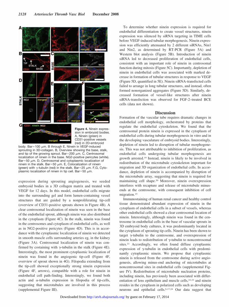
expression during sprouting angiogenesis, we seededembryoid bodies in a 3D collagen matrix and treated withVEGF for 12 days. In this model, endothelial cells migrateinto the surrounding gel and form lumen-containing vesselstructures that are guided by a nonproliferating tip-cell(overview of CD31-positive sprouts shown in Figure 4B). Aclear centrosomal localization of ninein was seen in the baseof the endothelial sprout, although ninein was also distributedin the cytoplasm (Figure 4C). In the stalk, ninein was foundin the centrosomes and cytoplasm of endothelial cells, as wellas in NG2-positive pericytes (Figure 4D). This is in accor-dance with the cytoplasmic localization of ninein we detectedin smooth muscle cells surrounding arteries eg, in the kidney(Figure 3A). Centrosomal localization of ninein was con-firmed by costaining with �-tubulin in the stalk (Figure 4E).Interestingly, the most prominent cytoplasmic localization ofninein was found in the angiogenic tip-cell (Figure 4F,overview of sprout shown in 4G). Filopodia extending fromthe tip-cell showed exceptionally strong ninein expression(Figure 4F, arrows), compatible with a role for ninein inendothelial cell path-finding. Interestingly, we found bothactin and �-tubulin expression in filopodia of tip-cells,suggesting that microtubules are involved in this process(supplemental Figure III).
To determine whether ninein expression is required forendothelial differentiation to create vessel structures, nineinexpression was silenced by siRNA targeting in TIME cellsbefore VEGF-induced tubular morphogenesis. Ninein expres-sion was efficiently attenuated by 2 different siRNAs, Nin1and Nin2, as determined by RT-PCR (Figure 5A) andWestern blot analysis (Figure 5B). Introduction of nineinsiRNA led to decreased proliferation of endothelial cells,consistent with an important role of ninein in centrosomalfunction during mitosis (Figure 5C). Importantly, depletion ofninein in endothelial cells was associated with marked de-crease in formation of tubular structures in response to VEGF(Figure 5D, quantified in 5E). Ninein siRNA-transfected cellsfailed to arrange in long tubular structures, and instead, oftenformed nonorganized aggregates (Figure 5D). Similarly, de-creased formation of vessel-like structures after nineinsiRNA-transfection was observed for FGF-2–treated BCEcells (data not shown).
DiscussionFormation of the vascular tube requires dramatic changes inendothelial cell morphology, orchestrated by proteins thatregulate the endothelial cytoskeleton. We found that thecentrosomal protein ninein is expressed in the cytoplasm ofendothelial cells during tubular morphogenesis in vitro and inthe developing vasculature of embryoid bodies. Interestingly,depletion of ninein led to disruption of tubular morphogene-sis. This was not attributable to inhibition of proliferation, asendothelial cells undergoing tubular morphogenesis aregrowth arrested.19 Instead, ninein is likely to be involved inredistribution of the microtubule cytoskeleton important formigration and 3D organization of endothelial cells. In accor-dance, depletion of ninein is accompanied by disruption ofthe microtubule array, suggesting that ninein is required formaintaining cell shape.20 Moreover, ninein overexpressioninterferes with recapture and release of microtubule minus-ends at the centrosome, with consequent inhibition of cellmigration.15
Immunostaining of human renal cancer and healthy controltissue demonstrated abundant expression of ninein in thecytoplasm of endothelial cells in a subset of vessels, whereasother endothelial cells showed a clear centrosomal location ofninein. Interestingly, although ninein was found in the cen-trosome in endothelial cells in the base of vascular sprouts in3D embryoid body cultures, it was predominantly located inthe cytoplasm of sprouting tip cells. Ninein has been shown totarget �-tubulin to the centrosome, and overexpression ofninein leads to redistribution of �-tubulin to noncentrosomalsites.21 Accordingly, we often found diffuse cytoplasmicexpression of �-tubulin in endothelial cells with predomi-nantly cytoplasmic ninein. We propose that cytoplasmicninein is released from the centrosome during active angio-genesis, allowing minus-end anchoring of microtubules atnoncentrosomal sites in endothelial cells (supplemental Fig-ure IV). Redistribution of microtubule nucleation proteins,including ninein, has previously been associated with differ-entiation of lens epithelium and muscle cells,22,23 and nineinresides in the cytoplasm in polarized cells such as developingneurons and epithelial cells.6,17,24 Our data suggest that
Figure 4. Ninein expres-sion in embryoid bodies.A, Ninein (green) inCD31-positive vessels(red) in 2D-embryoid
body. Bar�100 �m. B through E, Ninein in VEGF-inducedsprouting in 3D-collagen. B, Overview showing the base, stalk,and tip of the growing sprout. Bar�200 �m. C, Centrosomallocalization of ninein in the base. NG2-positive pericytes (white).Bar�50 �m. D, Centrosomal and cytoplasmic localization ofninein in the stalk. Bar�50 �m. E, Colocalization of ninein(green) with �-tubulin (red) in the stalk. Bar�25 �m. F,G, Cyto-plasmic localization of ninein in tip cell. Bar�50 �m.
2128 Arterioscler Thromb Vasc Biol December 2008
by guest on February 17, 2014http://atvb.ahajournals.org/Downloaded from

cytoplasmic ninein is instrumental in microtubule reorgani-zation required for adaptation of endothelial cell morphologyduring angiogenesis. The extent of colocalization of nineinwith �-tubulin or microtubules was not always prominent,however, and we cannot formally exclude other functions forninein in endothelial cells.
Microtubule-targeted drugs (MTD)s are widely used inanticancer treatment and have antimitotic and antiangiogenicproperties.25 It has been shown that MTDs used as vasculardisrupting agents specifically target tumor vasculature, sug-gesting a specific architecture of these vessels. Our resultsshowing a cytoplasmic localization of ninein in many tumorvessels, may indeed reflect changes in patterning and/orstability of the microtubule network. Microtubule dynamicsare modified by many different microtubule-interacting pro-teins, which are regulated by kinases and phosphatasesdownstream eg, growth factor signaling. Ninein interacts withGSK3�,26 and it has been shown that the coiled-coiled IIdomain can be phosphorylated by AuroraA and PKA.27 Inaddition, ninein can be modified by SUMOylation, resultingin translocation from the centrosome to the nucleus.28 Wefound that cytoplasmic ninein is tyrosine phosphorylatedduring tubular morphogenesis of endothelial cells. Of note,tyrosine phosphorylated ninein could not be detected in 293T
or U2OS cells, where it has a clear centrosomal localization.There are 20 tyrosine residues in human ninein. According tothe NetPhos phosphorylation site prediction bioinformatictool (http://www.cbs.dtu.dk/services/NetPhos/),29 10 of theseare putative tyrosine phosphorylation sites. Future goalsinclude to identify which tyrosine phosporylation site(s) inninein that are regulated during angiogenesis and to furtherinvestigate the function of ninein tyrosine phosphorylation inendothelial cells.
Sources of FundingThis study was supported by the Association for International CancerResearch (AICR Grant 04-069), the Astrid Karlsson foundation, theGustaf Adolf Johansson foundation, Svenska Sallskapet for Medi-cinsk Forskning, the Magnus Bergvall foundation, Nihon UniversityMultidisciplinary Research Grant for 2008 (08-020), Swedish Re-search Council (project no. K2005-32X-12552-08A), and the Swed-ish Cancer Society (project 4828-B03-01PAA and 3820-B05-10XBC). The Fresh Tissue Biobank at Uppsala University Hospitalwas supported by the National Biobank Platform funded by Wallen-berg Consortium North and Swegene.
DisclosuresNone.
Figure 5. Ninein silencinginhibits VEGF-induced tubularmorphogenesis. A and B,Ninein mRNA (A) and protein(B) in TIME cells treated withNin1, Nin2, or control siRNAs.C, Cell numbers of siRNA-treated cells at seeding andafter 48 hours. Mean�SD oftriplicates. D, Tubular struc-tures formed by control andninein-siRNA–treated TIMEcells (upper). Confocal imagesof phalloidin-Texas Red tubularstructures (lower). Bar�50 �m.E, Quantification of tubulararea (mean�SD, n�4,*P�0.05).
Matsumoto et al Role of Ninein in Tubular Morphogenesis 2129
by guest on February 17, 2014http://atvb.ahajournals.org/Downloaded from
References1. Olsson AK, Dimberg A, Kreuger J, Claesson-Welsh L. VEGF receptor
signalling - in control of vascular function. Nat Rev Mol Cell Biol. 2006;7:359–371.
2. Auguste P, Javerzat S, Bikfalvi A. Regulation of vascular development byfibroblast growth factors. Cell Tissue Res. 2003;314:157–166.
3. Ribatti D, Vacca A, Nico B, Roncali L, Dammacco F. Postnatal vascu-logenesis. Mech Dev. 2001;100:157–163.
4. Papetti M, Herman IM. Mechanisms of normal and tumor-derived angio-genesis. Am J Physiol Cell Physiol. 2002;282:C947–C970.
5. Bertolini F, Shaked Y, Mancuso P, Kerbel RS. The multifaceted circu-lating endothelial cell in cancer: towards marker and target identification.Nat Rev Cancer. 2006;6:835–845.
6. Bouckson-Castaing V, Moudjou M, Ferguson DJ, Mucklow S, Belkaid Y,Milon G, Crocker PR. Molecular characterisation of ninein, a newcoiled-coil protein of the centrosome. J Cell Sci. 1996;109(Pt 1):179–190.
7. Delgehyr N, Sillibourne J, Bornens M. Microtubule nucleation andanchoring at the centrosome are independent processes linked by nineinfunction. J Cell Sci. 2005;118:1565–1575.
8. Mogensen MM, Malik A, Piel M, Bouckson-Castaing V, Bornens M.Microtubule minus-end anchorage at centrosomal and non-centrosomalsites: the role of ninein. J Cell Sci. 2000;113:3013–3023.
9. Dimberg A, Rylova S, Dieterich LC, Olsson AK, Schiller P, Wikner C,Bohman S, Botling J, Lukinius A, Wawrousek EF, Claesson-Welsh L.{alpha}B-crystallin promotes tumor angiogenesis by increasing vascularsurvival during tube morphogenesis. Blood. 2008;111:2015–2023.
10. Venetsanakos E, Mirza A, Fanton C, Romanov SR, Tlsty T, McMahonM. Induction of tubulogenesis in telomerase-immortalized human micro-vascular endothelial cells by glioblastoma cells. Exp Cell Res. 2002;273:21–33.
11. Bohman S, Matsumoto T, Suh K, Dimberg A, Jakobsson L, Yuspa S,Claesson-Welsh L. Proteomic analysis of vascular endothelial growthfactor-induced endothelial cell differentiation reveals a role for chlorideintracellular channel 4 (CLIC4) in tubular morphogenesis. J Biol Chem.2005;280:42397–42404.
12. Nagy A, Rossant J, Nagy R, Abramow-Newerly W, Roder JC. Derivationof completely cell culture-derived mice from early-passage embryonicstem cells. Proc Natl Acad Sci U S A. 1993;90:8424–8428.
13. Magnusson P, Rolny C, Jakobsson L, Wikner C, Wu Y, Hicklin DJ,Claesson-Welsh L. Deregulation of Flk-1/vascular endothelial growthfactor receptor-2 in fibroblast growth factor receptor-1-deficient vascularstem cell development. J Cell Sci. 2004;117:1513–1523.
14. Mack GJ, Rees J, Sandblom O, Balczon R, Fritzler MJ, Rattner JB.Autoantibodies to a group of centrosomal proteins in human autoimmunesera reactive with the centrosome. Arthritis Rheum. 1998;41:551–558.
15. Abal M, Piel M, Bouckson-Castaing V, Mogensen M, Sibarita JB,Bornens M. Microtubule release from the centrosome in migrating cells.J Cell Biol. 2002;159:731–737.
16. Mogensen MM. Microtubule release and capture in epithelial cells. BiolCell. 1999;91:331–341.
17. Moss DK, Bellett G, Carter JM, Liovic M, Keynton J, Prescott AR, LaneEB, Mogensen MM. Ninein is released from the centrosome and movesbi-directionally along microtubules. J Cell Sci. 2007;120:3064–3074.
18. Jakobsson L, Kreuger J, Claesson-Welsh L. Building blood vessels–stemcell models in vascular biology. J Cell Biol. 2007;177:751–755.
19. Matsumoto T, Turesson I, Book M, Gerwins P, Claesson-Welsh L. p38MAP kinase negatively regulates endothelial cell survival, proliferation,and differentiation in FGF-2-stimulated angiogenesis. J Cell Biol. 2002;156:149–160.
20. Dammermann A, Merdes A. Assembly of centrosomal proteins andmicrotubule organization depends on PCM-1. J Cell Biol. 2002;159:255–266.
21. Stillwell EE, Zhou J, Joshi HC. Human ninein is a centrosomalautoantigen recognized by CREST patient sera and plays a regulatory rolein microtubule nucleation. Cell Cycle. 2004;3:923–930.
22. Dahm R, Procter JE, Ireland ME, Lo WK, Mogensen MM, Quinlan RA,Prescott AR. Reorganization of centrosomal marker proteins coincideswith epithelial cell differentiation in the vertebrate lens. Exp Eye Res.2007;85:696–713.
23. Bugnard E, Zaal KJ, Ralston E. Reorganization of microtubule nucleationduring muscle differentiation. Cell Motil Cytoskeleton. 2005;60:1–13.
24. Baird DH, Myers KA, Mogensen M, Moss D, Baas PW. Distributionof the microtubule-related protein ninein in developing neurons.Neuropharmacology. 2004;47:677– 683.
25. Jordan MA, Wilson L. Microtubules as a target for anticancer drugs. NatRev Cancer. 2004;4:253–265.
26. Hong YR, Chen CH, Chang JH, Wang S, Sy WD, Chou CK, Howng SL.Cloning and characterization of a novel human ninein protein thatinteracts with the glycogen synthase kinase 3beta. Biochim Biophys Acta.2000;1492:513–516.
27. Chen CH, Howng SL, Cheng TS, Chou MH, Huang CY, Hong YR.Molecular characterization of human ninein protein: two distinct sub-domains required for centrosomal targeting and regulating signals in cellcycle. Biochem Biophys Res Commun. 2003;308:975–983.
28. Cheng TS, Chang LK, Howng SL, Lu PJ, Lee CI, Hong YR. SUMO-1modification of centrosomal protein hNinein promotes hNinein nuclearlocalization. Life Sci. 2006;78:1114–1120.
29. Blom N, Gammeltoft S, Brunak S. Sequence and structure-based pre-diction of eukaryotic protein phosphorylation sites. J Mol Biol. 1999;294:1351–1362.
2130 Arterioscler Thromb Vasc Biol December 2008
by guest on February 17, 2014http://atvb.ahajournals.org/Downloaded from