Prolonged hypoxic culture and trypsinization increase the pro-angiogenic potential of human adipose...
12
Correspondence: Jeppe Grøndahl Rasmussen, Department of Pharmacology, Aarhus University, Wilhelm Meyers Allè 4, 8000 Aarhus C, Denmark. E-mail: [email protected] (Received 25 March 2010; accepted 29 June 2010) Prolonged hypoxic culture and trypsinization increase the pro-angiogenic potential of human adipose tissue-derived stem cells JEPPE GRØNDAHL RASMUSSEN 1,2 , OLE FRØBERT 3 , LINDA PILGAARD 2,4 , JENS KASTRUP 5 , ULF SIMONSEN 1 , VLADIMIR ZACHAR 2 & TRINE FINK 2 1 Department of Pharmacology, Aarhus University, Aarhus, Denmark, 2 Laboratory for Stem Cell Research, Aalborg University, Aalborg, Denmark, 3 Department of Cardiology, Örebro University Hospital, Örebro, Sweden, 4 Aalborg Hospital Science and Innovation Center, Aalborg, Denmark and Department of Haematology, Aarhus University Hospital, Aarhus, Denmark, and 5 Cardiac Stem Cell Laboratory, The Heart Centre, Rigshospitalet, Copenhagen University Hospital, Copenhagen, Denmark Abstract Background aims. Transplantation of mesenchymal stromal cells (MSC), including adipose tissue-derived stem cells (ASC), is a promising option in the treatment of vascular disease. Short-term hypoxic culture of MSC augments secretion of anti-apoptotic and angiogenic cytokines. We hypothesized that prolonged hypoxic (1% and 5% oxygen) culture and trypsinization would augment ASC expression of anti-apoptotic and angiogenic cytokines and increase the angiogenic potential of ASC-conditioned media. Methods. The effects of prolonged hypoxic culture on growth and pro-angiogenic properties were investigated using human ASC cultured at 1%, 5% and 21% oxygen. The effect of trypsinization on the expression of pro-angiogenic genes was also determined. Results. Trypsinization induced up-regulation of the vascular endothelial growth factor (VEGF) and insulin-like growth factor-1 (IGF-1) genes independent of oxygen concentration. The expression of VEGF and IGF-1 was up-regulated in ASC cultured at 1% oxygen for 13 days compared with 4 days. The VEGF concentration in ASC-conditioned media was higher after prolonged hypoxic culture compared with short- term culture, while the IGF-1 and chemokine (CXC motif) ligand 12 (CXCL12) concentrations were unchanged. The VEGF receptor blocker SU5416 abolished angiogenesis in a cultured rat aortic ring model. Media from cells exposed to hypoxia increased angiogenesis, an effect that was dependent on factors other than just the VEGF concentration in the added media. Conclusions. Optimization of the angiogenic potential of stem cell-based therapy in the treatment of vascular disease is important. We have demonstrated that prolonged hypoxic culture and trypsinization augment the therapeutic angiogenic potential of ASC. Key Words: adipose, angiogenesis, hypoxia, ischemia, mesenchymal stromal cells Introduction Cardiovascular diseases are a major cause of mor- tality and morbidity world-wide. In animal models, transplantation of mesenchymal stromal cells (MSC) shows promising results in reducing the devastating effects of myocardial infarction (1–3) and limb isch- emia (4,5), primarily through secretion of cytokines exerting angiogenic (6–8) and anti-apoptotic (9–11) effects in the ischemic tissue. In particular, vascu- lar endothelial growth factor (VEGF), insulin-like growth factor-1 (IGF-1) and secreted frizzled-related protein-2 (Sfrp2) are reported to counteract apop- tosis and promote angiogenesis (6–11). The long- term retention of transplanted cells is poor (12,13) possibly because of the ischemia in the recipient tissue; short-term (24–72 h) hypoxic culture of MSC increases VEGF gene expression and secretion (14–16), improves survival of the grafted cells (17) and increases the proliferation rate (18–20). Another key player in the retention and homing of progeni- tor and stem cells is the chemokine stromal cell- derived factor-1/chemokine (CXC motif) ligand 12 (SDF-1/CXCL12) and its receptor CXC chemokine receptor-4 (CXCR4) (21,22), with CXCL12 having chemotractive effects on cells expressing CXCR4. In addition, CXCL12 seems to exert protective effects against ischemia (23). The protease-activated receptor-2 agonist, trypsin, induces an up-regulation of VEGF at both the Cytotherapy, 2011; 13: 318–328 ISSN 1465-3249 print/ISSN 1477-2566 online © 2011 Informa Healthcare DOI: 10.3109/14653249.2010.506505
Transcript of Prolonged hypoxic culture and trypsinization increase the pro-angiogenic potential of human adipose...

Correspondence: Jeppe Gr ø ndahl Rasmussen , Department of Pharmacology, Aarhus University, Wilhelm Meyers All è 4, 8000 Aarhus C, Denmark. E-mail: [email protected]
(Received 25 March 2010; accepted 29 June 2010)
Prolonged hypoxic culture and trypsinization increase the pro-angiogenic potential of human adipose tissue-derived stem cells
JEPPE GR Ø NDAHL RASMUSSEN 1,2 , OLE FR Ø BERT 3 , LINDA PILGAARD 2,4 , JENS KASTRUP 5 , ULF SIMONSEN 1 , VLADIMIR ZACHAR 2 & TRINE FINK 2
1 Department of Pharmacology, Aarhus University, Aarhus, Denmark, 2 Laboratory for Stem Cell Research, Aalborg University, Aalborg, Denmark, 3 Department of Cardiology, Ö rebro University Hospital, Ö rebro, Sweden, 4 Aalborg Hospital Science and Innovation Center, Aalborg, Denmark and Department of Haematology, Aarhus University Hospital, Aarhus, Denmark, and 5 Cardiac Stem Cell Laboratory, The Heart Centre, Rigshospitalet, Copenhagen University Hospital, Copenhagen, Denmark
Abstract Background aims. Transplantation of mesenchymal stromal cells (MSC), including adipose tissue-derived stem cells (ASC), is a promising option in the treatment of vascular disease. Short-term hypoxic culture of MSC augments secretion of anti-apoptotic and angiogenic cytokines. We hypothesized that prolonged hypoxic (1% and 5% oxygen) culture and trypsinization would augment ASC expression of anti-apoptotic and angiogenic cytokines and increase the angiogenic potential of ASC-conditioned media. Methods. The effects of prolonged hypoxic culture on growth and pro-angiogenic properties were investigated using human ASC cultured at 1%, 5% and 21% oxygen. The effect of trypsinization on the expression of pro-angiogenic genes was also determined. Results. Trypsinization induced up-regulation of the vascular endothelial growth factor (VEGF) and insulin-like growth factor-1 (IGF-1) genes independent of oxygen concentration. The expression of VEGF and IGF-1 was up-regulated in ASC cultured at 1% oxygen for 13 days compared with 4 days. The VEGF concentration in ASC-conditioned media was higher after prolonged hypoxic culture compared with short-term culture, while the IGF-1 and chemokine (CXC motif) ligand 12 (CXCL12) concentrations were unchanged. The VEGF receptor blocker SU5416 abolished angiogenesis in a cultured rat aortic ring model. Media from cells exposed to hypoxia increased angiogenesis, an effect that was dependent on factors other than just the VEGF concentration in the added media. Conclusions. Optimization of the angiogenic potential of stem cell-based therapy in the treatment of vascular disease is important. We have demonstrated that prolonged hypoxic culture and trypsinization augment the therapeutic angiogenic potential of ASC.
Key Words: adipose , angiogenesis , hypoxia , ischemia , mesenchymal stromal cells
Introduction
Cardiovascular diseases are a major cause of mor-tality and morbidity world-wide. In animal models, transplantation of mesenchymal stromal cells (MSC) shows promising results in reducing the devastating effects of myocardial infarction (1 – 3) and limb isch-emia (4,5), primarily through secretion of cytokines exerting angiogenic (6 – 8) and anti-apoptotic (9 – 11) effects in the ischemic tissue. In particular, vascu-lar endothelial growth factor (VEGF), insulin-like growth factor-1 (IGF-1) and secreted frizzled-related protein-2 (Sfrp2) are reported to counteract apop-tosis and promote angiogenesis (6 – 11). The long-term retention of transplanted cells is poor (12,13)
possibly because of the ischemia in the recipient tissue; short-term (24 – 72 h) hypoxic culture of MSC increases VEGF gene expression and secretion (14 – 16), improves survival of the grafted cells (17) and increases the proliferation rate (18 – 20). Another key player in the retention and homing of progeni-tor and stem cells is the chemokine stromal cell-derived factor-1/chemokine (CXC motif) ligand 12 (SDF-1/CXCL12) and its receptor CXC chemokine receptor-4 (CXCR4) (21,22), with CXCL12 having chemotractive effects on cells expressing CXCR4. In addition, CXCL12 seems to exert protective effects against ischemia (23).
The protease-activated receptor-2 agonist, trypsin, induces an up-regulation of VEGF at both the
Cytotherapy, 2011; 13: 318–328
ISSN 1465-3249 print/ISSN 1477-2566 online © 2011 Informa HealthcareDOI: 10.3109/14653249.2010.506505

Adipose stem cells, trypsin and hypoxia 319
transcriptional and translational levels in cancer cell lines. Because trypsinization is often used to detach adherent cells from growth surfaces, it is important to know whether this procedure might infl uence their expression of angiogenic and anti-apoptotic cytokine genes.
Adipose tissue-derived stem cells (ASC) are a new source of MSC, which can be harvested in larger numbers with less patient discomfort than bone marrow-derived stem cells (BMSC). In addi-tion, ASC produce angiogenic and anti-apoptotic cytokines such as VEGF and IGF-1 (11,24).
We hypothesized that prolonged hypoxic (1% and 5%) oxygen culture would augment ASC expres-sion of anti-apoptotic and angiogenic cytokines and increase the angiogenic potential of ASC-conditioned media. To address the hypothesis we investigated the effects of long-term hypoxic culture on proliferation and expression of VEGF, IGF-1 and the homing fac-tor CXCL12 at both the transcriptional and trans-lational levels in human ASC. Angiogenic capacity was evaluated using ASC-conditioned media in a rat aortic ring model. Recently, the protease-activated receptor-2 agonist trypsin was found to induce an up-regulation of VEGF at both the transcriptional and translational levels in cancer cell lines (25). There-fore, we also investigated the effects of using trypsin to detach adherent ASC, regarding ASC expression of angiogenic and anti-apoptotic genes.
Methods
Isolation and culture of adipose tissue-derived stem cells
All patients gave written informed consent and the protocol was approved by the regional committee on biomedical research ethics of Northern Jutland, Denmark. The three patients were two females and one male, aged 42, 58 and 52 years, respectively. The patients were not receiving any medication and had no cardiovascular disease. ASC were isolated from subcutaneous adipose tissue obtained from three patients undergoing elective liposuction, as described previously (26), using minor modifi ca-tions. Briefl y, the adipose tissue was washed repeat-edly with phosphate-buffered saline (PBS; Invitrogen, Taastrup, Denmark) and then for 60 min at 37 ° C dissociated enzymatically with 65 U/mL crude col-lagenase (Wako, Richmond, VA, USA) in PBS sup-plemented with 20 mg/mL bovine serum albumin (Europa Bioproducts Ltd, Cambridge, UK). The stromal vascular fraction within the dissociated tis-sue was pelleted by centrifugation at 400 g for 10 min. The pellet was resuspended in distilled water for 10 s to lyse erythrocytes, and immediately there-after the salt balance and pH were adjusted back
to physiologic levels. The suspension was fi ltered through a 70- μ m fi lter and incubated overnight in control medium [Dulbecco ’ s modifi ed Eagle medium (DMEM)/F12], 10% fetal bovine serum (FBS), 100 IU/mL penicillin, 0.1 mg/mL streptomycin and 0.05 mg/mL gentamicin (all from Invitrogen) at 37 ° C in an atmosphere containing 5% CO 2 . After 24 h the non-adherent mononuclear cells were removed. For the remainder of the experiments, cells were cultured in α-Minimum Essential Medium ( α -MEM) (Invitro-gen) with Glutamax, 10% FBS, 100 IU/mL penicillin, 0.1 mg/mL streptomycin and 0.05 mg/mL gentami-cin. We have demonstrated previously the multilin-eage potential of these cells (27,28). All experiments were performed with cells at passage 3 using cells from three different patients. When passaging cells, a mix-ture of 0.125% trypsin and 0.01% ethylene diamine tetra acetic acid (EDTA) was used. Hypoxic culture experiments were performed in a hypoxic worksta-tion (Xvivo; BioSpherix, Redfi eld, NY, USA) at 37 ° C in an atmosphere containing 5% CO 2 balanced with nitrogen to reach oxygen concentrations of 1% and 5%. Normoxic culture was performed in a standard incubator in an atmosphere containing 5% CO 2 .
Proliferation assay
To determine the effect of hypoxia on cell prolifera-tion, the ASC were seeded at a density of 300/cm 2 in 48-well plates (Costar, Acton, MA, USA) and cul-tured at 1%, 5% or 21% oxygen. On days 0, 2, 4, 5, 6, 7, 8, 9, 10, 11, 12 and 13, the cells were lysed in 200 μ L 0.02% sodium dodecyl sulphate (SDS) in DNase-free water, after which the cell number in the samples was determined using a PicoGreen dsDNA quantitation kit (Invitrogen). Briefl y, aliquots of 100 μ L cell lysate were mixed with 100 μ L PicoGreen diluted 1:200 in 1 � Tris EDTA (TE) buffer and placed in a black microtiter plate. The microtiter plate was incubated at room temperature under gentle agitation for 10 min, protected from light. Fluo-rescence was measured using a Wallac 1420 Victor multilabel counter (PerkinElmer, Hvidovre, Denmark) with excitation at 485 nm and emission at 535 nm. A standard curve for measuring DNA concentra-tions was made based on known Lambda DNA (CyQuant; Molecular Probes, Eugene, OR, USA) concentrations. The number of cells was calculated using a theoretical value of 6.6 pg DNA/cell. The experiment was performed twice as two independent experiments, each in quadruplicate.
Quantifi cation of apoptotic and dead cells
The relative number of apoptotic ASC in culture was calculated after Annexin V binding. Briefl y, ASC

320 J. G. Rasmussen et al.
were cultured as for the proliferation experiment and, after 13 days, the media were removed and the cells incubated with Alexa 488-conjugated Annexin V (Molecular Probes), according to the manufacturer ’ s guidelines, in order to label apoptotic cells. Ethidium homodimer-2 was used to counterstain dead cells and Hoechst 33342 (both from Molecular Probes) was used to counterstain cell nuclei. The experiment was conducted in duplicates.
Cytokine gene expression during hypoxic culture
In order to determine the effect of hypoxic culture on the gene expression of VEGF, IGF-1, Sfrp2 and CXCL12, ASC were seeded in 12-well plates (Costar), with 10 000 cells/cm 2 for day 0 and day 4 harvest, 3000 cells/cm 2 for day 7 harvest and 300 cells/cm 2 for day 10 and day 13 harvest. The cells were cultured at 1%, 5% and 21% oxygen and harvested for RNA isolation on days 0, 4, 7, 10 and 13. Isolation of RNA and quantitative real-time reverse transcription (RT)-polymerase chain reaction (PCR) procedures were performed as described below. The experiment was conducted twice as two independent experiments.
Trypsinization response analysis
In order to distinguish between the effect of trypsini-zation and the effect of hypoxia on gene expression, ASC were plated in six 12-well plates at a density of 10 000/cm 2 , placing half of the plates at the three oxygen concentrations (1%, 5% and 21%) immedi-ately and the other half after 72 h, harvesting cells for quantitative real-time RT-PCR after another 24 h of incubation at the respective oxygen concentra-tions. The expression of VEGF, IGF-1, Sfrp2 and CXCL12 genes was measured with RNA isolation and quantitative real-time RT-PCR procedures as described below. The experiment was conducted twice as two independent experiments.
Quantitative real-time RT-PCR
Total cellular RNA was isolated using an Aurum total RNA mini kit (Bio-Rad, Copenhagen, Denmark). The purity and concentration were determined by
spectrophotometry (Nanodrop; Thermo Science, Wilmington, DE, USA). RT to produce cDNA was performed with an iScript cDNA synthesis kit (Bio-Rad). All primers (Table I) were designed using the open-source software Primer3 and produced by DNA Technology (Aarhus, Denmark). Quantita-tive PCR were performed on a My-Cycler real-time PCR system (Bio-Rad) in a fi nal volume of 25 μ L containing 5 pmol of each of the two primers and 0.25 μ L cDNA using the SYBR Green PCR supermix (Bio-Rad). Thermal cycling parameters were initially 3 min at 95 ° C, followed by 40 cycles of 10 s at 95 ° C for DNA denaturation and 30 s of annealing and elongation at 60 ° C. After amplifi cation, a melt curve was performed to test product specifi city. A four-fold serially diluted standard curve derived from a pool of all the cDNA samples was used to calcu-late relative expression of each gene. For normaliza-tion, the geometric mean of the two reference genes cyclophilin A (PPIA) and tyrosine 3-/tryptophan 5-monooxygenase activation protein (YWHAZ) was used. Our group has previously shown that these genes are expressed relatively stably in human ASC during hypoxic exposure (27).
ELISA analyzes of conditioned media
ASC were seeded in six-well plates (Costar) with 10 000 cells/cm 2 for day 1 harvest and 300 cells/cm 2 for day 13 harvest and cultured at 1%, 5% and 21% oxy-gen. For both time-points, the media were changed 24 h prior to media harvest. At media harvest, the cell numbers were determined as described for the proliferation experiments. The content of VEGF, IGF-1 and CXCL12 in the conditioned media was measured using ELISA kits (R&D Systems Europe, Abingdon, UK). The experiments were conducted twice as two independent experiments.
Culture of rat aortic rings
Rat aortic rings were obtained by harvesting thoracic aortas from 8-month-old Sprague – Dawley rats. After cleaning away the periaortic fi broadipose tissue, the aortas were cross-sectioned into rings of 1 – 2 mm in length. The rings were taken through fi ve consecutive
Table I. Primer sequences for primers used in quantitative real-time RT-PCR analysis.
Gene Forward primer sequence Reverse primer sequence
PPIA 5 ′ TCC TGG CAT CTT GTC CAT G 3 ′ 5 ′ CCA TCC AAC CAC TCA GTC TTG 3 ′ YWHAZ 5 ′ ACT TTT GGT ACA TTG TGG CTT CAA 3 ′ 5 ′ CCG CCA GGA CAA ACC AGT AT 3 ′ VEGF 5 ′ CGA TTC AAG TGG GGA ATG G 3 ′ 5 ′ CAT TGA TCC GGG TTT TAT CC 3 ′ IGF-1 5 ′ GAT GGG GTC TCG CAC TGT CC 3 ′ 5 ′ GAG CCG AGA TCA TGC CAC TGC 3 ′ Sfrp2 5 ′ CAC CTG TGA GGA GAT GAA CG 3 ′ 5 ′ TGT CCC ATG ACC AGA TAG GG 3 ′ CXCL12/SDF-1 5 ′ TCT CAA CAC TCC AAA CTG TGC 3 ′ 5 ′ CTT TAG CTT CGG GTC AAT GC 3 ′
Adipose stem cells, trypsin and hypoxia 321
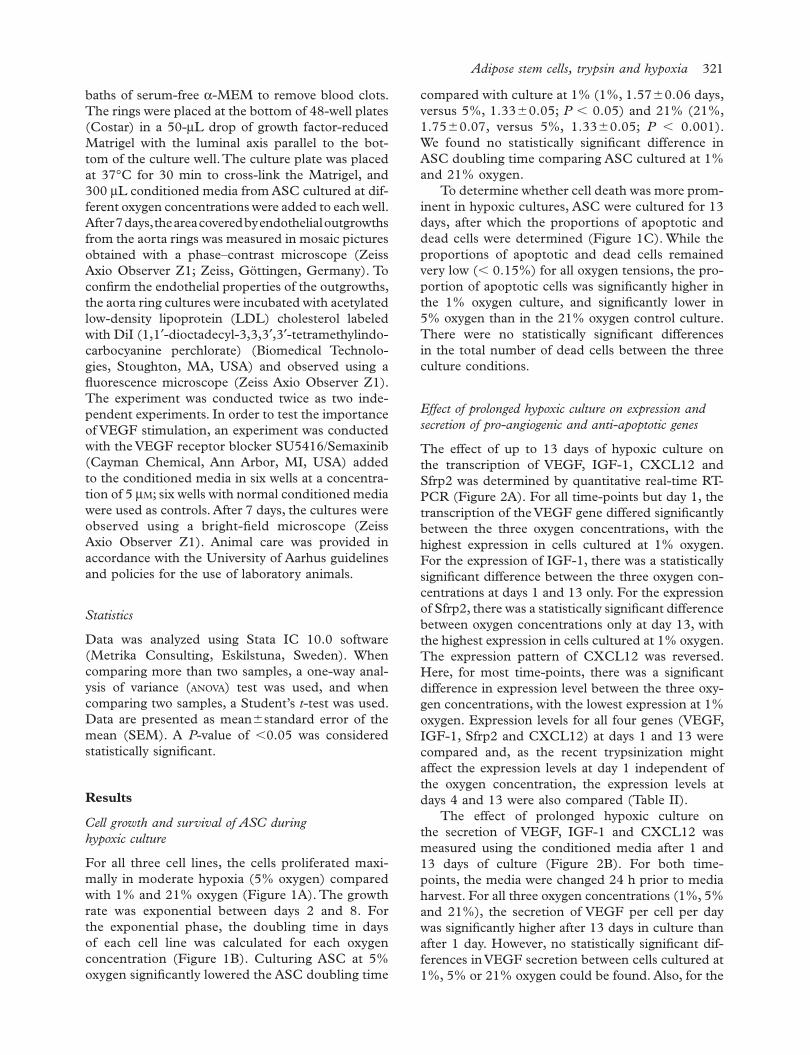
compared with culture at 1% (1%, 1.57 � 0.06 days, versus 5%, 1.33 � 0.05; P � 0.05) and 21% (21%, 1.75 � 0.07, versus 5%, 1.33 � 0.05; P � 0.001). We found no statistically signifi cant difference in ASC doubling time comparing ASC cultured at 1% and 21% oxygen.
To determine whether cell death was more prom-inent in hypoxic cultures, ASC were cultured for 13 days, after which the proportions of apoptotic and dead cells were determined (Figure 1C). While the proportions of apoptotic and dead cells remained very low ( � 0.15%) for all oxygen tensions, the pro-portion of apoptotic cells was signifi cantly higher in the 1% oxygen culture, and signifi cantly lower in 5% oxygen than in the 21% oxygen control culture. There were no statistically signifi cant differences in the total number of dead cells between the three culture conditions.
Effect of prolonged hypoxic culture on expression and secretion of pro-angiogenic and anti-apoptotic genes
The effect of up to 13 days of hypoxic culture on the transcription of VEGF, IGF-1, CXCL12 and Sfrp2 was determined by quantitative real-time RT-PCR (Figure 2A). For all time-points but day 1, the transcription of the VEGF gene differed signifi cantly between the three oxygen concentrations, with the highest expression in cells cultured at 1% oxygen. For the expression of IGF-1, there was a statistically signifi cant difference between the three oxygen con-centrations at days 1 and 13 only. For the expression of Sfrp2, there was a statistically signifi cant difference between oxygen concentrations only at day 13, with the highest expression in cells cultured at 1% oxygen. The expression pattern of CXCL12 was reversed. Here, for most time-points, there was a signifi cant difference in expression level between the three oxy-gen concentrations, with the lowest expression at 1% oxygen. Expression levels for all four genes (VEGF, IGF-1, Sfrp2 and CXCL12) at days 1 and 13 were compared and, as the recent trypsinization might affect the expression levels at day 1 independent of the oxygen concentration, the expression levels at days 4 and 13 were also compared (Table II).
The effect of prolonged hypoxic culture on the secretion of VEGF, IGF-1 and CXCL12 was measured using the conditioned media after 1 and 13 days of culture (Figure 2B). For both time-points, the media were changed 24 h prior to media harvest. For all three oxygen concentrations (1%, 5% and 21%), the secretion of VEGF per cell per day was signifi cantly higher after 13 days in culture than after 1 day. However, no statistically signifi cant dif-ferences in VEGF secretion between cells cultured at 1%, 5% or 21% oxygen could be found. Also, for the
baths of serum-free α -MEM to remove blood clots. The rings were placed at the bottom of 48-well plates (Costar) in a 50- μ L drop of growth factor-reduced Matrigel with the luminal axis parallel to the bot-tom of the culture well. The culture plate was placed at 37 ° C for 30 min to cross-link the Matrigel, and 300 μ L conditioned media from ASC cultured at dif-ferent oxygen concentrations were added to each well. After 7 days, the area covered by endothelial outgrowths from the aorta rings was measured in mosaic pictures obtained with a phase – contrast microscope (Zeiss Axio Observer Z1; Zeiss, G ö ttingen, Germany). To confi rm the endothelial properties of the outgrowths, the aorta ring cultures were incubated with acetylated low-density lipoprotein (LDL) cholesterol labeled with DiI (1,1 ′ -dioctadecyl-3,3,3 ′ ,3 ′ -tetramethylindo-carbocyanine perchlorate) (Biomedical Technolo-gies, Stoughton, MA, USA) and observed using a fl uorescence microscope (Zeiss Axio Observer Z1). The experiment was conducted twice as two inde-pendent experiments. In order to test the importance of VEGF stimulation, an experiment was conducted with the VEGF receptor blocker SU5416/Semaxinib (Cayman Chemical, Ann Arbor, MI, USA) added to the conditioned media in six wells at a concentra-tion of 5 μ M ; six wells with normal conditioned media were used as controls. After 7 days, the cultures were observed using a bright-fi eld micro scope (Zeiss Axio Observer Z1). Animal care was provided in accordance with the University of Aarhus guidelines and policies for the use of laboratory animals.
Statistics
Data was analyzed using Stata IC 10.0 software (Metrika Consulting, Eskilstuna, Sweden). When comparing more than two samples, a one-way anal-ysis of variance ( ANOVA ) test was used, and when comparing two samples, a Student ’ s t -test was used. Data are presented as mean � standard error of the mean (SEM). A P -value of � 0.05 was considered statistically signifi cant.
Results
Cell growth and survival of ASC during hypoxic culture
For all three cell lines, the cells proliferated maxi-mally in moderate hypoxia (5% oxygen) compared with 1% and 21% oxygen (Figure 1A). The growth rate was exponential between days 2 and 8. For the exponential phase, the doubling time in days of each cell line was calculated for each oxygen concentration (Figure 1B). Culturing ASC at 5% oxygen signifi cantly lowered the ASC doubling time

322 J. G. Rasmussen et al.
real-time RT-PCR (Figure 3); the statistical analysis of the data is shown in Table III. Expression of the VEGF and IGF-1 gene was signifi cantly higher 1 day after compared to 4 days after trypsinization and seeding, regardless of oxygen concentration. Trypsinization did not change the expression of Sfrp2 and CXCL12.
Effect of conditioned media from ASC after prolonged hypoxic culture on endothelial sprouting in the rat aortic ring model
When incubating the rat aortic rings in conditioned media from ASC cultured in 1%, 5%, and 21%
secretion of IGF-1 and CXCL12, no oxygen-related differences were found.
The effect of trypsinization on ASC cultured in hypoxia
In order to investigate whether trypsinization could contribute to the initial up-regulation of VEGF, IGF-1, and Sfrp2 genes observed in hypoxic cultures, we compared the effect of placing newly trypsinized and plated cell cultures in hypoxic conditions for 24 h with cells that were plated 3 days prior to the hypoxic treatment. Transcription of the VEGF, IGF-1, Sfrp2 and CXCL12 genes was determined by quantitative
25
20
15
10
5
0
30ASC12
ASC23
ASC21
A
1%5%21%
0 2 4 6 8 10 12
Days
Days
0 2 4 6 8 10 12
B
Oxygen conc. (%)1 5 21
Dou
blin
g tim
e (d
ays)
2.5
2.0
1.5
1.0
0.5
0.0
* ***
Apo
ptot
ic c
ells
(%)
Cel
l num
ber (
x103 )
Dea
d ce
lls (%
)
Oxygen conc. (%) Oxygen conc. (%)
25
20
15
10
5
0
CApoptosis
***
***
* *
0.25
0.20
0.15
0.10
0.05
0.001 5 21
0.25
0.20
0.15
0.10
0.05
0.001 5 21
Viability
Figure 1. ASC proliferation, apoptosis and cell death in response to prolonged hypoxic culture. (A) ASC proliferation curves at 1%, 5% and 21% oxygen with cell number as a function of time in days. (B) Plot showing cell doubling times in days. (C) Percentage apoptotic and dead ASC of the total number of ASC in culture at day 13. Values are represented as the mean and SEM; the asterisks indicate statistical signifi cance, ∗ P � 0.05, ∗ ∗ P � 0.01, ∗ ∗ ∗ P � 0.001.

Adipose stem cells, trypsin and hypoxia 323
cultured at 1% and 5% oxygen and least at 21% oxygen. There was a statistically non-signifi cant trend that conditioned media from ASC cultured in 1% oxygen resulted in larger outgrowths than conditioned media from ASC cultured in 5% oxy-gen (Figure 4B). When using the media from cells after 13 days of culture we observed the same trend;
oxygen, there was a marked cellular outgrowth, which was confi rmed to be of endothelial nature by DiI-labeled acetylated LDL uptake (Figure 4A). A quantitative assessment of the outgrowth area revealed that, when using conditioned media from cells after 1 day in culture, the endothelial sprouting was largest when using media from cells
1%5%21%
VEGF
CXCL12
IGF-1
Sfrp2
1 4 7 10 13
Fold
regu
latio
n
Days
0
4
16
64
0
4
16
64
-4
-16
-64
1 4 7 10 13
VEGF
CXCL12
IGF-1
* *
*
A
* *
Day 1
Day 13
1 5 21
4
3
2
1
0
B
Oxygen conc. (%)1 5 21
Nor
mal
ized
con
c. x
103
(pg/
ml)
Nor
mal
ized
con
c. x
103
(pg/
ml)
10
8
6
4
2
1.2
1.0
0.8
0.6
0.4
0.2
0
0.0
* ** ***
* *
** * *
-4
-16
-64
* * **
*
*** * *
Figure 2. ASC cytokine gene expression and secretion in response to prolonged hypoxic culture. (A) Expression of the VEGF, IGF-1, Sfrp2 and CXCL12 genes relative to the expression of housekeeping genes as a function of time in days, normalized to 21% oxygen day 1. (B) Secretion of VEGF, IGF-1 and CXCL12 at 1 and 13 days of culture, expressed as pg/mL per 10 6 cells. Values are represented as the mean and SEM; the asterisks indicate statistical signifi cance, ∗ P � 0.05, ∗ ∗ P � 0.01, ∗ ∗ ∗ P � 0.001.

324 J. G. Rasmussen et al.
Discussion
We have shown that expanding ASC in moderate hypoxia (5% oxygen) leads to an increased prolif-eration rate. We found that the number of apoptotic cells was slightly higher in the 1% oxygen cultures; the overall numbers were so small that they could not explain the apparent slower cell growth compared with the cultures at 5%. This is in line with the fi nd-ings by D ’ Ippolito et al. (20), who demonstrated that BMSC cultured at reduced oxygen tensions had increased proliferation rates compared with BMSC cultured at 21% oxygen. In contrast with the study by D ’ Ippolito et al. (20), we did not fi nd that cells
however, only the media from ASC cultured at 1% oxygen resulted in an outgrowth area signifi cantly larger than the media from ASC at 21% oxygen. Although there appeared to be a trend towards the media from day 1 cultures supporting outgrowths better than media from day 13 cultures, the differences were not statistically signifi cant. Adding the VEGF receptor blocker SU5416 to six aortic ring cultures resulted in the absence of endothe-lial outgrowths from the aortic rings, whereas the six control cultures without SU5416 displayed normal endothelial outgrowths from the aorta rings (Figure 4C).
Fold
regu
latio
n
248
163264
0-2
0
2
-2
-4-8
2
0
-2
24816
0-2-4-8-16-32
Fold
regu
latio
n
VEGF IGF-1
Days
1%5%21%
1 4 1 4
4
-4
CXCL12 Sfrp2
Figure 3. ASC expression of VEGF, IGF-1, Sfrp2 and CXCL12 genes in response to 24-h hypoxic culture 1 and 4 days after trypsinization and seeding. Expression is relative to the expression of housekeeping genes.
Table II. Comparison of gene expression levels in ASC after short- and long-term culture in hypoxia.
Gene (% oxygen concentration)
Day 1 (expression level � SEM)
Day 4 (expression level � SEM)
Day 13 (expression level � SEM)
P -value day 1 versus day 13
P -value day 4 versus day 13
VEGF (21) 1.00 � 0.30 0.33 � 0.05 0.51 � 0.02 NS � 0.05VEGF (5) 0.78 � 0.21 0.65 � 0.12 1.22 � 0.15 NS � 0.05VEGF (1) 1.38 � 0.22 1.19 � 0.17 2.73 � 0.61 NS � 0.05IGF-1 (21) 2.63 � 0.57 1.11 � 0.47 0.37 � 0.11 � 0.01 NSIGF-1 (5) 1.86 � 0.87 0.99 � 0.47 1.18 � 0.62 NS NSIGF-1 (1) 1.48 � 0.22 0.72 � 0.26 2.28 � 0.60 NS � 0.05Sfrp2 (21) 2.02 � 0.34 1.24 � 0.43 0.20 � 0.07 � 0.001 � 0.05Sfrp2 (5) 1.31 � 0.27 1.10 � 0.38 0.55 � 0.14 � 0.05 NSSfrp2 (1) 1.67 � 0.26 0.90 � 0.24 2.58 � 0.75 NS NSCXCL12 (21) 0.61 � 0.09 1.20 � 0.18 2.10 � 0.39 � 0.01 NSCXCL12 (5) 0.53 � 0.10 0.87 � 0.09 1.46 � 0.15 � 0.001 � 0.01CXCL12 (1) 0.36 � 0.06 0.52 � 0.04 0.68 � 0.11 � 0.05 NS
NS, not signifi cant.

Adipose stem cells, trypsin and hypoxia 325
the endothelial outgrowths; however, there was no statistically signifi cant difference comparing day 13 media with day 1 media, despite the higher VEGF concentration in day 13 media. These fi ndings dem-onstrate that factors other than a high VEGF con-centration are involved in stimulating the endothelial sprouting and that hypoxic preconditioning of ASC increases their angiogenic potential. Hence our fi nd-ings support the theory that the angiogenesis evoked by MSC therapy is a complex interplay between sev-eral paracrine factors (38,39).
We also investigated the effect of prolonged hypoxic culture on other factors that are believed to play a role in the clinical effect of stem cell treatment. A signifi cant difference in Sfrp2 gene expression was found between oxygen concentrations at day 13, with the highest expression in ASC grown at 1% oxygen. Cells cultured at 5% oxygen had signifi cantly up-regulated Sfrp2 gene at day 13 compared with day 1, whereas there was no difference in Sfrp2 expres-sion between days 13 and 4 in hypoxic cells. In nor-moxic cells, we found a signifi cant down-regulation of Sfrp2 at day 13 compared with days 1 and 4; we speculate that this could be the effect of trypsini-zation at day 1. Expression of the IGF-1 gene was in general high at day 1, with the highest expres-sion in ASC at 21% oxygen. These fi ndings indicate that trypsinization could have an effect on the gene expression level, supported by the fact that IGF-1-was up-regulated at day 1 compared with day 13 at 21% oxygen. However, this effect could not be found at the protein level. At day 13, ASC at 1% oxygen displayed the highest expression of IGF-1, indicat-ing an effect of long-term hypoxic culture, supported by the fact that IGF-1 was up-regulated at day 13 compared with day 4 at 1% oxygen. When testing whether prolonged hypoxic culture could infl uence the CXCL12 – CXCR4 axis that is important for pro-genitor cell homing, we demonstrated a signifi cant change in expression of the CXCL12 gene compar-ing day 1 with day 13 for all three oxygen concentra-tions. We believe that this might be related to the fact that the cells at day 13 were close to 100% confl u-ent in cultures at all three oxygen concentrations, and this might affect the gene expression. Although there was a trend towards higher secretion from cells cultured at 5% oxygen for 13 days compared with cells cultured at 1% and 21% oxygen, this was not statistically signifi cant. This observation could also be related to the degree of confl uence in the cell culture, as we found that cells proliferate faster at 5% oxygen compared with 1% and 21% oxygen. There-fore cells exposed to 5% oxygen should be the most confl uent at day 13.
Finally, we found that trypsinization induced an up-regulation of the VEGF and IGF-1 genes in
cultured at 1% proliferated signifi cantly faster than at ambient oxygen. These differences between ASC and BMSC in the proliferation response to hypoxia are interesting, as the two populations share expres-sion of a number of surface markers (29,30) but have a number of differentially expressed genes (31,32). The effects of hypoxia on MSC proliferation could be clinically relevant when treatment has to be insti-tuted within a given window of opportunity and a certain number of cells is needed.
For both ASC and BMSC, it has been shown previously that short term (24 – 72 h) hypoxic culture leads to increased expression of the VEGF gene as well as increased secretion of VEGF (8,15,24). The hypoxia-induced expression of VEGF is mediated, at least in part, by the transcription factor hypoxia-inducible factor-1 (HIF-1) (33 – 35). During hypoxic treatment of cells, decreasing HIF-1 levels typically are found after 12 h of hypoxic culture (36,37). However, we found that the amount of secreted VEGF per 24 h was signifi cantly higher after 2 weeks of hypoxic culture than after 24 h, suggesting the involvement of factors additional to HIF-1 in the long-term response to hypoxia. Because trypsin has been shown to increase expression of the VEGF gene in cancer cells (25), we compared the expression of the VEGF gene at day 4 (4 days after trypsiniza-tion) with day 13 and found that the VEGF gene was up-regulated at day 13 for all oxygen concentra-tions. The increased responsiveness to hypoxia with respect to VEGF gene expression and protein secre-tion suggests that prolonged hypoxic culture might augment the paracrine angiogenic effects of ASC. Using the rat aortic ring model to verify this sugges-tion, we demonstrated the permissive effect of VEGF on endothelial sprouting from the aorta rings using the VEGF receptor blocker SU5416. Then we found that conditioned media from cells grown at 1% oxygen were indeed superior to media from 21% in supporting
Table III. Comparison of gene expression levels 1 and 4 days after trypsin exposure.
Gene (% oxygen concentration)
Day 1 (expression
level � SEM)
Day 4 (expression
level � SEM) P -value
VEGF (21) 0.96 � 0.14 0.30 � 0.02 � 0.001VEGF (5) 1.70 � 0.27 0.42 � 0.03 � 0.001VEGF (1) 4.74 � 1.20 0.92 � 0.09 � 0.01IGF-1 (21) 1.54 � 0.17 0.78 � 0.20 � 0.05IGF-1 (5) 1.63 � 0.23 0.43 � 0.10 � 0.001IGF-1 (1) 2.19 � 0.20 0.67 � 0.14 � 0.001Sfrp2 (21) 0.99 � 0.13 1.10 � 0.26 NSSfrp2 (5) 0.84 � 0.08 0.87 � 0.13 NSSfrp2 (1) 1.14 � 0.11 1.08 � 0.17 NSCXCL12 (21) 0.79 � 0.11 0.99 � 0.13 NSCXCL12 (5) 0.84 � 0.11 1.13 � 0.2 NSCXCL12 (1) 0.73 � 0.10 0.99 � 0.23 NS

326 J. G. Rasmussen et al.
of VEGF and IGF-1 gene expression. Milia et al. (40) have shown that trypsin increases angiogenesis in an ischemic hind limb mouse model; this is in line with our fi ndings of a trend towards hypoxic conditioned day 1 media from recently trypsinized cells supporting a greater angiogenic response than hypoxic conditioned day 13 media. This effect was observed despite a smaller VEGF concentration in day 1 media compared with day 13 media, indicating that trypsin augments angiogenesis through addi-tional angiogenic effectors besides VEGF.
ASC independent of the oxygen concentration. Liu et al. (25) have shown that trypsin, through pro-tease-activated receptor-2 agonist activity, induces increased production of VEGF in cancer cell lines. Our fi nding regarding the effects of trypsinization on the expression of VEGF and IGF-1 in ASC is a novel observation that should be taken into account when interpreting VEGF and IGF-1 gene expres-sion profi les in stem cells that have been trypsinized before cell harvest, but it also opens up the pos-sible use of trypsinization as a potent stimulator
1% O2 5% O2 21% O2
Acetylated LDLHoechst
OutgrowthA
rea
(mm
2)
Day 1
Day 13
0
5
10
15
20
25
30
35
Oxygen conc. (%)
1 5 21
* **
*
(+)SU5416 (-)SU5416
A
B
C
Figure 4. Endothelial outgrowths from rat aortic rings in response to 7 days of culture in media incubated with ASC for 24 h at days 1 and day 13. (A) Representative images of endothelial outgrowths from rat aortic rings. Conditioned media from ASC cultured at 1%, 5% and 21% oxygen were used. Endothelial outgrowths are stained with DiI-acetylated LDL (red) and Hoechst 33342 (blue nuclei). Scale bar represents 300 μ m. (B) Endothelial outgrowth area, expressed as μ m 2 , after 7 days of culture with conditioned media from ASC cultured at 1%, 5% and 21% oxygen. Values are represented as the mean and SEM; the asterisks indicate statistical signifi cance, ∗ P � 0.05, ∗ ∗ P � 0.01. (C) Endothelial outgrowths from rat aortic rings in response to 7 days of culture in ASC-conditioned media with and without the VEGF receptor blocker SU5416. Scale bar represents 500 μ m.

Adipose stem cells, trypsin and hypoxia 327
encoding a broad spectrum of arteriogenic cytokines and pro-mote in vitro and in vivo arteriogenesis through paracrine mechanisms. Circ Res. 2004;19;94:678 – 85. Gehmert S, Sadat S, Song YH, Yan Y, Alt E. The anti-apoptotic 9. effect of IGF-1 on tissue resident stem cells is mediated via PI3-kinase dependent secreted frizzled related protein 2 (Sfrp2) release. Biochem Biophys Res Commun. 2008;371:752 – 5. Mirotsou M, Zhang Z, Deb A, Zhang L, Gnecchi M, 10. Noiseux N, et al. Secreted frizzled related protein 2 (Sfrp2) is the key Akt-mesenchymal stem cell-released paracrine factor mediating myocardial survival and repair. Proc Natl Acad Sci USA. 2007;104:1643 – 8. Sadat S, Gehmert S, Song YH, Yen Y, Bai X, Gaiser S, et al. 11. The cardioprotective effect of mesenchymal stem cells is mediated by IGF-I and VEGF. Biochem Biophys Res Com-mun. 2007;363:674 – 9. Muller-Ehmsen J, Krausgrill B, Burst V, Schenk K, Neisen 12. UC, Fries JW, et al. Effective engraftment but poor mid-term persistence of mononuclear and mesenchymal bone marrow cells in acute and chronic rat myocardial infarction. J Mol Cell Cardiol. 2006;41:876 – 84. Toma C, Pittenger MF, Cahill KS, Byrne BJ, Kessler PD. 13. Human mesenchymal stem cells differentiate to a cardio-myocyte phenotype in the adult murine heart. Circulation. 2002;105:93 – 8. Martin-Rendon E, Hale SJ, Ryan D, Baban D, Forde SP, 14. Roubelakis M, et al. Transcriptional profi ling of human cord blood CD133 � and cultured bone marrow mesenchymal stem cells in response to hypoxia. Stem Cells. 2007;25:1003 – 12. Ohnishi S, Yasuda T, Kitamura S, Nagaya N. Effect of hypoxia 15. on gene expression of bone marrow-derived mesenchymal stem cells and mononuclear cells. Stem Cells. 2007;25:1166 – 77. Thangarajah H, Vial IN, Chang E, El-Ftesi S, Januszyk M, 16. Chang EI, et al. IFATS series: adipose stromal cells adopt a proangiogenic phenotype under the infl uence of hypoxia. Stem Cells. 2009;27(1):266–74. Hu X, Yu SP, Fraser JL, Lu Z, Ogle ME, Wang JA, Wei L. 17. Transplantation of hypoxia-preconditioned mesenchymal stem cells improves infarcted heart function via enhanced survival of implanted cells and angiogenesis. J Thorac Cardio-vasc Surg. 2008;135:799 – 808. Grayson WL, Zhao F, Bunnell B, Ma T. Hypoxia enhances 18. proliferation and tissue formation of human mesenchymal stem cells. Biochem Biophys Res Commun. 2007;358:948 – 53. Lennon DP, Edmison JM, Caplan AI. Cultivation of rat 19. marrow-derived mesenchymal stem cells in reduced oxygen tension: effects on in vitro and in vivo osteochondrogenesis. J Cell Physiol. 2001;187:345 – 55. D ’ Ippolito G, Diabira S, Howard GA, Roos BA, Schiller PC. 20. Low oxygen tension inhibits osteogenic differentiation and enhances stemness of human MIAMI cells. Bone. 2006;39:513 – 22. Askari AT, Unzek S, Popovic ZB, Goldman CK, Forudi F, 21. Kiedrowski M, et al. Effect of stromal-cell-derived factor 1 on stem-cell homing and tissue regeneration in ischaemic cardiomyopathy. Lancet. 2003;362:697 – 703. Ceradini DJ, Kulkarni AR, Callaghan MJ, Tepper OM, 22. Bastidas N, Kleinman ME, et al. Progenitor cell traffi cking is regulated by hypoxic gradients through HIF-1 induction of SDF-1. Nat Med. 2004;10:858 – 64. Hu X, Dai S, Wu WJ, Tan W, Zhu X, Mu J, et al. Stromal cell 23. derived factor-1 alpha confers protection against myocardial ischemia/reperfusion injury: role of the cardiac stromal cell derived factor-1 alpha CXCR4 axis. Circulation. 2007;116:654 – 63.
In conclusion, long-term culture at reduced oxygen levels is optimal for cell growth and VEGF production, and VEGF has a permissive effect on the angiogenic response evoked by ASC hypoxic con-ditioned media. Moreover, the angiogenic response, beyond the basic VEGF stimulation, appears to be independent of the VEGF concentration. Potentially, trypsinization could be used to precondition MSC in order to augment angiogenic effects.
Acknowledgments
This work was supported in part by grants from The Danish Heart Foundation (08-4-R64-A2020-B584-22460 and 07-4-B584-A1453-22364), The A. P. M ø ller Foundation for the Advancement of Med-ical Science, The John and Birthe Meyer and Toyota Foundations, The Medical Doctors Association for Northern Jutland, Medical Specialist Heinrich Kopps Grant, Helga and Peter Kornings Foundation and Jacob and Olga Madsens Foundation.
Disclosure of interest: The authors declare that they have no confl ict of interest. The authors alone are responsible for the content and writing of the paper.
References
Kocher AA, Schuster MD, Szabolcs MJ, Takuma S, Burkhoff 1. D, Wang J, et al. Neovascularization of ischemic myocardium by human bone-marrow-derived angioblasts prevents cardio-myocyte apoptosis, reduces remodeling and improves cardiac function. Nat Med. 2001;7:430 – 6. Orlic D, Kajstura J, Chimenti S, Jakoniuk I, Anderson SM, 2. Li B, et al. Bone marrow cells regenerate infarcted myocar-dium. Nature. 2001;410:701 – 5. Jackson KA, Majka SM, Wang H, Pocius J, Hartley CJ, 3. Majesky MW, et al. Regeneration of ischemic cardiac muscle and vascular endothelium by adult stem cells. J Clin Invest. 2001;107:1395 – 402. Nakagami H, Maeda K, Morishita R, Iguchi S, Nishikawa T, 4. Takami Y, et al. Novel autologous cell therapy in ischemic limb disease through growth factor secretion by cultured adi-pose tissue-derived stromal cells. Arterioscler Thromb Vasc Biol. 2005;25:2542 – 7. Shintani S, Murohara T, Ikeda H, Ueno T, Sasaki K, Duan J, 5. Imaizumi T. Augmentation of postnatal neovascularization with autologous bone marrow transplantation. Circulation. 2001;103:897 – 903. Banai S, Jaklitsch MT, Shou M, Lazarous DF, Scheinowitz M, 6. Biro S, et al. Angiogenic-induced enhancement of collateral blood fl ow to ischemic myocardium by vascular endothelial growth factor in dogs. Circulation. 1994;89:2183 – 9. Harada K, Friedman M, Lopez JJ, Wang SY, Li J, Prasad PV, 7. et al. Vascular endothelial growth factor administration in chronic myocardial ischemia. Am J Physiol. 1996;270:H1791 – 802. Kinnaird T, Stabile E, Burnett MS, Lee CW, Barr S, Fuchs 8. S, Epstein SE. Marrow-derived stromal cells express genes

328 J. G. Rasmussen et al.
Forsythe JA, Jiang BH, Iyer NV, Agani F, Leung SW, Koos 33. RD, Semenza GL. Activation of vascular endothelial growth factor gene transcription by hypoxia-inducible factor 1. Mol Cell Biol. 1996;16:4604 – 13. Goldberg MA, Schneider TJ. Similarities between the oxygen-34. sensing mechanisms regulating the expression of vascular endothelial growth factor and erythropoietin. J Biol Chem. 1994;269:4355 – 9. Liu Y, Cox SR, Morita T, Kourembanas S. Hypoxia regulates 35. vascular endothelial growth factor gene expression in endo-thelial cells. Identifi cation of a 5 ′ enhancer. Circ Res. 1995;77:638 – 43. Holmquist-Mengelbier L, Fredlund E, Lofstedt T, Noguera R, 36. Navarro S, Nilsson H, et al. Recruitment of HIF-1alpha and HIF-2alpha to common target genes is differentially regu-lated in neuroblastoma: HIF-2alpha promotes an aggressive phenotype. Cancer Cell. 2006;10:413 – 23. Uchida T, Rossignol F, Matthay MA, Mounier R, Couette S, 37. Clottes E, Clerici C. Prolonged hypoxia differentially regu-lates hypoxia-inducible factor (HIF)-1alpha and HIF-2alpha expression in lung epithelial cells: implication of natural anti-sense HIF-1alpha. J Biol Chem. 2004;279:14871 – 8. Gnecchi M, Zhang Z, Ni A, Dzau VJ. Paracrine mechanisms 38. in adult stem cell signaling and therapy. Circ Res. 2008;103:1204 – 19. Van BE, Witzenbichler B, Chen D, Silver M, Chang L, 39. Schwall R, Isner JM. Potentiated angiogenic effect of scatter factor/hepatocyte growth factor via induction of vascular endothelial growth factor: the case for paracrine amplifi cation of angiogenesis. Circulation. 1998;97:381 – 90. Milia AF, Salis MB, Stacca T, Pinna A, Madeddu P, 40. Trevisani M, et al. Protease-activated receptor-2 stimulates angiogenesis and accelerates hemodynamic recovery in a mouse model of hindlimb ischemia. Circ Res. 2002;91:346 – 52.
Rehman J, Traktuev D, Li J, Merfeld-Clauss S, Temm-Grove 24. CJ, Bovenkerk JE, et al. Secretion of angiogenic and antiap-optotic factors by human adipose stromal cells. Circulation. 2004;109:1292 – 8. Liu Y, Mueller BM. Protease-activated receptor-2 regulates 25. vascular endothelial growth factor expression in MDA-MB-231 cells via MAPK pathways. Biochem Biophys Res Commun. 2006;344:1263 – 70. Zuk PA, Zhu M, Mizuno H, Huang J, Futrell JW, Katz AJ, 26. et al. Multilineage cells from human adipose tissue: implica-tions for cell-based therapies. Tissue Eng. 2001;7:211 – 28. Fink T, Lund P, Pilgaard L, Rasmussen JG, Duroux M, 27. Zachar V. Instability of standard PCR reference genes in adipose-derived stem cells during propagation, differentiation and hypoxic exposure. BMC Mol Biol. 2008;9:98. Pilgaard L, Lund P, Rasmussen JG, Fink T, Zachar V. 28. Comparative analysis of highly defi ned proteases for the isola-tion of adipose tissue-derived stem cells. Regen Med. 2008;3:705 – 15. Gronthos S, Franklin DM, Leddy HA, Robey PG, Storms RW, 29. Gimble JM. Surface protein characterization of human adipose tissue-derived stromal cells. J Cell Physiol. 2001;189:54 – 63. Planat-Benard V, Silvestre JS, Cousin B, Andre M, 30. Nibbelink M, Tamarat R, et al. Plasticity of human adipose lineage cells toward endothelial cells: physiological and thera-peutic perspectives. Circulation. 2004;109:656 – 63. Jansen BJ, Gilissen C, Roelofs H, Schaap-Oziemlak A, 31. Veltman J, Raymakers RA, et al. Functional differences between mesenchymal stem cell populations are refl ected by their transcriptome. Stem Cells Dev. 2010;19(4):481–90; Wagner W, Wein F, Seckinger A, Frankhauser M, Wirkner U, 32. Krause U, et al. Comparative characteristics of mesenchymal stem cells from human bone marrow, adipose tissue, and umbilical cord blood. Exp Hematol. 2005;33:1402 – 16.

Copyright of Cytotherapy is the property of Taylor & Francis Ltd and its content may not be copied or emailed
to multiple sites or posted to a listserv without the copyright holder's express written permission. However,
users may print, download, or email articles for individual use.