Structural basis for catalysis in a CDP-alcohol phosphotransferase
10
ARTICLE Received 10 Mar 2014 | Accepted 8 May 2014 | Published 13 Jun 2014 Structural basis for catalysis in a CDP-alcohol phosphotransferase Giuliano Sciara 1, *, Oliver B. Clarke 2, *, David Tomasek 1, *, Brian Kloss 3 , Shantelle Tabuso 3 , Rushelle Byfield 1 , Raphael Cohn 1 , Surajit Banerjee 4 , Kanagalaghatta R. Rajashankar 4 , Vesna Slavkovic 5 , Joseph H. Graziano 5 , Lawrence Shapiro 2 & Filippo Mancia 1 The CDP-alcohol phosphotransferase (CDP-AP) family of integral membrane enzymes catalyses the transfer of a substituted phosphate group from a CDP-linked donor to an alcohol acceptor. This is an essential reaction for phospholipid biosynthesis across all kingdoms of life, and it is catalysed solely by CDP-APs. Here we report the 2.0 Å resolution crystal structure of a representative CDP-AP from Archaeoglobus fulgidus. The enzyme (AF2299) is a homodimer, with each protomer consisting of six transmembrane helices and an N-terminal cytosolic domain. A polar cavity within the membrane accommodates the active site, lined with the residues from an absolutely conserved CDP-AP signature motif (D 1 xxD 2 G 1 xxARyG 2 xxxD 3 xxxD 4 ). Structures in the apo, CMP-bound, CDP-bound and CDP-glycerol-bound states define functional roles for each of these eight conserved residues and allow us to propose a sequential, base-catalysed mechanism universal for CDP-APs, in which the fourth aspartate (D 4 ) acts as the catalytic base. DOI: 10.1038/ncomms5068 1 Department of Physiology and Cellular Biophysics, Columbia University, New York, New York 10032, USA. 2 Department of Biochemistry and Molecular Biophysics, Columbia University, New York, New York 10032, USA. 3 New York Consortium on Membrane Protein Structure, New York Structural Biology Center, 89 Convent Avenue, New York, New York 10027, USA. 4 Department of Chemistry and Chemical Biology, Cornell University, NE-CAT, Advanced Photon Source, Argonne, Illinois 60439, USA. 5 Department of Environmental Health Sciences, Mailman School of Public Health, Columbia University, New York, New York 10032, USA. *These authors contributed equally to this work. Correspondence and requests for materials should be addressed to F.M. (email: [email protected]). NATURE COMMUNICATIONS | 5:4068 | DOI: 10.1038/ncomms5068 | www.nature.com/naturecommunications 1 & 2014 Macmillan Publishers Limited. All rights reserved.
-
Upload
independent -
Category
Documents
-
view
0 -
download
0
Transcript of Structural basis for catalysis in a CDP-alcohol phosphotransferase

ARTICLEReceived 10 Mar 2014 | Accepted 8 May 2014 | Published 13 Jun 2014
Structural basis for catalysis in a CDP-alcoholphosphotransferaseGiuliano Sciara1,*, Oliver B. Clarke2,*, David Tomasek1,*, Brian Kloss3, Shantelle Tabuso3, Rushelle Byfield1,
Raphael Cohn1, Surajit Banerjee4, Kanagalaghatta R. Rajashankar4, Vesna Slavkovic5, Joseph H. Graziano5,
Lawrence Shapiro2 & Filippo Mancia1
The CDP-alcohol phosphotransferase (CDP-AP) family of integral membrane enzymes
catalyses the transfer of a substituted phosphate group from a CDP-linked donor to an
alcohol acceptor. This is an essential reaction for phospholipid biosynthesis across all
kingdoms of life, and it is catalysed solely by CDP-APs. Here we report the 2.0 Å resolution
crystal structure of a representative CDP-AP from Archaeoglobus fulgidus. The enzyme
(AF2299) is a homodimer, with each protomer consisting of six transmembrane helices and
an N-terminal cytosolic domain. A polar cavity within the membrane accommodates the
active site, lined with the residues from an absolutely conserved CDP-AP signature motif
(D1xxD2G1xxARyG2xxxD3xxxD4). Structures in the apo, CMP-bound, CDP-bound and
CDP-glycerol-bound states define functional roles for each of these eight conserved residues
and allow us to propose a sequential, base-catalysed mechanism universal for CDP-APs, in
which the fourth aspartate (D4) acts as the catalytic base.
DOI: 10.1038/ncomms5068
1 Department of Physiology and Cellular Biophysics, Columbia University, New York, New York 10032, USA. 2 Department of Biochemistry and MolecularBiophysics, Columbia University, New York, New York 10032, USA. 3 New York Consortium on Membrane Protein Structure, New York Structural BiologyCenter, 89 Convent Avenue, New York, New York 10027, USA. 4 Department of Chemistry and Chemical Biology, Cornell University, NE-CAT, AdvancedPhoton Source, Argonne, Illinois 60439, USA. 5 Department of Environmental Health Sciences, Mailman School of Public Health, Columbia University,New York, New York 10032, USA. * These authors contributed equally to this work. Correspondence and requests for materials should be addressed to F.M.(email: [email protected]).
NATURE COMMUNICATIONS | 5:4068 | DOI: 10.1038/ncomms5068 | www.nature.com/naturecommunications 1
& 2014 Macmillan Publishers Limited. All rights reserved.
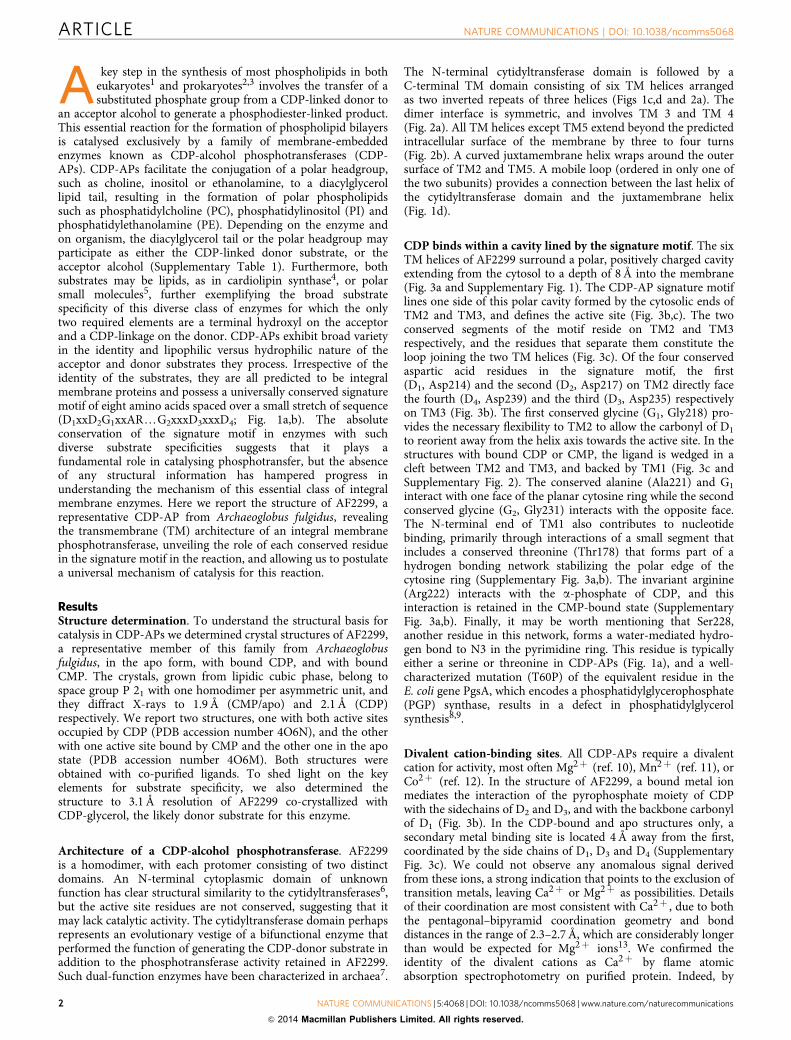
A key step in the synthesis of most phospholipids in botheukaryotes1 and prokaryotes2,3 involves the transfer of asubstituted phosphate group from a CDP-linked donor to
an acceptor alcohol to generate a phosphodiester-linked product.This essential reaction for the formation of phospholipid bilayersis catalysed exclusively by a family of membrane-embeddedenzymes known as CDP-alcohol phosphotransferases (CDP-APs). CDP-APs facilitate the conjugation of a polar headgroup,such as choline, inositol or ethanolamine, to a diacylglycerollipid tail, resulting in the formation of polar phospholipidssuch as phosphatidylcholine (PC), phosphatidylinositol (PI) andphosphatidylethanolamine (PE). Depending on the enzyme andon organism, the diacylglycerol tail or the polar headgroup mayparticipate as either the CDP-linked donor substrate, or theacceptor alcohol (Supplementary Table 1). Furthermore, bothsubstrates may be lipids, as in cardiolipin synthase4, or polarsmall molecules5, further exemplifying the broad substratespecificity of this diverse class of enzymes for which the onlytwo required elements are a terminal hydroxyl on the acceptorand a CDP-linkage on the donor. CDP-APs exhibit broad varietyin the identity and lipophilic versus hydrophilic nature of theacceptor and donor substrates they process. Irrespective of theidentity of the substrates, they are all predicted to be integralmembrane proteins and possess a universally conserved signaturemotif of eight amino acids spaced over a small stretch of sequence(D1xxD2G1xxARyG2xxxD3xxxD4; Fig. 1a,b). The absoluteconservation of the signature motif in enzymes with suchdiverse substrate specificities suggests that it plays afundamental role in catalysing phosphotransfer, but the absenceof any structural information has hampered progress inunderstanding the mechanism of this essential class of integralmembrane enzymes. Here we report the structure of AF2299, arepresentative CDP-AP from Archaeoglobus fulgidus, revealingthe transmembrane (TM) architecture of an integral membranephosphotransferase, unveiling the role of each conserved residuein the signature motif in the reaction, and allowing us to postulatea universal mechanism of catalysis for this reaction.
ResultsStructure determination. To understand the structural basis forcatalysis in CDP-APs we determined crystal structures of AF2299,a representative member of this family from Archaeoglobusfulgidus, in the apo form, with bound CDP, and with boundCMP. The crystals, grown from lipidic cubic phase, belong tospace group P 21 with one homodimer per asymmetric unit, andthey diffract X-rays to 1.9 Å (CMP/apo) and 2.1 Å (CDP)respectively. We report two structures, one with both active sitesoccupied by CDP (PDB accession number 4O6N), and the otherwith one active site bound by CMP and the other one in the apostate (PDB accession number 4O6M). Both structures wereobtained with co-purified ligands. To shed light on the keyelements for substrate specificity, we also determined thestructure to 3.1 Å resolution of AF2299 co-crystallized withCDP-glycerol, the likely donor substrate for this enzyme.
Architecture of a CDP-alcohol phosphotransferase. AF2299is a homodimer, with each protomer consisting of two distinctdomains. An N-terminal cytoplasmic domain of unknownfunction has clear structural similarity to the cytidyltransferases6,but the active site residues are not conserved, suggesting that itmay lack catalytic activity. The cytidyltransferase domain perhapsrepresents an evolutionary vestige of a bifunctional enzyme thatperformed the function of generating the CDP-donor substrate inaddition to the phosphotransferase activity retained in AF2299.Such dual-function enzymes have been characterized in archaea7.
The N-terminal cytidyltransferase domain is followed by aC-terminal TM domain consisting of six TM helices arrangedas two inverted repeats of three helices (Figs 1c,d and 2a). Thedimer interface is symmetric, and involves TM 3 and TM 4(Fig. 2a). All TM helices except TM5 extend beyond the predictedintracellular surface of the membrane by three to four turns(Fig. 2b). A curved juxtamembrane helix wraps around the outersurface of TM2 and TM5. A mobile loop (ordered in only one ofthe two subunits) provides a connection between the last helix ofthe cytidyltransferase domain and the juxtamembrane helix(Fig. 1d).
CDP binds within a cavity lined by the signature motif. The sixTM helices of AF2299 surround a polar, positively charged cavityextending from the cytosol to a depth of 8 Å into the membrane(Fig. 3a and Supplementary Fig. 1). The CDP-AP signature motiflines one side of this polar cavity formed by the cytosolic ends ofTM2 and TM3, and defines the active site (Fig. 3b,c). The twoconserved segments of the motif reside on TM2 and TM3respectively, and the residues that separate them constitute theloop joining the two TM helices (Fig. 3c). Of the four conservedaspartic acid residues in the signature motif, the first(D1, Asp214) and the second (D2, Asp217) on TM2 directly facethe fourth (D4, Asp239) and the third (D3, Asp235) respectivelyon TM3 (Fig. 3b). The first conserved glycine (G1, Gly218) pro-vides the necessary flexibility to TM2 to allow the carbonyl of D1to reorient away from the helix axis towards the active site. In thestructures with bound CDP or CMP, the ligand is wedged in acleft between TM2 and TM3, and backed by TM1 (Fig. 3c andSupplementary Fig. 2). The conserved alanine (Ala221) and G1interact with one face of the planar cytosine ring while the secondconserved glycine (G2, Gly231) interacts with the opposite face.The N-terminal end of TM1 also contributes to nucleotidebinding, primarily through interactions of a small segment thatincludes a conserved threonine (Thr178) that forms part of ahydrogen bonding network stabilizing the polar edge of thecytosine ring (Supplementary Fig. 3a,b). The invariant arginine(Arg222) interacts with the a-phosphate of CDP, and thisinteraction is retained in the CMP-bound state (SupplementaryFig. 3a,b). Finally, it may be worth mentioning that Ser228,another residue in this network, forms a water-mediated hydro-gen bond to N3 in the pyrimidine ring. This residue is typicallyeither a serine or threonine in CDP-APs (Fig. 1a), and a well-characterized mutation (T60P) of the equivalent residue in theE. coli gene PgsA, which encodes a phosphatidylglycerophosphate(PGP) synthase, results in a defect in phosphatidylglycerolsynthesis8,9.
Divalent cation-binding sites. All CDP-APs require a divalentcation for activity, most often Mg2þ (ref. 10), Mn2þ (ref. 11), orCo2þ (ref. 12). In the structure of AF2299, a bound metal ionmediates the interaction of the pyrophosphate moiety of CDPwith the sidechains of D2 and D3, and with the backbone carbonylof D1 (Fig. 3b). In the CDP-bound and apo structures only, asecondary metal binding site is located 4 Å away from the first,coordinated by the side chains of D1, D3 and D4 (SupplementaryFig. 3c). We could not observe any anomalous signal derivedfrom these ions, a strong indication that points to the exclusion oftransition metals, leaving Ca2þ or Mg2þ as possibilities. Detailsof their coordination are most consistent with Ca2þ , due to boththe pentagonal–bipyramid coordination geometry and bonddistances in the range of 2.3–2.7 Å, which are considerably longerthan would be expected for Mg2þ ions13. We confirmed theidentity of the divalent cations as Ca2þ by flame atomicabsorption spectrophotometry on purified protein. Indeed, by
ARTICLE NATURE COMMUNICATIONS | DOI: 10.1038/ncomms5068
2 NATURE COMMUNICATIONS | 5:4068 | DOI: 10.1038/ncomms5068 | www.nature.com/naturecommunications
& 2014 Macmillan Publishers Limited. All rights reserved.

this method we could measure a signal from Ca abovebackground and equimolar to the concentration of AF2299(Methods and Supplementary Table 2), while we could not detectthe presence of any protein-associated Mg.
Two substrate-binding pockets are located within the polarcavity. Deeper in the active site of the CMP and apo structures,
a tetrahedral oxyanion, probably sulphate given its presence inthe crystallization solution, is bound by Arg240 and Arg304(Fig. 4a and Supplementary Fig. 3d). The sulphate also forms ahydrogen bond with the amide nitrogen of Arg301, which lies atthe N-terminal end of the short TM5, consistent with a role forthe helix dipole in stabilization of anion binding14. The oxyanion-binding site is formed by residues that are conserved in a manner
TM2 TM3 TM4 TM5
TM2
TM1
TM3
TM4
TM6
TM5-O-P-O--OH
+ CDP-AP
M2+ +
=
=R2
R1CDP
R2R1
CMP
* S228AF2299 -------YLTTLLAGVIIQLHSIIDGCDGEIARLKFMESKYGAWLDGVLDRYSFFIIVFSITYVLSAS------NPVYWIIGFLAAFASLMIA------- 275PGSA_ECOLI -------TWSPFAAALIFCVAAVTDWFDGFLARRWNQSTRFGAFLDPVADKVLVAIAMVLVTEHYHSWW----VTLPAATMIAREIIISALRE------- 110CEPT1_A_THALIANA QLDSPPPRWVHFAHGLLLFLYQTFDAVDGKQARRTNSSSPLGELFDHGCDALACAFEAMAFGSTAMCGR---DTFWFWVISAIPFYGATWEHYFTNTLIL 167CEPT1_H_SAPIENS TATEQAPLWAYIACACGLFIYQSLDAIDGKQARRTNSSSPLGELFDHGCDSLSTVFVVLGTCIAVQLGTNPDWMFFCCFAGTFMFYCAHWQTYVSGTLRF 208CPT1_H_SAPIENS TATEEAPYWTYLLCALGLFIYQSLDAIDGKQARRTNSCSPLGELFDHGCDSLSTVFMAVGASIAARLGTYPDWFFFCSFIGMFVFYCAHWQTYVSGMLRF 186PIS1_S_CEREVISIAE ---------HPTAFTWLYSTSCLLDALDGTMARKYNQVSSLGAVLDMVTDRSSTAGLMCFLCVQYPQW------CVFFQLMLGLDITSHYMHM------- 118PIS1_A_THALIANA ---------NKPLFSVLYFFSFCCDAVDGWVARRFNQVSTFGAVLDMVTDRVSTACLLVILSQIYRP-------SLVFLSLLALDIASHWLQM------- 116PIS1_H_SAPIENS ---------CPLTASSFYLLSGLLDAFDGHAARALNQGTRFGAMLDMLTDRCSTMCLLVNLALLYPGA------TLFFQISMSLDVASHWLHL------- 109
D1xxD2G1xxAR....G2xxxD3xxxD4
O
O
90°
90°
Figure 1 | The structure of AF2299 reveals the architecture of a CDP-alcohol phosphotransferase. (a) The CDP-AP signature motif is absolutely conservedacross all kingdoms of life. Alignment of eight CDP-APs of divergent sequence shows that the CDP-AP signature motif (D1xxD2G1xxARyG2xxxD3xxxD4)is absolutely conserved, and is present in AF2299. Alignment performed using PROMALS3D48. Sequences, from top, (as species—protein; Uniprot ID) are:A. fulgidus—AF2299; O27985, E. coli—PGP synthase PgsA; P0ABF8, A. thaliana—choline/ethanolamine phosphotransferase; O82567, H. sapiens—choline/ethanolamine phosphotransferase; Q9Y6K0, H. sapiens—choline phosphotransferase; Q8WUD6, S. cerivisiae—phosphatidylinositol synthase; P06197,A. thaliana—phosphatidylinositol synthase; Q8LBA6, H. sapiens—phosphatidylinositol synthase; O14735. (b) CDP-alcohol phosphotransferases catalyse thetransfer of a phosphate group, with donor substituent R1 (where R1 may represent choline, ethanolamine, or diacylglycerol, amongst others) to an alcohol, withsubstituent R2 (where R2 may represent inositol, inositol-1-phosphate, glycerol-3-phosphate choline or diacylglycerol, for example). (c) Each subunit of AF2299possesses six TM helices arranged in two repeats of three. TM5 is noticeably shorter than the rest. Intracellular loops are represented by blue lines andextracellular loops by black lines. The juxtamembrane helix and N-terminal domain are not shown. The left half of this panel is represented in an open form suchthat the two repeats are side by side. (d) AF2299 is a dimer of two subunits each with six TM helices and an N-terminal domain of the cytidyltransferase fold. Theprotein is depicted with one subunit in grey and the other in rainbow coloured ribbon representation, from blue (N terminus) to red (C terminus), viewed (on theleft) in the plane of the membrane, and (on the right) from the extracellular side of the membrane down the dimer interface.
NATURE COMMUNICATIONS | DOI: 10.1038/ncomms5068 ARTICLE
NATURE COMMUNICATIONS | 5:4068 | DOI: 10.1038/ncomms5068 | www.nature.com/naturecommunications 3
& 2014 Macmillan Publishers Limited. All rights reserved.

that appears to correlate with the identity of the acceptor. In theCDP structure, for which tartrate rather than sulphate waspresent in the crystallization solution, a tartrate ion is found inthe same position occupied by the sulphate in the CMP and apostructures (Supplementary Fig. 4). Sequence analysis reveals thatArg240 is conserved in all enzymes that take either inositol orinositol phosphate as the acceptor and that a short motif(R301xxR304, in AF2299 numbering) is specifically conserved inthe subset of these enzymes that utilize inositol-1-phosphate(Fig. 4b,c). We hypothesize that Arg240 forms hydrogen bondswith hydroxyls 2/3 or 5/6 of the inositol, fixing the orientation ofthe ring in a catalytically competent orientation, with the 4-OHpositioned in-line with the pyrophosphate moiety of theCDP-donor. The data that we present here are insufficient toassign the identity of the acceptor alcohol for AF2299; however,they do allow us to identify with reasonable certainty the locationof the acceptor binding-site, which we would expect to beconserved throughout the family of CDP-APs.
Another apparent binding pocket extends into the wall of thepolar cavity, perpendicular to the CDP pyrophosphate (Fig. 4b).This site is lined by residues specifically conserved in the archaealfamily of CDP-APs that utilize donors with small, polarsubstituents, such as CDP-inositol and CDP-glycerol, to generatesoluble phosphodiester-linked osmolytes that apparently helptheir host organisms endure thermal and osmotic stress15.This family includes the di-myo-inositol-1,30-phosphate-10-phosphate synthase (DIPPS) subfamily of CDP-APs, which arethe best-characterized enzymes of this class16. Based on thisobservation, we suggest that this pocket may accommodate thedonor substituent, and that AF2299 may catalyse the reaction ofinositol-1-phosphate with a small molecule, possibly CDP-glycerol or CDP-inositol5. Indeed, co-crystallization with CDP-glycerol reveals additional density in this pocket consistent withbinding of the glycerol moiety (Fig. 5). This is in agreement withthe observation that the AF2299 sequence is most closely relatedto those of the DIPPS family (Figs 4c and 6). Given our structure
1
2
3
4
56
JM
TM1
TM2TM3
TM4TM5
TM6
Dimer interface
Figure 2 | Architecture of the AF2299 dimer. (a) Two inverted repeats of three helices (TM1-3 and TM4-6) surround a central cavity in each monomer.The dimer interface is formed by TM 3 and 4, and buries an area of 1,963 Å2. Here, on the left, a dimer is represented with the N-terminal domains ingrey and the TM subunits in rainbow ribbon representation, from blue (N-terminal) to red (C-terminal). A 4 Å thick slab (purple shading) is withdrawnfrom the protein, and on the right, viewed from the extracellular side of the membrane down the dimer interface (red dashed line). The helices are labelledTM1–TM6, and connections between them are represented either in black (for extracellular loops) or blue (for intracellular loops). (b) All TM helicesexcept TM5 extend beyond the intracellular border of the membrane. Here, the TM helices of AF2299 are represented as cylinders, and the secondarystructure elements of the protomer on the left are coloured in rainbow representation, from blue (N terminus) to red (C terminus). The TM helicesare labelled from 1 to 6. The dimer is represented inside a translucent molecular surface coloured by the Kyte–Doolitle hydrophobicity scale from"4.5 (most polar, light blue) to 4.5 (most hydrophobic, orange). The approximate boundaries of the membrane are represented by black lines.
ARTICLE NATURE COMMUNICATIONS | DOI: 10.1038/ncomms5068
4 NATURE COMMUNICATIONS | 5:4068 | DOI: 10.1038/ncomms5068 | www.nature.com/naturecommunications
& 2014 Macmillan Publishers Limited. All rights reserved.

with bound CDP-glycerol and the fact that an A. fulgidus DIPPSenzyme (AF0263) has already been identified7, the most plausiblehypothesis is that AF2299 catalyses the condensation of CDP-glycerol with L-myo-inositol-1-phosphate, to yield the glycerylphosphodiester of inositol-1-phosphate, although experimentalconfirmation of this conjecture awaits further studies.
DiscussionThe structure of AF2299 reveals that CDP-APs adopt what is, tothe best of our knowledge, a novel fold, with six TM helicessurrounding a polar cavity. The active site of AF2299 is locatednear the surface of the membrane with direct access to thecytosol, consistent with the function of many CDP-APs inbinding one soluble and one membrane-embedded substrate. It istempting to speculate that the gap in the wall of the polar cavitycreated by the shorter TM5 helix may facilitate access ofCDP-linked lipids to the active site, in those enzymes that utilizeCDP-archaeol or CDP-diacylglycerol as donor substrates. Accessof CDP-linked lipids by such a route would not be possible inAF2299 from our current structures, as the N-terminal end of thejuxtamembrane helix occludes this hypothetical pathway, butonly a slight alteration in the length or conformation of this helixwould be necessary to facilitate lipid binding. Sequence alignmentand secondary structure prediction for other CDP-alcoholphosphotransferases suggests that at least the PI-synthases sharethe same fold as the TM domain of AF2299, with an amphipathic
juxtamembrane helix followed by six TM helices, of which thefifth is considerably shorter than the other five (SupplementaryFig. 5).
Interestingly, enzymes that utilize a lipid acceptor, such asthe eukaryotic phosphatidylcholine synthases, which take CDP-choline as a donor and diacylglycerol as an acceptor, have asomewhat different architecture, characterized by the presence ofan additional 3–4 TM helices at the C terminus, perhaps in orderto accommodate the bulkier lipid acceptor.
The membrane localization of CDP-APs such as AF2299 andthe DIPP synthases, which condense two soluble substrates, isintriguing and we suggest that it may have an evolutionaryexplanation. If the ancestral proto-CDP-AP from which allcurrent CDP-APs are derived was an integral membrane proteinwith hydrophobic substrates, it is difficult to envision how theenzyme architecture could be modified during evolution tobecome a soluble protein, but it is not unlikely that slightmodifications around the active site could result in entry of andaffinity for a pair of soluble substrates.
The structures of AF2299 in the apo, CMP-bound and CDP-bound forms presented here provide us with the opportunity todissect the structural basis for catalysis in a CDP-AP. It has beensuggested that reactions catalysed by CDP-APs proceed by asequential mechanism involving a nucleophilic attack of theacceptor on the b-phosphorous of the CDP-donor17,18, but theroles of residues in the signature motif have remained unclear,although it has been previously suggested that either D3 or D4
Membrane
Out
In
D1214
D2217
D3235G1218
A221R222
G2231
D4239
TM2 TM3
D1xxD2G1xxAR.....G2xxxD3xxxD4
a
b c
Figure 3 | The signature motif of the CDP-alcohol phosphotransferase family is located around a cleft between TM helices 2 and 3. (a) Eachsubunit possesses a polar cavity near the surface of the membrane. Here, the molecular surface of AF2299 is coloured by Kyte–Doolitle hydrophobicity49
from "4.5 (most polar, light blue) to 4.5 (most hydrophobic, orange). On the right, the front half of the dimer has been removed to expose the polarcavities in each subunit that penetrate 8 Å beyond the intracellular border of the membrane. The approximate borders of the membrane, as calculated bythe PPM server50, are indicated by black lines. (b) The CDP-alcohol phosphotransferase signature motif spans the cytosolic ends of TM2 and TM3(highlighted here in purple; the conserved residues of the signature motif are shown in green stick representation in a zoom-in panel on the right,as is the CDP (in grey stick) and the primary divalent cation (as a pink sphere). (c) TM 2 and 3 (blue cylinders) form a groove, backed by TM1, linedby the residues of the signature motif (solid red line; the non-conserved segment between the two helices is indicated by a dashed red line).
NATURE COMMUNICATIONS | DOI: 10.1038/ncomms5068 ARTICLE
NATURE COMMUNICATIONS | 5:4068 | DOI: 10.1038/ncomms5068 | www.nature.com/naturecommunications 5
& 2014 Macmillan Publishers Limited. All rights reserved.

may act as a catalytic base19. The AF2299 structures reveal thatD1, D2 and D3 coordinate the pyro-phosphate of CDP via theprimary divalent cation. D4, in contrast, based on its positionapproximately equidistant between the acceptor and donorbinding sites (Supplementary Fig. 4), is likely to play a keyrole in catalysis. In S. cerevisiae choline phosphotransferase,mutation of the equivalent residue results in completeabolition of enzymatic activity19. Likewise, mutation ofD4 in an aminoalcoholphosphotransferase from Brassica napus(rapeseed) results in the loss of detectable phosphotransferase
activity20. A more extensive alanine mutagenesis scan inRhizobium meliloti phosphatidylcholine synthase yielded thesame conclusions21. Furthermore, D4 does not interact with theCDP either directly or via the bridging divalent cation. Insummary, D4 is absolutely conserved, essential for activity inenzymes from two different kingdoms (Plantae and Fungi), butdoes not appear to participate in substrate binding, and is locatedbetween the binding sites of the acceptor and donor substrates.These observations support a direct role for D4 in the mechanismof phosphotransfer in CDP-APs. We propose that this residue
SO4R304
R240D4239
2.9 Å
2.9 Å
3.1 Å
D3235
D1214
*R240
*R304
TM5
a b
c
Figure 4 | Two binding pockets within the cavity form putative binding sites for the donor subtituent and the acceptor alcohol. (a) The putativecatalytic aspartate, D239, lies approximately equidistant between the bound CMP and a bound sulphate ion, located between two arginine residues, R240and R304. (b) Family-specific conservation patterns delineate three regions within the cavity. Residues conserved in all members of the CDP-alcoholphosphotransferase family are coloured in cyan, those conserved only in enzymes that utilize inositol-1-phosphate as an acceptor are coloured in purple,and those that are conserved in all enzymes known to bind CDP-inositol or CDP-glycerol in light blue. The bound sulphate and CDP are shown in stickrepresentation. (c) An alignment of AF2299 with DIPP synthases shows several highly conserved regions outside the signature motif; notably, the twoarginines that coordinate the bound sulfate, R240 and R304 (indicated in red), are absolutely conserved.
Figure 5 | Binding of CDP-glycerol in the active site. (a) A co-crystal structure with CDP-glycerol reveals that the glycerol moiety binds in the putative donor-substituent binding pocket (Colors as for Fig. 4). (b) 2Fo" Fc omit maps (calculated prior to ligand placement) contoured at 1s for the CDP and CDP-glycerolstructures are represented as purple and blue mesh, respectively. The CDP-glycerol ligand from the final refined structure is depicted in stick representation.
ARTICLE NATURE COMMUNICATIONS | DOI: 10.1038/ncomms5068
6 NATURE COMMUNICATIONS | 5:4068 | DOI: 10.1038/ncomms5068 | www.nature.com/naturecommunications
& 2014 Macmillan Publishers Limited. All rights reserved.

acts as a catalytic base, abstracting a proton from the terminalhydroxyl of the acceptor alcohol, facilitating nucleophilic attackby the activated acceptor on the b-phosphorus of the CDP(Fig. 7a). Following either an associative or dissociativemechanism (Fig. 7b) this yields CMP and the phosphodiesterproduct (Fig. 7c). We cannot unambiguously determine whetherthis reaction occurs by an associative mechanism via apentacoordinate, trigonal bipyramidal transition state or bya dissociative mechanism via a trigonal planar intermediate.A plausible associative mechanism is depicted in Fig. 7. Althoughthe pKa of an isolated aspartate is too low to act as an efficientcatalytic base under physiological conditions, substantialelevation of the pKa of catalytic aspartates has been previouslydescribed22, particularly in the presence of neighbouring negative
charges, as is observed here with the clustering of the fourconserved aspartates. A base-catalysed mechanism is supportedby the observation that many CDP-APs have alkaline pHoptima23,24. Consistent with the presence of a general acid–basecatalyst, rabbit phosphatidylinositol (PI) synthase has an alkalinepH optimum for the forward synthase reaction, while displaying amildly acidic optimum for the reverse activity25.
The role of the primary divalent cation appears to be to primethe pyro-phosphate for catalysis via an in-line mechanism ofsubstrate transfer, in addition to withdrawing charge from thepyrophosphate, making it more electrophilic and increasingsusceptibility to nucleophilic attack. The function of thesecondary cation is less obvious. It may contribute to orientingthe carboxyl of D4 appropriately to deprotonate the acceptor.Secondary divalent cation binding sites have also been describedfor other phosphotransferases, such as cAMP-dependent proteinkinase26. Strikingly, this mechanism appears to have manycommon features with that of other phosphotransferasesincluding kinases, which have a completely unrelated fold, but avery similar configuration of active site residues and substrates27.
A remarkable feature of the CDP-AP family is the chemicaldiversity of both acceptor and donor substrates. In the active siteof the AF2299 structure, in addition to the nucleotide-bindingsite, we observe two pockets that are likely to host the donorsubstituent and the acceptor alcohol (Fig. 4b). This hypothesis isconfirmed for the donor group by our structure showing CDP-glycerol bound to AF2299 (Fig. 5). Given the absolute conserva-tion of residues involved in catalysis and nucleotide binding, theposition of the donor and acceptor substrates will most probablyalso remain constant, so as to allow the reaction to occur.Therefore, the structures reported here are likely to provide aframework in which to investigate the molecular elementsunderlying substrate specificity.
AF2299 appears likely to take inositol-1-phosphate as theacceptor alcohol, an observation that is of particular significancewhen considered in the context of mycobacterial PI synthesis,which proceeds via a somewhat different pathway to that in otherorganisms. PI synthases from eukaryotes catalyse the directformation of phosphatidylinositol from CDP-diacylglycerol(CDP-DAG) and myo-inositol, and they cannot process inositolphosphate28. In contrast, mycobacterial phosphatidylinositol isformed in two steps. In the first step, a PI-synthase-like CDP-APcatalyses the conjugation of CDP-DAG with inositol-1-phosphate. Importantly, this enzyme does not recognizeinositol, instead specifically recognizing inositol-1-phosphate29.This reaction generates phosphatidylinositol-phosphate, which isthen processed by a phosphatase of unknown identity to yieldphosphatidylinositol. Phosphatidylinositol biosynthesis is aparticularly attractive target for the development of anti-tuberculosis therapeutics, as all downstream steps in thesynthesis of lipoarabinomannan, an important component ofthe cell wall, are contingent upon the attachment of mannanchains to a phosphatidylinositol anchor3,30. The orthogonalsubstrate specificities of mycobacterial and eukaryotic PIsynthases suggest that this may be a feasible avenue fortherapeutic intervention. The transmembrane domain ofAF2299 shares only 20% identity with M. tuberculosis PIsynthase, but the high degree of conservation of specificelements present in AF2299, MT-PI synthases and the DIPPsynthases (Supplementary Fig. 6) suggests that AF2299 mayprovide a useful framework for the design of inositol–phosphateanalogues for the inhibition of the mycobacterial PI-synthases.Previous development of inositol-1-phosphate analogues hasfocused on replacement of the 1-phosphate with an analogousnon-hydrolyzable moiety such as phosphonate, but thesemodifications resulted in inhibitors with significantly lower
DIPPS
34 5
6
7
8
10 PIPS/AIPS
ProkaryoticPC/PE synthases
ProkaryoticPGP synthases
Cardiolipin synthases
Phosphatidylinositol synthases
EukaryoticPC/PE synthases
1516
14
1213
19
17 18
26
22
2021
25 2324
9
11
12
Figure 6 | AF2299 is most closely related in sequence to the di-inositol-phosphate synthases. AF2299 (3, highlighted by a red circle), is mostsimilar in sequence to the previously characterized DIPP synthases (4–8).Here, an unrooted phylogenetic tree, generated using CLUSTALW-PHYLOGENY51 from a multiple-alignment of 26 sequences (performedusing CLUSTAL-OMEGA52), was visualized in radial representation usingT-REX53, and manually edited for clarity. The sequences cluster into distinctfamilies of related function (shaded triangles), and AF2299 is most closelyrelated to those that have been functionally characterized as DIPPsynthases (orange shaded triangle). The species and Uniprot IDs of thesequences used to generate the tree are as follows: (1) Q9KJY8; Rhizobiummeliloti. (2): Q98MN3; Rhizobium loti. (3) O27985; Archeoglobus fulgidus(AF2299). (4) O29976; Archaeoglobus fulgidus. (5) Q5JDA9; Thermococcuskodakaraensis. (6) Q8U1Z6; Pyrococcus furiosus. (7) O67379; Aquafexaeolicus. (8) Q1AWQ0; Rubrobacter xylanophilus. (9) O27726;Methanothermobacter thermautotrophicus. (10) Q9F7Y9; Mycobacteriumsmegmatis. (11) Q7D6W6; Mycobacterium tuberculosis. (12) Q8WUD6;Homo sapiens. (13) Q5ZKD1; Gallus gallus. (14) Q9Y6K0; Homo sapiens.(15) P17898; Saccharomyces cerevisiae. (16) O82567; Arabidopsis thaliana.(17) Q8LBA6; Arabidopsis thaliana. (18) P06197; Saccharomyces cerevisiae.(19) O14735; Homo sapiens. (20) Q8MZC4; Drosophila melanogaster.(21) Q9UJA2; Homo sapiens. (22) O01916; Caenorhabditis elegans.(23) Q68XS5; Ricksettia typhi. (24) P0ABF8; Escherichia coli. (25) P44528;Haemophilus influenza. (26) P63753; Mycobacterium tuberculosis.
NATURE COMMUNICATIONS | DOI: 10.1038/ncomms5068 ARTICLE
NATURE COMMUNICATIONS | 5:4068 | DOI: 10.1038/ncomms5068 | www.nature.com/naturecommunications 7
& 2014 Macmillan Publishers Limited. All rights reserved.

affinity than the cognate substrate31. We would predict that the1-phosphate interacts with two arginine residues (Arg301 andArg304 in AF2299), and likely makes a significant contribution tobinding affinity, suggesting that alterations at this position arelikely to result in reduced affinity. A more productive avenue forexploration may involve replacement of the terminal hydroxylwith a less reactive substituent.
The best-characterized role of CDP-APs is in the biosynthesisof phospholipids. In the scheme of this reaction, the lipidicsubstrate can participate either as a donor substituent, as an
acceptor alcohol, or two lipids may act as donor and acceptorrespectively. The prokaryotic PGP-synthases and eukaryoticPI-synthases process a lipophilic donor (CDP-DAG) and a polaracceptor, respectively glycerol-3-phosphate32 and myo-inositol33.By contrast, the eukaryotic choline and ethanolamine phospho-transferases that participate in the Kennedy pathway of zwitterionicphospholipid biosynthesis utilize CDP-choline/CDP-ethanolamineas donors, and diacylglycerol as the acceptor alcohol34,35.Eukaryotic cardiolipin synthases4 represent a third class inwhich CDP-DAG condenses with phosphatidyl-glycerol. While
H
-
O
O
O
O P PO O O
O
O O
O
O
O
O O
O
O
O
O
O
O
O
R1
D4
R2
D2
D3 M2+
D1 HO
R
M2+
OO
O
O
O
O
R1
D4
R2
D2
D3M2+
D1 OH
R
M2+
O O O O
O
D4
D2
D3M2+
D1 OH O
OO
OO
OO
OP
R
OH
O OPO
O
R2R1
OH
O P P
a b c
Figure 7 | CDP-alcohol phosphotransferases effect phosphotransfer by a base-catalysed mechanism. (a) The fourth aspartate in the signature motif,D4, deprotonates the acceptor alcohol (R2, red), activating it for nucleophilic attack upon the beta-phosphorus of the CDP (the pyrimidine ring of which isshown in pale green), to which the donor substituent (R1, purple) is attached by an ester linkage. (b) Here, the transition state of the reaction is shownassuming an associative mechanism proceeding via a penta-coordinate transition state, to yield the product, (c) a phosphodiester linked conjugate of thedonor, R1, and the acceptor R2. A water molecule (aqua) replaces the beta-phosphate in coordinating the primary divalent cation.
Table 1 | Data collection and refinement statistics.
CMP/Apo (4O6M) CDP (4O6N) CDP-glycerol (4Q7C) SeMet
Data collectionSpace group P 21 P 21 P 21 P 21
Cell dimensionsa, b, c (Å) 46.36, 91.15, 107.02 46.33, 90.67, 106.44 46.42, 90.85, 106.61 46.35, 91.11, 106.92b (!) 92.5 92.07 92.2 91.88Wavelength (Å) 1.024 0.979 0.979 0.979Resolution (Å) 46.33–1.90 (1.97–1.90)* 46.30–2.10 (2.18–2.10)* 69.13–3.10 (3.32–3.10)* 69.33–2.60 (2.72–2.60)*Rmerge 0.071 (1.72) 0.124 (1.62) 0.183 (0.780) 0.174 (1.018)I / sI 11.73 (0.72) 7.43 (0.81) 8.1 (2.0) 10.8 (2.2)Completeness (%) 99.29 (99.15) 98.08 (98.08) 99.8 (99.8) 99.8 (100)Redundancy 3.8 (3.8) 3.4 (3.5) 4.2 (4.2) 7.4 (7.6)Resolution (I/sI¼ 2) 2.15 2.35 3.21 2.60
RefinementResolution (Å) 46.33–1.90 (1.97–1.90) 46.30–2.10 (2.18–2.10) 69.13–3.10 (3.21–3.10) —No. unique reflections 69,560 (6,905) 50,389 (5,000) 16,102 (1,562) —Rwork/Rfree 0.198 (0.343)/0.224 (0.360) 0.226 (0.351)/0.262 (0.379) 0.224 (0.313)/0.274 (0.347) —No. non-hydrogen atoms 5,630 5,584 5,412 —Protein 5,257 5,282 5,272 —Ligand/ion 209 158 140 —Water 164 144 0 —B-factors 47.3 46.8 41.1 —Protein 47.0 46.7 41.3 —Ligand/ion 55.3 47.6 33.6 —Water 47.2 47.8 — —R.m.s. deviations —Bond lengths (Å) 0.005 0.003 0.004 —Bond angles (!) 0.86 0.62 0.77 —
*Values in parentheses are for highest-resolution shell.
ARTICLE NATURE COMMUNICATIONS | DOI: 10.1038/ncomms5068
8 NATURE COMMUNICATIONS | 5:4068 | DOI: 10.1038/ncomms5068 | www.nature.com/naturecommunications
& 2014 Macmillan Publishers Limited. All rights reserved.

representing the first reported structure for any CDP-AP, ourcurrent model of AF2299 does not allow us to draw firmconclusions regarding the binding mode of lipid substrates.However, attachment of a bulky, hydrophobic lipid substituent tothe b phosphate of CDP would appear to restrict the number ofpossibilities for the overall disposition of the alkyl chains to onlytwo, of which only one seems plausible. In one orientation, thealkyl chain would project into the polar cavity of the active sitetowards the acceptor. The more likely scenario, in agreement withthe structure of bound CDP-glycerol, has the glycerol moiety of alipid positioned in the secondary pocket, with the alkyl chains ofthe lipid exiting into the bilayer between TM2 and TM5.
MethodsTarget identification and cloning. A member of the CDP-alcohol phosphati-dyltransferase class-I family from Archaeoglobus fulgidus DSM 4304 (AF2299,UniProt accession no. O27985) was identified as a promising candidate forcrystallization experiments by the NYCOMPS (New York Consortium onMembrane Protein Structure) high-throughput screening pipeline36,37. The geneencoding AF2299 was amplified from Archaeoglobus fulgidus DSM 4304 genomicDNA using (50-tacttccaatccaatgccATGAGGTTAGCGTACGTTAAAAAC-30) asthe forward primer and (50-ttatccacttccaatgCTAGTTCGTGTAACTTCTGAGTG-30) as the reverse primer for PCR, where upper case letters indicate gene-specificsequences and lower case letters indicate sequences incorporated into the PCRproduct that are required for ligation independent cloning. The gene was clonedinto the bacterial expression vector pMCSG7-10xHis, introducing an N-terminaldecahistidine tag and a TEV protease cleavage site.
Protein expression and purification. AF2299 was overexpressed in E. coli strainBL21 (DE3) pLysS. Bacterial cultures were grown at 37 !C in 2xYT mediumsupplemented with 100mg ml" 1 ampicillin and 50 mg ml" 1 chloramphenicol.Once the OD600 reached 1.0, the temperature was reduced to 22 !C, and cells wereinduced with 0.2 mM isopropyl b-D-1-thiogalactopyranoside (IPTG) after 15 min.After overnight induction at 22 !C, cells were harvested by centrifugation andstored at " 80 !C. Cells expressing selenomethionine-substituted protein weregrown with a kit containing a minimal medium of M9 salts (M9 SeMET High-Yield Growth Media Kit, Shanghai Medicilon Inc.). A total of 150 mg seleno-methionine was added per liter of culture in addition to the selenomethionineprovided in the kit. The rest of the protocol was the same as for native proteinexpression.
For large-scale purification of both native and selenomethionine-substitutedprotein, frozen cell pellets were resuspended in lysis buffer containing 20 mMNa-HEPES (pH 7.5), 200 mM NaCl, 1 mM EDTA, 1 mg ml" 1 lysozyme, 20 mMMgSO4, 10 mg ml" 1 DNase I, 10mg ml" 1 RNase A, 1 mM TCEP, 1 mM PMSF andComplete Mini EDTA-free protease inhibitor cocktail (Roche) as described in theinstructions. Cells were lysed with an Emulsiflex C3 homogenizer (Avestin). Lysatewas solubilized for 1.5 h with 1% (w/v) n-decyl-b-D-maltopyranoside (DM,Anagrade, Affymetrix) in a volume of B40 ml per cell pellet from 800 ml culture(B6 g). After solubilization, insoluble material was pelleted by ultracentrifugationat 134,000 g for 30 min at 4 !C. Protein was purified by metal-affinitychromatography (Ni-NTA, Qiagen) followed by size-exclusion chromatography(SEC) on a Superose 12 column (GE Healthcare) in a buffer of 20 mM Na-HEPES(pH 7.0), 200 mM NaCl, 0.2% (w/v) DM and 1 mM tris(2-carboxyethyl)phosphinehydrochloride (TCEP-HCl). Approximately 1.5 mg of purified protein could beobtained from an 800 ml bacterial culture. Selenomethionine-substituted proteinwas purified using the same protocol.
Crystallization. Protein from peak fractions from size-exclusion chromatographywas concentrated to 30–35 mg ml" 1 (approximated by A280 nm) for crystallizationusing a centrifugal concentrator (Millipore) with a 100 kDa MWCO. Seleno-methionine-substituted protein was concentrated to 25–30 mg ml" 1. Crystals weregrown at room temperature (20–22 !C) in lipidic cubic phase. Concentratedprotein was mixed with monoolein (Sigma) in a 1:1.5 (w/w) ratio of protein:monoolein. A Mosquito LCP (TTP Labtech) robot was used to dispense a typicalvolume of 50–100 nl of protein/monoolein mixture onto a 96-well glass plate,which was covered with 750–1,000 nl precipitant solution and sealed with a glasscover slip. Glass sandwich plates were stored in a 22 !C incubator. Largest crystalsgrew to B10$ 30$ 100 mm3 in 1–2 days in (a) 14–20% (v/v) PEG 550 MME,0.1 M Na-HEPES or Tris–HCl (pH 7.0–8.0), 0.2 M lithium sulphate (CMP/apostructure) (b) 25–30% (v/v) PEG 400, 0.1 M Na-HEPES (pH 7.0–7.5), 0.2 Mpotassium sodium tartrate (CDP structure) or (c) 15% PEG 550 MME, 0.1 MNa-HEPES (pH 7.0), 0.2 M potassium sodium tartrate, 2.5 mM CDP-glycerol and1 mM TCEP (CDP-glycerol structure). Crystals of selenomethionine-substitutedprotein grew in similar conditions. A tungsten carbide glass-cutter (HamptonResearch) was used to cut and remove the glass cover slip, and crystals were
harvested using 20–100 mm MicroLoops and MicroMounts (MiTeGen). Crystalswere flash-cooled directly in liquid nitrogen without additional cryoprotection.
Diffraction data collection and processing. Diffraction data were collected onNECAT beamlines 24-ID-C and 24-ID-E at the Advanced Photon Source(Argonne, IL, USA). Crystals were centred by diffraction, rastering across a grid inone orientation, and then along the orthogonal axis intersecting the position wherethe best diffraction was obtained in the first orientation. All diffraction data wereindexed, integrated scaled using XDS and XSCALE19,38. Data sets collected abovethe Se K-edge from two selenomethionine-substituted crystals were used forstructure solution by anomalous diffraction methods. Selenium sites were locatedusing SHELXD39,40, and refined in SHARP41. Density modification, includingsolvent flattening, histogram matching and twofold non-crystallographic symmetryaveraging were performed using DM to give an initial experimentally phasedelectron density map42.
Model building and refinement. An initial model was built usingBUCCANEER43. The model was manually completed using COOT and refinedusing the PHENIX crystallographic software package, alternating between cycles ofmanual building in COOT44 and refinement in PHENIX45. Local torsion angleNCS restraints were employed throughout refinement. Riding hydrogens wereincluded in the last round of refinement. The quality of the model was analysedusing the validation module of the PHENIX package, which incorporatesMolprobity clash score, density correlation and rotamer analysis46. Data collection,refinement and validation statistics for both structures are provided in Table 1.Protein structure figures were prepared using UCSF Chimera47. Within the crystal,the protein packs as stacks of two-dimensional sheets (Supplementary Fig. 7).Within each layer, adjacent dimers are arranged in an up–down orientation, suchthat the N-terminal domains of adjacent layers in the stack are interdigitated.A stereo figure of representative electron density for the CMP/apo structure and aC-alpha trace depicting the fold is given in Supplementary Fig. 8.
Ca and Mg measurements. Ca and Mg measurements were conducted using adual flame/graphite furnace Atomic Absorption Spectrometer AAnalyst 800,manufactured by Perkin Elmer. Data were collected and processed with WinLab32software. Ca and Mg concentrations were determined at wavelengths of 422.7 nmand 285.2 nm, respectively, using an air–acetylene flame and standard nebulizerwith flow spoiler. Detection limits were 0.02 mg ml" 1 and 0.003 mg ml" 1 for Caand Mg, respectively. Each run included calibration with three standards, as well asquality control samples with known concentrations of Ca and Mg. Calibrationstandards were made in 1% (v/v) HCl. All other samples were diluted in 1% (v/v)HCl. Duplicate samples of AF2299 at a concentration of 40 mg ml" 1 wereanalysed, as well as a control sample of protein-free buffer. Raw data and derivedresults are reported in Supplementary Table 2.
References1. Vance, J. E. & Vance, D. E. Phospholipid biosynthesis in mammalian cells.
Biochem. Cell Biol. 82, 113–128 (2004).2. Cronan, J. E. Bacterial membrane lipids: where do we stand? Annu. Rev.
Microbiol. 57, 203–224 (2003).3. Jackson, M., Crick, D. C. & Brennan, P. J. Phosphatidylinositol is an essential
phospholipid of mycobacteria. J. Biol. Chem. 275, 30092–30099 (2000).4. Houtkooper, R. H. et al. Identification and characterization of human
cardiolipin synthase. FEBS Lett. 580, 3059–3064 (2006).5. Borges, N. et al. Biosynthetic pathways of inositol and glycerol phosphodiesters
used by the hyperthermophile Archaeoglobus fulgidus in stress adaptation.J. Bacteriol. 188, 8128–8135 (2006).
6. Brito, J. A., Borges, N., Vonrhein, C., Santos, H. & Archer, M. Crystal structureof Archaeoglobus fulgidus CTP:inositol-1-phosphate cytidylyltransferase, a keyenzyme for di-myo-inositol-phosphate synthesis in (hyper)thermophiles.J. Bacteriol. 193, 2177–2185 (2011).
7. Rodrigues, M. V. et al. Bifunctional CTP:inositol-1-phosphatecytidylyltransferase/CDP-inositol:inositol-1-phosphate transferase, the keyenzyme for di-myo-inositol-phosphate synthesis in several(hyper)thermophiles. J. Bacteriol. 189, 5405–5412 (2007).
8. Miyazaki, C., Kuroda, M., Ohta, A. & Shibuya, I. Genetic manipulation ofmembrane phospholipid composition in Escherichia coli: pgsA mutantsdefective in phosphatidylglycerol synthesis. Proc. Natl Acad. Sci. USA 82,7530–7534 (1985).
9. Usui, M., Sembongi, H., Matsuzaki, H., Matsumoto, K. & Shibuya, I.Primary structures of the wild-type and mutant alleles encoding thephosphatidylglycerophosphate synthase of Escherichia coli. J. Bacteriol. 176,3389–3392 (1994).
10. Woodard, D. S., Lee, T. C. & Snyder, F. The final step in thede novo biosynthesis of platelet-activating factor. Properties of a uniqueCDP-choline:1-alkyl-2-acetyl-sn-glycerol choline-phosphotransferase inmicrosomes from the renal inner medulla of rats. J. Biol. Chem. 262, 2520–2527(1987).
NATURE COMMUNICATIONS | DOI: 10.1038/ncomms5068 ARTICLE
NATURE COMMUNICATIONS | 5:4068 | DOI: 10.1038/ncomms5068 | www.nature.com/naturecommunications 9
& 2014 Macmillan Publishers Limited. All rights reserved.

11. de Rudder, K. E., Sohlenkamp, C. & Geiger, O. Plant-exuded choline is used forrhizobial membrane lipid biosynthesis by phosphatidylcholine synthase. J. Biol.Chem. 274, 20011–20016 (1999).
12. Sandoval-Calderon, M., Geiger, O., Guan, Z., Barona-Gomez, F. & Sohlenkamp,C. A eukaryote-like cardiolipin synthase is present in Streptomyces coelicolorand in most actinobacteria. J. Biol. Chem. 284, 17383–17390 (2009).
13. Zheng, H., Chruszcz, M., Lasota, P., Lebioda, L. & Minor, W. Data mining ofmetal ion environments present in protein structures. J. Inorg. Biochem. 102,1765–1776 (2008).
14. Copley, R. R. & Barton, G. J. A structural analysis of phosphate and sulphatebinding sites in proteins. Estimation of propensities for binding andconservation of phosphate binding sites. J. Mol. Biol. 242, 321–329 (1994).
15. Martins, L. O. & Santos, H. Accumulation of mannosylglycerate anddi-myo-inositol-phosphate by Pyrococcus furiosus in response to salinity andtemperature. Appl. Environ. Microbiol. 61, 3299–3303 (1995).
16. Rodionov, D. A. et al. Genomic identification and in vitro reconstitution of acomplete biosynthetic pathway for the osmolyte di-myo-inositol-phosphate.Proc. Natl Acad. Sci. USA 104, 4279–4284 (2007).
17. Pontoni, G. et al. Studies on enzyme-substrate interactions ofcholinephosphotransferase from rat liver. Biochim. Biophys. Acta 836, 222–232(1985).
18. Schlame, M., Zhao, M., Rua, D., Haldar, D. & Greenberg, M. L. Kinetic analysisof cardiolipin synthase: a membrane enzyme with two glycerophospholipidsubstrates. Lipids 30, 633–640 (1995).
19. Williams, J. G. & McMaster, C. R. Scanning alanine mutagenesis of theCDP-alcohol phosphotransferase motif of Saccharomyces cerevisiaecholinephosphotransferase. J. Biol. Chem. 273, 13482–13487 (1998).
20. Qi, Q., Huang, Y.-F., Cutler, A. J., Abrams, S. R. & Taylor, D. C. Molecular andbiochemical characterization of an aminoalcoholphosphotransferase (AAPT1)from Brassica napus: effects of low temperature and abscisic acid treatments onAAPT expression in Arabidopsis plants and effects of over-expression ofBnAAPT1 in transgenic Arabidopsis. Planta 217, 547–558 (2003).
21. Solıs-Oviedo, R. L., Martınez-Morales, F., Geiger, O. & Sohlenkamp, C.Functional and topological analysis of phosphatidylcholine synthase fromSinorhizobium meliloti. Biochim. Biophys. Acta 1821, 573–581 (2012).
22. Harris, T. K. & Turner, G. J. Structural basis of perturbed pKa values ofcatalytic groups in enzyme active sites. IUBMB Life 53, 85–98 (2002).
23. Nowicki, M., Muller, F. & Frentzen, M. Cardiolipin synthase of Arabidopsisthaliana. FEBS Lett. 579, 2161–2165 (2005).
24. Antonsson, B. E. Purification and characterization of phosphatidylinositolsynthase from human placenta. Biochem. J. 297, 517–522 (1994).
25. Bleasdale, J. E., Wallis, P., MacDonald, P. C. & Johnston, J. M. Characterizationof the forward and reverse reactions catalysed by CDP-diacylglycerol:inositoltransferase in rabbit lung tissue. Biochim. Biophys. Acta 575, 135–147 (1979).
26. Zheng, J. et al. 2.2 A refined crystal structure of the catalytic subunit ofcAMP-dependent protein kinase complexed with MnATP and a peptideinhibitor. Acta Crystallogr. Sect. F Struct. Biol. Cryst. Commun. 49, 362–365(1993).
27. Matte, A., Tari, L. W. & Delbaere, L. T. How do kinases transfer phosphorylgroups? Struct. Folding Design 6, 413–419 (1998).
28. Morii, H., Ogawa, M., Fukuda, K. & Taniguchi, H. Ubiquitous distribution ofphosphatidylinositol phosphate synthase and archaetidylinositol phosphatesynthase in Bacteria and Archaea, which contain inositol phospholipid.Biochem. Biophys. Res. Commun. 443, 86–90 (2014).
29. Morii, H., Ogawa, M., Fukuda, K., Taniguchi, H. & Koga, Y. A revisedbiosynthetic pathway for phosphatidylinositol in Mycobacteria. J. Biochem.148, 593–602 (2010).
30. Berg, S., Kaur, D., Jackson, M. & Brennan, P. J. The glycosyltransferases ofMycobacterium tuberculosis—roles in the synthesis of arabinogalactan,lipoarabinomannan, and other glycoconjugates. Glycobiology 17, 35R–56R(2007).
31. Morii, H. et al. Studies of inositol 1-phosphate analogues as inhibitors of thephosphatidylinositol phosphate synthase in mycobacteria. J. Biochem. 153,257–266 (2013).
32. Hagio, M. et al. Direct evidence for requirement of phosphatidylglycerol inphotosystem II of photosynthesis. Plant Physiol. 124, 795–804 (2000).
33. Nikawa, J. & Yamashita, S. Phosphatidylinositol synthase from yeast. Biochim.Biophys. Acta 1348, 173–178 (1997).
34. WEISS, S. B., SMITH, S. W. & Kennedy, E. P. The enzymatic formation oflecithin from cytidine diphosphate choline and D-1,2-diglyceride. J. Biol. Chem.231, 53–64 (1958).
35. Gibellini, F. & Smith, T. K. The Kennedy pathway—de novo synthesis ofphosphatidylethanolamine and phosphatidylcholine. IUBMB Life 62, 414–428(2010).
36. Nishijima, M. & Raetz, C. R. Membrane lipid biogenesis in Escherichia coli:identification of genetic loci for phosphatidylglycerophosphate synthetase andconstruction of mutants lacking phosphatidylglycerol. J. Biol. Chem. 254,7837–7844 (1979).
37. Mancia, F. & Love, J. High throughput platforms for structural genomics ofintegral membrane proteins. Curr. Opin. Struct. Biol. 21, 517–522 (2011).
38. Kabsch, W. XDS. Acta Crystallogr. D Biol. Crystallogr 66, 125–132 (2010).39. Sheldrick, G. M. Experimental phasing with SHELXC/D/E: combining chain
tracing with density modification. Acta Crystallogr. D Biol. Crystallogr. 66,479–485 (2010).
40. Pape, T. & Schneider, T. R. HKL2MAP: a graphical user interface formacromolecular phasing with SHELX programs. J. Appl. Cryst. 37, 843–844(2004).
41. Vonrhein, C., Blanc, E., Roversi, P. & Bricogne, G. Automated structuresolution with autoSHARP. Methods Mol. Biol. 364, 215–230 (2007).
42. Cowtan, K. & Main, P. Miscellaneous algorithms for density modification. ActaCrystallogr. Sect. F Struct. Biol. Cryst. Commun. 54, 487–493 (1998).
43. Cowtan, K. The Buccaneer software for automated model building. 1. Tracingprotein chains. Acta Crystallogr. Sect. F Struct. Biol. Cryst. Commun. 62,1002–1011 (2006).
44. Emsley, P., Lohkamp, B., Scott, W. G. & Cowtan, K. Features and developmentof Coot. Acta Crystallogr. D Biol. Crystallogr. 66, 486–501 (2010).
45. Adams, P. D. et al. PHENIX: a comprehensive Python-based system formacromolecular structure solution. Acta Crystallogr. D Biol. Crystallogr. 66,213–221 (2010).
46. Chen, V. B. et al. MolProbity: all-atom structure validation for macromolecularcrystallography. Acta Crystallogr. D Biol. Crystallogr. 66, 12–21 (2010).
47. Pettersen, E. F. et al. UCSF Chimera—a visualization system for exploratoryresearch and analysis. J. Comput. Chem. 25, 1605–1612 (2004).
48. Pei, J., Kim, B.-H. & Grishin, N. V. PROMALS3D: a tool for multiple proteinsequence and structure alignments. Nucleic Acids Res. 36, 2295–2300 (2008).
49. Kyte, J. & Doolittle, R. F. A simple method for displaying the hydropathiccharacter of a protein. J. Mol. Biol. 157, 105–132 (1982).
50. Lomize, M. A., Pogozheva, I. D., Joo, H., Mosberg, H. I. & Lomize, A. L. OPMdatabase and PPM web server: resources for positioning of proteins inmembranes. Nucleic Acid. Res. 40, D370–D376 (2011).
51. Larkin, M. A. et al. Clustal W and Clustal X version 2.0. Bioinformatics 23,2947–2948 (2007).
52. Sievers, F. et al. Fast, scalable generation of high-quality protein multiplesequence alignments using Clustal Omega. Mol. Syst. Biol. 7, 539 (2011).
53. Makarenkov, V. T-REX: reconstructing and visualizing phylogenetic trees andreticulation networks. Bioinformatics 17, 664–668 (2001).
AcknowledgementsCrystallographic data for this study were measured at the NE-CAT beamlines 24ID-Cand E (supported by NIH-NIGMS grant P41 GM103403) at the Advanced PhotonSource. This work was supported by an NIH-NIGMS initiative to the New YorkConsortium on Membrane Protein Structure (NYCOMPS; U54 GM095315). Flameatomic absorption spectrophotometry experiments were performed in a dedicated corefacility in the Mailman School of Public Health of Columbia University (supported byNIH-NIEHS P30 ES 09089). G.S. was supported by the European Union SeventhFramework Programme (FP7/2007-2013) under grant agreement no. 255508. We wouldlike to thank Wayne A. Hendrickson for his leadership of NYCOMPS, Helena Santos andGeorge Carman for their invaluable advice, and Leora Hamberger for her assistancerunning the Mancia Lab.
Author contributionsG.S. and B.K. designed the constructs. B.K. and S.T. performed the initial expressionscreens. G.S, D.T., R.C. and R.B. purified the protein and obtained the initial crystals.D.T. produced diffraction quality crystals. O.B.C. and D.T. screened crystals and col-lected the diffraction data. S.B. and K.R.R. assisted with data collection and processing.O.B.C. determined the structure and refined the model. V.S. and J.H.G. performed theatomic absorption spectrophotometry experiments. G.S., O.B.C., L.S. and F.M. analysedthe data and wrote the manuscript. F.M. coordinated and supervised the project.
Additional informationAccession codes: Coordinates and structure factors have been deposited in the ProteinData Base under accession codes 4O6M (CMP/apo), 4O6N (CDP), 4Q7C (CDP-glycerol).
Supplementary Information accompanies this paper at http://www.nature.com/naturecommunications
Competing financial interests: The authors declare no competing financial interests.
Reprints and permission information is available online at http://npg.nature.com/reprintsandpermissions/
How to cite this article: Sciara, G. et al. Structural basis for catalysis in a CDP-alcoholphosphotransferase. Nat. Commun. 5:4068 doi: 10.1038/ncomms5068 (2014).
ARTICLE NATURE COMMUNICATIONS | DOI: 10.1038/ncomms5068
10 NATURE COMMUNICATIONS | 5:4068 | DOI: 10.1038/ncomms5068 | www.nature.com/naturecommunications
& 2014 Macmillan Publishers Limited. All rights reserved.