Acquiring 4D thoracic CT scans using a multislice helical method
Upload
independentCategory
view
0download
0
Introduction
Brain herniation is the mechanical displacement ofbrain, cerebrospinal fluid, and blood vessels outside thecompartments in the head that they normally occupy.This occurs when pressure inside the skull (intracranialpressure) increases and displaces brain tissue. Usually,this is caused by space-occupying lesions such asprimary or metastatic brain tumour, haemorrhage, orstroke that produce swelling of the brain, but it canalso be found in head injury. Brain herniation itselfoften causes massive stroke resulting from poor bloodsupply to some areas of the brain and compression ofvital structures that compromise cardiovascular andrespiratory centres. This can rapidly lead to death orbrain death. Once herniation in the brain’s temporal
lobe or the cerebellum occurs, death is often inevitable.Herniation of other brain areas has a variable prog-nosis [1].
Traditionally, diagnosis of the atrium mortis brain inautopsy is established on the basis of macro- or micro-scopical findings of brain injuries or pathology. Com-bined computed tomography (CT) and magneticresonance imaging (MRI) is a new method suitable forpost-mortem analysis of brain injuries and pathology [2,3, 4].
We report three cases of tonsillar herniation due toblunt head injury documented by post-mortem multi-slice computed tomography (MSCT) and MRI. Theradiological diagnosis of herniated tonsils was performedprior to autopsy, but a retrospective comparison ofautopsy findings and clinical studies was carried out.
Emin Aghayev
Kathrin Yen
Martin Sonnenschein
Christoph Ozdoba
Michael Thali
Christian Jackowski
Richard Dirnhofer
Virtopsy post-mortem multi-slice computedtomograhy (MSCT) and magnetic resonanceimaging (MRI) demonstrating descendingtonsillar herniation: comparison to clinicalstudies
Received: 6 November 2003Accepted: 29 March 2004Published online: 4 June 2004� Springer-Verlag 2004
Abstract Descending cerebellar ton-sillar herniation is a serious andcommon complication of intracra-nial mass lesions. We documentedthree cases of fatal blunt head injuryusing post-mortem multi-slice com-puted tomography (MSCT) andmagnetic resonance imaging (MRI).The results showed massive boneand soft-tissue injuries of the headand signs of high intracranial pres-sure with herniation of the cerebellartonsils. The diagnosis of tonsillarherniation by post-mortem radio-logical examination was performedprior to autopsy. This paper de-scribes the detailed retrospectiveevaluation of the position of thecerebellar tonsils in post-mortem
imaging in comparison to clinicalstudies.
Keywords Virtopsy Æ Tonsillarherniation Æ Magnetic resonanceimaging Æ Computed tomography ÆForensic radiology Æ Post mortem
Neuroradiology (2004) 46: 559–564DOI 10.1007/s00234-004-1212-4 DIAGNOSTIC NEURORADIOLOGY
Supported by a grant from the GerbertRuef Foundation, Switzerland.
E. Aghayev (&) Æ K. Yen Æ M. ThaliC. Jackowski Æ R. DirnhoferInstitute of Forensic Medicine,University of Bern, IRM-Buehlstrasse 20,3012 Bern, SwitzerlandE-mail: [email protected].: +41-31-6318411Fax: +41-31-6318415
M. SonnenscheinInstitute of Diagnostic Radiology,Inselspital, 3010 Bern, Switzerland
C. OzdobaDepartment of Neuroradiology,Inselspital, 3010 Bern, Switzerland

Material and methods
Radiological and autopsy examination
For imaging studies, bodies were wrapped in tworadiological artefact-free body bags (Rudolf Egli A.G.,Bern, Switzerland). In all three cases, MSCT was per-formed. CT scanning was performed on a GE Light-speed QX/I unit (General Electric, Milwaukee, WI,USA). Slice thickness was 1.25 mm; about 900 axialcross-sections were acquired. Sagittal and coronalreformations were calculated on a dedicated workstation(Advantage Windows 4.1, General Electric). Theduration of each MSCT scan was about 10 min.
MRI was performed in only two cases. MRI scanningwas performed on a GE 1.5 T Signa Echospeed Horizon,version 5.8 unit (General Electric). T1-weightedspin-echo-series (TR/TE 400/14 ms) and T2-weightedfast-spin-echo-series (TR/TE 4000/105 ms) wereacquired. In addition, axial and coronal T2-weightedimages (TR/TE 4000/105 ms) were obtained. Imagingwas performed using a head coil and a 4- to 10-mm thickslice. MRI studies required approximately 2 h. Allmeasurements of the position of the cerebellar tonsilswere performed on a workstation (Advantage Windows4.1). Image interpretation was performed by board-cer-tified neuroradiologists. Autopsy was performed byboard-certified forensic pathologists. MSCT, MRI andautopsy results were compared.
Case studies
Case 1
A 72-year-old Caucasian man was struck by a motorvehicle while riding a bicycle. He was loaded on thehood of the car and he struck his head against the carroof girder. The impact point of the head was theocciput. The man wore no protective helmet. After theaccident, he was transported to a tertiary care centre.Burr-hole trepanation of the skull was carried out, and aventricle probe was inserted. The man remainedunconscious and died 21 h after the accident. Highintracranial pressure and brain oedema were consideredas the cause of death. CT was performed 6 h postmortem, and an autopsy was carried out 24 h afterdeath.
Case 2
A 22-year-old Caucasian man was struck by a motorvehicle while crossing a street. He was accelerated for-wards and struck his head on the tarmac. The impactpoint of the head was the occiput. He was unconscious
after the accident, and he was immediately taken to atertiary care centre. The man died 9 h after the accidentdue to high intracranial pressure and brain oedema. CTand MRI were both performed 24 h post mortem, andan autopsy was carried out 38 h after death.
Case 3
An 87-year-old Caucasian woman fell down two steps.She struck her head on the floor. The impact point wasthe left temporal side of the skull. She was comatoseafter the accident and died 3 h later in the emergencyroom of a tertiary care centre. Clinical diagnoses wereintracranial haematoma and beginning tonsillar hernia-tion. CT and MRI were performed 6 h post mortem,and autopsy was carried out 20 h after death.
Results
Radiological examination
The radiological diagnosis of tonsillar herniation wasmade prior to the autopsy. A detailed retrospectiveevaluation of post-mortem imaging of cerebellar tonsilswas carried out. For evaluating tonsillar position, thelevel of the foramen magnum was defined as a line fromthe inferior tip of the clivus (basion) to the inferiorborder of the posterior lip of foramen magnum (opis-thion) [5]. The tonsils were most clearly demonstrated inthe sagittal plane on reformatted CT images or on T2-weighted MRIs (Figs. 1, 2, 3, and 4). The definition ofopisthion and basion in MSCT examination was notcomplicated because of the high density of bone. InMRI, the opisthion was identified as the lowest point ofintensity of cortical bone of the subocciput and the ba-sion as the lowest point of low intensity of cortical boneof the clivus. When there was a discrepancy in positionbetween the two tonsils, the level of the lower one wasused for evaluation.
Case 1
CT showed a bent fracture of the occiput and a ringfracture of the base of the skull, which both corre-sponded to one impact point in the occiput. Above thisfracture, subcutaneous occipital haematoma was seen;below, massive sub-dural and subarachnoid haemor-rhage over both cerebral hemispheres and minorhaemorrhage along the cerebellum were detected. Deepdirect and contrecoup brain lesions and fractures of theroof of both orbits were also documented. A lacerationof the left cerebellar hemisphere was observed. Mid-lineshift towards the left side as a sign of the expandingintracranial masses and brain swelling was present. CT
560

showed the swelling of the cerebellum, especially on theleft side, with tonsillar herniation into the foramenmagnum. Displacement of the tonsils on sagittal CTimages was 5.5 mm below the foramen magnum line(Fig. 1).
Case 2
Radiological examination of the head showed anextended comminuted fracture of the base of the skull,which began at the right occiput and extended into the
contralateral sella turcica. Haemorrhage into thesubcutaneous fat tissue above and subdural and sub-arachnoidal haemorrhage below this fracture weredetected. Deep direct and contrecoup brain bruises aswell as the laceration of the left and contusion on theright cerebellar hemisphere were diagnosed. CT andMRI showed brain oedema and especially swelling ofthe cerebellum with herniated tonsils into the foramenmagnum (Figs. 2 and 5). Displacement of the tonsils onthe sagittal plane on T2- weighted MRIs was 5.1 mmbelow the foramen magnum line (Fig. 2). The position
Fig. 2 Position of left cerebellar tonsil in case 2 on sagittal plane ofT2–weighted magnetic resonance imaging (MRI). Measurement ofthe position composed 5.1 mm below the foramen magnum line
Fig. 3 Position of the tonsils in case 3 on sagittal plane of T2–weighted magnetic resonance imaging (MRI). Measurement of theposition composed 2.7 mm below the foramen magnum line
Fig. 1a,b Position of the leftcerebellar tonsil in case 1 onsagittal plane of reformattedcomputed tomography (CT)image. Measurement of theposition composed 5.5 mmbelow the foramen magnumline. a Whole brain, b enlargedforamen magnum
561
of the tonsils on sagittal reformatted CT images couldnot be determined because of artefacts.
Case 3
CT showed a spider-web fracture of the left temporalskull with prolongation of the fracture line into the base
of the skull up to the sella turcica. Massive subduralhaematoma over both hemispheres with mid-line shift tothe right was found. Temporo-basal contusions wereseen in MRI. Haematocephalous and blood over thebrain stem were diagnosed. CT and MRI detectedmassive swelling of the cerebrum and cerebellum withimpending tonsillar herniation. On sagittal T2-weightedMRIs and reformatted CT images, the tonsils werefound 2.7 mm below the foramen magnum line (Figs. 3and 4).
Autopsy
Autopsy confirmed the above-mentioned radiologicalfindings in all cases and, in this way, the diagnosis oftonsillar herniation was confirmed (Figs. 6 and 7). Noadditional relevant findings were discovered.
Discussion
Contusion and laceration of brain tissue results in rapidswelling. Swelling of the brain is common after sub-stantial head injury. Although it is an almost inevitableaccompaniment of almost all intracerebral damage aseither a local or general phenomenon, it can occur as thesole abnormality and not infrequently proves fatal,particularly in young victims [6]. Oedema is the mostcommon cause of elevated intracranial pressure. An-other common space-occupying lesion, being seen lessfrequently than oedema, is intracranial or intracerebralhaematoma. The presence of both space-occupyingfactors increases the gravity of the injury and in ourthree cases proved fatal.
Anatomically, tonsillar herniation is defined asdisplacement of the cerebellar tonsils downwardsthrough the foramen magnum into the cervical canal.
Fig. 4a,b Position of the tonsilsin case 3 on sagittal plane ofreformatted computed tomog-raphy (CT) image. The mea-surement of the positioncomposed same level as onmagnetic resonance imaging(MRI_ examination 2.7 mmbelow the foramen magnumline. a Whole brain, b enlargedforamen magnum
Fig. 5 Extensively symmetric axial plane of T2–weighted magneticresonance imaging (MRI) just under the foramen magnum.Predominant herniation of the left cerebellar tonsil in case 2(arrows)
562
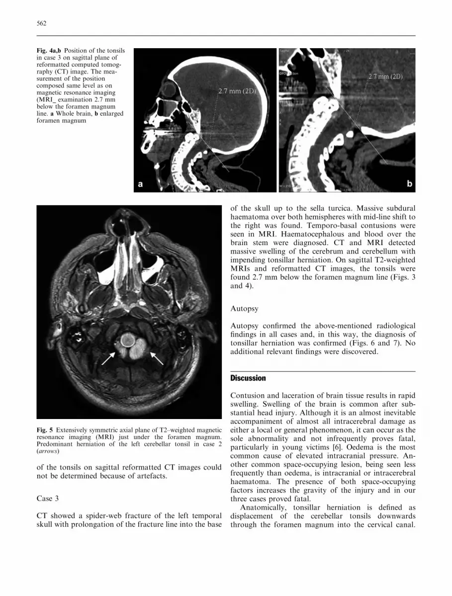
Predominantly, unilateral swelling and cerebellar herni-ation in cases 1 and 2 is explained by the unilaterallaceration of the cerebellum (Fig. 7). Detection of ton-sillar herniation in clinical MRI and MSCT examina-
tions was reported previously. On myelograms,O’Connor et al. reported that the cerebellar tonsils wereat least 3.8 mm higher than the line of the foramenmagnum in all of a series of 100 normal cases [7]. UsingT1-weighted sagittal MRIs, however, Barkovich et al.reported that the position of the tonsils ranged from8 mm above to 5 mm below the foramen magnum innormal subjects and that less than 2 mm of tonsillardisplacement on MRI would be of no clinical signifi-cance [8]. Ishikawa et al. noted tonsillar displacementbelow the foramen magnum line in only three from atotal of 18 cases of posterior fossa mass lesions [5].
We studied three cases of fatal blunt head injury. Thepost-mortem CT and MRI studies demonstratedtonsillar herniation. The position of the tonsils in ourcases was 5.5 mm, 5.1 mm, and 2.7 mm below theforamen magnum line. The tonsils were most clearlydemonstrated in the sagittal plane on T2-weightedMRIs.
Most authors recommend MRI for the diagnosis oftonsillar herniation in vivo [5, 7, 8, 9, 10]. The demon-stration of descending brain herniation in the foramenmagnum with CT is often difficult mostly because ofbony artefacts and partial volume effects [5]. The reso-lution of tonsillar herniation in MRI is superior to CT.MSCT and MRI used post mortem are comparativelyyoung examination methods in forensic medicine [2, 3, 4,11, 12, 13]. The sensitivity of MRI and CT to discoversigns of high intracranial pressure such as tonsillarherniation in clinical medicine is already known fromthe work of Barkovich [5], Ishikawa [8] and Reich [9].Today, CT and MRI have become indispensable in theclinical diagnostic of head injury [5, 7, 8, 9, 10]. Ourresults show that these methods are also sensitive in the
Fig. 6 Predominant swelling of the left cerebellar tonsil in case 1 inautopsy (arrow)
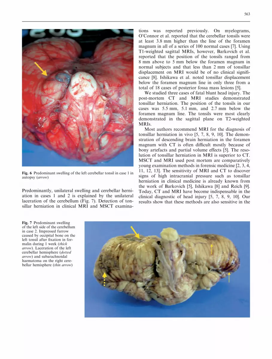
Fig. 7 Predominant swellingof the left side of the cerebellumin case 2. Impressed furrowcaused by occipital bone on theleft tonsil after fixation in for-malin during 1 week (thickarrow). Laceration of the leftcerebellar hemisphere (dottedarrow) and subarachnoidalhaematoma on the right cere-bellar hemisphere (thin arrow)
563

post-mortem diagnostics of descending tonsillar herni-ation.
Acknowledgements We are grateful to Karin Zwygart and ElkeSpielvogel (Departments of Diagnostic Radiology and Neurora-
diology, University Hospital of Bern) and to Roland Dorn and UrsKoenigsdorfer (Institute of Forensic Medicine, University of Bern)for the excellent help in data acquisition during the radiologicexamination and the forensic autopsy.
References
1. Medline Plus (2003) Medical encyclo-pedia,http://www.nlm.nih.gov/medline-plus/ency/article/001421.htm. Cited 20Nov 2003
2. Thali MJ, Yen K, Schweitzer W, VockP, Boesch C, Ozdoba C, Schroth G, IthM, Sonnenschein M, Doernhoefer T,Scheurer E, Plattner T, Dirnhofer R(2003) Virtopsy, a new imaging horizonin forensic pathology: virtual autopsyby post-mortem multislice computedtomography (MSCT) and magneticresonance imaging (MRI)–a feasibilitystudy. J Forensic Sci 48:386–403
3. Thali MJ, Yen K, Plattner T, Schweit-zer W, Vock P, Ozdoba C, Dirnhofer R(2002) Charred body: virtual autopsywith multi–slice computed tomographyand magnetic resonance imaging.J Forensic Sci 47:1326–1331
4. Thali MJ, Yen K, Schweitzer W, VockP, Ozdoba C, Dirnhofer R (2003) Intothe decomposed body–forensic digitalautopsy using multislice–computedtomography. Forensic Sci Int134:109–114
5. Ishikawa M, Kikuchi H, Fujisawa I,Yonekawa Y (1988) Tonsillar hernia-tion on magnetic resonance imaging.Neurosurgery 22:77–81
6. Knight B (1991) Head and spinalinjuries. In: Knight B (ed) Forensicpathology. Arnold, London,pp 156–197
7. O’Connor S, du Boulay G, Logue V(1973) The normal position of thecerebellar tonsils as demonstrated bymyelography. J Neurosurg 39:387–389
8. Barkovich AJ, Wippold FJ, ShermanJL, Citrin CM (1986) Significance ofcerebellar tonsillar position on MR. AmJ Neuroradiol 7:795–799
9. Reich JB, Sierra J, Camp W, ZanzonicoP, Deck MD, Plum F (1993) Magneticresonance imaging measurements andclinical changes accompanying tran-stentorial and foramen magnum brainherniation. Ann Neurol 33:159–170
10. Greenberg JO (1984) Neuroimaging inbrain swelling. Neurol Clin 2:677–694
11. Thali MJ, Schwab CM, Tairi K, Dir-nhofer R, Vock P (2002) Forensicradiology with cross-section modalities:spiral CT evaluation of a knife woundto the aorta. J Forensic Sci 47:1041–1045
12. Thali MJ, Schweitzer W, Yen K, VockP, Ozdoba C, Spielvogel E, Dirnhofer R(2003) New horizons in forensicradiology:the 60–second digitalautopsy-full-body examination of agunshot victim by multislice computedtomography. Am J Forensic MedPathol 24:22–27
13. Thali M, Vock P (2003) Role of andTechniques in Forensic Imaging. In:Payen-James J, Busuttil A, Smock W(eds) Forensic medicine: clinical andpathological aspects. GreenwichMedical Media, London, pp 731–745
564
Copyright © 2022 FDOKUMEN