Determination of Strain Field on the Superior Surface of Excised Larynx Vocal Folds Using DIC
Upload
mayoclinicCategory
view
3download
0
Vibro-acoustography Imaging of Permanent ProstateBrachytherapy seeds in an excised human prostate — PreliminaryResults and Technical Feasibility
F.G. Mitri*,1, B.J. Davis2, M.W. Urban1, A. Alizad1, J.F. Greenleaf1, G.H. Lischer3, T.M.Wilson3, and M. Fatemi11Mayo Clinic, Department of Physiology and Biomedical Engineering, Ultrasound ResearchLaboratory, 200 First Street SW, Rochester, MN 559052Mayo Clinic, Department of Radiation Oncology, 200 First Street SW, Rochester, MN 559053Mayo Clinic, Department of Urology, 200 First Street SW, Rochester, MN 55905
AbstractObjective—The objective in this work is to investigate the feasibility of using a new imaging toolcalled vibro-acoustography (VA) as a means of permanent prostate brachytherapy (PPB) seedlocalization to facilitate post-implant dosimetry (PID).
Methods and Materials—Twelve OncoSeed (standard) and eleven EchoSeed (echogenic)dummy seeds were implanted in a human cadaver prostate. Seventeen seeds remained after radicalretropubic prostatectomy. VA imaging was conducted on the prostate that was cast in a gel phantomand placed in a tank of degassed water. 2-D magnitude and phase VA image slices were obtained atdifferent depths within the prostate showing location and orientation of the seeds.
Results—VA demonstrates that twelve of seventeen (71%) seeds implanted were visible in the VAimage, and the remainder were obscured by intra-prostatic calcifications. Moreover, it is shown herethat VA is capable of imaging and locating PPB seeds within the prostate independent of seedorientation, and the resulting images are speckle free.
Conclusion—The results presented in this research show that VA allows seed detection within ahuman prostate regardless of their orientation, as well as imaging intraprostatic calcifications.
KeywordsBrachytherapy; Prostate cancer; Seed; Ultrasound; Vibro-acoustography; 43.20.+g General linearacoustics; 43.25.+y Nonlinear acoustics; 43.40.+s Structural acoustics and vibration; 43.35.+dUltrasonics, quantum acoustics, and physical effects of sound
© 2008 Elsevier B.V. All rights reserved.*Corresponding Author: F.G. Mitri Mayo Clinic College of Medicine 200 First St SW Rochester, MN 55905 Email: E-mail:[email protected] , E-mail: [email protected]'s Disclaimer: This is a PDF file of an unedited manuscript that has been accepted for publication. As a service to our customerswe are providing this early version of the manuscript. The manuscript will undergo copyediting, typesetting, and review of the resultingproof before it is published in its final citable form. Please note that during the production process errors may be discovered which couldaffect the content, and all legal disclaimers that apply to the journal pertain.
NIH Public AccessAuthor ManuscriptUltrasonics. Author manuscript; available in PMC 2010 March 1.
Published in final edited form as:Ultrasonics. 2009 March ; 49(3): 389–394. doi:10.1016/j.ultras.2008.10.011.
NIH
-PA Author Manuscript
NIH
-PA Author Manuscript
NIH
-PA Author Manuscript
1. IntroductionProstate cancer is the most common cancer in males in the United States. When prostate canceris suspected, a transrectal ultrasound-guided prostate biopsy is performed to establish thediagnosis [1]. If the biopsy confirms the presence of cancer, treatment may occur shortlythereafter, or a watchful waiting approach may be undertaken. Radical prostatectomy, externalbeam radiation therapy (EBRT) and brachytherapy are widely used as definitive therapies forlocalized cancer.
Brachytherapy (BT) [2-4] is one method that may be used to deliver high radiation doses tothe prostate, while sparing normal tissues that are located adjacent to the prostate. In BT,transrectal ultra-sonography (TRUS) is used to guide the placement of encapsulated 125Iodine(125I) or 103 Palladium (103Pd) seeds throughout the prostate gland [5]. Typical seeds arecylindrical in shape having a diameter of 800 microns and length of 4.5 mm. One possibledrawback of PPB implantation is the potential for under-dosing a portion or portions of thegland. This may occur if seeds are misplaced during the procedure.
Dynamic real-time calculation of the dose distribution based on actual seed placement duringthe implantation procedure is recommended to optimize the treatment planning procedure[6-10]. Magnetic resonance imaging (MRI) provides improved visualization of the prostateand its surrounding anatomy [11], which should make it the image modality of choice to guidebrachytherapy seed placement. However, the principal limitation to its routine use in PPB, isthe complex and challenging environment inherent to MRI technology and the constrainedergonomics of closed-bore scanners. Computed tomography (CT) has been very useful inidentifying seeds and is the standard for determining postimplantation dosimetry. However,with CT, the delineation of the prostate gland and the adjacent structures is limited because ofrelatively weak intrinsic contrast among soft tissues. Furthermore, because the prostate(pseudo)capsule is not visualized on CT, assessment of the volume of the gland is difficult andinaccurate [12,13]. Transperineal, three-dimensional (3-D) transperineal— and transrectal-guided ultrasonography were developed as other alternatives [5,14]. Ultrasound techniquespredicted prostate size with a high degree of accuracy but seeds give rise to artifacts [15], andmovement of the gland during imaging may hamper evaluation of the prostate and seeddistribution.. Although a tissue/titanium interface is expected to give excellent ultrasoundechoes, practical experience indicates that a majority of implanted seeds are difficult to detectwith ultrasound imaging. Typically, only 30%-50 % of the encapsulated seeds are identifiedunder in vivo conditions by scanning the whole prostate volume [16,17]. Thus, TRUSexamination of the prostate is of limited value for post-implant seed localization.
Recent breakthroughs in imaging have catalyzed the development of novel techniques, suchas “elasticity imaging methods” that are sensitive to the elastic properties of tissue [18-20].Acoustical radiation force-based methods are successfully used in many biomedicalapplications including ophthalmology [21], detecting and characterizing lesions [22], stagingdeep venous thrombosis [23], imaging breast calcifications [24], etc. One possible methodamong these techniques that can be used to improve seed visualization and enhance thepotential for real-time quantitative and meaningful feedback during the procedure is Vibro-acoustography (VA) [25]. Previous work with VA has shown substantial capabilities of thistechnique versus pulse-echo ultrasound to detect PPB seeds at various orientations in anidealized laboratory setting, in which no human tissue was involved [26]. Moreover,quantitative analysis has confirmed the validity of this approach [27], yet evaluation of theseed localization in a biological prostate has not been previously undertaken.
The purpose of the present study is to examine PPB seed imaging in an excised human prostateusing the VA technique. This is an interim step to the realistic case of a prostate in a patient.
Mitri et al. Page 2
Ultrasonics. Author manuscript; available in PMC 2010 March 1.
NIH
-PA Author Manuscript
NIH
-PA Author Manuscript
NIH
-PA Author Manuscript

The objective of this research is to show the feasibility of VA in imaging PPB seeds in anexcised human prostate. Here, a fresh cadaver prostate is implanted with standard andechogenic seeds. Then, images showing seed location and orientation in the excised prostateare produced. The results as well as the potential use of VA in detecting PPB seeds arediscussed, and a summary is presented.
2. Materials and methodsThe VA imaging system that is used to scan the prostate is described in Fig. 1. The VA imagingtechnique produces a map of the mechanical response of an object to a dynamic force appliedat each point. The method utilizes ultrasound radiation force to remotely exert a localizedoscillating stress field, or in other words a tapping force, at a desired frequency within, or onthe surface of an object, and records the resultant acoustic response or acoustic emission. Thisacoustic response, which is normally in the low kHz range, is a function of the viscoelasticproperties of the object and can be used to produce an image of the object. To confine theoscillating radiation stress to the desired region, VA uses two ultrasound beams driven atslightly different frequencies, propagating along separate paths. The beams are arranged tocross each other at their respective foci, and thus produce a modulated field at a confined, smallcross-sectional region. The mix of two ultrasound beams operating at different frequencies andintersecting in space generates a radiation force on the object by which multiple otherfrequencies are produced, including a component at the difference of the two operatingfrequencies. The object to be imaged is placed at the joint focal plane of the ultrasoundtransducer, also known as the scanning plane. Depending on the elastic properties of the object,the radiation force may cause a portion of the object, or the entire object, to vibrate at thedifference frequency. The acoustic emission resulting from object vibration is received by ahydrophone which is placed nearby. If the wavelength of vibration is large compared with theobject size, the acoustic emission pressure field is almost omnidirectional. Therefore, at lowfrequencies, the hydrophone position is not a critical parameter in the measurement of theacoustic emission signal. To form an image, the focal point of the transducer is moved acrossthe scanning plane on the object in a raster pattern. The acoustic emission is received at eachposition, and an image is formed by displaying the magnitude (or phase) of such signals atcorresponding positions on the image plane. The spatial resolution of this imaging method isdetermined by the ultrasound beam-width at the focal plane, which is normally of the order ofthe incident ultrasound wavelength.
For this experiment, the two ultrasound beams were generated by a two-element homemadeconfocal transducer with a diameter of 45 mm, a focal distance of 70 mm and a center frequencyof 3 MHz. The elements were driven by two continuous-wave (CW) signals at frequencies of3 MHz and 3 MHz + 20 kHz. The driving radio frequency (RF) signals were obtained fromtwo stable function generators (HP 33120 A, Hewlett-Packard Company, Houston TX, USA).The transducer was mounted on a three-axes positioning system and immersed in a tank ofdegassed water to ensure good acoustical coupling. For this particular confocal transducer, theon-and off-axes resolutions, defined as the focal spot at full-width at half-maximum, were 10and 0.7 mm respectively. The acoustic emission pressure field was detected by a submergedaudio hydrophone (Model ITC—6050 C, Santa Barbara CA, USA) with sensitivity —157dBre 1V/μPa, diameter and length of 50 and 200 mm respectively (sensitive area of 70 mm), andfrequency response between 1 Hz and 60 kHz, placed in the water tank. The signal receivedwas bandpass filtered and amplified (Stanford Research Systems, SR650, Sunnyvale CA,USA) to eliminate noise, then digitized by a 12-bits/sample digitizer (National InstrumentsVXI-1000, Austin TX, USA) at a rate sufficiently higher than the Nyquist rate. The data werethen recorded on a computer. Magnitude and phase images were produced after processing thedata. The phase images acquired in the experiment contained phase wraps. Therefore, themeasured phase should be processed (unwrapped) prior to use as a comprehensible data. To
Mitri et al. Page 3
Ultrasonics. Author manuscript; available in PMC 2010 March 1.
NIH
-PA Author Manuscript
NIH
-PA Author Manuscript
NIH
-PA Author Manuscript
provide a continuous phase image, two-dimensional phase unwrapping was performed. Anunweighted least-squares multigrid phase unwrapping algorithm was used [28]. This methodis iterative and operates on multiple scales to eliminate high-frequency components of the error.Absence of noise in the images presented leads us to believe that no errors were encountered.The phase unwrapping process performs very quickly with C code provided in Ref. [28]implemented on a modern computer.
A cadaver prostate was implanted with the patient in the dorsal lithotomy position usingstandard TRUS and fluoroscopic guidance within 24 hours of the patient’s expiration. Theseeds were implanted in the cadaver to simulate an actual PPB therapy procedure. Seedorientation and placement would have been different if the implantation procedure is performedwithin the excised prostate tissue. A total of 23 seeds, 12 standard and 11 echogenic, wereimplanted. Then, the prostate was removed via conventional radical retropubic prostatectomy(RRP) as performed by a urologic surgeon (G.H. Lischer). The cadaver prostate was then castin formalin-catalyzed porcine gelatin (Sigma-Aldrich G2500 Type A, 300 bloom, Saint LouisMO, USA) at 15% concentration by volume to hold it in place. The gland was centered in thegelatin block having the dimensions 13×13×5 cm3. Seventeen seeds remained in the prostatefollowing prostatectomy as determined by fluoroscopic imaging (Fig. 2). Six seeds wereinadvertently extruded from the prostate during the extirpation even though they wereimplanted well within the gland. In addition, it was observed that the seeds were rearrangedwithin the gland with the substantial prostate manipulation required during the RRP andplacement in the gelatin block. All procedures used on the cadaver were approved by the MayoClinic Institutional Review Board (protocol ID 06-003629). The post-implanted prostate wasthen immersed in a tank of degassed water for scanning (Fig. 1).
Magnitude and phase VA images at different depths were acquired by scanning the transducerover the gland surface by a 1 mm incremental step for a total range of 24 mm deep inside theprostate gland. The images covered an area of 50 mm by 50 mm, scanned at 0.25 mm/pixelincremental step. Only representative magnitude and phase VA images taken at 1, 5, 10, and15 mm deep from the surface of the prostate, showing seed location and orientation in theprostate are presented in Fig. 3 and 4, respectively. These VA images show some of the seedsat various angles. Other seeds were visible when the scan was performed at different depths.As the scan was performed in depth to cover the entire volume of the prostate, some of theseeds start to disappear from the VA image as they fall out of the focal region of the confocaltransducer (See Figs. 3-(d), 4-(d)). After analyzing qualitatively the whole set of magnitudeand phase VA images, 12 seeds were detected out of 17 (71%) seeds implanted within theprostate. The detection of seeds and images review were carried out by visual inspection asperformed by urologists (GHL, TMW) and radiation oncologist (BJD). This preliminary result(detection rate of 71%) has already surpassed the seed detection rate typically reported forconventional TRUS in in vivo applications, to be approximately 30%-50% [16]. This detectionrate should not be understood as an absolute value since one case (obtained from an in vitrostudy) is reported here.
3. DiscussionWe have performed an initial study of imaging permanent prostate brachytherapy (PPB) seedsby vibroacoustography (VA). VA imaging of PPB seeds offers a unique advantage comparedto pulse echo ultrasound because it is relatively insensitive to seed orientation. In addition, VAimaging enables detection of intra-prostatic calcifications as confirmed by histology.Calcifications are a common finding in the prostate and appear to be mostly associated withbenign hyperplasia [29]. However, a recent report showed that calcifications can also occur indirect association with prostatic adenocarcinoma [29], although the incidence of thisassociation is not as high as in breast carcinoma [30]. The results of this work are consistent
Mitri et al. Page 4
Ultrasonics. Author manuscript; available in PMC 2010 March 1.
NIH
-PA Author Manuscript
NIH
-PA Author Manuscript
NIH
-PA Author Manuscript

with those previously obtained in well-controlled laboratory experiments [26,27], and provideencouraging data for further evaluation of VA as a means of developing intraoperative-baseddosimetry of PPB. Upon visual inspection of the magnitude and phase VA images (obtainedat 20 kHz), it is noticeable that seeds are clearly identified and calcifications are evidentlydisplayed. Such images have high spatial resolution, without speckle, good contrast, and highsignal to noise ratio. For this specific excised prostate, the 71% seed detection rate within theprostate has provided an impetus to further develop, evaluate the performance, and optimizeVA to achieve an improved seed detection rate and to evaluate this approach in vivo. The 29%-lack of seed detection was attributed to intra-prostatic calcifications near the center of theprostate gland (See Figs. 3 and 4) which reflect ultrasound waves and obscure the seeds. Thechoice of vibration frequency used in this experiment, 20 kHz, was determined based onexperience with imaging breast calcifications. The selected difference frequency does notcorrespond to the resonance frequency of the seeds. The reason is that the seeds’ first naturalresonance frequency was estimated (using finite element analysis) to be around 150 kHz, afrequency that is beyond the sensitivity of our hydrophone. Further evaluation of otheroptimized vibration frequencies is warranted.
One also notices a contrast reversal in the magnitude images. This artifact may be the result ofsound reverberations within the gel block. Moreover, interference of sound waves bouncingfrom the walls of the water tank may also contribute to the contrast change, depending on thewaves’ relative phases. This issue requires further investigation to properly study the effectsof reverberations on the contrast of VA images.
The unwrapped phase of a vibration-based signal has been shown to provide unique high-contrast images [31]. The unwrapped phase of the acoustic emission from prostate tissue isrelatively constant. The unwrapped phase contrast of the calcification differs with the focaldistance. The borders of the calcification are well-defined in the unwrapped phase images. Thebrachytherapy seeds are well visualized for focal distances of 1-12 mm (Fig. 4-(a)-(d)). In themagnitude images (Fig. 3-(a)-(d)), the contrast of the seeds with respect to the surroundingtissue reverses with increasing focal distance; however, the seeds are always brighter than thesurrounding tissue providing a positive contrast. This unwrapped-phase contrast mechanismmay provide better visualization of embedded brachytherapy seeds for therapy monitoring.
The image spatial resolution improves as frequency increases. The transducer used in thisexperiment operated at 3.0 MHz which resulted of a lateral spatial resolution of 0.7 mm. VAimage resolution would be better (< 0.7 mm) at ultrasound frequencies in the range of 5.0–7.5MHz which is the range used in conventional transrectal ultrasound (TRUS) imaging [6].
In the present study, the scanning mechanism used for this experiment scans the object onepoint at a time, making the data collection a relatively lengthy process (up to 4 minutes perimage covering an area of 50 mm by 50 mm). A long imaging time is not desirable for prostateimaging because body motions within this period can introduce “motion artifact” in the imagesand the procedure anesthesia time could be impractical. One way to reduce the scanning timesignificantly is to use electronic beam focusing and steering [32] providing high-speedscanning. Current work in our group is directed towards the development and testing of a lineararray probe with dynamic focusing whereby the imaging time should be reduced by at leastone order of magnitude.
Although VA uses ultrasound as a non-invasive energy source, the system properties aredifferent from those of conventional pulse-echo ultrasound imaging (B-scan), and, inparticular, TRUS. As a result, VA promises new diagnostic applications not normally offeredby conventional ultrasound. A notable advantage of VA over pulse echo ultrasound is that VAcan image PPB seeds practically at any angle [26], whereas TRUS imaging is highly sensitive
Mitri et al. Page 5
Ultrasonics. Author manuscript; available in PMC 2010 March 1.
NIH
-PA Author Manuscript
NIH
-PA Author Manuscript
NIH
-PA Author Manuscript

to seed orientation [33]. The seeds start to break up and each appears as two separate dots whenthey are imaged slightly off the perpendicular angle of incidence [33]. On the other hand, VAshows a considerable ability to detect the PPB seeds independent of their orientation. Therefore,VA is expected to detect randomly oriented seeds in and around the prostate, while TRUS canmiss seeds depending on their angle relative to the beam axis. In addition to imaging soft tissue,VA can image stiff metallic implants, such as calcifications in breast [24,34] and carotid andiliac arteries [25] with great accuracy and efficiency. Furthermore, it has been shown that VAcan detect very small hard objects, even breast microcalcifications [35] having diametersranging from 0.1 to 1 mm.
The focus of the present study was to assess the capability of VA in detecting seeds in a humannon pathologic prostate specimen. Cancerous prostatic tissue, as well as healthy prostatictissue, may contain calcifications [29] of sizes comparable to PPB seeds. Once detected by theVA system, calcifications may therefore be confused with seeds, hence, producing falsepositives. It is doubtful, however, that the calcifications will be of the same three-dimensionalsize and shape of the seeds but this potential problem is nonetheless acknowledged. A similarproblem also exists with conventional TRUS. In this initial in vitro study, no attempt has beenmade here to assess the false positive rate in seed detection. Further investigation is requiredto address this issue.
A key question raised for in vivo use of VA is whether the system can function properly at areasonably low intensity level that is considered safe. Previous work [36] has shown that VAoperates at intensities below the FDA recommended value [37] of 720 mW/cm2. For thisexperiment, it was verified that using ultrasound intensities at or below the FDA recommendedlevel was achieved. In comparison to fluoroscopy [38], where the use of X-Ray may be ofmodest concern for the safety of the operating room personnel and the patient, the operationof diagnostic level ultrasound used in VA is considered safe.
Another limitation is the result of two effects: phase aberration and sound speed variations intissue. First, the ultrasound beams tend to defocus; second, the two beams fail to intersect attheir mutual focal point. The first effect results in beam broadening, which in turn results inthe loss of spatial resolution and decreased sensitivity due to decreased peak radiation intensity.The second effect results in similar outcomes. In addition, this effect may cause the beams tointersect at an unanticipated location in the object, and hence introduce image distortion.
4. ConclusionThese results represent the first study of the efficacy of using the VA technique for imagingPPB seeds in an excised human prostate in vitro. Results presented here show that VA allowsdetection regardless of seed orientation as well as imaging intraprostatic calcifications. Thesepromising results suggest that VA may be useful as a clinical tool in seed localization for PPB.Ongoing work is directed towards evaluating the performance of VA in imaging PPB seeds ina large number of excised prostates. The design of specific probes for brachytherapyapplications is currently under investigation.
AcknowledgementsResearch supported by the National Institute of Health under Grant No. EB 00535-04, Grant No. CA 91956-06P2,and a Grant by Oncura Inc., Plymouth Meeting, PA. Disclosure: Parts of the techniques used here are patented by M.Fatemi and J.F. Greenleaf. The authors are grateful to M.A. Hadaway, W.N. Lajoie and R.R. Kinnick for technicaland laboratory assistance.
Mitri et al. Page 6
Ultrasonics. Author manuscript; available in PMC 2010 March 1.
NIH
-PA Author Manuscript
NIH
-PA Author Manuscript
NIH
-PA Author Manuscript
REFERENCES[1]. Brawer MK, Ploch NR, Bigler SA. Prostate-Cancer Tumor Location as Predicted by Digital Rectal
Examination Transferred to Ultrasound and Ultrasound-Guided Prostate Needle-Biopsy. J. Cell.Biochem 1992:74–77. [PubMed: 1322917]
[2]. Beyer DC. The Evolving Role of Prostate Brachytherapy. Cancer Control 2001;8:163–170. [PubMed:11326171]
[3]. Porter AT, Forman JD. Prostate Brachytherapy - an Overview. Cancer 1993;71:953–958. [PubMed:8428345]
[4]. Syed AMN, Puthawala A, Sharma A, Gamie S, Londrc A, Cherlow JM, Damore SJ, Nazmy N, SheikhKM, Ko SJ. High-Dose-Rate Brachytherapy in the Treatment of Carcinoma of the Prostate. CancerControl 2001;8:511–521. [PubMed: 11807421]
[5]. Holm HH, Juul N, Pedersen JF, Hansen H, Stroyer I. Transperineal 125iodine seed implantation inprostatic cancer guided by transrectal ultrasonography. J. Urol 1983;130:283–286. [PubMed:6876274]
[6]. Nag S, Beyer D, Friedland J, Grimm P, Nath R. American brachytherapy society (ABS)recommendations for transperineal permanent brachytherapy of prostate cancer. Int. J. Radiat.Oncol. Biol. Phys 1999;44:789–799. [PubMed: 10386635]
[7]. Blake CC, Elliot TL, Slomka PJ, Downey DB, Fenster A. Variability and accuracy of measurementsof prostate brachytherapy seed position in vitro using three-dimensional ultrasound: An intra- andinter-observer study. Med. Phys 2000;27:2788–2795. [PubMed: 11190962]
[8]. Wei ZP, Gardi L, Downey DB, Fenster A. Automated localization of implanted seeds in 3D TRUSimages used for prostate brachytherapy. Med. Phys 2006;33:2404–2417. [PubMed: 16898443]
[9]. Jain AK, Zhou Y, Mustufa T, Burdette EC, Chirikjian GS, Fichtinger G. Matching and reconstructionof brachytherapy seeds using the Hungarian algorithm (MARSHAL). Med. Phys 2005;32:3475–3492. [PubMed: 16372418]
[10]. Fichtinger G, Burdette EC, Tanacs A, Patriciu A, Mazilu D, Whitcomb LL, Stoianovici D.Robotically assisted prostate brachytherapy with transrectal ultrasound guidance - Phantomexperiments. Brachytherapy 2006;5:14–26. [PubMed: 16563993]
[11]. Yu KK, Hricak H. Imaging prostate cancer. Radiologic Clinics of North America 2000;38:59–85.[PubMed: 10664667]
[12]. Roach M, FaillaceAkazawa P, Malfatti C, Holland J, Hricak H. Prostate volumes defined bymagnetic resonance imaging and computerized tomographic scans for three-dimensional conformalradiotherapy. Int. J. Radiat. Oncol. Biol. Phys 1996;35:1011–1018. [PubMed: 8751410]
[13]. Rasch C, Barillot I, Remeijer P, Touw A, van Herk M, Lebesque JV. Definition of the prostate inCT and MRI: A multi-observer study. Int. J. Radiat. Oncol. Biol. Phys 1999;43:57–66. [PubMed:9989514]
[14]. Stock RG, Stone NN, Wesson MF, Dewyngaert JK. A Modified Technique Allowing InteractiveUltrasound-Guided 3-Dimensional Transperineal Prostate Implantation. Int. J. Radiat. Oncol. Biol.Phys 1995;32:219–225. [PubMed: 7721619]
[15]. Nag S, Ciezki JP, Cormack R, Doggett S, DeWyngaert K, Edmundson GK, Stock RG, Stone NN,Yu Y, Zelefsky MJ. Intraoperative planning and evaluation of permanent prostate brachytherapy:Report of the American Brachytherapy Society. Int. J. Radiat. Oncol. Biol. Phys 2001;51:1422–1430. [PubMed: 11728703]
[16]. Su Y, Davis BJ, Herman MG, Robb RA. Estimation and variability of D90 as a function of seeddetectability rates: Do all seeds need to be detected in order to compute useful Intraoperativedosimetry during prostate brachytherapy? Int. J. Radiat. Oncol. Biol. Phys 2004;60:S596–S597.
[17]. Su Y, Davis BJ, Herman MG, Manduca A, Robb RA. Examination of dosimetry accuracy as afunction of seed detection rate in permanent prostate brachytherapy. Med. Phys 2005;32:3049–3056. [PubMed: 16266119]
[18]. Greenleaf JF, Fatemi M, Insana MF. Selected methods for imaging elastic properties of biologicaltissues. Ann. Rev. Biomed. Eng 2003;5:57–78. [PubMed: 12704084]
[19]. Fatemi M, Manduca A, Greenleaf JF. Imaging elastic properties of biological tissues by low-frequency harmonic vibration. Proc. IEEE 2003;91:1503–1519.
Mitri et al. Page 7
Ultrasonics. Author manuscript; available in PMC 2010 March 1.
NIH
-PA Author Manuscript
NIH
-PA Author Manuscript
NIH
-PA Author Manuscript

[20]. Gao L, Parker KJ, Lerner RM, Levinson SF. Imaging of the elastic properties of tissue - A review.Ultrasound Med. Biol 1996;22:959–977. [PubMed: 9004420]
[21]. Negron LA, Viola F, Black EP, Toth CA, Walker WF. Development and characterization of avitreous mimicking material for radiation force imaging. IEEE Trans. Ultrason. Ferroelectr. Freq.Control 2002;49:1543–1551. [PubMed: 12484477]
[22]. Nightingale K, Soo MS, Nightingale R, Trahey G. Acoustic radiation force impulse imaging: invivo demonstration of clinical feasibility. Ultrasound Med. Biol 2002;28:227–235. [PubMed:11937286]
[23]. Xie H, Kim K, Aglyamov SR, Emelianov SY, Chen X, O’Donnell M, Weitzel WF, Wrobleski SK,Myers DD, Wakefield TW, Rubin JM. Staging deep venous thrombosis using ultrasound elasticityimaging: Animal model. Ultrasound Med. Biol 2004;30:1385–1396. [PubMed: 15582239]
[24]. Alizad A, Whaley DH, Greenleaf JF, Fatemi M. Potential applications of vibro-acoustography inbreast imaging. Technol. Cancer Res. Treat 2005;4:151–157. [PubMed: 15773784]
[25]. Fatemi M, Greenleaf JF. Ultrasound-stimulated vibro-acoustic spectrography. Science1998;280:82–85. [PubMed: 9525861]
[26]. Mitri FG, Davis BJ, Greenleaf JF, Fatemi M. Comparative study of vibro-acoustography versuspulse-echo ultrasound in imaging permanent prostate brachytherapy seeds. Ultrasonics Accepted.2008(in press)
[27]. Mitri FG, Trompette P, Chapelon JY. Improving the use of vibro-acoustography for brachytherapymetal seed imaging: A feasibility study. IEEE Trans. Med. Imag 2004;23:1–6.
[28]. Ghiglia, DC.; Pritt, MD. Algorithms, and Software. John Wiley & Sons, Inc.; New York, NY: 1998.Two-Dimensional Phase Unwrapping--Theory.
[29]. Suh JH, Gardner JM, Kee KH, Shen S, Ayala AG, Ro JY. Calcifications in prostate and ejaculatorysystem: a study on 298 consecutive whole mount sections of prostate from radical prostatectomyor cystoprostatectomy specimens. Ann. Diag. Path 2008;12:165–170.
[30]. Gibbs NM, Price JL, Ainsworth RW, Korpal K. A radiological and histological study of prostaticcalcification associated with primary carcinoma; a comparison with mammary microcalcification.Clin. Oncol 1975;1:305–313.
[31]. Urban MW, Kinnick RR, Greenleaf JF. Measuring the phase of vibration of spheres in a viscoelasticmedium as an image contrast modality. J. Acoust. Soc. Am 2005;118:3465–3472. [PubMed:16419793]
[32]. Silva GT, Greenleaf JF, Fatemi M. Linear arrays for vibro-acoustography: A numerical simulationstudy. Ultrasonic Imaging 2004;26:1–17. [PubMed: 15134390]
[33]. Davis BJ, Kinnick RR, Fatemi M, Lief EP, Robb RA, Greenleaf JF. Measurement of the ultrasoundbackscatter signal from three seed types as a function of incidence angle: Application to permanentprostate brachytherapy. Int. J. Radiat. Oncol. Biol. Phys 2003;57:1174–1182. [PubMed: 14575850]
[34]. Fatemi M, Wold LE, Alizad A, Greenleaf JF. Vibro-acoustic tissue mammography. IEEE Trans.Med. Imag 2002;21:1–8.
[35]. Alizad A, Fatemi M, Wold LE, Greenleaf JF. Performance of vibro-acoustography in detectingmicrocalcifications in excised human breast tissue: A study of 74 tissue samples. IEEE Trans. Med.Imag 2004;23:307–312.
[36]. Fatemi M, Greenleaf JF. Vibro-acoustography: An imaging modality based on ultrasound-stimulated acoustic emission. Proc. Natl. Acad. Sci. USA 1999;96:6603–6608. [PubMed:10359758]
[37]. Patton CA, Harris GR, Phillips RA. Output Levels and Bioeffects Indexes from DiagnosticUltrasound Exposure Data Reported to the FDA. IEEE Trans. Ultrason. Ferroelectr. Freq1994;41:353–359.
[38]. French D, Morris J, Keyes M, Goksel O, Salcudean S. Computing intraoperative dosimetry forprostate brachytherapy using TRUS and fluoroscopy. Acad. Rad 2005;12:1262–1272.
Mitri et al. Page 8
Ultrasonics. Author manuscript; available in PMC 2010 March 1.
NIH
-PA Author Manuscript
NIH
-PA Author Manuscript
NIH
-PA Author Manuscript
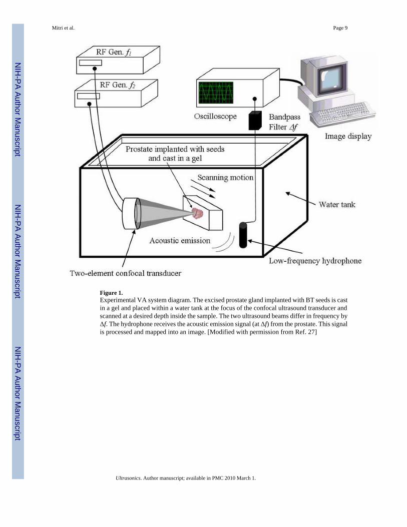
Figure 1.Experimental VA system diagram. The excised prostate gland implanted with BT seeds is castin a gel and placed within a water tank at the focus of the confocal ultrasound transducer andscanned at a desired depth inside the sample. The two ultrasound beams differ in frequency byΔf. The hydrophone receives the acoustic emission signal (at Δf) from the prostate. This signalis processed and mapped into an image. [Modified with permission from Ref. 27]
Mitri et al. Page 9
Ultrasonics. Author manuscript; available in PMC 2010 March 1.
NIH
-PA Author Manuscript
NIH
-PA Author Manuscript
NIH
-PA Author Manuscript

Figure 2.X-ray of the cadaver prostate after prostatectomy. Seventeen seeds remained in the prostateafter RRP. Intra-prostatic calcifications are faintly visible since the image was taken at 100kVp. The three identification pins of a spherical end shape appear as bright spherical dots inthe top of the image. These pins attached to the gel phantom at different locations have servedas a reference for imaging.
Mitri et al. Page 10
Ultrasonics. Author manuscript; available in PMC 2010 March 1.
NIH
-PA Author Manuscript
NIH
-PA Author Manuscript
NIH
-PA Author Manuscript
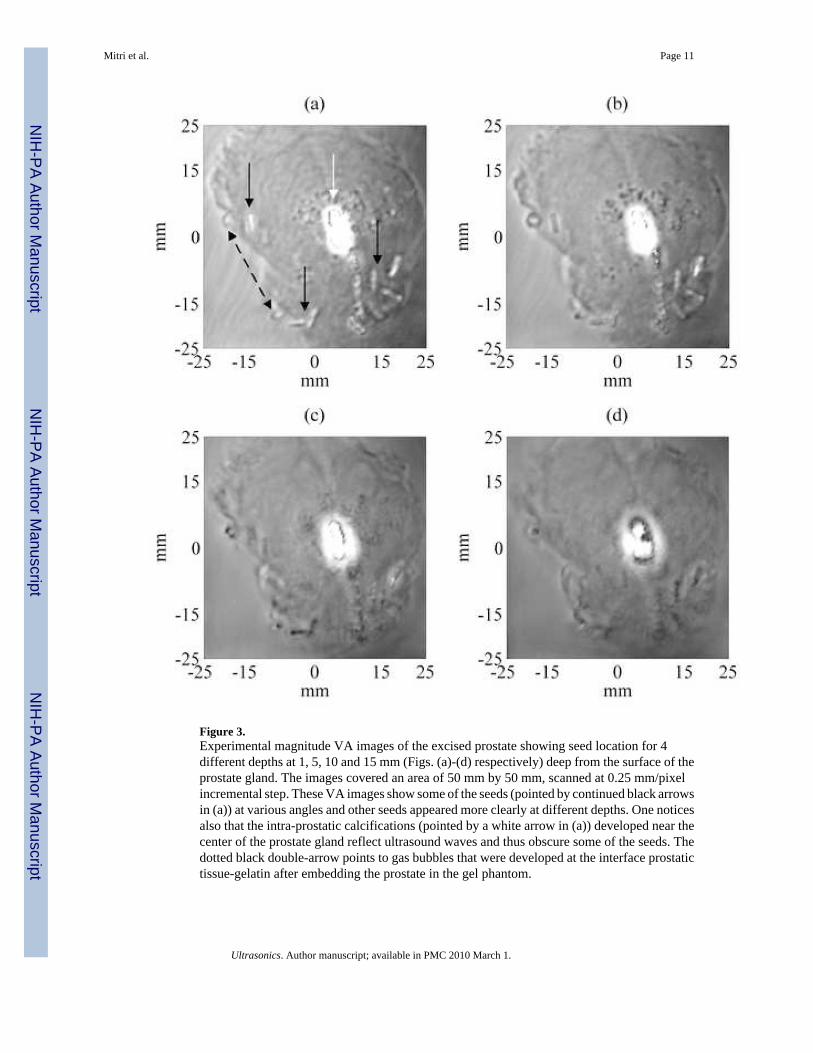
Figure 3.Experimental magnitude VA images of the excised prostate showing seed location for 4different depths at 1, 5, 10 and 15 mm (Figs. (a)-(d) respectively) deep from the surface of theprostate gland. The images covered an area of 50 mm by 50 mm, scanned at 0.25 mm/pixelincremental step. These VA images show some of the seeds (pointed by continued black arrowsin (a)) at various angles and other seeds appeared more clearly at different depths. One noticesalso that the intra-prostatic calcifications (pointed by a white arrow in (a)) developed near thecenter of the prostate gland reflect ultrasound waves and thus obscure some of the seeds. Thedotted black double-arrow points to gas bubbles that were developed at the interface prostatictissue-gelatin after embedding the prostate in the gel phantom.
Mitri et al. Page 11
Ultrasonics. Author manuscript; available in PMC 2010 March 1.
NIH
-PA Author Manuscript
NIH
-PA Author Manuscript
NIH
-PA Author Manuscript

Figure 4.Experimental unwrapped phase VA images of the excised prostate showing seed location for4 different depths at 1, 5, 10 and 15 mm (Figs. (a)-(d) respectively) deep from the surface ofthe prostate gland. Displaying the unwrapped phase of the acoustic emission provides anothersource of image contrast for examining the prostate tissue and embedded objects. Theunwrapped phase of the acoustic emission from prostate tissue is relatively constant. Theunwrapped phase contrast of the calcification differs with the depth of the focal plane. Theborder of the calcification is well-defined and the brachytherapy seeds are well visualized atfocal depths of 1-10 mm (i.e. Figs. 4-(a)-(c)).
Mitri et al. Page 12
Ultrasonics. Author manuscript; available in PMC 2010 March 1.
NIH
-PA Author Manuscript
NIH
-PA Author Manuscript
NIH
-PA Author Manuscript
Copyright © 2022 FDOKUMEN