Assessment of Agreement between Two Difference Prostate ...
10
biology Article Assessment of Agreement between Two Difference Prostate-Specific Antigen Assay Modalities Jae Hoon Chung 1 , Minsu Park 2 , Hyun Cho 3 , Wan Song 1 , Minyong Kang 1 , Hyun Hwan Sung 1 , Hwang Gyun Jeon 1 , Byong Chang Jeong 1 , Seong IL Seo 1 , Hyun Moo Lee 1 and Seong Soo Jeon 1, * Citation: Chung, J.H.; Park, M.; Cho, H.; Song, W.; Kang, M.; Sung, H.H.; Jeon, H.G.; Jeong, B.C.; Seo, S.I.; Lee, H.M.; et al. Assessment of Agreement between Two Difference Prostate-Specific Antigen Assay Modalities. Biology 2021, 10, 297. https://doi.org/10.3390/ biology10040297 Academic Editor: Paul S. Cooke Received: 31 January 2021 Accepted: 29 March 2021 Published: 5 April 2021 Publisher’s Note: MDPI stays neutral with regard to jurisdictional claims in published maps and institutional affil- iations. Copyright: © 2021 by the authors. Licensee MDPI, Basel, Switzerland. This article is an open access article distributed under the terms and conditions of the Creative Commons Attribution (CC BY) license (https:// creativecommons.org/licenses/by/ 4.0/). 1 Samsung Medical Center, Department of Urology, Sungkyunkwan University School of Medicine, Seoul 06351, Korea; [email protected] (J.H.C.); [email protected] (W.S.); [email protected] (M.K.); [email protected] (H.H.S.); [email protected] (H.G.J.); [email protected] (B.C.J.); [email protected] (S.I.S.); [email protected] (H.M.L.) 2 Department of Statistics, Keimyung University, Daegu 42403, Korea; [email protected] 3 Statistics and Data Center, Samsung Biomedical Research Institute, Samsung Medical Center, Seoul 06351, Korea; [email protected] * Correspondence: [email protected]; Tel.: +82-2-3410-3558; Fax: +82-2-3410-6992 Simple Summary: Prostate-specific antigen is a biomarker for prostate cancer. If the level of prostate- specific antigen is high, a prostate biopsy is needed to diagnose prostate cancer. However, the definite level of prostate-specific antigen that requires prostate biopsy has not been established. Currently, there are many kinds of assay modalities that have been used for prostate-specific antigen testing. This study was conducted under the hypothesis that there will be differences between different assay modalities; therefore, there is no definite prostate-specific antigen level for prostate biopsy. In our study, the level of prostate-specific antigens was measured in one blood sample per patient, with two different assay modalities in 4810 patients. As a result, we confirmed that the overall agreement between the two modalities is excellent, but the agreement is slightly different in some ranges that may give clinical significance. Accordingly, the conformity between each assay modality should be secured in the future, and the threshold for the level of prostate-specific antigens for biopsy by each assay modality should be independently determined. Abstract: There is controversy over the usefulness of prostate-specific antigen (PSA) as a prostate cancer (PCa) biomarker. This controversy arises when there are differences in the results of PSA assay modalities. In this study, which aimed to evaluate a proper validation between the two PSA assay modalities, the agreement between the results of the two modalities was analyzed. PSA examinations were conducted using two PSA assay modalities in 4810 patients. The intra-class correlation coefficient (ICC) and weighted kappa analysis were used to evaluate the agreement between the two assay modalities. A linear regression was performed to evaluate the association between the two assay modalities. According to ICC values (ICC: 0.999, p < 0.001) and weighted kappa analysis values (kappa: 0.951, alpha’s standard error (ASE): 0.001, p < 0.0001), the agreement between the assay modalities was rated as excellent. However, the strength of agreement was poor in the following PSA sub-groups: 0.05–0.1 ng/mL (ICC: 0.281, p = 0.0860); 0.15–0.2 ng/mL (ICC: 0.288, p = 0.0036); 1.5–2.0 ng/mL (ICC: 0.360, p = 0.0860); and 2.0–2.5 ng/mL (ICC: 0.303, p = 0.0868). In linear regression analysis, when modality B PSA yielded a value of 0.2 ng/mL, the expected value for modality A was 0.258 ng/mL (95% CI: 0.255–0.260), and when modality B PSA yielded a value of 4 ng/mL, the expected value for modality A was 3.192 ng/mL (95% CI: 3.150–3.235). The difference in the PSA values between the two PSA assay modalities is confirmed, and this difference may be clinically meaningful. Keywords: prostate-specific antigen; diagnosis; statistics Biology 2021, 10, 297. https://doi.org/10.3390/biology10040297 https://www.mdpi.com/journal/biology
-
Upload
khangminh22 -
Category
Documents
-
view
1 -
download
0
Transcript of Assessment of Agreement between Two Difference Prostate ...
biology
Article
Assessment of Agreement between Two DifferenceProstate-Specific Antigen Assay Modalities
Jae Hoon Chung 1 , Minsu Park 2 , Hyun Cho 3, Wan Song 1, Minyong Kang 1, Hyun Hwan Sung 1,Hwang Gyun Jeon 1, Byong Chang Jeong 1, Seong IL Seo 1, Hyun Moo Lee 1 and Seong Soo Jeon 1,*
�����������������
Citation: Chung, J.H.; Park, M.; Cho,
H.; Song, W.; Kang, M.; Sung, H.H.;
Jeon, H.G.; Jeong, B.C.; Seo, S.I.; Lee,
H.M.; et al. Assessment of Agreement
between Two Difference
Prostate-Specific Antigen Assay
Modalities. Biology 2021, 10, 297.
https://doi.org/10.3390/
biology10040297
Academic Editor: Paul S. Cooke
Received: 31 January 2021
Accepted: 29 March 2021
Published: 5 April 2021
Publisher’s Note: MDPI stays neutral
with regard to jurisdictional claims in
published maps and institutional affil-
iations.
Copyright: © 2021 by the authors.
Licensee MDPI, Basel, Switzerland.
This article is an open access article
distributed under the terms and
conditions of the Creative Commons
Attribution (CC BY) license (https://
creativecommons.org/licenses/by/
4.0/).
1 Samsung Medical Center, Department of Urology, Sungkyunkwan University School of Medicine,Seoul 06351, Korea; [email protected] (J.H.C.); [email protected] (W.S.);[email protected] (M.K.); [email protected] (H.H.S.); [email protected] (H.G.J.);[email protected] (B.C.J.); [email protected] (S.I.S.); [email protected] (H.M.L.)
2 Department of Statistics, Keimyung University, Daegu 42403, Korea; [email protected] Statistics and Data Center, Samsung Biomedical Research Institute, Samsung Medical Center,
Seoul 06351, Korea; [email protected]* Correspondence: [email protected]; Tel.: +82-2-3410-3558; Fax: +82-2-3410-6992
Simple Summary: Prostate-specific antigen is a biomarker for prostate cancer. If the level of prostate-specific antigen is high, a prostate biopsy is needed to diagnose prostate cancer. However, the definitelevel of prostate-specific antigen that requires prostate biopsy has not been established. Currently,there are many kinds of assay modalities that have been used for prostate-specific antigen testing.This study was conducted under the hypothesis that there will be differences between different assaymodalities; therefore, there is no definite prostate-specific antigen level for prostate biopsy. In ourstudy, the level of prostate-specific antigens was measured in one blood sample per patient, withtwo different assay modalities in 4810 patients. As a result, we confirmed that the overall agreementbetween the two modalities is excellent, but the agreement is slightly different in some ranges thatmay give clinical significance. Accordingly, the conformity between each assay modality should besecured in the future, and the threshold for the level of prostate-specific antigens for biopsy by eachassay modality should be independently determined.
Abstract: There is controversy over the usefulness of prostate-specific antigen (PSA) as a prostatecancer (PCa) biomarker. This controversy arises when there are differences in the results of PSAassay modalities. In this study, which aimed to evaluate a proper validation between the two PSAassay modalities, the agreement between the results of the two modalities was analyzed. PSAexaminations were conducted using two PSA assay modalities in 4810 patients. The intra-classcorrelation coefficient (ICC) and weighted kappa analysis were used to evaluate the agreementbetween the two assay modalities. A linear regression was performed to evaluate the associationbetween the two assay modalities. According to ICC values (ICC: 0.999, p < 0.001) and weightedkappa analysis values (kappa: 0.951, alpha’s standard error (ASE): 0.001, p < 0.0001), the agreementbetween the assay modalities was rated as excellent. However, the strength of agreement was poor inthe following PSA sub-groups: 0.05–0.1 ng/mL (ICC: 0.281, p = 0.0860); 0.15–0.2 ng/mL (ICC: 0.288,p = 0.0036); 1.5–2.0 ng/mL (ICC: 0.360, p = 0.0860); and 2.0–2.5 ng/mL (ICC: 0.303, p = 0.0868). Inlinear regression analysis, when modality B PSA yielded a value of 0.2 ng/mL, the expected valuefor modality A was 0.258 ng/mL (95% CI: 0.255–0.260), and when modality B PSA yielded a value of4 ng/mL, the expected value for modality A was 3.192 ng/mL (95% CI: 3.150–3.235). The differencein the PSA values between the two PSA assay modalities is confirmed, and this difference may beclinically meaningful.
Keywords: prostate-specific antigen; diagnosis; statistics
Biology 2021, 10, 297. https://doi.org/10.3390/biology10040297 https://www.mdpi.com/journal/biology

Biology 2021, 10, 297 2 of 10
1. Introduction
Prostate-specific antigen (PSA) was first introduced by Wang et al. in 1979 [1]. PSAis a glycoprotein produced by the epithelial component of the prostate gland. Variousprostatic diseases, such as prostate cancer (PCa), benign prostatic hyperplasia, and acuteprostatitis, cause structural distortion of the prostate gland, which lead to the enhancementof the production of PSA. Stamey et al. reported a correlation between PCa volume andPSA in 1987 [2]. From this point on, PSA began to be recognized as the most importantserum biomarker associated with PCa. In addition, in 1991, Catalona et al. reported thatPSA was superior to PCa detection compared to digital rectal examination and proposed itas a useful tool for screening PCa [3]. Prostatic biopsies are considered a gold standard fordiagnosis of PCa. However, prostatic biopsies are generally performed in patients whohave increased levels of PSA. Traditionally, if the PSA value exceeds 4 ng/mL, there isa possibility of PCa, and additional tests such as transrectal prostate biopsy or magneticresonance image are recommended [4]. However, the PSA cut-off value of 4 ng/mL isstill controversial. Many studies have suggested that the PSA cut-off value for PCa set as4 ng/mL is too high. In 1994, Littrup et al. reported that the PSA cut-off value should belowered to the level of 3 ng/mL [5], and several studies have since suggested that the PSAcut-off value can be reduced to 2.5 ng/mL [6,7]. In particular, Kim et al. reported that therewas no significant difference in the detection rate of PCa when they compared the group ofPSA levels with 2.5–4.0 ng/mL and those with 4.0–10.0 ng/mL [8]. In summary, there isstill no definite PSA cut-off value for prostate biopsy for the detection of PCa. Moreover,PSA is not only used in PCa screening but also in the monitoring of disease progressionin an untreated group and in the evaluation of treatment response [2,9–12]. After thetreatment of PCa, using radical prostatectomy, radiation therapy, or high-intensity focusedultrasound, the PSA value is measured to confirm the treatment outcomes. The PSA valuemay also help to diagnose recurred cancer by increasing its level. Biochemical recurrence(BCR) after radical prostatectomy is diagnosed when the PSA level is measured to be0.2 ng/mL or higher. Therefore, PSA in post-treatment follow-up has clinical significanceat a very low level, and the accuracy and reliability of the PSA test are required.
When the PSA assay was first developed, it could be reported only in the rangeof 0.3–0.6 ng/mL, which was not clinically useful [12,13]. However, since then, manyadvances in technology have made it possible to confirm PSA levels even below 0.2 ng/mL,and recently, equipment that can report even 0.001 ng/mL has been developed [14]. Inspite of technological development and the many PSA assay modalities that have beenintroduced, there is still controversy over the PSA cut-off value for PCa and the usefulnessof PSA as a PCa biomarker. Although there may be several factors that contribute to thiscontroversy, a premise is required to clearly define the clinical significance of PSA level.The premise is that the results of a PSA assay must be equal between all assay modalities.However, there are differences among PSA assay modalities at each medical institution,and there has been no report on the consistency between PSA assay results by different PSAassay modalities. The most important information on whether each PSA assay modalitymeasurement has been properly validated has not yet been obtained. Therefore, in thisstudy, PSA values were measured using two independent PSA assay modalities froma single sample per patient, and correlation evaluation was performed to confirm theagreement between the two modalities.
2. Materials and Methods2.1. Study Design and Patients
From October 2019 to December 2019, PSA examinations were conducted using twoPSA assay modalities drawn from one sample for all patients requiring PSA examinationsat the Urology department. In our institution, before the change of PSA assay modality,PSA measurement was performed using two assay modalities in the same patients toevaluate the reliability of the new assay modality and reduce the possible confusion in theclinic. There was no additional cost for the patients. A total of 5302 tests were conducted,

Biology 2021, 10, 297 3 of 10
and in order to maintain statistical independence, the trials repeated by the same patientwere excluded. Therefore, the results of the PSA values of 4810 patients were analyzed.
PSA measurement was performed within 8 h of sampling, routinely. In most cases, itwas stored at room temperature (20–25 ◦C). When test could not be performed within 8 h,blood samples were stored in a refrigerator of 2–8 ◦C. Frozen samples were not used.
2.2. PSA Assay Modalities
The PSA assays used in this study were ADVIA Centaur® XP, Siemens HealthcareDiagnostics Inc., Deerfield, IL, USA (modality A) and Cobas e 801, Roche DiagnosticsGmbH, Mannheim, Germany (modality B). The measuring range of PSA assay modality Ais 0.01 to 100 ng/mL and that of modality B is 0.006 to 100 ng/mL. Calibration of the assaywas performed according to the recommendation of both companies (Siemens HealthcareDiagnostics Inc and Roche Diagnostics GmbH).
2.3. Statistical Analysis
The intra-class correlation coefficient (ICC) was used to evaluate the agreement be-tween the two assay modalities. After that, through sub-grouping (G1: 0.01–0.05, G2:0.05–0.1, G3: 0.1–0.15, G4: 0.15–0.2, G5: 0.2–0.5, G6: 0.5–1.0, G7: 1.0–1.5, G8: 1.5–2.0, G9:2.0–2.5, G10: 2.5–4.0, G11: 4.0~), the agreement between each PSA numerical group wasalso assessed using the ICC procedure. In addition, categorization was conducted throughsub-grouping, and the degree of agreement was further evaluated using weighted kappaanalysis. A linear regression was performed to evaluate the association between the twoassay modalities.
Statistical analyses were performed using SAS version 9.4 (SAS Institute, Cary, NC, USA)and R 3.6.1 (Vienna, Austria; http://www.R-project.org/, accessed on 11 February 2020).
2.4. Ethics Statement
The study was performed in agreement with applicable laws and regulations, goodclinical practices, and ethical principles as described in the Declaration of Helsinki. The In-stitutional Review Board of Samsung Medical Center approved the present study (approvalno. 2020-01-155-001). Informed consent was waived by the Board.
3. Results
In a total of 4810 patients, the mean age was 67.73 ± 9.35 years (age range: 21 to97 years). Among them, 2431 (50.54%) were PCa patients. PSA measurements wereperformed on both assay modalities simultaneously from a single sample per patient.In order to evaluate the agreement between the two assay modalities, ICC values wereobtained between modalities using the entire dataset for each, and the strength of agreementwas excellent (ICC: 0.999, p < 0.001). In addition, ICC values between modalities werealso obtained by PSA sub-group based on modality A. The strength of agreement betweenthe PSA value of the 0.2–0.5 ng/mL group (ICC: 0.862, p < 0.001) and that of the over4.0 ng/mL group (ICC: 0.999, p < 0.001) was excellent. However, the strength of agreementwas poor in the following PSA sub-groups: 0.05–0.1 ng/mL (ICC: 0.281, p = 0.0860);0.15–0.2 ng/mL (ICC: 0.288, p = 0.0036); 1.5–2.0 ng/mL (ICC: 0.360, p = 0.0860); and2.0–2.5 ng/mL (ICC: 0.303, p = 0.0868) (Table 1).
Weighted kappa analysis values were 0.951 (95% confidence interval (CI): 0.948–0.954,alpha’s standard error (ASE): 0.001, p < 0.0001), and the agreement between the assaymodalities was rated as excellent (Table 2).

Biology 2021, 10, 297 4 of 10
Table 1. Analysis of agreement between two PSA assay modalities.
PSA Modality A N ICC95% CI
p ValueLower Upper
0.01~0.05 1012 0.092 −0.045 0.228 0.10630.05~0.1 377 0.281 −0.085 0.558 0.08600.1~0.15 119 0.492 0.333 0.622 <0.00010.15~0.2 86 0.288 0.081 0.471 0.00360.2~0.5 382 0.862 0.705 0.923 <0.00010.5~1.0 531 0.774 0.130 0.913 0.01031.0~1.5 315 0.488 −0.064 0.754 0.04781.5~2.0 249 0.360 −0.095 0.660 0.08602.0~2.5 184 0.303 −0.090 0.592 0.08682.5~4.0 564 0.594 −0.085 0.845 0.0620
4.0~ 991 0.999 0.999 0.999 <0.0001Total 4810 0.999 0.999 0.999 <0.0001
Intra-class correlation coefficient.
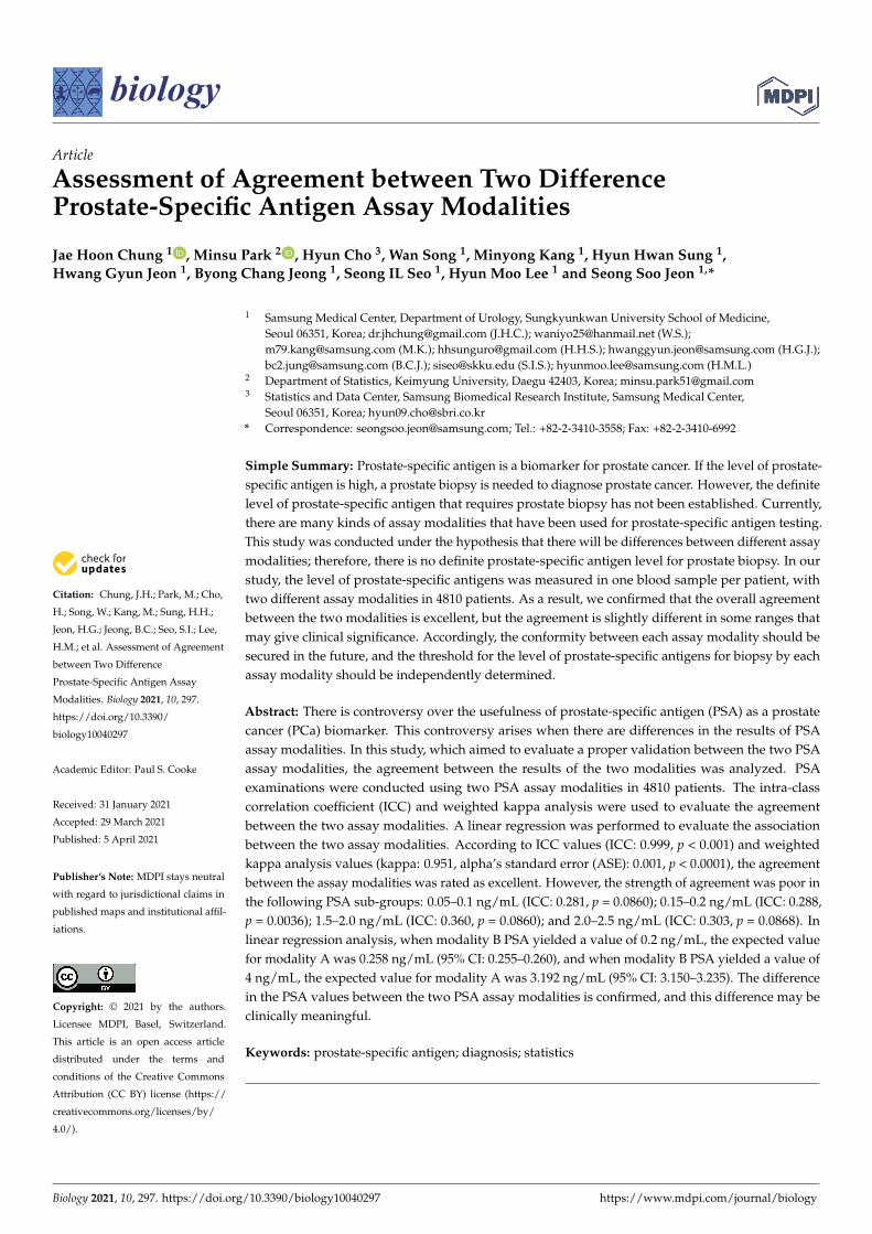
Linear regression analysis after excluding an outlier case (where the PSA value was over10 ng/mL) for modality A showed that the regression line met the axis at 1.133 (slope of 1),and the parameter estimate was 1.160 (standard error (SE): 0.003). Furthermore, based onmodality A, except for an outlier case (where the PSA value exceeded 4 ng/mL), the pointwhere the regression line met the axis was 0.879 (slope of 1), and the parameter estimatewas 1.178 (SE 0.004) (Figure 1). In addition, the values predicted for modality A comparedto modality B are shown in Table 3. If modality B PSA yielded a value of 0.2 ng/mL, theexpected value for modality A was 0.258 ng/mL (95% CI: 0.255–0.260); the expected value at3 ng/mL (modality B) was 2.507 ng/mL (modality A, 95% CI: 2.476–2.539); and at 4 ng/mL(modality B), the expected value was 3.192 ng/mL (modality A, 95% CI: 3.150–3.235) (Table 3).
Biology 2021, 10, x FOR PEER REVIEW 4 of 10
the point where the regression line met the axis was 0.879 (slope of 1), and the parameter
estimate was 1.178 (SE 0.004) (Figure 1). In addition, the values predicted for modality A
compared to modality B are shown in Table 3. If modality B PSA yielded a value of 0.2
ng/mL, the expected value for modality A was 0.258 ng/mL (95% CI: 0.255–0.260); the
expected value at 3 ng/mL (modality B) was 2.507 ng/mL (modality A, 95% CI: 2.476–
2.539); and at 4 ng/mL (modality B), the expected value was 3.192 ng/mL (modality A, 95%
CI: 3.150–3.235) (Table 3).
Figure 1. Logistic regression ‐4 −4 according to two prostate‐specific antigen (PSA) assay modalities. The point where the
regression line meets the straight line with a slope of 1 in PSA < 4 ng/mL group: 0.878808304. The point where the
regression line meets the straight line with a slope of 1 in PSA < 10 ng/mL group: 1.133175017. DF: degrees of freedom, Pr:
probability.
Table 1. Analysis of agreement between two PSA assay modalities.
PSA Modality A N ICC 95% CI
p Value Lower Upper
0.01~0.05 1012 0.092 −0.045 0.228 0.1063
0.05~0.1 377 0.281 −0.085 0.558 0.0860
0.1~0.15 119 0.492 0.333 0.622 <0.0001
0.15~0.2 86 0.288 0.081 0.471 0.0036
0.2~0.5 382 0.862 0.705 0.923 <0.0001
0.5~1.0 531 0.774 0.130 0.913 0.0103
1.0~1.5 315 0.488 −0.064 0.754 0.0478
1.5~2.0 249 0.360 −0.095 0.660 0.0860
2.0~2.5 184 0.303 −0.090 0.592 0.0868
2.5~4.0 564 0.594 −0.085 0.845 0.0620
4.0~ 991 0.999 0.999 0.999 <0.0001
Total 4810 0.999 0.999 0.999 <0.0001
Intra‐class correlation coefficient.
Figure 1. Logistic regression according to two prostate-specific antigen (PSA) assay modalities. The point where theregression line meets the straight line with a slope of 1 in PSA < 4 ng/mL group: 0.878808304. The point where theregression line meets the straight line with a slope of 1 in PSA < 10 ng/mL group: 1.133175017. DF: degrees of freedom,Pr: probability.

Biology 2021, 10, 297 5 of 10
Table 2. Analysis of the agreement between two PSA assay modalities.
Modality AModality B
G1 G2 G3 G4 G5 G6 G7 G8 G9 G10 G11 Sum
G1 1008 (99.6) 4 (0.4) 1012
G2 241 (63.93) 126 (33.42) 10 (2.65) 377
G3 2 (1.68) 34 (28.57) 71 (59.66) 12 (10.08) 119
G4 3 (3.49) 16 (18.6) 51 (59.3) 16 (18.6) 86
G5 12 (3.14) 314 (82.2) 56 (14.66) 382
G6 6 (1.13) 402 (75.71) 123 (23.16) 531
G7 1 (0.32) 1 (0.32) 231 (73.33) 80 (25.4) 2 (0.63) 315
G8 4 (1.61) 133 (53.41) 109 (43.78) 3 (1.2) 249
G9 2 (1.09) 90 (48.91) 92 (50) 184
G10 378 (67.02) 186 (32.98) 564
G11 1 (0.1) 990 (99.9) 991
Sum 1251 167 97 75 337 459 215 201 474 1176 4810
Kappa: 0.9513 (95% CI: 0.94849–0.954034, alpha’s standard error (ASE): 0.001414, p < 0.0001). G1: 0.01–0.05, G2: 0.05–0.1, G3: 0.1–0.15, G4: 0.15–0.2, G5: 0.2–0.5, G6: 0.5–1.0, G7: 1.0–1.5, G8: 1.5–2.0, G9: 2.0–2.5,G10: 2.5–4.0, G11: 4.0~. Weighted kappa analysis.

Biology 2021, 10, 297 6 of 10
Table 3. Estimated PSA value by different assay modalities.
Modality B, PSA ValueModality A Estimated Value
Fitted 95% CI (Lower, Upper)
0.01 0.021 0.020 0.0210.02 0.037 0.037 0.0380.03 0.052 0.052 0.0530.04 0.067 0.066 0.0680.05 0.080 0.079 0.0810.06 0.094 0.093 0.0950.07 0.107 0.105 0.1080.08 0.119 0.118 0.1210.09 0.132 0.130 0.1330.1 0.144 0.142 0.1460.11 0.156 0.154 0.1580.12 0.168 0.166 0.1700.13 0.180 0.178 0.1810.14 0.191 0.189 0.1930.15 0.202 0.200 0.2050.16 0.214 0.212 0.2160.17 0.225 0.223 0.2270.18 0.236 0.234 0.2380.19 0.247 0.244 0.2490.2 0.258 0.255 0.2600.3 0.362 0.359 0.3660.4 0.462 0.457 0.4660.5 0.557 0.551 0.5620.6 0.649 0.642 0.6550.7 0.738 0.731 0.7460.8 0.826 0.818 0.8340.9 0.912 0.903 0.9211.0 0.996 0.986 1.0071.1 1.079 1.068 1.0911.2 1.161 1.149 1.1741.3 1.242 1.229 1.2551.4 1.322 1.307 1.3361.5 1.401 1.385 1.4161.6 1.479 1.462 1.4951.7 1.556 1.538 1.5741.8 1.632 1.614 1.6511.9 1.708 1.689 1.7282.0 1.784 1.763 1.8042.1 1.858 1.836 1.8802.2 1.932 1.909 1.9552.3 2.006 1.982 2.0302.4 2.079 2.054 2.1042.5 2.151 2.125 2.1772.6 2.223 2.196 2.2512.7 2.295 2.267 2.3232.8 2.366 2.337 2.3962.9 2.437 2.406 2.4673.0 2.507 2.476 2.5393.1 2.577 2.545 2.6103.2 2.647 2.613 2.6813.3 2.716 2.681 2.7513.4 2.785 2.749 2.8213.5 2.854 2.817 2.8913.6 2.922 2.884 2.9603.7 2.990 2.951 3.0303.8 3.058 3.018 3.0983.9 3.125 3.084 3.1674.0 3.192 3.150 3.235

Biology 2021, 10, 297 7 of 10
Table 3. Cont.
Modality B, PSA ValueModality A Estimated Value
Fitted 95% CI (Lower, Upper)
4.5 3.524 3.477 3.5735.0 3.851 3.797 3.9055.5 4.171 4.112 4.2316.0 4.488 4.423 4.5536.5 4.800 4.730 4.8717.0 5.108 5.032 5.1857.5 5.413 5.331 5.4968.0 5.714 5.627 5.8038.5 6.013 5.920 6.1079.0 6.309 6.210 6.4099.5 6.602 6.497 6.70810.0 6.892 6.782 7.004
4. Discussion
This study showed that the overall correlation between the PSA values of the twoassay modalities was consistent between assays. However, as a result of sub-analyses toidentify clinical implications, it was confirmed that there were poor agreements at specificpoints of PSA value.
The PSA value is widely used as a parameter for PCa detection and assessment.Traditionally, the PSA cut-off value for PCa detection was set to 4 ng/mL, but in thiscase, the specificity has been shown to be 21%, and sensitivity is 91% [15]. This can leadto high false positive rates and unnecessary prostate biopsies, and finally, to an increasein the frequency of complications due to unnecessary prostate biopsies [16]. Because ofthese limitations of PSA, many other additional tests have been suggested, e.g., free PSA,prostate health index, 4Kscore, PCA3, Select MDx, and ExoDx Prostate [17–22]. In addition,recently, prostate magnetic resonance imaging and prostate-specific membrane antigenpositron emission tomography have also been used [23,24]. However, performing theseadditional tests is also determined based on the PSA cut-off value of 4 ng/mL, and theyrequire additional economic burden for both patients and society. Therefore, a definitePSA cut-off value is important not only because it determines the prostate biopsy at riskof complications, but also because it may have an influence on the occurrence of medicalopportunity cost.
However, several studies have reportedly lowered the PSA cut-off value to 2.5 or3 ng/mL [5–7]. The basis for these reports was that the prevalence of PCa was not low,even when the PSA value was below 4 ng/mL. Although PSA has been used as a parameterof Pca for over 30 years, there is still a controversy over the cut-off value for Pca detection,and this controversy raises a concern with the effectiveness of PSA itself.
As a result of the present study, when the PSA value of modality B was 4.0 ng/mL, theestimated value in modality A was 3.19 ng/mL. In addition, when the PSA value obtainedwith modality B was 3.0 ng/mL, the estimated value in modality A was 2.51 ng/mL. Byassessing these results, it is possible to predict whether the claims for the PSA cut-offvalue for Pca are different among researchers because the used PSA assay modality wasdifferent among researchers. In the present study, although the overall agreement betweenthe two assay modalities was excellent, clinical decision-making can be affected, evenwhen the PSA value changes by as little as 0.1 ng/mL. Therefore, it can be suggestedthat statistical validation and clinical validation produce different results. Although thisstudy was not able to evaluate the sensitivity and specificity of the two modalities for PCadetection, we could suggest the reason why there had been no definite PSA cut-off valuefor PCa screening.
In our institution (Samsung Medical Center, Seoul, Korea), we have used modality Ato measure PSA for a long time. The studies, which were conducted in our institution using

Biology 2021, 10, 297 8 of 10
modality A, showed that the PCa detection rate was 21.8 to 25.0% in patients with PSA of2.5 to 4 ng/mL [8,25]. Moreover, among them, over 75% of the patients were diagnosedwith clinically significant PCa [25]. Based on these data, we set a PSA value of 2.5 ng/mLas the cut-off value for PCa screening. The present study was conducted when the PSAassay modality was converted to modality B, and it is expected that the PSA cut-off valuewill be adjusted to 3 ng/mL through this analysis. In addition, when the PSA value inmodality B is 3 to 5 ng/mL, a similar PCa detection rate of 2.5 to 4 ng/mL in modality Ais expected.
This difference between PSA assay modalities can also affect the determination ofbiochemical recurrence after radical prostatectomy and evaluation of recurrence after radia-tion therapy or high-intensity focus ultrasound. The American Urological Association andthe European Association of Urology panel have defined the BCR as above 0.2 ng/mL [26].Several previous studies tried to reduce cancer-specific mortality by predicting BCR andperforming rapid adjuvant/salvage treatment after prostatectomy [27–29]. However, the re-sults of the present study showed that when the PSA value of modality A was increased by1, the result seen in modality B was confirmed to increase by 1.160 (in the PSA < 10 ng/mLgroup) or 1.178 (in the PSA < 4 ng/mL group). In addition, when the PSA value was0.2 ng/mL for modality B, the estimated value for modality A was 0.25 ng/mL. Althoughit is impossible to definitively determine which assay modality is best for cancer evalu-ation, assuming that a relatively high value is a true result, there is a problem becausethe diagnosis of recurrence might be underestimated in the group tested with a modalitythat yields relatively low values. Obviously, it is important in the clinic to assess sensitivechanges in PSA over time using repeat PSA measurements, PSA doubling time, and PSAvelocity. However, if the absolute value is different for each assay modality, this will be animportant factor to be sufficiently controlled by clinicians.
In this study, among the enrolled patients, 50.54% (2431/4810) were diagnosed withPCa. In PCa patients, 57.14% (1389/2431) had a PSA value of lower than 0.1 ng/mL afterreceiving definite treatment such as radical prostatectomy. A low ICC was confirmed ina specific section with a low PSA value. The lower limit of the measurement range was0.006 ng/mL for modality A and 0.01 ng/mL for modality B, and both assay modalitieswere calibrated according to the schedule and method specified by each company. There-fore, low ICC might be caused by a difference between the values measured by the twoassay modalities rather than the measurement error at low values. Low ICC is a significantfinding, because a small difference in PSA at low levels can affect the patient’s treatmentstrategy and prognosis during follow up.
The problem presented in this study will not cause clinical problems in real practice ifthe PSA cut-off value is independently determined for each clinical center and treatment isperformed based on long-term medical records and statistical analysis maintained by eachcenter. However, confusion may arise when a patient is transferred to another institutionthat uses a different PSA assay modality. Moreover, it is difficult to propose a globallyagreed upon PSA modality guideline. Furthermore, medical information, which is difficultto agree on, can disrupt public health practices at the present time, because it is commonfor conflicting information to be spread through the Internet. In addition, confusion mayoccur in treatment decisions when the PSA assay modality is changed, even in a singleclinical center. Recently, a controversy has been reported with respect to the usefulness ofPSA as a biomarker [30]; this may be due to the lack of meticulous validation between PSAassay modalities.
A key limitation of this study is that it is retrospective. However, in order to preventother biases, only the PSA values measured by two assay modalities drawn from a singlesample for each person were assessed. As this was not a comparative study, it was notpossible to evaluate superiority and efficiency between the two assay modalities. Inaddition, as the range of the measured value became wider, an error may have occurred inthe regression analysis, so correlation was obtained for PSA values when they were lessthan 4 ng/mL and less than 10 ng/mL. In addition, the overall comparison between the two

Biology 2021, 10, 297 9 of 10
modalities was rated excellent in terms of concordance. Because this study did not assessthe clinical implications, it was impossible to determine which of the two modalities yielded“true” results. Well-designed, large-scale, prospective studies will be required for assessingefficiency, sensitivity, and specificity of PCa detection according to PSA assay modalities.However, this study suggests, for the first time, that there may be differences between PSAassay modalities and cut-off points in terms of clinical significance. Thus, these resultsindicate the current medical situation in terms of treatment and biopsy decisions, and theymay be clinically helpful.
5. Conclusions
Through this study, the overall agreement between two PSA assay modalities wasconfirmed. However, it was also confirmed that there was a difference in the result valuesof the two PSA assay modalities, and that the difference may be clinically meaningful.This result may have been the cause of controversy over the clinical usefulness of the PSAvalues, and, therefore, the cut-off value of PCa may not be established.
Author Contributions: Conceptualization, S.S.J. and J.H.C.; methodology, J.H.C. and M.P.; validation,M.P. and H.C.; formal analysis, M.P. and H.C.; investigation, W.S., M.K., H.H.S. and H.G.J.; resources,B.C.J., S.I.S. and H.M.L.; data curation, J.H.C.; writing—original draft preparation, J.H.C.; writing—review and editing, S.S.J.; supervision, S.S.J. All authors have read and agreed to the publishedversion of the manuscript.
Funding: This research received no external funding.
Institutional Review Board Statement: The study was conducted according to the guidelines of theDeclaration of Helsinki and approved by the Institutional Review Board (or Ethics Committee) ofSamsung Medical Center, Seoul, Korea (protocol code 2020-01-155-001; 5 February 2020).
Informed Consent Statement: Informed consent was waived by the Board.
Data Availability Statement: The datasets used and/or analyzed during the current study areavailable from the corresponding author on reasonable request.
Conflicts of Interest: The authors declare no conflict of interest.
References1. Wang, M.C.; Valenzuela, L.A.; Murphy, G.P.; Chu, T.M. Purification of a human prostate specific antigen. Investig. Urol. 1979, 17,
159–163.2. Stamey, T.A.; Yang, N.; Hay, A.R.; McNeal, J.E.; Freiha, F.S.; Redwine, E. Prostate-specific antigen as a serum marker for
adenocarcinoma of the prostate. N. Engl. J. Med. 1987, 317, 909–916. [CrossRef]3. Catalona, W.J.; Smith, D.S.; Ratliff, T.L.; Dodds, K.M.; Coplen, D.E.; Yuan, J.J.; Petros, J.A.; Andriole, G.L. Measurement of
prostate-specific antigen in serum as a screening test for prostate cancer. N. Engl. J. Med. 1991, 324, 1156–1161. [CrossRef]4. Andriole, G.L.; Crawford, E.D.; Grubb, R.L., 3rd; Buys, S.S.; Chia, D.; Church, T.R.; Fouad, M.N.; Isaacs, C.; Kvale, P.A.; Reding,
D.J.; et al. Prostate cancer screening in the randomized Prostate, Lung, Colorectal, and Ovarian Cancer Screening Trial: Mortalityresults after 13 years of follow-up. J. Natl. Cancer Inst. 2012, 104, 125–132. [CrossRef]
5. Littrup, P.J.; Kane, R.A.; Mettlin, C.J.; Murphy, G.P.; Lee, F.; Toi, A.; Badalament, R.; Babaian, R. Cost-effective prostate cancerdetection. Reduction of low-yield biopsies. Investigators of the American Cancer Society National Prostate Cancer DetectionProject. Cancer 1994, 74, 3146–3158. [CrossRef]
6. Catalona, W.J.; Smith, D.S.; Ornstein, D.K. Prostate cancer detection in men with serum PSA concentrations of 2.6 to 4.0 ng/mL andbenign prostate examination. Enhancement of specificity with free PSA measurements. JAMA 1997, 277, 1452–1455. [CrossRef][PubMed]
7. Schroder, F.H.; van der Cruijsen-Koeter, I.; de Koning, H.J.; Vis, A.N.; Hoedemaeker, R.F.; Kranse, R. Prostate cancer detection atlow prostate specific antigen. J. Urol. 2000, 163, 806–812. [CrossRef]
8. Kim, H.S.; Jeon, S.S.; Choi, J.D.; Kim, W.; Han, D.H.; Jeong, B.C.; Seo, S.I.; Lee, K.S.; Lee, S.W.; Lee, H.M.; et al. Detection rates ofnonpalpable prostate cancer in Korean men with prostate-specific antigen levels between 2.5 and 4.0 ng/mL. Urology 2010, 76,919–922. [CrossRef]
9. Catalona, W.J.; Richie, J.P.; Ahmann, F.R.; Hudson, M.A.; Scardino, P.T.; Flanigan, R.C.; DeKernion, J.B.; Ratliff, T.L.; Kavoussi,L.R.; Dalkin, B.L.; et al. Comparison of digital rectal examination and serum prostate specific antigen in the early detection ofprostate cancer: Results of a multicenter clinical trial of 6630 men. J. Urol. 1994, 151, 1283–1290. [CrossRef]

Biology 2021, 10, 297 10 of 10
10. Killian, C.S.; Emrich, L.J.; Vargas, F.P.; Yang, N.; Wang, M.C.; Priore, R.L.; Murphy, G.P.; Chu, T.M. Relative reliability of fiveserially measured markers for prognosis of progression in prostate cancer. J. Natl. Cancer Inst. 1986, 76, 179–185.
11. Killian, C.S.; Yang, N.; Emrich, L.J.; Vargas, F.P.; Kuriyama, M.; Wang, M.C.; Slack, N.H.; Papsidero, L.D.; Murphy, G.P.; Chu, T.M.Prognostic importance of prostate-specific antigen for monitoring patients with stages B2 to D1 prostate cancer. Cancer Res. 1985,45, 886–891.
12. Stamey, T.A.; Kabalin, J.N. Prostate specific antigen in the diagnosis and treatment of adenocarcinoma of the prostate. I. Untreatedpatients. J. Urol. 1989, 141, 1070–1075. [PubMed]
13. Lightner, D.J.; Lange, P.H.; Reddy, P.K.; Moore, L. Prostate specific antigen and local recurrence after radical prostatectomy. J.Urol. 1990, 144, 921–926. [CrossRef]
14. Arai, Y.; Okubo, K.; Aoki, Y.; Maekawa, S.; Okada, T.; Maeda, H. Ultrasensitive assay of prostate-specific antigen for earlydetection of residual cancer after radical prostatectomy. Int. J. Urol. 1998, 5, 550–555. [CrossRef] [PubMed]
15. Thompson, I.M.; Ankerst, D.P.; Chi, C.; Goodman, P.J.; Tangen, C.M.; Lucia, M.S.; Feng, Z.; Parnes, H.L.; Coltman, C.A., Jr.Assessing prostate cancer risk: Results from the Prostate Cancer Prevention Trial. J. Natl. Cancer Inst. 2006, 98, 529–534. [CrossRef][PubMed]
16. Togo, Y.; Yamamoto, S. Prevention of infectious complications after prostate biopsy procedure. Int. J. Urol. 2017, 24, 486–492.[CrossRef]
17. Christensson, A.; Bjork, T.; Nilsson, O.; Dahlen, U.; Matikainen, M.T.; Cockett, A.T.; Abrahamsson, P.A.; Lilja, H. Serum prostatespecific antigen complexed to alpha 1-antichymotrypsin as an indicator of prostate cancer. J. Urol. 1993, 150, 100–105. [CrossRef]
18. Catalona, W.J.; Partin, A.W.; Sanda, M.G.; Wei, J.T.; Klee, G.G.; Bangma, C.H.; Slawin, K.M.; Marks, L.S.; Loeb, S.; Broyles, D.L.;et al. A multicenter study of [-2]pro-prostate specific antigen combined with prostate specific antigen and free prostate specificantigen for prostate cancer detection in the 2.0 to 10.0 ng/mL prostate specific antigen range. J. Urol. 2011, 185, 1650–1655.[CrossRef]
19. Vickers, A.J.; Cronin, A.M.; Aus, G.; Pihl, C.G.; Becker, C.; Pettersson, K.; Scardino, P.T.; Hugosson, J.; Lilja, H. A panel of kallikreinmarkers can reduce unnecessary biopsy for prostate cancer: Data from the European Randomized Study of Prostate CancerScreening in Goteborg, Sweden. BMC Med. 2008, 6, 19. [CrossRef]
20. Bussemakers, M.J.; van Bokhoven, A.; Verhaegh, G.W.; Smit, F.P.; Karthaus, H.F.; Schalken, J.A.; Debruyne, F.M.; Ru, N.; Isaacs,W.B. DD3: A new prostate-specific gene, highly overexpressed in prostate cancer. Cancer Res. 1999, 59, 5975–5979.
21. Leyten, G.H.; Hessels, D.; Smit, F.P.; Jannink, S.A.; de Jong, H.; Melchers, W.J.; Cornel, E.B.; de Reijke, T.M.; Vergunst, H.; Kil, P.;et al. Identification of a Candidate Gene Panel for the Early Diagnosis of Prostate Cancer. Clin. Cancer Res. 2015, 21, 3061–3070.[CrossRef]
22. Donovan, M.J.; Noerholm, M.; Bentink, S.; Belzer, S.; Skog, J.; O’Neill, V.; Cochran, J.S.; Brown, G.A. A molecular signature ofPCA3 and ERG exosomal RNA from non-DRE urine is predictive of initial prostate biopsy result. Prostate Cancer Prostatic Dis.2015, 18, 370–375. [CrossRef]
23. Futterer, J.J. Multiparametric MRI in the Detection of Clinically Significant Prostate Cancer. Korean J. Radiol. 2017, 18, 597–606.[CrossRef] [PubMed]
24. Esen, T.; Kilic, M.; Seymen, H.; Acar, O.; Demirkol, M.O. Can Ga-68 PSMA PET/CT replace conventional imaging modalities forprimary lymph node and bone staging of prostate cancer? Eur. Urol. Focus 2020, 6, 218–220. [CrossRef] [PubMed]
25. Chung, J.H.; Yu, J.; Song, W.; Kang, M.; Sung, H.H.; Jeon, H.G.; Jeong, B.C.; Seo, S.I.; Lee, H.H.; Jeon, S.S. Strategy for prostatecancer patients with low prostate specific antigen level (2.5 to 4.0 ng/mL). J. Korean Med. Sci. 2020, 35, e342. [CrossRef] [PubMed]
26. Cookson, M.S.; Aus, G.; Burnett, A.L.; Canby-Hagino, E.D.; D’Amico, A.V.; Dmochowski, R.R.; Eton, D.T.; Forman, J.D.;Goldenberg, S.L.; Hernandez, J.; et al. Variation in the definition of biochemical recurrence in patients treated for localizedprostate cancer: The American Urological Association Prostate Guidelines for Localized Prostate Cancer Update Panel report andrecommendations for a standard in the reporting of surgical outcomes. J. Urol. 2007, 177, 540–545. [PubMed]
27. Shariat, S.F.; Karakiewicz, P.I.; Roehrborn, C.G.; Kattan, M.W. An updated catalog of prostate cancer predictive tools. Cancer 2008,113, 3075–3099. [CrossRef]
28. Kang, J.J.; Reiter, R.E.; Steinberg, M.L.; King, C.R. First Postprostatectomy Ultrasensitive Prostate-specific Antigen PredictsSurvival in Patients with High-risk Prostate Cancer Pathology. Eur. Urol. Oncol. 2018, 1, 378–385. [CrossRef]
29. Skove, S.L.; Howard, L.E.; Aronson, W.J.; Terris, M.K.; Kane, C.J.; Amling, C.L.; Cooperberg, M.R.; Moreira, D.M.; Freedland, S.J.Timing of Prostate-specific Antigen Nadir After Radical Prostatectomy and Risk of Biochemical Recurrence. Urology 2017, 108,129–134. [CrossRef]
30. Kirby, R.; Moul, J.W. Is the PSA test useless? Prostate Cancer Prostatic Dis. 2004, 7, 271–272. [CrossRef]