Variation in local carrying capacity and the individual fate of ...
10
ORIGINAL ARTICLE Variation in local carrying capacity and the individual fate of bacterial colonizers in the phyllosphere Mitja NP Remus-Emsermann 1 , Robin Tecon 1,2 , George A Kowalchuk 1,3 and Johan HJ Leveau 1,2 1 Department of Microbial Ecology, Netherlands Institute of Ecology (NIOO-KNAW), Wageningen, The Netherlands; 2 Department of Plant Pathology, University of California, Davis, CA, USA and 3 Department of Ecological Science, VU University Amsterdam, Amsterdam, The Netherlands Using a phyllosphere model system, we demonstrated that the term ‘carrying capacity’, as it is commonly used in microbial ecology, needs to be understood as the sum of many ‘local carrying capacities’ in order to better explain and predict the course and outcome of bacterial colonization of an environment. Using a green fluorescent protein-based bioreporter system for the quantification of reproductive success (RS) in individual Erwinia herbicola cells, we were able to reconstruct the contribution of individual immigrants to bacterial population sizes on leaves. Our analysis revealed that plant foliage represents to bacteria an environment where individual fate is determined by the local carrying capacity of the site where an immigrant cell lands. With increasing inoculation densities, the RS of most immigrants declined, suggesting that local carrying capacity under the tested conditions was linked to local nutrient availability. Fitting the observed experimental data to an adapted model of phyllosphere colonization indicated that there might exist three types of sites on leaves, which differ in their frequency of occurrence and local carrying capacity. Specifically, our data were consistent with a leaf environment that is characterized by few sites where individual immigrants can produce high numbers of offspring, whereas the remainder of the leaf offered an equal number of sites with low and medium RS. Our findings contribute to a bottom–up understanding of bacterial colonization of leaf surfaces, which includes a quantifiable role of chance in the experience at the individual level and in the outcome at the population level. The ISME Journal (2012) 6, 756–765; doi:10.1038/ismej.2011.209; published online 19 January 2012 Subject Category: microbial population and community ecology Keywords: individual-based ecology; environmental heterogeneity; bioreporter; phyllosphere; single-cell microbiology; modeling Introduction In ecology, the ‘carrying capacity’ of an environment is the maximum number of individuals that the environment can support (McArthur, 2006). In phyllosphere microbiology, that is, the study of microorganisms that colonize leaf surfaces, this term is invariably referred to as the number of microbial cells that a leaf is able to sustain (Wilson and Lindow, 1994a, b; Wilson et al., 1995; Woody et al., 2007; Nix et al., 2009). Carrying capacity of leaf surfaces is typically determined through counts of colony-forming units (CFUs) on agar plates. It is a conditional value that depends on several biotic and abiotic factors (Leveau, 2006). For example, different plant species are known to sustain different micro- bial population sizes (Kinkel et al., 2000; Knief et al., 2010b). Furthermore, bacterial species may vary in the densities which they can achieve on leaf surfaces depending on their ability to deal with the environmental conditions on the leaf surface (Jacques et al., 1995; Jacobs et al., 2005). Water availability on the leaf has frequently been high- lighted as another driver for microbial abundance in the phyllosphere (Leben, 1965; Elad and Kirshner, 1993; Wilson and Lindow, 1994a, b). One key factor in determining foliar carrying capacity is the availability of nutrients. Carbon is a limiting resource during phyllosphere colonization (Mercier and Lindow, 2000; Leveau and Lindow, 2001a,b). The process of leaching (Tukey, 1966) deposits photosynthates, such as the sugars fructose, sucrose and glucose, on the leaf surface where they become readily available for consumption by microbes (Mercier and Lindow, 2000; Leveau and Lindow, 2001a,b; van der Wal and Leveau, 2010). Spraying of a carbon source onto plant leaves Received 20 September 2011; revised 2 December 2011; accepted 3 December 2011; published online 19 January 2012 Correspondence: JHJ Leveau, Department of Plant Pathology, University of California, One Shields Avenue, Davis, CA 95616, USA. E-mail: [email protected] The ISME Journal (2012) 6, 756–765 & 2012 International Society for Microbial Ecology All rights reserved 1751-7362/12 www.nature.com/ismej
-
Upload
khangminh22 -
Category
Documents
-
view
0 -
download
0
Transcript of Variation in local carrying capacity and the individual fate of ...

ORIGINAL ARTICLE
Variation in local carrying capacity and theindividual fate of bacterial colonizers inthe phyllosphere
Mitja NP Remus-Emsermann1, Robin Tecon1,2, George A Kowalchuk1,3 andJohan HJ Leveau1,2
1Department of Microbial Ecology, Netherlands Institute of Ecology (NIOO-KNAW), Wageningen,The Netherlands; 2Department of Plant Pathology, University of California, Davis, CA, USA and3Department of Ecological Science, VU University Amsterdam, Amsterdam, The Netherlands
Using a phyllosphere model system, we demonstrated that the term ‘carrying capacity’, as it iscommonly used in microbial ecology, needs to be understood as the sum of many ‘local carryingcapacities’ in order to better explain and predict the course and outcome of bacterial colonization ofan environment. Using a green fluorescent protein-based bioreporter system for the quantificationof reproductive success (RS) in individual Erwinia herbicola cells, we were able to reconstruct thecontribution of individual immigrants to bacterial population sizes on leaves. Our analysis revealedthat plant foliage represents to bacteria an environment where individual fate is determined by thelocal carrying capacity of the site where an immigrant cell lands. With increasing inoculationdensities, the RS of most immigrants declined, suggesting that local carrying capacity under thetested conditions was linked to local nutrient availability. Fitting the observed experimental data toan adapted model of phyllosphere colonization indicated that there might exist three types of siteson leaves, which differ in their frequency of occurrence and local carrying capacity. Specifically, ourdata were consistent with a leaf environment that is characterized by few sites where individualimmigrants can produce high numbers of offspring, whereas the remainder of the leaf offered anequal number of sites with low and medium RS. Our findings contribute to a bottom–upunderstanding of bacterial colonization of leaf surfaces, which includes a quantifiable role ofchance in the experience at the individual level and in the outcome at the population level.The ISME Journal (2012) 6, 756–765; doi:10.1038/ismej.2011.209; published online 19 January 2012Subject Category: microbial population and community ecologyKeywords: individual-based ecology; environmental heterogeneity; bioreporter; phyllosphere;single-cell microbiology; modeling
Introduction
In ecology, the ‘carrying capacity’ of an environmentis the maximum number of individuals that theenvironment can support (McArthur, 2006). Inphyllosphere microbiology, that is, the study ofmicroorganisms that colonize leaf surfaces, this termis invariably referred to as the number of microbialcells that a leaf is able to sustain (Wilson andLindow, 1994a, b; Wilson et al., 1995; Woody et al.,2007; Nix et al., 2009). Carrying capacity of leafsurfaces is typically determined through counts ofcolony-forming units (CFUs) on agar plates. It is aconditional value that depends on several biotic andabiotic factors (Leveau, 2006). For example, different
plant species are known to sustain different micro-bial population sizes (Kinkel et al., 2000; Kniefet al., 2010b). Furthermore, bacterial species mayvary in the densities which they can achieve on leafsurfaces depending on their ability to deal withthe environmental conditions on the leaf surface(Jacques et al., 1995; Jacobs et al., 2005). Wateravailability on the leaf has frequently been high-lighted as another driver for microbial abundance inthe phyllosphere (Leben, 1965; Elad and Kirshner,1993; Wilson and Lindow, 1994a, b).
One key factor in determining foliar carryingcapacity is the availability of nutrients. Carbon is alimiting resource during phyllosphere colonization(Mercier and Lindow, 2000; Leveau and Lindow,2001a, b). The process of leaching (Tukey, 1966)deposits photosynthates, such as the sugars fructose,sucrose and glucose, on the leaf surface wherethey become readily available for consumption bymicrobes (Mercier and Lindow, 2000; Leveau andLindow, 2001a, b; van der Wal and Leveau, 2010).Spraying of a carbon source onto plant leaves
Received 20 September 2011; revised 2 December 2011; accepted3 December 2011; published online 19 January 2012
Correspondence: JHJ Leveau, Department of Plant Pathology,University of California, One Shields Avenue, Davis, CA 95616,USA.E-mail: [email protected]
The ISME Journal (2012) 6, 756–765& 2012 International Society for Microbial Ecology All rights reserved 1751-7362/12
www.nature.com/ismej

(Wilson and Lindow, 1995) or making an additionalcarbon source available through genetic modifica-tion of the plant (Wilson et al., 1995) can alsoincrease the carrying capacity of leaves for bacterialpopulations.
The use of bacterial bioreporters has confirmedthat sugars, such as fructose, are not distributedevenly across the leaf surface (Leveau and Lindow,2001a, b). It has also been reported that unusedsugars can still be detected on leaves that arecolonized by bacteria at the apparent carryingcapacity (Mercier and Lindow, 2000). These twoobservations are in line with the hypothesizedexistence of discrete sites on the leaf surface thatcontain disparate amounts of resources (for example,sugars) and that may or may not be colonized bybacteria (Kinkel et al., 2002; Lindow and Brandl,2003). At high inoculation densities, all sites on aleaf can be expected to be colonized by bacteria, andin these cases, carrying capacity will be determinedby the amount of nutrients available in all sitescombined. At low inoculation densities, some siteswill remain uncolonized, including some thatcontain nutrients. In the latter case, the carryingcapacity is not reached and instead becomes afunction of how many, and which, sites are actuallyoccupied by bacterial colonizers. This model of leafcolonization was partly verified in experiments withthe epiphytic bacterium Pseudomonas syringae(Wilson and Lindow, 1994a, b), revealing that popu-lations derived from low inoculum concentrationsnever were able to reach the carrying capacity thatwas realized at high inoculum densities. However,what is still lacking is an experimental, quantitativedemonstration of the variation that exists in nutrientavailability, or local carrying capacity, betweensites on a leaf surface. How big is this variation,and how does it contribute to the course andoutcome of leaf colonization? Population-basedmeasurements are not suited to address thesequestions, as they only examine averages acrossthe population as a whole. In order to tackle theseissues properly, an individual-based approachseems necessary.
Here, we used the bacterial bioreporter CUSPER(reverse of REPSUC, for REProductive SUCcess;Remus-Emsermann and Leveau, 2010) to describehow increasing inoculation densities are experi-enced by individual bacterial colonizers of bean leafsurfaces. From this information, we were able toderive estimates for the variation in local carryingcapacity and assess the impact of this variation onachieving final foliar population sizes. The CUSPERbioreporter is based on dilution of green fluorescentprotein (GFP) from dividing cells in such a waythat GFP fluorescence of a single cell is inverselyproportional to the reproductive success (RS) of theoriginal immigrant from which the observed cellwas derived. The CUSPER bioreporter was recentlyused to reveal that in a heterogeneous environment,such as the phyllosphere, individual bacterial
immigrants contribute unequally to future popula-tion sizes (Remus-Emsermann and Leveau, 2010). Inthis study, we utilized CUSPER for an individual-based examination of leaf colonization at differentinoculation densities in order to gain a quantitativeunderstanding of variation in local carryingcapacities in the phyllosphere. Such knowledgeis potentially important to general ecological under-standing as well as practical applications such asplant pathogen control.
Materials and methods
Bacterial strain, culture conditions and plantexperimentsCUSPER is a bacterial bioreporter of RS (Remus-Emsermann and Leveau, 2010). It is based onErwinia herbicola 299RHJBA28 (pCPP39; Leveauand Lindow, 2001a, b), which constitutivelyexpresses LacIq from plasmid pCPP39 and carries achromosomal mini-Tn5-Km insertion that expressesstable GFP from a LacIq-repressible PA1/04/03 promo-ter fusion to gfpmut3. The transposon confersresistance to kanamycin and the plasmid to tetra-cycline. The concept of CUSPER lies in the loadingof bacterial cells with GFP through derepressionof the PA1/04/03 promoter with isopropyl b-D-1-thiogalactopyranoside, followed by release of theseGFP-loaded cells into an b-D-1-thiogalactopyrano-side-free environment. Without de novo GFP pro-duction, GFP is diluted every time a cell divides, sothat GFP fluorescence in each cell becomes aninverse measure for RS. CUSPER cells were culturedto mid-exponential phase at 300 r.p.m. and 28 1C inLB broth supplemented with 50 mg kanamycin and15 mg tetracycline per ml in addition to 1mM b-D-1-thiogalactopyranoside. Bacteria were harvested bycentrifugation at 3900� g for 10min, and resus-pended in 1� phosphate-buffered saline to approx-imate densities of 105, 106, 107 and 108 CFUml�1.Two fully expanded cotyledon leaves of 14-day-oldPhaseolus vulgaris plants (green snap bean, varietyBlue Lake Bush 274; Burpee, Warminster, PA, USA)were inoculated by evenly spraying the adaxialsurface of each leaf with B0.5ml of one of the fourbacterial suspensions using an airbrush paint gun(Pilot I Spray gun, Walther Pilot, Wuppertal-Vohwinkel, Germany). Inoculated plants were trans-ferred to closed translucent boxes and incubated athigh relative humidity and 21 1C.
Recovery of bacteria and analysis by microscopyAfter 0, 3, 5, 7 and 24h, three leaves were sampledfrom the plants that had been sprayed with oneof the four bacterial suspensions, for a subtotal of 12leaves per time point and a grand total of 60 leaves.Each leaf was transferred to a 50ml Falcon tube (BDBiosciences, Breda, The Netherlands) with 20ml1�phosphate-buffered saline buffer, vortexedbriefly, sonicated for 7min and vortexed briefly
Local carrying capacity in the phyllosphereMNP Remus-Emsermann et al
757
The ISME Journal

again. Aliquots (50 ml) from each leaf wash wereplated using an Eddy Jet spiral plater (IUL, Barce-lona, Spain) onto duplicate LB agar-plates supple-mented with 50 mg kanamycin per ml to determineCFUs.
For two of the three leaves per time point andtreatment (that is, a total of 40 leaves), the remainderof the leaf wash was filtered over a 0.2-mm Isoporefilter (Millipore, Amsterdam, The Netherlands).CUSPER cells were recovered from the filter byvortexing for 15 s in 1ml 1� phosphate-bufferedsaline, fixed as previously described (Leveau andLindow, 2001a, b) and stored in 1� phosphate-buffered saline at 4 1C for no longer than 3 days.Cells were analyzed with an Axio Imager M1epifluorescent microscope (Zeiss, Oberkochen,Germany) using standard phase contrast and ZeissFilter 38 (BP 470/ 40, FT 495, BP 525/ 50) for thevisualization of GFP. Digital images were capturedusing an EC Plan-Neofluar 100� /1.30 oil objective(Zeiss) with an AxioCam MRm camera (Zeiss).Single-cell fluorescence was quantified as the meanpixel intensity of individual cells (Leveau andLindow, 2001a, b) using the AxioVision 2.8 softwarepackage (Zeiss, Jena, Germany). Downstream dataanalysis was performed in Microsoft Excel 2010(Microsoft Corporation, Redmond, WA, USA). Toreconstruct the relative contribution of originalleaf immigrants to the final population based uponsingle-cell green fluorescence measurements, wedivided the green fluorescence of every one of theN cells in each sample at t¼ 24 by the average cell’sgreen fluorescence in the inoculum at time t¼ 0. Foreach cell, this relative GFP content was divided bythe sum of all GFP contents within the same sample(SGFP) to give a GFP/SGFP value for each cell.For each cell, we also calculated the RS of itsoriginal progenitor as 2log(1/GFP). Cells were thenbinned into one of the following RS classes:RSo0.5, 0.5pRSo1.5, 1.5pRSo2.5, 2.5pRSo3.5,3.5pRSo4.5, 4.5pRSo5.5 and RSX5.5. For eachbin, the GFP/SGFP values of all cells were summed
to give S(GFP/SGFP). Values of S(GFP/SGFP) werethen plotted as a function of RS in a histogram(see Figures 2b, 3, and 5).
Simulation of phyllosphere colonization using alogistic growth modelWe adapted a previously developed logistic-growthmodel for bacteria in a heterogeneous environment(Kinkel et al., 2002). The model simulates a leaf withS¼ 104 sites that vary in their carrying capacity J,that is, the maximal number of bacteria that a singlesite can sustain. Bacteria and their offspring werenot allowed to move from one site to another. At thebeginning of the simulation, bacteria were distrib-uted over the S sites following a Poisson distributionassuming an initial population of 103, 104 or 105
bacteria per leaf. With each iteration n, the numberof bacteria N in a site doubled, up to a maximumvalue N¼ J. Heterogeneity in J was introduced byassuming a normal or log-normal distributionthat was created using a custom script for NetLogo4.2.2 (Wilensky, 1999). Other distributions werecreated with a custom Visual Basic Script for MSExcel (http://azzalini.stat.unipd.it/SN/stephen_ger-suk.excel). The trimodal distribution that provided agood fit to the observed experimental data (seeSupplementary Information) assumed that 48.5% ofthe sites had a J value of 2, another 48.5% had aJ value of 48, whereas the remaining 3% of sites hada J value of 1152.
Results
Determining the carrying capacity of bean leavesTo determine the bacterial carrying capacity of beanleaves under laboratory conditions, we inoculatedfour sets of cotyledon leaves with a suspension ofE. herbicola strain 299R (Eh299R) CUSPER bacteriato reach four different densities, that is, 4.5� 103,6.9� 104, 5.7� 105 or 1.2� 107CFU per leaf(Figure 1a). Plants were incubated at conditions of
8
7
3
62
5
4
1
3 3 4 5 6 70
0 5 10 15 20 252
8
-1bact
eria
l pop
ulat
ion
size
(log[
CF
U p
er le
af])
log
(fol
d in
crea
se in
popu
latio
n si
ze)
time since inoculation (hours)
population size at t=0(log [CFU per leaf])
ba
Figure 1 Colonization of bean leaf surfaces by Eh299R CUSPER bacteria at different inoculation densities. (a) Changes in populationsize were monitored as CFUs, normalized per leaf and plotted as a function of time. Inoculation densities (CFUs per leaf) at t¼0:(K) 4.5� 103; (’) 6.9� 104; (m) 5.7�105; and (.) 1.2� 107. (b) Increase in population size between t¼0 and t¼ 24, that is,Nt¼ 24/Nt¼0, asa function of the population size at time t¼ 0. The trendline crosses the x axis at 6.535, which back-transforms to 3.4�106CFU per leaf.The error bars represent standard deviations from the mean.
Local carrying capacity in the phyllosphereMNP Remus-Emsermann et al
758
The ISME Journal
high relative humidity, and we monitored changesin the bacterial population sizes by determiningCFU counts per leaf at 3, 5, 7 and 24h afterinoculation (Figure 1a). With the two lowestinoculation densities, population sizes increasedduring the first 7 h of the experiment, albeit atdifferent rates. At the two highest inoculationdensities, population sizes either remained constantor decreased during this time period. Carryingcapacity was estimated from Figure 1b, whichshows a plot of the fold increase in population sizeover 24h as a function of the bacterial density at thetime of inoculation. The trendline crosses the x axisat 3.4� 106CFU per leaf, which represents theinoculation density where no net growth is expectedto occur, that is, the total carrying capacity underthese conditions.
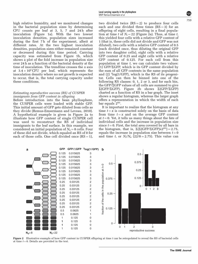
Estimating reproductive success (RS) of CUSPERimmigrants from GFP content in offspringBefore introduction into the bean phyllosphere,the CUSPER cells were loaded with stable GFP.This initial amount of GFP gets diluted from cells asthey divide (Remus-Emsermann and Leveau, 2010).A hypothetical example is given in Figure 2a toillustrate how GFP content of single CUSPER cellwas used to reconstruct the RS of individualimmigrants to the leaf surface. In this example, weconsidered an initial population of N0¼ 8 cells. Fourof these did not divide, which equaled an RS of 0 foreach of those cells. One cell divided once (RS¼ 1),
two divided twice (RS¼ 2) to produce four cellseach and one divided three times (RS¼ 3) for anoffspring of eight cells, resulting in a final popula-tion at time t of Nt¼ 22 (Figure 2a). Thus, at time t,this yielded four cells with a relative GFP content of1 (that is, these cells did not divide and GFP was notdiluted), two cells with a relative GFP content of 0.5(each divided once, thus diluting the original GFPinto two daughter cells), eight cells with a relativeGFP content of 0.25 and eight cells with a relativeGFP content of 0.125. For each cell from thispopulation at time t, we can calculate two values:(1) GFP/SGFP, which is its GFP content divided bythe sum of all GFP contents in the same populationand (2) 2log(1/GFP), which is the RS of its progeni-tor. Cells can then be binned into one of thefollowing RS classes: 0, 1, 2 or 3, and for each bin,the GFP/SGFP values of all cells are summed to giveS(GFP/SGFP). Figure 2b shows S(GFP/SGFP)charted as a function of RS in a bar graph. The insetshows a regular histogram, whereas the larger graphoffers a representation in which the width of eachbar equals 2RS.
It is important to realize that the histogram at anytime t¼ x is constructed solely on the basis of datafrom time t¼ x and on the average GFP contentat t¼ 0. Yet, it tells us many things about the fate ofindividual cells and the increase in population sizesince t¼ 0. First, the total area covered by all bars inthe histogram, that is, S(S(GFP/SGFP)x2RS)¼ 2.75,equals the increase in population size between t¼ 0and time t, or Nt/N0 (22/8¼ 2.75). This value can
GFP
0.125 0.015625 3 0.50.125 0.015625 3
a b
0.1250.125
0.0156250.015625 0.5
33
0.375
0.125 0.015625 30.125 0.015625 3 0.25
0.125 0.015625 30.125 0.015625 0.3753
20.125
0.250.25
0.031250.03125 2 0
0.25 0.03125 2 1 2 300.25 0.03125 0.252
3
0.25 0.03125 20.250.25
0.03125 22
0.250.03125
20.5 0.0625
0.1251
0.5 0.0625 111
0.1250.125
001
1 0.125 0011
0.1251 2 30
frac
tion
of th
e ce
lls in
the
popu
latio
n at
t=
0
0
Nt=22N0=8reproductive success
0.03125
2log(1 /GFP)GFP/ΣGFP
Figure 2 Illustrative example of how GFP content in CUSPER offspring at time t can be retropolated to reveal the RS of bacterial cellsat time t¼ 0. Details are provided in the text.
Local carrying capacity in the phyllosphereMNP Remus-Emsermann et al
759
The ISME Journal

also be calculated directly from Nt/SGFP, that is, thenumber of cells at time t divided by the sum of allGFP contents. Second, the graph reveals howsuccessful different fractions of the original popula-tion were in producing offspring. For example, itshows that half (that is, S(GFP/SGFP)¼ 0.5) of thecells in the population at t¼ 0 did not divide(RS¼ 0) and that a quarter of the cells (S(GFP/SGFP)¼ 0.25) at t¼ 0 divided two times to producefour cells at time t (RS¼ 2). Third, the area of anysingle bar in the graph divided by the total areacovered by all bars in the histogram gives theproportional contribution of cells from the corre-sponding RS bin to the population size at time t. Forexample, the area of the third bar (RS¼ 2) is S(GFP/SGFP)� 2RS¼ 0.25� 22¼ 1, which, divided by 2.75(¼ total area, see above), equals 0.36, meaning that25% of the cells in the original population dividedtwice to produce enough offspring to accountfor 36% of the population at time t.
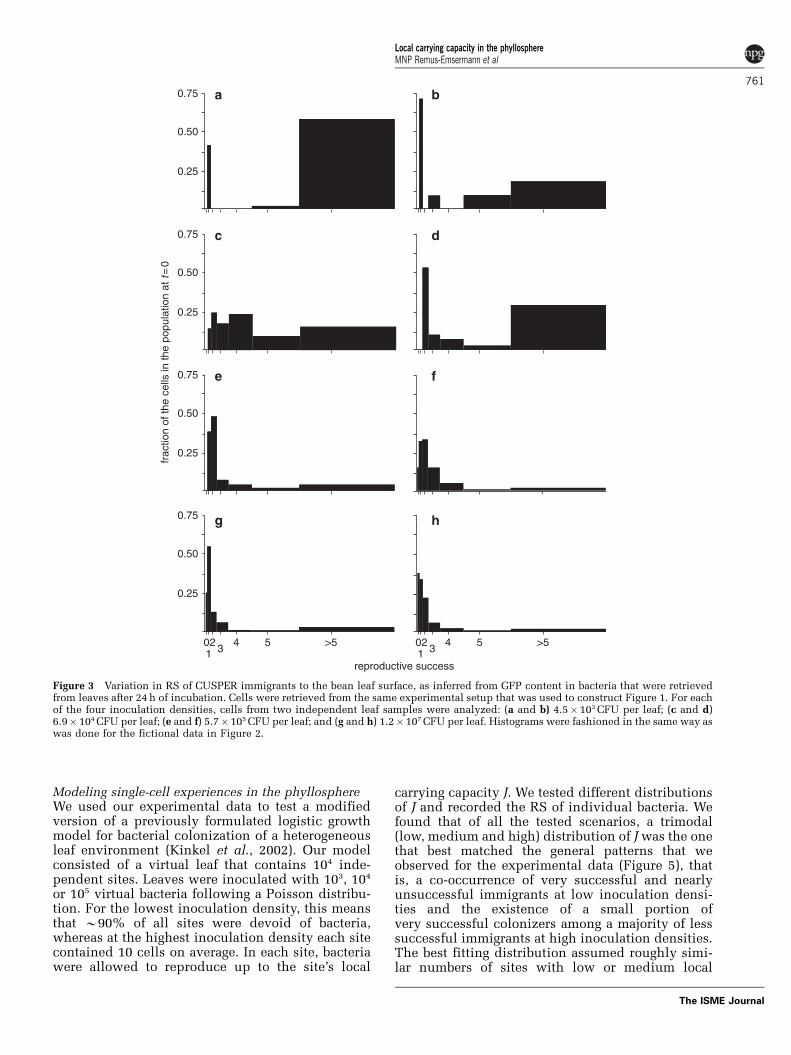
Single-cell experiences of leaf colonization afterinoculation at different densitiesUsing the analysis illustrated in Figure 2, weretropolated GFP content of leaf-recovered bacteriato the RS of individual bacteria in the t¼ 0population. Specifically, we analyzed bacterial cellsretrieved at the t¼ 24 time point from eight leaves(four inoculation densities, two leaf washings each)from the experiment for which population data areshown in Figure 1. The results are summarized inFigure 3, which shows eight histograms (a–h), thatis, two leaves from different plants for each density.These graphs indicate great variation in the fate ofindividual bacteria after they were introduced ontothe leaf surface at different starting densities.
On the two leaves that received the lowestinoculation density (Figures 3a and b), 41% and70%, respectively, of the initial colonizers dividedonce after arrival on the bean leaf surface. Weinterpret this to mean that a substantial portion ofthe immigrant population landed in a spot that didnot support much growth. On the other hand, 59%and 21%, respectively, of the cells at t¼ 0 was verysuccessful, with an RS value of 5 or greater, that is,they produced X32 offspring. These very successfulimmigrants reproduced to become 98% and 80%,respectively, of the final population size, comparedwith only 2% and 13%, respectively, for the cellsthat underwent only one division upon arrival.
With increasing inoculation density, the relativefraction of very successful cells (RSp5) diminished(Figure 3), down to 2.4% and 0.7%, respectively, onthe two leaves that were sprayed with the highestinoculation density (Figures 3g and h). At thisdensity, 91% and 93%, respectively, of the bacterialimmigrants each produced four offspring at most,with a majority not dividing at all or only once.Despite their low initial abundance of 2.4%and 0.7%, the very successful cells contributed a
considerable fraction, 31% and 11%, respectively,of the final population size.
It is evident from Figure 3 that with increasinginoculation density, the sum of the area underneathall of the bars (representing N24/N0, as explained inFigure 2) decreases. This decline in fold increasein population size is consistent with our findings inFigure 1b, and can be explained by assuming that athigher inoculation densities, similar amounts ofresources need to be shared by more bacteria,resulting in fewer offspring per cell. The notion thatthere were still few very successful immigrants atthe highest inoculation density suggests that a smallportion of the leaf surface (2.4% and 0.7% in thiscase) consisted of sites that allow considerably morereproduction than most other sites. Another trendthat is apparent in Figure 3 is the increasein resemblance of the two histograms from eachinoculation density, as inoculation densityincreases. This suggests that the course and outcomeof leaf colonization become more predictable athigher inoculation densities.
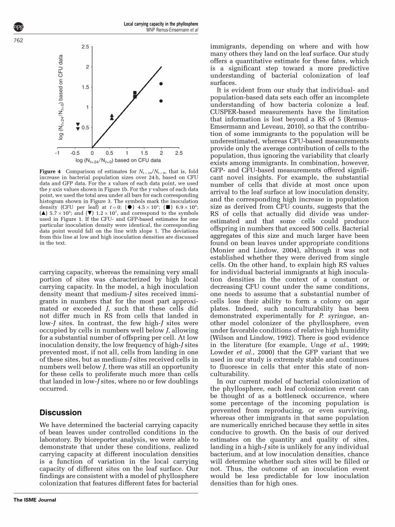
For each of the eight histograms shown in Figure 3,we plotted in Figure 4 the sum of the area under-neath all of the bars as a function of the N24/N0 foldincrease that was obtained from CFU-based data.The fact that data points did not fall on a line withslope 1 through the origin indicates that GFP- andCFU-based results do not fully agree with eachother. At the lowest inoculation density, thisdiscrepancy can be explained by the known limita-tion of CUSPER (Remus-Emsermann and Leveau,2010) that after five cell divisions GFP gets dilutedto the point where we can no longer reliably detectand quantify it. For the two leaves inoculated at thisdensity, we calculated that in order to get the GFP-based estimates for increase in population size up tothe one derived from CFU data, one would haveto assume average RS values of 7.4% and 9.0%respectively, for cells that were binned in the RS45category in Figure 3. This corresponds to 169 and512 offspring, respectively, for each of these cells. Atthe highest inoculation density, the discrepancybetween GFP- and CFU-based results may beexplained by assuming that a proportion of the cellson the leaf were not able to form a colony on theplate. For the two leaves that were inoculated atthis density, we calculated that in order to get theCFU-based estimates for increase in population sizeto match those derived from GFP data, one wouldhave to assume that 90% and 87%, respectively, ofthe cells lost their ability to form a colony betweent¼ 0 and t¼ 24. Another hypothetical explanationfor the discrepancy between CFU- and GFP-baseddata is that aggregates that were washed fromthe leaf surface did not break up into single cellsduring the wash procedure, so that one CFU mightrepresent more than one cell. However, we canexclude this possibility, based on the fact thatsuch aggregates were never observed under themicroscope.
Local carrying capacity in the phyllosphereMNP Remus-Emsermann et al
760
The ISME Journal
Modeling single-cell experiences in the phyllosphereWe used our experimental data to test a modifiedversion of a previously formulated logistic growthmodel for bacterial colonization of a heterogeneousleaf environment (Kinkel et al., 2002). Our modelconsisted of a virtual leaf that contains 104 inde-pendent sites. Leaves were inoculated with 103, 104
or 105 virtual bacteria following a Poisson distribu-tion. For the lowest inoculation density, this meansthat B90% of all sites were devoid of bacteria,whereas at the highest inoculation density each sitecontained 10 cells on average. In each site, bacteriawere allowed to reproduce up to the site’s local
carrying capacity J. We tested different distributionsof J and recorded the RS of individual bacteria. Wefound that of all the tested scenarios, a trimodal(low, medium and high) distribution of Jwas the onethat best matched the general patterns that weobserved for the experimental data (Figure 5), thatis, a co-occurrence of very successful and nearlyunsuccessful immigrants at low inoculation densi-ties and the existence of a small portion ofvery successful colonizers among a majority of lesssuccessful immigrants at high inoculation densities.The best fitting distribution assumed roughly simi-lar numbers of sites with low or medium local
0.75 a b
dc
e f
hg
0.50
0.25
0.75
0.50
0.25
0.75
0.50
0.25
0.75
0.50
frac
tion
of th
e ce
lls in
the
popu
latio
n at
t=
0
0.25
20 4 520 4 531
>531
>5
reproductive success
Figure 3 Variation in RS of CUSPER immigrants to the bean leaf surface, as inferred from GFP content in bacteria that were retrievedfrom leaves after 24h of incubation. Cells were retrieved from the same experimental setup that was used to construct Figure 1. For eachof the four inoculation densities, cells from two independent leaf samples were analyzed: (a and b) 4.5�103CFU per leaf; (c and d)6.9�104CFU per leaf; (e and f) 5.7�105CFU per leaf; and (g and h) 1.2�107CFU per leaf. Histograms were fashioned in the same way aswas done for the fictional data in Figure 2.
Local carrying capacity in the phyllosphereMNP Remus-Emsermann et al
761
The ISME Journal
carrying capacity, whereas the remaining very smallportion of sites was characterized by high localcarrying capacity. In the model, a high inoculationdensity meant that medium-J sites received immi-grants in numbers that for the most part approxi-mated or exceeded J, such that these cells didnot differ much in RS from cells that landed inlow-J sites. In contrast, the few high-J sites wereoccupied by cells in numbers well below J, allowingfor a substantial number of offspring per cell. At lowinoculation density, the low frequency of high-J sitesprevented most, if not all, cells from landing in oneof these sites, but as medium-J sites received cells innumbers well below J, there was still an opportunityfor these cells to proliferate much more than cellsthat landed in low-J sites, where no or few doublingsoccurred.
Discussion
We have determined the bacterial carrying capacityof bean leaves under controlled conditions in thelaboratory. By bioreporter analysis, we were able todemonstrate that under these conditions, realizedcarrying capacity at different inoculation densitiesis a function of variation in the local carryingcapacity of different sites on the leaf surface. Ourfindings are consistent with a model of phyllospherecolonization that features different fates for bacterial
immigrants, depending on where and with howmany others they land on the leaf surface. Our studyoffers a quantitative estimate for these fates, whichis a significant step toward a more predictiveunderstanding of bacterial colonization of leafsurfaces.
It is evident from our study that individual- andpopulation-based data sets each offer an incompleteunderstanding of how bacteria colonize a leaf.CUSPER-based measurements have the limitationthat information is lost beyond a RS of 5 (Remus-Emsermann and Leveau, 2010), so that the contribu-tion of some immigrants to the population will beunderestimated, whereas CFU-based measurementsprovide only the average contribution of cells to thepopulation, thus ignoring the variability that clearlyexists among immigrants. In combination, however,GFP- and CFU-based measurements offered signifi-cant novel insights. For example, the substantialnumber of cells that divide at most once uponarrival to the leaf surface at low inoculation density,and the corresponding high increase in populationsize as derived from CFU counts, suggests that theRS of cells that actually did divide was under-estimated and that some cells could produceoffspring in numbers that exceed 500 cells. Bacterialaggregates of this size and much larger have beenfound on bean leaves under appropriate conditions(Monier and Lindow, 2004), although it was notestablished whether they were derived from singlecells. On the other hand, to explain high RS valuesfor individual bacterial immigrants at high inocula-tion densities in the context of a constant ordecreasing CFU count under the same conditions,one needs to assume that a substantial number ofcells lose their ability to form a colony on agarplates. Indeed, such nonculturability has beendemonstrated experimentally for P. syringae, an-other model colonizer of the phyllosphere, evenunder favorable conditions of relative high humidity(Wilson and Lindow, 1992). There is good evidencein the literature (for example, Unge et al., 1999;Lowder et al., 2000) that the GFP variant that weused in our study is extremely stable and continuesto fluoresce in cells that enter this state of non-culturability.
In our current model of bacterial colonization ofthe phyllosphere, each leaf colonization event canbe thought of as a bottleneck occurrence, wheresome percentage of the incoming population isprevented from reproducing, or even surviving,whereas other immigrants in that same populationare numerically enriched because they settle in sitesconducive to growth. On the basis of our derivedestimates on the quantity and quality of sites,landing in a high-J site is unlikely for any individualbacterium, and at low inoculation densities, chancewill determine whether such sites will be filled ornot. Thus, the outcome of an inoculation eventwould be less predictable for low inoculationdensities than for high ones.
2.5
2
1.5
1
0.5
-1 -0.5 0 0.5 1 1.5 2 2.5log (Nt=24/Nt=0) based on CFU data
log
(Nt=
24/N
t=0)
bas
ed o
n C
FU
dat
a
Figure 4 Comparison of estimates for Nt¼24/Nt¼0, that is, foldincrease in bacterial population sizes over 24h, based on CFUdata and GFP data. For the x values of each data point, we usedthe y axis values shown in Figure 1b. For the y values of each datapoint, we used the total area under all bars for each correspondinghistogram shown in Figure 3. The symbols mark the inoculationdensity (CFU per leaf) at t¼ 0: (K) 4.5�103; (’) 6.9�104;(m) 5.7�105; and (.) 1.2� 107, and correspond to the symbolsused in Figure 1. If the CFU- and GFP-based estimates for oneparticular inoculation density were identical, the correspondingdata point would fall on the line with slope 1. The deviationsfrom this line at low and high inoculation densities are discussedin the text.
Local carrying capacity in the phyllosphereMNP Remus-Emsermann et al
762
The ISME Journal

Ecological theory holds that if a nutrient resourceis limited and the number of organisms competingfor this resource is high, the average success of thepopulation goes down (Galliard et al., 2005; Rankinand Kokko, 2006). This is precisely what weobserved in our experiments and simulations: highinoculation densities forced more bacterial indivi-duals to share a site and resulted in overall reducedRS. This seems to support the notion that localcarrying capacity on leaves is determined by localnutrient availability. Being perfectly isogenic, CUS-PER cells can be considered each other’s ultimatecompetitors, with a niche overlap index of 1 (Wilsonand Lindow, 1994a, b). Thus, unless a site offeredvery high levels of nutrients, bacterial immigrantswould be relatively unable to reproduce, represent-ing a convincing case of the ‘tragedy of thecommons’ (Hardin, 1968). However, a one-timeimmigration event of up to 107 bacteria per leaf isnot a likely event in nature (Lindemann and Upper,1985). Instead, immigration is a gradual process,with cells arriving to the leaf from the surroundingair at different times and typically at low rates(Upper and Hirano, 2002). Another aspect of ourexperimental setup that can be considered artefac-tual is that all immigrants were of clonal descent. Inreality, the leaf surface is colonized by very diversebacteria and other microorganisms with differentniche overlap indexes, which allows for variousdegrees of coexistence (Knief et al., 2010a). How-ever, it is important to note that the ability to set upsuch artificial colonization events allowed us toextract data on the quality of the leaf surface as ahabitable environment. This high level of experi-mental amenability is quite unique to microbialecology and is one of the advantages microbialecologists have over ‘macro’-bial ecologists (Fierer,2007). Future applications of CUSPER will addressmore realistic scenarios by assessing variation in RSof CUSPER cells in competition with other bacterialspecies and after immigration onto precolonizedleaves.
Our observation that a small fraction (0.7–2.4%) ofthe cells was very successful in reproducing even at
the highest inoculation density suggests the exis-tence of sites on the leaf surface that offer high levelsof nutrients, allowing these cells to contributedisproportionally (11–31%) to the final populationsize. Interestingly, this finding agrees with reportsshowing that relatively rare features on the leafsurface tend to harbor large numbers of bacteria oncolonized leaves. Specifically, it is worth notingthat only 0.6% of the bean leaf surface is coveredby hooked and glandular trichomes (Monier andLindow, 2005a, b), yet 44.1% of Eh299R bacteria inaggregates were found to be associated with thesestructures after incubation under moist conditionsfor 5 days (Monier and Lindow, 2005a, b). In at leastone study (Yadav et al., 2005), the population size ofepiphytic bacteria was positively correlated with thedensities of glandular and nonglandular trichomes.This link between bacterial fate, population size andleaf topography calls for further investigation intothe predictive value of trichome density for bacterialcolonization of leaf surfaces.
As was pointed out before (Remus-Emsermannand Leveau, 2010), application of the CUSPERconcept is not limited to leaf surfaces, but canbe extended widely to many other bacterialhabitats. Several bioreporter-based studies haverevealed variation in the bacterial experience ofsuch habitats (Moller et al., 1998; Jaspers et al.,2001; Herron et al., 2010), and all point to micro-scale heterogeneity in terms of the physical,chemical and biological conditions that an indivi-dual bacterium can be exposed to in a givenenvironment. The CUSPER approach offers aninsight in bacterial fate, which does not follow fromknowing exactly what these conditions were;instead, it settles for knowing that these conditionswere (or were not) permissive to the production ofoffspring. This interpretation of environmentalheterogeneity matches quite well with the conceptof local carrying capacity, where the microbiallandscape, be it natural or man-made (for example,Keymer et al., 2006; Battin et al., 2007) featureslocal variation in the ability of patches to sustainbacterial growth.
0.5
0.4
0.3
0.2
0.1
0
frac
tion
of th
e ce
lls in
the
popu
latio
n at
t=
0
312 3
12 3
120 4 5 >5 0 4 5 >5 0 4 5 >5
reproductive success
a b c
Figure 5 Variation in RS of virtual bacterial immigrants to a simulated leaf environment. The model is described in the text. It assumes aleaf with 104 discrete sites that vary in their carrying capacity J. This leaf was inoculated with 103 (a), 104 (b) or 105 (c) bacteria per leaf.For this simulation, we assumed that J¼ 2 for 48.5% of the sites, J¼ 48 for another 48.5% of the sites and J¼ 1152 for the remaining 3% ofthe sites.
Local carrying capacity in the phyllosphereMNP Remus-Emsermann et al
763
The ISME Journal
Acknowledgements
The study was funded by the Netherlands Organisation ofScientific Research (NWO) in the form of a personal VIDIgrant to JHJL. This is NIOO-KNAW publication number5156.
References
Battin TJ, Sloan WT, Kjelleberg S, Daims H, Head IM,Curtis TP et al. (2007). Microbial landscapes: newpaths to biofilm research. Nat Rev Microbiol 5: 76–81.
Elad Y, Kirshner B. (1993). Survival in the phylloplane of anintroduced biocontrol agent Trichoderma harzianumand populations of the plant pathogen Botrytis cinerea;as modified by abiotic conditions. Phytoparasitica 21:303–313.
Fierer N. (2007). Finding a place for microorganisms in thefield of ecology. Ecology 88: 1336–1337.
Hardin G. (1968). The tragedy of the commons. Science162: 1243–1248.
Herron PM, Gage DJ, Cardon ZG. (2010). Micro-scale waterpotential gradients visualized in soil around plant roottips using microbiosensors. Plant Cell Environ 33:199–210.
Jacobs JL, Carroll TL, Sundin GW. (2005). The role ofpigmentation, ultraviolet radiation tolerance, andleaf colonization strategies in the epiphyticsurvival of phyllosphere bacteria. Microb Ecol 49:104–113.
Jacques MA, Kinkel LL, Morris CE. (1995). Populationsizes, immigration, and growth of epiphytic bacteriaon leaves of different ages and positions of field-grownendive (Cichorium endivia var. latifolia). Appl EnvironMicrobiol 61: 899–906.
Jaspers MCM, Meier C, Zehnder AJ, Harms H, van derMeer JR. (2001). Measuring mass transfer processes ofoctane with the help of an alkS-alkBHgfp-taggedEscherichia coli. Environ Microbiol 3: 512–524.
Keymer JE, Galajda P, Muldoon C, Park S, Austin RH.(2006). Bacterial metapopulations in nanofabri-cated landscapes. Proc Natl Acad Sci USA 103:17290–17295.
Kinkel LL, Newton M, Leonard KJ. (2002). Resourceaggregation in the phyllosphere: implications formicrobial dynamics across spatial scales. In: LindowS, Hecht-Poinar E, Elliott V (eds). PhyllosphereMicrobiology. APS Press: Saint Paul, MN, USA,pp 317–340.
Kinkel LL, Wilson M, Lindow SE. (2000). Plant speciesand plant incubation conditions influence variabilityin epiphytic bacterial population size. Microb Ecol 39:1–11.
Knief C, Frances L, Vorholt JA. (2010a). Competitivenessof diverse Methylobacterium strains in the phyllo-sphere of Arabidopsis thaliana and identificationof representative models, including M. extorquensPA1. Microb Ecol 60: 440–452.
Knief C, Ramette A, Frances L, Alonso-Blanco C, VorholtJA. (2010b). Site and plant species are importantdeterminants of the Methylobacterium communitycomposition in the plant phyllosphere. ISME J 4:719–728.
Leben C. (1965). Influence of humidity on migration ofbacteria on cucumber seedlings. Can J Microbiol 11:671–676.
Le Galliard JF, Fitze PS, Ferriere R, Clobert J. (2005).Sex ratio bias, male aggression, and populationcollapse in lizards. Proc Natl Acad Sci USA 102:18231–18236.
Leveau JHJ. (2006). Microbial Communities in thePhyllosphere. Biology of the Plant Cuticle. Riederer Mand Muller C, Blackwell Publishing Ltd: Oxford, UK,pp 334–367.
Leveau JHJ, Lindow SE. (2001a). Appetite of an epiphyte:quantitative monitoring of bacterial sugar consump-tion in the phyllosphere. Proc Natl Acad Sci USA 98:3446–3453.
Leveau JHJ, Lindow SE. (2001b). Predictive and inter-pretive simulation of green fluorescent proteinexpression in reporter bacteria. J Bacteriol 183:6752–6762.
Lindemann J, Upper CD. (1985). Aerial dispersal ofepiphytic bacteria over bean plants. Appl EnvironMicrobiol 50: 1229–1232.
Lindow SE, Brandl MT. (2003). Microbiology ofthe phyllosphere. Appl Environ Microbiol 69:1875–1883.
Lowder M, Unge A, Maraha N, Jansson JK, Swiggett J,Oliver JD. (2000). Effect of starvation and theviable-but-nonculturable state on green fluorescentprotein (GFP) fluorescence in GFP-tagged Pseu-domonas fluorescens A506. Appl Environ Microbiol66: 3160–3165.
McArthur JV. (2006). Microbial Ecology: An EvolutionaryApproach. Academic Press: Oxford, UK.
Mercier J, Lindow SE. (2000). Role of leaf surface sugars incolonization of plants by bacterial epiphytes. ApplEnviron Microbiol 66: 369–374.
Moller S, Sternberg C, Andersen JB, Christensen BB,Ramos JL, Givskov M et al. (1998). In situ geneexpression in mixed-culture biofilms: evidence ofmetabolic interactions between community members.Appl Environ Microbiol 64: 721–732.
Monier JM, Lindow SE. (2004). Frequency, size, andlocalization of bacterial aggregates on bean leafsurfaces. Appl Environ Microbiol 70: 346–355.
Monier JM, Lindow SE. (2005a). Aggregates of residentbacteria facilitate survival of immigrant bacteria onleaf surfaces. Microb Ecol 49: 343–352.
Monier JM, Lindow SE. (2005b). Spatial organization ofdual-species bacterial aggregates on leaf surfaces. ApplEnviron Microbiol 71: 5484–5493.
Nix S, Burpee LL, Buck JW. (2009). Responses of 2epiphytic yeasts to foliar infection by Rhizoctoniasolani or mechanical wounding on the phylloplaneof tall fescue. Can J Microbiol 55: 1160–1165.
Rankin DJ, Kokko H. (2006). Sex, death and tragedy. TrendEcol Evol 21: 225–226.
Remus-Emsermann MNP, Leveau JHJ. (2010). Linkingenvironmental heterogeneity and reproductive suc-cess at single-cell resolution. Isme J 4: 215–222.
Tukey HB. Jr. (1966). Leaching of metabolites fromabove-ground plant parts and its implications.Bulletin Torrey Bot Club 93: 385–401.
Unge A, Tombolini R, Molbak L, Jansson JK. (1999).Simultaneous monitoring of cell number andmetabolic activity of specific bacterial populationswith a dual gfp-luxAB marker system. Appl EnvironMicrobiol 65: 813–821.
Upper C, Hirano S. (2002). Revisiting the rolesof immigration and growth in the developmentof populations of Pseudomonas syringae in the
Local carrying capacity in the phyllosphereMNP Remus-Emsermann et al
764
The ISME Journal

phyllosphere. In: Lindow S, Hecht-Poinar E, Elliott V(eds). Phyllosphere Microbiology. APS Press: SaintPaul, MN, USA, pp 69–79.
Van Der Wal A, Leveau JHJ. (2010). Modelling sugardiffusion across plant leaf cuticles: the effect of freewater on substrate availability to phyllosphere bacter-ia. Environ Microbiol 13: 792–797.
Wilensky U. (1999). NetLogo. Center for ConnectedLearning and Computer-Based Modeling, Northwes-tern University: Evanston, IL.
Wilson M, Lindow S. (1992). Relationship of total viableand culturable cells in epiphytic populations ofPseudomonas syringae. Appl Environ Microbiol 58:3908–3913.
Wilson M, Lindow S. (1995). Enhanced epiphytic coex-istence of near-isogenic salicylate-catabolizing andnon-salicylate-catabolizing Pseudomonas putidastrains after exogenous salicylate application. ApplEnviron Microbiol 61: 1073–1076.
Wilson M, Lindow SE. (1994a). Ecological similarityand coexistence of epiphytic ice-nucleating (Ice+)Pseudomonas syringae strains and a non-ice-nucleat-ing (Ice�) biological control agent. Appl EnvironMicrobiol 60: 3128.
Wilson M, Lindow SE. (1994b). Inoculum density-depen-dent mortality and colonization of the phyllosphereby Pseudomonas syringae. Appl Environ Microbiol 60:2232–2237.
Wilson M, Savka MA et al. (1995). Altered epiphyticcolonization of mannityl opine-producing transgenictobacco plants by a mannityl opine-catabolizing strainof Pseudomonas syringae. Appl Environ Microbiol 61:2151–2158.
Woody ST, Ives AR, Nordheim EV, Andrews JH. (2007).Dispersal, density dependence, and populationdynamics of a fungal microbe on leaf surfaces. Ecology88: 1513–1524.
Yadav R, Karamanoli K, Vokou D. (2005). Bacterialcolonization of the phyllosphere of Mediterraneanperennial species as influenced by leaf structural andchemical features. Microb Ecol 50: 185–196.
This work is licensed under the CreativeCommons Attribution-NonCommercial-No
Derivative Works 3.0 Unported License. To view acopy of this license, visit http://creativecommons.org/licenses/by-nc-nd/3.0/
Supplementary Information accompanies the paper on The ISME Journal website (http://www.nature.com/ismej)
Local carrying capacity in the phyllosphereMNP Remus-Emsermann et al
765
The ISME Journal