Vaccine induced antibodies to the first variable loop of human immunodeficiency virus type 1 gp120,...
36
Vaccine Induced Antibodies to the First Variable Loop of Human Immunodeficiency Virus Type 1 gp120, Mediate Antibody- Dependent Virus Inhibition in Macaques Izabela Bialuk 1,2 , Stephen Whitney 5 , Vibeke Andresen 1 , Ruth H. Florese 3 , Janos Nacsa 1 , Valentina Cecchinato 1 , Valerio W. Valeri 1 , Jean-Michel Heraud 1 , Shari Gordon 1 , Robyn Washington Parks 1 , David C. Montefiori 6 , David Venzon 4 , Thorsten Demberg 3 , Marjorie Robert Guroff 3 , Gary Landucci 7 , Donald N. Forthal 7 , and Genoveffa Franchini 1,* 1 Animal Models and Retroviral Vaccines Section, National Cancer Institute, Bethesda, Maryland 20892 2 Department of General and Experimental Pathology, Medical University of Bialystok, 15-222 Bialystok, Poland 3 Section of Immune Biology of Retroviral Infection, National Cancer Institute, Bethesda, Maryland 20892 4 Biostatistics and Data Management Section, National Cancer Institute, Bethesda, Maryland 20892 5 Advanced BioScience Laboratories Kensington, Maryland 20895 6 Department of Surgery, Laboratory for AIDS Vaccine Research and Development, Duke University Medical Center, Durham, North Carolina 27710 7 Division of Infectious Diseases, Department of Medicine, University of California, Irvine School of Medicine, Irvine, California 92697 Abstract The role of antibodies directed against the hyper variable envelope region V1 of human immunodeficiency virus type 1 (HIV-1), has not been thoroughly studied. We show that a vaccine able to elicit strain-specific non-neutralizing antibodies to this region of gp120 is associated with control of highly pathogenic chimeric SHIV 89.6P replication in rhesus macaques. The vaccinated animal that had the highest titers of antibodies to the amino terminus portion of V1, prior to challenge, had secondary antibody responses that mediated cell killing by antibody-dependent cellular cytotoxicity (ADCC), as early as two weeks after infection and inhibited viral replication by antibody-dependent cell-mediated virus inhibition (ADCVI), by four weeks after infection. There was a significant inverse correlation between virus level and binding antibody titers to the envelope protein, (R = -0.83, p 0.015), and ADCVI (R = -0.84 p=0.044). Genotyping of plasma virus demonstrated in vivo selection of three SHIV 89.6P variants with changes in potential N- linked glycosylation sites in V1. We found a significant inverse correlation between virus levels and titers of antibodies that mediated ADCVI against all the identified V1 virus variants. A significant inverse correlation was also found between neutralizing antibody titers to SHIV 89.6 and virus levels (R = -0.72 p =0.0050). However, passive inoculation of purified immunoglobulin from animal M316, the macaque that best controlled virus, to a naïve macaque, resulted in a low serum neutralizing antibodies and low ADCVI activity that failed to protect from SHIV 89.6P challenge. * corresponding author: Animal Models & Retroviral Vaccine Section MD, NIH, National Cancer Institute, 9000 Rockville Pike, Building 41, Room D-804, Bethesda, Maryland, USA 20892-5065, Phone: 301-496-2386, Fax: 301-402-0055, [email protected]. Conflict-of-Interest Statement Conflict-of-interest disclosure: The authors confirm that they have read the Journal’s position on issues involved in ethical publication and affirm that this report is consistent with those guidelines. Publisher's Disclaimer: This is a PDF file of an unedited manuscript that has been accepted for publication. As a service to our customers we are providing this early version of the manuscript. The manuscript will undergo copyediting, typesetting, and review of the resulting proof before it is published in its final citable form. Please note that during the production process errors may be discovered which could affect the content, and all legal disclaimers that apply to the journal pertain. NIH Public Access Author Manuscript Vaccine. Author manuscript; available in PMC 2012 December 9. Published in final edited form as: Vaccine. 2011 December 9; 30(1): 78–94. doi:10.1016/j.vaccine.2011.10.040. NIH-PA Author Manuscript NIH-PA Author Manuscript NIH-PA Author Manuscript
-
Upload
independent -
Category
Documents
-
view
0 -
download
0
Transcript of Vaccine induced antibodies to the first variable loop of human immunodeficiency virus type 1 gp120,...
Vaccine Induced Antibodies to the First Variable Loop of HumanImmunodeficiency Virus Type 1 gp120, Mediate Antibody-Dependent Virus Inhibition in Macaques
Izabela Bialuk1,2, Stephen Whitney5, Vibeke Andresen1, Ruth H. Florese3, Janos Nacsa1,Valentina Cecchinato1, Valerio W. Valeri1, Jean-Michel Heraud1, Shari Gordon1, RobynWashington Parks1, David C. Montefiori6, David Venzon4, Thorsten Demberg3, MarjorieRobert Guroff3, Gary Landucci7, Donald N. Forthal7, and Genoveffa Franchini1,*
1Animal Models and Retroviral Vaccines Section, National Cancer Institute, Bethesda, Maryland20892 2Department of General and Experimental Pathology, Medical University of Bialystok,15-222 Bialystok, Poland 3Section of Immune Biology of Retroviral Infection, National CancerInstitute, Bethesda, Maryland 20892 4Biostatistics and Data Management Section, NationalCancer Institute, Bethesda, Maryland 20892 5Advanced BioScience Laboratories Kensington,Maryland 20895 6Department of Surgery, Laboratory for AIDS Vaccine Research andDevelopment, Duke University Medical Center, Durham, North Carolina 27710 7Division ofInfectious Diseases, Department of Medicine, University of California, Irvine School of Medicine,Irvine, California 92697
AbstractThe role of antibodies directed against the hyper variable envelope region V1 of humanimmunodeficiency virus type 1 (HIV-1), has not been thoroughly studied. We show that a vaccineable to elicit strain-specific non-neutralizing antibodies to this region of gp120 is associated withcontrol of highly pathogenic chimeric SHIV89.6P replication in rhesus macaques. The vaccinatedanimal that had the highest titers of antibodies to the amino terminus portion of V1, prior tochallenge, had secondary antibody responses that mediated cell killing by antibody-dependentcellular cytotoxicity (ADCC), as early as two weeks after infection and inhibited viral replicationby antibody-dependent cell-mediated virus inhibition (ADCVI), by four weeks after infection.There was a significant inverse correlation between virus level and binding antibody titers to theenvelope protein, (R = -0.83, p 0.015), and ADCVI (R = -0.84 p=0.044). Genotyping of plasmavirus demonstrated in vivo selection of three SHIV89.6P variants with changes in potential N-linked glycosylation sites in V1. We found a significant inverse correlation between virus levelsand titers of antibodies that mediated ADCVI against all the identified V1 virus variants. Asignificant inverse correlation was also found between neutralizing antibody titers to SHIV89.6 andvirus levels (R = -0.72 p =0.0050). However, passive inoculation of purified immunoglobulin fromanimal M316, the macaque that best controlled virus, to a naïve macaque, resulted in a low serumneutralizing antibodies and low ADCVI activity that failed to protect from SHIV89.6P challenge.
*corresponding author: Animal Models & Retroviral Vaccine Section MD, NIH, National Cancer Institute, 9000 Rockville Pike,Building 41, Room D-804, Bethesda, Maryland, USA 20892-5065, Phone: 301-496-2386, Fax: 301-402-0055,[email protected] Statement Conflict-of-interest disclosure: The authors confirm that they have read the Journal’s position onissues involved in ethical publication and affirm that this report is consistent with those guidelines.Publisher's Disclaimer: This is a PDF file of an unedited manuscript that has been accepted for publication. As a service to ourcustomers we are providing this early version of the manuscript. The manuscript will undergo copyediting, typesetting, and review ofthe resulting proof before it is published in its final citable form. Please note that during the production process errors may bediscovered which could affect the content, and all legal disclaimers that apply to the journal pertain.
NIH Public AccessAuthor ManuscriptVaccine. Author manuscript; available in PMC 2012 December 9.
Published in final edited form as:Vaccine. 2011 December 9; 30(1): 78–94. doi:10.1016/j.vaccine.2011.10.040.
NIH
-PA Author Manuscript
NIH
-PA Author Manuscript
NIH
-PA Author Manuscript

Collectively, while our data suggest that anti-envelope antibodies with neutralizing and non-neutralizing FcγR-dependent activities may be important in the control of SHIV replication, theyalso demonstrate that low levels of these antibodies alone are not sufficient to protect frominfection.
INTRODUCTIONThe HIV envelope gene encodes four variable regions (V1–V4) [1;2]. The V3 region isimportant for viral infectivity and tropism and is the principal target for neutralizingantibodies of laboratory-adapted viruses [3-8]. Similarly, the V1/V2 regions of HIVinfluence viral receptor and co-receptor usage and tropism [9-15]. Selection of genotypeswith changes in V1/V2 occurs during the early phase of HIV infection [16-18]. HIVsequences of isolates, obtained during the chronic phase of infection, have extended V1/V2regions and a higher number of potential N-linked glycosylation sites [12;19]. The turnoverof V1 and V2, in the later stage of HIV infection, is suggestive of in vivo selection [20] anddeletion or mutations that modify glycosylation sites within these regions, affect theneutralization susceptibility of HIV and SIV isolates in vitro [13;21-26]. In infected rhesusmacaques, the selection of SIVmac239 strains that became resistant to neutralization has beenlinked to changes in N-linked and O-linked glycosylation in V1 [27]. Interestingly, deletionof the V1 region within the SIVmac239 molecular clone, results in decreased viral fitness invivo and greater neutralization susceptibility in vitro [23]. Similarly, single amino acidchanges affecting N-glycans in the V1/V2 of an HIV molecular clone, impacted viral fitnessand showed resistance to antibody neutralization in vitro [28]. The plasticity of the V1/V2region of HIV/SIV suggests its importance for viral fitness, particularly in the context of anactive host immune response. However, there is no direct evidence that supports a protectiverole of antibodies to the V1/V2 region in HIV or SIV infection. Here, we used the SHIV89.6Prhesus macaque model and investigated the role of antibody responses to V1 in the controlof viral replication. We used a vaccine based on a cDNA encoding a chimeric HIV protein,generated by an unusual splicing of the Tat open reading frame to the V1 envelope regionand the last exon of Rev (Tat-Env-Rev=TEV) [29-31] in a DNA prime-protein boostregimen. The combination of these vaccines induced modest T-cell responses and antibodiesto the V1 that mediated a type specific antibody-dependent cell-mediated virus inhibition invitro. Correlative analysis suggests that ADCVI function contribute to the control of viralreplication in animals that nevertheless, become infected. However, passive transfer ofvaccine elicited antibodies to a neonate macaque failed to protect from infection. Thus, it ispossible that other immune responses, in addition to antibodies to V1, induced by thisvaccine regimen may have contributed to control of viral replication.
MATERIALS AND METHODSDNA plasmids and protein expression
The tev genes for the HIV-1 isolates HIVBa-L, HIVSF162, and HIV89.6 were designed [32]using the published sequences for each isolate (Genbank M68893, M65024, and U39362,respectively) and were based on the published HXB2 tev sequence (Genbank M37898). Thegenes were synthetically constructed and cloned into pPCR-Script Amp SK (Strategene, LaJolla, CA) cloning vectors by Geneart (Regensburg, Germany). The tev genes weresynthesized using human and E .coli codon bias to optimize translation in both systems.
DNA vaccine vectors were constructed for Ba-L, SF162, and SHIV89.6 Tev, pCMV Ba-LTev, pCMV SF162 Tev, and pCMV 89.6 Tev, respectively. Briefly, for each tev construct, aSacII/EcoRI DNA fragment encoding the tev gene was ligated into a mammalian expressionvector derived from pVR1332 (Vical, San Diego, CA) [33]. These plasmids are kanamycin
Bialuk et al. Page 2
Vaccine. Author manuscript; available in PMC 2012 December 9.
NIH
-PA Author Manuscript
NIH
-PA Author Manuscript
NIH
-PA Author Manuscript

resistant, and the expression of tev is under control of the CMV immediate-early promoter.Plasmid DNA was produced at Althea Technologies (San Diego, CA), at the concentrationof 1mg/ml in sterile water. DNA was found to be >95% circular and predominantlysupercoiled by gel electrophoresis, with endotoxin levels below 1.5 EU/mg DNA.
The tev vaccine vectors were tested and found to be functional by radioimmunoprecipitationassay (RIPA) of transiently transfected HEK 293 cells. Transfections were performed usingLipofectamine 2000 (Invitrogen, Carlsbad, CA), according to the manufacturer’srecommended protocol. Wells of a twenty-four-well tissue culture plate were seeded with2×105 HEK 293 cells in 500 μl DMEM (Quality Biological, Gaithersburg, MD),supplemented with 10% heat-inactivated Fetal Bovine Serum (FBS; Hyclone, Logan, UT)and 2 mM L-glutamine (Quality Biological). Cultures were incubated for 18h at 37°C with5% CO2. For each plasmid, 2.5 μl of Lipofectamine 2000 reagent was mixed with 47.5 μlOpti-MEM Reduced Serum Media (Invitrogen) and set at room temperature for 10 min. Foreach plasmid, 1 μl (1 μg) DNA was mixed with 49 μl Opti-MEM Reduced Serum Media,and set at room temperature for 5 min. DNA and Lipofectamine 2000 solutions werecombined and set at room temperature for 20 min, then added to the 18h HEK 293 cultures.Cultures were further incubated at 37°C with 5% CO2 for 29h.
At 29h post-transfection, transfected and non transfected HEK 293 cells as negative control,were washed once with 1 ml D-PBS (Quality Biological) and overlaid with 500 μl L-methionine and L-cystine free DMEM (Invitrogen) supplemented with 2% heat-inactivateddialyzed FBS (Invitrogen), 2 mM L-glutamine (Quality Biological), 1 mM sodium pyruvate(Quality Biological), and 30 mg/ml L-methionine (Sigma, St. Louis, MO). Cells werelabeled with 50 μCi 35 S Cysteine (Perkin Elmer, Shelton, CT) and incubated for 16h at37°C with 5% CO2. Media was removed and cells were lysed in PBS-TD (PBS with 0.5 %Triton-X-100 and 1% deoxycholic acid) containing 1 μl/ml benzonase nuclease (EMDBiosciences, Madison, WI) and 1 μl/ml Protease Inhibitor Cocktail for Mammalian Cells(Sigma) for 10 min at room temperature. Lysates were clarified by centrifugation at 10,000× g for 5 min. Lysates were precleared by shaking with 20 μl protein. A sepharose (PAS;Amersham Biosciences, Piscataway, NJ), and 5 μl normal rabbit serum was at roomtemperature for 1 hour. Ten percent glycerol was added to the precleared lysates, and lysateswere immunoprecipitated with 20 μl PAS and 5 μl rabbit anti-HIV-1MN Rev serum(Advanced BioScience Laboratories, Kensington, MD), or 5 μl rabbit anti-HIV-1IIIB Tatserum (Advanced BioScience Laboratories), for 2.5h at room temperature. Precipitatedproteins were separated by electrophoresis on 10% SDS-PAGE gels. The gels weresubsequently soaked in destaining solution (40% methanol and 7 % acetic acid) for 45minutes, and then soaked in Amplify (Amersham Biosciences) for 30 min. The gels weredried to filter paper and exposed to x-ray film (BioMax MR, Eastman Kodak, Rochester,NY) and developed after 20h.
Animal studiesAll fifteen animals were colony-bred rhesus macaques (Macaca mulatta), obtained fromCovance Research Products (Alice, TX). The animals were housed and handled inaccordance with the standards of the Association for the Assessment and Accreditation ofLaboratory Animal Care International, and the study was reviewed and approved by theanimal care and use committees at Advanced BioScience Laboratories (Kensington, MD).The care and use of the animals were in compliance with all relevant institutional (NIH)guidelines. A total of eight animals were immunized in two rounds: M316, M218, M308,M490, and M599, M600, M607, M611, using the same DNA and protein preparation. Eachimmunized animal received 3 mg of the TEV89.6, TEVSF162, and TEVBa-L plasmid DNA bythe intramuscular route at weeks 0, 3, and 8. Protein boost was performed using 100μg ofthe purified HIV89.6 TEV proteins in CpG (2 μg /ml; ODN 2006, Coley Pharmaceutical
Bialuk et al. Page 3
Vaccine. Author manuscript; available in PMC 2012 December 9.
NIH
-PA Author Manuscript
NIH
-PA Author Manuscript
NIH
-PA Author Manuscript

Group, Wellesley, MA). Challenge exposure of all vaccinated and control animals wereperformed by the intravenous route with SHIV89.6P titer XXX, kindly provided by Dr.Norman L. Letvin. The following groups of animals were challenged simultaneously withidentical viral stock: animals M316, M218, M308, M490, L915, M320; animals M599,M600, M607, M611, M604; animals L941, L942, L947, and L962.
ELISABriefly, ELISA plates were coated overnight at 4°C with 100 μl/well 50 mM of sodiumbicarbonate buffer (pH 9.6) containing 100 ng/well BSA plus: 50 ng/well of rHIV-1HXB2Tat, rHIV-189.6 Tev, rHIV-1MN Rev proteins, a pool of 25 ng/well of each recombinantgp140 (from Ba-L, SF162 P3, or HIV89.6). Plates were blocked at room temperature for 2hwith 200 μl/well 5% sucrose, 1.25% nonfat dry milk and 2.5% BSA. Serum samples werediluted in Dilsim II (BioMerieux, Durham, NC), and 100 μl/well were added to plates, induplicate, and incubated for 1h at 37°C. Plates were washed four times with a solution ofPBS at a 0.5% concentration of Tween 20. Plates were incubated at 37°C with 100μl/wellgoat anti-human IgG HRP conjugate diluted in PBS plus 20% NGS and 0.1% Triton-X-100for 1h. Plates were washed four times in PBS plus 0.5% Tween 20, incubated for 30 min atroom temperature with 100 μl/well TMB substrate, and stopped with 100 μl/well 2 Nsulfuric acid. Absorbance was read at 450 nm. End point titers were calculated bydetermining the dilution at which a sample’s absorbance value equals two times theabsorbance of the pre-bleed sera at 1:50 dilution.
PepscansA library of forty-eight peptides spanning the entire length of the HIV-189.6 Tev protein wassynthesized. Peptides are fifteen residues in length and overlapped the flanking peptides byeleven residues. Peptides 1 to 18 are derived from the first exon of Tat; peptides 16 to 28 arederived from the Env V1 region; peptides 26 to 48 are derived from the second exon of Rev.ELISA plates were coated at 4°C overnight with 100 μl/well 50 mM sodium bicarbonatebuffer (pH 9.6) containing 100 ng/well BSA plus 1 μg/well individual peptides. Plates wereblocked at room temperature for 2h with 200 μl/well 5% sucrose, 1.25% nonfat dry milk,and 2.5% BSA. Serum samples were diluted 1:100 or 1:5000 in Dilsim II, and 100 μl/wellwere added to plates, and incubated for 1h at 37°C. Plates were washed four times with PBSplus 0.5% Tween 20. Plates were incubated at 37°C with 100 μl/well goat anti-human IgGHRP conjugate diluted in PBS plus 20% NGS and 0.1% Triton-X-100 for 1h. Plates werewashed four times in PBS + 0.5% Tween 20. Plates were incubated for 30 min at roomtemperature with 100 μl/well TMB substrate, and stopped with 100 μl/well 2 N sulfuric acid.Absorbance was read at 450 nm. Absorbance values from pre-bleed samples were subtractedas background from the absorbance values of reactive samples.
ELISPOT assayELISPOT assay was performed with the monkey-specific IFN-γ Millipore kit (Billerica,Massachusetts), according to the manufacture’s specifications. Briefly, after coating withanti-IFN-γ monoclonal antibody, plates were incubated overnight at 4°C and washed, andwells were covered with 5% human serum in PBS at room temperature for 2 hours. Afterwashing, PBMCs 2×10⁵ in 50μl of RPMI-1640 containing 5% heat-inactivated humanserum were plated in each well and stimulated in triplicate with the HIV-I89.6 Env peptidepool at 1 μg/ml. Concanavalin A (5μg/ml) was used as a positive control. After 18hincubation at 37°C in a 5% CO2 atmosphere, plates were washed and coated with filteredrabbit polyclonal anti-IFN-γ biotinylated detector antibody for 2 hours. Plates weredeveloped with chromogen substrate activated by alkaline phosphatase in streptavidin APconjugate and SFC’s counted on an ImmunoSpot Analyzer (CTL LLC, Cleveland, Ohio).
Bialuk et al. Page 4
Vaccine. Author manuscript; available in PMC 2012 December 9.
NIH
-PA Author Manuscript
NIH
-PA Author Manuscript
NIH
-PA Author Manuscript

Antibody-dependent cellular cytotoxicity and cell-mediated virus inhibition (ADCC andADCVI) assays
Antibody-dependent cellular cytotoxicity (ADCC) was evaluated by a flow-cytometry-basedassay as previously described [34;35]. Target cells were CEMx174 T-cells infected withSHIV89.6P and the effector cells were human PBMCs. An effector to target [E:T] ratio of50:1 was used. Titers are defined as the reciprocal of the serum dilution at which cell killingwas greater than the mean of week 0 samples of all macaques plus 3 standard deviations.
The ADCVI assay was based on methods described previously for measles virus and HIV[36]. Target cells consisted of CEM.NKR--CCR5 cells infected with the SHIV89.6P or theSHIV89.6P variants. After adsorption for 1h, target cells were washed, incubated in 5% CO2at 37°C for 72h in medium, and washed an additional two times. Infected target cells(5×10⁴) were next plated in 96-well round-bottom microtiter plates, to which heat-inactivated test plasma and fresh human PBMC effector cells (E:T ratio of 10:1) were added.Plasma in the absence of effector cells was also tested for anti-viral activity. After 8 daysincubation at 37°C in 5% CO2, supernatant fluid was collected and assayed for p27 byELISA (Zeptometrix, Buffalo, NY). Virus inhibition due to ADCVI was calculated asfollows: % inhibition = 100(1 - ([p27p]/[p27n])), where [p27p] and [p27n] are theconcentrations of p27 in supernatant fluid from wells containing SHIV-positive or -negativeplasma, respectively.
HIV89.6 Tev protein purificationThe HIV89.6 Tev protein was produced in E. coli and purified using immunoaffinitychromatography. The HIV89.6 tev gene was amplified from the pPCR-Script Amp SK vectorcontaining the synthetic HIV89.6 tev gene using the following primers: HIV89.6 tev 5’ Nde(acttagcatatggaacctgtgaaccctagcctgga) containing the NdeI site and HIV89.6 tev 3’ Xho(ctaagtctcgagttactccttggtgccaggttc) containing the XhoI site. The NdeI/XhoI DNA fragmentencoding the tev gene was ligated into the bacterial expression plasmid pET26 (EMDBiosciences, Madison, WI). The resulting pET26 HIV89.6 tev plasmid is kanamycinresistant, and the expression of tev is under control of the IPTG-inducible T7 lac promoter.BL21 (DE3) competent cells (EMD Biosciences) were transformed with pET26 HIV89.6 tevand the tev sequence of clones were confirmed by DNA sequence analysis.
One liter culture of the HIV89.6 tev E. coli was grown at 37°C to 0.9 OD600nm, cooled to22°C and induced with 1 mM IPTG for 6h. Cells were harvested by centrifugation and lysedin 40 ml of a PBS-based buffer containing 0.5 M sodium chloride, 10 mM ascorbic acid, 50mM mannitol, 2.5 % glycerol, 0.5 % Triton-X-100, protease inhibitor cocktail, 50 μg/mllysozyme, 1 μl/ml benzonase nuclease (EMD Biosciences), and 5 mM DTT. Lysates weresonicated and clarified by centrifugation. TEV was purified from the lysate on mousemonoclonal antibody to HIV-1 Tat sepharose, using 100 mM sodium carbonate with 5 mMDTT for elution. The eluate was buffered with phosphate buffer and the pH was adjusted to7.4 with hydrochloric acid. Protein was passed through a goat anti-mouse IgG sepharosecolumn to remove any mouse antibody that may have leached off of the immunoaffinitycolumn. Endotoxin was reduced to about 400 EU/mg protein using Detoxigel (PierceBiotechnology, Rockford, IL). The final Tev product runs as a 28 kDa protein on SDS-PAGE with a purity of about 90%. In western blot, HIV89.6 Tev reacts well with rabbit anti-sera to HIV-1 gp120, HIV-1 Tat, and HIV-1 Rev, demonstrating that the Tat, Env, and Revcomponents that compose Tev are present and recognized immunologically.
RNA extraction and RT-PCRViral RNA was extracted from 140 μl of plasma using a QIAamp viral RNA kit (Qiagen,Valencia, CA) with the final elution in 60 μl of elution buffer. A Titan One-Tube RT-PCR
Bialuk et al. Page 5
Vaccine. Author manuscript; available in PMC 2012 December 9.
NIH
-PA Author Manuscript
NIH
-PA Author Manuscript
NIH
-PA Author Manuscript

system (Roche Molecular Biochemicals, Indianapolis, IN) was used for the viral RNAreverse transcription according to the manufacturer’s protocol. The followingoligonucleotides were used for amplification of V1/V2 region: Fwd 89.6 D1 5’-GCCACACATGCCTGTGTACCCACAGA and Rev 89.6 D2 5’-CCAGCCGGGACACAATAATGTATGGGA. PCR product was treated with 1 μl of Taqpolymerse (New England Biolabs, Inpswich, MA) for 15 min at 72°C and purified usingQIAquick gel extraction kit (Qiagen, Valencia, CA). V1/V2 PCR products were cloned intopCR®2.1 TA TOPO vector (Invitrogen, Carlsbad, CA) according to the manufacturer’sprotocol. QIAprep spin miniprep kit (Qiagen, Valencia, CA) was used for plasmidsisolation. Samples were sequenced by ABI dye terminator sequencing (Perkin-Elmer Corp)and analyzed using internet available tools: http://www.expasy.ch/tools/dna.html andhttp://www.ebi.ac.uk/Tools/clustalw2/index.html for translation of nucleotide sequences andamino acids alignment, respectively.
Site-specific mutagenesisThe infectious molecular clone of SHIV89.6 was obtained as two plasmids (pSHIV-89.6 5’,cat. number 4130, Lot number 1 and pSHIV-89.6 3’, cat. number 4131, Lot number3041818) through the AIDS Research and Reference Reagent Program, Division of AIDS,NIAID, National Institutes of Health from Dr. Joseph Sodroski [37].
SHIV89.6 P 3’ plasmid was used as a template for generation of envelope mutants by PCR.The following site-specific mutagenic oligonucleotides were used and the sequence ofplasmid clones was analyzed to confirm the mutations.
SHIV89.6 3’ PtoL Fwd 5’ GAATACTACTAATCTCACTAGTAGCAGCTGG, (aa sequenceNTTNLTSSSW), SHIV89.6 3’ PtoL Rev 5’CCAGCTGCTACTAGTGAGATTAGTAGTATTC (aa sequence NTTNLTSSSW);SHIV89.6 3’ StoN Fwd 5’ CTACTAATCTCACTAATAGCAGCTGGGGAATG (aasequence TNLTNSSWGM), SHIV89.6 3’ StoN Rev 5’CATTCCCCAGCTGCTATTAGTGAGATTAGTAG (aa sequence TNLTNSSWGM). 5 μgof SHIV89.6 5’ cut SphI and ClaI and 5 μg of SHIV89.6 P 3’ wild type plasmid or itsenvelope mutants cut SphI and AflII were purified by phenol/chloroform extraction followedby ethanol precipitation using standard procedures and ligated using T4 DNA ligase (USBCorporation, Cleveland, OH). 2 μg of linear DNA product were used for transfection of293T-cells, seeded 24h earlier in 6-well plate, supplemented with Dulbecco’s modifiedEagle medium (DMEM) containing 10% of FBS, 2 mM penicillin-streptomycin and 5 mML-glutamine using Effectene (Qiagen, Valencia, CA) according to the manufacturer’sprotocol. 48h after transfection supernatant was collected, stored at -80°C for future p27GagELISA measurements and infection of CEMx174 cells.
Luciferase assaySupernatent fluid from SHIV89.6 wild type or envelope glycosylation mutant virus-infectedCEMx174 cells, normalized for p27 Gag content, was used for infection of TZM-bl cells.Briefly, TZM cells were seeded in a 24-well plate and infected the following day. At the dayof infection, cells were incubated in 0.5 ml of supernatant containing SHIV89.6P wild type orits envelope mutant viruses. After 4 hours, the cells were washed three times with PBS andsupplemented with fresh DMEM medium containing 10% of FBS, 2 mM penicillin-streptomycin and 5 mM L-glutamine (Invitrogen, Carlsbad, CA). Cells and supernatant werecollected 48h post-infection. The Dual-Luciferase Reporter Assay System was obtainedfrom Promega (Madison, WI) and all measurements were performed according to themanufacture’s protocol. Luciferase values were normalized to protein content and proteinconcentration was determined using the Bio-Rad (Hercules, CA) Protein Assay.
Bialuk et al. Page 6
Vaccine. Author manuscript; available in PMC 2012 December 9.
NIH
-PA Author Manuscript
NIH
-PA Author Manuscript
NIH
-PA Author Manuscript

Kinetics assayCEMx174 cells (2×10⁶ cells/ml) were incubated with 2 ml of supernatant containingSHIV89.6P virus or its envelope mutant viruses in 15 ml tubes at 37°C and 5% CO2. Every15 minutes cells were gently swirled. After 2h, the cells were washed three times with PBSand aliquoted. 2×10⁵ cells were resuspended in 1 ml fresh RPMI medium containing 10% ofFBS, 2 mM penicillin-streptomycin and 5 mM L-glutamine (Invitrogen, Carlsbad, CA) andfurther incubated at 37°C. Supernatant was collected daily for four days, and stored at-80°C.
p27 ELISAIn experiments measuring the growth kinetics of SHIV variants, the SIV p27 AntigenCapture Assay Kit (Advanced BioScience Laboratories Inc., Kensington, MD) was used toquantify p27Gag in the supernatant. Undiluted or diluted cell culture supernatants were usedfor analyses to obtain values within the standard range. Experiments were repeated threetimes and all measurement performed according to the manufacture’s protocol. Results areshown as average and standard deviation of duplicates.
Neutralization assayNeutralization was measured as a reduction in luciferase reporter gene expression after asingle round of infection in TZM-bl cells as described [38;39]. TZM-bl cells were obtainedfrom the NIH AIDS Research and Reference Reagent Program, as contributed by JohnKappes and Xiaoyun Wu. Briefly, 200 TCID50 of virus was incubated with serial 3-folddilutions of test sample in duplicate in a total volume of 150 μl for 1 hour at 37°C in 96-wellflat-bottom culture plates. Freshly trypsinized cells (10,000 cells in 100 μl of growthmedium containing 75 μg/ml DEAE dextran) were added to each well. One set of controlwells received cells + virus (virus control) and another set received cells only (backgroundcontrol). After 48-hour incubation, 100 μl of cells was transferred to a 96-well black solidplate (Costar) for measurements of luminescence using the Britelite Luminescence ReporterGene Assay System (PerkinElmer Life Sciences). Neutralization titers are the dilution atwhich relative luminescence units (RLU) were reduced by 50% compared to virus controlwells after subtraction of background RLUs. Assay stocks of the molecularly clonedSF162.LS Env-pseudotyped virus and of the proviral NL-ADArs clone were prepared bytransfection in 293T cells. An assay stock of uncloned SHIV-89.6P was prepared in humanPBMC. All viruses were titrated in TZM-bl cells as described [39].
Rhesus Serum IgG PurificationIgG was purified from pooled sera collected from animal M316 at weeks 30, 34, 38 basedon methods previously described [40]. Pooled serum was heat inactivated at 56° C for 1hour and delipidated by centrifugation at 90,000 × g for 16 hours. The lower phase wasremoved from the lipid-containing upper phase and clarified with a 0.22 μm filter. Theclarified serum was buffered with PBS and bound to Prosep® Ultra Plus (Millipore,Billerica, MA) protein A resin. Bound IgG was washed with PBS and eluted in 0.5 MAcetate buffer, pH 3. The eluate was buffered with Tris, and the pH was adjusted to 7.2 with3 N sodium hydroxide. The eluate was concentrated using a 30 kDa molecular weight cutofffilter, and the buffer was exchanged with normal saline. The concentration of the purifiedIgG was calculated based on the absorbance at 280 nm and the extinction coefficient of 1.35for a 1mg/ml solution. From 102 ml of serum, 1018 mg IgG was purified (18.5 mg/ml).Identity of purified IgG was verified by reducing and non-reducing SDS-PAGE. Asdescribed for animal sera, binding to rHIV-189.6 Tev, pooled recombinant gp140 (from Ba-L, SF162 P3, or HIV89.6P) and pooled rHIV-189.6 V1 peptides (peptides 16-28) was
Bialuk et al. Page 7
Vaccine. Author manuscript; available in PMC 2012 December 9.
NIH
-PA Author Manuscript
NIH
-PA Author Manuscript
NIH
-PA Author Manuscript

confirmed using ELISA. Endotoxin levels were determined to be <0.03 EU/mg using theQCL2000 assay (Lonza, Walkersville, MD).
Statistical methodsTwo-group comparisons of viral load and immune response data were made using the exactWilcoxon rank sum test. Correlations between these quantitative factors were assessed bythe exact Spearman rank correlation method. Tests of mutant viral replication in vitro wereperformed by the analysis of variance of log-transformed activity relative to wild type. All pvalues are two-tailed, and p values from simultaneous tests of three viral variants werecorrected by the Hochberg method.
RESULTSTev vaccines partially protect rhesus macaques from pathogenic SHIV89.6P challenge
Both the laboratory adapted HIVIIIB strain and the pHXB2 molecular clone derivativeproduce Tev protein that is encoded by a doubly spliced mRNA and that juxtaposes the firstexon of Tat to the V1 region of the envelope and to the second exon of Rev [29-31]. Thus,Tev contains only the V1 region of the HIV-1 envelope gene. We investigated whether Tevwould be a useful vaccine platform to present V1 to the host’s immune system. We havechosen the SHIV89.6P macaque model because we wished to test the immunogenicity andprotective potential of HIV-1 epitopes within V1. In addition, it has been shown that Tatbased vaccines do not protect from SHIV89.6 high virus levels so protection in this modelwould likely be due to V1 as opposed to Tat immune responses [41;42]. This model hasbeen criticized because the SHIV89.6P isolate uses the CXCR4 receptor for infection ofrhesus macaques, whereas most HIV-1 transmission to human occurs with HIV strains thatuse the CCR5 receptor [23;24;43], However, CXCR4 user HIV strains are found during thechronic HIV infection and can also be transmitted although rarely [44;45]. Importantly, isthe SHIV89.6P challenge model, Tat–based vaccines alone do not confer protection from ahigh level of plasma virus [42;46].
We confirmed that transfection of the pHXB2 molecular clone into 293T-cells results in theexpression of the 27-28 kDa Tev protein, which occurs simultaneously with the expressionof Tat, Rev, Nef, and Env as expected [29-31] (Fig. 1A). The vaccines consisted of TevcDNA plasmids derived from the HIVSF162, HIVBAL, and HIV89.6 strains whose sequencediffers greatly in V1 (Fig. 1B). All three Tev cDNAs yielded the expected 28 kDa proteinupon transfection in 293T-cells (Fig. 1C). Because our aim was to induce an antibodyresponse to V1, we also expressed and purified the HIV89.6 Tev protein in bacteria (Fig. 1C)and used it in a DNA prime-protein boost approach that is known to induce good antibodyresponses, to vaccinate the animals with the regimen depicted in Fig. 1D.
Eight macaques were immunized with the HIV89.6, Ba-L, and SF162 Tev plasmids by theconventional intradermal and intramuscular route (weeks 0, 3, 8), followed by two boostswith the HIV89.6 Tev protein in CpG adjuvant at week 12 and 18 (Fig. 1D). Of seven controlanimals, three were immunized with CpG (M320, L915 and M604), and four were left un-immunized. All animals were challenged intravenously with the same stock of the highlypathogenic SHIV89.6P virus at 22 weeks from the first immunization (Fig. 1D).
Virus levels following intravenous challenge with SHIV89.6P were measured by NASBA inplasma of vaccinated and control rhesus macaques. The eight vaccinated macaques hadsignificantly lower plasma virus levels at weeks 10, 12, 14, and 16 weeks post-challenge(p<0.013 for each) than the seven unvaccinated controls (Fig. 1E). Among the eightvaccinated animals, four (M316, M490, M308, and M607) had significantly lower plasmavirus levels than the other vaccinated macaques over weeks 10 to 16 (p=0.029 by the
Bialuk et al. Page 8
Vaccine. Author manuscript; available in PMC 2012 December 9.
NIH
-PA Author Manuscript
NIH
-PA Author Manuscript
NIH
-PA Author Manuscript

Wilcoxon rank sum test). The remaining four vaccinated rhesus macaques also had lowerplasma virus levels than the control animals at all time points (p<0.05, data not shown).There was a trend toward better preservation of CD4+ T-cell counts in vaccinated animalsfollowing infection with SHIV89.6P (Fig. 1E).
Type specific antibodies to V1 but not ELISPOT inversely correlate with plasma viruslevels
Vaccination elicited low levels of ELISPOT responses against 89.6 Tev overlappingpeptides, which were negligible at the time of challenge (see week 0 in Fig. 2A and data notshown). In some of the vaccinated macaques however, ELISPOT responses were detectedfollowing challenge with SHIV89.6P (Fig. 2A). Animal M218 mounted a broad response tothe Tev peptides derived from HIVBa-L, HIVSF162, and HIV89.6, likely because of persistentvirus replication post-challenge; animal M308 responded better to peptides from HIV89.6and less so to HIVBa-L and HIVSF162, whereas animal M316 had a poor response to all thepeptides in the pool, likely because viral replication was blunted early (Fig. 1E).Interestingly, the ELISPOT responses were not elicited by infection with SHIV89.6P in thenaïve control animals, even though these animals responded to Concanavalin A (Fig. 2A anddata not shown). There was no significant correlation between plasma virus levels at anytime and T-cell responses before or after challenge (data not shown and Table 3). Becausethis vaccine regimen induced poor T-cell responses, ELISPOT assays were not performed inthe remaining vaccinated or control macaques.
We measured ELISA antibody titers in sera of rhesus macaques against recombinant 89.6-derived Tev, Tat, Rev, and a pool of recombinant Ba-L, SF162P3, and 89.6P Env proteinspurified from the conditioned media of HEK 293 Env-expressing cell lines. All vaccinatedanimals developed antibody to 89.6 Tev (range of titers = 6×10³ to 5×10⁴) and to Tat (range= 6×10³ to 3.5×10⁴) (Fig. 2B and 2C). Titers to Rev were lower and peaked at 8.3×10³ inanimal M600 and at 5×10³ in animal M316 (Fig. 2D). SHIV89.6P infection did not boostsecondary responses to Tat, Tev, or Rev with the exception of animal M607, whichexperienced an increase in antibody titers to Rev following infection (Fig. 2D). This resultsuggest that there may be an insufficient expression of Tat and Rev protein in vivo to inducea clear secondary antibody response, as also observed by others [47]. Titers to the Envincreased above 2 × 10⁴ by week 25 (three weeks following challenge) in the vaccinatedanimals (M316, M308, M490, and M607) (Fig. 2E) and anti-Env antibody titers measuredfive weeks after challenge correlated inversely with plasma viremia level obtained at thesame time (R = -0.83, p=0.015). Similarly, anti-Tev antibody titers inversely correlated withplasma virus level five weeks post infection (R=-0.87, p=0.0079). In contrast, there were nosignificant correlations between plasma virus level and titers of antibody against Tat or Revat any time point. These data suggest that a secondary antibody response to the V1 region ofthe Env might have played a significant role in the controlling of the virus levels in thevaccinated rhesus macaques.
To investigate this hypothesis, we first tested serum reactivity against overlapping peptidesthat encompassed the entire HIV89.6 Tev amino acid sequence. Sera obtained one week afterthe last immunization (week 19), recognized peptide clusters at the amino terminus region ofTat and at least two regions within Rev (Fig. 2F and data not shown). By this time point, theserum from animal M316 also recognized the peptide cluster 21-24 that correspond to theV1 region. However, following challenge sera from animals M316 (weeks 24, and 27),M308 (week 27), and M490 (week 24 and 27), also recognized peptides that spanned withinthe V1 region, whereas, sera from animal M218 and all of the other vaccinated animalsbound minimally or not at all, to this amino acid stretch (Fig 2F and data not shown).Animal M316 serum recognized V1 peptides at a 1:5000 dilution five weeks post challenge(data not shown).
Bialuk et al. Page 9
Vaccine. Author manuscript; available in PMC 2012 December 9.
NIH
-PA Author Manuscript
NIH
-PA Author Manuscript
NIH
-PA Author Manuscript

To further characterize the amino acids within V1 recognized by the animal sera, we usedoverlapping peptides encompassing only the V1 region of HIV89.6, SF162, and Ba-L.Interestingly, only the sera from the immunized animals that were able to contain plasmavirus levels (M316, M308, M490, and M607) recognized the overlapping peptides 21 to 23derived from the SHIV89.6P V1 amino acid sequence (Fig. 2G). The sera of the remainingvaccinated or control macaques did not bind or minimally bound to these peptides (Fig. 2Gand data not shown). The amino acid sequence recognized, TKNTTNPTSSSW, is depictedin Fig. 2H and corresponds to the amino terminus of the SHIV89.6 V1 region. The antibodyresponse to V1 was strain–specific, as sera that recognized the 89.6V1 did not recognize thepeptides from the equivalent region of the HIVBa-L or HIVSF162 before or after challengeexposure (Fig. 2I and data not shown). Animal M316 was an exception as its serumrecognized the V1 of SF162 at week 10 before challenge exposure (Fig. 2I).
In vitro replication of viral variants found in the plasma of vaccinated and controlmacaques
Masking of B-cell epitopes by N-linked or O-linked glycosylation within V1 is thought tofavor escape from immune recognition [22;25;27]. The data presented above suggests thatantibodies to the amino terminus of the V1 region could play a role in protection from high-level viral replication. Therefore, we studied the genetic composition of viral variants in theplasma of vaccinated and control animals following challenge of SHIV89.6P. RNA wasobtained from longitudinal plasma samples from vaccinated and control animals and fromthe cell free SHIV89.6P challenge stock. The RNA was reverse transcribed and the V1 DNAsequence was obtained from a minimum of 6 molecular clones per sample. Genetic analysisof the challenge virus stock demonstrated the presence of two genotype variants: the mostfrequent variant has at amino acid position 143 a leucine whereas, a less frequent variant hasa proline (Fig. 3A-B), similar to the non pathogenic clone SHIV89.6. Of note, the V1sequence included in the HIV89.6 Tev used to vaccinate the rhesus macaques has a proline atposition 143 (Fig. 1B) [37]. Importantly, the leucine to proline change at position 143eliminates a potential N-linked glycosylation site, as the consensus sequence of putativeglycosylation sites is NXT/S and X cannot be a proline.
Analysis of the in vivo selection of these two variants in the plasma of infected macaquesdemonstrated that the leucine 143 variant predominates in the plasma of most animals by thesecond week of infection (Fig. 3A and B). This predominant V1 variant was substituted bythe variant that has a proline at position 143 in the plasma of animal M316 during primaryinfection and in animals M607, L941, and L947 during the chronic phase (Fig. 3A and B).We also observed a novel V1 variant with an asparagine to serine substitution at position145. This mutation adds a potential N-glycosylation site (KNTTNLTNSSW) and this variantwas predominant by week 8 in animal L915, but constituted a small fraction of the viruspresent in the remaining animals (Fig. 3A and B).
Because changes in glycosylation of the envelope protein have been associated withdifferences in HIV plasma virus levels [12], we investigated whether these amino acidchanges affected viral replication in vitro. We used the non pathogenic molecular clone ofSHIV89.6 that carries the proline at position 143 and designated it as SHIV89.6P WT. Theproline at position 143 was mutagenized to a leucine and this clone was designatedSHIV89.6 L. The P to L change reconstitutes a potential glycosylation site within V1, and thisvariant predominates in all animals within two weeks from infection as demonstrated in Fig.3. In addition, we mutagenized the asparagine at position 145 to a serine in the cloneSHIV89.6LN that introduces a new potential glycosylation site within V1. These constructswere transfected into 293T-cells, and the cell supernatant fluid, normalized for p27Gagcontent, was used to infect TZM-bl cells, human PBMCs, and the CEMx174 T-cell line[38]. Infection of the TZM cells did not reveal significant differences between the ability of
Bialuk et al. Page 10
Vaccine. Author manuscript; available in PMC 2012 December 9.
NIH
-PA Author Manuscript
NIH
-PA Author Manuscript
NIH
-PA Author Manuscript
SHIV89.6 WT, SHIV89.6 L, and SHIV89.6 LN to increase HIV-LTR driven luciferase activity(Fig. 4A). Accordingly, production of p27Gag was not significantly different among theseviruses in the CEMx174 T-cell line (Fig. 4B), primary human PBMCs (Fig. 4C) and inprimary rhesus macaques PBMCs (data not shown).
Sensitivity to ADCVI and ADCC of SHIV 89.6 variantsNext, we investigated whether and when the sera of vaccinated macaques could mediateantibody-dependent cell-mediated inhibition of SHIV89.6P [36]. At a serum dilution of1:100, inhibition of viral replication was observed in five of the six animals tested at time ofchallenge (Fig. 5A) and this activity was boosted following viral challenge exposure (Fig.5B and 5C). Correlative analysis of ADCVI activity at week 4, with the antibody titers to theEnvelope, Tat, and Rev proteins indicated a correlation that approached statisticalsignificance only for the Envelope protein (R=0.84; p=0.067) consistent with the hypothesisthat antibodies to V1 mediate ADCVI. Importantly, analysis of the geometric mean ADCVItiter at week 4 and 6 and plasma virus levels during the chronic phase of infection (weeks 7to 16) revealed a significant inverse correlation, (R=-0.84; p=0.044), suggesting that thisimmune response contributed to the control of viral replication observed in these macaques.
As demonstrated in Fig. 3A and 3B, the SHIV89.6P challenge stock contains at least twomajor genotypes that are present at different frequency in the plasma of infected macaques.Because these variants may vary in their susceptibility to ADCVI, we tested the ADCVIactivity in a 1:100 dilution of the sera of the vaccinated and control macaques against theSHIV89.6 WT, SHIV89.6L, and SHIV89.6 LN viruses. As demonstrated in Fig. 5D, the sera ofvaccinated animals had significantly higher ADCVI activity than control macaques at week5 against only the SHIV89.6 WT (p=0.024). No significant differences were observed amongvaccinated and control macaques in the ADCVI activity against all variants at week 22, (Fig.5E). Importantly, a significant inverse correlation was found between plasma virus levels(week 7-16 interval) and the extent of ADCVI at week five post challenge (14 macaques)against the SHIV89.6L (R=-0.72p=0.0094), the SHIV89.6 WT (R=-0.79 p=0.0034), and theSHIV89.6 LN (R=-0.64,p=0.017) (Table 3). This inverse correlation was significant for allthree variants also at week 22 SHIV89.6L (R=-0.85p=0.0086), SHIV89.6 WT (R=-0.76p=0.015), and SHIV89.6 LN (R=-0.78,p=0.021). ADCC was also measured at week 2 and 10post-challenge using CEMx174 cells infected with SHIV89.6P. As demonstrated in the Table1, the only serum that had ADCC at week 2 was that obtained from animal M316. Thus,animal M316 had the highest titers to V1 and controlled plasma virus level to undetectablelevels by week 6 post-infection. Only a few sera had ADCC activity at week 10 post-challenge (Table 1).
Neutralizing antibody to SHIV89.6P is delayed compared to ADCVIRhesus macaques that mounted a secondary ADCVI antibody response by week 4 or 6 post-challenge, controlled virus replication better than those which did not, suggesting aprotective role for these antibodies. To further study the functionality of these antibodies, weperformed neutralization assays using TZMb1 cells [38] that expresses the CD4 and CCR5HIV receptors. At the time of challenge, none of the animals’ sera had detectableneutralizing antibodies titers to SHIV89.6P, or the HIVSF162.LS and HIVNL-ADArs strains(Table 2). By week 4, only three of the vaccinated and none of the control macaques diddevelop low titers of neutralizing antibody to SHIV89.6P and none to SHIVSF162.LS (Fig. 6A-C and Table 2). By week 6 however, most vaccinated animals developed higher titers ofneutralizing antibody to both SHIV89.6P, HIVSF162.SL (Table 2 and Fig. 6A-C), and titers toSHIV89.6P at week six inversely correlated with plasma virus levels (R=-0.72;p=0.005)(Table 3). Interestingly, neutralizing antibody to HIVNL-ADArs developed in most animals(Table 2) and correlated inversely with plasma virus levels (week 7-16) at both week 4 and 6
Bialuk et al. Page 11
Vaccine. Author manuscript; available in PMC 2012 December 9.
NIH
-PA Author Manuscript
NIH
-PA Author Manuscript
NIH
-PA Author Manuscript

(R=-0.90;p=0.0001 and R=-83;p=0.0004 respectively, Table 3). HIVNL-ADArs is a CD4-independent virus that is highly sensitive to neutralization and in particular, appears topossess highly exposed epitopes in the V3 loop and co-receptor binding domains of gp120[48]. Because of involvement of V1 in co-receptor usage, this virus would be expected to bea sensitive indicator of the vaccine-elicited neutralizing antibody response and of secondaryV1-specific antibodies induced post-infection that are not necessarily capable of directlyneutralizing the challenge virus. Thus, ADCVI was elicited as early as 4 weeks frominfection, whereas a high titer of neutralizing antibody to the challenge virus SHIV89.6P,developed two weeks later (week 6), and by then both neutralizing antibody and ADCVIcorrelated with suppression of viral replication. Collectively, these data suggest that ADCVImediated by non-neutralizing antibodies may be important in the early control of viralreplication.
Passive transfer of immunoglobulins to naïve macaques fails to protect from SHIV89.6Pinfection and high virus levels
Since we demonstrated a correlation between antibodies able to mediate ADCVI, as well asneutralization and plasma virus levels, we wished to test whether purified immunoglobulins(Ig) could confer some level of protection from SHIV89.6P. To do so, we obtained serumfrom the vaccinated macaque M316 because this animal had ADCVI activity that wasmaintained up to 24 weeks after challenge exposure (Fig. 6D) even in the absence of overtplasma viremia (Fig. 1E).
Ig was purified from the serum collected from macaque M316 at time of euthanasia (week24 post infection). At this time point, the serum of animal M316 still had ADCVI activity(Fig. 5D) as well as, neutralizing antibody titer to 89.6P of 1:263 in TZM-bl cells. Weinoculated 400mg/kg of purified Ig from macaque M316 into naïve macaque P155 by thesubcutaneous route and exposed this animal by the intravenous route to SHIV89.6P six hoursfollowing the Ig inoculation. A control naïve animal, macaque P156, was inoculated withthe equivalent amount of a commercially available human immunoglobulin preparation. Atthe time of challenge the titers of neutralizing antibody and ADCVI to SHIV89.6P in theserum of macaque P155 was 68 and 1,600 respectively. At one week after infection, theneutralization titer dropped to 31, whereas ADCVI activity was sustained. Nevertheless,despite the presence of ADCVI and low neutralizing antibody titers, macaque P155 becameinfected following exposure to SHIV89.6P and its plasma virus level did not differ from thatof the control macaque P156 (Fig. 6E).
DISCUSSIONA protective role of CD8+ T-cells [49;50], including SIV-specific CD8+ T-cells [51], in thecontrol of SIV replication has been inferred by several studies in non-human primates.However, in rhesus macaques, vaccines able to induce T-cell responses have afforded only alimited degree of protection from high virus level and at most have delayed, rather thanprevented AIDS [52-56]. The recent partial success of a combination of vaccine able toinduce both T and B-cell response suggest the importance of antibody to the envelopeprotein [57]. Thus, there is a pressing need for continued evaluation of the role of antibodyin controlling HIV/SIV replication. Early studies in chimpanzees have demonstrated thatantibodies to the V3 loop, provide type-specific immunity to laboratory isolates of HIV [58]and that chimpanzees develop both neutralizing and ADCC antibodies to V3 [59]. Theprotective role of neutralizing antibodies has also been demonstrated in macaques, wherebypassive administration of a large quantity of antibodies that neutralize a number of HIVstrains has prevented infection of non human primates exposed mucosally or intravenouslyto high doses of SHIV [60;61]. Of note, passive administration of lgG1b12, a broadlyneutralizing monoclonal antibody, has protected rhesus macaques from infection. However,
Bialuk et al. Page 12
Vaccine. Author manuscript; available in PMC 2012 December 9.
NIH
-PA Author Manuscript
NIH
-PA Author Manuscript
NIH
-PA Author Manuscript

the in vivo mechanism of protection was in part, Fcγ-R receptor mediated. Thus, it appearsthat antibody functions apart from neutralization contributed to protection [62]. Indeed,vaccine studies in non human primates [34;63-66] and analyses of sera from HIV-infectedindividuals [67;68] have shown an inverse correlation between the level of antibodies thatmediate either ADCC or ADCVI and virus level. Antibodies directed against HIV/SIV Env,Tat, and Nef proteins [66;69;70] have been shown to mediate ADCC or ADCVI. Wehypothesized that the high variability and plasticity of the HIV-1 V1 envelope region mayhave evolved to avoid the pressure of innate and/or adaptive host responses. Indeed, ourstudy suggests that pre-existing antibody to V1 may affect viral replication in the immunizedmacaques, as also suggested by studies on viral fitness in vivo [27]. The notion that V1 mayaffect viral fitness is also suggested by studies in humans demonstrate that, early selection ofgenotypic variants in V1 occurs in humans during mucosal transmission of HIV [17;18].Additionally, neutralizing antibodies directed to V1 have been described [71]. To test thishypothesis, we have chosen as a vaccine platform the Tev gene which encodes the V1 regionof the HIV-1 envelope protein and the first and last exons of the Tat and Rev proteinrespectively. The Tev mRNA is generated by a rare natural mutation in the HIV envelopegene that leads to the inclusion of a cryptic exon in most HIV mRNAs and results indecreased viral production [29-31;72]. We chose a DNA prime protein boost vaccinationregimen because the primary purpose was to elicit antibodies to V1. Furthermore, we usedthe SHIV89.6P model as it has been previously demonstrated that Tat-based vaccines do notprotect rhesus macaques against SHIV89.6P challenge[41;42], and because antibodies to Revare not known to mediate ADCC or neutralization since Rev is nuclear/nucleolar protein.Our results demonstrate that this vaccine platform elicited low level of cell mediatedimmune responses that did not correlate with protection. Rather, we found that this vaccineregimen induced type-specific antibody responses to the amino terminus of V1.Interestingly, the vaccinated macaque M316 that developed the highest titers to V1 andADCC by week two post challenge also had a decrease in peak plasma virus levels andcontrolled virus replication to undetectable levels by week six. In the remaining immunizedmacaques, a substantial level of ADCVI was measured at four weeks following challenge.Consistent with this observation, ADCVI at week 4 and 6 was not associated with a decreasein plasma virus level during acute infection but was inversely correlated with plasma viruslevel during the chronic phase of infection. These data suggest that both the extent and thetiming of the secondary antibody response to the Env may be key in curtailing primary virusreplication. While the present work does not formally demonstrate that V1 is a direct targetof ADCC or ADCVI, it demonstrates that the pre-existence of this strain-specific antibodyresponse to V1 significantly correlates with the development of ADCVI by 4 weeks post-infection. ADCVI was likely directed to the envelope, as little or no secondary response toTat or Rev was observed in the macaques that developed ADCVI. Interestingly, we foundthat the development of ADCVI was followed by the development of neutralizing antibodies[67], similar to the detection of ADCVI before neutralizing antibody titers in macaquesimmunized with adenovirus-based SIV vaccines [73]. The overall implication of ourfindings is that non-neutralizing antibody to HIV envelope, and possibly to V1 maycontribute to viral control protection, as summarized in Table 3, suggesting that the envelopeprotein should be a component of an effective HIV vaccine. Our result that demonstrated thelack of protection from SHIV89.6P, mediated by the passive administration of antibody froma protected macaque to a naïve macaque, suggests the possibility that T-cell responsescontribute together with ADCC, ADCVI, and neutralizing antibodies to protection from highlevel of virus replication [74].
AcknowledgmentsThis research was supported by the Intramural Research Program of the NIH, National Cancer Institute, Center forCancer Research. We thank Teresa Habina for editorial assistance; Norman L. Letvin for providing the virus
Bialuk et al. Page 13
Vaccine. Author manuscript; available in PMC 2012 December 9.
NIH
-PA Author Manuscript
NIH
-PA Author Manuscript
NIH
-PA Author Manuscript

challenge stock; Maria Grazia Ferrari, Vaniambadi S., Kalyanaraman, and Ranajit Pal for help with antibodyanalysis, George Pavlakis and Barbara Felber for helpful discussions; Phillip D. Markham, Jim Treece, DeborahWeiss, and Sharon Orndorff for the coordination of the animal studies.
Reference List1. Kijak GH, McCutchan FE. HIV diversity, molecular epidemiology, and the role of recombination.
Curr Infect Dis Rep. 2005 Nov; 7(6):480–8. [PubMed: 16225787]2. Korber B, Gaschen B, Yusim K, Thakallapally R, Kesmir C, Detours V. Evolutionary and
immunological implications of contemporary HIV-1 variation. Br Med Bull. 2001; 58:19–42.[PubMed: 11714622]
3. Nolan KM, Jordan AP, Hoxie JA. Effects of partial deletions within the human immunodeficiencyvirus type 1 V3 loop on coreceptor tropism and sensitivity to entry inhibitors. J Virol. 2008 Jan;82(2):664–73. [PubMed: 17977968]
4. Laakso MM, Lee FH, Haggarty B, et al. V3 loop truncations in HIV-1 envelope impart resistance tocoreceptor inhibitors and enhanced sensitivity to neutralizing antibodies. PLoS Pathog. 2007 Aug24.3(8):e117. [PubMed: 17722977]
5. Kowalski M, Potz J, Basiripour L, et al. Functional regions of the envelope glycoprotein of humanimmunodeficiency virus type 1. Science. 1987 Sep 11; 237(4820):1351–5. [PubMed: 3629244]
6. Krachmarov CP, Kayman SC, Honnen WJ, Trochev O, Pinter A. V3-specific polyclonal antibodiesaffinity purified from sera of infected humans effectively neutralize primary isolates of humanimmunodeficiency virus type 1. AIDS Res Hum Retroviruses. 2001 Dec 10; 17(18):1737–48.[PubMed: 11788025]
7. Gorny MK, Revesz K, Williams C, et al. The v3 loop is accessible on the surface of most humanimmunodeficiency virus type 1 primary isolates and serves as a neutralization epitope. J Virol. 2004Mar; 78(5):2394–404. [PubMed: 14963135]
8. Gorny MK, Xu JY, Karwowska S, Buchbinder A, Zolla-Pazner S. Repertoire of neutralizing humanmonoclonal antibodies specific for the V3 domain of HIV-1 gp120. J Immunol. 1993 Jan 15;150(2):635–43. [PubMed: 7678279]
9. Ly A, Stamatatos L. V2 loop glycosylation of the human immunodeficiency virus type 1 SF162envelope facilitates interaction of this protein with CD4 and CCR5 receptors and protects the virusfrom neutralization by anti-V3 loop and anti-CD4 binding site antibodies. J Virol. 2000 Aug;74(15):6769–76. [PubMed: 10888615]
10. Ogert RA, Lee MK, Ross W, Buckler-White A, Martin MA, Cho MW. N-linked glycosylationsites adjacent to and within the V1/V2 and the V3 loops of dualtropic human immunodeficiencyvirus type 1 isolate DH12 gp120 affect coreceptor usage and cellular tropism. J Virol. 2001 Jul;75(13):5998–6006. [PubMed: 11390601]
11. Pollakis G, Kang S, Kliphuis A, Chalaby MI, Goudsmit J, Paxton WA. N-linked glycosylation ofthe HIV type-1 gp120 envelope glycoprotein as a major determinant of CCR5 and CXCR4coreceptor utilization. J Biol Chem. 2001 Apr 20; 276(16):13433–41. [PubMed: 11278567]
12. Shioda T, Oka S, Xin X, et al. In vivo sequence variability of human immunodeficiency virus type1 envelope gp120: association of V2 extension with slow disease progression. J Virol. 1997 Jul;71(7):4871–81. [PubMed: 9188549]
13. Stamatatos L, Cheng-Mayer C. An envelope modification that renders a primary, neutralization-resistant clade B human immunodeficiency virus type 1 isolate highly susceptible to neutralizationby sera from other clades. J Virol. 1998 Oct; 72(10):7840–5. [PubMed: 9733820]
14. Toohey K, Wehrly K, Nishio J, Perryman S, Chesebro B. Human immunodeficiency virusenvelope V1 and V2 regions influence replication efficiency in macrophages by affecting virusspread. Virology. 1995 Oct 20; 213(1):70–9. [PubMed: 7483281]
15. Walter BL, Wehrly K, Swanstrom R, Platt E, Kabat D, Chesebro B. Role of low CD4 levels in theinfluence of human immunodeficiency virus type 1 envelope V1 and V2 regions on entry andspread in macrophages. J Virol. 2005 Apr; 79(8):4828–37. [PubMed: 15795268]
16. Ritola K, Pilcher CD, Fiscus SA, et al. Multiple V1/V2 env variants are frequently present duringprimary infection with human immunodeficiency virus type 1. J Virol. 2004 Oct; 78(20):11208–18. [PubMed: 15452240]
Bialuk et al. Page 14
Vaccine. Author manuscript; available in PMC 2012 December 9.
NIH
-PA Author Manuscript
NIH
-PA Author Manuscript
NIH
-PA Author Manuscript

17. Derdeyn CA, Decker JM, Bibollet-Ruche F, et al. Envelope-constrained neutralization-sensitiveHIV-1 after heterosexual transmission. Science. 2004 Mar 26; 303(5666):2019–22. [PubMed:15044802]
18. Chohan B, Lang D, Sagar M, et al. Selection for human immunodeficiency virus type 1 envelopeglycosylation variants with shorter V1-V2 loop sequences occurs during transmission of certaingenetic subtypes and may impact viral RNA levels. J Virol. 2005 May; 79(10):6528–31. [PubMed:15858037]
19. Sagar M, Wu X, Lee S, Overbaugh J. Human immunodeficiency virus type 1 V1-V2 envelope loopsequences expand and add glycosylation sites over the course of infection, and these modificationsaffect antibody neutralization sensitivity. J Virol. 2006 Oct; 80(19):9586–98. [PubMed: 16973562]
20. Kitrinos KM, Hoffman NG, Nelson JA, Swanstrom R. Turnover of env variable region 1 and 2genotypes in subjects with late-stage human immunodeficiency virus type 1 infection. J Virol.2003 Jun; 77(12):6811–22. [PubMed: 12768001]
21. Cao J, Sullivan N, Desjardin E, et al. Replication and neutralization of human immunodeficiencyvirus type 1 lacking the V1 and V2 variable loops of the gp120 envelope glycoprotein. J Virol.1997 Dec; 71(12):9808–12. [PubMed: 9371651]
22. Cheng-Mayer C, Brown A, Harouse J, Luciw PA, Mayer AJ. Selection for neutralization resistanceof the simian/human immunodeficiency virus SHIVSF33A variant in vivo by virtue of sequencechanges in the extracellular envelope glycoprotein that modify N-linked glycosylation. J Virol.1999 Jul; 73(7):5294–300. [PubMed: 10364275]
23. Johnson WE, Morgan J, Reitter J, et al. A replication-competent, neutralization-sensitive variant ofsimian immunodeficiency virus lacking 100 amino acids of envelope. J Virol. 2002 Mar; 76(5):2075–86. [PubMed: 11836385]
24. Reitter JN, Means RE, Desrosiers RC. A role for carbohydrates in immune evasion in AIDS. NatMed. 1998 Jun; 4(6):679–84. [PubMed: 9623976]
25. Rudensey LM, Kimata JT, Long EM, Chackerian B, Overbaugh J. Changes in the extracellularenvelope glycoprotein of variants that evolve during the course of simian immunodeficiency virusSIVMne infection affect neutralizing antibody recognition, syncytium formation, and macrophagetropism but not replication, cytopathicity, or CCR-5 coreceptor recognition. J Virol. 1998 Jan;72(1):209–17. [PubMed: 9420217]
26. Blay WM, Gnanakaran S, Foley B, Doria-Rose NA, Korber BT, Haigwood NL. Consistentpatterns of change during the divergence of human immunodeficiency virus type 1 envelope fromthat of the inoculated virus in simian/human immunodeficiency virus-infected macaques. J Virol.2006 Jan; 80(2):999–1014. [PubMed: 16379001]
27. Chackerian B, Rudensey LM, Overbaugh J. Specific N-linked and O-linked glycosylationmodifications in the envelope V1 domain of simian immunodeficiency virus variants that evolvein the host alter recognition by neutralizing antibodies. J Virol. 1997 Oct; 71(10):7719–27.[PubMed: 9311856]
28. Wolk T, Schreiber M. N-Glycans in the gp120 V1/V2 domain of the HIV-1 strain NL4-3 areindispensable for viral infectivity and resistance against antibody neutralization. Med MicrobiolImmunol. 2006 Sep; 195(3):165–72. [PubMed: 16547752]
29. Benko DM, Schwartz S, Pavlakis GN, Felber BK. A novel human immunodeficiency virus type 1protein, tev, shares sequences with tat, env, and rev proteins. J Virol. 1990 Jun; 64(6):2505–18.[PubMed: 2186172]
30. Salfeld J, Gottlinger HG, Sia RA, Park RE, Sodroski JG, Haseltine WA. A tripartite HIV-1 tat-env-rev fusion protein. EMBO J. 1990 Mar; 9(3):965–70. [PubMed: 2178928]
31. Furtado MR, Balachandran R, Gupta P, Wolinsky SM. Analysis of alternatively spliced humanimmunodeficiency virus type-1 mRNA species, one of which encodes a novel tat-env fusionprotein. Virology. 1991 Nov; 185(1):258–70. [PubMed: 1926777]
32. Hartikka J, Sawdey M, Cornefert-Jensen F, et al. An improved plasmid DNA expression vector fordirect injection into skeletal muscle. Hum Gene Ther. 1996 Jun 20; 7(10):1205–17. [PubMed:8793545]
Bialuk et al. Page 15
Vaccine. Author manuscript; available in PMC 2012 December 9.
NIH
-PA Author Manuscript
NIH
-PA Author Manuscript
NIH
-PA Author Manuscript

33. Rosati M, von Gegerfelt A, Roth P, et al. DNA vaccines expressing different forms of simianimmunodeficiency virus antigens decrease viremia upon SIVmac251 challenge. J Virol. 2005 Jul;79(13):8480–92. [PubMed: 15956591]
34. Gomez-Roman VR, Patterson LJ, Venzon D, et al. Vaccine-Elicited Antibodies Mediate Antibody-Dependent Cellular Cytotoxicity Correlated with Significantly Reduced Acute Viremia in RhesusMacaques Challenged with SIVmac251. J Immunol. 2005 Feb 15; 174(4):2185–9. [PubMed:15699150]
35. Gomez-Roman VR, Florese RH, Patterson LJ, et al. A simplified method for the rapid fluorometricassessment of antibody-dependent cell-mediated cytotoxicity. J Immunol Methods. 2006 Jan 20;308(1-2):53–67. [PubMed: 16343526]
36. Forthal DN, Landucci G. In vitro reduction of virus infectivity by antibody-dependent cell-mediated immunity. J Immunol Methods. 1998 Nov 1; 220(1-2):129–38. [PubMed: 9839934]
37. Karlsson GB, Halloran M, Li J, et al. Characterization of molecularly cloned simian-humanimmunodeficiency viruses causing rapid CD4+ lymphocyte depletion in rhesus monkeys. J Virol.1997 Jun; 71(6):4218–25. [PubMed: 9151808]
38. Montefiori DC. Evaluating neutralizing antibodies against HIV, SIV, and SHIV in luciferasereporter gene assays. Curr Protoc Immunol. 2005 Jan.Chapter 12 Unit.
39. Li M, Gao F, Mascola JR, et al. Human immunodeficiency virus type 1 env clones from acute andearly subtype B infections for standardized assessments of vaccine-elicited neutralizing antibodies.J Virol. 2005 Aug; 79(16):10108–25. [PubMed: 16051804]
40. Haigwood NL, Montefiori DC, Sutton WF, et al. Passive immunotherapy in simianimmunodeficiency virus-infected macaques accelerates the development of neutralizingantibodies. J Virol. 2004 Jun.Nov.78:5983–95. [PubMed: 15140996]
41. Liang X, Casimiro DR, Schleif WA, et al. Vectored Gag and Env but not Tat show efficacy againstsimian-human immunodeficiency virus 89.6P challenge in Mamu-A*01-negative rhesus monkeys.J Virol. 2005 Oct; 79(19):12321–31. [PubMed: 16160159]
42. Silvera P, Richardson MW, Greenhouse J, et al. Outcome of simian-human immunodeficiencyvirus strain 89.6p challenge following vaccination of rhesus macaques with humanimmunodeficiency virus Tat protein. J Virol. 2002 Apr; 76(8):3800–9. [PubMed: 11907220]
43. Moore JP, Wallace LA, Follett EA, McKeating JA. An enzyme-linked immunosorbent assay forantibodies to the envelope glycoproteins of divergent strains of HIV-1. AIDS. 1989 Mar; 3(3):155–63. [PubMed: 2540772]
44. Wang YH, Han XX, An MH, et al. Coreceptor usage of human immunodeficiency virus-1 inprimary infection through sexual contact transmission. Zhonghua Yi Xue Za Zhi. 2011 Jun 7;91(21):1457–62. [PubMed: 21914280]
45. Cicala C, Arthos J, Fauci AS. HIV-1 envelope, integrins and co-receptor use in mucosaltransmission of HIV. J Transl Med. 2011; 9(Suppl 1):S2. [PubMed: 21284901]
46. Liang X, Casimiro DR, Schleif WA, et al. Vectored Gag and Env but not Tat show efficacy againstsimian-human immunodeficiency virus 89.6P challenge in Mamu-A*01-negative rhesus monkeys.J Virol. 2005 Oct; 79(19):12321–31. [PubMed: 16160159]
47. Demberg T, Florese RH, Heath MJ, et al. A replication-competent adenovirus-humanimmunodeficiency virus (Ad-HIV) tat and Ad-HIV env priming/Tat and envelope protein boostingregimen elicits enhanced protective efficacy against simian/human immunodeficiency virusSHIV89.6P challenge in rhesus macaques. J Virol. 2007 Apr; 81(7):3414–27. [PubMed:17229693]
48. Kolchinsky P, Kiprilov E, Sodroski J. Increased neutralization sensitivity of CD4-independenthuman immunodeficiency virus variants. J Virol. 2001 Mar; 75(5):2041–50. [PubMed: 11160708]
49. Matano T, Shibata R, Siemon C, Connors M, Lane H, Martin MA. Administration of an anti-CD8monoclonal antibody interferes with the clearance of chimeric simian/human immunodeficiencyvirus during primary infections of rhesus macaques. J Virol. 1998; 72(1):164–9. [PubMed:9420212]
50. Schmitz JE, Johnson RP, McClure HM, et al. Effect of CD8+ lymphocyte depletion on viruscontainment after simian immunodeficiency virus SIVmac251 challenge of live attenuated
Bialuk et al. Page 16
Vaccine. Author manuscript; available in PMC 2012 December 9.
NIH
-PA Author Manuscript
NIH
-PA Author Manuscript
NIH
-PA Author Manuscript

SIVmac239delta3-vaccinated rhesus macaques. J Virol. 2005 Jul; 79(13):8131–41. [PubMed:15956558]
51. Vaccari M, Mattapallil J, Song K, et al. Reduced protection from simian immunodeficiency virusSIVmac251 infection afforded by memory CD8+ T cells induced by vaccination during CD4+ T-cell deficiency. J Virol. 2008 Oct; 82(19):9629–38. [PubMed: 18667509]
52. Amara RR, Villinger F, Altman JD, et al. Control of a mucosal challenge and prevention of AIDSby a multiprotein DNA/MVA vaccine. Science. 2001 Apr 6; 292(5514):69–74. [PubMed:11393868]
53. Malkevitch N, Rohne D, Pinczewski J, et al. Evaluation of combination DNA/replication-competent Ad-SIV recombinant immunization regimens in rhesus macaques. AIDS Res HumRetroviruses. 2004 Feb; 20(2):235–44. [PubMed: 15018712]
54. Patterson LJ, Malkevitch N, Venzon D, et al. Protection against Mucosal SimianImmunodeficiency Virus SIV(mac251) Challenge by Using Replicating Adenovirus-SIVMultigene Vaccine Priming and Subunit Boosting. J Virol. 2004 Mar 1; 78(5):2212–21. [PubMed:14963117]
55. Shiver JW, Fu TM, Chen L, et al. Replication-incompetent adenoviral vaccine vector elicitseffective anti-immunodeficiency-virus immunity. Nature. 2002 Jan 17; 415(6869):331–5.[PubMed: 11797011]
56. Vaccari M, Boasso A, Ma ZM, et al. CD4+ T-cell loss and delayed expression of modulators ofimmune responses at mucosal sites of vaccinated macaques following SIV(mac251) infection.Mucosal Immunol. 2008 Nov; 1(6):497–507. [PubMed: 19079217]
57. Rerks-Ngarm S, Pitisuttithum P, Nitayaphan S, et al. Vaccination with ALVAC and AIDSVAX toprevent HIV-1 infection in Thailand. N Engl J Med. 2009 Dec 3; 361(23):2209–20. [PubMed:19843557]
58. Girard M, Kieny MP, Pinter A, et al. Immunization of chimpanzees confers protection againstchallenge with human immunodeficiency virus. Proc Natl Acad Sci U S A. 1991 Jan 15; 88(2):542–6. [PubMed: 1988952]
59. Belo M, Yagello M, Girard M, et al. Antibody-dependent cellular cytotoxicity against HIV-1 insera of immunized chimpanzees. AIDS. 1991 Feb; 5(2):169–76. [PubMed: 2031689]
60. Baba TW, Liska V, Hofmann-Lehmann R, et al. Human neutralizing monoclonal antibodies of theIgG1 subtype protect against mucosal simian-human immunodeficiency virus infection. Nat Med.2000; 6(2):200–6. [PubMed: 10655110]
61. Mascola JR, Lewis MG, Stiegler G, et al. Protection of Macaques against pathogenic simian/human immunodeficiency virus 89.6PD by passive transfer of neutralizing antibodies. J Virol.1999 May; 73(5):4009–18. [PubMed: 10196297]
62. Hessell AJ, Hangartner L, Hunter M, et al. Fc receptor but not complement binding is important inantibody protection against HIV. Nature. 2007 Sep 6; 449(7158):101–4. [PubMed: 17805298]
63. Hidajat R, Xiao P, Zhou Q, et al. Correlation of vaccine-elicited systemic and mucosalnonneutralizing antibody activities with reduced acute viremia following intrarectal simianimmunodeficiency virus SIVmac251 challenge of rhesus macaques. J Virol. 2009 Jan; 83(2):791–801. [PubMed: 18971271]
64. Banks ND, Kinsey N, Clements J, Hildreth JE. Sustained antibody-dependent cell-mediatedcytotoxicity (ADCC) in SIV-infected macaques correlates with delayed progression to AIDS.AIDS Res Hum Retroviruses. 2002 Nov 1; 18(16):1197–205. [PubMed: 12487826]
65. Ohkawa S, Xu K, Wilson LA, Montelaro R, Martin LN, Murphey-Corb M. Analysis of envelopeglycoprotein-specific antibodies from SIV-infected and gp110-immunized monkeys in ACC andADCC assays. AIDS Res Hum Retroviruses. 1995 Mar; 11(3):395–403. [PubMed: 7786584]
66. Chung A, Rollman E, Johansson S, Kent SJ, Stratov I. The utility of ADCC responses in HIVinfection. Curr HIV Res. 2008 Nov; 6(6):515–9. [PubMed: 18991616]
67. Forthal DN, Landucci G, Daar ES. Antibody from patients with acute human immunodeficiencyvirus (HIV) infection inhibits primary strains of HIV type 1 in the presence of natural-killereffector cells. J Virol. 2001 Aug; 75(15):6953–61. [PubMed: 11435575]
68. Forthal DN, Landucci G, Cole KS, Marthas M, Becerra JC, van Rompay K. Rhesus macaquepolyclonal and monoclonal antibodies inhibit simian immunodeficiency virus in the presence of
Bialuk et al. Page 17
Vaccine. Author manuscript; available in PMC 2012 December 9.
NIH
-PA Author Manuscript
NIH
-PA Author Manuscript
NIH
-PA Author Manuscript

human or autologous rhesus effector cells. J Virol. 2006 Sep; 80(18):9217–25. [PubMed:16940533]
69. Yamada T, Watanabe N, Nakamura T, Iwamoto A. Antibody-dependent cellular cytotoxicity viahumoral immune epitope of Nef protein expressed on cell surface. J Immunol. 2004 Feb 15;172(4):2401–6. [PubMed: 14764710]
70. Florese RH, Demberg T, Xiao P, et al. Contribution of nonneutralizing vaccine-elicited antibodyactivities to improved protective efficacy in rhesus macaques immunized with tat/env comparedwith multigenic vaccines. J Immunol. 2009 Mar 15; 182(6):3718–27. [PubMed: 19265150]
71. He Y, Honnen WJ, Krachmarov CP, et al. Efficient isolation of novel human monoclonalantibodies with neutralizing activity against HIV-1 from transgenic mice expressing human Igloci. J Immunol. 2002 Jul 1; 169(1):595–605. [PubMed: 12077293]
72. Wentz MP, Moore BE, Cloyd MW, Berget SM, Donehower LA. A naturally arising mutation of apotential silencer of exon splicing in human immunodeficiency virus type 1 induces dominantaberrant splicing and arrests virus production. J Virol. 1997 Nov; 71(11):8542–51. [PubMed:9343212]
73. Liu J, O’Brien KL, Lynch DM, et al. Immune control of an SIV challenge by a T-cell-basedvaccine in rhesus monkeys. Nature. 2009 Jan 1; 457(7225):87–91. [PubMed: 18997770]
74. Florese RH, Van Rompay KK, Aldrich K, et al. Evaluation of passively transferred,nonneutralizing antibody-dependent cellular cytotoxicity-mediating IgG in protection of neonatalrhesus macaques against oral SIVmac251 challenge. J Immunol. 2006 Sep 15; 177(6):4028–36.[PubMed: 16951366]
Bialuk et al. Page 18
Vaccine. Author manuscript; available in PMC 2012 December 9.
NIH
-PA Author Manuscript
NIH
-PA Author Manuscript
NIH
-PA Author Manuscript

Highlights
• Antibodies to the SIV V1 region
• Macaque model of HIV infection
• ADCC in protection
• ADCVI in protection
Bialuk et al. Page 19
Vaccine. Author manuscript; available in PMC 2012 December 9.
NIH
-PA Author Manuscript
NIH
-PA Author Manuscript
NIH
-PA Author Manuscript

Bialuk et al. Page 20
Vaccine. Author manuscript; available in PMC 2012 December 9.
NIH
-PA Author Manuscript
NIH
-PA Author Manuscript
NIH
-PA Author Manuscript

Figure 1. Relative efficacy of Tev vaccines in macaques(A) The expression of Env, Nef, Rev, and Tev proteins was assessed by western blot in celllysates at 4, 8, 14, and 18 hours post-transfection of the pHXB2 molecular clone in 293Tcells.(B) Amino acid sequence alignment of the putative HIVHXB2, HIVBa-L, HIVSF162, andHIV89.6 Tev proteins.(C) HIVBa-L, HIVHXB2, HIVSF162, and HIV-189.6 Tev proteins expressed in HEK 293 cells.The Tev proteins produced by the cDNAs were identified using antibodies to Rev. The 89.6Tev protein was purified from E. coli using a mouse anti-HIV-1 Tat column and separatedon 10-20 % SDS-PAGE (right panel). Tev protein was recognized by Rabbit antibody toRev (lane 1), but not with normal rabbit serum (lane 2). Molecular weight marker is shownin lane 3.(D) Study design. The HIVBa-L, HIV89.6, and HIVSF162 Tev cDNAs were used in each DNAimmunization, where “prot” stands for boosts with the purified HIV89.6 Tev protein.Challenge exposure to SHIV89.6P was performed via the intravenous route.(E) Plasma virus levels in vaccinated and control macaques (left and center panels)following challenge exposure to SHIV89.6P. Right panel depicts mean level for both groups.
Bialuk et al. Page 21
Vaccine. Author manuscript; available in PMC 2012 December 9.
NIH
-PA Author Manuscript
NIH
-PA Author Manuscript
NIH
-PA Author Manuscript

(F) CD4+ T-cell counts in vaccinated and control macaques (left and center panels)following challenge exposure to SHIV89.6P. Right panel depicts median level for bothgroups.
Bialuk et al. Page 22
Vaccine. Author manuscript; available in PMC 2012 December 9.
NIH
-PA Author Manuscript
NIH
-PA Author Manuscript
NIH
-PA Author Manuscript

Bialuk et al. Page 23
Vaccine. Author manuscript; available in PMC 2012 December 9.
NIH
-PA Author Manuscript
NIH
-PA Author Manuscript
NIH
-PA Author Manuscript

Bialuk et al. Page 24
Vaccine. Author manuscript; available in PMC 2012 December 9.
NIH
-PA Author Manuscript
NIH
-PA Author Manuscript
NIH
-PA Author Manuscript
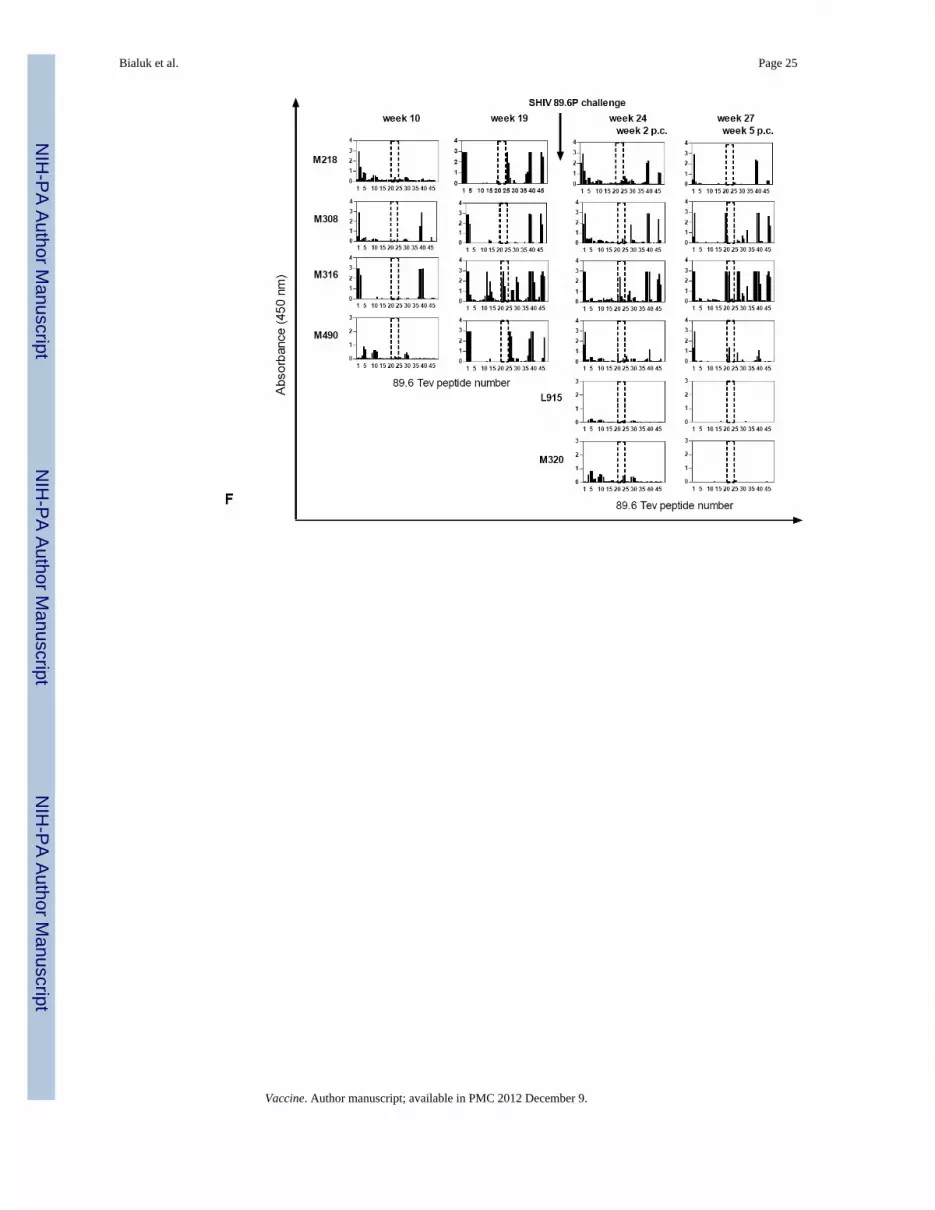
Bialuk et al. Page 25
Vaccine. Author manuscript; available in PMC 2012 December 9.
NIH
-PA Author Manuscript
NIH
-PA Author Manuscript
NIH
-PA Author Manuscript

Bialuk et al. Page 26
Vaccine. Author manuscript; available in PMC 2012 December 9.
NIH
-PA Author Manuscript
NIH
-PA Author Manuscript
NIH
-PA Author Manuscript

Figure 2. Immune responses in the vaccinated/challenged macaques(A) ELISPOTs were performed using HIVBa-L, HIVSF162, and HIV89.6 Tev peptide poolsfor the stimulation of macaque blood. ELISPOT responses were also obtained usingConcanavalin A, a stimulus to control for cell viability (data not shown).Antibody titers to Tev (B), Tat(C), Rev(D), and the pooled HIVBa-L, SHIVSF162P3, andSHIV89.6P Envelope proteins (E). The arrows refer to DNA immunizations (weeks 0, 4, 8),protein boosts (weeks 12, 18), and challenge exposure (week 22); titers to the purified Tev,Tat, Rev, and pooled HIVBa-L, HIVSF162P3, and HIV89.6P Envelope proteins. The weeks 0,2, 6, and 10 refer to the weeks post challenge (p.c.). Data from control macaques are notshown.(F) Overlapping peptides of the entire HIV89.6 Tev protein whose sequence is depicted inFig. 1B were used with the sera (1:100 dilution) of the macaques after immunization (weeks10, 19) and challenge exposure (weeks 2, 5 p.c.) in some of the vaccinated and controlmacaques. The area included in the dotted line area defines the V1 peptides (16 to 23),proceeded by the peptides derived from the first exon of Tat and followed by the peptidesderived from the second exon of Rev.(G) Sera (1:100 dilution) of all immunized macaques after challenge exposure (weeks 2, 5p.c.) and the two control macaques L915 and M320 were reacted with overlapping peptides16–28 for the 89.6 V1.(H) Amino acid sequences of the 21-24 overlapping peptides recognized by the sera ofmacaques M308, M316, and M490 and M607 within the V1 region of SHIV89.6P.
Bialuk et al. Page 27
Vaccine. Author manuscript; available in PMC 2012 December 9.
NIH
-PA Author Manuscript
NIH
-PA Author Manuscript
NIH
-PA Author Manuscript

(I): Sera (1:100 dilution) of the immunized macaques M218, M308, M316, and M490before and after challenge exposure (weeks 10 and 19 and 2, 5 respectively) and the twocontrol macaques. L915 and M320 were reacted with overlapping peptides 16–28 for theSF162 V1 region.
Bialuk et al. Page 28
Vaccine. Author manuscript; available in PMC 2012 December 9.
NIH
-PA Author Manuscript
NIH
-PA Author Manuscript
NIH
-PA Author Manuscript

Figure 3. Viral variants within the V1 region following challenge exposure to SHIV89.6PPutative amino acid sequence of the SHIV89.6P V1 region obtained by DNA sequencing ofplasma viral RNA following RT/PCR. The putative amino acid sequences of V1 in thevaccinated (A) and control (B) macaques were aligned with the V1 region of the SHIV89.6used in the Tev vaccine. The number of viral clones sequenced is depicted on the left ofeach figure.
Bialuk et al. Page 29
Vaccine. Author manuscript; available in PMC 2012 December 9.
NIH
-PA Author Manuscript
NIH
-PA Author Manuscript
NIH
-PA Author Manuscript

Figure 4. In vitro replication and sensitivity to ADCVI of SHIV89.6 mutant virusesThe supernatant of 293T-cells transfected with the HIV89.6WT, HIV89.6L, and HIV89.6LNmutants was normalized for p27Gag content and applied to TZM cells, CEMx174 cells, orprimary human and rhesus PBMCs. Virus production was measured as activation of theHIV-! LTR Luciferase reporter gene in the TZM cells (A) or as p27Gag production in thesupernatant fluid within the first 4 days of infection of the CEMx174 T-cells (B) or inhuman PBMCs up to day six post-infection (C).
Bialuk et al. Page 30
Vaccine. Author manuscript; available in PMC 2012 December 9.
NIH
-PA Author Manuscript
NIH
-PA Author Manuscript
NIH
-PA Author Manuscript

Figure 5. Sensitivity to ADCVI of SHIV 89.6 viral variantsSHIV89.6P-infected human CD4+ lymphocyte target cells were incubated with humanPBMC effector cells (E:T = 10:1) and with plasma at the dilutions indicated. Eight dayslater, supernatant fluid p27Gag was determined by ELISA, and virus inhibition wasdetermined as described in the Methods. Data are representative of several experiments,each performed in duplicate. Antibody-dependent cell-mediated virus inhibition (ADCVI)activity in plasma (1:100 dilution) of the animals was performed at first using the SHIV89.6Pbefore immunization week -2 (data not shown), or at time of challenge (A), or thereafter atweek 4 (B) and 6 (C). ADCVI titers to the SHIV89.6 viral variants a week 5 (D) and 22 (E)post challenge.
Bialuk et al. Page 31
Vaccine. Author manuscript; available in PMC 2012 December 9.
NIH
-PA Author Manuscript
NIH
-PA Author Manuscript
NIH
-PA Author Manuscript

Figure 6. Neutralizing antibody titers and passive transfer of purified Ig to a naïve macaqueNeutralization antibody titers were measured as the highest serum/plasma dilution thatinduced a 50% reduction in luciferase reporter gene expression after a single round ofinfection in TZM-bl cells as described [38;39] using HIV NL-ADArs(A) SF162.LS, (B) orSHIV89.6P. (C) The data presented were obtained with plasma collected at week 4 and 6 postchallenge. (D) Titers of ADCVI in macaque M316 at 24 weeks post exposure to SHIV89.6P.(E) Virus plasma levels in macaques challenged with SHIV89.6P following treatment withpurified Ig from macaques M316 (macaque P155) or normal human Ig (macaque P156).
Bialuk et al. Page 32
Vaccine. Author manuscript; available in PMC 2012 December 9.
NIH
-PA Author Manuscript
NIH
-PA Author Manuscript
NIH
-PA Author Manuscript

NIH
-PA Author Manuscript
NIH
-PA Author Manuscript
NIH
-PA Author Manuscript
Bialuk et al. Page 33
TABLE 1
Titers of sera mediating ADCC activity in vaccinated and control macaques’ post-SHIV89.6P challenge
ADCC titer post-challenge
Vaccinated Macaques Week 2 Week 10
M218 <10 10000
M308 <10 <10
M316 1000 <10
M490 <10 100
M599 <10 <10
M600 <10 <10
M607 <10 10
M611 <10 10
Control Macaques
M320 <10 <10
L915 <10 1000
L962 <10 <10
Vaccine. Author manuscript; available in PMC 2012 December 9.

NIH
-PA Author Manuscript
NIH
-PA Author Manuscript
NIH
-PA Author Manuscript
Bialuk et al. Page 34
TABLE 2
Neutralizing antibody titers at time of challenge and post infection in the sera of immunized and controlmacaques.
ID50 in TZM-bl cells1
Macaque Week SF162.LS SHIV89.6P NL-ADArs
M316 -2 <20 <20 <20
0 <20 <20 <20
4 <20 48 883
6 57 50 3177
M308 -2 <20 <20 <20
0 <20 <20 <20
4 <20 29 1850
6 40 163 3,611
M490 -2 <20 <20 <20
0 <20 <20 <20
4 <20 <20 1,832
6 87 107 15,732
M218 -2 <20 <20 <20
0 <20 <20 <20
4 <20 <20 37
6 <20 <20 538
L915 * -2 <20 <20 <20
0 <20 <20 <20
4 <20 <20 <20
6 <20 47 1,949
M320* -2 <20 <20 <20
0 <20 <20 <20
4 <20 <20 <20
6 <20 <20 21
0 <20 <20 <20
M599 4 <20 <20 <20
6 <20 <20 59
M600 4 <20 <20 41
6 <20 <20 47
M607 4 <20 26 95
6 <20 446 1,752
M611 4 <20 <20 669
6 <20 <20 285
L941* 4 ND ND ND
Vaccine. Author manuscript; available in PMC 2012 December 9.

NIH
-PA Author Manuscript
NIH
-PA Author Manuscript
NIH
-PA Author Manuscript
Bialuk et al. Page 35
ID50 in TZM-bl cells1
Macaque Week SF162.LS SHIV89.6P NL-ADArs
6 <20 <20 21
L942* 4 <20 <20 30
6 <20 <20 78
L947* 4 <20 <20 <20
6 <20 <20 <20
L962* 4 <20 <20 <20
6 <20 <20 <20
1Values are the serum dilution at which relative luminescence units (RLUs) were reduced 50% compared to virus control wells (no test sample).
ND=Not done.
*Non vaccinated
Vaccine. Author manuscript; available in PMC 2012 December 9.

NIH
-PA Author Manuscript
NIH
-PA Author Manuscript
NIH
-PA Author Manuscript
Bialuk et al. Page 36
Table 3
Immune correlate of protection against high plasma virus levels
Immune Response Post challenge Virus in plasma Correlation
ELISA Ab titer to envelope (week 5) Week 5 Post challenge -0.83 p=0.015
ELISA Ab titers to Tat Any time post Post challenge No Correlation
ELISA Ab titers to REV Any time post Post challenge No Correlation
Week 4 / 6 ADCVI SHIV89.6 swarm Week 7 to 16 Post challenge -0.84 p -0.044
ADCV1 week 5 Week 7 to 16 Post challenge
WT SHIV89.6A -0.079 p=0.0034
Variant SHIV89.6L -0.72 p=0.0094
Variant SHIV89.6LN -0.64 p=0.017
Week 22 Week 7or 16 Post challenge
SHIV89.6W -0.76 p=0.015
SHIV89.6L -0.85 p=0.0086
SHIV89.6LN -0.78 p=0.021
Neutralizing Antibodies titers to SHIV89.6P Week 6 Week 7 to 16 Post challenge -0.72 p=0.0050
Neutralizing Antibodies titers to HIV NL-ADArs Week 4 Weeks 7 to 16 Post challenge -090 p=0.0001
Neutralizing Antibodies titers to HIV NL-ADArs Week 7 to 16 Post challenge -0.83 p=0.0004
Vaccine. Author manuscript; available in PMC 2012 December 9.