Vaccination with the Leishmania major ribosomal proteins plus CpG oligodeoxynucleotides induces...
9
Original article Vaccination with the Leishmania major ribosomal proteins plus CpG oligodeoxynucleotides induces protection against experimental cutaneous leishmaniasis in mice Salvador Iborra a , Nuria Parody b , Daniel R. Aba ´nades b , Pedro Bonay b , Deboraci Prates c , Fernanda O. Novais c , Manoel Barral-Netto c , Carlos Alonso b , Manuel Soto b, * a Unidad de Inmunologı ´a Viral, Centro Nacional de Microbiologı ´a, Instituto de Salud Carlos III, Crta. Pozuelo Km 2, 28220 Majadahonda, Madrid, Spain b Centro de Biologı ´a Molecular Severo Ochoa, Departamento de Biologı ´a Molecular, Facultad de Ciencias, Universidad Auto ´noma de Madrid, 28049 Madrid, Spain c Fundac ¸ ~ ao Oswaldo Cruz (FIOCRUZ), Centro de Pesquisas Gonc ¸alo Moniz, Rua Waldemar Falcao, 121, Candeal 40, 296-710 Salvador, Bahia, Brazil Received 6 February 2008; accepted 9 June 2008 Available online 17 June 2008 Abstract In the present work we analyze the antigenicity of Leishmania major ribosomal proteins (LRP) in infected BALB/c mice. We show that BALB/c mice vaccinated with LRP in the presence of CpG oligodeoxynucleotides (CpG-ODN) were protected against the development of dermal pathology and showed a reduction in the parasite load after challenge with L. major. This protection was associated with the induction of an IL-12 dependent specific-IFN-g response mediated mainly by CD4 þ T cell, albeit a minor contribution of CD8 þ T cells cannot be ruled out. Induction of Th1 responses against LRP also resulted in a reversion of the Th2 responses associated with susceptibility. A marked reduction of IgG1 antibody titer against parasite antigens besides an impaired IL-4 and IL-10 cytokine production by parasite specific T cells was observed. In addition, we show that the administration of the LRP plus CpG-ODN preparation also conferred protection in the naturally resistant C57BL/6 mice. In this strain protection was associated with a LRP specific IFN-g production in lymph nodes draining the challenge site. We believe that these evolutionary conserved proteins, combined with adjuvants that favor Th1 responses, may be relevant components of a pan-Leishmania vaccine. Ó 2008 Elsevier Masson SAS. All rights reserved. Keywords: Leishmania; BALB/C mice; C57BL/6 mice; Th1/Th2 immune responses; Ribosomal proteins; Vaccines 1. Introduction Leishmaniases comprise several diseases caused by intra- cellular protozoan parasites belonging to the genus Leish- mania that mainly infect macrophages of mammals including human and dogs. Outcome of infection is deter- mined by interactions between the host immune system and different parasite molecules. A model for Leishmania viru- lence involving two different groups of parasite molecules has been proposed. The first one is formed by surface and secreted products that are necessary for the establishment of the infection, as a prerequisite for virulence. A second group is formed by highly conserved intracellular molecules referred as ‘‘pathoantigens’’ because an inadequate humoral response against them is thought to result in pathology mainly due to the adverse effects of the immune complexes formed [1,2]. Several lines of evidence suggest that Leishmania ribo- somal proteins are immunologically relevant molecules during infection. In some cases, ribosomal proteins can contribute to the host immune system dysfunction through their capacity to modulate cell activities and cytokine release during infection [3e6]. Thus, injection of the L. major ribosomal protein S3a into BALB/c mice induce the polyclonal expansion of B-cell clones inhibiting T-cell proliferation [3]. In addition, parasite * Corresponding author. Tel.: þ34 91 497 8454; fax: þ34 91 497 4799. E-mail address: [email protected] (M. Soto). 1286-4579/$ - see front matter Ó 2008 Elsevier Masson SAS. All rights reserved. doi:10.1016/j.micinf.2008.06.002 Microbes and Infection 10 (2008) 1133e1141 www.elsevier.com/locate/micinf
Transcript of Vaccination with the Leishmania major ribosomal proteins plus CpG oligodeoxynucleotides induces...
Microbes and Infection 10 (2008) 1133e1141www.elsevier.com/locate/micinf
Original article
Vaccination with the Leishmania major ribosomal proteins plus CpGoligodeoxynucleotides induces protection against experimental
cutaneous leishmaniasis in mice
Salvador Iborra a, Nuria Parody b, Daniel R. Abanades b, Pedro Bonay b, Deboraci Prates c,Fernanda O. Novais c, Manoel Barral-Netto c, Carlos Alonso b, Manuel Soto b,*
a Unidad de Inmunologıa Viral, Centro Nacional de Microbiologıa, Instituto de Salud Carlos III, Crta. Pozuelo Km 2, 28220 Majadahonda, Madrid, Spainb Centro de Biologıa Molecular Severo Ochoa, Departamento de Biologıa Molecular, Facultad de Ciencias,
Universidad Autonoma de Madrid, 28049 Madrid, Spainc Fundac~ao Oswaldo Cruz (FIOCRUZ), Centro de Pesquisas Goncalo Moniz, Rua Waldemar Falcao, 121, Candeal 40, 296-710 Salvador, Bahia, Brazil
Received 6 February 2008; accepted 9 June 2008
Available online 17 June 2008
Abstract
In the present work we analyze the antigenicity of Leishmania major ribosomal proteins (LRP) in infected BALB/c mice. We show that BALB/cmice vaccinated with LRP in the presence of CpG oligodeoxynucleotides (CpG-ODN) were protected against the development of dermal pathologyand showed a reduction in the parasite load after challenge with L. major. This protection was associated with the induction of an IL-12 dependentspecific-IFN-g response mediated mainly by CD4þ T cell, albeit a minor contribution of CD8þ T cells cannot be ruled out. Induction of Th1responses against LRP also resulted in a reversion of the Th2 responses associated with susceptibility. A marked reduction of IgG1 antibody titeragainst parasite antigens besides an impaired IL-4 and IL-10 cytokine production by parasite specific T cells was observed. In addition, we showthat the administration of the LRP plus CpG-ODN preparation also conferred protection in the naturally resistant C57BL/6 mice. In this strainprotection was associated with a LRP specific IFN-g production in lymph nodes draining the challenge site. We believe that these evolutionaryconserved proteins, combined with adjuvants that favor Th1 responses, may be relevant components of a pan-Leishmania vaccine.� 2008 Elsevier Masson SAS. All rights reserved.
Keywords: Leishmania; BALB/C mice; C57BL/6 mice; Th1/Th2 immune responses; Ribosomal proteins; Vaccines
1. Introduction
Leishmaniases comprise several diseases caused by intra-cellular protozoan parasites belonging to the genus Leish-mania that mainly infect macrophages of mammalsincluding human and dogs. Outcome of infection is deter-mined by interactions between the host immune system anddifferent parasite molecules. A model for Leishmania viru-lence involving two different groups of parasite moleculeshas been proposed. The first one is formed by surface and
* Corresponding author. Tel.: þ34 91 497 8454; fax: þ34 91 497 4799.
E-mail address: [email protected] (M. Soto).
1286-4579/$ - see front matter � 2008 Elsevier Masson SAS. All rights reserved.
doi:10.1016/j.micinf.2008.06.002
secreted products that are necessary for the establishment ofthe infection, as a prerequisite for virulence. A second groupis formed by highly conserved intracellular molecules referredas ‘‘pathoantigens’’ because an inadequate humoral responseagainst them is thought to result in pathology mainly due tothe adverse effects of the immune complexes formed [1,2].
Several lines of evidence suggest that Leishmania ribo-somal proteins are immunologically relevant molecules duringinfection. In some cases, ribosomal proteins can contribute tothe host immune system dysfunction through their capacity tomodulate cell activities and cytokine release during infection[3e6]. Thus, injection of the L. major ribosomal protein S3ainto BALB/c mice induce the polyclonal expansion of B-cellclones inhibiting T-cell proliferation [3]. In addition, parasite

1134 S. Iborra et al. / Microbes and Infection 10 (2008) 1133e1141
acidic ribosomal P proteins induce strong humoral responsesin dogs and humans suffering from leishmaniasis (reviewedin ref. [7]). On the other hand, the administration of someribosomal constituents using Th1 inducing adjuvants hasbeen related to the generation of protective responses inmice models. Immunization with the L. infantum acidic ribo-somal protein P0 as a DNA vaccine or as a combination ofthe recombinant protein and CpG-ODN protects C57BL/6mice and induce partial protection in BALB/c mice after chal-lenge with L. major [8,9]. Also, genetic immunization withtwo L. major ribosomal protein coding genes (L22 and S19)has been related with the generation of protective responsesin BALB/c mice challenged with this parasite [10].
We decided to analyze the immunogenicity of leishmanialribosome, because these intracellular ribonucleoprotein parti-cles may be relevant either as pathoantigens, but also inducingprotective responses. Here we show that during L. majorinfection, susceptible BALB/c mice develop a mixedTh1/Th2 response against several parasite ribosomal proteins.We also show that the Th1 immune response induced by theco-inoculation of LRP plus CpG-ODN confers protection againsta challenge with L. major parasites in the BALB/c mouse strain.Protection correlates to a LRP specific IL-12 dependent produc-tion of IFN-g and a diminished production of IL-4 and IL-10.Co-inoculation of LRP plus CpG-ODN also induces protectionagainst a low dose challenge with metacyclic L. major parasitesin C57BL/6 mouse strain, a model that more closely mimicsthe human disease in terms of route and infection dose [11].
2. Materials and methods
2.1. Mice strains and parasites
Female BALB/c and C57BL/6 mice (6e8 week old) werepurchased from Harlan Interfauna Iberica S.A. (Barcelona,Spain). L. major parasites (WHOM/IR/-/173) and clone V1(MHOM/IL/80(Friedlin)) were kept in a virulent state by pas-sage in BALB/c mice. L. major amastigotes were obtained andtransformed to promastigote by culturing at 26 �C in Schneider’smedium (Gibco, BRL, Grand Island, NY, USA) supplementedwith 20% fetal calf serum. Metacyclic promastigotes ofL. major (clone V1) were isolated from stationary culturesby negative selection using peanut agglutinin (Vector Labora-tories, Burlingame, CA, USA).
2.2. CpG-ODN, leishmanial antigens and mouseribosomal proteins
For preparation of L. major LRP, 109 promastigotes wereharvested, washed twice in pre-chilled PBS and resuspendedin 1 ml NP40 lysis buffer (10 mM TriseHCl, pH 8.0, 150 mMNaCl, 1.5 mM MgCl2 and 0.5% NP40) and pipetted up anddown 10 times. After lyses, samples were microfuged at3000 � g for 2 min at 4 �C to pellet the nuclei. Supernatantwas twice microfuged at 13,000 � g for 15 min at 4 �C andthe ribosomes were prepared from the cytosolic supernatant asdescribed in ref. [12]. Briefly, cytosol was submitted to high
speed centrifugation at 90,000 rpm for 30 min at 4 �C in a Beck-man TL100.3 rotor. The crude ribosomal pellet was resuspendedin buffer A (20 mM TriseHCl, pH 7.4, 500 mM AcNH4,100 mM MgCl2, 5 mM b-mercaptoethanol) and centrifugedthrough a discontinuous sucrose gradient (20/40%) in buffer Aat 90,000 rpm at 4 �C in a TL100.3 rotor. The pellet of washedribosomes was dissolved in PBS and sonicated until completeribosomal RNA degradation. Mouse ribosomal proteins extracts(MRP) were prepared from 5 � 107 RAW 264.7 murine macro-phage cells using the same procedure.
Total proteins of L. major (soluble Leishmania antigen[SLA]) was prepared as described [9].
Phosphorothioate-modifiedCpG-ODN(50-TCAACGTTGA-30 and 50-GCTAGCGTTAGCGT-30) were synthesized by Isogen(TheNetherlands).
2.3. Immunizations, parasite challenge andparasite quantification
BALB/c mice (six per group) were subcutaneously (s.c.)inoculated in the right footpad with either 12 mg of L. majorLRP alone or plus 25 mg of each CpG-ODN, CpG-ODN alone,or phosphate saline buffer (PBS). Each group was boosted 2and 4 weeks later with the same dose used for priming. Para-site challenge was carried out by s.c. inoculation with 5 � 104
stationary-phase promastigotes of L. major (WHOM/IR/-/173)into the left (untreated) footpad 4 weeks after the last inocula-tion. Footpad swelling was measured with a metric calliperand calculated as thickness of the left footpad minus thicknessof the right footpad.
C57BL/6 mice (12 per group) were injected s.c. in the foot-pad with 12 mg of L. major LRP plus 25 mg of each CpG-ODNor with the CpG-ODN alone, and boosted 2 and 4 weeks laterwith the same immunization regime. The infection was per-formed 4 weeks after the last vaccination by intradermal(i.d.) inoculation of 300 metacyclic promastigotes of L. major(clone V1) in both ears. Indurations of the ear lesion weremeasured with a metric calliper.
The number of parasites was determined in the ears, drain-ing lymph nodes (DLN) and spleen by limiting dilution assayas described [9].
2.4. Measurement of cytokines in supernatants
The release of IFN-g, IL-10 and IL-4 was measured in thesupernatants of splenocytes or DLN cells cultures stimulatedwith LRP (12 mg ml�1) or SLA (12 mg ml�1) or MRP(12 mg ml�1) s described previously [9], using commercialELISA kits (Diaclone, Besancon, France). When indicated,and in order to block IL-12, CD4 and CD8 mediated T cellcytokine release, DLN cells stimulated with 12 mg ml�1 ofLRP were incubated in the presence of 10 mg ml�1 of mono-clonal antibody (mAb) against either mouse IL-12 (C17.8)CD4 (GK 1.5), mouse CD8 (53e6.7) [13]. Appropriate iso-type-matched controls were also analyzed in the assay. The an-tibodies (no azide/low endotoxin�) were purchased from BD(PharMingen, San Diego, CA, USA).
176.5113.7
80.5
63.849.5
37.4
26.019.6
14.9
KDa SLA LRP
anti-H2A
SLA LRP
anti -β tub anti-P0
SLA LRPSLA LRP
Coomassiestained
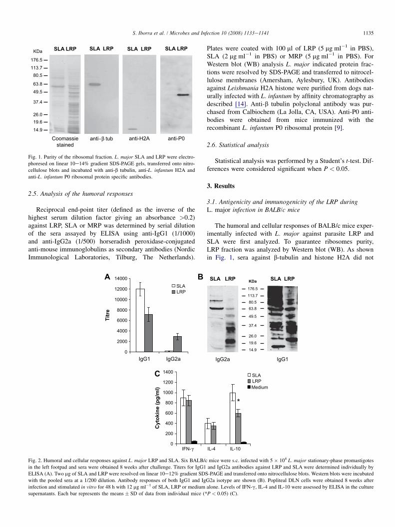
Fig. 1. Purity of the ribosomal fraction. L. major SLA and LRP were electro-
phoresed on linear 10e14% gradient SDS-PAGE gels, transferred onto nitro-
cellulose blots and incubated with anti-b tubulin, anti-L. infantum H2A and
anti-L. infantum P0 ribosomal protein specific antibodies.
1135S. Iborra et al. / Microbes and Infection 10 (2008) 1133e1141
2.5. Analysis of the humoral responses
Reciprocal end-point titer (defined as the inverse of thehighest serum dilution factor giving an absorbance >0.2)against LRP, SLA or MRP was determined by serial dilutionof the sera assayed by ELISA using anti-IgG1 (1/1000)and anti-IgG2a (1/500) horseradish peroxidase-conjugatedanti-mouse immunoglobulins as secondary antibodies (NordicImmunological Laboratories, Tilburg, The Netherlands).
0
2000
4000
6000
8000
10000
12000
14000
IgG1 IgG2a
Titre
SLALRP
A B
0
200
400
600
800
1000
1200
1400
IFN-γ
Cyto
kin
e (p
g/m
l)
C
Fig. 2. Humoral and cellular responses against L. major LRP and SLA. Six BALB/c
in the left footpad and sera were obtained 8 weeks after challenge. Titers for IgG1
ELISA (A). Two mg of SLA and LRP were resolved on linear 10e12% gradient SDS
with the pooled sera at a 1/200 dilution. Antibody responses of both IgG1 and Ig
infection and stimulated in vitro for 48 h with 12 mg ml�1 of SLA, LRP or medium
supernatants. Each bar represents the means � SD of data from individual mice (*
Plates were coated with 100 ml of LRP (5 mg ml�1 in PBS),SLA (2 mg ml�1 in PBS) or MRP (5 mg ml�1 in PBS). ForWestern blot (WB) analysis L. major indicated protein frac-tions were resolved by SDS-PAGE and transferred to nitrocel-lulose membranes (Amersham, Aylesbury, UK). Antibodiesagainst Leishmania H2A histone were purified from dogs nat-urally infected with L. infantum by affinity chromatography asdescribed [14]. Anti-b tubulin polyclonal antibody was pur-chased from Calbiochem (La Jolla, CA, USA). Anti-P0 anti-bodies were obtained from mice immunized with therecombinant L. infantum P0 ribosomal protein [9].
2.6. Statistical analysis
Statistical analysis was performed by a Student’s t-test. Dif-ferences were considered significant when P < 0.05.
3. Results
3.1. Antigenicity and immunogenicity of the LRP duringL. major infection in BALB/c mice
The humoral and cellular responses of BALB/c mice exper-imentally infected with L. major against parasite LRP andSLA were first analyzed. To guarantee ribosomes purity,LRP fraction was analyzed by Western blot (WB). As shownin Fig. 1, sera against b-tubulin and histone H2A did not
SLALRP
176.5
113.780.563.8
49.5
37.4
26.0
19.6
14.9
KDaSLA LRP SLA LRP
IgG2a IgG1
IL-4 IL-10
Medium
*
mice were s.c. infected with 5 � 104 L. major stationary-phase promastigotes
and IgG2a antibodies against LRP and SLA were determined individually by
-PAGE and transferred onto nitrocellulose blots. Western blots were incubated
G2a isotype are shown (B). Popliteal DLN cells were obtained 8 weeks after
alone. Levels of IFN-g, IL-4 and IL-10 were assessed by ELISA in the culture
P < 0.05) (C).

1136 S. Iborra et al. / Microbes and Infection 10 (2008) 1133e1141
show reactivity against LRP whereas anti-P0 antibodies shownan increased reactivity in LRP relative to SLA. Next we ana-lyzed the IgG1/IgG2a isotype polarization against LRP andSLA in L. major infected BALB/c mice. A mixed IgG1/IgG2a response against LRP was detected by ELISA(Fig. 2A) and WB assays (Fig. 2B). By contrast, antibodiesagainst SLA were mainly of the IgG1 isotype. Similar levelsof IFN-g and IL-4 production were detected after in vitro stim-ulation of DLN cells with LRP and SLA, whereas the IL-10production was significantly lower in the DLN cells stimulatedwith LRP (Fig. 2C).
The immunogenicity of the ribosomal proteins was evalu-ated in BALB/c mice after administration of the LRP in theabsence or presence of CpG-ODN. After vaccination withLRP þ CpG-ODN the anti-LRP humoral response was pre-dominantly of the IgG2a isotype, whereas a lower titer ofantibodies of the IgG1 isotype was detected in the serafrom mice immunized with LRP alone (Fig. 3A). No reactiv-ity against MRP was found in the sera for any of the groups(data not shown). Following in vitro stimulation with LRP,spleen cells from mice immunized with LRP þ CpG-ODNsecreted higher levels of IFN-g than those secreted by spleencells from controls and from mice immunized with LRPalone (Fig. 3B). No increase in IL-4 production was observedafter stimulation with LRP in any experimental group(Fig. 3C). LRP specific IL-10 was also detected in the super-natant of cultures established from spleens of LRP þ CpG-ODN vaccinated mice being the IFN-g/IL-10 ratio z40(Fig. 3D).
Titre
02000400060008000
10000120001400016000
Saline CpG LRP LRP+CpG
IgG1
IgG2a
A
LRPMedium
***
IL
-4 (p
g/m
l)
020406080
100120140160180
Saline CpG LRP LRP+CpG
C
Fig. 3. Antibody responses and cytokine production induced by vaccination in BA
individually by ELISA at week 4 after last dose (***P < 0.001 significant difference
three groups) (A). Spleen cells were obtained 4 weeks after vaccination and culture
IL-4 (C) and IL-10 (D) was assessed by ELISA in culture supernatants. Each bar rep
(***P < 0.001) and in the IL-10 (**P < 0.01) levels between LRP þ CpG-ODN
repeated with similar results.
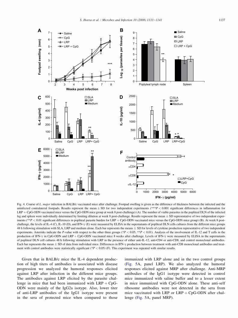
3.2. Vaccination with LRP þ CpG-ODN protectsBALB/c mice against L. major challenge
We analyzed whether LRP administration was able toinduce protection against L. major infection. Footpad swellingof LRP þ CpG-ODN vaccinated mice was significantly lowercompared with controls and mice vaccinated with LRP alone(Fig. 4A). In addition, DLN from LRP þ CpG-ODN immu-nized mice showed a 3-log reduction in parasite burden andno parasites could be detected in their spleens (Fig. 4B). Ithas been reported that CpG alone may protect BALB/c miceagainst L. major infection [15]. This unspecific effect wasnot observed in our study, indicating that the transient protec-tion conferred by this adjuvant is not effective 4 weeks afterinoculation.
To determine the immunological parameters protection, theSLA and the LRP-driven production of IL-4, IL-10 and IFN-gwas assayed. SLA or LRP specifically-induced IL-4 and IL-10production was detected in DLN cells from controls (saline andCpG) and from mice immunized with LRP alone (Fig. 4C,D). Incontrast, DLN cells from mice immunized with LRP þ CpG-ODN produced higher amounts of IFN-g than those detectedin the other three groups (Fig. 4E). The contribution of CD4þ
and CD8þ T cells and the dependence on IL-12 to the LRPspecific production of IFN-g was also analyzed (Fig. 4F).Production of IFN-g was completely suppressed by anti-IL-12 or anti-CD4 monoclonal antibodies. The addition ofanti-CD8 antibodies to the DLN cell cultures only partially re-duced the amounts of this cytokine in the supernatants.
0
10002000
300040005000600070008000
IF
N-
γ (p
g/m
l)
Saline CpG LRP LRP+CpG
LRPMedium
LRPMedium
***
**
B
020406080
100120140160180
IL
-10 (p
g/m
l)
Saline CpG LRP LRP+CpG
D
LB/c mice. Titer for IgG1 and IgG2a antibodies against LRP was determined
s in IgG2a titer between mice vaccinated with LRP þ CpG-ODN and the other
d in vitro for 48 h in the presence of LRP or medium. The levels of IFN-g (B),
resents the means � SD of data from individual mice. Differences in the IFN-g
and the other three groups were statistically significant. This experiment was
Weeks post infection
Fo
otp
ad
sw
ellin
g (m
m)
0
1
2
3
4
5
6
7
2 3 4 5 6 7 8
SalineCpGLRPLRP + CpG
***
0
1
2
3
4
5
6
7
8
9
Poplyteal lymph node Spleen
Lo
g 10 (p
arasites p
er tissu
e)
Saline
CpG
LRP + CpGLRP
**
0 1000 2000 3000 4000 5000 6000
IL-12
CD8
CD4
Controlantibody
IFN- γ (pg/ml)
LRP+CpGCpG
0
100
200
300
400
500
600
IL
-4 (p
g/m
l)
Saline CpG LRP LRP+ CpG
SLALRPMedium
**
*
**
**
* * *0
500
1000
1500
2000
2500
IL
-10 (p
g/m
l)
Saline CpG LRP LRP+CpG
SLALRPMedium
0
1000
2000
3000
4000
5000
6000
IF
N-γγ
(p
g/m
l)
Saline CpG LRP LRP+ CpG
SLALRPMedium
A B
C D
EF
Fig. 4. Course of L. major infection in BALB/c vaccinated mice after challenge. Footpad swelling is given as the difference of thickness between the infected and the
uninfected contralateral footpads. Results represent the mean � SD for two independent experiments (***P < 0.001 significant differences in inflammation for
LRP þ CpG-ODN vaccinated mice versus the CpG-ODN mice group at week 8 post-challenge) (A). The number of viable parasites in the popliteal DLN of the infected
leg and spleen were individually determined by limiting dilution at week 8 post-challenge. Results represent the mean� SD representative of two independent exper-
iments (**P < 0.01 significant differences in popliteal parasite burden for LRP þ CpG-ODN vaccinated mice versus the CpG-ODN mice group) (B). At week 8 post-
challenge, the levels of IL-4 (C), IL-10 (D), and IFN-g (E) were measured by ELISA in the supernatants of popliteal DLN cells cultures from the different mice groups
48 h following stimulation with SLA, LRP and medium alone. Each bar represents the means � SD for levels of cytokine production representative of two independent
experiments. Asterisks indicate the P-value with respect to the other three groups (*P < 0.05; **P < 0.01). Analysis of the involvement of IL-12 and T cells in the
production of IFN-g in CpG-ODN and LRP þ CpG-ODN vaccinated mice 8 weeks after challenge. Levels of IFN-g were measured by ELISA in the supernatants
of popliteal DLN cell cultures 48 h following stimulation with LRP in the presence of either anti-IL-12, anti-CD4 or anti-CD8, and control monoclonal antibodies.
Each bar represents the mean� SD of data from individual mice. Differences in IFN-g production between treatment with anti-CD8 monoclonal antibodies and treat-
ment with control antibodies were statistically significant (*P < 0.05) (F). This experiment was repeated with similar results.
1137S. Iborra et al. / Microbes and Infection 10 (2008) 1133e1141
Given that in BALB/c mice the IL-4 dependent produc-tion of high titers of antibodies is associated with diseaseprogression we analyzed the humoral responses elicitedagainst LRP after infection in the different mice groups.The antibodies against LRP elicited by the parasite chal-lenge in mice that had been immunized with LRP þ CpG-ODN were mainly of the IgG2a isotype. Also, lower titerof anti-LRP antibodies of the IgG1 isotype were presentin the sera of protected mice when compared to those
immunized with LRP alone and in the two control groups(Fig. 5A, panel LRP). We also analyzed the humoralresponses elicited against MRP after challenge. Anti-MRPantibodies of the IgG1 isotype were detected in controlmice immunized with saline buffer and to a lesser extentin mice immunized with CpG-ODN alone. These anti-selfribosome antibodies were not detected in the sera frommice vaccinated with LRP or LRP þ CpG-ODN after chal-lenge (Fig. 5A, panel MRP).

0
5000
10000
15000
20000
25000
Saline CpG LRP LRP+CpG
Titre
**
***
120100
56
38
20
29
KDaSaline CpG LRP LRP+CpG Saline CpG LRP LRP+CpG
IgG1 IgG2a
0
10000
20000
30000
40000
50000
60000
Saline CpG LRP LRP+CpG
Titre
IgG1IgG2a IgG1
IgG2a
IgG1IgG2a
**
***
LRP
0
2000
4000
6000
8000
10000
12000
14000
Saline CpG LRP LRP+CpG
MRP
***
*
Titre
A
B
C
Fig. 5. Analysis of the IgG1/IgG2a polarization. Serum samples obtained 8 weeks after challenge were individually tested by ELISA to determine the presence of
anti-LRP (Panel LRP) or anti-MRP (Panel MRP) IgG1 and IgG2a antibodies. Each bar represents the means � SD of data from individual mice. Differences in the
IgG1 (**P < 0.01) and IgG2a (***P < 0.001) titer between mice vaccinated with LRP þ CpG-ODN and the other three groups were statistically significant (panel
LRP). Differences in the IgG1 (***P < 0.001; *P < 0.05) titer between mice immunized with saline buffer or CpG-ODN and mice vaccinated with LRP or
LRP þ CpG-ODN were statistically significant (panel MRP) (A). L. major LRP were resolved on linear 10e14% gradient SDS-PAGE gel transferred onto nitro-
cellulose blots and incubated with the pooled sera from the indicated mice groups a 1/200 dilution. Antibody responses of both IgG1 and IgG2a isotypes are shown
(B). The same sera were employed for the determination of the IgG1 and IgG2a titers against SLA. Each bar represents the means � SD of data from individual
mice. Differences in the IgG1 (**P < 0.01) and IgG2a (***P < 0.001) titer between mice vaccinated with LRP þ CpG-ODN and the other three groups were
statistically significant (C).
1138 S. Iborra et al. / Microbes and Infection 10 (2008) 1133e1141
The IgG1 isotype antibodies from sera of mice immunizedwith saline, CpG-ODN or LRP alone recognized a highernumber of protein bands in LRP blots, whereas only a fewbands were recognized by the IgG1 antibodies of the protectedmice (Fig. 5B). Two protein bands were recognized withhigher intensity by the IgG2a antibodies in the sera fromLRP þ CpG-ODN vaccinated mice when compared with theother three groups (Fig. 5B). Vaccination with LRP þ CpG-ODN also conditioned the global anti-Leishmania humoralresponse induced by L. major infection. Antibodies producedby parasite-challenged mice immunized with LRP þ CpG-ODN were mainly of the IgG2a isotype being the anti-SLAtiter of the IgG1 isotype antibodies significantly lower thanthose detected in the other three groups (Fig. 5C).
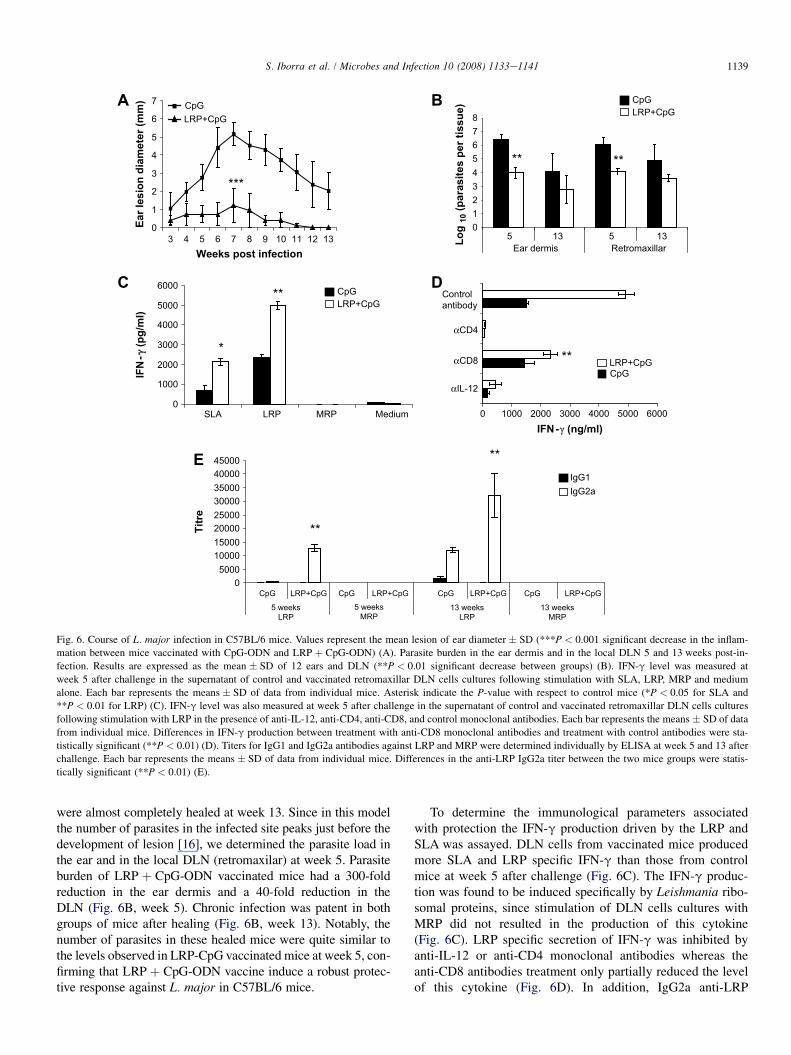
3.3. Vaccination with LRP þ CpG-ODN confersprotection against dermal pathology due toL. major challenge in C57BL/6 mice
Given that vaccination with LRP þ CpG-ODN protectsagainst L. major infection in susceptible BALB/c mice by theredirection the Th2 immune response against LRP towardsa Th1 response, we analyzed the effect of the administrationof this vaccine in C57BL/6 mice. This resistant mice strain nat-urally develops Th1 responses against Leishmania antigens.LRP þ CpG-ODN vaccinated C57BL/6 mice were protectedagainst the development of dermal lesions since little or nopathology was observed (Fig. 6A). CpG-ODN immunized con-trol mice developed lesions that reached a peak at week 7 and
0
1
2
3
4
5
6
7
3 654 7 8 9 10 11 12 13Weeks post infection
Ear lesio
n d
iam
eter (m
m) CpG
LRP+CpG
***
A
012345678
5 13 13Ear dermis Retromaxillar
CpGLRP+CpG
Lo
g 10 (p
arasites p
er tissu
e)
** **
5
B
**
*
0
1000
2000
3000
4000
5000
6000
SLA LRP MRP Medium
IF
N-γ
(p
g/m
l)
C
0 1000 2000 3000 4000 5000 6000
αIL-12
αCD8
αCD4
Controlantibody
IFN-γ (ng/ml)
**
D
05000
1000015000200002500030000350004000045000
CpG LRP+CpG CpG LRP+CpG
5 weeksLRP
13 weeksLRP
Titre
IgG1IgG2a
**
**
CpG LRP+CpG
5 weeksMRP
13 weeksMRP
CpG LRP+CpG
E
CpGLRP+CpG
CpGLRP+CpG
Fig. 6. Course of L. major infection in C57BL/6 mice. Values represent the mean lesion of ear diameter � SD (***P < 0.001 significant decrease in the inflam-
mation between mice vaccinated with CpG-ODN and LRP þ CpG-ODN) (A). Parasite burden in the ear dermis and in the local DLN 5 and 13 weeks post-in-
fection. Results are expressed as the mean � SD of 12 ears and DLN (**P < 0.01 significant decrease between groups) (B). IFN-g level was measured at
week 5 after challenge in the supernatant of control and vaccinated retromaxillar DLN cells cultures following stimulation with SLA, LRP, MRP and medium
alone. Each bar represents the means � SD of data from individual mice. Asterisk indicate the P-value with respect to control mice (*P < 0.05 for SLA and
**P < 0.01 for LRP) (C). IFN-g level was also measured at week 5 after challenge in the supernatant of control and vaccinated retromaxillar DLN cells cultures
following stimulation with LRP in the presence of anti-IL-12, anti-CD4, anti-CD8, and control monoclonal antibodies. Each bar represents the means � SD of data
from individual mice. Differences in IFN-g production between treatment with anti-CD8 monoclonal antibodies and treatment with control antibodies were sta-
tistically significant (**P < 0.01) (D). Titers for IgG1 and IgG2a antibodies against LRP and MRP were determined individually by ELISA at week 5 and 13 after
challenge. Each bar represents the means � SD of data from individual mice. Differences in the anti-LRP IgG2a titer between the two mice groups were statis-
tically significant (**P < 0.01) (E).
1139S. Iborra et al. / Microbes and Infection 10 (2008) 1133e1141
were almost completely healed at week 13. Since in this modelthe number of parasites in the infected site peaks just before thedevelopment of lesion [16], we determined the parasite load inthe ear and in the local DLN (retromaxilar) at week 5. Parasiteburden of LRP þ CpG-ODN vaccinated mice had a 300-foldreduction in the ear dermis and a 40-fold reduction in theDLN (Fig. 6B, week 5). Chronic infection was patent in bothgroups of mice after healing (Fig. 6B, week 13). Notably, thenumber of parasites in these healed mice were quite similar tothe levels observed in LRP-CpG vaccinated mice at week 5, con-firming that LRP þ CpG-ODN vaccine induce a robust protec-tive response against L. major in C57BL/6 mice.
To determine the immunological parameters associatedwith protection the IFN-g production driven by the LRP andSLA was assayed. DLN cells from vaccinated mice producedmore SLA and LRP specific IFN-g than those from controlmice at week 5 after challenge (Fig. 6C). The IFN-g produc-tion was found to be induced specifically by Leishmania ribo-somal proteins, since stimulation of DLN cells cultures withMRP did not resulted in the production of this cytokine(Fig. 6C). LRP specific secretion of IFN-g was inhibited byanti-IL-12 or anti-CD4 monoclonal antibodies whereas theanti-CD8 antibodies treatment only partially reduced the levelof this cytokine (Fig. 6D). In addition, IgG2a anti-LRP

1140 S. Iborra et al. / Microbes and Infection 10 (2008) 1133e1141
specific antibodies were detected earlier presenting highertiters in vaccinated mice (Fig. 6E). Anti-self ribosome anti-bodies were not detected in the sera from both mice groups(Fig. 6E).
4. Discussion
Research for the development of second generation vac-cines based on Leishmania crude parasite fractions or basedon defined parasite antigens was addressed to the identificationof different parasite molecules that have been tested as vaccinecandidates in several experimental models. Some of them aresurface or secreted parasite antigens and others have been con-sidered as good vaccine candidates because they elicit primar-ily a Th1 immune response in infected mice or human patientcells (reviewed in ref. [17]). On the other hand, some otherparasite antigens presented to the immune system during thenatural course of infection are conserved intracellular proteinslike heat shock proteins, histones or ribosomal proteins. Theseproteins predominantly stimulate specific humoral responsesin human or dogs suffering VL or Th2-mediated humoral re-sponses in experimentally infected mice (reviewed in ref. [2]).
Data presented here show that in the sera from infectedBALB/c mice there are IgG1 and IgG2a anti-LRP antibodies.Since the induction of IgG1 and IgG2a antibodies is consid-ered a marker of Th2- and Th1-type responses, respectively[18], it can be concluded that a predominant Th2 response ismounted against parasite ribosomal proteins. In addition, inspite of the strongly biased Th2 responses developed inBALB/c infected mice some ribosomal antigens were capableto elicit a Th1 response. In accordance, after in vitro stimula-tion of the DLN cells from infected mice with LRP, highamounts of both IFN-g and IL-4 were detected in the culturesupernatants. Also, it was observed comparable levels of IL-10, a pleiotropic anti-inflammatory cytokine that rendersinfected macrophages unresponsive to the activation signalsfor parasite destruction [19]. Furthermore, given that theeffects of IL-4 and IL-10 in promoting disease in BALB/cmice appear to be additive (reviewed in ref. [20]), stimulationof these cytokines by LRP would indicate that the hostresponses against ribosomal antigens might favor parasiteexpansion and persistence in BALB/c mice. We may assumethat early after Leishmania infection, the host immune systemis primed by the high abundance of ribosomal proteinsreleased by parasite cytolysis that afterwards, may be boostedas a result of parasite proliferation. Thus, the strong immuno-genicity of LRP and their pathoantigenic role probably relieson their high abundance and antigenic specificity in spite oftheir evolutionary conserved character.
In BALB/c mice the administration of LRP withoutadjuvants induced only weak IgG1 humoral responses, butco-administration with CpG-ODN an adjuvant that confersa Th1-related long term immunity and protection when usedwith different leishmanial antigens [21] and that also can sup-press some parasite specific Th2 responses in mouse [22,23],boosted a Th1-like response against these antigens. Thus,LRP plus CpG-ODN vaccinated mice developed anti-LRP
antibodies of the IgG2a isotype and their splenocytes producedhigh amounts of IFN-g, but not IL-4, after in vitro stimulationwith LRP. The LRP-driven production of IL-10 observedmight be related to the homeostatic control of the Th1responses since it has been recently reported that the IFN-gproducing Th1 cells can also be implicated in the productionof IL-10 as a mechanism of feedback control [24]. Moreover,the high IFN-g/IL-10 ratio values obtained might providea good prediction of vaccine outcome [4].
Co-administration of LRP þ CpG-ODN induce protectionin two different models of cutaneous experimental leishmani-asis: high dose inocula in the footpad of BALB/c mice (widelyused in cutaneous leishmaniasis vaccine assays), and low dosein the ear of C57BL/6 mice (a model that more closely mimicsthe human disease in terms of route and infectious dose). Inboth strains LRP plus CpG-ODN vaccinated mice showedreduction on parasite burden and were protected againstpathology. Protection is correlated to the generation of Th1specific immune responses against LRP being the IFN-gresponse IL-12 dependent and mainly produced by CD4þ Tcell. Generation of the Th1 responses in the protected micecorrelates to the generation of predominant IgG2a specificantibodies against LRP in both mice strains.
Remarkably, we have found that protection showed in theBALB/c mice after vaccination with LRP þ CpG-ODN isalso related to a significant reduction in the production ofantigen driven IL-4 and IL-10 after stimulation in vitro withLRP or SLA. These cellular responses correlated in vivowith the reversion of the Th2-mediated antibody responsesagainst ribosomal proteins. Thus, sera from protected BALB/c mice presented a significant decrease in the titer and, nota-bly, in the number of antigens recognized by IgG1 antibodiesspecific for LRP. In addition, immunization of BALB/c micewith LRP CpG-ODN also had a clear effect on the globalhumoral response elicited in mice by the L. major infection.Thus, the infection of vaccinated mice induced limited IgG1anti-Leishmania specific antibodies, whereas the humoralresponse in the control groups was higher and with a predom-inance of Th2-type antibodies (i.e., IgG1 isotype).
In our opinion, generation of vaccines against such a com-plex parasite as Leishmania, would be optimized by incorpo-rating different target antigens in the vaccine formulation,taking advantage of these antigens that induce the requiredimmunity (mainly CD4þ and CD8þ IFN-g mediatedresponses), and redirecting towards a Th1 bias the immuneresponses that result in pathology (IL-4 Th2-driven and IL-10 deactivating responses). Notwithstanding, it should betaken into account that the Th2-response against some of theseantigens may not be redirected by the usual Th1-inducers, asoccur with the meta-1 antigen of L. major [25] or the parasiteP2a and P2b acidic ribosomal proteins [26]. In this work weshow that vaccination with LRP þ CpG-ODN has direct influ-ences on the immune system decisions at the time of Leish-mania infection in both, resistant and susceptible mice,employing a pure preparation of Leishmania intracellular ribo-somal proteins. Although these proteins are highly conservedin evolution, we have not detected IgG1 or IgG2a antibodies

1141S. Iborra et al. / Microbes and Infection 10 (2008) 1133e1141
against murine ribosomal proteins in the LRP or LRP þ CpG-ODN vaccinated mice. In addition, DLN cells from C57BL/6vaccinated and infected mice did not produce IFN-g after invitro stimulation with MRP. These data support the idea thatvaccination with LRP induces an immune response specificfor Leishmania, since no humoral or cellular responses werefound against self-ribosomal proteins. Our results indicatethat the parasite ribosomal proteins may be useful as compo-nents in a pan-Leishmania vaccine. Further characterizationof the ribosomal constituents implicated in both type ofresponses should contribute to a more rational developmentof effective molecular defined vaccines against leishmaniasis.
Acknowledgements
We thank Angela Privat for her helpful discussion related tothis manuscript. This work was supported by grants PHB2006-0006-PC from Ministerio de Educacion y Ciencia, A/7692/07from AECI, and ISCIII-RETIC RD06/0021/0008-FEDERfrom FIS. An institutional grant from Fundacion Ramon Are-ces is also acknowledged.
References
[1] K.P. Chang, S.G. Reed, B.S. McGwire, L. Soong, Leishmania model for
microbial virulence: the relevance of parasite multiplication and pathoan-
tigenicity, Acta Trop 85 (2003) 375e390.
[2] N. Santarem, R. Silvestre, J. Tavares, M. Silva, S. Cabral, J. Maciel,
A. Cordeiro-da-Silva, Immune response regulation by leishmania secreted
and nonsecreted antigens, J. Biomed. Biotechnol 2007 (2007) 85154.
[3] A. Cordeiro-Da-Silva, M.C. Borges, E. Guilvard, A. Ouaissi, Dual role of
the Leishmania major ribosomal protein S3a homologue in regulation of
T- and B-cell activation, Infect. Immun 69 (2001) 6588e6596.
[4] M.T. Roberts, C.B. Stober, A.N. McKenzie, J.M. Blackwell, Interleukin-
4 (IL-4) and IL-10 collude in vaccine failure for novel exacerbatory
antigens in murine Leishmania major infection, Infect. Immun. 73
(2005) 7620e7628.
[5] M. Soto, J.M. Requena, L. Quijada, S.O. Angel, L.C. Gomez, F. Guzman,
M.E. Patarroyo, C. Alonso, During active viscerocutaneous leishmaniasis
the anti-P2 humoral response is specifically triggered by the parasite P
proteins, Clin. Exp. Immunol 100 (1995) 246e252.
[6] M. Soto, J.M. Requena, L. Quijada, F. Guzman, M.E. Patarroyo,
C. Alonso, Identification of the Leishmania infantum P0 ribosomal protein
epitope in canine visceral leishmaniasis, Immunol. Lett. 48 (1995) 23e28.
[7] J.M. Requena, C. Alonso, M. Soto, Evolutionarily conserved proteins as
prominent immunogens during Leishmania infections, Parasitol. Today
16 (2000) 246e250.
[8] S. Iborra, M. Soto, J. Carrion, A. Nieto, E. Fernandez, C. Alonso,
J.M. Requena, The Leishmania infantum acidic ribosomal protein P0
administered as a DNA vaccine confers protective immunity to Leish-
mania major infection in BALB/c mice, Infect. Immun 71 (2003)
6562e6572.
[9] S. Iborra, J. Carrion, C. Anderson, C. Alonso, D. Sacks, M. Soto, Vacci-
nation with the Leishmania infantum acidic ribosomal P0 protein plus
CpG oligodeoxynucleotides induces protection against cutaneous leish-
maniasis in C57BL/6 mice but does not prevent progressive disease in
BALB/c mice, Infect. Immun 73 (2005) 5842e5852.
[10] C.B. Stober, U.G. Lange, M.T. Roberts, B. Gilmartin, R. Francis,
R. Almeida, C.S. Peacock, S. McCann, J.M. Blackwell, From genome to
vaccines for leishmaniasis: screening 100 novel vaccine candidates against
murine Leishmania major infection, Vaccine 24 (2006) 2602e2616.
[11] Y. Belkaid, S. Mendez, R. Lira, N. Kadambi, G. Milon, D. Sacks, A nat-
ural model of Leishmania major infection reveals a prolonged ‘‘silent’’
phase of parasite amplification in the skin before the onset of lesion for-
mation and immunity, J. Immunol 165 (2000) 969e977.
[12] M.A. Rodriguez-Gabriel, M. Remacha, J.P. Ballesta, The RNA interact-
ing domain but not the protein interacting domain is highly conserved in
ribosomal protein P0, J. Biol. Chem. 275 (2000) 2130e2136.
[13] S. Gurunathan, C. Prussin, D.L. Sacks, R.A. Seder, Vaccine requirements
for sustained cellular immunity to an intracellular parasitic infection,
Nat. Med 4 (1998) 1409e1415.
[14] M. Soto, J.M. Requena, L. Quijada, M. Garcia, F. Guzman,
M.E. Patarroyo, C. Alonso, Mapping of the linear antigenic determinants
from the Leishmania infantum histone H2A recognized by sera from dogs
with leishmaniasis, Immunol. Lett. 48 (1995) 209e214.
[15] K.J. Stacey, J.M. Blackwell, Immunostimulatory DNA as an adjuvant in
vaccination against Leishmania major, Infect. Immun. 67 (1999) 3719e
3726.
[16] Y. Belkaid, S. Kamhawi, G. Modi, J. Valenzuela, N. Noben-Trauth,
E. Rowton, J. Ribeiro, D.L. Sacks, Development of a natural model of
cutaneous leishmaniasis: powerful effects of vector saliva and saliva pre-
exposure on the long-term outcome of Leishmania major infection in the
mouse ear dermis, J. Exp. Med 188 (1998) 1941e1953.
[17] R.N. Coler, S.G. Reed, Second-generation vaccines against leishmania-
sis, Trends Parasitol 21 (2005) 244e249.
[18] R.L. Coffman, Mechanisms of helper T-cell regulation of B-cell activity,
Ann. N.Y. Acad. Sci. 681 (1993) 25e28.
[19] K.W. Moore, R. de Waal Malefyt, R.L. Coffman, A. O’Garra, Interleu-
kin-10 and the interleukin-10 receptor, Annu. Rev. Immunol 19 (2001)
683e765.
[20] N. Peters, D. Sacks, Immune privilege in sites of chronic infection: Leish-mania and regulatory T cells, Immunol. Rev. 213 (2006) 159e179.
[21] E.G. Rhee, S. Mendez, J.A. Shah, C.Y. Wu, J.R. Kirman, T.N. Turon,
D.F. Davey, H. Davis, D.M. Klinman, R.N. Coler, D.L. Sacks,
R.A. Seder, Vaccination with heat-killed Leishmania antigen or recombi-
nant leishmanial protein and CpG oligodeoxynucleotides induces long-
term memory CD4þ and CD8þ T cell responses and protection against
Leishmania major infection, J. Exp. Med 195 (2002) 1565e1573.
[22] M.G. Chiaramonte, M. Hesse, A.W. Cheever, T.A. Wynn, CpG oligonu-
cleotides can prophylactically immunize against Th2-mediated Schisto-
some egg-induced pathology by an IL-12-independent mechanism, J.
Immunol 164 (2000) 973e985.
[23] S. Zimmermann, O. Egeter, S. Hausmann, G.B. Lipford, M. Rocken,
H. Wagner, K. Heeg, CpG oligodeoxynucleotides trigger protective and
curative Th1 responses in lethal murine leishmaniasis, J. Immunol 160
(1998) 3627e3630.
[24] C.F. Anderson, M. Oukka, V.J. Kuchroo, D. Sacks,
CD4(þ)CD25(�)Foxp3(�) Th1 cells are the source of IL-10-mediated
immune suppression in chronic cutaneous leishmaniasis, J. Exp. Med
204 (2007) 285e297.
[25] C.H. Serezani, A.R. Franco, M. Wajc, J.K. Umada Yokoyama-Yasunaka,
G. Wunderlich, M.M. Borges, S.R. Uliana, Evaluation of the murine
immune response to Leishmania meta 1 antigen delivered as recombinant
protein or DNA vaccine, Vaccine 20 (2002) 3755e3763.
[26] S. Iborra, D.R. Abanades, N. Parody, J. Carrion, R.M. Risueno,
M.A. Pineda, P. Bonay, C. Alonso, M. Soto, The immunodominant T
helper 2 (Th2) response elicited in BALB/c mice by the LeishmaniaLiP2a and LiP2b acidic ribosomal proteins cannot be reverted by strong
Th1 inducers, Clin. Exp. Immunol 150 (2007) 375e385.