Using a synoptophore to test Listing's law during vergence in normal subjects and strabismic...
11
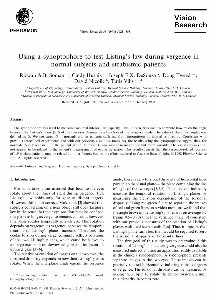
Vision Research 38 (1998) 3621 – 3631 Using a synoptophore to test Listing’s law during vergence in normal subjects and strabismic patients Rizwan A.B. Somani c , Cindy Hutnik b , Joseph F.X. DeSouza a , Doug Tweed a,c , David Nicolle b , Tutis Vilis a,b, * a Department of Physiology, Uni6ersity of Western Ontario, Medical Science Building, London, Ontario N6A 5C1, Canada b Deparment of Opthalmology, Uni6ersity of Western Ontario, Medical Science Building, London, Ontario N6A 5C1, Canada c Graduate Program in Neuroscience, Uni6ersity of Western Ontario, Medical Science Building, London, Ontario N6A 5C1, Canada Received 14 August 1997; received in revised form 23 January 1998 Abstract The synoptophore was used to measure torsional interocular disparity. This, in turn, was used to compute how much the angle between the Listing’s plane (LP) of the two eyes changes as a function of the vergence angle. The ratio of these two angles was defined as G. We measured G in normals and in patients suffering from intermittent horizontal strabismus. Consistent with previous search-coil experiments and with our previous visual test measures, the results using the synoptophore suggest that, for normals, G is less than 1. In the patient group the mean G was similar in magnitude but more variable. The variations in G did not appear to be related to the patient’s measurement of ocular deviation. This result suggests that the vergence-related rotation of LP in these patients may be related to other factors besides the effort required to fuse the lines of sight. © 1998 Elsevier Science Ltd. All rights reserved. Keywords: Listing’s law; Vergence; Torsional disparity; Synoptophore; Visual test 1. Introduction For some time it was assumed that because the eyes rotate about their lines of sight during vergence [1,2], Listing’s law holds only for gaze at distant targets. However, this is not correct. Mok et al. [3] showed that subjects converging on a near object still obey Listing’s law in the sense that their eye position remains confined to a plane as long as vergence remains constant, however, this plane rotates temporally in each eye. This rotation depends on vergence: as vergence increases the temporal rotation of Listing’s planes increase. Therefore, the ocular torsion during vergence simply reflects rotations of the two Listing’s planes, which cause both eyes to undergo extorsion on downward gaze and intorsion on upward gaze [3–6]. The relative orientation of images on the two eyes, the torsional disparity, depends on how their Listing’s planes rotate. When the interplane angle equals the vergence angle, there is zero torsional disparity of horizontal lines parallel to the visual plane — the plane containing the line of sight of the two eyes [5,7,8]. Thus one can indirectly measure the temporal rotation of Listing’s planes by measuring the elevation dependence of the torsional disparity. Using red-green filters to separate the images of red and green lines on a video monitor, we found that the angle between the Listing’s planes was on average 0.7 (range 0.5 – 0.90) times the vergence angle [9] consistent with our previous measures of the rotation of Listing’s planes with dual search coils [3,6]. Thus it appears that Listing’s plane turns less than would be required to zero the torsional disparity of horizontal lines. The first goal of this study was to determine if this rotation of Listing’s plane during vergence could also be measured indirectly, using an apparatus readily available in the clinic: a synoptophore. A synoptophore presents separate images to the two eyes. These images can be precisely rotated horizontally, inducing various degrees of vergence. The torsional disparity can be measured by asking the subject to rotate the image torsionally until this disparity becomes zero. * Corresponding author. Fax.: +1 519 6613827; e-mail: [email protected]. 0042-6989/98/$19.00 © 1998 Elsevier Science Ltd. All rights reserved. PII: S00 42- 6989(98)00057 -1
Transcript of Using a synoptophore to test Listing's law during vergence in normal subjects and strabismic...
Vision Research 38 (1998) 3621–3631
Using a synoptophore to test Listing’s law during vergence innormal subjects and strabismic patients
Rizwan A.B. Somani c, Cindy Hutnik b, Joseph F.X. DeSouza a, Doug Tweed a,c,David Nicolle b, Tutis Vilis a,b,*
a Department of Physiology, Uni6ersity of Western Ontario, Medical Science Building, London, Ontario N6A 5C1, Canadab Deparment of Opthalmology, Uni6ersity of Western Ontario, Medical Science Building, London, Ontario N6A 5C1, Canada
c Graduate Program in Neuroscience, Uni6ersity of Western Ontario, Medical Science Building, London, Ontario N6A 5C1, Canada
Received 14 August 1997; received in revised form 23 January 1998
Abstract
The synoptophore was used to measure torsional interocular disparity. This, in turn, was used to compute how much the anglebetween the Listing’s plane (LP) of the two eyes changes as a function of the vergence angle. The ratio of these two angles wasdefined as G. We measured G in normals and in patients suffering from intermittent horizontal strabismus. Consistent withprevious search-coil experiments and with our previous visual test measures, the results using the synoptophore suggest that, fornormals, G is less than 1. In the patient group the mean G was similar in magnitude but more variable. The variations in G didnot appear to be related to the patient’s measurement of ocular deviation. This result suggests that the vergence-related rotationof LP in these patients may be related to other factors besides the effort required to fuse the lines of sight. © 1998 Elsevier ScienceLtd. All rights reserved.
Keywords: Listing’s law; Vergence; Torsional disparity; Synoptophore; Visual test
1. Introduction
For some time it was assumed that because the eyesrotate about their lines of sight during vergence [1,2],Listing’s law holds only for gaze at distant targets.However, this is not correct. Mok et al. [3] showed thatsubjects converging on a near object still obey Listing’slaw in the sense that their eye position remains confinedto a plane as long as vergence remains constant, however,this plane rotates temporally in each eye. This rotationdepends on vergence: as vergence increases the temporalrotation of Listing’s planes increase. Therefore, theocular torsion during vergence simply reflects rotationsof the two Listing’s planes, which cause both eyes toundergo extorsion on downward gaze and intorsion onupward gaze [3–6].
The relative orientation of images on the two eyes, thetorsional disparity, depends on how their Listing’s planesrotate. When the interplane angle equals the vergence
angle, there is zero torsional disparity of horizontal linesparallel to the visual plane—the plane containing the lineof sight of the two eyes [5,7,8]. Thus one can indirectlymeasure the temporal rotation of Listing’s planes bymeasuring the elevation dependence of the torsionaldisparity. Using red-green filters to separate the imagesof red and green lines on a video monitor, we found thatthe angle between the Listing’s planes was on average 0.7(range 0.5–0.90) times the vergence angle [9] consistentwith our previous measures of the rotation of Listing’splanes with dual search coils [3,6]. Thus it appears thatListing’s plane turns less than would be required to zerothe torsional disparity of horizontal lines.
The first goal of this study was to determine if thisrotation of Listing’s plane during vergence could also bemeasured indirectly, using an apparatus readily availablein the clinic: a synoptophore. A synoptophore presentsseparate images to the two eyes. These images can beprecisely rotated horizontally, inducing various degreesof vergence. The torsional disparity can be measured byasking the subject to rotate the image torsionally untilthis disparity becomes zero.
* Corresponding author. Fax.: +1 519 6613827; e-mail:[email protected].
0042-6989/98/$19.00 © 1998 Elsevier Science Ltd. All rights reserved.
PII: S0042-6989(98)00057-1
R. A.B. Somani et al. / Vision Research 38 (1998) 3621–36313622
The second goal of this study was to use the synop-tophore to determine how much Listing’s planes ro-tated in patients suffering from various forms ofintermittent horizontal strabismus. When viewingmonocularly, these patients displayed various degreesof eso- or exo-deviation without any prominent verticaldisparity. In the synoptophore they were able to fuse abinocularly viewed dot and circle. Van den Berg et al.[10], have shown that subjects who have an exo-devia-tion display an elevation-dependent cyclovergence evenwhen viewing distant targets. As a result, their Listing’splanes are not perfectly parallel but are rotated tempo-rally even at a vergence angle of zero. Van den Berg etal. [10] have suggested that this occurs because conver-gence is used to align the diverging eyes of thesesubjects. In this paper we use the synoptophore and ourvisual tests to re-examine this idea.
1.1. Methods
In total, ten normals and ten patients participated inthe study. The patients were selected according to thefollowing criteria: an eso- or exo-deviation measurablein the distance or at near, a vertical ocular deviation ofless than two diopters, Snellen visual acuity of 20/25 orbetter in the distance (6 m), near visual acuity ofJ1+ (at 33 cm), motor fusional convergence amplitudesof ten prism diopters or better in the distance and 30prism diopters or better at near, binocular single visionboth at near and distance, as well as near stereopsis of100%% or better. Specifically, both intermittent eso- andexo-tropias as well as eso- and exo-phorias made up thegroup of deviations studied. Permission was obtainedfrom the referring ophthalmologists to invite these can-didates for a repeat, comprehensive orthoptic examina-tion to confirm eligibility for study participation. Allpatients provided informed consent. Stereoacuity wasrecorded with the Titmus system. Sensory status wasreported as a fusion response for near and distanttargets using the Worth four-dot test. A cover-uncovertest demonstrated the ocular deviation both at near andin the distance, with and without refractive correction.Best corrected visual acuity was recorded. The alternatecover test with prism bars was used to quantify theocular deviation both at near (33 cm) and in thedistance (6 m). The subjects in the control group wereidentified by the above criteria with the exception ofhaving a horizontal ocular deviation limited to twodiopters
We used a synoptophore [11] to measure the eleva-tion-dependent torsional disparity during various de-grees of convergence. With a synotophore the subjectviews two images at optical infinity through two eyepieces. These images fill the visual field so that nothing
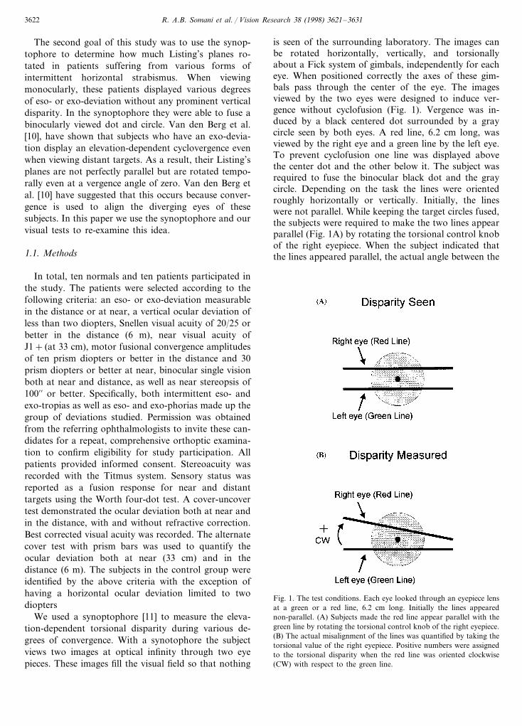
is seen of the surrounding laboratory. The images canbe rotated horizontally, vertically, and torsionallyabout a Fick system of gimbals, independently for eacheye. When positioned correctly the axes of these gim-bals pass through the center of the eye. The imagesviewed by the two eyes were designed to induce ver-gence without cyclofusion (Fig. 1). Vergence was in-duced by a black centered dot surrounded by a graycircle seen by both eyes. A red line, 6.2 cm long, wasviewed by the right eye and a green line by the left eye.To prevent cyclofusion one line was displayed abovethe center dot and the other below it. The subject wasrequired to fuse the binocular black dot and the graycircle. Depending on the task the lines were orientedroughly horizontally or vertically. Initially, the lineswere not parallel. While keeping the target circles fused,the subjects were required to make the two lines appearparallel (Fig. 1A) by rotating the torsional control knobof the right eyepiece. When the subject indicated thatthe lines appeared parallel, the actual angle between the
Fig. 1. The test conditions. Each eye looked through an eyepiece lensat a green or a red line, 6.2 cm long. Initially the lines appearednon-parallel. (A) Subjects made the red line appear parallel with thegreen line by rotating the torsional control knob of the right eyepiece.(B) The actual misalignment of the lines was quantified by taking thetorsional value of the right eyepiece. Positive numbers were assignedto the torsional disparity when the red line was oriented clockwise(CW) with respect to the green line.
R. A.B. Somani et al. / Vision Research 38 (1998) 3621–3631 3623
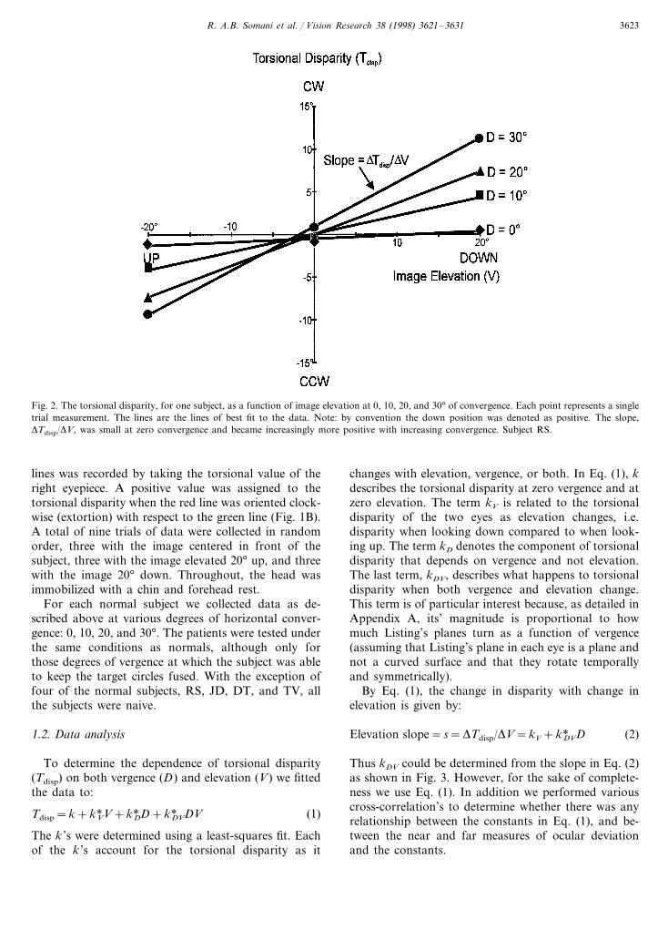
Fig. 2. The torsional disparity, for one subject, as a function of image elevation at 0, 10, 20, and 30° of convergence. Each point represents a singletrial measurement. The lines are the lines of best fit to the data. Note: by convention the down position was denoted as positive. The slope,DTdisp/DV, was small at zero convergence and became increasingly more positive with increasing convergence. Subject RS.
lines was recorded by taking the torsional value of theright eyepiece. A positive value was assigned to thetorsional disparity when the red line was oriented clock-wise (extortion) with respect to the green line (Fig. 1B).A total of nine trials of data were collected in randomorder, three with the image centered in front of thesubject, three with the image elevated 20° up, and threewith the image 20° down. Throughout, the head wasimmobilized with a chin and forehead rest.
For each normal subject we collected data as de-scribed above at various degrees of horizontal conver-gence: 0, 10, 20, and 30°. The patients were tested underthe same conditions as normals, although only forthose degrees of vergence at which the subject was ableto keep the target circles fused. With the exception offour of the normal subjects, RS, JD, DT, and TV, allthe subjects were naive.
1.2. Data analysis
To determine the dependence of torsional disparity(Tdisp) on both vergence (D) and elevation (V) we fittedthe data to:
Tdisp=k+kV*V+kD* D+kDV* DV (1)
The k ’s were determined using a least-squares fit. Eachof the k ’s account for the torsional disparity as it
changes with elevation, vergence, or both. In Eq. (1), kdescribes the torsional disparity at zero vergence and atzero elevation. The term kV is related to the torsionaldisparity of the two eyes as elevation changes, i.e.disparity when looking down compared to when look-ing up. The term kD denotes the component of torsionaldisparity that depends on vergence and not elevation.The last term, kDV, describes what happens to torsionaldisparity when both vergence and elevation change.This term is of particular interest because, as detailed inAppendix A, its’ magnitude is proportional to howmuch Listing’s planes turn as a function of vergence(assuming that Listing’s plane in each eye is a plane andnot a curved surface and that they rotate temporallyand symmetrically).
By Eq. (1), the change in disparity with change inelevation is given by:
Elevation slope=s=DTdisp/DV=kV+kDV* D (2)
Thus kDV could be determined from the slope in Eq. (2)as shown in Fig. 3. However, for the sake of complete-ness we use Eq. (1). In addition we performed variouscross-correlation’s to determine whether there was anyrelationship between the constants in Eq. (1), and be-tween the near and far measures of ocular deviationand the constants.

R. A.B. Somani et al. / Vision Research 38 (1998) 3621–36313624
Fig. 3. The slope, DTdisp/DV, as a function of the disconjugate angle (vergence). A line of best fit taken to the data shows that the points lie veryclose to a line. Note that kDV represents the torsional disparity as it depends on both elevation and vergence.
2. Results
2.1. Normals
When normal subjects were required to align hori-zontal lines at various degrees of convergence theyshowed an elevation-dependent torsional disparity.That is, when the subject stated that the lines viewed bythe two eyes were parallel, the torsional dial settings onthe synoptophore were different. Fig. 2 shows thistorsional disparity as a function of image elevation at 0,10, 20, and 30° of convergence for one subject (RS).
There was a consistent clockwise (CW) disparitywhen the image was lowered by 20° and a counter-clockwise (CCW) disparity when the image was raisedby 20°. When the image was placed at center, disparitywas small. A line fitted to the disparity as a function ofelevation shows a positive slope for all degrees ofconvergence. Note that the slopes have positive valuesbecause we adopted a convention in which down ispositive in accordance with the right-handed co-ordi-nate system used in most 3-D studies of eye move-ments. This slope, Ddisp/DV, was small at zeroconvergence and became increasingly more positivewith increasing convergence. Fig. 3 shows that thisincrease was linear and that the data lie close to the lineDTdisp/DV=kV+kDV* D where kDV=0.016 deg−1 andkV=0.05, r=0.999 in subject RS.
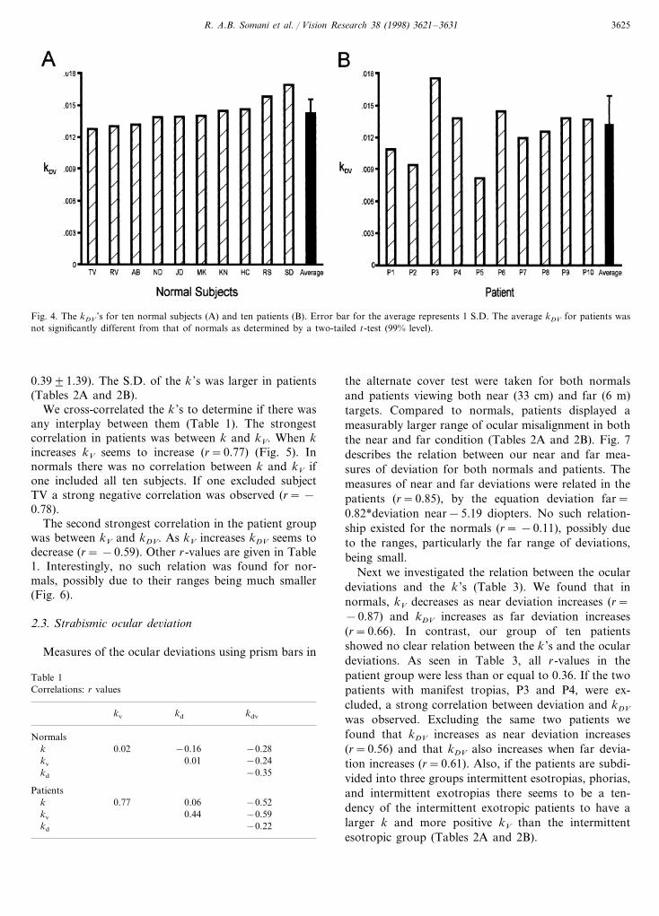
In total, ten normals were tested under the sameconditions, and across all normals DTdisp/DV becamemore positive with increasing convergence. The averagekDV across the ten normals was 0.01490.001 degree−1
(1 S.D.) (Fig. 4A).
In the vertical lines task, a similar elevation-depen-dent disparity was observed. The kDV on average acrossall ten normals was 0.01590.003 degree−1 (1 S.D.), avalue that was not significantly different from thatobtained with horizontal lines (two-tailed, t-test, PB0.12). Due to no difference being found, patients weretested with horizontal lines only.
2.2. Patients
We also measured the torsional disparities of tenpatients viewing the same horizontal lines at variousdegrees of convergence. Like the normal subjects, pa-tients showed an elevation-dependent torsional dispar-ity which increased with convergence. Fig. 4B shows aplot of kDV for ten patients tested under the sameconditions as the normals in this study. The kDV onaverage was 0.01390.003 degree−1 (1 S.D.), a valueslightly smaller than, although not significantly differ-ent from that of the normals (two-tailed, t-test, P\0.01). However, the range of values obtained wasgreater; for normals the range was 0.013–0.017 whilefor patients it was 0.008–0.018 (S.D. normals=0.001and S.D. patients=0.003). While the maximum kDV
was comparable to that of the normals, the minimumwas below that of the normals. A more detailed de-scription of the interpatient variability is given in thesection on strabismic ocular deviation.
There was no difference between normals and pa-tients in their mean kD values or in their kV ’s. The meank values, in contrast, were significantly more positivefor patients (k normal= −0.4690.57, k patient=
R. A.B. Somani et al. / Vision Research 38 (1998) 3621–3631 3625
Fig. 4. The kDV ’s for ten normal subjects (A) and ten patients (B). Error bar for the average represents 1 S.D. The average kDV for patients wasnot significantly different from that of normals as determined by a two-tailed t-test (99% level).
0.3991.39). The S.D. of the k ’s was larger in patients(Tables 2A and 2B).
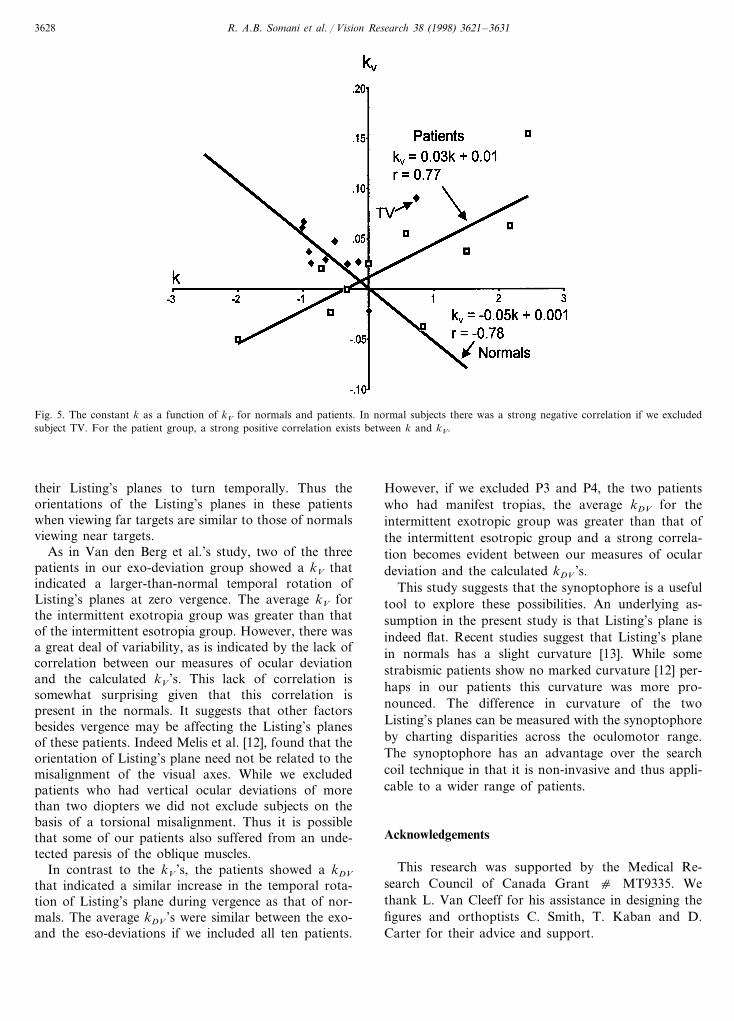
We cross-correlated the k ’s to determine if there wasany interplay between them (Table 1). The strongestcorrelation in patients was between k and kV. When kincreases kV seems to increase (r=0.77) (Fig. 5). Innormals there was no correlation between k and kV ifone included all ten subjects. If one excluded subjectTV a strong negative correlation was observed (r= −0.78).
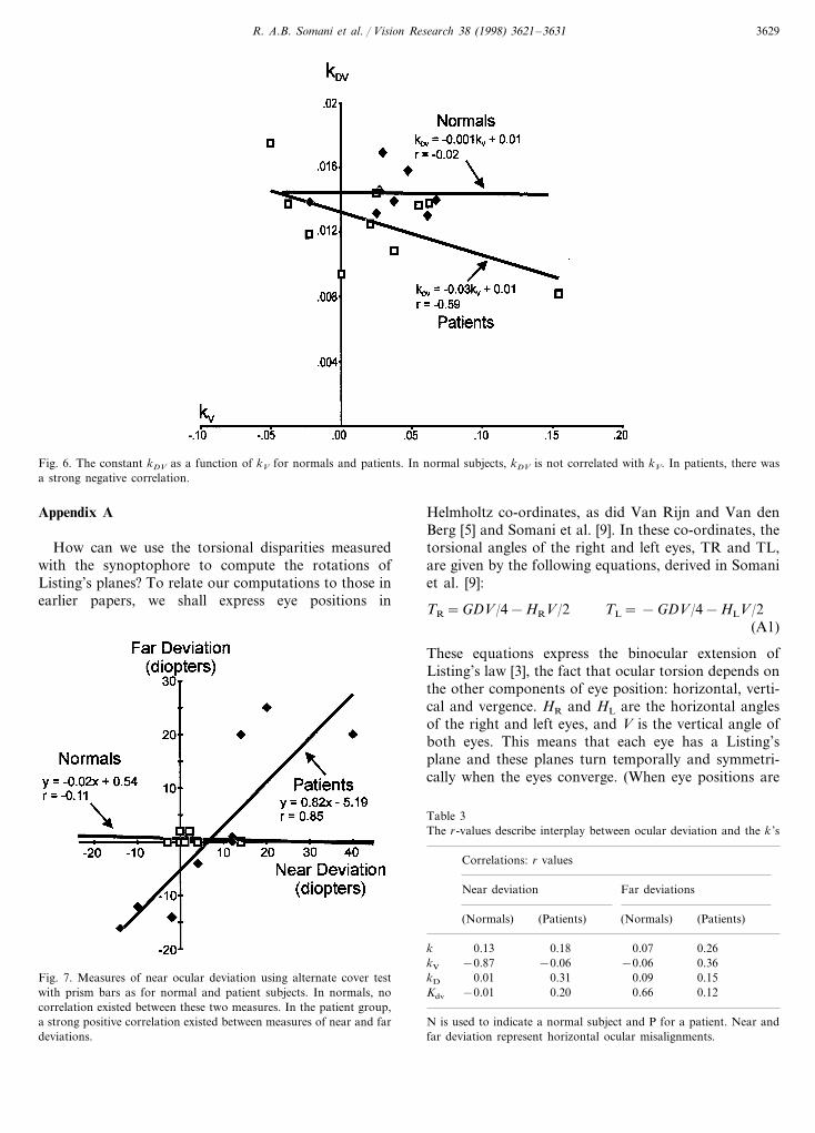
The second strongest correlation in the patient groupwas between kV and kDV. As kV increases kDV seems todecrease (r= −0.59). Other r-values are given in Table1. Interestingly, no such relation was found for nor-mals, possibly due to their ranges being much smaller(Fig. 6).
2.3. Strabismic ocular de6iation
Measures of the ocular deviations using prism bars in
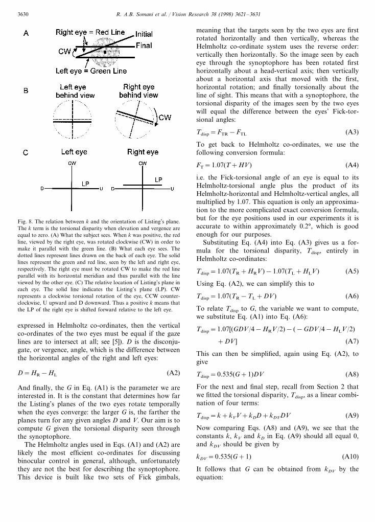
the alternate cover test were taken for both normalsand patients viewing both near (33 cm) and far (6 m)targets. Compared to normals, patients displayed ameasurably larger range of ocular misalignment in boththe near and far condition (Tables 2A and 2B). Fig. 7describes the relation between our near and far mea-sures of deviation for both normals and patients. Themeasures of near and far deviations were related in thepatients (r=0.85), by the equation deviation far=0.82*deviation near−5.19 diopters. No such relation-ship existed for the normals (r= −0.11), possibly dueto the ranges, particularly the far range of deviations,being small.
Next we investigated the relation between the oculardeviations and the k ’s (Table 3). We found that innormals, kV decreases as near deviation increases (r=−0.87) and kDV increases as far deviation increases(r=0.66). In contrast, our group of ten patientsshowed no clear relation between the k ’s and the oculardeviations. As seen in Table 3, all r-values in thepatient group were less than or equal to 0.36. If the twopatients with manifest tropias, P3 and P4, were ex-cluded, a strong correlation between deviation and kDV
was observed. Excluding the same two patients wefound that kDV increases as near deviation increases(r=0.56) and that kDV also increases when far devia-tion increases (r=0.61). Also, if the patients are subdi-vided into three groups intermittent esotropias, phorias,and intermittent exotropias there seems to be a ten-dency of the intermittent exotropic patients to have alarger k and more positive kV than the intermittentesotropic group (Tables 2A and 2B).
Table 1Correlations: r values
kv kd kdv
Normals0.02 −0.28k −0.16
kv 0.01 −0.24kd −0.35
Patients−0.52k 0.77 0.06
kv 0.44 −0.59kd −0.22
R. A.B. Somani et al. / Vision Research 38 (1998) 3621–36313626
Table 2ACorrelations between the k ’s in the ten normals and ten patients
Normal subjects Near deviation (diopters) Far deviation (diopters) k (°) kV kD kDV (degree−1) Gain (G)
0TV 0.806−3E 0.084 0.056 0.013 0.370 −0.989 0.0670 0.048MK 0.014 0.50
0KN 0 −0.878 0.026 0.068 0.014 0.550JD −0.9110 0.037 0.084 0.014 0.49
2X −0.517 0.0480 0.043RS 0.016 0.691XRV 0 −1.011 0.061 0.160 0.013 0.39
2X −0.156 0.027HC 0.1462X 0.015 0.561 −0.656 0.0292X 0.051SD 0.017 0.81
4AB 0 −0.322 0.025 0.082 0.013 0.410 0.006ND −0.02214 0.064 0.014 0.48
−0.463 0.038Average 0.080 0.014 0.530.571S.D. 0.029 0.041 0.001 0.08
3. Discussion
3.1. What do the k’s in Eq. (1) imply about therotation of Listing’s plane?
We fitted torsional disparity Tdisp as the sum of fourcomponents: a constant component k, a component kV
that depends only on elevation, one that depends onlyon the degree of vergence kD, and one that depends onboth elevation and vergence kDV. Each of these compo-nents expresses some difference in the orientations ofthe Listing’s planes in the two eyes.
The first term in Eq. (1), k, is determined by thetorsional disparity that exists when elevation and ver-gence are both zero. From Eq. (1) it can be seen thatwhen V=0 and D=0, Tdisp=k. A zero k value meansthat the torsional disparity of the two eyes is zero, i.e.the two eyes are aligned torsionally. A positive k meansthat the image of the red line viewed by the right eyewas rotated CW relative to the left eye (Fig. 8A). Whatdoes this mean in terms of Listing’s plane? If the redline seen by the right eye had to be turned CW in orderto align it with the green line, then the right eye musthave been rotated in the same CW direction (Fig. 8B).Therefore, a positive k, means that the Listing’s planeof the right eye is shifted forward in the CW directionrelative to that of the left eye (Fig. 8C).
The main difference between the normal subjects andthe patients was the larger variability in the k ’s andthus the relative shift of the Listing’s planes in the twoeyes. The mean k for patients, 0.39°, was significantlymore positive than that of the normals, −0.46°. Therewas no clear correlation between the measured oculardeviations and k if all ten patients were included.However, two of the three patients in the intermittentexo-tropic group had positive k ’s while two of the threepatients in the intermittent esotropic group showednegative k ’s.
The next term, kV, describes the elevation-dependentdisparity at zero vergence. This is related to the initialnasal/temporal orientation of the Listing’s planes of thetwo eyes when the target is viewed in the distance. Apositive kV indicates a temporal rotation of the planes,whereas a negative kV indicates a nasal rotation. Onecan illustrate this to oneself by picturing what the eyesare doing. Consider what happens when the planes arerotated temporally. In that case, when both eyes arelooking up, the right eye is rotated CCW and the lefteye is rotated CW, which means that torsional disparitywill be negative on up gaze. When the eyes look down,torsional disparity is positive. Given that down is thepositive direction for vertical eye position V, this meansthat the disparity increases as V increases — i.e. kV ispositive when the planes rotate temporally. The factthat our normal subjects had positive kV ’s on averageshows that their Listing’s planes were rotated tempo-rally in the absence of any vergence. Similarly, patientson average, and in the intermittent exotropic group inparticular, showed an initial temporal rotation of theirListing’s planes.
The third term, kD, describes the change in torsionaldisparity that occurs when vergence changes while theeyes are at zero elevation. This would occur if theListing’s planes in the two eyes were pitched relative toeach other. As described in the results there did notappear to be any clear difference between the kD ’s ofnormals and patients.
The term kDV represents how disparity changes as afunction of the product of elevation and vergence. Thiscorresponds to how much more the Listing’s planesrotate during convergence. As with the kV term, apositive kDV also represents a temporal rotation ofListing’s plane, although, in this case rotation thatincreases with the vergence angle. In both normals andpatients, the kDV values were positive, implying thattheir Listing’s planes rotated temporally during conver-gence. The mean kDV values of patients and normals
R. A.B. Somani et al. / Vision Research 38 (1998) 3621–3631 3627
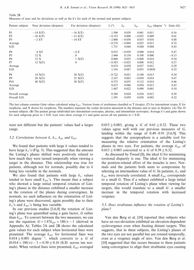
Table 2BMeasures of near and far deviations as well as the k’s for each of the normal and patient subjects
Far deviation (diopters) k (°) kV kDNear deviation (diopters) kDV (degree−1)Patient subjects Gain (G)
−16 E(T) 1.500 0.038 0.061 0.011P1 0.16−14 E(T)−12 E(T) −0.333 0.000−10 E(T) −0.025P2 0.009 0.01−14 ET −2.000 −0.050P3 0.033−2 ET 0.018 0.88
−0.278 −0.004 0.023Average 0.013 0.351.751S.D. 0.044 0.044 0.004 0.43
−4 E 0.833 −0.038P4 0.0004 XT 0.014 0.470 2.444 0.154P5 0.10812 X 0.008 −0.131 X(T) 0.000 0.02512 X −0.008P6 0.014 0.54
12 X(T)P7 0 −0.583 −0.023 0.008 0.012 0.270.674 0.030 0.027Average 0.012 0.29
S.D. 1.316 0.087 0.055 0.0036 0.32
20 X(T) −0.722P8 0.02114 X(T) 0.100 0.013 0.3425 X(T) 2.167 0.06320 X(T) −0.050P9 0.014 0.4720 X(T) 0.575 0.055P10 0.11240 X(T) 0.014 0.46
0.673 0.046 0.054Average 0.013 0.431.447 0.022S.D. 0.090 0.001 0.10
0.388 0.024Overall average 0.034 0.013 0.391.391Overall S.D. 0.059 0.058 0.003 0.32
The last column contains Gain values calculated using kDV. Various forms of strabismus classified as T (tropia), (T) for intermittent tropia, E foresophoria, and X shown for exophoria. The numbers represent the ocular deviation measured in the distance and at near in diopters. (A) The 10normal subjects. (B) The patient group subdivided into intermittent esotropias, phorias, and intermittent exotropias. Average k ’s and gains shownfor each subgroup given to 1 S.D. Last rows show average k ’s and gains across all ten patients to 1 S.D.
were not different but the patients’ values had a largerrange.
3.2. Correlations between k, kV, kD, and kDV
We found that patients with large k values tended tohave large kV ’s (Fig. 5). This suggested that the amountthe Listing’s planes shifted were positively related tohow much they were turned temporally when viewing atarget in the distance. This relationship was true forpatients, although not for normals, possibly due to kbeing less variable in the normals.
We also found that patients with large kV valuestended to have small kDV ’s. This meant that a subjectwho showed a large initial temporal rotation of List-ing’s planes in the distance exhibited a smaller increasein the rotation of the planes during convergence. Innormals, no such influences on the orientation of List-ing’s plane were discovered, again possibly due to theirkV ’s and kDV ’s being less variable.
In our previous studies [3,6,9] the rotation of List-ing’s plane was quantified using a gain factor, G ratherthan kDV. To convert between the two measures, we canuse the equation G=1.87kDV×180/p−1 (derived inAppendix A). Tables 2A and 2B shows the calculatedgain values for each subject when horizontal lines werepresented. The average kDV for horizontal lines was0.01490.001 degree−1, which converts to a G of(0.014×180/p)−1=0.5090.16 (S.D. across ten nor-mals). When vertical lines were presented, kDV averaged
0.01590.003, giving a G of 0.6190.32. These twovalues agree well with our previous measures of G,landing within the range of 0.49–0.91 [3,6,9]. Thissuggests that the synoptophore is a suitable tool formeasuring the relative orientation of the Listing’splanes in two eyes. For patients, the average kDV of0.01390.003 converted to a G of 0.3990.32.
As discussed by Tweed [8], the ideal G for minimizingtorsional disparity is one. The ideal G for minimizingthe position-related effort of the muscles is zero. Nor-mals and the patients both seem to compromise byselecting an intermediate value of G. In patients, kV andkDV were inversely correlated. A small kDV correspondsto a small G. Thus if a subject exhibited a large initialtemporal rotation of Listing’s plane when viewing fartargets, this would translate to a small G : a smallerincrease in the temporal rotation with increasedvergence.
3.3. Does strabismus influence the rotation of Listing’splane?
Van den Berg et al. [10] reported that subjects whohave an exo-deviation exhibited an elevation-dependentcyclovergence even when fixating distant targets. Thissuggests, that in these subjects, the Listing’s planes ofthe two eyes are not parallel but are rotated temporally,even at a vergence angle of zero. Van den Berg et al.[10] suggested that this occurs because to these patientsusing convergence to align their strabismic eyes causing
R. A.B. Somani et al. / Vision Research 38 (1998) 3621–36313628
Fig. 5. The constant k as a function of kV for normals and patients. In normal subjects there was a strong negative correlation if we excludedsubject TV. For the patient group, a strong positive correlation exists between k and kV.
their Listing’s planes to turn temporally. Thus theorientations of the Listing’s planes in these patientswhen viewing far targets are similar to those of normalsviewing near targets.
As in Van den Berg et al.’s study, two of the threepatients in our exo-deviation group showed a kV thatindicated a larger-than-normal temporal rotation ofListing’s planes at zero vergence. The average kV forthe intermittent exotropia group was greater than thatof the intermittent esotropia group. However, there wasa great deal of variability, as is indicated by the lack ofcorrelation between our measures of ocular deviationand the calculated kV ’s. This lack of correlation issomewhat surprising given that this correlation ispresent in the normals. It suggests that other factorsbesides vergence may be affecting the Listing’s planesof these patients. Indeed Melis et al. [12], found that theorientation of Listing’s plane need not be related to themisalignment of the visual axes. While we excludedpatients who had vertical ocular deviations of morethan two diopters we did not exclude subjects on thebasis of a torsional misalignment. Thus it is possiblethat some of our patients also suffered from an unde-tected paresis of the oblique muscles.
In contrast to the kV ’s, the patients showed a kDV
that indicated a similar increase in the temporal rota-tion of Listing’s plane during vergence as that of nor-mals. The average kDV ’s were similar between the exo-and the eso-deviations if we included all ten patients.
However, if we excluded P3 and P4, the two patientswho had manifest tropias, the average kDV for theintermittent exotropic group was greater than that ofthe intermittent esotropic group and a strong correla-tion becomes evident between our measures of oculardeviation and the calculated kDV ’s.
This study suggests that the synoptophore is a usefultool to explore these possibilities. An underlying as-sumption in the present study is that Listing’s plane isindeed flat. Recent studies suggest that Listing’s planein normals has a slight curvature [13]. While somestrabismic patients show no marked curvature [12] per-haps in our patients this curvature was more pro-nounced. The difference in curvature of the twoListing’s planes can be measured with the synoptophoreby charting disparities across the oculomotor range.The synoptophore has an advantage over the searchcoil technique in that it is non-invasive and thus appli-cable to a wider range of patients.
Acknowledgements
This research was supported by the Medical Re-search Council of Canada Grant c MT9335. Wethank L. Van Cleeff for his assistance in designing thefigures and orthoptists C. Smith, T. Kaban and D.Carter for their advice and support.
R. A.B. Somani et al. / Vision Research 38 (1998) 3621–3631 3629
Fig. 6. The constant kDV as a function of kV for normals and patients. In normal subjects, kDV is not correlated with kV. In patients, there wasa strong negative correlation.
Appendix A
How can we use the torsional disparities measuredwith the synoptophore to compute the rotations ofListing’s planes? To relate our computations to those inearlier papers, we shall express eye positions in
Helmholtz co-ordinates, as did Van Rijn and Van denBerg [5] and Somani et al. [9]. In these co-ordinates, thetorsional angles of the right and left eyes, TR and TL,are given by the following equations, derived in Somaniet al. [9]:
TR=GDV/4−HRV/2 TL= −GDV/4−HLV/2(A1)
These equations express the binocular extension ofListing’s law [3], the fact that ocular torsion depends onthe other components of eye position: horizontal, verti-cal and vergence. HR and HL are the horizontal anglesof the right and left eyes, and V is the vertical angle ofboth eyes. This means that each eye has a Listing’splane and these planes turn temporally and symmetri-cally when the eyes converge. (When eye positions are
Fig. 7. Measures of near ocular deviation using alternate cover testwith prism bars as for normal and patient subjects. In normals, nocorrelation existed between these two measures. In the patient group,a strong positive correlation existed between measures of near and fardeviations.
Table 3The r-values describe interplay between ocular deviation and the k ’s
Correlations: r values
Near deviation Far deviations
(Normals) (Patients)(Normals)(Patients)
0.260.13 0.18 0.07k−0.87 −0.06kV −0.06 0.36
0.01 0.31kD 0.150.09−0.01Kdv 0.20 0.66 0.12
N is used to indicate a normal subject and P for a patient. Near andfar deviation represent horizontal ocular misalignments.
R. A.B. Somani et al. / Vision Research 38 (1998) 3621–36313630
Fig. 8. The relation between k and the orientation of Listing’s plane.The k term is the torsional disparity when elevation and vergence areequal to zero. (A) What the subject sees. When k was positive, the redline, viewed by the right eye, was rotated clockwise (CW) in order tomake it parallel with the green line. (B) What each eye sees. Thedotted lines represent lines drawn on the back of each eye. The solidlines represent the green and red line, seen by the left and right eye,respectively. The right eye must be rotated CW to make the red lineparallel with its horizontal meridian and thus parallel with the lineviewed by the other eye. (C) The relative location of Listing’s plane ineach eye. The solid line indicates the Listing’s plane (LP). CWrepresents a clockwise torsional rotation of the eye, CCW counter-clockwise, U upward and D downward. Thus a positive k means thatthe LP of the right eye is shifted forward relative to the left eye.
meaning that the targets seen by the two eyes are firstrotated horizontally and then vertically, whereas theHelmholtz co-ordinate system uses the reverse order:vertically then horizontally. So the image seen by eacheye through the synoptophore has been rotated firsthorizontally about a head-vertical axis; then verticallyabout a horizontal axis that moved with the first,horizontal rotation; and finally torsionally about theline of sight. This means that with a synoptophore, thetorsional disparity of the images seen by the two eyeswill equal the difference between the eyes’ Fick-tor-sional angles:
Tdisp=FTR−FTL (A3)
To get back to Helmholtz co-ordinates, we use thefollowing conversion formula:
FT=1.07(T+HV) (A4)
i.e. the Fick-torsional angle of an eye is equal to itsHelmholtz-torsional angle plus the product of itsHelmholtz-horizontal and Helmholtz-vertical angles, allmultiplied by 1.07. This equation is only an approxima-tion to the more complicated exact conversion formula,but for the eye positions used in our experiments it isaccurate to within approximately 0.2°, which is goodenough for our purposes.
Substituting Eq. (A4) into Eq. (A3) gives us a for-mula for the torsional disparity, Tdisp, entirely inHelmholtz co-ordinates:
Tdisp=1.07(TR+HRV)−1.07(TL+HLV) (A5)
Using Eq. (A2), we can simplify this to
Tdisp=1.07(TR−TL+DV) (A6)
To relate Tdisp to G, the variable we want to compute,we substitute Eq. (A1) into Eq. (A6):
Tdisp=1.07[(GDV/4−HRV/2)− (−GDV/4−HLV/2)
+DV ] (A7)
This can then be simplified, again using Eq. (A2), togive
Tdisp=0.535(G+1)DV (A8)
For the next and final step, recall from Section 2 thatwe fitted the torsional disparity, Tdisp, as a linear combi-nation of four terms:
Tdisp=k+kVV+kDD+kDVDV (A9)
Now comparing Eqs. (A8) and (A9), we see that theconstants k, kV and kD in Eq. (A9) should all equal 0,and kDV should be given by
kDV=0.535(G+1) (A10)
It follows that G can be obtained from kDV by theequation:
expressed in Helmholtz co-ordinates, then the verticalco-ordinates of the two eyes must be equal if the gazelines are to intersect at all; see [5]). D is the disconju-gate, or vergence, angle, which is the difference betweenthe horizontal angles of the right and left eyes:
D=HR−HL (A2)
And finally, the G in Eq. (A1) is the parameter we areinterested in. It is the constant that determines how farthe Listing’s planes of the two eyes rotate temporallywhen the eyes converge: the larger G is, the farther theplanes turn for any given angles D and V. Our aim is tocompute G given the torsional disparity seen throughthe synoptophore.
The Helmholtz angles used in Eqs. (A1) and (A2) arelikely the most efficient co-ordinates for discussingbinocular control in general, although, unfortunatelythey are not the best for describing the synoptophore.This device is built like two sets of Fick gimbals,
R. A.B. Somani et al. / Vision Research 38 (1998) 3621–3631 3631
G=1.87kDV−1 (A11)
Because we use degrees not radians,
G=1.87kDV* 180/p−1 (A12)
There is a loose end to tidy up: we have seen in theResults that k, kV and kD are not, in fact, always 0,which means that Eq. (A8) is not strictly correct. How-ever, this is not really a problem for our analysis. Inderiving Eq. (A8) we started from Eq. (A1), and theseearlier equations ignored the fact that ocular torsioncan depend on factors besides DV, the product ofdisconjugate and vertical position. For example, torsioncan depend to some extent on V or D alone. Had webuilt these other dependencies into Eq. (A1), then Eq.(A8) would have had some more terms correspondingto k, kV and kD, although the parameter G would nothave appeared in any of those terms. So for our pur-poses the extra terms would merely have cluttered thederivation, and we would still have wound up with Eq.(A12) as our formula for G.
References
[1] Enright JT. Ocular translation and cyclotorsion due to changesin fixation distance. Vis Res 1980;20:595–601.
[2] Nakayama K. Kinematics of normal and strabismic eyes. In:Schor CM and Ciuffreda KJ, editors. Vergence eye movements:
basic and clinical aspects. Boston, MA: Butterworths, 1983:543–564.
[3] Mok D, Ro A, Cadera W, Crawford JD, Vilis T. Rotation ofListing’s plane during vergence. Vis Res 1992;32:2055–64.
[4] Minken AWH, Van Gisbergen JAM. A three-dimensional analy-sis of vergence movements at various levels of elevation. ExpBrain Res 1994;101:331–45.
[5] Van Rijn LJ, Van den Berg AV. Binocular eye orientationduring fixations: Listing’s law extended to include eye vergence.Vis Res 1993;33:691–708.
[6] Mikhael S, Nicolle D, Vilis T. Rotation of Listing’s plane byhorizontal, vertical and oblique prism-induced vergence. Vis Res1995;35:3243–54.
[7] Minken AWH, Gielen CC, Van Gisbergen JAM. An alternativethree-dimensional interpretation of Hering’s law equal-innerva-tion law for version and vergence eye movements. Vis Res1995;35:93–102.
[8] Tweed D. Visual-motor optimization in binocular control. VisRes 1997;37:1939–51.
[9] Somani RAB, DeSouza JFX, Tweed D and Vilis T. Visual testof Listing’s law during vergence. Vis Res 1998;38:911–923.
[10] Van den Berg AV, Van Rijn LJ, De Faber JTJHN. Excesscyclovergence in patients with intermittent exotropia. Vis Res1995;35:3265–78.
[11] Von Noorden GK. Binocular vision and ocular motility. Theoryand management of strabismus. 4th ed. St. Louis: C. V. Mosby,1990, pp. 176–178.
[12] Melis BJM, Cruysberg JRM, van Gisbergen JAM. Listing’splane dependence on alternating fixation in a strabismic patient.Vis Res 1997;37:1355–66.
[13] DeSouza JFX, Nicolle DA, Vilis T. Task-dependent changes inthe shape and thickness of Listing’s plane. Vis Res1997;37:2271–82.
..