Mapping lateralization of click trains in younger and older populations

�������� ����� ��
Usage of fMRI for pre-surgical planning in tumor and vascular lesion patients:task and statistical threshold effects on language lateralization
Tanvi N. Nadkarni, Matthew J. Andreoli, Veena A. Nair, Peng Yin, BrittanyYoung, Bornali Kundu, Joshua Pankratz, Andrew Radtke, Ryan Holdsworth,John S. Kuo, Aaron S. Field, Mustafa K. Baskaya, Chad H. Moritz, M.Elizabeth Meyerand, Vivek Prabhakaran
PII: S2213-1582(14)00199-5DOI: doi: 10.1016/j.nicl.2014.12.014Reference: YNICL 411
To appear in: NeuroImage: Clinical
Received date: 1 October 2014Revised date: 19 December 2014Accepted date: 22 December 2014
Please cite this article as: Nadkarni, Tanvi N., Andreoli, Matthew J., Nair, Veena A., Yin,Peng, Young, Brittany, Kundu, Bornali, Pankratz, Joshua, Radtke, Andrew, Holdsworth,Ryan, Kuo, John S., Field, Aaron S., Baskaya, Mustafa K., Moritz, Chad H., Meyerand,M. Elizabeth, Prabhakaran, Vivek, Usage of fMRI for pre-surgical planning in tumor andvascular lesion patients: task and statistical threshold effects on language lateralization,NeuroImage: Clinical (2014), doi: 10.1016/j.nicl.2014.12.014
This is a PDF file of an unedited manuscript that has been accepted for publication.As a service to our customers we are providing this early version of the manuscript.The manuscript will undergo copyediting, typesetting, and review of the resulting proofbefore it is published in its final form. Please note that during the production processerrors may be discovered which could affect the content, and all legal disclaimers thatapply to the journal pertain.

ACC
EPTE
D M
ANU
SCR
IPT
ACCEPTED MANUSCRIPT
1
Usage of fMRI for pre-surgical planning in tumor and vascular lesion
patients: task and statistical threshold effects on language lateralization
Usage of fMRI for pre-surgical planning in tumor and vascular lesion patients:
task and statistical threshold effects on language lateralization
Tanvi N. Nadkarni*, Matthew J. Andreoli*, Veena A. Nair3, Ph.D, Peng Yin,
Brittany Young2, Bornali Kundu2, Joshua Pankratz1, Andrew Radtke1, Ryan
Holdsworth3, MD, John S. Kuo1,6, MD, Ph.D, Aaron S. Field3, MD, Ph.D, Mustafa
K. Baskaya1,6, MD, Chad H. Moritz3,4, M. Elizabeth Meyerand3,4,5, Ph.D, Vivek
Prabhakaran3, MD, Ph.D
School of Medicine and Public Health1, Medical Scientist Training Program2,
Departments of 3Radiology, 4Medical Physics, and 5Biomedical Engineering,
6Neurological Surgery, University of Wisconsin - Madison.
Corresponding Author:
Vivek Prabhakaran M.D., Ph.D.
Assistant Professor, Director of Functional Neuroimaging in Radiology
UWHealth UW Hospital and Clinics Department of Radiology
Wisconsin Institutes for Medical Research
Rm 1314, 1111 Highland Avenue
Madison, WI 53705, USA.
Office phone: 1-608-265-5269 Fax: 1-608-265-9840
Email: [email protected]

ACC
EPTE
D M
ANU
SCR
IPT
ACCEPTED MANUSCRIPT
2
Author Contributions
*Both Nadkarni TN and Andreoli MJ contributed equally and are co-principal
authors on this manuscript.
Andreoli MJ and Nadkarni TN were involved in data analysis, interpretation,
literature search, and writing.
Nair VA was involved in data collection, analysis, interpretation, literature search,
and writing.
Yin P was involved in data analysis.
Young B was involved in data collection and analysis.
Kundu B was involved in review and writing.
Pankratz J was involved in data analysis.
Radtke A was involved in data analysis.
Kuo JS was involved in patient referrals.
Field AS was involved in study design.
Holdsworth R was involved in data collection.
Baskaya MK was involved in patient referrals.
Moritz CH was involved in data collection.
Meyerand ME was involved in study design.
Prabhakaran V was involved in study design and data collection.
Conflict of Interest: The authors declare no conflict of interest.

ACC
EPTE
D M
ANU
SCR
IPT
ACCEPTED MANUSCRIPT
3
Name and Department Affiliation
Tanvi N. Nadkarni:
Department of Radiology
University of Wisconsin Madison
School of Medicine and Public Health
600 Highland Avenue
Madison, WI 53792-3252, USA.
Email: [email protected]
Matthew J. Andreoli:
Department of Radiology
University of Wisconsin Madison
School of Medicine and Public Health
600 Highland Avenue
Madison, WI 53792-3252, USA.
Email: [email protected]
Veena A. Nair:
Department of Radiology
Wisconsin Institutes for Medical Research
Box 3252 Clinical Science Center
600 Highland Ave
Madison, WI 53705, USA.
Email: [email protected]

ACC
EPTE
D M
ANU
SCR
IPT
ACCEPTED MANUSCRIPT
4
Peng Yin:
Department of Radiology
University of Wisconsin Madison
School of Medicine and Public Health
600 Highland Avenue
Madison, WI 53792-3252
Email: [email protected]
Brittany Young:
Medical Scientist Training Program
University of Wisconsin Madison
Department of Radiology
Wisconsin Institute of Medical Research
3252 Clinical Science Center
600 Highland Ave
Madison, WI 53792
Email: [email protected]
Bornali Kundu:
Medical Scientist Training Program
University of Wisconsin Madison

ACC
EPTE
D M
ANU
SCR
IPT
ACCEPTED MANUSCRIPT
5
2140, Med 3 Mailbox Health Sciences Learning Center
750 Highland Ave
Madison, WI 53705, USA
Email: [email protected]
Joshua Pankratz:
Department of Radiology
University of Wisconsin Madison
School of Medicine and Public Health
600 Highland Avenue
Madison, WI 53792-3252
Email: [email protected]
Andrew Radtke:
Department of Radiology
University of Wisconsin Madison
School of Medicine and Public Health
600 Highland Avenue
Madison, WI 53792-3252
Email: [email protected]
Ryan Holdsworth:
Department of Radiology
University of Wisconsin Hospital and Clinics
600 Highland Ave

ACC
EPTE
D M
ANU
SCR
IPT
ACCEPTED MANUSCRIPT
6
Madison WI 53792-3252
Email: [email protected]
John S. Kuo:
Department of Neurological Surgery
University of Wisconsin Madison Hospital and Clinics
School of Medicine and Public Health
University of Wisconsin Madison
Box 8660 Clinical Science Center
600 Highland Ave
Madison, WI 53792-3252
Email: [email protected]
Aaron S. Field:
Department of Radiology
University of Wisconsin Madison Hospital and Clinics
School of Medicine and Public Health
University of Wisconsin Madison
Box 3252 Clinical Science Center
600 Highland Ave
Madison, WI 53792-3252
Email: [email protected]
Mustafa K. Baskaya:
Department of Neurological Surgery

ACC
EPTE
D M
ANU
SCR
IPT
ACCEPTED MANUSCRIPT
7
University of Wisconsin Madison Hospital and Clinics
School of Medicine and Public Health
Box 8660 Clinical Science Center
600 Highland Ave
Madison, WI 53792-3252
Email: [email protected]
Chad H. Moritz:
Department of Radiology
Wisconsin Institute of Medical Research
L1/1232 1111 Highland Avenue
Madison, WI, 53705, USA.
Phone: (608) 669-2315
Email: [email protected]
M. Elizabeth Meyerand:
Departments of Biomedical Engineering & Medical Physics
Chair – Department of Biomedical Engineering
University of Wisconsin Madison
1111 Highland Ave., Suite 1129
Wisconsin Institutes for Medical Research (WIMR)
Madison, WI 53705, USA.
Phone: (608) 263-1685; Fax: (608) 265-9842
Email: [email protected]

ACC
EPTE
D M
ANU
SCR
IPT
ACCEPTED MANUSCRIPT
8
Vivek Prabhakaran:
Department of Radiology
University of Wisconsin Madison
Wisconsin Institutes for Medical Research
Rm 1314, 1111 Highland Avenue
Madison, WI 53705, USA.
Office phone: 1-608-265-5269 Fax: 1-608-265-9840
Email: [email protected]
Keywords: fMRI, lateralization index, thresholding, surgical planning
Running title: task/thresholding effects on LI in lesion patients

ACC
EPTE
D M
ANU
SCR
IPT
ACCEPTED MANUSCRIPT
9
Grant support: UW Institute of Clinical and Translational Research CTSA
program, through the NIH National Center for Advancing Translational Sciences
(NCATS), grant UL1TR000427 KL2 Scholar and Pilot Awards (VP), and TL1
award (BMY); K23NS086852 NIH NINDS (VP); American Heart Association
Midwest Postdoctoral Fellowship (VN), RC1MH090912-01 National Institute of
Health, National Institute of Mental Health Challenge Grant (VN, VP, EM) and the
University of Wisconsin Madison – School of Medicine and Public Health Medical
Scientist Training Program (BMY,BK).
Parts of this data were presented as a poster at the International Stroke
Conference, 2012, in New Orleans and at the Radiological Society of North
America Conference, 2013, in Chicago, Illinois.
This is an original manuscript that does not contain material that has previously
been published. It is currently not under review elsewhere for publication.

ACC
EPTE
D M
ANU
SCR
IPT
ACCEPTED MANUSCRIPT
10
Highlights
FMRI calculation of language lateralization is valuable for pre-surgical planning.
Both applied statistical thresholds and task specificity affect lateralization index.
A continuum of threshold values provides a dynamic range for presurgical planning.
Reorganization of language function is dependent on brain pathology.
Identification of critical language areas will optimize lesion resection (e.g. tumor).
Abstract
Background and Purpose
Functional magnetic resonance imaging (fMRI) is a non-invasive pre-surgical tool
used to assess localization and lateralization of language function in brain tumor
and vascular lesion patients in order to guide neurosurgeons as they devise a
surgical approach to treat these lesions. We investigated the effect of varying the
statistical thresholds as well as the type of language tasks on functional
activation patterns and language lateralization. We hypothesized that language
lateralization indices (LIs) would be threshold- and task-dependent.
Materials and Methods
Imaging data were collected from brain tumor patients (n=67, average age 48
years) and vascular lesion patients (n=25, average age 43 years) who received

ACC
EPTE
D M
ANU
SCR
IPT
ACCEPTED MANUSCRIPT
11
pre-operative fMRI scanning. Both patient groups performed expressive
(antonym and/or letter-word generation) and receptive (tumor patients performed
text-reading; vascular lesion patients performed text-listening) language tasks. A
control group (n=25, average age 45 years) performed the letter-word generation
task.
Results
Brain tumor patients showed left-lateralization during antonym-word generation
and text-reading tasks at high threshold values and bilateral activation during
letter-word generation task, irrespective of the threshold values. Vascular lesion
patients showed left-lateralization during the antonym and letter-word generation,
and text-listening task at high threshold values.
Conclusion
Our results suggest that the type of task and the applied statistical threshold
influence LI and that the threshold effects on LI may be task-specific. Thus
identifying critical functional regions and computing LIs should be conducted on
an individual subject basis, using a continuum of threshold values with different
tasks to provide the most accurate information for surgical planning to minimize
post-operative language deficits.

ACC
EPTE
D M
ANU
SCR
IPT
ACCEPTED MANUSCRIPT
12
Keywords: fMRI, lateralization index (LI), thresholding, task-specific
Abbreviations
AWG antonym-word generation LWG letter-word generation task LI lateralization
index

ACC
EPTE
D M
ANU
SCR
IPT
ACCEPTED MANUSCRIPT
13
1. Introduction
Functional magnetic resonance imaging (fMRI) is used to non-invasively
map task-specific brain activation for pre-surgical planning1. These maps are
generated from changes in the blood oxygen level-dependent (BOLD) signal
related to neural activity2. Lateralization index (LI), a measure of hemispheric
dominance, quantifies information representing BOLD activation during language
and cognitive tasks3,8
. The degree to which functional activity lateralizes in brain
tumor and vascular lesion patients is a useful guide in designing a surgical
approach that minimizes post-operative neurological deficits4,5,6,7,9
. Pre-surgical
maps help determine the extent to which a brain tumor or vascular lesion may be
resected in patients by localizing key regions of eloquent cortex.
A number of neuroimaging studies have examined different types of
language LIs and different factors that may influence the LI. A study by Ruff et al
(2008) addressed how brain tumors may modulate LI by using a range of
statistical threshold values and showed that patients demonstrate greater
variability in LI during language tasks than controls10
. Compared to strong left-
lateralization in controls, brain tumor patients showed bilateral dominance at low
threshold values, but left-lateralization at more stringent threshold values. The
study, however, reported results on nine patients. A study by Partovi et al (2012)
used a fMRI protocol to determine language LI. Measurements were limited to
Broca’s (Brodmann areas 44 and 45) and Wernicke’s (Brodmann area 22) areas,
which are known to be primarily activated during expressive and receptive

ACC
EPTE
D M
ANU
SCR
IPT
ACCEPTED MANUSCRIPT
14
language tasks, respectively. However, other higher-order language association
areas (i.e. supramarginal, middle and inferior frontal gyrus) may be involved with
language function, but few studies have examined reorganization in these
regions following a lesion11,12.
Janecek et al (2013) investigated language lateralization in a large group
of patients to evaluate the clinical effectiveness of fMRI compared to WADA
testing. Upon utilization of fMRI and WADA protocols, the scientists found that
80% of normal subjects were left lateralized in language function. Overall,
Janecek et al (2013) determined that the discordance rates between LI obtained
from fMRI and WADA protocols were quite small, suggesting that the utilization
of fMRI for pre-surgical planning is just as effective of a tool as the WADA
testing. Janecek et al (2013) found that the degree of bilateral and rightward shift
of language lateralization was more clearly evident in the fMRI results. However,
it cannot be concluded that the altered language lateralization seen in the fMRI
results are more or less accurate compared to the WADA results. The Janecek et
al (2013) study suggests that fMRI may have increased sensitivity to language
processing in the right hemisphere compared to the WADA test. The previous
study stated that the discordance rates could have been influenced by the types
of tasks used in fMRI13. Utilizing fMRI in our study, we investigated the impact of
varying the thresholds after performance of different task types on the patients’
patterns of language lateralization.
A study by Friederici et al (2000) showed that language lateralization
patterns differ based on the type of task performed. Additionally, Ruff et al

ACC
EPTE
D M
ANU
SCR
IPT
ACCEPTED MANUSCRIPT
15
(2008b) proposed that language lateralization is dependent on the statistical
threshold utilized10,14
. Studies have shown that the presence of brain tumors and
vascular lesions affect neurological function in a language network4,6,9,15
.
However, a large number of these studies have focused on patients with brain
tumors. Few studies have examined influence of a lesion on language
reorganization patterns in patients with AVMs and cavernomas16. Thus the
analyses were repeated for both tumor patients and a collection of vascular
lesion patients.
Wellmer et al (2009) investigated the impact of lesions on language
lateralization through fMRI and Wada test protocols and determined that a
variable fMRI language lateralization is observed in vascular lesion patients17
.
The study categorized average LI measurements between 0.3-1.0 as unilateral
dominant, while the current study utilizes the ranges of +0.2<LI<+1.0 (left
lateralized), -0.2<LI<+0.2 (bilateral), and -0.2<LI<-1.0 (right lateralized) as
determined by Springer et al (1999) to evaluate the strength of lateralization in
patients3,17
. Categorization of the LI values helps summarize the general effect
that brain tumors or vascular lesions may have on the language network.
Studies have shown that a fixed threshold may be insufficient to detect
differences in voxel activation between hemispheres, resulting in inaccurate
calculation of the LI9,18
. Thus the present study investigated the effect of varying
threshold values on the ability to detect pseudoreorganization of task-specific
language function, as measured by average LI, in a large population of brain
tumor (n=67) and vascular lesion patients (n=25). We used a network of ROIs

ACC
EPTE
D M
ANU
SCR
IPT
ACCEPTED MANUSCRIPT
16
that incorporated these areas and the two dominant language areas of the brain
to determine average hemispheric LI and activation patterns11,12
. Lastly, we
report how LI varies with task type and the level of statistical thresholding9. We
propose that threshold-dependence and task selection are important factors to
consider in identifying subject-specific lateralization patterns and localizing critical
areas of activation.
2. Methods
2.1. Subjects
Anatomical and functional images were derived from a database (between
2004-2011) of patients who underwent a fMRI pre-surgical planning protocol. We
retrospectively selected 92 right-handed patients diagnosed with either left-
hemispheric brain tumors or vascular lesions who received a pre-operative fMRI
at the University of Wisconsin-Madison, and had provided written informed
consent for their data to be used for applicable research purposes. We selected
only right handed patients to reduce the confounding effects due to handedness,
with the notion that right-handed individuals display left-lateralized language
activation patterns19
. The analysis reported includes data from 67 brain tumor
patients (43 male, 24 female) and 25 vascular lesion patients (9 male, 16
female). Also, 25 right-handed healthy controls (13 males, 12 females) performed
the letter-word generation task and were used as a comparison to patient data.
Patient demographics are reported in Tables 1, 2, and 3. There was no
significant difference in age between the groups; however, age may influence

ACC
EPTE
D M
ANU
SCR
IPT
ACCEPTED MANUSCRIPT
17
language lateralization. Szaflarski et al (2006) concluded that there is a slow
decrease in dominant hemisphere lateralization in adults as they became older20.
The present study utilized data including young to older adults and, therefore,
may show age-related effects on LI, but this was controlled for between the three
groups. Gender differences were significant among tumor patients and among
vascular lesion patients (p<0.004). Preliminary evaluation was conducted to
investigate the effect that gender has on thresholded task-specific LI (Table 4).
The protocol was approved by the University of Wisconsin Madison Health
Sciences institutional review board.

ACC
EPTE
D M
ANU
SCR
IPT
ACCEPTED MANUSCRIPT
18
Table 1. Patient Demographics.
Table 2. Tumor Patient Lesion Demographics
Characteristics of Patients Tumor
Patients
(n=67)
Vascular
Lesion Patients
(n=25)
Controls
(n=25)
P-Value
(p<0.05)
Males (%) 64% 36% 52% T vs. V:
P=0.004 Females (%) 36% 64% 48%
Age Range [Mean] (years) 21-82 [48] 20-72 [43] 20-75 [45] NS
Number of patients per task
Antonym-word Generation
Letter-word Generation
Text-reading
Text-listening
57
42
25
0
23
10
0
12
0
25
0
0

ACC
EPTE
D M
ANU
SCR
IPT
ACCEPTED MANUSCRIPT
19
Tumor Number of Patients Type
Astrocytoma
Oligodendroglioma
Glioblastoma Multiforme
Meningioma
Metastatic
Oligoastrocytoma
Fibrillary Astrocytoma
DNET
Anaplastic astrocytoma
Hemangiopericytoma
Benign cyst
Undefined
Other
22
11
9
4
4
3
2
2
1
1
1
2
5
Grade
1
2
3
4/Metastasis
Benign
Undefined
7
20
10
24
4
2
Location
Left Frontal
Left Temporal
Left Parietal
Left Insula
Left Frontal +
Left Parietal +
Left Temporal +
35
12
5
3
8
3
1
Table 3. Vascular Lesion Patient Lesion Demographics

ACC
EPTE
D M
ANU
SCR
IPT
ACCEPTED MANUSCRIPT
20
Table 4. Average LI by Gender Task Gender/Lesion n t<2 t<2.67 t<3.5 t<4
Vascular Lesion Number of
Patients Type
AVM
Cavernoma
Hemorrhage (no underlying
lesion found)
Aneurysm
13
8
3
1
Location
Left Frontal
Left Temporal
Left Parietal
Left Insula
Left Frontotemporal
Left Frontoparietal
Left Temporoparietal
Left Parietal-occipital
3
9
2
1
1
5
2
2

ACC
EPTE
D M
ANU
SCR
IPT
ACCEPTED MANUSCRIPT
21
LWG Male Tumor 26 BL BL BL BL
Female Tumor 16 BL BL BL BL
Male Vascular Lesion 3 BL BL LL LL
Female Vascular Lesion 7 LL LL LL LL
AWG Male Tumor 34 BL LL LL LL
Female Tumor 23 BL BL BL* BL*
Male Vascular Lesion 8 BL BL LL LL
Female Vascular Lesion 15 BL BL LL LL
TXTREAD Male Tumor 13 BL BL LL LL
Female Tumor 12 BL BL BL LL
TXTLISTEN Male Vascular Lesion 2 BL BL RL* BL*
Female Vascular Lesion 10 BL LL LL LL
aBL= bilateral, LL= left-lateral, RL= right-lateral
b *Pattern deviates from gender non-specific population averages.
2.2. fMRI Image Acquisition
FMRI scanning was conducted using a 1.5 or 3 T commercial MR imaging

ACC
EPTE
D M
ANU
SCR
IPT
ACCEPTED MANUSCRIPT
22
scanner (Sigma General Electric Healthcare, Milwaukee, Wisconsin) with high-
speed gradient capabilities. Technical scanner parameters include: FOV, 24cm;
matrix 64 X 64; TR, 2000 ms; TE, 40 ms (for 1.5T) or 27 ms (for 3T); flip angle,
85° (for 1.5T) or 75° (for 3T); 6-mm coronal plane sections (for 1.5T) or 5-mm
axial plane sections (for 3T). Additional high-resolution anatomical scans,
including 3D volumetric T1- and T2-weighted sequences were acquired as part of
the pre-operative assessment.
2.3. fMRI Task Paradigms
Patients were instructed to perform expressive and/or receptive language
tasks based on individual medical conditions to help the neurosurgical team
evaluate specific language function. All tasks followed a block design paradigm,
which consisted of subjects alternating between 20 seconds of rest (blank screen
or non-relevant information) and 20 seconds of task (activation state) for five
cycles approximating three and a half minutes total.
2.3.1. Expressive language tasks
The expressive language tasks were designed to primarily activate
Broca’s area (inferior frontal gyri) and secondarily activate Wernicke’s area
(posterior superior temporal gyri) and surrounding higher order language
association areas, such as the supramarginal and middle frontal gyri. The two
expressive language tasks were the antonym-word generation (AWG) (tumor,
n=57; vascular lesion, n=23) and the letter-word generation (LWG) (tumor, n=42;
vascular lesion, n=10). Due to the retrospective nature of the study, each patient
performed a different number of language tasks. Each tumor and vascular lesion

ACC
EPTE
D M
ANU
SCR
IPT
ACCEPTED MANUSCRIPT
23
patient performed one or both of the expressive language tasks (AWG and/or
LWG). During the AWG task, subjects covertly produced a list of antonyms to the
words on the screen. During the LWG task, subjects covertly generated a list of
words beginning with the letters on the screen. Comparison between tumor and
vascular lesion patient performance on expressive tasks was not conducted
because it was outside the parameters of the present study’s focus and the
subjects within each patient group did not perform both tasks equally.
2.3.2. Receptive language tasks
The receptive language tasks (text-reading and text-listening tasks) were
designed to primarily activate Wernicke’s area and secondarily activate Broca’s
area and surrounding higher order language association areas. Each patient
group performed a different receptive language task. Of the tumor patients who
performed a receptive language task, they performed a text-reading task (n=25).
During this task, patients covertly read a block of text for comprehension. Of the
vascular lesion patients who performed a receptive language task, they
performed the text-listening task (n=12). During this task, patients listened to a
narrated text for comprehension via in-ear sound isolating headphones.
Comparison between tumor and vascular lesion patient performance on
receptive tasks was not conducted because it was outside the parameters of the
present study’s focus and each patient group performed a different receptive
language task.
2.4. Data Analysis

ACC
EPTE
D M
ANU
SCR
IPT
ACCEPTED MANUSCRIPT
24
The fMRI data were processed using AFNI (Analysis of Functional
NeuroImages) available at http://afni.nimh.nih.gov/afni/. Preprocessing steps
included dropping the first three volumes, motion correction, spatial smoothing
(6mm FWHM), and spatial normalization to 3x3x3mm. AFNI’s 3dDeconvolve
program was used to perform voxel-wise regression analysis to generate
thresholded t-maps. The six motion parameters (x,y,z, pitch, roll, yaw) were
regressed out in the analysis. 5mm regions of interest (ROI) were created based
off of MNI (Montreal Neurological Institute) coordinates reported in a meta-
analysis of language areas including Brodmann areas 44 and 45 (Broca’s area)
and areas 22, 39, 40 (Wernicke’s area) and other associated motor and
language areas described in Vigneau et al (2006)12
. We obtained left hemisphere
language areas based on these coordinates, generated homologous ROIs in the
right hemisphere, and computed LI for each predefined network ROI-pair. The
same protocol for analysis was applied to both tumor and vascular lesion data.
2.5. Calculation of Lateralization Index
The following equation was used to determine the LI values: LI = (VL-
VR)/(VL+VR), where VL and VR correspond to the number of active voxels in the
left and right hemisphere, respectively3. The scale used to categorize LI was as
follows: +0.2<LI<+1.0 (left-lateralized), -0.2<=LI<=+0.2 (bilateral), and -0.2<LI<-
1.0 (right-lateralized)3. These values were calculated for both hemispheres at
four different thresholds (t<2, t<2.67, t<3.5, t<4), which correspond to the
following p values (p<0.05, p<0.01, p<0.001 and p<0.0001), respectively.

ACC
EPTE
D M
ANU
SCR
IPT
ACCEPTED MANUSCRIPT
25
2.6. Statistical Analysis
To determine if there was an effect of threshold value between the patient
groups and the control group, we compared each patient group’s average LI at
all t-values with the control using an independent two-sample t-test. We
compared the difference between LIs at all pairs of t-values (e.g., t<2 vs. t<2.67,
t<2 vs. t<3.5, etc.) to specifically analyze the effect of t-values on different tasks
within each patient group. Bonferroni correction was used to correct for multiple
comparisons. A two-sample t-test was performed to generate group activation
maps.
3. Results
The predominant activation pattern we observed in brain tumor and
vascular lesion patients indicated that as the threshold was increased from a low
to a high threshold, the average LI became more left-lateralized, except for the
brain tumor patient group performing the LWG task (Figs. 1 and 2). The control
group showed left-lateralization in the inferior, middle, and superior frontal gyri
across all threshold values during the LWG task. In contrast, the brain tumor
patient group showed bilateral activity in the inferior, middle, and superior frontal
gyri, supramarginal and inferior parietal gyri across all threshold values. The
vascular lesion patient group showed bilateral activity in the inferior, middle, and
medial frontal gyri, inferior parietal gyri, and superior temporal gyri at a low
threshold value (t<2.0), but showed robust left hemispheric activity in the inferior
and middle frontal gyri at high threshold values (ts<3.5) (Fig. 2). There was

ACC
EPTE
D M
ANU
SCR
IPT
ACCEPTED MANUSCRIPT
26
significantly more bilateral activity in the tumor patient group compared to the
control group across all t-values. The vascular lesion patient group showed a
significantly bilateral LI only at t<2.0 compared to the control group. (Fig. 2 and
Table 4).
Fig. 1. An example of variable thresholding in a tumor patient. These images are of a tumor patient after performing the receptive language, text-reading task showing the general trend we observed for most of the tasks of shifting from a bilateral pattern of language activity to a left lateral pattern as the threshold becomes more stringent. The images follow a radiological convention with left hemisphere (L) on the right and right hemisphere (R) on the left. Talairach functional maps of all the patients are averaged together and overlaid onto the MNI 152 standard anatomical template.

ACC
EPTE
D M
ANU
SCR
IPT
ACCEPTED MANUSCRIPT
27
Fig. 2. Average hemispheric LIs (left) and group activation (right) for letter-word generation task in the control, tumor, and vascular lesion patient groups. Left: The average LIs for tumor patients (n=42), vascular lesion patients (n=10), and controls (n=25), from the letter-word generation task, are shown at different threshold values for the hemispheric mask. Tumor patients show bilateral dominance regardless of t-value. Tumor patient LIs are significantly different from control LIs at each threshold (ps<0.0125, Table 4). Vascular lesion patients show bilateral dominance at t<2.0 and left lateralization at t<4.0. Vascular lesion patient LIs are significantly different from control LIs at at t<2.0 (p<0.0125, Table 4). The control group maintains left lateralization despite variable threshold effects. Right: Images display the group activation maps at low threshold values (ts<2.0) and at higher threshold values (ts<4.0). The control subjects (A) show average left language lateralization, the vascular lesion patients (B) show average bilateral dominance, and the tumor patients (C) show average bilateral dominance during the letter word generation task.

ACC
EPTE
D M
ANU
SCR
IPT
ACCEPTED MANUSCRIPT
28
Table 4. Patient LI versus control LI
Controls vs. Tumor p-values (df)=65
Controls vs Vascular Lesion p-values (df)=34
t<2.0 2.97E-09* 0.002428*
t<2.6 4.07E-10* 0.013949
t<3.5 8.88E-10* 0.142843
t<4.0 3.15E-09* 0.061795
a *Statistically significant at p<0.0125, corrected.
3.1. Expressive Tasks.
The average LIs for the tumor patient group that performed the AWG task
showed bilateral activation at low threshold values (ts<2.6), but left-lateral
activation at high threshold values (ts<4.0) (Table 5 and Fig. 3, top). In contrast,
despite varying the threshold, bilateral activation was maintained during the LWG
task. (Fig. 3, top). Also all of our patients had brain tumors in left hemisphere
with no differences between frontal and non-frontal tumor patients with both
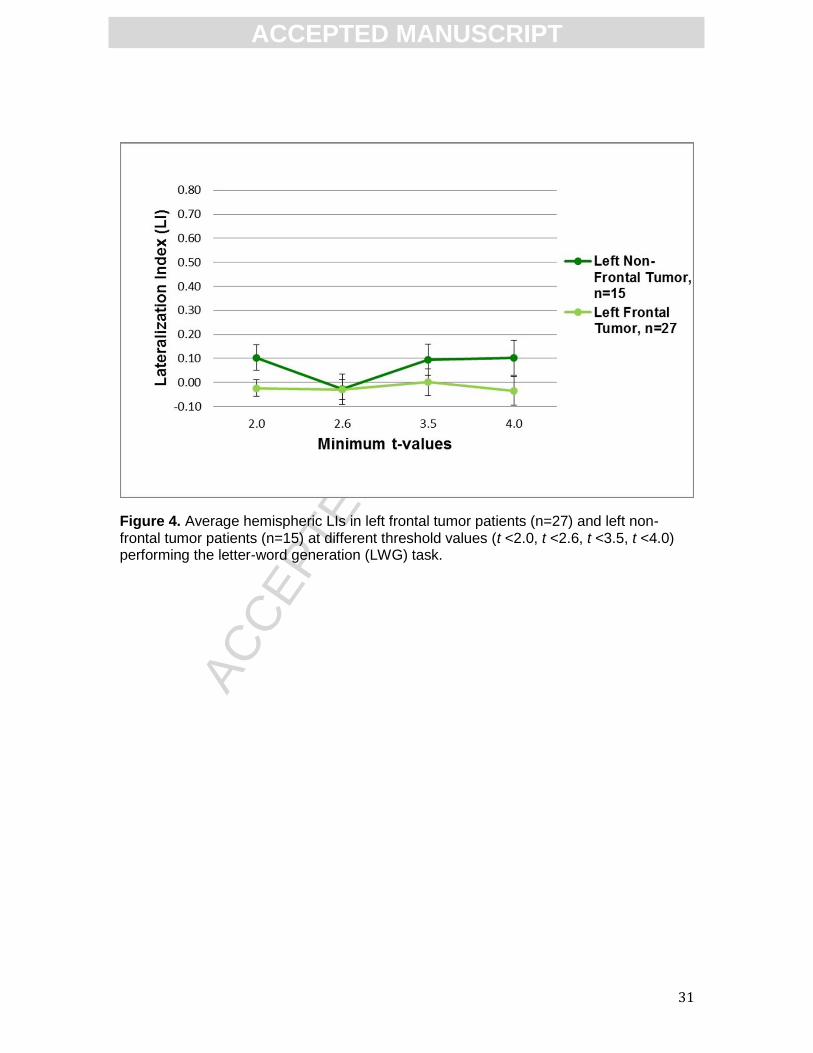
demonstrating bilateral activation during the LWG task (Fig. 4).
The average LIs for the vascular lesion patient group that performed the
AWG task showed bilateral activation at low threshold values (ts<2.6) and left-
lateral activation at high threshold values (ts<4.0) (Fig. 3, bottom). Similarly, LIs
derived from the LWG task indicated bilateral activation at a low threshold value
(t<2.0) and left-lateral activation at high threshold values (ts<4.0); however, this
effect was not statistically significant (Table 5 and Fig. 3, bottom).

ACC
EPTE
D M
ANU
SCR
IPT
ACCEPTED MANUSCRIPT
29
Table 5. LI comparison at different threshold values within patient group
Tumor p-values Vascular lesion p-values
Antonym (df)=112
Letter (df)=82
Text-Read (df)=48
Antonym (df)=44
Letter (df)=18
Text-Listen (df)=22
t<2.0 vs. t<2.6 0.017315 0.261239 0.982891 0.171803 0.261758 0.364700
t<2.0 vs. t<3.5 0.002512* 0.785271 0.026236 0.001628* 0.072703 0.625992
t<2.0 vs. t<4.0 0.001654* 0.46902 0.003302* 0.000018* 0.142270 0.011144
t<2.6 vs. t<3.5 0.014164 0.59038 0.004038* 0.000156* 0.334689 0.859786
t<2.6 vs. t<4.0 0.002848* 0.294369 0.012397 0.000074* 0.507890 0.017450
t<3.5 vs. t<4.0 0.015845 0.142492 0.094924 0.006476* 0.820798 0.175559 a *Statistically significant at p<0.008, corrected.

ACC
EPTE
D M
ANU
SCR
IPT
ACCEPTED MANUSCRIPT
30
Fig. 3. Average hemispheric LIs in tumor (top) and vascular lesion (bottom) patients at different threshold values. Top: Tumor patients average LIs for antonym-word generation task (AWG) (n=57), letter-word generation task (LWG) (n=42), and text-reading task (TXTREAD) (n=25) shown for four t-values (t<2.0, t<2.6, t<3.5, t<4.0). Bottom: Vascular lesion patients average LIs for antonym-word generation task (AWG) (n=23), letter-word generation task (LWG) (n=10), and text-listening task (TXTLISTEN) (n=13) shown for four t-values (t<2.0, t<2.6, t<3.5, t<4.0).
ACC
EPTE
D M
ANU
SCR
IPT
ACCEPTED MANUSCRIPT
31
Figure 4. Average hemispheric LIs in left frontal tumor patients (n=27) and left non-frontal tumor patients (n=15) at different threshold values (t <2.0, t <2.6, t <3.5, t <4.0) performing the letter-word generation (LWG) task.

ACC
EPTE
D M
ANU
SCR
IPT
ACCEPTED MANUSCRIPT
32
3.2. Receptive Tasks
The average LI of the tumor patients performing the text-reading task
showed bilateral activation at a low threshold value (t<2.0) and left-lateral
activation at higher threshold values (ts<4.0) (Table 5 and Fig 3, top).
Vascular lesion patients performed the text-listening task. This task
demonstrated the same qualitative effects as tumor patients performing the text-
reading task in that the average LI was bilateral at low threshold values and left-
lateralized at high threshold values (Fig 3, bottom); however, this effect was not
statistically significant (Table 5).
3.2. Gender Differences
Gender differences may influence language processing in this patient
population. Calculating average LI within each patient group, based on gender,
we found bilateral activity across thresholds in the female tumor patients
performing the AWG task (n=23) and LWG task (n=16), but differences in male
tumor patients with more left lateralization at higher thresholds in the AWG task
(n=34) and bilateral activity in LWG task (n=26). This suggests that in brain tumor
patients there maybe a gender effect with females showing greater bilateral
involvement irrespective of the expressive task. Vascular lesion patients lacked
sufficient male numbers to assess gender effects in the present study. However,
the female vascular lesion patients showed predominantly a left-lateralized
activity at higher thresholds irrespective of task type in contrast to female brain
tumor patients who showed bilateral activity across all thresholds (Table 4 and

ACC
EPTE
D M
ANU
SCR
IPT
ACCEPTED MANUSCRIPT
33
Fig 3). This suggests that lesion type as well as gender may influence language
lateralization.
3.3. Results Summary
At a more stringent threshold along a threshold continuum, we generally
saw a shift from bi-lateralization to left-lateralization (results summary Table 6),
with the exception of the tumor patient group and control subjects performing the
LWG task. Thresholding had no effect on the tumor patient group’s average LI for
this task; the subjects showed bilateral activation regardless of threshold value.
Thresholding had no significant effect on the control group LI; this group showed
left-lateralization despite the variable threshold. In summary, average LI values in
vascular lesion patients are sensitive to varying the threshold in this study; in
contrast, average LI values in tumor patients demonstrated sensitivity to varying
the threshold and task specificity in this study.

ACC
EPTE
D M
ANU
SCR
IPT
ACCEPTED MANUSCRIPT
34
Table 6. Change in Lateralization Summary
LWG AWG Text-reading Text-listening
Tumor B (no change) BLL BLL N/A
Vascular Lesion BLL BLL N/A BLL
Control LL (no change) N/A N/A N/A
a B=bilateralization; LL=left-lateralization; LWG=letter-word generation; AWG=antonym-word generation

ACC
EPTE
D M
ANU
SCR
IPT
ACCEPTED MANUSCRIPT
35
4. Discussion
This study aimed to utilize a fMRI-based LI to quantify how sensitive
language dominance was to the threshold applied to BOLD activations and the
specific task type in tumor and vascular lesion patients. Within the brain tumor
patient group, varying threshold values affected LI derived from the AWG
(expressive) task and the text-reading (receptive) task. During these tasks, on
average, patients showed a shift from bilateral to left-lateralization as the t-values
became more stringent. Our results are consistent with previous studies
suggesting that LI during the AWG and text-reading tasks performed by brain
tumor patients is sensitive to thresholding effects21,22. LI derived from the LWG
task, however, was not affected by threshold variance, yielding bilateral LIs
across all t-values maintained. This suggests that the average LI in tumor
patients is sensitive to both task type and thresholding effects.
Variability in activation observed in language associated cortical regions
during different tasks performed by the patient groups, provides evidence that
independent pathways may be activated to complete the task. Robust activity
was seen in the inferior and middle frontal gyri during expressive tasks. Activity
dominated in the inferior parietal/superior temporal gyri during receptive tasks.
Expressive language tasks rely on semantic memory and word generation14. In
contrast, receptive language tasks rely on visual and auditory comprehension14.
The difference observed in the two expressive language tasks performed by
brain tumor patients may be explained by a comprehension component that is
common to the AWG (expressive) task and text-reading (receptive) task, but

ACC
EPTE
D M
ANU
SCR
IPT
ACCEPTED MANUSCRIPT
36
absent from the LWG (expressive) task. The AWG task requires the subject to
comprehend the word presented before generating antonyms. Therefore, we
believe there may be a comprehension component that prompts activation of a
separate language pathway further explaining the difference between the
performances of the two expressive language tasks in brain tumor patients.
Within the vascular lesion patient group, varying threshold values affected LI
derived for both the expressive and receptive tasks. These patients showed
bilateral activation at low threshold values, but left-lateral activation at higher
threshold values. These results suggest that thresholding has an effect on the LI
measurements in vascular lesion patients. Average LI values were similar across
tasks, suggesting that language lateralization in vascular lesion patients are not
dependent on task type.
Variation of the activation patterns due to varying threshold values
provides evidence that different tasks utilize different language networks9,14.
Furthermore, the differences in our results between brain tumor and vascular
lesion patients suggest that pathology differentially affects the BOLD response,
which influences the activation patterns. The LWG (expressive) task primarily
activates Broca’s area in healthy controls. In contrast, the activation map for the
LWG task performed by brain tumor patients showed bilateral activity in the
frontal, parietal and temporal regions. In addition, since the LWG task activates
predominantly the left frontal regions of the brain in the normal subjects, we
evaluated whether the majority of our patients that had left frontal tumors may
have contributed to the bilateral activity seen across threshold values during the

ACC
EPTE
D M
ANU
SCR
IPT
ACCEPTED MANUSCRIPT
37
LWG task. Further investigation of the tumor patients that performed the LWG
task revealed that patients with a left frontal tumor (n=27) and patients without a
left frontal tumor (n=15) maintained bilateral activity at each threshold value.
Given that all of our patients had left hemisphere tumors, this may have
influenced left frontal as well as other left hemisphere language areas (parietal,
temporal) in recruiting right hemisphere areas. Vascular lesion patients only
showed bilateral activity at low threshold values for both expressive tasks (AWG
and LWG), suggesting that the non-affected right hemisphere was recruited but
became left-lateralized at higher threshold values. Compared to the control
group, both brain tumor and vascular lesion patient groups demonstrated greater
bilateral dominance. So lesion type also plays a role in the ability to recruit right
hemisphere areas. These results are consistent with previous studies that report
activation in homologous language associated regions of the unaffected right
hemisphere indicating pseudoreorganization in brain tumor and vascular lesion
patients compared to controls6,11,16. We also found differences in gender with
greater bilateral activity irrespective of the expressive task in female brain tumor
patients. The female vascular lesion patients showed predominantly a left-
lateralized activity at higher thresholds irrespective of task type.in contrast to
female brain tumor patients who showed bilateral activity across all thresholds.
This suggests that lesion type as well as gender may influence language
lateralization. Overall, different network pathways may have undergone
pseudoreorganization of language processing directed towards the unaffected

ACC
EPTE
D M
ANU
SCR
IPT
ACCEPTED MANUSCRIPT
38
right hemisphere in vascular lesion and brain tumor patients, which may be the
result of compensation or reduced neural efficiency6,11,13,17.
The application of a variable statistical threshold influences the presumed
hemispheric dominance of language, as determined by LI, for both tumor and
vascular lesion patients. Our findings indicate that LI in tumor patients is also
influenced by the task performed. Clinically, these findings indicate that during
pre-surgical planning, data from different language tasks and thresholds should
be considered cumulatively to identify regions that may be critical for language
function. Niskanen et al (2012) investigated fMRI data from an array of language
tasks to characterize language dominance for clinical purposes. Consistent with
our study, Niskanen et al (2012) found that auditory and visual tasks that activate
a range of language regions (i.e. word generation and sentence comprehension
tasks) are effective for measuring language dominance23. Activation maps
derived from a range of threshold values and tasks can help neurosurgeons
devise an optimal approach for resection while sparing eloquent cortex important
for language function.
Our results show that LI calculated at t<4.0 is a prime indicator of
language hemispheric dominance, as it incorporates robust (p<0.0001)
activation. FMRI activity at threshold values more stringent than t<4.0 provide
less activation, resulting in less reliable LI calculations. Therefore, an appropriate
range of threshold values should be utilized to obtain accurate data and improve
post-surgical outcomes. The measurements of average LI at different threshold
values shows that patients may show bilateral activity at low t-values, but only left

ACC
EPTE
D M
ANU
SCR
IPT
ACCEPTED MANUSCRIPT
39
hemispheric activity may be present at more stringent t-values. Measuring LI at a
fixed threshold value provides an inaccurate representation of the cortex involved
in language activation. A fixed threshold value may lead clinicians to inaccurately
perceive the right hemisphere to be equally as dominant as the left hemisphere
in language function, prompting imprecise surgical resection. A variable statistical
threshold is a valuable tool to identify consistent robust bilateral activity, as seen
in the brain tumor patient LWG task data. This tool may maximize the precision,
optimize the degree of resection, and minimize post-operative neurological
deficits.
There are several limitations to this study. Foremost, there was no task-
equivalent control group for the AWG and text-reading/listening tasks. In addition,
due to the retrospective nature of this study, each subject did not perform an
equal number of language tasks. Therefore, some patients within each group
performed one expressive language task, while others performed both
expressive language tasks. Among the patients that performed a receptive
language task, the tumor patients performed a text-reading task, while vascular
lesion patients performed a text-listening task. Due to this limitation, each subject
within the patient groups performed a different number of tests compared to each
other as well as the subjects in the control group. Therefore, the patients within
each group had a differing amount of language task practice/experience
compared to others, which may have an influence on LI values. Additionally, the
degree of language-related deficits (e.g. aphasia) present while performing the
different tasks was not considered, but is important when deciding the functional

ACC
EPTE
D M
ANU
SCR
IPT
ACCEPTED MANUSCRIPT
40
significance of an activated area on fMRI. Also majority of these patients did not
receive WADA testing, which is not the clinical practice for brain tumor and
vascular lesion patients at our institution. So we’re unable to compare Wada and
fMRI results in our study. Future studies should undertake Wada testing in order
to compare with fMRI lateralization or LI. Besides LI, individual patient
performance on the tasks was not incorporated into our analysis due to the
covert nature of their performance in order to minimize motion in the mri scanner.
Each patient group did not perform the same receptive task and gender was not
appropriately matched between groups. Finally, we were not able to stratify data
by lesion type, size, or location due to the sample sizes. Future studies with
larger samples sizes could evaluate the influence of these factors on language
lateralization.
5. Conclusion
Language lateralization is dependent on the statistical threshold applied to
task-derived fMRI data in tumor and vascular lesion patients. We observed that
as the applied threshold increased from low to high, the average LI became more
left-lateralized, with the exception of a task performed by the tumor patient group.
Pre-surgical teams should consider the lesion pathology and task specificity
along a continuum of statistical threshold values to evaluate language function
when assessing fMRI results. Application of this protocol to different language
tasks may further improve post-operative outcomes in tumor and vascular lesion
patients.

ACC
EPTE
D M
ANU
SCR
IPT
ACCEPTED MANUSCRIPT
41
Acknowledgements
We would like to thank all the members of the clinical fMRI team who were
involved in data collection and Dr. David A Niemann and Dr. Beverley Aagard
Kienitz for their support and patient referrals.
Funding
This work was supported by UW Institute of Clinical and Translational Research
CTSA program, through the NIH National Center for Advancing Translational
Sciences (NCATS), grant UL1TR000427 KL2 Scholar and Pilot Awards (VP),
and TL1 award (BMY); K23NS086852 NIH NINDS (VP);, , American Heart
Association Midwest Postdoctoral Fellowship (VN), RC1MH090912-01 National
Institute of Health, National Institute of Mental Health Challenge Grant (VN, VP,
EM), and the University of Wisconsin Madison – School of Medicine and Public
Health Medical Scientist Training Program (BMY).

ACC
EPTE
D M
ANU
SCR
IPT
ACCEPTED MANUSCRIPT
42
References:
1. Stippich C, Rapps N, Dreyhaupt J, et al. Localizing and lateralizing
language in patients with brain tumors: feasibility of routine preoperative
functional MR imaging in 81 consecutive patients. Radiology 2007: 243: 828-836.
2. Detre JA, and Wang J. Technical aspects and utility of fMRI using
BOLD and ASL. Clin Neurophysiol 2002: 113: 621-634.
3. Springer JA, Binder JR, Hammeke TA, et al. Language dominance in
neurologically normal and epilepsy subjects: a functional MRI study. Brain 1999:
122(11): 2033-2046.
4. Vikingstad EM, Cao Y, Thomas AJ, et al. Language hemispheric
dominance in patients with congenital lesions of eloquent brain. Neurosurgery
2000: 47(3): 562-70.
5. Wood JM, Kundu B, Utter A, et al. "Impact of brain tumor location on
morbidity and mortality: a retrospective functional MR imaging study." AJNR Am
J Neuroradiol. 2011 Sep;32(8):1420-5.
6. Prabhakaran V, Raman SP, Grunwald MR, et al. "Neural substrates of
word generation during stroke recovery: the influence of cortical hypoperfusion."
Behavioural neurology 18, no. 1 (2007): 45-52.
7. Kundu B, Penwarden A, Wood JM, et al. "Association of functional
magnetic resonance imaging indices with postoperative language outcomes in
patients with primary brain tumors.“ Neurosurg Focus. 2013 Apr;34(4):E6.
8. Seghier ML. Laterality index in functional MRI: methodological issues.
Magn Reson Imaging 2008: 26(5): 594–601.

ACC
EPTE
D M
ANU
SCR
IPT
ACCEPTED MANUSCRIPT
43
9. Sabsevitz DS, Swanson SJ, Hammeke TA, et al. Use of preoperative
functional neuroimaging to predict language deficits from epilepsy surgery.
Neurology 2003: 60: 1788-1792.
10. Ruff IM, Petrovich Brennan NM, Peck KK, et al. Assessment of the
language laterality index in patients with brain tumor using functional MR
imaging: effects of thresholding, task selection, and prior surgery. AJNR Am J
Neuroradiol 2008: 29: 528–535.
11. Partovi S, Jacobi B, Rapps N, et al. Clinical standardized fMRI reveals
altered language lateralization in patients with brain tumor. AJNR Am J
Neuroradiol 2012: 2151-2157.
12. Vigneau M, Beaucousin V, Herve PY, et al. Meta-analyzing left
hemisphere language areas: phonology, semantics, and sentence processing.
NeuroImage 2006: 30(4): 1414–1432.
13 Janecek JK, Swanson SJ, Sabsevitz DS, et al. Language Lateralization
by fMRI and Wada Testing in 229 Epilepsy Patients: Rates and Predictors of
Discordance. Epilepsia 2013 Feb;54(2): 314-322.
14 Friederici AD, Opitz B, Cramon DY. Segregating Semantic and
Syntactic Aspects of Processing in the Human Brain: an fMRI Investigation of
Different Word Types. Cereb Cortex 2000: 10(7): 698-705.
15. Bookheimer S. Pre-Surgical language mapping with functional magnetic
resonance imaging. Neuropsychol Rev 2007: 17: 145-155.
16. Lee DJ, Pouratian N, Bookheimer SY, et al. Factors predicting language
lateralization in patients with perisylvian vascular malformation. Clinical article. J

ACC
EPTE
D M
ANU
SCR
IPT
ACCEPTED MANUSCRIPT
44
Neurosurg. 2010: 113(4): 723-30.
17. Wellmer J, Weber B, Urbach H, et al. Cerebral lesions can impair fMRI-
based language lateralization. Epilepsia 2009: 50(10): 2213-2224.
18. Pillai JJ, Zaca D. Relative utility for hemispheric lateralization of different
clinical fMRI activation tasks within a comprehensive language paradigm battery
in brain tumor patients as assessed by both threshold-dependent and threshold-
independent analysis methods. Neuroimage 2011: 54: S136-145.
19. Knecht S, Drager B, Deppe M, et al. Handedness and hemispheric
language dominance in healthy humans. Brain 2000: 123 (12): 2512-2518.
20. Szaflarski JP, Holland SK, Schmithorst VJ, et al. An fMRI study of
language lateralization in children and adults. Human Brain Mapp 2006
Mar;27(3): 202-212.
21. Zaca D, Nickerson JP, Deib G, et al. Effectiveness of four different
clinical fMRI paradigms for preoperative regional determination of language
lateralization in patients with brain tumors. Neuroradiology 2012: 54: 1015-1025.
22. Suarez RO, Whalen S, Nelson AP, et al. Threshold-independent fMRI
determination of language dominance: A validation study against clinical gold-
standards. Epilepsy Behav 2009: 16(2): 288-297.
23. Niskanen E, Könönen M, Villberg V, et al. The effect of fMRI task
combinations on determining the hemispheric dominance of language functions.
Neuroradiology 2012: 54(4): 393-405.
Copyright © 2022 FDOKUMEN