Umbilical Cord Blood Derived Endothelial Progenitor Cells for Tissue Engineering of Vascular Grafts
7
2004;78:2094-2098 Ann Thorac Surg Neuenschwander, Gregor Zund, Marko Turina and Simon P. Hoerstrup Dörthe Schmidt, Christian Breymann, Alberto Weber, Christina I. Guenter, Stefan of Vascular Grafts Umbilical Cord Blood Derived Endothelial Progenitor Cells for Tissue Engineering http://ats.ctsnetjournals.org/cgi/content/full/78/6/2094 located on the World Wide Web at: The online version of this article, along with updated information and services, is Print ISSN: 0003-4975; eISSN: 1552-6259. Southern Thoracic Surgical Association. Copyright © 2004 by The Society of Thoracic Surgeons. is the official journal of The Society of Thoracic Surgeons and the The Annals of Thoracic Surgery by on June 1, 2013 ats.ctsnetjournals.org Downloaded from
Transcript of Umbilical Cord Blood Derived Endothelial Progenitor Cells for Tissue Engineering of Vascular Grafts

2004;78:2094-2098 Ann Thorac SurgNeuenschwander, Gregor Zund, Marko Turina and Simon P. Hoerstrup
Dörthe Schmidt, Christian Breymann, Alberto Weber, Christina I. Guenter, Stefan of Vascular Grafts
Umbilical Cord Blood Derived Endothelial Progenitor Cells for Tissue Engineering
http://ats.ctsnetjournals.org/cgi/content/full/78/6/2094located on the World Wide Web at:
The online version of this article, along with updated information and services, is
Print ISSN: 0003-4975; eISSN: 1552-6259. Southern Thoracic Surgical Association. Copyright © 2004 by The Society of Thoracic Surgeons.
is the official journal of The Society of Thoracic Surgeons and theThe Annals of Thoracic Surgery
by on June 1, 2013 ats.ctsnetjournals.orgDownloaded from

UPGDCMDH
prsawHupp
uagcschihgm
Afsac
wbtvrt
A
PS
ASS
©P
CA
RD
IOV
AS
CU
LA
R
mbilical Cord Blood Derived Endothelialrogenitor Cells for Tissue Engineering of Vascularrafts
örthe Schmidt, MD, Christian Breymann, MD, Alberto Weber, MD,hristina I. Guenter, MD, Stefan Neuenschwander, PhD, Gregor Zund, MD,arko Turina, MD, and Simon P. Hoerstrup, MD
epartment of Surgical Research and Clinic for Cardiovascular Surgery, Department of Gynecology and Obstetrics, University
ospital, Zurich, Switzerlandta
sdWtmsdtt
deafpvc
Background. A substantial limitation regarding presentediatric cardiac surgery is the lack of appropriate mate-ials for the repair of congenital defects. To address thishortcoming, tissue engineering is a scientific field thatims at in vitro fabrication of living autologous graftsith the capacity of growth, repair, and regeneration.ere we focused on tissue engineered vascular graftssing human umbilical cord blood derived endothelialrogenitor cells (EPCs), as a noninvasive cell source forediatric applications.Methods. EPCs were isolated from 20 ml fresh human
mbilical cord blood by Ficoll gradient centrifugationnd cultured in endothelial basal medium containingrowth factors. After proliferation and differentiationells were analyzed by immunohistochemistry andeeded onto three-dimensional (3D) biodegradable vas-ular scaffolds (porosity > 95%, n � 22). Twenty-fourours after seeding the vascular grafts were positioned
nto a pulse-duplicator-in vitro system and grown for 48ours under biomimetic conditions. A second group wasrown 6 days statically and an additional 6 days biomi-
etically. Controls were cultured statically. Analysis ofahiabmctdEvtia
iesa
urgery, University Hospital Zurich, Raemistrasse 100, CH 8091 Zurich,witzerland; e-mail: [email protected].
2004 by The Society of Thoracic Surgeonsublished by Elsevier Inc
ats.ctsnetjournDownloaded from
he grafts included immunohistochemistry, histology,nd scanning electron microscopy.Results. Preseeding differentiated EPCs indicated con-
tant endothelial phenotypes including acetylated low-ensity lipoprotein, cluster of differentiation 31, vonillebrand factor, and endothelial nitric oxide syn-
hetase. Seeded EPCs established favorable cell-to-poly-er attachment and proliferation into the 3D tubular
caffolds. Both conditioned and static cellular constructsemonstrated positive staining for cluster of differentia-
ion 31, von Willebrand factor, and expression of endo-helial nitric oxide synthase.
Conclusions. Human umbilical cord derived EPCs in-icated exceptional growth characteristics used for tissuengineering of vascular grafts. These cells demonstratedconstant endothelial phenotype and related functional
eatures. Based on these results EPCs seem to be aromising autologous cell source with regard to cardio-ascular tissue engineering, particularly for the repair ofongenital defects.
(Ann Thorac Surg 2004;78:2094–8)
© 2004 by The Society of Thoracic Surgeonssubstantial limitation regarding present pediatriccardiac surgery is the lack of appropriate materials
or the repair of congenital defects. To address thishortcoming tissue engineering is a scientific field aimingt in vitro fabrication of living autologous grafts with theapacity for growth, repair, and regeneration.
An important functional component of native vesselalls is the endothelium. Various cell sources haveeen investigated with regard to tissue engineering for
heir ability to form neoendothelium in vitro (eg,ascular derived endothelial cells) [1]. These cellsequire an invasive surgical procedure associated withhe sacrifice of intact vascular donor tissue. Optimally
ccepted for publication June 2, 2004.
resented at the Poster Session at the Fortieth Annual Meeting of Theociety of Thoracic Surgeons, San Antonio, TX, Jan 26–28, 2004.
ddress reprint requests to Dr Hoerstrup, Clinic for Cardiovascular
cell source should be easily accessible and exhibitigh growth and repair potential. A potentially prom-
sing cell source is endothelial progenitor cells (EPCs),subpopulation of stem cells in human peripheral
lood. EPCs are a unique circulating subtype of bonearrow cells differentiated from hemangioblasts, a
ommon progenitor for both hematopoetic and endo-helial cells. These cells manifest the potential toifferentiate into mature endothelial cells. CurrentlyPCs have been investigated for the repair of injuredessels, neovascularization or regeneration of ischemicissue [2–5], coating of vascular grafts [6], endothelial-zation of decellularized grafts in an animal model [7],nd seeding of hybrid grafts [8].In previous studies we have demonstrated the feasibil-
ty of using human bone marrow derived cells for tissuengineering of functional cardiovascular replacementsuch as heart valves [9]. However in most congenital
pplications the necessity for surgical correction is early0003-4975/04/$30.00doi:10.1016/j.athoracsur.2004.06.052
by on June 1, 2013 als.org
adbnvrhkvtawcc
M
ITp
amScI(BadglfdawEbtrcfS(e[UfilftkCOlnnL
Fpl
Flfi
2095Ann Thorac Surg SCHMIDT ET AL2004;78:2094–8 UMBILICAL CORD BLOOD DERIVED EPC
CA
RD
IOV
AS
CU
LA
R
fter birth making these and frequently used vascularerived cells less appropriate. Therefore cells that coulde obtained before birth would be ideal for tissue engi-eering purposes by providing sufficient time for the initro generation of autologous replacement materialeady for use at or shortly after birth. Cells can bearvested by ultrasound-guided chordocentesis. Little isnown regarding the feasibility and use of EPCs for initro engineered endothelium in humans. We presenthe results of human umbilical cord blood derived EPCss an alternative and less invasive autologous cell sourceith regard to tissue engineering of endothelialized vas-
ular graft tissue. For pediatric applications, EPCs indi-ate potential as a future therapeutic option.
aterial and Methods
solation of EPCshe human umbilical cord vein of newborns (n � 6) wasunctured and 20 ml of fresh blood was obtained directly
ig 1. Example of biodegradable vascular scaffolds (biodegradableolyurethane foam, porosity � 95%, 0.5 cm diameter, 2.5 cmength).
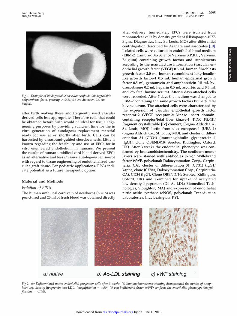
ig 2. (a) Differentiated native endothelial progenitor cells after 3 weated low-density lipoprotein (Ac-LDL) (magnification � �50). (c) v
cation � �100).ats.ctsnetjournDownloaded from
fter delivery. Immediately EPCs were isolated fromononuclear cells by density gradient (Histopaque-1077,
igma Diagnostics, Inc., St. Louis, MO) after differentialentrifugation described by Asahara and associates [10].solated cells were cultured in endothelial basal mediumEBM-2; Cambrex Bio Science Verviers S.P.R.L., Verviers,elgium) containing growth factors and supplementsccording to the manufacture information (vascular en-othelial growth factor (VEGF) 0.5 ml, human fibroblastsrowth factor 2.0 ml, human recombinant long-insulin-
ike growth factor-1 0.5 ml, human epidermal growthactor 0.5 ml, gentamycin and amphotericin 0.5 ml, hy-rocortisone 0.2 ml, heparin 0.5 ml, ascorbic acid 0.5 ml,nd 2% fetal bovine serum). After 4 days attached cellsere reseeded. After 7 days the medium was changed toBM-2 containing the same growth factors but 20% fetalovine serum. The attached cells were characterized by
he expression of vascular endothelial growth factoreceptor-2 (VEGF receptor-2; kinase insert domain-ontaining receptor/fetal liver kinase-1 [KDR, Flk-1])/ragment crystallizable [Fc] chimera; [Sigma Aldrich Co.,t. Louis, MO]) lectin from ulex europeus-1 (UEA 1)
Sigma Aldrich Co., St. Louis, MO), and cluster of differ-ntiation 34 (CD34) (immunoglobulin glycoprotein 1IgG1], clone QBEND/10; Serotec, Kidlington, Oxford,K). After 3 weeks the endothelial phenotype was con-rmed by immunohistochemistry. The confluent mono-
ayers were stained with antibodies to von Willebrandactor (vWF, polyclonal; Dakocytomation Corp., Carpin-eria, CA), cluster of differentiation 31 (CD31) (IgG1/appa, clone JC/70A; Dakocytomation Corp., Carpinteria,A), CD34 (IgG1, Clone QBEND/10; Serotec, Kidlington,xford, UK) and examined for uptake of acetylated
ow-density lipoprotein (Dil-Ac-LDL; Biomedical Tech-ologies, Stoughton, MA) and expression of endothelialitric oxide synthase (eNOS, polyclonal; Transductionaboratories, Inc., Lexington, KY).
b) Immunofluorescence staining demonstrated the uptake of acety-illebrand factor (vWF) confirms the endothelial phenotype (magni-
eks. (on W
by on June 1, 2013 als.org
SCeflvmsv11thbpmrcm
v6gtdsCcfhtwsTmsd
Faadm
2096 SCHMIDT ET AL Ann Thorac SurgUMBILICAL CORD BLOOD DERIVED EPC 2004;78:2094–8
CA
RD
IOV
AS
CU
LA
R
eeding and Three-Dimensional (3D) In Vitro Cultureharacterized cells were expanded up to passage 4 andxposed to laminar shear stress using rotating cultureasks. Thereafter EPCs were seeded onto biodegradableascular scaffolds (n � 22; 3 � 106 cells/cm2). For assess-ent of cell attachment and in-growth, two different
caffold materials were used for the fabrication of theascular scaffolds (0.5 cm diameter and 2.5 cm length; Fig): a nonwoven mesh (polyglycolic-acid [PGA] thickness:.0 mm, specific gravity: 69 mg/cm3; Albany Interna-ional , Mansfield , MA) coated with poly(4-ydroxybutyric acid (10% wt/vol P4HB; TEPHA, Cam-ridge, MA) and a 3D porous biodegradableolyurethane foam (Degrapol, porosity � 95%; Depart-ent of Materials, Federal Institute of Technology, Zu-
ich, Switzerland). Before seeding, the scaffolds wereold-gas sterilized and incubated for 24 hours in growthedium.
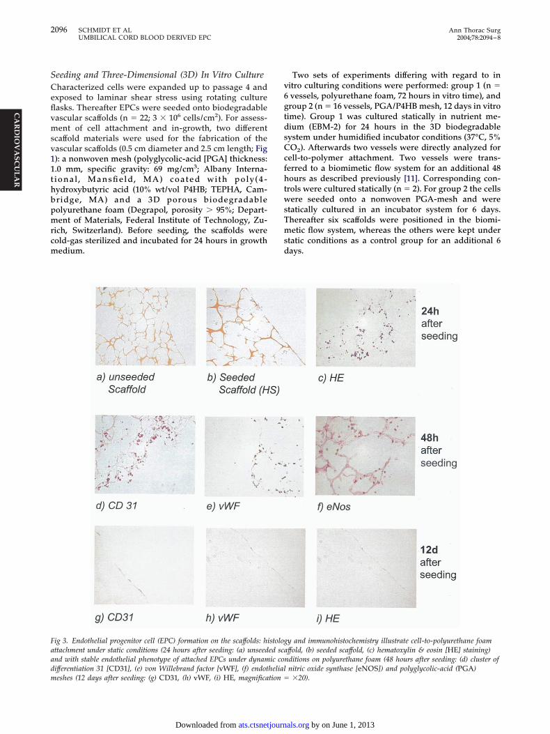
ig 3. Endothelial progenitor cell (EPC) formation on the scaffolds: hittachment under static conditions (24 hours after seeding: (a) unseednd with stable endothelial phenotype of attached EPCs under dynamifferentiation 31 [CD31], (e) von Willebrand factor [vWF], (f) endot
eshes (12 days after seeding: (g) CD31, (h) vWF, (i) HE, magnification �ats.ctsnetjournDownloaded from
Two sets of experiments differing with regard to initro culturing conditions were performed: group 1 (n �vessels, polyurethane foam, 72 hours in vitro time), androup 2 (n � 16 vessels, PGA/P4HB mesh, 12 days in vitroime). Group 1 was cultured statically in nutrient me-ium (EBM-2) for 24 hours in the 3D biodegradableystem under humidified incubator conditions (37°C, 5%O2). Afterwards two vessels were directly analyzed for
ell-to-polymer attachment. Two vessels were trans-erred to a biomimetic flow system for an additional 48ours as described previously [11]. Corresponding con-
rols were cultured statically (n � 2). For group 2 the cellsere seeded onto a nonwoven PGA-mesh and were
tatically cultured in an incubator system for 6 days.hereafter six scaffolds were positioned in the biomi-etic flow system, whereas the others were kept under
tatic conditions as a control group for an additional 6ays.
y and immunohistochemistry illustrate cell-to-polyurethane foamffold, (b) seeded scaffold, (c) hematoxylin & eosin [HE] staining)
nditions on polyurethane foam (48 hours after seeding: (d) cluster ofnitric oxide synthase [eNOS]) and polyglycolic-acid (PGA)
stologed scaic co
helial
�20).by on June 1, 2013 als.org

ATaw(fipisseL
R
MEAt4dcfdild
PDTVcksc
CHvodu(vs
AHEInda
C
OcfavcaecvcosdfdwwpaHrpdcm
hvaatftbcvw
Ftth
2097Ann Thorac Surg SCHMIDT ET AL2004;78:2094–8 UMBILICAL CORD BLOOD DERIVED EPC
CA
RD
IOV
AS
CU
LA
R
nalysiso document the presence of EPCs and cell-to-scaffoldttachment, fixed (4% formalin) samples of each groupere examined histologically by eosin-hematoxylin
H&E) and hematoxylin-sudan (H&S) staining. To con-rm the phenotype of EPCs after in vitro culture, ex-lanted scaffold samples of each group were character-
zed by immunohistochemistry (vWF, CD 31, and eNOStaining) as in the preseeding analysis. Samples forcanning electron microscopy (SEM) were collected fromach group and fixed in 2% glutaraldehyde (Sigma, St.ouis, MO) for 24 hours and gold spluttered.
esults
orphology of EPCsPCs could be isolated from all of the blood samples.fter isolation the plated cells were initially round. The
otal amount of isolated cells in 20 ml was 3 � 106. Afterdays cells were attached and formed clusters. Two
ifferent types of EPCs were found: spindle-like shapedells (70%) and polymorph cells (30%). Polymorph EPCsormed clusters and established satisfactory growth un-er in vitro conditions. After 3 weeks they differentiated
nto mature endothelial cells forming cobblestone mono-ayers (Fig 2A), whereas the spindle-like shaped cellsetached in the meantime.
henotype of EPCsifferentiated EPCs indicated an endothelial phenotype.hey stained positive for lectin from ulex europeus,EGF receptor-2, vWF, CD31, CD34, eNOS, and indi-
ated Ac-LDL-uptake (Fig 2B, 2C). Differentiated EPCsept this phenotype when cultivated on both types of 3Dcaffolds in the biomemetic flow system and under static
ig 4. Cell-to-polymer attachment (polyglycolic-acid [PGA] meshes) inhe scanning electron microscope (measurement � 70 �m): (a) differen-iated endothelial progenitor cell, (b) fiber of polyglycolic-acid/poly(4-ydroxybutric acid) (PGA/P4HB) scaffold.
onditions (Fig 3D–I). m
ats.ctsnetjournDownloaded from
ell-to-Polymer Attachmentistologic examination of the seeded tubular grafts re-
ealed suitable cell-to-polymer attachment in both typesf scaffold. After 24 and 48 hours, differentiated EPCsemonstrated suitable growth into the 3D scaffolds (poly-rethane foam) in both the dynamic and static controls
Fig 3B–F). A similar picture was observed after 12 days initro culture (PGA/PH4B mesh) under dynamic andtatic conditions (Fig 3G–I).
nalysis&E staining revealed a monolayer of differentiatedPCs on the lumen of the vascular scaffolds after 12 days.
mmunohistochemistry confirmed the endothelial phe-otype of the neotissue (Fig 3G–I). Analysis by SEMemonstrated endothelial tissue formation with favor-ble cell-to-polymer attachment in all groups (Fig 4).
omment
ne of the present limitations with regard to pediatricardiac surgery is the lack of appropriate graft materialsor the repair of congenital defects. Tissue engineering isscientific field that addresses this shortcoming using in
itro fabrication of living autologous grafts with theapacity for growth, repair, and remodeling. Previouspproaches using cell sources for cardiovascular tissuengineering were burdened by the disadvantage of ne-essitating an invasive surgical procedure for cell har-esting. EPCs are a subpopulation of bone marrow cellsirculating in the peripheral blood. Thus they can bebtained easily by venous puncture and without theacrifice of intact donor tissue. The results presentedemonstrated and confirmed that EPCs can be isolated
rom fresh human umbilical cord blood. Isolated EPCsifferentiated in culture into mature endothelial cells andere expandable in vitro. Two different types of EPCsere indicated: polymorph and spindle-like cells. Theolymorph EPCs demonstrated remarkable growth char-cteristics and constant phenotype, as also described byur and associates [12], whereas the spindle-like EPCs
evealed limited proliferation properties. EPCs exhibitroperties similar to those of angioblasts which can beefined as migratory endothelial cells that manifest theapacity to circulate, proliferate, and differentiate intoature endothelial cells [13–15].The present study demonstrated that differentiated
uman umbilical cord blood derived EPCs seeded onascular scaffolds formed neotissue in both a biomemeticnd static in vitro environment. These tissues were char-cterized as endothelial monolayers with related func-ions (eg, the production of eNOS indicating features ofunctional endothelium). Because of their stable pheno-ype and their growth and repair potential human um-ilical cord blood derived EPCs seem to be a promisingell source with regard to the tissue engineering ofascular grafts, particularly for pediatric applicationshere EPCs could adjust to the specific growth require-
ents of the cardiovascular system during childhood.by on June 1, 2013 als.org

Too[otf
mbHbttn
tgcctItcFwdEcc
dstptag
TizqZtUircgip
R
1
1
1
1
1
1
1
1
1
1
2
2098 SCHMIDT ET AL Ann Thorac SurgUMBILICAL CORD BLOOD DERIVED EPC 2004;78:2094–8
CA
RD
IOV
AS
CU
LA
R
he focus of this study was to investigate the attachmentf EPCs on an implantable biodegradable scaffold. Basedn previous animal studies using PGA/P4HB scaffolds16] and the subsequent successful transfer of the meth-dology to human cell systems [17, 18], we investigatedhe EPC attachment by including these established scaf-olds in the present study.
A critical limitation regarding the availability of EPCsay be the low number that exists in the peripheral
lood, because they are a scant circulating population.owever previous studies revealed that umbilical cordlood contains increasing amounts of EPC that approach
enfold compared with the blood of adults [19, 20] poten-ially providing sufficient cell numbers for tissue engi-eering purposes.In many instances congenital cardiac defects are de-
ected by ultrasound at approximately 16-20 weeks ofestation. One option regarding the tissue-engineeringoncept involves obtaining the EPCs before birth usingordocentesis. This would allow enough time for isola-ion, differentiation, and fabrication of the graft material.n some instances a successful surgical correction of thisype of defect depends on the weight of the infant and soannot be performed directly after birth (eg, Tetralogy ofallot). In these occurrences a second option exists inhich the blood can be obtained from the umbilical cordirectly after birth and the isolation and cultivation ofPCs can be performed at that time. After differentiationells can be deep-frozen and stored until operatingonditions are optimal.
Based on our results human umbilical cord blooderived EPCs seem to be a promising autologous cellource for cardiovascular tissue engineering because ofheir physiologic properties and easy accessibility. Theossibility of prenatally harvesting cells may allow for
he option of preparing autologous grafts for use at birthnd may overcome the limitations of the present-dayraft materials for treating congenital defects.
he authors thank Sirpa Price (Laboratory for Tissue Engineer-ng and Cell Transplantation, University Hospital, Zurich, Swit-erland) for her valuable work on cell culture and Klaus Mar-uardt (Center of Electron Microscopy, University Hospital,urich, Switzerland) for providing the SEM pictures. We further
hank Professor Bernhard Odermatt (Department of Pathology,niversity Hospital, Zurich, Switzerland) for performing the
mmunohistochemistry analysis and Astrid Morger for supportegarding the histologic examination. The authors recognize theontributions afforded by Professor Takayuki Asahara and hisroup and we also acknowledge his collaboration regarding cell
solation. Finally, we thank Professor Stefanie Dimmeler forroviding valuable discussions.
eferences
1. Niklason LE, Gao J, Abbott WM, et al. Functional arteries
grown in vitro: Science 1999;284:489–93.ats.ctsnetjournDownloaded from
2. Kawamoto A, Gwon HC, Iwaguro H. Therapeutic potentialof ex vivo expanded endothelial progenitor cells for myocar-dial ischemia. Circulation 2002;103:634–7.
3. Kocher AA, Schuster MD, Szabolcs MJ, et al. Neovasculari-sation of ischemic myocardium by human bone-marrow-derived angioblasts prevents cardiomyocyte apoptosis re-duces remodeling and improves cardiac function. Nat Med2001;7:430–6.
4. Assmus B, Schachinger V, Teupe et al. Transplantation ofprogenitor cells and regeneration enhancement in acutemyocardial infarction. Circulation 2002;106:3009–17.
5. Pesce M, Orlandi A, Iachininoto MG, et al. Myoendothelialdifferentiation of human umbilical cord blood-derived stemcells in ischemic limb tissues. Circ Res 2003;93:e51–62. Epub2003 Aug 14.
6. Shirota T, Yasui H, Shimokawa H, Matsuda T. Fabrication ofendothelial progenitor cell (EPC)-seeded intravascular stentdevices and in vitro endothelialization on hybrid vasculartissue. Biomaterials 2003;24:2295–302.
7. Kaushal S, Amiel GE, Guleserian KJ, et al. Functional small-diameter neovessels created using endothelial progenitorcells expanded in vivo. Nat Med 2001;7:1035–40.
8. Shirota T, Hongbing HE, Yasui H, Matsuda T. Humanendothelial progenitor cell-seeded hybrid graft: proliferativeand antithrombogenic potentials in vitro and fabricationprocessing. Tiss Eng 2003;9:127–36.
9. Hoerstrup SP, Kadner A, Melnitchouk S, et al. Tissue engi-neering of functional trileaflet heart valves from humanmarrow stromal cells. Circulation 2002;106(12 Suppl 1):I143–50.
0. Asahara T, Murohara T, Sullivan A, et al. Isolation ofputative progenitor endothelial cells for angiogenesis. Sci-ence 1997;275:964–7.
1. Hoerstrup SP, Sodian R, Sperling JS, Vacanti JP, Mayer JE.New pulsatile bioreactor for in vitro formation of tissueengineered heart valves. Tissue Eng 2000;6:75–9.
2. Hur J, Yoon CH, Kim HS, et al. Characterization of two typesof endothelial progenitor cells and their different contribu-tions to neovasculogenesis. Arterioscl Thromb Vasc Biol2004;24:1–6.
3. Coffin JD, Harrison J, Schwartz S, et al. Angioblast differen-tiation and morphogenesis of the vascular endothelium inthe mouse embryo. Dev Biol 1991;148:51–62.
4. Robert B, St. John PL, Hyink DP, Abrahamson DR. Evidencethat embryonic kidney cells expressing flk-1 are intrinsic,vasculogenic angioblasts. Am J Physiol Renal Physiol 1996;271:F744–53.
5. Caprioli A, Jaffredo T, Gautier R, Dubourg C, Dieterlen-Lievre F. Blood-borne seeding by hematopoietic and endo-thelial precursors from the allantois. Proc Natl Acad Sci USA1998;95:1641–6.
6. Hoerstrup SP, Sodian R, Daebritz S, et al. Functional livingtrileaflet heart valves grown in vitro. Circulation 2000;102(Suppl 3):III44–9.
7. Hoerstrup SP, Zund G, Sodian R, Schnell AM, Grunenfelder J,Turina MI. Tissue engineering of small caliber vascular grafts.Eur J Cardiothorac Surg 2001;20:164–9.
8. Hoerstrup SP, Kadner A, Breymann C, et al. Living, autolo-gous pulmonary artery conduits tissue engineered fromhuman umbilical cord cells. Ann Thorac Surg 2002;74:46–52discussion 52.
9. Peichev M, Afzal J, Naiyer DP et al. Expression of VEGFR-2and AC133 by circulating human CD34� cells identifies apopulation of functional endothelial precursors. Blood 2000;95:952–8.
0. Murohara T, Ikeda H, Duan J, et al. Transplanted cordblood-derived endothelial precursor cells augment postnatal
neovascularization. J Clin Invest 2000;105:1527–36.by on June 1, 2013 als.org

2004;78:2094-2098 Ann Thorac SurgNeuenschwander, Gregor Zund, Marko Turina and Simon P. Hoerstrup
Dörthe Schmidt, Christian Breymann, Alberto Weber, Christina I. Guenter, Stefan of Vascular Grafts
Umbilical Cord Blood Derived Endothelial Progenitor Cells for Tissue Engineering
& ServicesUpdated Information
http://ats.ctsnetjournals.org/cgi/content/full/78/6/2094including high-resolution figures, can be found at:
References http://ats.ctsnetjournals.org/cgi/content/full/78/6/2094#BIBL
This article cites 19 articles, 9 of which you can access for free at:
Citations
shttp://ats.ctsnetjournals.org/cgi/content/full/78/6/2094#otherarticleThis article has been cited by 3 HighWire-hosted articles:
Permissions & Licensing
[email protected]: orhttp://www.us.elsevierhealth.com/Licensing/permissions.jsp
in its entirety should be submitted to: Requests about reproducing this article in parts (figures, tables) or
Reprints [email protected]
For information about ordering reprints, please email:
by on June 1, 2013 ats.ctsnetjournals.orgDownloaded from