bFGF release is dependent on flow conditions in experimental vein grafts
9
Eur J VascEndovascSurg 10, 450-458 (1995) bFGF Release is Dependent on Flow Conditions in Experimental Vein Grafts Sandro Lepidi, Antonio V. Sterpetti, Alessandra Cucina, Anna Di Carlo, Anna L. Patrizi, Roberta Palumbo, Paola Bernucci, Luciana Santoro-D'Angelo and Antonino Cavallaro I Istituto di Clinica Chirurgica, Istituto Istologia & Embriologia and Istituto Anatomia Patologica, University of Rome "La Sapienza " Italy Objectives: Basic Fibroblastic Growth Factor (bFGF) is a powerful mitogen for smooth muscle cells and has been implicated in the genesis of Myointimal hyperplasia. The aim of this study was to determine the release of bFGF by veins in different haemodynamic conditions. Design and setting: Laboratory animal study. Materials: In 39 Lewis rats, a 1 cm long segment of inferior vena cava was inserted at the level of the abdominal aorta. The segments of inferior vena cava were obtained from syngenic Lewis rats. Arterial Vein Grafts (AVG) were harvested after 4 weeks (AVG 4) and 12 weeks (AVG 12). In 16 animals the arterial vein grafts were explanted 4 weeks after the initial operation and reimplanted (Reimplanted Vein Grafts: RVG) in syngenic Lewis rats as venous-venous bypass grafts at the level of the left iliac vein and harvested after 2 weeks (RVG 2) and 8 weeks (AVG 8). Outcome measures: The tissue was studied in organ culture in a serum-free system for (1) release of bFGF (immunoassay) and (2) mitogenic activity of the conditioned media. Scanning electron and light microscopy studies were also performed. Results: bFGF release by veins increased significantly (p < 0.01) when veins were inserted in the arterial circulation, and decreased significantly (p < 0.01) when grafts where reimplanted in the venous system, bFGF release (ng/cm2): Control AVG (4 weeks) AVG (12 weeks) RVG (2 weeks) RVG (8 weeks) 55 + 7 295 + 20 340 +_18 86 + 10 92 +_12 Conclusion: Vein inserted in the arterial circulation release a higher quantity of bFGF. This could explain in part, the formation of myointimal hyperplasia in arterial vein graft. Key Words: Arterial vein graft; Shear stress; Fibroblastic growth factor; Myointimal hyperplasia. Introduction Arterial vein bypass grafting is a common procedure in cardiac and peripheral vascular surgery. Approx- imately 20% of all vein grafts develop stenosis and nearly 80% of these lesions occur in the first year after surgery, often because of myointimal hyperplasia (MH).I-4 The basic histopathology of MH is a proliferative response of smooth muscle cells (SMC). In MH, smooth muscle cells proliferate and migrate from the media to the intima. 5'6 This abnormal proliferative response can lead to vein wall thickening and graft Please address all correspondenceto: AntonioV. Sterpetti, I Istituto di Clinica Chirurgica, Laboratorio del Castro Laurenziano, Via Antonio Scarpa 14, 00161 Roma,Italy. occlusion by reducing the flow channel diameter, especially in small calibre veins. The factors leading to MH in arterial vein grafts are ill-defined. Growth factors, cytokines and other chemicals induce and regulate numerous cell functions during the process of MH and atherogenesis. They may act in cell recruit- ment and migration, cell proliferation and the control of protein synthesis, including extracellular matrix proteins. One of the best characterised mitogen for smooth muscle cells is basic Fibroblastic Growth Factor (bFGF). bFGF is a member of heparin binding mito- gens and is widely distributed. SMC and endothelial cells contain bFGF in large quantities. 7-9 Recently Lindner and Reidy have shown that bFGF is a potent pharmacological stimulant of smooth muscle cells 1078-5884/95/080450 + 09 $12.00/0 © 1995W. B. Saunders CompanyLtd.
-
Upload
independent -
Category
Documents
-
view
3 -
download
0
Transcript of bFGF release is dependent on flow conditions in experimental vein grafts
Eur J Vasc Endovasc Surg 10, 450-458 (1995)
bFGF Release is Dependent on Flow Conditions in Experimental Vein Grafts
Sandro Lepidi, Antonio V. Sterpetti, Alessandra Cucina, Anna Di Carlo, Anna L. Patrizi, Roberta Palumbo, Paola Bernucci, Luciana Santoro-D'Angelo and Antonino Cavallaro
I Istituto di Clinica Chirurgica, Istituto Istologia & Embriologia and Istituto Anatomia Patologica, University of Rome "La Sapienza " Italy
Objectives: Basic Fibroblastic Growth Factor (bFGF) is a powerful mitogen for smooth muscle cells and has been implicated in the genesis of Myointimal hyperplasia. The aim of this study was to determine the release of bFGF by veins in different haemodynamic conditions. Design and setting: Laboratory animal study. Materials: In 39 Lewis rats, a 1 cm long segment of inferior vena cava was inserted at the level of the abdominal aorta. The segments of inferior vena cava were obtained from syngenic Lewis rats. Arterial Vein Grafts (AVG) were harvested after 4 weeks (AVG 4) and 12 weeks (AVG 12). In 16 animals the arterial vein grafts were explanted 4 weeks after the initial operation and reimplanted (Reimplanted Vein Grafts: RVG) in syngenic Lewis rats as venous-venous bypass grafts at the level of the left iliac vein and harvested after 2 weeks (RVG 2) and 8 weeks (AVG 8). Outcome measures: The tissue was studied in organ culture in a serum-free system for (1) release of bFGF (immunoassay) and (2) mitogenic activity of the conditioned media. Scanning electron and light microscopy studies were also performed. Results: bFGF release by veins increased significantly (p < 0.01) when veins were inserted in the arterial circulation, and decreased significantly (p < 0.01) when grafts where reimplanted in the venous system, bFGF release (ng/cm2):
Control AVG (4 weeks) AVG (12 weeks) RVG (2 weeks) RVG (8 weeks) 55 + 7 295 + 20 340 +_ 18 86 + 10 92 +_ 12
Conclusion: Vein inserted in the arterial circulation release a higher quantity of bFGF. This could explain in part, the formation of myointimal hyperplasia in arterial vein graft.
Key Words: Arterial vein graft; Shear stress; Fibroblastic growth factor; Myointimal hyperplasia.
Introduction
Arterial vein bypass grafting is a common procedure in cardiac and peripheral vascular surgery. Approx- imately 20% of all vein grafts develop stenosis and nearly 80% of these lesions occur in the first year after surgery, often because of myointimal hyperplasia (MH).I-4
The basic histopathology of MH is a proliferative response of smooth muscle cells (SMC). In MH, smooth muscle cells proliferate and migrate from the media to the intima. 5'6 This abnormal proliferative response can lead to vein wall thickening and graft
Please address all correspondence to: Antonio V. Sterpetti, I Istituto di Clinica Chirurgica, Laboratorio del Castro Laurenziano, Via Antonio Scarpa 14, 00161 Roma, Italy.
occlusion by reducing the flow channel diameter, especially in small calibre veins. The factors leading to MH in arterial vein grafts are ill-defined. Growth factors, cytokines and other chemicals induce and regulate numerous cell functions during the process of MH and atherogenesis. They may act in cell recruit- ment and migration, cell proliferation and the control of protein synthesis, including extracellular matrix proteins.
One of the best characterised mitogen for smooth muscle cells is basic Fibroblastic Growth Factor (bFGF). bFGF is a member of heparin binding mito- gens and is widely distributed. SMC and endothelial cells contain bFGF in large quantities. 7-9 Recently Lindner and Reidy have shown that bFGF is a potent pharmacological stimulant of smooth muscle cells
1078-5884/95/080450 + 09 $12.00/0 © 1995 W. B. Saunders Company Ltd.

bFGF Release 451
growth in injured arteries. In addition, they have shown that the administration of a blocking antibody to bFGF inhibits the proliferation of SMC, in balloon injured carotid artery by 80%. l° The results of their study suggest that endogenous bFGF is a major mitogen controlling the growth of vascular smooth muscle cells following injury.
In our laboratory we found that, in vitro, increasing shear stress induce the release of higher levels of bFGF by arterial smooth muscle cellsJ ~ In vivo, wall shear stress is directly proportional to flow velocity and inversely proportional to the cube of the vessel radius. Autogenous veins when inserted into the arterial circulation are subjected to increased blood pressure and higher shear stress compared to haemodynamic conditions of the native venous system. Thus, theoret- icall)~ bFGF could be responsible for MH formation in arterial vein graft. Attempts have been made to induce the structural alterations of the intimal hyperplasia and then to produce its regression by altering the haemodynamic characteristics of arterial blood flow.12'lBDavis et al. investigated the regression of intimal hyperplasia in arterial vein grafts once reim- planted in the venous circulation. 14 They correlated the reduction of intimal hyperplasia with the restora- tion of endothelium-dependent relaxing factor. In recent experiments we found that vein grafts when inserted in the arterial circulation release a higher quantity of growth factors including bFGF. This phenomenon could explain in part, the formation of MH in arterial vein grafts.
The purpose of this study was to determine the release of bFGF by vein grafts when inserted into the arterial circulation and by arterial vein grafts when reimplanted in the venous system. The relationship between the release of bFGF and the structural changes of vein graft was also investigated.
Material and Methods
Experimental Design
In 39 male inbred Lewis rats (average weight 250 g), a 1 cm long segment of inferior vena cava was inserted into the arterial system, at the level of the abdominal aorta. The segments of inferior vena cava were obtained from syngenic Lewis rats (same genetic pattern). Arterial Vein Grafts (AVG) were harvested after 4 weeks (AVG 4:15 animals) and 12 weeks (AVG 12:8 animals). In 16 animals the arterial vein grafts were explanted 4 weeks after the initial operation and reimplanted (Reimplanted Vein Grafts: RVG) in syn- genic Lewis rats as venous-venous bypass grafts at the
level of the left iliac vein and harvested after 2 weeks (RVG 2:8 animals) and 8 weeks (AVG 8:8 animals). At the time of harvest, segments of similar dimensions of endogenous inferior vena cava, aorta or left iliac vein above and below the graft were removed.
Operative Procedure
The animals were anaesthetised with intramuscular Xylazine (3 mg/kg) and intramuscular Ketamine (50 mg/kg) supplemented by intraperitoneal Ketamine for maintenance. Surgery was performed using an operating microscope (Zeiss OPMI 7-D).
A 1 cm long segment of supra diaphragmatic inferior vena cava was obtained from donor rats. The chest was opened, and the supra diaphragmatic portion of inferior vena cava was identified and excised (Fig. 1A). The animal was sacrificed at this time. The vein was kept moist in Dulbecco Modified Eagle Medium (DMEM) for approximately 20 rain, while the receiving rat was anaesthetised and the abdominal aorta identified, and both proximal and distal control obtained. The vein grafts were inserted with end to end anastomoses using 10-0 monofilament nylon sutures (Ethicon Inc) (Fig. 1B). The same microsurgical procedure and materials were used to fashion venous-venous bypass grafts at the level of the left iliac vein (Fig. 1C). The total ischemia time was between 30 and 45 min. Graft patency was evaluated at the time of surgery and thereafter by weekly palpation of the femoral pulse in AVG or inspection for a swollen leg. Throughout each procedure, care was taken to avoid unnecessary damage of the graft tissue.
Harvesting procedure
With the animal under deep anaesthesia, abdomen and chest were opened. The aorta or the left iliac vein above and below the graft, the vein graft and a segment of same dimensions of supra diaphragmatic Inferior Vena Cava were exposed and excised. In each group, three grafts were used for scanning electron and light microscopy. The other grafts were examined for bFGF production.
Histologic Examination
Grafts were perfusion fixed with an initial infusion of Hanks balanced salt solution, followed by 10% formal- dehyde. Transverse 5 ~zm sections were stained with
Eur J Vasc Endovasc Surg Vol 10, November 1995

452 S. Lepidi et aL
Haematoxylin and Eosin or with elastic tissue stains by means of standard techniques. Intimal and medial thickness as well as luminal radius were defined and calculated by videomorphometry (Quantimet 500) at different levels of vein graft.
For scanning electron microscopy, specimens were fixed in 2.5% glutaraldehyde made up in 0.1 mol/1 cacodylate buffer (pH 7.2), rinsed several times and left for 1 h in the same buffer. The specimens were then fixed for 1 h in osmium 1%, dehydrated in ascending concentrations of ethanol (15% to 100%), critical point dried in CO2, mounted in specimen stubs, and sputter-coated with gold-palladium accord- ing to standard techniques. All specimens were examined in a scanning electron microscope (Hitachi S. 570) at an accelerating voltage of 15 KV.
Organ culture
The arterial, vein and graft segments were rinsed thoroughly for 10 minutes with DMEM supplemented with antibiotics (Gentamycin 200/~g/ml, Streptomy- cin 100/~g/ml, Penicillin 100 IU/ml). The vessels were then opened longitudinally and placed in organ culture. The specimens were placed in 48-well Costar tissue culture plates for organ culture.The tissue was
incubated in 1 ml of DMEM supplemented with antibiotics (Gentamycin 200/~g/ml Streptomycin 50 izg/ml, Penicillin 50/~g/ml) at pH 7.4. The tissue was incubated for 5 days at 37°C in a 5% CO2 atmosphere. Aliquots of conditioned medium were collected at 24, 72 and 120 h and centrifuged for 5 min at 15000 rpm and the supernatant stored at -80°C for assay of mitogenic activity and assay of bFGF release.
Assay for mitogenic activity of conditioned media
Sub-confluent bFGF receptor-bearing Swiss 3T3 cells were used to assay the DNA synthesis stimulation activity of the conditioned media. The cells were plated in 96 wells plates (Flacon Plastic) at a density of 4 × 10⁴ cells/ml in 200~1 of DMEM supplemented with 0.1% Foetal Calf Serum. Conditioned serum free media from AVG and RVG at different hours and control media were added to Swiss 3T3 cells (20 tzl). The positive controls received an equivalent volume of DMEM plus human recombinant bFGF (Boehringer Mannheim, Germany); negative controls received serum-free DMEM. Tritiated thymidine (0.5 tzCi per well plate) was added, the cultures were incubated for 18 h and collected on Skatron Filters (Skatron Instru- ments Inc., Sterling, VA, U.S.A.) for radioactivity determination in a LKB scintillation counter.
A B C Fig. 1. Schematic drawing of the operative procedure. A segment of supradiaphragmatic inferior vena cava from syngenic Lewis rat (A) was interposed at the level of the abdominal aorta (B). 4 weeks after the initial operation the arteriaal vein graft was reimplanted at the level of left iliac vein (C).
Eur J Vasc Endovasc Surg Vol 10, November 1995

bFGF Release 453
Analysis of reduction of mitogenic activity by anti-bFGF antibody
Measurement of the Swiss 3T3 cells DNA synthesis- stimulating activity of the conditioned media from vein grafts and control veins was repeated in presence of an excess of monospecific anti-bFGF antibody produced in our laboratory (50]zg/ml). Tritiated thy- midine was again added and the cultures were incubated for 18 h. After further processing, the radioactivity was measured.
bFGF assay in the conditioned media
The presence of bFGF molecules in the serum-free conditioned media from vein grafts and control veins was determined by inhibition antibody-binding assay. A dilution of anti-bFGF mouse monoclonal antibody that showed about 50% maximal reactivity against bFGF was incubated with various dilutions of con- ditioned media in 400/zl tubes precoated with PBS- gelatine 2%. After 20 h of incubation at 4°C, staphylo- coccus A was added, and the immunoaggregates were removed by centrifugation. The residual antibody- binding activity in the supernatant was measured by enzyme-linked immunosorbent assay (ELISA). 13 Plas- tic wells (96 wells, Falcon Plastic) were coated with bFGF (10 ng per well) for 8 h at 4°C. Plates were then washed twice with PBS and saturated with PBS- gelatin 1% for 2 h at 37°C. Washed wells were then filled with 50/~1 per well of supernatant obtained after immunoprecipitation. After 2 h of incubation at 37°C the wells were washed with PBS-gelatine 0.1%. Perox- idase-labelled goat anti-mouse immunoglobulin anti- body was added. After 60 minutes of incubation at 37°C the plate was washed three times in PBS-gelatine 0.1% and once in distilled water. Finally O-Phenylene- diamine Dihydrochloride (0.4 mg/ml) (SIGMA, St. Louis, MO, U.S.A.) was added as substrate for the enzyme. Bound-specific antibody was quantitatively measured by optical density reading at 492 nm with a spectrophotometer (Beckman Instruments, Inc., Brea, CA, U.S.A.). We used mouse anti-bFGF monoclonal antibody as positive control and antibody without specificity as negative control.
The amount of bFGF in the conditioned media was quantified by comparison with a standard curve obtained using known quantities of human recombi- nant bFGF.
Statistical analysis
Statistical analysis was performed with Statview
software (Abacus Concepts, Inc. Berkeley, CA, U.S.A.) on a Macintosh Powerbook microcomputer (Apple Computer, Inc., Cupertino, CA, U.S.A.) Data were expressed as mean + S.D. Differences between the groups were analysed by use of two-tailed Student's t-test.
Results
Structural changes
At harvest, arterial vein grafts were firm and rigid, compared with control veins. Twelve weeks after surgery, AVG showed an increase in external diameter compared with AVG 4, whereas 8 weeks after the reimplantation, RVG showed a decrease in external diameter compared with RVG 2.
Microscopically, intact endothelial cells were pre- sent on the surface of AVG and RVG as well as in control vein (Fig. 2). There were scattered platelets and leukocytes adhering to endothelial cells. Myointimal hyperplasia was present in all the arterialised vein grafts; large amount of connective tissue and several layers of smooth muscle cells were present. In the reimplanted veins, connective tissue was preponder- ant over smooth muscle cells. Control veins consisted of a single layer of endothelial cells and a few layers of smooth muscle cells. Table 1 shows the total area of intima and media (/~m 2) of AVG, RVG and control vein. In arterial vein grafts, intima-media area increased significantly compared with control vein 4 and 12 weeks after the initial operations (p < 0.01) (Fig. 3). Two weeks after reimplantation, vein grafts showed a decrease in the total area of intima and media as compared with the arterial vein grafts, and the difference became significant after 8 weeks (p < 0.05). RVG after 8 weeks showed a significant reduction in lumenal radius as compared with RVG2 and AVG (p < 0.01) (Fig. 4).
Mitogenic activity of the conditioned media and its reduction by anti-bFGF antibody
Addition of serum-free conditioned medium from AVG 4 and AVG 12 collected after different time intervals (12, 72 and 120 h) of incubation in organ culture produced a mean increase of tritiated thymi- dine uptake of ten-fold (p < 0.01) and fourteen-fold (p<0.01) respectively as compared with negative control. Addition of monospecific anti-bFGF to the
Eur J Vasc Endovasc Surg Vo110, November 1995

454 S. Lepidi et al.
(A) (B) Fig. 2. Scanning electron microscopy of arterial vein graft 4 weeks following implantation (A) and control vein (B). Endothelial ceils form the internal surface.
med ium of 3T3 cell cultures exposed to the con- dit ioned med ium from AVG decreased the uptake of tritiated thymidine by 55% and 40% in AVG 4 and AVG 12 respectively (p < 0.01) (Fig. 5). Two weeks after reimplantat ion in the venous system, the mean tri- tiated thymidine uptake was significantly reduced as compared with the level of the vein grafts at the time of reimplantat ion (4 weeks) (p < 0.01). The mitogenic activity re turned to the level of negative control (DMEM) and remained constant in the following weeks (RVG 8). Again, addit ion of monospecific anti- bFGF to the med ium of 3T3 cell cultures exposed to the condit ioned med ium from RVG decreased the uptake of tritiated thymidine (Fig. 6).
Table 1. Structural changes of vein graft
Group Intima and media area (~m 2) x 105
AVG4 16.8_+ 4.47 AVG12 17.5_+ 3.58 RVG2 15 + 6.02 RVG8 5.9 + 3.35 Control vein 1.9_+ 0.8
bFGF assay by ELISA
Table 2 show the release of bFGF from AVG, RVG and control veins. The release of bFGF was significantly higher in AVG as compared with control vein (p < 0.01), and decreased 2 weeks after the reimplanta- tion (p<0.01) and remained at approximately the same level dur ing the following weeks (8 weeks- RVG).
Discussion
MH has been implicated as a cause of stenosis and possible occlusion in small to midsized arterial recon- structive procedures. While findings from numerous
Table 2. Release of bFGF (ng/cm 2)
Control vein AVG AVG RVG RVG (4 weeks) (12 weeks) (2 weeks) (8 weeks)
55_+7 295_+20 340_+18 86_+10 92_+12
Eur J Vasc Endovasc Surg Vol 10, November 1995
bFGF Release 455
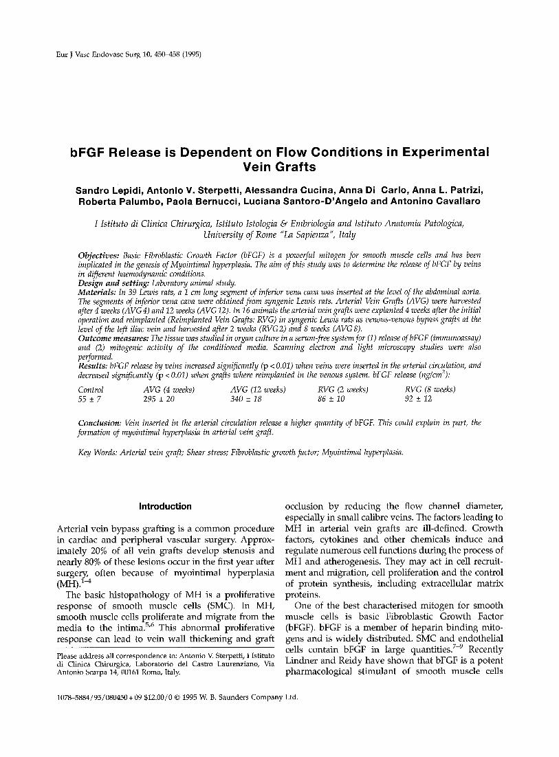
A B C Fig. 3. Light microscopy of control vein (A) and arterial vein graft 4 weeks (B) and 12 weeks from implantation (C). Evidence of myointimal hyperplasia in vein graft. The intima-media layer of AVG 4 and AVG 12 was statistically thicker than control vein (p < 0,01). L = lumen. (H.E. × 4).
h'
A B Fig. 4. Light microscopy of reimplanted vein graft 2 weeks (A) and 8 weeks (B) from operation. RVG 8 showed a significant reduction of intima-media area as compared with arterial vein graft at the time of harvest for reimplantation (p < 0.05). RVG after 8 weeks showed a significant reduction in lumenal radius compared with the 2 weeks-RVG and AVG (p < 0.01). L = lumen (H.E. × 4).
Eur J Vasc Endovasc Surg Vol 10, November 1995
456 S. Lepidi et aL
clinical reports have outlined the lesion seen at arterial reconstruction, 1-4 our understanding of the under- lying mechanisms is still at an early stage.
In a recent article, Gibbons and Dzau expressed the emerging concept of vascular remodelling) s They considered the vessel wall as an active, integrated organ composed of endothelial, smooth muscle, and fibroblast cells coupled to each other in a complex autocrine-paracrine set of interactions. The vascu- lature is capable of sensing changes within its milieu, integrating these signals by intercullular communica- tions and changing itself through the local production of mediators that influence structure as well as function. The results of our study fit their theory well.
Areas of intima and media as well as the lumenal radius of vein grafts changes according to different haemodynamic conditions. Once inserted into the arterial circulation vein grafts are subjected to a higher pressure and level of shear stress, release a higher quantity of growth factors, including bFGF and undergo significant MH. Moreover, the lumenal radius increases slightly 4 weeks and, more sig-
nificantl:~ 12 weeks after the operation. Area of intima and media return to the level of control veins when a graft implanted for 4 weeks at the level of abdominal aorta is returned to venous circulation and harvested after an additional 8 weeks. After the same period of time, RVG show a decrease in lumenal radius below the level of control vein. In RVG, growth factor release returned to the level of negative control after 2 weeks only, and remain constant during the following weeks.
Two similar recent experiments of reimplanted vein grafts in a canine 16 and in a rabbit model 14 showed a regression of intimal hyperplasia. Davis et al. sug- gested that restoration of endothelium dependent relaxing factor-mediated relaxation was responsible of MH regression) 4 In our opinion, growth factors, including bFGF play a more important role in vascular remodelling and in the development of MH.
According to the theory of Gibbons and Dzau, we can recognise four different components in the process of vascular remodelling: the detection of signals due to
30000
30000
e ~
® 20000
-~ lOOOO ?.-2
AVG4 AVG12 bFGF DMEM
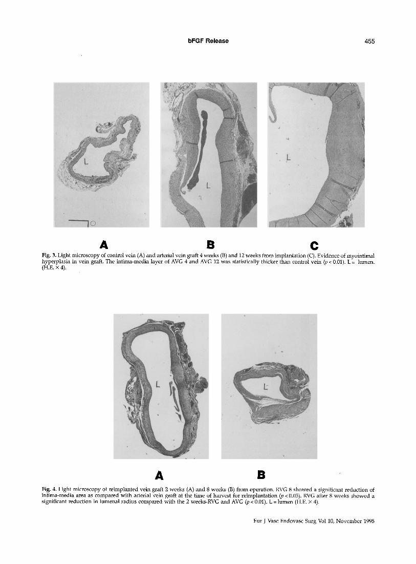
Fig. 5. Tritiated thymidine uptake in Swiss 3T3 cells after addition of serum-free conditioned medium from AVG 4 and 12 collected after different time intervals of incubation in organ culture. DMEM: negative control. Human recombinant bFGF: positive control. AVG 4 and AVG 12 showed a mean increase of tritiated thymidine uptake of tenfold (p<0.01) and fourteenfold (p<0.01) respectively as compared with negative control. Addition of anti bFGF (empty column) to the medium of 3T3 cell cultures exposed to the conditioned medium from AVG 4 and AVG 12 decreased the uptake of tritated thymidine by 55% and 40% respectively ( < 0.01).
20000
10000
RVG 2 RVG 8 bFGF DMEM
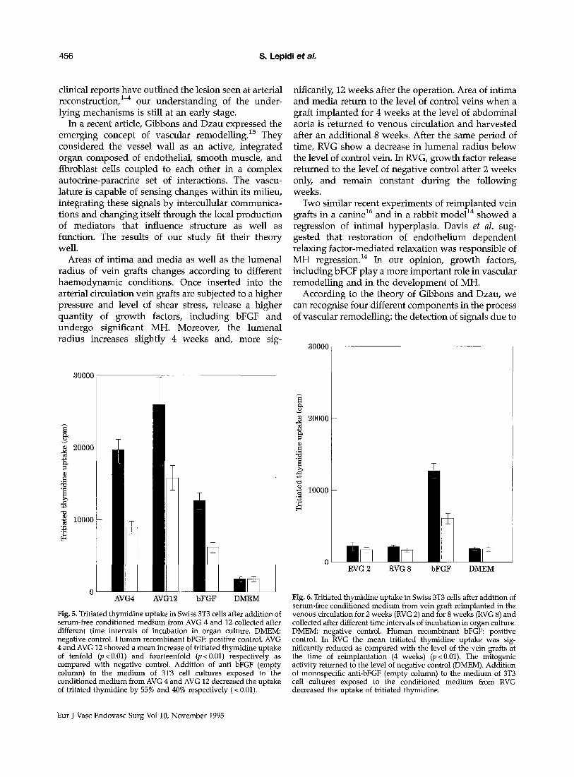
Fig. 6. Tritiated thymidine uptake in Swiss 3T3 cells after addition of serum-free conditioned medium from vein graft reimplanted in the venous circulation for 2 weeks (RVG 2) and for 8 weeks (RVG 8) and collected after different time intervals of incubation in organ culture. DMEM: negative control. Human recombinant bFGF: positive control. In RVG the mean tritiated thymidine uptake was sig- nificantly reduced as compared with the level of the vein grafts at the time of reimplantation (4 weeks) (p < 0.01). The mitogenic activity returned to the level of negative control (DMEM). Addition of monospecific anti-bFGF (empty column) to the medium of 3T3 cell cultures exposed to the conditioned medium from RVG decreased the uptake of tritiated thymidine.
Eur J Vasc Endovasc Surg Vol 10, November 1995

bFGF Release 457
changes in haemodynamic conditions (sensors); the relay of signals within the cell and to adjacent cells (transducers); the synthesis and release or activation of substances that influence cell growth, death and migration or the composition of the extracellular matrix (mediators); and the resultant structural changes in vessel wall (both cellular and noncellular components).
The results of this study suggest that bFGF released in AVG and RVG is mainly derived from EC and SMC. Although macrophages and leukocytes may produce bFGF, neither type of cells appeared to account of differences observed in bFGF release between AVG, RVG and control vein. bFGF has a number of properties that are similar to other factors such as binding to plasma membrane receptors that are tyrosine kinases, multipotent functions, and unique expression during development. 7 bFGF does have several unique properties that differentiate it from most other growth factors. One of these is the lack of a signal peptide normally required for vectorial translation into the endoplasmic reticulum and secre- tion. A second property is that bFGF exists in two different forms, an high molecular weigh form (HMW) and an 18 kDA form. 17 The subcellular distribution of. these different forms of bFGF is not equivalent, ls'~9 The intracellular localisation raises question concerning how and where bFGF is released.
It has been shown that severe injury and /or all lysis results in release of bFGF. 2° However, in our organ culture the tissue remain viable, as observed in previous studies using thymidine uptake. Alterna- tively, Yu et al. 21 found bFGF immunoreactive cyto- plasmatic vesicles in bovine EC and in some instances the vesicles were seen to fuse with the plasma membrane, appearing to discharge their immunor- eactive bFGF at the luminal surface. Recently, Rifkin et
al. 22 has shown modulation of cellular function by altering bFGF levels on isolated, single living cells. Under these conditions all bFGF must come from the cell under observation, and the possible contribution from dying cells is eliminated. The result of this study indicate that bFGF is released from living cells and can act as a true autocrine factor.
In conclusion, we suggest that increase release of bFGF by arterial vein grafts might be responsible for MH and wall thickening. Serial studies in time showed that bFGF production is probably the cause of MH and not the opposite. RV6 harvested 2 weeks after surgery had still evidence of MH, but bFGF release was low, similar to control veins. These findings support the view that pharmacological intervention
directed to reduce growth factors release might be beneficial in preventing MH in arterial vein grafts.
References
1 SZILAGYI DE, ELLIOTr JP, HAGEMAN JH, SMITH RF, DALL'OLMO CA. Biologic fate of autogenous vein implants as arterial substitutes: clinical angiographic and histopathologic observations in femoro-popliteal operations for atherosclerosis. Ann Surg 1973; 178: 232-246.
2 ATKINSON JB, FORMAN MB, VAUGHN WK, ROBINOWITZ M, MCALLISTER HA, VIRMAN~ R. Morphologic changes in long-term saphenous vein bypass grafts. Chest 1985; 88: 341-348.
3 MILLS JL, FUJITANI RIV[ t TAYLOR SM. The characteristics and anatomic distribution of lesions that cause reversed vein graft failure: a five-year prospective study. J Vasc Surg 1993; 17: 195-206.
4 MARRY K t ALLEN KE, BELL PR, LONDON NJ. Infrainguinal vein graft stenosis. Br ] Surg 1993; 80: 825--833.
5 CLOWES AW, RErDY MA. Prevention of stenosis after vascular reconstruction: pharmacologic control of intimal hyperpla- s i a - - a review. ] Vasc Surg 1991; 13: 885~91.
6 Ross R. The pathogenesis of atherosclerosis: a perspective for the 1990s. Nature 1993; 362: 801-809.
7 R~aN DB, MOSCATELLI D. Recent developments in the cell biology of basic fibroblast growth factor. J Cell Biol 1989; 109: 1-6.
8 EDELMAN ER, NUGENT MA, KARNOVSKY MJ. Perivascular and intravenous administration of basic fibroblast growth factor: vascular and solid organ deposition. Proc Natl Acad Sci USA 1993; 90: 1513-1517.
9 JACKSON CL, REIDY MA. Basic fibroblast growth factor: its role in the control of smooth muscle cell migration. Am ] Pathol 1993; 143: 1024-1031.
10 LINDNER V~ REIDy MA. Proliferation of smooth muscle cells after vascular injury is inhibited by an antibody against basic fibroblast growth factor. Proc Natl Acad Sci USA 1991;88: 3739-3743.
11 ST~RPETTI AV, CUCrNA A, FRAGALE A, LEP1DI S, CAVALLARO At SANTORO-D'ANGELO L. Shear stress influence the release of platelet derived growth factor and basic fibroblastic growth factor by arterial smooth muscle cells. Eur J Vasc Surg 1994; 8: 138-142.
12 MORINAGA K, EGUCHI H, M1YAZAKI TI OKADOME K, SUGIMACHI K. Development and regression of intimal thickening of arterially transplanted autologous vein grafts in dogs. J Vasc Surg 1987; 5: 719-730.
13 STERPETrI AV, CUCINA A, SANTORO DtANGELO L, CARDILLO B, CAVALLARO A. Shear stress modulates the proliferation rate, protein synthesis, and mitogenic activity of arterial smooth muscle ceils. Surgery 1993; 113: 691-699.
14 DAVIES MG, KLYACHKIN ML, DALEN H, SVENDSEN E, HAGEN PO. Regression of intimal hyperplasia with restoration of endothe- lium-dependent relaxing factor-mediated relaxation in experi- mental vein grafts. Surgery 1993; 114: 258--271.
15 GIBBONS GH, DZAU VJ. The emerging concept of vascular remodelling. N Eng ] Med 1994; 330: 1431-1438.
16 FANN JI, SOKOLOFF M H t SARRIS GE, YUN KL t KOSEK JC, MILLER DC. The reversibility of canine vein graft arterialization. Circulation 1990; 82(Suppl IV): 9-18.
17 PRATS H, KAGHAD M, PRATS AC et al. High molecular mass forms of basic fibroblast growth factor are initiated by alternative CUG codons. Proc Natl Acad Sci USA 1989;86: 1836-1840.
18 ACLAND P, DIXON M, PETERS G, DICKSON C. Subcellular fate of the int-2 oncoprotein is determined by choice of initiation codon. Nature 1990; 343: 662-665.
19 RENKO M, QUARTO N, MORIMOTO T, RIFKIN DB. Nuclear and cytoplasmic localization of different basic fibroblast growth factor species. J Cell Physiol 1990; 144: 1108--1114.
Eur J Vasc Endovasc Surg Vol 10, November 1995

458 S. Lepidi et aL
20 GAJDUSEK CM, CARBON S, Injury-induced release of basic fibroblast growth factor from bovine aortic endothelium. J Cell Physiol 1989; 139: 570-579.
21 Yu ZX, BIRO S, Fu YM et al. Localization of basic fibroblast growth factor in bovine endothelial cells: immunohistochemical and biochemical studies. Exp Cell Res 1993; 204: 247-259.
22 I~FKLN DB, QUARTO N, MIGNATrI P~ BIZIK J, MOSCATELLI D. New observations on the intracellular localization and release of bFGF. Ann NY Acad Sci 1991; 638: 204-206.
Accepted 15 February 1995
Eur J Vasc Endovasc Surg Vol 10, November 1995