Increasing the Inherent Risks of Baseball: Liability for Injuries ...
Upload
khangminh22Category
view
3download
0
The International Journal of Sports Physical Therapy | Volume 8, Number 4 | August 2013 | Page 472
ABSTRACTBackground and Purpose: Accurate diagnosis of deep vein thrombosis in an outpatient setting is difficult; however, proper screening and prompt referral can be lifesaving. The purpose of this case report is to pres-ent the unusual findings of a Deep Vein Thrombosis (DVT) in an otherwise healthy young male following an upper extremity surgery.
Case Description: An 18 year-old male high school baseball pitcher presented to the clinic for his four month follow up visit after Ulnar Collateral Ligament (UCL) reconstruction surgery. Patient complained of a recent “groin strain” and “calf strain” following baseball conditioning, that upon examination demonstrated signs and symptoms consistent with a deep vein thrombosis (DVT).
Outcomes: Following emergent referral the patient was diagnosed with multiple emboli and was treated with Lovenox and Coumadin.
Discussion: Lower extremity DVT is a serious and potentially life threatening disorder. Physical therapists need to be vigilant in their subjective and objective examination of any patient that presents with lower extremity pain and swelling. This case report presents the unlikely findings of a DVT in a young, healthy, male high school baseball pitcher after surgical repair of the UCL.
Keywords: Deep vein thrombosis, ulnar collateral ligament
Level of Evidence: 4
IJSP
TCASE REPORT
RESIDENTS CASE REPORT: DEEP VEIN THROMBOSIS
IN A HIGH SCHOOL BASEBALL PITCHER FOLLOWING
ULNAR COLLATERAL LIGAMENT UCL
RECONSTRUCTION
Joseph Hannon, PT, DPT, CSCS1 Craig Garrison, PhD, PT, ATC, SCS1 John Conway, M.D1
1 Texas Health Ben Hogan Sports Medicine – Fort Worth, TX, USA
CORRESPONDING AUTHOR
Joseph HannonTexas Health Ben Hogan Sports Medicine800 5th Ave, Suite 150, Fort Worth, TX 76104Phone: 817-250-7500Fax: 817-250-7501E-mail: [email protected]

The International Journal of Sports Physical Therapy | Volume 8, Number 4 | August 2013 | Page 473
INTRODUCTION AND BACKGROUNDDeep vein thrombosis (DVT) is a serious and poten-tially life threatening condition. Each year approxi-mately 2 million Americans are diagnosed with a DVT, making proper screening and diagnosis impera-tive.1,2 DVTs can be seen in both the upper and lower extremities and when in the lower extremity, are typically classified as either proximal, affecting the popliteal, femoral, or iliac veins, or as distal affect-ing the veins of the calves and lower leg.3 While all DVTs are dangerous, a proximal DVT is more likely to result in a pulmonary embolism, which is reported to be the cause of 10% of all hospital deaths.4 The importance of monitoring for and diagnosing DVTs in patients who are hospitalized is well recognized, but DVTs identified in the course of outpatient care, especially in the younger population are less fre-quently discussed.5
Research has been performed in order to identify risk factors for DVTs and these have been previously summarized in an article by Anderson et al.6 Out-patients who are at highest risk for DVTs are those who have undergone major general surgery, major orthopedic surgery, and major trauma.3 For example, a proximal DVT is reported to be the most common complication following hip or knee arthroplasty and traumatic orthopedic injuries.7 Unfortunately in the majority of cases the DVT does not appear until after a patient has been discharged from the hospital.8-10
If outpatients with proximal DVTs can be identified earlier rather than later, the risk for severe morbid-ity and mortality can potentially be lessened.11,12
Complicating this scenario further is that the accu-rate diagnosis of DVT is difficult; as research has shown the clinical signs and symptoms alone are unreliable in making a diagnosis.3,13-15 Additionally, special tests such as the Homan’s sign have been shown to have high false-positive and false-negative values, making the test by itself a poor diagnostic tool.16, 17 In addition, approximately 80% of deep vein thromboses are clinically asymptomatic. Of the 20% of those that are symptomatic, the signs and symp-toms reported by the patient can be easily confused with symptoms of other common musculoskeletal disorders.18 Referred pain from the lumbopelvic or hip joints can mimic this pain. Additionally, more local sources of pain such as calf or posterior tibilais
pain can also be confused with DVT pain. Despite the difficulty in making an accurate diagnosis pre-vious case reports have described the presentation of DVTs in young healthy individuals presenting to outpatient clinics for apparent musculoskeletal conditions.19,20
To the authors’ knowledge, there is no current lit-erature presenting the findings of a DVT in a young, healthy, male high school baseball pitcher recovering from an ulnar collateral ligament (UCL) reconstruc-tion surgery. Therefore, the purpose of the following case report is to detail the evaluative process used to diagnose a DVT in a young healthy male high school baseball pitcher following UCL reconstruc-tion surgery.
DIAGNOSISThe following case occurred at Texas Health Ben Hogan Sports Medicine physical therapy clinic in Fort Worth, Texas. This clinic is a hospital based out-patient facility in which the therapists see primarily sports and orthopedic related musculoskeletal con-ditions as well as post-operative orthopedic cases.
SUBJECT PRESENTATIONAn 18 year-old male high school baseball pitcher (165.1 cm & 127.2 kg) presented to the clinic for his four month follow up visit after a right UCL recon-struction surgery was performed by a fellowship-trained orthopedic surgeon (J.E.C) who performs approximately 80 of these procedures each year. At the time of his initial elbow injury, the patient vol-untarily enrolled in an ongoing UCL study tracking baseball players from the time of diagnosis to return to play. As part of the study, the patient was required to return to the clinic for data collection throughout the recovery process. This data collection included shoulder and elbow ROM, rotator and grip strength testing, lower extremity balance testing, and upper limb neural tension testing. The examination which led to emergent referral occurred during the patients four month follow up visit.
During the subjective portion of the follow up examination the patient reported that although his elbow was feeling fine, he recently began experi-encing “cramping” in his left calf when he would run, that he described as a “muscle strain”. This

The International Journal of Sports Physical Therapy | Volume 8, Number 4 | August 2013 | Page 474
pain was described as “dull”, “achy” and nagging in nature. The patient reported the onset of pain dur-ing a recent conditioning session at baseball practice approximately two weeks prior to his visit. During this session he was required to run sprint intervals. While accelerating during one of these sprints he felt a “pull” in his groin and his calf and had to remove himself from the remainder of the conditioning ses-sion. Following the onset of pain, he rested and did not practice for three days. After this initial rest period he was able to perform daily activities, but felt limited in running during practice.
EARLY DIFFERENTIAL DIAGNOSISBased upon the subjective information gathered, a differential diagnosis list including hip flexor strain, adductor strain, calf strain, and potentially referred pain from his low back contributing to both the groin and calf pain was generated. An examination of this injury was warranted and carried out following the completion of the data collection process related to the elbow surgery.
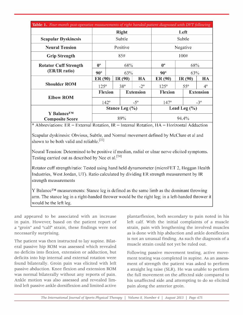
Objective Tests and Measures During the data collection the patient participated in a variety of tests as part of the standard procedure for the ongoing UCL study. The following measure-ments were taken as part of the data collection pro-cess, but were not necessarily part of the differential diagnosis for the patients’ lower extremity com-plaints. (Table 1) Shoulder passive ROM was assessed in supine. Measurements of glenohumeral internal and external rotation and shoulder horizontal adduc-tion were taken in a manner previously described in the literature.21 Elbow range of motion (ROM) was an active motion assessment completed in sitting. All ROM measurements were taken using a goniometer with an attached level. Rotator cuff strength testing was completed and carried out with resisted inter-nal and external rotation at 0 degrees and again at 90 degrees using a hand held dynamometer (micro-FET 2, Hoggan Health Industries, West Jordan, UT). Grip strength testing was carried out with a handheld dynamometer. All range of motion and strength test-ing was completed bilaterally. Scapular dyskinesis assessment was done by visual assessment of active resisted shoulder flexion as previously described in the literature.22 Single leg balance testing was
completed with the Y-Balance Test™ and compos-ite scores were calculated for each leg.23 The stance leg was identified as the same leg as the dominant throwing arm. The patient described in this report is right handed; as such his right leg is designated as his stance leg. Lastly upper limb neural tension testing was completed in supine, assessing neural mobility of the median, ulnar, and radial nerves in a manner consistent with those reported by Nee et al.24
Following the data collection portion of the visit, examination of the patients’ lower extremity com-plaints of groin pain was carried out. Table 2 describes the algorithm that was used as part of the decision making process in referring the patient for further testing. It is the thought of the authors and the phi-losophy of this clinic to assess functional move-ment first as part of the examination process.25-27
The assessment began by watching the patient walk, which revealed no gross abnormalities. Following this assessment the patient was asked to perform a double leg squat.26,27 The patient was able to perform a complete squat with adequate control and depth; however, pain was noted along the left proximal anterior thigh during the lowering portion of the movement. The patient was then asked to perform a single leg squat which he was unable to successfully perform on either leg.28 At this time the functional movement portion of the exam was complete. Based on the findings of pain during squatting, no change in the potential differential diagnosis was made.
The patient was then instructed to sit at the edge of the table with his leg positioned at 90 degrees of hip flexion and knee flexion. In this position a brief sensory screen was completed which was negative; reflex testing was also unremarkable. In this position muscle testing of the hip flexors, knee extensors and flexors, and ankle musculature was carried out.29 The rationale for using these muscle tests was to elicit a painful response and to screen for specific muscle weakness. All muscle testing on the right LE tested at 5/5 strength. Left LE hip flex-ion, knee extension, and ankle dorsiflexion were <3+/5 with pain noted on each test. With hip flex-ion and knee extension, pain was noted along the anterior thigh. With resisted ankle dorsiflexion pain was noted along the calf musculature. At this time the weakness that was found was of some concern
The International Journal of Sports Physical Therapy | Volume 8, Number 4 | August 2013 | Page 475
and appeared to be associated with an increase in pain. However, based on the patient report of a “groin” and “calf” strain, these findings were not necessarily surprising.
The patient was then instructed to lay supine. Bilat-eral passive hip ROM was assessed which revealed no deficits into flexion, extension or adduction, but deficits into hip internal and external rotation were found bilaterally. Groin pain was elicited with left passive abduction. Knee flexion and extension ROM was normal bilaterally without any reports of pain. Ankle motion was also assessed and revealed lim-ited left passive ankle dorsiflexion and limited active
plantarflexion, both secondary to pain noted in his left calf. With the initial complaints of a muscle strain, pain with lengthening the involved muscles as is done with hip abduction and ankle dorsiflexion is not an unusual finding. As such the diagnosis of a muscle strain could not yet be ruled out.
Following passive movement testing, active move-ment testing was completed in supine. As an assess-ment of strength the patient was asked to perform a straight leg raise (SLR). He was unable to perform the full movement on the affected side compared to his unaffected side and attempting to do so elicited pain along the anterior groin.
Table 1. Four-month post-operative measurements of right handed patient diagnosed with DVT following UCL reconstruction

The International Journal of Sports Physical Therapy | Volume 8, Number 4 | August 2013 | Page 476
Table 2. Differential diagnosis algorithm for determination of DVT

The International Journal of Sports Physical Therapy | Volume 8, Number 4 | August 2013 | Page 477
Summary of Key Findings:At the ankle, the patient demonstrated weakness into dorsiflexion, and limited ROM into dorsiflexion and plantar flexion with pain noted on both movements. The ankle presentation could potentially be contrib-uted to a strain. At this time the patient demonstrated pain and weakness with hip flexion, resulting in an inability to perform a SLR. The profound hip weak-ness in an otherwise healthy young male was trou-bling. However, the lumbar spine and sacroiliac joint had still not been ruled out, and could potentially be the underlying cause of these findings.30 As such an SIJ and low back screen was initiated. Leg length was assessed by checking malleolar alignment, Illiac crest height, and ASIS height. The screen of this area was negative for any relative leg length discrepancy. How-ever in doing this evaluation, grasping the patients’
malleoli was done to assess leg length and revealed significant swelling around the left calf musculoten-dinous junction when compared to the right. This was not noted earlier in the examination as this was the first time the patients’ ankle was physically grasped.
At this time, DVT was added to the potential differ-ential diagnosis list. However, a low back screen was still completed to rule out this area and any poten-tial referred pain.31,32 This screen included a passive SLR, slump test, and posterior to anterior lumbar mobilizations, all of which were negative. A review of the findings can be found in Table 3.
While a calf strain could still have explained his calf complaints, no obvious findings helped to explain his hip complaints. At this time, further examina-tion of the lower leg edema was warranted as a
Table 3. Findings from lower extremity exam.

The International Journal of Sports Physical Therapy | Volume 8, Number 4 | August 2013 | Page 478
DVT or multiple DVT at different points along his leg could explain all of his symptoms.15,33-36 As such, further examination was carried out. Further palpa-tion revealed that the entire left lower leg was warm compared to the right lower extremity. Additionally, palpation along the deep venous system was tender through the calf veins and more proximally into pop-liteal veins. The combination of these findings sug-gested a potentially more serious condition.15,33-36
Advanced Differential DiagnosisAt this time the findings from the examination revealed significant left lower extremity pain, weakness, and swelling with temperature changes. These findings together increased the possibility of a potential DVT. Further testing was performed to rule-in or out the potential DVT. Objective edema measures were taken approximately 10 cm below the tibial tuberosity and revealed a 2 cm difference bilaterally.15,33-36 Despite the poor clinical utility of the Homan’s sign,16,17 it was administered and found to be positive on the affected side and negative on the unaffected side. Supple-mentary testing included passive forced dorsiflexion which provoked pain in the patient’s left calf.
At this time it was believed that the patient may potentially have had a DVT and was in need of a referral for further work up. The clinical findings of weakness, edema, tenderness to palpation, a positive Homan’s sign and pain with passive dorsiflexion were all considered in the decision making process. Based on these findings and having ruled out other likely diagnoses, the patient was immediately referred back to the orthopedic surgeon for a potential DVT.
Immediate care of the patientThe patient was then examined by the orthopedic surgeon who also noted a two centimeter difference in calf girth, and found a positive Homan’s Sign. Although the patient had previously undergone a Doppler study within two weeks of his UCL sur-gery due to localized calf swelling, the current pre-sentation warranted additional testing, thus, he was immediately referred to the emergency department for more in-depth analysis.
The patient was admitted to the hospital for Doppler testing which revealed an extensive non-inclusive clot throughout his calf and lower thigh. Pulmonary
studies indicated minor pulmonary emboli. He was placed on a combination of Lovenox and Coumadin. The patient remained in the hospital for two weeks as his condition was monitored by a hematologist. After the two weeks he was discharged from the hospital, but continued to receive Coumadin for 6 months following the incident. He reported back to the clinic for his 6 month follow up and appeared in good health.
DISCUSSIONProper screening for potentially serious conditions in the outpatient setting is an important clinical skill that all outpatient therapists need to implement. As the transition to a direct access environment contin-ues in most states, improving these skills become even more important to ensure safe and comprehen-sive care of all patients. In this case, what began as a simple follow up visit for a completely unrelated incident, transitioned into an emergent referral to the emergency department.
The diagnosis of a DVT in an otherwise healthy, male, high school pitcher following an upper extremity surgery is very unusual. However, this case report highlights the importance of performing a thorough differential diagnosis with every patient in order to assist with ruling in or out suspected diagnoses. Despite the poor reliability and validity of the Homan’s sign16,17 other clinical signs such as swelling, warmth, and edema in addition to muscle testing, were used as an adjunct to special testing to assist with making a clinical decision regarding referral. In this case these signs, symptoms, and spe-cial tests were able to accurately identify a DVT.
Diamond and Macciocchi37 examined the predic-tive value of lower extremity edema, increased skin temperature, fever, and lower limb girth asymme-try in the diagnosis of a DVT. They found that an asymmetrical limb girth greater than 2.5 cm had the highest positive predictive value of 0.66. While the clinician should not rely on this alone, it may help the clinical decision making process. In the cur-rent case, the patient had a limb girth asymmetry of only 2 cm, so relying exclusively on the findings of Diamond and Macciocchi37 could have resulted in missing an emergent referral. This is not to suggest that the findings of Diamond and Macciocchi are not

The International Journal of Sports Physical Therapy | Volume 8, Number 4 | August 2013 | Page 479
useful, but to point out that relying solely on one test or finding may result in a missed diagnosis.
Despite the accurate identification of a DVT in this case, controversy exists regarding the use of clinical findings alone to consistently and accurately iden-tify a DVT. As such, Wells et al15,33-36 have developed a clinical prediction rule, to assist the clinician in identifying a DVT.15,33-36 Table 4 outlines Well’s clini-cal prediction rule (CPR). If this CPR was applied to the patient presented in this case, he would have scored a 3 indicating a high probability of a DVT. This may indicate that this CPR alone could be adequate for the accurate identification of a DVT. However, in a similar case report by Theiss et al,20 a DVT was diagnosed in a healthy 21 year old marathon runner when the score on this CPR would have been a nega-tive 1, indicating a low likelihood of a DVT. Again, this is not to suggest that the Well’s CPR is a poor tool, rather, this highlights the importance of relying on more than just clinical findings or a CPR to make an accurate diagnosis. Likely, it is a combination of clinical findings and the utility of a CPR that would most consistently lead to an accurate diagnosis.
In addition to the CPR presented by Wells et al15,33-36 other risk factors to assist in the diagnosis of a DVT exist and can potentially help with clinical decision making. Fink and Stoneman19 identified a DVT in a 21 year old athletic male with knee pain.19 As part of their clinical reasoning they used a list of 21 poten-tial risk factors which included a thorough history and clinical findings of increased skin temperature, fever, asymmetrical lower limb girth, and edema to help with accurate identification. While the list presented in their case is more comprehensive than the CPR presented by Wells15,33-36 and included blood work, physical therapists in the outpatient setting are often limited to subjective and objective tests that can be performed in the clinic.
Accurate identification would be most easily and correctly made with advanced imaging and medical testing such as ultrasound imaging, D-dimer testing, and contrast venography. However, access to these tests in an outpatient physical therapy environment is not readily available. Additionally, these tests can be expensive and should be reserved for situations in which a DVT is likely. In our case, a DVT was sus-pected by both the physical therapist and referring surgeon, thus an immediate referral for additional testing was made.
At this time there is no gold standard clinical test or CPR that will consistently and accurately iden-tify a DVT in the outpatient physical therapy setting. Thus, thorough history taking and good clinical rea-soning skills are essential in the proper screening, differential diagnosis, and referral. In this case, thor-ough clinical examination and referral skills were vital in providing a definite diagnosis and poten-tially lifesaving referral. This case report highlights the importance of using all clinical findings in the decision-making process. Relying solely on just one of the following: subjective information, girth asym-metries, Homan’s sign, or other clinical findings in this situation may have led to an incorrect or missed diagnosis. This case involved a cumulative effect of all findings and sound reasoning which led to the appropriate referral.
The authors would like to see continued research published in order to assist clinicians, especially those in the outpatient setting, to correctly screen for DVT. In our opinion the Well’s clinical prediction
Table 4. Wells DVT classifi cation24-28

The International Journal of Sports Physical Therapy | Volume 8, Number 4 | August 2013 | Page 480
rule15,33-36 is currently one of the better tools available to assist clinicians. While the authors’ understand that no tool will ever demonstrate 100% sensitivity and specificity, the need to validate these tools with a variety of patients and in varied settings is needed. Additionally, continued publications of case series and studies, such as the one presented in this paper, can help better expose the risk of DVT in the outpa-tient setting, and can serve as a reminder to always screen for serious medical conditions, regardless of the patient age or condition.
CONCLUSIONThis case report presents an unusual scenario in which an apparent groin strain in a young, otherwise healthy baseball pitcher was diagnosed as a DVT. The results underscore the importance of careful screening and differential diagnosis in all patients, regardless of the perceived certainty or musculo-skeletal presentation of the diagnosis. Clinical tests like the Homan’s sign, findings of asymmetrical girth measures, edema, increased skin tempera-ture, and fever may be helpful in the diagnosis of possible DVT when used in conjunction with one another. Additionally, clinical prediction rules like the one presented by Wells et al15,33-36 may be a use-ful tool to assist clinicians in the accurate identifica-tion of a DVT. In general, increased knowledge of risk factors, such as the ones presented by Wells and by Fink15,19,33-36 and an increased degree of suspicion even in younger patients, may assist the clinician with early and accurate identification of a DVT.
REFERENCES 1. Anand SS, Wells PS, Hunt D, Brill-Edwards P, Cook
D, Ginsberg JS: Does this patient have deep vein thrombosis? JAMA: the journal of the American Medical Association 1998; 279(14): 1094-1099.
2. Autar R: Nursing assessment of clients at risk of deep vein thrombosis (DVT): the Autar DVT scale. J Adv Nurs 1996; 23(4): 763-770.
3. Riddle D, Hillner B, Wells P, Johnson R, Hoffman H, Zuelzer W: Diagnosis of lower-extremity deep vein thrombosis in outpatients with musculoskeletal disorders: a national survey study of physical therapists. Phys Ther 2004; 84(8): 717-728.
4. Stein PD, Matta F, Dalen JE: Is the campaign to prevent VTE in hospitalized patients working? CHEST 2011; 139(6): 1317-1321.
5. Janku GV, Paiement GD, Green HD: Prevention of venous thromboembolism in orthopaedics in the United States. Clin Orthop 1996; 325: 313.
6. Anderson FA, Spencer FA: Risk factors for venous thromboembolism. Circulation 2003; 107(23 suppl 1): I-9-I-16.
7. Clagett GP, Anderson FA, Heit J, Levine MN, Wheeler HB: Prevention of venous thromboembolism. CHEST Journal 1995; 108(4_Supplement): 312S-334S.
8. Dahl OE, Gudmundsen TE, Haukeland L: Late occurring clinical deep vein thrombosis in joint-operated patients. Acta Orthopaedica 2000; 71(1): 47-50.
9. Agnelli G, Sonaglia F: Prevention of venous thromboembolism. Thromb Res 2000; 97(1): 49-62.
10. Perrier A, Desmarais S, Miron MJ, et al.: Non-invasive diagnosis of venous thromboembolism in outpatients. Lancet 1999; 353(9148): 190-195.
11. Tapson V, Carroll B, Davidson B, et al.: The diagnostic approach to acute venous thromboembolism. Clinical practice guideline. American Thoracic Society. Am J Respir Crit Care Med 1999; 160(3): 1043.
12. Prandoni P, Mannucci PM: Deep-vein thrombosis of the lower limbs: diagnosis and management. Clinical Haematology 1999; 12(3): 533-554.
13. Akman MN, Cetin N, Bayramoglu M, Isiklar I, Kilinc S: Value of the D-dimer test in diagnosing deep vein thrombosis in rehabilitation inpatients1. Arch Phys Med Rehabil 2004; 85(7): 1091-1094.
14. Delis K, Hunt N, Strachan R, Nicolaides A: Incidence, natural history and risk factors of deep vein thrombosis in elective knee arthroscopy. Thromb Haemost 2001; 86(3): 817-821.
15. Wells P: Integrated strategies for the diagnosis of venous thromboembolism. Thromb Haemost 2007; 5(s1): 41-50.
16. Heim SW, Schectman JM, Siadaty MS, Philbrick JT: D-dimer testing for deep venous thrombosis: a metaanalysis. Clinical chemistry 2004; 50(7): 1136-1147.
17. O’Donnell TA, WM Athanasoulis ,CA, Millan ,VG Callow , AD: Diagnosis of deep venous thrombosis in the outpatient by venography. surgical gynecology obstetrics journal 1980; 150: 69-74.
18. Horsburgh J: Case report of a deep vein thrombosis in the femoral vein, an atypical presentation. Clinical Chiropractic 2004; 7(3): 120-126.
19. Fink ML, Stoneman PD: Deep vein thrombosis in an athletic military cadet. J Orthop Sports Phys Ther 2006; 36(9): 686-697.
20. Theiss J, Fink M, Gerber J: Deep vein thrombosis in a young marathon athlete. J Orthop Sports Phys Ther 2011; 41(12): 942-947.

The International Journal of Sports Physical Therapy | Volume 8, Number 4 | August 2013 | Page 481
21. Wilk KE RM, Macrina LC: Glenohumeral internal rotation measurements differ depending on stabilization techniques. Sports Health 2009; 1(2): 131-136.
22. McClure P, Tate AR, Kareha S, Irwin D, Zlupko E: A clinical method for identifying scapular dyskinesis, part 1: reliability. J Athl Train 2009; 44(2): 160.
23. Plisky PJ RM, Kaminski TW, Underwood FB: Star Excursion Balance Test as a Predictor of Lower Extremity Injury in High School Basketball Players. J Orthop Sports Phys Ther 2006; 36(12): 911-919.
24. Nee R JG, Vicenzino B, Coppieters M: The validity of upper limb neurodynamic tests for detecting eripheral neuropathic pain. J Sports Phy ther 2012; 42(5): 413-424.
25. Kisel K PP, Butler R: Functional movement test scores improve following a standarized off-season intervention program in professional football players. Scand J Med Sci Sports 2011; 21: 287-292.
26. Cook G BL, Hoogenboom B: Pre-Participation screening: The Use of Fundamental Movements As An Assessment Of Function-Part 2. N Am J Sports Phys Ther 2006; 1(3): 132-139.
27. Cook G BL, Hoogenboom B: Pre-Participation screening: The Use of Fundamental Movements As An Assessment Of Function-Part 1. N Am J Sports Phys Ther 2006; 1(2): 62-72.
28. Crossley KM ZW, Schache AG, Bryant A, Cowan SM: Performance on the SIngle-Leg Squat indicates Hip Abductor Muscle Function. AM J Sports Med 2011; 39(4): 866-874.
29. Kendall FP ME, Provance PG: Muscles Testing anf Function, 4 ed. Philadelphia: Lippincott Williams & Williams, 1993. (JP B, ed.)
30. Cibulka MT KR: Clinical usefulness of a Cluster of Sacroiliac Joint Tests in Patients With and Without
Low Back Pain. J Orthop Sports Phys Ther 1999; 29(2): 83-92.
31. Childs JD FJ, Flynn TW, Irrgang JJ, Johnson KK, Majkowski GR, Delitto A.: A Clinical Prediction Rule to Identify Pateints with Low Back Pain Most Likely To Benefi t from Spinal Manipulation: A validation study. Ann Intern Med 2004; 141(12): 920-928.
32. Clelan JA FJ, Whitman JM, Childs JD, Palmer JA: The Use of a lumbar Spine Manipulation Technique by Physical Therapists in Patients Who Satisfy a Clinical Prediction Rule: A Case Series. J Orthop Sports Phys Ther 2006; 36(4): 209-214.
33. Wells PS, Hirsh J, Anderson DR, et al.: Accuracy of clinical assessment of deep-vein thrombosis. Lancet 1995; 345(8961): 1326-1330.
34. Wells PS, Anderson DR, Ginsberg J: Assessment of deep vein thrombosis or pulmonary embolism by the combined use of clinical model and noninvasive diagnostic tests. Seminars in thrombosis and hemostasis, 2000.
35. Wells PS, Anderson DR, Rodger M, et al.: Derivation of a Simple Clinical Model to Categorize Patients Probability of Pulmonary Embolism-Increasing the Models Utility with the SimpliRED D-dimer. Thromb Haemost 2000; 83(3): 416-420.
36 Wells PS, Owen C, Doucette S, Fergusson D, Tran H: Does this patient have deep vein thrombosis? J Am Med Assoc 2006; 295(2): 199-207.
37. Diamond PT, Macciocchi SN: Predictive Power of Clinical Symptoms in Patients With Presumptive Deep Venous Thrombosis1. Am J Phys Med Rehabil 1997; 76(1): 49-51.
Copyright © 2022 FDOKUMEN