Animal Models of Thrombosis and Hemorrhagic Diseases
220
FROM THE ARCHIVES Find Similar Titles More Information Visit the National Academies Press online and register for... Distribution, posting, or copying of this PDF is strictly prohibited without written permission of the National Academies Press. Unless otherwise indicated, all materials in this PDF are copyrighted by the National Academy of Sciences. To request permission to reprint or otherwise distribute portions of this publication contact our Customer Service Department at 800-624-6242. Copyright © National Academy of Sciences. All rights reserved. Instant access to free PDF downloads of titles from the 10% off print titles Custom notification of new releases in your field of interest Special offers and discounts NATIONAL ACADEMY OF SCIENCES NATIONAL ACADEMY OF ENGINEERING INSTITUTE OF MEDICINE NATIONAL RESEARCH COUNCIL This PDF is available from The National Academies Press at http://www.nap.edu/catalog.php?record_id=19903 Pages 221 Size 5 x 9 ISBN 0309333962 Animal Models of Thrombosis and Hemorrhagic Diseases (1976) Institute of Laboratory Animal Resources; National Academy of Sciences; National Heart and Lung Institute
-
Upload
khangminh22 -
Category
Documents
-
view
0 -
download
0
Transcript of Animal Models of Thrombosis and Hemorrhagic Diseases

FRO
M T
HE
ARCH
IVES
Find Similar Titles More Information
Visit the National Academies Press online and register for...
Distribution, posting, or copying of this PDF is strictly prohibited without written permission of the National Academies Press. Unless otherwise indicated, all materials in this PDF are copyrighted by the National Academy of Sciences.
To request permission to reprint or otherwise distribute portions of thispublication contact our Customer Service Department at 800-624-6242.
Copyright © National Academy of Sciences. All rights reserved.
Instant access to free PDF downloads of titles from the
10% off print titles
Custom notification of new releases in your field of interest
Special offers and discounts
NATIONAL ACADEMY OF SCIENCES
NATIONAL ACADEMY OF ENGINEERING
INSTITUTE OF MEDICINE
NATIONAL RESEARCH COUNCIL
This PDF is available from The National Academies Press at http://www.nap.edu/catalog.php?record_id=19903
Pages221
Size5 x 9
ISBN0309333962
Animal Models of Thrombosis and Hemorrhagic Diseases (1976)
Institute of Laboratory Animal Resources; National Academy of Sciences; National Heart and Lung Institute

DISCRIMINATION PROHIBITED- Title VI of the Civil Rights Act of 1964 states: "No person in the United States shall, on the ground of race, color, sex, age, or national origin, be excluded from participation in, be denied the benefits of, or be subjected to discrimination under any program or activity receiving Federal financial assistance." Therefore, the Division of Blood Diseases and Resources, NHl..ll , like every program or activity receiving financial assistance from the Department of Health, Education, and Welfare, must be operated in compliance with this law.
Copyright © National Academy of Sciences. All rights reserved.
Animal Models of Thrombosis and Hemorrhagic Diseaseshttp://www.nap.edu/catalog.php?record_id=19903
Animal Models of Thrombosis and Hemorrhagic Diseases
co-sponsored by the NATIONAL HEART AND LUNG INSTITU'IE
and the INSTITUTE OF LABORATORY ANIMAL RESOURCES NATIONAL ACADEMY OF SCIENCES
U.S. DEPARTMENT OF HEALTH, EDUCATION, AND WELFARE Public Health Service National Institutes of Health
DHEW Publication No. (NIH) 76-982
NAS-NAE
AUG 16 1976
LIBRARY
Copyright © National Academy of Sciences. All rights reserved.
Animal Models of Thrombosis and Hemorrhagic Diseaseshttp://www.nap.edu/catalog.php?record_id=19903

NOTICE: The project that is the subject of this report was approved by the Governing Board of the National Research Council, whose members are drawn from the Councils of the National Academy of Sciences, the National Academy of Engineering, and the. Institute of Medicine. The members of the committee responsible for the report were chosen for their special competences and with regard for appropriate balance.
This report has been reviewed by a group other than the authors according to procedures approved by a Report Review Committee consisting of members of the National Academy of Sciences, the National Academy of Engineering, and the Institute of Medicine.
The primary support for this workshop and publication was provided by Contract NIH 74-C-799 with the Division of Blood Diseases and Resources, National Heart and Lung Institute. Additional support was provided by Contract RC-IR with the American Cancer Society; Contract C-310, Task Order 173 with the National Science Foundation; Contracts PH-43-64-44, Task Order 12, and NOl-RR-5-2128 with the Animal Resources Branch, National Institutes of Health; Contract APHIS 12-16-140-155-91 with the U. S. Department of Agriculture; Contracts NOl-CP-45617 and NOl-CP-33338 with the National Cancer Institute, NIH; Contract E(ll-1)-3369 with the Energy Research and Development Administration; Contract N0014-76-C-0242 with the Office of Naval Research.
Ubrllry of CODp'ell Cataloging in Publication Data
Workshop on Animal Models of Thrombosis and Hemorrhagic Diseases, National Academy of Sciences, 1975.
. Animal models of thrombosis and hemorrhagic diseases.
Bibliography: p. 1. Hemorrhagic diseases-Congresses. 2. Thrombosis-Congresses. 3. Diseases-
Animal models-Congresses. I. National Research Council. Institute of Laboratory Animal Resources. II. Title. [DNLM: 1. Animals, Laboratory-Congresses. 2. Throm-, bosis-Congresses. 3. Hemorrhage-Congresses. 4. Disease models, AnimalCongresses. WH312 W926a 1975] RC636.W67 1975 616.1'35'027 76-10652
Available from Institute of Laboratory Animal Resources National Academy of Sciences 2101 Constitution Avenue, N.W. Washington, D.C. 20418
Printed in the United States of America
Copyright © National Academy of Sciences. All rights reserved.
Animal Models of Thrombosis and Hemorrhagic Diseaseshttp://www.nap.edu/catalog.php?record_id=19903

Preface
The Workshop on Animal Models of Thrombosis and Hemorrhagic Diseases was developed by the Institute of Laboratory Animal Resources and held at the National Academy of Sciences, Washington, D.C., March 12-13, 1975, under terms of a contract with the Division of Blood Diseases and Resources, National Heart and Lung Institute, National Institutes of Health. Participants in the workshop, who included specialists from the United States, Canada, the Netherlands, and England, examined the past, present, and future uses and limitations of animal models in scientific investigations of thrombosis and hemorrhagic diseases. The workshop was intended to facilitate exchange of information and establish guidelines for maintaining diffei;ent types of stocks and strains and to assure their availability to qualified investigators. It was further intended that the workshop result in a series of recommendations to serve various granting agencies as a basis for formulating policy.
Although a substantial amount of comparative work is being done on thrombosis and hemorrhagic diseases, the workshop was deemed necessary to overcome the lack of communication among specialists in the various scientific disciplines. It was deemed essential that information concerning the recognition, maintenance, and treatment of animals with hemorrhagic and thrombotic diseases be made available to investigators using them. A better understanding of hemostasis through education, practical experience, and availability of materials would enable more effective diagnosis, management, and treatment of ani-
iii
Copyright © National Academy of Sciences. All rights reserved.
Animal Models of Thrombosis and Hemorrhagic Diseaseshttp://www.nap.edu/catalog.php?record_id=19903

iv Preface
mal~ with these disorders. The workshop offered a means of lntemational, as well as national, communication among veterinarians, physicians, and allied scientists of information about the similarities and differences in clotting mechanisms of man and animals.
The workshop was divided into seven sessions, six of which were devoted to specific topics. The seventh was divided into four working groups, charged with providing detailed evaluations of various animal models (present and future), dealing with problem areas, and recommending remedies. The working groups were composed of participants in the workshop, including the public. l)le workshop results (ii;icluding the seven sessions) were then evaluated by the Committee on Animal Models for Thrombosis and Hemorrhagic Diseases, which generated the series of recommendations at the end of this volume designed to advance health research and resources in the field of thrombosis and hemorrhagic disease.
The Committee wishes to acknowledge the special assistance of the four working group co-chairmen who helped prepare Part VI of these proceedings: Dr. Louis M. Aledort, Vice Chairman, Department of Medicine, Mount Sinai School of Medicine, New York; Dr. Daniel Deykin, Chief, Medical Service, Veterans Administration Hospital, Boston; Dr. Albert M. Jonas, Chief, Section of Comparative Medicine, Yale University School of Medicine, New Haven; and Dr. Edwin Salzman, Professor of Surgery, Harvard Medical School, Boston.
W. Jean Dodds, D.V.M. Chairman
Copyright © National Academy of Sciences. All rights reserved.
Animal Models of Thrombosis and Hemorrhagic Diseaseshttp://www.nap.edu/catalog.php?record_id=19903

COMMITTEE ON ANIMAL MODELS FOR THROMBOSIS AND HEMORRHAGIC DISEASES
w. JEAN DODDS, Chairman, Associate Research Scientist, New York State Department of Health, Albany
E. J. WALTER BOWIE, Mayo Clinic, Rochester, Minnesota KENNETH M. BRINKHous., Department of Pathology, University <;>f
North Carolina School of Medicine, Chapel Hill ROBERT w. BULL, Department of Medicine. Michigan State University'
East Lansing CHARLES A. OWEN, JR., Mayo Clinic, Ro~hester, Minnesota GEORGE A. PADGETT, Department of Veterinary Pathology,
Washington State University, Pullman SCOTT N. SWISHER, Department of Medicine, Michigan State Univer
sity, East Lansing LEO A. WHITEHAIR, Animal Resources Branch, National Institutes of
Health, Bethesda, Maryland
INSTITUTE OF LABORATORY ANIMAL RESOURCES
LYDIA E. KOUTZE, Administrative Assistant CARMELA F. JACKSON' Secretary ELL~ c. JACKSON, Secretary
v
Copyright © National Academy of Sciences. All rights reserved.
Animal Models of Thrombosis and Hemorrhagic Diseaseshttp://www.nap.edu/catalog.php?record_id=19903

Copyright © National Academy of Sciences. All rights reserved.
Animal Models of Thrombosis and Hemorrhagic Diseaseshttp://www.nap.edu/catalog.php?record_id=19903
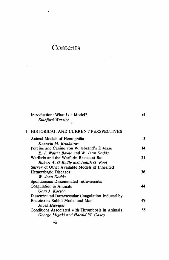
Contents
Introduction: What Is a Model? xi Stallford VVessler
I HISTORICAL AND CURRENT PERSPECTIVES
Animal Models of Hemophilia 3 Kenneth M. Brinkhous
Porcin~ and Canine. von Willebrand's Disease 14 E. J. VValter Bowie and VV. Jean Dodds
Warfarin and the Warfarin-Resistant Rat 21 Robert A. O'Reilly and Judith G. Pool
Survey of Other Available Models of Inherited Hemorrhagic Diseases 36
VV. Jean Dodds Spontaneous Disseminated lntravascular Coagulation in Animals 44
Gary J. Kociba Disseminated Intravascular Coagulation Induced by Endotoxin: Rabbit Model and Man 49
Jacek Hawiger Conditions Associated with Thrombosis in Animals 55
George Migaki and Harold VV. Casey
vii
Copyright © National Academy of Sciences. All rights reserved.
Animal Models of Thrombosis and Hemorrhagic Diseaseshttp://www.nap.edu/catalog.php?record_id=19903
II COMPARATIVE HEMOSTASIS
Hemostasis in Mammals 69 Christine Hawkey
Blood Coagulation in the Horseshoe Crab (Limulus polyphemus ): A Model for Mammalian Coagulation and Hemostasis 87
Jack Levin Discussion: A Comment on the Ultrastructure of Amebocytes from the Horseshoe Crab (J.imulus polyphemus) 97
James G. White Discussion: Nature of the Contents of Large
101 Granules of Umulus Amebocytes Frank A. Belamarich
III AREAS TO BE DEVELOPED
Canine lmmunohematology 105 Robert W. Bull
Cost-Effectiveness of Developing Animal Models 110 Albert M. Jonas
Cost-Effectiveness of Replacement Therapy in Animal Models of Hemorrhagic Disorders: Lessons from Experience with H. sapiens 114
Peter H. Levine Cost-Effectiveness: Utilization of a Sperm Bank in Animal Breeding Colonies 118
Stephen W. J. Seager Requirements for Diagnostic Procedures Used in the Definition and Interpretation of Animal Models of Hemorrhage and Thrombosis 122
Jan J. Veltkamp, Allan T. van Oosterom, and Johan A. van der Does
Detection and Comparison of Fibrinogen and Its Derivatives in Different Species by the Staphylococcal Clumping Test 127
Jacek Hawiger Atherosclerosis 132
Sean Moore
viii
Copyright © National Academy of Sciences. All rights reserved.
Animal Models of Thrombosis and Hemorrhagic Diseaseshttp://www.nap.edu/catalog.php?record_id=19903

IV PUBLIC RELATIONS
. How To Find an Animal Model of Human Disease 147 George A. Padgett and G. A. Hegreberg
Humane Considerations for Aninial Mod~ls 151 Christine Stevens
Legislative Considerations for Studies with Animal Models 159 Louis M. Aledort and John T. Grupenhoff
v COMPARATIVE MEDICINE OVERVIEW
Animal Models in Human Medicine 169 Robert W. Prichard
The Need for Human Experimentation 173 Laurence A. Harker
VI PROJECTED NEEDS AND RECOMMENDATIONS
Working Group A: Preservation of Unique Models 189 Working Group B: Developmental Needs 191 Working Group C: Diagnostic Methods of
Identifying Models 194 Working Group D: Mechanics of Support and
Cost-Effectiveness 196
Contributors and Working Group Co-Chairmen 199
Participants 201
ix
Copyright © National Academy of Sciences. All rights reserved.
Animal Models of Thrombosis and Hemorrhagic Diseaseshttp://www.nap.edu/catalog.php?record_id=19903
STANFORD WESSLER, M.D.
Introduction: What Is a Model?
The Oxford English Dictionary defines a model as something that accurately resembles something else. Complex mathematical models are coming into increasing vogue, for example, in economics, industry, and meteorology to predict outcome in advance of the event, though arguments over their efficacy abound.
No dictionary describes an animal model, much less an animal model of disease, and so we can define it for our purposes here as· a living organism with an inherited, naturally acquired, or induced pathological process that in one or more respects closely resembles the same phenomenon occurring in man. Animal models, in this sense, never provide final answers but offer only approximations, for no single animal model can ever duplicate a disease in man. Thus, animal models should not be expected to be ideal, nor to be universally suited to all forseeable uses. On the other hand, for a model to be a good one, it must provide a new insight, have relevance to a particular problem and respond predictably. 1
In his Introduction to the Study of Experimental Medicine Claude Bernard stated: "I not only conclude that experiments made on animals from the physiological, pathological and therapeutic points of view have results that are applicable to theoretic medicine, but I think that without such comparative study of animals, practical medicine can
Supported in pari by Grant No. HL 1833 from the National Heart and Lung Institute, National Institutes of Health.
xi
Copyright © National Academy of Sciences. All rights reserved.
Animal Models of Thrombosis and Hemorrhagic Diseaseshttp://www.nap.edu/catalog.php?record_id=19903
xii STANFORD WESSLER
never acquire a scientific character," and he goes on to quote Buff on as saying that if animals did not exist, man's nature would be still more incomprehensible. 2
I mention Bernard because there is resistance to using animal models. Individuals of this bent refer to Pope's line: "The proper study of mankind is man"; or to Koch's admonition: "Gentlemen, never forget that mice are not human beings." They also point to the use of "experiments of nature" among human subjects and to the ability to test hypotheses as well as therapies through human clinical trials.
Man, however, is not only a slow-breeding creature with a long life and few offspring but, in terms of heterozygosity, is unmatched in the animal kingdom. These are motives enough to search out animal models, aside from the issues offeasibility, of rights of individuals, and of cost effectiveness.
There are special reasons for desiring animal models in the areas of thrombosis and hemorrhage. For one thing, we are in the midst of an explosion of in vitro knowledge within the field of hemostasis, and animal models can help separate the trivia from the observations of potential intrinsic biological importance as well as from those of potential clinical value. The animal model can thus serve as a convenient and at times essential bridge between an understanding of nature, on the one hand, and its conquest, on the other. It is also clear that problems associated with clinical trials may be alleviated by meaningful experimentation in animal models. The record of clinical trials in thromboembolic disease in the past 25 years has been a sad oneattributable, largely, to our naivete in methodology. Many of the difficulties are the result of a profound underestimation of the size of the required trial population. 3
Why do we want animal models of hemostasis? To seek or create an animal model of a disease one must have an understanding of the disease process itself, an appreciation of the unanswered questions to be resolved, and an awareness of the state of the art.
Let me use thrombosis as an. illustration. One can divide this subject today into four parts: arterial, venous, microcirculatory, and foreign surface thrombosis. Each of these four forms of intra vascular coagulation is different and requires a different model. Arterial thrombosis is so intimately related to atherosclerosis that if the latter condition could be prevented, arterial thromboembolism, as a major contributor to arterial occlusion, would be essentially eliminated. On the other hand, the question of whether thrombosis causes atherosclerosis is still unanswered; the likelihood that deposits of platelet debris and fibrin contribute to the growth of the atherosclerotic lesion is real; and, .
Copyright © National Academy of Sciences. All rights reserved.
Animal Models of Thrombosis and Hemorrhagic Diseaseshttp://www.nap.edu/catalog.php?record_id=19903
lntro4uction: What ls a Model? xiii
finally, the sudden gelation of fluid blood on an atherosclerotic plaque often is the terminal event converting a partially narrowed vessel into a total vascular occlusion.
Experimental atherosclerosis has been produced in chickens, dogs, pigs, and nonhuman primates, usually through dietary manipulation. Atheroma that occludes 60 percent of the lumen of the coronary arteries has been induced in primates, and further dietary alteration has resulted in regression. In thrombus formation, the intrinsic anatomy of the atherosclerotic lesion may be even more jmportant than the extent of the obstruction itself. Thus, atheromatous lesions that undergo hemorrhage and ulceration with only moderate narrowing may be more conducive to thrombosis than lesions producing 90 percent obstruction without ulceration. Preliminary, anecdotal studies have, in fact, suggested that ulcerative, atheromatous lesions associated'with thrombosis can be produced in nonhuman primates; but, unless new techniques are uncovered, these experimental models may suffer from the fact that experiments with them require years rather than months to reach fruition. It may also be of significance that although the greatest obstructions have been achieved in the rhesus monkey, the best correlations with levels of procoagulant concentrations in man have been observed in the baboon.
A second illustration is venous thromboembolism, in which investigators are concerned with the prediction of thrombosis. This has led not only to a codification of "risk" factors' but also to efforts to identify alterations in the circulating blood that reflect "hypercoagulability." Although the concept of hypercoagulability is more than three centuries old, evidence for its existence in Q1an has remained elusive. The persistence of the term reflects not so much acceptable evidence of its existence but rather its usefulness, conceptually, in explaining intravascular coagulation under certain circumstances. If hypercoagulability were to be demonstrated, one or more blood tests might be devised to alert the physician to the patient's increased tendency to thrombosis; such tests might also allow a definitive classification of high-risk populations, so desirable for further epidemiological studies of thrombotic states.
The term hypercoagulability has often been abused in the literature: It has been invoked to explain or describe a wide variety of experimental and clinical phenomena, often for the convenience of a specific investigation. One definition, attractive to me, states that hypercoagulability represents an altered state of circulating blood in which a smaller quantity of a clot-initiating substance is sufficient to induce intravascular coagulation than is required to produce comparable
Copyright © National Academy of Sciences. All rights reserved.
Animal Models of Thrombosis and Hemorrhagic Diseaseshttp://www.nap.edu/catalog.php?record_id=19903

xiv STANFORD WESSLER
thrombosis in a normal subject. This definition has one cardinal advantage over many previous formulations: It can be tested not only in man but also in experimental models-whether they be primitive forms, animals with deficiencies in sp~cific clotting enzymes, or animals with altered physiological states, such as pregnancy, hibernation, or shock.
Actually, there is no paucity of induced animal models for the study of arterial, venous, microcirculatory or foreign surface thrombosis. Such models are well described in comprehensive reviews by Henry, 5
Beller,6 and Didisheim,7 as well as by other investigators. One drawback has actually been their profusion. It seems that almost every investigator requiring an induced animal model of thrombosis finds it necessary to develop his own model or his own modification of someone else's model. While ingenuity and originality are to be applauded, the result has been difficulty in comparing data reported by different laboratories. A workshop that could establish a few models as prime examples of their classes might limit the present chaos in this field.
In striking contrast to the plethora of induced varieties stands the paucity of natural models of thrombosis. There are, of course, a few, such as the warfarin-resistant rats, which will be discussed by O'Reilly and Pool (p. 21). Surveys have also revealed naturally occurring arterial lesions in primates. 8 Spontaneous thrombosis of the left atrium is common in aged hamsters. 9 Cardiomyopathy in cats, an acquired heart disease of unknown etiology, is frequently complicated by the formation of systemic and especially aortic thromboembolism. 10 In Aleutian mink disease the disseminated intravascular coagulation is similar to that secondary to the Shwartzman phenomenon induced in rabbits by endotoxin.11
In addition to the completely induced models of thrombosis, moreover, there are the models that can be termed partially induced. Swiss albino mice fed special diets develop mitral atrial thrombi, 12 and guinea pigs bearing a human transplantable tumor develop a coagulopathy that can be manipulated to produce thrombi. 13
Natural animal models of disease have made their greatest contributions in the field of infectious disease and tumor formation; they have less often been useful in studies of retrogressive phenomena such as thrombosis.
The utility of animal models is influenced by a number of key factors, including appropriateness, standardization, simplicity, reproducibility, versatility, and the recognition of species differences. There are two additional factors that are not often emphasized in seeking good animal
Copyright © National Academy of Sciences. All rights reserved.
Animal Models of Thrombosis and Hemorrhagic Diseaseshttp://www.nap.edu/catalog.php?record_id=19903

Introduction: What ls a Model? xv
models: The first is the necessity of examining one's assumptions and the second can best be encompassed by the word serendipity.
If one assumes that one can translate data from a microcirculatory model to the arteries and veins, one is asking for trouble, for normal spontaneous fibrinolytic activity plays a greater antithrombotic role in arterioles, venules, and capillaries than it does in medium-sized arteries and veins; this difference is probably related to the initial cross section lumenal. 14
Perhaps more important than false assumptions in devising useful models is serendipity. In a 1970 address before the General Session of the 62nd Meeting of The American Society of Animal Science, Roy Greep15 pointed out that the conquests of many diseases are based on the fortuitous discovery of an animal model in which a particular disease could be studied systematically; he referred to the classic contributions of Pasteur, Koch, Osler, Rous, and Shope. 15 Greep goes on to point out how the study of strictly animal diseases resulted in unexpected fallout benefits to man. In our own field this approach led .to Henrik Dam's recognition of vitamin Kand Paul Link's isolation and chemical characterization of dicumarol. ' In conclusion, as one looks at the present status of natural animal models in hemostasis, one is reminded of an incident purported to have occurred on Winston Churchill's final day in the House of Commons. By way of introduction, the speaker remarked that if all the brandy that the statesman had consumed during his lif etim~ were poured into the chamber, it would reach the level of the podium. When Churchill rose to speak, he looked down at the podium and sighed: "Ah, so little accomplished"; then, looking up at the chandelier, he added, "and, so far to go."
REFERENCES
1. Krenkel, J. K. 1969. Introduction: Symposium on choice of animal models for the study of disease processes in man. Fed. Proc. 28: 160.
2. Bernard, C. 1927. An introduction to the study of experimental medicine (transl. by H. C. Greene). Henry Schuman.
3. Wessler, S., R. E. Kleiger, J. Cornfield, and S. L. Teitelbaum. 1974. Coumarin therapy in acute myocardial infarction: A Hobson's choice. Arch. Intern. Med. 134:774.
4. Foster, C. S., E. Genton, M. Henderson, S. Sherry, and S. Wessler (eds.). 1972. The epidemiology of venous thrombosis. Milbank Mem. Fund Q. 50(1), part 2.
5. Henry, R. L. 1962. Methods for inducing experimental thrombosis. An annotated bibliography. Angiology 13:554.
6. Beller, F. K. 1971. Experimental animal models for the production of disseminated intravascularcoagulation. Page 514in N. U. Bang, F. K. Beller, E. Deutsch, E. F.
Copyright © National Academy of Sciences. All rights reserved.
Animal Models of Thrombosis and Hemorrhagic Diseaseshttp://www.nap.edu/catalog.php?record_id=19903

I
HISTORICAL AND CURRENT PERSPECTIVES
Copyright © National Academy of Sciences. All rights reserved.
Animal Models of Thrombosis and Hemorrhagic Diseaseshttp://www.nap.edu/catalog.php?record_id=19903

/
Copyright © National Academy of Sciences. All rights reserved.
Animal Models of Thrombosis and Hemorrhagic Diseaseshttp://www.nap.edu/catalog.php?record_id=19903
KENNETH M. BRINKHOUS, M.D.
Animal Models of Hemophilia
HEMOPHILIA A (FACTOR VIII DEFICIENCY; CLASSICAL
HEMOPHILIA)
Genetic models of hemophilia A have been successfully exploited over the years and have contributed much to the understanding of the disease and to the improved medical care of persons with this inherited disorder. The number of hemophiliacs in the human population in the United States was estimated in 1972 at between 10,000 and 25,000, with 80 percent of them suffering from hemophilia A. 1 Of hemophilia A patients, 60 percent had the severe form of the disease. Large amounts of human blood are required to normalize the clotting mechanism. The antihemophilic factor (AHF), or factor VIII, from all of the nearly 18 million units of whole blood or plasma collected by plasmapheresis annually in this country would be needed for treatment of the hemophilic population if each hemophiliac received his daily requirement of AHF to normalize his clotting mechanism.
The advent of modem therapy of hemophilia was a direct outgrowth of studies ofhemophilic dogs. The animal model has helped us advance from essentially no technology a quarter of a century ago to the present half technology, and it may well be that the same models will be instrumental in helping us arrive at a full technology, where the
Supported by research grants HL 01648, HL 06350, and HL 14228 from the National Heart and Lung Institute, National Institutes of Health.
3
Copyright © National Academy of Sciences. All rights reserved.
Animal Models of Thrombosis and Hemorrhagic Diseaseshttp://www.nap.edu/catalog.php?record_id=19903
4 KENNETH M. BRINKHOUS
hemophiliac's body .will make its own AHF and will thus be ho different in its hemostatic capacity from the nonhemophiliac.
Hemophilia A has been encountered in dogs, cats, and horses, all apparently X-linked and comparable to hemophilia A in humans. Hemophilia in horses was first recognized in 1961 2 ; it occurs in thoroughbreds2•3 and standardbreds. M Four separate cases of feline hemophilia have been recently recognized. 8 It is the canine model, however, that has been most extensively studied since the 1940's and that has made the ll)Ost direct contributions to human welfare. 7
Hemophilia A has been recognized in most breeds of dogs, 8 including Irish setters,9•10 German shepherds and collies, 11 •12 Labrador retrievers, 11 ·1 3 beagles ,1 2 • 14 Shetland sheepdogs, 111 greyhounds ,18.1 7
weimaraners and chihuahuas,1 8 samoyeds,19 vizslas,20 St. Bernards,21
English bulldogs, miniature poodles, schnauzers, 11 and mongrels. 23
There are three large breeding colonies for canine hemophilia A in the United States: Department of Pathology, School of Medicine, University of North Carolina, Chapel Hill; College of Veterinary Medicine, Oklahoma State University, Stillwater; and Division of Laboratories and Research, New York State Department of Health, Albany. Ttte oldest, at Chapel Hill, has been in existence for 28 years. 10 There are other breeding colonies elsewhere in the United States, in Canada at Guelph, in England at Oxford, in the Netherlands at Leiden, and in Scandinavia.
To keep a breeding colony going, one must raise the dogs to a breedable age. The natural history of the Chapel Hill strain of the disease is that most of the affected dogs are either severely affected or die of extensive internal hemorrhages by the age of 3 months. The oldest untreated hemophilic dog that we have seen was 10 months old. 10 Transfusions, either with fresh-frozen plasma or cryoprecipitate,
_correct the hemorrhagic tendency, and death from hemorrhage is prevented. The genetic model has many advantages over acquired models, as it provides opportunities for studies with antibodies to AHF
or transplantation of a hemophilic liver to a normal animal, economy, and an exactly predictable phenotypic expression from dog to dog. Like human hemophilia, canine hemophilia presents a stereotyped clinical picture.
A large number of studies done with the canine hemophilia model have provided basic information on the nature of hemophilia. Initial investigations in the dogs have also pointed the way for current diagnostic and therapeutic studies in humans with hemophilia. A few of these studies will be briefly outlined to illustrate the value of exploiting an animal model to advance the understanding and management of the human disorder.
Copyright © National Academy of Sciences. All rights reserved.
Animal Models of Thrombosis and Hemorrhagic Diseaseshttp://www.nap.edu/catalog.php?record_id=19903

Animal Models of Hemophilia 5
One of the most practical developments stemming from studies on canine hemophilia was the one-stage partial thromboplastin time (FIT) and the assay of factor VIII. 24 This test and its current modifications with kaolin or ellagic acid probably account for the majority_ of factor VIII assays performed in the world today. The canine hemophilic plasmas served as the substrate in the original test; The FIT assay has been invaluable for diagnosing hemophilia in man and animals, in assessing the results of treatment, and in preparation and testing of plasma clotting factor concentrates. Gi¥en an assay and a hemophiiic animal, it became possible for the first time to follow accurately the post-transfusion level of factor VIII. 25•28 The rapid increase in plasma AHF following transfusion and the subsequent fall were recognized. This spike in AHF levels is often referred to as the first-phase fall-off and has been attributed to distribution of AHF from plasma to extravas- . cular space. The slower second-phase fall-off was also recognized as the biologic half-life of transfused factor VIII. This half-life was estimated as being 8 to 10 hours in dogs. Refinements in these basic studies have been carried out in humans by a number of workers, even up to the present.27
The biochemical characterization of factor VIII was f~mentary and even inaccurate until work with the dogs could be undertaken. The first dissociation studies of AHF by ultracentrifugation were accomplished through the comparison of normal and hemophilic dog plasmas. 28
Hemophilic canine plasma in dissociation studies continues to be a valuable resource in the elucidatiori of the exact nature of factor VIII. 29
In a more practical way, the preparation of concentrates of canine factor VIII and their testing in hemophilic animals30 was a direct precursor of high-potency human concentrates. ·
More recent studies on the characterization of the canine factor VIII molecule have shown that canine hemophiliacs, like their human counterparts, have normal or increased levels of factor VIII-related antigen in their plasma. 31 •32 This finding has been particularly useful for carrier detection amongst female progeny of affected human and canine families, as carriers have reduced factor VIII procoagulant activity but normal or increased levels of factor VIII-related antigen. 33•34 Other immunologic studies of the canine factor VIII-related antigen are discussed in the following paper on von Willebrand's disease.
One of the significant but unsung contributions of the animal model of hemophilia A relates to recognition of a seemingly promising research lead as, in fact, a dead end. It has been well documented in the history of disease and advances in therapy that the route from idea to goal is circuitous indeed, and many dead ends, often costly, are
Copyright © National Academy of Sciences. All rights reserved.
Animal Models of Thrombosis and Hemorrhagic Diseaseshttp://www.nap.edu/catalog.php?record_id=19903

6 KENNETH M. BRINKHOUS
explored. 35 The sooner the dead ends are recognized, the quicker is the direction of research changed to fruitful and more cost-effective channels. Two examples of the contribution of the canine hemophilia model in this manner will be cited. One is the relative inefficacy of subcutaneous and intramuscular injections of factor VIII concentrates in correcting the hemostatic defect in hemophilia, compared to intravenous injections. 28 Another relates to the role of the spleen in hemophilia. The uselessness of splenectomy38 and of splenic transplantation37-39 in correcting the hemostatic defect was clearly demonstrated.
One of the greatest boons for the hemophiliac today is the availability of dry concentrates that can be administered at home at the first indication of a hemorrhagic episode. 40 Such·home treatment programs have made possible a nearly normal life in the school, on the job, and socially for the hemophiliac. The first successful home treatment was carried out with the Chapel Hill strain of hemophilia A dogs; the results of a comparative study of two types of transfusion therapy were reported41 (Table 1). In one period, 1947-1952, transfusions with fresh-frozen plasma were given only after serious hemorrhages. While deaths from massive hemorrhage were rare with this regimen, serious and crippling joint disease frequently occurred. 42 In contrast, in 1956-1964, a program of intensive transfusion therapy was instituted at the first sign of joint or other hemorrhage. These animals suffered no disability and no crippling resulting from their hemorrhagic state. However, it was demonstrated at autopsy that these animals still had synovial alterations in their large joints. 43 While there were minimal pathologic changes of limited functional significance compared to changes in animals with minimal therapy, these findings illustrate that even intensive transfusion therapy will not completely prevent joint alterations.
TABLE 1 Effect of Early and Intensive Transfusions with Fresh-Frozen Plasma on Joint Crippling of Hemophilic Dogs
AHF
Transfusion Animals Transfusion (units per Joint Crip-Regimen Period (no.) (no./yr/dog) episode) piing at 1 year
Late and limited 1947-1952 10 18 110 yes
Early and intense 1956-1964 10 102 525 no
Copyright © National Academy of Sciences. All rights reserved.
Animal Models of Thrombosis and Hemorrhagic Diseaseshttp://www.nap.edu/catalog.php?record_id=19903

Animal Models of Hemophilia 7
Many genetic studies have been done on these hemophilic dogs. These studies have been related mainly to Mendelian genetics, X-chromosome mapping or linkage, and dosage compensation. Studies on the genetics of the AHF-like antigen are in progress. The colony has been maintained most efficiently by mating homozygous females with hemophilic males, following the demonstration that the hemophilic female is viable and fertile. 44 Linkage of hemophilia A to male pseudohermaphroditism45 and to carpal subluxation48 was demonstrated. The free recombination of the hemophilia A and hemophilia B (Christmas disease) genes on the X chromosome permitted the development of a new entity, double hemophilia (or hemophilia AB), in both the male and female. 47 The animals with the heavier genetic load are more difficult to maintain than either of the singlehemophilia strains. It was shown early that the female carriers of hemophilia A can be identified early in life by their reduced plasma AHF level, averaging about 50 percent of normal, as predicted by the Lyon hypothesis. 48•49 In the double-hemophilia carriers, the levels of both factors VIII and IX are reduced, often below the expected 50 percent value. 47
Considerable information is available on the genetics of hemophilia in humans and dogs. 7•44•48•47 Should genetic engineering become a reality, the animal model could be used in attempts to normalize the genome and provide the hemophiliac with his own AHF-synthesizing mechanism.
One of the great values of the animal model has been its use in organ transplantation studies. 50 There has been a great deal of interest in the relationship of the spleen to hemophilia. Years ago, it was suggested that splenectomy was useful in the treatment of hemophilia. This was disproven by Gross and associates. 36 With the perfusion studies of Weaver, Price, and Langdell51 and of many other workers, 52 - 53 it became obvious that the spleen was a reservoir for factor VIII and might indeed be the organ source of the antihemophilic factor. However, splenic allografts to hemophilic dogs resulted in only limited increase in factor VIII in the circulation,37•38•54 little more than was brought about by single transfusion of normal whole blood. After 4 days, the antihemophilic factor level was back to baseline.
Transplantation of normal kidneys to hemophilic dogs gave an even less impressive response in factor VIII. In contrast, an orthotopic normal liver transplant to hemophilic dogs cured them of their
. hemophilia. The dog that survived the longest, 4 months, died from immunologic rejection of the transplant. This survival time compares favorably to survival in liver transplants in man for other conditions,
Copyright © National Academy of Sciences. All rights reserved.
Animal Models of Thrombosis and Hemorrhagic Diseaseshttp://www.nap.edu/catalog.php?record_id=19903

8 KENNETH M. BRINKHOUS
when the relative longevity of dog and man is considered. While these studies show conclusively the role of the liver in the manufacture of the antihemophilic factor, they do not by themselves indicate the cell of manufacture. The relative role of the reticuloendothelial cell and of the liver cdl remains to be elucidated. That the liver cell alone is not concerned with the fabrication of the antihemophilic factor was illustrated by the reverse transplantation experiment of trying to make a normal dog hemophilic by transplanting a hemophilic liver to the normal animal. While the AHF was moderately depressed, classical hemophilia could not be produced in this manner. If the immunologic complications of liver transplant are overcome, hemophiliacs might be considered candidates for such transplants on the basis of the experience with the hemophilic dogs.
In spite of tremendous advances in the management of hemophilia growing out of studies on the hemophilic dogs, the fundamental biologic defect remains uncertain and is the subject of much difference of opinion. About 20 years ago, in part because of the contributions of the hemophilic dogs, there was a symposium published in the journal Blood entitled ''What Is Hemophilia?'' 55 Today, the question is ''What Is Factor VIII?" A macromolecule with repeating subunits? A carrier protein and a dissociable small active piece? Or some other macromolecular complex? Many tentative or certain answers are given by various investigators. Whatever the outcome of the current debate, knowledge will be advanced. Should new therapeutic preparations appear, whether on the basis of replacement therapy or on the basis of alteration in the genome, we should all be thankful that the hemophilic dog model is available to help in our definitive understanding of the disease.
_HEMOPHILIA B (FACTOR IX DEFICIENCY;
CHRISTMAS DISEASE)
Animal models of hemophilia B or Christmas disease were first reported in 1%0. 56•57 The disorder was recognized in a family of inbred cairn terriers and was found to be identical in its genetic, clinical, and laboratory aspects to human hemophilia B. Since that time, the disease has been reported in three other breeds of dog-black and tan coonhounds, 7•22 St. Bernards, 7•22 and most recently in cocker spaniels (W. J. Dodds, personal communication). It has not yet been recognized in other animal species. Breeding colonies of dogs with hemophilia B are maintained at the University of North Carolina in Chapel Hill, at the New York State Department of Health in Albany, and at the l.J:niversity of Guelph in Guelph, Ontario.
Copyright © National Academy of Sciences. All rights reserved.
Animal Models of Thrombosis and Hemorrhagic Diseaseshttp://www.nap.edu/catalog.php?record_id=19903

Animal Models of Hemophilia 9
The inheritance of canine hemophilia B is identical to that of its human counterpart; like hemophilia A, it is an X-linked recessive trait.11·57•58 As with hemophilia A, females affected with hemophilia B can be produced by mating affecte~ males with heterozygous females. 11 •22 Additional genetic data have been derived from the linkage studies with hemophilia A dogs discussed earlier. 47
Detection of female carriers heterozygous for the hemophilia B gene can readily be achieved by factor IX assays. 11 •2M 7 The ease of identifying carriers of canine hemophilia A or B, as compared with their human counterparts, probably reflects the uniformity of these inbred canine populations raised under constant environmental conditions. 7
Each of the recognized mutant strains of canine hemophilia B manifests a severe hemorrhagic diathesis and has factor IX levels below 1 percent. This contrasts with canine hemophilia A, in which three clinical forms of the disease exist-severe, moderate, and mild. 7•22
The larger breeds of dog affected with either hemophilia A or B tend to experience more severe and frequent clinical bleeding episodes than do the smaller breeds. 22 The St. Bernards with hemophilia B have the most clinically severe form of the disease and usually do not live beyond early adulthood despite extensive transfusion therapy. 22
One important and practical use of dogs with hemophilia B has been the utilization of their plasma as a deficient substrate for assaying factor IX activity of animals and humans. 7•59 A new quantitative bioassay for factor IX was developed with this canine substrate reagent. 59
The half-life of canine factor IX was determined to be 12-24 hr by transfusion of normal serum and plasma into dogs with hemophilia B.50•58 The data agree with comparable human studies and resolve the previous conflict about the relative efficacy of serum and plasma for treatment of this disease. 7
Hemostatic plug formation in normal dogs, dogs with hemophilia A and B, and dogs with drug:.induced multiple coagulation defects was studied by Hovig et al. 80•81 These studies showed that, as in hemophilia A, the primary bleeding time was normal in hemophilia B, but the secondary bleeding time was markedly prolonged.
Dogs with hemophilia B have also been used to investigate the site(s) of synthesis and release of factor IX. 50•82 These studies involved orthotopic transplantation of normal livers and spleens into hemophilia B dogs and indicated that the liver is the primary site of factor IX synthesis.
Other studies have included the in vitro and in vivo effect of adrenalin on blood coagulation of normal and hemophilia B dogs. 83
Copyright © National Academy of Sciences. All rights reserved.
Animal Models of Thrombosis and Hemorrhagic Diseaseshttp://www.nap.edu/catalog.php?record_id=19903
10 KENNETH M. BRINKHOUS
These experiments demonstrated that adrenalin produces a nonspecific thromboplastic or clot-promoting effect.
The principles of management and treatment of canine hemophilia B are similar to those used for hemophilia A. 22 Therapy with fresh-frozen canine plasma or prothrombin-complex concentrates is effective in controlling bleeding episodes22 and can be used prophylactically to prepare an affected animal for major surgical procedures. 82 Thus, although the hemophilia B model has not been as extensively utilized as that of hemophilia A, it has made major contributions to basic studies of coagulation and to the better understanding of its human counterpart.
REFERENCES
1. Stengle, J.M. 1975. The hemophiliac's demand on blood resources: The magnitude of the problem. Ann. N.Y. Acad. Sci. 240:155.
2. Archer, R. K. 1961. True haemophilia (haemophilia A) in a thoroughbred foal. Vet. Rec. 73:383.
3. Nossel, H. L., R. K. Archer, and R. G. Macfarlane. 1962. Equine haemophilia: Report of a case and its response to multiple infusions of heterospecific AHG. Br. J. Haematol. 8:335.
4. Sanger, V. L., R. E. Mairs, and A. L. Trapp. 1964. Hemophilia in a foal. J. Am. Vet. Med. Assoc. 144:25.
5. Hutchins, D.R., E. E. Lepherd, and I. G. Crook. 1967. A case of equine hemophilia. Aust. Vet. J. 43:83.
6. Osbaldiston, G. W. 1974. Hemophilia in the cat. Bull. Am. Soc. Vet. Clin. Pathol. 3:64.
7. Graham, J. B., K. M. Brinkhous, and W. J. Dodds. 1975. Canine and equine hemophilia. Pages 119-139 in K. M. Brinkhous and H. C. Hemker (eds.), Handbook of hemophilia. Excerpta Medica, Amsterdam.
8. Hall, D. E. 1972. Blood coagulation and its disorders in the dog. Williams & Wilkins, Baltimore.
9. Field, R. A., C. G. Rickard, and F. B. Hutt. 1946. Hemophilia in a family of dogs. Cornell Vet. 36:285.
10. Graham, J.B., J. A. Buckwalter, L. J. Hartley, and K. M. Brinkhous. 1949. Canine hemophilia: Observations on the course, the clotting anomaly, and the effect of blood transfusions. J. Exp. Med. 90:97.
11. Rowsell, H. C., and J. F. Mustard. 1966. Hemostasis in domestic animals. Can. J. Med. Technol. 28:2.
12. Rowsell, H. C. 1968. The hemostatic mechanism of mammals and birds in health and disease. Adv. Vet. Sci. 12:337.
13. Archer, R. K., and R. S. Bowden. 1959. A case of true haemophilia in a Labrador dog. Vet. Rec. 71:560.
14. Brock, W. E., R. G. Buchner, J. W. Hampton, R. M. Bird, and C. E. Wulz. 1963. Canine hemophilia: Establishment of a new colony. Arch. Pathol. 76:464.
15. Wurzel, H. A., and W. C. Lawrence. 1961. Canine hemophilia. Thromb. Diath. Haemorrh. 6:98. ,
16. Merkens, J. 1938. Haemophilie bij honden. Ned.-Ind. Bladen Diergeneeskd. Dierenteelt 50:149.
Copyright © National Academy of Sciences. All rights reserved.
Animal Models of Thrombosis and Hemorrhagic Diseaseshttp://www.nap.edu/catalog.php?record_id=19903
Animal Models of Hemophilia 11
17. Sharp, A. A., and G. W. R. Dike. 1963. Haemophilia in the dog: Treatment with heterologous anti-haemophilic globulin. Thromb. Diath. Haemorrh. 10:494.
18. Kaneko, ·1. J., D. R. Cordy, and J. Carson. 1967. Canine hemophilia resembling classic hemophilia A. J. Am. Vet. Med. Assoc. 150:15.
19. Stormorken, H., 0. Egeberg, and R. Austad. 1965. Haemophilia A in samoyed dog. Scand. J. Haematol. 2:174.
20. Buckner, R. G., J. W. Hampton, R. M. Bird, and W. E. Brock. 1967. Hemophilia in the vizsla. J. Small Anim. Pract. 8:511.
21. Lewis, E. F .• and H. H. Holman. 1951. Haemophilia in a St. Bernard dog. Vet. Rec. 63:666.
22. Dodds, W. J. 1974. Hereditary and acquired hemorrhagic disorders in animals. Pages 215-247 in T. H. Spaet (ed.), Progress in hemostasis and thrombosis. Vol. 2. Grune & Stratton, New York.
23. Didisheim, P., and D. L. Bunting. 1964. Canine hemophilia. Thromb. Diath. Haemonb. 12:377.
24. Langdell, R. D., R.H. Wagner. and K. M. Brinkhous. 1953. Effect ofantihemophilic factor on one-stage clotting tests; presumptive test for hemophilia and a simple one-stage antihemophilic assay procedure. J. Lab. Clin. Med. 41:637.
25. Langdell, R: D., R. H. Wagner, and K. M. Brinkhous. 1955. Antihemophilic factor (AHF) levels following transfusions of blood, plasma. and plasma fractions. Proc. Soc. Exp. Biol. Med. 88:212.
26. Wagner, R. H., R. D. Langdell, B. A. Richardson. R. A. Farrell. and K. M. Brinkhous. 1957. Antihemophilic factor (AHF): Plasma levels after administration of AHF preparations to hemophilic dogs. Proc. Soc. Exp. Biol. Med. 96:152.
27. Weiss. A. E., W. P. Webster, L. E. Strike. and K. M. Brinkhous. 1976. Survival of transfused factor VIII in hemophilic patients treated with epsilon aminocaproic acid. Transfusion 16: May-June.
28. Thelin, G. M., and R. H. Wagner. 1961. Sedimentation of plasma antihemophilic factor. Arch. Biochem. Biophys. 95:70.
29. Cooper, H. A., and R. H. Wagner. 1974. The defect in hemophilic and von Willebrand's disease plasmas studied by a recombination technique. J. Clin. Invest. 54:1093.
30. Wagner, R.H., W. D. McLester, M. Smith, and K. M. Brinkhous. 1964. Purification of antihemophilic factor (factor VIII) by amino acid precipitation. Thromb. Diath. Haemonb. II :64.
31. Dodds, W. J. 1975. Further studies in canine von Willebrand's disease. Blood 45:221.
32. Benson, R. E., B. N. Bouma, and W. J. Dodds. 1975. Factor VIII-related antigen in canine hemophilia and von Willebrand's disease: Variation when measured with different agaroses. Thromb. Res. 7:383.
33. Veltkamp, J. J. 1975. Diagnosis ofnrriers of hemophilia A. Pages 277-281 in K. M. Brinkhous and H. C. Hemker (eds.), Handbook of hemophilia. Excerpta Medica, Amsterdam.
34. Dodds, W. J. 1975. Inherited hemonbagic disorders. J. Am. Anim. Hosp. Assoc. 11:366.
35. National Science Foundation. 1968. Technology in retrospect and critical events in science. Vol. I. Washington, D.C.
36. Gross, J. D., R. C. Hartmann,J. B. Graham, and C. B. Taylor. 1957. Splenectomy in hemophilia. Bull. Johns Hopkins Hosp. 100:223.
37. Webster, W. P., G. D. Penick, E. E. Peacock, and K. M. Brinkhous. 1967. Allotransplantation of spleen in hemophilia. N .C. Med. J. 28:505.
Copyright © National Academy of Sciences. All rights reserved.
Animal Models of Thrombosis and Hemorrhagic Diseaseshttp://www.nap.edu/catalog.php?record_id=19903

12 KENNETH M. BRINKHOUS
38. Nonnan, J.C., V. H. Covelli, and H. S. Sise. 1968. Transplantation4)fthe spleen: Experimental cure of hemophilia. Surgery 64: I.
39. Hathaway, W. E., M. M. Mull, J. H. Githens, C. G. Groth, T. L. Marchioro, and T. E. Starzl. 1969. Attempted spleen transplant in classical hemophilia. Transplantation 7:73.
40. Levine, P.H. 1974. Efficacy of self-therapy in hemophilia A and B. N. Engl. J. Med. 291:1381.
41. Brinkhous, K. M., M. C. Swanton, W. P. Webster, and H. R. Roberts. 1966. Hemophilic arthropathy: Transfusion therapy in its amelioration-canine and human studies. Pages 18-21 in Proceedings of Symposium on Haemophilia IV, World Congress of Haemophilia. Sydney, Australia.
42. Swanton, M. C. 19S9. Hemophilic arthropathy in dogs. Lab. Invest. 8:1269. 43. Wysocki, G. P., and K. M. Brinkhous. 1971. Scanning electron microscopy of
synovial membrane changes in canine hemophilic arthropathy. Fed. Proc. 30:202Abs.
44. Brinkhous, K. M., and J.B. Graham. 19SO. Hemophilia in the female dog. Science 111:723.
4S. Brown, R. C., M. C. Swanton, and K. M. Brinkhous. 1963. Canine hemophilia and male pseudohennaphroditism. Lab. Invest. 12:961.
46. Pick, J. R., R. A. Goyer, J.B. Graham, andJ. H. Renwick. 1967. Subluxation of the carpus in dogs. An X-chromosomal defect closely linked with the locus for hemophilia A. Lab. Invest. 17:243.
47. Brinkhous, K. M., P. D. Davis, J.B. Graham, and W. J. Dodds. 1973. Expressibn and linkage of genes for X-linked hemophilias A and B. Blood: 41:S77.
48. Parks, B. J .• K. M. Brinkhous, P. F. Harris, and G.D. Penick. 1969. Laboratory detection of female carriers of canine hemophilia. Thromb. Diath. Haemorrh. 12:368.
49. Bird, R. M., J. W. Hampton, K. C. Lain, W. E. Brock, and R. G. Buckner. 1964. Mild canine hemophilia: Clinical and cytogenetic studies in females. Trans. Am. Clin. Climatol. Assoc. 76:131.
SO. Webster, W. P., W. J. Dodds, S. R. Mandel, and G.D. Penick. 197S. Biosynthesis of factors VIII and IX: Organ transplantation and perfusion studies. Pages 149-164 in K. M. Brinkhous and H. C. Hemker (eds.), Handbook of hemophilia. Excerpta Medica, Amsterdam.
SI. Weaver, R. A., R. E. Price, and R. D. Langdell. 1964. Antihemophilic factor in cross-circulated normal and hemophilic dogs. Am. J. Physiol. 206:33S.
S2. Webster, W. P., R. L. Reddick, H. R. Roberts, and G.D. Penick. 1967. Release of factor VIII (antihaemophilic factor) from perfused organs and tissues. Nature 213:1146.
S3. Nonnan, J.C., J.P. Lambilliotte, Y. Kojima, and H. S. Sise. 1967. Antihemophilic factor release by perfused l(ver and spleen: Relationship to hemophilia. Science IS8:1060.
54. Webster, W. P., C. F. Zukoski, P. Hutchin, R. L. Reddick, S. R. Mandel, and G.D. Penick. 1971. Plasma factor VIII synthesis and control as revealed by canine organ transplantation. Am. J. Physiol. 220:1147.
SS. Symposium: What Is Hemophilia? 19S4. Blood 9:244. 56. Rowsell, H. C., H. G. Downie, J. F. Mustard, J. E. Leeson, and J. A. Archibald.
1960. A disorder resembling hemophilia B (Christmas disease) in dogs. J. Am. Vet. Med. Assoc. 137:347.
S1. Mustard, J. F., H. C. Rowsell, G. A. Robinson, T. D. Hoeksema, and H. G. Downie. 1960. Canine haemophilia B (Christmas disease). Br. J. Haematol. 6:2S9.
Copyright © National Academy of Sciences. All rights reserved.
Animal Models of Thrombosis and Hemorrhagic Diseaseshttp://www.nap.edu/catalog.php?record_id=19903

Animal Models of Hemophilia 13 . 58. Mustard, J. F., W. Basser, G. Hedgardt, D. Secord, H. C. Rowsell, and H. G.
Downie. 1962. A comparison of the effect of serum and plasma transfusions on the clotting defect in canine haemophilia B. Br. J. Haematol. 8:36.
59. Aronson, D. L., W. J. Dodds, and A. J. Mustafa. 1972. A quantitative two-stage assay for factor IX (Christmas factor) using plasma from dogs with Christmas disease. Thromb. Diath. Haemorrh. 27:529.
60. Hovig, T., H. C. Rowsell, W. J. Dodds, L. Jfllrgensen, and J. F. Mustard. 1967. Experimental hemostasis in normal dogs and dogs with congenital disorders of blood coagulation. Blood 30:636.
61. Hovig, T., W. J. Dodds, H. C. Rowsell, andJ. F. Mustard. 1968. The transformation of hemostatic platelet plugs in normal and factor IX deficient dogs. Am. J. Pathol. 53:355.
62. Webster, W. P., S. R. Mandel, R. L. Reddick, J. L. W8gner, and G.D. Penick. 1974. Orthotopic liver transplantation in canine hemophilia B. Am. J. Pbysiol. 226:496.
63. Oz.ge, A.H., H. C. Rowsell, H. G. Downie, and J. F. Mustard. 1966. The effect of adrenaline infusions on blood coagulation in normal and hemophilia B dogs. Thromb. Diath. Haemorrh. 15:349.
Copyright © National Academy of Sciences. All rights reserved.
Animal Models of Thrombosis and Hemorrhagic Diseaseshttp://www.nap.edu/catalog.php?record_id=19903

E. J. WALTER BOWIE, B. M., B. CH. AND W. JEAN DODDS, D.V.M.
Porcine and Canine von Wille brand's Disease
Many hemostatic abnormalities have been described in von Willebrand's disease. In von Willebrand's original description1•2 prolongation of the bleeding time was the main abnormal finding. Since that time decreased levels of factor VIII coagulant activity, 3 decreased retention of platelets in glass bead columns, 4•5 and absent or reduced aggregation of platelets by ristocetin have also been reported. 8 One of the most intriguing characteristics of the disease is that the level of coagulant factor VIII rises after transfusion of normal or hemophilic plasma or serum. 7-9 The rise is quite different from that in hemophilia, because within a few hours the factor VIII reaches a peak much higher than can be explained by the amount of factor VIII transfused.
An important observation was made by Zimmerman and colleagues, 10 who showed that'an antibody raised against a factor VIII preparation detects an antigen in normal plasma that is present at much lower levels in von Willebrand plasma. By contrast, this factor VIII-like antigen is present in normal or increased amounts in plasma from patients with classic hemophilia.
There is compelling evidence that the abnormality of platelet function in von Willebrand patients consists of a reduction or absence of activity rather than an intrinsic abnormality of the platelets. 5 The platelet retention and ristocetin aggregation defects can be corrected by the addition of normal plasma or of an isolated high-molecular-
Supported in part by USPHS grants HL 17430 and HL 09902 from the National Heart and Lung Institute, National Institutes of Health.
14
Copyright © National Academy of Sciences. All rights reserved.
Animal Models of Thrombosis and Hemorrhagic Diseaseshttp://www.nap.edu/catalog.php?record_id=19903

Porcine and Canine von Wille brand's Disease 15
weight component of plasma closely related to factor VIII. 11 -1• Several workers have now reported variants of von Willebrand's
disease in which the levels of factor VIII activity, factor VIII-related antigen, and von Willebrand factor detected by ristocetin-induced aggregation are not reduced in parallel. 15- 19 Factor VIII activity, for example, may be much higher than the rest.
PORCINE VON WILLEBRAND'S DISEASE
As the early knowledge of human von Willebrand's disease was developing, a bleeding disease of Poland China swine was described by Hogan and colleagues.20 A year later, Mertz21 reported that pigs with this disease had long bleeding times as measured by the saline immersion method, and in 1952 Brinkhous and associates22 reported reduced levels of factor VIII activity. The animals bled severely, and the defect was reported to have an autosomal recessive inheritance.13·24 When the pigs were transfused with plasma or serum from a normal pig, their factor VIII level showed the over-response typical of human von Willebrand's disease. 25•28 Splenectomy did not alter the factor VIII level or this response to transfusion. 27 The factor VIIIstimulating activity was identified in fractions of plasma and serum chromatographed on Sephadex G200 and DEAE cellulose. 28 The molecular weight of the stimulating substance was estimated to be over 200,000.
Cornell and colleagues28 showed that platelet adhesiveness was decreased in the bleeder swine. This finding with a Celite system has been confirmed by a glass-bead method. 24 Further platelet studies have shown that the bleeder pigs have normal platelet counts and normal platelet aggregation with ADP and epinephrine. 24
In contrast to human platelets, pig platelets do not aggregate upon the addition of ristocetin to platelet-rich plasma. 29 It is possible, however, to measure the ristocetin-Willebrand factor using a washed gel-ftltered human platelet system.30 This activity is low or absent in the bleeder pigs. It has recently been shown that the factor VIII-related antigen is also low or absent in these pigs (Fass, D. N., unpublished observations). It would appear, therefore, that these animals manifest the same findings as humans with von Willebrand's disease and afford an excellent model for investigations of this complex disorder.
Inheritance of porcine and human von Willebrand's disease is autosomal, 23·31 unlike that of classic hemophilia. It has been difficult to recognize heterozygote pigs on the basis of their bleeding time, factor VIII activity, and platelet retention. However, discrimination was
Copyright © National Academy of Sciences. All rights reserved.
Animal Models of Thrombosis and Hemorrhagic Diseaseshttp://www.nap.edu/catalog.php?record_id=19903
16 BOWIE AND DODDS
improved by use of a hemostatic score, 31 based on data from all three tests, that distinguished 57 percent of the carrier pigs from the normal population. Because there is considerable genotypic heterogeneity, it is possible that more than one gene locus is involved. A recent report that the von Willebrand factor is reduced approximat-ely 50 percent in heterozygous pigs suggests a promising way of identifying these animals32 (Fass, unpublished observations). The carriers of porcine von Willebrand's disease have also been shown to have two different kinds of factor VIII. 33 Factor VIII from normal pigs, when incubated at 37°C, has a half-life of approximately 6 hours. Carriers of porcine von Willebrand's disease have both a labile and a stable component of their plasma factor VIII.
In other genetic studies the disease has been bred into a strain of Yorkshire-Hampshire pigs and into a strain of miniature pigs. It has also been possible to produce a line of pigs with long bleeding times and decreased platelet retention and levels of ristocetin-Willebrand factor, but normal levels of factor VIII. 33 These characteristics are similar to those of one of the human variants of von Willebrand's disease.
The availability of an animal model allows the investigation of the organ or organs responsible for the reduced or absent plasmatic activity in von Willebrand's disease. Webster and colleagues34 showed that the reduced factor VIII activity, platelet retention, and long bleeding time were corrected by a normal hepatic allograft. Normal kidney allografts did not affect the hemostatic abnormalities. In another series of liver transplants using the auxiliary liver technique, 35
somewhat different results were obtained. When a normal liver was transplanted into a pig with von Willebrand's disease (leaving the original liver intact), factor VIII activity rose to normal levels and then gradually decreased, reaching low levels after about 2 weeks, concomitant with the rejection of the liver. The bleeding time and platelet retention, however, were not corrected, and the ristocetin-Willebrand factor remained low. When a von Willebrand liver was inserted orthotopically into a normal pig, there was no change in any hemostatic parameter. It was concluded from these experiments that the reduced activity or activities in von Willebrand's disease are not completely attributable to the liver and that there are extrahepatic sites of synthesis.
CANINE VON WILLEBRAND'S DISEASE
A family of German shepherd dogs with von Willebrand's disease was described by Dodds in 197038•37 and has now been studied for three
Copyright © National Academy of Sciences. All rights reserved.
Animal Models of Thrombosis and Hemorrhagic Diseaseshttp://www.nap.edu/catalog.php?record_id=19903

Porcine and Canine von Wille brand's Disease .17
generations.38 The discovery of von Willebrand's disease in dogs was of particular interest because classic hemophilia and several other bleeding diseases have also been described in dogs. These animals manifest all the characteristics of the human and porcine diseases, including prolongation of the bleeding time, decreased platelet retention, and low levels of factor VIII activity. They also show an over-response of factor VIII after transfusion with normal dog plasma, the level reaching a peak at 4-8 hours and being sustained for 24 hours. 38 Since the original description in German shepherds, the disease has also been identified in families of golden retrievers, miniature schnauzers, Doberman pinschers, and Scottish terriers. Several other isolated cases have been recognized in dogs. Studies of the factor VIII-related antigen using a rabbit anticanine factor VIII shows low levels in von Willebrand dogs and increased levels in hemophilic dogs. 38•39 Platelet aggregation with a variety of aggregating agents is normal. Platelet aggregation induced by ristocetin is decreased in von Willebrand dogs.
The survivals of platelets and fibrinogen using [75Se]methionine were normal, a8.4o but platelets from affected dogs- incorporated more radioactivity than the platelets from normal dogs.
Platelet nucleotides, ATP/ADP ratio, and platelet protein content were normal in 21 dogs with von Willebrand's disease. The bleeding times of these animals were measured by direct microscopic observation of the subcutaneous tissue of the inner thigh, and hemostatic plug formation was found to be markedly delayed. On examination by light and electron microscopy, however, the hemostatic plugs appeared normaI.
The effect of repeated pregnancy on the severity of the disease was studied in several bitches. 38•38 The defect became less severe with successive pregnancies and also with advanced age. In contrast to the human disease, the hemostatic tests never returned to the baseline levels after delivery. Several other multiparous bitches with von Willebrand's disease had hemQstatic tests that overlapped the normal range.
Like human and porcine von Willebrand's disease, the canine disorder is autosomally inherited and affected progeny show a phenotypic heterogeneity. 38 Mating of mildly affected dogs produced mildly to moderately affected animals, a few normal animals, and several with an incomplete form of the disease. Incompletely affected dogs may have only a reduction of factor VIII activity or decreased platelet retention with normal factor VIII activity and either normal or prolonged bleeding times. As in the porcine form of the disease, severely affected animals always have prolonged bleeding times.
Copyright © National Academy of Sciences. All rights reserved.
Animal Models of Thrombosis and Hemorrhagic Diseaseshttp://www.nap.edu/catalog.php?record_id=19903

18 BOWIE AND DODDS
CONCLUSION
The evidence is now convincing that there are porcine and canine models of human von Willebrand's disease.41 The availability of such models allows the investigator to perform transfusion studies, controlled genetic studies, and organ transplantation, which would be impossible in human subjects. The results of these studies will be of direct benefit to the human patient and will allow better management and treatment of the disease in both the human and animal invalid. 42
REFERENCES
1. von Willebrand, E. A. 1926. Hereditirpseudohemofili. Fin. Laekaresaellsk. Hand!. 68:87-112.
2. von Willebrand, E. A. 1931. Uber hereditire Pseudohamophilie. Acta Med. Scand. 76:521-550.
3. Lanieu, M.-J., and J. P. Soulier, 1953. Deficit en facteur antihemophilique A chez une fille, associe a un trouble du saignement. Rev. Hematol. 8:361-373.
4. Salzman, E. W. 1963. Measurement of platelet adhesiveness. A simple in vitro technique demonstrating an abnormality in von Willebrand's disease. J. Lab. Clin. Med. 62:7~735.
5. Bowie, E. J. W., and C. A. Owen, Jr. 1972. Platelet abnormalities in von Willebrand's disease. Ann. N.Y. Acad. Sci. 201:400-420.
6. Howard, M. A., and B. G. Firkin. 1971. Ristocetin-a new tool in the investigation of platelet aggregation. Thromb. Diath. Haemorrh. 26:363-369.
7. Nilsson, I. M., M. Blomback, R. Jorpes, B. Blomback, and S.-A. Johansson. 1957. Von Willebrand's disease and its correction with human plasma fraction I-0. Acta Med. Scand. 159:179-188.
8. Biggs, R., and J. M. Matthews. 1963. The treatment of haemorrhage in von Willebrand's disease and the blood level of factor VIII (AHG) Br. J. Haematol. 9:203-214.
9. Bowie, E. J. W., P. Didisheim, J. H. Thompson, Jr., and C. A. Owen, Jr. 1967. The spectrum of von Willebrand's disease. Thromb. Diath. Haemorrh. 18:40-56.
10. Zimmerman, T. S., 0. D. Ratnoff, and A. E. Powell. 1971. Immunologic differentiation of classic hemophilia (Factor VIII deficiency) and von Willebrand's disease: With observations on combined deficiencies of antihemophilic factor and proaccelerin (Factor V) and on acquired circulating anticoagulant against antihemophilic factor. J. Clin. Invest. 50:244-252.
II. Bowie, E. J. W., C. A. Owen, Jr., J. H. Thompson, Jr., and P. Didisheim. 1969. Platelet adhesiveness in von Willebrand's disease. Am. J. Clin. Pathol. 52:69-77.
12. Firkin, B. G., and M. A. Howard. 1971. Ristocetin-A new approach to the investigation of von Willebrand's disease. Aust. N.Z. J. Med. 1:279 (abstract).
13. Bouma, B. N., Y. Wiegerinck, J. J. Sixma, J. A. van Mourik, and I. A. Mochtar. 1972. Immunological characterization of purified antihaemophilic factor. A (factor VIII) which corrects abnormal platelet retention in von Willebrand's disease. Nature (New Biol.) 236:104-106.
14. Weiss, H. J., and J. Rogers. 1972. Correction of the platelet abnormality in von Willebrand's disease by cryoprecipitate. Am. J. Med. 53:734-738.
Copyright © National Academy of Sciences. All rights reserved.
Animal Models of Thrombosis and Hemorrhagic Diseaseshttp://www.nap.edu/catalog.php?record_id=19903
Porcine and Canine von Willebrand' s Disease 19
15. Firkin, B., F. Firkin, and L. Stott. 1973. Von Willebrand's disease type B: A newly defined bleeding diathesis. Aust. N.Z. J. Med. 3:225-229.
16. Veltkamp, J. J., and N. H. van Tilburg. 1973. Detection of heterozygotes for recessive von Willebrand's disease by the assay of antihaemophilic-like antigen. N. Engl. J. Med. 289:882-885.
17. Veltkamp, J. J., and N. H. Tilburg. 1974. "Autosomal Haemophilia." A variant of von Willebrand's disease. Br. J. Haematol. 26:141-152.
18. Kemotf, P. B. A., R. Gruson, and C. R. Rizza. 1974. A variant of factor VIII related antigen. Br. J. Haematol. 26:425-440.
19. Meyer, D., C. S. P. Jenkins, M. Dreyfus, and M.-J. Larrieu. 1974. Willebrand factor activity and antigen in von Willebrand's disease. Lancet 1:512.-513.
20. Hogan, A. G., M. E. Muhrer, and R. Bogart. 1941. A hemophilia-like disease in swine. Proc. Soc. Exp. Biol. Med. 48:217-221.
21. Mertz, E.T. 1942. The anomaly of a normal Duke's and a very prolonged saline bleeding time in swine suffering from"n inherited bleeding disease. Am. J. Physiol. 136:360-362.
22. Brinkhous, K. M., F. C. Morrison, Jr., and M. E. Muhrer. 1952. Comparative study of clotting defects in human, canine, and porcine hemophilia. Fed. Proc. 11 :409 (abstract).
23. Bogart, R .• and M. E. Muhrer. 1942. The inheritance ofa hemophilia-like condition in swine. J. Hered. 33:59-64.
24. Bowie, E. J. W., C. A. Owen, Jr., P. E. Zollman, J. H. Thompson, Jr., and D. N. Fass. 1973. Tests of hemostasis in swine: Normal values in pigs affected with von Willebrand's disease. Am. J. Vet. Res. 34:1405-1407.
25. Muhrer, M. E., E. Lechler, C. N. Cornell and J. L. Kirkland. 1965. Antihemophilic factor levels in bleeder swine following infusions of plasma and serum. Am. J. Physiol. 208:508-510.
26. Chan, J. Y. S., C. A. Owen, Jr., E. J. W. Bowie, P. Didisheim, J. H. Thompson, Jr., M. E. Muhrer, and P. E. Zollman. 1968. Von Willebrand's disease "stimulating factor" in porcine plasma. Am. J. Physiol. 214:1219-1224.
27. Cornell, C. N., R. G. Cooper, M. E. Muhrer, and S. Garb. 1972. Splenectomy and factor VIII response in bleeder swine. Am. J. Physiol. 22:1610-1612.
28. Cornell, C. N., R. G. Cooper, R. A. Kahn, and S. Garb. 1969. Platelet adhesiveness in normal and bleeder swine as measured in a Celite system. Am. J. Physiol. 216: 1170-1175.
29. Olson, J. D., D. N. Fass. E. J. W. Bowie, and K. G. Mann. 1973. Ristocetin-induced aggregation of gel filtered platelets. A study of von Willebrand's disease and the effect of aspirin. Thromb. Res. 3:501-514.
30. Olson, J. D., W. J. Brockway, D. N. Fass, M.A. Magnusson, and E. J. W. Bowie. 1975. Evaluation ofristocetin-Willebrand factor assay and ristocetin-induced platelet aggregation. Am. J. Clio. Path. 63:210-218.
31. Owen, C. A., Jr., E. J. W. Bowie, P. E. Zollman, D. N. Fass. and H. Gordon. 1974. Carrier of porcine von Willebrand's disease. Am. J. Vet. Res. 35:245-248.
32. Griggs, T. R., W. P. Webster, H. A. Cooper, R. W. Wagner. and K. M. Brinkhous. 1974. Von Willebrand factor: Gene dosage relationships and transfusion r~sponse in bleeder swine-a new bioassay. Proc. Nat. Acad. Sci. USA 71 :2087-2090.
33. Fass, D. N., E. J. W. Bowie, C. A. Owen, Jr., and K. G. Mann. 1975. Stability of porcine factor VIII. Thromb. Res. 6: 109-118.
34. Webster, W. P., S. R. Mandel, M. Muhrer, C. N. Cornell, J. L. Wagner, G. D. Penick, and K. M. Brinkhous. 1972. A study of synthesis of factor VIII and the
Copyright © National Academy of Sciences. All rights reserved.
Animal Models of Thrombosis and Hemorrhagic Diseaseshttp://www.nap.edu/catalog.php?record_id=19903

20 BOWIE AND DODDS
vascular factors following liver and kidney allografts in bleeder swine. Page 156 in Program of the 3rd Congress of the International Society on Thrombbsis and Haemostasis, Washington, D.C. (abstract).
35. Bowie, E. J. W., J.E. Woods, D. N. Fass, P. E. Zollman, and C. A. Owen, Jr. 1975. Liver transplantation in pigs with von Willebrand's disease. Br. J. Haematol. 31 :37.
36. Dodds, W. J. 1970. Canine von Willebrand's disease. J. Lab. Clio. Med. 76:713-721. 37. Dodds, W. J. 1970. Congenital thrombopathies and related coagulation disorders in
dogs. Pages 317-326 in M. Sabourdy (ed.), Proceedings of "Les mutants pathologiques chez l'animal, leur interet dans la recherche bio-m~dicale," Orleans-laSource. CNRS Press, Paris.
38. Dodds, W. J. 1975. Further studies of canine von Witiebrand's disease. Blood 45:221-230.
39. Benson, R. E., B. N. Bouma, and W. J. Dodds. 1975. Factor VIII-related antigen in canine hemophilia and von Willebrand's disease: Variation when measured with different agaroses. Thromb. Res. 7:383-386.
40. Dodds, W. J., and F. S. Blaisdell. 1972. Platelet and fibrinogen survivals in canine von Willebrand's disease. Program of the 15th Meeting of the American Society of Hematology, Hollywood, Florida.
41. Dodds, W. J., W. P. Webster, K. M. Brinkhous, C. A. Owen, Jr., and E. J. W. Bowie. 1975. Porcine and canine von Willebrand's disease. Pages 140-147 in K. M. Brinkhous and H. C. Hemker (eds.), Handbook of hemophilia. Excerpta Medica, Amsterdam.
42. Dodds, W. J. 1974. Hereditary and acquired hemorrhagic disorders in animals. Pages 21.5-247 in T. H. Spaet (ed.), Progress in hemostasis and thrombosis. Vol. 2. Grune & Stratton, New York.
Copyright © National Academy of Sciences. All rights reserved.
Animal Models of Thrombosis and Hemorrhagic Diseaseshttp://www.nap.edu/catalog.php?record_id=19903

ROBERT A. O'REILLY, M.D. AND JUDITH G. POOL, Ph.D.*
W atf arin and the Watfarin-Resistant Rat
This presentation reviews the development of the Stanford strain of the warfarin-resistant rat, its relevance to warfarin-resistant man, the alterations of vitamin K metabolism in the disorder, its use in the development of rodenticide for both sensitive and resistant'rats, and current theories on vitamin K metabolism and warfarin action.
WARFARIN-RESISTANT RAT
Warfarin resistance was first discovered in 1958 in Scotland by Boyle. 1
In 1961, we discovered the first human kindred with hereditary resistance to oral anticoagulant drugs. 2 In 1963, Price-Evans of the United Kingdom became aware of this family and began correspondence with us because he had received representative resistant rats from both Scotland and Wales. 3 In 1965 resistant rats from Wales were brought to Stanford, where the resistant wild rats were successfully bred into the Sprague-Dawley strain oflaboratory rats. 4 Figure 1 shows a dark resistant wild rat, a normal albino Sprague-Dawley rat, and an albino but resistant fifth-generation Stanford rat.
The resistant rats were selected by adding warfarin to their drinking water at a concentration of 250 ppm. This resulted in a marked
Supported in part by U .S, Public Health Service Grants HL 8058-12 and HL 5280 and the Bank of America-Giannini Foundation. *Deceased.
21
Copyright © National Academy of Sciences. All rights reserved.
Animal Models of Thrombosis and Hemorrhagic Diseaseshttp://www.nap.edu/catalog.php?record_id=19903
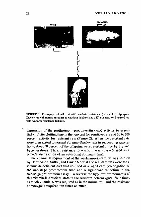
22
WILD
ft I f2 I
F3 I F4 I F
O'REILLY AND POOL
SPRAGUE· DAWLEY
FIGURE 1 Photograph of wild rat with warfarin resistance (dark color), SpragueDawley rat with normal response to warfarin (albino), and a fifth-generation Stanford rat with warfarin resistance (albino).
depression of the prothrombin-proconvertin (P&P) activity to essentially infinite clotting time in the P&P test for sensitive rats and 10 to 100 percent activity for resistant rats (Figure 2). When the resistant rats were then mated to normal Sprague-Dawley rats in succeeding generations, about 50 percent of the offspring were resistant in the F 3, F 4, and F5 generations. Thus, resistance to warfarin was characterized as a bimodal distribution of an autosomal dominant trait.
The vitamin K requirement of the warfarin-resistant rat was studied by Hermodson, Suttie, and Link. 5 Normal and resistant rats were fed a vitamin-K-deficient diet that resulted in a significant prolongation of the one-stage prothrombin time and a significant reduction in the two-stage prothrombin assay. To reverse the hypoprothrombinemia of this vitamin-K-deficient state in the resistant heterozygote, four times as much vitamin K was required as in the normal rat, and the resistant homozygous required ten times as much.
Copyright © National Academy of Sciences. All rights reserved.
Animal Models of Thrombosis and Hemorrhagic Diseaseshttp://www.nap.edu/catalog.php?record_id=19903

Warfarin and the Warfarin-Resistant Rat 23
WARFARIN-RESISTANT MAN
In 1970, we reported the second kindred with hereditary resistance to oral anticoagulant drugs. 6 The propositus had a documented pulmonary embolism in 1965 and intravenous heparin therapy produced a normal response; oral anticoagulants were not prescribed. In 1967 signs and symptoms of pulmonary embolism developed and sodium warfarin was administered, but no hypoprothrombinemic response could be obtained even at a dosage of 50 mg a day; the patient was referred to us. All drugs were stopped, and a series of single-dose experiments was carried out. The blood levels of warfarin after a large
30
20
10
>- 50 u z ..... ::> F4 F5 0 40 ..... Ge .....
30
20
10
10 100% 10 100%
P & P TEST
FIGURE 2 Distribution of response for prothrombin-proconvertin (P&P) clotting test in four generations of offspring from back-crosses of warfarin-resistant rats and Sprague-Dawley rats 48 hours after ingestion of drinking water containing 250 ppm warfarin. All rats with less than I percent P&P activity at 48 hours were considered warfarin responsive.
Copyright © National Academy of Sciences. All rights reserved.
Animal Models of Thrombosis and Hemorrhagic Diseaseshttp://www.nap.edu/catalog.php?record_id=19903

24 O'REILLY AND POOL
2
~ 70 • .. u
1~ 50 • !r:~ .ao •• ~ 20 ... o~ ii:
FIGURE 3 Plasma drug concentrations and prothrombin responses in the propositus of human kindred with warfarin resistance after single oral doses of 1.5 and 11 mg of warfarin per kg of body weight. Data are plotted semilogarithmically. Shaded areas represent the effects of a single oral dose of 1.5 mg of warfarin per kilogram body weight in 50 normal subjects.
single oral dose of warfarin, 1.5 mg/kg, or 82.5 mg, were entirely within the normal range for our laboratory for the same dose of warfarin, but the hypoprothrombinemic response was almost nil (Figure 3).
Similar results were obtained when the same dose of warfarin was given intravenously. When the single dose of warfarin was increased to 660 mg orally, a response that was in the normal hypoprothrombinemic range did occur for the first 48 hours but then reversed rapidly to 100 percent prothrombin activity. Thus, the propositus had normal absorption and elimination of warfarin, yet was highly resistant to the hypoprothrombinemic effect of warfarin. On chronic maintenance therapy, the propositus required 75 mg of warfarin, ten times the average dose, to achieve normal therapeutic hypoprothrombinemia, and the blood levels of warfarin were tenfold greater (Figure 4) than those of normal subjects achieving therapeutic hypoprothrombinemia. The warfarin-resistant propositi require a chronic daily dose of warfa-
Copyright © National Academy of Sciences. All rights reserved.
Animal Models of Thrombosis and Hemorrhagic Diseaseshttp://www.nap.edu/catalog.php?record_id=19903

Warfarin and the Warfarin-Resistant Rat 25
rin of 80 and 120 mg to maintain a therapeutic level of anticoagulation7
(Figure 5). The pedigree of the kindred of the second propositus is shown in
Figure 6. Twenty members of the kindred were as resistant to the hypoprothrombinemic effect of warfarin as the propositus. Of the 19 children in generation III from eight resistant parents in generation II, 10 had a resistant response. Male-to-male transmission of the resistance to four sons (IIl-26, III-27, III-29, and III-30) from two fathers (11-22 and 11-23) ruleii out a locus for the gene on the X chromosome. These data indicate the transmission of a single-gene effect as an autosomal dominant trait.
The hypoprothrombinemic response of all 59 members of botlt resistant kindreds compared with that of 50 normal subjects given 1.5 mg sodium warfarin/kg body weight is shown in Figure 7. The average decline in one-stage prothrombin activity for the normal subjects was 82 ± 4 percent; 34 kindred members showed a normal response, and 25
~- 80 cD~ ~!..... o>- 60 QI: !::: ::c > .... -oti ~<
0
100
~ 7S QI: w -< U) 0 ~ 0 ~ so QI: 0 -; 25
. ... ........ . .... . l' ..... ... .,............... • ... . · ....
..
DAYS
. .................. . ,. . .. .
FIGURE 4 Concentration of drug in plasma and activity of the prothrombin complex in the propositus of the second human kindred with warfarin resistance during long-term treatment with sodium warfarin . The warfarin dose and warfarin levels in plasma were tenfold greater than in normal subjects.
Copyright © National Academy of Sciences. All rights reserved.
Animal Models of Thrombosis and Hemorrhagic Diseaseshttp://www.nap.edu/catalog.php?record_id=19903
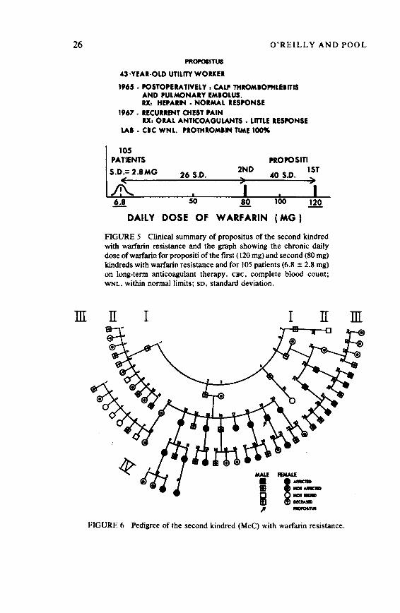
26
m
O'REILLY AND POOL
PROPOllTUS
43·YEAR·OLD UTILITY WORKER
1965. POSTOPERATIVELY 1 CALF THIOMIOPHLEllTIS AND PULMONARY EMIOLUS. U. HEPARIN • NORMAL RESPONSE
1967 • RECUHENT CHEST PAIN RX1 ORAL ANTICOAGULANTS • LITTLE RESPONSE
LAI. CIC WNL. PROTHIOMllN TIME 100"9
105 PATIENTS
S.D.=2.8MG (
26 S.D.
50
2ND )
PROPOSITI lST "'° S.D. )
100
DAILY DOSE OF W ARFARIN (MG )
FIGURE 5 Clinical summary of propositus of the second kindred with warfarin resistance and the graph showing the chronic daily dose of warfarin for propositi of the first ( 120 mg) and second (80 mg) kindreds with warfarin resistance and for 105 patients (6.8 ± 2.8 mg) on long-term anticoagulant therapy. cec, complete blood count; WNL, within normal limits; so, standard deviation.
n I I II
flMAll!=------FIGURE 6 Pedigree of the second kindred (McC) with warfarin resistance.
m
Copyright © National Academy of Sciences. All rights reserved.
Animal Models of Thrombosis and Hemorrhagic Diseaseshttp://www.nap.edu/catalog.php?record_id=19903

Warfarin and the Warfarin-Resistant Rat
10 > u z w 8 :::> 0 w a: 6 ....
..
2
0 100
I MEAN:t3 S.D.
L i )I I NORMAL RESPONSE I I I I I I I I I I I I I I
80 60 20
REDUCTION OF PROTHROMBIN ACTIVITY AT 48 HOURS (%)
27
0
FIGURE 7 Mean reduction of one-stage prothrombin activity in the 59 members of both kindreds with warfarin resistance 0 to 48 hours after a single oral dose of sodium warfarin, 1.5 mg/kg body weight. The dotted lines represent the 99 percent confidence limits (mean ± 3 standard deviation equals 82 ± 12 percent) for the 0- to 48-hour response in 50 normal subjects given the same single dose. The 34 members of both kindreds with a normal response are indicated by the hatched bars and the 25 members with a resistant response are indicated by the dark bars. The means of this bimodal distribution (82 percent and 10 percent respectively) are 18 standard deviations apart .
members showed a markedly lessened response, with a mean decline of 10 percent and a range from 0 to 25 percent. The difference between the two means, lO percent for the resistant members and 82 percent for the nonresistant members of the kindreds, was 18 standard deviations apart, a highly significant difference. This bimodality of the prothrombin response allowed an unbiased assessment of the data for genetic analysis.
MECHANISM OF RESISTANCE
The two human kindreds of warfarin-resistant man and the warfarinresistant rats showed a remarkable similarity with respect to the pharmacology of the oral anticoagulant drug, the genetic transmission of the resistant trait, and the physiologic responses to vitamin K. 4 The possible mechanisms for the hereditary resistance to oral anticoagulant
Copyright © National Academy of Sciences. All rights reserved.
Animal Models of Thrombosis and Hemorrhagic Diseaseshttp://www.nap.edu/catalog.php?record_id=19903
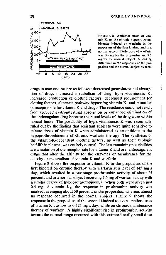
28 • PROPOSITUS
~ o NORMAL SUBJECT
~ 60 ),...
·~ ::!;:40 i::: ~30 ~ ~20 ~ 15
~ -6 0 6 12 18 24 30 36
DAYS
O'REILLY AND POOL
FIGURE 8 Antidotal effect of vitamin K1 on the chronic hypoprothrombinemia induced by warfarin in the propositus of the first kindred and in a normal subject. Daily dose of warfarin was 145 mg for the propositus and 7.5 mg for the normal subject. A striking difference in the responses of the pri>positus and the normal subject is seen.
drugs in man and rat are as follows: decreased gastrointestinal absorption of drug, increased metabolism of drug, hypervitaminosis K, increased production of clotting factors, decreased requirement for clotting factors, alternate pathway bypassing vitamin K, and mutation of receptor site for vitamin K and drug. 8 The resistance could not result from reduced gastrointestinal absorption or enhanced elimination of the anticoagulant drug because the blood levels of the drug were within normal limits. The possibility of hypervitaminosis K was essentially ruled out by the finding that resistant subjects were quite sensitive to minute doses of vitamin K when administered as an antidote to the hypoprothrombinemia of chronic warfarin therapy. The synthesis of the vitamin-K-dependent clotting factors, as well as their biologic half-life in plasma, was entirely normal. The last remaining possibilities are a mutation of the receptor site for vitamin K and oral anticoagulant drugs that alter the affinity for the enzymes or membranes for the activity or metabolism of vitamin K and warfarin.
Figure 8 shows the response to vitamin K in the propositus of the first kindred on chronic therapy with warfarin at a level of 145 mg a day, which resulted in a one-stage prothrombin activity of about 25 percent, and in a normal subject receiving 7 .5 mg of warfarin a day with a similar degree of hypoprothro'mbinemia. When both were given just 0.5 mg of vitamin Ki. the response in prothrombin activity was marked, averaging about 50 percent, in the propositus, whereas almost no response occurred in the normal subject. Figure 9 shows the response in the propositus of the second kindred to even smaller doses of vitamin Ki. as low as 0.125 mg a day, while on chronic maintenance therapy of warfarin. A highly significant rise in prothrombin activity toward the normal range occurred with this extraordinarily small dose
Copyright © National Academy of Sciences. All rights reserved.
Animal Models of Thrombosis and Hemorrhagic Diseaseshttp://www.nap.edu/catalog.php?record_id=19903

Warfarin and the Warfarin-Resistant Rat 29
of vitamin K, a dose that does not elicit any response in normal subjects during therapeutic hypoprothrombinemia with warfarin.
VITAMIN K
The response to vitamin K in the propositus of the second kindred and four normal subjects on a vitamin-K-deficient diet for at least 30 days is shown in Figure IO. 9 Throughout the course of the experiment, the normal subjects maintained a one-stage prothrombin activity that did not fall below the normal range, whereas the propositus showed marked hypoprothrombinemia within 14 days after introduction of the vitamin-K-deficient diet. These data indicate that the propositus has a markedly increased requirement for vitamin K, compared with normal subjects in the absence of any anticoagulant therapy, as was observed in the warfarin-resistant rats.
To determine whether the warfarin-resistant rat showed the same marked sensitive response to vitamin K during warfarin administration that was ~een in warfarin-resistant man, the experiments depicted in Figure 11 were performed. 10 Warfarin was added to drinking water at a
100 -~ 80
VITAMIN K1
> o 0 .5 mg
!:: • 0 .25 mg
> 60 • 0.125 mg
j::: 50 u
< 40 z c:Q
~ 30 0 °' ::c .... 0 °' Q. _,
0 ' 8 12 16 20
DAYS FIGURE 9 Antidotal effect of0.5, 0.25, and 0.125 mg of vitamin K1 during the chronic hypoprothrombinemia induced by 75 mg of sodium warfarin/day in the propositus of second kindred. A highly significant response is seen with even the smallest doses of vitamin K.
Copyright © National Academy of Sciences. All rights reserved.
Animal Models of Thrombosis and Hemorrhagic Diseaseshttp://www.nap.edu/catalog.php?record_id=19903
30
-' z >!::: > ;::: u c z lllD ~ 0 ...: ::c ... 0 ...: A.
40
20
0
O'REILLY AND POOL
e PROPOSITUS 2ND KINDRED
DIET:
RICE
EGG WHITE
GELATIN
VITAMINS & SALT
COFFEE & TEA
TETRACYCLINE 2.0 m OR NEOMYCIN 4.0 gm P.O. OD
8 16
DAYS
32
FIGURE 10 Mean (± standard error of the mean) one-stage prothrombin times in percent of normal activity for four normal subjects and the propositus of the second kindred after a vitamin-K-deficient diet and oral antibiotics. The propositus became hypoprothrombinemic on the regimen within 14 days, whereas the normal subjects did not become hypoprothrombinemic in 30 days.
level of IO ppm for the normal Sprague-Dawley rats and 250 ppm for the resistant Stanford strain of rats. Both groups showed similar marked hypoprothrombinemia at 48 hours, with 94-98 percent reduction of activity. When each group received the same level of warfarin plus IO ppm of vitamin K, no significant protection against warfarininduced hypoprothrombinemia occurred in the normal rats, but total proteation occurred in the resistant rats (Figure 11). A highly significant degree of protection also occurred in the resistant rats when as little as 3 ppm of vitamin K was administered. These data indicate that the warfarin-resistant rat, like warfarin-resistant man, had a marked responsiveness to the antidotal effect of vitamin K administered with warfarin.
A halogenated analogue of vitamin K (chloro-K) was prepared by Lowenthal et al. in Canada by replacing the methyl group in the 2 position of the naphthoquinone nucleus. 11 When single doses of chloro-K were administered to normal and warfarin-resistant rats (Figure 12), the highest concentrations of chloro-K were lethal to
Copyright © National Academy of Sciences. All rights reserved.
Animal Models of Thrombosis and Hemorrhagic Diseaseshttp://www.nap.edu/catalog.php?record_id=19903
Warfarin and the Warfarin-Resistant Rat 31
warfarin-resistant rats, but some of the normal rats survived. Conversely, single doses of warfarin were lethal in normal rats but ineffective in warfarin-resistant rats. When chloro-K and warfarin were combined into a single rodenticide, all warfarin-sensitive and warfarinresistant rats were killed within IO days. Thus, the combination of chloro-K and warfarin is perhaps the most effective rodenticide yet produced for the control of all rats.
The present theory on the relationship of warfarin and vitamin K is shown in Figure 13. The role of vitamin K1 oxide in the metabolism of vitamin K is controversial. 12 The conversion of the precursors of the prothrombin complex into active clotting factors through the action of vitamin K can be blocked at two or more sites, a warfarin-sensitive site
WARFARIN, ppm
V. K1 ppm
P & P TEST IN~
20
RATS
250 250 250 250
0 10 3
I S.E.M.
10
0
10
10
o'--~~~~~~~~~.__-=-=~~~
RESISTANT NORMAL
FIGURE 11 Mean ( ± standard error of the mean) hypoprothrombinemic responses from 0 to 48 hours, measured by the P&P test, in resistant and normal rats after continuous oral ingestion of various concentrations of sodium warfarin and vitamin K (versus K ,) added to the drinking water. The stippled area indicates the normal range of the P&P test in percent of normal activity. Vitamin K 1 at 10 ppm in the resistant rats completely blocked the hypoprothrombinemic effect of warfarin but did not protect the normal rats.
Copyright © National Academy of Sciences. All rights reserved.
Animal Models of Thrombosis and Hemorrhagic Diseaseshttp://www.nap.edu/catalog.php?record_id=19903

Vo) N
I ., Surviving eo 1 0 19eoo !.•00 11 • '1 20 d • o • 1 eo •""."• • o ays eo I o i•eoo •.oo ' • I . ,
20 ' I I '
18
16
-; 14
:a 12 0 -.c .,_ 10 .. "' =i ., llt"CI 8 &.e ;,e 6
-o.!
! 4
2
0
l oo I 00 : 10 I I
WR I N 3M 3F ~ &M &F
• I 0 0 I I I I O I I 0 I I I
e I 00 I 0: eo e eo~ ....,J e I
• eo
0
ee ~ e 0 eo I I e I eeo
e I ee 0 I 0 eo : 001 e 0 0 e : 0 j e o0 I I •• • 0 I eoo I 00 1 1 eoo1 o • leeo •• :o I I e I leeO e I e I I I I I I I I I I
WR I N I WR I N -, WR ,-N ,-WR I N I WR I N 5M 5F ~ 5M 5F 5M 5F ~ 6M 6F 5M 4F : 5M 5F 5M 5F: 5M 5F 6M 5F ~ 5M 5F
•• • I 0
0 •o
0 I •
e I eoc 'eeo I I
WR I N 5M 3F I 4M 4F
0 025 t; WARF I 0 005 t; Cl - K 0010 t; Cl-K 0.0201' Cl·K 0.025 t; WARF I 0.025 t; WARF I 0.025 t; WARF 0.0051' Cl·K 0.010 t; Cl·K 0.020 t; Cl·K
FIGURE 12 Effect of diets containing varying amounts of waifarin (WARF) or chloro-K (Cl-K) or mixtures of the two anticoagulants on the survival of normal (N) and waifarin-resistant (WR) rats. The diets containing the anticoagulants were the only foods available to the rats during the 20-day period. The closed circles (male rats) and open circles (female rats) indicate the number of days after exposure to the diets that each rat was found dead. (Reprinted with permission from Suttie, J. W. 1973. Anticoagulant-resistant rats: Possible control by the use of the chloro-analog of Vitamin K. Science 180:741-743. Figure l, 18 May 1973. Copyright© 1973 by the American Association for the Advancement of Science.)
Copyright © National Academy of Sciences. All rights reserved.
Animal Models of Thrombosis and Hemorrhagic Diseaseshttp://www.nap.edu/catalog.php?record_id=19903

Warfarin and the Warfarin-Resistant Rat
CHLORO-K PRECURSOR I ~ PROTHROMBIN
PROTEIN T rn'\ COMPLEX
VITAMIN VITAMIN
NAD+~. 2X:~DH CAD
FIGURE 13 Relationships ofvitamin K, and prothrombin completion and its blockade by the halogenated analogue of vitamin K (chloro-K) and by coumarin anticoagulant drugs (CAD). Vitamin K, converts a precursor protein of the prothrombin complex to active prothrombin by facilitating the addition of a calcium-binding moiety to the protein while itself being converted into inactive vitamin K1-epoxide. This step is chloro-K sensitive and warfarin insensitive, whereas the regeneration step of vitamin K.-epoxide is warfarin sensitive and may be the mutation site for the development of warfarin resistance.
33
and a chloro-K-sensitive site. Vitamin K converts a postribosomal precursor protein of prothrombin into active clotting factor by carbonation of eight or more glutamyl residues to form gammacarboxyglutamic acid. 13 This novel amino acid, unique to the N-terminal end of the four vitamin-K-dependent clotting factors, binds calcium and thereby permits attachment of prothrombin (factor II) to a lipoprotein complex of phospholipid (PL) and factors V and Xa (activated form of factor X), which produces active thrombin (Ila)14
(Figure 14). Mutation of the warfarin-sensitive step for the regeneration of vitamin K from its epoxide form in warfarin-resistant man and rat may produce the warfarin resistance, 15 but many other theoretic possibilities fit the existing data. 16
SUMMARY
The danger in the emergence of warfarin-resistant rats has been reduced considerably by the finding that the Stanford strain is so sensitive to chloro-K. The Stanford strain of the warfarin-resistant rat has proved invaluable in unraveling vitamin K metabolism and
Copyright © National Academy of Sciences. All rights reserved.
Animal Models of Thrombosis and Hemorrhagic Diseaseshttp://www.nap.edu/catalog.php?record_id=19903
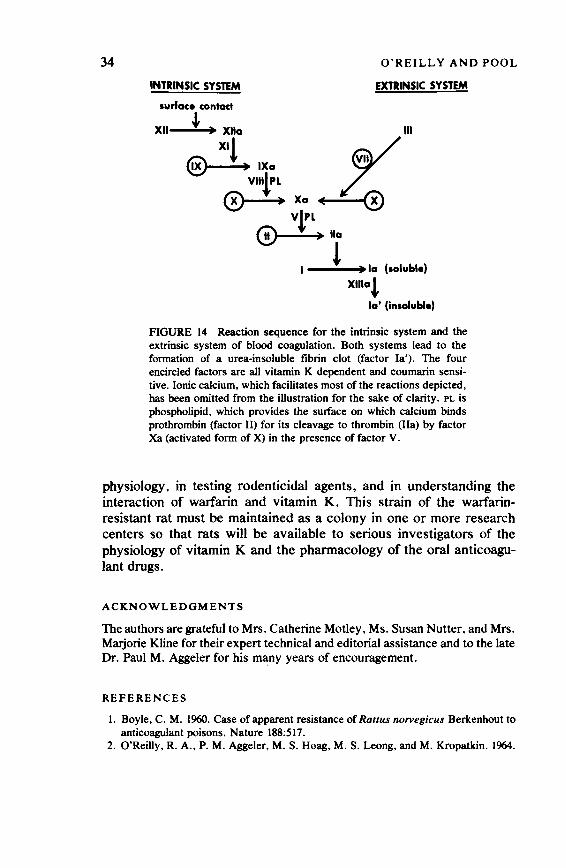
34
INTRINSIC SYSTEM
surface contact
O'REILLY AND POOL
EXTRINSIC SYSTEM
XII ' • Xlla HI
~IXo w VlltPL 7r-.
0--!.+xa~ Vrl
@---!-+Ila
I l lo (soluble)
xmat la' (insoluble)
FIGURE 14 Reaetion sequence for the intrinsic system and the extrinsic system of blood coagulation. Both systems lead to the formation of a urea-insoluble fibrin clot (factor la'). The four encircled factors are all vitamin K dependent and coumarin sensitive. Ionic calcium, which facilitates most of the reactions depicted, has been omitted from the illustration for the sake of clarity. PL is phospholipid, which provides the sutface on which calcium binds prothrombin (factor II) for its cleavage to thrombin (Ila) by factor Xa (activated form of X) in the presence of factor V.
physiology, in testing rodenticidal agents, and in understanding the interaction of warfarin and vitamin K. This strain of the warfarinresistant rat must be maintained as a colony in one or more research centers so that rats will be available to serious investigators of the physiology of vitamin K and the pharmacology of the oral anticoagulant drugs.
ACKNOWLEDGMENTS
The authors are grateful to Mrs. Catherine Motley, Ms. Susan Nutter, and Mrs. Marjorie Kline for their expert technical and editorial assistance and to the late Dr. Paul M. Aggeler for his many years of encouragement.
REFERENCES
I. Boyle, C. M. 1960. Case of apparent resistance of Rattus norvegicus Berkenhout to anticoagulant poisons. Nature 188:517.
2. O'Reilly, R. A., P. M. Aggeler, M. S. Hoag, M. S. Leong, and M. Kropatkin. 1964.
Copyright © National Academy of Sciences. All rights reserved.
Animal Models of Thrombosis and Hemorrhagic Diseaseshttp://www.nap.edu/catalog.php?record_id=19903

Warfarin and the Warfarin-Resistant Rat 35
Hereditary transmission of exceptional resistance to coumarin anticoagulant drugs: First reported kindred. N. Engl. J. Med. 271:809-815.
3. Cuthbert, J. H. 1963. Further evidence of resistance to warfarin in the rat. Nature 198:807-808.
4. Pool, J. G .• R. A. O'Reilly, L. J. Schneiderman, and M. Alexander. 1968. Warfarin resistance in the rat. Am. J. Physiol. 215:627-631.
5. Hennodson, M. A., J. W. Suttie, and K. P. Link. 1969. Warfarin metabolism and vitamin K requirement in the warfarin-resistant rat. Am. J. Physiol. 217:131&-1319.
6. O'Reilly, R. A. 1970. The second reported kindred with hereditary resistance to oral anticoagulant drugs. N. Engl. J. Med. 282:1448-1451.
7. O'Reilly, R. A. 1972. Symposium on pharmacogenetics. Genetic factors in the response to the oral anticoagulant drugs. Pages 297-312 in 4th International Congress of Human Genetics, International Congress Series. Excerpta Medica, Amsterdam.
8. O'Reilly, R. A., and P. M. Aggeler. 1965. Coumarin anticoagulant drugs: Hereditary resistance in man. Fed. Proc. 24:126&-1273.
9. O'Reilly, R. A. 1971. Vitamin Kin hereditary resistance to oral anticoagulant drugs. Am. J. Physiol. 221:1327-1330.
10. O'Reilly, R. A .• J. G. Pool, and P. M. Aggeler. 1968. Hereditary resistance to coumarin anticoagulant drugs in man and rat. Ann. N.Y. Acad. Sci. 151:913-931.
II. Lowenthal, J., J. A. MacFarlane, and K. M. McDonald. 1960. Inhibition of the antidotal activity of vitamin K, against coumarin anticoagulant drugs by its chloro analogue. Experientia 16:428-429.
12. Bell, R. G., and J. T. Matschiner. 1972. Warfarin and the inhibition of vitamin K activity by an oxide metabolite. Nature 237:32-33.
13. Nelsestuen, G. L .. and J. W. Suttie. 1973. The mode ofaction of vitamin K. Isolation of a peptide containing the vitamin K-dependent portion of prothrombin. Proc. Nat. Acad. Sci. USA 70:336&-3370.
14. Stenflo, J., P. Fernlund, W. Egan, and P. Roepstorff. 1974. Vitamin K dependent modifications of glutamic acid residues in prothrombin. Proc. Nat. Acad. Sci. USA 71:273~2733.
15. Zimmermann, A .. and J. T. Matschiner. 1974. Biochemical basis of hereditary resistance to warfarin in the rat. Biochem. Pharmacol. 23: 1033-1040.
16. Suttie, J. W. In press. Review and newer information on the mechanism of action of vitamin K and the coumarin anticoagulants. Throm. Diath. Haemorrh. Suppl.
Copyright © National Academy of Sciences. All rights reserved.
Animal Models of Thrombosis and Hemorrhagic Diseaseshttp://www.nap.edu/catalog.php?record_id=19903
W. JEAN DODDS, D. V .M.
Survey of Other Available Models of Inherited Hemorrhagic Diseases
Comparative studies of the hemorrhagic diseases of man have concen" trated during the past 30 years on animals with hemophilia and von Willebrand's disease. By contrast, other models of the hemorrhagic disorders have only recently been described and have not yet been exploited to their full potential.
This paper reviews the history and current status of inherited hemorrhagic disorders, other than hemophilia and von Willebrand's disease, described to date in domestic animals. Most of these models have been described in dogs, but other species, including horses, cattle, goats, swine, cats, and rats, are also involved. 1 Animal models for hereditary deficiencies of prothrombin, factor V, and factor XIII have yet to be discovered.
FACTOR VII DEFICIENCY
In 1962 Mustard and associates2 reported the first animal model of human factor VII deficiency' in a family of beagles. Subsequent reports by Garner et al., 3 Capel-Edwards and Hall, 4 Poller et al., 5 and Spurling et al. 6 also involved colonies of beagles. The condition has been recognized in other commercial beagle colonies and in an Alaskan malamute. 1 The apparent high incidence in beagles probably reflects
Supported in part by USPHS grant HL 09902 from the National Heart and Lung Institute, National Institutes of Health.
36
Copyright © National Academy of Sciences. All rights reserved.
Animal Models of Thrombosis and Hemorrhagic Diseaseshttp://www.nap.edu/catalog.php?record_id=19903

Survey of Other Available Models 37
the widespread use of this breed for biomedical research. The disease has most often been discovered fortuitously during hematologic screening for drug testing.
Affected beagles have a mild disease that may manifest itself in easy bruisability but that results in few or no clinical bleeding problems. There is an apparent predisposition to systemic demodicosis probably because the defective extrinsic clotting system provides an ideal, moist tissue environment for mange mites. 1
This coagulation defect is inherited as an autosomal dominant trait: The heterozygous state can be readily detected by prothrombin times measured with human brain thromboplas(m 1 or by specific factor VII assays. 1•3 - 8 The clinical expression of the disease, however, follows an autosomal recessive mode of inheritance; heterozygotes are asymptomatic, and homozygotes rarely bleed.
Factor VII-deficient animals have prolonged prothrombin times but normal Russell's viper venom times. The factor VII levels vary from 1 percent to 4 percent in homozygotes and from 35 percent to 65 percent in heterozygotes. 1
Studies of this animal model have made several significant contributions to medical science. Factor VII-deficient dog plasma provides an excellent standard source of deficient substrate for use as Jl diagnostic and investigational reagent. I.3·11•7 The optimum. sensitivity is obtained when this reagent is used with human brain tissue extract as the activator, rather than commercial rabbit brain thromboplastin. 1•5•7•8
The plasma also provides a substrate for quality-control studies of various tissue thromboplastins. 5
Beagles with factor VII deficiency and normal controls were used by Garner and Evensen9 to investigate the role of factor VII in mediating endotoxin-induced intravascular coagulation and shock. These studies showed that the development of shock and thrombotic lesions was delayed or prevented in the deficient dogs. Cross-circulation studies to determine the in vivo half-life of factor VII in deficient beagles10 have confirmed the rapid turnover of this clotting factor, as previously reported for man.
FACTOR X DEFICIENCY
Factor X deficiency, manifest as a mild to moderate bleeding diathesis, has been studied in a large family of inbred cocker spaniels. 11 Over 900 related dogs have been screened for this disorder to date because it affects one of the top-quality show lines of cocker spaniels. The disease has not been reported in other breeds or other animal species.
Copyright © National Academy of Sciences. All rights reserved.
Animal Models of Thrombosis and Hemorrhagic Diseaseshttp://www.nap.edu/catalog.php?record_id=19903

38 W. JEAN DODDS
Most affected mature dogs have a clinically mild disease, although some young adult dogs have experienced life-threatening or fatal bleeding. By contrast, hemorrhagic problems are frequently serious or lethal in newborns, probably because of the concomitant physiologic hypoprothrombinemia common to all newborn mammals. The disease mimics the "fading puppy syndrome," as some affected puppies live for up to 2 weeks, then suddenly fade and die. 11
Genetic studies of 14 test matings involving each of the three possible mating types (normal to normal; affected to normal; affected to affected) indicated that the defect is inherited as an autosomal dominant trait with variable expression. 11 These data agree with studies of the analogous human disorder. The trait appears to be lethal in the homozygous state, as 30 of the 48 puppies born from the test matings of mildly affected dogs died within the first weeks of life. Denson et al. 12 have postulated that in man the complete absence of factor X may be a lethal mutation because of the central position of factor X in the clotting mechanism. Our studies agree with theirs; all affected animals that survived to maturity have had some factor X activity, which suggests that they are heterozygotes. 1•11
Treatment of affected pregnant bitches and newborns with oral administration of vitamin K1 to promote the synthesis of vitamin K-dependent clotting factors has not been successful in reducing or preventing neonatal mortality. We have not yet tried transfusion of newborns via the umbilical vein with prothrombin-complex concentrates.
The coagulation defect of affected animals is measured by mild to moderate abnormalities of extrinsic and intrinsic clotting and reduction of factor X activity to 18--69 percent of normal.
The analogy of this animal model to human factor X deficiency has been established. Its potential usefulness has yet to be determined but could include genetic cross-breeding studies with models of the other prothrombin-complex deficiencies. By cross-breeding animals with factor II, VII, IX, and X deficiencies, it might be possible to investigate Seeger' s hypothesis that these clotting factors form part of a single molecular complex. 13
FACTOR XI DEFICIENCY
Factor XI (plasma thromboplastin antecedent, or PTA) deficiency analogous to that of man was reported first by Kociba et al. in Holstein cattle14 and subsequently by our laboratory in an inbred family of
Copyright © National Academy of Sciences. All rights reserved.
Animal Models of Thrombosis and Hemorrhagic Diseaseshttp://www.nap.edu/catalog.php?record_id=19903
Survey of Other Available Models 39
springer spaniels. 15 Another report of this disorder in cattle was recently published. 16 The disorder is characterized by autosomal, incompletely dominant inheritance; minor spontaneous bleeding episodes, such as hematuria; and severe, protracted, or lethal hemorrhage 12-24 hr after surgical procedures.
Affected animals have an intrinsic system coagulation defect with low factor XI activity. Homozygotes have 1-10 percent and heterozygotes 25-60 percent factor XI activity.
As in treatment of the disease in man, small doses of fresh-frozen plasma infused before surgery usually prevent excessive bleeding, whereas massive doses are required to control bleeding once it starts. 15
The major contribution of the bovine model thus far has been to provide an excellent source of deficient substrate for factor XI assays in man14 and other species. 15 Relatively large quantitities of plasma are readily obtained from the affected cattle. The canine model is not currently available, as the propositus died recently of acute gastric torsion and the owners of her heterozygous offspring have been reluctant to use them for breeding.
FIBRINOGEN DEFICIENCIES
Afibrinogenemia
In 1972 Breukink and associates reported afibrinogenemia in a large family of Saanen dairy goats.17 The defect is characterized by an extremely severe or fatal hemorrhagic diathesis ·in newborn and young goats. Fibrinogen levels varied from total absence to a trace as measured by biologic, immunologic, and physical methods. Reduced platelet retention, erythrocyte sedimentation, and plasma viscosity and prolonged bleeding times were also observed. ·
The trait appears to follow an autosomal, incompletely dominant inheritance. The heterozygous state is usually detected by finding subnormal fibrinogen levels in relatives of affected goats and/or by transmission of the defect in test matings. The current status of this animal model has not been reported.
Hypofibrinogenemia
A family of St. Bernards with hypofibrinogenemia was described by Kammennan et al. in 1971. 18 The propositus manifested a severe bleeding diathesis and died despite massive transfusions of fresh whole
Copyright © National Academy of Sciences. All rights reserved.
Animal Models of Thrombosis and Hemorrhagic Diseaseshttp://www.nap.edu/catalog.php?record_id=19903

40 W. JEAN DODDS
blood. Several of his relatives of both sexes were found to have low-normal fibrinogen levels and were considered to be heterozygotes. As far as is known, these animals are no longer available for study.
PLATELET FUNCTION DEFECTS
Thrombasthenic Thrombopathia
In 1967 we described a family of otterhounds with a hereditary platelet function defect similar, except for a large number of bizarre giant platelets, to human Glanzmann's thrombasthenia, in which the platelets usually appear normal. 19•20 The morphologic abnormality resembles that of the Bernard-Soulier syndrome, so this defect appears to be mixed, i.e., thrombasthenic thrombopathia. A human patient with a similar platelet function defect has also been studied in our laboratory. 21
Platelet function tests that identify the canine disorder as primarily thrombasthenic are poor clot retraction; long bleeding time; low platelet retention; defective platelet aggregation with all aggregating agents, except cobra venom factor22 ; normal platelet release reaction; reduced platelet fibrinogen and prothrombin consumption; and failure of intact platelets to support thromboplastin generation. The thrombopathic component is manifested by reduced platelet factor 3 availability and the high proportion (50--80 percent) of giant platelets, which are readily identified by phase contrast22 and electron microscopy. 19•20
The giant platelets may represent the thrombopathic population, and the normal-appearing platelets, 1 the thrombasthenic population.
Further support for the presence of two platelet populations in these animals comes from platelet survival studies with [75Se]methionine. 1
Platelets from the affected dogs developed two peaks of radioactivity, the first representing a rapidly labeled, short-lived population and the second, a more slowly labeled population with a normal survival time. Plasma fibrinogen levels and turnover rates were normal. 1
The defect is inherited as an autosomal dominant trait with variable expression. Both the homozygous and heterozygous states are expressed and can be readily detected by platelet function tests. 1• 19 The clinical severity of the disease varies from moderate in homozygotes to mild in heterozygotes.
The large size of these dogs permits collection of sufficient volumes of blood for molecular characterization of the platelet abnormality and comparison of the defect with that of similar human disorders. Studies
Copyright © National Academy of Sciences. All rights reserved.
Animal Models of Thrombosis and Hemorrhagic Diseaseshttp://www.nap.edu/catalog.php?record_id=19903
Survey of Other Available Models 41
of the membrane and subcellular fractions of these platelets are in progress.
Thrombopathia
Another family of dogs with an inherited platelet function defect has ·been studied by Lotz and associates. 23 The disorder, in basset hounds, is a thrombopathia attributed to a platelet membrane defect and manifested primarily by defective platelet aggregation with adenosine diphosphate (ADP). Other platelet abnormalities were observed, 1•23 but a more detailed characterization of this model is not available.
Two additional cases of congenital platelet dysfunction have also been recognized. 1 One involved a young foxhound with a platelet defect similar to that in the otterhounds; the other, a primary hemostatic disorder in a Scottish terrier puppy. In the latter case, a diagnosis of von Willebrand' s disease cannot be ruled out, since platelet function tests and specific coagulation factor assays were not performed. 1
The most extensively characterized animal model of thrombopathic disorders involves an inbred line of fawn-hooded (FH) rats. The disorder was first reported in 1972 by Tschopp and Zucker, who determined it to be a platelet storage pool deficiency similar to that observed in man. 24•211 Recent studies in our laboratory have confirmed the original observations and have extended the characterization Qf the model to include other hemostatic and metabolic parameters. 28
The defect in FH rats is characterized by long bleeding times; normal clot retraction, ADP-induced platelet aggregation and platelet ADP; variable aggregation with collagen; minimal aggregation with adren-
. aline and cobra venom factor; and reduced platelet retention, platelet ·ATP, ATP/ADP ratio, serotonin and nucleotide content; and [14C]serotonin release. 24•28 In comparison to age- and sex-matched normal Wistar rats, FH rats have significantly prolonged partial thromboplastin times, shortened Russell's viper venom times, and increased factor X and XI activity. 28 Other coagulation values are normal, as are plasma proteins, glycoproteiris, ceruloplasmin, and preliminary studies of immunoglobulins and complement. An interesting, as yet unexplained, finding was significantly lower glucose and higher cholesterol levels in FH rats than in matched Wistar rats.
Several colonies of FH rats are currently maintained for ongoing hemostatic and behavioral studies. The advantages of this laboratory animal model include its size, short gestation period, multiple births, docile nature, and relatively low maintenance costs. Further characterization of the model with respect to the genetic transmission of the
Copyright © National Academy of Sciences. All rights reserved.
Animal Models of Thrombosis and Hemorrhagic Diseaseshttp://www.nap.edu/catalog.php?record_id=19903
42 W. JEAN DODDS
defect and the molecular aspects of the platelet release abnormality is in progress in several laboratories. Studies of this animal should yield useful information about the analogous human disorders.
REFERENCES
I. Dodds, W. J. 1974. Hereditary and acquired hemorrhqic disorders in animals. Pages 215-247 in T. H. Spaet (ed.), Progress in hemostasis and thrombosis. Vol 2. Grune & Stratton, New York.
2. Mustard, J. F., D. Secord, T. D. Hoeksema, H. G. Downie, and H. C. Rowsell. 1962. Canine factor VII deficiency. Br. J. Haematol. 8:43.
3. Gamer, R., C. Hermoso-Perez, and D. M. Conning. 1967. Factor VII deficiency in beagle dog plasma and its use in the assay of human factor VII. Nature 216: 1130.
4. Capel-Edwards, K., and D. E. Hall. 1968. Factor VII deficiency in the beagle dog. Lab. Anim. 2:105.
5. Poller, L., J.M. Thomson, C.H. J. Sear, and W. Thomas. 1971. Identification ofa congenital defect of factor VII in a colony of beagle dogs: The clinical use of the plasma. J. Clin. Pathol. 24:626.
6. Spurling, N. W., L. K. Burton, R. Peacock, and T. Pilling. 1972. Hereditary factor VII deficiency in the beagle. Br. J. Haematol. 23:59.
7. Gamer, R., and D. M. Conning. 1970. The assay of human factor VII by means ofa modified factor VII deficient dog plasma. Br. J. Haematol. 18:57.
8. Hall, D. E. 1970. Sensitivity of different thromboplastin reagents to factor VII deficiency in the blood of beagle dogs. Lab. Anim. 4:55.
9. Gamer, R., and S. A. Evensen. 1974. Endotoxin-induced intravascular coagulation and shock in dogs: The role of factor VII. Br. J. Haematol. 27:655.
10. Dodds, W. J., M.A. Packham, H. C. Rowsell, and J. F. Mustard. 1967. Factor VII survival and turnover in dogs. Am. J. Physiol. 213:36.
II. Dodds, W. J. 1973. Canine factor X (Stuart-Prower factor) deficiency. J. Lab. Clin. Med. 82:560.
12. Denson, K. W. E., A. Lurie, F. DeCataldo, and P. M. Mannucci. 1970. The factor X defect: Recognition of abnormal forms of factor X. Br. J. Haematol. 18:317.
13. Mamman, E. F. 1971. Physiology and biochemistry of blood coagulation. Page 1 in N. U. Bang, F. K. Beller, E. Deutsch, and E. F. Mamman (eds.), Thrombosis and bleeding disorders. Academic Press, New York.
14. Kociba, G. J., 0. D. Ratnoff, W. F. Loeb, R. L. Wall, and L. E. Heider. 1969. Bovine plasma thromboplastin antecedent (factor XI) deficiency. J. Lab. Clin. Med. 74:37.
15. Dodds, W. J., and J. E. Kull. 1971. Canine factor XI (plasma thromboplastin antecedent) deficiency. J. Lab. Clin. Med. 78:746.
16. Gentry, P. A., S. Crane, and F. Lotz. 1975. Factor XI (plasma thromboplastin antecedent) deficiency in cattle. J. Can. Vet. Med. Assoc. 16:160.
17. Breukink, J. H., H. C. Hart, C. Arkel, N. A. Yeldon, and C. C. Watering. 1972. Congenital afibrinogenemia in goats. Zentralbl. Veterinaermed. Reihe A 19:661.
18. Kammerman, B., J. Gmiir, and H. Stiinzi. 1971. Atibrinogenimie beim hund. Zentralbl. Veterinaermed. Reihe A 18:192.
19. Dodds, W. J. 1967. Familial canine thrombocytopathy. Thromb. Diath. Haemorrh. Suppl. 26:241.
20. Dodds, W. J. 1970. Congenital thrombopathies and related coagulation disorders in
Copyright © National Academy of Sciences. All rights reserved.
Animal Models of Thrombosis and Hemorrhagic Diseaseshttp://www.nap.edu/catalog.php?record_id=19903
Survey of Other Available Models 43
dogs. Pages 317-326 in M. Sabourdy (ed.), Proceedings of "Les mutants pathologiques chez l'animal, leur interet dans la recherche bio-medicale," Orleans-laSource, France. CNRS Press, Paris.
21. Smith, T. P .• W. J. Dodds, and A. P. Tartaglia. 1973. Thrombasthenicthrombopathic thrombocytopenia with giant, "Swiss-cheese" platelets. Ann. Intern. Med. 79:828.
22. Dodds, W. J., and R. J. Pickering. 1972. Cobra venom factor: Effect on blood platelets. Proc. Soc. Exp. Biol. Med. 140:429.
23. Lotz, F., S. Crane, and H. G. Downie. 1972. A study of a specific congenital platelet functional abnormality in dogs. Page 220 in Program of the 3rd Congress of the International Society on Thrombosis and Haemostasis, Washington, D.C. (abstract)
z.i. Tschopp, T. B., and M. B. Zucker. 1972. Hereditary defect in platelet function in rats. Blood 40:217.
25. Holmsen, H., and H. J. Weiss. 1970. Hereditary defect in the platelet release reaction caused by a deficiency in the storage pool of adenine nucleotides. Br. J. Haematol. 19:643.
26. Raymond, S. L., and W. J. Dodds. 1975. Characterization of the fawn-hooded ratas a model for hemostatic studies. Thromb. Diath. Haemorrh. 33:361.
Copyright © National Academy of Sciences. All rights reserved.
Animal Models of Thrombosis and Hemorrhagic Diseaseshttp://www.nap.edu/catalog.php?record_id=19903

GARY J. KOCIBA, D.V.M.
Spontaneous Disseminated Intra vascular Coagulation in Animals
Spontaneous disseminated intravascular coagulation (DIC) has been recognized as a pathophysiologic state associated with a variety of diseases in many species besides man, including the dog, cat, horse, cow, pig, sheep, mink, and chicken. Even though the dog has been used in many experimental studies of induced forms of DIC, the number of reported cases of DIC associated with spontaneous diseases has been limited. This does not appear to reflect the true incidence of the disease. During the last 2 years increased awareness and the availability of improved diagnostic methods have led to the recognition of a significant number of spontaneous diseases with associated DIC in the Veterinary Teaching Hospital at Ohio State University. The laboratory criteria used for the diagnosis of this syndrome are described below (p. 46).
An early indication that DIC may be associated with spontaneous diseases of animals was the recognition of bilateral renal cortical necrosis in pigs 1 and horses. 2 These lesions suggested that a generalized Shwartzman-like reaction does occur as a spontaneous disease of domestic animals. One horse with bilateral renal cortical necrosis was a pregnant mare in the late stages of gestation. 3 The horse appears to be very sensitive to endotoxin, and the above cases may be linked to syndromes characterized by endotoxemia.
The greatest number of reported cases of spontaneous DIC in nonhuman species ·involve the dog. 4 -s DIC has been found in association with carcinomas, heat stroke, shock, hepatic necrosis, pneumonia,
44
Copyright © National Academy of Sciences. All rights reserved.
Animal Models of Thrombosis and Hemorrhagic Diseaseshttp://www.nap.edu/catalog.php?record_id=19903

Spontaneous Disseminated lntravascular Coag__ulation 45
valVUlar fibrosis, lymphatic leukemia, dirofilariasis, and trypanosomiasis. Other canine diseases in which we have diagnosed DIC are leptospirosis, suppurative bronchopneumonia, gastric torsion, trauma, hemangiosarcoma, myeloproliferative disease, acute pancreatitis, and infectious canine hepatitis.
The majority of cases of DIC that we have observed in ho.rses have been associated with intestinal lesions and possible endotoxemia. These animals generally had colic related to severe intestinal lesions such as torsion of the large colon. A single case of DIC was associated with metastatic hepatoceUular carcinoma. , Equine viral arteritis, a virus-induced disease of horses that is characterized by widespread endothelial cell damage, has been suggested as a disease possibly associated with DIC. 11 Fluctuating coagulation factor activity and platelet levels have been detected in equine infectious anemia and were interpreted as evidence for DIC related to hemolysis. 10
We have detected DIC in only four cows with the following diseases: colibacillosis, bovine virus diarrhea, vegetative endocarditis, and tetralogy of Fallot.
DIC has been associated with some cases of panleukopenia in the cat by Hoffman 11 and in our laboratory.
Hog cholera virus induces endothelial lesions and multiple defects in hemostasis that are related to DIC, direct platelet damage, and megakaryocyte degeneration. 12•13 Platelet alterations that suggest accelerated thrombocytopoiesis do occur; these alterations include an increase in size, increased content of dense granules and mitochondria, and presence of endoplasmic reticulum. The coagulation parameters and morphological features of platelets of the pig are quite similar to those of man. This disease has been proposed as an excellent model for the study of virus-related hemorrhagic diatheses of man. 13 DIC has also been recognized in association with spontaneous E. coli enterotoxemia in swine. 14
Mink with Aleutian disease have been shown to have increased platelet and fibrinogen consumption suggestive for DIC. 15•16
Deer with epizootic hemorrhagic disease have thrombocytopenia, prolonged coagulation screening tests, hemorrhages, and fibrin thrombi compatible with Dic.17·1s
Fowl plague, an acute disease of chickens, is characterized by widespread hemorrhages and hepatic necrosis, 19 as well as by thrombocytopenia and depletion of factors I, II, VIII, and X. Fibrin thrombi are detectable in necropsy specimens. The disease is caused by a myxovirus that replicates in many cells, including endothelial cells, and has been proposed as an animal model for virus-induced DIC. 19
Copyright © National Academy of Sciences. All rights reserved.
Animal Models of Thrombosis and Hemorrhagic Diseaseshttp://www.nap.edu/catalog.php?record_id=19903
46 GARY J. KOCIBA
Clinical signs in spontaneous DIC of animals have been quite variable, with evidence of severe systemic disease attributable to the primary disease process. Abnormal bleeding from venipuncture sites and petechial hemorrhages have been common findings. Hematuria and gastrointestinal bleeding have been noted in some cases.
The laboratory findings in DIC are quite variable (as they are in man); thrombocytopenia, prolonged one-stage prothrombin times, prolonged .activated partial thromboplastin times, and prolonged thrombin times ·are expected. Increased levels of fibrinolytic split products are a consistent finding. Variable degrees of clotting factor depletion have been noted in the dog, especially of factors V and VIII but also involving factors I, II, VII, and X. 6 Microthrombi and/or fibrin deposits have been noted in capillaries in some cases. Schistocytes have been observed in some dogs with DIC. 8 Variable degrees of azotemia have been associated with DIC in the dog. 6
The vast majority of the cases of spontaneous DIC that have been recognized in nonhuman species have been severe with dramatic hemostatic defects. Less severe syndromes with variable amounts of compensation for intravascular coagulation and fibrinolysis undoubtedly must occur but are as yet uncharacterized.
We have studied infectious canine hepatitis (ICH) in our laboratories as an experimental model for virus-induced DIC. 20 The disease is readily reproduced by inoculating dogs with canine adenovirus type I. This virus has a tropism for endothelium and hepatic parenchymal cells, and it induces widespread vascular lesions and hepatic necrosis. 21 -za Clinical features of the disease include petechial hemorrhages, gastrointestinal bleeding, and abnormal bleeding from venipuncture sites. The hemostatic defect is fairly reproducible, with thrombocytopenia, prolonged one-stage prothrombin time, prolonged activated partial thromboplastin time, and abnormal platelet-retention tests. Circulating fibrinolytic split products are found in increased concentration using a latex agglutination test and serial dilution protamine sulfate procedure. Although changes in fibrinogen concentration are not as consistent, decreases in fibrinogen do occur in association with ICH. Significant decreases in factor VIII activity also occur. Thrombin times are usually normal. The changes in the hemostatic mechanism suggest that the hemostatic defect in ICH is related to DIC.
Complicating features of the disease that are potential disadvantages of this model are hypoplasia of the bone marrow related to virus infection and the possibility of direct virus effects on platelets and megakaryocytes. This disease may provide an animal model for controlled in vivo studies of the pathogenesis, detection, and therapy of virus-induced hemostatic defects.
Copyright © National Academy of Sciences. All rights reserved.
Animal Models of Thrombosis and Hemorrhagic Diseaseshttp://www.nap.edu/catalog.php?record_id=19903

Spontaneous Disseminated Intravascular Coagulation 47
The potential of spontaneous diseases of animals with associated DIC
has not been exploited. Diseases such as infectious canine hepatitis or hog cholera might be effectively used as animal models for mc. These diseases are easily reproduced with a viral inoculum. The dog and pig are of convenient size for serial sampling procedures. The hemostatic mechanism is becoming better characterized for these species, and a number of congenital hemostatic defects are available for utilization. The anatomy and temperament of the dog facilitates serial treatment and sampling procedures.
The spontaneous syndromes of DIC accurring in domestic animals are inadequately characterized, and only a few are reported. Recognition and characterization of these defects will very likely provide some very useful animal models for DIC. Although the numerous species differences may appear to be a disadvantage, the natural species variations and congenital abnormalities in hemostatic mechanisms may be exploited in studies of pathogenic mechanisms of acquired hemostatic defects. The further characterization and utilization of these animal diseases will lead to advances in the recognition and treatment of hemostatic defects in man and our domestic animals.
REFERENCES
l. Lawson, G. H. K., and C. Dow. 1964. Production of the generalized Shwartzman reaction with Salmonella cholerae suis. J. Comp. Path. Ther. 74:482-486.
2. Nordstoga, K. 1967. Spontaneous bilateral renal cortical necrosis in animals. Pathol. Vet. 4:233-244.
3. Nordstoga, K., 0. Sparboe, and M. Stokkan. 1968. Generalized Shwartzman reaction including extensive adrenal hemorrhage in a·pregnant mare. Nord. Vet. Med. 20:330-337.
4. Slappendel, R. J .. C.E.M. deMaat, A. Rijnberk, and C. V. Arkel. 1970. Spontaneous consumption coagulopathy in a dog with thyroid cancer. Thromb. Diath. Haemorrh. 24:129-134.
5. Slappendel, J. F., C. V. Arkel, H. W. Mieog, and B. N. Bouma. 1972. Response to heparin of spontaneous disseminated intravascular coagulation in the dog. Zentralbl. Vet. Med. 19:502-513.
6. Slappendel, R. J. 1973. Diffuse intravasale stolling (DIS) II; Spontane DIS bij de bond. Tijdschr. Diergeneesk. 98:61~28.
7. Prasse, K. W., J. D. Hoskins, R. D. Glock, and G. A. Kelso. 1972. Factor V deficiency and thrombocytopenia in a dog with adenocarcinoma. J. Am. Vet. Med. Assoc. 160:204-207.
8. Kociba, G. J., and J. E. Hathaway. 1974. Disseminated intravascular coagulation associated with heartwonn disease in the dog. J. Am. Anim. Hosp. Assoc. 10:373-378 .
. 9. Estes, P. C., and Cheville, N. F. 1970. The ultrastructure of vascular lesions in equine viral arteritis. Am. J. Pathol. 58:235-253.
10. Kaplan, H. S. 1966. Coagulation changes during the course of equine infectious anemia. Abstract. Fed. Proc. 25:620.
Copyright © National Academy of Sciences. All rights reserved.
Animal Models of Thrombosis and Hemorrhagic Diseaseshttp://www.nap.edu/catalog.php?record_id=19903
48 GARY J. KOCIBA
11. Hoffman, 0. R. 1973. Consumption coagulopathy in spontaneous panleucopenia of domestic and European wild cats. Tieriirztl. Wochenschr. 86:72-73.
12. Heene, D .• G. Hoffman-Fezer, R. Hoffman, E. Weiss, G. Miiller-Berghaus, and H. G. Lasch. 1971. Gerinnungsstorungen bei akuter Schweinepest. Beitr. Pathol. 144:259--261.
13. Weiss, E., A. Teredesai, R. Hoffman, and G. Hoffman-Fezer. 1973. Volume distribution and ultrastructure of platelets in acute hog cholera. Thromb. Diath. Haemorrh. 30:371-380.
14. Hoffman, R., and F. Hartig. 1972. Disseminated intravascular coagulation in spontaneous E. coli enterotoxemia in swine. Tierlirztl. Wochenschr. 85:221-240.
15. McKay, D. G., L. L. Philips, H. Kaplan, and J. B. Henson. 1967. Chronic intravascular coagulation in Aleutian disease of mink. Am. J. Pathol. 50:899--916.
16. Karstad, L., and J. Budd. 1967. Disst..ninated intravascular coagulation in mink. Can. Vet. 8:239--241.
17. Debbie, J. G., H. C. Rowsell, L. H. Karstad, and J. Ditchfield. 1965. An approach to the pathogenesis of viral hemorrhagic disease of deer. Trans. North Am. Wild!. Conf. 30:196-205.
18. Fletch, A. L., and L. H. Karstad. 1971. Studies on the pathogenesis of experimental epizootic hemorrhagic disease of white-tailed deer. Can. J. Comp. Med. 35:224-229.
19. Gagel, C., M. Linder, G. Miiller-Berghaus, and H. G. Lasch. 1973. Virus infection and blood coagulation. Thromb. Diath. Haemorrh. 1:1-11.
20. Wigton, D. H., G. J. Kociba, and E. A. Hoover. 1976. Infectious canine hepatitis: Animal model for viral-induced disseminated intravascular coagulation. Blood· 47:287-293.
21. Rubarth, S. 1947. An acute virus disease with liver lesions in dogs (hepatitis contagiosa canis}. A pathologico-anatomical and etiological investigation. Acta
' Pathol. Microbiol. Scand. 69:9-22. 22. Coffin, D. L., and V. J. Cabasso. 1953. The blood and urine findings in infectious
canine hepatitis. Am. J. Vet. Res. 14:254-259. 23. Lindblad, G., and A. W. Backgren. 1964. Megakaryocytes, thrombocytes, and blood
clotting time in dogs with experimental hepatitis contagiosa canis. Acta Vet. Scand. 5:370-383.
Copyright © National Academy of Sciences. All rights reserved.
Animal Models of Thrombosis and Hemorrhagic Diseaseshttp://www.nap.edu/catalog.php?record_id=19903
JACEK HAWIGER, M.D.
Disseminated Intravascular Coagulation Induced by Endotoxin: Rabbit Model and Man
Bloodstream infections due to endotoxin-producing gram-negative bacteria prevail as a cause of disseminated intravascular coagulation (DIC) in man.1-3 Nevertheless, we have observed DIC in sepsis due to Staphylococcus aureus, 4 fungal infections with Histoplasma capsulatum, 5 and in Rocky Mountain spotted fever. ii Dr. Kociba's discussion of infectious canine hepatitis illustrates the role of viruses or virus-antibody complexes7 in triggering DIC. Thus, the syndrome of DIC may complicate a variety of infections of bacterial, fungal, rickettsial, and viral etiology.
Patients suffering bacteremia due to endotoxin-producing gramnegative. rods represent the highest-risk group for development of DIC.
Approximately 71,000 patients per year in the United States develop gram-negative bacteremia; it is estimated that 18,000 deaths result. 8
DIC and shock complicating these bacteremic episodes contribute substantially to mortality in this large population of patients. 9 Many of the patients undergo a variety-of procedures (surgery, immunosuppression for organ transplants, and cancer chemotherapy), and infections associated with DIC become serious and costly consequences of medical progress. The need for a model of gram-negative bacterial infection that uniformly results in DIC is apparent.
Any model of disease resulting in DIC needs to correspond to the accepted clinical and laboratory criteria of DIC in man. The clinical
Supported by NIH grants HL 16646 and HL 08399 and by the Veterans Administration.
49
Copyright © National Academy of Sciences. All rights reserved.
Animal Models of Thrombosis and Hemorrhagic Diseaseshttp://www.nap.edu/catalog.php?record_id=19903
so JACEK HAWIGER
criteria include bleeding tendency with or without hypotension. 1 The laboratory criteria include (a) reduced platelet count (relative or absolute), (b) prolongation of prothrombin and partial thromboplastin times, (c) reduced factor V and VIII activity levels, and (d) evidence of enhanced fibrinolysis manifested by increased amounts of degradation products of fibrinogen and fibrin (FDP, fibrin breakdown products, fibrinogen-related antigen). FDP are detectable in serum by immunologic techniques as developed by Dr. Merskey and associates10 or by the staphylococcal clumping test. 11 The rise in the level of FDP in serum is accompanied by a significant decrease in the plasminogen level. 3
Enhanced fibrinolysis is probably responsible for the disappearance of fibrin thrombi in advanced cases of DIC, making it difficult to det~ct them at autopsy. However, reports by Israeli workers and by a Boston group indicate that it is possible to detect the presence of capillary thrombi by skin biopsy in some patients with Dic. 11.13 Additional evidence of DIC is based on red cell deformities characteristic of microangiopathic anemia. 14
The experiments of Sanarelli and Shwartzman, 15•18 who used gramnegative bactei;ia or the endotoxin obtained from them to induce intravascular coagulation, represent the most popular and most studied of several models of DIC. Rabbits challenged with sequential injections of two doses of endotoxin develop capillary thrombosis in the kidneys, lungs, and other organs (generalized reaction) or in the skin Oocal reaction). Studies on pathogenesis of the endotoxin-induced Shwartzman reaction in rabbits suggest that cellular elements of the blood such as platelets and granulocytes play a role.17·18 Vascular effects of endotoxin in rabbits appear to be mediated by complement. Elimination of complement with an anticomplementary agent, cobra venom factor, prevented development of intravascular coagulation. 19
However, endotoxin elicited the Shwartzman reaction in rabbits deficient in C6, suggesting a lack of involvement of the late-acting complement components. 20
Therapeutic approaches to intravascular coagulation were examined in rabbits with endotoxin-induced DIC. Heparin given before the second injection of endotoxin prevented the development of vascular changes. 21 Streptokinase infused after the second dose of endotoxin reversed the process of DIC. 22 A similar effect was obtained with the defibrinating agent Arvin, the venom of Agkistrodon rhodostoma. 23 In contrast, the inhibitor of fibrinolysis, E-aminocaproic acid (EACA),
enhanced development of diffuse capillary thrombi in the kidneys and other organs. 24
Copyright © National Academy of Sciences. All rights reserved.
Animal Models of Thrombosis and Hemorrhagic Diseaseshttp://www.nap.edu/catalog.php?record_id=19903

Endotoxin-Induced Disseminated lntravascular Coagulation 51
TABLE 1 Intra vascular Coagulation Induced by Endotoxin in Rabbit and Man
Characteristic
Sensitivity to endotoxin (pyrogenic dose)
Blood platelet storage pool of serotonin
Blood platelet immune adherence C3b receptor
Blood fibrinolytic response Plasminogen activator content in
PMN leukocytes
Rabbit
0.05 µglkg
High
Present
Slow and low Low
Man
0.005 µg/kg
Low
Absent
Prompt and high High
Despite numerous observations, accumulated over the past 40 years, on the nature and mechanism of endotoxin-induced intravascular coagulation in rabbits, it would still be presumptuous and premature to extrapolate from these data to man. Table 1 illustrates the reasons for such caution. They are based on the following differences between rabbit and man in terms of biologic characteristics important in endotoxin responsiveness.
• Sensitivity to endotoxin. Injection of endotoxin into rabbit and man evokes a fever. This pyrogenic response serves as an index of the biologic activity of endotoxins. u In this respect, the rabbit is approximately" to times more resistant to endotoxin than man.
• Blood platelet characteristics. Rabbit platelets differ in many respects from human platelets. Rabbit platelets contain about 50 times more serotonin, a potent amine causing vasoconstriction, than human platelets. 28 Immune adherence receptor for C3b is easily demonstrable on rabbit platelets, whereas human platelets lack the receptor or it is not detectable under in vitro conditions. This receptor appears to play an important role in complement-dependent injury of rabbit platelets by endotoxin. 27
• Fibrinolysis. The response of the rabbit fibrinolytic system to a variety of known inducers of fibrinolysis is slow. In contrast, the fibrinolytic response in man is prompt and reaches a high level of activity. 28 This is illustrated by experiments on 67 volunteer patients. They revealed that intravenous injection of two different bacterial lipopolysaccharides (endotoxin) in doses of 0.2 and 300 µ,g, respectively, caused striking activation of fibrinolysis within 2 hours follow-
Copyright © National Academy of Sciences. All rights reserved.
Animal Models of Thrombosis and Hemorrhagic Diseaseshttp://www.nap.edu/catalog.php?record_id=19903

52 JACEK HAWIGER
ing a single endotoxin injection. 211 Activation of fibrinolysis could dot be induced experimentally with the same preparation of endotoxin in rabbits. This suggests that reactivity of the fibrinolytic system to endotoxin differs in man and this experimental animal. Interestingly, comparisons of plasminogen activator release measured by the 125Ifibrin plate method30 revealed that human polymorphonuclear leukocytes are significantly richer in plasminogen activator than those of the rabbit (J. Hawiger, unpublished observations). These leukocytes play a role in host response to endotoxin. 31 They possess material in their lysosomes that interacts with fibrinogen and its derivatives. 32 A high reserve of plasminogen activator releasable by endotoxin from human cells may facilitate prompt dissolution of fibrin deposits under favorable 'Conditions.
These examples of differences in the selected elements of blood clotting and fibrinolytic systems of the rabbit and man may shed light on the well-known fact that with 'endotoxin it is extremely difficult to elicit changes suggesting intravascular coagulation in the blood of normal healthy people. 25 In contrast, similar injections into rabbits33 or guinea pigs34 induce such changes. This paradox calls for caution in applying the results of endotoxin-induced DIC in rabbits to the clinical situation in man.
The efforts to find new models of DIC induced by endotoxin, resembling more closely human response, should take into consideration those characteristics of blood clotting and fibrinolytic systems that are responsible for sensitivity or resistance to endotoxin. The dynamics of response to endotoxin in elements of blood clotting and fibrinolytic systems may produce an entirely different pattern of changes in different species.
REFERENCES
I. Colman, R. W., S. J. Robboy, and J. D. Minna. 1972. Disseminated intravascular coagulation (me): An approach. Am. J. Med. 52:679--689.
2. Levine, J., T. E. Poore, N. S. Young, S. Margolis, N. P. Zauber, A. S. Townes, and W.R. Bell. 1972. Gram-negative sepsis:'Detection ofendotoxemia with the Limulus test. Ann. Intern. Med. 76: 1-7.
3. Satterwhite, T., J. Hawiger, S. Burklow, and M. G. Koenig. 1973. Degradation products of fibrinogen and fibrin in bacteremia due to gram-negative rods. J. Infect. Dis. 127:437-441.
4. Hawiger, J., and M. G. Koeni~. In press. Staphylococcal diseases. In J.P. Sanford (ed.), Science and Practice of Clinical Medicine. Grune & Stratton, New York.
5. Ha~iger, J. 1975. Intravascular coagulation in disseminated histoplasmosis. Human platelet injury by histoplasma antigen as a new pathogenetic mechanism. Clin. Res. 23:48A.
Copyright © National Academy of Sciences. All rights reserved.
Animal Models of Thrombosis and Hemorrhagic Diseaseshttp://www.nap.edu/catalog.php?record_id=19903

Endotoxin.-Induced Disseminated lntravascular Co~gulation 53
6. Graybill, J. R., J. Hawiger, and R. M. Des Prez. 1973. Complement and coagulation in Rocky Mountain spotted fever. South. Med. J. 66:410-413.
7. Bokisch, V. A., F. H. Top, Jr., P. K. Russel, F. J. Dixon, and H. J. MiillerEberhard. 1974. The potential pathogenic role of complement in dengue hemorrhagic shock syndrome. N. Engl. J. Med. 289:996-1000.
8. Wolff, S. M., J. V. Bennet. 1974. Gram-negative-rod bacteremia. N. Engl. J. Med. 291:733-734.
9. Corrigan, J. J., Jr., and C. M. Jordan. 1970. Heparin therapy in septicemia with disseminated intravascular coagulation. N. Engl. J. Med. 283:~782.
10. Merskey, C., G. J. Kleiner, and A. J. Johnson. 1966. Quantitative estimation ofsplit products of fibrinogen in human serum. Relation to diagnosis and treatment. Blood 28:1-18.
II. Hawiger, J., S. Niewiarowski, V. Gurewich, and D. P. Thomas. 1970. Measurement of fibrinogen and fibrin degradation products in serum' by staphylococcal clumping test. J. Lab. Clin. Med. 75:93-108.
12. Haim, S., I. Tatarski, M. Zeltzer, and E. Anilcam. 1970. Disseminated intravascular coagulation presenting with cutaneous symptoms. Dermatologica 141:239-246.
13. Robboy, S. J., M. C. Mihm, R. W. Colman, and J. D. Minna. 1973. The skin in disseminated intravascular coagulation. Br. J. Dennatol. 88:221-229.
14. Robboy, S. J ., K. Salisbury, B. Ragsdale, L. M. Bobroff, B. M. Jacobson, and R. W. Colman. l~l. Mechanism of aspergillus-induced microangiotathic hemolytic anemia. Arch. Intern. Med. 128:790-793.
15. Sanarelli, G. 1924. Experimental cholera. Ann. Inst. Pasteur Paris 38: 11-72. 16. Shwartzman, G. 1937. Phenomenon of local tissue reactivity and its immunological,
pathological and clinical consequences. Hoeber, New York. 461 pp. 17. McKay, D. G., and S.S. Shapiro. 1958. Alterations in the blood.coagulation system
induced by bacterial endotoxin. 1. In vivo (generalized Shwartzman reaction). J. Exp. Med. 107:353-367.
18. Thomas, L., and R. A. Good. 1952. Studies on the generalized Shwartzman reaction. 1. General observations concerning the phenomenon. J. Exp. Med. 96:60~24.
19. Fong, J. S. C., and R. A. Good. 1971. Prevention of the localized and generalized Shwartzman reaction by an anticomplementary agent, cobra venom factor. J. Exp. Med. 134:64~55.
20. MDller-Berghaus, G., and E. Lohman. 1974. The role of complement in endotoxininduced disseminated intravascular coagulation: Studies in congenitally Crdeficient rabbits. Br. J. Haematol. 28:403-418.
21. Good, R. A., and L. Thomas. 1953. Studies on the generalized Shwartzman reaction. IV. Prevention of the local and generalized Shwartzman reaction with heparin. J. Exp. Med. 97:871-888.
22. Condie, R. M., C. Y. Hong, and R. A. Good. 1957. Reversal of the lesions of the generalized Shwartzman phenomenon by treatment of rabbits with streptokinase. J. Lab. Clin. Med. 50:803-804.
23. Bell, W. R:, R. E. Miller, and J. Levine. 1972. Inhibition of the generalized Schwartzman reaction by hypofibrinogenemia. Blood 40:697-708.
24. Lee, L. 1962. Reticuloendothelial clearance of circulating fibrin in the pathogenesis of the generalized Shwartzman Reaction. J. Exp. Med. 115:1065-1082.
25. Wolff, S. M. 1973. Biological effects of bacterial endotoxins in man. J. Infect. Dis. 128(Suppl.):S259-264.
26. Pletscher, A., M. DaPrada, and J. P. Trauzer. 1968. Metabolism, subcellular transport and storage of 5-hydroxytryptamine in blood platelets. Ann. Med. Exp. Biol. Fenn. 46:399-406.
Copyright © National Academy of Sciences. All rights reserved.
Animal Models of Thrombosis and Hemorrhagic Diseaseshttp://www.nap.edu/catalog.php?record_id=19903

54 JACEK HAWIGER
27. Henson, P. M. 1969. The adherence ofleukocytes and platelets induced by fixed IgG antibody or complement. Immunology 16:107-121.
28. Tasnon, H. J., S. M. Levenson, C. S. Davidson, and F. H. L. Taylor. 1946. The occurrence of fibrinolysis in shock, with observations on the prothrombin time and the plasma fibrinogen during hemorrhagic shock. Am. J. Med. Sci. 211 :88-96.
29. von KauUa, K. N. 1958. Intravenous protein-free pyrogen (a powerful fibrinolytic agent in man). Circulation 17:187-198.
30. Gordon, S., J. C. Unkeless, and Z. A. Cohn. 1974. Induction of macrophage plasminogen activator by endotoxin stimulation and phagocytosis. J. Exp. Med. 140:995-1010.
31. Hom, R. G. 1973. Evidence for participation of granulocytes in the pathogenesis of the generalized Shwartzman reaction: A review. J. Infect. Dis. 128(Suppl.):Sl34-143.
32. Hawiger, J., R. D. Collins, and R. G. Hom. 1969. Precipitation of soluble fibrin monomer complexes by lysosomal protein fraction of polymorphonuclear leukocytes. Proc. Soc. Exp. Biol. Med. 131:349-353.
33. Lipinski, B., A. Nowak, and V. Gurewich. 1974. The organ distribution of ml-fibrin in the generalized Shwartzman reaction and its relation to leukocytes. Br. J. Haematol. 28:221-231.
34. Kane, M.A., J.E. May, and M. M. Frank. 1973. Interactions of the classical and alternate complement pathway with endotoxin lipopolysaccharide. J. Clin. Invest. 52:37~376.
Copyright © National Academy of Sciences. All rights reserved.
Animal Models of Thrombosis and Hemorrhagic Diseaseshttp://www.nap.edu/catalog.php?record_id=19903
GEORGE MIGAKI, D.V.M. AND HAROLD W. CASEY, D.V.M., Ph.D.*
Conditions Associated with Thrombosis in Animals
The purpose of this report is to present an overview of the common causes as well as the spontaneous or naturally occurring conditions associated with thrombosis in animals. Although very little information is available on the incidence of thrombosis in a given animal species, the identification of the conditions and causes associated with thrombosis may aid in the better understanding of its pathogenesis and the discovery of potential new animal models.
Thrombosis, in this report, is defined as the formation of blood clots in the living animal within the lumina of blood vessels and/or chambers of the heart. 8•88•72 Classically, the basic factors contributing to thrombosis are (a) damage to the endothelium, (b) changes in the blood flow, and (c) changes in the constituents of the blood. 88•72 Most of the thrombotic conditions presently recognized in animals are associated with infectious agents that result in inflammation and damage to the vascular endothelium. Other types of thrombosis undoubtedly occur in animals but are usually not readily apparent microscopically or recognized clinically. In this report the commonly diagnosed thrombotic conditions are classified by the infective agent associated with the lesions or are discussed under a broad heading that includes entities of known or unknown causes.
The opinions or assertions contained herein are the private views of the authors and are not to be construed as official or as reflecting the views of the Department of the Army, of the Air Force, or of Defense.
55
Copyright © National Academy of Sciences. All rights reserved.
Animal Models of Thrombosis and Hemorrhagic Diseaseshttp://www.nap.edu/catalog.php?record_id=19903
56 MIGAKI AND CASEY
PARASITIC CAUSES
In this type of thrombus, both the endothelium and the vessel wall are traumatized by a parasite whose life cycle includes moving into and out of the cardiovascular system of the host.
Dirofilariasis, which is caused by Dirofilaria immitis, is primarily a parasitic disease of the dog; foxes, wolves, coyotes, dingoes, cats, and man are considered accidental and unusual hosts. D. immitis is widely distributed throughout the world, and in the United States it is most common along the coastal regions of the southern and eastern states, where there is an associated high density of mosquitoes. 60 The adult worms are generally found in the right ventricle of the heart, but they can also be found in the pulmonary arteries, right atrium and vena cava and in vessels of the peripheral circulatory system. The principal effect of these worms is their interference with blood flow in the right heart, which can result in cardiac. insufficiency. It is well documented that within the pulmonary arteries, the live adult worms cause a rugose and villous endarteritis and the dead parasites cause thrombosis and a granulomatous inflammatory response. 1 Cerebral infarction can occur when D. immitis finds its way to the cerebral arteries. 63
Strongylosis, due to Strongylus vulgaris, is a common parasitic disease in both young and adult horses, asses, and mules. The adult worms are found in the cecum, and in severe infestation affected animals are debilitated and anemic. The most serious damage is caused by migrating larvae, resulting in severe endarteritis, thrombosis, and aneurysms in various arteries, especially in the anterior mesenteric. 19
Though the exact migratory pathway of the larva in its host is not completely understood, an explanation of the possible routes will aid in a better understanding of the development of the arterial lesions. It is generally assumed that larvae invade the intestinal mucosa, and those penetrating the blood vessels eventually reach the heart and become deposited in the arterial system. 80 Recently, it has been shown experimentally that the larvae invade an4 enter the lumina of the small arteries of the intestine and migrate to the anterior mesenteric artery. 21
The ultimate localization of the larvae in the anterior mesenteric artery is not fortuitous but a definite and necessary stage in the development of the parasite. 79
Elaeophorosis in elk2 and in sheep, 39 caused by Elaeophora schneideri, is a common parasitic disease in the Rocky Mountain region of the United States. In elk, the disease is generally seen in calves and yearlings; the principal clinical signs are blindness and neurologic disorders that can be attributed to the vascular lesions induced by
Copyright © National Academy of Sciences. All rights reserved.
Animal Models of Thrombosis and Hemorrhagic Diseaseshttp://www.nap.edu/catalog.php?record_id=19903

Conditions A:Sociated with Thrombosis in Animals 57
this parasite. In the development of the vascular lesions, the fourth and fifth larval stages occur predominantly in the arteries supplying the head of the animal, including the ascending aorta and the common brachiocephalic trunk to the small arteries of the brain and eyes. Microfilariae are found mostly in the small arteries and capillaries of the brain, eyes, optic nerve, and other tissues of the head. The vascular lesions produced by both the microfilariae and the fourth and fifth larval stages are similar, in that mechanical irritation on the endothelium results in swelling and degeneration of the endothelial cells, which is followed by thrombosis, endarteritis, and vascular obstruction. 2
Another parasitic condition that is accompanied by thrombosis is schistosomiasis, caused by trematodes, the adults of which are found·. in the veins of various organs. In sheep and cattle, phlebitis and venous thrombosis may be caused by these adult flukes. 55 The ova of these parasites may cause a granulomatous endarteritis, periarteritis, phlebitis, and thrombosis. There are many genera and species of this group of flukes, which infect various mammals and birds. 79
The lung mite Pneumonyssus simicola is a common pulmonary parasite in the rhesus monkey. Occasionally, the adult mites penetrate the pulmonary arteries, resulting in intimal sclerosis, asymmetric muscular hyperplasia, and thrombosis. 90 •
A necrotizing vasculitis with thrombosis has been associated with Encephalitozoon cuniculi infection in dogs. 5
SEPTIC CAUSES
Most of the septic thrombi are of bacterial origin, while others are of viral or fungal origin. Thrombi in the heart may accompany endocarditis and may be located on the atrioventricular or semilunar valves, or they may be mural, with the thrombi occurring on the walls of the left or right chambers. A predisposing factor in the development of endocarditis is the presence of a suppurative inflammatory process elsewhere in the body leading to septicemia. Though the mechanism is not clearly understood, it is believed that swelling of the endothelium exposes the underlying collagen, which attracts platelets, thus initiating thrombus formation. Cardiac insufficiencies and the threat of embolism are important considerations in cardiac thrombi. 74
Bacteria
A high incidence of endocarditis accompanied by valvular thrombosis caused by Streptococcus viridans has been reported in captive opos-
Copyright © National Academy of Sciences. All rights reserved.
Animal Models of Thrombosis and Hemorrhagic Diseaseshttp://www.nap.edu/catalog.php?record_id=19903
58 MIGAKI AND CASEY
sums; the disease can also be produced experimentally by a single intravenous injection of this organism. 70 Spontaneous cases of endocarditis have been reported in various animals: in horses from Shigella equirulis, 34 in pigs from Streptococcus sp. 1s and Erysipelothrix insidiosa, 75 in sheep from Streptococcus sp. 37 and Listeria monocytogenes, 59 and in chickens from Streptococcus faecalis. 42
Infectious thromboembolic meningoencephalitis is an acute septicemic condition of young cattle that causes fever, neurologic disorders, and death. While vessels in many organs can be involved, those in the central nervous system are most seriously affected and are characterized by intense vasculitis and thrombosis, with or without infarction. The disease, first reported in 1956,25 is caused by a gram-negative bacterium that has been identified by various names, including Actinobacillus actinoides-like organism4 and Haemophiluslike organism. 44.81
An acute septicemia in lambs caused by Hemophilus agni and characterized by rapid onset a.r,d death was reported in 1958. 45 Multiple hemorrhages throughout the carcass were the principal necropsy. findings. The basic histologic lesions were generalized septic thrombi and vasculitis with predilection for the vessels of the liver and skeletal muscles.
A pseudotuberculosis in monkeys, caused by Yersinia enterocolitica, is characterized by multiple areas of necrosis, especially of the liver, spleen, and lymph nodes. 54 Large colonies of bacteria are readily observed in the sinusoids of the liver and in the capillaries of the affected organs.
Actinobacillosis, a common infection in horses caused by Actinobacillus equuli, has also been observed in several species of monkeys. 57 The disease is characterized by widespread thromboembolism, especially in the kidney, where numerous emboli containing bacteria are found in the glomerular and intertubular capillaries.
Necrobacillosis of the liver, caused by Spherophorus necrophorus, is a common observation in young cattle. 38 Multiple large, discrete areas of coagulation necrosis of the liver are frequently the only significant necropsy finding. Large colonies of bacteria are found in the sinusoids adjacent to the necrotic areas. Infected ulcers of the rumen may serve as a source of the hepatic infection.
Septic thrombophlebitis caused by Salmonella enteritidis in the lungs of golden hamsters has been reported both as a spontaneous and an experimental phenomenon. Hamsters with this infection may be a useful animal model for the study of certain aspects of venous thrombosis. 35 It is of interest that these thrombi are not accompanied by bronchopneumonia or other inflammatory processes in the lungs.
Copyright © National Academy of Sciences. All rights reserved.
Animal Models of Thrombosis and Hemorrhagic Diseaseshttp://www.nap.edu/catalog.php?record_id=19903

Conditions Associated with Thrombosis in Animals 59
There are other causes -and conditions associated with bacterial thrombosis. For example, thrombosis is a frequent histologic observation in vessels, especially veins and lymph vessels, in areas of extensive inflammation such as those occurring in suppurative mastitis, metritis, and pneumonia.
Viruses
Phlebothrombosis with little or no evident vasculitis is a principal manifestation of simian hemorrhagic fever, a highly fatal virus disease. 3 Thrombi are found in the veins and venules of various organs. In cats affected with feline infectious peritonitis, another disease in which a viral cause is suspected. phlebitis and thrombophlebitis of the subserosal vessels are well demonstrated. 86•89
Thrombosis resulting from direct viral invasion of the endothelial cells is a characteristic observation in several viral diseases, including hog cholera in pigs, 12•22 viral hemorrhagic encephalopathy in rats, 52
and epizootic hemorrhagic disease in deer. 83 The measles virus has been suspected of causing venous angiitis and thrombosis of the placental base plate in pregnant rhesus monkeys caught in the wild. 27
Abortion occurred at or near term. Eosinophilic intranuclear inclusions were found in the vascular endothelial cells.
Thrombosis occurs in the arteries of the intestinal tract, spleen, and lungs of horses affected with equine viral arteritis. 40 The lesions include fibrinoid necrosis of the media of the small arteries. This is a secondary effect resulting from a direct viral attack on the vascular endothelium. A similar situation exists in cattle affected with malignant catarrhal fever, in which there is generalized necrotizing vasculitis and thrombosis. 73
Fungi
Though there are few reports involving fungal origin of thrombi, fungi of the class Phycomycetes have been incriminated in arteritis and thrombosis in nonhuman primates. 9•28•53
OTHER CAUSES OF THROMBI
Mural endocarditis with thrombus formation in the left atrium of dogs has been associated with acute renal insufficiency. 65 A high incidence of thrombi has been reported in the left atrium of inactive BALB/c female breeding mice. 24 Pregnancy and having large litters have been correlated with this high incidence. 58 Gonadectomy appeared to induce
Copyright © National Academy of Sciences. All rights reserved.
Animal Models of Thrombosis and Hemorrhagic Diseaseshttp://www.nap.edu/catalog.php?record_id=19903
60 MIGAKI AND CASEY
thrombi in the left atrium of aged male Syrian hamsters, Qut females were not affected. 16 In rats, cardiac thrombi are often seen as an incidental finding in older animals. 87
Numerous reports have appeared in the literature concerning aortic embolism occurring at the terminal aorta in the cat. 8·23·29·50 Posterior paralysis. is a principal clinical sign. The emboli are often associated with atrial thrombi, but the cause is undetermined.
Thrombosis has been associated with indwelling vascular catheters32·91 and fragments of hair in dogs. 7
A high incidence of thrombi in the pulmonary arteries of dogs affected with renal amyloidosis h~s been reported. 78 Though the pathogenesis of the thrombi is not fully understood, it is suggested that increased viscosity of the blood from dehydration, hemoconcentration, and hyperfibrinogenemia may contribute by slowing the blood flow.
Neoplasms have been associated with thrombosis. Adenocarcinoma of the prostate gland in a. dog36 was associated with thrombosis of the left external iliac and femoral arteries. In another canine case, thrombosis was found in the right external iliac artery. 62 This dog had an osteosarcoma of the right hind foot. "
Microthrombi have been reported in the small vessels of the myocardium in vitamin-E-deficient pigs. 58 Endothelial damage is a consistent finding in this microangiopathy and no doubt precipitates the development of the thrombi.
Polyarteritis is a term used to describe a primary necrotizing inflammatory condition, generally of the small and medium-sized arteries, in which the entire arterial wall eventually becomes involved.
· The cause and pathogenesis are not completely understood, but the cpndition may be the result of a hypersensitivity or immune-complex disease. This condition has been reported in rats,1 7·92 sheep, 26 mice, 84 cats, 51 and a dog. 43 It is easily induced by injection of foreign serum into rabbits. 30·67 Though the lesions are not identical in all species, the initial microscopic findings are necrosis of the media and infiltration by leukocytes, both mononuclear and polymorphonuclear, followed by fibrosis and luminal occlusion. Thrombosis may be seen at various stages in the development of the lesions.
Atherosclerosis is a common condition associated with thrombosis in human beings, and controversy still exists among investigators concerning the relationship between atherosclerosis and thrombosis in animals and people. Pertinent reports on atherosclerosis are included in the reference list for the reader's convenience. 14·18·20,41·64.69 Spontaneous atherosderosis, often accompanied by thrombosis, has been described in many, animal species, including pigs,16.46·71 ·77 dogs,49
Copyright © National Academy of Sciences. All rights reserved.
Animal Models of Thrombosis and Hemorrhagic Diseaseshttp://www.nap.edu/catalog.php?record_id=19903

Conditions Associated with Thrombosis in'Animals 61
nonhuman primates, 10•11 •82 rabbits, 13•33 zoo animals, 81 cattle, 47 cats, 48
kangaroos,85 pigeons,66 chickens,64 and rats. 31 Experimental animal models of human atherosclerosis have also been reviewed. 88
ACKNOWLEDGMENT
This work was supported in part by Public Health Service Research Grant RR00301-10 from the Divi~ion of Research Resources, United States Depart-. ment of Health, Education, and Welfare, under the auspices of Universities Associated for Research and Education in Pathology, Inc.
REFERENCES
1. Adcock, J. L. 1961. Pulmonary arterial lesions in canine dirotilariasis. Am. J. Vet. Res. 22:65~2.
2. Adcock, J. L., and C. P. Hibler. 1969. Vascular and neuro-ophthalmic pathology of elaeophorosis in elk. Pathol. Vet. 6:185-213.
3. Allen, A. M., A. E. Palmer, N. M. Tauraso, and A. Shelokov. 1968. Simian hemorrhagic fever. II. Studies in pathology. Am. J. Trop. Med. Hyg. 17:413-421.
4. B'ailie, W. E., H. D. Anthony, and K. D. Weide. 1966. Infectious thromboembolic meningoencephalomyelitis (sleeper syndrome) in feedlot cattle. J. Am. Vet. Med. Assoc. 148:162-166.
5. Basson, P.A., R. M. McCully, and W. E. J. Warnes. 1966. Nosematosis: Reportofa canine case in the republic of South Africa. J. S. Afr, Vet. Med. Assoc. 37:3-9.
6. Brinkhous, K. M., and J. H. Johnson. 1972. Thrombus. Universities Associated for Research and Education ~n Pathology, Inc., Bet'1esda, Md.
7. Brown, R. J. 1971. Lesions observed at necropsies of mature dogs. Mod. Vet. Pract. 52:51-53.
8. Buchanan, J. W., G. J. Baker, and J. D. Hill. 1966. Aortic embolism in cats: Prevalence, surgical treatment and electrocardiography. Vet. Rec. 79:496-505.
9. Cantmermeyer, J. 1973. Phycomycetes and mast cells in hypependymal cysts of the area postrema in Macaca arctoides. Acta Neuropathol. 23:1-18.
10. Chakrabarti, R. N., and G. Pal. 1970. Effect of lipaemia and acute haemodynantic disturbances on thrombus formation in rhesus monkeys. Indian J. Med. Res. 58:223-229.
II. Chawla, K. K., C. D.S. Murthy, R. N. Chakrabarti, and P. N. Chhuttani. 1967. Arteriosclerosis and thrombosis in wild rhesus monkeys. Am. Heart J. 73:85-91.
12. Cheville, N. F., and W. L. Menseling. 1969. The pathogenesis of chronic hog cholera (swine fever). Histologic, immunotluorescent, and electron microscopic studies. Lab. Invest. 20:261-274.
13. Child, G. V., W. M. Thurlbeck, and B. I. Weigensbers. 1974. Severe pulmonary atherosclerosis in hypercholesterolemic rabbits. Arch. Pathol. 98:47-50.
14. Clarkson, T. B. 1972. Animal models ofatherosclerosis. Adv. Vet. Sci. Comp. Med. 16:151-173.
15. Cotchin, E., and A. Hayward. 1953. Streptococcal endocarditis in a pig following irtjection of an organism from a natural case. J. Comp. Pathol. 63:68-73.
16. Craig, I. H., F. P. Bell, and C. J. Schwartz. 1973. Thrombosis and atherosclerosis: The orsanization of pulmonary thromboemboli in the pig. Exp. Mol. Pathol. 18:290-304.
Copyright © National Academy of Sciences. All rights reserved.
Animal Models of Thrombosis and Hemorrhagic Diseaseshttp://www.nap.edu/catalog.php?record_id=19903
62 MIGAKI AND CASEY
17. Cutts, J. H. 1966. Vascular lesions resembling polyarteritis nodosa in rats undergoing prolonged stimulation with oestrogen. Br. J. Exp. Pathol. 47:401-404.
18. Dahme, E.G. 1965. Atherosclerosis and arteriosclerosis in domestic animals. Ann. N.Y. Acad. Sci. 127:657-670.
19. Davis, R. W., and G. P. Epling. 1948. Verminous aneurysm of the anterior mesenteric artery in a filly. J. Am. Vet. Med. Assoc. 113:339-342.
20. Detweiler, D. K., H. L. Ratcliffe, and L. H. Luginbuhl. 1968. The significance of naturally occurring coronary and cerebral arterial disease in animals. Ann. N.Y. Acad. Sci. 149:868-881.
21. Duncan, J. L., and H. M. Pirie. 1972. The life cycle of Strongylus vulgaris in the horse. Res. Vet. Sci. 13:374-379.
22. Dunne, H. W. 1970. Hog cholera. In H. W. Dunne (ed.), Diseases of swine. 3rd ed. The Iowa State University Press, Ames.
23. Freak, M. J. 1966. Symposium on thrombosis. I. Arterial thrombosis (embolism) in the cat. J. Small Anim. Pract. 7:711-717.
24. Fry, R. J.M., K. H. Hamilton, and H. Lisco. 1965. Thrombosis in the left atrium of the heart in mice. Arch. Pathol. 80:308-313.
25. Griner, L. A., R. Jensen, and W. W. Brown. 1956. Infectious embolic meningoencephalitis in cattle. J. Am. Vet. Med. Assoc. 129:417-421.
26. Helmboldt, C. F., E. L. Jungherr, and J. Hwang. 1959. Polyarteritis in sheep. J. Am. Vet. Med. Assoc. 134:556-561.
27. Hertig, A. T., N. W. King, and J. MacKey. 1971. Spontaneous abortion in wild-caught rhesus monkeys, Macaca mulatta. Lab. Anim. Sci. 21:510-519.
28. Hessler, J. R., J. C. Woodard, R. J. Beattie, and A. F. Moreland. 1967. Mucormycosis in a rhesus monkey. J. Am. Vet. Med. Assoc. 151:930-934.
29. Holzworth, J., R. Simpson, and A. Wind. 1955. Aortic thrombosis with posterior paralysis in the cat. Cornell Vet. 45:468-487.
30. Hopps, H. C., and R. W. Wissler. 1946. The experimental production of generalized arteritis and periarteritis (periarteritis nodosa). J. Lab. Clin. Med. 31 :939-957.
31. Humphreys, E. M. 1964. Atherosclerosis in the coronary arteries of rats. J. Atheroscler. Res. 4:416-434.
32. Hysell, D. K., and G. D. Abrams. 1967. Complication~ in the use of indwelling vascular catheters in laboratory animals. Lab. Anim. Care 17:273-280.
33. Imai, H. K., K. T. Lee, S. Pastori, E. Panlilio, R. Florentin, and W. A. Thomas. 1966. Atherosclerosis in rabbits. Exp. Mol. Pathol. 5:273-310.
34. Innes, J. R. M., J. Berger, and J. Francis. 1950. Subacute bacterial endocarditis with pulmonary embolism in a horse associated with Shigella equirulis. Br. Vet. J. 106:245-250.
35. Innes, J. R. M., C. Wilson, and M.A. Ross. 1956. Epizootic Salmonella enteritidis infection causing septic pulmonary phlebothrombosis in hamsters. J. Infect. Dis. 98:133-141.
36. Ishmael, J., and N. D. Udall. 1970. Iliac thrombosis in a dog associated with an adenocarcinoma of the prostate gland. Vet. Rec. 86:620-623.
37. Jamieson, S., and J. Stuart. 1950. Streptococcal endocarditis in lambs. J. Pathol. Bacteriol. 62:235-239.
38. Jensen, R., J.C. Flint, and L.A. Griner. 1954. Rumenitis-liver abscess complex in beef cattle. Am. J. Vet. Res. 15:202-216.
39. Jensen, R., and L. Seghetti. 1955. Elaeophoriasis in sheep. J. Am. Vet. Med. Assoc. 127:499-505.
40. Jones, T. C. 1959. Equine viral arteritis. Lab. Invest. 8:1471-1472. 41. Jl!Srgensen, L., M.A. Packham, H. C. Rowsell, and J. F. Mustard. 1972. Deposition
Copyright © National Academy of Sciences. All rights reserved.
Animal Models of Thrombosis and Hemorrhagic Diseaseshttp://www.nap.edu/catalog.php?record_id=19903

Conditions Associated with Thrombosis in Animals 63
of formed elements of blood on the intima and signs of intimal injury in the aorta of rabbit, pig, and man. Lab. Invest. 27:341-350.
42. Jortner, B. S., and C. F. Helmboldt. 1971. Streptococcal bacterial endocarditis in chickens. Vet. Pathol. 8:54-62.
43. Kelly, D. F., C. S. G. Grunsell, and C. J. Kenyon. 1973. Polyarteritis in a dog: A case report. Vet. Rec. 92:363-366.
44. Kennedy, P. C., E. L. Biberstein, J. A. Howarth, L. M. Frazier, and D. L. Dungworth. 1960. Infectious meningo-encephalitis in cattle, caused by a Haemophilus-like organism. Am. J. Vet. Res. 21:403-409.
45. Kennedy, P. C., L. M. Frazier, G. H. Theilen, and E. L. Biberstein. 1958. A septicemic disease oflambs caused by Hemophilus agni (new species). Am. J. Vet. Res. 19:645-{;54.
46. Lee, K. T., S. C. Nam, R. A. Florentin, and W. A. Thomas. 1974. Genesis of atherosclerosis in swine fed high fat-cholesterol diets. Med. Clin. North Am. 58:281-292.
47. Likar, I. N., L. J. Likar, R. W. Robinson, and A. Gouvelis. 1969. Microthrombi and intimal thickening in bovine coronary arteries. Arch. Pathol. 87:146-153.
48. Lindsay, S., and I. L. Chaikoff. 1955. Arteriosclerosis in the cat. Arch. Pathol. 60:29-38.
49. Lindsay. S .• I. L. Chaikoff, and J. W. Gilmore. 1952. Arteriosclerosis in the dog. Arch. Pathol. 53:281-300.
50. Lord, P. F., A. Wood, L. P. Tilley, and S. K. Lui. 1974. Radiographic and hemodynamic evaluation of cardiomyopathy and thromboembolism in the cat. J. Am. Vet. Med. Assoc. 164: 154-165.
51. Lucke, V. M. 1968. Renal polyarteritis nodosa in the cat. Vet. Rec. 82:622-624. 52. Margolis, G .• and L. Kilham. 1970. Parvovirus infections, vascular endothelium, and
hemorrhagic encephalopathy. Lab. Invest. 22:478-488. 53. Martin, J.E., D. J. Kroe, R. E. Bostrom, D. J. Johnson, and R. A. Whitney. 1969.
Rhino-orbital phycomycosis in a rltesus monkey (Macaca mulatta). J. Am. Vet. Med. Assoc. 155:1253-1257.
54. McClure, H. M., R. E. Weaver, and A. F. Kaufman. 1971. Pseudotuberculosis in nonhuman primates: Infection with organisms of the Yersinia enterocolitica group. Lab. Anim. Sci. 21:376-382. ·
55. McCully, R. M., and S. P. Kruger. 1969. Observations on bilharziasis of domestic ruminants in South Africa. Onderstepoort J. Vet. Res. 36:129-162.
56. Meier, H., and W. Hoag. 1961. Studies on the left auricular thrombosis in mice. Exp. Med. Surg. 19:317-322.
57. Moon, H. W., D. M. Barnes, and J. M. Higbee. 1969. Septic embolic actinobacillosis. Pathol. Vet. 6:481-486.
58. Nafstad, I. 1971. The vitamin-E-deficiency syndrome in pigs. Vet. Pathol. 8:239-255. 59. Osebold, J. W., and D. R. Cordy. 1963. Valvular endocarditis associated with
Listeria monocytogenes infections in sheep. J. Am. Vet. Med. Assoc. 143:990-993. 60. Otto, G. F. 1969. Geographical distribution and life cycle of Dirofilaria immitis. J.
Am. Vet. Med. Assoc. 154:370-373. 61. Panciera, R. J .• R. R. Dahlgren, and H. B. Rinker. 1968. Observations on a
septicemia of cattle caused by Haemophilus-like organism. Pathol. Vet. 5:212-226. 62. Parker, A. J., J. D. Conroy, E. E. Musselman, and R. Slusher. 1972. Iliac artery
thrombosis and osteosarcoma in a dog. J. Am. Anim. Hosp. Assoc. 8:254-258. 63. Patton, C. S., and F. M. Gamer. 1970. Cerebral infarction caused by heartworms
(J)irofilaria immitis) in a dog. J. Am. Vet. Med. Assoc. 156:600-605.
Copyright © National Academy of Sciences. All rights reserved.
Animal Models of Thrombosis and Hemorrhagic Diseaseshttp://www.nap.edu/catalog.php?record_id=19903

64 MIGAKI AND CASEY
64. Pick, R., J. Stamler, and L. Katz. 1952. The inhibition of coronary atherosclerosis by estrogens in cholesterol-fed chicks. Circulation 6:276-280.
65. Platt, H. 1952. Morphologic changes in the cardiovascular system associated with nephritis in dogs. J. Pathol. Bacteriol. 64:539-549.
66. Prichard, R. W., T. B. Clarkson, H. B. Lofland, H. D. Goodman, c~ N. Herndon, and M. G. Netsky. 1962. Studies on atherosclerotic pigeon. J. Ain. Vet. Med. Assoc. 179:49-52.
67. Rich, A. R., and J. E. Gregory. 1943. On the anaphylactic nature of rheumatic pneumonitis. Bull. Johns Hopkins Hosp. 73:46~78.
68. Robbins, S. L. 1974. Pathologic basis of disease. W. B. Saunders Co., Philadelphia. 69. Roberts, J.C., Jr., and R. Strauss. 1965. Comparative atherosclerosis. Harper and
Row, New York. 70. Rowlands, D. T. 1972. Bacterial endocarditis, No. 4. In Handbook: Animal models
of human disease. Registry of Comparative Pathology, Armed Forces Institute of Pathology, Washington, D. C.
11. Rowsell, H. C., J. F. Mustard, and H. G. Downie. 1965. Experimental atherosclerosis in swine. Ann. N.Y. Acad. Sci. 127:743-762.
72. Runnells, R. A., W. S. Monlux, and A. W. Monlux. 1965. Principles of veterinary pathology. 7th ed. The Iowa State University Press, Ames.
73. Selman, I. E., A. Wiseman, M. Murray, and N. G. Wright. 1974. A clinicopathological study of bovine malignant catarrhal fever in Great Britain. Vet. Rec. 94:483-490.
74. Shouse, C. L., and H. Meier. 1956. Acute vegetative endocarditis in the dog and cat. J. Am. Vet. Med. Assoc. 129:278-289.
15. Shuman, R. D., and R. L. Wood. 1970. Swine erysipelas. In H. W. Dunne (ed.), Diseases of swine. 3rd ed. The Iowa State University Press, Ames.
76. Sichuk, G., R. E. Bettigole, B. K. Der, and J. G. Fortner. 1965. Influence of sex hormones on thrombosis ofleft atrium in Syrian (golden) hamsters. Am. J. Physiol. 208:46~70.
77. Skold, B. H., R. Getty, a~ F. K. Ramsey. 1966. Spontaneous atherosclerosis in the arterial system of aging swine. Am. J. Vet. Res. 27:257-273.
78. Slauson, D. 0., and D. H. Gribble. 1971. Thrombosis complicating renal amyloidosis in dogs. Vet. Pathol. 8:352-363.
79. Smith, H. A., T. C. Jones, and R. D. Hunt. 1972. Veterinary pathology. 4th ed. Lea & Febiger, Philadelphia.
80. Soulsby, E. J. L. 1968. Helminths, arthropods and protozoa of domesticated animals (Monnig). 6th ed. Williams & Wilkins, Baltimore.
81. Stout, C. 1969. Atherosclerosis in exotic camivora and pinnipedia. Am. J. Pathol. 57:673-687.
82. Strong, J.P., D. A. Eggen, W. P. Newman, III, and R. D. Martinez. 1968. Naturally occurring and experimental atherosclerosis in primates. Ann. N.Y. Acad. Sci. 149:882-894.
83. Tsai~ K., and L. Karstad. 1973. The pathogenesis of epizootic hemorrhagic disease of deer. Am. J. Pathol. 70:379-400.
84. Upton, A. C., J. W. Conklin, G. E. Cosgrove, W. D. Gude, and E. B. Darden. 1967. Necrotizing polyarteritis in aging RF mice. Lab. Invest. 16:483-487.
85. Ward, B. C., and J. N. Shively. 1974. Atherosclerosis in rat kangaroos. Arch. Pathol. 98:90-93.
86. Ward, J. M., and G. H. Theilen. 1969. Generalized phlebitis and peritonitis in a kitten. Vet. Med. Small Anim. Clin. 64:1052-1056.
Copyright © National Academy of Sciences. All rights reserved.
Animal Models of Thrombosis and Hemorrhagic Diseaseshttp://www.nap.edu/catalog.php?record_id=19903

Conditions Associated with Thrombosis in Animals 65
87. Wilens, S. L., and E. E. Sproul. 1938. Spontaneous cardiovascular disease in ~e rat. Am. J. Pathol. 14:177-200.
88. Wissler, R. W., and D. Vesselinovitch. 1968. Experimental models of human atherosclerosis. Ann. N.Y. Acad. Sci. 149:907-922.
89. Wolfe, L. G., and R. A. Griesemer. 1971. Feline infectious peritonitis: Review of gross and histopathologic lesions. J. Am. Vet. Med. Assoc. 158:987-993.
90. Woodard, J.C. 1968. Acarous (J'neumonyssus simicola) arteritis in rhesus monkeys. J. Am. Vet. Med. Assoc. 153:905-909.
91. Wyand, D. S., and Jonas, A. M. 1967. Pseudomonas aeruginosa infection in rats following implantation of an indwelling jugular catheter. Lab. Anim. Care 17:261-266.
92. Yang, Y. H. 1965. Polyarteritis nodosa in laboratory rats. Lab. Invest. 14:81-88.
Copyright © National Academy of Sciences. All rights reserved.
Animal Models of Thrombosis and Hemorrhagic Diseaseshttp://www.nap.edu/catalog.php?record_id=19903

/
Copyright © National Academy of Sciences. All rights reserved.
Animal Models of Thrombosis and Hemorrhagic Diseaseshttp://www.nap.edu/catalog.php?record_id=19903

II
COMPARATIVE HEMOSTASIS
Copyright © National Academy of Sciences. All rights reserved.
Animal Models of Thrombosis and Hemorrhagic Diseaseshttp://www.nap.edu/catalog.php?record_id=19903

\
•
Copyright © National Academy of Sciences. All rights reserved.
Animal Models of Thrombosis and Hemorrhagic Diseaseshttp://www.nap.edu/catalog.php?record_id=19903
CHRISTINE HAWKEY, Ph.D.
Hemostasis in Mammals
For the past 10 years the Haematology Department at tile Nuffield Institute of Comparative Medicine has been carrying out a survey of blood coagulation and related mechanisms in mammals in the collection of the Zoological Society of London. The main purpose of this survey is to establish the normal pattern of hemostasis among mammals so that individuals and groups of animals that do not fit the expected pattern can be identified. Detailed study of these variants should provide basic information about the mammalian hemostatic mechanism in the same way that study of human patients with hemostatic defects has contributed to the understanding of the mechanism in man. The survey also provides the opportunity of discovering animals with congenital or acquired abnormalities that may be useful as models of specific human hemorrhagic diseases. In addition, the accumulated data provide a background for the selection of species suitable for particular experimental investigations.
So far, more than 1,000 individual animals of about 160 different mammalian species have been studied. Some domesticated and laboratory species have been included for purposes of comparison. This paper reports the results of tests of coagulation and fibrinolytic activity on clinically normal mammals classified in the orders Primates, Carnivora, Artiodactyla, Lagomorpha, and Rodentia. In some instances other species of particular interest are also discussed.
69
Copyright © National Academy of Sciences. All rights reserved.
Animal Models of Thrombosis and Hemorrhagic Diseaseshttp://www.nap.edu/catalog.php?record_id=19903

70 CHRISTINE HAWKEY
MATERIALS AND METHODS
Full details of species studied and methods of obtaining blood samples are reported elsewhere. 1 Throughout the survey man has been considered the reference species, and standard laboratory tests in general use for investigating human hemostasis have been employed whenever possible. Calibration curves for the interpretation of clotting factor assays are prepared from pooled normal human plasma, and factordeficient substrate plasmas are derived from human sources. Average results for each species have been compared with the normal human range. Only those results that have been obtained in test systems not influenced by species specificity of reagents are discussed in detail.
RESULTS
Platelets
Total platelet counts for the groups of animals studied are given in Figure I. For many species the count lies within the normal human range but in some the count is high. In our series the highest counts
7
I . !
, . . • • .
• • • • • )( 1 • • • •
~ •
I I • • • • a • • • ' • • • I • • ~ ' . • • • • • • • • • •
FIGURE I Platelet counts (shaded band represents the normal human range).
Copyright © National Academy of Sciences. All rights reserved.
Animal Models of Thrombosis and Hemorrhagic Diseaseshttp://www.nap.edu/catalog.php?record_id=19903
Hemostasis in Mammals 71
(greater than I x 106 per mm3 of blood) have been recorded in elephants and some rodents. Counts below the normal human range are found in some Perissodactyla and Cetacea. 1 There is much species variation in platelet size, and observation suggests an inverse relationship between size and count, indicating that the total circulating volume of platelets may be relatively constant from species to species.
Tests of platelet activity have shown species differences in response to adenosine diphosphate and other aggregating agents2 - 7 and also to inhibition of aggregation. 4•6 •8 The relative amount of platelet adenine nucleotides6 •11 and of 5-hydroxytryptamine and histamine 10•11 varies with species, and species differences in platelet surface glycoproteins have been identified. 12• 13 Thus, great caution is necessary when results of platelet studies on animal models are extrapolated to man, but comparative studies could be very rewarding in elucidating relationships between platelet structure and function.
Blood Coagulation
The Conversion of Fibrinogen to Fibrin Plasma fibrinogen, measured as clottable protein, 14 is rarely below but often above the normal human range (Figure 2). These high fibrinogen .levels are also reflected in the wide maximum amplitude (ma value) recorded in most species by thrombelastography (Figure 3). There are no significant species differences in the rate of conversion of fibrinogen to fibrin using standardized bovine thrombin solution. Factor XIII is present in all mammals tested. 1 High levels of plasma antithrombin levels have been found in some Perissodactyla and in the aotus monkey (Aotus trivirgatus) 1 ; in other species plasma antithrombin activity is within the normal human range.
Prothrombin (Factor II) Prothrombin levels measured by direct activation with Taipan snake venom 15 are high in some Camivora, but levels below the normal human range have been found in many other species (Figure 4). These animals show no evidence of abnormal bleeding, they do not have liver disease as judged by liver function tests, and they do not respond to injection of vitamin K 1• Their factor V activity is, however, very high, and since factor V influences not only the rate of conversion of prothrombin to thrombin but also the amount of thrombin produced, it is possible that high levels of factor V compensate for low prothrombin. Alternatively, the low prothrombin levels might be explained by reduced reaction with Taipan venom. This would imply species differences in prothrombin at a molecular level.
Copyright © National Academy of Sciences. All rights reserved.
Animal Models of Thrombosis and Hemorrhagic Diseaseshttp://www.nap.edu/catalog.php?record_id=19903

72 CHRISTINE HAWKEY
FIGURE 2 Fibrinogen levels (shaded band represents the nonnal human range).
Prothrombin Activation In man the rate of prothrombin activation by the extrinsic pathway is usually measured by the one-stage prothrombin test using human or rabbit tissue factor. This reaction is, however, complicated by the fact that the protein component of tissue factor is species specific 16 and may not react optimally with heterologous factors VII and X.17·18 Ideally. tissue factor and plasma should be homologous for each species tested, but in comparative surveys this raises the problem of how to standardize the multiple tissue reagents that would then be necessary.
From our comparative survey we have evidence that in homologous test systems, each species will give a one-stage prothrombin time similar to the accepted normal human value, implying that prothrombin activation via the extrinsic pathway is effective. Furthermore, we have evidence that brain extract from any individual of a given zoological order will cross-react equally well with plasma from other species of the same order. Thus the reaction between tissue factor and plasma may be designated order specific rather than species specific. However, our results are far from complete and are not reported in the present communication.
Copyright © National Academy of Sciences. All rights reserved.
Animal Models of Thrombosis and Hemorrhagic Diseaseshttp://www.nap.edu/catalog.php?record_id=19903
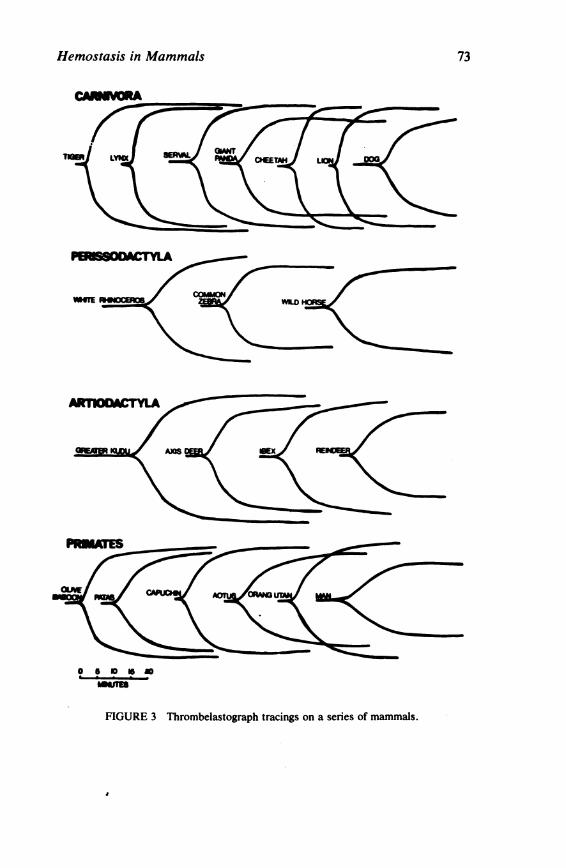
Hemostasis in Mammals 73
FIGURE 3 Thrombelastograph tracings on a series of mammals.
Copyright © National Academy of Sciences. All rights reserved.
Animal Models of Thrombosis and Hemorrhagic Diseaseshttp://www.nap.edu/catalog.php?record_id=19903

74 CHRISTINE HAWKEY
•
i 1- . . .
I.
FIGURE 4 Prothrombin (factor II) assays (shaded band represents the normal human range).
The rate of activation of prothrombin via the intrinsic pathway has been measured by the partial thromboplastin test 19 and by thrombelastography.20 For the partial thromboplastin test, human brain phospholipid has been used21 ; this reaction is not complicated by interspecies lack of cross-reactivity. Compared with man, the partial thromboplastin time in many species is short, suggesting a more rapid rate of generation of intrinsic prothrombin activator (Figure 5). This is particularly evident in Cebidae and Camivora and is confirmed by thrombelastography (Figure 3) which shows shorter reaction times (r values) and times reflecting the rate of increasing elasticity of the clots (K values) in most other species compared with man. Significantly prolonged partial thromboplastin times and abnormal thrombelastograph r times have been recorded in the species Tragelephas strepsiceros (greater kudu). These findings are explained by a deficiency of factor XI (see below).
Factor V Factor V activity in most mammals is higher than that found in human plasma (Figure 6). The possible influence of this on prothrombin conversion has already been discussed.
Factor Vil Assay of this factor is complicated by lack of availability of suitable substrate plasma for most groups of animals. Those tests
Copyright © National Academy of Sciences. All rights reserved.
Animal Models of Thrombosis and Hemorrhagic Diseaseshttp://www.nap.edu/catalog.php?record_id=19903

Hemostasis in Mammals 75
that were possible indicate that factor VII activity is higher in nonhuman primates and in most Camivora than in man. 1
Factor VIII Factor Vlll activity is very high in most nonhuman mammals. In many species the activity is ten times that found in normal human plasma (Figure 7). Factor VIII is one of the coagulation factors known to be increased in man by stress and by adrenaline infusion,22 and high levels of this factor have been suggested as a possible contributory cause of thrombosis. 23 •24 However, the factor VIII increases recorded in these situations in man are rarely more than 2-3 times the original level, so the activity remains far lower than is apparently normal for most other species. Since spontaneous thrombosis is rare in animals other than man, these findings suggest that high factor Vlll is not an importaat risk factor in thrombosis.
Factor IX The level of Factor IX in Pongidae and Cebidae is within the normal human range, but low activity has been recorded in the Cercopithecidae. In other mammals the level is generally high (Figure 8).
I j
I
FIGURE 5 Partial thromboplastin times (shaded band represents the normal human range).
Copyright © National Academy of Sciences. All rights reserved.
Animal Models of Thrombosis and Hemorrhagic Diseaseshttp://www.nap.edu/catalog.php?record_id=19903
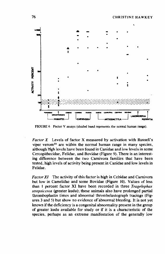
76
. .
... I
• -
CHRISTINE HAWKEY
-
I . :
FIGURE 6 Factor V assays (shaded band represents the normal human range).
Factor X Levels of factor X measured by activation with Russell's viper venom25 are within the normal human range in many species, although high levels have been found in Canidae and low levels in some Cercopithecidae, Felidae, and Bovidae (Figure 9). There is an interesting difference between the two Camivora families that have been tested, high levels of activity being present in Canidae and low levels in Felidae.
Factor XI The activity of this factor is high in Cebidae and Camivora but low in Camelidae and some Bovidae (Figure 10). Values of less than I percent factor XI have been recorded in three Tragelephas strepsiceros (greater kudu)~ these animals also have prolonged partial thromboplastin times and abnormal thrombelastograph tracings (Figures 3 and 5) but show no evidence of abnormal bleeding. It is not yet known if the deficiency is a congenital abnormality present in the group of greater kudu available for study or if it is a characteristic of the species, perhaps as an extreme manifestation of the generally low
Copyright © National Academy of Sciences. All rights reserved.
Animal Models of Thrombosis and Hemorrhagic Diseaseshttp://www.nap.edu/catalog.php?record_id=19903

Hemostasis in Mammals 77 . >-L • • • • - - • a • -
• • • • • • • •
•
Ii ~ • • •
~ I • a •
~ • •
•
FIGURE 7 Factor VIII assays (shaded band represents the normal human range).
levels found in Bovidae. It is interesting that congenital factor XI deficiency has been found in Holstein cattle. 28•27 These animals, however, suffer mild haemorrhagic symptoms.
Factor XII Factor XII activity is near to the normal human range in nonhuman primates but higher in most other groups studied (Figure 11). An interesting exception reported by Robinson, Kropatkin, and Aggeler28 and confirmed in this laboratory is that in members of the Cetacea (e.g., whales, dolphins) this factor is absent. Like human beings with congenital factor XII deficiency, these animals do not have an increased tendency to bleed. It has been suggested that activation of factor XII by acidosis during diving is a cause of the diffuse intra vascular coagulation in severe decompression sickness29 and that absence of factor XII may therefore be advantageous in diving mammals. However there is no evidence of a similar deficiency in members of the Pinnipedia (seals and sea lions).
In primates an inverse relationship has been found between factor
Copyright © National Academy of Sciences. All rights reserved.
Animal Models of Thrombosis and Hemorrhagic Diseaseshttp://www.nap.edu/catalog.php?record_id=19903

78 CHRISTINE HAWKEY
- •
• • .
FIGURE 8 Factor IX assays (shaded band represents the normal human range) .
•
-• • • . . - , _._" ............. ~.-- r r L-:~--:J ~NI~ L ARTI<XlACTYlA _J ~ ROOEHTlA
FIGURE 9 Factor X assays (shaded band represents the normal human range).
Copyright © National Academy of Sciences. All rights reserved.
Animal Models of Thrombosis and Hemorrhagic Diseaseshttp://www.nap.edu/catalog.php?record_id=19903

Hemostasis in Mammals 79
- • -
•
I . • • •
FIGURE JO Factor XI assays (shaded band represents the normal human range).
XII and plasminogen. This is interesting in light of the fact that factor Xlla is an activator of plasminogen. 30 This relationship does not hold for other groups of mammals examined. In cetaceans circulating plasminogen activator can be demonstrated in the absence of factor XII.
Fibrinolytic Activity
Blood Activator Circulating levels of plasminogen activator measured by a modification of the euglobulin lysis test 1 are within the normal human range in Pongidae, Carnivora, Cervidae, and some Cercopithecidae and Rodentia, but very little activity can be detected in Suidae, Camelidae, or Bovidae (Figure 12). Cebus monkeys have
, relatively high activity; this group therefore shows evidence of a coagulation/fibrinolysis equilibrium. In some mammals circulating plasminogen activator cannot be detected by the euglobulin lysis test; in elephants, for example, the euglobulin clot remains stable for longer than 48 hours.
Copyright © National Academy of Sciences. All rights reserved.
Animal Models of Thrombosis and Hemorrhagic Diseaseshttp://www.nap.edu/catalog.php?record_id=19903

80 CHRISTINE HAWKEY
=--t - -- • •
- , ------- r r L:,..::J l-..,J LMTKXMCTYLA_J LMDUMIA RCIDINTlA
FIGURE 11 Factor XII assays (shaded band represents the normal human range).
Plasminogen Plasminogen has been measured by caseinolysis after activation by human urokinase. Compared with man the level is high in nonhuman primates, Carnivora, Rodentia, Marsupialia, and some Perissodactyla; levels below the human range have been found in some Bovidae and in rabbits (Figure 13). Since the rate of lysis of artificially prepared in vitro thrombi increases directly in proportion to their plasminogen content, 31 it appears likely that high plasma plasminogen levels offer potential protection against intravascular thrombosis. This may be an important factor in explaining the rarity of spontaneous thrombosis in animals other than man.
Fibrinolytic Inhibitors No significant species differences in the inhibitory action of plasma on fibrinolysis has been found in our study.
DISCUSSION
Although our survey has so far covered only a small proportion of the more than 4,000 living species of mammals, some interesting and
Copyright © National Academy of Sciences. All rights reserved.
Animal Models of Thrombosis and Hemorrhagic Diseaseshttp://www.nap.edu/catalog.php?record_id=19903
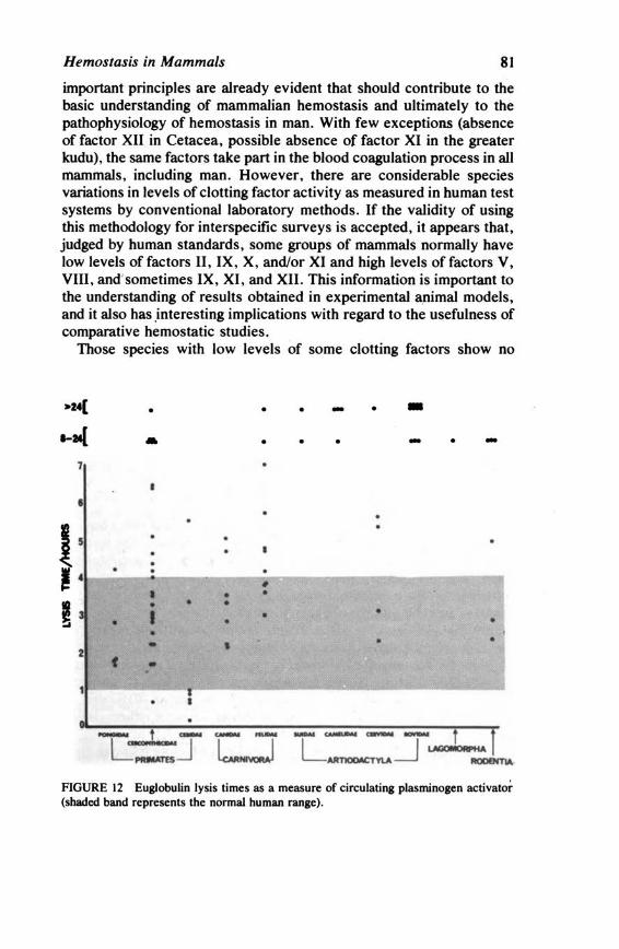
Hemostasis in Mammals 81
important principles are already evident that should contribute to the basic understanding of mammalian hemostasis and ultimately to the pathophysiology of hemostasis in man. With few exceptions (absence of factor XII in Cetacea, possible absence of factor XI in the greater kudu), the same factors take part in the blood coagulation process in all mammals, including man. However, there are considerable species variations in levels of clotting factor activity as measured in human test systems by conventional laboratory methods. If the validity of using this methodology for interspecific surveys is accepted, it appears that, judged by human standards, some groups of mammals normally have low levels of factors II, IX, X, and/or XI and high levels of factors V, VIII, and' sometimes IX, XI, and XII. This information is important to the understanding of results obtained in experimental a.nimal models, and it also has .interesting implications with regard to the usefulness of comparative hemostatic studies.
Those species with low levels of some clotting factors show no
>Z4[ • • • - • -1-M{ .. • • • - • -
7
• • 15 •
• • I , • • • • ~ • • •
I • • • " • • • 1 • • •
2 f • .
• - ' - - - ..... ~ mvmM ....-u: f L-:.::;J lu.._J L~_J~
FIGURE 12 Euglobulin lysis times as a measure of circulating plasminogen activator (shaded band represents the normal human range).
Copyright © National Academy of Sciences. All rights reserved.
Animal Models of Thrombosis and Hemorrhagic Diseaseshttp://www.nap.edu/catalog.php?record_id=19903

82 CHRISTINE HAWKEY
•
• 4 ' 5 . .
~ · •
i 3
I 2 •
FIGURE 13 Plasminogen levels (shaded band represents the normal human range).
evidence of liver disease or vitamin K deficiency, nor do they have an increased tendency to bleed, although in some instances the levels of activity detected would be incompatible with normal hemostasis in man. The deficiencies may therefore be counterbalanced by some other factor; the possibility that high levels of factor V compensate for reduced factor II has already been discussed. It would be of interest to study the kinetics of various stages of the coagulation reaction in species selected for differences in the activity of relevant clotting factors. This approach would help to establish the relatiye importance of individual clotting factors and might provide answers to questions such as why the hemorrhagic symptoms of factor VIII and IX deficiencies are worse than those of deficiencies of other clotting factors.
With regard to the high levels of some clotting factors found in many groups, it is difficult to know what this means in terms of hemostasis in the animals concerned, but the results of basic tests such as the whole blood clotting time and thrombelastography suggest that the potential
Copyright © National Academy of Sciences. All rights reserved.
Animal Models of Thrombosis and Hemorrhagic Diseaseshttp://www.nap.edu/catalog.php?record_id=19903

Hemostasis in Mammals 83
coagulation activity in these mammals is high compared with man; the results obtained in many species would be interpreted as indicating hypercoagulability if they were found in human patients. Either these animals are comparatively hypercoagulable or the test systems used are imperfectly understood. It is conceivable that wild animals in their natural habitat require a more efficient hemostatic mechanism than civilized man in his protected environment. However, by this reasoning, primitive man must have been relatively vulnerable; the apparent reduction in potential coagulation activity now evident may therefore be a secondary adaptatiorJ and, if so, the interesting questions of how and why this happened are raised. Also of great interest is the fact that thrombotic diseases are rare in mammals other than man; apparently, high clotting factor lev«;!s are not a primary risk factor in these diseases.
Apart from the symptomless factor XI deficiency found in the greater kudu, which may be an inherited defect, wild animals with congenital coagulation disorders have not been detected in our survey and have not been reported in the literature, although, judging by the fact that the mutations causing these conditions occur in man and domesticated animals, they probably have a widespread distribution. However, considering the hemorrhagic problems associated with these diseases, affected animals would be unlikely to survive the processes of natural selection in the wild. It is perhaps surprising that very few cases of congenital coagulation deficiencies have been described in laboratory animals; the probable explanation for this is that coagulationists do not usually work on rats and mice and people who do use these animals are not interested in the survival of individual animals with symptoms of bleeding. Acquired coagulation disorders in wild animals could theoretically be similar in etiology to those occurring in man, but it would appear important that species differences in baseline coagulation activity be taken into account when assessing these defects.
The main function of the blood fibrinolytic mechanism is probably to counterbalance coagulation and to prevent the buildup of intravascular fibrin deposits. The mechanism should therefore be present in all animals in which hemostasis involves the production of a fibrin clot. In our survey the inactive fibrinolytic precursor, plasminogen, has been found in all mammals tested, although circulating plasminogen activators cannot be detected in some species. There ·are interesting differences in the amount of plasminogen present. In man the level is relatively low; this may be significant as a risk factor for thrombosis, as the rate of lysis of artificial thrombi is dependent on the plasminogen
Copyright © National Academy of Sciences. All rights reserved.
Animal Models of Thrombosis and Hemorrhagic Diseaseshttp://www.nap.edu/catalog.php?record_id=19903
84 CHRISTINE HAWKEY
content. In nonhuman primates and Camivora, plasminogen is usually above the normai human range. Thus. these groups have high potential fibrinolytic activity, which may offset their relative hypercoagulability. These findings appear to support the hypothesis that coagulation and fibrinolysis are present in a balanced equilibrium; such an equilibrium cannot be demonstrated, however, in terms of circulating plasminogen activator levels. When measured by the euglobulin lysis test, activator levels are high only in the Cebidae among those groups of animals showing relatively increased coagulation activity. In some species, circulating activator is apparently very low, and in some it is undetectable. There may be methodological reasons for this since we have used standard conditions for precipitating the euglobulin fraction; these may not be optimal for all species. An alternative explanation that should be considered is that there are species differences in activator availability and release. A comparative study of this possibility would be of interest. ·
Less can be said about blood platelets because fewer species have been fully studied. Although it appears that platelets carry out similar hemostatic functions in all mammals, it is increasingly apparent \hat species differences in levels of active constituents and surface structure as well as in behavior in conventional laboratory tests, are widespread. A much wider comparative survey of platelet similarities and differences should be undertaken, not only to reduce the present difficulty in extrapolating results from one species to another' but also because the comparative approach is likely to contribute greatly to the understanding of relationships between platelet structure and function.
ACKNOWLEDGMENTS
This study has been supported by the British Heart Foundation and the Medical Research Council. We are grateful to the Veterinary Officers of the Zoological Society of London for supplying most of the blood samples.
REFERENCES
I. Hawkey, C. 1975. Comparative mammalian haematology. Heinneman Scientific and Medical Publications, London.
2. Mason, R. G., and M. S. Read. 1967. Platelet response to six agglutinating agents. Species similarities and differences. Exp. Mol. Pathol. 6:370.
3. Sinakos, Z., and J.P. Caen. 1967. Platelet aggregation in mammalians (human, rat, rabbit, guinea pig, horse, dog). A comparative study. Thromb. Diath. Haemorrh. 17:99.
4. Hawkey, C., and C. Symons. 1968. Variation in ADP-induced platelet aggregation in vitro in Primates as a result of differences in plasma ADP inhibitor levels. Thromb. Diath. Haemorrh. 19:29.
Copyright © National Academy of Sciences. All rights reserved.
Animal Models of Thrombosis and Hemorrhagic Diseaseshttp://www.nap.edu/catalog.php?record_id=19903

Hemostasis in Mammals 85
5. Tschopp, T. B. 1970. Aggregation of cat platelets in vitro. Thromb. Diath. Haemorrh. 23:601.
6. Mills, D. C. B. 1970. Platelet aggregation and platelet nucleotide concentration in different species. Symp. Zool. Soc. London No. 27:99.
7. Loeb, W. F., and B. Mackey. 1973. A comparative study ofp\atelet aggregation in Primates.-J. Med. Primatol. 2:195.
8. Donner, L., and J. Houskova. 1972. Some properties of blood platelets in animal species. Folia Haematol. (Leipzig) 98:296.
9. Mills, D. C. B., and D·. Thomas. 1969. Blood platelet nucleotides in man and other species. Nature 222:991. ·
10. Humphrey, J. H., and C. C. Toh. : 1954. Absorption of serotinin (5-hydroxytryptamine) and histamine by dog platelets. J. Physiol. 124:300.
11. Baumgartner, H. R., and G. V. R. Born. 1968. 5-hydroxytryptamine in rabbit · platelets and their aggregation. J. Physiol. 194:92.
12. Nurden, A. T. 1974. Observations on platelet macroglycopeptide. Nature 251:151. 13. Nurden, A. T. 1975. Personal communication. 14. Ratnoff, 0. D., and C. Menzies. 1951. A new method for the determination of fibrin
in small samples of plasma. J. Lab. Clin. Med. 37:316. 15. Denson, K., R. Borrett, and R. Biggs. 1971. The specific assay ofprothrombin using
Taipan snake venom. Br. J. Haematol. 21:219. 16. lrsigler, K., K. Lechner, and E. Deutsch. 1965. Studies on tissue thromboplastin. II.
Species specificity. Thromb. Diath. Haemorrh. 9:8. 17. Mann, F. D., and N. M. Hurn. 1952. Species specificity ofthromboplastin: Role of
the co-thromboplastin reaction. Proc. Soc. Exp. Biol. Med. 79: 19. 18. Hawkey, C., and J. L. Stafford. 1961. Influence of serum factors on the species
specificity of tissue thromboplastin. Nature 191 :920. · 19. Rodman, N. F., E. M. Barrow, and J.B. Graham. 1958. Diagnosis and control of the
haemophiloid states with the partial thromboplastin time (FIT) test. Am. J. Clin. Pathol. 29:525.
20. Hartert, H. 1948. Blutgerinnungsstudien mit der Thrombelastographie, einen neuen Untersuchungsverfahren. Klin. Wochenschr. 25:577.
21. Bell, W. N., and H. G. Alton. 1954. A brain extract as a substitute for platelet suspensions in the thromboplastin generation test. Nature 174:880.
22. Ingram, G. I. C. 1961. Increase in antihaemophilic globulin following infusion of adrenaline. J. Physiol. 156:217.
23. Egeberg, 0. 1962. Changes in the coagulation system following major surgery. Acta Med. Scand. 17:679.
24. Penick, G.D., I. Dejanov, H. R. Roberts, and W. P. Webster. 1966. Elevation of factor VIII in hypercoagulable states. Thromb. Diath. Haemorrh. 20:39.
25. Denson, K. W. 1961. The specific assay of Prower-Stuart factor and factor VII. Acta Haematol. 25: 105. ·
26. Kociba, G. J., 0. D. Ratnoff, W. F. Loeb, R. L. Wall, and L. E. Heider. 1969. Bovine plasma thromboplastin antecedent (factor XI) deficiency. J. Lab. Clin. Med. 74:37.
27. Gentry, P. A., S. Crane, and F. Lotz. 1975. Factor XI (plasma thromboplastin antecedent) deficiency in cattle. J. Can. Vet. Med. Assoc. 16:160.
28. Robinson, A. J., M. Kropatkin, and P. Aggeler. 1969. Hageman factor (factor XII) deficiency in marine mammals. Science 166: 1420.
29. Holland, J. A. 1969. Discussion of disseminated intravascular coagulation in decompression sickness. U.S. Naval Submarine Medical Center Report No. 585. Groton, Conn.
Copyright © National Academy of Sciences. All rights reserved.
Animal Models of Thrombosis and Hemorrhagic Diseaseshttp://www.nap.edu/catalog.php?record_id=19903
86 CHRISTINE HAWKEY
30. latridis, S. G., and J. H. Ferguson. 1961. Effect of surface and Hageman factor on the endogenous or spontaneous activation of the fibrinolytic system. Thromb. Diath. Haemorrh. 6:411.
31. Hawkey, C. 1965. The effect in man of plasminogen activator on coagulation and fibrinolytic factors. Thesis. University of London.
BIBLIOGRAPHY
Abildgaard, C. F., J. Harrison, and C. A. Johnson. 1971. Comparative study of blood coagulation in nonhuman primates. J. Appl. Physiol. 30:400.
Archer, R. K. 1970. Blood coagulation in nonhuman vertebrates. Symp. Zool. Soc. Lond. 27:121.
Birndorf, N. I., J. D. Pearson, and A. Wredman. 1971. The clotting system of monkeys. A comparison of coagulation factors and tests between cynomologous monkeys (Macaca irus) and humans. Comp. Biochem. Physiol. 38:157.
Bolton, C. H., and P. R. Emmons. 1967. Adenosine diphosphate breakdown by the plasma of different species and by human whole blood and white cells. Thromb. Diath. Haemorrh. 18:799.
Cartwright, T., and R. G. 0. Kekwick. 1971. A comparative study of human, cow, pig, and sheep fibrinogen. Biochem. Biophys. Acta 236:550.
Clift\on, E. E., and G. Mootse. 1967. Species variation in fibrinolytic activity. Thromb. Diath. Haemorrh. 18:291.
Didisheim, P., K. Hattori, and J. H. Lewis. 1959. Haematologic and coagulation studies in various animal species. J. Lab. Clio. Med. 53:866.
Fanti, P. 1961. A comparative study of blood coagulation in vertebrates. Aust. J. Exp. Biol. 39:403.
Fanti, P., and H. A. Ward. 1957. Comparison of blood clotting in marsupials and man. Aust. J. Exp. Biol. 35:209.
Gallimore, M. J., M. V. Nulkar, and J. J.B. Shaw. 1965. A comparative study of the inhibitors offibrinolysis in human, dog, and rabbit blood. Thromb. Diath. Haemorrh. 14:145.
Hampton, J. W., and C. Matthews. 1966. Similarities between baboon and human blood clotting. J. Appl. Physiol. 21:1713.
Hawkey, C. 1970. Fibrinolysis in animals. Symp. Zool. Soc. Lond. 27:133. Hawkey, C. 1974. The relationship between blood coagulation and thrombosis and
atherosclerosis in man, monkeys, and carnivores. Thromb. Diath. Haemorrh. 31:103. Mason, R. G., and M. S. Read. 1971. Some species differences in fibrinolysis and blood
coagulation. J. Biomed. Res. 5:121. Osbaldiston, G. W., E. C. Stowe, and P. R. Griffiths. 1970. Blood coagulation.
Comparative studies in dogs, cats, horses, and cattle. Br. Vet. J. 126:512. Quick, A. J. 1941. The prothrombin concentration in the blood of various species. Am. J.
Physiol. 132:239. Seaman, M. J., and M. R. Malinow. 1968. Blood clotting in nonhuman primates. Lab.
Anim. Care 18:80. Stormorken, H. 1958. Species differences of clotting factors in ox, dog, horse, and sheep.
Acta Physiol. Scand. 41:301.
Copyright © National Academy of Sciences. All rights reserved.
Animal Models of Thrombosis and Hemorrhagic Diseaseshttp://www.nap.edu/catalog.php?record_id=19903

JACK LEVIN, M.D.
Blood Coagulation in the Horseshoe 'crab (Limulus polyphemus): A Model for Mammalian Coagulation and Hemostasis
COAGULATION IN Limulus
Amebocytes
The amebocyte is the only type of circulating cell in the blood of Limulus polyphemus, the horseshoe crab.* It is a nucleated cell, the cytoplasm of which is packed with granules (Figure 1). When Limulus blood is shed or exposed to bacterial endotoxins, there is rapid aggregation of the amebocytes. Aggregation is associated with marked changes in cell shape, flattening with an- apparent increase in cell diameter, development of long processes that extend from cell to cell, and striking degranulation 1- 3 (Figure 2).
Although the important role of amebocytes in the coagulation of the blood of Limulus has been recognized for many years, 4 - 7 it was not until the development of new techniques that it was unequivocally shown that amebocytes contain all components of the coagulation system of Limulus blood.8 •9 Not only the clottable protein but all other
Supported in part by Research Grant HL 01601 from the National Heart and Lung Institute, National Institutes of Health, Bethesda, Maryland. Dr. Levin was a John and Mary R. Markle Scholar in Academic Medicine during the performance of some of these studies and is the recipient of a Research Career Development A ward (K04 29906) from the National Heart and Lung Institute.
*The term horseshoe crab is a misnomer in the zoological sense because Limulus is not a crustacean, but a member of the class Merostomata (subclass, Xiphosura).
87
Copyright © National Academy of Sciences. All rights reserved.
Animal Models of Thrombosis and Hemorrhagic Diseaseshttp://www.nap.edu/catalog.php?record_id=19903

88 JACK LEVIN
FIGURE I Photomicrograph of a single Limulus amebocyte . Densely packed granules are present in the cytoplasm of the amebocyte (magnification x 4,300, using a polarizing microscope).
factors required for coagulation are contained within amebocytes. Cell-free plasma does not clot and is not required for coagulation, since extracts of washed amebocytes (amebocyte lysate) are gelled following incubation with bacterial endotoxins . 8 •11 Therefore , it is important to discriminate between aggregation of amebocytes and the subsequent coagulation of blood, which can occur only after disruption of amebocytes and their granules (see below) , with release of the various factors required for coagulation.
The morphological prominence of granules in the cytoplasm and their rapid disruption and disappearance during aggregation of amebocytes (at a time when other investigations indicated that the coagulation system was being released from amebocytes) led to studies of their possible role in the coagulation of Limulus blood. Utilization of
Copyright © National Academy of Sciences. All rights reserved.
Animal Models of Thrombosis and Hemorrhagic Diseaseshttp://www.nap.edu/catalog.php?record_id=19903
Blood Coagulation in the Horseshoe Crab 89
propranolol and a sucrose gradient that contained heparin allowed preparation of amebocyte granules essentially free of other cellular components. to Studies of extracts of washed granules have demonstrated that the granules contain substances that are apparently identical to those present in lysates of washed amebocytes, as indicated by analytical ultracentrifugation and polyacrylamide gel electrophoresis. to Extracts of granules are gelled by bacterial endotoxin, in a manner similar to the reaction between endotoxin and amebocyte lysate. Therefore, it appears that the factors required for blood coagulation in Limulus are contained within the cytoplasmic granules of the amebocytes and are released into the surrounding medium (i.e., into the plasma, in vivo) following disruption or'the granules.
The number and nature of the factors that constitute the coagulation system of Limulus are still unknown. However, some important data are available. Column chromatography using Sephadex G-50 has revealed three protein peaks, one of which contains the clottable protein. 11 The molecular weight of the clottable protein is approximately 25,000.11·t 2 Another fraction contains other factors that are necessary if coagulation is to occur. Coagulation produced by endotoxin is the apparent result of activation by endotoxin of an enzyme or series of enzymes that in tum react with -the clottable protein.tr Endotoxin does not react directly with the clottable protein. The reaction, the rate of which depends upon the concentration of endotoxin, provides the basis for the most sensitive in vitro assay for endotoxin now available. t3•14 Following coagulation, the clottable protein is not detectable in the serum. The gel that forms is stable, but mechanically fragile. Furthermore, following mechanical disruption, the gel does not re-form but remains a viscous, semifluid mass. These gels do not retract.
Summary
These data indicate that the coagulation system of Limulus is contained exclusively within the amebocytes, which circulate in the blood. Recent studies suggest that factors necessary for blood coagulation are localized within the cytoplasmic granules of amebocytes. Aggregation and disruption of cells follow exposure of amebocytes to foreign surfaces. This is not prevented by nonwettable surfaces, heparin, or agents that chelate calcium. Endotoxin produces similar changes and, in addition, results in coagulation of the blood. Heparin or calcium and magnesium-free solutions do not prevent coagulation. In the absence of endotoxin, disruption of amebocytes, in vitro, is not followed by the coagulation of blood, despite the release into the plasma of the factors
Copyright © National Academy of Sciences. All rights reserved.
Animal Models of Thrombosis and Hemorrhagic Diseaseshttp://www.nap.edu/catalog.php?record_id=19903

90 JACK LEVIN
required for coagulation. 8 •9 N-ethyl maleimide, which effectively prevents aggregation and disruption of amebocytes , does not block the reaction between endotoxin and amebocyte lysate. 11
Although it is reasonable to presume that other exogenous agents produce disruption of amebocytes and/or coagulation, this is difficult to evaluate because of the ubiquitous nature of endotoxins, the contamination by endotoxin of many reagents and of equipment not specifically prepared to b.e endotoxin-free, and the marked sensitivity of amebocytes or their extracts to minute concentrations of bacterial endotoxins.
COAGULATION IN Limulus AS A MODEL FOR MAMMALIAN
COAGULATION AND HEMOSTASIS
The Shwartzman reaction is a model used by many investigators to study hemorrhage and thrombosis. The similarity between this reaction
• __ J Copyright © National Academy of Sciences. All rights reserved.
Animal Models of Thrombosis and Hemorrhagic Diseaseshttp://www.nap.edu/catalog.php?record_id=19903

Blood Coagulation in the Horseshoe Crab 91
FIGURE 2 Photomicrographs of Limulus amebocytes obtained with a phase-contrast microscope . After Limulus blood was shed , the amebocytes ofteri developed long filamentous processes (Figure 2A) (magnification x 180). At the same time, the cells aggregated (Figure 28). degranulated. and assumed irregular and varied shapes (Figure 2C) (magnification x 423). (Modified with permission from Levin . J., and F. B. Bang. A description of cellular coagulation in the Limulus . Bull . Johns Hopkins Hosp. 115:337-345, 1964. © The Johns Hopkins University Press.)
Copyright © National Academy of Sciences. All rights reserved.
Animal Models of Thrombosis and Hemorrhagic Diseaseshttp://www.nap.edu/catalog.php?record_id=19903
92 JACK LEVIN
to endotoxin in rabbits1s·18 and the response of the coagulation system of Limulus to gram-negative organisms or endotoxin (Table I) was recognized by Dr. Frederik Bang17 and led to subsequent studies of blood coagulation in this species. 1•8 ·u It is currently believed (although it has not been proven) that endotoxemia, associated with gramnegative infection, also activates coagulation in man, with production of thrombocytopenia, hypofibrinogenemia, and the syndrome now defined as "disseminated intra vascular coagulopathy." 18•1 u
Table 2 provides a comparison between Limulus amebocytes and mammalian platelets. In both limuli and mammals, disruption of the integrity of the circulatory syste~ or exposure of blood to foreign surfaces results in aggregation of amebocytes or platelets, with subsequent changes in shape, disruption of their granules, and release of cellular constituents into the surrounding environment. Some platelet factors (i.e., platelet factor 3 and ADP) accelerate or facilitate hemostasis and coagulation .. Booyse et al. 20 have reported that following activation, platelets extrude a material they have designated "cytogel"; their photomicrographs of this phenomenon resemble those we have obtained in our studies of amebocytes. Both cell types ~e capable of preventing loss of blood from damaged blood vessels.
Both cells are required for hemostasis but, in addition, amebocytes are necessary for the coagulation of blood, since they contain the factors required for coagulation. True coagulation can occur only after these components have been released into the plasma of Limulus. In contrast, in mammals the coagulation factors are present in solution in the blood plasma. Both amebocytes and platelets contain clottable protein, but in Limulus the amebocyte is the sole source of the clottable protein, whereas the mammalian platelet is associated with only a small proportion of the fibrinogen present in blood. 21 The presence of the entire coagulation system, including the clottable protein, in the amebocytes provides the basis for suggesting that coagulation in animals was initially a cellularly based function and that, as evolution
TABLE 1 Effects of Gram-Negative Infection or Endotoxin in Limulus
Amebocytopenia Intravascular coagulation Clots contain gram-negative organisms Blood becomes incoagulable Death
Copyright © National Academy of Sciences. All rights reserved.
Animal Models of Thrombosis and Hemorrhagic Diseaseshttp://www.nap.edu/catalog.php?record_id=19903

Blood Coagulation in the Horseshoe CraJ? 93
TABLE 2 Comparison of Limulus Amebocytes and Mammalian Platelets
Limulus Mammalian Characteristics or Functions Amebocyte Platelet
Hemostasis Essential Essential Blood coagulation Essential Ancillary Clottable protein Yes Yes Nucleus Yes No Viscous metamorphosis Yes(?) Yes Cellular processes Yes Yes Granules Yes. Yes Response to endotoxin Yes Yes Phagocytosis No(?) Yes Antibacterial function Yes Yes
occurred, this function gradually shifted into the plasina. 21.23 Limulus also provides a biological precedent for the concept that mammalian platelets contain functionally significant fibrinogen. 24
Amebocyte'> and platelets both contain granules, and both degranulate during the initial stages of blood coagulation. Clotfable protein is present in these granules, and a significant proportion of the fibrinogen in platelets is present in their granules. 21 However, whereas amebocyte granules apparently primarily contain coagulation factors, the granules of mammalian platelets contain a variety of substances, such as serotonin, that may have no direct relatior:iship to coagulation. Platelet granules alsQ contain hydrolytic enzymes characteristic of lysosomes. 25 In contrast, preliminary studies indicate that betaglucuronidase is not present in ameb9cyte granules. 10
Phagocytosis has not been reported in amebocytes, although definitive studies have not yet been done. 26.27 In contrast, although this is not considered one of their primary functions, platelets are capable of phagocytosis and have been shown to have limited bactericidal activity. 28 Amebocytes, which clearly play an important role in the control of bacterial infection by sealing wounds and trapping bacteria in masses of ge12u 7 also have been shown to have some bactericidal activity. 27
Endotoxin, a lipopolysaccharide present in the cell wall of all gram-negative organisms, is capable of interacting with amebocytes or platelets and of triggering blood coagulation. However, there are considerable species differences in the ability of endotoxin to produce
Copyright © National Academy of Sciences. All rights reserved.
Animal Models of Thrombosis and Hemorrhagic Diseaseshttp://www.nap.edu/catalog.php?record_id=19903
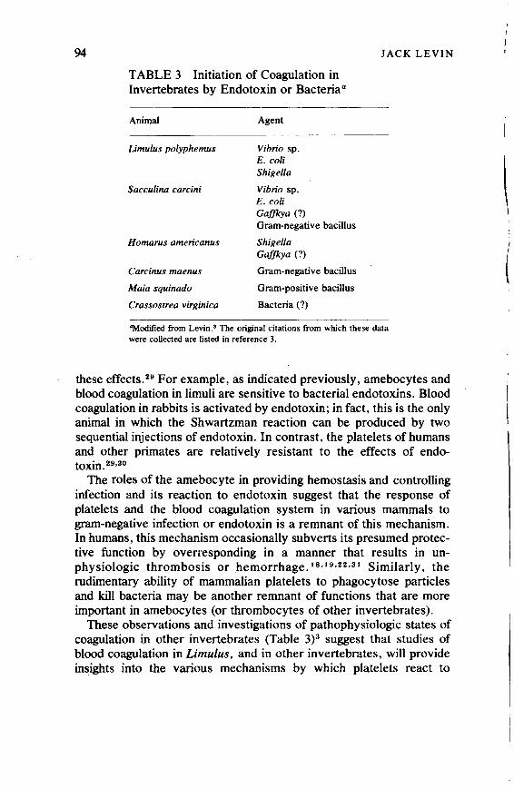
94
TABLE 3 Initiation of Coagulation in Invertebrates by Endotoxin or Bacteria a
Animal
Limulus polyphemus
Sacculina carcini
Homarus americanus
Carcinus maenus
Maia squinado
Crassostrea virginica
Agent
Vibrio sp. E.coli Shigella
Vibrio sp. E.coli Gaffkya (?) Gram-negative bacillus
Shigella Gaffkya (?)
Gram-negative bacillus
Gram-positive bacillus
Bacteria (?)
JACK LEVIN
"Modified from Levin.• The original citations from which these data were collected are listed in reference 3.
these effects. 29 For example, as indicated previously, amebocytes and blood coagulation in limuli are sensitive to bacterial endotoxins. Blood coagulation in rabbits is activated by endotoxin; in fact, this is the only animal in which the Shwartzman reaction can be produced by two sequential injections of endotoxin. In contrast, the platelets of humans and other primates are relatively resistant to the effects of endotoxin. 29•30
The roles of the amebocyte in providing hemostasis and controlling infection and its reaction to endotoxin suggest that the response of platelets and the blood coagulation system in various mammals to gram-negative infection or endotoxin is a remnant of this mechanism. in humans, this mechanism occasionally subverts its presumed protective function by overresponding in a manner that results in unphysiologic thrombosis or hemorrhage. 18 • 19 •22 •31 Similarly, the rudimentary ability of mammalian platelets to phagocytose particles and kill bacteria may be another remnant of functions that are more important in amebocytes (or thrombocytes of other invertebrates).
These observations and investigations of pathophysiologic states of coagulation in other invertebrates (Table 3)3 suggest that studies of blood coagulation in Limulus, and in other invertebrates, will provide insights into the various mechanisms by which platelets react to
\
i
Copyright © National Academy of Sciences. All rights reserved.
Animal Models of Thrombosis and Hemorrhagic Diseaseshttp://www.nap.edu/catalog.php?record_id=19903
Blood Coagulation in the Horseshoe Crab 95
different stimuli and by which mammalian blood coagufation is initiated.
REFERENCES
I. Levin, J., and F. B. Bang. 1964. A description of cellular coagulation in theLimulus. Bull. Johns Hopkins Hosp. 115:337-345.
2. Dumont, J. N .. E. Anderson, and G. Winner. 1966. Some cytologic characteristics of the hemocytes of Limulus during clotting. J. Morphol. 119: 181-207.
3. Levin, J. 1967. Blood coagulation and endotoxin in invertebrates. Fed. Proc. 26:1707-1712.
4. Howell, W. H. 1885. Observations upon the chemical composition and coagulation of the blood of Limulus polyphemus, Callinectes hastatus, and Cucumaria sp. Johns Hopkins Univ. Circ. 5:4-5.
5. Loeb, L. 1904. On the spontaneous agglutination of blood cells ofarthropods. Univ. Pa. Med. Bull. 16:441-443.
6. Maluf, N. S. R. 1939. The blood of arthropods. Q. Rev. Biol. 14:149-191. 7. Copley, A. L. 1952. The rheology of blood. A survey. J. Colloid Sci. 7:323-333. 8. Levin, J., and F. B. Bang. 1964. The role of endotoxin in the extracellular
coagulation of Limulus blood. Bull. Johns Hopkins Hosp. 115:265-274. 9. Levin, J., and F. B. Bang. 1968. Clottable protein in Limulus: ·Its localization and
kinetics of its coagulation by endotoxin. Thromb. Diath. Haemorrh. 19:18(rl97. 10. Miirer, E. H., J: Levin, and R. Holme. 1975. Isolation and studies of the granules of
the amebocytes of Limulus polyphemus, the horseshoe crab. J. Cell. Physiol. 86:533-542.
II. Young, N. S., J. Levin, and R. A. Prendergast. 1972. An invertebrate coagulation system activated by endotoxin: Evidence for enzymatic mediation. J.·Clin. Invest. 51:1790--1797.
12. Solum, N. 0. 1973. The coagulogen of Limulus polyphemus hemocytes. A comparison of the clotted and non-clotted forms of the molocule. Thromb. Res. 2:55-69.
13. Levin, J .. P.A. Tomasulo, and R. S. Oser. 1970. Detection of endotoxin in human blood and demonstration of an inhibitor. J. Lab. Clio. Med. 75:903-911.
14. Cooper, J. F., J. Levin, and H. N. Wagner, Jr. 1971. Quantitative comparison of in vitro and in vivo methods for the detection of endotoxin. J. Lab. Clio. Med. 78:138-148.
15. Levin, J., and L. E. Cluff. 1965. Platelets and the Shwartzman phenomenon. J. Exp. Med. 121:235-246.
16. Levin, J., and E. Beck. 1966. A re-appraisal of the role of blood coagulation and platelets in the generalized Shwartzman phenomenon. Thromb. Diath. Haemorrh. 15:519-538.
17. Bang. F. B. 1956. A bacterial disease ofLimulus polyphemus. Bull. Johns Hopkins Hosp. 98:325-351.
18. Rosner, F., and N. D. Ritz. 1966. The defibrination syndrome. Arch. Int. Med. 117:17-24.
19. Deykin, D. 1970. The clinical challenge of disseminated intravascular coagulation. N. Engl. J. Med. 283:63~.
20. Booyse, F. M., T. P. Hoveke, D. Kisieleski, and M. E. Rafelson, Jr. 1972. Mechanism and control of platelet-platelet interaction I. Effects of inducers and inhibitors of aggregation. Microvasc. Res. 4:179-198.
Copyright © National Academy of Sciences. All rights reserved.
Animal Models of Thrombosis and Hemorrhagic Diseaseshttp://www.nap.edu/catalog.php?record_id=19903

96 JACK LEVIN
21. Nachman, R. L., and A. J. Marcus. 1968. Immunological studies of proteins associated with the subcellular fractions of thrombasthenic and afibrinogenaemic platelets. Br. J. Haemat. 15: 181-189.
22. Lalci, K. 1972. Our ancient heritage in blood clotting and some of its consequences. Ann. N. Y. Acad. Sci. 202:297-307.
23. Quick, A. J. 1967. Hemostasis as an evolutionary development. Thromb. Diath. Haemorrh. 18: 1-11.
24. Levin. J., and F. B. Bang. 1966. A comparison between human blood platelets and Limulus amebocytes. Blood 28:984-985 (abstract).
25. Marcus, A. J., and M. B. Zucker. 1965. The physiology of blood platelets. Grune & Stratton, New York. p. 9.
26. Loeb, L. 1902. On the blood lymph cells and inflammatory processes of Limulus. J. Med. Res. 7:145-158.
27. Shirodkar, M. V., A. Warwick, and F. B. Bang. 1960. The in vitro reaction of Limulus amebocytes to bacteria. Biol. Bull. 118:324-337.
28. Kahn, R. A., and L. J. Flinton. 1974. The relationship between platelets and bacteria. Blood 44:715-721.
29. Spielvogel, A. R. 1967. An ultrastructural study of the mechanisms of plateletendotoxin interaction. J. Exp. Med. 126:235-250.
30. Ream, V. J., D. Deykin, V. Gurewich, and S. Wessler. 1965. The aggregation of human platelets by bacterial endotoxin. J. Lab. Clin. Med. 66:245-252.
· 31. Thomas, L. 1972. Notes of a biology-watcher. Germs. N. Engl. J. Med. 287:553-555.
BIBLIOGRAPHY
Belamarich, F. A., R. A. Stiller, and D. Shepro. 1973. The release reaction: ls it a common mechanism in all types ofhemostatic cells? Ser. Haematol. 6:418-428.
Gaintner, J. R., D. P. Jackson, and C. L. Conley. 1962. Morphologic studies of clot retraction. Bull. Johns Hopkins Hosp. 111:266-278.
Horowitz, H. l., R. M. Des Prez, and E.W. Hook. 1962. Effects of bacterial endotoxin on rabbit platelets. II. Enhancement of platelet factor 3 activity in vitro and in vivo. J. Exp. Med. 116:619-633.
Kenney, D. M., F. A. Belamarich, and D. Shepro. 1972. Aggregation of horseshoe crab (Limulus polyphemus) amebocytes and reversible inhibition of aggregation by EDTA.
Biol. Bull. (Woods Hole, Mass.) 143:548-567. Loeb, L. 1910. Uber die Blutgerinnung bei Wirbellosen. Biochem. Z. 24:487-495. Loeb, L. 1927. Amoeboid movement and agglutination in amoebocytes of Limulus and
the relation of these processes to tissue formation and thrombosis. Protoplasma 2:512-553.
McKay, D. G .• and S. S. Shapiro. 1958. Alterations in the blood coagulation system induced by bacterial endotoxin. I. In vivo (generalized Shwartzman reaction). J. Exp. Med. 107:353-376.
Pearson, A. D .• M. W. Calder, and P. D. Meers. 1973. Assay ofendotoxin (letter to the editor). Lancet 1:1194-1195.
Yoshikawa, T., K. R. Tanaka, and L. B. Guze. 1971. Infection and disseminated intravascular coagulation. Medicine 50:237-258.
Copyright © National Academy of Sciences. All rights reserved.
Animal Models of Thrombosis and Hemorrhagic Diseaseshttp://www.nap.edu/catalog.php?record_id=19903

Discussion
DISCUSSION A COMMENT ON THE ULTRASTRUCTURE OF AMEBOCYTES
FROM THE HORSESHOE CRAB (Limulus polyphemus)
James G. White, M.D.
97
Dr. Levin has set forth both here and in his previous work lucid and convincing arguments for consideriiig the process of coagulation in the horseshoe crab (Limulus polyphemus) as a model for human hemostasis. 1- 3 Ample support for this position has been obtained in the course of invc:stigations carried out in other laboratories. 4 --e Although my experience with nonmammalian thrombocytes is very limited, I concur in emphasizing the importance of investigations into host defense mechanisms protecting the integrity of the vascular system in nonmammals. In addition, I would like to urge the extension of such studies into other areas basic to the understanding of human biology and pathology. I have in mind two problems that are of particular interest: the secretory process and the mechanism of sol-gel transformation.
The transport of chemical substances confined to specific storage organelles from a cytoplasmic location to the exterior of cells is a generally accepted phenomenon throughout nature. Yet, the fundamental mechanisms involved i~ the secretory process remain obscure. For example, Grette7 was the first worker to characterize the extrusion in parallel of specific chemical substances from blood platelets without loss of cytoplasmic constituents, a process he termed the release reaction. Subsequent efforts have established the critical role of secretion in the platelet hemostatic response8 and failure of platelet secretion as a pathological basis for some hemorrhagic disorders. 9 While considerable progress has been made, we still lack a clear understanding of precisely how the products stored in platelet secretory organelles reach the exterior after stimulation of the cells by aggregating agents. 10
Aniebocytes constitute approximately 90 percent of the cells in the hemolymph of the horseshoe crab. The hemolymph lacks a clottable protein, and hemostasis is entirely dependent on the response of amebocytes to vascular injury. Adhesion and aggregation of amebocytes at the site of vessel damage provides a first line of defense. Secretion of clottable protein from
· storage organelles within the cell supplies additional protection as well as a means of sealing off invasive foreign particulates such as endotoxin. Thus, amebocyte secretion is a critical facet of the limited defense mechanisms in this primitive species.
The secretory p!Qcess in amebocytes, as in most cells, involves the movement of secretory organelles to the surface, fusion of membranes enclosing storage contents with the cell wall, and extrusion of the storage products into the surrounding hemolymph (Figure 1). 2•4•8 Initial investigations into amebocyte secretion suggest that the membrane-membrane interaction
Copyright © National Academy of Sciences. All rights reserved.
Animal Models of Thrombosis and Hemorrhagic Diseaseshttp://www.nap.edu/catalog.php?record_id=19903
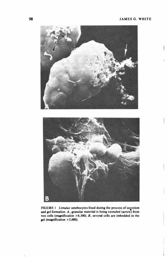
98 JAMES G. WHITE
FIGURE I Limulus amebocytes fixed during the process of se~retion and gel formation . A, granular material is being extruded (arrow) from two cells (magnification x6,500). B, several cells are imbedded in the gel (magnification x3,000).
Copyright © National Academy of Sciences. All rights reserved.
Animal Models of Thrombosis and Hemorrhagic Diseaseshttp://www.nap.edu/catalog.php?record_id=19903

Discussion 99
leading to fusion of storage granules with the cell wall is relatively simple compared to similar events in mammalian cells. Secretion can occur in the absence of extracellular calcium and is not inhibited by most of the agents which block secretion in human blood cells .2.4-e Careful study of secretion in the amebocyte may provide clues essential for our understanding of the more complex but similar process in mammalian cell systems.
Sol-gel transformation is also a basic process common to nearly all biological systems. Cell movement, cytoplasmic streaming, cytoskeletal support , and blood coagulation are typical manifestations of this phenomenon. Sol-gel transformation can be involved in the genesis of human disease, and sickle cell anemia is a classic example. 11 Conversion of liquid hemoglobin inside sickled erythrocytes into a gel of rodlike polymers after exposure to low oxygen tension causes distortion of the cells, development of extreme rigidity. early removal by the reticuloendothelial system, and occlusion of the microcirculation (Figure 2). Thus, in si~kle cell anemia sol-gel transformation is a pathological process resulting in all of the morbid features of the disease .
Efforts to define the nature of the sickling process have generally been confined to intact erythrocytes and cell-free hemoglobin solutions from patients with sickle cell anemia. Animal models of sickle cell anemia have been reported , 12 but the conditions under which erythrocyte sickling develops and the structure of the polymers formed differ significantly from those observed in
·,'.
AGURE 2 Thin section of a sickled human erythrocyte (£). The rodlike polymers (P) of sickled hemoglobin extend in all directions but remain separated from each other (magnification x 27 ,000).
Copyright © National Academy of Sciences. All rights reserved.
Animal Models of Thrombosis and Hemorrhagic Diseaseshttp://www.nap.edu/catalog.php?record_id=19903

100 JAMES G. WHITE
FIGURE 3 Thin section of Limulus amebocyte. Portions of three granules are shown. The internal matrix of granule I is homogeneous. Granules 2 and 3 contain tubular polymers (P) similar in many respects to polymers of sickled hemoglobin (magnification x33,000).
the human disease. No attention has yet been given to the possibility that polymerization of nonhemoglobin proteins might provide a useful approach to understanding the basic process of sol-gel transformation as well as the pathogenesis of sickle cell disease.
It is of some interest, therefore, to observe the transformation that occurs within the storage granules of amebocytes from the horseshoe crab before and during the process of their extrusion from the cytoplasm (Figure 3). 4 The protein content of the organelles is homogeneous in undisturbed organelles and resembles in appearance hemoglobin in thin sections of normal human red blood cells. 11 Before and during secretion, however, the homogeneous material in the granules is transformed into rodlike polymers that are strikingly similar to those observed inside sickled erythrocytes and cell-free solutions of sickled hemoglobin from human patients. The granule transformation must occur very rapidly. because organelles containing polymers are evident in amebocytes fixed immediately after aspiration. Labilization of granule membranes, akin to the response of lysosomes in human leukocytes after ingestion of bacteria, is probably involved in initiating granule transformation. However, the subsequent polymerization of granule protein appears to be spontaneous. As a result, the process is of great interest to those concerned about the nature of sol-gel transformation and the pathogenesis of human sickle cell disease.
Copyright © National Academy of Sciences. All rights reserved.
Animal Models of Thrombosis and Hemorrhagic Diseaseshttp://www.nap.edu/catalog.php?record_id=19903

Discussion 101
I hope that this comment will stimulate other workers to study the amebocyte. Investigations of fundamental processes in these simple cells may help clarify phenomena involved in the genesis of human disease.
REFERENCES
l. Levin, J., and F. B. Bang, 1964. A description of cellular coagulation in the Limulus .. Bull. Johns Hopkins Hosp. 115:337-345.
2. Levin, J., and F. B. BaJ)g. 1966. A comparison between human blood platelets and Limulus amebocytes. Blood 28:984-985.
3. Levin, J., and F. B. Bang. 1968. Clottable protein in Limulus: Its localization and kinetics of its coagulation of endotoxin. Thromb. Diath. Haemorrh. 19:18~197.
4. Dumont, J. N., E. Anderson, and G. Winner. 1966. Some cytologic characteristics of the hemocytes of limulus during clotting. J. Morph. 119: 181-208.
5. Kenney, D. M .. F. A. Belamarich, and D. Shepro. 1972. Aggregation of horseshoe crab (Limulus polyphemus) amebocytes and reversible inhibition of aggregation by EDTA. Biol. Bull. (Woods Hole, Mass.) 143:548-567.
6. Belamarich, F. A., R. A. Stiller, and D. Shepro. 1973. The release reaction: Is it a common mechanism in all kinds of hemostatic cells? Ser. Haematol. 6:418-428.
7. Grette, K. 1962. Studies on the mechanism of thrombin-catalyzed hemostatic reactions in blood platelets. Acta Physiol. Scand. (Suppl). 56:1-195.
8. Holmsen, H., H. J. Day, and H. Stormorken. 1969. The blood platelet release reaction. Scand. J. Haematol. (Suppl.) 8: 1-26.
9. Weiss, J. 1972. Abnormalities in platelet function due to defects in the release reaction. Ann. N.Y. Acad. Sci. 201:161-173.
10. White, J. G. 1974. Electron microscopic studies of platelet secretion. Prog. Hemostasis Thromb. 2:49-98.
11. White, J. G. 1974. Ultrastructural features of erythrocyte and hemoglobin sickling. Arch. Intern. Med. 133:545-562.
12. Taylor, W. J., and C. W. Easley. 1974. Sickling phenomena of deer. Ann. N.Y. Acad. Sci. 241:59~.
DISCUSSION NATURE OF THE CONTENTS OF LARGE GRANULES OF Limulus AMEBOCYTES
Frank A. Belamarich, Ph.D.
Examination of Limulus amebocytes at the Boston University Marine Program, Woods Hole, has revealed differences in the nature of the contents of the large granules that occur in these cells. This has also been shown by Dumont, Anderson, and Winner. 1 The appearance of the granule contents seems to be time dependent, for if cells are fixed immediately, the contents are extremely electron dense and amorphous. Within minutes, and perhaps seconds, granules are noted that have a highly ordered interior. Further changes are noted with
Copyright © National Academy of Sciences. All rights reserved.
Animal Models of Thrombosis and Hemorrhagic Diseaseshttp://www.nap.edu/catalog.php?record_id=19903

102 FRANK A. BELAMARICH
time, and it is our belief that the granular contents undergo transition from one form to the other until the contents are released. This, of course, needs a great deal of further study.
I would like to point out that with respect to the gelation induced by endotoxin, the time course for this occurrence is quite long compared with the time course of aggregation of the amebocytes. In a system stirred at approximately 300 rpm, aggregation is essentially complete in 30 seconds, while gelation measured in the usual way will not occur for 30 minutes or longer. The initial hemostatic event in Limulus appears to involve aggregation. of the amebocytes, which then may be followed by protein coagulation if endotoxin is present.
REFERENCE
1. Dumont, J. N., E. Anderson, and G. Winner. 1966. Some cytologic characteristics of the hemocytes of Limulus during clotting. J. Morphol. 119:181.
Copyright © National Academy of Sciences. All rights reserved.
Animal Models of Thrombosis and Hemorrhagic Diseaseshttp://www.nap.edu/catalog.php?record_id=19903

III
AREAS TO BE DEVELOPED
Copyright © National Academy of Sciences. All rights reserved.
Animal Models of Thrombosis and Hemorrhagic Diseaseshttp://www.nap.edu/catalog.php?record_id=19903
Copyright © National Academy of Sciences. All rights reserved.
Animal Models of Thrombosis and Hemorrhagic Diseaseshttp://www.nap.edu/catalog.php?record_id=19903

ROBERT W. BULL, D.V.M.
Canine Immunohematology
The development of animal models of thrombosis and hemorrhagic disease, in addition to the search for appropriate animals, also requires supportive efforts for the long-term maintenance of such animals. For optimal preservation of a thrombosis and/or hemorrhagic disease model, administration of therapeutic biologic agents, i.e., specific blood component therapy, is routinely necessary. This requires appropriate matching of the potential donor of the agent with the recipient. The current studies to better characterize these disease states by tissue transplantation necessitate histocompatibility matching by serological and biological tissue-typing techniques. Finally, since a single animal that exhibits a disease state is generally not suilicient for extensive study, there is always a pressing need for propagation of the strain and characterization of the genetic expression of the condition. Immunogenetic markers, such as blood groups, histocompatibility antigens, and serum and cellular enzyme polymorphisms, are excellent tools for the genetic characterization of described models.
The purpose of this presentation is to outline, with reference to the dog, the current status of the immunohematology supportive services and to emphasize the pressing need to expand these services.
CANINE ERYTHROCYTE ANTIGENS
As early as 1910, 14 four different blood groups in the dog were recognized. Subsequently, numerous investigators 7- 10•18- 18 mentioned
105
Copyright © National Academy of Sciences. All rights reserved.
Animal Models of Thrombosis and Hemorrhagic Diseaseshttp://www.nap.edu/catalog.php?record_id=19903
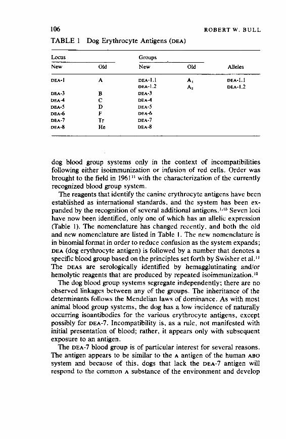
106 ROBERT W. BULL
TABLE 1 Dog Erythrocyte Antigens (DEA)
Locus Groups
New Old New Old Alleles
DEA-I A DEA-1.1 A, DEA-1.1 DEA-1.2 A2 DEA-1.2
DEA-3 B DEA-3
DEA-4 c DEA-4
DEA-5 D DEA-5
DEA-6 F DEA-6
DEA-7 Tr DEA-7
DEA-8 He DEA-8
dog blood group systems only in the context of incompatibilities following either isoimmunization or infusion of red cells. Order was brought to the field in 1961 11 with the characterization of the currently recognized blood group system.
The reagents that identify the canine erythrocyte antigens have been established as international standards, and the system has been expanded by the recognition of several additional antigens. 1•15 Seven loci have now been identified, only one of which has an allelic expression (Table I). The nomenclature has changed recently, and both the old and new nomenclature are listed in Table I. The new nomenclature is in binomial format in order to reduce confusion as the system expands; DEA (dog erythrocyte antigen) is followed by a number that denotes a specific blood group based on the principles set forth by Swisher et al. 11
The DEAS are serologically identified by hemagglutinating and/or hemolytic reagents that are produced by repeated isoimmunization.12
The dog blood group systems segregate independently; there are no observed linkages between any of the groups. The inheritance of the determinants follows the Mendelian laws of dominance. As with most animal blood group systems, the dog has a low incidence of naturally occurring isoantibodies for the various erythrocyte antigens, except possibly for DEA-7. Incompatibility is, as a rule, not manifested with initial presentation of blood; rather, it appears only with subsequent exposure to an antigen.
The DEA-7 blood group is of particular interest for several reasons. The antigen appears to be similar to the A antigen of the human ABO
system and because of this, dogs that lack the DEA-7 antigen will respond to the common A substance of the environment and develop
Copyright © National Academy of Sciences. All rights reserved.
Animal Models of Thrombosis and Hemorrhagic Diseaseshttp://www.nap.edu/catalog.php?record_id=19903
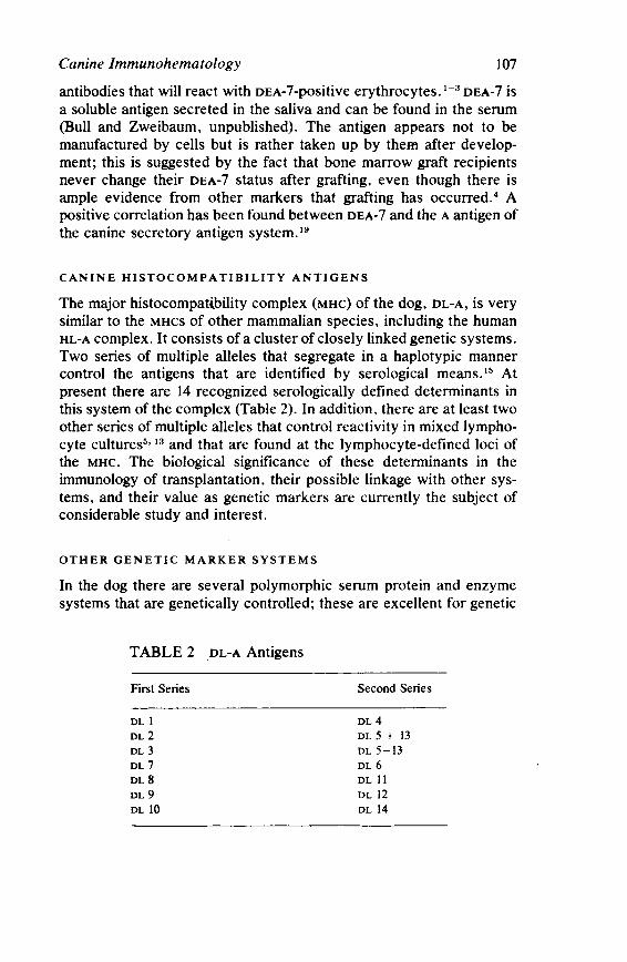
Canine lmmunohematology 107
antibodies that will react with DEA-7-positive erythrocytes. 1- 3 DEA-7 is a soluble antigen secreted in the saliva and can be found in the serum (Bull and Zweibaum, unpublished). The antigen appears not to be manufactured by cells but is rather taken up by them after development; this is suggested by the fact that bone marrow graft recipients never change their DEA-7 status after grafting, even though there is ample evidence from other markers that grafting has occurred. 4 A positive correlation has been found between DEA-7 and the A antigen of the canine secretory antigen system. 19
CANINE HISTOCOMPATIBILITY ANTIGENS
The major histocompatf.bility complex (MHC) of the dog, DL-A, is very similar to the MHCs of other mammalian species, including the human HL-A complex. It consists of a cluster of closely linked genetic systems. Two series of multiple alleles that segregate in a haplotypic manner control the antigens that are identified by serological means. 1s At present there are 14 recognized serologically defined determinants in this system of the complex (Table 2). In addition, there are at least two other series of multiple alleles that control reactivity in mixed lymphocyte culturess· 13 and that are found at the lymphocyte-defined loci of the MHC. The biological significance of these determinants in the immunology of transplantation, their possible linkage with other systems, and their value as genetic markers are currently the subject of considerable study and interest.
OTHER GENETIC MARKER SYSTEMS
In the dog there are several polymorphic serum protein and enzyme systems that are genetically controlled; these are excellent for genetic
TABLE 2 .DL-A Antigens
First Series
DL l DL 2 DL3
DL 7 DL8
DL9
DL IO
Second Series
DL 4 DL 5 + 13 DL 5-13 DL6
DL II DL 12 DL 14
Copyright © National Academy of Sciences. All rights reserved.
Animal Models of Thrombosis and Hemorrhagic Diseaseshttp://www.nap.edu/catalog.php?record_id=19903

108 ROBERT W. BULL
TABLE 3 Seruµi and Enzyme Polymorphism
Loci
Serum or plasma Tf (transferrin) Al (albumin)
Red cell enzymes Pep-D (peptidase-D) POM2 (phosphoglucomutase) o-To (dimerictetrazolium oxidase)
White cell enzymes POM3 (phosphoglucomutase)
Alleles
TfA, TP, Tf'C A', A2
Pep-I>", Pep-Db, Pep-I>" POM21, POM22,
D·T08 , 0-TOb
characterization of the species. These include (Table 3) transferrin, albumin, peptidase-D .(Pep-D), phosphoglucomutase (PGM2 and PGMa)
and dimerictetrazolium oxidase (D-T0).6• 15 An additional allelic system of antigens, the canine secretory antigens (CSA) within and secreted by mucosal cells of the digestive tract has been described. 19 Natll(Wly occurring antibodies are present against the antigens. The A antigen of this system is antigenically cross-reactive with DEA-7 of the red cell group as well as with the A of the human ABO blood group system.
SUMMARY
It is evident that considerable progress is being made in the immunohematologic and immunogenetic characterization of the dog. This knowledge can be of benefit and aid in the preservation and maintenance of dogs with thrombotic and hemorrhagic diseases. The field is not yet fully developed, but recognition of the supportive role of immunohematology is paramount to the success of any program that utilizes these animal models.
REFERENCES
I. Bowdler, A. J., R. W. Bull, R. Slating, and S. N. Swisher. 1971. Tr: A canine red cell antigen related to the A antigen of human red cells. Vox Sang. 20:542.
2. Bowdler, A. J., R. W. Bull, C. Dries, R. Slating, and S. N. Swisher. 1973. Representation of the ABH blood group system in the dog. Vox Sang. 24:228.
3. Bowdler, A. J., G. Uhlenbrouck, C. Dries, and R. W. Bull. 1973. The reactivity of canine red cells with heterophile agglutinins having anti-A activity. Anim. Blood Groups Biochem. Genet. 4:89.
4. Bull, R. W., H. M. Vriesendorp, A. Zweibaum, and S. N. Swisher. In press. The inapplicability of CEA-7 as a canine bone marrow transplantation marker. Transplantation Proc.
Copyright © National Academy of Sciences. All rights reserved.
Animal Models of Thrombosis and Hemorrhagic Diseaseshttp://www.nap.edu/catalog.php?record_id=19903

Canine Immunohematology 109
5. Grosse Wilde, H., H. M. Vriesendorp, B. Netzel, W. Mempel, and D. L. Westbroek. 1974. Identification of four MLC-specificities in the dog. Tissue Antigens 4:229.
6. Meera Khan, P., W. R. T. Los, J. A. van der Does, and R. B. Epstein. 1973. lsoenzyme markers· in dog blood cells. Transplantation 15:624.
7. Melnick, D. E., E. Buracks, and G. R. Cowgill. 1935. Development of incompatibilities in dog by repeated infusion of red blood cells. Proc. Soc. Exp. Biol. Med. 33:616.
8. Melnick, D. E., and G. R. Cowgill. 1937. Differentiation of blood groups in dogs based on antigenic complexes present in the erythrocyte. Proc. Soc. Exp. Biol. Med. 36:.97.
9. McEnery, E. T., A. C. Ivy, and C. E. Peckous. 1924. On the presence of iso-agglutinins in the blood of dogs. Am. J. Physiol. 68:133. ·
10. Ottenbery, R., D. J. Kaliski, and S.S. Fri.mann. 1913. Experimental agglutive and hemolytic transfusions. J. Med. Res. 28:141.
11. Swisher, S. N., and L. E. Young. 196i. The blood grouping systems of the dog. Physiol. Rev. 41:495.
12. Swisher, S. N., L. E. Young, and N. Trabold. 1962.In vitro andin vivo studies ofthe behavior of canine erythrocyte isoantibody systems. Ann. N.Y. Acad. Sci. 97:15. ·
13. van derTweel, J. G., H. M. Vriesendorp, A. Termijtelen, D. L. Westbroek, M. L. Bach, and J. J. Van Rood. In press. Genetic aspects of canine mixed leukocyte cultures. Transplantation.
l'l. Von Dungem, E., and L. Hirszfeld. 1910. Ueber nachweis und un venerbung .biochemischer strukturen. Z. Immunitaetsforsch. 4:531.
15. Vriesendorp, H. M., D. L. Westbroek, J. d'Amaro, et al. 1973. Joint report of 1st international workshop on canine immunogenetics. Tissue Antigens 3: 145.
16. Young, L. E., R. M. Christian, D. M. Ervin, S. N. Swisher, W: A. O'Brien, W. B. Stewart, and C. L. Yuile. 1950. Erythrocyte isoantibody reaction in dogs. Proc. Int. Soc. Hematol. 226: 1950.
17. Young, L. E., R. M. Christian, D. M. Ervin, S. N. Swisher, W. A. O'Brien, W. B. Stewart and C. L. Uiie. 1951. Erythrocyte isoantibody reaction in.dogs. Trans. N.Y. Acad. Sci. Ser. 11 13:209.
18. Young, L. E., W. A. O'Brien, S. N. Swisher, G. Miller, and C. L. Yuile. 1952. Blood groups in dogs. Their significance to the veterinarian. Am. J. Vet. Res. 13:(07.
19. Zweibaum, A., R. Oriol, N. Feingold, E. Dussaulx, M. Roussel, and G. Chevalier. 1974. Studies on canine secretory alloantigens (CSA). Tissue Antigens 4:115.
Copyright © National Academy of Sciences. All rights reserved.
Animal Models of Thrombosis and Hemorrhagic Diseaseshttp://www.nap.edu/catalog.php?record_id=19903

ALBERT M. JONAS, D. V .M.
Cost-Effectiveness of Developing Animal Models
An animal model may be defined as a disease or condition that occurs naturally in a particular species, has significant features similar or identical to a known human disease, and can be reproduced or obtained in enough animals to be studied effectively.
If any of these three components is lacking, the term animal model is inappropriate. For example, if a naturally occurring canine disease mimics a human disease but animals are not available for study, it is not an established animal model, but rather a potential animal model. Similarly, a sterile mutant may have a disease identical to a human condition, but its value as a model system is extremely limited.
The costs of developing animal models encompass a wide array of factors which may be summarized as follows:
1. Maintenance of the primary healthy population 2. Screening of animals for naturally occurring diseases of potential
medical interest 3. Characterization of the condition to determine its possible merit
as a model system for a human disease 4. Procurement of enough animals for study
FJfectiveness is more difficult to quantitate, but it may be evaluated by considering such factors as (a) number of models obtained, (b) number of research projects using the model, (c) research dollars funded for research projects using this model, (d) quantity and quality
110
Copyright © National Academy of Sciences. All rights reserved.
Animal Models of Thrombosis and Hemorrhagic Diseaseshttp://www.nap.edu/catalog.php?record_id=19903

Cost-Effectiveness of Developing Animal Models 111
of scientific publications related to this model, (e) impact of the research on the treatment of human patients, (0 contribution to training of medical scientists, and (g) feedback information to the veterinary profession enabling improved animal patient care.
It is unlikely that all of these factors will be included and properly weighed in determining "effectiveness." Potential biases and gross inaccuracies must be recognized and considered in any figures generated by cost-effectiveness ratios.
Animal models have been obtained from four main sources:
Domestic animals (horses, cattle, sheep, swine, poultry, dogs, and cats). Populations of these animals number in the millions and represent an immense reservoir of natural diseases of significant interest to comparative medicine. The cost to research is negligible for maintenance of these animal stocks. Screening for disease is routinely performed by animal owners who refer these animals to veterinarians. In the course of their daily practice veterinarians recognize and identify a wide range of diseases, many of which have potential relevance to comparative medical research. Veterinary and comparative medical literature is rich with descriptions of these diseases. Characterization and procurement, however, involve significant costs. Suspect animal diseases must be characterized, and if they are of sufficient interest, a mechanism must be established to obtain adequate numbers of animals for study. Examples of models among domestic animals familiar to investigators include dogs, cats, and pigs with hereditary clotting disorders. Identification is followed b)C characterization, but until a colony of animals is established, usually at rather' high cost, an animal model is not available for research purposes. Success is contingent upon developing contacts with veterinarians and establishing good professional relationships. In order to tap this resource some research groups have specifically recruited veterinary scientists as liaison personnel. The cost of such recruitment may be critical to success (effectiveness) of developing models for that research group.
Feral animals. Much that was stated about domestic animals applies to feral animals. Zoos such as the Philadelphia Zoo (Penrose Laboratory) have been studying diseases in their animal collections for many years. Aquatic mammals, fish, nonhuman primates, and many other species have potential diseases and physiological and/or behavioral characteristics that, if properly identified, may stimulate their establishment as animal models. The cost structure is similar to the previous category' but since only a fraction of the existing healthy populations is under scrutiny, the discovery of models may be more difficult.
Copyright © National Academy of Sciences. All rights reserved.
Animal Models of Thrombosis and Hemorrhagic Diseaseshttp://www.nap.edu/catalog.php?record_id=19903
112 ALBERT M. JONAS
Establishing animals with diseases or desirable characteristics may require more ingenuity, and cost factors will be extremely variable. .
Systematic breeding of normal animal stocks and strains for the specific purpose of discovering hereditary diseases. This approach has not been practiced except in unusual circumstances. The breeding of animals to uncover genetic abnormalities is difficult and costly. A large number of strains with genetic diseases are now available but these animals were obtained from animal research colonies (see below). The developmental costs include all four cost factors associated with prospective breeding for genetic abnormalities.
Animal research colonies. Millions of laboratory animals are seen and examined by investigators during the course of their animal experiments. These colonies are composed largely of normal animals, but occasionally an interesting model may be detected. All too often, however, the investigator thinks of the abnormal situation strictly as a problem and is interested only in its elimination. A condition may exist in a colony for years at a low level before someone takes an interest in it and establishes its importance. With the increase in numbers of comparative pathologists on medical school faculties and the establishment of veterinary diagnostic laboratories to monitor animal research colonies, more of these "problems" can be studied and potential animal models identified. Cost factors in developing models from research animal colonies are favorable in that animal maintenance costs are shared by existing grants, and detection of models is usually a spinoff from these projects. Costs of screening are borne by investigators or by the diagnostic laboratory, which must diagnose cases as part of its quality control obligations. Characterization must be funded separately. Costs of developing animal models are relatively low in animal research colonies since maintenance and screening costs are not added to initial projects.
The word "effectiveness" in cost-effectiveness has been deliberately downplayed during this discussion. Cost for establishing previous models can be estimated as projected costs in the four main areas discussed (domestic animals, feral animals, systematic breeding, and animal research colonies), but estimations of effectiveness may have to be more qualitative than quantitative.
There are pitfalls in applying cost-effectiveness ratios prospectively. Poor animal care or insufficient veterinary medical staffing may reduce basic animal costs, but this certainly is not the direction scientists should take. The benefit to science· and human health should be of paramount concern, and spurious cost:-effectiveness ratios that favor
Copyright © National Academy of Sciences. All rights reserved.
Animal Models of Thrombosis and Hemorrhagic Diseaseshttp://www.nap.edu/catalog.php?record_id=19903
Cost-Effectiveness of Developing Animal Models 113
poor conditions should be recognized and challenged. Costs will also vary in different parts of the country. Animal costs should be audited by standard accounting procedures1 to establish proper documentation of costs, rather than making them comparable to those established by other institutions. Quality and creativity should always be the critical factors in evaluating costs and effectiveness.
REFERENCE
U. S. Department of Health, Education, and Welfare, Division of Research Resources, National Institutes of Health, and Association of American ~edical Colleges. 1974. Cost analysis and rate setting manual for animal resource facilities. Animal Resources Branch, Division of Research Resources, NIH, Bethesda, Maryland. 109 p.
BIBLIOCJR.APHY
Cornelius, E. E., and I. M. Arias. 1966. Biomedical models in veterinary medicine. Am. J. Med. 40:165-169.
Leader, R. E. 1967. The kinship of animal and human diseases. Sci. Am. 216:100-116. Melby, E. C., and N. H. Altman (ed.). 1974. Handbook of laboratory animal science.
Vol. II, cac Press, Cleveland.
Copyright © National Academy of Sciences. All rights reserved.
Animal Models of Thrombosis and Hemorrhagic Diseaseshttp://www.nap.edu/catalog.php?record_id=19903
PETER H. LEVINE, M.D.
Cost-Effectiveness of Replacement Therapy in Animal Models of Hemorrhagic Disorders: Lessons from Experience with H. sapiens
Our present level of knowledge about hemophilia and related diseases, and the therapeutic advances that have resulted from this knowledge, owe a great deal to studies on animal models of these disorders. One has only to read the recent hematologic literature to see the continuing benefits of maintaining colonies of animals with bleeding tendencies. The maintenance of such colonies is expensive. Is this expense justified?
The extensive national survey of blood resources carried out recently by the National Heart and Lung Institute5 outlines the magnitude of the hemophilia problem in humans. Given this country's 25,500 currently treated severely and moderately severely affected hemophiliacs, the presently available therapeutic material, and the presently available supply of blood donors, the application of prophylactic therapy to all hemophiliacs would more than exhaust the present national blood supply, and probably would also exhaust the budgets of most third-party health care providers. Further, it is highly unlikely that we will approach 100 percent efficiency in blood fractionation within the next several years. For these reasons, and because of the already tremendous welfare burden attributable to patients with hemophilia, cost-benefit analyses of spending federal money on animal models of the hemophilias are highly likely to yield data that strongly support such spending.
It seems crystal clear to me, a physician who deals only with human animals with hemorrhagic tendencies, that we must maintain colonies
114
Copyright © National Academy of Sciences. All rights reserved.
Animal Models of Thrombosis and Hemorrhagic Diseaseshttp://www.nap.edu/catalog.php?record_id=19903

Cost-Effectiveness of Replacement Therapy 115
of nonhuman an'imals with hemorrhagic tendencies in order to answer the many new questions that continuously arise as our data base expands. Many such questions cannot be studied in man. How may the benefits of animal models be maximized, and costs minimized?
Recent clinical research studies at our center have yielded certain principles that are applicable to future maintenance and study of animal models. (There is no reason why information should not flow in both directions.)
In a program that now involves approximately 90 severely and moderately severely affected hemophiliacs of all ages, an attempt has been made to apply replacement therapy in a way that would guarantee immediate correction of the coagulation defect at the very earliest suspicion of hemorrhage, and that also guarantees that hemostatically effective levels of factor VIII or factor IX are reliably achieved in the plasma. In order to achieve these goals, the patients themselves were provided with intensive theoretical and practical instruction, and necessary therapeutic materials, and were indoctrinated in a general philosophy that states, "When in doubt, infuse." Further, the therapeutic material used was either lyophilized cryoprecipitate or factor VIII or IX concentrate, because of the reliability in dose calculation when using these therapeutic materials. We have come to use standard nonlyophilized cryoprecipitate less and less, because of multiple episodes of in vivo therapeutic failure with this material. These failures were explained by in vivo factor VIII levels far below the expected ones, explained in tum by particular batches of cryoprecipitate that assay out as containing 25-35 factor VIII units/bag as opposed to the expected 100-150 units/bag. Further, at a cost to us of $10.00/ bag, this material becomes extremely expensive.
What are the benefits of immediate and predictably intensive replacement therapy? We have previously reported some of the benefits attributable to this sort of therapy using data collected during a year of standard, emergency-room-based health care delivery as a control and comparing this to a year of patient-administered therapy given at the very earliest sign of hemorrhage.2 In that study, we reported obvious improvement in all parameters of health and well being. For example, there was a 74 percent decrease in days lost from work or school because of ill health, an 89 percent decrease in days hospitalized, and a 76 percent decrease in number of physician visits made. Furthermore, in an evaluation of one of the most important long-term sequelae of hemophilia, hemophilic arthropathy, we showed that during the control period we could demonstrate a 90 percent incidence of new or progressive hemophilic arthropathy as opposed to an incidence of
Copyright © National Academy of Sciences. All rights reserved.
Animal Models of Thrombosis and Hemorrhagic Diseaseshttp://www.nap.edu/catalog.php?record_id=19903

116 PETER H. LEVINE
approximately 10 percent following a year of immediate aproication of intensive therapy. 3 ·
What are the costs of this sort of therapy? For the human, this intensive therapy achieves great reduction in cost, even though many more hemorrhagic episodes are treated. In our initial study, we demonstrated the yearly per capita cost of health care for a group of 45 patients with severe or moderately severe hemophilia A or B could be reduced from a mean of $5, 780 to a mean of $3 ,209. This reduction in cost was due not only to a decrease in days spent in the hospital but also to a 15 percent decrease in the mean number of factor VIII infusion episodes per year, which fell from 32.8/patient/year to 27.9/ patient/year. It should be emphasized that these patients had many more individual episodes of hemarthrosis treated under the selftherapy regimen, but that because a single infusion given early suffices to shut off a bleeding episode, their overall health was much improved at decreased expense. Further, the arthropathy failure rate occurred in a small group of patients whose therapy was significantly less intensive than that of the group as a whole. One limitation must be stressed. In order to achieve these results, with maximum benefit to the patient~ and, parenthetically, maximum yield of useful data, care should be delivered through a limited number of large hemophilia centers, which have the necess;uy professional expertise, health care facilities, and laboratory capability.
Colonies of bleeder animals are an important national resource that must be maintained. They are expensive, and they are difficult to manage. The problem is compounded by the lack of canine typing sera and by the difficulties in obtaining canine plasma and plasma products. Thus, hemorrhage must often be treated with plasma or whole blood. 1
This is clearly suboptimal and may lead to the death of valuable experimental animals. These conditions in the animal colonies are entirely analogous to the human situation a number of years ago. It seems clear to me that we should bring our animal colony capabilities to a more modem level so that these colonies can answer some of the questions now being raised in humans. Aside from basic research projects of obvious importance, certain applied clinical research projects require study. For example, the effects of repeated infusions of plasma products need study, as do the hepatic abnormalities frequently observed in the intensively infused hemophiliac4 and the occurrence of splenomegaly in approximately 25 percent of these patients (Levine, unpublished data). All could easily be studied in animal colonies, with great potential gain to afflicted humans.
In conclusion, two principles derived from human clinical research
Copyright © National Academy of Sciences. All rights reserved.
Animal Models of Thrombosis and Hemorrhagic Diseaseshttp://www.nap.edu/catalog.php?record_id=19903

Cost-Effectiveness of Replacement Therap) 117 . deserve emphasis. First, there should be a limited number of carefully chosen, nationally funded centers that maintain colonies of animals with hemorrhagic tendencies. These centers should be willing to make their models, facilities, and academic talents available for appropriate collaborative projects. Second, a single center should be funded to produce and distribute a canine factor VIII preparation, presumably a lyophilized cryoprecipitate, as well as other appropriate animal plasma products.
REFERENCES
I . Dodds. W. J. 1974. Hereditary and acquired hemorrhagic disorders in animals. Pages 215-247 in T. H. Spaet (ed.), Progress in hemostasis and thrombosis. Vol 2. Grune & Stratton, New York. .
2. Levine, P. H., and A. F. H. Britten. 1973. Supervised patient-management of hemophilia. Ann. Intern. Med. 78:195-201.
3. Levine, P.H. 1974. Efficacy ofself-therapy in hemophilia, a study of72 patients with hemophilia A and B. N. Engl. J. Med. 291:1381-1384.
4. Lewis, J. H. 1970. Hemophilia, hepatitis and HAA. Vox Sang. 19:406-409. S. National Heart and Lung Institute. 1972. N.H.L.l.'s blood resource studies, Vol. 3.
Pilot study of hemophilia treatment in the United States. DREW Pub. No. NIH-73-419. U.S. Government Printing Office, Washington, O.C.
Copyright © National Academy of Sciences. All rights reserved.
Animal Models of Thrombosis and Hemorrhagic Diseaseshttp://www.nap.edu/catalog.php?record_id=19903
STEPHEN W. J. SEAGER, M.V.B.,
M.R.C.v.s.
Cost-Effectiveness: Utilization of a Sperm Bank in Animal Breeding Colonies
Frozen semen has proven invaluable in the storage of genetic lines in the bovine, ovine, and porcine species. Investigators have utilized these stored specimens, particularly from bulls, for many years after the sire has died, either for the utilization of his good qualities or to trace inheritance pathways of congenital anomalies. Bull semen has been frozen successfully for as long as 12 years. 1 Until comparatively recently, canine genetic material could not be stored for extended periods. Seager published the first reported pregnancy from the use of frozen canine semen,2 and Van Gemert3 and Anderson4 have since reported successful pregnancies. These achievements have opened up great possibilities for frozen semen utilization in the study of canine genetics.
During the past 5 years studies have been conducted in the laboratories at the University of Oregon Medical School and Baylor College of Medicine · into the preservation of genetic material of an inbred, histocompatible line of dogs, as well as intensively investigating frozen dog, cat, and other mammalian semen. Genetic material from male dogs with congenital diseases and rare blood groups has been stored in liquid nitrogen. This stored material has subsequently been used in attempting to determine the pathways of such congenital diseases as hip dysplasia. 5 Semen of dogs suffering from a variety of conditions has been frozen: these conditions include entropion, extropion, anophthalmia, hip dysplasia, monorchidism, various types of hernias, maxillary and mandibular prognathism, heart conditions (including
118
Copyright © National Academy of Sciences. All rights reserved.
Animal Models of Thrombosis and Hemorrhagic Diseaseshttp://www.nap.edu/catalog.php?record_id=19903

Cost-Effectiveness: Utilization of a Sperm Bank 119
ventricular and atrial septal defects, pulmonic and aortic stenosis, and tetralogy of Fallot), and a number of neurological conditions (including epilepsy). We are presently investigating semen freezing of males whose direct descendents have been diagnosed as narcoleptic.
There are a number of reasons why frozen semen is of great benefit to investigators of animal models. The cost of keeping research animals has increased alarmingly in the past few years. If a breeding colony is being maintained for the study of genetic traits as part of a research program, semen from the afflicted mature males can be collected and frozen. Once a sufficient amount of semen has been stored, the males no longer need to be kept in the colony for breeding purposes. The semen can subsequently be used to artificially inseminate suitable heterozygous females.
For example, the sperm count of normal dogs ranges from 500 million to 2.5 billion spermatozoa per ejaculate. If the sperm have a normal recovery rate after the freezing process, one can expect to inseminate four to ten bitches from each ejaculate. This expectation may not be valid in dogs afflicted with certain forms of congenital disease. Males afflicted with congenital heart diseases often,show poor libido, possibly due to hormonal deficiency or to inability to withstand the physical stress of mounting a bitch. This low libido is often coupled with either oligospermia or aspermia. If the total sperm count in such dogs is as low as 25 million, a number of ejaculates may be pooled to obtain sufficient live sperm for insemination. Many of these dogs are incapable of fertilizing a bitch naturally because of their physical inability or the poor quality of their semen. Do,gs suffering debilitating anatomical conditions such as hip dysplasia, achondroplasia of the shoulder, or lower lumbar malformations may desire to mount and breed naturally, but their affliction causes such a severe physical limitation on them that successful copulation is impossible.
Air shipment of frozen canine semen has become desirable as a result of the increasing reluctance of airlines to ship large dogs, plus the shipping expenses and danger to the dog's life from overexcitement or trauma (particularly those suffering from heart conditions). Research involving the study of genetic anomalies in animal colonies in various institutes could greatly benefit from shipment of frozen semen. The ease and safety of transport of frozen semen has been documented. Canine semen has been shipped in liquid nitrogen from the University of Oregon Medical School and inseminated successfully in St. Louis, Missouri. 6 Large shipments of canine semen have been sent to Hawaii and Alaska, evaluated, and returned by air with no visible loss in motility or apparent morphological change.
Copyright © National Academy of Sciences. All rights reserved.
Animal Models of Thrombosis and Hemorrhagic Diseaseshttp://www.nap.edu/catalog.php?record_id=19903

120 STEPHEN W. J. SEAGER
Space and housing of dogs has become an increasingly important factor in research institutions, in large part because of the increased kennel space requirements for the housing of dogs. The rising costs for square footage and new cage design further enhance the utility of frozen semen in the maintenance of dog breeding colonies, as only relatively few sires need to be retained for breedipg. Frozen semen is an inexpensive and safe method of securing the viability of at least half the genetic material if a colony is destroyed by disease or disaster.
METHODS AND MATERIALS
Dog semen has been frozen in the Baylor laboratory using three methods: (a) in 1.0-ml glass vials; (b) in 0.25-, 0.50-, and 1.0-ml plastic straws; and (c) in "pellets. " 7 To date, the pellet method has been the most successful. 8 Over 500 puppies have been born from frozen semen thus far in the study. Conception rate in one trial with 13 beagle bitches using frozen beagle semen was 92 percent. This rate was obtained under laboratory conditions.
Freezing semen by the pellet method involves dilution in an egg yolk-glycerol solution, equilibration at 5"C, depositing the semen dropwise on a block of solid carbon dioxide, inverting the block over a bath of liquid nitrogen and thus depositing the pellets into the liquid nitrogen. 8 In preparation for final storage, the pellets are placed in Nalgene vials identified by sire and date frozen. Position of the semen in the liquid nitrogen tank is recorded in a catalog. The semen is thawed just before insemination in 0.9 percent saline at 38"C. 9 Insemination is performed as soon as possible after thawing. The thawed semen is inseminated by means of a 10-ml disposable syringe attached by a 1/2-inch rubber connector to a bovine artificial insemination rod. The semen is deposited at the external cervical os. The bitch is elevated for approximately 10 minutes after insemination. -
RESULTS AND DISCUSSION
To date in this study, no adverse effects of the frozen semen have been shown in either the puppies or the dam. Male dogs that have ejaculated many times by manual stimulation are willing and able to breed naturally unless there is a physical condition that prevents it. A number of bitches have had a litter of frozen semen puppies, followed by one with natural breeding, followed by further ones from frozen semen.
One of our aims is to record any adverse factors appearing in a number Qf successive generations of frozen semen puppies, such as
Copyright © National Academy of Sciences. All rights reserved.
Animal Models of Thrombosis and Hemorrhagic Diseaseshttp://www.nap.edu/catalog.php?record_id=19903

Cost-FJfectiveness: Utilization of a Sperm Bank 121
reproductive failure, poor growth, or abnormal inheritance of genetic traits. A litter of fourth-generation frozen semen puppies was whelped January 9, 1975. So far, no dogs in the first through fourth generations of frozen semen litters have experienced any congenital or acquired defect that could be attributed to the semen freezing process. Sex ratios, litter sizes, and birth and weaning 'weights of all frozen semen litters have been within the average for the group.
A litter of nine healthy puppies born recently resulted from artificial insemination using semen frozen for over 3 years. Semen has been stored in the program since 1968, and as of this date shows excellent motility upon thawing. As mentioned above, bull semen has been stored for extended periods; results thus far indicate that canine semen will have a similar longevity. Extensive data-gathering continues in this program on the utilization of frozen dog, cat, and captive wild mammal semen.
'REFERENCES
I. Mixner, J. P. 1968. Fertility of bull semen frozen for twelve years. VI Cong. de Reprod. et Insem. Artif., Paris, 2:1095-1098.
2. Seager, S. W. J. 1969. Successful pregnancies utilizing frozen dog semen. Art. lnsem .. Dig. 17(12):6. •
3. Van Gernert, W. 1970. Diepvries-pups. Tiidschr. Diergeneesk. 95:13. 4. Anderson, K. 1972. Fertility of frozen dog semen. Acta Vet. Scand. 13:1~130. 5. Templeton, J. W., S. Seager, L. Erickson, and W. S. Fletcher. 1975. Hip dysplasia
studies in a colony of large dogs. (Submitted to Lab. Anim. Sci.) 6. Seager, S. W. J. 1974. Letter: Transporting frozen canine semen. Vet. Rec. 95(2):47. 7. Nagase, H., and Niwa, T. 1964. Deep freezing bull semen in concentrated pellet form.
V Cong. Intern. Reprod. Anim. lnsem. Artif., Trento, 4:410. 8. Seager, S. W. J., and W. S. Fletcher. 1973. Progress on the use of frozen semen in the
canine. Vet. Rec. 92:frl0. 9. Sealer, S. W. J., W. S. Fletcher, and C. C. Platz. 1975. Pregnancy rates and related
data using frozen dog semen. J. Reprod. Pert. 45:189-191.
Copyright © National Academy of Sciences. All rights reserved.
Animal Models of Thrombosis and Hemorrhagic Diseaseshttp://www.nap.edu/catalog.php?record_id=19903

JAN J. VELTKAMP, M.D. ALLAN T. VAN OOSTEROM, M.D. AND JOHAN A. VAN DER DOES, M.D.
Requirements for Diagnostic Procedures Used in the Definition and Interpretation of Animal Models of Hemorrhage and Thrombosis
Animal models for the study of genetically determined hemorrhagic disorders have to be carefully investigated before conclusions relevant to human pathology can be reached. Points of concern are the clinical picture of the disease in the animal, the pattern of inheritance, and the nature of the biochemical defect.
The first point, i.e., the clinical picture of the disease in the animal, has to be elucidated by clinical observation of a certain number of animals. The dogs with severe classic hemophilia in the Leiden and Albany colonies rarely suffer from hemarthroses. Whether this is due to their housing conditions, their gait, the nature of their biochemical defect, or modifying genes is not known. Hemophilic dogs in Chapel Hill, living in similar housing and by present standards having the same biochemical defect, are reported to have seriously damaged joints (see Brinkhous, this volume, p. 3). The importance of gait in provoking joint bleeding might be suggested by the high incidence of hemarthroses in hemophilic horses.
A bleeding site, very rare in man, but quite common in our dogs is the mediastinum, in which bleeding might be evoked by barking. Not only the site of bleeding but also the sequelae of bleeding can be very different: The dog's skin is in many places very loosely attached to the underlying musculoskeletal structures; this results in an enormous extension of subcutaneous bleeding, rare in man, which, without intervention by substitution therapy, leads to death. Furthermore, in our experience, the smaller cages without a walking area sometimes
122
Copyright © National Academy of Sciences. All rights reserved.
Animal Models of Thrombosis and Hemorrhagic Diseaseshttp://www.nap.edu/catalog.php?record_id=19903
Requirements for Diagnostic Procedures 123
employed to house a dog with a bleeding episode are not helpful in prevention of further bleeding. Thus, our hemophilic dogs are not a good model in which to evaluate the effect of prophylactic treatment, which aims at the prevention of the most disabling bleedings in man and their sequelae, a syndrome called hemophilic arthropathy.
Apart from the important contributions of hemophilic dogs to the development of a rational therapy-Le., to obtain a survival rate of transfused material, to determine optimal routes of administration of such material, and to develop control methods to monitor substitution therapy (see Brinkhous, this volume, p. 3)-very important questions remain to be answered, and the investigations must be carried out in animals. One of those qu'{stions pertains to the elucidation of the occurrence and treatment of circulating anticoagulants, i.e., inhibitors of clotting factor activity. To find the underlying mechanism for antibody formation, whether it is a genetically determined predisposition of the recipient or is due to heterogeneity of the normal factor, it will be important to have access to all available hemophilic mutations in animals. This could be accomplished by development of regional centers in which all animal models are freely exchanged.
The ascertainment of dogs with hemophilia is best carried out in a small-animal clinic or similar animal diagnostic facility. For example, blood samples from dogs with hemorrhagic diatheses were sent from the small-animal clinic in Utrecht to our laboratory for diagnostic evaluation. First, we found a number of dogs with acute myeloid leukemia showing a bleeding tendency because of severe thrombocytopenia. Our first dog with severe AHF deficiency, a 3-month-old Labrador puppy, had been imported from England and was reported to have normal siblings. His mother had previously given birth to several litters in which no bleeding disorders were apparent. Therefore, in order to prove the assumption of classic hemophilia in this dog, we had to mate him and study his progeny. Sex-linked inheritance of the clotting defect was subsequently established.
When assaying dog plasma for factor VIII activity according to the partial thromboplastin time method in a human-specific system-i.e., human AHF-deficient plasma as substrate, human brain phospholipid as accelerator, and human pooled normal plasma as standard materialone will find that dogs have on the average six to ten time.s higher AHF activity than humans. This figure corresponds fairly well with the observation that clinically severely affected dogs show about 4 to 5 percent of human AHF activity in this assay, a figure that could be interpreted as less than I percent of dog AHF. Indeed, when using a human system with a normal dog standard, it was found that the
Copyright © National Academy of Sciences. All rights reserved.
Animal Models of Thrombosis and Hemorrhagic Diseaseshttp://www.nap.edu/catalog.php?record_id=19903

124 VELTKAMP et al.
severely affected dogs showed less than I percent of normal canine AHF activity. Because of the high relative activity of dog plasma compared to human plasma, not only for AHF but for most clotting factors, including those of the prothrombin complex, samples of dog plasma are usually assayed at relatively high dilutions. This is necessary to stay in the rectilinear part of the normal human standard reference curve, which is S-shaped on the log-log plot (clotting time versus calculated clotting factor activity) that most of us use for empirical reasons. In a canine-specific assay system, such high dilutions of normal and test plasmas need not be used.
Until now, a more representative plot, where linear clotting time is plotted against the inverse of the clotting factor concentration, could not be applied to the AHF assay. This new method, in use for factor II, V, VII, and X assays, 1 not only yields a rectilinear relationship but also provides a means of calculating the true clotting factor activity in the so-called deficient plasma. By contrast, extrapolation from the experimentally determined conventional log-log plot will lead to error, which is unacceptable for critical experimental situations. This is also true for the low activity area of the plot, where clotting times will lead to an overestimation of AHF activity when extrapolated (Figure 1).
The specificity of our dog AHF activity assay was tested when it was used to measure rather low levels of AHF in hemophilic dogs after lung transplantation.2 In liver transplants, i.e., grafting normal livers into hemophilic recipients, large amounts of AHF are produced. 3- 5 These relatively high levels of AHF could readily be detected by conventional factor VIII assays. In the lung transplant experiments, samples were assayed in the I : IO and I : 20 dilutions, which added another IO percent
HC 200 , 180 -l- -
=r-=~~~ 120 --- .
' ·----~ . 90 ---80 ·----
1 I I I I
5
11200
10
11100
50 .,. factor Viii
111000 1120 dilution
FIGURE I Part of the S-shaped standard-reference graph on log-log scale (clotting time versus calculated clotting factor activity). The dotted line would result if extrapolation beyond the 1/200 dilution of reference plasma were applied.
Copyright © National Academy of Sciences. All rights reserved.
Animal Models of Thrombosis and Hemorrhagic Diseaseshttp://www.nap.edu/catalog.php?record_id=19903

Requirements for Diagnostic Procedures 125
or 5 percent, respectively, of the other clotting factors to the system and resulted in artifactually high values for factor VIII activity. By comparing the data on a standard normal plasma dilution curve with those for hemophilic dog plasma diluted in buffer ~o as to provide the . same final concentration of other clotting factors as in the 1: 10 dilution of experimental plasma, we found the contribution of the other factors to be about 0.3 petcent of (pseudo) factor VIII activity.2
As reference material for the ~anine AHF assay we used ·pooled, high-spun, deep-frozen ( -80°C) c~rated plasma from 4 normal male dogs from the same colony. Our experience with AHF levels in random stray dogs is that variability seems to be even greater than in normal dogs derived from one colony; therefore, it is advisable to establish normal values for a specific model by using strain-specific reference material. Our reference dogs were about 1 year old when utilized. Influence of age on coagulation factor levels is most marked for those of the prothrombin complex but be.insignificant except in the neonatal
. period for other coagulation factors such as AHF. 8 Sex-related effects are also to be considered when interpreting assay data and establishing normal reference standards.
As discussed already by O'Reilly and Pool (see this volume, p. 21), the sensitivity of the vitamin K receptor in liver cells of various rooents for competitive occupation by coumarin drugs is genetically determined. 7 From the work of Pyoriila8 it is also known that the turnover rate of coagulation factors in rats, after a blocking dose ofwarfarin, can be different from strain to strain.
All these facts indicate that we should be concerned about the signific~ce of diagnostic assays or routine surveillance carried out for the testing of drugs because so little is known about pharmacogenetics. This is pertinent not only to the genetic differences between strains of animals but also to the diagnostic procedures. Recently, we observed that in a strain of inbred Wistar rats, now in its sixty-fifth generation of brother-sister matings, prothrombin times with ox-brain thromboplastin were very long and therefore poorly reproducible, whereas with rabbit brain thromboplastin, warfarin anticoagulation could be monitored quite well. 9
For coagulation factor assays on rat plasma, we prepared rat brain thromboplastin to avoid problems of species specificity. We found, in contrast to other investigators, 10 that rat clotting factor assays performed with human brain thromboplastin were insensitive, as the standard reference curve produced a plot with a very shallow slope and so had poor reproducibility.
For long-term coagulation studies, only male rats should be used
Copyright © National Academy of Sciences. All rights reserved.
Animal Models of Thrombosis and Hemorrhagic Diseaseshttp://www.nap.edu/catalog.php?record_id=19903

126 VELTKAMP et al.
because of the variation of coagulation factor levels in female rats due to the estrous cycle. 11
Turning to the practical problems of keeping animal colonies, we must mention that one of the biggest problems is funding, especially for long-range planning. We have been able to use our hemophilic dog colony for several purposes and have thus used funds- from various sources for its support:
1. As a source of normal dogs for blood banking and experimental surgery per se
2. For breeding experiments to elucidate the genetic transmission of dog leukocyte antigens; this is not hampering the production of hemophilic ·dogs as long as obligatory carrier bitches are used in the breeding program
3. For the study of the effects of antilymphocytic serum on platelet behavior
4. For transplantation of organs into the hemophilic dogs.
REFERENCES
1. Hemker, H. C., and A . .Q. Muller. 1968. Kinetic aspects of the interaction of blood clotting enzymes V. Thromb. Diath. Haemorrh. 19:368.
2. Veltkamp, J. J., E. Asfaou, K. Torren,J. A. van derDoes, N. H. van Tilburg, and E. K. J. Pauwels. 1974. Extrahepatic factor VIII synthesis. Transplantation 18:56.
3. Marchioro, T. L., C. Hougie, H. Ragde, R. B. Epstein, and E. D. Thomas. 1969. Organ homografts for hemophilia. Transplant. Proc. 1:316.
4. Penick, G.D., W. P. Webster, E. E. Peacock, P. Hutchin, and C. F. Zukoski. 1970. Organ transplantation in animal hemophilia. Page 97 in K. M. Brinkhous (ed.), Hemophilia and new hemorrhagic states. University of North Carolina Press, Chapel Hill.
5. Veltkamp, J. J., K. van deTorren, S. W. Schalm, J. A. van derDoes, R. B. Epstein, and J. L. Terpstra. 1973. Organ transplantation in canine hemophilia. Page 19 in F. Ala and K. W. E. Denson (eds.), Hemophilia. Excerpta Medica, Amsterdam.
6. Dodds, W. J., A. C. Moynihan, R. E. Benson, and C. A. Hall. 1975. The value of age- and sex-matched controls for coagulation studies. Br. J. Haematol. 29:305.
7. O'Reilly, R. A., J. G. Pool, and P. M. Aggeler. 1968. Hereditary resistance to coumarin anticoagulant drugs in man and rat. Ann. N.Y. Acad. Sci. 151:913.
8. Pyorila, K. 1970. Genetic control of the response to coumarin anticoagulants in the rat. Humangenetik 9:265.
9. Van Oosterom, A. T., and J. J. Veltkamp. 1974. Control of anticoagulation in rats. Experientia 30:953.
10. Raymond, S. L., and W. J. Dodds. 1975. Characterization of the fawn-hooded rat as a model for hemostatic studies. Thromb. Diath. Haemorrh. 33:361.
11. Pyorili, K. 1965. Determinants of the clotting factor response to warfarin in the rat. Ann. Med. Exp. Biol. Fenn. 43: Suppl. 3.
Copyright © National Academy of Sciences. All rights reserved.
Animal Models of Thrombosis and Hemorrhagic Diseaseshttp://www.nap.edu/catalog.php?record_id=19903

JACEK HAWIGER, M.D.
Detection and Comparison of Fibrinogen and Its Derivatives in Different Species by the Staphylococcal Clumping Test_
Specific, sensitive, and simple diagnostic tests are essential for current and future study of thrombotic and hemorrhagic diseases. Such diagnostic tests need to meet the following criteria: (a) They should be relatively uniform to allow comparisons of the same factor studied in man and animal models, and (b) they should be simple to allow routine laboratory use by personnel not necessarily familiar with intricate and complex procedures. The staphylococcal clumping test for quantitative measurement of fibrinogen and fibrin degradation products (FDP) appears to meet these criteria. In this method the same test reagent can be used to measure and compare fibrinogen and its derivatives in man and in several animal species. This is an advantage over immunologic techniques, which are usually limited to fibrinogen derivatives of one species only. For each species a monospecific antiserum needs to be prepared to achieve optimal sensitivity. Thus, for comparative studies of the role of fibrinogen and its derivatives in hemorrhagic and thromboembolic disorders of man and animal models, the staphylococcal clumping test may serve as a valuable laboratory tool.
The origin of the staphylococcal clumping phenomenon' dates back to 1908, when the German bacteriologist, Much, observed that some staphylococci, when emulsified in plasma, form thick clumps visible to the naked eye. 1 He suggested that the plasma protein responsible for clumping was related to fibrinogen. Almost 50 years later, Duthie
Supported by NIH grant HL 16646 and the Veterans Administration.
127
Copyright © National Academy of Sciences. All rights reserved.
Animal Models of Thrombosis and Hemorrhagic Diseaseshttp://www.nap.edu/catalog.php?record_id=19903
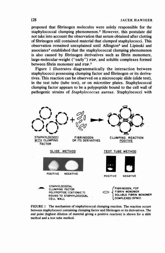
128 JACEK HAWIGER
proposed that fibrinogen molecules were solely responsible for the staphylococcal dumping phenomenon. 2 However, this postulate did not take into account the observation that serum obtained after clotting of fibrinogen still contained material that clumped staphylococci. This observation remained unexplained until Allington3 and Lipinski and associates4 established that the staphylococcal clumping phenomenon is also caused by fibrinogen derivatives such as fibrin monomers, large-molecular-weight ("early" ) FDP, and soluble complexes formed between fibrin monomer and FDP. 5
Figure 1 illustrates , diagrammatically the interaction between staphylococci possessing clumping factor and fibrinogen or its derivatives. This reaction can be observed on a microscopic slide (slide test), in the test tube (tube test), or on microtiter plates. Staphylococcal clumping factor appears to be a polypeptide bound to the cell wall of pathogenic strains of Staphylococcus aureus . Staphylococci with
STAPHYLOCOCCI YLill! CLUMPING
FACTOR
FIBRINOGEN OR ITS DERIVATIVES
SLIDE METHOD
POSITIVE NEGATIVE
STAPHYLOCOCCAL CLUMPING FACTOR POLYPEPTIDE (CATIONIC?) BOUND TO STAPHYLOCOCCAL CELL WALL
CLUMPING REACTION POSHIVE
POSITIVE NEGATIVE
{
FIBRINOGEN, FOP <::=:> FIBRIN MONOMER
SOLUBLE FIBRIN MONOMER COMPLEXES (SFMC)
FIGURE 1 The mechanism of staphylococcal clumping reaction. The reaction occurs between staphylococci containing clumping factor and fibrinogen or its derivatives. The end point (highest dilution of material giving a positive reaction) is shown for a slide method and a test tube method.
Copyright © National Academy of Sciences. All rights reserved.
Animal Models of Thrombosis and Hemorrhagic Diseaseshttp://www.nap.edu/catalog.php?record_id=19903

Detection and Comparison of Fibrinogen and Its Derivatives 129 , clumping factor have binding sites on that portion of the fibrinogen molecule or its derivatives that is not associated with fibrinopeptides but that is located between the N-terminal disulfide knot and the C-terminal end of polypeptide chains. 8 The reaction between fibrinogen and staphylococci is extremely sensitive, and it is possible to obtain visible clumping in a slide test with as little as 0.03 µg of human or bovine fibrinogen, 5 indicating that approximately four molecules of fibrinogen per bacterial cell are necessary te- induce the clumping reaction.
Previous attempts to use the staphylococcal clumping reaction to detect traces of fibrinogen in biological fluids were impeded by difficulty in obtaining a standardized source of staphylococci containing the clumping factor that would give reproducible and quantitative results. The majority of workers were using various strains of staphylococci that gave a somewhat different reaction from day to day, making results difficult to compare. These difficulties led us to work out a procedure which provides standardized, specific, sensitive, stable, and safe material for the clumping reaction. 5 The strain used for the test is a coagulase-negative variant of Newman D2 strain of Staphylococcus aureus. Lack of detec!able staphylocoagulase.affords an opportunity to increase the specificity of the interaction between cells and fibrinogen. For use, nonviable bacterial powder is resuspended at a concentration of 10 mg/ml in buff er containing a slight amount of albumin to stabilize cells, and this suspension is examined with purified human fibrinogen to determine the smallest amount of fibrinogen that will give a positive clumping reaction. Values necessary for optimal clumping should be on an average 0.5 µg of fibrinogen per ml in the test tube and 0.04 µg per ml on the slide. These concentrations represent so-called fibrinogen equivalents, so if the material tested gives a clumping reaction only in undiluted samples, it means that it contains 0.5 µg of fibrinogen equivalent per ml as measured in a test tube method. Analogous values can be established for fibrinogen of different origin, e.g., bovine; in this case the smallest concentration of bovine fibrinogen giving positive clumping reaction was identical with that of human.
The species whose fibrinogen was tested for staphylococcal clumping reaction in our laboratory and in other laboratories are listed below:
Positive: man, monkey, ox, horse, pig, dog, cat, rabbit, mouse, rat, fox, raccoon, mink, seal, echidna, wallaby, hedgehog, armadillo, porpoise, manatee, guanaco
Negative: sheep, goat, guinea pig, opossum, elephant
Copyright © National Academy of Sciences. All rights reserved.
Animal Models of Thrombosis and Hemorrhagic Diseaseshttp://www.nap.edu/catalog.php?record_id=19903

130 JACEK HAWIGER
The binding sites for staphylococci were localized by us on A (a) and B ({I) chains of human fibrinogen between the N-terminal disulfide knot and the C-terminal end of polypeptide chains. 6 The negative clumping reaction for some species7 raises an interesting possibility about the species differences in polypeptide chains of the fibrinogen molecule.
The staphylococcal clumping test has been used to measure the level of FOP in serum of patients with disseminated intravascular coagulation, 8 - 10 in patients with thromboembolic complications such as deep vein thrombosis11 and pulmonary emboli, 11 •12 and in patients with preeclampsia and eclampsia, 13 liver cirrhosis, 5•8 and hepatic vein thrombosis. 14 The clumping assay was also applied to measure abnormal fibrinogens with a defect in the N-terminal portion causing impaired reactivity with thrombin. 6
Potential for use of the staphylococcal clumping test in animal models of intravascular coagulation is well illustrated by elegant experiments with the generalized Shwartzman reaction in rabbits described by Fong and Good. 15 Rabbits that received an intravenous injection of 270 µg of endotoxin demonstrated marked elevation of FDP
in serum as measured by the staphylococcal clumping test. A second injection of endotoxin 24_ hours later caused a persistence of elevated FOP for at least 3 days and resulted in the generalized Shwartzman reaction. The staphylococcal clumping test was also used to detect fibrinogen derivatives in experimentally induced inflammatory exudate in rabbits. 16 FOP were also found in the cytoplasmic fraction of polymorphonuclear cells migrating to the inflammatory exudate. The staphylococcal clumping test was thus measuring the end products of ingestion and intracellular degradation of fibrin by inflammatory cells in the rabbit peritoneal cavity.
The staphylococcal clumping test can therefore provide a rapid and simple method of obtaining information about the level of fibrinogen and its derivatives in the blood and other biologic fluids of different species. In addition, it can be used to follow and compare the dynamics of disseminated intravascular coagulation in animal models and to monitor development of pulmonary emboli in established models in which appearance of FOP has been documented. 17 The staphylococcal clumping test illustrates how a scientific observation made by Much and of curiosity to other workers for 50 years can be put to practical use.
REFERENCES
1. Much, H. 1908. Uber eine Vorstufe des Fibrinfermented in Kulturen von Staphylokokkus aureus. Biochem. Z. 14:143-155.
Copyright © National Academy of Sciences. All rights reserved.
Animal Models of Thrombosis and Hemorrhagic Diseaseshttp://www.nap.edu/catalog.php?record_id=19903

Detection and Comparison of Fibrinogen and Its Derivatives 131
2. Duthie. E. S. 1955. The action of fibrinogen on certain pathogenic cocci. J. Gen. Microbiol. 13:383-393.
3. Allington. M. J. 1967. Fibrinogen and fibrin degradation products and the clumping of staphylococci by serum. Br. J. Haematol. 13:55a-567.
4. Lipinski, B .. J. Hawiger, and J. Jeljaszewicz. 1967. Staphylococcal clumping with soluble fibrin monomer complexes. J. Exp. Med. 126:979--998.
5. Hawiger, J., S. Niewiarowski, and D. P. Thomas. 1970. Measurement offibrinogen and fibrin degradation products in serum by staphylococcal clumping test. J. Lab. Clin. Med. 75:93-108.
6. Hawiger, J., D. K. Hammond, and S. Timmons. 1975. Pathobiology of human fibrinogen. Binding site for staphylococci on Aa. and B/3 chains of fibrinogen molecule. Page 592 in Program of the Vth Congress of the Int. Soc. on Thrombosis and Haemostasis, Paris (abstract).
7. Lewis, J. H., and J. H. Wilson. 1973. Variation in abilities of animal fibrinogens to clump staphylococci. Thromb. Res. 3:419--421.
8. Thomas. D. P., S. Niewiarowski, A1.R. Myers, K. J. Bloch. and R. W. Colman. 1970. A comparative study of four methods for detecting fibrinogen degradation products in patients with various diseases. N. Engl. J. Med. 283:66~.
9. Satterwhite, T. K .. J. Hawiger, S. L. Burklow, and M. G. Koenig. 1973. Degradation products offibrinogen and fibrin in bacteremia due to gram-negative rods. J. Infect. Dis. 127:437-441.
10. Marder, V. J., M. 0. Matchett, and S. Sherry. 1971. Detection of serum fibrinogen and fibrin degradation products. Am. J. Med. 51 :71-82.
11. Gurewich, V ., and B. Hutchinson. 1971. Detection of intravascular coagulation by a serial-dilution protamine sulfate test. Ann. Intern. Med. 75:895-902.
12. Rickman. F .. R. Handin. J.P. Howe, J. S. Alpert. L. Dexter, and J.E. Dalen. 1973. Fibrin split products in acute pulmonary embolism. Ann. Intern. Med. 79:664--668.
13. Henderson. A. H .. D. J. Pugsley. and D. P. Thomas. 1970. Fibrin degradation products in pre-eclamptic toxaemia and eclampsia. Br. Med. J. 3:545-547.
14. Hawiger. J .. R. C. Hartmann, and D. E. Jenkins. Unpublished data. 15. Fong. J. S. C., and R. A. Good. 1971. Prevention of the localized and generalized
Shwartzman Reaction by an anticomplementary agent, cobra venom factor. J. Exp. Med. 134:642--t>55.
16. Hawiger. J., A. Hawiger, and M. G. Koenig. 1971. Staphylococcal clumping and fibrinogen and fibrin degradation products in inflammatory exudate. Proc. Soc. Exp. Biol. Med. 136:132-136.
17. Cade, J. F., J. Hirsh, and E. Regoeczi. 1974. Mechanism for elevated fibrin/ fibrinogen degradation products (FDP) in acute experimental pulmonary embolism. Blood 44:95a-95 I.
Copyright © National Academy of Sciences. All rights reserved.
Animal Models of Thrombosis and Hemorrhagic Diseaseshttp://www.nap.edu/catalog.php?record_id=19903
SEAN ~OORE, M.B.
Atherosclerosis
SUMMARY
There has been a recent revival of interest in the thrombogenic theory of atherogenesis following the demonstration that smooth muscle cells in tissue culture can be induced to proliferate by a factor present in platelets. An intimal plaque composed of smooth muscle cells is the classical response to injury of arteries in animals fed a standard diet without lipid supplement. Further, it has been shown that repeated or continuous intimal injury causes a range of lipid-containing and nonlipid-containing atherosclerotic lesions in normolipemic rabbits. Both mechanical and immunological injury cause these lesions. In the development of the lesions, fatty streaks and edematous plaques are initially observed and are inore evident during regression of raised lipid-containing lesions. Lesions that heal, i.e., develop a new covering of endothelium or endothelium-1.ike cells, seem to change from fatty streaks or edematous lesions to fibrous plaques composed of smooth muscle cells free of lipid. Lesions that progress are covered by fresh thrombus and lack an endothelial covering; they eventually develop into atheromas .with a central lipid pool. Since we do not know whether injury or tl)e thrombosis accompanying injury is necessary for the induction and progression of lesions, it is important to study this process in situations where the thrombotic process is modified or impaired. Animals showing defects in the coagulation system would
132
Copyright © National Academy of Sciences. All rights reserved.
Animal Models of Thrombosis and Hemorrhagic Diseaseshttp://www.nap.edu/catalog.php?record_id=19903

Atherosclerosis 133
provide an opportunity to define the relative importance of injury and thrombosis in lesion development.
INTRODUCTION
It is widely recognized that thrombosis occurs as a complication of advanced atheroma1 - 11 and that it may contribute to the increase in size of atherosclerotic lesions. 5•18 - 18 Its role in the inception of this disease process in the arterial wall17 has been Qvershadowed in the last 50 years by the idea that lipid infdtration ii the primary event in atherogenesis. 18 Lesions develop first where perturbation of blood flow might facilitate the development of endothelial injury11•11- 37 or facilitate influx of plasma lipoproteins. 11•88-48 Such sites are susceptible to increased permeability and focal endothelial injury, 11•15•81•45
formed element accumulation, 11•15.45•47- 53 , the eventual development of fatty streaks, 87•54 and, especially in man, subsequent occurrence of atheromas. 55 There is as yet no clear understanding of what might cause such lesions. A single intimal injury, as slight as the removal of only the endothelial cell layer, causes an intimal plaque composed of smooth muscle cells devoid of lipid. 58.57 Ross has recently shown that a factor or factors in platelets stimulates smooth muscle cell proliferation in tissue culture. 58 Repeated or continuous intimal injury induces the development of all the lesions seen in human atherosclerosis. 59•80
These occur in the arteries of rabbits maintained on a diet without lipid supplement. Similar lesions have been observed in human neonates as' a response to the presence of an indwelling aortic catheter placed to monitor blood gases. 81
ARTERIAL WALL INJURY
The universal response of the arterial wall to injury of any type, whether physical, chemical, or biological, is the development of a proliferation of smooth muscle cells within the intima. 91 - 74 There is good evidence to support the idea that these cells migrate from the media to the intima,83•75 - 85 although what proportion of the lesion that evolves is made up of cells that have migrated or proliferated from intimal or medial myocytes is not clear. Such lesions eventually become covered by cells that resemble endothelium, 84•88•87 and the lesion is somewhat reduced in size· by decrease in the original cell population and by reduction in size of the cells. 57•74 Collagen production also tends t0 increase with time.
It is important to note that these lesions do not show stainable lipid.
Copyright © National Academy of Sciences. All rights reserved.
Animal Models of Thrombosis and Hemorrhagic Diseaseshttp://www.nap.edu/catalog.php?record_id=19903

134 SEAN MOORE
A single injury, in the presence of hyperlipemia induced by dietary lipid supplement, results in a foam-cell-rich intimal lesion. 88 - 102 Such lesions may be marked even though the injury is as little as the removal of the endothelium. This method has been used to produce markedly stenosing lesions of the coronary arteries of pigs. 101 These and other experiments have given information on the synergism of injury and hyperlipemia in the induction of atherosclerotic lesions. 103 - 105 With repeated immunological injury and dietary lipid supplement, fatty proliferative lesions were produced in rabbits. 103 - 105 Lipid feeding subsequent to repeated immunological injury caused the plaques formed to take up lipid preferentially. 108
CONTINUOUS OR REPEATED INJURY
The types of lesion that develop after continuous or repeated injury without dietary lipid supplement differ in a number of respects from the smooth-muscle-cell (fibrous) plaque that follows a single episode of injury.
We have studied two experimental designs. The first, the insertion of an indwelling aortic polyethylene catheter, provides an injury that is either continuous or repeated. 59•60 However, it is difficult to know where and for how long injury has existed at any one site. This experimental design came about accidentally, since in the course of experiments designed to produce embolism of the kidneys, catheters were inserted into rabbit aortas for periods of 6 months and occasionally as long as 8 months. 107•108 At points where there seemed to be contact between the catheter and the aortic wall, raised lipidcontaining lesions covered by fresh thrombus material were observed. 80
These lesions had a central lipid pool and often showed calcification and occasionally bone formation. In other areas where the catheter had become embedded in the aortic wall or did not seem to be in contact with it, fibrous lesions without lipid were seen. However, in some of the tunnel lesions (where the catheter was incorporated into the wall) lipid was seen in relation to thrombus material in the central cavity.
Further study of this experiment revealed other lesions-namely, fatty streaks and edematous plaques that occurred earlier after catheter insertion and were not seen after 3 weeks. It seemed likely that these lesions resulted from injury shortly after catheter insertion but, once the catheter became fixed in position, they did not progress, healed to a fibrous non-lipid-containing plaque, or (less likely) disappeared. 109
Raised lipid-containing lesions show marked accumulation of choles-
Copyright © National Academy of Sciences. All rights reserved.
Animal Models of Thrombosis and Hemorrhagic Diseaseshttp://www.nap.edu/catalog.php?record_id=19903

Atherosclerosis 135
terol ester as early as 2 weeks after catheter insertion, and this accumulation increases further at 2 months and 4 months. 110 Part of this increased accumulation results from synthesis in the arterial wall. Since raised lesions are also characterized by the continuing presence of thrombus material and lack of a complete endothelial covering, continuing thrombosis or continuing injury may contribute to this synthesis and accumulation. It is of some importance to determine which of these factors might be involved or if both are necessary.
The patterns of lesion regression during 2 weeks following placement of aortic catheters for 1 week shows fatty streak lesions increasing in amount to a maximum around 7 days and thereafter declining to 0 at 14 days. 109 At the same time, the area of fibrous plaque lesions begins to increase sharply at 9 days and continues to increase. This suggests that early-that is, fatty streak-lesions, in the absence of continuing injury or thrombosis, regress to fibrous non-lipid-containing plaques. This is associated with the development of a covering of endothelium-like cells.
Because of the difficulty in this experiment of determining where injury was occurring and the duration of injury, we decided to investigate this problem using repeated injury induced by immunological damage to the endothelium. O'Connell and Mowbray have shown that human serum injected into a segment of rabbit carotid artery results in a fibrous intimal thickening, which in the presence of hyperlipemia contains stainable lipid. 99•102 We have modified this experiment to produce repeated intimal injury in rabbits on a normal diet, unsupplemented by lipid. Human serum was injected into a segment of carotid artery at weekly intervals for4 we6ks. 111 The serum was not effective in inducing lesions unless it was shown to be cytotoxic for rabbit lymphocytes. When the animals were killed 1 week after the 4-week injection regimen, raised thromboatherosclerotic lesions were found. These often contained a central lipid pool and always showed lipid-containing cells and a covering thrombus. The lipid-containing cells were present in a thickened intima, which sometimes also showed calcification. During regression of these lesions, fatty streak lesions composed of lipid-filled foam cells were observed.112 Eventually, fibrous plaques without raised lesions or fatty streaks were found. This again indicates that raised lipid-containing lesions occur as a response to repeated injury. During regression of these lesions, fatty streaks and eventually fibrous non-lipid-containing plaques occur.
This healing or regression is associated with disappearance of thrombus and acquisition of a new covering layer of endothelium-like
Copyright © National Academy of Sciences. All rights reserved.
Animal Models of Thrombosis and Hemorrhagic Diseaseshttp://www.nap.edu/catalog.php?record_id=19903

136 SEAN MOORE
cells. This suggests that endothelial integrity or, rather, the ability of endothelium to ·regenerate may be important in preventing progression of lesions. 113 This is supported by the observations of Bondjers and Bjorkerud on restoration of normal wall structure. including disappearance of lipid, following a deep transverse injury to the rabbit aortic wall. 114 These authors conclude that accumulation of cJ:iolesterol and cholesteryl esters appears to be related to decreased endothelial integrity of the lesions.
One curious observation for which we have no explanation at this time is that serum cholesterol increases significantly during progression of the lesions and decreases again during regression. This finding, although preliminary and in need of confirmation, indicates that the presence of raised thrombotic lesions in arteries is associated with an elevation in serum lipids, particularly cholesterol. The mechanism by which this increase in serum cholesterol is brought about needs investigation.
RELEVANCE TO ANIMAL MODELS
In general, the experiments that I have briefly described indicate that continuing injury and thrombosis may be of importance in the development and progression of atherosclerotic lesions. It is clearly of importance to know whether injury per se or thrombosis per se is significant in this process. One approach to this problem.is to study the outcome of repeated intimal injury in animals having a genetic defect in the hemostatic process. Since platelet function is compromised in von Willebrand's disease, 115•116 animals with this disease might provide useful information on how their vessels respond to repeated intimal damage. Information on atherosclerosis in humans afflicted with von Willebrand's disease is sparse. 111-119
The.re is another aspect of this problem in which an appropriate animal model might be of great value. Benditt and Benditt have provided evidence that the smooth muscle cells of atherosclerotic plaques in man are monoclonal. 120•121 This conclusion rests on the observation that about a third of all black women in the United States are heterozygous for glucose-6-phosphate dehydrogenase and exhibit mixtures in tissue extracts of the A and B types of this enzyme as an expression of their mosaicism. If an animal model were available, it could be of great interest to study the lesions produced by repeated injury to determine if the cells are monoclonal. Female hybrid offspring of two wild European hares, Lepus europaeus and Lepus timidus, are heterozygous for glucose-6-phosphate dehydrogenase. 122 The finding
Copyright © National Academy of Sciences. All rights reserved.
Animal Models of Thrombosis and Hemorrhagic Diseaseshttp://www.nap.edu/catalog.php?record_id=19903
Atherosclerosis 137
that early lesions might be monoclonal would support the injury hypothesis. If early lesions were polyclonal and older lesions were shown to be monoclonal, this might indicate selection of one cell type as the lesions mature. 123 •
The concept that injury may lead to the development and progression of atherosclerotic lesions rests on the anim'al experiments I have outlined. The evidence in man is largely circumstantial. Recently, we have had the opportunity to observe similar lesions in human subjects in two clinical situations. The presence of an active neonatal unit in the McMaster University Medical Centre has permitted the observation of lesions at autopsy on premature babies who have had indwelling aortic catheters placed to monitor blood gases. Lesions similar to those produced in rabbits were observed. 81
Patients on hemodialysis for chronic renal failure have provided the opportunity to study venous segments distal to the shunts, when these are removed for obstruction to blood flow. 124 These show abundant lipid staining, both intracellular and extracellular, in the intimal thickening related to thrombus deposition; lipid is also often present in the medial smooth muscle cells. Stehbens has described somewhat similar findings in veins distal to an arteriovenous carotid-jugular fistula in sheep125 and more recently in the veins of hemodialysis shunts. 128
These observations, which show that mechanical or hemodynamic injury to vessels can cause lipid-containing atherosclerotic lesions in man, impart some urgency to the search for appropriate animal models to study the effects of repeated or continuous ir.jury to vessel walls and their relationship to the thrombotic process.
REFERENCES
1. Benson, R. L. 1926. The present status of coronary arterial disease. Arch. Pathol. 2:870.
2. Leary, T. 1934-35. Coronary spasm as a possible factor producing sudden death. Am. Heart J. 10:338.
3. Clarke, E., I.. Graef, and H. Chasis. 1936. Thrombosis of the aorta and coronary arteries with special reference to fib.rinoid lesions. Arch. Pathol. 22:183.
4. Saphir, 0., W. S. Priest, W. W. Hamburger, and L. N. Katz. 1935. Coronary arteriosclerosis, coronary thrombosis and the resulting myocardial changes. An evaluation of their respective clinical pictures including the electrocardiographic records based on the anatomical findings. Am. Heart J. 10:567, 762.
5. Morgan, A. D. 1956. The pathogenesis of coronary occlusion. Page 171 in Oxford Blackwell Scientific Publications, C. C Thomas Publishers, Springfield, Ill.
6. Crawford, T. 1963. Thrombotic occlusion and the plaque. Page 279 in R. J. Jones (ed.), Evolution of the atherosclerotic plaque. University of Chicago Press.
7. Fischer, S. 1964. Simple and atherogenic thrombosis in the coronary vessels. J. Atheroscler. Res. 4:230.
Copyright © National Academy of Sciences. All rights reserved.
Animal Models of Thrombosis and Hemorrhagic Diseaseshttp://www.nap.edu/catalog.php?record_id=19903
138 SEAN MOORE
8. Chapman, I. 1965. Morphogenesis of occluding coronary artery thrombosis. Arch. Pathol. 80:256.
9. Constantinides, P. 1966. Plaque fissures in coronary atherosclerosis. J. Atheroscler. Res. 6: I.
10. Friedman, M., and G. Van den Bovenkamp. 1966. The pathogenesis ofa coronary thrombus. Am. J. Pathol. 48:19.
11. Harland, W. A., and A. M. Holbum. 1966. Coronary thrombosis and myocardial infarction. Lancet ii: 1158.
12. Mustard, J. F., and M. A. Packham. 1971. Role of platelets and thrombosis in atherosclerosis. Pase 215 in K. M. Brinkhous and S. K. Shermer (ed.}, The platelet. International Academy of Pathology Monograph No. 11. Williams & Wilkins, Baltimore.
13. Duguid, J. B. 1946. Thrombosis as a factor in the pathogenesis of coronary atherosclerosis. J. Pathol. Bacteriol. 58:201.
14. Duguid, J. B. 1948. Thrombosis as a factor in the pathogenesis of aortic atherosclerosis. J. Pathol. Bacteriol. 60:57.
15. Crawford, T., and C. I. Levene. 1952. Incorporation offibrin into the aortic intima. J. Pathol. Bacteriol. 64:523.
16. Duguid, J. B. 1955. Mural thrombosis in arteries. Br. Med. Bull. 11:36. 17. More, R.H., H. Z. Movat, and M. D. Haust. 1957. The role of mural fibrin thrombi
of the aorta in the genesis of arteriosclerotic plaques. Arch. Pathol. 63:612. 18. More, R.H., and M. D. Haust. 1961. The role of thrombosis in occlusive disease of
coronary arteries. Pase 143 in R. L. MacMillan and J. F. Mustard (eds.}, Anticoaaulants and fibrinolysins. Lea & Febiger, Philadelphia.
19. Woolf, N. 1961. The distribution of fibrin within the aortic intima. Am. J. Pathol. 39:521.
20. Crawford, T. 1964. Thrombotic occlusion and the plaque. Pase 229 in R. J. Jones (ed.}, Evolution of the atherosclerotic plaque. University of Chicaao Press.
21. Wyllie, J.C., R.H. More, and M. D. Haust. 1964. Demonstration offibrin in yellow aortic streaks by the fluorescent antibody technique. J. Pathol. Bacteriol. 88:335.
22. Carstairs, K. C. 1965. The identification of platelets and platelet antigens in histological sections. J. Pathol. Bacteriol. 90:225.
23. Woolf, N., and K. C. Carstairs. 1967. Infiltration and thrombosis in atherogenesis. A study using imm_unofluorescent techniques. Am. J. Pathol. 51:373.
24. Walton, K. W., and N. Williamson. 1968. Histological and immunofluorescence studies in the evolution of human atherosclerotic plaque. J. Atheroscler. Res. 8:599.
25. Chandler, A. B. 1970. Thrombosis and the development ofatherosclerotic plaques. Pase 88 in R. J. Jones (ed.}, Atherosclerosis. Proceedings of the Second International Symposium. Springer Verlaa, New York.
26. Hudson, J., and W. T. E. Mccaughey. 1974. Mural thrombosis and atherogenesis in coronary arteries and aorta. Atherosclerosis 19:543.
27. von Rokitansky, C. 1852. Handbuch der pathologischen anatomie, p. 1841. Braumuller U. Seidel, Vienna.
28. Anitschkow, N., and S. Chalatow. 1913. Uber experimentelle Cholesterinsteatose und ihre Bedeutung fijr die Entstehung einiger pathologischer Prozesse. Zentralbl. Allg. Pathol. Pathol. Anat. 24: l.
29. Willis, G. C. 1954. Localizing factors in atherosclerosis. Can. Med. Assoc. J. 70: I. 30. Duguid, J.B., and W. B. Robertson. 1957. Mechanical factors in atherosclerosis.
Lancet i:l205.
Copyright © National Academy of Sciences. All rights reserved.
Animal Models of Thrombosis and Hemorrhagic Diseaseshttp://www.nap.edu/catalog.php?record_id=19903
Atherosclerosis 139
31. Schwartz, C. J., and J. R. A. Mitchell. 1962. Observations on the localization of atherosclerotic plaques. Circ. Res. 11:63.
32. Texon, M. 1963. The role of vascular dynamics in the development of atherosclerosis. Page 20 in M. Sandler and G. H. Broune (ed.}, Atherosclerosis and its origins. Academic Press, New York.
33. Fry, D. L. 1968. Acute vascular endothelial changes associated with increased blood velocity gradients. Cir. Res. 22:165.
34. Caro, C. G., J. M. Fitzgerald, and J. C. Schroter. 1969. Arterial wall shear and distribution of atheroma in man. Nature 223: 1159.
35. Glagow, S. 1972. Hemodynamic risk factors, mechanical stress, muljll architecture, medial nutrition and the vulnerability of the arteries to atherosclerosis. Page 214in R. W. Wissler and J. C. Geer (eds.}, The pathogenesis of atherosclerosis. Williams & Wilkins, Baltimore.
36. Flaherty, J. T., V. J. Gerrans, J. E. Pierce, T. E. Carew, and D. L. Fry. 1972. Localizing factors ~n experimental atherosclerosis. Page 40 in W. Likoff, B. L. Segall, W. Insall, and J. H. Moger (ed.}, Atherosclerosis and coronary heart disease. Grune & Stratton, New York.
37. Gutstein, W. H., G. A. Farrel, and C. Armellini. 1973. Blood flow disturbance and endothelial iajury in pre-atherosclerotic swine. Lab. Invest. 29: 134.
38. Ribbert, H. 1904. Ueber die Genese der Arterioscklerosen veranderungen der Intima. Verb. Dtsch Ges. Pathol. 8:168.
39. Rossie, R. 1944. Uber die serosen entzundungen der Organe. Virchow Arch. Pathol. Anat. 311:252.
40. Haust, M. D., and R. H. More. 1972. Development of modem theories on the pathogenesis of atherosclerosis. Page 25 in R. W. Wissler and J.C. Geer (ed.}, The pathogenesis of atherosclerosis. Williams & Wilkins, Baltimore.
41. Duncan, L. E., Jr., J. Cornfield, and K. Buck. 1962. The effect of blood pressure and stretching on the passage of labelled plasma albumin into canine aortic wall. J. Clio. Invest. 41:1537.
42. Doerr, W. 1963. Peafusion theorie derarteriosklerose. Page 18in W. Bargmann and W. Doerr (ed.}, Zwanglose abhandlungen aus dem gebiet der normalen und pathologischen anatomie. Heft 13. Georg Thieme Verlag, Stuttgart.
43. Duncan, L. E., Jr. 1963. Mechanical factors in the localization ofatheromata. Page 171 in R. J. Jones (ed.), Evolution of the atherosclerotic plaque. University of Chicago Press.
44. Packham, M.A., H. C. Rowsell, L. Jf1Jrgensen, and J. F. Mustard. 1967. Localized protein accumulation in the wall of the aorta. Exp. Mol. Pathol. 7:214.
45. Jf1Jrgensen, L., M.A. Packham, H. C. Rowsell, and J. F. Mustard. 1972. Deposition of formed elements of blood on the intima and signs of intimal injury in the aorta of rabbit, pig, and man. Lab. Invest. 27:341.
46. Hollis, T. M., and R. A. Ferrone. 1974. Effects of shearing stress on aortic histamine synthesis. Exp. Mol. Pathol. 20: I.
47. Geissinger, M., J. F. Mustard, and H. C. Rowsell. 1962. The occurrence of microthrombi on the aortic endothelium of swine. Can. Med. Assoc. J. 87:405.
48. Shimamoto, T. 1963. The relationship of oedematous reaction in arteries to atherosclerosis and thrombosis. J. Atheroscler. Res. 3:.87.
49. Mustard, J. F. 1967. Recent advances in molecular pathology: A review. Platelet aggregation, vascular iajury and atherosclerosis. Exp. Mol. Pathol. 7:366.
50. Ashford, T. P., and D. G. Freiman. 1968. Platelet aggregation at sites of minimal endothelial iitjury. An electron microscopic study. Am. J. Pathol. 53:599.
Copyright © National Academy of Sciences. All rights reserved.
Animal Models of Thrombosis and Hemorrhagic Diseaseshttp://www.nap.edu/catalog.php?record_id=19903

140 SEAN MOORE
51. Likar, J. N., L. J. Likar, R. W. Robinson, and A. Goll'Velis. 1968. Microthrolnbi and intimal thickening in bovine coronary arteries. Arch. Pathol. 7: 146.
52. Hoff, H. F. 1970. Vascular injury: A review. Thromb. Diath. Haemorrh. Suppl. 40: 121.
53. Jt11rgensen, L., J. W. Haerem, and N. Moe. 1973. Platelet thrombosis and non-traumatic intimal injury in mouse aorta. Thromb. Diath. Haemorrh. 29:470.
54. Haust, M. D. 1971. The morphogenesis and fate of potential and early atherosclerotic lesions in man. Hum. Pathol. 2: 1.
55. Geer, J. C., and M. D. Haust. 1972. Smooth muscle cells in atherosclerosis. Monographs on Atherosclerosis Vol. 2. S. Karger, Basel.
56. Stemerman, M. B., and T. H. Spaet. 1972. The subendothelium and thrombogenesis. Bull. N.Y. Acad. Med. 48:289.
57. Stemerman, M. B., and R-. Ross. 1972. Experimental atherosclerosis. Fibrous plaque formation in primates, an electron microscope study. J. Exp. Med. 136:769.
58. Ross, R., J. Glomset, B. Kariya, and L. Harker. 1974. A platelet-dependent serum factor that stimulates the proliferation of arterial smooth muscle cells in vitro. Proc. Nat. Acad. Sci. USA 71:1207.
59. Moore, S. 1971. Plaque lipid, an expression of endothelial damage (Abstr. 85) Circulation 44 (Suppl. 11):23.
60. Moore, S. 1973. Thromboatherosclerosis in normolipemic rabbits: A result of continued endothelial damage. Lab. Invest. 29:478.
61. deSa, D. J., and S. Moore. 1975. Thromboatherosclerotic lesiq_ns caused by neonatal umbilical artery catheterization. Am. Heart J. 50 (Suppl. 111):262 (abstract).
62. Williams, A. W., and G. L. Montgomery. 1959. Chemical injury of arteries. J. Pathol. Bacteriol. 77:63.
63. Prior, J. T., and R. V. P. Hutter. 1955. Intimal repair of the aorta of the rabbit following experimental trauma. Am. J. Pathol. 31: 107.
64. Buck, R. C. 1961. Intimal thickening after ligature of arteries: An electron microscopic study. Cir. Res. 9:418.
65. Friedman, M., and S. 0. Byers. 1961. Experimental thrombo-ath~osclerosis. J. Clin. Invest. 40: 1139.
66. Friedman, M., and S. 0. Byers. 1961. Employment of polyethylene tubing for production of intra-arterial thrombi in rabbits and rats. Proc. Soc. Exp. Biol. Med. 106:976.
67. Wynn-Williams, A. 1961. The relation of atheroma to local trauma. J. Pathol. Bacteriol. 81:419.
68. Friedman, M., and S. 0. Byers. 1962. Intimal reaction of the rat's aorta to intra-aortic thrombus formation. Proc. Soc. Exp. Biol. Med. 109:191.
69. Friedman, M. 1963. Spontaneous atherosclerosis and experimental thromboatherosclerosis:·Effect of arterial and venous environments. Arch. Pathol. 76: 115.
70. Woolf. N., J. W. P. Bradley, T. Crawford, and K. C. Carstairs. 1964. Experimental mural thrombi in the pig aorta: The early natural history. Br. J. Pathol. 49: 257.
71. Jt11rgensen, L., H. C. Rowsell, T. Hovig, and J. F. Mustard. 1967. Resolution and organization of platelet rich mural thrombi in carotid arteries in swine. Am. J. Pathol. 51 :681.
72. Bjorkerud, S. 1969. Atherosclerosis initiated by mechanical trauma in normolipidemic rabbits. J. Atheroscler. Res. 9:209.
73. Prathap, P. 1973. The morphology of two-year healed platelet-rich thrombi in femoral arteries ofnormocholesterolemic monkeys: Light and electron microscopic observations. J. Pathol. 110:145.
Copyright © National Academy of Sciences. All rights reserved.
Animal Models of Thrombosis and Hemorrhagic Diseaseshttp://www.nap.edu/catalog.php?record_id=19903
Atherosclerosis 141
74. Ross, R., and J. A. Glomset. 1973. Atherosclerosis and the arterial smooth muscle cell. Science 180:1332.
75. Haust, M. D., H. Z. Movat, and R.H. More. 1957. Organization by smooth muscle cells. Am. J. Pathol. 33:626.
76. Parker, F. 1960. An electron microscopic study of experimental atherosclerosis. Am. J. Pathol. 36:19.
77. Thomas, W. A., R. Jones, R. F. Scott, E. Morrison, F. Goodale, and H. Imai. 1963. Production of early atherosclerotic lesions in rats characterized by proliferation of "modified smooth muscle cells." Exp. Mot. Pathol. Suppl. 1:40.
78. Scott, R. F., R. Jones, A. S. Daoud, 0. Zumbo, F. Coulston, and Y(. A. Thomas. 1967. Experimental atherosclerosis in rhesus monkeys. II. Cellular elements of proliferative lesions and possible role of cytoplasmic degeneration in pathogenesis as studied by electron microSCC?PY. Exp. Mol. Pathol. 7:34.
79. Murray, M., G. R. Schrodt, and H.F. Berg. 1966. Role ofsmooth muscle cells in healing of injured arteries. Arch. Pathol. 82: 138.
80. Veress, B., A. Kadar, and N. Jellinek. 1969. Ultrastructural elements in experimental intimal thickening. Exp. Mol. Pathol. 11:200.
81. Hassler, 0. 1970. The origin of cells constituting arterial thickening. Lab. Invest. 22:286.
82. Helin, P., I. Lorenzen, C. Garl>arsch, and M. E. Mathiessen. 1971. Repair in arterial tissue. Morphological and biochemical changes in rabbit aorta after a single dilatation injury. Circ. Res. 29:542.
83. Poole, J. C. F., S. B. Cromwell, and E. P. Benditt. 1971. Behavior of smooth muscle cells and formation of extracellular structures in the reaction of arterial walls to irtjury. Am. J. Pathol. 62:391.
84. Webster, W. S., S. 0. Bishop, and J.C. Geer. 1974. Experimental aortic intimal thickening. l. Morphology and source of intima cells. Am. J. Pathol. 76:245.
85. Spaet, T. H., M. B. Stemerman, F. J. Veith, and I. Lejnieks. 1975. Intimal irtjury and regrowth in the rabbit aorta. Medial smooth muscle cells as a source of neointima. Circ. Res. 36:58.
86. Poole, J.C. F., A.G. Sanders, and H. W. Florey. 1958. The regeneration of aortic endothelium. J. Pathol. Bacteriol. 75:133.
87. Poole, J.C. F., A.G. Sanders, and H. W. Florey. 1959. Further observations on the regeneration of aortic endothelium in the rabbit. J. Pathol. Bacteriol. 77:637.
88. Schlichter, J. G., L. N. Katz, and J. Meyers. 1949. The occurrence ofatheromous lesions after cauterization of the aorta followed by cholesterol administration. Am. J. Med. Sci. 218:603.
89. Prior, J. T., and W. H. Hartman. 1955. The effect of hypercholesterolemia upon intimal repair of the aorta of the rabbit following experimental trauma. Am. J. Pathol. 31:417.
90. Friedman, M., and S. 0. Byers. 1961. Experimental thromboatherosclerosis. J. Clio. Invest. 40: 1134.
91. Courtice, F. C., and A. Schmidt-Diedrichs. 1962. Lipid deposition in the irtjured wall of the carotid artery in the hypercholesterolaemic and hyperlipidemic rabbit. Q. J. Exp. Physiol. 47:228.
92. Friedman, M., L. Felton, and S. 0. Byers. 1964. Anti-atherogenic effect of iridium191 upon cholesterol fed rabbits. J. Clin. Invest. 43: 105.
93. Friedman, M., S. 0. Byers, and A. E. Brown. 1965. Induction of hemorrhag~s in pre-existing plaques. Am. J. Pathol. 47:447.
94. Friedman, M., and S. 0. Byers. 1965. Aortic atherosclerosis intensification by prior endothelial denudation. Arch. Pathol. 79:345.
Copyright © National Academy of Sciences. All rights reserved.
Animal Models of Thrombosis and Hemorrhagic Diseaseshttp://www.nap.edu/catalog.php?record_id=19903

142 SEAN MOORE
95. Friedman, M., and S. 0. Byers. 1965. Induction of thrombi on pre-existing arterial plaques. Am. J. Pathol. 46:567.
96. Byers, S. 0., and M. Friedman. 1966. Lipidization induced by bone and glass fragments within an arterial plaque. Br. J. Exp. Pathol. 47:405.
97. Levy, L. 1967. A form of immunological atherosclerosis. Page 426 in N. R. Diluzzio and R. Paoletti (ed.), The recticulo-endothelial system and atherosclerosis. Advances in Experimental Medicine and Biology Vol. I. Plenum Press, New York.
98. Constaninides, P. 1968. Lipid deposition in iajured arteries. Electron microscopic study. Arch. Pathol. 85: 280.
99. O'Connell, T. X., and J. F. Mowbray. 1972. Rapid production of localized atherosclerosis. Surg. Forum 23:256.
100. Julian, M., M.-Th. Pieraggi, and H. Bouissou. 1972. Lathyrisme et regime atherogene chez le rat. Pathol. Biol. 20:951.
101. Nam, G. C., w:M. Lee, J. Jarmolych, K. T. Lee, and W. A. Thomas. 1973. Rapid production of advanced atherosclerosis in swine by a combination of endothelial iajury and cholesterol feeding. Exp. Mol. Pathol. 18:369.
102. O'Connell, T. X., and J. F. Mowbray. 1973. Effects of humoral transplantation antibody on the arterial intima of rabbits. Surgery 74: 145.
103. Minick, C.R., G. E. Murphy, and W. G. Campbell. 1966. Experimental induction of athero-arteriosclerosis by the synergy of allergic damage to arteries and a lipid rich diet. J. Exp. Med. 124:635.
104. Howard, A. N., J. Patelski, D. E. Bowyer, and G. A. Gresham. 1971. Atherosclerosis induced in hypercholesterolaemic baboons by immunological injury and the effects of intravenous polyunsaturated phosphatidyl choline. Atherosclerosis 14:17.
105. Minick, C. R., and G. E. Murphy. 1973. Experimental induction of atheroarteriosclerosis by the synergy of allergic iajury to arteries and lipid-rich diet. II. Effect of repeatedly injected foreign protein in rabbits fed a lipid-rich, cholesterol poor diet. Am. J. Pathol. 73:265.
106. Hardin, N. J., C.R. Minick, and G. E. Murphy. 1973. Experimental induction of athero-arteriosclerosis by the synergy of allergic iajury to arteries and lipid-rich diet. III. The role of earlier acquired fibromuscular intimal thickening in the pathogenesis of late developing atherosclerosis. Am. J. Pathol. 73:301.
107. Moore, S., and W. A. Mersereau. 1968. Microembolic renal ischemia, hypertension and nephrosclerosis. Arch. Pathol. 85:623.
108. Mersereau, W. A., S. Moore, and H. Fernandez. 1972. Renal renin in experimental nephrosclerosis. J. Pathol. 108:319.
10!>. Moore, S., R. J. Friedman, and M. Gent. 1975. Evolution of fatty streak to fibrous plaque in il\iury induced atherosclerosis. Fed. Proc. 34:875.
110. Day, A. J., F. P. Bell, S. Moore, and R. J. Friedman. 1974. Lipid composition and metabolism of thromboatherosclerotic lesions produced by continued endothelial damage in normal rabbits. Circ. Res. 29:467. ·
Ill. Friedman, R. J., S. Moore, and D. P. Singal. 1975. Repeated endothelial injury and induction of atherosclerosis in normolipemic rabbits by human serum. Lab. Invest. 30:404.
112. Friedman, R. J., S. Moore, D. P. Singal, and M. Gent. In press. Regression of injury induced atheromatous lesions in rabbits. Arch. Pathol.
113. Moore, S. 1974. Thrombosis and atherosclerosis. Thromb. Diath. Haemorrh. Suppl. 60:205.
114. Bondjers, G., and S. Bjorkerud. 1972. Arterial repair and atherosclerosis after
Copyright © National Academy of Sciences. All rights reserved.
Animal Models of Thrombosis and Hemorrhagic Diseaseshttp://www.nap.edu/catalog.php?record_id=19903

Atherosclerosis 143
mechanical injury. Part 4. Uptake and composition of cholesteryl ester in morphologically defined regions of atherosclerotic lesions. Atherosclerosis 15:273.
115. Bowie, E. J. W., and C. A. Owen. 1972. Platelet abnormalities in von Willebrand's disease. Page 400 in Harvey J. Weiss (ed.), Platelets and their role in hemostasis. Ann. N.Y. Acad. Sci. 201:400.
116. Tschopp, T., H.J. Weiss. and H. R. Baumgartner. 1974. Decreased adhesion of platelets to subendothelium in von Willebrand's disease. J. Lab. Clin. Med. 83:296.
117. Boivin, J.M. 1954. lnfarctus de myocarde chez un hemophile. Arch. Mal. Coeur Vaiss. 47:351.
118. Stewart, J. W .• and E. D. Acheson. 1957. Atherosclerosis in a hemophiliac. Lancet i: 1121.
119. Silwer. J .• S. Cronberg, and I. M. Nilsson. 1966. Occurrence of arteriosclerosis in von Willebrand's disease. Acta Med. Scand. 180:475.
120. Benditt, E. P., and J.M. Benditt. 1973. Evidence for a monoclonal origin of human atherosclerotic plaques. Proc. Nat. Acad. Sci. USA 70:1753.
121. Benditt. E. P. 1974. Evidence for a monoclonal origin of human atherosclerotic plaques and some implications. Circulation 50:650.
122. Ohno, S., J. Poole. and I. Gustavsson. 1965. Sex linkage of erythrocyte glucose-6-phosphate dehydrogenase in two species of wild hares. Science 150: 1737.
123. Rattazzi, M. C., and M. M. Cohen. 1972. Further proof of genetic inactivation of the X chromosome. Nature 237:393.
124. Moore, S. 1975. Clinical correlations. [Paper presented at Symposium on Relationship between Arteriosclerosis and Thrombogenesis (Rokitansky Revisited) of the National Conference on Thrombosis and Hemostasis, Dallas, Texas, Nov. 1974.] Thromb. Diath. Haemorrh. 33:417.
125. Stehbens, W. E. 1974. Haemodynamic production oflipid deposition, intimal tears, mural dissection and thrombosis in the blood vessel wall. Proc. R. Soc. London 185:357.
126. Stehbens, W. E., and A. M. Karmody. 1975. Venous atherosclerosis associated with arteriovenous fistulas for hemodialysis. Arch. Surg. 110:176.
Copyright © National Academy of Sciences. All rights reserved.
Animal Models of Thrombosis and Hemorrhagic Diseaseshttp://www.nap.edu/catalog.php?record_id=19903

Copyright © National Academy of Sciences. All rights reserved.
Animal Models of Thrombosis and Hemorrhagic Diseaseshttp://www.nap.edu/catalog.php?record_id=19903

IV
PUBLIC RELATIONS
Copyright © National Academy of Sciences. All rights reserved.
Animal Models of Thrombosis and Hemorrhagic Diseaseshttp://www.nap.edu/catalog.php?record_id=19903

Copyright © National Academy of Sciences. All rights reserved.
Animal Models of Thrombosis and Hemorrhagic Diseaseshttp://www.nap.edu/catalog.php?record_id=19903

GEORGE A. PADGETT, D. V .M. AND G. A. HEGREBERG, D.V.M.
How To Find an Animal Model of Human Disease
Locating animal models of human disease is not an easy task. Although it is clear that there are numerous undefined, naturally occurring hemorrhagic and thrombotic disorders of animals that could be used for study, getting those animals into the hands of the appropriate investigator often takes one on a very circuitous pathway.
The standard screening procedures used in human medicine to identify various genetic traits in the newborn are ineffective if applied to a search for animal models. Few animals are born in an animal hospital, so access to the newborn is difficult, if not impossible, to achieve under ordinary circumstances. In addition, breeders may know of traits that occur in their animals and from experience know that animals with the traits will not survive. As a result, they often dispose of animals with a genetic defect, and a veterinarian may never see them. Furthermore, the yield of models that would result from a random screening process clearly would not justify the cost of such a program. Finally, the expectation that a significant number of different models would pass through any given clinical institution, so that screening tests could be appropriately applied, would be hazardous. To expect such a situation to occur would be tantamount to expecting numerous mutations to occur within a 50- or even a 100-mile radius of that institution, as opposed to all other institutions.·
The location and identification of models on an economical and
This work was supported in part by Public Health Service grants RR 00515 and FR 05465.
147
Copyright © National Academy of Sciences. All rights reserved.
Animal Models of Thrombosis and Hemorrhagic Diseaseshttp://www.nap.edu/catalog.php?record_id=19903

148 PADGETT AND HEGREBERG
effective basis requires the use of numerous "free" services. These ''free'' services can be, and are, provided by local practicing veterinarians, breed clubs, individual breeders of animals, and other interested people. Services range from a specific diagnosis of a disease entity by the veterinarian to general descriptions of the disease by others, who frequently assist in obtaining those animals, locating additional animals, and shipping them to a research institution for complete studies. Tracing and furnishing complete pedigrees are of considerable benefit in developing a genetics program, and this type of support is generally provided without charge.
However, perhaps more important than the "free" services provided by these individuals is their access to animals, their knowledge of a particular breed and its problems, and the information they can provide about who is most likely to cooperate in future work. To my knowledge, there is not one major veterinary institution that could compete with three or four dozen strategically selected practicing veterinarians with regard to numbers of animals observed and breadth of experience in new animal disorders. It is the use of these practitioners, not mass screening procedures, that will economically locate the diseases we are interested in. Publication of lay articles in breM club newsletters and magazines may not enhance a researcher's scientific reputation, but it will surely provide him with access to thousands of pairs of eyes that he can reach in no other way. Furthermore, those eyes are doing exactly what you want them to. They are watching the purebred animal populations. It is clear that most recessive diseases are found with greater frequency among inbred and line-bred animals and, therefore, more often in purebred stock. Thus, breeders of purebred animals are an important group of individuals who should be reached in a program to develop animal models.
To obtain long-term cooperation from veterinarians and breeders is not a particularly difficult task. It requires, as one would expect, effort on their part as well as your own. You must be able to clearly describe what you are looking for in terms of changes that can be readily recognized, and you must be able to get that description into the hands of breeders and veterinarians. This requires seminars for local and state veterinary assodations, as well as for breed clubs. It requires articles in practitioner-aimedjoumals, as well as in breed magazines and newsletters. You must provide services, such as genetic counseling, that are critically needed by breeders, breed clubs, and veterinarians alike. Definition of genetic disorders and genetic counseling are services that clearly must be emphasized to a greater extent in the veterinary profession. You must see that information derived from the
Copyright © National Academy of Sciences. All rights reserved.
Animal Models of Thrombosis and Hemorrhagic Diseaseshttp://www.nap.edu/catalog.php?record_id=19903

How To Find an Animal Model of 8'lman Disease 149
animals under study goes back to the source of the animals, as well as on to the scientific community. You must be able to provide assurances as to humane care and handling of the animals involved, in order to counter the negative connotations that animal research has for some people. Finally, you must be able to assure some breeders of the confidentiality of the source of animals, since the public knowledge ofa defect in their animals may clearly damage their source of income.
After locating and obtaining the animals, one must be able to provide the scientific base to adequately define the model. This, of course, means that there must be a group of people interested in the problem at band, since it is unlikely that one individual would have all the necessary techniques in his or bee scientific armamentarium. Further, it is necessary that the individuals involved in the primary studies of the models have some knowledge about, and interest in, human disorders, so that they can describe the model in the appropriate context. Collaboration between veterinarians and physicians is vital to the rapid and economical development of models.
A major point for consideration of any model is the reproducibility of • the system. Genetic disorders meet this criterion very well, since, within the limits of variability of any disease, they are usually reproducible from the clinical and pathologic standpoint. Induced disorders are generally more variable and require strict control of the methods of induction. ·
The location, acquisition, and definition of models are important steps in the development of an essential biomedical resource. However, the culmination of these efforts lies in making use of the animals in basic research, as well as in clinical studies. Results of studies on models, whether applicable to man or other animals, must be interpreted with caution and applied with restraint until an assessment of their value has been made. It is clear that experimental methods of therapy, as well as long-term effects of therapy, can be evaluated in spontaneous or induced animal models of human disease with speed and effectiveness. Further, the effect of a given disease on the peripheral aspects of the disorder may be examined in models, even when there is no clear benefit to be derived for the animal itself, as is required for experimental procedures on man.
The methods available for making use of models are manifold, and in an effective program all will probably be employed at one time or another. Methods include shipping affected animals and/or breeding stock to other laboratories; shipping appropriate affected and control tissues to other laboratories; shipping semen to other laboratories, so that new colonies can be established; and locating affected animals in
Copyright © National Academy of Sciences. All rights reserved.
Animal Models of Thrombosis and Hemorrhagic Diseaseshttp://www.nap.edu/catalog.php?record_id=19903

150 PADGETT AND HEGREBERG
the area of the laboratory that desires them and making space available, so that investigators can come and work on the animals in the laboratory where the models originate. Clearly, few models will be appropriately or adequately utilized if they are restricted to one investigator or one laboratory.
The last point to be made is a significant one in relationship to success of a program that is intended to develop model systems. Our laboratory has been looking for an animal model system for a specific disease, cystic fibrosis, for 12 years, and we have not found it. However, we have located and defined 25 other genetic diseases that we believe will contribute significantly to medical research. If a program is designed to locate a specific disease, its chances of success are dim. On the other hand, if we look for any model within an area such as hemorrhagic and thrombotic disorders, we are certain to achieve success, and the use of these models will lead to major advances in the diagnosis and treatment of such disorders in man and other animals.
SUGGESTED READING
1. Cornelius, C. E. 1969. Animal models-a neglected medical resource. N. Engl. J. Med. 281:934-944.
2. Gay, W. I. 1967. Comparative medicine. Science 158:1220-1237. 3. Jones, T. C. 1969. Mammalian and avian models of disease in man. Fed. Proc.
28:162-169. 4. Patterson, D. F. 1966. Hereditary diseases of the dog. JAVMA 1949:1741-1754. 5. Leader, R. W. 1967. The kinship of animal and human diseases. Sci. Am. 216: 110-
116. 6. Mulvihill, J. J. 1972. Congenital and genetic disease in domestic animals. Science
176: 132-137. 7. Arkin, M. 1967. Animal models (spontaneous) for human diseases. Arch. Dermatol.
95:524-531. 8. Jolly, R. D., and H. W. Leipold. 1973. Inherited diseases of cattle-a perspective.
N.Z. Vet. J. 21:147-155.
Copyright © National Academy of Sciences. All rights reserved.
Animal Models of Thrombosis and Hemorrhagic Diseaseshttp://www.nap.edu/catalog.php?record_id=19903

CHRISTINE STEVENS
Humane Considerations for Animal Models
I was asked to speak on ''the humane care of animals as it relates to the specific study of spontaneous animal disease." The title "Humane Considerations for Animal Models" was suggested as appropriate, but those animal models that suffer from diseases chemically or mechanically induced by man do not fall into the area marked out for me to discuss at this workshop.
The animals whose welfare I will address have, therefore, departed from the norm in a natural manner. Their ailments may or may not bother them very seriously-for example, white cats with inherited deafness can live comfortable, contented lives as pets. They do not need therapy. At the same time, those deaf white cats involved in studies on the causes of deafness and disturbances of the inner ear at the University of California College of Medicine at Irvine are not subjected to experiments that are painful, frightening, or dangerous. According to a news report, 1 the ''cats ride up and down in a lift while their eye movements are observed and photographed. That is the extent of the experimentation to which they are subjected." With friendly treatment, good food, and comfortable quarters, these cats can contribute to science in much the same way as a human volunteer.
A second category of animal model is exemplified by the allergic fox terrier who was a much publicized subject of research at the University of Michigan Medical School some years ago. 2 While not disabling, the dog's allergies, similar to those of human hay fever and asthma sufferers, were unpleasant. Trying potential remedies on her was,
151
Copyright © National Academy of Sciences. All rights reserved.
Animal Models of Thrombosis and Hemorrhagic Diseaseshttp://www.nap.edu/catalog.php?record_id=19903
152 CHRISTINE STEVENS
when they worked, helpful to her. Again, so long as friendly treatment, good food, and comfortable quarters are provided in a situation of this kind, the animals can live a good life, whether in a scientific institution or, preferably, at home with short visits to the institution for observation and testing.
In a third category, animals with very serious diseases, such as the most severely affected hemophilic dogs cared for by Dr. Dodds, 3 may require constant professional supervision. In such cases, the question arises of the availability of such supervision and the dedication of the staff caring for such delicate animals. Standards for their care and treatment must exceed the relatively simple requirements of the first two categories of animals-those that, despite some disability, are relatively healthy. A high grade of nursing care by dedicated workers with a genuine interest in the animal patients is required and must be carried out in surroundings that are pleasant for the animal nurses as well as for the subjects of the studies.
The fourth and final category comprises animals so ill that euthanasia is necessary to prevent further suffering. Like aborted human fetuses, the dead bodies of these animals, before decomposition sets in, may prove useful for some studies. Such animals must be killed by an anesthetic overdose while their owners (if they are owned animals) hold or stroke them to prevent them from suffering fear.
"Fear," as Dr. Lawrence Abel has stated, "is the psychological aspect of pain in experimental work with animals. " 4 As such, it is to be avoided in the care, handling, and treatment of laboratory animals to the very greatest extent possible. An understanding of animals, and especially of the species being studied, and a personal lack of fear is essential. People who are afraid of animals are very likely to frighten the animals by the unnecessary roughness they so often use.
Too many laboratories resemble prisons, with their combination of overcrowding of some and isolation of other inmates, their bad food, inferior sanitation, and unsympathetic and sometimes brutal caretakers.
Frederick Wiseman's documentary film, Primate, which has caused some scientists to bristle defensively, in fact showed remarkably high standards of sanitation, space for animals, availability of trained staff, lack of rough handling, and generally advanced standards of care. Had Mr. Wiseman wished to make a point of showing the seamy side of laboratory animal treatment and care, as the National Society for ~edical Research seems to think he did, 5 he surely would have selected a grimier and less well-financed institution for his cinema verite. I recently showed a documentary to veterinarians of the
Copyright © National Academy of Sciences. All rights reserved.
Animal Models of Thrombosis and Hemorrhagic Diseaseshttp://www.nap.edu/catalog.php?record_id=19903

Humane Considerations for Animal Models 153
Department of Agriculture to demonstrate the inadequacy of enforcement of the Animal Welfare Act. This film shows animals at Right in a large and badly run series of research animal rooms. Numerous violations of the federal law were visible. In Primate no violations were to be seen, no filth, no crowding of six or eight creatures in a cage scarcely large enough for two, no suppurating wounds, no births on bare, cold metal, no wild rodents, no insect infestations, as in the simpler documentary. In short, the American public saw in Primate a standard of laboratory animal treatment that is far above average. Yet there is no doubt the public was shocked, and I believe I am correct in summing up the cause of the shock as the self-explained attitudes of the scientists.
The public likes to think of research scientists as selfless fighters against disease struggling under difficult circumstances of the kind that Paul de Kruif used to describe. In Primate, however, they saw something else; as one major journal' put it:
Primate is a tough film, and like almost all of Wiseman's previous work, it is raising outraged bowls from its subjects. As usual, these take the form of demands for a narration that would "explain" what they think they are doing. But Wiseman believes that showing unpremeditated behavior (plus the subjects' own dialogue) tells more about the human reality of an institution than after-the-fact Mtionalizations of that behavior. He does not pretend to be an objective reporter. Primate is obviously one man's honest, if controversial view of an institution. Nevertheless, this assault on scientism and social scientism, the unquestioning belief that "pure" research must-perhaps because people insist on calling it pure-be valuable for its own sake, raises an issue of extraordinary urgency. More than that, and more than any of the Yerkes experiments, it also raises questions about the nature of man and suggests disturbing answers.
At the end of the film, the viewing public heard a round-table discussion by research scientists on how to get more money for research. The rationale presented was weak and self-serving to the point of comedy, and the New York Times headline, "Fred Wiseman's Primate Makes Monkeys Out of Scientists," 7 seemed an impolite but accurate assessment.
This brings us to the question that has long been skirted: Once standards of care, housing, and handling of animals have reached those shown in Primate, is society justified in leaving the rest to the institution and its funders? I doubt if most viewers would answer atrmnatively.
Since this session is entitled "Public Relations," I assume public relations advice is wanted from those of us assigned to this part of the program. The advice I have to give, based on 23 years as a full-time volunteer with the Animal Welfare Institute, is for everyone associated
Copyright © National Academy of Sciences. All rights reserved.
Animal Models of Thrombosis and Hemorrhagic Diseaseshttp://www.nap.edu/catalog.php?record_id=19903

154 CHRISTINE STEVENS
in any way with animal experimentation to start looking at animals as animals, not as useful items of laboratory equipment or as "tools." In order to develop a broader perspective, biomedical researchers should do more reading in other disciplines. Ethologists, field zoologists, and some psychologists are publishing work that may often be just as good as or better scientifically than that in the biomedical researcher's own special field of interest.
If he uses rats, for example, he should be familiar with the work of Bennett et al.,8•9 which has demonstrated that rats in an "enriched environment" develop brains that weigh more than those oflittermates confined to the usual bare laboratory cage devoid of any objects of interest to a rat. Bennett et al. have used larger cages with a few simple additions-ladders, nest boxes, and exercise wheels among them-to be used communally by a number of rats.
The studies of Lee Kavanau with Peromyscus reveal that these wild deer mice like to control their own environment, particularly the degree of light in their cages. 10 Since they are nocturnal, the equivalent of pale moonlight appeals to them, and they readily operate switches to attain the particular amount oflight they prefer. They also run for miles in a single night in exercise wheels if these are provided.
In an article on zoos entitled "The Shame of the Naked Cage," Desmond Morris decried the barrenness of cages generally provided for zoo animals. 11 It is an unfortunate fact that most laboratory cages for the same species are even more barren and usually smaller to boot. Yet it has been known for a good many years that the great apes have individual artistic styles12 not necessarily corresponding with what might be expected from their appearance. For example, the delicate, wavy, tentative lines consistently produced by a tremendous female gorilla who sat towering over her keeper at their drawing sessions compare surprisingly with the bold and colorful creations of a very small young male chimp.
I once had a chance conversation with the wife of a French abstract painter who had the misfortune to follow a chimpanzee at a New York gallery. The chimpanzee's paintings were a matter of dismay to the young woman, who assured me that it was "not good for art."
The joke told at the recent Forum on Human Experimentation13 held here about the chimpanzee who communicated so well that it was asked if he had given his informed consent points in a direction that users of primates should consider. Already the early work with American sign language is being stepped up, 14 and a chimpanzee infant has learned the signs far earlier even than the famous Lucy, who was once observed trying to teach her pet cat the sign for ''book'' (open palms joined like a
Copyright © National Academy of Sciences. All rights reserved.
Animal Models of Thrombosis and Hemorrhagic Diseaseshttp://www.nap.edu/catalog.php?record_id=19903

Humane Considerations for Animal Models 155
book cover}. So far, Lucy has not succeeded in bringing still another species into the magic circle of expression in specific words and sentences (R. Fouts, personal communication, 1975), but it's clear she has an open mind! No interspecies prejudice mars her outlook.
When Konrad Lorenz, Niko Tinbergen, and Karl Von Frisch won the Nobel Prize for Physiology and Medicine in 1973, the world was put officially on notice that ethology must be taken seriously by the medical research establishment. However, these laureates, not unlike Darwin, who also raised the dignity of animals and who is still under attack for daring to do so, have not really penetrated the consciousness, the thinking, the general mentality of the vast majority of people who use animals for research or testing.
If the weight of the'-scientific achievements of Lorenz, Tinbergen, and Von Frisch have not been able to do so on their own, my advocacy is indeed a weak reed-except that public relations is a real consideration. And the public has grown conscious of animals and their needs in a way that is quite foreign to the attitudes of those old-fashioned bogey men, the antivivisectionists. In 1975, even the word itself has an unreal, dated ring that goes with antivaccinationists, and now that smallpox vaccination itself is no longer a stem requirement for travelers every 3 years, the whole structure of the early confrontations seems to be falling apart.
As it falls apart, the old-fashioned animal room with its series of identical cages, like the old-fashioned zoo with its rows of barred cages, should be giving way to the more truthful, realistic, and certainly more scientific way to see animals through the eyes of serious students of their behavior. Konrad Lorenz wrote: " ... one can only get to know the higher and mentally active animals by letting them move about freely. How sad and mentally stunted is a caged monkey or parrot and how incredibly alert, amusing and interesting is the same animal in complete freedom." 15
Jane Goodall has sought to translate some of what she has learned at the Gombe Stream from the wild chimpanzees to ways to keep captive animals at the Stanford Primate Center. She wrote (J. von LawickGoodall, personal communication, 1972):
The design of this facility has been deliberately influenced by an understanding of the requirements of chimpanzees gleaned from 12 years of association with these apes in the wild. Each enclosure will be one and a quarter acres, will provide· structures for climbing and locomotor exercise, structures designed so that the individuals in a group (6 to 10) can escape visual contact with each other, and will provide the maximum amount of interesting stimuli possible in the setting.
We know from countless experiments on isolation stress that solitary
Copyright © National Academy of Sciences. All rights reserved.
Animal Models of Thrombosis and Hemorrhagic Diseaseshttp://www.nap.edu/catalog.php?record_id=19903
156 CHRISTINE STEVENS
confinement for the large majority of warm-blooded animals is unbiological.11-20 We know that the wild ancestors of Canis familiaris, the dog, were social pack animals adapted to running long distances to capture prey, yet, with a degree of stubborn defiance of the obvious that will surely be looked back upon with amazement and disgust, a segment of the scientific community has fought almost a decade against a regulation to require institutions that maintain their experimental dogs in cages to let them out once a day21 ! This must be one of the lowest of low points in public relations for the medical establishment.
Albert Schweitzer called for a boundless ethics that will include the animals also. He wrote: ''Thought cannot avoid the ethic of reverence and love for all life. It will abandon the old confined systems of ethics and be forced to recognize the ethics that knows no bounds. " 22 Those words should be applied to all use of animal models. Dr. Schweitzer's entire philosophy .focuses on the development of individua~ conscience-and that, of course, is the very opposite of what is generally thought of as public relations. But there could be no better way to develop public approval than to conscientiously and in \t!Very single instance give consideration to the feelings and the well-being. of each individual animal studied in conducting biomedical research. If that were done, there would be a huge reduction in laboratory animal suffering.
However, it would be extremely naive to suppose that an ethical revolution is about to occur spontaneously in animal experimentation. If a reasonable facsimile of ethical progress is to be made, those who think of animals as mere tools must be controlled by suitable incentives and regi.ilations.
If we look at the problem from the standpoint of Powers' Behavior: The Control of Perception, 23 it is clear that a failure of perception of animals as sentient beings with individual desires and needs is likely to result in callousness. Those whose education and training have created blind spots with respect to animals are incapable of creating the trust that could exist if the double standard for experimental animals were ended.
Animal models with spontaneous disease, especially pet animals studied on an out-patient basis with the cooperation of their owners, can help to end this unfair and inhumane double standard from which laboratory animals have suffered so long. A friend of mine regularly took her dog for immunotherapy at oncology clinics in New York City for a period of 3 months. The dog lived at home and made a temporary partial recovery as a result of the experimental treatments. The owner at no time gave up responsibility for or control of the dog but, in effect,
Copyright © National Academy of Sciences. All rights reserved.
Animal Models of Thrombosis and Hemorrhagic Diseaseshttp://www.nap.edu/catalog.php?record_id=19903

Humane Considerations for Animal Models 157
acted in a role comparable to a parent of a sick child too young to understand or give consent for experimental therapy. It takes more human effort to test experimental treatments in this manner than it would to collect sick animals and maintain them in a .single facility, but the latter system would mean that many of the animals would lte subject to anxiety and mental suffering. In the case of aged dogs used to the personal attention of a loved owner, the degree of such suffering can be extremely severe. Thus it w~uld be unacceptable both from the humane standpoint and from the standpoint of sound scientific procedure in which the introduction ~f variables should be kept to the minimum. Even if a treatment in itself is beneficial, it may be impossible to judge its benefits if the animal subject is in a constant state of depression and protest against being kept captive in a strange restrictive environment away from the owner to which it has been bonded for years.
Under no circumstances should an attempt be made to persuade owners to abandon diseased animals to be caged for the remainder of their lives, under conditions provided by the average laboratory at the present time.
As in all experimentation, efforts should be made to keep the experimental treatments as pleasant as possible, and, if a promising treatment cannot be pleasant, the numbers of animals receiving it should be very small in preliminary tests.
To sum up, animals suffering from spontaneous diseases that approximate human disorders can be considered in a category for experimentation that parallels similar research in man. If the animals receive carefully designed and administered experimental treatments aimed at alleviating or curing their diseases, and if the attention and care they receive is successful in maintaining them in a contented condition, either in the homes of their owners or in quarters specifically arranged to meet their basic needs and individual preferences, the main criteria for acceptable use of their spontaneous disease to learn how best to control or manage it will have been met.
REFERENCES
1. Getze, G. 1973. Deaf cats aid in ear research. Los Angeles Times, July 29. 2. Patients "Treated Like Dogs" at U-M unit. 1965. The News-Palladium (Boston
Harbor, Mich.), April 7. 3. Dodds, J. 1974. Hereditary and acquired hemorrhagic disorders in animals. Pages
215-247 in Theodore H. Spaet (ed.), Prog. Hemostasis Thrombosis. Vol. 2. Grune & Stratton, New York.
4. Abel, L. Laboratory Dogs, Animal Welfare Institute film, produced by Crawley Films, Ottawa.
Copyright © National Academy of Sciences. All rights reserved.
Animal Models of Thrombosis and Hemorrhagic Diseaseshttp://www.nap.edu/catalog.php?record_id=19903

158 CHRISTINE STEVENS
5. Yerkes director files complaint with Pee on mm inaccuracy. 1975. Nat. Soc. Med. Res. Bull., 26: (1):3.
6. Schickel, R. 1974. Viewpoints: Shooting the institution. Time December 9:95. 7. Kraemer, C. 1974. Fred Wiseman'sPrimate makes monkeys out of scientists. New
York Times, Dec. 1, Sec. 2, p. I. 8. Bennett, E. L., M. C. Diamond, D. Krech, and M. R. Rosenzweig. 1964. Chemical
and anatomical plasticity of brain. Science 146:610. 9. Bennett, E. L., M. C. Diamond, and M. R. Rosenzweig. 1969. Rat brain: effects of
environmental enrichment on wet and dry weights. Science 163:825. 10. Kavanau, L. 1967. Behavior of captive white-footed mice. Science 155:1623. _ 11. Morris, D. 1968. The shame of the naked cage: Must we have zoos? Life 65:78-80. 12. Morris, D. 1962. The biology of art. Methuen, London. 13. National Academy of Sciences. 1975. Forum on Experiments and Research with
Humans: Values in Conflict. Washington, D.C. 14. Baur, S. February 24, 1975. First message from the planet of the apes. New York
Magazine: 30. ' 15. Lorenz, K. 1952. King Solomon's ring. Thomas Y. Crowell Co., New York. 16. Bexton, W. H., W. Heron, and T. H. Scott. 1954. Effects of decreased variation in
the Sensory environment. Can. J. Psycho!. 8:70. 17. Essman, W. B. 1966. The development of activity differences in isolated and
aggregated mice. Anim. Behav. 14(4):406. 18. Menzel, E.W., Jr. 1963. The effect of cumulative experience on responses to novel
objects in young isolation-reared chimpanzees. Behaviour XXI: 1. 19. Griffiths, W. J., Jr. 1961. Effect of isolation on treadmill running in the albino rat.
Psychol. Rep. 8:243. 20. Weitman, A. S., A. M. Sackler, R. Schwartz, and H. Owens. Effects of isolation
stress on female albino mice. Lab. Anim. Care, 18:426. 21. Fed. Reg. 31. 1966. No. 242, Part III, p. 16113. Federal Register 39. 1974. No. 187,
Part III, p. 34420. 22. Joy, C.R. 1950. The animal world of Albert Schweitzer. Beacon Press, Boston. 23. Powers, W. T. 1973. Behavior: The control of perception. Aldine Publishing Co.,
Chicago.
Copyright © National Academy of Sciences. All rights reserved.
Animal Models of Thrombosis and Hemorrhagic Diseaseshttp://www.nap.edu/catalog.php?record_id=19903

LOUIS M. ALEDORT, M.D. AND JOHN T. GRUPENHOFF, Ph.D.
Legislative Considerations for Studies with Animal Models
It is clear that biomedical research on animals is becoming increasingly necessary. The constraints being placed by legislation and regulations on human research, in conjunction with the Edelin case in Boston, have created a deep mood of uncertainty among all biomedical researchers about what the future is likely to hold. It is certain that these interrelated issues will not be settled this year or next, nor perhaps in the next decade.
We believe that the new respect for human life and environment that is surfacing among the young well may contain an element of respect for animal life that would militate against greater acceptance of the use of animals for research.
We can expect from the Congress an increasing interest in human research, especially with regard to the right of informed consent. It is clear that two groups will continue their competition-the groups supporting right to life of the unborn, and others supporting the right of the parents, especially the mother, to determine their own future. It is to be expected that these conflicts will continue for many years. They are unlikely to be resolved on a judicial level. Solutions are more likely to evolve through national debate that will result in legislation largely based on lay and scientific input.
Along with these themes, Congress is demonstrating a heightened . concern with the delivery of health care to all, almost certainly accompanied by a decreasing interest in science and research. This attitude will undoubtedly cause national health insurance to be a reality
159
Copyright © National Academy of Sciences. All rights reserved.
Animal Models of Thrombosis and Hemorrhagic Diseaseshttp://www.nap.edu/catalog.php?record_id=19903
160 ALEDORT AND GRUPENHOFF
in one form or another in the next several years. The problems caused by broadening the delivery of care, with its ensuing shortage of manpower, the regularization of reimbursement programs, peer review, and other matters, will take much of the energies of the Congress and Administration that would ordinarily be expended in the other health issues.
Thus the emphasis of public policymakers will be on health delivery, to the detriment of biomedical science. Probable consequences include lower funding than is required for animal research, at a time of increasing need. At the same time, constraints on human research will make research utilizing animals more vital than it is now.
During the 1960's and through the year 1970, there was a good deal of public agitation for more humane laws regulating many aspects of the care of animals. It was no historical accident that the drive for the "Great Society" for the people of the United States corresponded with the drive for more humane animal laws. The book Animals and Their Legal Rights, was published in 1968 by the Animal Welfare Institute. Chapter IV of the book, written by Christine Stevens, gives a fascinating account of the struggle of the humane groups for a comprehensive Laboratory Animal Welfare Act. It is an interesting case study of a lobby putting together a coalition for a legislative victory.
The landmark Act of 1966 (Public Law 89-544), commonly referred to as the Laboratory Animal Welfare Act, and the amending legislation of 1970 (Public Law 91-579), known as The Animal Welfare Act, were a direct result of the strenuous efforts of the humane groups, and were the first federal laws to protect nonfarm animals.
The laws cover the usual laboratory research animals-dogs, cats, monkeys, and others. Highly specific standards were set, including those for research (adequate veterinary care of animals before, during, and after research, including appropriate use of anesthetic, analgesic, or tranquilizing drugs), as well as for shelter, handling, watering and feeding, sanitation, and ventilation. While research facilities are required to submit annual reports showing that standards of care have been met, the law specifically prohibits federal regulation of design and performance of actual research or experimentation.
It is important to understand that the agency most deeply involved in the animal programs, the Department of Health, Education, and Welfare, was distrusted by the humane groups, and the central struggle over the legislation had to do with the question of which agency would enforce the regulatory authorities.
The National Institutes of Health were perceived as researcher-
Copyright © National Academy of Sciences. All rights reserved.
Animal Models of Thrombosis and Hemorrhagic Diseaseshttp://www.nap.edu/catalog.php?record_id=19903

Legislative Considerations for Studies with Animal Models 161
dominated, with such strong ties to the academic scientific society .that there would be little protection for animals.
On the other hand, the Department of Agriculture, generally perceived by the public to be concerned almost exclusively with farm matters, was the humane groups' agency of choice to carry out the law. Their judgment was perceptive: In fact, the USDA's Animal Health Division was structurally well suited to a national task of protection of animals, and, since the passage of the laws, the humane groups, like every other interest group in our society, have made every attempt to make their interests those of the agency and, as we will see later, have met with a good measure of success.
The choice of agency, Agriculture, was paralleled in the House of Representatives by the choice of legislative committee, the Agriculture Committee, rather than the Interstate and Foreign Commerce Committee and its Subcommittee on Public Health and Environment. In the Senate the choice of committee was Commerce because of the sympathetic chairman and members there. Because the 1966 and 1970 bills were reported out by the Agriculture and Commerce Committees, it will be difficult in the future for the health committees to reclaim jurisdiction over the field.
Herein lies a central problem of the biomedical community: Those congressional committees, and those federal agencies, that have an understanding of the issues that confront the field do not have authority over the regulation of activities regarding research animals. To be sure, HEW has adopted policies on the use of animals in research, including those in NIH-supported studies. At present approximately half of all programs involving animal research are supported by the NIH. But these policies do not have the force of law, whereas the rules and regulations promulgated by the Department of Agriculture do.
Let us consider for a moment the impact of this separation: While legislation emanates from the Agriculture Committee and regulations are issued from the Department of Agriculture, it is in fact the Department of HEW that is responsible for national biomedical research programs. The HEW appropriations subcommittee must fund the changes and improvements mandated by the Agriculture Committees.
The Agriculture Committees, especially now, when economic difficulties are affecting farmers everywhere, cannot be expected to exercise legislative supervision on these issues with the understanding required for good biomedical research policy. However, it should be noted that the new Chairman of the House Agriculture Committee is very much aware of the biomedical research issues and has indicated as
Copyright © National Academy of Sciences. All rights reserved.
Animal Models of Thrombosis and Hemorrhagic Diseaseshttp://www.nap.edu/catalog.php?record_id=19903

162 ALEDORT AND GRUPENHOFF
late as February l, 1975, his assurances that medical research utilizing animals would continue unhampered.*
Generally, it is true that the HEW appropriations committee members are heavily supportive of biomedical research, but it is likely that in an era of tight funds, they will much prefer to spend funds on research rather than on protection of animals. Consequently, USDA regulations enforcing improvements in animal housing and the like can require improvements to take place more rapidly than federal funding is made available for them.
Although NIH has worked to improve the treatment of research animals, especially since the publication of the policy directives on "Care and Treatment of Laboratory Animals" in June 1971 and the subsequent policy paper of May 14, 1973, on "Animal Welfare" (both stemming from Agriculture regulations published consequent to the 1970 Act), the funding problems have been severe. For example, in Fiscal Years 1971, 1972, 1973, and 1974, the number of institutional improvement projects for animal care has increased from 14 in 1971 to 24, 28, and 48 for the next 3 years; dollars awarded have increased from $673,000 to $2.1 million, $2.3 million, and $3.2 million. While the increases do not seem large at first glance, especially in the last 3 years, it should be noted that in order to produce these sums the budgetary effect on the Laboratory Animal Sciences Program (LASP) has been severe, with the percentage of the LASP budget spent for such improvements going from 11 percent in 1971, to 35 percent in 1972, to 37 percent in 1973, and to 55 percent in 1974. In spite of this increase, there were, as of FY 1974, 19 projects approved but not funded.
The drain upon this budget is very severe and very important to us as researchers. The Laboratory Animal Sciences Program is responsible for research programs, including support for animal colonies of unusual and special value for research, of studies directed at finding animal models that are needed for research on human diseases, of laboratories for the diagnosis and control of diseases of laboratory animals, of research related to improving health care and determining environmental responses of animals used in research, and of training specialists in the field of laboratory animal medicine. It is also responsible for complying with the legal and policy statements for care of laboratory animals.
Consider the budgetary implications, and the public policy implications that result. The LASP program has had a budget in the $6 million
*H.R. 5808, introduced by Congressman Foley (D.-Wash.) on April 9, 1975, does not impinge on freedom of biomedical research.
Copyright © National Academy of Sciences. All rights reserved.
Animal Models of Thrombosis and Hemorrhagic Diseaseshttp://www.nap.edu/catalog.php?record_id=19903
Legislative Considerations for Studies with Animal Models 163
range, with some diminution in 1974, since 1971. From this generally constant dollar figure, the amount for institutional improvement projects for animal care rose almost 500 percent between 1971 and 1974. In 1972, an extra $1.5 million was spent beyond the 1971 figure; in 1973, it was $1.7 million, and 1974, $2.6 million. Conservatively then, $5.8 million was lost from the other vitally important activities of the program at the precise time that the need for animal models has been increasing.
It seems to us that concomitant with the drive to improve the care of animals on the part of humane groups, there should be a drive by the same interest groups to increase the funding for the NIH programs responsible for animal care so that improving the lot of the animals does not interfere with rese~ch for human health.
These examples of policymaking at cross purposes bring us to the next point that should be made. There is a wide range of legislative and regulatory proposals that are being offered to improve animal protection. It is to be hoped that they will not impede our research efforts further. The humane groups may well continue to press for a more restrictive laboratory animal act. They are now pushing for their interests by means of regulation, through the Department of Agriculture, as well as through other regulatory agencies. In addition, there are some who propose the use of tissue culture and computer models in lieu of animal research.
There are several issues confronting the research community and the regulatory agencies, including the scientific use of impounded animals, the proposed "injurious wildlife" regulations, and Civil Aeronautics Board proposals on air carrier shipments, with the researchers seeking a lack of hindrance in their work and humane groups seeking a better situation for the animals.
But one issue stands out at the moment, because of the severity of the budget crunch. That issue involves the proposed regulations of the USDA published in the Federal Register on September 25, 1974, which would impose greatly increased costs on research institutions.
The requirements are well known: Random-source dogs must be released, after 21 days, from minimum-size cages into larger areas, 30 minutes each day for 5 days every week; institutions must provide communal housing of dogs, give prompt care and treatment or sacrifice dogs that develop abnormal behavior, and allow dogs and .cats to have audiovisual contact with their own species.
It is not our purpose here to discuss the efficacy of these measures: You will be hearing a good deal of argument on the issue here. We all can agree that heavily increased costs for research institutions will
Copyright © National Academy of Sciences. All rights reserved.
Animal Models of Thrombosis and Hemorrhagic Diseaseshttp://www.nap.edu/catalog.php?record_id=19903

164 ALEDORT AND GRUPENHOFF
result. Facilities will Jiave to be redesigned and rebuilt, and labor costs for personnel will be increased.
The research institutions, already having financial difficulties because of severe budget cuts, inflation, and the low~level 1976 Administration budget, will be in even worse straits if these new regulations are enforced, unless the regulations carry with them increased funds for implementing them.
We have several recommendations to make:
I. That researchers and humanitarian groups, whenever they talk to the Congress about these matters, whether in appropriations, authorimtions, or overview hearings, stress the need for increased funding for the animal care programs to prevent lessened biomedical research. In addition, it is essential to collect data on the cost-effectiveness of research and to characterize research goals.
2. That regulations, when established, be contingent for their implementation upon increased availability of funds to the research institutions required to make changes.
3. That researchers and humane groups alike work to convince th~ ultimate arbiter off ederal funding programs to have the Administration budget reflect the increased requirements caused by the regulations.
4. That regulations give sufficient time for performance. Lead-time is important, as no institution of any complexity moves swiftly.
5. That the research community, already under the severe strains described above, not be subject to unnecessary governmental red tape regarding data collecting and reporting.
These recommendations, we feel, are important to assure continued good biomedical research. However, we feel that still another recommendation is necessary:
6. That our young people be taught the value of the use of animals in research as part of their science education, so that the pro-life trends will not result in lowered support for research.
SELECTED BIBLIOGRAPHY
Federal Government Documents Public Law 89-544, Laboratory Animal Welfare Act (H.R. 13881), August 24, 1966. Public Law 91-579, Animal Welfare Act of 1970 (H.R. 19846), December 24, 1970. Animal welfare, Chapter 1-43, Grants Administration Manual, Part l, National Institutes
of Health, Department of Health, Education, and Welfare, Publication No. HEW TN
73.2 (5/14/73).
Copyright © National Academy of Sciences. All rights reserved.
Animal Models of Thrombosis and Hemorrhagic Diseaseshttp://www.nap.edu/catalog.php?record_id=19903

Lfgislative Considerations for Studies with Animal Models 165
Animal Resources Branch, Divison of Research Resources, National Institutes of Health, U.S. Department of Health, Education, and Welfare. Guide for the care and use of laboratory animals. DHEW Publication No. (NIH) 74-23, Second Printing, 1974.
National Institutes of Health. A comprehensive animal program for a college of medicine, Department of Health, Education, and Welfare.
Fiscal year annual report, Animal Resources Programs, Division of Research Resources, National Institutes of Health, Department of Health, Education, and Welfare. 1974.
Other Documents
The periodical that covers the issue of use of animaps in research most completely, and reflects medical research's position, is the Bulletin of the National Society for Medical Research, a monthly, published at ttie Society's headquarters, 1330 Massachusetts Avenue, N.W., Washington, D.C. 2000.5.
Leavitt, E., APlllS-USDA, C. Stevens, F. Ulmer, Jr., L. Sharpley, and P. Twyne. 1970. Animals and their legal rights. Animal Welfare Institute, Washington, D.C.
Visscher, M. B. Medical research and ethics. JAMA 199:631-636. Galton, L.1967. Pain is cruel, but disease is cruel, too. N.Y. Times Mag. Feb. 26:30-31. Damon, A. D. 1968. Animal experimentations-bow humane. Schol. Mag. Visscber. M. B. 1971. The Animal Welfare Act of 1970. Science 172:91~917. Anon.-1974. A manpower development program for laboratory animal science. Lab
Anim. 3(2):38-.58.
Copyright © National Academy of Sciences. All rights reserved.
Animal Models of Thrombosis and Hemorrhagic Diseaseshttp://www.nap.edu/catalog.php?record_id=19903

Copyright © National Academy of Sciences. All rights reserved.
Animal Models of Thrombosis and Hemorrhagic Diseaseshttp://www.nap.edu/catalog.php?record_id=19903

v
COMPARATIVE MEDICINE OVERVIEW
Copyright © National Academy of Sciences. All rights reserved.
Animal Models of Thrombosis and Hemorrhagic Diseaseshttp://www.nap.edu/catalog.php?record_id=19903

Copyright © National Academy of Sciences. All rights reserved.
Animal Models of Thrombosis and Hemorrhagic Diseaseshttp://www.nap.edu/catalog.php?record_id=19903
ROBERT W. PRICHARD, M.D.
Animal Models in Human Medicine
The history of animal models of disease goes back into the time of Hammurabi at least, and likely much further. 6 WheJl a god accepted an animal sacrifice in an important matter, which included disease in the case of wealthy and prominent people, the god's intentions concerning that person were revealed in the organs of the animal, especially the liver. An extremely elaborate ritual developed around this practice of hepatoscopy, and there are echoes of it in modem medical practice, in some cases even the medical practice of physicians.
Much closer to the ways of today's scientists (though not to the ways of the magic-oriented public) was the use of animal models for anatomy and physiology by a few Greek physicians in Alexandria, and most notably four centuries later by Galen. 5 Though he became a symbol of medical conservatism a thousand years after his death, when he could not defend himself, he was in his day a most progressive investigator. Greek anatomy an~ physiology were extremely limited by the attitudes of the times concerning human dissection. Leaving aside some stories of Galen's dissecting bodies washed out of graves by floods, his main innovations involved dissection and experimentation upon animals, notably the misnamed Barbary ape, Macaca sylvanus. The innovative approaches of his earlier years culminated in much teleologically oriented philosophy, mixed with religion, and delivered to the Dark Ages something they could appreciate and ossify. Thus it was to be more than a thousand years until experimentation was revived in Europe during the Renaissance.
169
Copyright © National Academy of Sciences. All rights reserved.
Animal Models of Thrombosis and Hemorrhagic Diseaseshttp://www.nap.edu/catalog.php?record_id=19903

170 ROBERT W. PRICHARD
Perhaps animal experimentation was carried on during this millennium in times and places of which we have no record; all we can say is that written and illustrated accounts of experiments involving animals began to,appear again, notably in Vesalius' great sixteenth century anatomic text, which closes with a chapter on vivisection.
One recognizes in these early animal models a more or less random approach-whatever animal was available was used, except for Galen's no doubt anthropomorphic interest in primates. Even in the massive amount of animal experimentation done by John Hunter there is little to suggest the selection of a species for characteristics that might bear analogy to the human condition. Part, perhaps the major part, of this hit-or-miss approach could be related to the very poorly developed concept of specific disease entities that existed prior to the late eighteenth century.
It is really in the mid-nineteenth century's creation of the science of microbiology that one finds the direct antecedents of our modem views about animal models. The early bacteriologic experimenters quickly found marked differences in species susceptibility to the microbiologic agents of disease, and these observations have been codified in the popular notion of Koch's postulates. Specifically, they are stated in the usual version of his third postulate as • •. . . inoculations from such [pure] culture must reproduce the disease in susceptible animals .... " 3
Lester King, in an incisive review of these postulates,3 points out that the third is the most misunderstood and does not represent what Koch actually said. Koch asked that an isolated bacterium produce "the same clinical picture . . . as is obtained empirically by the injection of naturally developed" material. For example, the sputum of a patient with pneumococcal lobar pneumonia, injected intraperitoneally in a mouse, will produce peritonitis and kill the mouse, but it does not produce lobar pneumonia. However, if that same sputum is cultured and four bacteria are isolated, three of which do not affect mice and the fourth of which kills by producing peritonitis, Dr. Koch's actual statement is fulfdled; the pure culture killed the mouse in the same way as did the "naturally developed" material, i.e., the sputum. King thus points out that an animal model does not have to reproduce the exact human disease to be satisfactory. As he says,
It makes very little difference whether a disease transmitted to animals reproduces the original disease of humans. The important thing is that there be a correlation between a human disease and some manifestation in an experimental animal. The condition in the artificial host must be a/unction, in the mathematical sense, of the condition in the original host. There must be constancy of response so that, given certain attendant circumstances, certain results may be predicted and verified; given variations in the original host, concomitant variation can be demonstrated in the experimental host.
Copyright © National Academy of Sciences. All rights reserved.
Animal Models of Thrombosis and Hemorrhagic Diseaseshttp://www.nap.edu/catalog.php?record_id=19903

Animal Models in Human Medicine 171
I have described what can be regarded as two eras of animal model use-the thousands of years in which animals provided magical mirrors of disease or rough approximations of human anatomy and physiology, followed by a short, intense period of animal model activity in infectious disease research. The insights gained from the infectious disease work allowed evolution into the third, and present situation, in which animals are sought for comparison with human disease at the molecular level. The increasing difficulties that attend human experimentation-that indeed attend primate experimentation generallymake this new sophistication important practically as well as intellectually. For example, certain strains of swine can mimic human malignant hyperthermia in some respects, motivating investigators to work with their isolated muscle, where increased cycling of fructose-6-phosphate has be~n shown in both species, despite a different environmental trigger for the clinical syndrome. No longer, in this case, is the approach of Koch a matter of concern. The etiology of the hyperthermia seems to be different, but at least in part the metabolic changes are similar, affording reason to think that pigs are worthy of trial in therapy, for instance. 1 The occurrence of increased amounts of acid-soluble collagen in the skin of men and dogs with the EhlersDanlos syndrome2 cannot but whet the enthusiasm with which that animal model is studied.
One aspect of animal model development that has received little or no attention is the light it sheds on deficiencies in the descriptions of human disease. Often one selects an animal model on the basis of resemblance to a given human disease, only to find the presence or absence of certain characteristics of the disease in the animal model that are not known in the human being. An example is the description of small-vessel disease in the human myocardium; there is no study of these vessels that parallels the depth of studies, anatomic and epidemiologic, of the large human coronary vessels or of the small vessels of various animal models of atherosclerosis. Alternatively, we may demand of animal models certain features that tum out later to be less surely, if at all, characteristic of the human disease. An example is the hope that animal models of diabetes would show the capillary basement thickening that occurs in human diabetes, and that now seems related to a separate genetic defect. 4 Ironically enough, it is often easier to obtain research funding for studies of animal models than for studies of the descriptive characteristics of human disease, which are often derisively referred to as "fishing expeditions" in grant review groups.
At present the search for animal models seems to have at least two objectives. First, to seek a system that approximates the human as
Copyright © National Academy of Sciences. All rights reserved.
Animal Models of Thrombosis and Hemorrhagic Diseaseshttp://www.nap.edu/catalog.php?record_id=19903

172 ROBERT W. PRICHARD
closely as possible on the basis of present anatomic and physiologic knowledge and, second, to take whatever animal provides' a usable manifestation of the normal physiologic/metabolic process or disease state under study. If the model is far removed from the human being on the phylogenetic scale, yet offers something that appeals to the experimenter, no one objects strenuously. But if the model is close to the human being, acceptance is more ready, even though a detailed study of the particular phenomenon under investigation might show an ameba to be closer to the human condition than a chimpanzee.
Unless we recognize our innate biases in animal model choice, we limit our potential as experimenters. Two biases seem common from my observations. First is the anthropomorphism that we all seem to get from the monkeys in zoos and circuses, coming as it does long before we aspire to be scientists. Second is for the animal or animals with which we worked during our early days in our fields. Both of these are easy to understand and .are forgivable. What has neither of these saving attributes is an unwillingness to consider the entire biologic kingdom as a source of possible models of one or another human function, normal or diseased.
REFERENCES
I. Heffron, J. J. A., and G. Mitchell. 1975. Procaine for malignant hyperthermia. N. Engl. J. Med. 292:266-267.
2. Hegreberg, G. A., G. A. Padgett, and R. C. Page. 1970. The Ehlers-Dantos syndrome of dogs and mink. Pages 80-90 in Animal models for biomedical research III. National Academy of Sciences, Washington, D.C.
3. King, L. S. 1952. Dr. Koch's postulates. J. Hist. Med. Allied Sci. 7:350-361. 4. Lacy, P. E., and J.M. Kissane. 1971. Pancreas and diabetes mellitus. Page 1291 in W.
A. D. Anderson (ed.), Pathology. C. V. Mosby Co., St. Louis. 5. Sarton, G. 1954. Galen of Pergamon. University of Kansas Press, Lawrence, pp.
39-60. 6. Sigerist, H. 1951. A history of medicine. Vol. I. Oxford University Press, New York,
p. 461.
Copyright © National Academy of Sciences. All rights reserved.
Animal Models of Thrombosis and Hemorrhagic Diseaseshttp://www.nap.edu/catalog.php?record_id=19903
LAURENCE A. HARKER, M.D.
The Need for Human Experimentation
SUMMARY
Since most research in hemostasis and thrombosis is directed toward characterizing the pathophysiology and rational management of human disease, clinical investigation is essential to demonstrate relevance to man of findings in experimental animals. Some disorders are found uniquely in humans, and other studies are most efficiently carried out in man. Many biologic questions, however, are reliably clarified only in the controlled experimental situation, free from the genetic and economic limitations of clinical research. The fallacy of applying experimental animal data to man is well illustrated by the species variability characteristic of platelet function studies and antiplatelet drug effects both in vitro and in vivo. Similar frustration is experienced in the application to man of conflicting observations in various animal models of thrombosis. Considerable confusion has resulted from the many studies of so-called disseminated intravascular coagulation (DIC) in animals when the results were translated directly to therapy of consumption coagulopathy in man. Without controlled clinical therapeutic trials, no therapeutic regimen developed in animals can be assumed to apply to man.
Since governmental support for biomedical research is justified by potential improvements in patient care, investigation is appropriately directed toward advancing the understanding of the pathogenesis and
173
Copyright © National Academy of Sciences. All rights reserved.
Animal Models of Thrombosis and Hemorrhagic Diseaseshttp://www.nap.edu/catalog.php?record_id=19903

174 LAURENCE A. HARKER
management of human disease. Clinical investigation is essential in this strategy. Animal experimentation is useful in formulating hypotheses about basic biological phenomena when direct invasive examination and strictly controlled experimental design are required. The pathophysiologic relevance of animal research to human disease must, however, be established by direct studies in man. Therefore, progress in clinical medicine ultimately depends on human experimentation despite the inherent ethical, genetic, and financial considerations.
RESEARCH UNIQUE TO MAN
Autoexperimentation
Basic biological concepts have often been derived from observations made originally in man. Indeed, the investigator himself has frequently served as the experimental subject; medicine has a rich tradition of autoexperimentation. 1 The experiments of Hunter and Forssman illustrate the pitfalls and benefits, respectively, of conclusions inferred from the results of autoexperimentation. 2- 5 Forssman performed the daring autoexperiment of cardiac catheterization on himself and proved a scientific concept of sufficient importance to warrant a share in the Nobel Prize in physiology and medicine. Hunter, on the other hand, inoculated himself with the urethral discharge of a patient who apparently had a dual infection of syphilis and gonorrhea that resulted both in disastrous consequences to himself and in the erroneous conclusion that gonorrhea and syphilis are the same disease. Pharmacologists have frequently made initial determinations of the effects of new drugs on themselves.
Harrington's experiments illustrate autoexperimentation in the field of hemostasis. 8 He transfused himself with plasma from a patient with idiopathic thrombocytopenia purpura and reproduced the entire clinical and laboratory syndrome in himself within 3 hours. Subsequently, he required hospitalization for 5 days. Obviously, there are limitations to self-experimentation, which serves to emphasize that autoexperimentation should not be used to justify experimentation on other ~individuals.
Hereditary Disorders
Our understanding of hemostasis and thrombosis has, to a very significant degree, been dependent upon the characterization of genetically determined inborn errors affecting the hemostatic mechanism.
Copyright © National Academy of Sciences. All rights reserved.
Animal Models of Thrombosis and Hemorrhagic Diseaseshttp://www.nap.edu/catalog.php?record_id=19903

The Need for Human Experimentation 175
Abnormal bleeding results clinically from a spectrum of genetic abnormalities affecting each component of the hemostatic mechanism, i.e., vessel, platelet, coagulation factors and fibrinolysis. Our current concepts of coagulation factors have evolved in large part through the careful and systematic study of patients with hereditary defects. 7- 9 It seems likely that similar insights into platelet function will follow work presently being conducted in patients with hereditary defects of platelet function such as thrombasthenia, Bernard-Soulier syndrome, and platelet storage diseases. 10- 12 It is also hoped that greater understanding of vascular function will result as examples of hereditary abnormalities are characterized. 13
There are several examples of genetically determined defects that predispose to thrombotic occlusion. Hereditary thrombophilia in the venous system is reported in families demonstrating antithrombin III deficiency, 14·15 antiplasmin excess, 18 and familial elevation of specific clotting factors. 17 Genetically determined arterial occlusion is found in association with homocystinemia mediated through vascular injury and accelerated atherogenesis. 18- 20 Undoubtedly, as underlying mechanisms are worked out, our understanding of thrombogenesis in man will be advanced.
Applied Research
Obviously, new treatment programs for human disease must be based upon appropriately controlled trials. The developmental work usually involves a variable amount of background animal research. Since valid controlled clinical trails have special design requirements in addition to ethical limitations, they are generally difficult to carry out, timeconsuming, and expensive. 21
Trials testing the antithrombotic efficacy of antiplatelet drugs in the prevention of arterial thromboembolic occlusion illustrate many of the associated difficulties. 22·23 Despite the large number of trials, only dipyridamole in combination with anticoagulants24 and sulfinpyrazone25 have been convincingly shown to have antithrombotic efficacy, although the results with aspirin are promising. 28·27 Clofibrate has been clearly shown to have no benefit. 28 Most of the other studies proved to be inadequate when critically reviewed. 22.23
RESEARCH EFFICIENTLY PERFORMED IN MAN
Frequently, both basic and applied research can be performed initially in man without the use of an animal intermediate. For example, the
Copyright © National Academy of Sciences. All rights reserved.
Animal Models of Thrombosis and Hemorrhagic Diseaseshttp://www.nap.edu/catalog.php?record_id=19903

176 LAURENCE A.HARKER
biochemical and immunologic characterization of coagulation factors, including factor VIII and von Willebrand's factor, have been studied largely in man. 29•30 Similarly, other human blood components are rapidly accessible for study.
There is additional rationale for conducting investigation directly on human blood components that are required therapeutically. Studies of the development of techniques for preparing and storing platelet concentrates illustrates the efficiency of this approach. 31 •32 In these studies it was shown that 85 percent of the platelets in whole blood could be harvested by optimal differential centrifugation, i.e., 1,000 g for 9 minutes to form platelet-rich plasma and 3 ,000 g for 20 minutes to produce the platelet pellet. Storage of the resultant concentrates without loss of viability or function required 60 ml of plasma and continuous mixing at room temperature in special plastic bags (Figures 1 and 2). Since human platelets are more sensitive to collection and storage injury than other mammalian platelets, little practical purpose would have been served by carrying out preliminary experiments in animals. The optimal preparation of cryoprecipitated concentrates of factor VIII has been similarly characterized in man. 33
RESEARCH TO DEFINE RELEVANCE IN MAN
Models of "Disseminated Intravascu}ar Coagulation"
The fallacy of translating the results from animal studies directly to patient management without experimental substantiation in man is well illustrated by the current controversy regarding heparin treatment in patients with "disseminated intravascular coagulation" (DIC). Animal studies have generally been simplistically interpreted as providing
FIGURE I Platelet viability index of acid-citrate-dextrose (Aco) (•), acidified ACD (0), and CPD(V') concentrates stored at 22°C at specified storage intervals demonstrates a progressive Joss of viability over 72 hours and a marked Joss at 96 hours.
24 48 72 Storage Time (Hours)
96
Copyright © National Academy of Sciences. All rights reserved.
Animal Models of Thrombosis and Hemorrhagic Diseaseshttp://www.nap.edu/catalog.php?record_id=19903
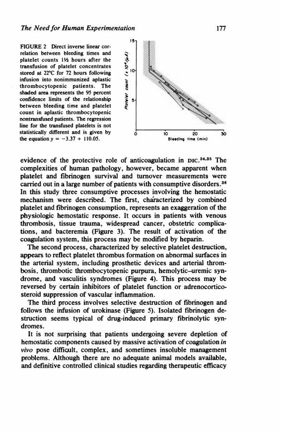
The Need for Human Experimentation
FIGURE 2 Direct inverse linear correlation between bleeding times and platelet counts I ¥.z hours after the transfusion of platelet concentrates stored at 22°C for 72 hours following infusion into nonimmunized aplastic thrombocytopenic patients. The shaded area represents the 95 percent confidence limits of the relationship between bleeding time and platelet count in aplastic thrombocytopenic nontransfused patients. The regression line for the transfused platelets is not statistically different and is given by the equation y = -3.37 + 110.05.
l!!I
0
177
10 20 30 Bleed Ing time (min)
evidence of the protective role of anticoagulation in DIC. 34•3 ~ The complexities of human pathology, however, became apparent when platelet and fibrinogen survival and turnover measurements were carried out in a large number of patients with consumptive disorders. 36
In this study tl).ree consumptive processes involving the hemostatjc mechanism were described. The first, characterized by combined platelet and fibrinogen consumption, represents an exaggeration of the physiologic hemostatic response. It occurs in patients with venous thrombosis, tissue trauma, widespread cancer, obstetric complications, and bacteremia (Figure 3). The result of activation of the coagulation system, this process may be modified by heparin.
The second process, characterized by selective platelet destruction, appears to reflect platelet thrombus formation on abnormal surfaces in the arterial system, including prosthetic devices and arterial thrombosis, thrombotic thrombocytopenic purpura, hemolytic-uremic syndrome, and vasculitis syndromes (Figure 4). This process may be reversed by certain inhibitors of platelet function or adrenocorticosteroid suppression of vascular inflammation.
The third process involves selective destruction of ftbrinogen and follows the infusion of urokinase (Figure 5). Isolated fibrinogen destruction seems typical of drug-induced primary ftbrinolytic syndromes.
It is not surprising that patients undergoing severe depletion of hemostatic components caused by massive activation of coagulation in vivo pose difficult, complex, and sometimes insoluble management problems. Although there are no adequate animal models available, and definitive controlled clinical studies regarding therapeutic efficacy
Copyright © National Academy of Sciences. All rights reserved.
Animal Models of Thrombosis and Hemorrhagic Diseaseshttp://www.nap.edu/catalog.php?record_id=19903
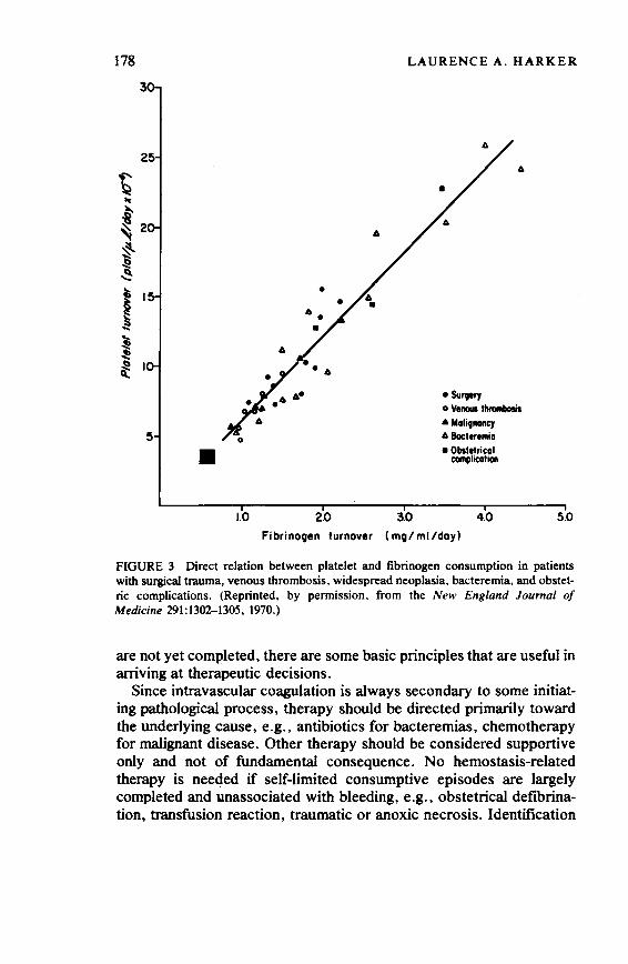
178
• 1.0
6
•
2.0
LAURENCE A. HARKER
3.0
6
• Survery 0 v... ""1llllbolil • MalitnallCJ 6 Bocteremia • Obstetrical
coinplicalian
4.0 5.0
Fibrino9en turnover ( mQ/ ml /doyl
FIGURE 3 Direct relation between platelet and fibrinogen consumption in patients with surgical trauma, venous thrombosis, widespread neoplasia, bacteremia, and obstetric complications. (Reprinted, by permission, from the New England Journal of Medicine 291: 1302-1305, 1970.)
are not yet completed, there are some basic principles that are useful in arriving at theral>eutic decisions.
Since intravascular coagulation is always secondary to some initiating pathological process, therapy should be directed primarily toward the underlying cause, e.g., antibiotics for bacteremias, chemotherapy for malignant disease. Other therapy should be considered supportive only and not of fundamental consequence. No hemostasis-related therapy is nee<Jed if self-limited consumptive episodes are largely completed and unassociated with bleeding, e.g., obstetrical defibrination, transfusion reaction, traumatic or anoxic necrosis. Identification
Copyright © National Academy of Sciences. All rights reserved.
Animal Models of Thrombosis and Hemorrhagic Diseaseshttp://www.nap.edu/catalog.php?record_id=19903
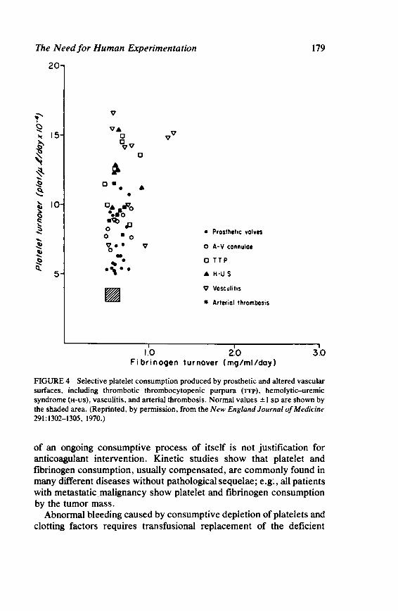
The Need for Human Experimentation
20
f' v
~ VA vv .. 15 0
i' iv
~ 0
~ ~ .... -S! 0. • ~ • • ... 10 o• r'o ~ ~
.... o ~~ ~ 0 ....
0 • 0 .... ~ Y,•. v ~ --S! ' . ~
5 ., .. ~
179
• Prosthetic valves
o A-V cannulae
0 TTP
AH-US
V Vasculilis
• Arterial thrombosis
1.0 2.0 3.0 Fi brinooen turnover (mg/ml/day)
FIGURE 4 Selective platelet consumption produced by prosthetic and altered vascular sulfaces, including thrombotic thrombocytopenic purpura (TIP), hemolytic-uremic syndrome (H-US), vasculitis, and arterial thrombosis. Normal values ±I so are shown by the shaded area. (Reprinted, by permission, from the New England Journal of Medicine 291:1302-1305, 1970.)
of an ongoing consumptive process of itself is not justification for anticoagulant intervention. Kinetic studies show that platelet and fibrinogen consumption, usually compensated, are commonly found in many different diseases without pathological sequelae; e.g:, all patients with metastatic malignancy show platelet and fibrinogen consumption by the tumor mass.
Abnormal bleeding caused by consumptive depletion of platelets and clotting factors requires transfusional replacement of the deficient
Copyright © National Academy of Sciences. All rights reserved.
Animal Models of Thrombosis and Hemorrhagic Diseaseshttp://www.nap.edu/catalog.php?record_id=19903

180 LAURENCE A.HARKER
30 • • •
'fu x
• • • ITP
6 Uroki1111S11 Infusion > 20 ~ ~ .,J .!!
• • • ••
~
I 10 • •
~ ~
"! i: 6
1.0 2.0 3.0 8.0 9.0 10.0 11.0 12.0 Fibrinogen Turnover (mg/ml/day)
FIGURE 5 Independent destruction of platelets and fibrinogen. Selective platelet destruction is observed in patients with idiopathic thrombocytopenic purpura (~ selective fibrinogen destruction is produced by urokinase infusion. The shaded area indicates the results in 35 normal subjects ±1 so. (Reprinted, by permission, from the New England Journal of Medicine 291:1302-1305, 1970.)
components in order to restore hemostasis. Platelets are supplied as concentrates, and clotting factors are provided by whole blood when intravascular volume is also needed (as is usually the case), by fresh-frozen plasma, or, rarely, by concentrates, e.g., cryoprecipitates. Since heparin is a potent inhibitor of fibrin formation, bleeding is usually aggravated by its use. The rise in fibrinogen concentration that usually follows heparin blockade of fibrinogen conversion should not be interpreted as evidence of improved hemostatic competence but rather as a reflection of defective fibrin formation. A decrease in bleeding after heparin therapy frequently is the result of coincident platelet transfusions and resultant improved platelet plug formation. Platelet counts are usually not increased by heparin administration alone.
Microcirculatory occlusion ("organ dysfunction") is usually due to endothelial injury and resultant microcirculatory occlusion by platelet plugs, rather than to primary fibrin deposition, as illustrated by endotoxin-induced injury to glomerular endothelium, hemolyticuremic syndrome, thrombotic thrombocytopenic purpura, and other vasculitides. Ordinarily, the appearance of "fibrin" on pathologic
... Copyright © National Academy of Sciences. All rights reserved.
Animal Models of Thrombosis and Hemorrhagic Diseaseshttp://www.nap.edu/catalog.php?record_id=19903

The Need for Human Experimentation 181
sections prepared from such patients reflects expected deposition of platelet plugs rather than primary intravascular fibrin deposits. The sequelae of microcirculatory occlusion are more likely to be prevented by inhibiting platelet function or suppressing vasculitis with steriods, although the further addition of anticoagulation can complement the antithrombotic effect. The latter possibility may explain the exceptional effectiveness ofheparinization reported in pediatric patients who develop the rare, infection-related, purpura fulminans. Since fibrinolysis plays a major role in re-establishing and maintaining microcir:culatory patency' fibrinolytic inhibitors such as epsilon-aminocaproic acid (EACA) may be harmful. Platelet tninsfusions in these patients should logically be held to the minimum heeded for hemostasis in order to limit the thrombotic mass formed. It is therefore concluded that heparin and EACA, alone or in combination, are seldom, if ever, useful in the management of patients with combined platelet and fibrinogen consumption. This conclusion contrasts with the initial implication of animal work.
Models of Thrombosis
Many induced animal models of thrombosis have been Ul)satisfactory since they have produced conflicting findings_ and do not appear to predict human thrombus formation. Useful techniques for studying thrombogenesis and its prevention in man have been recently developed that enable quantitative, predictive, and in vivo studies to be completed.
For example, platelet, fibrinogen, and plasminogen kinetic measurements appear to measure the rate and predominant composition of ongoing thrombus formation. 3s-3s The effectiveness of antithrombotic therapy can be quantitatively determined in vivo by measuring its capacity to interrupt thrombotic consumption (Figure 6).
Venous thrombosis results in combined platelet and fibrinogen consumption, which reflects the predominant role of fibrin in thrombus formation. Interruption of the consumptive process by heparin, but not by dipyridamole, correlates with the antithrombotic effectiveness of anticoagulant therapy and the reported failure of platelet-function inhibitors to decrease venous thrombosis. In contrast, arterial thrombosis involves selective platelet consumption that is interrupted by dipyridamole but not by heparin administration. Circulating fibrinogen is not consumed in this setting because procoagulants are swept away from the thrombogenic focus by the rapid arterial flow before coagulation becomes fully activated. These observations are in accord with the
Copyright © National Academy of Sciences. All rights reserved.
Animal Models of Thrombosis and Hemorrhagic Diseaseshttp://www.nap.edu/catalog.php?record_id=19903
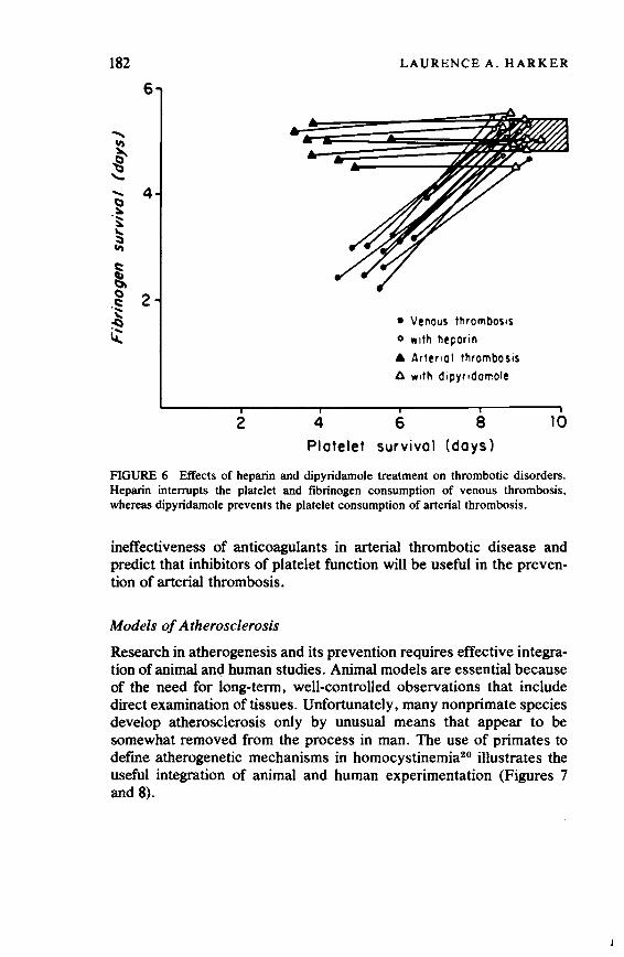
182
6
2
LAURENCE A. HARKER
4
• Venous thrombosis o with heporin
A Arter•ol thrombosis A with dipyr1domole
6 8 Platelet survival (days)
10
FIGURE 6 Effects of heparin and dipyridamole treatment on thrombotic disorders. Heparin interrupts the platelet and fibrinogen consumption of venous thrombosis, whereas dipyridamole prevents the platelet consumption of arterial thrombosis.
ineffectiveness of anticoagulants in arterial thrombotic disease and predict that inhibitors of platelet function will be useful in the prevention of arterial thrombosis.
Models of Atherosclerosis
Research in atherogenesis and its prevention requires effective integration of animal an<J human studies. Animal models are essential because of the need for long-term, well-controlled observations that include direct examination of tissues. Unfortunately, many nonprimate species develop atherosclerosis only by unusual means that appear to be somewhat removed from the process in man. The use of primates to define atherogenetic mechanisms in homocystinemia20 illustrates the useful integration of animal and human experimentation (Figures 7 and 8).
Copyright © National Academy of Sciences. All rights reserved.
Animal Models of Thrombosis and Hemorrhagic Diseaseshttp://www.nap.edu/catalog.php?record_id=19903
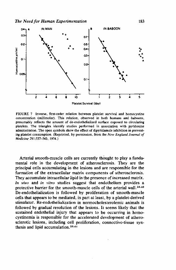
The Need for Human Experimentation
e e :I.
04 A
03
0.2
~ 0.1
l 8 ~
::c 0
2
IN MAN
"' 6 8 10
0.7
0.5
0.4
03
0.2
0.1
B
Platelet Survival (day)
183
IN BABOON
2 3 4
FIGURE 7 Inverse, first-order relation between platelet survival and homocystine concentration (millimolar). This relation, observed in both humans and baboons, presumably reflects the amount of de-endothelialized surface exposed to circulating platelets. The triangles identify studies performed in association with pyridoxine administration. The open symbols show the effect of dipyridamole inhibition in preventing platelet consumption. (Reprinted, by permission, from the New England Journal of Medicine 291 :537-543, 1974.)
Arterial smooth-muscle cells are currently thought to play a fundamental role in the development of atherosclerosis. They are the principal cells accumulating in the lesions and are responsible for the formation of the extracellular matrix components of atherosclerosis. They accumulate intracellular lipid in the presence of increased matrix. In vivo and in vitro studies suggest that endothelium provides a protective barrier for the smooth-muscle cells of the arterial wall. 39•40
De-endothelialization is followed by proliferation of smooth-muscle cells that appears to be mediated, in part at least, by a platelet-derived stimulator. Re-endothelialization in normocholesterolemic animals is followed by gradual resolution of the lesions. It seems likely that the sustained endothelial injury that appears to be occurring in homocystinemia is responsible for the accelerated development of atherosclerotic lesions, including cell proliferation, connective-tissue synthesis and lipid accumulation. 20•41
Copyright © National Academy of Sciences. All rights reserved.
Animal Models of Thrombosis and Hemorrhagic Diseaseshttp://www.nap.edu/catalog.php?record_id=19903

184 LAURENCE A. HARKER
FIGURE 8 Homocystine-induced arteriosclerosis in a baboon. Homocystinemia causes patchy endothelial cell loss that results in typical focal intimal lesions of proliferating smooth muscle cells and the connective tissue matrix composed of collagen and elastic fibers. Dipyridamole therapy markedly reduces lesion formation .
CONCLUSION
In today's climate of ethical and financial restrictions, 42.43 clinical investigation is becoming increasingly difficult to carry out. Despite the resultant appeal of exclusive reliance upon animal experimentation, human research remains vital if improved patient care is to be achieved.
REFERENCES
I. Altman, L. K. 1972. Auto-experimentation. N. Engl. J. Med. 286:346. 2. Hunter, J. 1837. The works of John Hunter. Vol. 11. Edited by J . F. Palmer.
Longman, England. 3. Hunter, J . 1859. A treatise on the venereal disease. With notes by George C.
Babington. Haswell , Barrington and Haswell, Philadelphia. 4. Forssman, W. 1929. Die sondierrung des rechten herzens. Klin. Wochenschr.
8:2085. 5. Forssman, W. 1931. Ueber kontrastdarstellung der hohlen des lebenden rechten
herzens und der lungenschlagader. Munchen Med. Wochenschr. 78:489. 6. Harrington, W. J., V. Minnich, J. W. Hollingsworth , and C. V. Moore. 1951.
Demonstration of a thrombocytopenic factor in the blood of patients with thrombocytopenic purpura. J . Lab. Clin. Med. 38:1.
Copyright © National Academy of Sciences. All rights reserved.
Animal Models of Thrombosis and Hemorrhagic Diseaseshttp://www.nap.edu/catalog.php?record_id=19903
The Need for Human Experimentation 185
7. Biggs, R. (ed.). 1972. Human blood coagulation, haemostasis and thrombosis. F. A. Davis, Philadelphia.
8. Davie, E. W., and E. P. Kirby. 1973. Molecular mechanisms in blood coagulation. Curr. Top. Cell. Regul. 7:51.
9. Ratnoff, 0. D. 1972. The molecular basis of hereditary clotting disorders. Page 39 in T. H. Spaet, Progress in hemostasis and thrombosis. Vol. 1. Grune & Stratton, New York.
10. Glanzmann, E. 1918. Hereditare haemorrhagische thrombasthenie, ein beitras zur pathologie der blutplattchen. Jahrb. Kinderheilk. 88: 1.
11. Bernard, J., and J. P. Soulier. 1948. Sur une nouvelle variete de dystrophie thrombocytaire hemmorrhagipare congenitale. Sem. Hop. Paris 24:217.
12. Weiss, H.J., P. A; Chervenick, R. Zalusky, and A. Factor. 1969. A familial defect in platelet function associated with impaired release of adenosine diphosphate. N. Engl. J. Med. 281:1264.
13. Gottlieb, A. J. 1972. Disorders ofhemostasis-nonthrombocytopenic purpuras. Page 1176 in W. J. Williams, E. Beutler, A. J. Erslev, and R. W. Rundles (ed.), Hematology. McGraw-Hill, New York.
14. Egeberg, O. 1967. Inherited antithrombin deficiency causing thrombophilia. Thromb. Diath. Haemorrh. 17:176.
15. Abildgaard, U. 1967. Purification of two progressive antithrombins of human plasma. Scand. J. Clin. Lab. Invest. 19:190.
16. Nilsson, I. M., H. Krook, N. H. Stemby, E. Soderberg, and N. Soderstrom. 1961. Severe thrombotic disease in a young man with bone marrow and skeletal changes and with a high content of an inhibitor in the tibrinolytjc system. Acta Med. Scand. 169:323.
17. Gaston, L. W. 1966. Studies on a family with an elevated plasma level of factor V (proaccelerin) and a tendency to thrombosis. J. Pediat. 68: 367.
18. Carson, N. A. J., C. E. Dent, and C. M. B. Field. 1965. Homocystinuria: Clinical and pathological review of ten cases. J. Pediat. 66:565.
19. Schimke, R. N., V. A. McKusick, and T. Huang. 1965. Homocystinuria: Studies of 20 families with 38 affected members. JAMA 193:711.
20. Harker, L.A., S. J. Stichter, C.R. Scott, and R. Ross. 1974. Homocystinemia: Vascular injury and arterial thrombosis. N. Engl. J. Med. 291:537.
21. Gifford, R. H., and A. R. Feinstein. 1969. A critique of methodology in studies of antiCoagulant therapy for acute myocardial. N. Engl. J. Med. 280:351.
22. Harker, L.A., J. Hirsh, M. Gent, and E. Genton. 1975. Critical evaluation of platelet inhibiting drugs in thrombotic disease. Page 229 in E. Brown (ed.). Progress in hematology. Volume IX. Brown and Stratton, New York.
23. Genton, E., M. Gent, J. Hirsh, and L. A. Harker. 1975. Platelet inhibiting drugs in the prevention of clinical thrombotic disease. A review. N. Engl. J. Med. 293: 1174; 1236; 1296.
24. Sullivan, J. M., D. E. Harken, and R. Gorlin. 1971. Pharmacologic control of thromboembolic complications of cardiac-valve replacement. N. Engl. J. Med. 284:1391.
25. Kaegi, A., G. F. Pineo, A. Shimizu, H. Trivedi, J. Hirsh, and M. Gent. 1974. Arteriovenous-shunt thrombosis: Prevention by sultinpyrazone. N. Engl. J. Med. 290:304.
26. Elwood, P. C., A. L. Cochrane, M. L. Burr, P. M. Sweetname, G. Williams, E. Welsby, S. J. Hughes, and R. Renton. 1974. A randomized controlled trial of acetylsalicylic acid in the secondary prevention of mortality from myocardial infarction. Br. Med. J. 1:436.
Copyright © National Academy of Sciences. All rights reserved.
Animal Models of Thrombosis and Hemorrhagic Diseaseshttp://www.nap.edu/catalog.php?record_id=19903
186 LAURENCE A. HARKER
Tl. Boston Collaborative Study Surveillance Group. 1974. Regular aspirin intake and acute myocardial infarction. Br. Med. J. 1:440.
28. The Coronary Drug Project Research Group. 1975. Clofibrate and niacin in coronary heart disease. JAMA 231:360.
29. Zimmennan, T. S., O. D. Ratnoff, and A. E. Powell. 1971. Immunologic differentiation of classic hemophilia (factor VIII deficiency) and von Willebrand's disease. J. Clin. Invest. 50:244.
30. Holmberg, L., and I. M. Nillson. 1973. Two genetic variants of von Willebrand's disease. N. Engl. J. Med. 288:595.
31. Slichter, S. J., and L. A. Harker. (In press) Preparation and storage of platelet concentrates I. Preparation: factors influencing the harvest of viable platelets from whole blood. Br. J. Haematol.
32. Slichter, S. J., and L. A. Harker. (In press) Preparation and storage of platelet concentrates II. Storage variables influencing platelet viability and function. Br. J. Haematol.
33. Slichter, S. J., R. B. Counts, and L. A. Harker. (In press) Preparation of cryoprecipitated factor VIII concentrates. Transfusion.
34. McKay, D. G. 1965. Disseminated intravascular coagulation: An intermediary mechanism of disease. Hoeber Medical Division, Harper and Row, New York.
35. Hardaway, R. M. 1966. Syndromes of disseminated intravascular coagulation: With special reference to shock and hemorrhage. Charles C Thomas, Springfield, Illinois.
36. Harker, L.A., and S. J. Slichter. 1972. Platelet and fibrinogen consumption in man. N. Engl. J. Med. 287:999.
37. Harker, L. A., and S. J. Slichter. 1974. Arterial and venous thromboembolism: kinetic characteriz.ation and evaluation of therapy. Thromb. Diath. Haemorrh. 31:188.
38. Harker, L. A. 1974. In vivo evaluation of antithrombotic therapy in man. Thromb. Diath. Haemorrh. Suppl. 60: 481:495.
39. Ross, R., J. Glomset, and B. Kariya. 1974. A platelet-dependent serum factor that stimulates the proliferation of arterial smooth muscle cells in vitro. Proc. Nat. Acad. Sci. USA 71:1207.
40. Ross, R., and J. A. Glomset. 1973. Atherosclerosis and the arterial smooth muscle cell: Proliferation of smooth muscle is a key event in the genesis of the lesions of atherosclerosis. Science 180: 1332.
41. Harker, L.A., R. Ross, S. J. Slichter, and R. C. Scott. 1974. Homocystine induced arteriosclerosis. J. Clin. Invest. 53:31.
42. Freund, P.A. 1965. Ethical problems in human experimentation. N. Engl. J. Med. 273:687.
43. Inglefinger, F. J. 1973. Ethics of experiments on children. N. Engl. J. Med. 288:791.
Copyright © National Academy of Sciences. All rights reserved.
Animal Models of Thrombosis and Hemorrhagic Diseaseshttp://www.nap.edu/catalog.php?record_id=19903
VI
PROJECTED NEEDS AND RECOMMENDATIONS
Specific discussion and recommendations pertaining to genetic and spontaneous animal models of thrombosis and hemorrhage were the main focus of the workshop. Other experimentally induced models, although considered, were not emphasized because of the tremendous variety of such models and the wealth of published information available on this subject.
The projected needs and recommendations listed in this section were contributed by Working Groups that discussed the following topics:
A. Preservation of Unique Models B. Developmental Needs C. Diagnostic Methods of Identifying Models D. Mechanics of Support and Cost-Effectiveness
Copyright © National Academy of Sciences. All rights reserved.
Animal Models of Thrombosis and Hemorrhagic Diseaseshttp://www.nap.edu/catalog.php?record_id=19903

Copyright © National Academy of Sciences. All rights reserved.
Animal Models of Thrombosis and Hemorrhagic Diseaseshttp://www.nap.edu/catalog.php?record_id=19903

WORKING GROUP A
Preservation of Unique Models
1. It is recommended that unique animal models of thrombosis and hemorrhage be preserved.
2. In order to achieve the above, it is recommende<J that animal research resource facilities to study thrombosis and hemorrhage be organized as follows:
a. Establishing four or five regional research and resource centers distributed nationally to breed, maintain, and develop individual models or groups of models of thrombosis and hemorrhage for research; each center would be capable of diagnosis, maintenance, treatment, clinical research, and investigations
b. Establishing a centralized national sperm bank c. Establishing a centralized registry for information exchange d. Establishing a national animal blood products reference center
(to include reagents for blood typing, histocompatibility matching, immunogenetic marker assays, deficient substrates, and therapeutic materials)
Establishing such centers of research on thrombosis and hemorrhage would (a) ensure accessibility of resources to the scientific community; (b) establish the scientific validity of newly discovered models; (c) provide a repository of expertise to maintain and utilize the model; and (d) screen and refer potential models to experts for evaluation. In addition, the use of such centers would require fewer animals to supply
189
Copyright © National Academy of Sciences. All rights reserved.
Animal Models of Thrombosis and Hemorrhagic Diseaseshttp://www.nap.edu/catalog.php?record_id=19903

190 WORKING GROUP A
the needs of the scientific community, would be more productive, provide more potential, and should be more cost-effective than multiple individual programs.
3. It is recommended that regionalization not inhibit an individual investigator from developing his own model of thrombosis and hemorrhage and applying for an individual grant to study that model on its own scientific merit.
4. It is recommended that criteria for defining a model as unique be established. For example, one of kind, one of group, one species, strains within species, or common model with new application.
5. It is recommended that the feasibility of preserving a specific model of thrombosis and hemorrhage be defined in terms of cost, maintenance, life span, chance of contagion, and biohazard.
Copyright © National Academy of Sciences. All rights reserved.
Animal Models of Thrombosis and Hemorrhagic Diseaseshttp://www.nap.edu/catalog.php?record_id=19903

WORKING GROUP B
Developmental Needs
1. It is recognized that an urgent need exists for animal models of spontaneous thrombotic disease as well as for improved induced models. Inherited or acquired spontaneous thrombotic models, not readily available, provide an important resource for comparative studies. It should be emphasized, however, that induced models have been and will continue to be useful if they closely resemble the disease of interest. It is suggested that a current survey of potential models of arterial thrombosis be undertaken. Some potential models are atherosclerosis in pigeons, hypercholesterolemia in rats, saddle embolism in cats, and equine viral arteritis. Additional models of venous thrombosis are probably not needed because the condition is common and readily studied in humans.
2. Some aspects of the animal model should mimic the human counterpart. It is not necessary that the model be identical to all aspects of the human disease process, since, for instance:
a. Infectious canine hepatitis represents a useful animal model for the coagulopathies associated with acute and chronic liver disease. The coagulopathy involves both decreased synthesis and increased utilization of platelets and clotting factors, and a disseminated intravascular coagulation syndrome may occur. However, the viral agent and lesions produced differ from those of infectious hepatitis in man. · b. Intra-articular hemorrhages are common in human hemophiliacs and they result in joint disabilities, whereas in some strains of hemophilic dogs joint lesions are minimal as hemorrhage
191
Copyright © National Academy of Sciences. All rights reserved.
Animal Models of Thrombosis and Hemorrhagic Diseaseshttp://www.nap.edu/catalog.php?record_id=19903
192 WORKING GROUP B
tends to be periarticular; Other strains of dogs have severe hemarthroses. This variability does not detract from, but enhances, the scope of the model.
3. More than one animal species with a disease: analogous to one found in humans should be maintained and supported, as this will enhance the credibility of the model-e.g., Chediak-Higashi syndrome in cattle, mink, and mice.
4. It is recognized that pound animals are a source of research material to provide blood products for support of the medical services required under the regionalization concept and for acute experiments such as surgical techniques and to provide tissues. It is also recognized, however, that they are inferior to purebred or kennel-reared animals of defined background for chronic or physiological experiments.
5. It is strongly urged that national and international cooperation be encouraged among veterinarians, breed organizations, animal welfare organizations, and the biomedical community for exchange of information mutually beneficial to animals and humans. Areas of specific interest are prevention, prognosis, disease processes, necropsy, maintenance, therapeutic regimens, and research findings. If regionalization is adopted, the above groups would interact with the centers.
6. It is recommended that an investigator developing an animal model supported by federal funds be allowed whatever time is required to develop the model. After this time, other appropriate investigators should have access to the model and/or sperm stored in central banks for development of their own satellite colonies. Additional long-range goals to be supported are the development of functional ovum and/or embryo banks.
7. It is recognized that the following defined specific models still need to be developed:
a. Antithrombin-111-deficient animals to provide insight into thrombotic tendencies in humans.
b. Animals with spontaneous clotting factor inhibitors (e.g., those occurring in hemophilia, autoimmune disease, and drug dyscrasias) because of the difficult problems encountered in treating patients.
c. Models of factor I, II, V, and XIII and kallikrein-kinin deficiencies. Factor I and II deficiencies would provide models for the dysfibrinogenemic and dysprothrombinemic states of humans. FactorV-deficient animals could be crossbred with hemophilic animals to
Copyright © National Academy of Sciences. All rights reserved.
Animal Models of Thrombosis and Hemorrhagic Diseaseshttp://www.nap.edu/catalog.php?record_id=19903
Developmental Needs 193
create the combined factor V-VIII defect for re-examining the onelocus hypothesis for these clotting proteins. Factor-XIII-deficient animals would be valuable for studying the processes of tissue repair and wound healing. Kallikrein-kinin-deficient animals could be used to study the interactions between initiating events of the coagulation, -kinin, and complement mechanisms.
d. Models of disseminated intravascular coagulation; these are more common in animals than realized and should be further investigated to provide useful data on this important syndrome in humans and animals.
e. Atherosclerotic and arteriosclerotic models, e.g., spawning salmon; these may generate new insight into the pathogenesis of these disease processes.
f. Models of complement deficiencies. Complement deficiencies such as C6-deficient rabbits and C4-deficient guinea pigs have already provided important information on the interactions of complement and hemostasis. Other such models would therefore be desirable.
g. Miscellaneous models: e.g., hemorrhagic gastroenteritis in dogs; aortic embolism in cats; equine viral arteritis; thrombocytopenias-idiopathic, immune-mediated, and drug:induced.
8. It is recommended that maintenance of muitiple mutant strains of such models as hemophilia A and B be supported because of their value to studies of potential heterogeneity of the abnormal proteins and of the different levels of clinical severity of these diseases in humans and animals. The most practical way to maintain all of these mutations would be through sperm-banking.
9. It .is recognized that it is important to maintain the specific genotype of animal models: e.g., for basic studies of the molecular defect, a hemophilic dog should be transfused only when essential in order to minimize stimulation of the immune mechanism. By contrast, in order to mimic the multiply transfused .hemophilic patient, one should give the animal many-transfusions.
10. It is recognized that studies of spontaneously acquired hemorrhagic and thrombotic diseases should be continued, e.g., equine viral arteritis, infectious canine hepatitis, and thrombocytopenic states. Additional information on the incidence of thrombosis is obtainable from retrospective studies of necropsy material.
11. It is recommended that random-source populations of animals as well as specific purebred strains be maintained. In a search for certain genetic characteristics such as blood group and histocompatibility antigens and enzyme polymorphisms, the random-source dog is extremely valuable because of its relatively low inbreeding coefficient.
Copyright © National Academy of Sciences. All rights reserved.
Animal Models of Thrombosis and Hemorrhagic Diseaseshttp://www.nap.edu/catalog.php?record_id=19903
WORKING GROUP C
Diagnostic Methods of Identifying Models
1. It is recognized as extremely important in identifying models by diagnostic methods to have appropriate homologous, age- and sexmatched controls. These controls should be both random and strainspecific.
2. It is recommended that new areas of diagnostic investigation be developed for animal species:
a. Establish standard diagnostic methods for screening animals for potential bleeding or thrombotic abnormalities.
b. Devise assays for progressive antithrombins, e.g., antithrombin III, a 1-antitrypsin and armacroglobulin assays.
c. Improve methods for identifying carriers of hemophilia and the heterozygous state of other clotting defects.
d. Establish standard techniques and reagents for assays of coagulation, platelet function, and fibrinolysis for each animal species.
e. Establish diagnostic criteria for defining models of disseminated intravascular coagulation.
f. Develop better tests for evaluating hemostatic parameters in thrombotic diseases of animals and man.
3. It is recommended that diagnostic substrate plasmas from the various clotting-factor-deficient animals be made available for performing routine coagulation assays. These animal substrate plasmas have been proven to be reliable for specific clotting assays of all species,
194
Copyright © National Academy of Sciences. All rights reserved.
Animal Models of Thrombosis and Hemorrhagic Diseaseshttp://www.nap.edu/catalog.php?record_id=19903

Diagnostic Methods of Identifying Models 195
including man, and should be kept at the regional centers and at the centralized resource facility for blood products. Another advantage of the animal substrates is their ready availability in large quantities from a specific mutant strain of animal.
4. It is suggested that animals at risk for thrombosis following such procedures as reconstructive hip surgery (e.g., for correction of severe canine hip dysplasia) be evaluated by a series of hemostatic tests. Monitoring of these animals before, during, and after reconstructive surgery may provide information about the development of thrombotic complications with or without disseminated intravascular coagulation.
Copyright © National Academy of Sciences. All rights reserved.
Animal Models of Thrombosis and Hemorrhagic Diseaseshttp://www.nap.edu/catalog.php?record_id=19903

WORKING GROUP D
Mechanics of Support and Cost-Effectiveness
1. It is recommended that the current NIH policy with regard to federal funding continue: i.e., (a) for resource grants, the investigator must make the animal model accessible to other investigators (after a period of development has passed); (b) for research grants, the, investigator has the option of collaborating or providing accessibility.
2. Because of the concerns expressed regarding the cost of upgrading obsolete animal quarters and proper designs of such quarters, a representative of the ILAR Committee on Animal Models for Thrombosis and Hemorrhagic Diseases has been made a member of the Organizing Committee for the Symposium on Laboratory Animal Housing, which is to take place in the fall of 1976 at the National Academy of Sciences.
3. It is recommended that a uniform cost-accounting method for specific animal models be utilized, e.g., the Cost Analysis and Rate Setting Manual for Animal Resource Facilities, prepared by the Division of Research Resources, NIH, and the Association of American Medical Colleges, 1974. Although animal models require maintenance, care, and facilities that differ from those required by normal animals, this does not necessarily influence the type of cost-accounting procedure to be utilized. The reason for utilizing an established costaccounting method is to assure NIH and the public that funds are properly allocated and that charges against research grants are correct in that they reflect actual costs of the care of these research animals.
4. It is further recommended that additional funding be provided to the Animat Resources Branch, Division of Research Resources, NIH,
for the specific purpose of providing institutional grants for modernization of research-holding facilities. It is to be emphasized that the scientific community is in favor of good animal facilities and wishes to
196
Copyright © National Academy of Sciences. All rights reserved.
Animal Models of Thrombosis and Hemorrhagic Diseaseshttp://www.nap.edu/catalog.php?record_id=19903

Mechanics of Support and Cost-Effectiveness._ 197
mee\ and/or exceed the guidelines established by DREW and the Animal Welfare Act of 1970 but that this requires financial aid by the granting institutions. For example, in 1971 $92.5 million additional funds were estimated to be required for optimal compliance with the Animal Welfare Act ($50 million for construction, $18 million for renovation, $21 million for cages and equipment, $3.5 million for training and additional operating costs).*
5. It is recommended that animal models of thrombosis and hemorrhagic diseases be maintained in an environment specifically designed to minimize injury. By its nature (i.e., individual or dual units), this· type of housing is more expensive. The overall costs associated with housing such an animal colony, and the high cost of providing the proper veterinary care, experienced personnel, and special materials for maintenance and treatment, are not fully recognized by the funding agencies. In order to optimize the use of these animals and their productivity, increased funding is needed. Such costs are real and should be covered when properly documented in research proposals. It may be possible to reduce the cost by utilizing the sperm bank concept, which ·should limit the numbers of animals required but not the potential of the resource.
6. Under the regional center concept (the recommendations of Working Group A, p. 189) support must be given to provide! laboratory facilities for in-house and visiting scientists to utilize the resource. A grant-application mechanism should be established to decide which investigators both within and outside the center qualify to use the resource. The grant proposal should include a statement of assurance from the director of the center where the research is to be performed that the center can accommodate the project. It is recommended that a peer review committee evaluate applications according to their scientific merit. This committee could be appointed by NIH and should exclude the directors of regional centers.
7. It is recognized that the level of current support for animal models of thrombosis and hemorrhage is inadequate in terms of allocating funds for developing new model programs and for establishing the regional centers. Although funds are available for existing meritorious programs, they are insufficient for maximizing productivity and for allowing justified expansions. Thus, a higher proportion of funds allocated to the various categorical institutes of NIH should be available for animal model support and development.
*Charles W. McPherson. 1975. Personal Communication. Animal Resource Branch, NIH, DHEW.
Copyright © National Academy of Sciences. All rights reserved.
Animal Models of Thrombosis and Hemorrhagic Diseaseshttp://www.nap.edu/catalog.php?record_id=19903

Copyright © National Academy of Sciences. All rights reserved.
Animal Models of Thrombosis and Hemorrhagic Diseaseshttp://www.nap.edu/catalog.php?record_id=19903
Contributors and Working Group Co-Chairmen
LOUIS M. ALEDORT. Associate Professor of Medicine, Mount Sinai School of Medicine, The City University of New York. New York
FRANK A. BELAMARICH, Professor of Biology. Biological Science Center. Marine Biological Laboratory. Boston University Marine Program, Woods Hole, Massachusetts
E. J. WALTER BOWIE, Head of Section. Hematology Research. Mayo Clinic, Rochester. Minnesota
KENNETH M. BRINKHOUS. Professor. Department of Pathology. School of Medicine, University of North Carolina at Chapel Hill
ROBERT w. BULL, Assistant Professor, Department of Medicine. College of Human Medicine, Michigan State University, East Lansing
HAROLD w. CASEY. Chairman. Department of Veterinary Pathology, Armed Forces Institute of Pathology. Washington, D.C.
DANIEL DEYKIN, Chief. Medical Service, Veterans Administration Hospital, Boston; and Professor of Medicine, Boston University and Tufts University Schools of Medicine
w. JEAN DODDS, Associate Research Scientist. Division of Laboratories and Research. New York State Department of Health. Albany
JOHAN A. VAN DER DOES, Hemostasis and Thrombosis Research Unit and Division of Hematology, Department of Medicine. University Hospital, Leiden, the Netherlands
JOHN T. GRUPENHOFF, Chairman. Science and Health Communications Group. Inc .• Washington, D.C.
GEORGE A. HEGREBERG, Institute of Comparative Medicine and Veterinary Science, Washington State University, Pullman
LAURENCE A. HARKER, Professor, Division of Hematology. Harbor View Medical Center, Seattle, Washington
199
Copyright © National Academy of Sciences. All rights reserved.
Animal Models of Thrombosis and Hemorrhagic Diseaseshttp://www.nap.edu/catalog.php?record_id=19903
200 Contributors and Working Group Co-Chairmen
JACEK HAWIGER, Professor. Department of Medicine, Vanderbilt University, and Veterans Administration Hospital, Nashville, Tennessee
CHRISTINE HAWKEY, Head, Department of Hematology, Nuffield Institute of Comparative Medicine, The Zoological Society of London, England
ALBERT M. JONAS, Professor and Chief, Section of Comparative Medicine, Yale University School of Medicine, New Haven, Connecticut
GARY J. KOCIBA, Director of Laboratories, Department ·of Veterinary Clinical Sciences, College of Veterinary Medicine, Ohio State University, Columbus
JACK LEVIN, Associate Professor of Medicine, Hematology Division, Department of Medicine, Johns Hopkins Medical Institution, Baltimore, Maryland
PETER H. LEVINE, Chief of Medicine and Director of the Hemophilia Center, The Memorial Hospital, Worcester, Massachusetts
GEORGE MIGAKI, Chief, Division of Comparative Pathology, Registry of Comparative Pathology, Armed Forces Institute of Pathology, Washington, D.C.
SEAN MOORE, Professor and Chairman, Department of Pathology, McMaster University, Hamilton, Ontario, Canada
ALLAN T. VAN OOSTEROM, Hemostasis and Thrombosis Research Unit and The Division of Hematology, Department of Medicine, University Hospital, Leiden, the Netherlands
ROBERT A. o'REILLY, Chairman, Department of Medicine, Santa Clara Valley Medical Center, San Jose, California
CHARLES A. OWEN, JR. Head, Department of Biochemistry, Mayo Clinic, Rochester, Minnesota
GEORGE A. PADGETT, Associate Professor, Department of Veterinary Pathology, Washington State University, Pullman
JUDITH G. POOL (deceased), Department of Medicine, Santa Clara Valley Medical Center, San Jose, California
ROBERT w. PRICHARD, Professor of Pathology, Department of Pathology, Bowman Gray School of Medicine, Wake Forest University, WinstonSalem, North Carolina
EDWIN w. SALZMAN, Professor of Surgery, Harvard Medical School and Beth Israel Hospital, Boston, Massachusetts
STEPHEN w. J. SEAGER, Associate Director, Institute of Comparative Medicine, Texas A&M University/Baylor College of Medicine, Houston, Texas
CHRISTINE STEVENS, President, Animal Welfare Institute, Washington, D. C. JAN J. VELTKAMP, Professor, Hemostasis and Thrombosis Research Unit,
Department of Medicine, University Hospital, Leiden. the Netherlands STANFORD WESSLER, Professor of Medicine and Associate Dean, Post
Graduate Medical School, New York University, New York JAMES G. WHITE, Professor, Department of Pediatrics, School of Medicine,
University of Minnesota, Minneapolis
Copyright © National Academy of Sciences. All rights reserved.
Animal Models of Thrombosis and Hemorrhagic Diseaseshttp://www.nap.edu/catalog.php?record_id=19903

Participants
SAMUEL ABRAMSON, Medical Advisory Committee, National Academy of Sciences, Washington, D.C.
DAVID L. ARONSON, Bureau of Biologics, Food and Drug Administration, Bethesda, Maryland
TAGE ASTRUP, Institute for Medical Research, Washington, D.C. RALPH G. BUCKNER, College of Veterinary Medicine, Oklahoma State Univer-
sity, Stillwater MARJORIE K. COOKE, Animal Welfare Institute, Washington, D.C. JULIAN DONOSO, Environmental Protection Agency, Washington, D.C. BRUCE H. EWALD, Cornell University Medical College, New York PATRICIA w. GENTRY, Ontario Veterinary College, University of Guelph,
Guelph, Ontario, Canada CLAIRE E. GRAHAM, Consultant, Washington, D.C. JOHN B. GRAHAM, School of Medicine, University of North Carolina, Chapel
Hill HARVEY R. GRALNICK, National Institutes of Health Clinical Center, Bethesda,
Maryland THOMAS R. GRIGGS, School of Medicine, University of North Carolina, Chapel
Hill JOHN T. GRUPENHOFF, Science and Health Communication Group, Inc.,
Washington, D.C. M. MASON GUEST, University of Texas Medical Branch, Galveston FANN HARDING, National Heart and Lung Institute, National Institutes of
Health, Bethesda, Maryland ROBERT F. HIGHSMITH, College of Medicine, University of Cincinnati,
Cincinnati, Ohio
201
Copyright © National Academy of Sciences. All rights reserved.
Animal Models of Thrombosis and Hemorrhagic Diseaseshttp://www.nap.edu/catalog.php?record_id=19903

202 Participants
PAULK. HILDEBRANDT' Walter Reed Anny Institute of Research, Washington, D.C.
ROBERT L. HUMMER, The Ameqcan Humane Association, Denver, Colorado ARTHUR 1. HURVITZ, The Animal Medical Center, New York JAMES M. IACONO, Lipid Nutrition Laboratory, U.S. Department of Agricul
ture, Beltsville, Maryland PANAYOTIS G. IATRIDIS, School of Medicine, Indiana University, Gary CAROL M. INGERMAN, Cardeza Foundation, Thomas Jefferson University,
Philadelphia, Pennsylvania MARILYN F. M. JOHNSTON, Medical School, St. Louis University, St. Louis,
Missouri HARRY E. KINGMAN, JR., National Society for Medical Research, Washington,
D.C. HARVEY G. KLEIN, National Heart and Lung Institute, National Institutes of
Health, Bethesda, Maryland DANIELL. KLINE, College of Medicine, University of Cincinnati, Cincinnati,
Ohio MARK B. KNUDSON, College of Veterinary Medicine, Washington State Univer-
sity, Pullman ROBERT G. LERNER, New York Medical College, New York JESSICA H. LEWIS, Central Blood Bank of Pittsburgh, Pittsburgh, Pennsylvania WALTER F. LOEB, Litton Bionetics Inc., Kensington, Maryland GARDNER c. McMILLAN, National Heart and Lung Institute, National Insti
tutes of Health, Bethesda, Maryland CHARLES w. MCPHERSON, Animal Resources Branch, National Institutes of
Health, Bethesda, Maryland WILLIAM MEDWAY, School of Veterinary Medicine, University of Pennsyl
vania, Philadelphia THOMAS J. MENDE, School of Medicine, University of Miami, Miami, Florida KENNETH M. MYERS, Washington State University College of Veterinary
· Medicine, Pullman ED NISHIZAWA, The Upjohn Company, Kalamazoo, Michigan DONALD F. PATTERSON, University of Pennsylvania, School of Veterinary
Medicine, Philadelphia LOUISE L. PHILLIPS, The Roosevelt Hospital, Columbia University, New
York CHARLEY SCHREGER, Medill News, Washington, D.C. BRUCE SCHUMER, Temple University Health Sciences Center, Philadelphia WALTER B. SMALLEY, Peanuts International Ltd., Washington, D.C. SCOTT N. SWISHER, College of Human Medicine, Michigan State University,
East Lansing ALEXSANDR TRETJAKOV, Embassy of the Soviet Union, Washington, D.C. DEMETRIOS c. TRIANTAPHYLLOPOULOS, American National Red Cross
Blood Research Laboratory, Bethesda, Maryland ROGER L. VIGDAHL, Riker Laboratories, St. Paul, Minnesota
Copyright © National Academy of Sciences. All rights reserved.
Animal Models of Thrombosis and Hemorrhagic Diseaseshttp://www.nap.edu/catalog.php?record_id=19903

Participants 203
WILLIAM P. WEBSTER, School of Medicine, University of North Carolina, Chapel Hill
GILBERT c. WHITE, School of Medicine, University of North Carolina, Chapel Hill
WILLO P. WHITE, American Psychological Association, Washington, D.C. LEO A. WHITEHAIR, Animal Resources Branch, National Institutes of Health,
Bethesda, Maryland ROBERT J. WILKINS, The Animal Medical Center. New York SIDNEY K. WOLFSON, JR., The Montefiore Hospital, Pittsburgh, Pennsylvania
Copyright © National Academy of Sciences. All rights reserved.
Animal Models of Thrombosis and Hemorrhagic Diseaseshttp://www.nap.edu/catalog.php?record_id=19903

Copyright © National Academy of Sciences. All rights reserved.
Animal Models of Thrombosis and Hemorrhagic Diseaseshttp://www.nap.edu/catalog.php?record_id=19903