Superior depletion of alloreactive T cells from peripheral blood stem cell and umbilical cord blood...
12
Superior Depletion of Alloreactive T Cells from Peripheral Blood Stem Cell and Umbilical Cord Blood Grafts by the Combined Use of Trimetrexate and Interleukin-2 Immunotoxin Paul Szabolcs, 1,2 Kyung-Duk Park, 1 Luciana Marti, 1 Divinomar DeOliveria, 1 Young-Ah Lee, 1 Michael O. Colvin, 3 Joanne Kurzberg 1 1 Department of Pediatrics, Pediatric Stem Cell Transplant Program; 2 Department of Immunology; and 3 Department of Medicine, Medical Oncology, Duke University Medical Center, Durham, North Carolina Correspondence and reprint requests: Paul Szabolcs, MD, Pediatric Stem Cell Transplant Program, Box 3350, Duke University Medical Center, Durham, NC 27705 (e-mail: [email protected]). Received July 10, 2004; accepted July 23, 2004 ABSTRACT Acute graft-versus-host disease, a major obstacle to the overall success of allogeneic hematopoietic stem cell transplantation, is primarily induced by a subset of donor T cells. Most strategies to prevent acute graft- versus-host disease target all T cells regardless of their specificity, and this leads to prolonged posttransplan- tation immunodeficiency. Selective depletion of alloreactive T cells could spare protective immunity and facilitate engraftment and graft-versus-leukemia effects. Recently described depletion strategies target acti- vation markers such as CD25 that are expressed by alloreactive T cells. However, incomplete depletion may occur when a single surface epitope or pathway of apoptosis is targeted that may not be fully and concurrently expressed among all alloreactive cells. We now report on a novel strategy effective in both cord blood and peripheral blood stem cell alloreactive T cells that simultaneously induces 2 independent pathways of apoptosis after stimulation by recipient dendritic cells or Epstein-Barr virus–transformed B cells. First, we demonstrate that the folate antagonist trimetrexate selectively depletes proliferating alloreactive precursors in vitro in a dose- and time-dependent manner. Similarly, a second agent, denileukin diftitox, kills activated alloreactive T cells expressing CD25. Most importantly, these 2 agents can exert their effects in concert with superior efficacy while sparing resting bystander T cells, which remain available to mount antimicrobial or third-party re- sponses. © 2004 American Society for Blood and Marrow Transplantation KEY WORDS Acute graft-versus-host disease ● Allogeneic hematopoietic stem cell transplantation ● Alloreactive T cells ● Trimetrexate ● Denileukin diftitox INTRODUCTION Alloreactive T-cell subsets present within the di- verse T-cell receptor (TCR) repertoire of allogeneic hematopoietic stem cell grafts can recognize host tis- sues in the context of HLA molecules and are critical in the pathogenesis of acute graft-versus-host disease (aGVHD) [1,2]. Proinflammatory T-helper type 1 cytokines such as tumor necrosis factor (TNF)–, interleukin (IL)–1, interferon-, and some T-helper type 2 cytokines (such as IL-5 and IL-13) produced by T cells contribute to the development of aGVHD independently and also in concert with direct cell- mediated cytotoxicity exerted by the alloreactive T cells themselves or by other recruited inflammatory leukocytes [3-9]. The development of aGVHD is crit- ically dependent on the initial reaction between allo- reactive donor T cells and host antigens displayed on antigen-presenting cells (APCs) such as dendritic cells (DC) [10]. Host APCs will induce the expansion of alloreactive clones identifiable by their unique TCR- chain sequence [1]. Alloreactive T cells are also iden- tifiable in vitro in a mixed lymphocyte reaction (MLR) upon activation by APCs as activated T cells enter the cell cycle, and during blastogenesis they upregulate Biology of Blood and Marrow Transplantation 10:772-783 (2004) 2004 American Society for Blood and Marrow Transplantation 1083-8791/04/1011-0005$30.00/0 doi:10.1016/j.bbmt.2004.07.005 772
Transcript of Superior depletion of alloreactive T cells from peripheral blood stem cell and umbilical cord blood...
SPGI
I
vhsi(citTi
Biology of Blood and Marrow Transplantation 10:772-783 (2004)� 2004 American Society for Blood and Marrow Transplantation1083-8791/04/1011-0005$30.00/0doi:10.1016/j.bbmt.2004.07.005
7
uperior Depletion of Alloreactive T Cells fromeripheral Blood Stem Cell and Umbilical Cord Bloodrafts by the Combined Use of Trimetrexate andnterleukin-2 Immunotoxin
Paul Szabolcs,1,2 Kyung-Duk Park,1 Luciana Marti,1 Divinomar DeOliveria,1 Young-Ah Lee,1
Michael O. Colvin,3 Joanne Kurzberg1
1Department of Pediatrics, Pediatric Stem Cell Transplant Program; 2Department of Immunology; and3Department of Medicine, Medical Oncology, Duke University Medical Center, Durham, North Carolina
Correspondence and reprint requests: Paul Szabolcs, MD, Pediatric Stem Cell Transplant Program, Box 3350,Duke University Medical Center, Durham, NC 27705 (e-mail: [email protected]).
Received July 10, 2004; accepted July 23, 2004
ABSTRACTAcute graft-versus-host disease, a major obstacle to the overall success of allogeneic hematopoietic stem celltransplantation, is primarily induced by a subset of donor T cells. Most strategies to prevent acute graft-versus-host disease target all T cells regardless of their specificity, and this leads to prolonged posttransplan-tation immunodeficiency. Selective depletion of alloreactive T cells could spare protective immunity andfacilitate engraftment and graft-versus-leukemia effects. Recently described depletion strategies target acti-vation markers such as CD25 that are expressed by alloreactive T cells. However, incomplete depletion mayoccur when a single surface epitope or pathway of apoptosis is targeted that may not be fully and concurrentlyexpressed among all alloreactive cells. We now report on a novel strategy effective in both cord blood andperipheral blood stem cell alloreactive T cells that simultaneously induces 2 independent pathways of apoptosisafter stimulation by recipient dendritic cells or Epstein-Barr virus–transformed B cells. First, we demonstratethat the folate antagonist trimetrexate selectively depletes proliferating alloreactive precursors in vitro in adose- and time-dependent manner. Similarly, a second agent, denileukin diftitox, kills activated alloreactive Tcells expressing CD25. Most importantly, these 2 agents can exert their effects in concert with superior efficacywhile sparing resting bystander T cells, which remain available to mount antimicrobial or third-party re-sponses.© 2004 American Society for Blood and Marrow Transplantation
KEY WORDSAcute graft-versus-host disease ● Allogeneic hematopoietic stem cell transplantation ●
Alloreactive T cells ● Trimetrexate ● Denileukin diftitox
mclira(actu
NTRODUCTION
Alloreactive T-cell subsets present within the di-erse T-cell receptor (TCR) repertoire of allogeneicematopoietic stem cell grafts can recognize host tis-ues in the context of HLA molecules and are criticaln the pathogenesis of acute graft-versus-host diseaseaGVHD) [1,2]. Proinflammatory T-helper type 1ytokines such as tumor necrosis factor (TNF)–�,nterleukin (IL)–1, interferon-�, and some T-helperype 2 cytokines (such as IL-5 and IL-13) produced by
cells contribute to the development of aGVHD
ndependently and also in concert with direct cell- c72
ediated cytotoxicity exerted by the alloreactive Tells themselves or by other recruited inflammatoryeukocytes [3-9]. The development of aGVHD is crit-cally dependent on the initial reaction between allo-eactive donor T cells and host antigens displayed onntigen-presenting cells (APCs) such as dendritic cellsDC) [10]. Host APCs will induce the expansion oflloreactive clones identifiable by their unique TCR-�hain sequence [1]. Alloreactive T cells are also iden-ifiable in vitro in a mixed lymphocyte reaction (MLR)pon activation by APCs as activated T cells enter the
ell cycle, and during blastogenesis they upregulate
taH
ormoa[tr(n
sbsTvttlmsapTmdaocshamm
oadhcvitmstamtmmtp
tccia(tda(ms
M
S
Ap1pampBtprtAtco�amfvppfrptaD
A
efsB6J
IL-2 IT and TMT Deplete Alloreactivity in Synergy
B
he expression of IL-2 receptor (IL-2R)� (CD25) [11],long with other molecules of activation such asLA-DR [12], OX40 [13,14], and CD69 [15].
Most clinical approaches currently used to preventr treat GVHD nonspecifically target the entire TCRepertoire and even other leukocytes either by phar-acologic suppression or by eliminating most T cells
n the basis of physical or immunophenotypic char-cteristics, regardless of the specificity of their TCR16]. These approaches contribute to profound post-ransplantation immunodeficiency, delaying immuneecovery and resulting in fewer graft-versus-leukemiaGVL) effects and an increased incidence of opportu-istic infections.
To reduce posttransplantation immunodeficiency,pecific depletion of alloreactive T cells is currentlyeing explored in the bone marrow transplantationetting as a new approach to preserve the third-partyCR repertoire while reducing the incidence or se-
erity of GVHD. Most studies have targeted alloreac-ive T-cell subsets via their upregulated expression ofhe IL-2R� chain surface molecules (CD25). The va-idity of this concept has been demonstrated in rodent
odels and lately in humans as well [17-24]. Cell-urface molecules other than CD25 may also be suit-ble targets for antibody-based allodepletion [25-27] ifreferentially expressed on the alloreactive subset.here are other pathways to induce apoptosis in hu-an alloreactive T cells, such as exploiting the re-
uced expression of an ion channel transporter onctivated alloreactive T cells [28]. However, regardlessf the target and pathway of apoptosis exploited, in-omplete depletion may occur when the allodepletiontrategy targets a single surface epitope that may notave full expression at a given time point among alllloreactive cells. Such “escape” of alloreactive T cellsay also occur if T cells are stimulated with subopti-al APCs, such as bulk mononuclear cells.
In an effort to enhance the sensitivity and efficacyf ex vivo allodepletion, we optimized methods ofllostimulation by using host-derived APCs and byeveloping a novel approach of allodepletion. Weypothesized that optimal depletion of alloreactive Tell precursors may occur only when they are targetedia more than one of their unique cellular character-stics acquired de novo after maximal stimulation. Onhe basis of the clinical success of in vivo–administeredethotrexate (MTX) in reducing the incidence and
everity of aGVHD [1,2], we tested (in vitro) an an-iproliferative agent (trimetrexate; TMT) along withn immunotoxin (IT) targeting the key activationolecule CD25. We demonstrate that the combina-
ion of these 2 exclusive and individually effectiveethods is superior to either agent alone in the re-oval of alloreactive T cells and that it preserves
hird-party T-cell reactivity. The host-reactive T-cell
ool undergoes apoptosis when proliferating alloreac- rB & M T
ive T cells are exposed to the antifolate drug TMT inombination with an IL-2 chimeric IT. Their futurelinical potential in ex vivo graft engineering is real-stic because both tested agents are commerciallyvailable as drugs with Food and Drug AdministrationFDA)–approved indications in oncology and infec-ious diseases. We also present experimental data toemonstrate that the cooperation between these 2gents is active in both peripheral blood stem cellPBSC) and umbilical cord blood (UCB) grafts, com-on cellular sources of HLA-mismatched allogeneic
tem cell transplants.
ATERIALS AND METHODS
pecimen Collection
Heparinized samples of fresh peripheral blood forPC preparation and 1% to 2% of the allograft (ap-roximately 1-2 � 107 total cells for cord blood and-2 � 108 for PBSC) for the responder lymphocytereparation were obtained from patients undergoingllogeneic stem cell transplantation from HLA-mis-atched donors after written informed consent (ap-
roved by the Duke Hospital Institutional Reviewoard) was obtained. Recipient heparinized blood 10
o 30 mL was also obtained before the initiation of thereparative regimen to generate host monocyte-de-ived DCs (MO/DCs). For children weighing lesshan 10 kg, the blood draw did not exceed 3 mL/kg.ccording to the protocol guidelines, a sample from
he graft was obtained to generate responder lympho-ytes; however, in cases of UCB grafts, this sample wasbtained only if it did not decrease the graft dose to3 � 107 cells per kilogram after thawing. When
lloreactivity was studied in unrelated HLA-mis-atched pairs with healthy volunteer donors, in-
ormed consent was obtained from healthy laboratoryolunteers before “responder” lymphocytes were pre-ared. Epstein-Barr virus (EBV)–transformed lym-hoblastoid cell lines (EBV/LCL) as APCs were usedrom the repository of the Pediatric Stem Cell Labo-atory generated from anonymous donors. UCB sam-les to generate responder T lymphocytes were ob-ained from discarded specimens that were collectednd processed by the Carolinas Cord Blood Bank atuke University.
ntigen-Presenting Cells
Peripheral blood MO/DCs from transplant recipi-nts or volunteer donors were prepared from the inter-ace after Ficoll density centrifugation according to alightly modified previously published method [29].riefly, plastic adherent monocytes were cultured in-well tissue culture plates (Falcon; BD Biosciences, Sanose, CA) in lymphocyte growth medium (LGM)-3 se-
um-free media (Clonetics, East Rutherford, NJ) supple-773
muTMitooafg(tCpurpl
M
cMfrdmpIre(sCs1itsMrra11s
WiigOapa
p�owf�Iw(mOTmccMttep(iRrTTtluogw�
MA
M8awEfamca
cs(CmFcC
P. Szabolcs et al.
7
ented with 1% autologous plasma, 800 IU/mL gran-locyte-macrophage colony-stimulating factor (Amgen,housand Oaks, CA), and 1000 IU/mL IL-4 (R&D,inneapolis, MN) for 5 to 6 days. DC maturation was
nduced by adding TNF-� 10 ng/mL (R&D) and pros-aglandin E2 0.01 mmol/L (Sigma, St. Louis, MO) [29]n the last day of culture. To verify sufficient maturationf DCs, they were monitored by 3-color fluorescence-ctivated cell sorting (FACS) for the expression of sur-ace CD80, CD86, and CD83 (all phycoerythrin conju-ated) and HLA-DR-peridinin chlorophyll proteinPerCP) while staining negative for fluorescein iso-hiocyanate (FITC)–conjugated lineage cocktail (CD3,D14, CD19, and CD20). The final product was ap-roximately 60% pure by these criteria. The entire prod-ct was �-irradiated before use as stimulators of allo-eactive lymphocytes. EBV/LCL was produced fromeripheral blood mononuclear cells by standard pub-
ished methods [30].
LR and Selective Depletion of Alloreactivity
Host APCs (DC or EBV/LCL) were washed free ofytokines and were irradiated at the Duke University
edical Center Blood Bank with cesium 137, 1500 rador DC and 7500 rad for EBV/LCL, before exposure toesponder T cells. Responder lymphocytes were Ficollensity gradient interface cells depleted of adherentonocytes over human immunoglobulin G–coated
lastic (Gammaimune; Bayer Healthcare, Berkley, CA).n experiments in which sufficiently high numbers ofesponder cells were available, further immunomagneticnrichment of T cells was achieved with DynabeadsDynal, Oslo, Norway) or magnetically activated cellorting (Miltenyi Biotec, Auburn, CA) to depleteD19� B cells and CD56� NK cells. The final re-
ponder cell concentration was maintained at 1 to 1.5 �06/mL in LGM-3 media supplemented with 1% recip-ent plasma. Cocultures were performed in round-bot-omed 96-microwell tissue culture plates for EBV/LCLtimulators, and flat-bottomed plates were used when
O/DCs served as APCs (Falcon) with 1 to 1.5 � 105
esponder cells per 0.1 to 0.15 mL per well at stimulator-esponder ratios of 1:10. In parallel to the microwells,dditional macrowells were set up (12- to 48-well plates,-2 mL per well, at APC-responder cell ratios of 1:10-:20) to generate responder lymphocytes for the re-timulation experiments (secondary MLRs).
TMT, a nonclassic folate antagonist (Neutrexin;arner Lambert, Ann Arbor, MI), was added at the
nitiation of APC and responder cell coculture for thendicated length of time. Recombinant IL-2 conju-ated with diphtheria IT (IL-2 IT; denileukin diftitox,ntak; Ligand Pharmaceutical, Inc., San Diego, CA),chimeric protein that is cytotoxic against cells ex-
ressing IL-2R� (CD25), in association with IL-2R�
nd � chains, was added directly into the media of the c74
rimary MLR at indicated time points at 0.5 to 1.5g/mL concentrations, as indicated. After 30 minutesf incubation at 37°C, the IL-2 IT was washed outith fresh LGM-3 media. All experiments were per-
ormed from thawed aliquots that were frozen at20°C of a single lot of TMT and 2 different lots of
L-2 IT. Unmanipulated control wells were treatedith media alone and were handled in identical ways
mock-treated), representing the control with maxi-al alloactivation but without depletion intervention.n the fifth day of the primary MLR, cells from theMT-treated, IL-2 IT–treated, and mock-treatedacrowells were harvested, washed, counted, and re-
ultured in secondary MLR, with fresh APCs as indi-ated at a stimulator-responder ratio of 1:10. Primary
LRs set up in 96-well microwells in duplicate orriplicate were analyzed in proliferation and flow cy-ometric assays as described below. To demonstratefficient depletion of alloreactivity, secondary MLRlates were set up in 96-microwell tissue culture platesFalcon) with fresh APCs (MO/DCs or EBV/LCLs)dentical to the ones stimulating the primary MLR.esponder lymphocytes were harvested from the mac-
owells that were treated as indicated, with or withoutMT or IL-2 IT. To test the preservation of theCR repertoire toward third-party stimulation, plas-
ic adherent monocytes autologous with the responderymphocytes were used as APCs in replicates for stim-lation with 10 �g/mL candida antigen (Greer Lab-ratories Inc., Lenoir, NC) or 5 �g/mL phytohemag-lutinin (PHA; Difco Laboratories, Detroit, MI),hereas anti–third-party alloreactivity was tested with-irradiated allogeneic EBV/LCL.
onitoring the Sensitivity and Selectivity ofllodepletion
Proliferation assays. On the fifth day of the primaryLR or third to fifth day of secondary MLR (day
-10 since the graft was obtained), the experimentalnd control wells in the 96-well plates were pulsedith 1 �Ci per well of 3H-thymidine (3HTdR; Newngland Nuclear, Boston, MA) and incubated at 37°C
or 8 to 12 hours, as described previously [31]. Resultsre expressed graphically as the mean counts perinute of triplicates/duplicates SD. Control wells
ontaining unstimulated T cells or irradiated APCslone were incorporated at �1000 counts per minute.
Flow cytometry to monitor activation, proliferation,ytokine secretion, and apoptosis. Harvested cells weretained with a commercial 4-color reagent, MultitestBecton Dickinson, San Jose, CA), to determine theD3� T-cell content within the lymphocyte gate. Toonitor surface markers of activation on T cells, theL-1 and FL-2 channels were used for the fluoro-hrome-conjugated antibodies of interest (CD25,D69, OX40, and HLA-DR), whereas fluorescent
hannel (FL)-3 was dedicated to gate on CD3-PerCP/

CwggcscpCwPfcLKsiAcdb8c6C
S
lMiipadu
�
Actf
R
TDI
teMH
iTce(espfTMcpeaTcsoagbTDmfcdwgpwstamarsTWdTaac
TT
tciMvp
IL-2 IT and TMT Deplete Alloreactivity in Synergy
B
y5� T cells. The CD4� or CD8� T-cell subsetsere identified in the FL-4 channel as direct conju-ates of allophycocyanin. All fluorochrome-conju-ated monoclonal antibodies and reagents were pur-hased from BD Biosciences. For intracellular (IC)taining, antibodies of interest and matching isotypeontrols were added at room temperature to fixed andermeabilized cells after surface staining with anti-D3 and anti-CD4 antibodies. The cells were fixedith FACS Lyse buffer and permeabilized (FACSerm Solution) before incubation with IC antibodies
or 30 minutes at room temperature, after which theells were washed again and resuspended in FACSyse buffer until acquisition. IC FACS staining withi-67/FITC identifies proliferating cells, whereas IC
taining to detect active caspase-3/phycoerythrindentifies cells undergoing apoptosis. IC granzyme/FITC staining was used to monitor T cells withytotoxic potential. For determining IC TNF-� pro-uction by alloreactive T cells, the secretion inhibitorrefeldin A was added to the MLR media at 10 �g/mLto 12 hours before cells were harvested. Fluorescent
ellular events were acquired on a dual laser (488 and35 nm; FACSCalibur [BD]) and analyzed withellQuest software (BD).
tatistics3HTdR incorporation by proliferating cells in rep-
icates was expressed graphically as the mean SD.easuring the effect of TMT time exposure on the
nhibition of proliferation was calculated by express-ng (in percentages) the mean SD reduction ofroliferation when TMT was kept in the media for andditional day. For example, to compare 3 versus 4ays of TMT exposure, the following formula wassed
MLR of 3 days with TMT�� �MLR of 4 days with TMT� ⁄
�MLR of 3 days with TMT� ⁄ 100
paired 2-tailed Student t test (Microsoft Excel; Mi-rosoft, Redmond, WA) was used to determine statis-ical significance between samples that evaluated dif-erent depletion methods.
ESULTS
MT Depletes Alloreactive Cells in a Dose-ependent Manner by Reducing Proliferation and
nducing Apoptosis
Potent activation of host-reactive T cells is essentialo critically evaluate alloreactive T cell–depletion strat-gies, best achieved by “professional” APCs. AllogeneicO/DCs or EBV/LCLs presenting mismatched major
LA antigens in a primary MLR fulfill this criterion and cB & M T
nduce a robust proliferative response in the alloreactive-lymphocyte subset. By the fifth day of stimulation,
lose to 50% of all T cells may enter the cell cycle andxpress Ki-67, a nuclear cell proliferation antigenFigure 1A), as the alloreactive precursors dramaticallyxpand. During and even preceding T-cell blastogenesis,everal activation markers (including CD25) are ex-ressed over time. We tested the effect of a nonclassicolate antagonist (TMT) on T-cell proliferation in vitro.his lipophilic antifolate, unlike the classic antifolateTX, is nonpolyglutamated and can enter cells via a
arrier-independent pathway. This renders it a moreotent suppressor of DNA synthesis and cellular prolif-ration than MTX [32]. In pilot experiments, we foundn inverse relationship between the concentration ofMT and the percentage of T cells entering the cell
ycle in PHA-driven T-cell proliferation (data nothown). Similarly, when increased graded concentrationsf TMT (10�8 to 10�6 mol) were added to the mediumt the initiation of an alloreactive MLR, there was pro-ressive reduction of proliferating T cells as monitoredy the IC/nuclear expression of Ki-67� (Figure 1B-D).he dose-dependent inhibition of proliferation andNA synthesis among alloreactive T cells was also de-onstrable in parallel by the reduced uptake of 3HTdR
rom the MLR medium (Figure 1F). Whereas TMTlearly reduced alloreactive T-cell blastogenesis in aose-dependent manner, alloreactive T-cell precursorsere undergoing apoptosis simultaneously in a similarlyraded and dose-dependent manner, as measured by therogressive increase in IC expression of active caspase-3ith higher TMT concentrations (Figure 1B-D). The
trong stimulation afforded by MO/DCs or EBV/LCLsriggered DNA synthesis in the alloreactive T-cell poolnd eventually led to cell death in the TMT-loadededia. Despite the proapoptotic effect of TMT on the
lloreactive T-cell subset, most of the residual nonallo-eactive T cells remained in the resting state and werepared from any bystander toxicity, even at the highestMT concentration tested (10�6 mol; Figure 1D).hen we investigated whether the effects of TMT were
ependent on the source of the graft, we found thatMT, by virtue of inducing apoptosis in proliferating
lloreactive precursors, induced depletion of activatedlloreactive T cells both from PBSC grafts and fromord blood, as depicted in Figure 2A and B, respectively.
MT Depletes Alloreactive Cells in aime-Dependent Manner
We hypothesized that TMT’s effect as a folate an-agonist on alloreactivity was dependent not only on itsoncentration in the media, but also on the duration ofts exposure, as predicted from in vitro studies with
TX [32]. To test this, we compared the effects of 3ersus 4 and 4 versus 5 days of TMT exposure in therimary MLR. Enhanced inhibition of alloreactive T-
ell proliferation as a result of prolonging TMT expo-775

saTlsPw
IP
MstisTUW
wMt1eoprutt5
TA
vc
FKosdoos( picted
P. Szabolcs et al.
7
ure was evident with the passage of each additional days measured by the percentage reduction in alloreactive-cell proliferation (Figure 3). Reduced alloreactive pro-
iferation was demonstrable when 4 days of TMT expo-ure was compared with 3 days (by 29% 11%; n 6;
.05) and when 5 days of exposure was comparedith 4 days (by 15% 9%; n 5; P .02).
L-2 IT Depletes Alloreactive Cells by Reducingroliferation and Inducing Apoptosis
In preliminary experiments (data not shown) withO/DCs or EBV/LCLs as APCs, we established that
urface CD25 upregulation was time dependent. Overime, an increasing percentage of the stimulated T cellsn a primary allogeneic MLR upregulate CD25 expres-ion and may exceed 50% of all T cells by the fifth day.his phenomenon was observed regardless of whetherCB or adult peripheral blood T cells were stimulated.
igure 1. Trimetrexate (TMT) depletes alloreactivity in a dose-deI-67) or apoptosis (cells expressing IC active caspase-3) among ariginal APCs (EBV/LCLs) on day 5 of the secondary MLR. A, Unmtimulation by EBV/LCLs. B-D, Proliferating alloreactive T celose-dependent manner with an increased molar TMT concentratif apoptotic T cells expressing active caspase-3 IC (cells in red) wan CD3� T cells. E, IL-2 IT added on day 3 at 0.5 �g/mL for 30 miimilar to TMT. F, Proliferation of alloreactive T cells was quantiCPM). The responder cells were from the same MLRs as cells de
hen we targeted alloreactive T cells expressing CD25 e
76
ith a commercially available IL-2 IT in the primaryLR, we saw comparable antiproliferative and proapop-
otic effects as with high TMT concentrations (FigureE and F; Figure 2A and B). Upon IL-2R–mediatedntry into the cytosol, the diphtheria toxin A-fragmentf denileukin diftitox is cytotoxic and inhibits cellularrotein synthesis by catalyzing adenosine diphosphateibosylation of elongation factor-2 [33,34]. However,nlike with TMT treatment, alloreactive T-cell deple-ion achieved by IL-2 IT was not time dependent whenhe addition of IL-2 IT was tested on day 3, day 4, or dayof the primary MLR (data not shown).
MT and IL-2 IT Act in Synergy to Depletelloreactive T Cells
We sought to investigate the 2 methods tested pre-iously for possible synergy in removing alloreactive Tells. We hypothesized that combining the 2 methods,
t manner. A-E, FACS profile of proliferation (cells expressing ICive peripheral blood T cells at the end of re-stimulation with theated alloreactive T cells were mock-treated while receiving maximalessing Ki-67� (cells in green) were progressively depleted in aed to an MLR for the first 3 days. Simultaneously, the percentagesed similarly in a dose-dependent way. All depicted cells are gatedo a primary MLR induced antiproliferative and proapoptotic effectsy 3H-thymidine incorporation and expressed in counts per minutein (A-E). Results are representative of 6 experiments.
pendenlloreactanipulls expron adds increanutes ttated b
ach capable of inducing apoptosis by nonoverlapping

wtrbtCefTtIe(nbmwTmwm(aIfwD
sTtaTMToeMroou
Terltstpigf
Fdv(
IL-2 IT and TMT Deplete Alloreactivity in Synergy
B
ays, would enhance the efficacy of depletion and reducehe probability of escape among alloreactive T cells. Theationale for the combination approach was supportedy flow cytometric analysis of alloreactive T-cell activa-ion at the end of the primary MLR when CD4� andD8� T cells were monitored for the simultaneous
xpression of IC Ki-67 along with surface CD25. Weound that �10% of proliferating (Ki-67�) alloreactive
cells do not simultaneously express CD25, renderinghese cells (Ki-67�/CD25�) potentially refractory toL-2 IT (data not shown). Similarly, �20% of CD25-xpressing cells may not proliferate, rendering these cellsCD25�/Ki-67�) potentially refractory to TMT (dataot shown). Indeed, each time the 2 agents were com-ined by using continuous exposure of TMT in theedia and IL-2 IT as a 30-minute pulse on days 3 or 4,e observed superior efficacy in reducing alloreactive-cell proliferation (Figure 2A-C) compared with eitherethod alone. The combination of TMT plus IL-2 ITas the most effective experimental arm in all experi-ents (n 12; P � .001), regardless of whether UCB
n 4) or peripheral blood (n 7) was the source of thelloreactive T cells. The combination of TMT plus ILT resulted in more than a log reduction (median, 21.4-old; 6.9; n 13) of alloreactive T-cell proliferation,ith no significant difference (P .12) whether MO/
igure 2. Trimetrexate depletes alloreactivity in a time-dependentependence on continued exposure to TMT at 10�6 mol, demonstrersus 4 or 5 days). Proliferation of alloreactive T cells was quantitCPM). Results are representative of 6 experiments.
Cs (n 5) or EBV/LCLs (n 8) were used as s
B & M T
timulators. To simulate the kinetics of the continuous-cell activation that presumably occurs in vivo in pa-
ients who receive allogeneic stem cell transplants andlso to verify the efficacy of alloreactive T-cell depletion,
cells that were harvested at the end of the primaryLR were rechallenged ex vivo with the original APCs.he responders harvested from the macrowells on day 5f the primary MLR were washed, counted, and re-xposed to fresh irradiated aliquots of APC in secondaryLRs in the absence of any further manipulation. Upon
e-stimulation in the secondary MLR, the combinationf TMT plus IL-2 IT remained the most effective armf allodepletion in 6 of 7 experiments (P .02; Fig-re 2C).
Once we established that the combined use ofMT and IL-2 IT is synergistic in removing prolif-
rating alloreactive T-cell precursors from both pe-ipheral blood and cord blood HLA-mismatched al-ografts, we evaluated other effects of this synergy onhe responding T-cell pool. We tested for TNF-�ecretion elicited by host APCs (Figure 4), an impor-ant soluble mediator and amplifier of GVHD, and inarallel for the relative size of the T-cell pool express-ng granzymes A and B and perforin, members of theranule exocytosis pathway considered to be crucialor cell-mediated cytotoxicity [8]. Although in the
r. The efficiency of alloreactive T-cell depletion was tested for itscreasing efficacy with each additional day passing (3 days’ exposure
3H-thymidine incorporation and expressed in counts per minute
manneating inated by
econdary MLR that had been repeatedly stimulated
777
tatmthb
4o
ORA
twnsTtwtvpepspaloipba
D
cwfmi
FooppaEdprsIwrosimsa
FpaeR
P. Szabolcs et al.
7
here was a marked expansion of activated TNF-�–nd granzyme A–secreting CD4� and CD8� alloreac-ive T cells in the fully alloactivated condition (“un-anipulated” in Figures 1-6), the TMT plus IL-2 IT
reatment deleted the alloreactive subset, leaving be-ind a spared viable T-cell population that expressedaseline levels of TNF-� and granzyme A (Figures
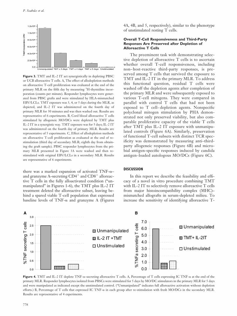
igure 3. TMT and IL-2 IT act synergistically in depleting PBSCr UCB alloreactive T cells. A, The effect of allodepletion methodsn alloreactive T-cell proliferation was evaluated at the end of therimary MLR on the fifth day by measuring 3H-thymidine incor-oration (counts per minute). Responder lymphocytes were gener-ted from PBSC grafts and were stimulated by HLA-mismatchedBV/LCLs. TMT exposure was 3, 4, or 5 days during the MLR, asepicted, and IL-2 IT was administered on the fourth day ofrimary MLR for 30 minutes and was then washed out. Results areepresentative of 6 experiments. B, Cord blood alloreactive T cellstimulated by allogeneic MO/DCs were depleted by TMT plusL-2 IT in a synergistic way. TMT exposure was for 5 days; IL-2 ITas administered on the fourth day of primary MLR. Results are
epresentative of 5 experiments. C, Effect of allodepletion methodsn alloreactive T-cell proliferation as evaluated at the end of re-timulation (third day of secondary MLR; eighth day from obtain-ng the graft sample). PBSC responder lymphocytes from the pri-
ary MLR presented in Figure 3A were washed and then re-timulated with original EBV/LCLs in a secondary MLR. Resultsre representative of 6 experiments.
igure 4. TMT and IL-2 IT deplete TNF-�–secreting alloreactiverimary MLR. Responder lymphocytes isolated from PBSCs were stnd were manipulated as indicated except the unstimulated control.fforts.) B, Percentage of T cells that expressed IC TNF-� in each
esults are representative of 4 experiments.78
A, 4B, and 5, respectively), similar to the phenotypef unstimulated resting T cells.
verall T-Cell Responsiveness and Third-Partyesponses Are Preserved after Depletion oflloreactive T Cells
The preeminent task with demonstrating selec-ive depletion of alloreactive T cells is to ascertainhether overall T-cell responsiveness, includingon– host-reactive third-party responses, is pre-erved among T cells that survived the exposure toMT and IL-2 IT in the primary MLR. To address
his functional question, residual T cells wereashed off the depletion agents after completion of
he primary MLR and were subsequently exposed toarious T-cell mitogens. They were compared inarallel with control T cells that had not beenxposed to T cell– depletion agents. Nonspecificolyclonal mitogen stimulation by PHA demon-trated not only preserved viability, but also com-arable proliferative capacity of the viable T cellsfter TMT plus IL-2 IT exposure with unmanipu-ated controls (Figure 6A). Similarly, preservationf functional T-cell subsets with distinct TCR spec-ficity was demonstrated by measuring anti–third-arty allogeneic responses (Figure 6B) and micro-ial antigen-specific responses induced by candidantigen–loaded autologous MO/DCs (Figure 6C).
ISCUSSION
In this report we describe the feasibility and effi-acy of a novel in vitro procedure combining TMTith IL-2 IT to selectively remove alloreactive T cells
rom major histoincompatibility complex (MHC)–ismatched allografts in serum-depleted milieu. To
ncrease the sensitivity of identifying alloreactive T-
s. A, Percentage of T cells expressing IC TNF-� at the end of thed for 5 days by MO/DC stimulators in the primary MLR for 5 daysanipulated” indicates full alloreactive activation without depletionafter re-stimulation with fresh MO/DCs in the secondary MLR.
T cellimulate(“Unmgroup

cpMrpi(oClaacb
psrmstfmtcalutggbaob
pa
ciaLtdtctmwc
FewwspiR
FpPawaadw(MMaE
IL-2 IT and TMT Deplete Alloreactivity in Synergy
B
ell precursors, we used professional APCs as the mostotent activators of alloreactivity. By using recipientO/DCs or EBV/LCLs, we triggered vigorous allo-
eactivity in a primary MLR that, when further am-lified by re-stimulation with fresh host-derived APCs
n a secondary MLR, led to the vigorous proliferationKi-67 expression and 3HTdR uptake) and expansionf activated TNF-�– and granzyme A–secretingD4� and CD8� alloreactive T cells. Although de-
eting this alloreactive population possessing a highlyctivated and cytotoxic immunophenotype, the testedllodepletion methods spared the residual surviving Tells in an unstimulated resting stage expressing near-asal levels of TNF-� and granzyme A.
Cavazzana-Calvo et al. [17,18] demonstrated inioneering work initially in mice that anti–IL-2R–pecific IT inhibited both MLR in vitro and HLA-estricted T-cell cytotoxicity in the haplotype-mis-atched setting. The T-cell depletion was host
pecific, because the third-party response was rela-ively unaffected and the T-cell reactivity against in-ectious agents was preserved [35]. An in vivo mouse
odel demonstrated attenuation of GVHD afterransplantation with grafts depleted of allospecific Tells [18]. The first human preclinical model that usednti-CD25 (anti–IL-2R�) IT in vitro, in human hap-otype-mismatched related individuals, demonstratedp to 90% depletion of alloreactivity, whereas 64% ofhird-party reactivity was preserved [19]. Notably, therowth of normal bone marrow colony-forming unit–ranulocyte-macrophage was not significantly affectedy the IT. Since the initial study, the feasibility oflloreactive T-cell depletion without loss of antiviralr GVL responses by the anti-CD25–linked IT haseen confirmed in further detail [20,21,23,24].
Although several of the previously described de-letion studies used bulk recipient mononuclear cells
igure 5. TMT and IL-2 IT deplete alloreactive CD8� T cells thatxpress granzyme A. Responder lymphocytes isolated from PBSCsere stimulated for 5 days by MO/DCs in a primary MLR, afterhich each group was re-stimulated with the same APCs in the
econdary MLR (except the unstimulated control that had no APCresent). The percentage of CD8�APC� T cells that expressed
ntracellular granzyme A/FITC after re-stimulation is presented.esults are representative of 4 experiments.
s stimulators of alloreactivity, it is only recently be- 3
B & M T
oming accepted that professional APCs are the keynitiators of alloreactivity [6,36]. The development ofGVHD in vivo requires host APCs such as skinangerhans cells, a member of the DC system, to
rigger the activation, expansion, and cytokine pro-uction of alloreactive lymphocytes, which will lead tohe recruitment of additional host and donor cells toontribute to the cytokine storm that is a hallmark ofhe effector phase of aGVHD [4,5,36]. DCs are al-ost 2 log more powerful stimulators of alloreactivityhen compared with bulk peripheral blood mononu-
lear cells [37,38]. DCs not only express MHC prod-
igure 6. TMT and IL-2 IT deplete alloreactive T cells butreserve overall T-cell responsiveness and third-party reactivity.eripheral blood T cells were stimulated by irradiated EBV/LCLsnd were treated with either TMT or IL-2 IT. Unmanipulated cellsere rested without any alloactivation or treatment with depleting
gents. On day 5, cells were washed and were re-stimulated withutologous monocytes and loaded with 5 �g/mL of PHA (A) for 3ays or 10 �g/mL of candida antigen (C) for 5 days before pulsingith 3H-thymidine. Proliferation is expressed in counts per minute
CPM). Third-party responsiveness (B) was tested in an allogeneicLR. On day 5 at the end of the primary MLR stimulated byO/DC responder cord blood, cells were washed off the deletion
gents indicated and were re-stimulated with irradiated third-partyBV/LCLs for another 5 days before the cultures were pulsed with
H-thymidine. Proliferation is expressed in CPM.
779

uplppsmO[rstpcsaaeg
eacocrscnvgopcaAesmf
watfenpcaiclcitt
Iirn
tcarvdwpactpuuaatatwestr
teoePfpTgstpistfmitpgs
rcT
P. Szabolcs et al.
7
cts at the highest density, but also retain boundeptides, also called minor histoincompatibility antigens,onger than other APCs [39]. These features thatrovide sustained and robust TCR signaling areaired in mature DCs with the most abundant acces-ory signaling capacity via the intercellular adhesionolecules, the B7 family [40], 4-1BBL, CD40 [41],X40L [42], and secreted cytokines such as IL-12
43,44]. In certain clinical scenarios, the generation ofecipient MO/DCs may not be feasible or desired,uch as for patients with myeloid malignancies. Forhese potential candidates of alloreactive T cell–de-leted grafts, to preserve myeloid lineage-specific T-ell clones that could exert a beneficial GVL effect, wehow that EBV/LCLs are equally potent activators oflloreactivity. As shown previously, they present HLAntigens and co-stimulatory molecules at very highxpression levels, rendering them highly immuno-enic [24,45,46].
In this study, we first demonstrated the ex vivofficacy of TMT, a potent lipophilic synthetic MTXnalog that induces apoptosis when host-reactive Tells enter the cell cycle, and then we identified theptimal conditions for TMT to remove alloreactive Tells. TMT is a competitive inhibitor of dihydrofolateeductase, a vital enzyme for purine and thymidineynthesis and cell replication. TMT is an excellentandidate for ex vivo graft engineering because it is aonclassic folate antagonist; unlike MTX, enters cellsia a non–energy-dependent pathway; and is not poly-lutamated. TMT was approved by the FDA as anrphan drug for the treatment of Pneumocystis cariniineumonia in patients with acquired immunodefi-iency syndrome and is also active against other par-sitic infections, such as malaria and toxoplasmosis.dditionally, in vivo–administered TMT has beenvaluated in a phase I clinical trial along with cyclo-porin A as GVHD prophylaxis in the HLA-mis-atched related donor transplant setting and was
ound to be similar in efficacy to MTX [47].Comparable in vitro depletion efficacy with TMT
as achieved by a commercially available IT that targetsctivated T cells that upregulate the surface expression ofhe IL-2R. This chimeric molecule is significantly dif-erent from the CD25 ITs that have been previouslyvaluated in vitro by several groups [20,21,23,24]. De-ileukin diftitox is a receptor-targeted cytotoxic fusionrotein generated in an Escherichia coli expression systemomposed of the recombinant chimeric molecule of IL-2nd diphtheria toxin. It is cytotoxic against cells express-ng IL-2R� (CD25) in association with IL-2R� and �hains. It has been FDA approved for cutaneous T-cellymphoma, a malignancy that is characterized by theonstitutive expression of CD25 on activated T cellsnfiltrating the skin. Most recently, it has been also foundo be effective as front-line treatment for steroid-refrac-
ory aGVHD [48]. Thus, both TMT (Neutrexin) and m80
L-2 IT (Ontak) have been FDA approved for in vivo usendividually for infectious or malignant conditions. Thisenders them eminently suitable for ex vivo graft engi-eering.
The key finding of our studies is the demonstra-ion of the principle, in already preclinical serum-freeonditions, that when these individually effectivegents are combined, superior efficacy of ex vivo allo-eactive T cells can be achieved after the potent acti-ation by 2 interchangeably used host APCs. We alsoemonstrated that for TMT, the efficacy of depletionas both time and concentration dependent. Severalreviously published methods have used 2 to 3 days ofctivation with bulk peripheral blood mononuclearells serving as surrogate APCs, thus achieving poten-ially suboptimal activation of some alloreactive T-cellrecursors. In contrast, our most effective conditionsed 5 days of TMT exposure of a 5-day MLR stim-lated by professional APCs with IL-2 IT treatmentdded on either day 3 or 4. We postulate that for somelloreactive T cells with relatively lower affinity, ex-ended activation may be necessary to generate thectivated phenotype that can be the subject for selec-ive targeting. In this study, the selective allodepletionas performed in a primary MLR; however, the truefficacy of allodepletion was best demonstrated in re-timulation experiments (secondary MLR) that mimiche continued activation state of alloreactivity occur-ing in vivo in the immediate postgrafting period.
In this feasibility study we also demonstrated thathe cooperation exerted by TMT and IL-2 IT isffective regardless of whether T cells are from PBSCr from UCB grafts, thus opening the field for graftngineering in both clinical scenarios. UCB andBSC are the most commonly used stem cell sources
or HLA-mismatched transplantation, and allode-leted aliquots could be used for various adoptive-cell strategies. Aliquots of cord blood or PBSCrafts devoid of alloreactive potential could be an idealtarting population for ex vivo expansion and adoptiveransfer as donor leukocyte infusions (DLI) to reduceosttransplantation opportunistic infections that arisen the T-cell lymphopenic period. If DLI could beafely administered relatively early after transplanta-ion, this could also aid engraftment and skew towardull donor chimerism in T cell–depleted HLA-mis-atched haploidentical stem cell grafts. Despite the
nherent immaturity of naive UCB T cells [49], selec-ively allodepleted UCB could be an ideal startingopulation for in vitro priming strategies aimed atenerating virus-specific cytotoxic T lymphocytes forubsequent DLI and adoptive immunotherapy [50].
We also propose that either fully mature host-de-ived myeloid DCs or EBV/LCLs may be used as effi-ient APCs to present recipient HLA antigens to donor
cells. For lymphoid malignancies, the use of APCs of
yeloid origin—such as mature MO/DCs—might be
pLtfcealbFnDaMawaawIagdwihpcnbAAonacptM
dLeMbldemiu[safimP
ttn
rCtlrtf
emtraiasiapficatbamahtcvci
A
rG(ats
R
IL-2 IT and TMT Deplete Alloreactivity in Synergy
B
referable, whereas in myeloid malignancies, EBV/CLs would be optimal host-derived APCs. This selec-
ive approach would potentially spare GVL precursorrequency directed against hematopoietic lineage-spe-ific antigens [51,52]. Although the main focus of ourxperiments was to develop, critically evaluate, and char-cterize the immunobiology of new and superior al-odepletion methods, we can estimate the clinical feasi-ility of this approach when DCs are used as host APCs.rom approximately 10 mL of peripheral blood withormal counts, typically approximately 1 � 106 MO/Cs can be generated. This is sufficient to stimulate
pproximately 1 � 107 purified T cells in a primaryLR. Allodepletion with the combination method is
ssociated with several steps of manipulations/washesith approximately 30% recovery of the input T cells. Ifthird of these are set aside to verify the efficacy of
llodepletion, approximately 2 � 106 CD3� T cellsould be available for potential adoptive T-cell therapy.
f a dose-escalation trial were initiated with 1 � 105
llodepleted HLA-mismatched CD3� T cells per kilo-ram, then 10 mL of peripheral blood lymphocytesrawn from a patient weighing approximately 20 kgould provide sufficient host MO/DCs for a single DLI
nfusion. Increasing the recipient blood draw and slightlyigher APC-responder ratios (such as 1:20 with highlyurified T-cell responders) could further increase the Tells available for potentially higher dose levels. Theumeric limitations of obtaining host MO/DCs can beypassed when immortalized EBV/LCLs are used asPCs. The key factor in deciding the source of hostPC if both are potentially available should be the typef malignancy for leukemia patients. For recipients withonmalignant conditions, primary MO/DCs are prefer-ble, as opposed to virally immortalized LCLs, if suffi-ient numbers can be generated. However, it may berudent to generate EBV/LCLs on all patients to havehose APCs available in case the yield or quality of
O/DC preparation is suboptimal.The concern that anti-EBV reactivity would be
epleted along with the alloreactive pool when EBV/CLs are used as APCs needs to be considered. How-ver, when recipient-donor pairs are mismatched atHC loci, the MHC-restricted minor histocompati-
ility antigen (EBV)–specific T-cell clones mayargely remain unperturbed in a primary MLR that isriven by direct MHC allostimulation. Supportingxperimental evidence was recently obtained by enu-erating interferon-�–secreting EBV-reacting T cells
n a haploidentical model in which EBV/LCLs weresed as stimulators before use of an anti-CD25 IT24]. However, cross-reacting epitopes have been de-cribed between viral epitopes and alloantigens [53],nd, consequently, depleting T cells with dual speci-city could lead to some reduction of virus- or othericrobe-specific T-cell precursor frequencies in both
BSC and marrow grafts. This is of lesser concern inB & M T
he context of unrelated UCB transplants because ofhe lack of previous microbial exposure and priming ofaive UCB T cells.
Using an IL-2 IT may theoretically also lead toemoval of regulatory CD25� T cells [54]. However,D25 targeting in current organ transplantation prac-
ice has shown a clinically beneficial reduction of al-oreactivity that has reduced the rate and severity ofejection. Similarly, it has been documented in hema-opoietic transplantation that GVHD can be success-ully treated by CD25 targeting [2].
In summary, these data are the first, to our knowl-dge, to demonstrate that the combined application of 2ethods that target different cellular profiles of alloreac-
ivity provides enhanced efficacy and preserves nonallo-eactive T cells in a resting state. The combinationpproach may be needed as a safeguard to achieve clin-cally relevant and safe allodepletion in cases in whichlloreactive T cells express low levels of CD25 (or otherurface markers of activation) or in cases in which theres a relative lack of proliferation and DNA synthesismong alloreactive T cells. In case host antigens areresented in a suboptimal way, either because of insuf-cient numbers or low activation states of APCs, oneould expect a heterogeneous activation pattern amonglloreactive precursors that would render them less likelyo be identifiable through a single activation marker oriological characteristic. Incomplete removal of partiallyctivated alloreactive T cells by a given depletionethod could lead to the adoptive transfer of expanded
lloreactive T cells that would be present at an evenigher frequency than were originally in the graft, thusriggering or exacerbating aGVHD. Experimental dataaution of the potential dangers of adoptive transfer of exivo–activated allosensitized host-reactive T cells be-ause they are less likely to respond to calcineurin inhib-tors such as cyclosporin A once activated in vitro [55].
CKNOWLEDGMENTSThis work was supported by The National Mar-
ow Donor Program-Marrow Transplant Researchrant, The Lisa Stafford Memorial Research Prize
P.S.), and grant nos. 1PO1-HL-67314-01A1 (P.S.nd J.K.) and N01-HB-67141 (J.K.). We are gratefulo Peter Cole and Reid Bissonette for helpful discus-ions.
EFERENCES
1. Michalek J, Collins RH, Hill BJ, Brenchley JM, Douek DC.Identification and monitoring of graft-versus-host specific T-cell clone in stem cell transplantation. Lancet. 2003;361:1183.
2. Goker H, Haznedaroglu IC, Chao NJ. Acute graft-vs-hostdisease: pathobiology and management. Exp Hematol. 2001;29:259.
3. Piguet PF, Grau GE, Allet B, Vassalli P. Tumor necrosis
781

1
1
1
1
1
1
1
1
1
1
2
2
2
2
2
2
2
2
2
2
3
3
3
3
3
3
3
P. Szabolcs et al.
7
factor/cachectin is an effector of skin and gut lesions of theacute phase of graft-vs.-host disease. J Exp Med. 1987;166:1280.
4. Ferrara JL. The cytokine modulation of acute graft-versus-hostdisease. Bone Marrow Transplant. 1998;21(suppl 3):S13.
5. Antin JH. Acute graft-versus-host disease: inflammation runamok? J Clin Invest. 2001;107:1497.
6. Teshima T, Ordemann R, Reddy P, et al. Acute graft-versus-host disease does not require alloantigen expression on hostepithelium. Nat Med. 2002;8:575.
7. Deeg HJ. Cytokines in graft-versus-host disease and the graft-versus-leukemia reaction. Int J Hematol. 2001;74:26.
8. Reddy P, Ferrara JL. Immunobiology of acute graft-versus-hostdisease. Blood Rev. 2003;17:187.
9. Jordan WJ, Brookes PA, Szydlo RM, Goldman JM, Lechler RI,Ritter MA. IL-13 production by donor T cells is prognostic ofacute graft-versus-host disease following unrelated donor stemcell transplantation. Blood. 2004;103:717.
0. Shlomchik WD, Couzens MS, Tang CB, et al. Prevention ofgraft versus host disease by inactivation of host antigen-pre-senting cells. Science. 1999;285:412.
1. Tazzari PL, Bolognesi A, De Totero D, et al. B-B10 (anti-CD25)-saporin immunotoxin—a possible tool in graft-versus-host disease treatment. Transplantation. 1992;54:351.
2. Favre A, Cerri A, Bacigalupo A, Lanino E, Berti E, Grossi CE.Immunohistochemical study of skin lesions in acute and chronicgraft versus host disease following bone marrow transplanta-tion. Am J Surg Pathol. 1997;21:23.
3. Ukyo N, Hori T, Yanagita S, Ishikawa T, Uchiyama T. Co-stimulation through OX40 is crucial for induction of an allo-reactive human T-cell response. Immunology. 2003;109:226.
4. Tittle TV, Weinberg AD, Steinkeler CN, Maziarz RT. Expres-sion of the T-cell activation antigen, OX-40, identifies alloreac-tive T cells in acute graft-versus-host disease. Blood. 1997;89:4652.
5. Craston R, Koh M, Mc Dermott A, Ray N, Prentice HG,Lowdell MW. Temporal dynamics of CD69 expression onlymphoid cells. J Immunol Methods. 1997;209:37.
6. Ho VT, Soiffer RJ. The history and future of T-cell depletionas graft-versus-host disease prophylaxis for allogeneic hemato-poietic stem cell transplantation. Blood. 2001;98:3192.
7. Cavazzana-Calvo M, Fromont C, Le Deist F, et al. Specificelimination of alloreactive T cells by an anti-interleukin-2 re-ceptor B chain-specific immunotoxin. Transplantation. 1990;50:1.
8. Cavazzana-Calvo M, Stephan JL, Sarnacki S, et al. Attenuationof graft-versus-host disease and graft rejection by ex vivo im-munotoxin elimination of alloreactive T cells in an H-2 haplo-type disparate mouse combination. Blood. 1994;83:288.
9. Mavroudis DA, Jiang YZ, Hensel N, et al. Specific depletion ofalloreactivity against haplotype mismatched related individualsby a recombinant immunotoxin: a new approach to graft-ver-sus-host disease prophylaxis in haploidentical bone marrowtransplantation. Bone Marrow Transplant. 1996;17:793.
0. Mavroudis DA, Dermime S, Molldrem J, et al. Specific deple-tion of alloreactive T cells in HLA-identical siblings: a methodfor separating graft-versus-host and graft-versus-leukaemia re-actions. Br J Haematol. 1998;101:565.
1. Montagna D, Yvon E, Calcaterra V, et al. Depletion of allo-reactive T cells by a specific anti-interleukin-2 receptor p55chain immunotoxin does not impair in vitro antileukemia andantiviral activity. Blood. 1999;93:3550.
2. Andre-Schmutz I, Le Deist F, Hacein-Bey-Abina S, et al. Im-
82
mune reconstitution without graft-versus-host disease afterhaemopoietic stem-cell transplantation: a phase 1/2 study.Lancet. 2002;360:130.
3. Solomon SR, Tran T, Carter CS, et al. Optimized clinical-scaleculture conditions for ex vivo selective depletion of host-reac-tive donor lymphocytes: a strategy for GvHD prophylaxis inallogeneic PBSC transplantation. Cytotherapy. 2002;4:395.
4. Amrolia PJ, Muccioli-Casadei G, Yvon E, et al. Selective de-pletion of donor alloreactive T cells without loss of antiviral orantileukemic responses. Blood. 2003;102:2292.
5. Koh MB, Prentice HG, Lowdell MW. Selective removal ofalloreactive cells from haematopoietic stem cell grafts: graftengineering for GVHD prophylaxis. Bone Marrow Transplant.1999;23:1071.
6. Koh MB, Prentice HG, Corbo M, Morgan M, Cotter FE,Lowdell MW. Alloantigen-specific T-cell depletion in a majorhistocompatibility complex fully mismatched murine modelprovides effective graft-versus-host disease prophylaxis in thepresence of lymphoid engraftment. Br J Haematol. 2002;118:108.
7. Hartwig UF, Robbers M, Wickenhauser C, Huber C. Murineacute graft-versus-host disease can be prevented by depletion ofalloreactive T lymphocytes using activation-induced cell death.Blood. 2002;99:3041.
8. Guimond M, Balassy A, Barrette M, Brochu S, Perreault C,Roy DC. P-glycoprotein targeting: a unique strategy to selec-tively eliminate immunoreactive T cells. Blood. 2002;100:375.
9. Rieser C, Bock G, Klocker H, Bartsch G, Thurnher M. Pros-taglandin E2 and tumor necrosis factor alpha cooperate toactivate human dendritic cells: synergistic activation of inter-leukin 12 production. J Exp Med. 1997;186:1603.
0. Smith CA, Ng CY, Heslop HE, et al. Production of geneticallymodified Epstein-Barr virus-specific cytotoxic T cells for adop-tive transfer to patients at high risk of EBV-associated lympho-proliferative disease. J Hematother. 1995;4:73.
1. Szabolcs P, Moore MA, Young JW. Expansion of immunos-timulatory dendritic cells among the myeloid progeny of hu-man CD34� bone marrow precursors cultured with c-kit li-gand, granulocyte-macrophage colony-stimulating factor, andTNF-alpha. J Immunol. 1995;154:5851.
2. Longo-Sorbello GS, Bertino JR. Current understanding ofmethotrexate pharmacology and efficacy in acute leukemias.Use of newer antifolates in clinical trials. Haematologica. 2001;86:121.
3. vander Spek J, Cassidy D, Genbauffe F, Huynh PD, Murphy JR.An intact transmembrane helix 9 is essential for the efficientdelivery of the diphtheria toxin catalytic domain to the cytosol oftarget cells. J Biol Chem. 1994;269:21455-21459.
4. Re GG, Waters C, Poisson L, Willingham MC, Sugamura K,Frankel AE. Interleukin 2 (IL-2) receptor expression and sen-sitivity to diphteria fusion toxin DAB389IL-2 in cultured he-matopoietic cells. Cancer Res. 1996;56:2590-2595.
5. Valteau-Couanet D, Cavazzana-Calvo M, Le Deist F, Fromont C,Fischer A. Functional study of residual T lymphocytes after spe-cific elimination of alloreactive T cells by a specific anti-interleu-kin-2 receptor Bk chain immunotoxin [corrected and republishedarticle originally printed in Transplantation 1993;56:737-8].Transplantation. 1993;56:1574.
6. Shlomchik WD, Couzens MS, Tang CB, et al. Prevention ofgraft versus host disease by inactivation of host antigen-pre-
senting cells. Science. 1999;285:412.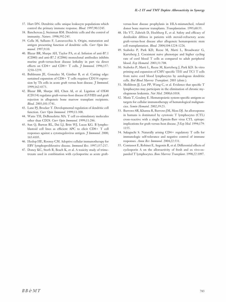
3
3
3
4
4
4
4
4
4
4
4
4
4
5
5
5
5
5
5
IL-2 IT and TMT Deplete Alloreactivity in Synergy
B
7. Hart DN. Dendritic cells: unique leukocyte populations whichcontrol the primary immune response. Blood. 1997;90:3245.
8. Banchereau J, Steinman RM. Dendritic cells and the control ofimmunity. Nature. 1998;392:245.
9. Cella M, Sallusto F, Lanzavecchia A. Origin, maturation andantigen presenting function of dendritic cells. Curr Opin Im-munol. 1997;9:10.
0. Blazar BR, Sharpe AH, Taylor PA, et al. Infusion of anti-B7.1(CD80) and anti-B7.2 (CD86) monoclonal antibodies inhibitsmurine graft-versus-host disease lethality in part via directeffects on CD4� and CD8� T cells. J Immunol. 1996;157:3250-3259.
1. Buhlmann JE, Gonzalez M, Ginther B, et al. Cutting edge:sustained expansion of CD8� T cells requires CD154 expres-sion by Th cells in acute graft versus host disease. J Immunol.1999;162:4373.
2. Blazar BR, Sharpe AH, Chen AI, et al. Ligation of OX40(CD134) regulates graft-versus-host disease (GVHD) and graftrejection in allogeneic bone marrow transplant recipients.Blood. 2003;101:3741.
3. Lane PJ, Brocker T. Developmental regulation of dendritic cellfunction. Curr Opin Immunol. 1999;11:308.
4. Watts TH, DeBenedette MA. T cell co-stimulatory moleculesother than CD28. Curr Opin Immunol. 1999;11:286.
5. Sun Q, Burton RL, Dai LJ, Britt WJ, Lucas KG. B lympho-blastoid cell lines as efficient APC to elicit CD8� T cellresponses against a cytomegalovirus antigen. J Immunol. 2000;165:4105.
6. Heslop HE, Rooney CM. Adoptive cellular immunotherapy forEBV lymphoproliferative disease. Immunol Rev. 1997;157:217.
7. Doney KC, Storb R, Beach K, et al. A toxicity study of trime-
trexate used in combination with cyclosporine as acute graft-B & M T
versus-host disease prophylaxis in HLA-mismatched, relateddonor bone marrow transplants. Transplantation. 1995;60:55.
8. Ho VT, Zahrieh D, Hochberg E, et al. Safety and efficacy ofdenileukin diftitox in patients with steroid-refractory acutegraft-versus-host disease after allogeneic hematopoietic stemcell transplantation. Blood. 2004;104:1224-1226.
9. Szabolcs P, Park KD, Reese M, Marti L, Broadwater G,Kurtzberg J. Coexistent naive phenotype and higher cyclingrate of cord blood T cells as compared to adult peripheralblood. Exp Hematol. 2003;31:708.
0. Szabolcs P, Marti L, Reese M, Kurtzberg J, Park KD. In vitropriming and expansion of CMV-specific TH1 and TC1 T cellsfrom naive cord blood lymphocytes by autologous dendriticcells. Biol Blood Marrow Transplant. 2003 (abstr.).
1. Molldrem JJ, Lee PP, Wang C, et al. Evidence that specific Tlymphocytes may participate in the elimination of chronic my-elogenous leukemia. Nat Med. 2000;6:1018.
2. Mutis T, Goulmy E. Hematopoietic system-specific antigens astargets for cellular immunotherapy of hematological malignan-cies. Semin Hematol. 2002;39:23.
3. Burrows SR, Khanna R, Burrows JM, Moss DJ. An alloresponsein humans is dominated by cytotoxic T lymphocytes (CTL)cross-reactive with a single Epstein-Barr virus CTL epitope:implications for graft-versus-host disease. J Exp Med. 1994;179:1155.
4. Sakaguchi S. Naturally arising CD4� regulatory T cells forimmunologic self-tolerance and negative control of immuneresponses. Annu Rev Immunol. 2004;22:531.
5. Contassot E, Robinet E, Angonin R, et al. Differential effects ofcyclosporin A on the alloreactivity of fresh and ex vivo-ex-
panded T lymphocytes. Bone Marrow Transplant. 1998;22:1097.783