UC Irvine - The Western Journal of Emergency Medicine
197
UC Irvine Clinical Practice and Cases in Emergency Medicine Title CPC-EM Full-Text Issue Permalink https://escholarship.org/uc/item/4b0305fp Journal Clinical Practice and Cases in Emergency Medicine, 1(4) ISSN 2474-252X Author Hernandez, Nancy G. Publication Date 2017 License https://creativecommons.org/licenses/by/4.0/ 4.0 Peer reviewed eScholarship.org Powered by the California Digital Library University of California
-
Upload
khangminh22 -
Category
Documents
-
view
3 -
download
0
Transcript of UC Irvine - The Western Journal of Emergency Medicine

UC IrvineClinical Practice and Cases in Emergency Medicine
TitleCPC-EM Full-Text Issue
Permalinkhttps://escholarship.org/uc/item/4b0305fp
JournalClinical Practice and Cases in Emergency Medicine, 1(4)
ISSN2474-252X
AuthorHernandez, Nancy G.
Publication Date2017
Licensehttps://creativecommons.org/licenses/by/4.0/ 4.0 Peer reviewed
eScholarship.org Powered by the California Digital LibraryUniversity of California

Volume I, Number 4, November 2017 Open Access at www.cpcem.org ISSN: 2474-252X
A Peer-Reviewed, International Professional Journal
Clinicopathological Cases from the University of Maryland272 55-year-old Male with Bilateral Lower Extremity Weakness
T Rao, A Roggio, ZDW Dezman, LJ, Bontempo
Astonishing Cases and Images in Emergency Medicine278 A Plumb Fit MF Harrison, K Rooney, B Jaskulka
Case Reports280 Not All Sore Throats Are Pharyngitis
A Sadowski, J Dougherty
283 Sulfur Mustard Exposure from Dredged Artillery Shell in a Commercial ClammerJ Otter, A Dawood, J D’Orazio
287 Post-operative Appendix Specimen Retention Presenting as Small Bowel ObstructionT Proffitt, K Whitworth, C Trigger
291 Myasthenic Crisis In PregnancyDM French, EP Bridges, MC Hoskins, CM Andrews, CH Nelson
295 25C-NBOMe IngestionJ Zygowiec, S Solomon, A Jaworski, M Bloome, A Gotlib
298 The Malingering IntussusceptionJ Le, J Labha, B Khazaeni
301 Reversible Acute Kidney Injury Associated with Chlorothalonil IngestionJR Suchard
305 Idiopathic Spinal Epidural Lipomatosis Causing Cauda Equina Syndrome JB Bushkar, LP MenkinSmith, DM Krywko
Clinical Practice and C
ases in Emergency M
edicine
VO
LUM
E 1 ISSUE 4 N
ovember 2017
PA
GES 272-449
Clinical Practice and Cases in Emergency Medicine
In Collaboration with the Western Journal of Emergency Medicine
Contents continued on page iii
We Are.
COMPREHENSIVE.RELEVANT. ESSENTIAL.
OhiO ACEPEmErgEnCy mEdiCinE BOArd rEviEw COursEThe course physicians have trusted for 33 years!
www.ohacep.org (614) 792-6506Approved for AMA PRA Category 1 Credits TM.
Ohio ACEP is excited to partner with
California ACEP to offer a course in
February 2018!
February 1 - 5, 2018 San Diego, California

Volume I, Number 4, November 2017 Open Access at www.cpcem.org ISSN: 2474-252X
A Peer-Reviewed, International Professional Journal
Clinicopathological Cases from the University of Maryland272 55-year-old Male with Bilateral Lower Extremity Weakness
T Rao, A Roggio, ZDW Dezman, LJ, Bontempo
Astonishing Cases and Images in Emergency Medicine278 A Plumb Fit MF Harrison, K Rooney, B Jaskulka
Case Reports280 Not All Sore Throats Are Pharyngitis
A Sadowski, J Dougherty
283 Sulfur Mustard Exposure from Dredged Artillery Shell in a Commercial ClammerJ Otter, A Dawood, J D’Orazio
287 Post-operative Appendix Specimen Retention Presenting as Small Bowel Obstruction: A Case Report
T Proffitt, K Whitworth, C Trigger
291 Myasthenic Crisis In PregnancyDM French, EP Bridges, MC Hoskins, CM Andrews, CH Nelson
295 25C-NBOMe IngestionJ Zygowiec, S Solomon, A Jaworski, M Bloome, A Gotlib
298 The Malingering IntussusceptionJ Le, J Labha, B Khazaeni
301 Reversible Acute Kidney Injury Associated with Chlorothalonil IngestionJR Suchard
305 Idiopathic Spinal Epidural Lipomatosis Causing Cauda Equina Syndrome JB Bushkar, LP MenkinSmith, DM Krywko
Clinical Practice and C
ases in Emergency M
edicine
VO
LUM
E 1 ISSUE 4 N
ovember 2017
PA
GES 272-449
Clinical Practice and Cases in Emergency Medicine
In Collaboration with the Western Journal of Emergency Medicine
Contents continued on page iii
We Are.
COMPREHENSIVE.RELEVANT. ESSENTIAL.
OhiO ACEPEmErgEnCy mEdiCinE BOArd rEviEw COursEThe course physicians have trusted for 33 years!
www.ohacep.org (614) 792-6506Approved for AMA PRA Category 1 Credits TM.
Ohio ACEP is excited to partner with
California ACEP to offer a course in
February 2018!
February 1 - 5, 2018 San Diego, California

Volume I, no. 4: November 2017 ix Clinical Practice and Cases in Emergency Medicine
The University of California, Davis School of Medicine, Department of Emergency Medicine (EM) is conducting a faculty search for EM physicians in either a clinician/educator or clinician/researcher track. Candidates must be residency trained in EM with board certification/preparation and be eligible for licensure in California. At least one year of post-training clinical experience and/or fellowship training is preferred. Candidates are expected to enter at the Assistant/Associate level, commensurate with experience and credentials. EM faculty members at UC Davis who have preference fornight shifts work fewer clinical shifts each month.
The University of California, Davis, Medical Center, one of the nation’s “Top 50 Hospitals,” is a 613 bed academic medical center with approximately 80,000 emergency department visits annually, including approximately 20,000 pediatric visits.The emergency services facility opened in 2010 and is state-of-the-art. Our program provides comprehensive emergency services to a large local urban and referral population as a level 1 trauma center, paramedic base station and training center. The department also serves as the primary teaching site for a fully accredited EM residency program and eight different EM fellowship programs. Our residency training program began more than twenty years ago and currently has 48 residents. The Department has a separate area for the care of children and is one of the leading centers in the Pediatric Emergency Care Applied Research Network (PECARN).
Salary and benefits are competitive and commensurate with training and experience. Sacramento is located near the northern end of California's Central Valley, close to Lake Tahoe, San Francisco, and the "wine country" of the Napa and Sonoma Valleys.Sports enthusiasts will find Sacramento's climate and opportunities ideal.
Interested candidates should submit a letter outlining interests and experience, and curriculum vitae to: recruit.ucdavis.edu/apply/JPF01809
Erik Laurin MD, Professor and Search Committee Chair ([email protected])UC Davis Department of Emergency Medicine
2315 Stockton Blvd., PSSB 2100Sacramento, CA 95817
The University of California is an affirmative action/equal opportunity employer.

Volume I, no. 4: November 2017 i Clinical Practice and Cases in Emergency Medicine
Clinical Practice and Cases in Emergency MedicineIn Collaboration with the Western Journal of Emergency Medicine
Rick A. McPheeters, DO, Editor-in-Chief Kern Medical
Mark I. Langdorf, MD, MHPE, Associate Editor University of California, Irvine School of Medicine
Shahram Lotfipour, MD, MPH, Managing Associate EditorUniversity of California, Irvine School of Medicine
Shadi Lahham, MD, MS, Associate EditorUniversity of California, Irvine School of Medicine
Editorial Board
Editorial Staff International Editorial Board
Advisory Board
Manish Amin, DO, Associate EditorKern Medical
Erik D. Barton, MD, MBAIcahn School of Medicine, Mount Sinai, New YorkPeter A. Bell, DO, MBALiberty UniversityCollege of Osteopathic MedicineBarry E. Brenner, MD, MPHCase Western Reserve UniversityDavid F.M. Brown, MDMassachusetts General Hospital/Harvard Medical SchoolFrancis Counselman, MDEastern Virginia Medical SchoolRobert W. Derlet, MDUniversity of California, DavisDaniel J. Dire, MD University of Texas Health Sciences Center San Antonio Steven Gabaeff, MDClinical Forensic MedicineBrent King, MD, MMMUniversity of Texas, HoustonEdward Michelson, MDTexas Tech UniversityLinda S. Murphy, MLISUniversity of California, Irvine School of Medicine Librarian
Jonathan Olshaker, MDBoston UniversityEdward Panacek, MD, MPHUniversity of South Alabama Niels K. Rathlev, MDTufts University School of Medicine Robert M. Rodriguez, MD University of California, San FranciscoScott Rudkin, MD, MBAUniversity of California, IrvinePeter Sokolove, MDUniversity of California, San FranciscoSamuel J. Stratton, MD, MPHOrange County, CA, EMS AgencyRobert Suter, DO, MHAUT Southwestern Medical CenterScott Zeller, MDUniversity of California, Riverside Leslie Zun, MD, MBAChicago Medical School
Peter A. Bell, DO, MBAAmerican College of Osteopathic Emergency PhysiciansLiberty UniversityCollege of Osteopathic Medicine
John B. Christensen, MDCalifornia Chapter Division of AAEM
Amal Khalil, MBAUC Irvine Health SOM
Mark I. Langdorf, MD, MHPEUC Irvine Health SOM
Elena Lopez-GusmanCalifornia ACEPAmerican College of Emergency Physicians
Shahram Lotfipour, MD, MPHUC Irvine Health SOM
Trevor Mills, MD, MPHCalifornia Chapter Division of AAEMLSU Medical Center
Aimee Moulin, MDCalifornia ACEPAmerican College of Emergency PhysiciansUniversity of California, Davis
Nicholas T. Sawyer, MD, MBACalifornia ACEPAmerican College of Emergency PhysiciansUniversity of California, Davis
Robert Suter, DO, MHAAmerican College of Osteopathic Emergency PhysiciansUT Southwestern Medical Center
Jan WachtlerAmerican College of Osteopathic Emergency Physicians
Pablo Aguilera Fuenzalida, MDPontificia Universidad Catolica de Chile, Región Metropolitana, ChileAnwar Al-Awadhi, MDMubarak Al-Kabeer Hospital, Jabriya, KuwaitArif A. Cevik, MDUnited Arab Emirates UniversityCollege of Medicine and Health Sciences, Al Ain, United Arab EmiratesFrancesco Dellacorte, MDAzienda Ospedaliera Universitaria “Maggiore della Carità,” Novara, ItalyAbhinandan A.Desai, MDUniversity of Bombay Grant Medical College, Bombay, IndiaGayle Galleta, MDSørlandet Sykehus HF, Akershus Universitetssykehus, Lorenskog, NorwayVijay Gautam, MBBSUniversity of London, London, EnglandWirachin Hoonpongsimanont, MD, MSBATSSiriraj Hospital, Mahidol University, Bangkok, Thailand
Rosidah Ibrahim, MDHospital Serdang, Selangor, MalaysiaKatsuhiro Kanemaru, MDUniversity of Miyazaki Hospital, Miyazaki, JapanAmin A. Kazzi, MAAEM, MD The American University of Beirut, Beirut, LebanonTerry Mulligan, DO, MPHACEP Ambassador to the Netherlands Society of Emergency PhysiciansSteven H. Lim, MDChangi General Hospital, Simei, SingaporeKhrongwong Musikatavorn, MDKing Chulalongkorn Memorial Hospital, Chulalongkorn University, Bangkok, Thailand Bandr Mzahim, MDKing Fahad Medical City, Riyadh, Saudi ArabiaJacob (Kobi) Peleg, PhD, MPHTel-Aviv University, Tel-Aviv, IsraelNadeem Qureshi, MDSt. Louis University, USA
Ahdilah Haswarey, BSEditorial Director
Jimmy To, BSAssociate Editorial Director
Tushank Chadha, BSMarketing Director
Nancy G. Hernandez, BAPublishing Director
Tracy Nguyen, BSAssociate Publishing Director
June Casey, BACopy Editor
Alissa FiorentinoWestJEM Staff Liaison

Clinical Practice and Cases in Emergency Medicine ii Volume I, no. 4: November 2017
Academic Department of Emergency Medicine Subscribers
Professional Society SponsorsAmericAn college of osteopAthic emergency physiciAnscAliforniA chApter Division of AmericAn AcADemy of emergency meDicine
cAliforniA Acep
Clinical Practice and Cases in Emergency MedicineIn Collaboration with the Western Journal of Emergency Medicine
Allegheny Health NetworkPittsburgh, PAAlbany Medical CollegeAlbany, NYAmerican University of Beirut Beirut, Lebanon Arrowhead Regional Medical Center Colton, CABaystate Medical Center/Tufts UniversitySpringfield, MABeaumont HospitalRoyal Oak, MIBeth Israel Deaconess Medical CenterBoston, MABoston Medical CenterBoston, MA Brigham and Women’s HospitalBoston, MABrown University/Rhode Island HospitalProvidence, RICalifornia Chapter Division of AAEMMilwaukee, WICalifornia State University FresnoFresno, CACarl R. Darnall Army Medical CenterFort Hood, TXConemaugh Memorial Medical CenterJohnstown, PADesert Regional Medical CenterPalm Springs, CADoctors Hospital-Ohio HealthColumbus, OHEastern Virginia Medical SchoolNorfolk, VAEinstein Healthcare NetworkPhiladelphia, PAEmory UniversityAtlanta, GAFlorida Hospital Medical CenterOrlando, FLGeorgia Regents University - Emergency Medicine Residency and FellowshipAugusta, GAGood Samaritan Hospital Medical CenterWest Islip, NYHartford HospitalHartford, CTHennepin County Medical CenterMinneapolis, MN
Henry Ford Medical CenterDetroit, MIHighland HospitalOakland, CAINTEGRIS HealthOklahoma City, OKKaweah Delta Health Care DistrictVisalia, CAKennedy University HospitalsTurnersville, NJKent Emergency Medicine Residency ProgramKent, United KingdomKern MedicalBakersfield, CALakeland HealthCareSt. Joseph, MILehigh Valley Hospital and Health NetworkAllentown, PALoma Linda University Medical CenterLoma Linda, CALouisiana State University Health Sciences CenterNew Orleans, LAMadigan Army Medical CenterTacoma, WAMaimonides Medical CenterBrooklyn, NYMaricopa Medical CenterPhoenix, AZ Massachusetts General HospitalBoston, MAMayo Clinic College of MedicineRochester, MNMedical College of WisconsinMilwaukee, WIMt. Sinai Medical CenterMiami Beach, FLNational University HospitalSingapore, SingaporeNorth Shore University HospitalManhasset, NYNorthwestern Medical GroupChicago, ILOhio State University Medical CenterColumbus, OHOhio Valley Medical CenterWheeling, WVOklahoma UniversityNorman, OKOregon Health and Science University
Portland, ORPenn State Milton S. Hershey Medical CenterHershey, PAPresence Resurrection Medical CenterChicago, ILRegions Hospital/ Health Partners Institute for Education and ResearchSt. Paul, MNRobert Wood Johnson HospitalNew Brunswick, NJRush University Medical CenterChicago, ILSouthern Illinois UniversityCarbondale, ILStanford University/KAiser Emergency Medicine Residency ProgramPalo Alto, CASUNY Upstate Medical CenterSyracuse, NYState University of New York Upstate Medical UniversitySyracuse, NYTemple UniversityPhiladelphia, PATexas Tech University Health Sciences Center El Paso, TXUniversity of Alabama, BirminghamBirmingham, ALUniversity of ArizonaTucson, AZUniversity of Arkansas for Medical SciencesLittle Rock, ARUniversity of California, Davis Medical CenterSacramento, CAUniversity of California, San FranciscoSan Francisco, CA University of California, San FranciscoFresno, CAUniversity of California IrvineOrange, CAUniversity of California, Los Angeles-Westwood Los Angeles, CAUniversity of California, Los Angeles-Olive View Sylmar, CAUniversity of California, Los Angeles-HarborTorrance, CAUniversity of California San DiegoLa Jolla, CAUniversity of Chicago Chicago, IL
University of Colorado & Denver HealthDenver, COUniversity of FloridaJacksonville, FLUniversity of Illinois at Chicago Chicago, ILUniversity of Illinois College of Medicine Peoria, IL
University of IowaIowa City, IAUniversity of LouisvilleLouisville, KYUniversity of MarylandBaltimore, MDUniversity of Massachusetts Medical SchoolWorcester, MAUniversity of MichiganAnn Arbor, MIUniversity of MissouriColumbia, MOUniversity of Nebraska Medical CenterOmaha, NEUniversity of Nevada Las Vegas, NVUniversity of OklahomaNorman, OKUniversity of South AlabamaMobile, ALUniversity of Southern California/Keck School of MedicineLos Angeles, CAUniversity of Tennessee, MemphisMemphis, TNUniversity of TexasHouston, TXUniversity of Texas HealthSan Antonio TXUniversity of Warwick LibraryCoventry, United KingdomUniversity of WashingtonSeattle, WAUniversity of Wisconsin Hospitals and ClinicsMadison, WIYork HospitalYork, MEWake Forest UniversityWinston-Salem, NCWright State UniversityDayton, OH
This open access publication would not be possible without the generous and continual financial support of our society sponsors, department and chapter subscribers.
ArizonA chApter Division of theAmericAn AcADemy of emergency meDicinecAliforniA chApter Division of theAmericAn AcADemy of emergency meDicinefloriDA chApter Division of theAmericAn AcADemy of emergency meDicinegreAt lAkes chApter Division of theAmericAn AcADemy of emergency meDicine
tennessee chApter Division of the AmericAn AcADemy of emergency meDicine UniformeD services chApter Division of the AmericAn AcADemy of emergency meDicine
virginiA chApter Division of the AmericAn AcADemy of emergency meDicine
State Chapter Subscribers
International Society Partners
Alissa Fiorentino CPC Emergerncy Medicine Staff Liaison Phone: 1-800-884-223
thAi AssociAtion for emergency meDicineemergency meDicine AssociAtion of tUrkey
norwegiAn society for emergency meDicinelebAnese AcADemy of emergency meDicine
socieDAD ArgentinA De emergenciAssocieDAD chileno meDicinA UrgenciAmeDiterrAneAn society of emergency meDicine

309 Anterior Urethral Laceration from a Human BiteC Shirk, W Eilbert
312 Sternoclavicular Septic Arthritis Caused by Streptococcus pyogenes in a ChildY Paez-Perez, T McGovern, A Flannery, F Naim
315 Dead Legs: A Case of Bilateral Leg ParalysisF Quenzer, J Stillings, J Le
319 Thoracic Compression Fracture as a Result of Taser® Discharge AC Tyagi, A Gill, B Felton
323 The Case of Ketamine Allergy W Bylund, L Delahanty, M Cooper
326 Fatal Vibrio vulnificus Bacteremia in Two Cirrhotic Patients with Abdominal Pain and Misty Mesentery K Lim, Y Tan, C Wu, L Lin
329 Bilateral Posterior Native Hip Dislocations after Fall from Standing J Xiao, JA Hamera, CH Hutchinson, DA Berger
333 Ultrasound-Guided Femoral Nerve Block to Facilitate the Closed Reduction of a Dislocated Hip Prosthesis E Carlin, B Stankard, A Voroba, M Nelson
337 An Unusual Consolidation: Lobar Pulmonary Hemorrhage Due to Antithrombotic Therapy K D’Amore, D Traficante, T McGovern, M Propersi, S Barnes
340 Successful Point-Of-Care Ultrasound-Guided Treatment of Submassive Pulmonary EmbolismSJ Myers, TE Kelly, JR Stowell
345 Inhaled Loxapine for the Treatment of Psychiatric Agitation in the Prehospital Setting: A Case Series A Cester-Martínez, JA Cortés-Ramas, D Borraz-Clares, M Pellicer-Gayarre
349 Reversal of Dabigatran with Idarucizumab in Acute Subarachnoid Hemorrhage J Balakumar, R Santiago, M Supino
Policies for peer review, author instructions, conflicts of interest and human and animal subjects protections can be found online at www.cpcem.org.
JOURNAL FOCUS
Clinical Practice and Cases in Emergency Medicine (CPC-EM) is an internationally recognized journal affiliated with the MEDLINE-indexed Western Journal of Emergency Medicine (WestJEM). It will offer the latest in patient care case reports, images in the field of emergency medicine and state of the art clinicopathological cases. CPC-EM is fully open-access, peer reviewed, well indexed and available anywhere with an internet connection. CPC-EM encourages submissions from junior authors, established faculty, and residents of established and developing emergency medicine programs throughout the world.
Table of Contents continued
Volume I, no. 3: August 2017 iii Clinical Practice and Cases in Emergency Medicine
Clinical Practice and Cases in Emergency MedicineIn Collaboration with the Western Journal of Emergency Medicine

354 Chagas Disease-induced Sudden Cardiac Arrest MM Neeki, M Park, K Sandhu, K Seiler, J Toy, M Rabiei, S Adigoupula
359 An Atypical Case of Warfarin-Induced Skin Necrosis LR Sklar, A Messman
362 Pacing-induced Cardiomyopathy A Koo, A Stein, R Walsh
365 UterineBodyStuffingConfirmedbyComputedTomography MG Abesamis, N Taki, R Kaplan
370 Point-of-care Ultrasound Aiding in the Diagnosis of Herlyn-Werner-Wunderlich Syndrome R Ellspermann, C Sirhari, E Chapin, M Nelson
374 Urachal Cyst Diagnosed by Point-of-care Ultrasound V James, J Sehuin, CW Kwan
377 Hand Compartment Syndrome Due to N-acetylcysteine Extravasation J Thoppil, A Berman, B Kessler, P Sud, J Nogar
380 Refractory Temporomandibular Joint Dislocation – Reduction Using the Wrist Pivot Method VWM Lum, J Poh
384 Delayed Migration and Perforation of the Jugular Vein by a Peripherally Inserted Central Catheter JJ Oliver, RE Connor, JR Powell, JM Oliver, B Long
387 Aortic Thrombus Causing a Hypertensive Emergency KE Schreyer, J Otter, Z Johnston
391 Tibial Osteomyelitis Following Prehospital Intraosseous Access D Yee, R Deolankar, J Marcantoni, SY Liang
395 Immediate Emergency Department Diagnosis of Pyloric Stenosis with Point-of-care Ultrasound N Dorinzi, J Pagenhardt, M Sharon, K Robinson, E Setzer, N Denne, J Minardi
399 Emergency Department Diagnosis of Idiopathic Pneumoparotitis with Cervicofacial Subcutaneous Emphysema in a Pediatric Patient K Pin Lee, V James, GY Ong
403 Pericardial Tamponade Masquerading as Abdominal Pain Diagnosed by Point-of-care Ultrasonography U Khalid, M Favot, F Ubaid
Policies for peer review, author instructions, conflicts of interest and human and animal subjects protections can be found online at www.cpcem.org.
Table of Contents continued
Volume I, no. 3: August 2017 iv Clinical Practice and Cases in Emergency Medicine
Clinical Practice and Cases in Emergency MedicineIn Collaboration with the Western Journal of Emergency Medicine

Images in Emergency Medicine407 Would You Reduce this Knee? MR Mohebbi, CS Sampson, MT Robinson
409 Traumatic Facial Nerve Palsy B Derksen, S Rudinsky
411 A Toddler with Spontaneous Pneumomediastinum JL Chow, I Green-Hopkins, CR Peabody
413 Female with Abdominal Wall Mass A Koo, S Young
415 Man with Scrotal Rupture A Perechocky, L Mahoney, G Lopez
417 Early Manifestations of Toxic Epidermal Necrolysis J Chapman, K Deblois
419 Hiatal Hernia Mimicking Aortic Aneurysm on Point-of-care Echocardiography S Langberg, M Favot
421 Frontal Headache – An Unusual Presentation of Pneumomediastinum R Root, B Bullock, K Schlicksup
423 Altered Mental Status Secondary to Extensive Pneumocephalus AJ Scumpia, G Lai, MI Rodriguez, DM Aronovich,G Dubrovich, S Jimsheleishvili
425 An Unexpected Diagnosis Presenting as Hip Pain After a Fall J Sauer, E Morgan
327 A Case of Syncope KH Dwyer, JS Rempell
430 Hyperkalemia-inducedLegParesisinPrimaryAdrenalInsufficiency G Mansella, FP Stephan, R Bingisser, CH Nickel
433 Severe Bilateral Ear Pain C Canine, L Masullo
435 Gastropericardial Fistula Presenting 27 Years after Bariatric Surgery RA Martin, B Reuhland, LS Carlson, M Love, RA Maxwell, JS Whittle
437 Point-of-care Ultrasound Diagnosis of Acute Sialolithiasis with Sialadenitis F Huang, R Caton, J Colla
439 Posterior Tibial Tendon Tenosynovitis Diagnosed by Point-of-Care Ultrasound SD Bellew, KM Colbenson, VR Bellamkonda
Policies for peer review, author instructions, conflicts of interest and human and animal subjects protections can be found online at www.cpcem.org.
Table of Contents continued
Volume I, no. 3: August 2017 v Clinical Practice and Cases in Emergency Medicine
Clinical Practice and Cases in Emergency MedicineIn Collaboration with the Western Journal of Emergency Medicine

Clinical Practice and Cases in Emergency Medicine vi Volume I, no. 4: November 2017
441 Nystagmus Associated with Carbamazepine Toxicity L Wirfs, K Whitworth, J Kellar
443 Hematuria Following Rapid Bladder Decompression C Gabriel, JR Suchard
446 Post-Cholecystectomy Syndrome T Moore, M Amin
448 Aortic Pseudo-dissection K Huesgen, S Gul, C Norman
Table of Contents continued
Available in Google Scholar.Editorial and Publishing Office: CPCEM/Depatment of Emergency Medicine, UC Irvine Health, 333 City Blvd, West, Rt 128-01, Orange, CA 92868, USA
Office: 1-714-456-6389; Email: [email protected]
Official Journal of the California Chapter of the American College of Emergency Physicians, the America College of Osteopathic Emergency Physicians, and the California Chapter of the American Academy of Emergency Medicine
VITAL STATISTICS
Clinical Practice and Cases in Emergency Medicine (CPC-EM) is an internationally recognized journal affiliated with the MEDLINE-indexed Western Journal of Emergency Medicine (WestJEM). CPC-EM is distributed electronically to 23,278 emergency medicine scholars. This includes our sponsors: California ACEP, the American College of Osteopathic Emergency Physicians, California Chapter of AAEM, and over 80 academic department of emergency medicine subscribers as well as six AAEM State Chapters.
Clinical Practice and Cases in Emergency MedicineIn Collaboration with the Western Journal of Emergency Medicine

Clinical Practice and Cases in Emergency Medicine 272 Volume I, no. 4: November 2017
CliniCopathologiCal Cases from the University of maryland
55-year-old Male with Bilateral Lower Extremity Weakness
Tejusve Rao, DO*Anthony Roggio, MD† Zachary D.W. Dezman, MD, MS†
Laura J. Bontempo, MD, MEd†
Section Editor:Rick A. McPheeters, DO Submission history: Submitted June 30, 2017; Revision received September 18, 2017; Accepted October 2, 2017 Electronically published November 3, 2017 Full text available through open access at http://escholarship.org/uc/uciem_cpcem DOI: 10.5811/cpcem.2017.8.35552[Clin Pract Cases Emerg Med. 2017;1(4):272–277.]
CASE PRESENTATIONA 55-year-old male presented to a Level I trauma center
via ambulance with a complaint of bilateral lower extremity weakness after falling. He stated he had slipped and fallen on his buttocks while showering. He discovered he was unable to stand, so he crawled to his bedroom and dialed 911. By the time the paramedics arrived to his home, he had no sensation or motor function below his knees bilaterally. A cervical collar was placed by the paramedics and the patient was transported to the hospital. Upon arrival, he continued to complain of pain to his buttocks. He denied any chest pain, shortness of breath, headache, syncope, abdominal pain, nausea, vomiting, or upper extremity weakness. He denied any past medical history or surgeries. He was not taking any medications and did not have any allergies. His family history was noncontributory. He denied smoking, alcohol, or any drug use.
Initial evaluation showed a well-developed, well-nourished male in no acute distress with a cervical collar in place. Triage vital signs were a temperature of 36.9° Celsius, heart rate of 77 beats per minute, respiratory rate of 23 breaths per minute, blood pressure of 139/95 millimeters mercury and pulse oximetry of 100% on room air. His body mass index was 23.79 kg/m2. His head was normocephalic and atraumatic. His pupils were equally reactive to light bilaterally with normal conjunctiva and sclera. His extraocular movements were intact. On cardiovascular exam, he had a regular rate and rhythm with normal heart sounds; specifically, no murmurs were auscultated. His upper extremity pulses were 2+ bilaterally, femoral pulses were 1+ bilaterally, and no dorsalis pedis or posterior tibial pulses were appreciated by palpation or with Doppler ultrasound. The patient was in no respiratory distress and his lungs were without wheezes, rhonchi or rales. His abdomen was soft and nontender with normal bowel sounds and no rebound or guarding. He had normal rectal tone but was not able to contract his anal sphincter on command.
Musculoskeletal exam had no cervical, thoracic or lumbar midline tenderness and no step-offs were palpated.
University of Maryland Medical Center, Baltimore, MarylandUniversity of Maryland School of Medicine, Department of Emergency Medicine, Baltimore, Maryland
*†
He had normal range of motion throughout his bilateral upper extremities. Neurological exam revealed normal motor strength and reflexes throughout his bilateral upper extremities, but he was unable to move any portion of his bilateral lower extremities, including no ability to dorsiflex or plantarflex his feet. Patellar and ankle reflexes could not be elicited, and the plantar reflex was equivocal bilaterally. He had normal upper extremity sensation bilaterally but no sensory functions below his knees, including no sensation between his great and second toes. The patient did not have any nystagmus. He was alert and oriented to person, place and time and had no cranial nerve deficit. His skin was dry. His upper extremities were warm to touch and his lower extremities were cool to touch. A focused assessment with sonography in trauma (FAST) exam did not show any abnormalities. His laboratory values are shown in Tables 1-3. Based on the suspicion of the clinician, an additional test was done that confirmed the diagnosis.
CASE DISCUSSION The first thing I noted was that this patient was brought to
the emergency department (ED) for bilateral lower extremity weakness of such severity that he had to crawl out of the bathroom. He reportedly has no sensation or ability to move below his knees. There are two important things to note right away: (1) this patient’s symptoms seemed to have happened suddenly; and (2) they happened around the time off the fall. The patient was routed by emergency medical services (EMS) to a Level I trauma center because they presumed a traumatic injury as the cause of his symptoms. However, I must not allow diagnostic inertia - in this case imposed by the EMS team’s assumption and the destination - to take hold. Keeping an open mind, the question arises: Which came first? Did he fall and then sustain neuromuscular weakness and numbness? Or did he develop sudden neuromuscular weakness and numbness, causing him to fall? My differential builds off of these two questions.

Volume I, no. 4: November 2017 273 Clinical Practice and Cases in Emergency Medicine
Rao et al. 55-year-old Male with Bilateral Lower Extremity Weakness
White blood cell count 19.6 K/mcL Hematocrit 32.3%Hemoglobin 10.8 g/dL Platelets 190 K/mcLSodium 142 mmol/L Bicarbonate 16 mmol/LPotassium 3.8 mmol/L Blood urea nitrogen 18 mg/dLChloride 109 mmol/L Creatinine 1.06 mg/dLAspartate aminotransferase 31 units/L Glucose 158 mg/dLAlanine aminotransferase 16 units/L Magnesium 1.8 mEq/LAlkaline phosphatase 59 units/L Phosphorus 3.4 mg/dLAnion gap 17* Lactate 6.6 mmol/LTroponin <0.02 ng/mL CK MB 0.6 ng/mL
Table 1. Hematology, chemistry and cardiac studies in patient with bilateral lower extremity weakness.
pH 6.0Color StrawBlood TraceGlucose Trace Acetaminophen < 10.0 mcg/mLSalicylate < 1.0 mg/dLEthanol < 10 mg/dLBenzodiazepine NegativeBarbiturates NegativeTricyclic NegativeRed blood cells 11-25 count/uLWhite blood cells 0-2 count/uLBacteria TraceSquamous epithelial NegativeAmphetamine NegativeCannabinoid NegativeCocaine NegativeMethadone NegativePhencyclidine NegativeOpiates Positive
Table 2. Urinalysis and toxicology screen.
The patient’s symptoms suggest that I am well situated in a neurologic “box” of possible diagnoses. Listing causes of extremity weakness and numbness, I can begin with the central nervous system and move outward. Stroke and intracranial hemorrhage (traumatic or otherwise) come to mind, as well as more insidious mixed brain and spinal cord disorders such as multiple sclerosis or the central and peripheral nerve effects of amyotrophic lateral sclerosis. In this list as well are brain and spinal cord tumors, complex migraines with neurologic deficits, seizures with Todd’s paralysis, and infectious possibilities such as
meningitis and encephalitis.Further down the central nervous system, injuries of the
spinal cord prevail. In this list are traumatic injuries such as traumatic disk herniation with sciatica, as well as spinal fractures and the spectrum of cord injuries such as Brown-Séquard’s hemisection, anterior and posterior traumatic cord injuries, cord contusion and spinal cord injury without radiographic abnormality. Added to this list are transverse myelitis, spontaneous or traumatic hemorrhage compressing the spinal cord, various causes of loss of circulation to the spinal cord such as embolism or vascular rupture, the dreaded epidural abscess, and the feared cauda equina.
In the peripheral nervous system I consider distal nerve disorders such as Guillain-Barré, neuromuscular endplate disorders, and myasthenic crisis, to name a few.
Furthermore, I cannot forget the toxic, metabolic, and endocrine causes of neurologic dysfunction as well. Hypokalemic periodic paralysis, severe hypo/hypernatremia, hypo/hypercalcemia, hypophosphatemia, hypoglycemia, hyperglycemic nonketotic syndrome, botulinum toxin, and ciguatera poisoning are all of concern.
Prothrombin time 14.7 secInternational normalized ratio 1.1TEG clotting time 3.1 minutesTEG K time 1.1 minutesTEG fibrinogen activity: (angle) 73.7 degreesActivated partial thromboplastin time 28 secTEG coagulation index 3.5TEG LYSE30 0.0%TEG platelet aggregation: (MA) 66.3mm
TEG, thromboelastography.
CK, creatine kinase; MB, muscle and brain.*Normal range: 4-16.
Table 3. Coagulation studies and thromboelastography.

Clinical Practice and Cases in Emergency Medicine 274 Volume I, no. 4: November 2017
55-year-old Male with Bilateral Lower Extremity Weakness Rao et al.
With my differential in hand, I tackle the remainder of the history – which is significantly insignificant. While this could mean he hasn’t seen a doctor in the last 55 years of his life, I will take it at face value. Unless it is a new diagnosis, this lowers suspicion for more chronic disorders, which one would imagine should have at least hints of symptoms before this point.
In the patient’s review of systems there is much to highlight. He had no fevers or recent illnesses or cold symptoms, lowering infectious causes such as an epidural abscess on my differential and decreasing my worry for Guillain-Barré (though the timing of illness to onset of symptoms may be prolonged). Suspicion for meningitis and encephalitis is also lessened with this information.
He notes lower back and buttock pain, but no headache, seizure, syncope, or lightheadedness. Also his weakness and numbness is bilateral. This particular set of information shuffles diagnostic likelihoods in my differential considerably. Lower back and buttock pain may be expected after a fall, and potentially escalates traumatic injury on my differential diagnosis list. In an alert patient without any headache, the patient is unlikely to have a complex migraine with neurologic deficit or intracranial bleeding. Todd’s paralysis is essentially removed from my thought process without a seizure. Suspicion for meningitis and encephalitis is similarly lowered without headache. The possibility of thromboembolic stroke is also lessened as few strokes can cause bilateral symptoms, and those that do would be presumably large-area strokes with multiple vessel occlusions likely affecting more than just the lower extremities.
I have whittled my differential diagnoses considerably with history alone. Some questions still remain unanswered, however. Exactly what areas of the body are affected by “numbness” and “weakness?” Are they equal bilaterally or is one side worse than the other? Does the deficit follow a dermatomal distribution? Are there signs of spinal cord injury? Are the patient’s symptoms improving? I remind myself of the increased reflex spasticity in upper motor neuron lesions compared to lower motor neuron lesions and hope that I can find a reflex hammer (or suitable approximation) nearby. I move on to the physical exam and specifically the neurologic examination to help answer these questions.
On initial review, aside from mild blood pressure elevation and respiratory rate elevation, vitals are essentially normal. I focus intently on the patient’s trauma and neurologic examinations. Of particular note, the patient has no spinal tenderness on exam and no palpable step offs/injuries. This goes against traumatic spinal cord injury but does not completely remove it from my thought process. Traumatic fracture or subluxation and related entities such as cord transection move down slightly in my differential.
In terms of motor function, the patient has normal tone on rectal exam but is unable to squeeze on command, and his lower extremities are completely unable to move distal to the
knees. He is also reportedly completely devoid of sensation in the same area. This is extremely important information, because while neurologic deficits below the knees could be due to a range of central or peripheral issues, the fact that his voluntary rectal muscle control (controlled by the sacral nerves) is affected as well allows me to conclude that his deficits are dermatomal at the lumbar 4-5 (L4-5) vertebral level and below. Could there be such a significant spinal cord injury without palpable abnormality? Perhaps in the case of contusion and hemorrhage. I again seem to be pointed to traumatic injury of the cord and move this set of diagnoses higher on the differential list. That is, until the peripheral cardiovascular exam…
The patient has normal upper extremity pulses but decreased femoral and absent dorsalis pedis and posterior tibial pulses. The patient has no diagnosed medical problems and no previous report of arterial disease (cardiac or peripheral). Why then does he have absent distal lower extremity pulses in the same areas he has acute neurologic complaints?
Looking at his lab work, a lactic acid of 6.6 supports that these findings are likely related to an acute injury resulting in ischemia (while his otherwise nonspecific labs help remove a significant portion of the toxic and metabolic components of my differential).
Immediately, alarm bells ring in my mind as an acute loss of pulses sends shockwaves through the differential, removing or significantly deprioritizing a considerable fraction of potential diagnoses. Disease processes that don’t include vascular abnormalities are completely removed from my mind in this instance, eliminating cauda equina, Guillan-Barré, transverse myelitis, brain tumors, distal neuron or endplate disorders, and the like. In breaking down the possible diagnoses for acute loss of pulses, I remember the four essential vascular causes by introducing the mnemonic “RODE.” I must test the patient’s symptoms and physical exam findings against these possibilities:
• Rupture• Occlusion (includes thromboembolism)• Dissection• External compression (includes compartment syndrome)
RuptureIt is possible a ruptured abdominal aortic aneurysm (AAA)
could present with loss of pulses and ischemia. However, the history doesn’t fit the classic story of AAA rupture. The patient has no abdominal pain that is typically associated with the disease and no history of hypertension or connective tissue disease, which are typically needed for an aneurysm to develop. In a significant rupture causing vascular and neurologic deficits, I would expect the patient to show signs of shock or sudden blood loss on exam, such as hypotension, pallor, and diaphoresis, of which there is no mention.

Volume I, no. 4: November 2017 275 Clinical Practice and Cases in Emergency Medicine
Rao et al. 55-year-old Male with Bilateral Lower Extremity Weakness
Also, while this diagnosis would explain his diminished femoral pulses and absent pedal pulses, it would not necessarily explain the dermatomal distribution of his neurologic deficits - if the patient has femoral pulses, we would expect the blood flow to the spinal arteries (which have a more proximal takeoff on the aorta) to continue to be adequate. An alternative and perhaps more reasonable explanation would be that if the patient did fracture and sublux his lumbosacral spine in the fall, he could have completely torn the radiculolumbosacral arteries or posterior spinal arteries feeding the spinal cord. This would account for the dermatomal distribution of his symptoms but it would not explain why the pulses were diminished in the lower extremities. Furthermore, there was no significant step off palpated in the spine exam to corroborate this line of thinking.
Occlusion and DissectionIn considering dissection and thromboembolic disease, I
have to take anatomy into account. The legs are individually supplied by the femoral arteries (rising from the iliac arteries), which split into the superficial and common femoral arteries and then divide further as you get more distal. Multiple distal emboli as the cause of the patient’s symptoms are a possibility. However, the patient has intact but diminished femoral pulses, signifying the vascular abnormality begins more centrally. A large complete central thrombus or dissection is possible, but this should make femoral pulses disappear and you would expect more signs of severe ischemia to the lower extremities such as mottling, cyanosis or pallor, and more lower extremity pain as well. If there is an occlusion or dissection, it is likely only partial.
External compressionCould the patient have bilateral lower extremity
compartment syndrome? Aside from no reported lower extremity trauma or crush injury and no swelling on exam, I consider the “5 P’s” of this diagnosis:
• Pain• Pallor• Poikilothermia• Paresthesias• Pulselessness
The patient has only three of the 5 P’s– poikilothermia, paresthesias, and pulselessness. He is not complaining of significant pain to his lower extremities and there is no reported mottling or pallor of the skin. While you don’t need all five signs to make a diagnosis, pulselessness is typically a late finding occurring after the others. This diagnosis is unlikely.
So using the mnemonic, I have tackled each vascular abnormality on its own and come up with little to explain diminished blood flow to the lower extremities leading to his neurologic symptoms. Remember the patient’s neurologic deficits appear to have a dermatomal distribution localized to the L4-5 level and below, meaning there must be spinal cord involvement.
How then can I marry the vascular finding of diminished pulses and neurologic findings of an insult at the L4-5 spinal cord: By narrowing my gaze directly to the site where the two unite – the vascular supply to the spinal cord.
With all other options on my differential accounted for, the combination of vascular symptoms and dermatomal distribution of neurologic abnormalities leads me to the only conclusion that will explain all symptoms – the patient has a loss of blood flow to the spinal cord at the L4-5 level. So which of the four “RODE” possibilities for loss of blood flow would account for that?
Based on the patient’s complaint of low back pain without significant traumatic injury, and the finding of diminished pulses distally, this spinal cord infarction is most likely from an aortic dissection occluding the spinal arteries and partially extending into the iliacs. When considering dissection, point-of-care ultrasound (POCUS) can help diagnose this condition; however, it is limited to anatomically accessible portions of the great vessels, is provider and experience dependent, and is more prone to error or missed diagnosis. Given this low sensitivity, a positive POCUS is useful to mobilize a surgical and/or vascular team quickly but may not adequately demonstrate the extent of disease; and a negative ultrasound cannot rule out the diagnosis. The test of choice, therefore, is a computed tomography angiogram (CTA) of the chest, abdomen and pelvis.
CASE OUTCOMEA CTA of the patient’s chest, abdomen, and pelvis revealed
a large Type A aortic dissection with hemopericardium. This patient’s dissection extended into the great vessels of the neck and the descending aorta. The dissection extended into the right renal, celiac, and superior mesenteric arteries with thrombosis of the lower abdominal aorta and left iliac artery. The thrombosis likely caused decreased flow to the spinal arteries and was the source of the patient’s lower extremity weakness. Cardiothoracic surgery and vascular surgery were immediately notified, while infusions of esmolol and nicardipine were started to slow the patient’s heart rate and lower his blood pressure.
The patient underwent emergency surgery for the placement of a thoracic endovascular aortic graft into the descending aorta and an ascending interposition graft. His aortic valve was re-suspended and the patient was given a left femoral to right femoral bypass with right iliac angioplasty and stenting (Image). He also required bilateral lower extremity fasciotomies. The patient did well during the immediate post-operative period and had closure of his fasciotomies a few days later. He was treated with beta blockers and amiodarone for blood pressure and rhythm control. A month after his initial presentation, he was discharged to home with regular home health visits.
RESIDENT DISCUSSIONAortic dissection is a life-threatening emergency with
high rates of morbidity and mortality. Since this illness is rapidly fatal, the incidence is difficult to obtain. However, some studies have noted the incidence to be about two to 3.5

Clinical Practice and Cases in Emergency Medicine 276 Volume I, no. 4: November 2017
55-year-old Male with Bilateral Lower Extremity Weakness Rao et al.
cases per 100,000 people.1-3 The mean age of a patient with acute aortic dissection is 63.1 years and about two thirds of patients are male.4,5 Women with dissections tend to be older and have higher mortality rates than men.6 The most common pathophysiologic process that occurs is an intimal tear, which creates a false lumen where blood can propagate in an anterograde or retrograde fashion. Intimal tears can also arise from atherosclerotic ulcers or a traumatic injury.4,7
The patients usually have a history of hypertension among other risk factors, which include prior cardiac surgery, atherosclerosis, connective tissue disorders such as Marfan syndrome or Ehlers-Danlos syndrome, family history, and known aortic aneurysm.4,8-10 Although the classic presentation has been described as chest pain that is tearing or ripping in nature, the abrupt onset of severe, “worst-ever” pain is the most common historical finding (90%).4,8,9 Presentations can vary because the false lumen can occlude any of the branching arteries along the aorta. Patients can present with chest pain radiating to the back or abdomen, but they can also have chest pain radiating below the diaphragm, chest pain with neurologic deficits, or chest pain associated with syncope and pulse deficits.9,11 There are reports of acute aortic regurgitation, myocardial ischemia or infarction, heart failure and shock, pericardial effusion and tamponade, paraplegia secondary to spinal cord malperfusion, and mesenteric ischemia.
Initial testing such as chest radiography (CXR) or
Image. Multiple three-dimensional reconstruction views of computer tomography angiogram of the aorta demonstrating the thoracic endovascular aortic graft (1) into the descending aorta, and an ascending interposition graft (2). Including the left femoral to right femoral bypass graft (3), and right iliac stent (4).
electrocardiogram can be very nonspecific. The classic presentation of mediastinal widening or abnormal aortic contour were absent in 37.4% of patients; thus, a CXR is not sensitive enough to definitively exclude a dissection.4 If a patient is determined to be high risk, a negative CXR should not delay you from obtaining definitive aortic imaging. Electrocardiography can be normal or show nonspecific changes in 31.3% of patients.4,6,11 Other diagnostic modalities such as echocardiography or magnetic resonance imaging/magnetic resonance angiography (MRI/MRA) can detect an aortic dissection, but CTA is the diagnostic test of choice. The sensitivities and specificities of all three modalities approach 100%.1 The advantages of a CTA include the almost universal availability, short acquisition time, and high accuracy. A potential pitfall is to focus imaging on one body region. Because a dissection can occur at any point along the aorta, a complete evaluation has to include imaging of the chest, abdomen, and pelvis.
The classifications that are used to characterize the type of aortic dissection are the Stanford, DeBakey, and Svensson.12 In the Stanford classification, which is more commonly used, Type B dissections involve the descending aorta whereas Type A involve the ascending and possibly the descending aorta. Irrespective of the anatomic location of the dissection, the American Heart Association (AHA) recommends urgent surgical consultation.1 Both Type A and Type B aortic dissections require aggressive medical management including blood pressure

Volume I, no. 4: November 2017 277 Clinical Practice and Cases in Emergency Medicine
Rao et al. 55-year-old Male with Bilateral Lower Extremity Weakness
reduction with beta blockers, or non-dihydropyridine calcium channel blockers intravenously to reduce the shear forces and aortic wall stress.12 Most patients with Type A aortic dissections are managed surgically12 and approximately 80% of Type B dissections are treated medically.4 The mortality rates continue to be high despite advances in imaging and medical therapy.
The AHA and the American College of Cardiology (ACC) in 2010 proposed the Aortic Dissection Detection Risk Score that risk stratified patients based on low, intermediate, and high probability of aortic dissection.1 Subsequent studies have shown that 4.3% of patients with aortic dissection were classified as low risk using this risk-scoring system.13 The American College of Emergency Physicians’ guidelines recommend against using these clinical decision rules, and suggest that the decision to pursue a further workup should be at the discretion of the treating physician (Evidence level C).14 There have been studies to evaluate the use of D-dimer for screening individuals if clinical suspicion exists for aortic dissection; however, the AHA and the ACC do not recommend routine serum D-dimer screening for patients being evaluated for aortic dissection.1
FINAL DIAGNOSISAortic Dissection
KEY TEACHING POINTS
1. Aortic dissection is a life-threatening medical emergency with a variety of presentations. Abrupt onset of severe chest pain is the most common presenting symptom.
2. Chest pain associated with syncope, neurologic deficits or any pulse deficits should raise suspicion for aortic dissection.
3. Imaging modalities include CTA, echocardiography, and MRI/MRA. CTA is fast, accurate, and widely available. It is the diagnostic test of choice.
4. Once an aortic dissection is confirmed, prompt surgical consultation and aggressive medical management is required.
Address for Correspondence: Laura J Bontempo, MD, MEd, Department of Emergency Medicine, University of Maryland, 110 South Paca Street, 6th Floor, Suite 200, Baltimore, MD 21201. Email: [email protected].
Conflicts of Interest: By the CPC-EM article submission agreement, all authors are required to disclose all affiliations, funding sources and financial or management relationships that could be perceived as potential sources of bias. The authors disclosed none.
Copyright: © 2017 Rao et al. This is an open access article distributed in accordance with the terms of the Creative Commons Attribution (CC BY 4.0) License. See: http://creativecommons.org/licenses/by/4.0/
REFERENCES1. Hiratzka LF, Bakris GL, Beckman JA, et al. 2010 ACCF/AHA/
AATS/ACR/ASA/SCA/ SCAI/SIR/STS/SVM guidelines for the diagnosis and management of patients with thoracic aortic disease. J Am Coll Cardiol. 2010;55(14):e27-129.
2. Mészáros I, Mórocz J, Szlávi J, et al. Epidemiology and clinicopathology of aortic dissection. Chest. 2000;117(5):1271-8.
3. Olsson C, Thelin S, Stahle E, et al. Thoracic aortic aneurysm and dissection: increasing prevalence and improved outcomes reported in a nationwide population-based study of more than 14 000 cases from 1987 to 2002. Circulation. 2006;114(24):2611–18.
4. Hagan PG, Nienaber CA, Isselbacher EM, et al. The International Registry of Acute Aortic Dissection (IRAD): new insights into an old disease. JAMA. 2000;283(7):897-903.
5. Rosman HS, Patel S, Borzak S, et al. Quality of history taking in patients with aortic dissection. Chest. 1998;114(3):793-5.
6. Nienaber CA, Fattori R, Mehta RH, et al. Gender-related differences in acute aortic dissection. Circulation. 2004;109(24):3014-21.
7. Neschis DG, Scalea TM, Flinn WR, et al. Blunt aortic injury. N Engl J Med. 2008;359(16):1708-16.
8. Collins JS, Evangelista A, Nienaber CA, et al. Differences in clinical presentation, management, and outcomes of acute type A aortic dissection in patients with and without previous cardiac surgery. Circulation. 2004;110(11 suppl 1):II237-42.
9. Suzuki T, Mehta RH, Ince H, et al. Clinical profiles and outcomes of acute type B aortic dissection in the current era: lessons from the International Registry of Aortic Dissection (IRAD). Circulation. 2003;108(10 suppl 1):II312-7.
10. Januzzi JL, Isselbacher EM, Fattori R, et al. Characterizing the young patient with aortic dissection: results from the International Registry of Aortic Dissection (IRAD). J Am Coll Cardiol. 2004;43(4):665-9.
11. Rampoldi V, Trimarchi S, Eagle KA, et al. International Registry of Acute Aortic Dissection (IRAD) Investigators. Simple risk models to predict surgical mortality in acute type A aortic dissection: the International Registry of Acute Aortic Dissection score. Ann Thorac Surg. 2007;83:55–61.
12. Erbel R, Alfonso F, Boileau C, et al. Diagnosis and management of aortic dissection. Eur Heart J. 2001;22(18):1642-81.
13. Rogers AM, Hermann LK, Booher AM, et al. Sensitivity of the Aortic Dissection Detection Risk Score, a novel guideline-based tool for identification of acute aortic dissection at initial presentation: Results From the International Registry of Acute Aortic Dissection. Circulation. 2011;123:2213-8.
14. American College of Emergency Physicians Clinical Policies Committee, Clinical Policies Committee Subcommittee on Suspected Pulmonary Embolism. Clinical policy: critical issues in the evaluation and management of adult patients presenting with suspected pulmonary embolism. Ann Emerg Med. 2003;41(2):257-70.

Clinical Practice and Cases in Emergency Medicine 278 Volume I, no. 4: November 2017
astonishing Cases and images in emergenCy mediCine
A Plumb Fit Michael F. Harrison MD, PhD*†‡ Kevin Rooney, MD, MS* Bradley Jaskulka, MD*
Section Editor: Rick A. McPheeters, DO Submission history: Submitted June 18, 2017; Revision received August 19, 2017; Accepted September 6, 2017Electronically published October 18, 2017 Full text available through open access at http://escholarship.org/uc/uciem_cpcem DOI: 10.5811/cpcem.2017.9.35222[Clin Pract Cases Emerg Med. 2017;1(4):278–279.]
CASE PRESENTATIONIn the middle of the night, a young male with no
significant past medical history presented from a local detention facility with an unusual chief complaint – entrapment of his right hand in the cell’s stainless steel toilet basin. The circumstances leading to this complaint were unclear, and the hand had been in the toilet for approximately three hours at the time of arrival. When detention facility staff, including a plumber, were unsuccessful in freeing the hand, the patient and the entire toilet and sink assembly were
Henry Ford Hospital, Department of Emergency Medicine, Detroit, MichiganHenry Ford Hospital, Department of Internal Medicine, Detroit, MichiganHenry Ford Hospital, Department of Internal Medicine, Division of Critical Care Medicine, Detroit, Michigan
*†
‡
transported to our emergency department (Image). To assist efforts to safely remove the hand, a plain radiograph identified its location with respect to the toilet’s inner structure (Image).
While preparations were being made to cut the toilet with a power saw, approximately 500mL of ultrasound gel was applied to the basin and allowed to seep into the outflow tract. Using firm manual traction, the patient’s hand was then safely freed. Physical exam of the liberated hand revealed water aging but no other anatomical, functional, or sensory abnormalities. A subsequent (more traditional) series of plain
Image. The patient’s right hand and intact sink / toilet assembly (Panel A) on the stretcher in the trauma bay; the lateral view radiograph of the entrapped hand (blue arrow – shaft of radius; black arrow – metacarpals; red arrow – distal tips of fingers) inside the toilet (Panel B).

Volume I, no. 4: November 2017 279 Clinical Practice and Cases in Emergency Medicine
Harrison et al. A Plumb Fit
CPC-EM Capsule
What do we already know about this clinical entity? As emergency physicians, we know people often get their hands or other appendages entrapped in usual manners and places. They come to us for help.
What is the major impact of the image(s)? The current image demonstrates an unusual manner of manual entrapment with an unorthodox use of lubrication and radiographs. We can provide solutions.
How might this improve emergency medicine practice?The ability to maintain professional composure while thinking on our feet and outside the box defines a successful emergency physician. These cases also make the job fun.
radiographs revealed no acute osseous injury; and laboratory analysis, including creatinine phosphokinase levels, were within normal limits. The patient and intact toilet were subsequently discharged to the detention center.
DISCUSSIONWhile using a toilet may seem like a benign common
process, injuries do occur.1-3 Alternatively, hand injuries are one of the most common complaints of prisoners requiring medical attention.4-6 Regardless of patient population, this case demonstrates an unusual marriage of hand and toilet injuries requiring medical intervention. The use of ultrasound gel to liberate the patient’s hand is an excellent example of the “thinking on your feet” skillset that makes our profession both challenging and enjoyable.
Address for Correspondence: Michael F Harrison, MD, PhD, Henry Ford Hospital, 2799 W Grand Boulevard, Detroit, MI 48202. Email: [email protected].
Conflicts of Interest: By the CPC-EM article submission agreement, all authors are required to disclose all affiliations, funding sources and financial or management relationships that could be perceived as potential sources of bias. The authors disclosed none.
Copyright: © 2017 Harrison et al. This is an open access article distributed in accordance with the terms of the Creative Commons Attribution (CC BY 4.0) License. See: http://creativecommons.org/licenses/by/4.0/
REFERENCES1. Dar TA, Sultan A, Dhar SA, et al. Toilet seat injury of the Achilles
tendon: a series of twelve cases. Foot Ankle Surg. 2011;17(4):284-6.2. Glass AS, Bagga HS, Tasian GE, et al. No small slam: increasing
incidents of genitourinary injury from toilets and toilet seats. BJU Int. 2013;112(3):398-403.
3. Farhadi MR, Becker M, Stippich C, et al. Transorbital penetrating head injury by a toilet brush handle. Acta Neurochir (Wien). 2009;151(6):685-7.
4. Henning J, Frangos S, Simon R, et al. Patterns of traumatic injury in New York City prisoners requiring hospital admission. J Correct Health Care. 2015;21(1):53-8.
5. Bond J, Butler T, Kariminia A, et al. Injury surveillance in the New South Wales prison system. Health Promot J Austr. 2004;15(2):151.
6. Centers for Disease Conrol and Prevention (CDC). Injury surveillance in correctional facilities--Michigan, April 1994-March 1995. MMWR Morb Mortal Wkly Rep. 1996;45(3):69-72.

Clinical Practice and Cases in Emergency Medicine 280 Volume I, no. 4: November 2017
Case report
Not All Sore Throats Are Pharyngitis
Adam Sadowski, DO Joesph Dougherty, DO
Section Editor: Rick A. McPheeters, DOSubmission history: Submitted December 12, 2016; Revision received April 19, 2017; Accepted May 12, 2017Electronically published September 29, 2017Full text available through open access at http://escholarship.org/uc/uciem_cpcemDOI: 10.5811/cpcem.2017.5.33316
Presentations to the emergency department (ED) can often appear to be simple and common. Only when a physician begins to think outside the box when confronting what seems to be a simple condition can a life-threatening situation be avoided. This case provides insight into a common chief complaint seen everyday in the ED – “sore throat.” Not until the patient was seen on several subsequent encounters was a further work-up initiated and the diagnosis made. [Clin Pract Cases Emerg Med. 2017;1(4):280–282.]
INTRODUCTIONA six-year-old female presented to an emergency department
(ED) with chief complaint of sore throat. On initial encounter she was diagnosed with viral pharyngitis and sent home. She presented several days later with continued sore throat and was again diagnosed with viral pharyngitis and discharged home. It wasn’t until a third visit that a more thorough work-up was completed and demonstrated suppurative thyroiditis. This rare medical condition is due to a bacterial infection located in the thyroid gland. Treatment requires antibiotic therapy and sometimes surgery.
CASE REPORTA six-year-old female presented to a small rural ED with her
mother for a four-day history of sore throat and fever. Fever at home was reported as high as 102 degrees Fahrenheit. The mother had been treating her child’s symptoms with over-the-counter acetaminophen and cold medicine. There was additional report of ear pain and a mild cough. There were no complaints of abdominal pain, urinary symptoms, or rash. Immunizations were reported as up to date. The patient had recently been seen by her pediatrician and had a rapid strep screen that was negative. There was no significant past medical history, surgical history, or allergies. The child took no scheduled medications at home. Reviews of systems were negative.
On initial presentation, triaged vitals were temperature 102.7 degrees Fahrenheit, heart rate 112 beats per minute (bpm) and blood pressure 97/72 mmHg. The child was awake, alert, and interactive. On exam, no significant posterior pharyngeal
Ohio Valley Medical Center, Department of Emergency Medicine, Wheeling, West Virginia
erythema or tonsillar exudates were noted. Bilateral ear canals were patent with no tympanic membrane abnormalities. There were no other noteworthy physical exam findings. Based on the history and physical exam, the patient’s pharynx was re-swabbed for rapid strep and she was given ibuprofen for her fever. The rapid strep throat screen resulted negative. She was diagnosed with viral pharyngitis and was discharged home with follow-up with her pediatrician in two days.
Six days later the patient presented a second time. This time the chief complaint was worsening sore throat and swollen hard neck. Temperature on arrival was 101.6 degrees Fahrenheit, heart rate 156 bpm, blood pressure 112/83 mmHg, and respiratory rate 20 breaths per minute. Pertinent exam findings were anterior neck that was firm and with fullness with palpation. Diffuse cervical lymphadenopathy was present. Patient was able to flex and extend her neck normally. No tonsillar exudates or erythema noted. Tympanic membranes were clear with patent canals bilaterally. Another rapid strep screen and culture were collected and recorded as negative. Radiographs of the chest and soft tissue of the neck were read by radiology as no acute findings. A mono-spot test resulted negative. Complete blood count (CBC) showed white blood cells (WBC) 19.1 K/uL; high end of assay was 11,000 K/uL. The patient was diagnosed with pharyngitis and was administered dexamethasone and amoxicillin/clavulanate. She was discharged to home with a prescription for amoxicillin/clavulanate and prednisolone and directed to follow up with her pediatrician.
Several hours later the attending emergency physician contacted the family to inquire how the child was doing post

Volume I, no. 4: November 2017 281 Clinical Practice and Cases in Emergency Medicine
Sadowski et al. Not All Sore Throats Are Pharyngitis
CPC-EM CapsuleWhat do we already know about this clinical entity?Suppurative thyroiditis is a rare medical condition, most often caused by a pyriform sinus fistula connection from the pharynx to the thyroid gland. The most common presenting symptoms are sudden onset of pain and warmth near the anterior neck. The preferred imaging modality is ultrasound or CT. Treatment includes antibiotic therapy and surgical correction. What makes this presentation of disease reportable? This presentation is reportable because of the disease anomaly. The patient presented three times to an ED and once with her primary care physician, before the diagnosis was made. On most of those visits the chief complaint, “sore throat,” was focused on rapid-strep tests that resulted negative. It wasn’t until the child presented with concerning vital signs that a more thorough work-up was initiated. This case reiterates that not all “sore throats” are simple pharyngitis. When the exam and clinical tests do not match, or if the patient is not improving, then a broader differential should be considered. What is the major learning point? The emergency physician must keep an open mind and avoid tunnel vision when evaluating patients who present for the same complaint multiple times. Rare and dangerous medical conditions may manifest as simple complaints such as vague abdominal pain, chest pain or, in this case, sore throat. Performing the same test with similar results on multiple visits can miss life-threatening pathology.
How might this improve emergency medicine practice? This case exposes a rare condition that presents as a common complaint but is really a life- threatening diagnosis. There are only a handful of cases seen each year. Understanding the presentations, mistakes of previous providers, diagnosis, and proper treatment can prevent medical complications and most importantly provide quality care.
discharge. When the mother reported that her child was vomiting, she was told to return to the ED. A third time to the ED, seven hours after recent discharge, the patient presented clammy and diaphoretic. Heart rate was 58bpm, blood pressure 123/61 mmHg, temperature 97.9, and respiratory rate 16 breaths per minute. CBC showed WBC 21.1 K/uL. Basic metabolic panel was within normal limits of assay, as was a urinalysis. Anti-streptolysin-O blood test was reported as negative. Thyroid-stimulating hormone was ordered and reported as undetectable and free T4 2.29 NG/DL elevated for this assay.
Computed tomography (CT) of the soft tissue of the neck was completed and read by radiology as multiloculated heterogeneous fluid and presence of a soft tissue lesion in the left thyroid lobe measuring 2.8 x 3.5cm in axial cross-section and about five centimeters in craniocaudal dimension (Images 1 and 2). Findings were most compatible with acute suppurative thyroiditis.
DISCUSSIONSuppurative thyroiditis is a rare medical condition caused by
infection of the thyroid gland. Fewer than 100 cases are reported in the literature each year.1 The condition can be life-threatening. The thyroid gland is usually resistant to infections due to a high blood supply, rich lymphatics, iodine content, presence of a tough capsule, and anatomical positioning.2 The most common cause of suppurative thyroiditis is from a pyriform sinus fistula connecting the pharynx to the thyroid tissue resulting from a third or fourth brachial arch anomaly during embryonic development, found in up to 70% of cases.3 The left thyroid lobe is affected in roughly 90% of cases.2
Image 1. Coronal computed tomography of the neck demonstrating multiloculated heterogeneous fluid (arrow) in the left thyroid lobe
Most common bacteria found to cause suppurative thyroiditis in children is Staphylococcus aureus, Streptococcus pyrogenes, S. epidermidis, and Streptococcus pneumonia, in descending order of frequency.2 The disease is not isolated to bacteria. Viruses, such as measles, influenza, enterovirus, Epstein-Barr, adenovirus, cytomegalovirus, echovirus and mumps, can also cause infection.2

Clinical Practice and Cases in Emergency Medicine 282 Volume I, no. 4: November 2017
Not All Sore Throats Are Pharyngitis Sadowski et al.
The most common presenting symptom is sudden onset of pain with firm, tender, red, and warm swelling in the anterior aspect of the neck.4 Unfortunately, the risk of recurrent infections from this connection is not well studied. Neck pain is usually unilateral and will radiate to the ears. A detailed focused neck exam is imperative when examining the patient. Differential diagnosis should include adenoma, goiter, or cervical lymphadenitis.2 The patient from this case demonstrated an overactive thyroid; however, most cases are euthyroid.
The preferred imaging method for diagnosing this condition is ultrasound.5 If visualized, the abscess should be aspirated or surgically drained. Ultrasound will most commonly reveal unilobular swelling with an ill-defined heterogeneous hypoechoic lesion5. CT of the neck and magnetic resonance imaging are generally not needed unless ultrasound is not available (as in our case), or if the clinician suspects a mediastinal etiology.6 Sometimes radiographs of the neck can be useful in looking for evidence of tissue edema or subcutaneous air.
The management of suppurative thyroiditis is not well studied since there are not many cases diagnosed. Primary therapy is surgical correction of the fistula tract anomaly, if seen on imaging, and drainage of the abscess . Antibiotic therapy should be initiated to include broad-spectrum coverage with clindamycin, amoxicillin-clavulanate, piperacillin with tazobactam, carbapenems, or metronidazole plus either a macrolide or amoxicillin.2
CONCLUSIONThe importance of this case is to recognize that not every
sore throat is simply pharyngitis. Other causes should be included in the differential. The patient presented febrile with concerning vital signs and was examined three times before the diagnosis was established. Most rapid strep screens today are 95% specific
and between 70-90% sensitive for Group A streptococcus.7 Even if the patient has a negative strep screen the clinician should look to other etiologies when the physical exam does not match the diagnostic test. In this case not once were signs of strep pharyngitis, such as pharyngeal erythema or exudates, documented. Instead, signs of swollen hard neck and anterior neck fullness were made. If the neck exam demonstrates findings of more concerning etiologies, suppurative thyroiditis should be included the differential. Ultrasound, which is relatively inexpensive and easy to perform and will not expose the child to unnecessary radiation, should be used if available.
Address for Correspondence: Adam Sadowski, DO, Ohio Valley Medical Center, 139 Hollie Drive,Triadelphia, WV 26059. Email: [email protected].
Conflicts of Interest: By the CPC-EM article submission agreement, all authors are required to disclose all affiliations, funding sources and financial or management relationships that could be perceived as potential sources of bias. The authors disclosed none.
Copyright: © 2017 Sadowski et al. This is an open access article distributed in accordance with the terms of the Creative Commons Attribution (CC BY 4.0) License. See: http://creativecommons.org/licenses/by/4.0/
REFERENCES1. Smith SL, Pereira KD. Suppurative thyroiditis in children: a
management algorithm. Pediatr Emerg Care. 2008;24(11):764-7.2. Itzhak B. Suppurative thyroiditis in children and adolescents. In:
UpToDate, Post TW (Ed), UpToDate, Waltham, MA. Updated January 19, 2016. Accessed on January 26, 2017.
3. Parida PK, Gopalakrishnan S, Saxena SK. Pediatric recurrent acute suppurative thyroiditis of third brachial arch origin--our experience in 17 cases. Int J Pediatr Otorhinolaryngol. 2014;78(11):1953-7.
4. Paes JE, Burman KD, Cohen J, et al. Acute bacterial suppurative thyroiditis: a clinical review and expert opinion. Thyroid. 2010;20(3):247-55.
5. Hong HS, Lee EH, Jeong SH, et al. Ultrasonography of Various Thyroid Diseases in Children and Adolescents: A Pictorial Essay. Korean J Radiol. 16(2):419-29.
6. Masuoka H, Miyauchi A, Tomoda C, et al. Imaging studies in sixty patients with acute suppurative thyroiditis. Thyroid. 2011;21(10):1075-80.
7. Gerber MA. Comparison of throat cultures and rapid strep tests for diagnosis of streptococcal pharyngitis. Pediatr Infect Dis J. 1989;8(11):820-4.
Image 2. Saggital computed tomography of the neck demonstrating multiloculated heterogeneous fluid (arrow) in the left thyroid lobe.

Volume I, no. 4: November 2017 283 Clinical Practice and Cases in Emergency Medicine
Case report
Sulfur Mustard Exposure from Dredged Artillery Shell in a Commercial Clammer
Jenna Otter, MD Alveena Dawood, MDJoseph D’Orazio, MD
Section Editor: Rick A. McPheeters, DOSubmission history: Submitted February 22, 2017; Revision received April 26, 2017; Accepted May 12, 2017Electronically published September 29, 2017Full text available through open access at http://escholarship.org/uc/uciem_cpcemDOI: 10.5811/cpcem.2017.5.34034
A 40-year-old commercial fisherman presented with a blistering second degree burn to the right arm after handling a dredged and undetonated World War I-era sulfur mustard artillery shell. He sustained isolated second degree cutaneous injury requiring wound care and skin grafting. Sulfur mustard, or dichlorethylsulphide, is a vesicant chemical warfare agent that causes significant cutaneous chemical burn and is managed with burn wound care. Long-term effects include cosmetic disfigurement and increased risk of developing cancer. Sulfur mustard exposure is a rare but devastating injury when discarded artillery shells are encountered in coastal waters. [Clin Pract Cases Emerg Med. 2017;1(4):283–286.]
INTRODUCTIONSulfur mustard, or mustard gas, was a common toxicological
exposure among soldiers fighting in World War I, causing severe burns and long-term disfiguration. After World War I, World War II, and until 1970, coastal dumping of unwanted munitions, including shells filled with chemical agents, was commonplace. This practice left an estimated 64 million pounds of nerve and sulfur mustard agents at the bottom of the coastal waters along with countless other chemical agents and undetonated shells.1,2 Of these agents, sulfur mustard may be the most hazardous in accidental exposure because of its ability to form a solid or semi-solid material that persists for decades.3 Sporadic cases of dredged artillery shells along the Atlantic coast have occurred in recent years, making sulfur mustard exposure a patient presentation of which physicians should be aware.
CASE REPORTA 40-year-old commercial fisherman with no medical
problems was working on a clamming boat 30 miles off the coast when he dredged up a World War I-era artillery shell. He handled the undetonated shell without protective equipment and threw the shell back into the water with his right arm. Within an hour of exposure to the yellow liquid contained in the shell, he developed pain, redness, and blistering from the right hand to the elbow. He immediately sought treatment at the emergency
Temple University Hospital, Department of Emergency Medicine, Philadelphia, Pennsylvania
department of a local hospital and was transferred to a burn intensive care unit (ICU).
Upon arrival to the burn ICU, his vital signs were a heart rate of 48 beats per minute, blood pressure of 123/75 mmHg, and oxygen saturation of 98% on room air with a normal respiratory rate. A skin exam revealed 5% total body surface area second-degree partial thickness and first degree burns of the lateral aspect of the right upper arm. His face had no burns, mucous membranes were normal and moist without signs of irritation, lungs were clear to auscultation, and abdominal exam was normal. Laboratory studies were normal other than baseline normocytic anemia.
The patient’s injury was consistent with cutaneous sulfur mustard exposure that had likely leaked out of the old shell he had dredged earlier in the day. He was initially treated with wound debridement and dressing changes with silver sulfadiazine ointment. Split thickness skin grafts to multiple sites over the right arm were performed several days after exposure. The patient was subsequently discharged from the burn service in stable condition with outpatient follow-up.
The incident was reported to the Department of Environmental Protection, which then involved the U.S. Coast Guard, Food and Drug Administration, and Delaware Department of Natural Resources and Environmental Control. The fishing vessel was evaluated but no hazardous materials were found.

Clinical Practice and Cases in Emergency Medicine 284 Volume I, no. 4: November 2017
Sulfur Mustard Exposure from Dredged Artillery Shell Otter et al.
CPC-EM Capsule What do we already know about this clinical entity? Sulfur mustard, or dichlorethylsulphide, is a vesicant chemical warfare agent that causes significant cutaneous chemical burn and is managed with burn wound care. What makes this presentation of disease reportable? Sulfur mustard exposure is a rare but devastating injury that may occur when discarded artillery shells are encountered in coastal waters. What is the major learning point? Sulfur mustard may be encountered in clinical practice, and care should be taken in decontamination, appropriate burn care of lesions, and consideration for other injured organ systems. How might this improve emergency medicine practice? Emergency physicians should recognize signs of cutaneous sulfur mustard exposure. It may alert them to mass casualties and aid in preventing further harm from secondary exposures.
Since the clams from the vessel had already been delivered to a seafood processing center, over 700 cases of clam chowder were recalled and discarded because of concern for contamination.
DISCUSSIONSulfur mustard, or dichlorethylsulphide, dates to before the
nineteenth century, when it was used as a pesticide and was explored unsuccessfully for use as a chemotherapeutic agent.4,5 World War I was the debut of sulfur mustard as an agent of chemical warfare. Exposure was most often cutaneous, causing painful blisters, permanent physical disfigurement, and significant psychological stress. Sulfur mustard was used against civilians in the Iraq-Iran War of the late 1980s.6,7 It is speculated that U.S. troops were exposed to sulfur mustard in the Gulf War, but no definitive evidence has been found.8 Sulfur mustard production in the U.S. ceased in 1968; however, it has since been stored in several sites across the country. Although these sites are guarded and precautions are taken against exposure, multiple incidences of accidental exposure have been reported.4
Sulfur mustard is a potent, lipophilic alkylating agent that causes blistering of the skin and mucosal surfaces leading to pain and disfigurement. When used in the world wars, troops were taught to recognize the distinct scent of sulfur mustard and taste of garlic to indicate active exposure. Troops were trained to don gas masks to prevent inhalational injury but cutaneous chemical burns would still occur. Cutaneous exposures most severely affect areas that are rich with eccrine and sebaceous glands on the human body, including the face, axillae and groin, due to its lipophilic nature.4,5 Upon skin- or mucous-membrane exposure, the chemical is directly absorbed, but injuries do not become apparent until 30 minutes later or may be delayed as late as 48 hours after exposure. Extent of cutaneous injury is dependent on duration of exposure, dose of agent, ambient temperature and humidity of the environment or exposed surface.4 Sulfur mustard causes degeneration of the basement membrane, keratinocyte death, and inflammatory changes. Apoptosis ensues and bullae form, filled with yellow fluid.10 Initially, melanocytes are stimulated, leading to hyperpigmentation, followed by sloughing of skin and hypopigmentation as skin heals.4,5,9
With more significant exposure or when exposed to its liquid form, sulfur mustard can also affect the respiratory tract, eyes, gastrointestinal tract and hematopoietic system. Inhalation of sulfur mustard causes airway inflammation, epithelial damage and necrosis. These injuries manifest as cough, hoarseness, hemoptysis, excess sputum production, and, in more severe high concentration exposures, pulmonary edema, bronchopneumonia and acute respiratory distress syndrome. Acute manifestations of ocular exposure include acute conjunctivitis, eyelid inflammation, and photophobia. The high metabolic turnover of corneal epithelium causes the eyes to be particularly sensitive to sulfur mustard exposure. Recovery from symptoms due to mild exposure occurs within a few days whereas significant contact may result in temporary blindness. Gastrointestinal exposure has
been associated with immediate nausea and vomiting, abdominal pain, and diarrhea. Sequelae of gastrointestinal exposure described include the development of Barrett’s esophagus and some malignancies.10 In large cutaneous exposures or systemic exposures, initial leukocytosis followed by leukopenia ensues.4
Sulfur mustard is proposed to exert its effects on the cell through a number of suggested pathways. Its absorption leads to an increase in free radicals and lipid peroxidation, ultimately causing oxidative cellular injury and apoptosis. In one proposed mechanism, sulfur mustard alkylates deoxyribonucleic acid leading to activation of the intracellular repair enzyme poly(adenosine diphosphate-ribose) polymerase (PARP). This exhausts intracellular nicotinic adenine dinucleotide + (NAD+) leading to decreased glycolysis, protease release and cell injury. Upon cell death, proteases are released from the cell, causing dermal-epidermal separation as blisters. In another proposed mechanism, sulfur mustard inactivates glutathione, which leaves the cell vulnerable to oxidative injury and leads to increased

Volume I, no. 4: November 2017 285 Clinical Practice and Cases in Emergency Medicine
Otter et al. Sulfur Mustard Exposure from Dredged Artillery Shell
Image. Sloughing bullae of cutaneous sulfur mustard exposure.
intracellular accumulation of calcium and ultimately cell death.4 The etiology of acute and delayed toxicity of sulfur mustard exposure is not fully elucidated.
The emergency physician may suspect an isolated cutaneous sulfur mustard burn after obtaining a detailed history of exposure, but in the absence of this information the yellow fluid contained in the bullae and history of delayed development of burn after exposure to a chemical may act as clues to diagnosis. In a mass casualty scenario, reports of garlic or onion smell at the scene with delayed presentation of mucocutaneous injuries with the characteristic yellow fluid-filled bullae may alert the clinician to sulfur mustard exposure. When exposure to sulfur mustard is suspected, immediate decontamination including removal of clothing and scrubbing with soap and water is imperative. The yellow fluid within sulfur mustard blisters may cause secondary cutaneous injury upon rupture.4
Management is largely symptomatic treatment and supportive care. Historical treatments such as topical bicarbonate solution and chlorinated soda have been shown to be of no added benefit over soap and water.5 Cutaneous burns are managed in a similar fashion to thermal burns with debridement, antibiotic ointment, collagen-laminated nylon dressings and fluid resuscitation as needed. Ocular injuries can be treated with irrigation, antibiotics if secondarily infected,
and analgesia. Respiratory symptoms are treated with supplemental oxygen, prophylactic antibiotics, and if needed, mechanical ventilation. Assessment of complete blood count and liver function tests is prudent in the exposed individual due to the potential for serious systemic effects.
Most patients with low concentration cutaneous exposure will survive the injuries, but may go on to have extensive scarring, as the median lethal dose (LD50) for cutaneous exposure is 100mg/kg.4 Sequelae include hypopigmented areas and scars that can be disfiguring depending on location and extent of injury. Long-term sequelae include development of malignancies later in life, particularly in long-term, chronic low-concentration respiratory exposures (such as in Japanese sulfur-mustard factory workers).10,11,12
The patient exposed to sulfur mustard in our case is projected to have a full recovery, with possible scarring and hypopigmented patches of skin of the right arm, but he will most likely have better cosmesis than victims of decades past due to optimized supportive care and skin grafting. He may experience persistent neuropathic pain, which has been described as a sequela of cutaneous exposure, due to cutaneous afferent nerve damage.4 The metabolite of sulfur mustard, thiodiglycol, might have been detectable in his urine for two weeks after exposure if tested, as has been observed in patients with significant cutaneous exposures.4

Clinical Practice and Cases in Emergency Medicine 286 Volume I, no. 4: November 2017
Sulfur Mustard Exposure from Dredged Artillery Shell Otter et al.
CONCLUSIONEmergency physicians should be aware of crucial
aspects of sulfur mustard exposures, given the potential for exposure at stockpile sites, from inadvertent dredging from the sea floor, or from its potential use as an agent of chemical warfare. Clues for sulfur mustard injury include a reported scent of onions or garlic at the exposure site and the delayed development of bullous lesions containing yellow fluid. Worsening cutaneous injury, pulmonary symptoms or systemic toxicity may be delayed up to 48 hours. Decontamination with soap and water is imperative to prevent further injury to the patient or the healthcare providers. Debridement of cutaneous bullae may cause further cutaneous injury from exposure to blister fluid. Prophylactic antibiotics may be administered in patients presenting with pulmonary symptoms, but care overall remains supportive. Death from sulfur mustard exposure is rare and usually related to large initial exposures or secondary infection developing days to weeks after exposure. After diagnosis, it is imperative to inform local authorities to prevent further exposures.
Address for Correspondence: Jenna Otter, MD, Temple University Hospital, 10th Floor Jones Hall, 1316 W. Ontario Street, Philadel-phia, PA 19140. Email: [email protected].
Conflicts of Interest: By the CPC-EM article submission agreement, all authors are required to disclose all affiliations, funding sources and financial or management relationships that could be perceived as potential sources of bias. The authors disclosed none.
Copyright: © 2017 Otter et al. This is an open access article distributed in accordance with the terms of the Creative Commons Attribution (CC BY 4.0) License. See: http://creativecommons.org/licenses/by/4.0/
REFERENCES1. Bull J. Vast Chemical Dumping Found at Sea. LA Times. October 30,
3005. Available at: http://articles.latimes.com/2005/oct/30/nation/na-chemweapons30. Accessed December 10, 2016.
2. Chase R, Cornfield J. Clammer is burned dredging up old bomb off NJ coast. The Associated Press. Aug 12, 2016. Available at: http://www.nj.com/ocean/index.ssf/2016/08/clammer_is_injured_dredging_up_old_bomb_chowder_ge.html. Accessed December 10, 2016.
3. Greenberg M, Sexton K, Vearrier D. Sea-dumped chemical weapons: environmental risk, occupational hazard. Clin Toxicol (Phila). 2016;54(2):79-91.
4. Rosemond Z, Beblo D, Amata R. Toxicological Profile for Sulfur Mustard. U.S. Department of Health and Human Services Agency for Toxic Substances and Disease Registry. 2003;1-217.
5. Maranda E, Ayache A, Taneja R, Cortizo, J, Nouri K. Chemical warfare’s most notorious agent against the skin: mustard gas – then and now. JAMA Derm. 2016;152(8):933.
6. Mansour Razavi S, Salamati P, Saghafina M, et al. A review on delayed toxic effects of sulfur mustard in Iranian veterans. Daru. 2012;20(1):51.
7. Eisenmenger W, Drasch G, Von Clarmann M, et al. Clinical and morphological findings on mustard gas [bis(2-chlorethyl)sulfide] poisoning. J Forensic Sci. 1991;36(6):1688-98.
8. Kerr K. Gulf War Illness: an overview of events, most prevalent health outcomes, exposures, and clues as to pathogenesis. Rev Environ Health. 2015;30(4):273-86.
9. Ghanei M, Poursaleh Z, Harandi A, et al. Acute and chronic effects of sulfur mustard on the skin: a comprehensive review. Cutan Ocul Toxicol. 2010;29(4): 269-77.
10. Ghabili K, Agutter P, Ghanei M, et al. Mustard gas toxicity: the acute and chronic pathological effects. J. Appl Toxicol. 2010;30(7):627-43.
11. Takeshima Y, Inai K, Bennett WP, et al. p53 mutations in lung cancers from Japanese mustard gas workers. Carcinogenesis. 1994;15(10):2075-9.
12. Murray V, Volans G. Management of injuries due to chemical weapons: most patients exposed to mustard gas recover completely. BMJ. 1991;302(6769):129-30.

Volume I, no. 4: November 2017 287 Clinical Practice and Cases in Emergency Medicine
Case report
Post-operative Appendix Specimen Retention Presenting as Small Bowel Obstruction
Tiffany Proffitt, DOKristen Whitworth, DOChristopher Trigger, MD
Section Editor: Rick A. McPheeters, DOSubmission history: Submitted February 27, 2017; Revision received May 2, 2017; Accepted May 12, 2017Electronically published October 3, 2017Full text available through open access at http://escholarship.org/uc/uciem_cpcem DOI: 10.5811/cpcem.2017.5.34078
One rare complication of appendectomy is a retained appendicolith, which can become a focal point for infection presenting hours to years after surgery. We present a case in which a 50-year-old male presented to the emergency department with a small bowel obstruction one week post appendectomy. A diagnostic laparoscopy was performed, and a necrotic appendiceal specimen containing a staple line across the base as well as an appendicolith was removed. It is crucial to include rare surgical complications in our differentials, alongside the more common pathologies when approaching and treating patients with abdominal pain. [Clin Pract Cases Emerg Med.2017;1(4):287–290.]
INTRODUCTIONThere is a myriad of post-operative complications from
laparoscopic surgery that we as emergency physicians may be prone to dismiss due to the prevalence and less-invasive nature of the procedure. The prevalence of laparoscopic surgery, however, should force us to remain keenly alert and aware of complications in both the acute and chronic state. It is imperative for us to keep the possibility of not only adhesions, but persistent infection of remnants (whether of appendiceal stump or biliary tree), as well as the rarer occurrence of retained surgical specimens, in our differentials for our abdominal pain patients. In some cases these pathologies may not present until decades later.1
CASE REPORT A 50-year-old male with a past medical history of ulcerative
colitis on azathioprine and mesalamine, Clostridium difficile colitis, and hernia repair, presented to the emergency department (ED) with a chief complaint of abdominal pain, nausea and diarrhea. The patient was one-week status post appendectomy performed at an out-of-state hospital. Patient was readmitted to the same hospital hours after the surgery with bloating and abdominal pain, was kept overnight and discharged the next day. Since the surgery, the patient had been experiencing worsening abdominal pain, distention, and nausea, with one bout of emesis a few days prior to presentation in our ED. On the day of
Lakeland Health, Department of Emergency Medicine, Saint Joseph, Michigan
presentation, the patient experienced three episodes of water diarrhea, night sweats without fever, and intermittent passage of flatus. An outpatient acute abdominal series performed the morning of presentation demonstrated numerous dilated small bowel loops with air-fluid levels suggestive of small bowel obstruction with no free air (Image 1).
Physical exam revealed an afebrile, uncomfortable-appearing male with abdominal distension and decreased bowel sounds throughout. He had generalized tenderness to palpation with no guarding or rigidity. Despite this presentation, his vital signs and initial blood work were unremarkable. A one-liter normal saline fluid bolus was administered and a nasogastric tube was placed with 200mL of nonbilious output. The patient refused anti-nausea or pain medication. After discussion with the general surgeon, computerized tomography (CT) of the abdomen and pelvis with intravenous (IV) contrast was performed for further investigation of a suspected intra-abdominal abscess in addition to a small bowel obstruction. Imaging demonstrated abnormal wall thickening along the cecum with dense fluid within the cecum concerning for hemorrhage, as well as a mildly distended small bowel loop in the left anterior abdomen. There was a surgical staple line adjacent to the inferior portion of the loop. Contiguous with the staple line was a small tubular density filled with air and a single calcification (Image 2).

Clinical Practice and Cases in Emergency Medicine 288 Volume I, no. 4: November 2017
Post-operative Appendix Specimen Retention Proffitt et al.
CPC-EM Capsule What do we already know about this clinical entity? Appendicitis is a common surgical emergency with over 270,000 appendectomies performed in the U.S, each year. Two rare complications of appendectomy are stump appendicitis and retained appendicoliths. What makes this presentation of disease reportable? Over the past 40 years, there have been 30 reported cases in the literature of retained appendicoliths causing intra-abdominal abscesses. What is the major learning point? Retained appendicoliths can have unusual presentations and present with complications days to years after surgery. We present a rare presentation of small bowel obstruction caused by an intra-abdominal abscess that developed from a retained appendicolith.
How might this improve emergency medicine practice? This case serves as an important reminder to include these rare surgical complications in our differentials, alongside the more common pathologies, when approaching and treating patients with abdominal pain.
Based upon these findings, there was concern for retained appendix in addition to small bowel obstruction and intra-abdominal abscess. Antibiotic coverage with IV piperacillin/tazobactam was started and the patient was admitted to general surgery. Diagnostic laparoscopy was performed that day, and in the operating room inflammation of the parietal peritoneum was noted as was a large purulent pocket located below the patient’s largest prior periumbilical port site. Additionally, a necrotic appendix was visualized and removed. Further examination demonstrated that the appendix specimen contained a staple line across the base of the appendix containing an appendicolith (Image 3). The patient was discharged seven days post operatively after resolution of a post-operative ileus. He followed up outpatient with general surgery eight days after discharge and reported no further complaints. Pathology report from the specimen retrieved during surgery was consistent with appendiceal tissue.
DISCUSSIONAppendicitis is a common surgical emergency with over
270,000 appendectomies performed in the United States each year.2 Although open appendectomies are still performed, the laparoscopic approach is becoming the preferred treatment of acute appendicitis. Benefits of laparoscopic appendectomy include shorter hospital stays, decreased requirement of postoperative analgesia, and earlier return to work when compared with open appendectomy.3 While laparoscopic appendectomies are less likely to have wound infections, the rate of intra-abdominal abscesses is increased compared to open appendectomies.4
Two rare complications of appendectomy are stump appendicitis and retained appendicoliths. Patients may present hours to years after surgery. Stump appendicitis is caused by a retained appendicular stump. A 60-year review of medical literature found a total of 57 reported cases of stump appendicitis.5 Presentation is similar to appendicitis, although it is not usually considered in differential diagnoses as patients report a history of a prior appendectomy. Of the known cases, patients presented a mean of 108 months after appendectomy; 34.5% of cases had been performed with a laparoscopic approach and 65.5% had undergone an open appendectomy.5 Treatment requires surgery to remove the remnant of the appendiceal base.
An appendicolith is a collection of fecal debris and calcium salts residing in the appendix that can lead to acute appendicitis. With increased use of CT, they are becoming a more common incidental finding. An extraluminal fecalith,
Image 1. Abdominal radiograph with numerous dilated small bowel loops with air-fluid levels.

Volume I, no. 4: November 2017 289 Clinical Practice and Cases in Emergency Medicine
Proffitt et al. Post-operative Appendix Specimen Retention
however, is a focal point for infection and should cause concern for appendiceal perforation. Over the past 40 years, there have been 30 reported cases in the literature of retained fecaliths causing intra-abdominal abscesses.6 Appendicoliths can be retained secondary to rupture of the appendix prior to surgery or from failure of their removal during surgery. Retained appendicoliths can have unusual presentations with one documented case of an empyema caused by an appendicolith that ventured into the chest cavity.7 Other unusual cases include a tubo-ovarian abscess secondary to migration of the appendicolith into the right fallopian tube and a case presenting as a retroperitoneal abscess two-years status post laparoscopic appendectomy.8,1 Management of retained fecaliths requires drainage of the abscess and surgical removal. More recently, there have been case reports of management with CT-guided drainage and percutaneous removal of the appendicolith using a basket retrieval device.9
CONCLUSIONAs emergency physicians, it is crucial for us to include
these rare surgical complications in our differentials, alongside the more common pathologies when approaching and treating our patients with abdominal pain.
Address for Correspondence: Kristen Whitworth, DO, Lakeland Health, 1234 Napier Avenue, Saint Joseph, Michigan 49085. Email: [email protected].
Conflicts of Interest: By the CPC-EM article submission agreement, all authors are required to disclose all affiliations, funding sources and financial or management relationships that could be perceived as potential sources of bias. The authors disclosed none.
Copyright: © 2017 Proffitt et al. This is an open access article distributed in accordance with the terms of the Creative Commons Attribution (CC BY 4.0) License. See: http://creativecommons.org/licenses/by/4.0/
REFERENCES1. Lapus, RM, Baker MD. An uncommon late complication of
appendicitis. Pediatr Emerg Care. 2010;26(10):757-8.2. Kozak LJ, Hall MJ, Owings MF. National hospital discharge survey:
2000 annual summary with detailed diagnosis and procedure data. Vital Health Stat 13. 2002;(153):1-194.
3. Biondi A, Di Stefano C, Ferrara F, et al. Laparoscopic versus open appendectomy: a retrospective cohort study assessing outcomes and cost-effectiveness. World J Emerg Surg. 2016;11(1):44.
4. Sauerland S, Lefering R, Neugebauer EA. Laparoscopic versus open surgery for suspected appendicitis. Cochrane Database Syst Rev. 2004;(4):CD001546.
5. Subramanian A, Liang MK. A 60-year literature review of stump
Image 2. Coronal computerized tomography of the abdomen and pelvis. An air-filled tubular density with a single calcification and staple line is visible adjacent to a distended loop of small bowel (arrow).
Image 3. The specimen retrieved in the operating room was a necrotic appendix.

Clinical Practice and Cases in Emergency Medicine 290 Volume I, no. 4: November 2017
Post-operative Appendix Specimen Retention Proffitt et al.
appendicitis: the need for a critical view. Am J Surg. 2012;203(4):503–7. 6. Sarkar S, Douglas L, Egun A. A complication of a dropped
appendicolith misdiagnosed as Crohn’s disease. Ann R Coll Surg Engl. 2011;93(6):e117–8.
7. Betancourt SL, Palacio D, Bisset GS 3rd. The “wandering appendicolith.” Pediatr Radiol. 2015;45(7):1091–4.
8. Vyas RC, Sides C, Klein DJ, et al. The ectopic appendicolith from perforated appendicitis as a cause of tubo-ovarian abscess. Pediatr Radiol. 2008;38(9):1006–8.
9. Singh AK, Hahn PF, Gervais D, et al. Dropped appendicolith: CT findings and implications for management. AJR Am. J. Roentgenol. 2008;190(3):707–11

Volume I, no. 4: November 2017 291 Clinical Practice and Cases in Emergency Medicine
Case report
Myasthenic Crisis In Pregnancy
David M. French, MD*E. Page Bridges, MD†
Matthew C. Hoskins, MD*Charles M. Andrews, MD*‡
Cecil H. Nelson, MD§
Section Editor: Rick A. McPheeters, DOSubmission history: Submitted December 20, 2016; Revision received April 30, 2017; Accepted May 11, 2017Electronically published October 3, 2017Full text available through open access at http://escholarship.org/uc/uciem_cpcemDOI: 10.5811/cpcem.2017.5.33404
This case reviews the management of a 27-year-old pregnant female in myasthenic crisis. She presented to the emergency department in respiratory distress refractory to standard therapy, necessitating airway and ventilatory support and treatment with plasmapheresis. Myasthenic crisis in the setting of pregnancy is rare and presents unique management challenges for emergency physicians. [Clin Pract Cases Emerg Med. 2017;1(4):291–294.]
INTRODUCTIONMyasthenia gravis (MG) is an autoimmune neuromuscular
disorder characterized by skeletal muscle weakness that worsens with repetitive use and improves with rest.1 It is a T-cell dependent, B-cell mediated disease, most commonly caused by production of auto-antibodies against post-synaptic skeletal muscle nicotinic acetylcholine receptors (AChR) at the neuromuscular junction (NMJ). They may have antibodies to muscle-specific tyrosine kinase receptors (MuSK).2,3 Thymomas are frequently associated with MG and can have atypical autoantibodies.4 The prevalence is 5-15/100,000 people and is twice as common in women.5 MG can be challenging in pregnancy, given its typical occurrence during the third decade of life.6
CASE REPORT
A 27-year-old 58-kilogram Caucasian female presented to the emergency department (ED) with acute onset cough, increased secretions, and dyspnea. She had a history of asthma, pulmonary embolism and double seronegative myasthenia gravis (MG) status post thymectomy and was gravida one para zero at 31-weeks gestation. Her MG had been refractory to standard medical therapies during pregnancy, necessitating scheduled plasmapheresis every three weeks. Additional management included systemic corticosteroids and Lovenox injections, though previous hypercoagulable testing had been negative. Her pregnancy had been uncomplicated except for mild MG symptoms of ptosis and difficulty with mastication.
Medical University of South Carolina, Emergency Medicine, Charleston, South CarolinaGreenville Health System, Emergency Medicine, Greenville, South CarolinaMedical University of South Carolina, Neurocritical Care, Charleston, South CarolinaMedical University of South Carolina, Department of Obstetrics and Gynecology, Charleston, South Carolina
*†
‡
§
Further review of systems revealed nasal congestion for three days with sore throat but no fever. She denied chest pain or edema but did complain of resting dyspnea and generalized weakness, similar to past myasthenic flares. That evening, she had increasing difficulty clearing her secretions despite her cough, and albuterol treatment by emergency medical services did not improve her symptoms.
On exam, she was in respiratory distress with accessory muscle usage. Vital signs were heart rate 120 beats/minute, blood pressure 120/84 millimeters of mercury (mm Hg), respiratory rate 26 breaths/minute, oxygen saturation 100% on 35% fraction of inspired oxygen by facemask, and temperature 36.7 degrees Celsius. She had stridor on exam, but otherwise heart and lungs were normal. Her abdomen was gravid but nontender. The remainder of her physical exam was unremarkable. Chest radiograph was normal. Her initial maximum inspiratory pressure (MIP) was -15 centimeters of water (cm H2O) and forced vital capacity (FVC) was 2.0 liters, with an arterial blood gas (ABG) showing pH 7.54, partial pressure of carbon dioxide (PaCO2) 21.5 mm Hg, bicarbonate 18 milliequivalents/liter (mEq/L), partial pressure of oxygen (PaO2) 177 mm Hg.
Given her poor respiratory status and worsening fatigue, the patient was trialed on bilevel positive airway pressure (BiPap) but had minimal improvement, so she was intubated without complications. A computed tomography pulmonary angiogram demonstrated a small, clinically insignificant subsegmental pulmonary embolism. Testing for rhino/enterovirus was positive, bilateral lower extremity Dopplers were normal, and the patient

Clinical Practice and Cases in Emergency Medicine 292 Volume I, no. 4: November 2017
Myasthenic Crisis In Pregnancy French et al.
CPC-EM Capsule
What do we already know about this clinical entity? Myasthenic crisis is rarely treated in emergency departments, though there are some emergency medicine articles on presentations of myasthenia gravis and management of myasthenic crisis. What makes this presentation of disease reportable? Myasthenic crisis is even more rarely seen during pregnancy and presents unique management challenges due to associated physiologic changes and limited treatment options. What is the major learning point? Evaluation of airway and ventilatory status are essential in the management of myasthenic crisis, and early intubation may be critical. How might this improve emergency medicine practice? This article reviews assessment tools and management techniques to successfully treat myasthenic crisis, with special consideration given to pregnancy.
was admitted to the intensive care unit for myasthenic crisis and respiratory failure.
She was started on pyridostigmine and dexamethasone for MG and fetal lung maturity. Intravenous immunoglobulin (IVIg) was previously ineffective, so she underwent plasmapheresis every other day for five treatments. She was successfully extubated and transferred to the antepartum service, but her course was further complicated by hemolysis, elevated liver enzymes and low platelet count (HELLP syndrome) with rising blood pressure, necessitating cesarean section. Magnesium was avoided for fear of worsening her MG. A vigorous female infant was delivered without evidence of neonatal MG or arthrogryposis and was discharged after one month of management for prematurity. The mother also recovered completely.
DISCUSSIONIn the initial evaluation of a patient with weakness and
suspected MG, history and physical exam are critical. Patients typically present with painless, fluctuating weakness of the skeletal muscles.7 Classically, the extra-ocular muscles are involved, leading to ptosis and diplopia, but bulbar symptoms may be seen, such as difficulty with mastication, swallowing, and speaking. Other symptoms can include proximal trunk and limb (upper more than lower) muscle weakness.8 While most patients present with ocular symptoms, many progress to generalized myasthenia within two years.9 The diagnosis is based on clinical symptoms but can be confirmed with electromyography (EMG) or with improvement of symptoms after administration of anti-cholinesterase medications (edrophonium).10 Such testing is not recommended in unstable patients, as there is a significant risk of false positive or negative results and potential for worsening symptoms.5 In patients without known diagnosis of MG, the differential includes neuromuscular diseases such as Lambert-Eaton, Guillain-Barre, botulism, overdose, tick paralysis, or intracranial pathology, as well as anticholinergic crisis for patients taking cholinesterase-inhibiting medications, such as pyridostigmine.1,3,5,11
In 15% of patients, life-threatening respiratory muscle weakness of the diaphragmatic, intercostal, and abdominal muscles, combined with an inability to maintain secretions can result in respiratory failure and is termed myasthenic crisis (MC).1,3,5,9 Most patients presenting with MC have a predisposing factor, usually a respiratory infection, though emotional stress, pregnancy, thyroid disease, electrolyte abnormalities, surgery, trauma, and medication changes can trigger a crisis.5,12,13 Without prompt intervention, patients with MC may have rapid deterioration of respiratory status, and early intubation is critical.1
Respiratory testing can help guide the decision to intubate, and the “20/30/40” rule is a commonly cited tool.5 This refers to FVC < 20 milliliters/kilogram, maximum inspiratory pressure (MIP) or negative inspiratory force (NIF) < -30 cm H2O, and maximum expiratory pressure (MEP) < 40 cm H20.12 Other factors, such as neck flexor muscle weakness with inability to
raise the head, difficulty handling secretions, and paradoxical muscle weakness, should also be considered.3,14 The ability to hold the head off the bed for five seconds and swallow five milliliters of liquid or count to 20 in one respiratory cycle are subjective but make respiratory failure unlikely.14,15 As patients with MC can experience rapid respiratory failure, close clinical monitoring of patients with frequent reassessment of respiratory status is essential. ABGs are less important than clinical parameters, as changes to PaO2 and PaCO2 are later findings in MC.15
In contrast to Guillain-Barre, noninvasive positive pressure ventilation may be attempted before intubation. In one retrospective cohort study, 14 of 24 patients in MC were successfully treated with BiPAP. The only predictor of BiPAP failure in this study was a PaCO2 level > 45 mm Hg when BiPAP was initiated.16

Volume I, no. 4: November 2017 293 Clinical Practice and Cases in Emergency Medicine
French et al. Myasthenic Crisis In Pregnancy
If intubation is required, medications to facilitate airway management should be chosen carefully. The NMJ during myasthenic crisis has a decreased number of functional AChRs, and paralytic agents have a different and sometimes unpredictable response. Depolarizing agents, such as succinylcholine, have resistance at the NMJ, resulting in a median effective dose (ED50) and dose required for effect in 95% of the population (ED95) that are 2.0 and 2.6 times normal, respectively.17 In other words, much higher doses of succinylcholine may be needed for rapid sequence intubation, with estimates of 2.6 times the usual dose.18 Simultaneously, many MG patients are medicated with anticholinesterase medications, such as pyridostigmine, which decrease plasma cholinesterase activity, leading to decreased hydrolysis of succinylcholine and prolonged neuromuscular blockade.14 This decrease in NMJ receptors also makes MG patients very sensitive to nondepolarizing drugs. For example, the dose of vecuronium needed for paralysis is approximately 0.4 to 0.55 times the usual dose (approximately 0.05 milligrams/kilogram).18
Ultimately, the mainstays of treatment of MC are plasma exchange (PE) or IVIg,19 which have similar efficacy.20,21 PE requires one exchange every other day for 10 days, while IVIg is usually administered for five days. Concurrent use is avoided, as PE may remove IVIg; however, these therapies can be given sequentially, if the patient demonstrates inadequate response to therapy. High dose prednisone, 60-100 milligrams daily, can also be initiated, with effects usually seen after two weeks.12
As with most auto-immune diseases in pregnancy, MG symptoms will improve in 30% of patients, remain stable in 30%, and worsen for 40%.22,23 Physiologic changes of pregnancy also make MG management difficult. While maternal respiratory rate remains constant, there is a 40% increase in tidal volume with a concomitant decrease in expiratory reserve volume and residual volume, resulting in baseline maternal hypocapnia and hyperventilation. This leads to baseline respiratory alkalosis, leaving less reserve in myasthenic crisis.
There is also an increased risk of preterm birth in the setting of congenital myasthenia; however, this is linked to the resultant polyhydramnios caused by the loss of fetal swallowing.22 Myasthenia has not been found to increase the risk of miscarriage, growth restriction or pre-eclampsia.24 However, in pre-eclampsia or eclampsia, magnesium sulfate is contraindicated, as it affects the NMJ, hindering acetylcholine release and worsening MG. If magnesium must be used, providers should monitor for respiratory depression and be prepared to provide immediate ventilatory support, if needed. Phenytoin and diazepam are alternatives to magnesium in these cases but are not as effective.25
General management of MG in pregnancy involves standard therapy, including acetylcholinesterase inhibitors, corticosteroids, and various immunosuppressants.26 Only a few medications should be avoided due to teratogenic potential during pregnancy, including methotrexate and mycophenolate mofetil;27 however,
these medications are typically used in disease maintenance and not for acute exacerbation. Corticosteroids have been associated with a substantial improvement or remission in almost 80% of patients.28 In the setting of myasthenic crisis, IVIg and plasmapheresis may be used, though IVIg therapy is preferable, as plasmapheresis may cause maternal hypotension and decreased placental perfusion.
The principal neonatal concerns in the setting of myasthenia include transient neonatal myasthenia gravis (TNMG) and arthrogryposis multiplex congenita (AMC). TNMG is caused by maternal antibodies crossing the placenta during the third trimester, which may affect 9-30% of pregnancies.22,29,30 The risk does not correlate with maternal disease severity.31 The symptoms of TNMG, include weak tone/cry, hypotonia, poor suck, ptosis, and respiratory problems, and typically develop over 12 hours to several days following delivery. Treatment is supportive, with nasogastric tube feeding, respiratory support, and oral or intravenous acetyl-cholinesterase inhibitors. Only severe cases require IVIg or PE.
In rare cases of myasthenia gravis, maternal antibody production is against the fetal gamma subunit of the AChR, leading to AMC. This condition has a constellation of neonatal findings, including non-progressive multiple joint contractures, small palate and jaw, and lung hypoplasia that often leads to neonatal or perinatal death.32 The most significant antenatal findings are polyhydramnios and evidence of limb contractures on ultrasound. AMC may occur in women who have minimal symptoms, so serial ultrasounds are recommended throughout pregnancy.
CONCLUSIONWhile some emergency medicine literature on myasthenia
gravis exists, myasthenic crisis is rarely seen in pregnancy and presents unique management challenges for emergency physicians. Close clinical monitoring of airway and ventilation are critical. However, practitioners should also consider longer term interventions that may require transfer to specialty care.
Address for Correspondence: David M. French, MD, Medical University of South Carolina, 169 Ashley Avenue, Charleston, SC 29425. Email:[email protected].
Conflicts of Interest: By the CPC-EM article submission agreement, all authors are required to disclose all affiliations, funding sources and financial or management relationships that could be perceived as potential sources of bias. The authors disclosed none.
Copyright: © 2017 French et al. This is an open access article distributed in accordance with the terms of the Creative Commons Attribution (CC BY 4.0) License. See: http://creativecommons.org/licenses/by/4.0/

Clinical Practice and Cases in Emergency Medicine 294 Volume I, no. 4: November 2017
Myasthenic Crisis In Pregnancy French et al.
REFERENCES1. Smulowitz PB, Zeller J, Sanchez LD, et al. Myasthenia gravis: Lessons
for the emergency physician. Eur J Emerg Med. 2005;12(6):324-6.2. Mays J, Butts CL. Intercommunication between the neuroendocrine
and immune systems: Focus on myasthenia gravis. Neuroimmunomodulation. 2011;18(5):320–7.
3. Chaudhuri A, Behan PO. Myasthenic crisis. QJM. 2009;102(2):97-107.4. Romi F. Thymoma in myasthenia gravis: from diagnosis to treatment.
Autoimmune Dis. 2011;2011:474512.5. Godoy DA, Vaz de Mello LJ, Masotti L, et al. The myasthenic patient
in crisis: An update of the management in neurointensive care unit. Arq Neuropsiquiatr. 2013;71(9-A):627-39.
6. Phillips LH 2nd. The epidemiology of myasthenia gravis. Neurol Clin. 1994;12(2):263–71.
7. Golden SK, Reiff CJ, Painter CJ, et al. Myasthenia gravis presenting as persistent unilateral ptosis with facial droop. J Emerg Med. 2015;49(1):e23-5.
8. Graves M, Katz JS. Myasthenia gravis. Curr Treat Options Neurol. 2004;6(2):163-71.
9. Statland JM, Ciafaloni E. Myasthenia gravis: Five new things. Neurol Clin Pract. 2013;3(2):126–33.
10. Howard JF. Electrodiagnosis of disorders of neuromuscular transmission. Phys Med Rehabil Clin N Am. 2013;24(1):169–92.
11. Ganti L, Rastogi V. Acute generalized weakness. Emerg Med Clin N Am. 2016;34(4):795-809.
12. Wendell LC, Levine JM. Myasthenic crisis. Neurohospitalist. 2011;1(1):16-22.
13. Van Berkel MA, Twilla JD, England BS. Emergency department management of a myasthenia gravis patient with community-acquired pneumonia: does initial antibiotic choice lead to cure or crisis? J Emerg Med. 2016;50(2):281-5.
14. Baraka A. Anaesthesia and myasthenia gravis. Can J Anaesth. 1992;39(5 Pt 1):476-86.
15. Ferguson IT, Murphy RP, Lascelles RG. Ventilatory failure in myasthenia gravis. J Neurol Neurosurg Psychiatry. 1982;45(3):217-22.
16. Seneviratne J, Mandrekar J, Wijdicks EF, et al. Noninvasive ventilation in myasthenic crisis. Arch Neurol. 2008;65(1):54-8.
17. Eisenkraft JB, Book WJ, Mann SM, et al. Resistance to succinylcholine in myasthenia gravis: a dose-response study. Anesthesiology. 1988;69(5):760-3.
18. Abel M, Eisenkraft JB. Anesthetic implications of myasthenia gravis.
Mt Sinai J Med. 2002;69(1-2):31-7. 19. Mantegazza R, Bonanno S, Camera G, Antozzi C. Current and
emerging therapies for the treatment of myasthenia gravis. Neuropsychiatr Dis Treat. 2011;7:151–60.
20. Gajdos P, Chevret S, Clair B, et al. Clinical trial of plasma exchange and high-dose intravenous immunoglobulin in myasthenia gravis. Myasthenia Gravis Clinical Study Group. Ann Neurol. 1997;41(6):789–96.
21. Barth D, Nabavi Nouri M, Ng E, et al. Comparison of IVIg and PLEX in patients with myasthenia gravis. Neurology. 2011;76(23):2017–23.
22. Batocchi AP, Majolini L, Evoli A, et al. Course and treatment of myasthenia gravis during pregnancy. Neurology. 1999;52(3):447–52.
23. Ferrero S, Pretta S, Nicoletti A, et al. Myasthenia gravis: management issues during pregnancy. Eur J Obstet Gyncecol Reprod Biol. 2005;121(2):129-38.
24. Varner M. Myasthenia gravis and pregnancy. Clin Obstet Gynecol Reprod Biol. 2013;56(2):372–81.
25. Walss Rodriguez RJ, Reyes Levario A. [Anticonvulsant treatment in severe preeclampsia. Comparison between diazepam and magnesium sulfate]. Ginecol Obstet Mex. 1992;60:331–5.
26. Gurjar M, Jagia M. Successful management of pregnancy-aggravated myasthenic crisis after complete remission of the disease. Aust N Z J Obstet Gynaecol. 2005;45(4):331-2.
27. Sifontis NM, Coscia LA, Constantinescu S, et al. Pregnancy outcomes in solid organ transplant recipients with exposure to mycophenolate mofetil or sirolimus. Transplantation. 2006;82(12):1698–702.
28. Evoli A, Batocchi AP, Palmisani MT, Lo Monaco M, Tonali P. Long-term results of corticosteroid therapy in patients with myasthenia gravis. Eur Neurol. 1992;32(1):37–43.
29. Djelmis J, Sostarko M, Mayer D, et al. Myasthenia gravis in pregnancy: Report on 69 cases. Eur J Obstet Gynecol Reprod Biol. 2002;104(1):21–5.
30. Podciechowski L, Brocka-Nitecka U, Dabrowska K, et al. Pregnancy complicated by myasthenia gravis – twelve years experience. Neuro Endocrinol Lett. 2005;26(5):603-8.
31. Hantaï D, Richard P, Koenig J, et al. Congenital myasthenic syndromes. Curr Opin Neurol. 2004;17(5):539–51.
32. Vincent A, Newland C, Brueton L, et al. Arthrogryposis multiplex congenita with maternal autoantibodies specific for a fetal antigen. Lancet. 1995;346(8966):24–5.

Volume I, no. 4: November 2017 295 Clinical Practice and Cases in Emergency Medicine
Case report
25C-NBOMe Ingestion
Jonathan Zygowiec, DO, MPHSpencer Solomon, DOAnthony Jaworski, PharmD, BCCCPMichael Bloome, PharmD, BCPSAri Gotlib, MD
Section Editor: Rick A. McPheeters, DOSubmission history: Submitted February 17, 2017; Revision received May 7, 2017; Accepted May 15, 2017Electronically published October 3, 2017Full text available through open access at http://escholarship.org/uc/uciem_cpcemDOI: 10.5811/cpcem.2017.5.33994
The popularity of recreational synthetic drug use has increased within the past several years. Emergency physicians, along with prehospital providers, are often the first to interact with patients who use these new drugs. We report the case of a 27-year-old male with two emergency department visits with confirmed ingestion of a relatively new synthetic drug of abuse. We discuss symptom management as well as the identification process of the ingestant. [Clin Pract Cases Emerg Med.2017;1(4):295–297.]
INTRODUCTION Within the past several years the popularity of recreational
synthetic drugs has risen significantly, leading to an increase of toxicological emergencies in the United States.1 People typically gain access to these drugs via the Internet, at gas stations or convenience stores.1 Emergent testing for these compounds proves difficult as these agents are not yet widely known by healthcare professionals and no rapid laboratory tests exist to identify them. These drugs can be ingested via tablet, capsule, sprays, eye droppers or as blotter papers that the user places under his tongue. Their street names include “legal acid,” “Mr. Happy” and “smiley paper.” Many of these drugs are consumed by younger individuals attending parties or music festivals.2
One of the newer classes of synthetic drugs are 2C agents, which are a modification of the hallucinogens known as phenethylamines. One of the compounds belonging to the 2C class that has become a synthetic drug of abuse is 2-(4-iodo-2,5-dimethoxyphenyl)-N- [(2-methoxyphenyl) methyl] ethanamine (also known as either 25I-NBOMe or “N-Bomb”).3 Through their very potent serotonin 2A receptor (5-HT2A) agonist activity,4 2C agents are known for their hallucinogenic and stimulant effects. Chemical manipulation of the parent structure can result in more potent effects.5 Since the discovery of this class, many such derivatives have been detected. With the higher potency of NBOMe derivatives compared to the 2C parent compound, certain adverse events associated with overdose (i.e., psychomotor agitation, altered mental status, rhabdomyolysis, and seizures) are more frequent.1
The differences in potency among the known derivatives and
Henry Ford Wyandotte Hospital, Department of Emergency Medicine, Wyandotte, Michigan
appropriate treatments are currently being studied. To date, a handful of case reports exist describing 25C-NBOMe.2,6-8 We describe two cases of intoxication with NBOMe (one of which was confirmed as 25C) in the same patient where high dose benzodiazepines were used to control the patient’s symptoms and prevented major adverse reactions from the synthetic drug.
CASE REPORT A 27-year-old man was brought to the emergency department
(ED) in police custody for medical evaluation. The police officers reported that they were called to a gas station after the patient was observed acting aggressively and confrontationally with patrons. He was observed attempting to enter several vehicles in the parking lot. When police arrived, the patient resisted arrest and was physically restrained.
Upon evaluation, the patient was alert and aggressive, requiring police officers and the hospital security guards to physically restrain him. His initial vital signs were blood pressure 139/90 mm Hg, heart rate 146 beats per minute (bpm), respiratory rate 28 breaths/min, temperature 36.6ºC, peripheral capillary oxygen saturation 98% on room air. There was no evidence of trauma, his mucous membranes and skin were dry, and he was tachycardic and agitated. An electrocardiogram was obtained, blood was drawn, urine was obtained via catheterization, and an intravenous (IV) fluid bolus of two liters of 0.9% normal saline was given.
When contacted, the Michigan Poison Control Center recommended IV fluid hydration, cardiopulmonary monitoring, and benzodiazepines as necessary. He was given 2 mg lorazepam

Clinical Practice and Cases in Emergency Medicine 296 Volume I, no. 4: November 2017
25C-NBOMe Ingestion Zygowiec et al.
CPC-EM Capsule What do we already know about this clinical entity? A relatively new synthetic drug, NBOMe, has potentially lethal outcomes even at small doses. There is no rapid testing to identify the substance. What makes this presentation of disease reportable? Emergency physicians are often the first clinicians to interact with these new synthetic drugs. Understanding the presentation, acute management, and testing options is important. What is the major learning point? Ingestions can require large doses of benzodiazepines to control symptoms. They can often present as a co-ingestion. Identification of the substance is by a send-out lab and takes several days. How might this improve emergency medicine practice? Emergency medicine is a dynamic specialty. Being aware of new synthetic drugs and the limitations of identification and symptom control is important.
and 5 mg of haloperidol lactate in the ED. As the patient’s mentation improved, he admitted to using “N-Bomb.” The remainder of the ED encounter was unremarkable and the patient was admitted to the hospital for continuous monitoring and psychiatry evaluation. It was noted that on the medical floor the next day the patient had removed his IV and telemetry leads and could not be located.
The same patient returned to the ED via ambulance approximately three weeks later after he was found wandering the streets. The patient admitted to ingestion of 2-4 tablets of “25I-NBOMe,” four tablets of lisdexamfetamine, and an unknown amount of fluoxetine. Upon presentation the patient was very agitated and pulled his monitor cords off. The patient’s vital signs were temperature 36.80 C, heart rate 120 bpm, blood pressure 153/119 mm Hg, and respiratory rate 22 breaths/min. On exam his pupils were approximately 4 mm and reactive bilaterally; he did not display myoclonus or sweating. He was given lorazepam 4 mg IV push for symptom control and given 1 L bolus of 0.9% normal saline. Laboratory studies were drawn, including a send-out qualitative serum assay for NBOMe. Poison control was contacted and recommended increasing doses of benzodiazepines for symptomatic control.
During the patient’s ED stay, he continued to be very agitated and combative with staff while constantly attempting to leave the ED and remove his IV catheter. As the patient started to hallucinate, he requested supplies to draw pictures. The patient continued to be symptomatic and eventually required lorazepam 16 mg IVP, after which he became calm. Despite the large doses of lorazepam, the patient’s vital signs remained stable and he did not require airway support. He was admitted to the hospital for continued monitoring and psychiatric evaluation. Over a 4.5-hour timeframe, the patient received a total of 34 mg of lorazepam (doses of 4 mg, 2 mg, 8 mg, and 16 mg) with improvement in symptoms.
The following morning, the patient’s primary care physician evaluated him, but he was lethargic and unable to provide any history. The patient was fully alert on the second day of admission, and psychiatry was able to evaluate him and recommended transferring him to an inpatient substance abuse center. The patient refused and signed out against medical advice approximately 48 hours after he presented to the ED. Approximately a week after leaving, the qualitative liquid chromatography result came back positive for 25C-NBOMe, resulting above the 0.50 ng/mL reporting limit. The lab rest requires a lavender top tube, which undergoes high-performance liquid chromatography-tandem mass spectrometry; the results take approximately three days and the cost listed is $165. (NMS Labs, Willow Grove, PA (www.nmbslabs.com).
DISCUSSION In early 2010, the synthetic drugs known as “N-Bombs”
were widely available for recreational use.9 These compounds, with their high affinity for 5-HT2A receptors, have potent hallucinogenic properties. The 25C derivative of NBOMe was
first reported by Ettrup et al. in 2011, and continued reports of 25C-NBOMe ingestion described patients with neuropsychiatric and autonomic symptoms, including tachycardia, tachypnea, dilated pupils, diaphoresis and hyperthermia. A case of a fatal 25C overdose was reported in 2015.7 Nevertheless, difficulties in the recognition of the NBOMe toxicology persist due to lack of familiarity with the agents and limited access to emergent confirmatory lab testing.
Confirmatory lab testing by the method of high-performance liquid chromatography-tandem mass spectrometry is available but few laboratories have the ability to test for these compounds. This qualitative blood test can detect the presence of the 25I, C, H, and B derivatives of NBOMe. Drug levels above 0.5ng/mL are considered positive results. In this case, the patient self-reported taking 25I-NBOMe as well as the other medications. Rapid testing for these agents was not available at our hospital at the time of treatment in the ED and the patient had to be treated symptomatically. His blood was transported to an affiliated tertiary center for a complete toxicology screen. The serum results came back days after the patient’s initial encounter, and it

Volume I, no. 4: November 2017 297 Clinical Practice and Cases in Emergency Medicine
Zygowiec et al. 25C-NBOMe Ingestion
was noted that the patient tested negative for 25I-NBOMe but was positive for the 25C derivative.
Previous cases describe treatment strategies that include IV fluids and benzodiazepines, with the resolution of symptoms typically ranging between 10 hours to three days after presentation.2,8 In this case, with the immediate and aggressive treatment of the patient with escalating doses of benzodiazepines, the symptoms of agitation were treated and controlled. Additionally, the patient did not develop hyperthermia, rhabdomyolysis, seizures, or any other complications of 25C-NBOMe overdose.
CONCLUSIONIn conclusion, this case report supports that the toxidrome
associated with 2C derivatives are similar to sympathomimetic and serotonergic toxidromes, and aggressive treatment with benzodiazepines can be effective. Testing for these new synthetic drugs can be difficult to obtain and do not directly affect management or treatment, although obtaining confirmatory testing can lead to awareness of what is being used locally.
Address for Correspondence: Jonathan Zygowiec, DO, MPH, Henry Ford Wyandotte Hospital, 2333 Biddle Ave., Wyandotte, MI 48192. Email: [email protected].
Conflicts of Interest: By the CPC-EM article submission agreement, all authors are required to disclose all affiliations, funding sources and financial or management relationships that could be perceived as potential sources of bias. The authors disclosed none.
Copyright: © 2017 Zygowiec et al. This is an open access article distributed in accordance with the terms of the Creative Commons Attribution (CC BY 4.0) License. See: http://creativecommons.org/licenses/by/4.0/
REFERENCES 1. Nikolaou P, Papoutsis I, Stefanidou M, et al. 2C-I-NBOMe, an
“N-Bomb” that kills with “Smiles.” Toxicological and legislative aspects. Drug Chem Toxicol. 2015;38(1):113-9.
2. Tang MH, Ching CK, Tsui MS, et al. Two cases of severe intoxication associated with analytically confirmed use of the novel psychoactive substances 25B-NBOMe and 25C-NBOMe. Clin Toxicol (Phila). 2014;52(5):561-5.
3. Rose SR, Poklis JL, Poklis A. A case of 25I-NBOMe (25I) intoxication: a new potent 5-HT2A agonist designer drug. Clin Toxicol (Philia). 2013;51(3):174-7.
4. Stellpflug SJ, Kealey SE, Hegarty CB, et al. 2-(4-Iodo-2,5-dimethoxyphenyl)-N-[(2-methoxyphenyl)methyl]ethanamine (25I-NBOMe): clinical case with unique confirmatory testing. J Med Toxicol. 2014;10(1):45-50.
5. Caspar AT, Helfer AG, Michely JA, et al. Studies on the metabolism and toxicological detection of the new psychoactive designer drug 2-(4-iodo-2,5-dimethoxyphenyl)-N-[(2-methoxyphenyl)methyl]ethanamine (25I-NBOMe) in human and rat urine using GC-MS, LC-MS(n), and LC-HR-MS/MS. Anal Bioanal Chem. 2015;407(22):6697-719.
6. Laskowski LK, Elbakoush F, Calvo J, et al. Evolution of the NBOMes: 25C- and 25B- Sold as 25I-NBOMe. J Med Toxicol. 2015;11(2):237-41.
7. Andreasen MF, Telving R, Rosendal I, et al. A fatal poisoning involving 25C-NBOMe. Forensic Sci Int. 2015;251:e1-8.
8. Armenian P, Gerona RR. The electric Kool-Aid NBOMe test: LC-TOF/MS confirmed 2C-C-NBOMe (25C) intoxication at Burning Man. Am J Emerg Med. 2014;32(11):1444.e3-5.
9. Ettrup A, Hansen M, Santini MA, et al. Radiosynthesis and in vivo evaluation of a series of substituted 11C-phenethylamines as 5-HT (2A) agonist PET tracers. Eur J Nucl Med Mol Imaging. 2011;38(4):681-93.

Clinical Practice and Cases in Emergency Medicine 298 Volume I, no. 4: November 2017
Case report
The Malingering Intussusception
Jacqueline Le, MD*Joel Labha, DO†
Babak Khazaeni, MD*
Section Editor: Rick A. McPheeters, DO Submission history: Submitted January 29, 2017; Revision received March 29, 2017; Accepted March 31, 2017Electronically published October 3, 2017 Full text available through open access at http://escholarship.org/uc/uciem_cpcem DOI: 10.5811/cpcem.2017.3.33793
While intussusception is rarely seen in adults, it is typically obstructive in nature when it does occur. Even less commonly seen is transient intussusception, which occurs without a radiological lead point or any evidence of bowel obstruction. Such findings consist of a “target pattern” seen on computed tomography (CT) but are incidental and do not require any surgical intervention. We report the case of a 31-year-old female who presented to the emergency department with abdominal pain, vomiting, and diarrhea. CT imaging revealed transient intussusception, a benign finding that is not well established in emergency medicine literature. [Clin Pract Cases Emerg Med.2017;1(4):298–300.]
INTRODUCTIONIn emergency medicine (EM), abdominal pain is a common
complaint. The etiology of abdominal pain is vast and we are constantly faced with multiple diagnostic challenges. This case highlights a presentation of abdominal pain with a diagnostic imaging dilemma: a relatively benign exam with an unusual finding on computed tomography (CT). Intussusception, often considered a surgical emergency, is a common pediatric diagnosis but rarely seen in adults. We discuss the case of an incidental finding of adult intussusception on CT imaging. With the increasing use of CT in patients with abdominal pain, we are likely to see more of this transient and benign finding in the emergency department (ED). Although it has been discussed in surgical and radiological literature,1,6-9 it is still a comparatively unfamiliar entity in EM literature, thereby motivating this case discussion.
CASE REPORTA 31-year-old Caucasian female with history significant for
chronic recurrent pancreatitis, endometriosis, anxiety, depression, and previous cholecystectomy presented to the ED with abdominal pain for two days. She described the pain as constant, stabbing, and localized to both lower quadrants without radiation. She also complained of non-bloody, bilious emesis “too numerous to count” with non-bloody diarrhea. She denied any fever, dysuria, or vaginal bleeding or discharge. On presentation, her vital signs were normal but she appeared anxious and in moderate distress. Her abdominal examination revealed a soft,
Desert Regional Medical Center, Department of Emergency Medicine, Palm Springs, CaliforniaArrowhead Regional Medical Center, Department of Emergency Medicine, Colton, California
*†
non-distended abdomen with normoactive bowel sounds. She was diffusely tender to palpation without rebound or guarding. There was no palpable mass, evidence of McBurney’s point tenderness, or Rovsing’s sign. The remainder of her physical examination was unremarkable.
Review of the patient’s laboratory tests, including complete blood count, basic metabolic panel, liver function tests, lipase, and urinalysis revealed no significant abnormalities. Human chorionic gonadotropin urine test was negative. The patient was given intravenous normal saline, ketorolac, ondansetron, and lorazepam for symptomatic control but she later noted only mild pain relief. A contrast-enhanced CT of the abdomen and pelvis was obtained and showed normal kidneys, pancreas, and appendix. There was no free air, free fluid, biliary dilatation, or pericolic inflammatory change. Stool was present in the right colon with fluid in the small bowel representing mild constipation. An incidental finding of a jejunal short segment intussusception in the left upper quadrant was seen without any evidence of bowel obstruction (see Image). Following discussion with the radiologist, it was determined to be a benign finding and completely asymptomatic in the absence of a small bowel obstruction. The patient was subsequently witnessed by nursing staff to induce vomiting while specifically requesting hydromorphone. The patient appeared comfortable and in no acute distress on multiple occasions while not being directly observed; however, when approached she promptly complained of unrelenting pain of 10 out of 10 severity. The patient was medicated with intravenous hydromorphone and shortly

Volume I, no. 4: November 2017 299 Clinical Practice and Cases in Emergency Medicine
Le et al. The Malingering Intussusception
CPC-EM Capsule What do we already know about this clinical entity?Intussusception is the telescoping of one segment of the intestine into an adjoining section. It is the most common surgical emergency in the pediatric population but a rare occurrence in adults. What makes this presentation of disease reportable?Unlike classic intussusception, transient intussusception is visualized on computed tomography (CT) as a “target pattern” without bowel obstruction and requires no further management. What is the major learning point?Adult transient intussusception is an incidental radiographic finding and should not prompt any surgical intervention. How might this improve emergency medicine practice?With the increasing use of CT imaging in patients with abdominal pain, knowledge of this benign and transient finding will lead to timely recognition and disposition.
thereafter reported complete resolution of her abdominal pain and nausea. She subsequently admitted to multiple other gastrointestinal (GI) workups at other EDs and with her primary care physician (PCP) with no abnormalities found other than chronic pancreatitis.
DISCUSSIONIntussusception, the telescoping of one portion of the
intestine into a contiguous segment, is a clinical entity that has
Image AB. Computed tomography (CT) images of the abdomen and pelvis demonstrating transient, non-obstructive jejunal intussusception: (A) Coronal view depicting the classic “target pattern” (small arrow) in the left upper quadrant; (B) Saggital view showing the jejunal short segment intussusception (big arrow) without any evidence of bowel obstruction.
been well described in children. It is a common cause of abdominal pain in the pediatric population and is usually idiopathic. However, intestinal intussusception is rare in adults, accounting for just 5% of all intussusceptions.2 With considerable variability, the symptoms of adult intussusception are broad; rarely seen is the classic triad of abdominal pain, a tender palpable mass, and bloody stools. Instead, the symptoms of vomiting, GI bleed, constipation, or abdominal distention are seen.2 The most common presentation in adults is intermittent abdominal pain, but this has been described in cases of intussusception caused by an organic lead point such as a mass or lesion that led to the intussusception and subsequently a mechanical small bowel obstruction.1,3 In one series, two cases of idiopathic adult jejunal intussusceptions were diagnosed on CT after both patients presented with nonspecific abdominal pain and nausea; neither of them required surgical intervention and no underlying abnormality or lead point was found.4

Clinical Practice and Cases in Emergency Medicine 300 Volume I, no. 4: November 2017
The Malingering Intussusception Le et al.
In the absence of an inciting factor such as an organic lesion as in this case, a transient non-obstructing intussusception without a lead point was identified. Although most often idiopathic, this type of intussusception has been seen in some patients with celiac or Crohn’s disease. It does not require surgical intervention and will resolve on its own.2 On the other hand, classic intussusception with a lead point typically involves an obstruction and has been attributed to conditions such as inflammatory bowel disease, adhesions, malignancy, and trauma.1,2 In this case, the discrepancy between the locations of her pain and the intussusception, a benign physical examination, normal laboratory results, no CT evidence of obstruction, and the patient’s possible malingering behavior all support that the intussusception was nothing more than an incidental finding.
Despite being operator dependent, ultrasound is currently considered the imaging diagnostic modality of choice in children.5 However, the most sensitive test in adults is the CT, with sensitivities between 58-100%.1,2 Transient non-obstructing intussusception in adults has been discussed in the radiological literature6-9 but is not commonly recognized in EM, thereby prompting this case discussion. We further investigated what CT findings would more likely represent a transient intussusception as opposed to an intussusception requiring either medical or surgical intervention. The features seen on CT that help distinguish transient intussusception from obstructing intussusception include a “short…soft tissue density structure extending into the bowel lumen,” “triangular or crescent-shaped fat density due to the eccentrically placed mesentery,” and “normal calib[er] of the involved loop….[and] loops proximal to the intussusception.”6 CT evidence of the classically described “target pattern,” as seen in this case, corresponds to an “initial intussusception” without any signs of ischemia.1,6 The progressive grades of obstruction seen on CT imaging correspond to what is described as a “reniform pattern” and then as a “sausage-shaped pattern” representing the last stage of the disease.6
In this case, the classic “target pattern” was clearly visualized on the coronal sections of the CT images of the abdomen and pelvis, and the patient was monitored in the ED with complete resolution of her symptoms. On reevaluation, abdominal examination revealed normoactive bowel sounds and a non-distended, soft abdomen without any tenderness to palpation. There were no palpable masses or evidence of rigidity, rebound, or guarding. Coupled with the patient’s clinical presentation and our discussion with the radiologist, the patient was discharged home and instructed to follow up with her PCP and established gastroenterologist. A follow-up telephone call was attempted five days after discharge; however, the contact phone number provided by the patient was found to be invalid.
Review of her chart later revealed five additional ED visits also for abdominal pain. The first of these five other visits occurred only two months after her initial presentation. Two subsequent CTs performed on her did not demonstrate any
target pattern, bowel obstruction, or other acute abnormality. She was discharged home in improved condition on all visits. These ensuing visits and CT images further support the transient nature of the intussusception seen initially.
CONCLUSIONIn conclusion, this case demonstrates a unique finding not
well documented in the EM literature. Unlike obstructive intussusception with a lead point, transient non-obstructing intussusception can present as an incidental finding that should not prompt emergent surgical evaluation in the ED.
Address for Correspondence: Jacqueline Le, MD, Desert Regional Medical Center, 1150 N. Indian Canyon Drive Palm Springs, CA 92262. Email: [email protected].
Conflicts of Interest: By the CPC-EM article submission agreement, all authors are required to disclose all affiliations, funding sources and financial or management relationships that could be perceived as potential sources of bias. The authors disclosed none.
Copyright: © 2017 Le et al. This is an open access article distributed in accordance with the terms of the Creative Commons Attribution (CC BY 4.0) License. See: http://creativecommons.org/licenses/by/4.0/
REFERENCES1. Honjo H, Mike M, Kusanagi H, et al. Adult intussusception: a
retrospective review. World J Surg. 2015;39(1):134-8. 2. Marinis A, Yiallourou A, Samanides L, et al. Intussusception of the
bowel in adults: a review. World J Gastroenterol. 2009;15(4):407-11. 3. Zubaidi A, Al-Saif F, Silverman R. Adult intussusception: a
retrospective review. Dis Colon Rectum. 2006;49(10):1546-51.4. Napora TE, Henry KE, Lovett TJ, et al. Transient adult jejunal
intussusception. J Emerg Med. 2003;24(4):395-400. 5. Ko HS, Schenk JP, Troger J, et al. Current radiological management
of intussusception in children. Eur Radiol. 2007;17(9):2411-21.6. Catalano O. Transient small bowel intussusception: CT findings in
adults. Br J Radiol. 1997;70(836):805-8. 7. Jain P and Heap SW. Intussusception of the small bowel discovered
incidentally by computed tomography. Australas Radiol. 2006;50(2):171-4.
8. Sandrasegaran K, Kopecky KK, Rajesh A, et al. Proximal small bowel intussusceptions in adults: CT appearance and clinical significance. Abdom Imaging. 2004;29(6):653-7.
9. Mateen MA, Saleem S, Rao PC, et al. Transient small bowel intussusceptions: ultrasound findings and clinical significance. Abdom Imaging. 2006;31(4):410-6.

Volume I, no. 4: November 2017 301 Clinical Practice and Cases in Emergency Medicine
Case report
Reversible Acute Kidney Injury Associated with Chlorothalonil Ingestion
Jeffrey R. Suchard, MD
Section Editor: Rick A. McPheeters, DO Submission history: Submitted April 30, 2017; Revision received June 1, 2017; Accepted June 1, 2017Electronically published October 3, 2017 Full text available through open access at http://escholarship.org/uc/uciem_cpcem DOI: 10.5811/cpcem.2017.6.34722
A 43-year-old man ingested a chlorothalonil-containing fungicide in a suicide attempt. The patient was found to have acute kidney injury from acute tubular necrosis on hospital admission (serum creatinine 2.9 mg/dL), although his renal function recovered with hydration and supportive care. Acute toxicity from chlorothalonil ingestion has been described very rarely, and no previous cases have described clinically-significant renal effects. [Clin Pract Cases Emerg Med.2017;1(4):301–304.]
INTRODUCTIONChlorothalonil is a fungicide commonly used in
agriculture and horticulture.1,2 Chronic workplace exposure to chlorothalonil is known to potentially cause pulmonary and dermatologic sensitization.3-8 Reports of acute human toxicity are very rare and have not included clinically-significant renal effects.9-10 In the case reported here, intentional ingestion of a commercially-available, chlorothalonil-containing fungicide resulted in reversible acute kidney injury.
CASE REPORTA 43-year-old man presented to the emergency
department (ED) by ambulance 12 hours after intentionally ingesting an estimated eight fluid ounces of Daconil® fungicide concentrate (29.6% chlorothalonil) and 16 ounces of a domestic multi-surface cleaner (Mr. Clean® with Febreze Freshness Antibacterial Spray; primary active ingredient ≤5% 3-butoxypropan-2-ol). The patient reported 5-6 episodes of vomiting and diarrhea since the ingestion, which was a suicide attempt in response to persecutory delusions. He had a history of alcohol abuse and seizures, and had been diagnosed with an unspecified psychotic disorder during previous hospitalizations. The patient had undergone a craniotomy to decompress a subdural hematoma one month prior to the current visit and was readmitted after another seizure two weeks later, but had not required further surgical intervention.
On initial evaluation in the ED, the patient was moderately tachycardic (118 beats/min) and hypertensive
University of California Irvine, School of Medicine, UC Irvine Health, UCI Medical Center, Department of Emergency Medicine and Pharmacology, Orange, California
(147/92 mmHg) although the physical examination, including the oral cavity and abdomen, was unremarkable. Laboratory evaluation revealed serum blood urea nitrogen (BUN) of 22 mg/dL and a serum creatinine of 2.9 mg/dL. Lab results from two weeks previously had shown a BUN of 11 mg/dL and a creatinine of 0.9 mg/dL. The serum anion gap was normal at 11 mEq/L, lactate was 1.6 mEq/L, calcium was 11.1 mg/dL, and creatine phosphokinase was mildly elevated at 394 IU/mL. Serum osmolality was 295 mOsm/kg, representing an osmolal gap of approximately 11 mOsm/kg. The patient had serum levels of ethanol, acetaminophen, and salicylate below the lab’s detection limits. A serum volatiles test detected no methanol or isopropanol to account for the patient’s osmolal gap. A urine drugs-of-abuse screen was positive only for benzodiazepines (negative for stimulants: amphetamines as a class, methylenedioxymethamphetamine, and cocaine). Urinalysis with microscopy showed 47 muddy-brown granular casts per low-power field, consistent with acute tubular necrosis; there was no crystalluria. The patient was diagnosed with acute kidney injury and consultations were made with nephrology, psychiatry, and toxicology.
The patient was treated initially with ondansetron and three liters intravenous (IV) 0.9% saline, and admitted to the internal medicine service. Neither nephrology nor toxicology recommended renal replacement therapy (dialysis) at the time of admission, recommending instead hydration, supportive care, and monitoring renal function through serial labs and urine output. Orders were submitted to the laboratory to obtain

Clinical Practice and Cases in Emergency Medicine 302 Volume I, no. 4: November 2017
Reversible Acute Kidney Injury Associated with Chlorothalonil Ingestion Suchard.
CPC-EM Capsule
What do we already know about this clinical entity? Chlorothalonil is a fungicide associated with dermatologic and pulmonary sensitization from chronic workplace exposure. Reports of acute human toxicity are rare.
What makes this presentation of disease reportable? Ingestion of chlorothalonil was associated with reversible acute kidney injury in this patient, which has not previously been reported.
What is the major learning point? In addition to seizure, gastrointestinal distress, and oral ulceration, acute chlorothalonil toxicity in humans may be associated with acute kidney injury.
How might this improve emergency medicine practice? Intravenous hydration therapy and adequate supportive care can reverse acute kidney injury from several causes, including chlorothalonil ingestion.
quantitative chlorothalonil levels in the patient’s serum and urine. The clinical pathologist on duty attempted to determine where such tests might be performed; however, no reference clinical laboratory could be identified to provide these services.
Repeat labs six hours after ED arrival showed BUN 23 mg/dL and creatinine 1.7 mg/dL, and these continued to decrease toward normal. On the morning of hospital day four, the serum BUN and creatinine were 7 mg/dL and 0.6 mg/dL respectively, and the patient was deemed medically stable for transfer to inpatient psychiatric care. The patient was discharged home on hospital day eight, having been restarted on phenytoin for seizures and risperidone for his psychotic symptoms. The patient was re-admitted five weeks later after ingesting a different household cleaning product, and was stable for further inpatient psychiatric care five days later.
DISCUSSIONChlorothalonil [see Figure] is a broad-spectrum
organochlorine fungicide widely used in agriculture and horticulture.1,2 The mechanism of action appears to be inactivation of fungal enzymes containing sulfhydryl groups and depletion of glutathione. Chlorothalonil is, however, considered practically non-toxic in mammals, with a rat LD50 (lethal dose 50%) greater than10,000 mg/kg.1 Most of the human medical literature regarding chlorothalonil relates to low-grade, chronic exposures as would be encountered in workplaces where the fungicide is used. In these settings, chlorothalonil may cause dermatologic and respiratory sensitization effects, including occupational asthma, allergic contact dermatitis, contact urticaria, erythema dyschromicum perstans (ashy dermatitis), and anaphylaxis.3-8
Renal effects of chlorothalonil, but not renal failure, have been demonstrated in animal studies to determine consequences of long-term exposure, including carcinogenicity. Chronic exposure in rats results in proximal convoluted tubule hyperplasia and increased kidney weight; neither acute tubular necrosis nor kidney failure were reported.11 Chlorothalonil is considered possibly carcinogenic to humans (International Agency for Research on Cancer category 2B) based on evidence of carcinogenicity in animals, although human carcinogenicity has not been demonstrated.2
Previous reports of acute human chlorothalonil toxicity are very limited, and are not associated with kidney injury, as occurred in this case. Eight patients with acute chlorothalonil poisoning have been reported, only six of whom ingested the fungicide.9,10 A case series from Sri Lanka reported six patients with intentional chlorothalonil ingestions (and one with a mild inhalational injury).9 The six patients ingesting chlorothalonil developed a burning sensation of the mouth, throat, and epigastrium, and had dysphagia and vomiting. One patient had mild oral ulceration, and another had a single self-limited seizure. All six patients received supportive care only, and the median hospital stay was two days; no renal toxicity was
mentioned. The authors concluded that human toxicity appears to be mild.9
One other case report suggested a link between inhalational exposure to chlorothalonil and development of diabetic ketoacidosis (DKA) two months later. Elevated chlorothalonil levels were confirmed in this patient’s serum and urine. The authors suggested that since chlorothalonil has been reported with “endocrine-disrupting” effects, this was a potential mechanism for inducing pancreatic beta-islet cell dysfunction. The only renal-related effect mentioned in this case report was the presence of intense ketonuria, as expected with DKA; serum markers of kidney function (BUN, creatinine) were not reported.10
The other household cleaning product the patient ingested is virtually non-toxic, and would not be expected to have caused any kidney injury; 3-Butoxypropan-2-ol was the primary active ingredient in the other product ingested. This compound (Chemical Abstracts Service 5131-66-8) is also known as butoxypropanol or propylene glycol n-butyl ether,

Volume I, no. 4: November 2017 303 Clinical Practice and Cases in Emergency Medicine
Suchard. Reversible Acute Kidney Injury Associated with Chlorothalonil Ingestion
and is a common ingredient in household and industrial cleaners where it helps solubilize hydrophobic greases and oils. The product’s Safety Data Sheet indicates that it may cause skin and eye irritation, but that it has no known toxicological effect on ingestion, and the only potential medical treatment indicated would be symptomatic care.12
As a relatively small, uncharged, water-soluble molecule, butoxypropanol could have affected the patient’s serum osmolal gap, which was mildly elevated (~11 mOsm/kg) on initial lab testing. The concern here is whether the butoxypropanol could account for the entire osmolal gap, or whether the osmolal gap and renal injury may have occurred due to unreported ethylene glycol ingestion, which would warrant specific treatment. If the estimated amount of the cleaning product ingested were correct, and that product contained 5% butoxypropanol (upper limit listed on the SDS), then the total amount consumed would be around 24 mL, which corresponds to ~21 g. If this entire amount of butoxypropanol were dissolved in the patient’s body water (easily accomplished, since its H2O solubility is 52 g/L), this would contribute ~4 mOsm/kg to the total serum osmolality. Therefore, the ingested butoxypropanol would likely not account for the entire osmolal gap observed. Nevertheless, the absence of an elevated anion gap, hypocalcemia, and
crystalluria argued against the osmolal gap occurring due to ethylene glycol exposure.
Other potential causes of this patient’s acute kidney injury that were considered but ruled out as unlikely include rhabdomyolysis (serum creatine phosphokinase too low), and acute tubular necrosis from shock (patient not hypotensive on arrival). Dehydration from repeated vomiting and diarrhea may have contributed to the acute kidney injury on admission; however, the initial BUN-to-creatinine ratio (BUN 22 mg/dl; creatinine 2.9 mg/dL) suggests intrinsic renal injury as a more likely cause.
CONCLUSIONDespite widespread use, reports of acute toxicity from human
exposures to the fungicide chlorothalonil are rare. In addition to the previously reported effects of gastrointestinal distress, oral ulceration, and a seizure, acute ingestion of chlorothalonil is very likely associated with reversible kidney injury. The patient reported here presented with acute kidney injury consistent with acute tubular necrosis, as demonstrated by elevated serum BUN and creatinine and granular casts in the urinary sediment, that resolved with IV hydration and supportive care.
Address for Correspondence: Jeffrey R. Suchard, MD, University of California Irvine Health, Department of Emergency Medicine, 333 City Boulevard West, Suite 640,,Orange, CA 92868-3201. Email: [email protected].
Conflicts of Interest: By the CPC-EM article submission agreement, all authors are required to disclose all affiliations, funding sources and financial or management relationships that could be perceived as potential sources of bias. The authors disclosed none.
Copyright: © 2017 Suchard. This is an open access article distributed in accordance with the terms of the Creative Commons Attribution (CC BY 4.0) License. See: http://creativecommons.org/licenses/by/4.0/
REFERENCES1. Van Scoy AR, Tjeerdema RS. Environmental fate and toxicology of
chlorothalonil. Rev Environ Contam Toxicol. 2014;232:89-105.2. Chlorothalonil. IARC Monogr Eval Carcinog Risks Hum.
1999;73:183-93.3. Honda I, Kohrogi H, Ando M, et al. Occupational asthma induced by
the fungicide tetrachloroisophthalonitrile. Thorax. 1992;47(4): 760-1.4. Dannaker CJ, Maibach HI, O’Malley M. Contact urticaria and
anaphylaxis to the fungicide chlorothalonil. Cutis. 1993;52(5):312-5.5. Huang J, Aoyama K, Ueda A, et al. Respiratory effects and skin
allergy in workers exposed to tetrachloroisophthalonitrile. Bull
Figure. Chemical structure of chlorothalonil (2,4,5,6-tetrachlo-roisophthalonitrile), a broad-spectrum organochlorine fungicide widely used in agriculture

Clinical Practice and Cases in Emergency Medicine 304 Volume I, no. 4: November 2017
Reversible Acute Kidney Injury Associated with Chlorothalonil Ingestion Suchard.
Environ Contam Toxicol. 1995;55(2):320-4.6. Penagos H, Jimenez V, Fallas V, et al. Chlorothalonil, a possible
cause of erythema dyschromicum perstans (ashy dermatitis). Contact Dermatitis. 1996;35(4):214-8.
7. Draper A, Cullinan P, Campbell C, et al. Occupational asthma from fungicides fluazinam and chlorothalonil. Occup Environ Med. 2003;60(1):76-7.
8. Penagos HG. Contact dermatitis caused by pesticides among banana plantation workers in Panama. Int J Occup Environ Health. 2002;8(1):14-8.
9. Shahmy S, Suhitaran T, Sriskanandarajah L, et al. Acute human self-poisoning with the substituted aromatic (organochlorine)
fungicide chlorothalonil – tetrachloroisophthalonitril [Abstract]. J Med Toxicol 2008;4:205-6.
10. Fernández-Garcia JC, Arrebola JP, González-Romero S, et al. Diabetic ketoacidosis following chlorothalonil poisoning. Occup Environ Med. 2014;71(5):382.
11. U.S. EPA. Chlorothalonil Reregistration Eligibility Decision (RED). Available at: http://www.epa.gov/pesticides/reregistration/status.htm. Accessed April 30, 2017.
12. Proctor & Gamble, Product Safety & Compliance. Available at: https://www.pg.com/productsafety/sds/SDS_2015/MR._CLEAN_WITH_FEBREZE_FRESHNESS_ANTIBACTERIAL_SPRAY_(CITRUS_&_LIGHT_SCENT).pdf. Accessed April 30, 2017.

Volume I, no. 4: November 2017 305 Clinical Practice and Cases in Emergency Medicine
Case report
Idiopathic Spinal Epidural Lipomatosis Causing Cauda Equina Syndrome
John B. Bushkar, MS*Lacey P. MenkinSmith, MD†
Diann M. Krywko, MD†
Section Editor: Rick A. McPheeters, DO Submission history: Submitted May 7, 2017; Revision received June 20, 2017; Accepted June 27, 2017Electronically published October 3, 2017 Full text available through open access at http://escholarship.org/uc/uciem_cpcem DOI: 10.5811/cpcem.2017.6.34778
Spinal epidural lipomatosis (SEL) is a rare condition defined by the hypertrophy of adipose tissue in the spinal epidural space, often resulting in compression of nerves in the region affected.1 This case describes a 64-year-old man who presented with cauda equina syndrome. Magnetic resonance imaging of the spine revealed extensive SEL of the lumbar spine. He underwent decompression and fusion with subsequent improvement of symptoms. This is one of the few cases reported of lumbar SEL in a non-obese patient in absence of long-term corticosteroid usage. We review possible etiologies. [Clin Pract Cases Emerg Med.2017;1(4):305–308.]
INTRODUCTIONSpinal epidural lipomatosis (SEL) is a rare condition
described by the hypertrophy of adipose tissue in the spinal epidural space, often resulting in compression of nerves in the region affected.1 This overgrowth of fatty tissue may cause spinal cord compression and in limited cases lead to cauda equina syndrome (CES).2 SEL most commonly occurs in the thoracic spine; however, it can also occur in the lumbar region. In 75% of SEL cases, the patient has a history of long-term corticosteroid use, defined by a mean of 30-100 mg per day for a duration of 5-11 years.3
In addition to long-term corticosteroid use, etiologies include obesity, Cushing’s disease, Cushing’s syndrome, pituitary prolactinoma, and hypothyroidism.3 Benign symmetrical lipomatosis, also known as Madelung’s disease, is a disorder attributed to chronic alcoholism that involves fat deposition throughout the neck, shoulders, and arms. In type 1 disease, these infiltrative, non-encapsulated lipomas will be limited to the nape of the neck and supraclavicular regions.4 To date, no cases of SEL have been attributed to Madelung’s disease.
We describe here a unique case of SEL confined to the lumbar region leading to CES in a normal-weight patient with history of recurrent non-Hodgkin lymphoma (NHL), chronic alcoholism, and a neck lipoma.
Medical University of South Carolina, Charleston, South Carolina Medical University of South Carolina, Department of Emergency Medicine, Charleston, South Carolina
*†
CASE REPORTA 64-year-old male presented with complaints of one month
of falls and two years of progressive lower extremity weakness, urinary incontinence, bowel incontinence and back pain without numbness or erectile dysfunction. Past medical history included NHL, hypertension, myocardial infarction, right middle cerebral artery (MCA) stroke with residual left-sided weakness, and chronic alcohol abuse. The patient admitted to drinking a pint of liquor per day, with recent rehab and cessation from drinking for one month prior to presentation.
After his diagnosis of NHL in 1985, the patient had received a drug regimen of rituximab, cyclophosphamide, hydroxydaunomycin, and oncovin, in addition to prednisone (collectively known as RCHOP). This resulted in remission until recurrence of disease in 2005, wherein treatment with chemotherapy and surgical resection was initiated. He received three of the six recommended cycles of RCHOP at that time. Other than those two remote time periods, the patient denied receiving any other corticosteroids.
On presentation, the patient had a blood pressure of 141/89 mmHg, pulse 82 beats per minute, temperature 36.9◦C, respiratory rate 18 breaths per minute, and oxygen saturation 97%. He had a body mass index (BMI) of 25 kg/m2. The patient was alert and oriented to person, place, and time and was

Clinical Practice and Cases in Emergency Medicine 306 Volume I, no. 4: November 2017
Idiopathic Spinal Epidural Lipomatosis Causing Cauda Equina Syndrome Bushkar et al.
CPC-EM Capsule What do we already know about this clinical entity? Spinal epidural lipomatosis is a rare cause of cauda equina syndrome in patients with long-term steroid use, obesity, Cushing’s syndrome, prolactinoma, or hypothyroidism. What makes this presentation of disease reportable? This is a unique presentation of cauda equina syndrome secondary to spinal epidural lipomatosis in a patient without any currently known risk factors. What is the major learning point? Spinal epidural lipomatosis may result in spinal cord compression. Though there are known risk factors, it may also present idiopathically. How might this improve emergency medicine practice? This case raises awareness of spinal epidural lipomatosis as an etiology to consider in patients with and without risk factors presenting to the ED with cauda equina syndrome.
normocephalic and atraumatic. His cardiovascular exam had a regular rate and rhythm with no murmurs, rubs, or gallops. Pulmonary was clear to auscultation with no wheezes, rhonchi, or rales. The abdomen was soft, non-tender, non-distended. He had normal fluent speech and there were no noted skin abnormalities. Further examination revealed difficulty with gait – particularly with lifting his feet off the ground, bilateral lower extremity weakness that was worse on the left, and mild tenderness to palpation in the lumbar spine. The patient was unable to ambulate in the department despite repeated efforts.
Complete blood count, basic metabolic panel, hepatic function, urinalysis, erythrocyte sedimentation rate, and C-reactive protein were all unremarkable. Non-contrast computed tomography (CT) of the head showed the chronic right MCA infarct with no acute processes. Magnetic resonance imaging (MRI) of the cervical spine revealed a 7x3 cm lipomatous lesion in the right side of the neck corresponding to a fat-containing mass noted to be enlarged from a prior CT in 2010 (Images 1-4). MRI of the thoracic spine displayed stable postsurgical and radiation changes with no epidural masses. MRI without and with intravenous contrast of the lumbar spine revealed progressive severe proliferation of epidural fat from Lumbar 3 (L3) to Sacral 1 (S1) segments consistent with SEL.
Neurosurgery was consulted and the patient was taken to the operating room where he underwent decompression and fusion. The neurosurgeon performed laminectomies of L3-S1, noting “extensive lipomatosis throughout.” The adipose tissues were removed and the nerve roots were extensively decompressed. In some areas, the ligamentum flavum and bone were attached to the dura. A cerebral spinal fluid (CSF) leak, discovered upon removal of some areas, was repaired. Rods were inserted and the wound was irrigated and closed using vicryl sutures.
Image 1. T1 weighted sagittal view of lumbar spine with arrows pointing to epidural lipomatosis.
Image 2. T1 weighted fat suppression sagittal image of lumbar spine with arrows pointing to lipomatosis (Blood would show bright in this view).

Volume I, no. 4: November 2017 307 Clinical Practice and Cases in Emergency Medicine
Bushkar et al. Idiopathic Spinal Epidural Lipomatosis Causing Cauda Equina Syndrome
Image 3. Short T1 inversion recovery image of lumbar spine with arrow pointing to lipomatosis.
Image 4. T1 weighted axial view of lumbar spine with arrow pointing to lipomatosis.
Recovery from the surgery went well at first, with the patient making gains in movement and strength. He was noted to be motivated and worked aggressively with nursing, physical therapy (PT), and occupational therapy (OT). The patient did have a brief setback five days into recovery when he became delirious, had difficulty working on his goals, and began refusing rehabilitation with PT. Upon resolution of the delirium, he reengaged with PT/OT. His sitting balance improved and he was able to bear weight on his bilateral lower extremities for transfers. The patient was recommended by therapy for sub-acute rehab and was discharged 15 days post-op.
DISCUSSIONThe first case of SEL was published in 1975 on a patient
receiving long-term corticosteroids for renal transplantation.5 Since that time, very few cases of this entity have been reported in the literature. More recently, an isolated case was reported in the emergency medicine literature, with the cause attributed to obesity.6
Each of the known causes of SEL (corticosteroid use, obesity, Cushing’s disease, Cushing’s syndrome, pituitary prolactinoma, and hypothyroidism3) are associated with abnormalities of adipose distribution. For example, long-term corticosteroid use causes weight gain, a moon face, and increased abdominal fat.3 Cushing’s disease results in the classic redistribution of fat to the face and neck. Obesity, which is also one of the top etiologies, presents with excess fat distribution throughout the body. Another condition known as Madelung’s disease (benign symmetrical lipomatosis) is associated with chronic alcoholism. It causes redistribution of fat to the neck and shoulders as well; however, it is not a known etiology of SEL.4
The patient in this case had none of the known causes of SEL, as he had no recent or long-term corticosteroid use,
endocrine disorder, or obesity. The threshold for determining obesity-related SEL is a BMI greater than 28 kg/m.2. From 28-35 kg/m2 there is a linear increasing relationship between BMI and SEL risk.7 The patient in this case had a BMI of 25 kg/m2 and would not meet obesity criteria for the disease. Further, his SEL presented in the lumbar spine. The rarely encountered lumbar SEL is more likely to be obesity-related or idiopathic. In contrast, the majority of SEL cases involve the thoracic spine and are related to long-term corticosteroid treatment or endocrinopathy.8
The patient in this case had a history of chronic alcohol abuse. Chronic alcohol consumption has a well-documented link to fat redistribution. The most notable adipose pathology associated with alcohol consumption is alcoholic fatty liver disease. Excess alcohol consumption leads to increased adipose tissue lipolysis and ectopic fat deposition into the liver and other peripheral organs of the body. Adipokines, such as leptin and adiponectin, play a vital role in the regulation of metabolism within various tissues like the brain, skeletal muscle, and liver. These proteins have been shown to be implicated in patients with alcoholic lipodystrophy and hepatic steatosis.9 Kim S-S et al. present a case discussion of a patient with chronic alcoholism and SEL, suggesting a possible link between chronic alcohol abuse, fat redistribution, Madelung’s disease, and the development of SEL.10 However, that particular patient had no appreciable signs of Madelung’s disease.
CONCLUSIONIn the absence of corticosteroid use, obesity, and other
endocrine diseases, our patient remains without a definitive cause for his rare lumbar SEL. While he did have remote, short-term exposure to steroids with his reoccurrence of NHL in 2005, he was not exposed for the typical duration that is commonly associated with SEL.3 The patient also had a BMI of 25 kg/m2,

Clinical Practice and Cases in Emergency Medicine 308 Volume I, no. 4: November 2017
Idiopathic Spinal Epidural Lipomatosis Causing Cauda Equina Syndrome Bushkar et al.
placing him in the non-obese weight category. Lastly, this patient did have a large lipoma of the neck in the context of chronic alcohol abuse, making Madelung’s disease a likely possibility. However, no biopsy or resection of the lesion was performed to verify the diagnosis. Although they are not known causes of SEL, Madelung’s disease and chronic alcoholism can cause redistribution of fat throughout the body similar to the other known etiologies of SEL. As such, there is a possibility of Madelung’s disease and chronic alcohol abuse being the causative etiology for the fat distribution causing this patient’s lumbar type of SEL.
Address for Correspondence: Diann, Krywko, MD, Medical University of South Carolina, 96 Jonathan Lucas Street, Charleston, SC 29425. Email: [email protected]
Conflicts of Interest: By the CPC-EM article submission agreement, all authors are required to disclose all affiliations, funding sources and financial or management relationships that could be perceived as potential sources of bias. The authors disclosed none.
Copyright: © 2017 Krywko et al. This is an open access article distributed in accordance with the terms of the Creative Commons Attribution (CC BY 4.0) License. See: http://creativecommons.org/licenses/by/4.0/
REFERENCES1. Lee SB, Park HK, Chang JC, et al. Idiopathic thoracic epidural
lipomatosis with chest pain. J Korean Neurosurg Soc. 2011;50(2):130–3.
2. Wells AJ, McDonald MJ, Sandler SJ, et al. Lumbosacral epidural lipomatosis causing rapid onset cauda equina syndrome. J Clin Neurosci. 2014;21(7):1262–3.
3. Fassett DR, Schmidt MH. Spinal Epidural Lipomatosis: a review of its causes and recommendations for treatment. Neurosurg Focus. 2004;16(4):E11.
4. Schoffer K, Grant I. Multiple lipomas, alcoholism, and neuropathy: Madelung’s disease or MERRF? Muscle Nerve. 33(1):142–6.
5. Lee M, Lekias J, Gubbay SS, et al. Spinal cord compression by extradural fat after renal transplantation. Med J Aust. 1975;1(7):201-3.
6. Noh, E. An unusual complication of morbid obesity: epidural lipomatosis. Am J Emerg Med. 33(5):742.e3–4
7. Jaimes R 3rd, Rocco AG. Multiple epidural steroid injections and body mass index linked with occurrence of epidural lipomatosis: a case series. BMC Anesthesiol. 2014; 14:70.
8. Al-Khawaja D, Seex K, Eslick GD. Spinal epidural lipomatosis--a brief review. J Clin Neurosci. 2008;15(12):1323-6.
9. Steiner JL, Lang CH. Alcohol, adipose tissue and lipid dysregulation. Biomolecules. 2017;7(1). pii:E16.
10. Kim SS, Lim DJ. Epidural lipomatosis with cauda equina syndrome in chronic alcoholic patient: A case report. Int J Surg Case Rep. 2017;33:12-5.

Volume I, no. 4: November 2017 309 Clinical Practice and Cases in Emergency Medicine
Case report
Anterior Urethral Laceration from a Human Bite
Chadwick Shirk, MDWesley Eilbert, MD
Section Editor: Rick A. McPheeters, DOSubmission history: Submitted March 23, 2017; Revision received June 20, 2017; Accepted June 27, 2017Electronically published October 3, 2017Full text available through open access at http://escholarship.org/uc/uciem_cpcemDOI: 10.5811/cpcem.2017.6.34323
Isolated anterior urethral injuries in males related to sexual activity have rarely been reported. Human bites to the penis are also rarely discussed in the medical literature. We report an isolated anterior urethral laceration in a male caused by a biting injury sustained during fellatio. [Clin Pract Cases Emerg Med.2017;1(4):309–311.]
INTRODUCTIONMost reported male genital injuries related to sexual activity
have been penile fractures, with rupture of the corpora cavernosa often due to vigorous or alternative positions during coitus.1
Associated urethral injury with penile fracture occurs in 10-38% of cases.2 Isolated male urethral injury due to sexual activity is exceptionally rare 3-6 and has never been reported as having been caused by fellatio.
Most of the published literature on bite injuries of the male genitalia has involved animal bites.7-9 Human bite injuries to the penis is a topic rarely discussed in the medical literature. These injuries are probably underreported because of embarrassment,10,11 and there is frequently a delay in seeking treatment.11 To date, only infectious complications and amputation from human bites to the penis have been reported.11-14
CASE REPORT
A 30-year-old male with no significant past medical history presented to the emergency department (ED) complaining of a bite wound to his penis that had occurred approximately one hour prior to arrival. The bite occurred while receiving oral sex from his girlfriend, and he was unsure if it was intentional or accidental. He stated there was some bleeding from the wound that stopped with direct pressure. He had not urinated since the injury. Physical examination revealed a 0.5 cm superficial skin avulsion on the ventral aspect of the mid-penile shaft in the midline. Several other superficial excoriations were noted on the penile shaft. A small amount of blood was noted at the urethral meatus. The patient was able to urinate, and urinalysis revealed >100 red blood cells per high power field (hpf) and 5-10 white
University of Illinois at Chicago, College of Medicine, Department of Emergency Medicine, Chicago, Illinois
blood cells/hpf. Because of concern for an anterior urethral injury, a retrograde urethrogram was performed (Image). The lumen of the midportion of the penile urethra was noted to be irregular with a small amount of contrast extravasation indicative of partial laceration of the urethra. A 16 gauge Foley catheter was placed without difficulty, and the patient was discharged with a leg bag and a prescription for seven days of prophylactic antibiotics. Due to a lack of medical insurance, the patient was unable to follow up with a urologist, and he returned to the ED six days later. His Foley catheter was removed at that time and he was able to void without difficulty.
DISCUSSIONBite injuries to the penis are rarely reported. A study of
human bites reported to the New York City Department of Health found that of 892 human bites, only two (0.2%) were to the penis.15 In a study of traumatic penile injuries, 85% were due to a blunt mechanism, with all of these blunt injuries occurring during either sexual intercourse or masturbation.16 In another study specifically examining urethral injuries from blunt penile trauma, 91% of cases occurred during sexual intercourse and all had associated corporal injury.1 To our knowledge, isolated urethral injury from a human bite has never been reported.
Blood at the urethral meatus after blunt penile trauma is the cardinal sign of anterior urethral injury, though it is only 75% sensitive.17 Other clinical signs include dysuria, hematuria and inability to void.18 Delays in diagnosis with urinary extravasation into the surrounding tissues may result in severe and necrotizing local infection as well as sepsis.18

Clinical Practice and Cases in Emergency Medicine 310 Volume I, no. 4: November 2017
Anterior Urethral Laceration from a Human Bite Shirk et al.
CPC-EM Capsule
What do we already know about this clinical entity?Human bites to the penis are rarely reported in the medical literature, and isolated urethral injury from a human bite has never been reported.
What makes this presentation of disease reportable? There have been no prior reports of injury to the male urethra from a human bite.
What is the major learning point? Human bites to the penis may result in significant urethral injury, even in the absence of major external damage.
How might this improve emergency medicine practice? Recognition and treatment of potential urethral injuries from human bites will prevent associated long-term complications.
A retrograde urethrogram remains the gold standard for diagnosing urethral injury.19 Retrograde urethrography can distinguish between complete transection and partial laceration, as seen with our patient. With partial urethral laceration, there is extravasation of contrast into the periurethral soft tissues with continued filling of the urethra proximally. With complete transection, contrast doesn’t progress proximal to the area of extravasation.
Management of complete urethral transections involves suprapubic urinary diversion by placement of a suprapubic catheter. This can usually be performed percutaneously in the ED, typically by the Seldinger technique. Delayed primary repair of the urethra can then be performed at a later date.17 Management of partial urethral laceration is more controversial. Traditionally, catheterization of partial tears was discouraged to prevent the potential conversion of a partial into a complete urethral injury. Little evidence exists to support this risk of conversion, and one gentle attempt to place a Foley catheter in a partial disruption is reasonable.17 If successful, the Foley catheter should remain in place for one to two weeks to allow adequate time for the urethra to heal.17
As with our patient, all human bites with associated injury to underlying structures should receive prophylactic antibiotics. Amoxicillin-clavulanate is the antibiotic of choice for this purpose.20
Image. Retrograde urethrogram demonstrating an irregular lumen of the midportion of the penile urethra with a small amount of contrast extravasation (arrow).
CONCLUSIONHuman bites to the penis are rarely discussed in the medical
literature. To our knowledge, this is the first reported case of urethral laceration caused by a human bite. Urethral injury should be considered with all penile bite injuries, no matter how innocuous the surface wound appears.
Address for Correspondence: Wesley Eilbert, MD, University of Illinois at Chicago, 1819 West Polk Street, Room 469, Chicago, IL 60612. Email: [email protected].
Conflicts of Interest: By the CPC-EM article submission agreement, all authors are required to disclose all affiliations, funding sources and financial or management relationships that could be perceived as potential sources of bias. The authors disclosed none.
Copyright: © 2017 Eilbert et al. This is an open access article distributed in accordance with the terms of the Creative Commons Attribution (CC BY 4.0) License. See: http://creativecommons.org/licenses/by/4.0/

Volume I, no. 4: November 2017 311 Clinical Practice and Cases in Emergency Medicine
Shirk et al. Anterior Urethral Laceration from a Human Bite
REFERENCES1. Cavalcanti AG, Krambeck R, Araujo A, et al. Management of urethral
lesions in penile blunt trauma. Int J Urol. 2006;13(9):1218-20.2. Eke N. Urologic complications of coitus. BJU Int. 2002;89(3):273-7.3. Mohapatra TP, Kumar S. Reverse coitus: mechanism of urethral
injury in male partner. J Urol. 1990;144(6):1467-8.4. Cheng YS, Lin JS, Lin YM. Isolated posterior urethral injury: an
unusual complication and presentation following male coital trauma. Asian J Androl. 2006;8(3):379-81.
5. Patel A, Kotkin L. Isolated urethral injury after coitus-related penile trauma. J Trauma. 2010;68(4):E89-90.
6. Chiu HC, Chang CH, Hsieh PF. Isolated urethral rupture related to sexual intercourse in male and literature review. J Urol. 2015;12(6):2462-4.
7. Wolf JS, Turzan C, Cattolica EV, et al. Dog bites to the male genitalia; characteristics, management and comparison with human bites. J Urol. 1993;149(2):286-9.
8. Gomes CM, Ribeiro-Filho L, Giron AM, et al. Genital trauma due to animal bites. J Urol. 2000;165(1):80-3.
9. Miodrag A, Uroš B, Aleksandar A, et al. Management of penile trauma caused by a dog bite. Acta Chir Iugosl. 2014;61(1):103-5.
10. Nabi G, Mishrik SF. Bite wounds of the genitalia: clinical presentation
and management. Hosp Med. 2005;66(2):112-3.11. Wolf JS, Gomez R, McAninch JW. Human bites to the penis. J Urol.
1992;147(5):1265-7.12. Rosen T, Conrad N. Genital ulcer caused by human bite to the penis.
Sex Transm Dis. 1999;26(9):527-30.13. Froissart A, Martinez V, Jaureguiberry S, et al. Local infections after
oral sex. Med Mal Infect. 2011;41(3):152-3.14. Ameh EA, Chirgan LB, Odigie VI. Amputation of the penis by human
bite: a case report. Ann Trop Paediatr. 1999;19(1):199-20.15. Marr JS, Beck AM, Lugo JA. An epidemiology study of the human
bite. Public Health Rep. 1979:94(6):514-21.16. Mydlo JH, Harris CF, Brown JG. Blunt, penetrating and ischemic
injuries to the penis. J Urol. 2002;168:1433-5.17. Brandes S. Initial management of anterior and posterior urethral
injuries. Urol Clin North Am. 2006;33(1):87-95.18. Rosenstein DI, Alsikafi NF. Diagnosis and classification of urethral
injuries. Urol Clin North Am. 2006;33(1):73-85.19. Dane B, Baxter AB, Bernstein MP. Imaging genitourinary trauma.
Radiol Clin North Am. 2017;55(2):321-35.20. Kennedy SA, Stoll LE, Lauder AS. Human and other than mammalian
bite injuries of the hand; evaluation and management. J Am Acad Orthop Surg. 2015;23(1):47-57.

Clinical Practice and Cases in Emergency Medicine 312 Volume I, no. 4: November 2017
Case report
Sternoclavicular Septic Arthritis Caused by Streptococcus pyogenes in a Child
Yenisleidy Paez-Perez, DOTerrance McGovern, DO, MPHAshley Flannery, DOFarid Naim, MD
Section Editor: Rick A. McPheeters, DOSubmission history: Submitted January 31, 2017; Revision received May 14, 2017; Accepted May 11, 2017Electronically published October 3, 2017Full text available through open access at http://escholarship.org/uc/uciem_cpcem DOI: 10.5811/cpcem.2017.5.33811
Septic arthritis can be a devastating condition that leads to further morbidity and potential mortality if not identified early in its course. Emergency providers must keep septic arthritis high on their differential of any joint-related pain in the pediatric population. We present a case of an eight-year-old female who initially presented with the chief complaint of chest pain and was subsequently diagnosed with septic arthritis of the left sternoclavicular joint in the emergency department. [Clin Pract Cases Emerg Med. 2017;1(4):312–314.]
INTRODUCTIONSeptic arthritis of the sternoclavicular joint is a rare entity,
accounting for approximately 1% of cases in the general population and is most commonly caused by Staphylococcus aureus.1 Delayed diagnosis and management of septic arthritis can lead to subsequent deleterious complications such as osteomyelitis, local abscess formation, mediastinitis and poor functional outcomes.2 The superficial location of the sternoclavicular joint allows for recognition of obviously apparent edema early during the course of the disease process; however, the rare occurrence of septic arthritis in the sternoclavicular joint in healthy adults and presumed pediatric age group makes the diagnosis rather elusive.1
CASE REPORTAn eight-year-old immunocompetent female with no past
medical history presented to the emergency department (ED) with a two-day history of chest pain following a recent pharyngitis one week prior. Per the patient’s mother, 48 hours prior she began to complain of anterior and superior chest wall pain followed by a fever of 103°F with erythema and swelling over the left sternoclavicular joint, which became apparent during the preceding 24 hours. There was a substantial amount of erythema and tenderness over the left sternoclavicular joint, but no fluctuance was noted. There was also pain with active and passive range of motion of the left shoulder, especially with
Department of Emergency Medicine, St. Joseph’s Regional Medical Center, Paterson, New Jersey
abduction and flexion of the joint. Radiographs showed no abnormalities including joint space widening, while the only laboratory abnormalities were a white blood cell count 23.5 K/mm3; C-reactive protein 38.9 mg/L; and erythrocyte sedimentation rate 44 mm/hr. Magnetic resonance imaging (MRI) of the chest was obtained in the ED to evaluate for possible septic arthritis, which revealed a marrow signal abnormality in the left medial clavicle and the sternum with an adjacent two centimeters fluid collection consistent with septic arthritis with an abscess. Blood cultures were obtained and the patient was started on clindamycin in the ED. Orthopedic surgery was consulted and patient was taken to the operating room for irrigation and debridement of the joint. Wound cultures grew Streptococcus pyogenes with no growth within the blood cultures. The patient was discharged from the hospital three days later with a peripherally inserted central catheter for continuous intravenous (IV) antibiotics for an additional four weeks. The patient has since made a full recovery.
DISCUSSIONSeptic arthritis of the sternoclavicular joint is an unusual
occurrence in the immunocompetent population, accounting for 1% of septic arthritis cases and increasing precipitously to 17% in IV drug abusers.1,3,4 Additional risk factors for development of septic arthritis in the sternoclavicular joint besides IV drug abuse include diabetes mellitus, trauma, immunosuppression, renal

Volume I, no. 4: November 2017 313 Clinical Practice and Cases in Emergency Medicine
Paez-Perez et al. Sternoclavicular Septic Arthritis Caused by Streptococcus pyogenes in a Child
CPC-EM Capsule
What do we already know about this clinical entity? Sternoclavicular joint septic arthritis (SCJSA) is a rare clinical entity that usually affects immunocompromised patients with contiguous or distant foci of infection.
What makes this presentation of disease reportable? We present the first reported pediatric case of SCJSA caused by Streptococcus pyogenes in an immunocompetent child.
What is the major learning point? SCJ infection may present with an insidious onset and the diagnosis may be missed, especially in a patient without predisposing risk factors.
How might this improve emergency medicine practice? Physicians should maintain a high index of suspicion for SCJSA in patients presenting with atraumatic, painful swelling of the sternoclavicular joint, despite the lack of risk factors.
failure, liver cirrhosis, distant site of infection or an infected central line, none of which our patient presented with.1 Current opinion holds that sternoclavicular joint septic arthritis is likely a result of hematogenous spread from a distant source or contiguous spread from a nearby infection into the sternoclavicular joint.5 Similar to the only previously reported occurrence of S. pyogenes sternoclavicular monoarthritis, we theorize that our patient developed septic arthritis of the sternoclavicular joint as a result of contiguous spread of the S. pyogenes from a previous pharyngitis acquired one week prior to presentation. Sternoclavicular septic arthritis can be a difficult diagnosis to make in the ED, especially within the pediatric age group where there is a paucity of reported cases. Within the adult literature, insidious onset of chest pain localizing the sternoclavicular joint with associated redness and swelling is a common complaint occurring in 78% of cases.1 The presence of leukocytosis is only reported in 26-56% of patients who typically present to the ED over the course of two weeks since the onset of symptoms.1,2,6 Within the ED plain radiographs are typically of low yield, with computed tomography (CT) offering an increased sensitivity of 93% in detecting bony and soft tissue changes compared to radiograph.7,8 CT pales in comparison to MRI, which according to Kendrick et. al. has a sensitivity approaching 100% in diagnosing sternoclavicular joint septic arthritis.9
Obtaining a surgical specimen from the joint itself is the best method to confirm septic arthritis of the sternoclavicular joint. S. aureus is the most common etiologic entity appearing in about half of the cases, with Pseudomonas aeruginosa, Brucella and Escherichia coli as the other common causes, in decreasing order.7,10,11 In our review of the literature, we found numerous reported cases of S. pyogenes causing sternoclavicular septic arthritis in an immunocompromised adult patient or IV drug abusers; however, there has been only one reported case of in an immunocompetent adult patient.1,12,13
Further management of sternoclavicular septic arthritis includes initiating empiric parenteral antibacterial therapy with coverage against methicillin resistant S. aureus (MRSA) and s. pyogenes and continued for 4-6 weeks. Clindamycin or vancomycin are adequate first line anti-staphylococcal agents in an otherwise-healthy patient.13 In patients with immunosuppression or concurrent peripheral infection, antibiotics that target Gram-negative bacteria should also be included.
CONCLUSIONTo the best of our knowledge, we present the first pediatric
case of sternoclavicular septic arthritis caused by S.pyogenes in an immunocompetent child who had no risk factors for this rare clinical entity. Septic arthritis must be considered in patients presenting with erythema overlying the joint, painful range of motion and fever. The exceedingly rare occurrence of this diagnosis makes it elusive in the ED, but the grave consequences of delayed diagnosis should raise the suspicion of any physician caring for children.
Address for Correspondence: Yenisleidy Paez-Perez, DO, St. Joseph’s Regional Medical Center, 703 Main Street, Paterson, NJ 07503. Email: [email protected].
Conflicts of Interest: By the CPC-EM article submission agreement, all authors are required to disclose all affiliations, funding sources and financial or management relationships that could be perceived as potential sources of bias. The authors disclosed none.
Copyright: © 2017 Paez-Perez et al. This is an open access article distributed in accordance with the terms of the Creative Commons Attribution (CC BY 4.0) License. See: http://creativecommons.org/licenses/by/4.0?/
REFERENCES1. Ross JJ, Shamsuddin H. Sternoclavicular septic arthritis: review of
180 cases. Medicine (Baltimore). 2004;83(3):139-48.2. Weston V, Jones A, Bradbury N, et al. Clinical features and outcome
of septic arthritis in a single UK Health District 1982-1991. Ann Rheum Dis. 1999;58(4):214-9.
3. Brancos M, Peris P, Miro J, et al. Septic arthritis in heroin addicts.

Clinical Practice and Cases in Emergency Medicine 314 Volume I, no. 4: November 2017
Sternoclavicular Septic Arthritis Caused by Streptococcus pyogenes in a Child Paez-Perez et al.
Semin Arthritis Rheum. 1991;21(2):81-7.4. Belzunegui J, Rodriguez-Arrondo F, Gonzales C, et al.
Musculoskeletal infections in intravenous drug addicts: report of 34 cases with analysis of microbiological aspects and pathogenic mechanisms. Clin Exp Rheumatol. 2000;18(3):383-6.
5. Gillis S, Friedman B, Caraco Y, et al. Septic arthritis of the sternoclavicular joint in healthy adults. J Intern Med. 1990; 228(3):275-8.
6. Lewis R, Sutter V, Finegold S. Bone infections involving anaerobic bacteria. Medicine (Baltimore). 1978;57(4):279-305.
7. Gerscovich E, Greenspan A. Osteomyelitis of the clavicle: clinical, radiologic, and bacterial findings in ten patients. Skeletal Radiol. 1994;23(3):205-10.
8. Moyer H, Ghazi B, Feliciano D. Sternoclavicular joint infection: a case report. J Thorac Cardiovasc Surg. 2009;57(8):500-1.
9. Kendrick A, Head H, Rehm J. Management of sternoclavicular joint infections. Am Surg. 2007;73(7):729-32.
10. Ely G. Septic arthritis of the sternoclavicular joint and osteomyelitis of the proximal clavicle caused by prevotella melaninogenicus: a case with several features delaying diagnosis. J Clin Rheumatol. 1999;5(6):354-9.
11. Lowy F. Staphylococcus aureus infections. N Engl J Med. 1998;339(8):520-32.
12. Chamot A, Gerster J. Sternoclavicular arthritis caused by streptococcus pyogenes group A complicated by mediastinal abscess in patient infected with the HIV virus. Rev Rhum Mal Osteoarctic. 1990;57(9):663-4.
13. Savicic-Kos R, Mali P, Abraham A, et al. Streptococcus pyogenes sternoclavicular septic arthritis in a healthy adult. Clin Med Res. 2014;12(3-4):155-9.

Volume I, no. 4: November 2017 315 Clinical Practice and Cases in Emergency Medicine
Case report
Dead Legs: A Case of Bilateral Leg Paralysis
Faith Quenzer, DOJoel Stillings, DOJacqueline Le, MD
Section Editor: Rick A McPheeters, DOSubmission history: Submitted February 28, 2017; Revision received May 18, 2017; Accepted May 24, 2017Electronically published October 3, 2017Full text available through open access at http://escholarship.org/uc/uciem_cpcem DOI: 10.5811/cpcem.2017.5.34091
Aortoiliac occlusive disease (AOD) is a rare presentation of thrombosis of the abdominal aorta. Also known as Leriche syndrome, its classic description entails claudication of the buttocks, thighs, and calves, absent femoral pulses, and impotence. AOD risk factors include smoking, hypertension, hyperlipidemia, diabetes, chronic renal insufficiency, and hypercoagulopathy. Ischemic complications of gastrointestinal malperfusion, renal infarction, and paralysis secondary to spinal cord ischemia are also noted. This case describes AOD complicated by a Stanford Type B aortic dissection leading to multi-system organ failure. A brief review of the literature further elucidates the key risk factors in identifying and treating Leriche syndrome. [Clin Pract Cases Emerg Med.2017;1(4):315–318.]
INTRODUCTIONAortoiliac occlusive disease (AOD) was first described by
Dr. Rene Leriche in 1940. Also known as Leriche syndrome, AOD is a rare presentation of thrombosis of the abdominal aorta. Leriche’s classic triad of claudication of the buttocks, thighs, and calves, absent femoral pulses, and impotence are all consistent with thrombotic obliteration at the end of the aorta.1,2 Found mostly in males in their third to sixth decades of life, the risk factors for AOD include smoking, hypertension, hyperlipidemia, and diabetes.3 Ischemic complications of AOD include gastrointestinal malperfusion, renal infarction, and paralysis secondary to spinal cord ischemia.3 In conjunction with the physical examination, computed tomography angiography (CTA) and point-of-care ultrasound help confirm the diagnosis. Operative management consists of aortobifemoral bypass grafting, endovascular intervention, or angioplasty and stenting.
CASE REPORTA 69-year-old man with a past medical history of untreated
hypertension, chronic back pain, and heavy smoking was brought in by ambulance to a community emergency department (ED) for severe bilateral leg pain and paraplegia. He had been unable to ambulate and remained lying on the floor for the prior three days. The patient also admitted to chronic low back pain that had worsened over the preceding week and not improved with his usual pain medications of naproxen and aspirin. He had no other complaints and denied any fevers or chills.
Desert Regional Medical Center, Department of Emergency Medicine, Palm Springs, California
The patient’s initial vital signs were as follows: temperature 36.6 degrees Celsius, blood pressure 157/84 mmHg, heart rate 95 beats/min, respiratory rate 38 breaths/min, oxygen saturation 94% on room air. On physical examination, the patient appeared cachectic and chronically ill. His cardiac exam revealed a regular heart rate and rhythm without murmur. He had no palpable femoral, popliteal, or dorsalis pedis pulses bilaterally. A Doppler ultrasound performed at bedside further demonstrated a lack of pulses bilaterally from the femoral to the dorsalis pedis arteries. Abdominal examination showed a scaphoid, soft, non-tender abdomen without a pulsatile mass. There was no midline spinal tenderness or step-offs on musculoskeletal examination. Neurological evaluation demonstrated complete sensory loss from T10 and ending on the S1dermatome bilaterally. He had 5/5 motor strength in both upper extremities and 0/5 strength in hip flexion and extension as well as ankle dorsiflexion and plantar flexion bilaterally. The dermatological examination of the lower extremities revealed pale, cold, and mottled-appearing skin from the hips to the ankles bilaterally.
Initial laboratory data for the patient revealed white blood cells (WBC) 26.9 x10⁹/L with neutrophil count 88.8%, hemoglobin 14.3 g/L, hematocrit 44.4%, and platelets 288 x 109/L complete metabolic panel showed the following: sodium (Na) 127 mmol/L, potassium (K) 7.3 mmol/L, chloride (Cl) 99 mmol/L, carbon dioxide (CO2) 18 mmol/L, blood urea nitrogen (BUN) 82 mg/dL, creatinine (Cr) 4.4 mg/dL, glucose 123 mg/dL, alanine transaminase 288 units/L, aspartate

Clinical Practice and Cases in Emergency Medicine 316 Volume I, no. 4: November 2017
Dead Legs: A Case of Bilateral Leg Paralysis Quenzer et al.
CPC-EM Caspule
What do we already know about this clinical entity? Leriche syndrome is an aortoiliac occlusion that classically presents as claudication of the thighs, calves, and buttocks, impotence, and pulselessness of the legs. What makes this presentation of disease reportable? This is the first reported presentation of Leriche syndrome that involved a Stanford Type B aortic dissection. What is the major learning point? Aortoiliac occlusion (Leriche syndrome) should still be in the differential diagnosis in a patient with bilateral leg paralysis and decreased pulses bilaterally. How might this improve emergency medicine practice? An awareness and understanding of Leriche syndrome may potentially improve morbidity and mortality of this rare yet devastating disease.
transaminase 1,372 units/L, albumin 3.4g/dL, alkaline phosphatase 86 units/L, direct bilirubin 1.0 mg/dL, indirect bilirubin 0.2 mg/dL, and total bilirubin 1.2 mg/dL. Prothrombin time, international normalized ratio, and partial thromboplastin time were 11.0, 1.0, and 26.9 seconds, respectively. Lactic acid was 3.36 mmol/L and creatinine kinase was 111,693 units/L. Troponin was also elevated at 3.850 ng/mL, and the electrocardiogram showed diffuse and deep T-wave inversions in the inferior, anterior, and lateral leads.
Point-of-care ultrasound of the abdominal aorta revealed a linear hyperechoic shadow in the aorta. Chest radiograph displayed a mildly tortuous ectatic aorta and evidence of chronic obstructive pulmonary disease (COPD). CT of the chest demonstrates a Stanford Type B aortic dissection (Image 1). The dissection starts from the subclavian and extends well into the infrarenal region of the abdominal aorta with significant mural thrombosis. On three-dimensional reconstructed images of the CTA abdominal aorta and bilateral lower extremities with runoff, significant stenosis is visible in both renal arteries, especially in the left (Image 2). The right lower extremity demonstrates near-complete occlusion of the right common iliac artery and both internal and external iliac arteries. The popliteal artery and arteries below the right knee are occluded. No blood flow to the right lower extremity is noted. The left lower extremity also demonstrates severe narrowing with moderate thrombosis in the iliac arteries with complete occlusion of the left external iliac artery. The left popliteal artery is likewise occluded, and no blood
Image 1. Computed tomography of the chest in coronal view demonstrating a Stanford Type B aortic dissection (arrow).
flow to the left lower extremity is visible. In addition to the above findings, intramuscular gas is seen in the thighs bilaterally, raising suspicion for myonecrosis (Image 3).
In the ED, the patient was aggressively resuscitated with an initial two-liter bolus of normal saline. A central venous catheter was inserted in the left internal jugular vein and the patient was started on an intravenous (IV) esmolol drip. He also received IV heparin, calcium chloride, sodium bicarbonate, insulin, glucose, and morphine during his ED stay. Shortly thereafter, the patient was taken for emergent dialysis in the intensive care unit (ICU) to further correct his electrolyte abnormalities, renal failure, and rhabdomyolysis prior to operative treatment. The laboratory abnormalities were partially corrected to a WBC of 17.5 with neutrophil count of 79.7%, K of 6.6, BUN of 87 and Cr at 3.6. The patient subsequently underwent open bilateral iliofemoral aortic popliteal thrombectomy, stent placement in the abdominal aorta and bilateral iliac arteries and bilateral femoral endarterectomy and patch angioplasty. While in the operating room, the patient became hypotensive and bradycardic and

Volume I, no. 4: November 2017 317 Clinical Practice and Cases in Emergency Medicine
Quenzer et al. Dead Legs: A Case of Bilateral Leg Paralysis
eventually suffered cardiopulmonary arrest. Despite a brief return of spontaneous circulation after resuscitative efforts, the patient died shortly thereafter in the ICU.
DISCUSSIONLeriche syndrome is a rare and potentially devastating
disease. It is often an advanced and indolent manifestation of thrombotic accumulation. Reported mortality rates of AOD range from 25% to 75%.1,2 The classic triad Leriche described includes claudication of the buttocks, thighs, and calves, absent femoral pulses, and impotence. These traits were found in 73% of men with aortoiliac occlusion. From 45% to 64% of patients will complain of claudication in the buttocks, thighs, and/or calves. Other clinical features include paralysis, pallor, or mottled appearance of the lower extremities. Massive ischemic complications have been observed in AOD, such as gastrointestinal hypoperfusion, renal infarction, and paralysis secondary to spinal cord ischemia. All of these symptoms correlate well to the anatomical region that has been occluded.2
Smoking, hypertension, coronary artery disease, COPD, and chronic renal insufficiency are noted to be risk factors for Leriche syndrome.2-3 In even rarer occurrences, AOD can present in a person younger than 50 years old. In these younger patients, there is a higher incidence of hypercoagulopathy such as in antiphospholipid syndrome (APS).2-4
Image 2. Computed tomography angiography of the abdominal aorta and bilateral lower extremities with runoff in three-dimen-sional reconstruction. Severe narrowing at the origin of the left renal artery is visible (arrow).
Image 3. Computed tomography angiography of bilateral lower extremities displaying intramuscular gas, signifying myonecrosis in both thighs (arrows).

Clinical Practice and Cases in Emergency Medicine 318 Volume I, no. 4: November 2017
Dead Legs: A Case of Bilateral Leg Paralysis Quenzer et al.
It should be noted that this disease could be confused with acute spinal myelopathy, particularly since patients will often complain of paraplegia and paresthesia of the lower extremities. To further differentiate aortoiliac occlusion from spinal myelopathy, the patient’s history will often reveal a complaint of intolerable pain in the legs, similar to our case patient. Diminished pulses, pallor, and decreased temperature are also more indicative of vascular compromise.5
The largest modern case series of Leriche syndrome was performed at Oregon Health and Science University with 29 patients. Most of their patients were male with a median age at diagnosis of 60.7 years, smokers, and diagnosed with hypertension, peripheral artery disease, and coronary artery disease. Five of the patients were predisposed to Leriche syndrome due to their hypercoaguability from APS.3-5 Seven of the patients had known malignancy. Their findings showed that patients who were over the age of 60 years, smokers, had motor and sensory deficits at the time of presentation, and had acute aortoiliac occlusion at the level of renal arteries were associated with the worst 30-day mortality rate.3
Management of aortoiliac occlusion has classically been open repair or endovascular intervention. For patients with critical limb ischemia and risk of future amputation, stents or open repair is acceptable. Thrombolysis and mechanical thrombectomy have been used as an adjunct in angioplasty and stenting. Axillobifemoral bypass also provides excellent short-term patency.2
Our patient exhibited the clinical features of pallor, mottled skin, pulselessness, paresthesia, and bilateral paraplegia of the lower extremities. These are unfortunately late manifestations of Leriche syndrome. We suspect his complaint of worsening chronic back pain was actually related to his not-yet-discovered Stanford Type B dissection. His AOD then manifested as new onset bilateral leg pain and paraplegia. His gender, age, smoking history, hypertension and COPD were all noted to be risk factors in developing AOD.5-8
High mortality rates in AOD are associated with cardiac complications, older patients, and COPD. In this case, the patient had multi-system organ failure along with a chronic Stanford Type B aortic dissection (Image 1). The physical examination demonstrated a complete lack of vascular perfusion in the bilateral lower extremities. Paralysis of the bilateral lower extremities in this case was likely due to hypoperfusion of the spinal arteries. Renal failure and rhabdomyolysis were likely inevitable due to the thrombosis and near-complete occlusion of the infrarenal arteries. The poorly perfused lower extremities eventually led to myonecrosis of the lower extremities, which further contributed to the rhabdomyolysis and renal failure (Image 3). The patient’s use of naproxen and aspirin may have exacerbated his renal failure as well. Unfortunately, this patient succumbed to severe limb ischemia and multi-system organ failure. While this is not the first case of Leriche syndrome causing multi-system organ failure, this case is the first noted
presentation of a Stanford Type B thoracic aortic dissection associated with aortoiliac occlusion.
CONCLUSIONLeriche syndrome is a rare and extremely severe form of
peripheral arterial disease whose clinical findings of vascular compromise manifest late in the course of the disease. While AOD remains associated with high morbidity and mortality, immediate recognition and operative management have shown to improve outcomes. The emergency physician should be aware of the demographics and risk factors associated with these patients to ensure prompt treatment.
Address for Correspondence: Faith Quenzer, DO, Desert Regional Medical Center, 1150 N. Indian Canyon Drive, Palm Springs, CA 92262. Email: [email protected].
Conflicts of Interest: By the CPC-EM article submission agreement, all authors are required to disclose all affiliations, funding sources and financial or management relationships that could be perceived as potential sources of bias. The authors disclosed none.
Copyright: © 2017 Quenzer et al. This is an open access article distributed in accordance with the terms of the Creative Commons Attribution (CC BY 4.0) License. See: http://creativecommons.org/licenses/by/4.0/
REFERENCES1. Leriche R, Morel A. The syndrome of thrombotic obliteration of the
aortic bifurcation. Ann Surg. 1948;127(2):193-206.2. Clair DG, Beach JM. Strategies for managing aortoiliac occlusions:
access, treatment and outcomes. Expert Rev Cardiovasc Ther. 2015;13(5):551-63.
3. Crawford JD, Perrone KH, Wong VW, et al. A modern series of acute aortic occlusion. J Vasc Surg. 2014;59(4):1044-50.
4. Bucci F, Fiengo L, Hamati S, et al. Abdominal aortic occlusion of young adults. Interact Cardiovasc Thorac Surg. 2012;14(1):99-101.
5. Lai CH, Wang CH, Wu SY, et al. Aortoiliac occlusive disease presenting as sudden onset paraplegia. Ann Vasc Surg. 2014;28(5):1321.e5-7.
6. Akhaddar A, Eljebbouri B, Saouab R, et al. Acute paraplegia revealing Leriche syndrome. Intern Med. 2012;51(8):981-2.
7. McCoy CE, Patierno S, Lotfipour S. Leriche syndrome presenting with multisystem vaso-occlusive catastrophe. West J Emerg Med. 2015;16(4):583-6.
8. Frederick M, Newman J, Kohlwes J. Leriche syndrome. J Gen Intern Med. 2010;25(10):1102-4.

Volume I, no. 4: November 2017 319 Clinical Practice and Cases in Emergency Medicine
Case report
Thoracic Compression Fracture as a Result of Taser® Discharge
Aaron C. Tyagi, MDAlex Gill, MDBrent Felton, DO
Section Editor: Shadi Lahham, MD Submission history: Submitted January 6, 2017; Revision received July 13, 2017; Accepted July 25, 2017 Electronically published October 3, 2017 Full text available through open access at http://escholarship.org/uc/uciem_cpcem DOI: 10.5811/cpcem.2017.7.33508
A conducted electrical device (CED), usually Taser®, is commonly used by law enforcement officers to aid in the incapacitation of subjects. While CEDs are considered “safe” for use on subjects, adverse events may rarely occur. We report a case of a 23-year-old male presenting with severe back pain following deployment of a CED with resulting acute compression fractures of the thoracic sixth, seventh, and eighth vertebral bodies. To the best of our knowledge, this represents the third case of traumatic injury from CED discharge to be reported in the literature since 1995. [Clin Pract Cases Emerg Med.2017;1(4):319–322.]
INTRODUCTIONThe Taser® is a commonly-used, conducted electrical device
(CED), used by law enforcement officials nationwide to incapacitate subjects by non-lethal means. These devices employ two electrodes to deliver a high-voltage, low-amperage shock resulting in widespread, involuntary muscle contractions halting further purposeful motor activity of the subject. A CED is intended to serve as a non-lethal alternative that provides an increased measure of safety for both law enforcement officials as well as subjects exposed to the electrical shock. However, its use is not without consequence. With the increased prevalence of CEDs among law enforcement and the general public, it is important for the emergency physician to be familiar with the potential adverse outcomes associated with use of these devices. We present a rare case of multiple thoracic compression fractures resulting from a CED shock that adds to the limited body of evidence regarding complications and injuries following CED deployment.
CASE REPORTA 23-year-old male presented to a rural emergency
department (ED) for evaluation of mid-back pain following electrocution via a CED. This occurred while the patient, an employee of the Department of Corrections, was volunteering as a model to experience deployment of the device. During the demonstration, Taser® leads were placed on the patient’s right shoulder and ankle and were followed by a five-second electrical discharge from the device. Immediately afterward, the patient
Michigan State University, Sparrow Hospital, Department of Emergency Medicine, Lansing, Michigan
complained of bilateral flank muscular contractions and severe pain to the mid-back area that was evident upon presentation. There was no loss of consciousness. The patient had no history of seizures, back trauma or fall either prior to or after the event. Past medical, surgical and social histories were non-contributory.
On examination he was in severe distress. Vital signs revealed a blood pressure of 168/100 mmHg, heart rate of 100 beats per minute (bpm), and were otherwise normal. Back examination revealed midline thoracic and bilateral paravertebral tenderness with limited range of motion secondary to pain. Examination of all four extremities revealed full range of motion without motor or sensory deficits. Examination of other systems was unremarkable.
Computed tomography (CT) of the chest with contrast was performed and revealed acute compression fractures of the superior endplates of the sixth, seventh and eighth thoracic vertebrae without retropulsion of any of the spinal fragments (Image). No further injuries were detected and CTs of the abdomen and pelvis were normal. Subsequently, the patient was transferred to a regional Level I trauma center for further care.
Upon examination at the receiving trauma center, vital signs revealed a blood pressure of 153/92 mmHg with a heart rate of 108 bpm. Laboratory investigations revealed a creatine phosphokinase of 607 units/L and a creatine kinase-MB of 8.9 ng/mL. Urine myoglobin was negative. Following consultation with the trauma service, the patient was admitted for further evaluation. Post-admission, neurosurgical evaluation was obtained and the decision was made for non-operative

Clinical Practice and Cases in Emergency Medicine 320 Volume I, no. 4: November 2017
Thoracic Compression Fracture as a Result of Taser® Discharge Tyagi et all.
CPC-EM Capsule
What do we already know about this clinical entity? Cases of traumatic injury from a conducted electrical device (CED), most commonly Tas-er®, have rarely been reported. In patients with known comorbidities such as osteoporo-sis or smoking history, CED-discharge injury may be more likely than in an otherwise healthy population. What makes this presentation of disease reportable?This is the first reported case of spinal fracture from CED discharge in an otherwise healthy patient.
What is the major learning point? A CED is not a benign entity. Injuries can occur in all populations.
How might this improve emergency medi-cine practice? Maintaining a high index of suspicion for injury secondary to CED discharge is paramount to the EM provider, as injury can occur even in the healthy population without any other risk factors or comorbidities.
management using a thoraco-lumbar-sacral-orthosis device, physical therapy and pain control. The patient was eventually discharged to home on post-admission day five after adequate pain control was achieved with recommendation for follow-up on outpatient basis.
DISCUSSIONReports on the development of CED devices are dated as
early as the 1960s (patented in 1972). The device, attributed to National Aeronautics and Space Administration researcher Jack Cover,1 was designed for aiding in the “immobilization and capture” of its intended targets.
Since then, the use of CEDs has become nearly ubiquitous among law enforcement agencies. The largest manufacturer is Taser International, and its devices are reportedly being used in approximately 17,800 of the nation’s 18,250 law enforcement agencies.1 CEDs in general can be used in either a “push-stun” or “probe” mode. In the push-stun mode, applying direct pressure with the device against a subject’s body, delivers an electric charge. In the probe-mode, compressed nitrogen is used to propel two barbed probes that are designed to hook onto the subject’s skin or clothing. The probes are attached to the device via thin, insulated copper wiring through which the charge is delivered. The most common CED in use today (TASER® X-26) can produce an electric shock of up to 50,000 volts in an open circuit that is delivered in 100 millisecond pulses at 19 hertz (Hz) over the course of five seconds.2
Data suggests that using these devices may reduce the likelihood of injury among both subjects and officers during instances where physical force is required.3 In 2008,a prospective analysis by Bozeman et al. estimated that among 1,201 Taser® victims, only 0.25% had significant injury (two intracranial injuries from falls and one case of rhabdomyolysis).4 Nonetheless, CEDs are not entirely benign. An increasing number of case reports in the literature describe significant adverse outcomes associated with the use of CEDs including cardiac dysrhythmias, puncture injuries to the cranium and eye, and even pharyngeal perforation.5-8
Based on our literature review, we identified only two cases of vertebral compression fractures resulting from CED deployment that have been reported over the last 10 years.(9,10) Similar to our case, the injuries encountered in both of the these cases resulted from CED deployment without involvement of a secondary injury such as fall or seizure. As such, to the best of our knowledge, this report represents the third case of vertebral compression fractures resulting from CED deployment to date. An interesting observation was that all three cases involved law enforcement officers acting as models during demonstration of the device. Further, the resultant vertebral fractures in all three cases were stable and therefore managed non-operatively. Our report however represents the only case to date to suffer a vertebral body fracture following CED deployment without any identifiable risk for fracture. Radiographic imaging of the two
previous case reports demonstrated potential risk factors for fracture or previous injury including diffuse osteopenia in one case, and a history of wedge deformity of the second lumbar vertebrae in the other.
Of question is the specific mechanism and dynamics resulting in those vertebral fractures. We hypothesize that this may have been due to diffuse and powerful contraction of paraspinous muscles induced by electrical current resulting in compression fractures. The fact that similar vertebral compression fractures have been reported in the literature as a result of brief, accidental electrocutions,11,12 as well as generalized, tonic-clonic seizure activity,13,14 supports our hypothesis. In the latter cases, vertebral compression fractures are thought to occur via a mechanism of sudden, severe, paraspinous muscle contractions. Similarly, the electrical impulses emitted by CEDs are designed to stimulate alpha-motor neurons, triggering “powerful, incapacitating levels of skeletal muscle force production.”15

Volume I, no. 4: November 2017 321 Clinical Practice and Cases in Emergency Medicine
Tyagi et all. Thoracic Compression Fracture as a Result of Taser® Discharge
CONCLUSIONEmergency physicians and first responders should be
aware of the potential injuries for individuals who have been subjected to deployment of a Taser®. While the few documented cases of vertebral injuries after the use of a conducted electrical device have resulted in stable fractures, providers should still take the appropriate precautions when assessing and transporting CED victims complaining of back pain or parasthesias. Emergency physicians should consider radiographic studies to assess for vertebral fractures in patients complaining of back pain or tenderness on exam following CED deployment. Vertebral body fractures represent a rare but clinically significant adverse event that can occur with the use of CEDs.
Address for Correspondence: Aaron C Tyagi, MD, Michigan State University, 1215 E Michigan Avenue, Lansing, MI 48912. Email: [email protected].
Conflicts of Interest: By the CPC-EM article submission agreement, all authors are required to disclose all affiliations, funding sources and financial or management relationships that could be perceived as potential sources of bias. The authors disclosed none.
Copyright: © 2017 Tyagi et al. This is an open access article distributed in accordance with the terms of the Creative Commons Attribution (CC BY 4.0) License. See: http://creativecommons.org/licenses/by/4.0/
REFERENCES1. Soleimanirahbar A, Lee BK. The TASER safety controversy. Expert
Rev Med Devices. 2011;8(6):661-3.2. Justice NIo. Study of Deaths Following Electro Muscular Disruption.
National Institute of Justice; May 2011. 233432.3. Smith MR, Kaminski RJ, Alper GP, et al. A Multi-Method Evaluation of
Police Use of Force Outcomes: Final Report to the National Institute of Justice. July 2010. 231176.
4. Bozeman WP, Hauda WE 2nd, Heck JJ, et al. Safety and injury profile of conducted electrical weapons used by law enforcement officers against criminal suspects. Ann Emerg Med. 2009;53(4):480-9.
5. Kim PJ, Franklin WH. Ventricular fibrillation after stun-gun discharge. N Engl J Med. 2005;353(9):958-9.
6. Le Blanc-Louvry I, Gricourt C, Toure E, et al. A brain penetration after Taser injury: controversies regarding Taser gun safety. Forensic Sci Int. 2012;221(1-3):e7-11.
7. Ng W, Chehade M. Taser penetrating ocular injury. Am J Ophthalmol. 2005;139(4):713-5.
8. Al-Jarabah M, Coulston J, Hewin D. Pharyngeal perforation secondary to electrical shock from a Taser gun. Emerg Med J. 2008;25(6):378.
9. Winslow JE, Bozeman WP, Fortner MC, et al. Thoracic compression fractures as a result of shock from a conducted energy weapon: a case report. Ann Emerg Med. 2007;50(5):584-6.
10. Sloane CM, Chan TC, Vilke GM. Thoracic spine compression fracture after TASER activation. J Emerg Med. 2008;34(3):283-5.
11. Sinha A, Dholakia M. Thoracic compression fracture caused by
Image. (A) Computed tomography of the chest (coronal view) showing fracture deformities of the thoracic 6th – 8th vertebral bodies (white arrow); (B) Sagittal view.
B
A

Clinical Practice and Cases in Emergency Medicine 322 Volume I, no. 4: November 2017
Thoracic Compression Fracture as a Result of Taser® Discharge Tyagi et all.
electrically induced injury. PM R. 2009;1(8):780-2.12. van den Brink WA, van Leeuwen O. Lumbar burst fracture due to low
voltage shock. A case report. Acta Orthop Scand. 1995;66(4):374-5.13. Takahashi T, Tominaga T, Shamoto H, et al. Seizure-induced thoracic
spine compression fracture: case report. Surg Neurol. 2002;58(3-
4):214-6; discussion 216-7.14. Roohi F, Fox A. Burst fracture of the first lumbar vertebra and
conus-cauda syndrome complicating a single convulsive seizure: a challenge of diagnosis in the Emergency Department. J Emerg Med. 2006;31(4):381-5.

Volume I, no. 4: November 2017 323 Clinical Practice and Cases in Emergency Medicine
Case report
The Case of Ketamine Allergy
William Bylund, MDLiam Delahanty, MDMaxwell Cooper, MD
Section Editor: Rick A. McPheeters, DO Submission history: Submitted April 3, 2017; Revision received June 29, 2017; Accepted July 11, 2017Electronically published October 3, 2017Full text available through open access at http://escholarship.org/uc/uciem_cpcem DOI: 10.5811/cpcem.2017.7.34405
Ketamine is often used for pediatric procedural sedation due to low rates of complications, with allergic reactions being rare. Immediately following intramuscular (IM) ketamine administration, a three-year-old female rapidly developed facial edema and diffuse urticarial rash, with associated wheezing and oxygen desaturation. Symptoms resolved following treatment with epinephrine, dexamethasone and diphenhydramine. This case presents a clinical reaction to ketamine consistent with anaphylaxis due to histamine release, but it is uncertain whether this was immunoglobulin E mediated. This is the only case reported to date of allergic reaction to IM ketamine, without co-administration of other agents. [Clin Pract Cases Emerg Med.2017;1(4):323–325.]
INTRODUCTIONKetamine is a common medication, used in isolation as well
as with other agents, for pediatric sedation in the emergency department (ED). It is often turned to because of its efficacy, ease of use, and favorable safety profile. Common side effects of ketamine when given intravenously or intramuscularly include over-sedation, increased oral secretions, tachycardia, vomiting and laryngospasm.1 The following is a case of an apparent anaphylactic reaction to a single dose of intramuscular (IM) ketamine for pediatric procedural sedation.
CASE REPORTA three-year-old female, without significant past medical
history, presented to the ED with a one-centimeter linear laceration through the right lower lip secondary to collision with a domestic dog. The laceration crossed the vermillion border but did not penetrate the buccal mucosa, and no other injuries were noted. Due to the location of the laceration and the desire for good cosmesis, a decision was made to repair the laceration under sedation with IM ketamine.
A pre-sedation history and physical exam was performed. The patient’s mother stated that the patient had a history of seasonal allergies and asthma triggered by environmental allergens, but had received no allergy or over-the-counter pain medications the day of presentation. The pre-sedation airway exam was unremarkable. The patient was attached to continuous
Department of Emergency Medicine, Naval Medical Center Portsmouth, Portsmouth, Virginia
cardiac monitor, end tidal CO2 (ETCO2) and pulse oximetry (POx), in addition to being placed on two liters nasal cannula (NC). No intravenous (IV) access was obtained prior to sedation. Seventy milligrams (4.4mg/kg) of ketamine was administered IM into the right thigh.
Within two minutes of administration, the patient developed facial edema and diffuse urticarial rash to face and torso. IV access was immediately obtained. Spontaneous breathing continued, but audible expiratory wheezing was noted. Auscultation of the patient’s lungs revealed diffuse wheezing. An immediate request was made for IM epinephrine and a bag valve mask (BVM) was placed over the NC. An associated POx decrease to the 80s was noted but improved quickly with BVM. Copious oral secretions were noted, requiring aggressive suctioning. The patient’s airway was repositioned into a sniffing position with folded blankets and 0.15mg of IM epinephrine was given approximately 15 minutes after initial ketamine. This was followed by 8mg dexamethasone IV and 25mg IV diphenhydramine. The patient’s wheezing and urticarial rash improved, and her lip laceration was repaired in the standard fashion. The patient emerged from the sedation approximately one hour after administration of ketamine. She recovered completely within the following 30 minutes and was monitored for an additional three hours prior to discharge. Of note, the time estimates above are based on the average retrospective recall of events. No staffing was available for real-time charting.

Clinical Practice and Cases in Emergency Medicine 324 Volume I, no. 4: November 2017
The Case of Ketamine Allergy Bylund et al.
CPC-EM Capsule What do we already know about this clinical entity?Ketamine is commonly used for procedural sedation. Allergic reactions are rare, and anaphylaxis is only reported in combination with other drugs. What makes this presentation of disease reportable? This is the first reported case of anaphylaxis definitively associated with ketamine, as it was the only drug administered.
What is the major learning point?Emergency physicians (EP) should have increased awareness of rare allergic reactions, with better preparedness for them. How might this improve emergency medicine practice? EPs will better anticipate anaphylaxis during procedural sedation, and subsequently treat more rapidly.
DISCUSSIONKetamine is commonly used for procedural sedation and
analgesia in the ED. Physicians often prefer the agent because adverse effects are very rare.1 The incidence of apnea is less than 0.1%. Aspiration, hypotension, and bradycardia similarly occur at low rates.1 One particularly feared adverse effect is laryngospasm, an involuntary titanic contraction of the vocal cords. It is characterized by stridor, and usually resolves spontaneously in less than a minute. However, it is a potentially life-threatening condition with an incidence of 0.5% secondary to ketamine sedation.1
Our patient’s symptomatology was consistent with activation of mast cells and release of preformed mediators (e.g., histamine), though we cannot be certain whether this event was immunoglobulin E (IgE) mediated. Previously, the term anaphylaxis was used to identify the IgE dependent pathway and anaphylactoid the IgE independent pathway. Both pathways lead to degranulation of basophils and mast cells with release of preformed mediators. The term anaphylaxis is now defined as a life-threatening allergic reaction occurring rapidly after exposure, and involves two or more organ systems.2 The rapid onset facial edema, urticarial rash, and diffuse pulmonary wheezing in our case is not typical of laryngospasm. A meta-analysis by Bellolio et al. revealed no cases of anaphylaxis in 13,883 pediatric sedations, most of whom received ketamine. Adverse drug reactions consistent with anaphylaxis induced by ketamine are rare events, and when present do not appear to be IgE mediated. Thus, it is impossible to predict future reactions.1
We performed a literature search for both adverse drug reactions and allergic reactions to ketamine, and discovered six case reports.3-8 In all but one case, ketamine was used for pediatric procedural sedation or general anesthesia. Ozcan et al. described a true type I hypersensitivity reaction to IV ketamine and midazolam infusion, manifested by pruritic urticarial rash and perioral edema. The sensitivity reaction was confirmed by elevated tryptase level taken two hours after the event, as well as intradermal skin testing afterwards. Nguyen et al. reported an allergic reaction to IV push ketamine, which followed administration of fentanyl and ondansetron. Diffuse morbilliform rash resolved within five minutes after administration of diphenhydramine.
Karayan et al. reported a generalized rash and laryngospasm following ketamine administration, though the full report was not available in English. Nwasor et al. reported an allergic reaction following administration of IM ketamine and IV atropine: urticarial rash, difficulty breathing and subsequent hypoxia to 90% by POx. Endotracheal intubation was performed, and symptoms resolved with IV hydrocortisone. Matheieu et al. report a case of extensive macular rash after ketamine and hyoscine were given IM. Boynes reported an allergic reaction with severe urticarial rash and wheezing similar to our case report, following IM ketamine and midazolam prior to a dental procedure. In their case, intubation was performed prophylactically.
Although these cases are similar, all of them involve co-administration of other agents. Our case is the only adverse drug reaction reported thus far in the setting of IM ketamine as a monotherapy. We performed an additional literature search for adverse drug reaction to benzethonium chloride, the preservative used in our ketamine supply. No studies were found to discuss this as a possible alternative cause of allergic reaction. Latex exposure was considered, but patient had no prior latex allergy, and patient had no known latex exposure prior to her reaction.
Adverse drug reactions are often immune-mediated hypersensitivity reactions, as opposed to anaphylactic reactions, which are mediated by IgE and classified as type I allergic reactions.5,8 The clinical criteria for anaphylaxis generally include urticaria and one of the following: respiratory distress, hypoxia, hypotension, or associated symptoms of organ dysfunction. Symptoms occur within minutes to hours after allergen exposure. Activation of mast cells and basophils from IgE crosslinking results in release of preformed mediators including histamine and tryptase, which then activate inflammatory cytokines and chemokines. It is this inflammatory cascade that leads to the symptoms of anaphylaxis.2

Volume I, no. 4: November 2017 325 Clinical Practice and Cases in Emergency Medicine
Bylund et al. The Case of Ketamine Allergy
Although no tryptase level was confirmed in our case, the temporal relationship of symptoms to ketamine exposure point to significant histamine release and possible IgE-mediated anaphylaxis.5 However, an in-vitro study performed by Fell et al. demonstrated that ketamine directly increases histamine efflux in the brain, without mediation by IgE.9 One small study used the Prausnitz-Kutzner test to confirm that a ketamine allergic reaction was indeed not mediated by an IgE mechanism, and was more likely to result from direct ketamine stimulation of mast cells. Serum (suspected to contain IgE against ketamine) from the patient who had an allergic reaction to ketamine was placed intradermally (ID) in two healthy controls. Twenty-four hours later, different dilutions of ketamine were injected ID and observed. No reaction was noted, which provides evidence against an IgE-mediated allergic reaction.6
CONCLUSIONOur case is consistent with a ketamine-induced adverse drug
reaction, but whether ketamine directly stimulates mast cells to increase histamine release or causes an IgE-mediated anaphylactic reaction requires additional studies. The few studies available point to possible direct mast cell stimulation.6,9 Regardless, the end result can be quite similar and practitioners should be prepared to administer epinephrine, diphenhydramine, and steroids and be ready to establish an advanced airway. This is the only case of anaphylaxis to IM ketamine as monotherapy in the literature thus far; it serves as a reminder to be prepared for severe allergic reactions in the ED.
Address for Correspondence: William Bylund, MD, Naval Medical Center Portsmouth, 620 John Paul Jones Circle, Portsmouth, VA 23323. E-mail: [email protected].
Conflicts of Interest: By the CPC-EM article submission agreement, all authors are required to disclose all affiliations, funding sources and financial or management relationships that could be perceived as potential sources of bias. The authors disclosed none. The views expressed in this article are those of the author(s) and do not necessarily reflect the official policy or
position of the Department of the Navy, Department of Defense, or the United States Government. This work was not prepared as part of military service official duties. Title 17 U.S.C. 105 provides that “Copyright protection under this title is not available for any work of the United States Government.” Title 17 U.S.C. 101 defines a United States Government work as a work prepared by a military service member or employee of the United States Government as part of that person’s official duties.
Copyright: © 2017 Bylund et al. This is an open access article distributed in accordance with the terms of the Creative Commons Attribution (CC BY 4.0) License. See: http://creativecommons.org/licenses/by/4.0/
REFERENCES1. Bellolio MF, Puls HA, Anderson JL, et al. Incidence of adverse events
in paediatric procedural sedation in the emergency department: a systematic review and meta-analysis. BMJ Open. 2016;6(6):e011384.
2. Tintinalli JE, Stapczynski JS, Ma JO, et al. Tintinalli’s Emergency Medicine: A Comprehensive Study Guide. 8th Edition. New York: McGraw-Hill Education, 2016:74-79.
3. Boynes SG, Lemak AL, Skradski DM, et al. An allergic reaction following intramuscular administration of ketamine and midazolam. J Clin Pediatr Dent. 2006;31(2):77–9.
4. Karayan J, Lacoste L. Breuil K. [Allergy to ketamine]. Ann Fr Anesth Reanim. 1990;9(4):396-7.
5. Ozcan J, Nicholls K, Jones K. Immunoglobulin E-mediated hypersensitivity reaction to ketamine. Pain Pract. 2016;16(7):E94-8.
6. Mathieu A, Goudsouzian N, Snider MT. Reaction to ketamine: anaphylactoid or anaphylactic? Br J Anaesth. 1975;47(5):624–7.
7. Nguyen TT, Baker B, Ferguson JD. “Allergic reaction to ketamine as monotherapy for procedural sedation.” J Emerg Med. 2017;52(4):562-4.
8. Nwasor EO, Mshelbwala PM. An unusual reaction to ketamine in a child. Ann Nigerian Med 2010;4:28–30.
9. Fell MJ, Katner JS, Johnson BG, et al. Activation of metabotropic glutamate (mGlu)2 receptors suppresses histamine release in limbic brain regions following acute ketamine challenge. Neuropharmacology. 2010;58(3):632-9.

Clinical Practice and Cases in Emergency Medicine 326 Volume I, no. 4: November 2017
Case report
Fatal Vibrio vulnificus Bacteremia in Two Cirrhotic Patients with Abdominal Pain and Misty Mesentery
Ket-Cheong Lim, MD*
Yin-Ling Tan, MD*
Chin-Chu Wu, MD†
Li-Wei Lin, MD, MS*‡
Section Editor: Rick A. McPheeters, DOSubmission history: Submitted December 22, 2016; Revision received May 2, 2017; Accepted May 11, 2017Electronically published October 6, 2017Full text available through open access at http://escholarship.org/uc/uciem_cpcemDOI: 10.5811/cpcem.2017.5.33420
Two cirrhotic patients with unexplained abdominal pain deteriorated rapidly and fatally after presenting to our emergency department. Abdominal computed tomography in both patients showed “misty mesentery”, which could not be explained by other etiologies. Both blood cultures revealed Vibrio vulnificus, which suggested the possible correlation of CT-finding and bacteremia.[Clin Pract Cases Emerg Med. 2017;1(4):326–328.]
INTRODUCTIONVibrio vulnificus infection is uncommon but potentially fatal.1
V. vulnificus is a Gram-negative, curved, rod-shaped bacterium found in warm seawater. V. vulnificus infections are generally acquired by eating contaminated raw seafood or through wound contamination by seawater or shellfish. These infections can result in three distinct syndromes: primary septicemia, wound infections, and gastrointestinal illness.2 We report two cirrhotic patients who presented with acute abdomen and “misty mesentery” on computed topography (CT), progressively worsening lactic acidosis and rapid demise in association with V. vulnificus bacteremia.
CASE REPORTCase #1
A 50-year-old man complained of abdominal pain with radiation to the back for four hours, and one episode of blood-streaked vomitus. He presented to the emergency department (ED) at night after having consumed alcohol during the day. His medical conditions included alcoholic liver cirrhosis, alcoholic pancreatitis, peptic ulcer disease, and prior cholecystectomy.
Initial examination of his vital signs revealed that he was febrile (39·3°C) and tachycardic (pulse, 132 beats per minute), but without hypotension. Physical examination revealed diffuse abdominal tenderness without guarding. His hemoglobin levels were 13·2 g/dL, and his white blood count (WBC) was 3500/uL with bandemia (band: 22%). His serum lipase was normal, but
Shin Kong Memorial Wu Ho-Su Hospital, Department of Emergency Medicine, Taipei, TaiwanShin Kong Memorial Wu Ho-Su Hospital, Department of Medical Imaging, Taipei, TaiwanFu Jen Catholic University, School of Medicine, New Taipei City, Taiwan
*
†
‡
hyperlactatemia (41·4 mg/dL), elevated aspartate aminotransferase (220 U/L), and hyperbilirubinemia (total bilirubin: 19·65 mg/dL) were noted. Because of intractable abdominal pain, abdominal CT was ordered, which revealed mesenteric vessels surrounded by new fat stranding with ascites when compared with his abdominal CT from four years prior (Image 1). He was subsequently admitted to the intensive care unit because of worsening hyperlactatemia (89·7 mg/dL) with newly developed metabolic acidosis (venous pH: 7·169 and bicarbonate (HCO3-): 14·1 mmol/L). No bullae or rash was revealed or documented during the whole process. Unfortunately, he expired nine hours after arrival, despite aggressive resuscitation and antibiotics. Blood culture analysis three days later revealed V. vulnificus growth.
Case #2A 61-year-old man presented to the ED with one episode of
hematemesis four hours prior and subsequent epigastric abdominal pain. He had a history of alcoholic liver cirrhosis, peptic ulcer disease, and type 2 diabetes and had received radiotherapy for esophageal cancer two years prior. The patient was oriented, afebrile, and tachycardic (pulse rate, 114 beats per minute), with normotension. Laboratory studies showed bandemia (WBC: 4300/uL, band: 19%) without any reduction in hemoglobin (10·9 gm/dL, same value as one year prior), acute kidney injury (creatinine: 3·4 mg/dL), and hyperlactatemia (67·3 mg/dL). Proton-pump inhibitors and terlipressin were

Volume I, no. 4: November 2017 327 Clinical Practice and Cases in Emergency Medicine
Lim et al. Fatal Vibrio vulnificus Bacteremia in Two Cirrhotic Patients
CPC-EM Capsule
What do we already know about this clinical entity? ‘Misty-mesentery’ on abdominal computed tomography (CT) has a broad differential diagnosis, consisting of mesenteric panniculitis,neoplasms (mesenteric lymphoma, infiltration of lymphatics by gastrointestinal adenocarcinoma), mesenteric edema (secondary to portal hypertension), adjacent inflammation and idiopathic cause.
What makes this presentation of disease reportable? Most etiologies of ‘misty-mesentery’ are relatively benign and non-life threatening. We are the first to propose such findingassociated with an acute disease that carries grave-prognosis if treated inappropriately.
What is the major learning point? ‘Misty-mesentery’ on abdominal CT in a cirrhotic patient with unexplained abdominal pain may be an early clue to Vibrio vulnificusbacteremia, although the exact mechanism is still to be discovered.
How might this improve emergency medicine practice? Recent sepsis guidelines reemphasizethe importance of early administration of antbiotics in septic patients. Our proposed finding could help physicians select appropriate antibiotics, and potentially improve patient outcome.
Image 1. Computed tomography showing “misty mesentery” fat stranding (arrow) surrounding mesenteric vessels with ascites
administered intravenously.Due to intractable pain emergent CT was arranged, revealing
mesenteric vessels surrounded by new fat stranding compared with CT one year prior (Image 2). An endoscopy identified angiodysplasia over the lower esophagus and duodenal ulcer without active bleeding. Eleven hours after the patient’s arrival at the ED, he became agitated and multiple areas of ecchymosis developed over his limbs. Neutropenia with worsening bandemia (WBC: 2500/uL, band: 34%), hyperlactatemia (153 mg/dL) with severe metabolic acidosis (venous pH: 7·030, HCO3-: 8·6 mmol/L), deteriorating kidney function (creatinine: 5·14 mg/dL), and disseminated intravascular coagulopathy ensued. Despite empirical antibiotics, continuous venovenous hemofiltration and intensive care, the patient expired 22 hours after his arrival in the ED. Blood culture analysis three days later revealed V. vulnificus growth.
DISCUSSIONV. vulnificus infection has been categorized into three distinct
syndromes: 1) primary septicemia related to the consumption of raw seafood; 2) wound infection related to necrotizing fasciitis and bacteremia; and 3) gastrointestinal illness without bacteremia.2 V. vulnificus septicemia is considerably more deadly than soft tissue infection, with mortality rates exceeding 50% and higher than 90% with septic shock.3,4
“Misty mesentery” is a radiological term used to describe an increase in mesenteric fat density without displacing the
surrounding vessels in abdominopelvic CT.5 Mesenteric panniculitis is one of an extensive range of disorders that show misty mesentery in CT, but other possible etiologies, such as edema, hemorrhage, neoplasia, lymphedema, and inflammation, should be excluded.6 Reported prevalence rates range from 0.16% to 7.80%.7 Patients may be asymptomatic or present with non-specific chronic abdominal pain. According to our research, no report has presented an association between misty mesentery and V. vulnificus infection. Patients with misty mesentery caused by mesenteric panniculitis rarely exhibit acute abdominal symptoms.

Clinical Practice and Cases in Emergency Medicine 328 Volume I, no. 4: November 2017
Fatal Vibrio vulnificus Bacteremia in Two Cirrhotic Patients Lim et al.
Image 2. Computed tomography showing “misty mesenteric” vessels surrounded by fat stranding (arrow)
Although both patients showed localized misty mesentery, their CT images and clinical conditions were non-suggestive of pancreatitis. Mesenteric edema due to liver cirrhosis was unlikely, because there was a lack of diffuse distribution of misty mesentery, subcutaneous edema and ascites.8 Previous CT also did not demonstrate any evidence of misty mesentery. Although the exact etiology and pathogenesis of the misty mesentery in our cases could not be identified, we suspect that the ingestion of uncooked seafood could have been the cause.
CONCLUSIONWe consider that our two patients’ intractable abdominal pain
was related to misty mesentery caused by V. vulnificus infection. These cases emphasize the need to consider V. vulnificus bacteremia in cirrhotic patients with intractable abdominal pain, unexplained metabolic acidosis, and misty mesentery, so that appropriate antibiotics and aggressive resuscitation can be provided in a timely manner.
Address for Correspondence: Li-Wei Lin, MD, MS, Shin-Kong Wu Ho-Su Memorial Hospital, No.95 Wen Chang Road, Shilin District, Taipei 111, Taiwan, R.O.C. [email protected].
Conflicts of Interest: By the CPC-EM article submission agreement, all authors are required to disclose all affiliations, funding sources and financial or management relationships that could be perceived as potential sources of bias. The authors disclosed none.
Copyright: © 2017 Lim et al. This is an open access article distributed in accordance with the terms of the Creative Commons Attribution (CC BY 4.0) License. See: http://creativecommons.org/licenses/by/4.0/
REFERENCES1. Chen SC, Chan KS, Chao WN, et al. Clinical outcomes and prognostic
factors for patients with Vibrio vulnificus infections requiring intensive care: a 10-yr retrospective study. Crit Care Med. 2010;38(10):1984-90.
2. Chiang SR, Chuang YC. Vibrio vulnificus infection: clinical manifestations, pathogenesis, and antimicrobial therapy. J Microbiol Immunol Infect. 2003;36(2):81–8.
3. Hlady WG , Klontz KC. The epidemiology of Vibrio infections in Florida, 1981–1993. J Infect Dis. 1996;173(5):1176–83.
4. Klontz KC, Lieb S, Schreiber M, et al. Syndromes of Vibrio vulnificus infections. Clinical and epidemiologic features in Florida cases, 1981-1987. Ann Intern Med. 1988;109(4):318 –23.
5. Daskalogiannaki M, Voloudaki A, Prassopoulos P, et al. CT evaluation of mesenteric panniculitis: prevalence and associated diseases. AJR Am J Roentgenol. 2000;174(2):427–31.
6. McLaughlin PD, Filippone A, Maher MM. The “misty mesentery”: mesenteric panniculitis and its mimics. AJR Am J Roentgenol. 2013;200(2):116–23.
7. van Putte-Katier N, van Bommel EF, Elgersma OE, et al. Mesenteric panniculitis: prevalence, clinicoradiological presentation and 5-year follow-up. Br J Radiol. 2014;87(1044):20140451.
8. Horton KM, Lawler LP, Fishman EK. CT findings in sclerosing mesenteritis (panniculitis): spectrum of disease. Radiographics. 2003;23(6):1561-7.

Volume I, no. 4: November 2017 329 Clinical Practice and Cases in Emergency Medicine
Case report
Bilateral Posterior Native Hip Dislocations after Fall from Standing
Jane Xiao, MD*Joseph A. Hamera, MD*Christopher H. Hutchinson, MD*David A. Berger, MD†
Section Editor: Rick A. McPheeters, DO Submission history: Submitted June 10, 2017; Revision received July 29, 2017; Accepted August 14, 2017Electronically published October 6, 2017 Full text available through open access at http://escholarship.org/uc/uciem_cpcem DOI: 10.5811/cpcem.2017.8.35161
We present a case of bilateral posterior native hip dislocations after a fall from standing. This exceedingly rare diagnosis is classically associated with younger patients whose bones are strong enough to dislocate rather than fracture in the setting of a high-momentum collision. We present an unusual case of an 88-year-old male with native hips who sustained a low-energy collision after falling from standing and was found to have bilateral posterior hip dislocations without associated pelvis or femur fractures. [Clin Pract Cases Emerg Med.2017;1(4):329–332.]
INTRODUCTIONNinety percent of native hip dislocations are posterior
dislocations.1 They are classically associated with motor vehicle collisions, which cause the vast majority of traumatic posterior hip dislocations.2 In these situations, patients often sustain multiple traumatic injuries due to the high momentum required to dislodge the femoral head. Both hips are held in flexion and adduction with axial loading to the femur, usually via a flexed knee striking a dashboard. About 400 newtons of force are required to cause hip joint separation.3 Due to the large force required to cause a native hip dislocation, there is a 95% incidence of injury to other areas of the body in these patients, especially injuries to the knee.4 Other risk factors include but are not limited to prior hip dislocation, hip prosthetics, joint laxity from underlying medical condition such as Down syndrome and Ehlers-Danlos syndrome. Children can get hip dislocations from smaller forces, such as a fall from standing, due to immature development of the joint.
A posteriorly dislocated hip usually presents foreshortened, flexed, internally rotated, and adducted. The greater trochanter and buttock may be more prominent to visualization and palpation. An anterior-posterior pelvis and lateral radiograph may easily confirm a hip dislocation. Obtaining radiographs prior to attempting reduction is important in order to identify associated fractures, which may make reduction more difficult. Fractures of the femoral neck and associated lower limb are
Oakland University William Beaumont School of Medicine, Department of Emergency Medicine, Rochester, MichiganBeaumont Health System, Department of Emergency Medicine, Michigan
*
†
relative contraindications to attempting closed reduction in the emergency department (ED).
Simultaneous bilateral traumatic hip dislocations is a true emergency. Prompt reduction within six hours is important to prevent complications including osteonecrosis, as well as the development of scar tissue and joint instability, which may impede joint reduction.5-7 Although several closed reduction techniques have been described, a popular method used in the ED is the Allis reduction maneuver. Indications for surgical management include multiple failed attempts at closed reduction, intraarticular bony fragments causing incongruous reduction, neurovascular compromise, dislocation-fracture combination injuries, and inability to tolerate bedside anesthesia.
Common complications of posterior dislocations include avascular necrosis and traumatic arthritis. Avascular necrosis, caused by disruption to the circumflex femoral artery, is the dreaded complication of hip dislocation and occurs in 10% of cases.5 Other complications include injury to sciatic nerve, specifically the peroneal branch that is stretched over the displaced femoral head, potentially causing transient or permanent nerve injury.8-9 Prognosis is determined by several factors including time to reduction, overall trauma severity, age, comorbidities and frailty.10 Patients are often allowed to weight bear as tolerated afterwards, with close orthopedic and radiologic follow-up.

Clinical Practice and Cases in Emergency Medicine 330 Volume I, no. 4: November 2017
Bilateral Posterior Native Hip Dislocations after Fall from Standing Xiao et al.
CPC-EM Capsule
What do we already know about this clinical entity? Posterior hip dislocations classically occur with high-momentum injuries in motor vehicle collisions or a fall from a great height. They present with an inwardly rotated and shortened leg.
What makes this presentation of disease reportable? We present a rare case of bilateral posterior hip dislocations in native hip joints after a fall from standing.
What is the major learning point? A thorough physical exam is crucial to securing this rare but important diagnosis in unresponsive patients for timely reduction in the emergency department.
How might this improve emergency medicine practice? In unstable and unresponsive patients, the physical exam is an important tool for the emergency physician to identify life- or limb-threatening diagnoses.
CASE REPORTAn 88-year-old Caucasian male presented to our ED by
ambulance after being found unresponsive on the floor of his home by family. On arrival he was pale and mottled, with a Glasgow Coma Scale of 3. He was normothermic, tachycardic in sinus rhythm, hypertensive, and severely hypoxic. Traumatic injuries on exam were significant for large anterior chest wall contusion, right leg laceration, and inwardly rotated legs of equal length with symmetric hips. Peripheral pulses were 1+ palpable. The patient was intubated in upright positioning due to oxygen desaturations while lying flat. An orogastric tube drained coffee-ground fluid and a urethral catheter initial efflux was clear yellow and then transitioned to gross hematuria. The patient’s initial labs were significant for influenza A, acute kidney injury, ischemic hepatitis, rhabdomyolysis, lactic acidemia, non ST-segment elevation myocardial infarction, and a negative comprehensive drug screen. His family arrived later and provided additional history. He had been a healthy, independent octogenarian who played tennis weekly, had no medical problems, and took no prescription medications. In the week prior to the incident, the patient exhibited flu-like symptoms but sounded well on the phone one day prior to presentation. When they found him unresponsive, he was not found near stairs.
Computed tomography revealed bilateral posterior hip dislocations with both femoral heads superior and posterior to the acetabulum (Image 1 and Image 2). There were no pelvic fractures. Bilateral closed hip reduction was performed at the bedside using the Allis reduction maneuver. With the hip
stabilized by an assistant, traction was applied to the femur with the knee in flexion, as the hip was slowly flexed to 90 degrees. An obvious “clunk” occurred as the femoral head slid back into the acetabulum. The hip was then slowly extended maintaining traction and the leg positioned in abduction and external rotation while post-reduction films were obtained. There was concern for right hip instability because the patient required multiple right hip reductions, including axial traction as described above, and modified Allis maneuver without knee flexion. The left hip was reduced easily with axial traction. Both legs were placed in knee immobilizers and positioned in hip abduction and flexion. Reduction was confirmed with AP pelvis radiograph (Image 3). Dorsalis pedis and posterior tibial pulses were palpable before and after the reduction. The patient was transferred to the intensive care unit (ICU).
The patient was awake and following commands while intubated on ICU day 1, and was extubated on ICU day 10. His only neurologic deficit was mild difficulty with concentration. On follow-up, patient recalled that he was standing on a level surface
Image 1. Computed tomography scout image demonstrating bilateral posterior hip dislocations (arrows).

Volume I, no. 4: November 2017 331 Clinical Practice and Cases in Emergency Medicine
Xiao et al. Bilateral Posterior Native Hip Dislocations after Fall from Standing
Image 3. Anterior-posterior pelvic radiograph demonstrating the hips after reduction (arrows).
and reaching for a glass of water when he lost balance and fell with his legs “doing the splits.” He denied head trauma or loss of consciousness with his fall. He was unable to call for help despite dragging himself across the floor, and he lay on the ground for approximately 30 hours. He had never sustained prior hip injury or dislocation. On ICU day 9, orthopedic surgery performed an exam under anesthesia and found no joint instability. Later, he developed decreased sensation in the right leg attributed to sciatic neuropraxia. His hospital course was complicated by multisystem organ failure requiring initiation of dialysis, possible aspiration pneumonia, atrial fibrillation with rapid ventricular rate, metabolic encephalopathy, and hematochezia with acute blood loss
anemia. He was eventually transferred to the inpatient hospice unit and expired on hospital day 23.
DISCUSSIONWe present an unusual case of bilateral posterior hip
dislocations with an atypical mechanism of low- momentum collision in native hip joints. Bilateral posterior hip dislocations are rare; there are more case reports of hip dislocations with one anterior and the other posterior compared to bilateral posterior hip dislocations.11-22 The majority were associated with classic high-momentum injuries such as a motor vehicle collision, fall from a great height, or inherent joint instability. Clinically a posterior hip dislocation presents with a foreshortened, adducted and internally rotated leg, most evident when compared to the contralateral leg.
In intubated patients with bilateral symmetric hip dislocations, the physician does not have the clue of leg asymmetry or a patient-provided history. In this case, it was initially mistakenly attributed to chronic contractures often seen in elderly bedridden patients. However, leg contractures usually present with the legs externally rotated. In addition, this patient’s physical exam findings of significant chest contusions and leg lacerations should have prompted a higher level of clinical suspicion of other traumatic injuries despite the limited history available at the time of initial evaluation. Although this case report represents an outlier in the grand scheme of hip dislocations, it is an interesting and atypical presentation of a classic emergency medicine diagnosis.
CONCLUSIONAlthough classically associated with high-momentum
collisions, we present a rare case of bilateral posterior native hip dislocations after a low-momentum injury. This diagnosis was not considered after initial physical examination because the patient was unable to provide a history and had symmetric appearance of the lower extremities. In these cases, emergency physicians should perform a thorough physical exam in order to secure the diagnosis and perform closed reduction in a timely manner.
Address for Correspondence: Jane Xiao, MD, Beaumont Health System, 3601 W. 13 Mile Road, Royal Oak, MI 48073. Email: [email protected].
Conflicts of Interest: By the CPC-EM article submission agreement, all authors are required to disclose all affiliations, funding sources and financial or management relationships that could be perceived as potential sources of bias. The authors disclosed none.
Copyright: © 2017 Xiao et al. This is an open access article distributed in accordance with the terms of the Creative Commons Attribution (CC BY 4.0) License. See: http://creativecommons.org/licenses/by/4.0/
Image 2. Computed tomography axial image demonstrating bilateral posterior hip dislocations (arrows).

Clinical Practice and Cases in Emergency Medicine 332 Volume I, no. 4: November 2017
Bilateral Posterior Native Hip Dislocations after Fall from Standing Xiao et al.
REFERENCES1. Clegg TE, Roberts CS, Greene JW, et al. Hip dislocations—
Epidemiology, treatment, and outcomes. Injury. 2010;41(4):329-23.2. Pietrafasa CA, Hoffman JF. Traumatic dislocation of the hip. JAMA.
1983;249(24):3342-6.3. Hak DJ, Goulet JA. Severity of injuries associated with traumatic hip
dislocation as a result of motor vehicle collisions. J Trauma. 1999:47(1):60-3.
4. Sener M, Sener U, Yildiz M, et al. Bilateral traumatic hip dislocation with bilateral sciatic nerve injury. Arch Orthop Trauma Surg. 1997;116(4):225-6.
5. Sahin V, Karakas ES, Aksu S, et al. Traumatic dislocation and fracture-dislocation of the hip: a long-term follow-up study. J Trauma. 2003;54(3):520-9
6. Stewart MG, Milford LW. Fracture-dislocation of the hip; an end-result study. J Bone Joint Surg Am. 1954;36(A:2):316-42.
7. Kellam P, Ostrum RF. Systematic Review and Meta-Analysis of Avascular Necrosis and Posttraumatic Arthritis After Traumatic Hip Dislocation. J Orthop Trauma. 2016;30(1):10-6.
8. Singh AP, Sidhu AS, Singh AP. Trauma bilateral hip dislocations with bilateral sciatic nerve palsy. Chin J Traumatol. 2010;13(2):126-8.
9. Hougaard K, Thomsen PB. Traumatic posterior dislocation of the hip--prognostic factors influencing the incidence of avascular necrosis of the femoral head. Arch Orthop Trauma Surg. 1986;106(1):32-5.
10. Alexa O, Cozma T, Puha B, et al. Bilateral hip dislocation in a 79 years patient. Chirurgia (Bucur). 2012;107(1):122-5.
11. Garg B, Singh AP, Singh AP, et al. Traumatic bilateral posterior hip dislocation in 10 year old male child. J Clin Orthop Trauma. 2014;5(3):154-6.
12. Patton MS, Banaskiewicz PA, Finlayson D. Traumatic bilateral posterior dislocation of the hip – an unusual mechanism resulting from agricultural accident. Acta Orthop Belg. 2004;70(4):371-2.
13. Thompson VP, Epstein HC. Trauma dislocation of the hip; a survey of two hundred and four cases covering a period of twenty-one years. J Bone Joint Surg Am. 1951;33-A(3):746-78; passim.
14. Kozin SH, Kolessar DJ, Guanche CA, et al. Bilateral femoral head fracture with posterior hip dislocation. Orthop Rev. 1994;Suppl:20-4.
15. Rath E, Levy O, Liberman N, et al. Bilateral dislocation of the hip during convulsions: a case report. J Bone Joint Surg Br. 1997;79(2):304-6.
16. Kuhn DA, Frymoyer JW. Bilateral traumatic hip dislocation. J Trauma. 1987;27(4):442-4.
17. Janzen DL, Munk PL, Connell DG, et al. Bilateral traumatic posterior hip dislocation: CT findings. Australas Radiol. 1991;35(3):264-5.
18. Ingram AJ, Turner TC. Bilateral traumatic posterior dislocation of the hip complicated by bilateral fracture of the femoral shaft: report of a case. J Bone Joint Surg Am. 1954;36-A(6):1249-55.
19. Meislin RJ, Zuckerman JD. Bilateral posterior hip dislocations with femoral head fractures. J Orthop Trauma. 1989;3(4):358-61.
20. Kriz J. Bilateral traumatic dislocation of the hip joint in a 10-year-old boy. Rozhl Chir. 1989;68(8-9):592-4.
21. Endo S, Yamada Y, Fuji N, et al. Bilateral traumatic hip dislocation in a child. Arch Orthop Trauma Surg. 1993;112(3):155-6.

Volume I, no. 4: November 2017 333 Clinical Practice and Cases in Emergency Medicine
Case report
Ultrasound-Guided Femoral Nerve Block to Facilitate the Closed Reduction of a Dislocated Hip Prosthesis
Edward Carlin, MDBrendon Stankard, PA-CAshley Voroba, MDMathew Nelson, DO
Section Editor: Shadi Lahham, MD Submission history: Submitted March 24, 2017; Revision received July 13, 2017; Accepted July 25, 2017Electronically published October 6, 2017 Full text available through open access at http://escholarship.org/uc/uciem_cpcem DOI: 10.5811/cpcem.2017.7.34328
Prosthetic hip dislocation is a common but unfortunate complication in patients who have undergone total hip arthroplasty. Successful closed reduction in the emergency department leads to a reduced length of stay and rate of hospitalization.1,2 The use of regional anesthesia by femoral nerve block represents a novel approach for controlling pain in patients with hip pathologies.3 Ultrasound-guided approaches have been used with great success for controlling pain in patients with hip fractures.4,5
Here we report the case of a 90-year-old male who presented with a dislocated hip prosthesis, which was subsequently corrected with closed reduction following delivery of regional anesthesia to the femoral nerve under ultrasound guidance. To our knowledge, this represents the first reported use of an ultrasound-guided femoral nerve block to facilitate closed reduction of a dislocated prosthetic hip, and highlights a novel approach that avoids the use of procedural sedation in an elderly patient. [Clin Pract Cases Emerg Med.2017;1(4):333–336.]
INTRODUCTIONProsthetic hip dislocation is an unfortunate complication
for patients who have previously undergone total hip arthroplasty (THA), occurring at a rate as high as 3% following primary THA.1 Patients presenting to the emergency department (ED) with a prosthetic hip dislocation have reduced length of stay and avoidance of hospitalization after undergoing closed reduction in the ED.2 Unfortunately, closed reduction often necessitates the administration of significant parenteral narcotics or sedatives. There is little data on the use of regional nerve blocks to facilitate closed reduction of a dislocated hip prosthesis; however, a report of two cases using a femoral block (administered after confirming needle position via nerve stimulator) demonstrated the potential to accomplish a closed reduction under regional anesthesia.3
Similar types of regional anesthesia, termed a three-in-one femoral nerve block and delivered under ultrasound (US) guidance, demonstrate feasibility for pain control in patients with hip fractures.6,7 The three-in-one femoral nerve block involves anesthetizing the lateral cutaneous, obturator, and
North Shore University Hospital, Department of Emergency Medicine, Manhasset, New York
femoral nerves using only one injection.7 The US probe is placed below the inguinal ligament and the femoral nerve is identified as a hyperechoic structure lateral to the femoral artery and vein, allowing direct visualization of the relevant structures throughout the entire procedure.6 Here we report the use of a US-guided femoral nerve block for the subsequent reduction of a superior-posterior prosthetic hip dislocation.
CASE PRESENTATIONA 90-year-old male presented to the ED with the
complaint of right hip pain and deformity, which occurred while attempting to raise his right leg out of bed approximately two hours prior to his arrival. He had a prior surgical history that included right THA approximately 10 years prior, and subsequently had two dislocations of the prosthetic joint requiring procedural sedation for closed reduction. He also had an extensive past medical history, including Wolff-Parkinson-White syndrome (with an implanted automatic implantable cardioverter-defibrillator), coronary artery disease, congestive heart failure, hypertension

Clinical Practice and Cases in Emergency Medicine 334 Volume I, no. 4: November 2017
Facilitation of the Closed Reduction of a Dislocated Hip Prosthesis Nelson et al.
CPC-EM Capsule What do we already know about this clinical entity? Regional anesthesia is widely used for pain control, and more recently femoral nerve blocks have been successfully used in the ED for analgesia in patients with proximal femur fractures. What makes this presentation of disease reportable?This case highlights the novel use of a femoral nerve block to facilitate the closed reduction of a dislocated prosthetic hip, a procedure that usually requires procedural sedation. What is the major learning point? Performing femoral nerve blocks for closed reductions of dislocated hips may reduce the risk of complications from parenteral analgesia or procedural sedation. How might this improve emergency medicine practice? The technique used in this case should be more thoroughly investigated for wider use in hip reductions or other related procedures.
and stage one chronic kidney disease. The patient’s family also noted that following his last prosthetic hip dislocation, he had experienced a prolonged reaction to the sedatives used during the reduction.
On initial evaluation, the patient was in significant discomfort and was found to have a right lower extremity that appeared shortened and internally rotated, with an obvious deformity of the right hip. Distal pulses and sensation in the right leg remained intact. Vital signs on presentation were heart rate of 98 beats/minute, blood pressure of 169/102 mm Hg, oxygen saturation of 97% on room air, and an oral temperature of 36.3 degrees Celsius. After placement of an intravenous (IV) line, the patient was administered a dose of four milligrams IV morphine, but remained in significant discomfort. His vital signs remained stable, with improvement of his blood pressure, and subsequently he received a dose of 50 micrograms IV fentanyl, which provided some relief. Radiographs of the right hip revealed a superior-posterior dislocation of the prosthesis, confirming the diagnosis (Image 1).
The patient then received a femoral nerve block following the three-in-one technique outlined previously (Image 2). The
patient was in Trendelenburg position at approximately 20 degrees with a tourniquet applied to the distal thigh. After localizing the femoral nerve one centimeter below the inguinal ligament using US, 30 milliliters of a 1% lidocaine solution were injected directly around the proximal femoral nerve under US guidance. After 15 minutes, the tourniquet was removed and the patient was assessed, revealing a significant reduction in pain while lying supine, and an increased ability to tolerate passive range of motion. After an additional 15 minutes, the patient was able to tolerate significant movement of the affected hip, allowing for several attempts at closed joint reduction.
After two attempts at closed joint reduction, the patient’s right hip prosthesis was reduced into the appropriate position, with minimal discomfort reported by the patient. Reduction was confirmed on radiographs (Image 3), and following an additional one hour of time for the anesthetic to wear off, the patient could ambulate successfully in the department with minimal discomfort. He was subsequently discharged from the ED.
Image 1. Anterior-posterior radiograph of the patient’s right hip demonstrating the hip prosthesis dislocated superiorly. Lateral films (not shown) indicated posterior displacement as well.

Volume I, no. 4: November 2017 335 Clinical Practice and Cases in Emergency Medicine
Nelson et al. Facilitation of the Closed Reduction of a Dislocated Hip Prosthesis
DISCUSSIONConventionally, patients with a hip dislocation undergo
procedural sedation to facilitate closed reduction of the joint; however, applying this approach to elderly patients with significant medical comorbidities increases the risk for adverse outcomes, such as hypotension and respiratory failure.8 Regional anesthesia of the femoral nerve represents a proven therapy for pain control in patients with hip fractures.4,5,7,9 US-guided femoral nerve blocks provide similarly demonstrated effective pain control for patients with hip fractures, and in some select surgical procedures.6
One case report in the literature identified a series of two patients who were found to have dislocated hip prostheses and subsequently underwent closed reduction after femoral nerve block; however, regional anesthesia in these cases was performed after identification of the femoral nerve via nerve stimulator, a more invasive technique.3
In comparing US-guided regional nerve blocks to more traditional anatomic “blind” approaches, US-guided blockade can be accomplished with improved accuracy and efficacy.10 Additionally, US-guided femoral nerve blocks have improved time to onset of anesthesia when compared to an approach using a nerve stimulator to identify the femoral nerve.11 Since US-guided approaches to regional anesthesia offer significant improvements over other approaches, and prior studies have demonstrated the efficacy of femoral nerve blocks or three-in-one blocks for analgesia, it is reasonable to assume that regional anesthesia could be applied to patients with hip dislocations.
Image 2. Ultrasonographic image of the right femoral nerve and associated vascular structures, obtained during injection of anesthesia around the femoral nerve. The series of arrows outlines the course of the needle inserted for delivery of the anesthetic. FN, femoral nerve; FA, femoral artery; FV, femoral vein; DFV, deep femoral vein.
CONCLUSIONIt is expected that the application of regional anesthesia
to facilitate closed reductions, such as in this case, would limit the total time to discharge and improve patient satisfaction. Regional anesthesia does not require the patient to have fasted or to have additional monitoring equipment in place, as would be necessary for procedural sedation techniques. This report represents the first case of ultrasound-guided, three-in-one femoral nerve block to facilitate the closed reduction of a dislocated hip prosthesis, thereby avoiding the use of procedural sedation in a patient with significant medical comorbidities. Going forward, additional studies will be necessary to compare the use of ultrasound-guided regional anesthesia for closed joint reductions to the use of procedural sedation.
Our case report includes only one patient, making our results impossible to generalize. Our patient in particular had experienced multiple dislocations of his prosthetic hip, and the laxity of his joint may have aided our reduction attempts. The patient’s post-discharge course is unknown.
Image 3. Post reduction anterior-posterior radiograph of the right hip demonstrating normal joint alignment.

Clinical Practice and Cases in Emergency Medicine 336 Volume I, no. 4: November 2017
Facilitation of the Closed Reduction of a Dislocated Hip Prosthesis Nelson et al.
Address for Correspondence: Mathew Nelson, DO, North Shore University Hospital, 300 Community Drive, Manhasset, NY 11030. Email: [email protected].
Conflicts of Interest: By the CPC-EM article submission agreement, all authors are required to disclose all affiliations, funding sources and financial or management relationships that could be perceived as potential sources of bias. The authors disclosed none.
Copyright: © 2017 Nelson et al. This is an open access article distributed in accordance with the terms of the Creative Commons Attribution (CC BY 4.0) License. See: http://creativecommons.org/licenses/by/4.0/
REFERENCES1. Seagrave KG, Troelsen A, Malchau H, et al. Acetabular cup position
and risk of dislocation in primary total hip arthroplasty. Acta Orthop. 2017;81(1):10-7.
2. Lawrey E, Jones P, Mitchell R. Prosthetic hip dislocations: Is relocation in the emergency department by emergency medicine staff better? Emerg Med Australas. 2012;24(2):166-74.
3. Sheth M, Liles CH, Phillips WJ, et al. Lumbar plexus block via a femoral approach for total hip arthroplasty dislocation reduction: a report of 2 cases. Eur J Emerg Med. 2008;15(4):226-30.
4. Morrison RS, Dickman E, Hwang U, et al. Regional nerve blocks
improve pain and functional outcomes in hip fracture: a randomized controlled trial. J Am Geriatr Soc. 2016;64(12):2433-9.
5. Ritcey B, Pageau P, Woo MY, et al. Regional Nerve Blocks For Hip and femoral neck fractures in the emergency department: a systematic review. CJEM. 2016;18(1):37-47.
6. Beaudoin FL, Haran JP, Liebmann O. A comparison of ultrasound-guided three-in-one femoral nerve block versus parenteral opioids alone for analgesia in emergency department patients with hip fractures: a randomized controlled trial. Acad Emerg Med. 2013;20(6):584-91.
7. Fletcher AK, Rigby AS, Heyes FLP. Three-in-one femoral nerve block as analgesia for fractured neck of femur in the emergency department: A randomized, controlled trial. Ann Emerg Med. 2003;41(2):227-33.
8. Taylor DM, Bell A, Holdgate A, et al. Risk factors for sedation-related events during procedural sedation in the emergency department. Emerg Med Australas. 2011;23(4):466-73.
9. Hartmann FVG, Novaes MRCG, Carvalho MRD. [Femoral nerve block versus intravenous fentanyl in adult patients with hip fractures – a systematic review]. Rev Bras Anesthesiol. 2017;67(1):67-71.
10. Williams SR, Chouinard P, Arcand G, et al. Ultrasound guidance speeds execution and improves the quality of supraclavicular block. Anesth Analg. 2003;97(5):1518-23.
11. Marhofer P, Schrogendorfer K, Koinig H, et al. Ultrasonographic guidance improves sensory block and onset time of three-in-one blocks. Anesth Analg. 1997;85(4):854-7.

Volume I, no. 4: November 2017 337 Clinical Practice and Cases in Emergency Medicine
Case report
An Unusual Consolidation: Lobar Pulmonary Hemorrhage Due to Antithrombotic Therapy
Katrina D’Amore, DO, MPHDavid Traficante, DOTerrance McGovern, DO, MPHMarco Propersi, DOStacey Barnes, DO
Section Editor: Rick A. McPheeters, DO Submission history: Submitted April 6, 2017; Revision received July 12, 2017; Accepted July 27, 2017 Electronically published October 6, 2017 Full text available through open access at http://escholarship.org/uc/uciem_cpcem DOI: 10.5811/cpcem.2017.7.34462
Alveolar hemorrhage is a rare yet devastating clinical entity if not identified and treated aggressively. Exceedingly rare are the cases of anticoagulant-induced alveolar hemorrhage with very few cases described in the current literature. The nonspecific presentation of an alveolar hemorrhage makes its diagnosis and appropriate treatment difficult in the emergency department. We report a case of a patient on warfarin for atrial fibrillation who was initially misdiagnosed as having community-acquired pneumonia, but subsequently was identified to have a fatal alveolar hemorrhage. [Clin Pract Cases Emerg Med.2017;1(4):337–339.]
INTRODUCTIONPulmonary hemorrhage can have numerous potential
etiologies such as pulmonary embolism, Wegener’s granulomatosis, Goodpasture syndrome, systemic lupus erythematosus and many drugs, including warfarin.1 To the best of our knowledge the incidence of pulmonary hemorrhage induced by anticoagulant use has not been elucidated; however, with very few cases reported we suspect it is a rare diagnosis that may be becoming more common with increasing use of anticoagulants.1-4 The hallmark of treatment is to address the coagulopathy and provide supportive care with suitable oxygenation and ventilation if needed.1
CASE REPORTA 62-year-old male presented to the emergency
department (ED) via Advanced Life Support with a two-day history of shortness of breath and subjective fevers. His past medical history was significant for congestive heart failure requiring an automatic implantable cardioverter-defibrillator, atrial fibrillation and chronic obstructive pulmonary disease. During the initial assessment, the
St. Joseph’s Regional Medical Center, Department of Emergency Medicine, Paterson, New Jersey
patient was found to be afebrile, tachycardic (heart rate: 117 beats per minute, paced rhythm) and normotensive (blood pressure:116/66 mmHg); however, the patient was moderately dyspneic and hypoxic, requiring support with bilevel positive airway pressure ventilation. On chest auscultation the patient was found to have rhonchi in the left lower lobe and he was mildly tachypneic with a respiratory rate of 22 breaths per minute; otherwise, his physical examination was unremarkable.
The ED laboratory evaluation revealed a leukocytosis of 21.4 K/mm3 with bandemia of 15% and presumed left lower lobe infiltrate seen on chest radiograph (Image 1). The patient was also acutely anemic with hemoglobin of 12 g/dL as compared to his baseline of 14.4 g/dL three months earlier. The patient was initially managed with intravenous (IV) ceftriaxone and azithromycin for community-acquired pneumonia and admitted to the intensive care unit for close monitoring of his cardiopulmonary status. After an episode of massive hemoptysis and desaturation to 84%, the patient required endotracheal intubation for airway protection. His warfarin-induced coagulopathy (prothrombin time: 33.7 seconds; international normalized ratio: 3.3) was reversed with

Clinical Practice and Cases in Emergency Medicine 338 Volume I, no. 4: November 2017
Lobar Pulmonary Hemorrhage Due to Antithrombotic Therapy D’Amore et al.
CPC-EM Capsule
What do we already know about this clinical entity? Pulmonary hemorrhage is a rare entity resulting from a myriad of causes and associated with high mortality. The classic presentation is a triad of hemoptysis, anemia and opacities on chest radiograph.
What makes this presentation of disease reportable? This case illustrates the difficulties in distinguishing pulmonary hemorrhage from other etiologies such as infectious consolidations and cardiogenic pulmonary edema on routine imaging.
What is the major learning point? Emergency physicians should be aware of this diagnosis as it can be rapidly fatal, and effective treatment must be initiated quickly to target the underlying etiology. How might this improve emergency medicine practice? Increased awareness of rare but life-threatening diagnoses can increase detection and improve patient outcomes.
two units of fresh frozen plasma and IV Vitamin K. Repeat chest radiograph (Image 2) two hours after the initial chest radiograph (Image 1) revealed interval worsening of the left lower lobe opacity confirmed as localized alveolar hemorrhage on bronchoscopy. Bronchial washings demonstrated hemosiderin-laden macrophages, while malignant cells were notably absent. Sputum cultures obtained after seven days post-intubation grew P. aeruginosa. The patient subsequently succumbed to his illness.
DISCUSSIONPulmonary hemorrhage is a disease process classically
characterized by hemoptysis, anemia, and pulmonary opacities on chest radiography; however, hemoptysis is initially absent in up to one-third of cases.5 Pulmonary hemorrhage can be further classified as diffuse alveolar or localized hemorrhage. While each of these sub-types can have a myriad of causes ranging from autoimmune and infectious to malignant etiology, both can arise as complications of medications.5 As in this case, anticoagulant therapies can be identified as a precipitating factor; however, a variety of medications such as amiodarone, nitrofurantoin, phenytoin, propylthiouracil, and abciximab are also commonly associated with pulmonary hemorrhage.6 Resultant mortality varies and is dependent on the underlying cause and comorbidities such as heart failure and renal disease. The presence of thrombocytopenia or sepsis is correlated with decreased survival.7 Treatment is aimed at addressing the underlying cause, prompt reversal of any anticoagulants, and providing supportive care, often in the form of invasive or non-invasive mechanical ventilation.
Image 1. The initial anterior-posterior chest radiograph demonstrating a left lower lobe infiltrate (arrow).
CONCLUSIONRegardless of type or location, alveolar hemorrhage can be
difficult to differentiate from infectious or cardiogenic etiologies on chest radiographs, making accurate diagnosis difficult in the ED.6 The opacities due to alveolar hemorrhage resolve slowly over several days and typically spare the costophrenic angles, the presence of which provide clues to the diagnosis.6 High resolution computed tomography will acutely demonstrate ground-glass attenuation, which can also characterize several other pulmonary conditions.8 Often, bronchoalveolar lavage is necessary and will show persistent or increasing blood return from repeated lavages. In the case presented here, the patient’s disproportional dyspnea compared to his initial radiographic findings and subsequent rapid respiratory failure may have been clues to an underlying pathology beyond community-acquired pneumonia. Differentiating the etiology of both localized or diffuse opacities on chest radiographs can be a diagnostic challenge, but can greatly influence the subsequent management and outcome for the patient.9

Volume I, no. 4: November 2017 339 Clinical Practice and Cases in Emergency Medicine
D’Amore et al. Lobar Pulmonary Hemorrhage Due to Antithrombotic Therapy
Address for Correspondence: Terrance McGovern DO, MPH, St. Joseph’s Regional Medical Center, 703 Main Street, Paterson, NJ 07503. Email: [email protected].
Conflicts of Interest: By the CPC-EM article submission agreement, all authors are required to disclose all affiliations, funding sources and financial or management relationships that could be perceived as potential sources of bias. The authors disclosed none.
Copyright: © D’Amore et al. This is an open access article distributed in accordance with the terms of the Creative Commons Attribution (CC BY 4.0) License. See: http://creativecommons.org/licenses/by/4.0/
REFERENCES1. Waness A, Aldabbagh T, Harakati M. Diffuse alveolar haemorrhage
secondary to warfarin therapy for atrial fibrillation: a case report and literature review. BMJ Case Rep. 2009;2009. pii: bcr08.2008.0757.
2. Brown O, Garvey J, Stern C. Roentgenogram of the month. Dis Chest. 1965;48(5):525-6.
3. Finley T, Aronow A, Cosentino A, et al. Occult pulmonary hemorrhage in anticoagulated patients. Am Rev Respir Dis. 1975;112(1):23-9.
4. Erdogan D, Kocaman O, Oflaz H, et al. Alveolar hemorrhage associated with warfarin therapy: a case report and literature review. Int J Cardiovasc Imaging. 2004;20(2):155-9.
5. Lynch J, Fishbein M, Wang T. “Alveolar Hemorrhage Syndromes.” Fishman’s Pulmonary Diseases and Disorders Chapter 68, Fifth Edition. Grippi MA, Elias JA, Fishman JA, Kotloff RM, Pack AI, Senior RM, Siegel MD. Grippi M.A., Elias J.A., Fishman J.A., Kotloff R.M., Pack A.I., Senior R.M., Siegel M.D. Eds. Michael A. Grippi, et al. New York, NY: McGraw-Hill, 2015.
6. Lichtenberger JP 3rd, Digumarthy SR, Abbott GF, et al. Diffuse pulmonary hemorrhage: Clues to the diagnosis. Curr Probl Diagn Radiol. 2014;43(3):128-39.
7. Rabe C, Appenrodt B, Hoff C, et al. Severe respiratory failure due to diffuse alveolar hemorrhage: Clinical characteristics and outcome of intensive care. J Crit Care. 2010;25(2):230-5.
8. Primack SL, Miller RR, Muller NL. Diffuse pulmonary hemorrhage: clinical, pathologic, and imaging features. AJR Am J Roentgenol. 1995;164(2):295-300.
9. Albelda SM, Gefter WB, Epstein DM, et al. Diffuse pulmonary hemorrhage: a review and classification. Radiology. 1985;154(2):289-97.
Image 2. The follow-up anterior-posterior chest radiograph following the patient’s clinical decompensation demonstrating a rapidly worsening left lower lobe infiltrate (arrow).

Clinical Practice and Cases in Emergency Medicine 340 Volume I, no. 4: November 2017
Case report
Successful Point-Of-Care Ultrasound-Guided Treatment of Submassive Pulmonary Embolism
Samantha J. Myers, MD Thomas E. Kelly, MDJeffrey R. Stowell, MD
Section Editor: Rick A. McPheeters, DO Submission history: Submitted May 10, 2017; Revision received August 1, 2017; Accepted July 28, 2017Electronically published October 6, 2017 Full text available through open access at http://escholarship.org/uc/uciem_cpcem DOI: 10.5811/cpcem.2017.7.34504
Pulmonary embolism is associated with significant mortality and impaired long-term functional outcomes. Timely identification and treatment is crucial for successful management. Unfortunately, prompt diagnosis can be challenging in patients without overt signs of cardiovascular compromise. Point-of-care cardiac ultrasound (POCCUS) can be used to identify signs of acute pulmonary embolism, risk stratify patients for adverse outcomes and assess response to therapy. This report describes a patient with submassive pulmonary embolism and evidence of acute right ventricular strain on POCCUS successfully treated with thrombolytic therapy. The dynamic changes observed on point-of-care ultrasound are presented. [Clin Pract Cases Emerg Med.2017;1(4):340–344.]
INTRODUCTION Pulmonary embolism (PE) is a common and significant
condition affecting one to two out of every 1,000 patients presenting to the emergency department (ED).1 PE morbidity and mortality is the result of acute right ventricular (RV) outflow obstruction, which leads to compromised RV function and cardiovascular collapse. Mortality varies by clot burden, reaching 65% in massive PE.1-2 Timely identification and treatment is crucial and has been associated with lower mortality rates and improved long-term functional outcomes.3
Unfortunately, prompt and accurate bedside diagnosis can be challenging. Only 5% of acute PE patients may present with observable signs of shock.4 The remaining patients, with non-massive PE, are at increased risk of delayed diagnosis. Further, patients with latent RV dysfunction may still develop rapid cardiopulmonary compromise, with significant risk of mortality within the first hour of presentation.5,6 Rapid identification of RV compromise in these patients is critical to guide treatment, decision-making and disposition planning.
While a variety of diagnostic modalities for identifying RV dysfunction exist, comprehensive transthoracic echocardiography (TTE) is the standard. Signs of RV strain, including RV dilation and hypokinesis, may be readily identified on TTE in up to 30%
Maricopa Integrated Health System, Department of Emergency Medicine, Phoenix, Arizona
of normotensive patients.7–11 Although evaluation of RV function aids in the management of acute PE, comprehensive TTE is not universally available. Alternatively, point-of-care cardiac ultrasound (POCCUS) is an available tool that can be used by emergency physicians (EP) at the bedside to identify signs of acute PE, risk stratify patients for adverse outcomes due to RV dysfunction and assess response to therapy.6 We report a case of submassive PE successfully managed with thrombolytic therapy guided by EP-performed POCCUS in which rapid improvement of RV dysfunction was observed.
CASE REPORTA 33-year-old female with a history of mild intermittent
asthma was sent from a mental and behavioral healthcare facility for evaluation of acute dyspnea. Her dyspnea began three hours prior to arrival and was associated with chest tightness, which she described as similar to previous asthma exacerbations. In addition, her medical history included hypertension, generalized anxiety disorder, bipolar disorder, and polysubstance abuse. She denied hemoptysis, lower extremity swelling, history of prior deep vein thrombosis or PE, estrogen use, current or prior malignancy, recent surgery or trauma.
Triage vital signs were temperature 36.5°C, blood pressure

Volume I, no. 4: November 2017 341 Clinical Practice and Cases in Emergency Medicine
Myers et al. Ultrasound-guided Treatment of Submassive Pulmonary Embolism
CPC-EM Capsule
What do we already know about this clinical entity?Point-of-care cardiac ultrasound (POCCUS) is a skill that emergency physicians can perform rapidly at the bedside to assess for right ventricular (RV) strain in submassive pulmonary embolism (PE) to identify patients at risk of hemodynamic decompensation.
What makes this presentation of disease reportable?We present POCCUS images obtained before and after successful thrombolysis of PE. Resolution of RV dysfunction was observed.
What is the major learning point?Resolution of RV dysfunction was visualized only one hour after thrombolytic infusion was initiated. POCCUS applications for evaluating RV strain are reviewed.
How might this improve emergency medicine practice? Protocols using POCCUS to guide systemic thrombolytic therapy with a goal of preventing cardiovascular collapse may pose a safe option for managing patients with submassive PE.
106/58 mmHg, heart rate 120 beats per minute, respiratory rate 32 breaths per minute, and oxygen saturation 92% on room air. She appeared anxious and unable to sit still on the gurney. Her lungs were clear to auscultation with symmetric air movement bilaterally without wheezing. Her exam was otherwise normal. Her electrocardiogram (ECG) demonstrated sinus tachycardia with right bundle branch block (RBBB) (Image 1). Portable chest radiography demonstrated no cardiomegaly, pulmonary edema, pleural effusion, or pneumonia.
On initial impression, the diagnosis of pulmonary embolism was considered. The patient was risk stratified with a Wells Criteria for Pulmonary Embolism score of one and a half, for tachycardia, and a Pulmonary Embolism Rule-out Criteria (PERC) score of two, for tachycardia and hypoxemia.12, 13 With a low- risk Wells score and a positive PERC score, a D-dimer was obtained in addition to cardiac biomarkers and lactic acid. She was treated initially with intravenous (IV) fluids and aspirin 324mg, but she continued to be restless, refusing oxygen and unable to sit comfortably.
Laboratory results were significant for elevated D-dimer of 2524 ng/mL (normal <230 ng/mL), B-type natriuretic peptide (BNP) 7295 pg/mL, troponin I 0.117 ng/mL, and lactic acid 5.4 mmol/L. POCCUS demonstrated signs of RV strain with a dilated RV on the parasternal long axis (PLAX) (Image 2) and septal flattening with RV dilation on the parasternal short axis (PSAX) view (Image 2). Computed tomography pulmonary angiography (CTPA) confirmed extensive thrombus within the distal right main pulmonary artery extending into the segmental and subsegmental branches, as well as the segmental and subsegmental branches supplying the left upper and lower lobes.
Treatment with therapeutic heparin was initiated. Despite this, the patient remained dyspneic, uncomfortable and anxious
Image 1. Pre-thrombolysis electrocardiogram demonstrating right heart strain pattern with a right bundle branch block.

Clinical Practice and Cases in Emergency Medicine 342 Volume I, no. 4: November 2017
Ultrasound-guided Treatment of Submassive Pulmonary Embolism Myers et al.
with no improvement in vital signs. The risks and benefits of thrombolytic therapy, compared to observation, were discussed in depth with the patient. She elected for thrombolytic therapy and was treated with alteplase 100 mg infused over two hours. Repeat POCCUS performed one hour after thrombolytic administration demonstrated significant improvement of RV strain (Image 3), and repeat ECG at 24 hours showed resolution of the RBBB (Image 4). Complications included only minor bleeding from her nose, mouth, and peripheral IV sites. She was admitted to the medical intensive care unit and discharged to the psychiatric facility on hospital day three on oral anticoagulation.
DISCUSSIONThis case highlights important concepts in the management
of acute PE, particularly the utility of EP-performed POCCUS for identifying RV dysfunction and treatment response. Real-time differentiation of normotensive patients with acute PE at risk for deterioration is often difficult. Patients may maintain normal vital signs until the moment of deterioration, elevation in troponin or BNP due to myocardial necrosis may be delayed for hours, and comprehensive TTE may not be available. EP-performed POCCUS is an important risk stratification tool in acute non-massive pulmonary embolism and has been established as highly accurate for identifying the presence of RV dysfunction.14,15 Further, Taylor et al. demonstrated that RV dysfunction on POCCUS was the most important predictive factor for in-hospital adverse events, including death, shock, respiratory failure, and transfer to a higher level of care, with an odds ratio of 9.2 and positive likelihood ratio (LR) of 4.0.6
The simplest POCCUS method to identify RV strain is through a visual estimation of the RV to left ventricular (LV) diameter ratio (Image 2). A normal ratio is <1:1, which can be
evaluated in the apical four-chamber (A4C) or PLAX views.16 In these views, each ventricle’s cross-sectional diastolic diameter can be measured for comparison, and RV dilation can be identified. Additionally, RV dilation has been demonstrated to be 98% specific for the diagnosis of PE, with a positive LR of 29.14
The diagnosis of RV strain can be further established by the presence of flattening of the interventricular septum or bowing of the septum into the LV. The PSAX view can aid in identifying these abnormalities. The “D” sign refers to a flattening of the interventricular septum in the PSAX view resulting in a “D”-shaped ventricle, compared to the normal “O” shape, indicating elevated RV pressures (Image 2 right). Paradoxical septal wall motion compromises LV output, contributes to hypotension, and may be a sign of impending hemodynamic collapse. POCCUS using the above methods has been demonstrated to have a sensitivity of 100% and specificity of 99% for RV dysfunction, when compared to comprehensive TTE.15
In the setting of increased pulmonary artery pressures, the RV free wall will also become hypokinetic, as it is too weak to contract against the sudden increase in filling pressures. The RV apex may continue to beat hyperdynamically in an attempt to maintain adequate cardiac output. This is best seen on an A4C view at the apex of the RV, where the hyperdynamic apex meets the hypokinetic and dilated RV free wall. This RV hypokinesis with apical sparing is referred to as “McConnell’s sign” and is 94% specific for acute PE.10
Tricuspid annular plane systolic excursion (TAPSE) measurement is another useful application for identifying RV strain in acute PE. TAPSE is measured as the displacement of the tricuspid annulus from end-diastole to end-systole. TAPSE measurement is obtained with the A4C view using motion mode (M-mode). The M-mode cursor is placed from the apex of the
Image 2. Point-of-care cardiac ultrasound performed pre-thrombolysis. Parasternal long axis view (left) and parasternal short axis view (right) demonstrating right heart strain. LV, left ventricle; RV, right ventricle.
Image 3. Point-of-care cardiac ultrasound post-thrombolysis. Parasternal long axis view (left) and parasternal short axis view (right) demonstrating resolution of right heart strain. LV, left ventricle; RV, right ventricle.

Volume I, no. 4: November 2017 343 Clinical Practice and Cases in Emergency Medicine
Myers et al. Ultrasound-guided Treatment of Submassive Pulmonary Embolism
heart through the lateral tricuspid annulus allowing for visualization and measurement of annulus movement over time. Displacement of the annulus of <16mm is consistent with RV strain. Kopecna et al. demonstrated TAPSE measurement to be the least user-dependent and most reproducible echocardiographic finding of RV dysfunction in normotensive patients with PE.17
The presence of right heart thrombus in the setting of PE is associated with worsened outcomes and may be identified with POCCUS. In an analysis of 1,113 patients with acute PE, the presence of RV thrombus on baseline echocardiography was associated with nearly twice the mortality rate at 14 days (21% vs. 11%) and three months (29% vs. 16%) as compared to those without.18 Further, the use of thrombolytic therapy in patients with RV thrombus in acute PE has been shown to reduce mortality as compared to heparin alone (p < 0.05).18,19
The acute echocardiographic abnormalities associated with PE resolve over time with treatment and recovery.20 Previous studies have demonstrated that with traditional heparin-based therapy, signs of RV dysfunction on TTE may persist for weeks to months after initiation of treatment.8, 16 Alternatively, abnormalities may resolve in hours after thrombolytic therapy.21 In our patient, improvement in RV dysfunction was identified within 60 minutes after completion of thrombolytic therapy (Image 3). While long-term benefit in functional outcomes with systemic thrombolytic therapy in normotensive patients with acute PE has not been consistently demonstrated, serial POCCUS application may be useful to identify early signs of successful treatment.
CONCLUSION POCCUS is a skill that EPs can perform rapidly at the
bedside to assess for RV strain in submassive PE. POCCUS applications for evaluating RV strain include identification of RV dilation and hypokinesis, impaired septal wall motion, McConnell’s sign, and TAPSE measurement. This case also highlights the utility of POCCUS in identifying resolution of RV dysfunction after the administration of systemic thrombolytic therapy. Protocols incorporating POCCUS to initiate and guide therapy with a goal of preventing cardiovascular collapse in high-risk patients may pose a safe option.
Address for Correspondence: Jeffrey Stowell, MD, Maricopa Integrated Health System, 2601 E Roosevelt Street Phoenix, AZ 85008. Email: [email protected].
Conflicts of Interest: By the CPC-EM article submission agreement, all authors are required to disclose all affiliations, funding sources and financial or management relationships that could be perceived as potential sources of bias. The authors disclosed none.
Copyright: © 2017 Stowell et al. This is an open access article distributed in accordance with the terms of the Creative Commons Attribution (CC BY 4.0) License. See: http://creativecommons.org/licenses/by/4.0/
Image 4. Post-thrombolysis electrocardiogram demonstrating resolution of right heart strain pattern.

Clinical Practice and Cases in Emergency Medicine 344 Volume I, no. 4: November 2017
Ultrasound-guided Treatment of Submassive Pulmonary Embolism Myers et al.
REFERENCES1. Cho JH, Kutti Sridharan G, Kim SH, et al. Right ventricular
dysfunction as an echocardiographic prognostic factor in hemodynamically stable patients with acute pulmonary embolism: A meta-analysis. BMC Cardiovasc Disord. 2014;14:64.
2. Konstantinides S V., Torbicki A, Agnelli G, et al. 2014 ESC Guidelines on the diagnosis and management of acute pulmonary embolism. Eur Heart J. 2014;35(43):3033-80.
3. Jelinek GA, Ingarfield SL, Mountain D, et al. Emergency department diagnosis of pulmonary embolism is associated with significantly reduced mortality: A linked data population study. Emerg Med Australas. 2009;21(4):269-76.
4. Fengler BT, Brady WJ. Fibrinolytic therapy in pulmonary embolism: an evidence-based treatment algorithm. Am J Emerg Med. 2009;27(1):84-95.
5. Wood KE. Major pulmonary embolism: review of a pathophysiologic approach to the golden hour of hemodynamically significant pulmonary embolism. Chest. 2002;121(3):877-905.
6. Taylor RA, Davis J, Liu R, et al. Point-of-care focused cardiac ultrasound for prediction of pulmonary embolism adverse outcomes. J Emerg Med. 2013;45(3):392-9.
7. Kucher N, Rossi E, De Rosa M, et al. Prognostic role of echocardiography among patients with acute pulmonary embolism and a systolic arterial pressure of 90 mm Hg or higher. Arch Intern Med. 2005;165(15):1777-81.
8. Jardin F, Dubourg O, Guéret P, et al. Quantitative two-dimensional echocardiography in massive pulmonary embolism: emphasis on ventricular interdependence and leftward septal displacement. J Am Coll Cardiol. 1987;10(6):1201-6.
9. Okubo S, Miyatake K, Nagata S, et al. Role of echocardiography in acute pulmonary embolism. Jpn Heart J. 1989;30(5):655-4.
10. McConnell M V, Solomon SD, Rayan ME, et al. Regional right ventricular dysfunction detected by echocardiography in acute pulmonary embolism. Am J Cardiol. 1996;78(4):469-73.
11. Grifoni S, Olivotto I, Cecchini P, et al. Short-term clinical outcome of patients with acute pulmonary embolism, normal blood pressure, and
echocardiographic right ventricular dysfunction. Circulation. 2000;101(24):2817-22.
12. Wolf SJ, McCubbin TR, Feldhaus KM, et al. Prospective validation of Wells Criteria in the evaluation of patients with suspected pulmonary embolism. Ann Emerg Med. 2004;44(5):503-10.
13. Kline JA, Courtney DM, Kabrhel C, et al. Prospective multicenter evaluation of the pulmonary embolism rule-out criteria. J Thromb Haemost. 2008;6(5):772-80.
14. Dresden S, Mitchell P, Rahimi L, et al. Right ventricular dilatation on bedside echocardiography performed by emergency physicians aids in the diagnosis of pulmonary embolism. Ann Emerg Med. 2014;63(1):16-24.
15. Weekes AJ, Thacker G, Troha D, et al. Diagnostic accuracy of right ventricular dysfunction markers in normotensive emergency department patients with acute pulmonary embolism. Ann Emerg Med. 2016;68(3):277-91.
16. Mansencal N, Joseph T, Vieillard-Baron A, et al. Comparison of different echocardiographic indexes secondary to right ventricular obstruction in acute pulmonary embolism. Am J Cardiol. 2003;92(1):116-9.
17. Kopecna D, Briongos S, Castillo H, et al. Interobserver reliability of echocardiography for prognostication of normotensive patients with pulmonary embolism. Cardiovasc Ultrasound. 2014;12:29.
18. Torbicki A, Galié N, Covezzoli A, et al. Right heart thrombi in pulmonary embolism: results from the International Cooperative Pulmonary Embolism Registry. J Am Coll Cardiol. 2003;41(12):2245-51.
19. Rose PS, Punjabi NM, Pearse DB. Treatment of right heart thromboemboli. Chest. 2002;121(3):806-14.
20. Come PC. Echocardiographic evaluation of pulmonary embolism and its response to therapeutic interventions. Chest. 1992;101(4 Suppl):151S-162S.
21. Come PC, Kim D, Parker JA, et al. Early reversal of right ventricular dysfunction in patients with acute pulmonary embolism after treatment with intravenous tissue plasminogen activator. J Am Coll Cardiol. 1987;10(5):971-8.

Volume I, no. 4: November 2017 345 Clinical Practice and Cases in Emergency Medicine
Case report
Inhaled Loxapine for the Treatment of Psychiatric Agitation in the Prehospital Setting: A Case Series
Armando Cester-Martínez, MD José A. Cortés-Ramas, MDDiego Borraz-Clares, RNMarta Pellicer-Gayarre, RN
Section Editor: Rick A. McPheeters, DO Submission history: Submitted February 6, 2017; Revision received May 2, 2017; Accepted May 12, 2017Electronically published October 6, 2017 Full text available through open access at http://escholarship.org/uc/uciem_cpcem DOI: 10.5811/cpcem.2017.5.33840
Rapid and effective control of agitated patients is crucial for ensuring their safety and proper management. We present a case series of 12 agitated psychiatric patients who were suitable for treatment with inhaled loxapine in the prehospital emergency setting. Two refused its administration and two required additional treatment. Loxapine was effective within 2-10 minutes, with no adverse effects or sedation. In our experience the use of inhaled loxapine enabled rapid and non-coercive control of agitation in most psychiatric patients, allowing us to avoid mechanical restraint and injectable drugs, and facilitating the transportation and transfer of the patients. [Clin Pract Cases Emerg Med. 2017;1(4):345–348.]
INTRODUCTIONThe incidence of psychiatric agitation in a prehospital
setting has been estimated to account for around 2–4% of all emergency calls,1 although these figures may not reflect its actual frequency. Agitation may be underdiagnosed due to methodological issues, given that different criteria are used to identify “agitated patients”, and substantial differences exist among emergency medical services (EMS).1-4 Thus, it is not uncommon for paramedics and emergency medical staff to deal with agitated and uncooperative patients – not an easy task as they are working under great pressure and managing different medical conditions. The potential risk of agitated patients escalating to aggressive and violent behavior puts patients, staff and crew at risk. Therefore, rapid control of these episodes becomes crucial in this scenario.2-6
The options for managing psychiatric agitation are limited and include verbal de-escalation, pharmacologic control (“rapid tranquilization”) and physical restraints.3-8 Although various drugs can be given to control agitation, there is no consensus on which is the best option to manage agitation in the prehospital setting. The route of administration and how to determine which patients require sedation have also been the subject of debate.6, 7 In this regard, in 2012 a workgroup of the American Association for Emergency Psychiatry issued a consensus
Zaragoza Fire Department, Emergency Medical Services, Zaragoza, Spain
statement about Project BETA, the Best Practices in Evaluation and Treatment of Agitation in the emergency department.5, 9 This consensus discussed the best-practice pharmacologic approaches to use when agitation requires emergency management before stabilizing the underlying etiology. Among them is a new formulation of a previously extensively marketed antipsychotic, inhaled loxapine, considered a good option in cooperative agitated patients in some cases.5 The efficacy and safety of inhaled loxapine has been demonstrated in the emergency and hospital setting,10 although experience in the prehospital field is limited.
CASE REPORT SERIESWe present a case series of 12 agitated psychiatric patients
who were suitable for treatment with inhaled loxapine in the prehospital setting by the Zaragoza (Spain) Fire Department EMS from December 2015 to July 2016. Our EMS covers a catchment area of around 700,000 inhabitants. Patients are attended by a team comprising a physician, a nurse and a paramedic.
Inhaled loxapine was used in patients with agitation related to schizophrenia, bipolar disorder or schizoaffective disorder. Patients with agitation not related to psychotic disease or with clinically significant acute or chronic pulmonary disease were not treated with inhaled loxapine.

Clinical Practice and Cases in Emergency Medicine 346 Volume I, no. 4: November 2017
Inhaled Loxapine for the Treatment of Psychiatric Agitation Cester-Martínez et al.
CPC-EM Capsule
What do we already know about this clinical entity? Agitated patients are seen frequently in emergency medical systems, and the risk of agitation escalating to aggressive and violent behavior demands rapid control of these patients. What makes this presentation of disease reportable? This is the first report of an emergency medical services clinical experience using inhaled loxapine in agitated patients in a prehospital setting. What is the major learning point? Inhaled loxapine allows a rapid and non-coercive control of agitation in certain psychiatric patients. How might this improve emergency medicine practice? In prehospital settings, the use of inhaled loxapine can replace the use of mechanical restraint and injectable drugs, thus facilitating patient transfer to the hospital.
The decision to treat each patient was based on the discretion of the attending physician. The psychiatric diagnosis was either reported by the family or caregiver, by medical discharge report, or already recorded in our files of previously known patients. Clinical diagnosis of agitation due to psychotic disease, absence of respiratory symptoms and absence of overt drug intoxication were confirmed during the verbal de-escalation procedure.
Agitation intensity was assessed on-site with the Clinical Global Impression–Severity scale (CGI–S),11 and with the Positive and Negative Syndrome Scale–Excited Component (PANSS–EC)12 during the debriefing of each case, back at the EMS base. The PANSS-EC evaluates acute agitation in psychiatric patients and consists of five items: excitement, tension, hostility, uncooperativeness, and poor impulse control. Each item is rated from 1 (not present) to 7 (extremely severe) and total PANSS-EC scores range from 5 to 35; scores above 25–30 correspond to severe agitation in clinical practice. The CGI-Severity scale (CGI-S) asks the clinician one question: “Considering your total clinical experience with this particular population, how mentally ill is the patient at this time?” which is rated on the following seven-point scale: 1=normal, not at all ill; 2=borderline mentally ill; 3=mildly ill; 4=moderately ill; 5=markedly ill; 6=severely ill; 7=among the most extremely ill patients.
Patient characteristics and treatment responses are shown in table. Regarding the severity of agitation, two patients were classified as “mild,” two as “moderate,” two as “moderately severe,” three as “severe” and, finally, three patients presented with “extreme agitation.” After initial verbal de-escalation was performed in all patients, inhaled loxapine was offered and administered to all patients except for two, both rated as “extremely agitated,” who required mechanical restraint and additional treatment with intranasal midazolam (10 mg) and intravenous haloperidol (10 mg). Most patients (8/10, 80%) received just one dose of inhaled loxapine, which was effective within minutes after administration (mean: 6 minutes, range: 2–10 minutes). Two patients required additional medication (intranasal midazolam) to control agitation. All patients were safely transported and transferred to the hospital within 30-45 minutes. No clinically significant adverse events were observed.
DISCUSSIONTo our knowledge, this case series represents the first
report of inhaled loxapine use in agitated patients in the prehospital setting. In our experience, inhaled loxapine was rapid, effective, well-tolerated and accepted by most patients, even in those severely agitated.
Our main aim in agitated patients is to ensure their safety and to control symptoms immediately, in order to assess and manage any risk to life and transport them to the hospital. The potential for escalation into aggressive and violent behavior makes it imperative to address agitated patients rapidly and
efficiently; however, following current recommendations we try to avoid the strategy of “restraint and sedate,” as this practice is far from optimal.3-5, 13, 14 In the hospital setting, a minimum of five health professionals is recommended for performing an appropriate mechanical restraint, which is not feasible in a prehospital setting.13, 14 Moreover, potential serious adverse events such as rhabdomyolysis, acidosis and sudden death have been associated with physical restraints.4,
7, 13 Finally, coercive interventions are traumatic and could impair the physician-patient relationship and effective longer-term management after resolving the agitation episode.2, 5, 13
After initial verbal de-escalation, we were able to convince all but two patients to use inhaled loxapine. The two patients who refused treatment were extremely agitated, and it was not possible to avoid restraining them. However, one additional patient who was in the same extreme state was persuaded through verbal de-escalation to use inhaled loxapine. In that case, as in another severely agitated patient, the rapid partial effect of inhaled loxapine allowed us to

Volume I, no. 4: November 2017 347 Clinical Practice and Cases in Emergency Medicine
Cester-Martínez et al. Inhaled Loxapine for the Treatment of Psychiatric Agitation
CGI PANSS-ECPatient number* Age Gender Psychiatric diagnosis Level of agitation Pre Post Pre Post Time to response (min.)1 52 F BPD Mild 3 1 17 5 22 60 F SCAD Moderate 4 1 21 6 43 46 F SCZD Severe 6 1 30 8 96 21 M SCAD Mild 3 1 14 5 37 47 M BPD Moderate 4 1 21 7 58 65 M SCZD Severe 6 1 31 10 109 63 M BPD Severe 6 3 28 16 10†‡
10 46 M BPD Moderate-severe 5 1 26 6 511 33 M SCZD Moderate-severe 5 1 24 8 412 37 M SCZD Moderate-severe 7 4 33 21 10†§
Table. Characteristics and treatment response of agitated patients treated with inhaled loxapine in the prehospital setting.
administer other non-coercive, more sedative medication, such as intranasal midazolam, without restraining them. This reinforces the notion that using non-invasive formulations improves the overall patient experience and furthers cooperation between patients and healthcare providers.
In relation to pharmacologic management, current prehospital treatments are based on the ED’s practices, although sedating agitated patients in the ED differs significantly from sedation in a prehospital environment.6, 7, 15,
16 Parenteral benzodiazepines, and first- and second-generation antipsychotics, alone or in combination, are primarily used because of their sedative effects and rapid onset of action. Intramuscular (IM) ketamine and intranasal midazolam have also been used as good sedative options.16, 17 Antipsychotics should be considered first-line treatment in psychotic agitated patients because they address the underlying disease.4, 5, 8, 13, 16
Until recently, loxapine as an IM formulation has been widely used in EDs in Canada and France to control agitation.18 Inhaled loxapine was approved by the FDA in 2012 and the European Medicines Agency in 2013, and is available in the U.S. and most European countries. The new inhaled formulation delivers loxapine as fast as an intravenous injection, and has demonstrated onset of action within 10 minutes of administration.5, 10, 13, 17, 18 In fact, we observed a rapid and effective response in eight of 10 patients, in some cases occurring well before 10 minutes had passed since administration, in agreement with what has been published in other case series.19, 20
In our patients no adverse reactions occurred and, significantly, we observed no over-sedation. Patients who received inhaled loxapine were easily transported and transferred, calm and awake, to the hospital in a suitable state for a formal psychiatric evaluation and proper treatment. Using inhaled loxapine could result in an improvement of the patient’s subsequent clinical management and shorten his length of stay in the ED, alleviating the burden on prehospital and ED staff.
Finally, it is worth noting that inhaled loxapine was not accepted by two patients, both classified as extremely agitated. It should be taken into account that inhaled loxapine is self-administered under medical supervision, and a minimal cooperation from patients is required. This medication is not suitable in situations where verbal de-escalation is not successful and patients are actively refusing treatment. More restrictive management must be followed in these cases.4, 13
While this case series reports a new approach for the immediate treatment of agitated patients in the prehospital setting, some limitations should be discussed. First, the lack of an active control did not allow for any direct comparison with existing treatments for agitation. And because psychiatric diagnoses were based mainly on family reports and through our clinical assessment during the verbal de-escalation procedure, other psychiatric comorbidities could not be ruled out. Finally, there was no patient follow-up following hospital transfer. Therefore, the absence of intoxication was not confirmed by tests and thus was not assessed.
F, female; M, male; BPD, bipolar disorder; SCAD, schizoaffective disorder; SCZD, schizophrenia; CGI, Clinical Global Impression; PANSS-EC, Positive and Negative Syndrome Scale–Excited Component.*Patients no. 4 and 5 refused treatment with inhaled loxapine.† 10 minutes after loxapine administration, 5 mg of intranasal midazolam was also administered to the patient.‡Agitation intensity after midazolam CGI = 1, PANSS-EC = 9.§ Agitation intensity after midazolam CGI = 1, PANSS-EC = 6.

Clinical Practice and Cases in Emergency Medicine 348 Volume I, no. 4: November 2017
Inhaled Loxapine for the Treatment of Psychiatric Agitation Cester-Martínez et al.
CONCLUSIONDespite the limitations noted above, these initial case
reports in a prehospital setting indicate that inhaled loxapine may represent an improvement in the management of certain agitated patients in this setting. Therefore, self-harm and associated problems may be considerably reduced. Future studies with a larger number of subjects and comparison with injectable as well as oral medications to control agitation are needed to corroborate these benefits.
ACKNOWLEDGEMENTSEditorial assistance was provided Dr. Jose Luis Lorenzo.
Address for Correspondence: Armando Cester-Martínez, MD, Zaragoza Fire Department, Valle de Broto 16, 50015 Zaragoza, Spain. Email: [email protected].
Conflicts of Interest: By the CPC-EM article submission agreement, all authors are required to disclose all affiliations, funding sources and financial or management relationships that could be perceived as potential sources of bias. The authors disclosed none.
Copyright: © 2017 Cester-Martínez et al. This is an open access article distributed in accordance with the terms of the Creative Commons Attribution (CC BY 4.0) License. See: http://creativecommons.org/licenses/by/4.0/
REFERENCES1. Pajonk FG, Schmitt P, Biedler A, et al. Psychiatric emergencies in
prehospital emergency medical systems: a prospective comparison of two urban settings. Gen Hosp Psychiatry. 2008;30(4):360-6.
2. San L, Marksteiner J, Zwanzger P, et al. State of acute agitation at psychiatric emergencies in Europe: the STAGE study. Clin Pract Epidemiol Ment Health. 2016;12:75-86.
3. Zun L. Care of psychiatric patients: the challenge to emergency physicians. West J Emerg Med. 2016;17(2):173-6.
4. Mavrogiorgou P, Brüne M, Juckel G. The management of psychiatric emergencies. Dtsch Arztebl Int. 2011;108(13):222-30.
5. Zeller SL, Citrome L. Managing Agitation associated with schizophrenia and bipolar disorder in the emergency setting. West J Emerg Med. 2016;17(2):165-72.
6. Scheppke KA, Braghiroli J, Shalaby M, et al. Prehospital use of
IM ketamine for sedation of violent and agitated patients. West J Emerg Med. 2014;15(7):736-41.
7. Weiss S, Peterson K, Cheney P, et al. The use of chemical restraints reduces agitation in patients transported by emergency medical services. J Emerg Med. 2012;43(5):820-8.
8. Nadkarni P, Jayaram M, Nadkarni S, et al. Rapid tranquillisation: An AGREEable ground? Open Journal Psychiatry. 2014;4:269-274.
9. Wilson MP, Pepper D, Currier GW, et al. The psychopharmacology of agitation: consensus statement of the American Association for Emergency Psychiatry Project Beta psychopharmacology workgroup. West J Emerg Med. 2012;13(1):26-34.
10. Pollack CV Jr. Inhaled loxapine for the urgent treatment of acute agitation associated with schizophrenia or bipolar disorder. Curr Med Res Opin. 2016;32(7):1253-60.
11. Busner J, Targum SD. The clinical global impressions scale: applying a research tool in clinical practice. Psychiatry. 2007;4(7):28-37.
12. Montoya A, Valladares A, Lizán L, et al. Validation of the excited component of the Positive and Negative Syndrome Scale (PANSS-EC) in a naturalistic sample of 278 patients with acute psychosis and agitation in a psychiatric emergency room. Health Qual Life Outcomes. 2011;9:18.
13. Garriga M, Pacchiarotti I, Kasper S, et al. Assessment and management of agitation in psychiatry: expert consensus. World J Biol Psychiatry. 2016;17(2):86-128.
14. Cester-Martínez A, Pérez-Ansón FJ, Borraz-Clares DM, et al. Dos alternativas al manejo inicial del paciente agitado. Emergencias. 2010;22:237-238.
15. Isenberg DL, Jacobs D. Prehospital Agitation and Sedation Trial (PhAST): A randomized control trial of intramuscular haloperidol versus intramuscular midazolam for the sedation of the agitated or violent patient in the prehospital environment. Prehosp Disaster Med. 2015;30(5):491-5.
16. Citrome L, Volavka J. The psychopharmacology of violence: making sensible decisions. CNS Spectr. 2014;19(5):411-8.
17. Nordstrom K, Allen MH. Alternative delivery systems for agents to treat acute agitation: progress to date. Drugs. 2013;73(16):1783-92.
18. Popovic D, Nuss P, Vieta E. Revisiting loxapine: a systematic review. Ann Gen Psychiatry. 2015;14:15.
19. Roncero C, Ros-Cucurull E, Grau-López L, et al. Effectiveness of inhaled loxapine in dual-diagnosis patients: a case series. Clin Neuropharmacol. 2016;39(4):206-9.
20. Kahl KG, Negt P, Wollmer A, et al. Inhaled loxapine for acute treatment of agitation in patients with borderline personality disorder: a case series. J Clin Psychopharmacol. 2015;35(6):741-3.

Volume I, no. 4: November 2017 349 Clinical Practice and Cases in Emergency Medicine
Case report
Reversal of Dabigatran with Idarucizumab in Acute Subarachnoid Hemorrhage
Jonathan Balakumar, MDRuben Santiago, PharmD, BCPSMark Supino, MD
Section Editor: Rick A. McPheeters, DOSubmission history: Submitted March 27, 2017; Revision received May 19, 2017; Accepted June 1, 2017Electronically published October 6, 2017Full text available through open access at http://escholarship.org/uc/uciem_cpcemDOI: 10.5811/cpcem.2017.6.34356
Dabigatran etexilate mesylate is a direct thrombin inhibitor used for reducing the risk of stroke and systemic embolism in patients with non-valvular atrial fibrillation. Dabigatran belongs to a new generation of oral agents for anticoagulation – the direct oral anticoagulants (DOACs). The DOACs also include the factor Xa inhibitors rivaroxaban, apixaban, and edoxaban. In the case of major or life-threatening bleeding and/or the need for emergent invasive procedures, a reversal agent is needed if a patient is taking one of these medications. Research has shown the efficacy of idarucizumab as an antidote in healthy volunteers, but data in the case of life-threatening bleeds remains limited. We report a case of a patient who suffered a traumatic subarachnoid hemorrhage and received effective treatment with idarucizumab. Along with other reports, our case demonstrates that dabigatran-related major and/or life-threatening bleeds may be effectively counteracted by idarucizumab. This provides an option to emergency department providers in managing clinically significant bleeds in patients taking dabigatran. [Clin Pract Cases Emerg Med.2017;1(4):349–353.]
INTRODUCTIONDabigatran etexilate mesylate is a direct thrombin inhibitor
used for reducing the risk of stroke and systemic embolism in patients with non-valvular atrial fibrillation. It is also indicated for the treatment or secondary prevention of venous thromboembolism.1 Dabigatran belongs to a new generation of oral agents for anticoagulation – the direct oral anticoagulants (DOACs). The DOACs also include the factor Xa inhibitors rivaroxaban, apixaban, and edoxaban. Compared to warfarin, observations from clinical trials and meta-analyses have suggested similar or lower major bleeding rates with the DOACs; however, in the case of major or life-threatening bleeding, and/or the need for emergent invasive procedures, a reversal agent is needed if a patient is taking one of these medications. Idarucizumab is the first antidote approved by the U.S. Food and Drug Administration (FDA) for the reversal of dabigatran.2 Research has shown the efficacy of idarucizumab as an antidote in healthy volunteers, but data in the case of life-threatening bleeds remains limited.3 Case reports have shown idarucizumab to be effective in emergency lumbar puncture procedures, acute subarachnoid hemorrhage, and reversing anticoagulation before
Jackson Memorial Hospital, Department of Emergency Medicine, Miami, Florida
administration of recombinant tissue plasminogen activator for ischemic stroke.4,5,6 We report a case of a patient who suffered a traumatic subarachnoid hemorrhage and received effective treatment with idarucizumab.
CASE REPORTAn 86-year-old female presented to the emergency
department (ED) after sustaining a head injury following a mechanical ground-level fall. Upon interviewing the family, it was determined that the patient was taking dabigatran. She had a left-sided blepharohematoma, left sided facial edema, blood on her lips, and a non-displaced fracture of the right mandible. Her medical history was significant for atrial fibrillation for which she was taking dabigatran 75mg twice daily with unknown timing of her last dose. Further history included hypertension, coronary artery disease, and a previous cerebral vascular accident. Pulse and blood pressure on admission were 118 beats per minute and 178/105 mm Hg respectively. She was alert and oriented to person, place, time, and situation, without focal neurological deficits. Her National Institutes of Health Stroke Scale was zero and her Glasgow Coma Scale was 15.

Clinical Practice and Cases in Emergency Medicine 350 Volume I, no. 4: November 2017
Reversal of Dabigatran with Idarucizumab in Acute Subarachnoid Hemorrhage Balakumar et al.
CPC-EM Capsule
What do we already know about this clinical entity? Subarachnoid hemorrhage is a bleeding between the arachnoid membrane and the pia mater that can be life- threatening and can result in poor neurological sequelae.
What makes this presentation of disease reportable? This case demonstrates a traumatic subarachnoid hemorrhage in a patient taking dabigatran that was effectively treated with its antidote idarucizumab.
What is the major learning point?Dabigatran can be treated with idarucizumab and effectively prevent further progression of intracranial bleeding in clinical practice.
How might this improve emergency medicine practice? This provides an option to emergency department providers in managing clinically significant bleeding in patients taking dabigatran.
The computed tomography (CT) showed signs of trace subarachnoid hemorrhage in the Sylvian fissures bilaterally and chronic bilateral subdural hygromas (Images 1 and 2). Laboratory findings showed an activated partial thromboplastin time (aPTT) of 30.8 seconds (s) (normal range 24.5-35.7 s), a mildly elevated prothrombin time (PT) of 12.7 s (normal 10.1-12.6 s) and a serum creatinine 1.17 mg/dL. The neurosurgery service, as well as the clinical pharmacy specialist for the ED, were consulted to initiate treatment with 5g of idarucizumab immediately to prevent further hemorrhage. Serial CT showed stabilization of the bleeding without further progression. Six days after admission the patient was transferred to a rehabilitation unit without incident.
DISCUSSIONIn October 2015, the FDA approved idarucizumab as the
antidote for dabigatran when the reversal of the anticoagulant effect of dabigatran is necessary for situations such as emergency surgery, urgent procedure(s), or life-threatening or uncontrolled hemorrhage. A recombinant immunoglobulin G1 isotype molecule, idarucizumab is a fully humanized monoclonal antibody fragment that binds to the thrombin-binding site of dabigatran, resulting in the inability of dabigatran to bind to thrombin, ultimately neutralizing dabigatran’s anticoagulant effect. The affinity of idarucizumab for dabigatran is approximately 350-fold stronger than the affinity of dabigatran for thrombin.7,8 Idarucizumab is eliminated renally, but its reversal of the anticoagulant effects of dabigatran is not affected by renal function.9
1A
Images 1. Coronal computed tomography demonstrating trace blood (arrows) at presentation (A) and four hours later (B) showing no progression of bleeding.
A. B.

Volume I, no. 4: November 2017 351 Clinical Practice and Cases in Emergency Medicine
Balakumar et al. Reversal of Dabigatran with Idarucizumab in Acute Subarachnoid Hemorrhage
Image 2. Axial computed tomography demonstrating trace blood (arrows) at presentation (A) and four hours later (B) showing no progression of bleeding.
Although it has structural features similar to thrombin, idarucizumab does not bind to thrombin substrates, does not affect platelet aggregation, and does not possess thrombin-like enzymatic activity. The recommended dose of idarucizumab is 5 g, administered as two consecutive intravenous infusions of 2.5 g in 50 mL, no more than 15 minutes apart. Monitoring for dabigatran is not routine; however, it may increase coagulation tests such as thrombin time (TT), aPTT, international normalized ratio, ecarin clotting time (ECT), and dilute thrombin time (dTT). In the research setting, these assays may be used to evaluate bleeding while on dabigatran and monitor the use of idarucizumab; however, these assays may not be routinely available in the clinical setting.7,8 Idarucizumab has been shown to promptly reverse the effects of dabigatran in individuals with serious bleeding or those who require urgent invasive procedures.10
In an interim analysis of a phase III, ongoing, multicenter, prospective cohort study of the Reversal Effects of Idarucizumab on Active Dabigatran (RE-VERSE AD), results of the first 90 patients were reported. The study included two distinct groups: group A (51 patients) were patients with overt, uncontrollable, or life-threatening bleeding; and group B (39 patients) were those who required surgery or other invasive procedure(s) that could not be delayed for more than eight hours. The primary endpoint was the maximum percentage of reversal of the anticoagulant effect of dabigatran as assessed by dTT or ECT within four hours after the complete administration of 5 g of idarucizumab. Idarucizumab was administered as two 2.5 g
50 mL bolus infusions, no more than 15 minutes apart. A majority of the patients were taking dabigatran for atrial fibrillation at a dose of 110 mg twice daily. The median plasma levels of dabigatran at baseline were at therapeutic levels prior to the administration of idarucizumab. After the first vial of idarucizumab, the unbound dabigatran concentration was less than 20 mg/mL, a level that produced little or no anticoagulant effect in all but one patient and was maintained for 24 hours in 62 of 78 (79%) available blood samples. Normalization of dTT was achieved in 98% and 93% of patients that could be evaluated in group A and group B, respectively. ECT was normalized in 89% and 88% of patients that could be evaluated in group A and group B, respectively. Median time to cessation of bleeding in group A as determined by the investigator was 11.4 hours. Normal intraoperative homeostasis was reported in 92% of patients in group B. Overall there were 18 deaths (nine in each group) and five thrombotic events (three venous thromboembolisms, one non-ST segment myocardial infarction, and one ischemic stroke).10
Updated results to the RE-VERSE AD trial were presented in 2016. This contained data from123 patients (group A: 66, group B: 57). After 5 g of idarucizumab infusion, complete reversal of dabigatran was achieved in greater than 89% of patients. In 48 assessable patients in group A, median time to cessation of bleeding was 9.8 hours. In group B, mean time to surgery was 1.7 hours after infusion with a normal intraoperative homeostasis reported in 48/52 assessable patients. A total of five patients experienced a
A. B.The computed tomography (CT) showed signs of trace
subarachnoid hemorrhage in the Sylvian fissures bilaterally and chronic bilateral subdural hygromas (Images 1 and 2). Laboratory findings showed an activated partial thromboplastin time (aPTT) of 30.8 seconds (s) (normal range 24.5-35.7 s), a mildly elevated prothrombin time (PT) of 12.7 s (normal 10.1-12.6 s) and a serum creatinine 1.17 mg/dL. The neurosurgery service, as well as the clinical pharmacy specialist for the ED, were consulted to initiate treatment with 5g of idarucizumab immediately to prevent further hemorrhage. Serial CT showed stabilization of the bleeding without further progression. Six days after admission the patient was transferred to a rehabilitation unit without incident.
DISCUSSIONIn October 2015, the FDA approved idarucizumab as the
antidote for dabigatran when the reversal of the anticoagulant effect of dabigatran is necessary for situations such as emergency surgery, urgent procedure(s), or life-threatening or uncontrolled hemorrhage. A recombinant immunoglobulin G1 isotype molecule, idarucizumab is a fully humanized monoclonal antibody fragment that binds to the thrombin-binding site of dabigatran, resulting in the inability of dabigatran to bind to thrombin, ultimately neutralizing dabigatran’s anticoagulant effect. The affinity of idarucizumab for dabigatran is approximately 350-fold stronger than the affinity of dabigatran for thrombin.7,8 Idarucizumab is eliminated renally, but its reversal of the anticoagulant effects of dabigatran is not affected by renal function.9
1A

Clinical Practice and Cases in Emergency Medicine 352 Volume I, no. 4: November 2017
Reversal of Dabigatran with Idarucizumab in Acute Subarachnoid Hemorrhage Balakumar et al.
thrombotic event two to 24 days post infusion. Of the 123 patients, 26 died either due to comorbidities or worsening of their emergency condition.11
While the results of the RE-VERSE AD trial are promising, there have been case reports with various outcomes reported in the literature. In a report by Alhashem et al., a 65-year-old male, who was taking dabigatran for atrial fibrillation, presented with a chief complaint of generalized weakness and shortness of breath. On examination, the patient had a gastrointestinal hemorrhage and vital signs significant for an irregular heart rate at 122 beats per minute, and a blood pressure of 74/52 mm Hg. The patient received packed red blood cells (PRBCs) along with an infusion of 5 g of idarucizumab. After visualization of a hemorrhaging vessel through an esophagogastroduodenoscopy, numerous attempts to control the bleeding were unsuccessful. The patient remained critically ill resulting in the administration of clotting factor concentrate (factor eight inhibitor bypassing activity), additional PRBCs, and emergency angiography. He was discharged on hospital day 4.12
Marino et al. reported a case of a 58-year-old woman with a history of atrial fibrillation for which she was taking dabigatran 150 mg twice daily. This patient developed acute kidney injury that resulted in coagulopathy. The patient received an infusion of idarucizumab 5 g and hemodialysis. Despite these interventions, there were rebound increases in PT and aPTT values, prompting administration of another idarucizumab 5 g and continued dialysis. PT and aPTT values remained in their appropriate ranges after this intervention. The patient was formally diagnosed with end-stage renal disease and was placed on dialysis.13
In a case report by Peetermans et al., a 68-year-old female attempted suicide by ingesting 125 capsules of 150 mg of dabigatran. Despite gastric lavage and administration of activated charcoal, the aPTT and PT remained prolonged. After administration of idarucizumab 5 g, the patient’s aPTT decreased from 75 s to 26 s and PT decreased from 26 s to 11 s. The patient was discharged home with psychiatric follow-up.14
A report by Thorborg and colleagues describes a 79-year-old female who presented to a community hospital with abdominal discomfort. She was taking dabigatran 110 mg twice daily for atrial fibrillation along with clopidogrel 75 mg once daily. Upon imaging, the patient showed rectal perforation and peritonitis and was taken to surgery. There was severe derangement of coagulation as evident through whole blood rotational thromboelasometry (ROTEM). She received antifibrinolytics and aggressive blood product support. Idarucizumab 5 g was infused resulting in rapid cessation of hemorrhage and complete normalization of ROTEM. Significant anticoagulant activity and bleeding reoccurred with elevated dabigatran concentrations. Due to the patient’s declining status, a second dose of idarucizumab was not administered.15
In a report by Henderson et al., a 79-year-old male with a past medical history of atrial fibrillation for which he was taking dabigatran 150 mg twice daily, required emergency repair of an acute type-A aortic dissection from the aortic root to femoral arteries. The patient was given an infusion of idarucizumab 5 g prior to surgery to reverse the anticoagulant effects of dabigatran. Reversal of anticoagulant effects was evident on thromboelastography (TEG). A challenging surgical course ensued, with a total cardiopulmonary bypass time of 345 minutes, including 23 minutes of hypothermic circulatory arrest. A repeat TEG during rewarming indicated severe coagulopathy. The weaning of cardiopulmonary bypass was unsuccessful despite multiple inotropic drugs, and the decision was made to withdraw care.16
We report a case of a traumatic subarachnoid hemorrhage in a patient receiving dabigatran 75 mg twice daily in which idarucizumab successfully antagonized the effects of dabigatran, preventing further progression of the subarachnoid hemorrhage. While laboratory parameters such as the aPTT were not appreciably prolonged, therapy was guided by the area of the bleed and was monitored with subsequent CT imaging. Discontinuing dabigatran, monitoring bleeding, and administering idarucizumab based primarily on bleeding rather than laboratory testing is suggested by the current Neurocritical Care Society and Society of Critical Care Medicine Guideline for Reversal of Antithrombotics in Intracranial Hemorrhage. If idarucizumab is not available, administration of clotting factor concentrates is recommended. Idarucizumab can also be re-dosed if intracranial hemorrhage persists in patients who have already received a dose of idarucizumab and clotting factor concentrates. Due to the limited availability of dTT and ECT, the manufacturer makes no recommendations currently regarding laboratory monitoring. As a result, there is no recommendation on monitoring or re-dosing reversal agents based on laboratory parameters. Re-dosing of idarucizumab should be based on evidence of clinically significant ongoing bleeding.17 When excessively high concentrations of dabigatran are present, such as that in overdose situations, repeat dosing may also be necessary. However, the safety and effectiveness of repeat treatment have not yet been established.9
CONCLUSIONOur case report demonstrates that in the presence of
subarachnoid hemorrhage, with the patient neurologically intact, early and aggressive management with idarucizumab prevented further progression of the bleed. This provides an option to ED providers in managing clinically significant bleeds in patients taking dabigatran. While our patient did not experience any adverse effects with administration of idarucizumab, further research should continue to explore the safety and efficacy of this agent.

Volume I, no. 4: November 2017 353 Clinical Practice and Cases in Emergency Medicine
Balakumar et al. Reversal of Dabigatran with Idarucizumab in Acute Subarachnoid Hemorrhage
Address for Correspondence: Jonathan Balakumar, MD, Jackson Memorial Hospital, 1611 NW 12th Avenue, Miami, FL 33136. Email: [email protected].
Conflicts of Interest: By the CPC-EM article submission agreement, all authors are required to disclose all affiliations, funding sources and financial or management relationships that could be perceived as potential sources of bias. The authors disclosed none.
Copyright: © 2017 Balakumar et al. This is an open access article distributed in accordance with the terms of the Creative Commons Attribution (CC BY 4.0) License. See: http://creativecommons.org/licenses/by/4.0/
2016;10(1):269.7. Smythe MA, Trujillo T, Fanikos J. Reversal agents for use with direct
and indirect anticoagulants. Am J Health Syst Pharm. 2016;73(10 Suppl 2):S27–48.
8. Miller L, Ferreira J, Tucker C. Idarucizumab for reversal of dabigatran-associated bleeding: misnomer or miracle? J Emerg Med. 2017;52(3):341–7.
9. Praxbind (idarucizumab) prescribing information. Ridgefield, CT: Boehringer Ingelheim Pharmaceuticals, Inc;2015 Oct.
10. Pollack CV, Reilly PA, Bernstein R, et al. Design and rationale for RE-VERSE AD: A phase 3 study of idarucizumab, a specific reversal agent for dabigatran. Thromb Haemost. 2015;114(1):198-205.
11. Pollack CV Jr, Reilly PA, Eikelboom J, et al. Idarucizumab for reversal of the anticoagulant effects of dabigatran in patients in an emergency setting of major bleeding, urgent surgery, or interventions. J Am Coll Cardiol. 2016; 67(13 Suppl): 664.
12. Alhashem HM, Avendano C, Hayes BD, et al. Persistent life-threatening hemorrhage after administration of idarucizumab. Am J of Emerg Med. 2017;35(1):193.e3-193.e5.
13. Marino KK, Santigao RA, Dew RB 3rd, et. al. Management of dabigatran-associated bleeding with two doses of idarucizumab plus hemodialysis. Pharmacotherapy. 2016;36 (10):e160–e165.
14. Peetermans M, Pollack C Jr, Reilly P, et al. Idarucizumab for dabigatran overdose. Clin Toxicol (Phila). 2016;54(8):644–6.
15. Thorborg C, Horn EP, Mofid H, et al. Reversal by the specific antidote, idarucizumab, of elevated dabigatran exposure in a patient with rectal perforation and paralytic ileus. Br J Anaesth. 2016;117(3):407–9.
16. Henderson RS Jr, Deshpande S, Williams B, et al. Idarucizumab for dabigatran reversal in emergency type-A aortic dissection. J Cardiothorac Vasc Anesth. [epub ahead of print]. 2016.
17. Frontera JA, Lewin JJ 3rd, Rabinstein AA, et al. Guideline for Reversal of Antithrombotics in Intracranial Hemorrhage: A Statement for Healthcare Professionals from the Neurocritical Care Society and Society of Critical Care Medicine. Neurocrit Care. 2016;24(1):6–46.
REFERENCES1. Pradaxa (dabigatran etexilate mesylate) [package insert]. Ridgefield,
CT: Boehringer Ingelheim Pharmaceuticals Inc; 2015. 2. Dager W, Hellwig T. Current knowledge on assessing the effects of and
managing bleeding and urgent procedures with direct oral anticoagulants. Am J Health Syst Pharm. 2016;73(10 Suppl 2):S14–26.
3. Glund S, Stangier J, Schmohl M, et al. Safety, tolerability, and efficacy of idarucizumab for the reversal of the anticoagulant effect of dabigatran in healthy male volunteers: a randomised, placebo-controlled, double-blind phase 1 trial. Lancet. 2015;386(9994):680-90.
4. Braemswig TB, Eschenfelder CC, Nolte CH. Emergency LP in a patient receiving dabigatran after antagonization with idarucizumab. Am J Emerg Med. 2017;35(4):662.e3-662.e4.
5. Held V, Eisele P, Eschenfelder CC, et al. Idarucizumab as antidote to intracerebral hemorrhage under treatment with dabigatran. Case Rep Neurol. 2016;8(3):224-8.
6. Gawehn A, Ayari Y, Heuschkel C, et al. Successful thrombolysis with recombinant tissue plasminogen activator after antagonizing dabigatran by idarucizumab: a case report. J Med Case Rep.

Clinical Practice and Cases in Emergency Medicine 354 Volume I, no. 4: November 2017
Case report
Chagas Disease-induced Sudden Cardiac Arrest
Michael M. Neeki, DO, MS*‡
Michelle Park, DO*Karan Sandhu, DO*Kathryn Seiler, DO*Jake Toy, BA*Massoud Rabiei, BS*Sasikanth Adigoupula, MD*†‡
Section Editor: Rick A. McPheeters, DOSubmission history: Submitted January 17, 2017; Revision received April 29, 2017; Accepted May 11, 2017Electronically published October 9, 2017Full text available through open access at http://escholarship.org/uc/uciem_cpcemDOI: 10.5811/cpcem.2017.5.33626
Sudden cardiac death (SCD) is the most common cause of death in patients with Chagas disease (ChD). There are over 300,000 ChD-infected individuals living in the United States, of whom 10-15% have undiagnosed Chagas cardiomyopathy (CCM). CCM patients have a higher risk of SCD compared to non-CCM patients, although early and appropriate treatment of CCM patients can result in a 95% relative risk reduction of SCD. Emergency physicians have a unique opportunity to improve outcomes among these patients by becoming more vigilant in recognizing the signs and symptoms of CCM in patients who present in sudden cardiac arrest. We report the case of a patient presenting to the emergency department with pulseless ventricular tachycardia and an undiagnosed history of CCM. [Clin Pract Cases Emerg Med. 2017;1(4):354–358.]
INTRODUCTIONChagas disease (ChD) has become a global public health
problem due to high rates of immigration from endemic to non-endemic countries.1,2 An estimated 7-8 million individuals worldwide are infected with ChD, mostly in Latin America.3,4 In the United States there are >300,000 ChD-infected individuals, of whom 30,000-45,000 are estimated to have undiagnosed Chagas cardiomyopathy (CCM), the primary form of chronic ChD.5,6 The Center of Excellence for Chagas Disease reports that about one in five Latin American patients in Los Angeles presenting with non-ischemic cardiomyopathy had CCM.7
However, the patient population at risk for ChD infection is not limited to patients of Latin American heritage. While vector-borne Trypanosoma cruzi transmission is responsible for most ChD infections, other routes of transmission include blood transfusion, organ transplantation, and vertical transmission from mother-to-fetus.1,3 Voluntary screening for T. cruzi antibodies in U.S. blood donations began in 2007 but only became mandatory in 2016.8 Vertical transmission accounts for 63-315 congenitally-infected infants per year in the U.S.9 Because these cases are often not recognized or treated, there is an increased risk for these children to develop CCM in adulthood.5
Arrowhead Regional Medical Center, Department of Emergency Medicine, Colton, CaliforniaLoma Linda University Medical Center, Department of Cardiology, Advanced Heart Failure and Transplantation, Loma Linda, CaliforniaCalifornia University of Science and Medicine, Colton, California
*
†
‡
There are a growing number of case reports on ChD patients who initially presented with cardiac signs and symptoms that confounded diagnosticians in non-endemic countries (Europe, Japan).10-19 We describe the case of a patient in the U.S. who presented to the emergency department (ED) with pulseless ventricular tachycardia (VT) and an undiagnosed history of ChD. The goal of this case report is to increase the awareness of ChD among U.S. emergency physicians and, consequently, contribute to reducing the outcome of sudden cardiac death (SCD) in CCM patients.
CASE REPORTA 48-year-old Hispanic male presented to the ED
complaining of palpitations, shortness of breath, generalized abdominal pain, and lightheadedness for eight hours with worsening symptoms in the preceding two hours. Two months prior to initial presentation, he had experienced these symptoms intermittently and been given recommendations for an outpatient workup; then he was lost to follow-up. Past medical history was significant for hypercholesterolemia and hypertension controlled by enalapril.
On initial presentation, the patient was in moderate respiratory distress but alert and oriented. Examination assessed

Volume I, no. 4: November 2017 355 Clinical Practice and Cases in Emergency Medicine
Neeki et al. Chagas Disease-induced Sudden Cardiac Arrest
CPC-EM Capsule
What do we already know about this clinical entity? Chagas disease (ChD) causes Chagas cardiomyopathy (CCM). CCM patients have a higher risk of sudden cardiac death (SCD) than patients with other cardiomyopathies: 60% of CCM deaths are due to SCD.
What makes this presentation of disease reportable? In the U.S., increasing numbers of patients with undiagnosed chronic ChD are seeking care in the ED for cardiac abnormalities that lead to SCD.
What is the major learning point? Timely treatment of acute ChD can be curative and prevent CCM. Management of CCM with implantable cardioverter defibrillator placement and amiodarone may reduce the relative risk of SCD by 95%.
How might this improve emergency medicine practice? Emergency physicians can reduce the risk of SCD among CCM patients by recognizing the risk factors for ChD in patients and by initiating the appropriate management of patients with severe CCM.
tachypnea, thready radial pulses and an unobtainable blood pressure. Cardiac telemetry displayed a wide complex VT of 206 beats per minute, as seen in the image below.
As the patient was being prepared for emergent synchronized cardioversion, he began to vomit and became unresponsive and apneic. He remained in VT with no pulse. Advanced cardiac life support was initiated and he received defibrillation at 200 J, biphasic. Epinephrine 1 mg intravenous (IV) bolus was administered every three minutes for a total of three doses. Overall, the patient received six biphasic defibrillation shocks; the first two at 200 J and the last four at 300 J. The patient intermittently regained pulses between shocks and remained hypotensive. After return of spontaneous circulation and normal sinus rhythm, the patient received a bolus of amiodarone 300 mg IV and was started on a drip at 1 mg/min. This was followed by an infusion of dobutamine titrated to 7mcg/kg/min. Levophed was given at 8 mcg/min and later titrated to 2 mcg/min. He was admitted in critical condition to the intensive care unit (ICU). As his initial troponin in the ED was 1.2 ng/mL, the cardiology service was consulted.
Further investigations into his social history uncovered relevant information. The patient’s wife stated that he was a construction worker who was born and raised in southern Mexico and that many people in his village suffered from heart conditions. ChD was considered as a potential diagnosis, and the cardiologist agreed with the primary team to initiate a ChD diagnostic workup.
An echocardiogram displayed severe left ventricular (LV) dysfunction with an ejection fraction (LVEF) of 20%. The following morning, a coronary angiogram demonstrated mild-to-moderate coronary artery disease that was unlikely to have caused his severe cardiomyopathy. Throughout his two-day ICU course, the patient continued to experience recurrent VT but was successfully cardioverted during each event.
Two days later, the laboratory analysis of T. cruzi immunoglobulin G (IgG) and immunoglobulin M was found to
Image. Electrocardiogram displaying ventricular tachycardia at 206 beats per minute.
be remarkable for titers of 1:128 and <1:20, respectively, indicating a past T. cruzi infection and confirming chronic ChD. The patient continued to improve throughout his one-week hospital course and was discharged home with referral to the cardiology clinic for implantable cardioverter-defibrillator (ICD) placement.
DISCUSSIONThe current body of literature reflects an increased awareness
among clinicians in certain specialties such as cardiology and infectious diseases of the emergence of ChD in the U.S.6,20,21 However, a 2010 survey administered by the Centers for Disease Control and Prevention (CDC) / Medscape reported that 27-68% (ranged by specialty) of U.S. physicians were not confident in their current knowledge of ChD and that 29-60% of respondents neglected to consider ChD in their differential diagnosis for their

Clinical Practice and Cases in Emergency Medicine 356 Volume I, no. 4: November 2017
Chagas Disease-induced Sudden Cardiac Arrest Neeki et al.
patient population.22 This lack of familiarity with ChD in the medical community poses significant barriers to accessing appropriate and timely healthcare for ChD patients.2,23
Delays in the diagnosis of ChD can be devastating to the patient as treatment of an acute infection can cure the patient and prevent chronic complications such as CCM and SCD.24,25 An acute T. cruzi infection consists of a nonspecific febrile illness that resolves within 4-8 weeks, although 5-10% of acutely infected patients rapidly progress to severe myocarditis.3,26 Most patients remain asymptomatic during a latent (indeterminate) phase that can last 10-30 years.27 These patients have the “indeterminate form” of ChD and exhibit normal electrocardiogram (ECG)/echocardiogram findings despite positive serology.3,28 The remainder of patients develop chronic ChD. Approximately 30% of these chronic ChD patients develop CCM.25,29 Other less common chronic complications include gastrointestinal megasyndromes (megaesophagus, megacolon) that can exist alone or in conjunction with cardiac disease.3,9,15,25
The exact pathogenesis of CCM secondary to chronic ChD is unknown, although it is commonly accepted that persistent parasitic injury to the myocardium causes fibrosis in the posteroinferior/apical left ventricle and sinus node; this fibrosis results in malignant arrhythmias that elicit sudden cardiac arrest and, ultimately, SCD.26,28,30,31 CCM is associated with a worse prognosis than non-CCM cardiomyopathies.7,28,30 The World Health Organization estimates that 50,000 deaths per year are related to CCM, mostly due to SCD (60%), heart failure (25%), and thromboembolic events (15%).28,29,32
The severity of adverse outcomes in CCM patients underlines the need for emergency physicians to take a more proactive approach in identifying patients with risk factors for T. cruzi exposure. Obtaining a detailed social and medical history is vital to assessing a patient’s risk of T. cruzi exposure. Pertinent questions about the patient include birthplace, countries of long-term residence, country of birth mother’s residence, and history of blood transfusions/organ transplantation.2 Since 2016, an online calculator has been available to aid clinicians in predicting a patient’s risk of chronic ChD.2 Certain characteristics associated with the CCM patient can also help physicians: most are young (30-50 years of age) and present with atypical chest pain, palpitations, bradycardia, syncope, symptoms of congestive heart failure, or thromboembolic manifestations.25,30,33,34
The standard 12-lead ECG is the first step in evaluating a patient with suspected ChD and can help emergency physicians to rule-out patients with a low risk of SCD. ChD patients with normal ECGs have prognoses comparable to patients without ChD.31 An ECG can also alert physicians to the presence of CCM, even in asymptomatic patients. The most common abnormal ECG finding for severe CCM is right bundle branch block.9,27 Other common abnormalities include both non-sustained (NSVT) and sustained ventricular tachycardia (SVT), ventricular premature beats, atrio-ventricular block, and prolonged QT intervals.27,31 ECG abnormalities can be detected
several years before the presentation of symptoms or cardiomegaly and can signal the need for a ChD diagnostic workup.26,31
The diagnosis of ChD requires two or more serologic tests (enzyme-linked immunosorbent assay, immunofluorescence, indirect hemagglutination) to detect T. cruzi-specific antibodies or radiographic/echocardiographic evidence of heart disease.7,26,28,35 An IgG titer greater than 1:16 is positive for chronic T. cruzi infection.12
Once the diagnosis is established, risk scores calculating morbidity and mortality for CCM patients can be used to identify patients at higher risk for SCD and other complications. The Rassi score for ChD prognosis employs six factors to predict 10-year all-cause mortality rates: 1) New York Heart Association functional class III or IV; 2) radiological evidence of cardiomegaly; 3) LV systolic dysfunction; 4) NSVT on 24-hour Holter monitoring; 5) low QRS voltage; and 6) male sex.35 Patients are classified as low risk with a 10% mortality rate, intermediate risk with a 44% mortality rate, or high risk with an 85% mortality rate.35 Our patient had a Rassi score of 18 (high risk) and LVEF<20%, which warranted an ICD placement.33
Appropriate management can reduce the occurrence of SCD. SCD accounted for 87% of deaths in CCM patients without pacemakers and 67% of deaths in CCM patients with pacemakers.32 ICD implantation is recommended for the prevention of SCD in patients with non-ischemic cardiomyopathy and LVEF≤35% who were resuscitated from sudden cardiac arrest caused by SVT.27,36 Amiodarone is used as the preferred antiarrhythmic agent in these patients.7,11,27,28,37 One study reported that treatment of CCM with ICDs and amiodarone resulted in a 95% relative risk reduction in SCD (p=0.006) and a 72% reduction in all-cause mortality (p=0.007).37
Other treatment options depend on the stage of ChD. Two antitrypanosomal drugs used to treat acute ChD, benznidazole and nifurtimox, are only available in the U.S. through the CDC.9 Treatment is recommended for all pediatric patients and those with reactivation from immunosuppression; due to severe adverse reactions, the regimen is considered on an individual basis for chronic ChD patients and those ≥50 years of age.10,25,38 The effectiveness of either drug for chronic ChD remains controversial.24,25,33,39-41
Patient education is an essential component of appropriate ChD management as patients are often lost to follow-up. Patients with confirmed ChD infections should be advised against blood or organ donation, while those with suspected ChD should be informed of treatment options for infected women and children to prevent congenital infections and chronic complications.25 Emergency physicians can decrease the propagation of acute ChD infection and chronic ChD complications in the U.S. by screening and educating patients at risk for ChD.
CONCLUSION Immigration is altering the epidemiology of ChD in the U.S.
and increasing the number of ChD-infected patients seeking care

Volume I, no. 4: November 2017 357 Clinical Practice and Cases in Emergency Medicine
Neeki et al. Chagas Disease-induced Sudden Cardiac Arrest
in the ED. These changes highlight the need for emergency physicians to update their knowledge of the clinical presentation, diagnostic workup and management of historically neglected diseases to provide patient care that reflects the changing demographic characteristics of their communities.
Our case report endeavors to increase the awareness of the cardiac complications of ChD among U.S. emergency physicians. Emergency physicians have a unique opportunity to reduce the outcome of SCD among CCM patients by becoming more vigilant in recognizing the risk factors for ChD in patients who present with sudden cardiac arrest and by initiating the appropriate management of patients with severe CCM.
Address for Correspondence: Michael M. Neeki, DO, MS, Arrowhead Regional Medical Center, Department of Emergency Medicine, 400 N. Pepper Ave, Medical Office Building Suite 7, Colton, CA, 92324. Email: [email protected].
Conflicts of Interest: By the CPC-EM article submission agreement, all authors are required to disclose all affiliations, funding sources and financial or management relationships that could be perceived as potential sources of bias. The authors disclosed none.
Copyright: © 2017 Neeki et al. This is an open access article distributed in accordance with the terms of the Creative Commons Attribution (CC BY 4.0) License. See: http://creativecommons.org/licenses/by/4.0/
REFERENCES1. Conners EE, Vinetz JM, Weeks JR, et al. A global systematic review
of Chagas disease prevalence among migrants. Acta Trop. 2016;156:68-78.
2. Brasil PE, Xavier SS, Holanda MT, et al. Does my patient have chronic Chagas disease? Development and temporal validation of a diagnostic risk score. Rev Soc Bras Med Trop. 2016;49(3):329-40.
3. World Health Organization Tropical Disease Research. Chagas disease (American Trypanosomiasis) Factsheet N°340 March 2017; Available at: http://www.who.int/mediacentre/factsheets/fs340/en/. Accessed April 15, 2017.
4. Andrade DV, Gollob KJ, Dutra WO. Acute Chagas disease: new global challenges for an old neglected disease. PLoS Negl Trop Dis. 2014;8(7):e3010.
5. Bern C, Montgomery SP. An estimate of the burden of Chagas disease in the United States. Clin Infect Dis. 2009;49(5):e52-4.
6. Manne-Goehler J, Umeh CA, Montgomery SP, et al. Estimating the burden of Chagas disease in the United States. PLoS Negl Trop Dis. 2016;10(11):e0005033.
7. Traina MI, Sanchez DR, Hernandez S, et al. Prevalence and impact
of Chagas disease among Latin American immigrants with nonischemic cardiomyopathy in Los Angeles, California. Circ Heart Fail. 2015;8(5):938-43.
8. Center for Biologics Evaluation and Research (CBER). Amendment to “Guidance for Industry: Use of Serological Tests to Reduce the Risk of Transmission of Trypanosoma Cruzi Infection in Whole Blood and Blood Components Intended for Transfusion”; Draft Guidance for Industry. Food and Drug Administration. Silver Spring, MD. November 10, 2016. Available at: https://www.fda.gov/downloads/BiologicsBloodVaccines/GuidanceComplianceRegulatoryInformation/Guidances/Blood/UCM528600.pdf. Accessed April 17, 2007.
9. Montgomery SP, Starr MC, Cantey PT, et al. Neglected parasitic infections in the United States: Chagas disease. Am J Trop Med Hyg. 2014;90(5):814-8.
10. Imai K, Maeda T, Sayama Y, et al. Chronic Chagas disease with advanced cardiac complications in Japan: case report and literature review. Parasitol Int. 2015;64(5):240-2.
11. Manzano-Fernandez S, Saura D, Pastor P, et al. Ventricular arrhythmias in chronic Chagas’ myocardiopathy: a case report and brief review of different antiarrhythmic strategies. Int J Cardiol. 2010;138(1):88-90.
12. Patel RA, DiMarco JP, Akar JG, et al. Chagas myocarditis and syncope. J Cardiovasc Magn Reson. 2005;7(4):685-8.
13. Hsu RC, Burak J, Tiwari S, et al. Chagas cardiomyopathy in New Orleans and the Southeastern United States. Ochsner J. 2016;16(3):304-8.
14. Liechti M, Baur HR, Gurtner HP, et al. [Cardiac complications of American trypanosomiasis (Chagas disease). Various case reports and general observations]. Schweiz Med Wochenschr. 1990;120(41):1493-6.
15. Satoh F, Tachibana H, Hasegawa I, et al. Sudden death caused by chronic Chagas disease in a non-endemic country: autopsy report. Pathol Int. 2010;60(3):235-40.
16. Pinto AY, Valente SA, Valente Vda C. Emerging acute Chagas disease in Amazonian Brazil: case reports with serious cardiac involvement. Braz J Infect Dis. 2004;8(6):454-60.
17. Vannucchi V, Tomberli B, Zammarchi L, et al. Chagas disease as a cause of heart failure and ventricular arrhythmias in patients long removed from endemic areas: an emerging problem in Europe. J Cardiovasc Med (Hagerstown). 2015;16(12):817-23.
18. Pearlman JD. Chagas’ disease in Northern California: no longer an endemic diagnosis. Am J Med. 1983;75(6):1057-60.
19. Bimbi BJ, Unger P, Vandenbossche JL, et al. Chagas disease: don’t forget it in Latin American patients with heart block! Acta Cardiol. 2014;69(2):206-8.
20. Manne-Goehler J, Reich MR, Wirtz VJ. Access to care for Chagas disease in the United States: a health systems analysis. Am J Trop Med Hyg. 2015;93(1):108-13.
21. Kuehn BM. Chagas heart disease an emerging concern in the United States. Circulation. 2016;134(12):895-6.
22. Stimpert KK, Montgomery SP. Physician awareness of Chagas

Clinical Practice and Cases in Emergency Medicine 358 Volume I, no. 4: November 2017
Chagas Disease-induced Sudden Cardiac Arrest Neeki et al.
disease, USA. Emerg Infect Dis. 2010;16(5):871-2.23. Center for Drug Evaluation and Research. The Voice of the Patient: A
Series of Reports from the U.S. Food and Drug Administration’s Patient-Focused Drug Development Initiative (Chagas Disease). 2015; FDA Report from Public Meeting April 28, 2015. Available at: https://www.fda.gov/downloads/Drugs/NewsEvents/UCM474051.pdf. Accessed April 19, 2017, 2017.
24. Pecoul B, Batista C, Stobbaerts E, et al. The BENEFIT trial: where do we go from here? PLoS Negl Trop Dis. 2016;10(2):e0004343.
25. Bern C, Montgomery SP, Herwaldt BL, et al. Evaluation and treatment of Chagas disease in the United States: A Systematic Review. JAMA. 2007;298(18):2171-81.
26. Gascon J, Albajar P, Canas E, et al. [Diagnosis, management and treatment of chronic Chagas’ heart disease in areas where Trypanosoma cruzi infection is not endemic]. Rev Esp Cardiol. 2007;60(3):285-93.
27. Barbosa MP, Carmo AA, Rocha MO, et al. Ventricular arrhythmias in Chagas disease. Rev Soc Bras Med Trop. 2015;48(1):4-10.
28. Healy C, Viles-Gonzalez JF, Saenz LC, et al. Arrhythmias in Chagasic cardiomyopathy. Card Electrophysiol Clin. 2015;7(2):251-68.
29. de Souza AC, Salles G, Hasslocher-Moreno AM, et al. Development of a risk score to predict sudden death in patients with Chagas’ heart disease. Int J Cardiol. 2015;187:700-4.
30. Bestetti RB, Cardinalli-Neto A. Sudden cardiac death in Chagas’ heart disease in the contemporary era. Int J Cardiol. 2008;131(1):9-17.
31. Salles G, Xavier S, Sousa A, et al. Prognostic value of Qt interval parameters for mortality risk stratification in Chagas’ disease: results of a long-term follow-up study. Circulation. 2003;108(3):305-12.
32. Rassi A, Jr., Rassi SG, Rassi A. Sudden death in Chagas’ disease.
Arq Bras Cardiol. 2001;76(1):75-96.33. Nunes MC, Dones W, Morillo CA, et al. Chagas disease: an overview
of clinical and epidemiological aspects. J Am Coll Cardiol. 2013;62(9):767-76.
34. Punukollu G, Gowda RM, Khan IA, et al. Clinical aspects of the Chagas’ heart disease. Int J Cardiol. 2007;115(3):279-83.
35. Rassi A Jr, Rassi A, Little WC, et al. Development and validation of a risk score for predicting death in Chagas’ heart disease. N Engl J Med. 2006;355(8):799-808.
36. Martinelli M, de Siqueira SF, Sternick EB, et al. Long-term follow-up of implantable cardioverter-defibrillator for secondary prevention in Chagas’ heart disease. Am J Cardiol. 2012;110(7):1040-5.
37. Gali WL, Sarabanda AV, Baggio JM, et al. Implantable cardioverter-defibrillators for treatment of sustained ventricular arrhythmias in patients with Chagas’ heart disease: comparison with a control group treated with amiodarone alone. Europace. 2014;16(5):674-80.
38. Viotti R, Alarcon de Noya B, Araujo-Jorge T, et al. Towards a paradigm shift in the treatment of chronic Chagas disease. Antimicrob Agents Chemother. 2014;58(2):635-9.
39. Morillo CA, Marin-Neto JA, Avezum A, et al. Randomized trial of benznidazole for chronic Chagas’ cardiomyopathy. N Engl J Med. 2015;373(14):1295-306.
40. Marin-Neto JA, Rassi A, Jr., Avezum A, Jr., et al. The BENEFIT Trial: Testing the hypothesis that trypanocidal therapy is beneficial for patients with chronic Chagas heart disease. Mem Inst Oswaldo Cruz. 2009;104 Suppl 1:319-24.
41. Coura JR, Borges-Pereira J. Chronic phase of Chagas disease: why should it be treated? a comprehensive review. Mem Inst Oswaldo Cruz. 2011;106(6):641-5.

Volume I, no. 4: November 2017 359 Clinical Practice and Cases in Emergency Medicine
Case report
An Atypical Case of Warfarin-Induced Skin Necrosis
Lindsay R. Sklar, MD*Anne Messman, MD, FACEP†
Section Editor: Rick A. McPheeters, DOSubmission history: Submitted December 19, 2016; Revision received March 8, 2017; Accepted March 30, 2017Electronically published October 11, 2017Full text available through open access at http://escholarship.org/uc/uciem_cpcemDOI: 10.5811/cpcem.2017.3.33373
Skin necrosis is a relatively rare, potentially fatal side effect of warfarin. It is most commonly reported within 10 days of initiation of therapy in warfarin-naïve patients. We report an atypical case of warfarin-induced skin necrosis upon recommencement of warfarin in a non-naïve warfarin patient. [Clin Pract Cases Emerg Med. 2017;1(4):359–361.]
INTRODUCTIONWarfarin is currently the most widely prescribed oral
anticoagulant in North America. Thrombosis is a rare, paradoxical, and potentially fatal adverse effect of the drug. Skin necrosis occurs secondary to the development of microthrombi and endothelial cell damage in the vessels of dermal and subcutaneous tissues. Since this rare complication was first recognized in 1943, there have been an estimated 300 cases reported, affecting approximately 0.01-0.1% of patients on the drug.1 Typically, warfarin-induced skin necrosis presents within three to ten days of warfarin initiation in warfarin-naïve patients. Herein, we present an unusual case of warfarin-induced skin necrosis that presented upon the recommencement of warfarin in a patient who had previously been on the anticoagulant for two years without complication.
CASE REPORTA 60-year-old morbidly obese Caucasian female with a
past medical history of coronary artery disease, pulmonary embolism, and deep vein thrombosis presented to the emergency department (ED) complaining of excruciating pain associated with scattered ecchymoses. The patient’s vital signs were stable and within normal limits. Admission medications included warfarin 7.5 mg three days a week and 10 mg four days a week, aspirin 81 mg per day, and ticagrelin 90 mg per day. Physical examination revealed sharply demarcated, mildly indurated and excruciatingly tender, violaceous-to-black dusky patches with areas of necrosis overlying the left breast, pannus, right upper extremity, and left inner groin extending onto the left thigh (Images 1, 2). Laboratory studies
Wayne State University, Department of Dermatology, Dearborn, Michigan Sinai-Grace Hospital, Department of Emergency Medicine, Detroit, Michigan
*†
were significant for a normal platelet level of 180,000/mm,³ partial thromboplastin time of 27.7 sec, prothrombin time of 21.8 sec, and an international normalized ratio (INR) of 1.87. Computerized tomography (CT) of the abdomen with and without intravenous contrast showed no evidence of malignancy or hemorrhage.
The patient had been on the same warfarin regimen (7.5mg three days a week and 10mg four days a week) for two years prior to presentation to our ED; it had been initiated after the development of a deep vein thrombosis and pulmonary embolism. She was compliant and maintained a therapeutic INR ranging from 2.3 to 2.8. Fifteen days prior to ED presentation, the patient underwent a cardiac catheterization, for which warfarin was held for a total of three days. The patient was anticoagulated with unfractionated heparin for the procedure. Upon recommencement of warfarin, the patient’s INR was sub-therapeutic, ranging from 1.7 to 1.8; enoxaparin was intermittently administered to achieve adequate anticoagulation. Six days after the catheterization, she developed atrial fibrillation, for which the warfarin dose was increased to 12.5 mg per day and the patient was placed on a continuous heparin infusion for three days. After day five of the 12.5 mg dose of warfarin and a continued sub-therapeutic INR, the patient noted minor left breast ecchymosis and tenderness. At that time, the warfarin dose was decreased to 7.5 mg per day. An ultrasound of the breast performed at this time to evaluate for a hematoma was normal. Within the subsequent five days, the benign-appearing ecchymosis had become unbearably tender, dark and dusky, and spread to involve the entire left breast, pannus, left upper extremity, and left inner groin and left thigh, which brought the patient to the ED.

Clinical Practice and Cases in Emergency Medicine 360 Volume I, no. 4: November 2017
An Atypical Case of Warfarin-Induced Skin Necrosis Sklar et al.
CPC-EM Capsule
What do we already know about this clinical entity? Skin necrosis is a rare, potentially fatal side effect of warfarin that is most commonly re-ported within 10 days of initiation of therapy in warfarin-naïve patients.
What makes this presentation of disease reportable? This is the fourth reported case of warfarin-induced skin necrosis in a warfarin non-naïve patient.
What is the major learning point? A prior history of being on warfarin without complication does not preclude warfarin-in-duced skin necrosis upon restarting warfarin in the future.
How might this improve emergency medi-cine practice? There should be a high level of suspicion of warfarin-induced skin necrosis in all patients on warfarin presenting with skin tenderness and bruising.
With the possible diagnosis of warfarin-induced skin necrosis, warfarin was immediately discontinued. The patient was placed on a heparin continuous infusion and given vitamin K, four units of fresh frozen plasma, and started on rivaroxaban 20 mg per day. Her pain was managed with hydromorphone and acetaminophen-oxycodone. By day 11 of the admission, there was moderate improvement in both the clinical appearance and subjective tenderness of the affected skin.
DISCUSSIONWhile the precise cause of warfarin-induced skin necrosis is
heavily debated, it is agreed that the rapid decline of vitamin K-dependent coagulation factors with short half-lives, such as protein C or S, predisposes one to a temporary hypercoagulable state. Hypercoagulable states such as protein C or S deficiency, antithrombin III deficiency, factor V Leiden mutation, antiphospholipid antibody syndrome, and infectious states have all been described in association with warfarin-induced skin necrosis.1-4 The necrosis typically manifests as abrupt, painful, well-demarcated areas of ecchymosis, which can progress to hemorrhagic bullae within 24 hours. An eschar then forms, which eventually sloughs, revealing necrosis that may extend to the subcutaneous tissue.
Warfarin-induced skin necrosis most commonly affects obese, middle-aged women. Favored areas of involvement include those high in subcutaneous fat such as the abdomen, thighs, breasts, and buttocks. The condition most commonly presents within the first 10 days of warfarin initiation in warfarin-naïve patients, with the highest incidence occurring between days
three to six. However, there are documented cases of warfarin-induced skin necrosis occurring months to years after the initiation of warfarin.2,3,5-7 Many of these late-onset cases involved patients who were predisposed to prothrombotic states.2,3,8,9 Furthermore, there have been three reported cases of warfarin-induced skin necrosis upon warfarin recommencement in warfarin non-naïve patients with no prior complications.8,9
O’Dempsey et al. reported the case of a male with a history of thrombophilia, factor V Leiden and prothrombin mutations who had been taking warfarin for 20 years without complication for thrombophilia. The warfarin was stopped for one day while he underwent repair of a ruptured aortic aneurysm. The patient developed warfarin-induced skin necrosis eight days after restarting warfarin, which was being administered with enoxaparin.8 Stewart et al. reported two patients who had been anticoagulated with warfarin without complication who then developed skin necrosis upon recommencement.9 The first patient was anticoagulated with warfarin for six months after developing a deep vein thrombosis. She developed another thrombus eight
Image 1. Ecchymoses secondary to warfarin-induced skin necrosis (arrow).

Volume I, no. 4: November 2017 361 Clinical Practice and Cases in Emergency Medicine
Sklar et al. An Atypical Case of Warfarin-Induced Skin Necrosis
years later at which time warfarin was restarted; she developed warfarin-induced skin necrosis three days later. Laboratory investigation revealed lupus anticoagulant. The second patient reported by Stewart et al. was on warfarin for a history of pulmonary embolism. Upon becoming pregnant, she was anticoagulated with heparin alone and the warfarin was restarted after delivery. She developed warfarin-induced skin necrosis seven days later.
CONCLUSIONThe patient presented herein was on warfarin for two
years; she discontinued warfarin for three days prior to recommencing the anticoagulant. She did experience a sudden increase in warfarin dosage, increasing from 7.5 mg to 12.5 mg shortly before presentation to the ED, which may offer a potential explanation for her presentation. Several authors argue that a large loading dose may predispose to warfarin-induced skin necrosis.8,9
The differential diagnosis of warfarin-induced skin necrosis includes heparin-induced thrombocytopenia, disseminated intravascular coagulation, purpura fulminans, necrotizing fasciitis, calciphylaxis, and cryoglobulinemia. It is crucial to take a careful history and physical examination in addition to ordering appropriate laboratory studies, as the timing of the onset of skin necrosis, the clinical clues upon exam, and pertinent laboratory findings often allow one to distinguish the true diagnosis. Biopsies are often non-diagnostic and are therefore not mandated in the diagnosis. This case should increase awareness that warfarin-induced skin necrosis can affect patients who are
restarting warfarin, despite a history of chronic warfarin therapy without complication. In the ED, physicians must always have a high clinical suspicion for this rare, yet potentially fatal, reaction to warfarin. There should be a high level of awareness in all patients on warfarin presenting with skin tenderness and bruising, and physicians must have a low threshold for the immediate discontinuation of warfarin, initiation of heparin, administration of fresh frozen plasma, and vitamin K in these patients. Warfarin may be cautiously restarted at a low dose and gradually increased.
Address for Correspondence: Lindsay R. Sklar, MD, Wayne State University, 18100 Oakwood Blvd, Dearborn, MI 48124. Email: [email protected].
Conflicts of Interest: By the CPC-EM article submission agreement, all authors are required to disclose all affiliations, funding sources and financial or management relationships that could be perceived as potential sources of bias. The authors disclosed none.
Copyright: © 2017 Sklar et al. This is an open access article distributed in accordance with the terms of the Creative Commons Attribution (CC BY 4.0) License. See: http://creativecommons.org/licenses/by/4.0/
REFERENCES1. Chan YC, Valenti D, Mansfield AO, Stansby G. Warfarin-induced skin
necrosis. Br J Surg. 2000;87(3):266-72.2. Ward CT, Chavalitanonda N. Atypical warfarin-induced skin necrosis.
Pharmacotherapy. 2006;26(8):1175-9.3. Goldberg SL, Orthner CL, Yalisove BL, et al. Skin necrosis following
prolonged administration of coumarin in a patient with inherited protein S deficiency. Am J Hematol. 1991;38(1):64–6.
4. Choudhary SJ, Madnani A, Singh AL. Late onset warfarin-induced skin necrosis in human immunodeficiency virus infected patient with pulmonary tuberculosis. Indian J Sex Transm Dis. 2013; 34(1): 47–9.
5. Franson, TR, Rose HD, Spivey MR, et al. Late-onset, warfarin-caused necrosis occurring in a patient with infectious mononucleosis. Arch Dermatol. 1984;120(7):927-31.
6. Scarff CE, Maker C, Hill P, et al. Late-onset warfarin necrosis. Australas J Dermatol. 2002;45(3):202-6.
7. Sternberg ML, Pettyjohn FS. Warfarin sodium-induced skin necrosis. Ann Emerg Med. 1995;26(1):94-7.
8. Stewart AJ, Penman ID, Cook MK, et al. Warfarin-induced skin necrosis. Postgrad Med J. 1999;75(882):233-5
9. O’Dempsey RM, Choong AM, Patel A, et al. Warfarin-induced skin necrosis following recommencement of warfarin after perioperative Prothrombinex-VF. Med J Aust. 2015;202(9):499-500.
Image 2. Ecchymoses secondary to warfarin-induced skin necrosis (arrow).

Clinical Practice and Cases in Emergency Medicine 362 Volume I, no. 4: November 2017
Case report
Pacing-induced Cardiomyopathy
Alex Koo, MD*Anna Stein, MD†
Ryan Walsh, MD*
Section Editor: Rick A. McPheeters, DOSubmission history: Submitted April 2, 2017; Revision received May 15, 2017; Accepted June 1, 2017Electronically published October 18, 2017Full text available through open access at http://escholarship.org/uc/uciem_cpcemDOI: 10.5811/cpcem.2017.6.34398
We present a case of pacing-induced cardiomyopathy. The patient presented with clinical symptoms of dyspnea, leg swelling, and orthopnea several months after a dual-chambered pacemaker was placed for third-degree heart block. The echocardiogram demonstrated a depressed ejection fraction. Coronary angiography was performed, which showed widely patent vessels. Single- and dual-chambered pacemakers create ventricular dyssynchrony, which in turn can cause structural, molecular changes leading to cardiomyopathy. With early intervention of biventricular pacemaker replacement, these changes can be reversible; thus, a timely diagnosis and awareness is warranted. [Clin Pract Cases Emerg Med.2017;1(4):362–364.]
INTRODUCTIONAs the population of the United States continues to age, the
number of implanted cardiac pacemakers has risen. Between 1993 and 2009, 2.9 million permanent pacemakers (PPM) were implanted in patients in the U.S., an increase in the use of these devices by nearly 56% within this time frame.1 Despite the improved quality of life pacemakers may provide for patients, these devices inherently have complications. The overall incidence of such adverse events is relatively low; however, complications of PPMs do exist with infection, lead malfunction, and venous thrombosis the most common.2 A more rare complication of PPM placement is pacing-induced cardiomyopathy. We present the first case of pacing-induced cardiomyopathy in the emergency medicine (EM) literature.
CASE REPORTA 70-year-old female with a medical history of coronary
artery disease (CAD), type II diabetes, and three-vessel coronary artery bypass grafting (CABG) presented to the emergency department (ED) for dyspnea and productive cough. The patient endorsed increasing dyspnea on exertion, leg swelling, orthopnea, paroxysmal nocturnal dyspnea, and episodes of angina-equivalent chest pressure over the preceding two weeks. A chart review revealed the patient had been hospitalized four months prior for complete heart block with subsequent dual-chambered pacemaker placement. During the prior hospitalization, the patient’s
Madigan Army Medical Center, Department of Emergency Medicine, Joint Base Lewis-McChord, Washington Madigan Army Medical Center, Department of Internal Medicine, Joint Base Lewis-McChord, Washington
*
†
echocardiogram demonstrated an ejection fraction of 55%. On physical examination, the patient had crackles in both
lung bases, jugular venous distention, and bilateral pitting lower-extremity edema. Her electrocardiogram (ECG) demonstrated a ventricular-paced, regular wide complex rhythm (Image). The N-terminal pro-brain natriuretic peptide (NT-pro-BNP) was elevated to >35,000 pg/mL and the troponin was elevated to 0.43 ng/mL. Cardiology was consulted for concern for a non ST-elevation myocardial infarction (NSTEMI) and acute heart failure.
During the hospital course, the patient had stable serial troponins, but a new echocardiogram demonstrated a decreased ejection fraction of 35%. The patient underwent left heart catheterization to evaluate for ischemic causes of cardiomyopathy and was found to have widely patent grafts and vessels without culprit lesions. The patient was medically optimized and routinely referred for biventricular (BiV) pacemaker placement after being diagnosed with pacing-induced cardiomyopathy. The patient returned to the ED two months later with exacerbation of her congestive heart failure symptoms, at which time she was upgraded to a BiV pacemaker. DISCUSSION
Pacing-induced cardiomyopathy (PICM) is a complication of single- and dual-chambered pacemakers, but is not well described in the EM literature. This complication is present in

Volume I, no. 4: November 2017 363 Clinical Practice and Cases in Emergency Medicine
Koo et al. Pacing-induced Cardiomyopathy
CPC-EM Capsule What do we already know about this clinical entity? Pacing-induced cardiomyopathy is a complication of single- and dual-chamber pacemakers. New onset of heart failure within a year of placement is a common presentation. What makes this presentation of disease reportable? Pacemakers are commonplace and this complication with recent placement can be reversed with early recognition and appropriate referral.
What is the major learning point? Pacing-induced cardiomyopathy should be included in the differential for a patient with new-onset heart failure with recent pacemaker placement. How might this improve emergency medicine practice? Understanding pacing-induced cardiomyopathy can help emergency physicians recognize and discuss this complication with our cardiology colleagues.
up to 9% of patients and is most prevalent within the first year after implantation.3 As such, it is a complication to consider in the ED with regard to a recent pacemaker implantation.
PICM is defined as a reduction in left ventricular ejection fraction (LVEF) of >10% after pacemaker placement. Additionally, paced beats must comprise >20% of the total QRS complexes.4 It is necessary to exclude other causes of a decreased LVEF including acute ischemia, valvular disease, and atrial arrhythmias before diagnosing PICM. Khurshid et al.4,5 retrospectively studied 1,750 patients with pacemakers to determine predictors of PICM. Male sex was an independent predictor of PICM. Additionally, wider paced QRS durations were associated with an increased PICM with a paced QRS duration > 150 ms 95% sensitive for PICM.
Interestingly, only half of patients with PICM had clinical evidence of heart failure as evidenced by the Framingham Heart Study: unexplained weight gain, dyspnea on exertion, paroxysmal nocturnal dyspnea, elevated jugular venous pressure, auscultatory crackles, S3 gallop, ascites, lower extremity edema, radiographic pulmonary edema or pleural effusion and need for initiation or uptitration of diuretic therapy.5
Our patient’s native rhythm was a right bundle branch pattern. During her hospitalization, she was monitored on telemetry with >90% paced beats, and paced QRS complexes ~188 ms. The combination of frequent pacing and widened paced QRS complexes contributed to an increased risk of our patient developing PICM. Shukla et al. also demonstrated wider paced QRS duration correlated with an increased incidence of hospitalizations for heart failure.6
The pathophysiology of PICM is that right ventricular apical pacing changes the ventricular activation sequence, generating regions of early and delayed contraction causing
Image. Paced electrocardiogram of patient with pacing-induced cardiomyopathy.

Clinical Practice and Cases in Emergency Medicine 364 Volume I, no. 4: November 2017
Pacing-induced Cardiomyopathy Koo et al.
important molecular changes. These result in a 20% decrease in systolic function, an increase in end-systolic volume and wall stress, and a delayed relaxation process. While the ventricular dyssynchrony is reversible with a BiV pacemaker, the changes become more difficult to reverse with time. BiV stimulation can clinically increase exercise tolerance and reduce heart failure hospitalizations.7
CONCLUSIONTo the best of our knowledge, this is the first case of
pacer-induced cardiomyopathy in the EM literature. It is always necessary to exclude ischemia as a cause of new-onset heart failure; however, with pacemakers becoming commonplace, it is important for emergency physicians to be familiar with this life-threatening diagnosis that does not always present with the classic signs and symptoms of heart failure.
Address for Correspondence: Alex Koo, MD, Madigan Army Medical Center, 9040 Jackson Avenue, Joint Base Lewis-McChord, WA 98431. Email: [email protected].
Conflicts of Interest: By the CPC-EM article submission agreement, all authors are required to disclose all affiliations, funding sources and financial or management relationships that could be perceived as potential sources of bias. The authors disclosed none. The views expressed are those of the author(s) and do not reflect the official policy of the Department of the Army, the Department of Defense or the U.S. Government. All authors listed have contributed sufficiently to the project to be included as authors, and all those who are qualified to be authors are listed in the author byline.
Copyright: © 2017 Koo et al. This is an open access article distributed in accordance with the terms of the Creative Commons Attribution (CC BY 4.0) License. See: http://creativecommons.org/licenses/by/4.0/
REFERENCES1. Greenspon AJ, Patel JD, Lau E, et al. Trends in permanent pacemaker
implantation in the United States from 1993 to 2009: increasing complexity of patients and procedures. J Am Coll Cardiol. 2012;60(16):1541-4.
2. Maisel WH. Pacemaker and ICD generator reliability: meta-analysis of device registries. JAMA. 2006;295(16):1929-34.
3. Dreger H, Maethner K, Bondke H, et al. Pacing-induced cardiomyopathy in patients with right ventricular stimulation for >15 years. Europace. 2012;14(2):238–242.
4. Khurshid S, Liang JJ, Owens A, et al. Longer paced QRS duration is associated with increased prevalence of right ventricular pacing-induced cardiomyopathy. J Cardiovasc Electrophysiol. 2016;27(10):1174-9.
5. Khurshid S, Epstein AE, Verdino RJ, et al. Incidence and predictors of right ventricular pacing-induced cardiomyopathy. Heart Rhythm. 2014;11(9):1619-25.
6. Shukla HH, Hellkamp AS, James EA, et al. Heart failure hospitalization is more common in pacemaker patients with sinus node dysfunction and a prolonged paced QRS duration. Heart Rhythm. 2005;2(3):245-51.
7. Leclercq C, Cazeau S, Lellouche D, et al. Upgrading from single chamber right ventricular to biventricular pacing in permanently paced patients with worsening heart failure: The RD-CHF Study. Pacing Clin Electrophysiol. 2007;30:S23-S30.

Volume I, no. 4: November 2017 365 Clinical Practice and Cases in Emergency Medicine
Case report
Uterine Body Stuffing Confirmed by Computed Tomography
Michael G. Abesamis, MD* Najeeb Taki, MD†
Richard Kaplan, MD†
Section Editor: Rick A. McPheeters, DOSubmission history: Submitted January 5, 2017; Revision received May 27, 2017; Accepted May 24, 2017Electronically published October 18, 2017Full text available through open access at http://escholarship.org/uc/uciem_cpcemDOI: 10.5811/cpcem.2017.5.33495
A 31-year-old woman presented to an emergency department for suspected vaginal “stuffing” of cocaine. Her physical and pelvic exams were unremarkable despite agitation, tachycardia and hypertension. Abdominal radiograph was concerning for foreign body; transabdominal ultrasound was non-diagnostic. A noncontrast abdominal/pelvic computed tomography (CT) revealed a radiopaque mass within the cervix extending into the uterus. Gynecology was consulted, but the patient refused removal and left against medical advice. Radiographs have varied sensitivity for detecting stuffed foreign bodies; CT is more sensitive and specific. This case suggests that CT is suitable to evaluate for this rare event. [Clin Pract Cases Emerg Med.2017;1(4):365–369.]
INTRODUCTIONBody packing allows people to conceal and transport
illicit drugs across borders without detection. With increased scrutiny at borders throughout the United States, law enforcement officials have noted that the rates of body packing and stuffing have increased.1 More often, body packers will ingest large amounts of drugs through the gastrointestinal tract for later extraction and sale. These include sites such as the mouth, intestine and rectum.2
Body stuffing is a unique form of body packing. “Stuffers” hastily place drugs in the mouth, vagina or rectum to quickly conceal available substances from law enforcement. Because of this, the amount of drug in “stuffers” is considerably less than “packers.”3 Moreover, compared to “packers,” “stuffers’” drugs are often placed in poorly wrapped packaging that may rupture. Rupture of drug contents into the patient’s system can lead to systemic effects and even death.3,4,5 Because of the packaging differences, if they become symptomatic, effects generally present earlier in body stuffers than with body packers.6 Common drugs used for body stuffing include cocaine, heroin, cannabinoids, and methamphetamines.1 We present a unique case of a 31-year-old female who was brought to the emergency
University of Pittsburgh Medical Center, Department of Emergency Medicine, Division of Medical Toxicology, Pittsburgh, Pennsylvania Allegheny General Hospital, Department of Emergency Medicine, Pittsburgh, Pennsylvania
*
†
department (ED) by police with suspected stuffing where the packet was eventually located in the uterus.
CASE REPORTA 31-year-old female was brought into the ED by local
police. A warrant for a cavity search accompanied the officers, as the patient had been suspected of stuffing cocaine into her vagina when they apprehended her. The patient refused to provide any further details except to deny placing any objects into her vagina or rectum.
Her past medical history was positive for hypertension and noncompliance with her medications. Past surgical history and family medical history was noncontributory. The patient denied alcohol, smoking or drug use, and she had no known drug allergies. On review of systems, the patient denied abdominal pain, chest pain, shortness of breath, pelvic pain, vaginal bleeding or vaginal discharge. The rest of the 10-point review of systems was otherwise negative.
On physical exam, vital signs included a blood pressure of 148/110 mmHg, pulse of 125 beats per minute, respiratory rate of 16 per minute, and pulse oximetry of 100% on room air. She appeared to be a well-developed, well-nourished female in no acute distress. Head was

Clinical Practice and Cases in Emergency Medicine 366 Volume I, no. 4: November 2017
Uterine Body Stuffing Confirmed by Computed Tomography Abesamis et al.
CPC-EM Capsule
What do we already know about this clinical entity? Drug “stuffers” often place poorly wrapped substances in orifices to conceal from law enforcement. Rupture of the packaging can lead to systemic toxicity and death.
What makes this presentation of disease reportable? We present the first confirmed case of uterine stuffing via computerized tomography.
What is the major learning point? Uterine stuffing is a rare but possible event which can be missed on physical exam. Given the vascularity of the uterus, systemic toxicity is theoretically possible.
How might this improve emergency medicine practice? When assessing for uterine stuffing a non-contrast computerized tomography scan of the abdomen and pelvis is adequate to evaluate for this rare event.
atraumatic, normocephalic, and pupils were equal, round and reactive to light and accommodation. Cardiopulmonary exam was only significant for tachycardia. Her abdominal exam was unremarkable with normal bowel sounds, and no organomegaly, rebound or guarding. Rectal exam revealed no evidence of foreign body. Pelvic exam disclosed normal external genitalia, no vaginal discharge, no cervical motion tenderness and no foreign body seen on speculum exam. The cervical os appeared closed and normal but was not digitally explored. Neurological and psychiatric exam were within normal limits.
Urine pregnancy was negative and a kidney, ureter, bladder (KUB) radiograph was initially negative, but over-read by radiology as concerning for foreign body in the right hemipelvis (Image 1). A transabdominal ultrasound was obtained, but was negative. The patient refused a transvaginal ultrasound. After consultation with radiology, a non-contrast computed tomography (CT) of her abdomen/pelvis was performed to further delineate the location of the foreign body, revealing a tablet-like, radiopaque mass within the cervix that extended into the uterus (Images 2 and 3).
Gynecology was consulted for removal of the uterine foreign body. However, the patient refused any further testing or procedures. The hospital attorney was consulted and the emergency physician was informed that the patient could not have the foreign body removed against her volition. The patient signed out of the ED against medical advice. Since no physical evidence could be produced, the patient was released by the police.
DISCUSSIONThis case demonstrates a unique event in relation to
illicit substance stuffing. This patient had a suspected cocaine bag in her uterus that we believed she stuffed. To our knowledge, this is the first documented case in the literature. This “stuffing” was concerning as intravaginal toxicity due to stuffing has been described in the past,4 and given the vascularity of the uterine bed, toxicity from drug exposure from this site is a significant possibility.
Evaluating which method would be best to assess a similar case was explored. In reviewing the past stuffing/packing literature, we found that radiographs have been reported to have a sensitivity that varies dramatically (47% - 95%) in finding stuffed/packed foreign bodies.7 This was thought to be secondary to the different radio-opacities of the substances (cannabis is radiopaque, cocaine is isodense, and heroin in radiolucent) and the packaging materials used. CT is considered to be more sensitive than radiographs in locating foreign bodies.8 One study suggests ultrasound as a possible screening tool with a positive predictive value of 97% and an accuracy of 94% in searching for intestinal foreign-body packing,9 but it has not
been studied in body stuffing, and utility would likely vary on the body cavity involved and the amount of drug placed.
In this case, ultrasound was not useful likely due to the uncooperative patient, inability to perform the study transvaginally, and operator-dependent differences. Abdominopelvic CT is considered the most accurate method of diagnosis of body packing/stuffing with sensitivity between 77-100% and specificity of 94-100%.10,11 Evidence for the use of CT with or without oral contrast in evaluating cases of body packers and stuffers has been limited. However, recent studies suggest that CT without oral contrast is more sensitive and has an equal positive predictive value compared to CT with oral contrast.12
“Stuffers” generally wrap drugs in materials such as cellophane, plastic bags, aluminum foil, glassine crack vials, or wax paper,3 due to the rapid manner in which they attempt to hide the substances. These are more likely to rupture or leak; therefore, one must keep a high vigilance especially in cases that have negative clinical findings. Rupture of the contents can lead to disastrous

Volume I, no. 4: November 2017 367 Clinical Practice and Cases in Emergency Medicine
Abesamis et al. Uterine Body Stuffing Confirmed by Computed Tomography
Image 1. Kidney, ureter, bladder radiograph with arrows showing an area concerning for a possible foreign body in the right hemipelvis
Image 2. Coronal computed tomography of the abdomen/pelvis with arrows revealing a tablet-like, radiopaque mass within the cervix extending into the uterus.

Clinical Practice and Cases in Emergency Medicine 368 Volume I, no. 4: November 2017
Uterine Body Stuffing Confirmed by Computed Tomography Abesamis et al.
consequences, ranging from drug toxicity to death.3,4,5 When assessing body packers/stuffers, physical exam should include a head-to-toe examination with focus on vital signs, neurological status, pupil size and abdominal exam.1 Examination of the rectum and vagina should be attempted as packets may be visible.3,4 Many clinicians have opted for conservative management in asymptomatic body packers/stuffers.1,6,7 Initially, body packers were taken to the operating room for laparoscopic removal of their drug pellets. Now, a watch- and-wait approach has been advocated to allow spontaneous passage of the drugs, possibly in an intensive care unit setting.1,6,7 Symptomatic body packers may require urgent operative management.1,7 However, since the amount of drugs in “stuffing” is less, a wait-and-watch approach can also be taken. These patients can be treated symptomatically unless the toxicity from the offending agent is severe.6,7
CONCLUSIONThe care of a body stuffer or packer can have legal and
ethical ramifications. How should one proceed with a patient brought in under suspicion of stuffing an illegal substance? In this case, the patient was accompanied with a warrant for a body cavity search. Beyond a physical exam, non-invasive methods were used during this patient encounter. If there is a concern for possible stuffing,
particularly for stuffing into a hard-to-access body cavity such as a uterus, we would recommend a non-contrast abdominopelvic CT as part of the evaluation based on this experience. Administering sedative medications for an invasive procedure with the sole purpose of extracting evidence is not covered under a standard cavity search. With complex and potentially life-threatening situations such as these, we strongly advocate the use of the hospital legal department and ethics committee to help resolve matters.
Address for Correspondence: Michael G. Abesamis, M.D., University of Pittsburgh Medical Center Presbyterian, 259 Mt Nebo Pointe Drive, Pittsburgh, PA 15237. Email: [email protected].
Conflicts of Interest: By the CPC-EM article submission agreement, all authors are required to disclose all affiliations, funding sources and financial or management relationships that could be perceived as potential sources of bias. The authors disclosed none.
Copyright: © 2017 Abesamis et al. This is an open access article distributed in accordance with the terms of the Creative Commons Attribution (CC BY 4.0) License. See: http://creativecommons.org/licenses/by/4.0/
Image 3. Axial computed tomography of the abdomen/pelvis with arrows revealing a tablet-like, radiopaque mass within the cervix that extends into the uterus.

Volume I, no. 4: November 2017 369 Clinical Practice and Cases in Emergency Medicine
Abesamis et al. Uterine Body Stuffing Confirmed by Computed Tomography
REFERENCES1. Traub SJ, Hoffman RS, Nelson LS. Body packing -- The internal
concealment of illicit drugs. N Engl J Med. 2003;349(26):2519-26.2. Neves FF, Cupo P, Muglia VF, et al. Body packing by rectal insertion of
cocaine packets: a case report. BMC Res Notes. 2013;6:178.3. June R, Aks, SE, Keys, N et al. Medical outcome of cocaine
bodystuffers. J Emerg Med. 2000;18(2):221-4.4. Kashani J, Ruha AM. Methamphetamine toxicity secondary to
intravaginal body stuffing. J Toxico Clin Toxicol. 2004;42(7):987-9.5. West PL, McKeown NJ, Hendrickson, RG. Methamphetamine body
stuffers: An observational case series. Ann Emerg Med. 2010:55(2):190-7.
6. Moreira, M, Buchanan, J, Heard, K. Validation of a 6-hour observation period for cocaine body stuffers. Am J Emerg Med. 2011:29(3):299-303.
7. Booker RJ, Smith JE, Rodger MP. Packers, pushers and stuffers – managing patients with concealed drugs in UK emergency departments: A clinical and medicolegal review. Emerg Med J. 2009;26(5):316-20.
8. Poletti PA, Canel L, Becker CD, et al. Screening of illegal intracorporeal containers (“body packing”): Is abdominal radiography sufficiently accurate? A comparative study with low-dose CT. Radiology. 2012;265(3):772-9.
9. Meijer R, Bots ML. Detection of intestinal drug containers by ultrasound scanning: an airport screening tool? Eur Radiol. 2003;13(6):1312–5.
10. Flach PM, Ross SG, Ampanozi G, et al. “Drug mule” as a radiological challenge: sensitivity and specificity in identifying internal cocaine in body packers, body pushers, and body stuffers by computed tomography, plain radiography and Lodox. Eur J Radiol. 2012;81(10):2518-26.
11. Bulakci M, Kaelioglu T, Bulakci BB, et al. Comparison of diagnostic value of multidetector computed tomography and X-ray in the detection of body packing. Eur J Radiol. 2013;82(8):1248-54.
12. Shahnazi, M, Hassanian-Moghaddam, H, Gachkar L, et al. Comparison of abdominal computed tomography with and without oral contrast in diagnosis of body packers and body stuffers. Clin Toxico (Phila). 2015;53(7):596-603.

Clinical Practice and Cases in Emergency Medicine 370 Volume I, no. 4: November 2017
Case report
Point-of-care Ultrasound Aiding in the Diagnosis of Herlyn-Werner-Wunderlich Syndrome
Robert Ellspermann, MD*Caroline Sirhari, MD†
Ethan Chapin, MD‡
Mathew Nelson, DO§
Section Editor: Rick A. McPheeters, DO Submission history: Submitted February 28, 2017; Revision received July 26, 2016; Accepted July 28, 2017Electronically published October 18, 2017 Full text available through open access at http://escholarship.org/uc/uciem_cpcem DOI: 10.5811/cpcem.2017.7.34089
We present a case of a 12-year-old female with a history of congenital solitary kidney presenting to an academic pediatric emergency department (ED) in acute abdominal pain. Using ultrasound as the initial diagnostic modality, the patient was found to have Herlyn-Werner-Wunderlich syndrome (HWWS), an abnormal development of the Müllerian system during embryogenesis resulting in obstructed hemivagina with resulting hematometrocolpos. The patient presented with undifferentiated abdominopelvic pain, and in the course of the ED workup was diagnosed with a disorder infrequently encountered by emergency physicians. We present a case of markedly abnormal point-of-care ultrasound findings prompting additional studies, ultimately leading to a diagnosis of HWWS during the initial ED visit. [Clin Pract Cases Emerg Med.2017;1(4):370–373.]
INTRODUCTIONAn exceedingly rare disorder, Herlyn-Werner-
Wunderlich syndrome (HWWS) is characterized by a triad of uterus didelphys, obstructed hemivagina, and unilateral renal agenesis. The pathogenesis of the disease arises from an abnormal development of the Müllerian system during embryogenesis resulting in an obstructed hemivagina with resulting hematometrocolpos. First described in 1922, it has an incidence of 1:20,000.1,2 The mean age of onset in patients with complete obstruction of the hemivagina is 13 years, with average time of four months from menarche to symptoms.1 However, cases in which the hemivagina is incompletely obstructed can have a much later onset of symptoms and diagnosis.1 Due to its rarity, there is a lack of awareness of HWWS, particularly among emergency physicians (EP). We present a case in which early diagnosis, made possible with point-of-care ultrasound (POCUS), led to prompt treatment and avoidance of further complications of this uncommon disease.
North Shore University Hospital, Department of Emergency Medicine, Manhasset, New YorkUniversity of Florida, Department of Emergency Medicine, Gainesville, Florida Jupiter Medical Center, Department of Emergency Medicine, Jupiter, FloridaNorth Shore University Hospital, Department of Emergency Medicine, Division of Emergency Ultrasound, Manhasset, New York
*
†
‡
§
CASE REPORTA 12-year-old female presented to an academic
emergency department (ED) with abdominal pain that was reported to have started one hour prior to arrival. The pain was localized to the lower abdomen, rated at 4/10 – 6/10, was constant, aching, persistent, and non-radiating. The patient denied fever, nausea, vomiting, diarrhea, vaginal bleeding or discharge. She had not taken anything to relieve the pain and it was not better with time. Her last bowel movement was the day before and typical. Further review of systems failed to elicit any dysuria, urinary frequency or urgency. Last menstrual period began one week previously and finished three days prior to presentation. Menarche was reported to be age 10 and menstrual cycles had been consistent monthly and occurring regularly around a 28-day cycle. When questioned, the patient recalled similar pain but to a lesser extent at four days after completion of menstruation the previous month. When questioned privately, the patient denied any sexual activity.

Volume I, no. 4: November 2017 371 Clinical Practice and Cases in Emergency Medicine
Ellspermann et al. Ultrasound Aiding in the Diagnosis of Herlyn-Werner-Wunderlich Syndrome
CPC-EM Capsule
What we already know about this clinical entity?Herlyn-Werner-Wunderlich Syndrome, is a congenital disorder resulting in obstructed hemivagina with resulting hematometrocolpos. It is characterized by a triad of uterus didelphys, obstructed hemivagina, and unilateral renal agenesis.
What makes this presentation of disease reportable?In this case presentation, despite her completely obstructed hemivagina, this patient did not present until two years after menarche, a relatively long time – making the diagnosis much more difficult.
What is the major learning point?With improved availability of point-of-care ultrasound, Emergency physicians are often scanning more frequently encountered, life threatening abnormalities . The use of ultrasound as a routine part of the abdominal exam increases the odds of identifying sonographically identifiable deviation from normal anatomy.
How might this improve emergency medicine practice?Without the use of early ultrasound in patients’ presentation to the emergency department, there is risk for diagnostic delay, overuse of imaging requiring radiation or misdiagnosis.
Vital signs on arrival were blood pressure 111/68 mmHg, heart rate 78 beats per minute, respiratory rate 22 breaths per minute, pulse oxygenation 95% on room air, and temperature 98.2 °F orally.
On exam, the patient appeared comfortable and well developed, in no apparent distress, and was alert and oriented to her surroundings. Her abdominal exam was significant for mild middle and right lower quadrant tenderness (slightly lateral from midline) that was not reproduced on subsequent exams. Her abdominal exam was negative for Rovsing’s sign and McBurney’s point tenderness. The patient exhibited no rebound or guarding, no costovertebral tenderness, and there were no other significant findings. The patient’s external genitalia were at normal development for a 12-year-old female, and no abnormalities were noted.
At this point the differential diagnosis consisted of ovarian cyst, ruptured hemorrhagic ovarian cyst, ovarian torsion due to the severity of pain, urinary tract infection and, less likely, appendicitis due to the absence of fever, chills, nausea, and vomiting. The history and exam along with our differential gave concern for the need of surgical therapy. In attempts to reduce radiation exposure and expedite disposition and treatment, a point-of-care ultrasound (POCUS) was ordered along with complete blood count, complete metabolic panel, prothrombin time, international normalized ratio (INR), partial thromboplastin time, urinalysis, and a type and screen. POCUS is readily available and frequently used in our ED and was available by an EP at the time of this patient’s presentation. It was performed to evaluate for ovarian torsion, an ovarian cyst, a ruptured ovarian cyst, or less likely appendicitis.
A trans-abdominal POCUS showed a hypoechoic structure of indeterminate etiology in the right lower quadrant, to the right of the uterus, filled with hypoechoic material (Image, Video). There was consideration for fluid collection, hemorrhage, and even for malformed kidney in the pelvis. These findings prompted a comprehensive radiologic US, which led to the diagnosis.
Laboratory findings were as follows: white blood cells 10.6 K/uL, hemoglobin/hematocrit 12.5 g/dL/36.7 %, platelets 275 K/uL, INR 1.03 ratio, sodium 144 mmol/L, potassium 4.3 mmol/L, chloride 107 mmol/L, carbon dioxide/bicarbonate 24 mmol/L, blood urea nitrogen 14 mg/dL, creatine 0.56 mg/dL, glucose 118 mg/dL. The urinalysis was negative (specifically for nitrite and leukocyte esterase concentration), as was the urine pregnancy test.
Obstetrics and gynecology was consulted to evaluate the patient in the ED after the radiology US was performed. With a probable diagnosis in hand, the patient was discharged home to follow up with a pediatric gynecologist for operative evacuation of the hematocolpos and repair of her blind-ending hemivagina.
DISCUSSIONThis is a near-classic presentation of HWWS in a post-
menarche female in early adolescence presenting with symptoms of abdominal pain. Patients are usually diagnosed after menarche, although some cases of diagnosis in early childhood and even in utero have been reported.3,4 Some cases have also been reported in which diagnosis occurs during patients’ pregnancies or secondary to infertility struggles in adulthood.5,6 As in this case, symptoms are often related to obstruction of the hemivagina, as endometrial effluent accumulates in a blind-ended pouch. However, despite her completely obstructed hemivagina, this patient did not present until two years after menarche, a relatively long time.
Due to its rarity, HWWS is often diagnosed after considerable delay, frequently after previous misdiagnoses and invasive surgical procedures.2,7,8 The patient in this case had a known ipsilateral renal agenesis identified in childhood, but no

Clinical Practice and Cases in Emergency Medicine 372 Volume I, no. 4: November 2017
Ultrasound Aiding in the Diagnosis of Herlyn-Werner-Wunderlich Syndrome Ellspermann et al.
further investigation was prompted at the time. The relative rarity of HWWS likely contributed to this, but in the presence of other urogenital anomalies, consideration for Müllerian abnormalities should be entertained.9 This will generally be done by an obstetrician/gynecologist, but in adolescent females in this age group who are not sexually active, it will often fall to the pediatrician to screen and to the EPs to diagnose once the patients become symptomatic.
Because the embryologic origins of the female reproductive system and the urinary system develop in tandem, the anomalies in HWWS are ipsilateral. The paramesonephric (Müllerian) and mesonephric ducts give rise to the superior two-thirds of the female reproductive tract and the urinary system, respectively. Therefore, the blind hemivagina and renal agenesis occur on the same side, as the fusion that normally joins one side of these bilateral systems to their contralateral counterparts fails to occur. Right-sided abnormalities are more common than left-sided abnormalities; we do not know the cause for this predominance.10 Right-sided abnormalities account for 60-70% of HWWS, thus expanding the already extensive differential for right-sided abdominal pain in a young female patient in the ED.1,10,11
In an emergency setting, rare diseases like HWWS are usually absent from the common differential of undifferentiated abdominal pain in adolescent females. Abdominal pain in these patients is often investigated with serial abdominal exams, formal radiologic US, or computed tomography where clinically warranted. The differential generally includes appendicitis,
Image. Phased-array, transverse ultrasound scan in the right adnexal region demonstrating a blind hemivagina (thin arrow) and the uterus (thick arrow). Both structures are filled with hypoechoic material and separated by a hyperechoic wall (star).
ovarian torsion, ruptured ovarian cyst, and pelvic inflammatory disease among others. However, improved availability of POCUS in emergency medicine is a key tool for improved identification of other, less common diagnoses, including this rare abnormality. Although EPs are often scanning more frequently encountered, life- threatening abnormalities, use of US as a routine part of the abdominal exam increases the odds of identifying this sonographically identifiable deviation from normal anatomy.
In this case, POCUS did not make the definitive diagnosis; rather, marked anomalies easily detected on POCUS prompted further imaging studies. A radiologic US ordered shortly after POCUS provided the definitive diagnosis. Without the use of US early in presentation to the ED, there is risk of diagnostic delay (potential for prolonged for worry, pain, and suffering in a child and her family), iatrogenic harm (in the form of radiation), or misdiagnosis. While magnetic resonance imaging is considered the gold standard for imaging of Müllerian ductal anomalies, the availability and accuracy of US make it not only ideal for initial screening and assessment, but also for definitive diagnosis in cases such as this presentation.12,13
CONCLUSIONThis diagnosis previously was managed with radical surgical
management, often hysterectomy or partial hysterectomy.14 However, with current management and successful surgical resection of the uterine septum and excision of the blind hemivagina, HWWS has an excellent prognosis for preserved

Volume I, no. 4: November 2017 373 Clinical Practice and Cases in Emergency Medicine
Ellspermann et al. Ultrasound Aiding in the Diagnosis of Herlyn-Werner-Wunderlich Syndrome
fertility.1 Failure to expeditiously and accurately diagnose this condition can lead to both short-term and long-term complications, including infection, pyocolpos, urinary retention, pelvic mass effect, and infertility.5,15,16 The use of POCUS as a diagnostic tool in this case led to prompt diagnosis and resection of the septum within one week.
Video. Trans-abdominal ultrasound demonstrating findings consistent with Herlyn-Werner-Wunderlich syndrome. While scanning through the region of the right adnexa anterior to the ovary you see a blind hemivagina in display, indicated by the thin arrow, and the uterus with the thick arrow. Note that both the hemivagina and uterus are filled with hypoechoic material, the hemivagina separated with a hyperechoic wall definitively separating it from the uterus. The thin arrow is pointing to the hematocolpos, which was noted to displace the normal anatomy on ultrasound.
Address for Correspondence: Mathew J Nelson, DO, North Shore University Hospital, 300 Community Drive, Manhasset, New York 11030. Email: [email protected].
Conflicts of Interest: By the CPC-EM article submission agreement, all authors are required to disclose all affiliations, funding sources and financial or management relationships that could be perceived as potential sources of bias. The authors disclosed none.
Copyright: © 2017 Ellspermann et al. This is an open access article distributed in accrdance with the terms of the Creative Commons Attribution (CC BY 4.0) License. See: http://creativecommons.org/licenses/by/4.0/
REFERENCES1. Tong J, Zhu L, Lang J. Clinical characteristics of 70 patients with
Herlyn–Werner–Wunderlich syndrome. Int J Gynaecol Obstet. 2013;121(2):173-5.
2. Kimble RM, Khoo SK, Baartz D, et al. The obstructed hemivagina, ipsilateral renal anomaly, uterus didelphys triad. Aust N Z J Obstet and Gynaecol. 2009;49(5):554-7.
3. Han BH, Park SB, Lee YJ, et al. Uterus didelphys with blind hemivagina and ipsilateral renal agenesis (Herlyn‐Werner‐Wunderlich
syndrome) suspected on the presence of hydrocolpos on prenatal sonography. J Clin Ultrasound. 2013;41(6):380-2.
4. Sanghvi Y, Shastri P, Mane SB, et al. Prepubertal presentation of Herlyn-Werner-Wunderlich syndrome: a case report. J Pediatr Surg. 2011;46(6):1277-80.
5. Rana R, Pasrija S, Puri M. Herlyn‐Werner‐Wunderlich syndrome with pregnancy: A rare presentation. Congenit Anom (Kyoto). 2008;48(3):142-3.
6. Sarac A, Demir MK. Herlyn-Werner-Wunderlich syndrome: a rare cause of infertility (2009: 2b). Euro Radiol. 2009;19(5):1306-8.
7. Takagi H, Matsunami K, Imai A. Uterovaginal duplication with blind hemivagina and ipsilateral renal agenesis: review of unusual presentation. J Obstet Gynaecol. 2010;30(4):350-3.
8. Luther AI, Georgiades M, Osborne R, et al. Uterus didelphys with unilateral distal vaginal agenesis and ipsilateral renal agenesis: Common presentation of an unusual variation. J Radiol Case Rep. 2011;5(1):1.
9. Aveiro AC, Miranda V, Cabral AJ, et al. Herlyn–Werner–Wunderlich syndrome: a rare cause of pelvic pain in adolescent girls. BMJ Case Rep. 2011;2011. pii: bcr0420114147.
10. Tzialidou-Palermo I, von Kaisenberg CS, Garcia-Rocha G, et al. Diagnostic challenges of hemihematocolpos and dysmenorrhea in adolescents: obstructed hemivagina, didelphys or bicornuate uterus and renal aplasia is a rare female genital malformation. Arch Gynecol Obstet. 2012;286(3):785-91.
11. Wang J, Zhu L, Lang J, et al. Clinical characteristics and treatment of Herlyn–Werner–Wunderlich syndrome. Arch Gynecol Obstet. 2014;290(5):947-50.
12. Servaes S, Victoria T, Lovrenski J, et al. Contemporary pediatric gynecologic imaging. Semin Ultrasound CT MR. 2010;31(2):116-40.
13. Del Vescovo R, Battisti S, Di Paola, et al. Herlyn-Werner-Wunderlich syndrome: MRI findings, radiological guide (two cases and literature review), and differential diagnosis. BMC Med Imaging. 2012;12:4.
14. Schutt AK, Barrett MR, Trotta BM. et al. Perioperative evaluation in Herlyn-Werner-Wunderlich syndrome. Obstet Gynecol. 2012;120(4):948-51
15. Mandava A, Prabhakar RR, Smitha S. OHVIRA Syndrome (obstructed hemivagina and ipsilateral renal anomaly) with uterus didelphys, an unusual presentation. J Pediatr Adolesc Gynecol. 2012;25(2):e23-5.
16. Asha B, Manila K. An unusual presentation of uterus didelphys with obstructed hemivagina with ipsilateral renal agenesis. Fertil Steril. 2008;90(3):849.e9-10.

Clinical Practice and Cases in Emergency Medicine 374 Volume I, no. 4: November 2017
Case report
Urachal Cyst Diagnosed by Point-of-care Ultrasound
Vigil James, MBBS, MD, MRCPCH*Jade Seguin, MD†
Charisse W. Kwan, MD*
Section Editor: Rick A. McPheeters, DO Submission history: Submitted May 20, 2017; Revision received September 6, 2017; Accepted September 5, 2017 Electronically published October 18, 2017 Full text available through open access at http://escholarship.org/uc/uciem_cpcem DOI: 10.5811/cpcem.2017.9.34905
Irreducible umbilical swelling in infants is considered a surgical emergency because a delay in surgical intervention for an incarcerated umbilical hernia can lead to bowel ischemia and necrosis. We report two patients who presented to a pediatric emergency department with history and symptoms of irreducible umbilical mass suggestive of umbilical hernia. Point-of-care ultrasound was used at the bedside to demonstrate the presence of urachal cyst remnants and accurately guided the care of these children. [Clin Pract Cases Emerg Med.2017;1(4):374–376.]
INTRODUCTIONIrreducible umbilical swelling in infants is considered a
surgical emergency because a delay in surgical intervention for an incarcerated umbilical hernia can lead to bowel ischemia and necrosis.1 Infants can be stable or unstable and present with or without associated features of vomiting, irritability, and abdominal distension, which may guide investigations and management. Although the diagnosis of umbilical hernia incarceration is high on the differential diagnosis, it is rare and other conditions need to be considered, including urachal cyst, midline dermoid cyst, hemangiomas, umbilical polyps and omphalomesenteric remnants. It often requires timely radiologic investigations and urgent consultation with the surgical team for definitive management. We report the first cases of two patients, one of whom was referred to us as an incarcerated umbilical hernia and the other with an infected irreducible umbilical mass. Point-of-care ultrasound (POCUS) performed by emergency providers helped diagnose urachal cyst as the cause for these umbilical masses.
CASE PRESENTATION One
A previously healthy 10-month-old female presented to our emergency department (ED) with a two-day history of multiple episodes of non-bilious vomiting and abdominal distension. One week prior to presentation, the parents had noticed an irreducible umbilical mass. There was no history
University of Toronto, The Hospital for Sick Children, Department of Pediatrics, Division of Emergency Medicine, Toronto, Canada McGill University, The Montreal Children’s Hospital, Department of Pediatrics, Division of Emergency Medicine
*
†
of diarrhea or fever. On examination she was alert, her heart rate was 108 beats per minute, blood pressure was 84/50 millimeters of mercury (mmHg), capillary refill was less than two seconds, and there were no signs of dehydration. Her abdomen was distended but soft and bowel sounds were present. An irreducible tense umbilical mass of 3x3 centimeters (cm) was noted. The skin was normal around the umbilicus, but appeared shiny over the mass. Her cardiovascular, respiratory and central nervous system examination was unremarkable.
An initial diagnosis of incarcerated umbilical hernia was considered. The pediatric emergency medicine (PEM) fellow performed a POCUS using linear transducer (14-5MHz), which showed a well-circumscribed hypoechoic structure suggestive of a fluid-filled sac. The sac did not communicate with the intra-abdominal cavity, and there was no evidence of bowel loops or peritoneum in the sac (Image 1). A diagnosis of urachal cyst was considered. In view of the multiple episodes of non-bilious vomiting, a trial of oral rehydration therapy was done in the ED for two hours, which the baby tolerated well. On reassessment, the abdomen was soft and there were no signs of dehydration. The baby was discharged home with a diagnosis of acute gastroenteritis with no dehydration and an incidental finding of probable urachal cyst. A comprehensive ultrasound done by the radiologist the next day confirmed the presence of urachal cyst with no evidence of vesico-urachal diverticulum, and the baby was followed up in the outpatient urology clinic.

Volume I, no. 4: November 2017 375 Clinical Practice and Cases in Emergency Medicine
James et al. Urachal Cyst Diagnosed by Point-of-care Ultrasound
CPC-EM Capsule
What do we already know about this clinical entity? Irreducible umbilical swelling in infants is a surgical emergency because a delay in surgical intervention for an incarcerated umbilical hernia can lead to bowel ischemia and necrosis.
What makes this presentation of disease reportable? We present the first report of point-of-care ultrasound being used in the diagnostic algorithm for irreducible umbilical swelling in infants.
What is the major learning point? Urachal cyst remnant is a rare but important differential diagnosis for irreducible umbilical swelling in infants.
How might this improve emergency medicine practice? Bedside ultrasound is a simple and useful clinical adjunct that can be used to facilitate a quick and early diagnosis of urachal cyst remnants in infants and children.
TwoA two-year-old male presented to the ED with redness
and a prominent protrusion in the umbilical area for two days. There was no fever, vomiting, abdominal pain or discharge from the umbilicus. He had a recent hospitalization for urinary tract infection requiring intravenous antibiotics. On examination he was alert, his heart rate was 96 beats per minute, blood pressure was 104/72 millimeters of mercury (mmHg) and capillary refill was less than three seconds. The abdomen was soft and non-tender, with no guarding or rebound tenderness. An umbilical mass of 2x2 cm was noted, and the overlying skin was erythematous and tender. The skin around the mass was not erythematous or indurated. The genitourinary examination showed normal, bilaterally descended testes and no inguinal hernia.
The PEM fellow performed a POCUS using linear transducer (14-5MHz), which revealed a protruding hypoechoic structure suggestive of a fluid-filled sac that did not communicate with the intra-abdominal cavity. There were no bowel loops within the sac and there was no cobblestoning of the overlying skin (Image 2). A probable diagnosis of infected urachal cyst was considered. Urology was consulted and agreed with the diagnosis of infected urachal cyst/remnant. The patient was started on oral cefprozil for 10 days and advised to follow up in the urology clinic. A comprehensive ultrasound done by the radiologist the next day revealed the urachal cyst with a vesico-urachal diverticulum.
Image 1. A linear transducer (14-5MHz) image in transverse view demonstrating urachal cyst (*) located between the abdominal recti (�) muscles, superficial to the peritoneal cavity. The hyperechoic lines with posterior shadowing represent bowel gas.
Image 2. A linear transducer (14-5MHz) image in transverse view demonstrating urachal cyst (*) located superficial to the peritoneal cavity. The hyperechoic lines with posterior shadowing represent bowel gas and heterogeneous material inside the peritoneum within the bowel loops represent stool.

Clinical Practice and Cases in Emergency Medicine 376 Volume I, no. 4: November 2017
Urachal Cyst Diagnosed by Point-of-care Ultrasound James et al.
DISCUSSION Median umbilical ligament, or urachus, is a midline tubular
structure that extends upward from the anterior dome of the bladder toward the umbilicus. It is a remnant of two embryonic structures: cloaca, which is the cephalic extension of the urogenital sinus, and allantois, which is derived from yolk sac.2 The tubular structure usually disappears before birth. The persistence of the urachal remnant can give rise to a spectrum of clinical conditions such as vesico-urachal diverticulum (distal communication to bladder persists); patent urachus (entire tubular structures remains open); umbilical-urachal sinus (opens proximally into umbilicus); and urachal cyst (both ends of the canal obliterate leaving only central portion open).2-4 In children, urachal anomalies most commonly present as umbilical mass, umbilical drainage or pain due to infection of the cyst.5-7
POCUS evaluation of the umbilical mass is ideally done using a high-frequency linear probe to visualize superficial structures; however, a phased array or curvilinear probe can also be used. The umbilical mass must be visualized in both transverse and longitudinal planes. Use of adjuncts such as color and power Doppler can provide vital information about the blood flow into the umbilical area and surrounding structures. The skin around the umbilicus should be scanned to look for cobblestoning. This finding, in conjunction with clinical signs such as skin erythema and tenderness, may suggest cellulitis.
The sonographic appearance of urachal cyst is a fluid-filled anechoic structure, which lies between the skin and anterior abdominal wall in the midline of the abdomen, below the umbilicus. No bowel content or peristalsis is visualized.8 If the cyst becomes infected, the contents can appear heterogeneous.3 Concurrent scanning of the suprapubic area of the abdomen to assess the bladder with a low-frequency (phased array) probe might demonstrate heterogeneous echogenic mass above the bladder in case of a vesico-urachal diverticulum.
CONCLUSIONWe present the first reports of point-of-care ultrasound
application by a pediatric emergency physician to identify urachal remnants in children. Ultrasonography can readily identify urachal cyst greater than a few millimeters in size.3-6 By integrating POCUS into clinical examination of children presenting with umbilical mass, the treating physicians were able to make a rapid bedside diagnosis of urachal cysts. This could help avoid unnecessary attempts and/or manipulations for reduction of the umbilical mass, the associated sedation required and ED surgical consultation for an incarcerated umbilical hernia. POCUS in the ED is known to result in increased time efficiency in emergency patient care.9,10 Better understanding of the role of POCUS in the diagnosis of this congenital anomaly can help to narrow differential diagnosis, expedite the diagnosis and management of these children, avoid unnecessary and potentially harmful interventions and tailor downstream care. When used carefully with full understanding of limitations, it can also help to
reduce physician cognitive burden and avoid cognitive bias. These cases cannot be used to make conclusions about the diagnostic accuracy of POCUS in this condition but warrants further research into this promising modality.
Address for Correspondence: Vigil James, MBBS, MD, MRCPCH, University of Toronto, Canada, The Hospital for Sick Children, 555 University Avenue, Toronto, Ontario, Canada. E-mail: [email protected].
Conflicts of Interest: By the CPC-EM article submission agreement, all authors are required to disclose all affiliations, funding sources and financial or management relationships that could be perceived as potential sources of bias. The authors disclosed none.
Copyright: © 2017 James et al. This is an open access article distributed in accordance with the terms of the Creative Commons Attribution (CC BY 4.0) License. See: http://creativecommons.org/licenses/by/4.0/
REFERENCES1. Ireland A, Gollow I, Gera P. Low risk, but not no risk, of umbilical hernia
complications requiring acute surgery in childhood. J Paediatr Child Health. 2014;50(4):291–3.
2. Choi YJ, Kim JM, Ahn SY, et al. Urachal anomalies in children: a single center experience. Yonsei Med J. 2006;47(6):782–6.
3. Ekwueme KC, Parr NJ. Infected urachal cyst in an adult: a case report and review of the literature. Cases J. 2009;2:6422.
4. Ashley RA, Inman BA, Routh JC, et al. Urachal anomalies: a longitudinal study of urachal remnants in children and adults. J Urol. 2007;178(4 Pt 2):1615–8.
5. Mesrobian HG, Zacharias A, Balcom AH, et al. Ten years of experience with isolated urachal anomalies in children. J Urol. 1997;158(3 Pt 2):1316–8.
6. Cilento BG Jr, Bauer SB, Retik AB, et al. Urachal anomalies: defining the best diagnostic modality. Urology. 1998;52(1):120–2.
7. Galati V, Donovan B, Ramji F, et al. Management of urachal remnants in early childhood. J Urol. 2008;180(4 Suppl):1824–6; discussion 1827.
8. Berrocal T, López-Pereira P, Arjonilla A, et al. Anomalies of the distal ureter, bladder, and urethra in children: embryologic, radiologic, and pathologic features. Radiographics. 2002;22(5):1139–64.
9. Blaivas M, Harwood RA, Lambert MJ. Decreasing length of stay with emergency ultrasound examination of the gallbladder. Acad Emerg Med. 1999;6(10):1020–3.
10. Rodgerson JD, Heegaard WG, Plummer D, et al. Emergency department right upper quadrant ultrasound is associated with a reduced time to diagnosis and treatment of ruptured ectopic pregnancies. Acad Emerg Med. 2001;8(4):331–6.

Volume I, no. 4: November 2017 377 Clinical Practice and Cases in Emergency Medicine
Case report
Hand Compartment Syndrome Due to N-acetylcysteine Extravasation
Joby Thoppil, MD, PhD*Adam Berman, MD*Benjamin Kessler, MD†
Payal Sud, MD*Joshua Nogar, MD*‡
Section Editor: Rick A. McPheeters, DO Submission history: Submitted June 9, 2017; Revision received September 7, 2017; Accepted September 22, 2017 Electronically published October 18, 2017 Full text available through open access at http://escholarship.org/uc/uciem_cpcem DOI: 10.5811/cpcem.2017.9.35152
N-acetylcysteine (NAC) is the antidote for acetaminophen (APAP)-induced hepatotoxicity. Both intravenous (IV) and oral (PO) NAC formulations are available with equal efficacy. Adverse events from either preparation are rare. We describe a hand compartment syndrome after extravasation of NAC requiring emergent fasciotomy during phase three of treatment for suspected APAP toxicity. Extravasation injuries leading to compartment syndrome are rare. It is unclear whether IV NAC induced a direct tissue-toxic insult, or functioned as a space-occupying lesion to cause a compartment syndrome. Compartment syndrome from extravasation of NAC is possible. In cases where IV access is difficult, PO NAC is an alternative. [Clin Pract Cases Emerg Med.2017;1(4):377–379.]
INTRODUCTIONAcetaminophen (APAP) is one of the most widely used
antipyretic and analgesic medications available without a prescription. It is also the leading cause of drug-induced acute liver failure in the United States.2-4. N-acetylcysteine (NAC) is an acetyl derivative of the amino acid cysteine known for its antioxidant properties, and is used worldwide as a well-tolerated and safe antidote for APAP toxicity.1 In overdose, APAP depletes endogenous hepatic stores of the anti-oxidant glutathione (GSH), whereas NAC, a GSH precursor, can replete GSH levels.1,4 NAC either rapidly binds to or detoxifies the highly reactive electrophilic intermediates of APAP metabolism, or it may enhance the reduction of the toxic intermediate of APAP, N-acetyl-p-benzoquinone imine (NAPQI).5 It is most effective in preventing acetaminophen-induced liver injury when given early. It may also be beneficial in reducing the severity of liver injury later in intoxication by several proposed mechanisms, such as improving blood flow and oxygen delivery to the liver, modifying cytokine production and scavenging free radicals.5 Therefore, it may also be used empirically when the severity of ingestion is unknown or serum concentrations are not immediately available.5
NAC can be administered either orally (PO) or intravenously
Long Island Jewish Medical Center, Northwell Health, New Hyde Park, Department of Emergency Medicine, New Hyde Park, New YorkStaten Island University Hospital, Northwell Health, Department of Emergency Medicine, Staten Island, New YorkNorth Shore University Hospital, Northwell Health, Department of Emergency Medicine, Manhasset, New York
*
†
‡
(IV), with most data demonstrating that they are equally efficacious.1-4 Some studies go as far as suggesting that PO NAC results in better outcomes than IV NAC by avoiding first-pass hepatic metabolism.1,4 However, IV NAC is preferentially ordered over the PO form due to practical concerns: 1) PO NAC smells like rotten eggs, which may limit patient adherence; 2) sedation and airway considerations frequently accompany the overdosed patient, thereby rendering PO medications unsafe; 3) the duration of IV NAC therapy is much shorter than the PO dosing scheme (21 hours vs. 72 hours, respectively).1-4 There are, however, some concerns with the administration of IV NAC, such as rate-related anaphylactoid reactions.1,4 In this case report, we describe a rare complication of IV NAC.
CASE REPORTA 26-year-old male with a psychiatric history significant
for polysubstance abuse, undifferentiated psychosis and depression with multiple suicide attempts presented to the emergency department (ED) 12 hours after a suicide attempt. He admitted to an overdose of approximately 55 tablets of aripiprazole (20 mg tabs), diphenhydramine (25 mg tabs), benztropine (1 mg tabs), haloperidol (5 mg tabs) and paroxetine (40 mg tabs). He denied any other ingestions. The

Clinical Practice and Cases in Emergency Medicine 378 Volume I, no. 4: November 2017
Hand Compartment Syndrome Due to N-acetylcysteine Extravasation Thoppil et al.
CPC-EM Capsule What do we already know about this clinical entity? There are no documented reports of N-acetylcysteine (NAC)-induced compartment syndrome.
What makes this presentation of disease reportable? We preferentially use IV formulations of NAC when oral (PO) formulations are just as efficacious. It may be prudent to consider PO formulations when indicated.
What is the major learning point?NAC is considered a relatively benign medication. However, even benign medications can still cause harm.
How might this improve emergency medicine practice? Our goal is to have physicians think more carefully about the decision to use intravenous vs. PO formulations when indicated.
patient reported a recent upper respiratory infection. Initial vital signs included a temperature of 98.1º F, pulse of 81 beats per minute, respirations of 18 breaths per minute and a blood pressure of 112/60 mmHg. Upon initial evaluation, the patient was asymptomatic, fully alert and oriented, with an unremarkable physical exam.
The electrocardiogram (ECG) had a normal sinus rate, rhythm and intervals. A 20-gauge IV was placed in the right hand. A complete blood count demonstrated an elevated white blood count (21.6 K/ul, 80.8% neutrophils, 14% lymphocytes). A complete metabolic panel was unremarkable except for a mildly elevated aspartate aminotransferase level of 41 IU/L. Surrogate markers of liver function, such as total protein, bilirubin and PT/INR were also within normal limits. No other metabolic abnormalities were noted. The serum toxicology screen resulted in an APAP level of 15.7 mcg/ml. The bedside toxicology service was consulted due to the elevated APAP level. Despite the patient’s report of ingestion as 12 hours prior to presentation, given the history of multiple suicide attempts and elevated APAP level, IV NAC was administered due to the possibility of a late-presenting acetaminophen overdose. A standard infusion pump was used to control the rate of infusion using standard pressure alarm settings at the appropriate rate for phase of infusion.
During phase three of NAC therapy, the patient began complaining of pain and swelling around the IV site. Exam showed tense swelling of the right hand to mid forearm, pain with passive movement, paresthesias, and a faint but palpable radial pulse. The patient also noted sensation deficits with light touch to the distal fingers as compared to the left hand. Hand surgery was immediately consulted for evaluation of possible compartment syndrome. Compartment pressures were measured as high as 45 mmHg with a delta pressure of 17 mmHg.
The patient underwent an emergent fasciotomy, and the surgical team noted a “rotten egg” odor upon compartment release. The patient was started on broad-spectrum antibiotics due to concerns for infection due to the rotten-egg odor noted during surgery. He was given one dose of vancomycin (1 gram) and piperacillin/tazobactam (3.375 grams). Antibiotics were discontinued once the surgical team was made aware that this odor is characteristic of NAC. No complications were noted post-operatively, and pain, paresthesias, sensory deficits, swelling and range of motion improved over the following two days. The patient continued to improve and was eventually deemed medically stable for transfer to a psychiatric care facility.
DISCUSSIONExtravasation injury is the inadvertent leakage of a
solution into the extravascular space. If the solution leaks into a confined space, it can result in elevated tissue pressures and decreased vascular flow. Specifically, vesicant solutions that extravasate may cause tissue inflammation, ischemia and possible necrosis that may lead to the accumulation of edema
fluid in a confined space.5-7 Acute compartment syndrome develops when the tissue pressure within the fascial sheath surrounding a group of muscles rises to within 30 mmHg of aortic diastolic pressure.8 Once compartment pressure reaches this level, microvascular compression results in progressive muscle and nerve ischemia.8
In our case, IV NAC likely acted to cause compartment syndrome of the right hand and forearm secondary to extravasation of a large volume of fluid into a confined compartment, as opposed to vesicant injury. This is evidenced by the lack of tissue destruction/inflammation/necrosis in the surgical report after fasciotomy. Despite the use of an infusion pump to control rate, fluid still extravasated into the extravascular space. There were no nursing reports of any malfunction of the pump or any alarms to our knowledge. Furthermore, it is unlikely that the patient could have sabotaged his own infusion, given that he was under constant observation because of the suicide attempt. Progression of care occurred due to symptom presentation and concern for compartment syndrome.
Extravasation injuries resulting in compartment syndromes are rare. However, there have been reports of extravasation injuries complicated by compartment syndromes from contrast dye, chemotherapeutic agents or mannitol. In

Volume I, no. 4: November 2017 379 Clinical Practice and Cases in Emergency Medicine
Thoppil et al. Hand Compartment Syndrome Due to N-acetylcysteine Extravasation
these instances, it was postulated that the compartment syndromes developed secondary to a combination of excess volume and vesicant tissue injury.6-9
To our knowledge, no case reports in the literature identify IV NAC as a cause of compartment syndrome. It is still unclear whether the compartment syndrome was due to a volume injury, a vesicant property of NAC, or both. Post-operative reports did not indicate any obvious signs of tissue destruction, suggesting the cause of increased compartmental pressure was a consequence of increased volume in a confined space, as opposed to vesicant injury. In addition, at least one study suggests that the anti-oxidant effect of NAC may prove to be beneficial in limiting injury associated with compartment syndrome.8
CONCLUSIONMonitoring IV sites during antidotal infusions is important
to avoid significant extravasation injuries. Although likely very rare, compartment syndrome due to NAC extravasation is a concern with difficult IV access. Oral NAC is a safe, cheap and efficacious alternative to IV NAC in cases where IV access is difficult and no potential contraindications exist. If NAC extravasation does cause compartment syndrome, normal odor characteristics of NAC should be communicated to surgical teams caring for affected patients.
Address for Correspondence: Joby Josekutty Thoppil, MD, PhD, Long Island Jewish Medical Center, Northwell Health, New Hyde Park, Department of Emergency Medicine, 270-05 76th Ave, Queens, NY 11040. Email: [email protected].
Conflicts of Interest: By the CPC-EM article submission agreement, all authors are required to disclose all affiliations, funding sources and financial or management relationships that could be perceived as potential sources of bias. The authors disclosed none.
Copyright: © 2017 Thoppil et al. This is an open access article distributed in accordance with the terms of the Creative Commons Attribution (CC BY 4.0) License. See: http://creativecommons.org/licenses/by/4.0/
\
REFERENCES1. Khayyat A, Tobwala S, Hart M, et al. N-acetylcysteine amide, a
promising antidote for acetaminophen toxicity. Toxicol Lett. 2016;241:133-42.
2. Janssen J, Singh-Saluja S. How much did you take? Reviewing acetaminophen toxicity. Can Fam Physician. 2015;61(4):347-9.
3. Lancaster EM, Hiatt JR, Zarrinpar A. Acetaminophen hepatotoxicity: an updated review. Arch Toxicol. 2015;89(2):193-9.
4. Koppen A, van Riel A, de Vries I, et al. Recommendations for the paracetamol treatment nomogram and side effects of N-acetylcysteine. Neth J Med. 2014;72(5):251-7.
5. Nelson LS, Lewin NA, Howland MA, et al. Chapter A4: Antidotes in Depth. In: Goldfrank’s Toxicologic Emergencies, 9th ed. NYC, NY. 2011
6. Loubani OM, Green RS. A systematic review of extravasation and local tissue injury from administration of vasopressors through peripheral intravenous catheters and central venous catheters. J Crit Care. 2015;30(3):653e9-17.
7. Le A, Patel S. Extravasation of noncytotoxic drugs: a review of the literature. Ann Pharmacother. 2014;48(7):870-86.
8. Kearns SR, O’Briain DE, Sheehan KM, et al. N-acetylcysteine protects striated muscle in a model of compartment syndrome. Clin Orthop Relat Res. 2010;468(8):2251-9.
9. Kreidieh FY, Moukadem HA, El Saghir NS. Overview, prevention and management of chemotherapy extravasation. World J Clin Oncol. 2016;7(1):87-97.

Clinical Practice and Cases in Emergency Medicine 380 Volume I, no. 4: November 2017
Case report
Refractory Temporomandibular Joint Dislocation – Reduction Using the Wrist Pivot Method
Vincent W.M. Lum, MBBS, MCEM*Juliana Poh, MBBS, MRCSEd†
Section Editor: Rick A. McPheeters, DO Submission history: Submitted July 31, 2017; Revision received September 3, 2017; Accepted September 24, 2017 Electronically published October 18, 2017 Full text available through open access at http://escholarship.org/uc/uciem_cpcem DOI: 10.5811/cpcem.2017.9.35834
We report a case of a 19-year-old male who presented to the emergency department with refractory atraumatic temporomandibular joint dislocation. Multiple attempts at reduction by emergency physicians before and after sedation were unsuccessful. The dislocation was eventually reduced using the wrist pivot technique. This case highlights the need to consider alternative methods of temporomandibular joint reduction, particularly in cases refractory to reduction despite the use of sedation. [Clin Pract Cases Emerg Med.2017;1(4):380–383.]
INTRODUCTIONTemporomandibular joint (TMJ) dislocation is a condition
that occurs when the mandibular condyle becomes displaced from the mandibular fossa. Anterior and bilateral dislocations are more common.1 It is frequently due to wide opening of the mouth such as during yawning, laughing or singing and may also occur during intubation, endoscopy or dental/ear nose throat (ENT) procedures.2 It may be a result of trauma2 as well. Following TMJ dislocation, the spasm of the masseter, temporalis and internal pterygoid muscles prevents return of the condyle to the mandibular fossa.1,2 This case report highlights the importance of considering alternative methods, such as the wrist pivot technique, in the management of patients with refractory TMJ dislocation.
CASE REPORTWe present a case of a 19-year-old Chinese male who
sustained refractory left TMJ dislocation while yawning in the early hours of the night. He presented to the emergency department (ED) within one hour of the dislocation. There was a history of TMJ dislocation, which had been reduced without sedation previously.
Eight different doctors, including a consultant emergency physician and two senior residents, attempted reduction multiple times unsuccessfully using the conventional
Changi General Hospital, Department of Accident & Emergency, Singapore, SingaporeSingapore General Hospital, Department of Emergency Medicine, Bukit Merah, Singapore
*
†
technique. The patient was sedated and reduction re-attempted without success with the same technique. Radiographs were subsequently ordered, which confirmed the dislocation and did not reveal any fractures or other structural causes to account for the difficulty.
Finally, the wrist pivot technique was attempted while the patient was still sedated. The mandible was relocated on the first attempt using this technique. Minimal force was required to maneuver the dislocated portion back in place. It was easy to master, as it was the author’s first attempt using the wrist pivot technique, following only written instructions. The method was discovered while performing a literature search online after the multiple failed attempts using the conventional technique.
DISCUSSIONThere are multiple techniques for reducing TMJ
dislocations.1 These include the conventional intraoral technique whereby the doctor stands facing the patient and inserts two thumbs wrapped with gauze onto the inferior molars with the rest of the fingers around the external mandible, then applying steady firm downward and backward pressure to relocate the jaw.1 This is also known as Nélaton’s maneuver or the Hippocratic technique.3 A variation of this technique has the doctor standing behind the patient instead, similarly inserting gauze-wrapped thumbs onto the inferior

Volume I, no. 4: November 2017 381 Clinical Practice and Cases in Emergency Medicine
Lum et al. Refractory Temporomandibular Joint Dislocation
CPC-EM Capsule What do we already know about this clinical entity? Temporomandibular joint dislocation can be reduced by the conventional technique of downward and backward pressure on the lower molars.
What makes this presentation of disease reportable? This temporomandibular joint dislocation was refractory to reduction despite multiple attempts under sedation using the conventional technique.
What is the major learning point? Consider alternative techniques for reduction of refractory temporomandibular dislocations, such as the wrist pivot technique.
How might this improve emergency medicine practice? This technique enables faster turnaround of patients in the ED for a distressing condition, while potentially avoiding the risks of sedation.
molars.1Disadvantages of Nélaton’s maneuver include the force required to reduce the jaw as well as the risk of injury to the thumbs from the forceful contraction of the masseters upon successful reduction.1 And sedation is often required, with its potential risk .
An alternative method called the wrist pivot technique4 was described by Lowery et al. in 2004. This technique involves the physician grasping the mandible at the mentum with both thumbs and placing the fingers on the inferior molars, applying upward force on the thumbs and downward pressure with fingers. The wrist is then pivoted to reduce the dislocated jaw. According to the authors, the forces should be applied bilaterally to avoid mandibular fracture. In their case report, the patient was sedated initially for the attempt using the conventional method and at the time of using the wrist pivot method; the most recent sedation had been 20 minutes prior.
Another technique involving an extra-oral approach5,6 was described by Chen et al. in 2007 and subsequently used by Ardehali in 2009. It involves placing the thumb of one hand on the malar eminence of the maxilla, with the remaining fingers around the angle of the mandible. At the same time, the thumb of the other hand palpates the coronoid process on the contralateral side, with the remaining fingers posterior to the mastoid process. Once in position, the doctor then pulls the angle of the mandible anteriorly while simultaneously using the other hand to push the coronoid process posteriorly. This relocates the TMJ on the side of the coronoid process. Once one side is reduced, the other side usually returns spontaneously. In Chen’s case series of seven patients, none required sedation. In addition, the fact that this technique does not require placement of thumbs or fingers into the patient’s mouth reduces the risk of inadvertent injury to the physician from the patient’s teeth during reduction.
A more recent method, described by Gorchynski et al. in 2014, is known as the syringe technique.7 The patient is instructed to bite down on a 5ml or 10ml syringe between the
Image 1. Wrist pivot technique. Empty mandibular fossa (thin white arrow), Dislocated mandibular condyle (black star). Wrist pivoting and movement of mandibular condyle (curved arrows). Relocation of mandibular condyle into fossa (thick black arrow).
molars on the affected side. He is then asked to roll the syringe to and fro between his teeth until relocation occurs. Neither sedation nor manual manipulation is required, unlike the other

Clinical Practice and Cases in Emergency Medicine 382 Volume I, no. 4: November 2017
Refractory Temporomandibular Joint Dislocation Lum et al.
Image 2. Extra-oral approach. 2a. Thumb on malar eminence of maxilla (white star), fingers around angle of mandible (thin black arrow). 2b. Thumb on coronoid process of contralateral side (white diamond), fingers on mastoid process (black diamond) 2c. Fingers pull angle of mandible anteriorly (thick black arrow) 2d. while thumb pushes coronoid process posteriorly (thick white arrow).
techniques described above. However, it does require the patient’s cooperation in understanding and complying with the instructions. Also, if the patient had been sedated prior to attempting this technique, it is necessary to allow him to regain consciousness first.
Stimulation of the gag reflex,8 the unified hands technique9 and manipulation for disk displacement10 have also been described as alternative methods.
As can be seen from the discussion above, multiple techniques11,12 exist for reduction of TMJ dislocations, each with its own pros and cons. No single technique has proven superior to other methods and can be used in all situations. The utility of some of these techniques in traumatic or non-anterior dislocations may vary as well. One possible way of approaching an acute atraumatic anterior dislocation might be to start with the syringe technique, followed by the extra-oral, then the wrist pivot method and finally the conventional approach with or without sedation.
Since this first attempt using the wrist pivot technique on the above patient with refractory TMJ dislocation, the author has used it on multiple other patients with good results and without sedation. There was even an instance whereby the dislocation was reduced with the patient still on the ambulance
gurney, before being transferred to the hospital wheelchair. This technique has enabled faster turnaround of patients in the ED for a painful and distressing condition.
CONCLUSIONIt is advisable for emergency physicians to consider
alternative techniques for reduction of refractory TMJ dislocations, such as the wrist pivot technique, especially in cases where sedation needs to be avoided. With other less traumatic methods available for reduction, perhaps the conventional technique should now be considered last.
Address for Correspondence: Vincent W.M. Lum, MBBS, MCEM, Changi General Hospital, Department of Accident & Emergency, 2 Simei Street 3, Singapore 529889. Email: [email protected]
Conflicts of Interest: By the CPC-EM article submission agreement, all authors are required to disclose all affiliations, funding sources and financial or management relationships that could be perceived as potential sources of bias. The authors disclosed none.
Copyright: © 2017 Lum et al. This is an open access article distributed in accordance with the terms of the Creative Commons Attribution (CC BY 4.0) License. See: http://creativecommons.org/licenses/by/4.0/
REFERENCES1. Chan TC, Harrigan RA, Ufberg J, et al. Mandibular reduction. J
Emerg Med. 2008;34(4):435-40.2. Akinbami BO. Evaluation of the mechanism and principles of
management of temporomandibular joint dislocation. Systematic review of literature and a proposed new classification of temporomandibular joint dislocation. Head Face Med. 2011;7:10.
3. Sharma NK, Singh AK, Pandey A, et al. Temporomandibular joint dislocation. Natl J Maxillofac Surg. 2015;6(1):16-20.
4. Lowery LE, Beeson MS, Lum KK. The wrist pivot method, a novel technique for temporomandibular joint reduction. J Emerg Med. 2004;27(2):167-70.
5. Chen Y, Chen C, Lin C, et al. A safe and effective way for reduction of temporomandibular joint dislocation. Ann Plast Surg. 2007;58(1):105-108.
6. Ardehali MM, Kouhi A, Meighani A, et al. Temporomandibular joint dislocation reduction technique: a new external method vs. the traditional. Ann Plast Surg. 2009;63(2):176-8.
7. Gorchynski J, Karabidian E, Sanchez M. The “syringe” technique: a hands-free approach for the reduction of acute nontraumatic temporomandibular dislocations in the emergency department. J

Volume I, no. 4: November 2017 383 Clinical Practice and Cases in Emergency Medicine
Lum et al. Refractory Temporomandibular Joint Dislocation
Emerg Med. 2014;47(6):676-81.8. Awang MN. A new approach to the reduction of acute dislocation of
the temporomandibular joint: a report of three cases. Br J Oral Maxillofac Surg. 1987;25(3):244-9.
9. Cheng D. Unified hands technique for mandibular dislocation. J Emerg Med. 2010;38(3):366-7.
10. Yabe T, Tsuda T, Hirose S, et al. Treatment of acute temporomandibular joint dislocation using manipulation technique for
disk displacement. J Craniofac Surg. 2014;25(2):596-7.11. Oliphant R, Key B, Dawson C, et al. Bilateral temporomandibular joint
dislocation following pulmonary function testing: a case report and review of closed reduction techniques. Emerg Med J. 2008;25(7):435-6.
12. White T, Hedderick V, Ramponi DR. Dislocation of the temporomandibular joint and relocation procedures. Adv Emerg Nurs J. 2016;38(3):177-82

Clinical Practice and Cases in Emergency Medicine 384 Volume I, no. 4: November 2017
Case report
Delayed Migration and Perforation of the Jugular Vein by a Peripherally Inserted Central Catheter
Joshua J. Oliver, MD*R. Erik Connor, MD*Jacob R. Powell, DO*Jessica M. Oliver, AND†
Brit Long, MD*
Section Editor: Rick A. McPheeters, DO Submission history: Submitted July 30, 2017; Revision received September 7, 2017; Accepted September 22, 2017 Electronically published October 18, 2017 Full text available through open access at http://escholarship.org/uc/uciem_cpcem DOI: 10.5811/cpcem.2017.9.35829
We report a case of peripherally inserted central catheter (PICC) migration and perforation of the left internal jugular vein in a home health setting in an 80-year-old female. A left sided PICC was placed for treatment of diverticulitis following hospital discharge. She complained of sudden onset left sided neck pain immediately after starting an infusion of vancomycin. In the emergency department the injury was identified by portable chest radiograph and computed tomography of her neck. Following removal of the line, she had an uneventful course. Emergency physicians should be aware of this possible PICC line complication. [Clin Pract Cases Emerg Med.2017;1(4):384–386.]
INTRODUCTIONThe use of peripherally inserted central catheters (PICC) has
become nearly ubiquitous in patients requiring long-term intravenous (IV) access. Many studies have been published describing the possible complications of these lines. One prospective study described migration, infection, and obstruction as complications after 4,349 patient-days of observed PICC line use.1 Another discusses PICC line fracture and local venous thrombosis.2 Other studies mention perforation as a risk, but only in the large vessels of the upper arm most commonly used for PICC sites: the cephalic, basilic, and brachial veins. While neither study provides numbers for their assertions, they suggest the most common causes of perforation are the initial insertion itself and erosion of the vessel wall after long-term use.3,4
Several studies have attempted to determine which arm is ideal; however, neither the left or right arm has demonstrated a decreased risk of adverse events nor increased ease of access.5,6 Indeed, standards of practice published by an infusion nursing society suggest that factors such as pain, overlying infection, and previous surgeries (e.g. mastectomy with lymph node dissection) should be considered when selecting the ideal site.7 Regarding migration of PICC lines, two studies have documented as much as nine millimeters (mm) of movement of the tip with respiration
San Antonio Uniformed Services Health Education, Department of Emergency Medicine, Fort Sam Houston, TexasBaptist Health System at Mission Trails, Intensive Care Unit, Department of Critical Care Nursing, San Antonio, Texas
*
†
and 21 mm of movement of the PICC line tip with abduction and adduction of the arm.4,8 Given these factors, it is not surprising that another author would conclude that even with ideal placement of a PICC line tip in the middle of the superior vena cava (mid SVC), said placement may not be protective against migration.9
The following case details a PICC line that migrated from the SVC and perforated the internal jugular vein (IJV) in the setting of home health. A literature search of Pubmed, Medline, and Ovid using the search “peripherally inserted central catheter perforation jugular vein” yielded no similar results.
CASE REPORTAn 80-year-old women with a distant history of right breast
cancer status post right mastectomy and lymph node dissection presented to the emergency department (ED) complaining of left-sided neck pain via ambulance from home health. Eleven days prior she had been discharged from the hospital after several days of treatment of IV vancomycin for newly diagnosed diverticulitis. The day she was discharged, a PICC line was placed in her left arm, with placement verified in the SVC via portable chest radiograph (Image 1a). The PICC continued to be used by home health for the intervening 11 days until she

Volume I, no. 4: November 2017 385 Clinical Practice and Cases in Emergency Medicine
Oliver et al. Delayed Migration and Perforation of the Jugular Vein by a Peripherally Inserted Central Catheter
CPC-EM Capsule
What do we already know about this clinical entity? It is known that peripherally inserted central catheters (PICC) can migrate after insertion.
What makes this presentation of disease reportable? This case documents a left sided PICC that migrated into the left internal carotid artery and subsequently perforated the artery.
What is the major learning point? At the onset of an antibiotic infusion via the PICC the patient complained of left sided neck pain, which led the ED team to investigate.
How might this improve emergency medicine practice?Recognition of this constellation of symptoms could aid future providers in early identification of this PICC line complication.
immediately complained of left-sided neck pain at the onset of vancomycin infusion. At the onset of pain, the patient described hearing a “whooshing” sound in her left ear. She did not complain of any numbness or weakness, and none was appreciated on her exam.
In her initial evaluation, a portable chest radiograph was obtained that demonstrated line migration (Image 1b). This migration was confirmed by computed tomography (CT) of the neck, which also demonstrated that the PICC had perforated her IJV. The CT also revealed mass effect on the hypopharyngeal airway due to extravasation of vancomycin, resulting in severe narrowing and rightward shift of the airway (Images 2a-c). However, at no time did the patient appear to be in respiratory distress or any other kind of extremis.
Following admission, the PICC line was removed under supervision of an interventional radiologist, although no special equipment or maneuvers were required. The patient finished her course of IV vancomycin in the hospital and was subsequently discharged without event. On follow-up she had no lingering neck complaints and had not developed any sequelae.
DISCUSSIONHome healthcare continues to be a popular option because of
patient preference and decreased cost.10 In this setting the PICC line is often a preferred form of IV access because it does not require a physician or an operating room to place, and unlike a peripheral or midline IV, vesicant or irritant medications can be delivered through it.7
The popularity of PICC lines in the home health setting demonstrates the likelihood that emergency physicians will be faced with the reality of identifying complications associated with these devises. This case in particular highlights a previously undocumented complication of PICC lines. If a provider suspects
Image 1. Portable chest radiographs demonstrating (A) initial placement of the left sided peripherally inserted central catheter in the middle superior vena cava; and (B) migration of the line to the left internal jugular vein (circles).
his patient’s PICC line has migrated to the IJV and possibly perforated it, imaging that encompasses the upper arm and chest such as a portable chest radiograph, as well as computed tomography imaging of the neck is prudent. Further, interventional radiology and vascular surgery consults may be indicated depending on the type and severity of the complication.

Clinical Practice and Cases in Emergency Medicine 386 Volume I, no. 4: November 2017
Delayed Migration and Perforation of the Jugular Vein by a Peripherally Inserted Central Catheter Oliver et al.
Image 2. Sagittal computed tomography with contrast of the patient’s left neck (A superficial to C deepest) demonstrating perforation of the internal jugular vein with the peripherally inserted central catheter (circles)
CONCLUSIONPeripherally inserted central catheters are a popular option in
the home healthcare setting for multiple reasons; however, their use is not without risk. Many of the complications associated with PICC lines have been well documented in the literature. This case describes a novel complication whereby a PICC line migrated from the SVC to the left IJV, resulting in subsequent perforation and extravasation of vancomycin in a home healthcare setting. It would be prudent for any provider who encounters a similar patient to obtain imaging and specialty consultation as appropriate.
Address for Correspondence: Joshua J. Oliver, MD, San Antonio Uniformed Services Health Education Consortium, Department of Emergency Medicine, 3551 Roger Brooke Dr., Fort Sam Houston, TX 78234. Email: [email protected].
Conflicts of Interest: By the CPC-EM article submission agreement, all authors are required to disclose all affiliations, funding sources and financial or management relationships that could be perceived as potential sources of bias. The authors disclose none. The view(s) expressed herein are those of the author(s) and do not reflect the official policy or position of Brooke Army Medical Center, the U.S. Army Medical Department, the U.S. Army Office of the Surgeon General, the Department of the Army, the Department of the Air Force, the Department of Defense, or the U.S. Government. They also do not reflect official policy or position of the Mission Trails Intensive Care unit or the Babtist Health System.
Copyright: © 2017 Oliver et al. This is an open access article distributed in accordance with the terms of the Creative Commons Attribution (CC BY 4.0) License. See: http://creativecommons.org/licenses/by/4.0/
REFERENCES1. Loewenthal MR, Dobson PM, Starkey RE, et al. The peripherally
inserted central catheter (PICC): a prospective study of its natural history after cubital fossa insertion. Anaesth intensive Care. 2002;30(1):21-4.
2. Amerasekera SS, Jones CM, Patel R, et al. Imaging of the complications of peripherally inserted central venous catheters. Clin Radiol. 2009;64(8):832-40.
3. Kidney DD, Nguyen DT, Deutsch LS. Radiologic evaluation and management of malfunctioning long-term central vein catheters. AJR. 1998;171:1251-7.
4. Roldan CJ, Paniagua L. Central venous catheter intravascular malpositioning: causes, prevention, diagnosis, and correction. West J Emerg Med. 2015;16(5):658-64.
5. Jeon EY, Cho YK, Yoon DY, et al. Which arm and vein are more appropriate for single-step, non-fluoroscopic, peripherally inserted central catheter insertion? J Vasc Access. 2016;17(3):249-55.
6. Li X, Wang H, Chen Y, et al. Multifactor analysis of malposition of peripherally inserted central catheters in patients with cancer. Clin J Oncol Nurs. 2015;19:E70-73.
7. Society IN. Infusion nursing standards of practice. J Infus Nurs. 2011;91:27-34.
8. Forauer AR, Alonzo M. Change in peripherally inserted central catheter tip position with abduction and adduction of the upper extremity. J Vasc Interv Radiol. 2000;11(10):1315-8.
9. Haygood TM, Malhotra K, Ng C, et al. Migration of central lines from the superior vena cava to the azygous vein. Clinical radiology. 2012;67(1):49-54.
10. Bentur N. Hospital at home: What is its place in the health system? Health Policy. 2001;55(1):71-9.

Volume I, no. 4: November 2017 387 Clinical Practice and Cases in Emergency Medicine
Case report
Aortic Thrombus Causing a Hypertensive Emergency
Kraftin E. Schreyer, MD*†
Jenna Otter, MD* Zachary Johnston, BS†
Section Editor: Rick A. McPheeters, DO Submission history: Submitted February 9, 2017; Revision received June 27, 2017; Accepted June 28, 2017Electronically published November 3, 2017 Full text available through open access at http://escholarship.org/uc/uciem_cpcem DOI: 10.5811/cpcem.2017.6.33876
Thoracic aorta thrombi are a rare condition typically presenting as a source for distal embolization in elderly patients with atherosclerotic risk factors. However, young patients with a variety of presentations resulting from such thrombi have rarely been reported. We describe a case of a young patient with refractory hypertensive emergency caused by a large thoracic aorta thrombus. Investigation was guided by abnormal physical exam findings. [Clin Pract Cases Emerg Med.2017;1(4):387–390.]
INTRODUCTIONThoracic aorta thrombi are exceedingly rare. When they
do occur, they typically present as a source of distal embolization, clinically resulting in stroke, transient ischemic attack, or arterial embolization of an extremity. Diagnosis of thoracic aorta thrombi is often made in older patients with atherosclerotic risk factors or known aneurysmal or atherosclerotic disease.1,2 Thrombi have also been described in patients with known hypercoagulable states, infection, and trauma.3 We present a case of a 31-year-old human immunodeficiency virus (HIV) positive male with an aortic thrombus after remote endovascular treatment for a traumatic aortic injury.
CASE REPORTA 31-year-old Hispanic male with a history of HIV on
highly active anti-retroviral therapy and prior endovascular stent placement in the thoracic aorta presented to the emergency department (ED) with leg weakness and shortness of breath. At the time of presentation, it was not known that the stent had been placed following a traumatic aortic dissection after a motor vehicle collision 13 years earlier. His other medical history included hypertension, small bowel obstruction, and a recent episode of Pneumocystis jiroveci pneumonia (PJP) complicated by ventilator-dependent respiratory failure.
On presentation, he complained of shortness of breath and leg weakness resulting in multiple falls over the preceding five days. His shortness of breath was continuous and aggravated by light activity including walking and
Temple University Hospital, Department of Emergency Medicine, Philadelphia, PennsylvaniaLewis Katz School of Medicine at Temple University, Philadelphia, Pennsylvania
*
†
supine positioning. It had progressively worsened over the course of one day and was associated with chest pain, subjective fever, and cough productive of blood-tinged sputum. According to the patient, he had experienced similar symptoms when he was diagnosed with PJP pneumonia.
On initial exam, the patient had a blood pressure (BP) of 247/128 mmHg, pulse of 135 beats per minute, and an oxygen saturation of 86% on room air. He was in distress, with labored breathing and decreased breath sounds bilaterally, with rhonchi auscultated in the lower lung fields. Although he was tachycardic, he was noted to be in a regular rhythm with normal heart sounds. He had palpable, strong radial pulses bilaterally. His abdomen was soft and non-tender without a pulsatile mass. He had full range of motion in his lower extremities with 5/5 strength and was ambulatory in the ED. He had no calf swelling, calf tenderness, or discoloration of the lower extremities. There was trace pedal edema bilaterally.
An electrocardiogram revealed sinus tachycardia with lateral ST-segment depressions. Labs were notable for a sodium of 114mg/dL, a bicarbonate of 18mg/dL, and a creatinine of 1.3mg/dL. Troponin was elevated at 0.24ng/mL, brain natriuretic peptide level was 2346pg/mL, lactate dehydrogenase was 231U/L, and the results of an arterial blood gas demonstrated a pH of 7.41, a PaCO2 of 29mmHg, a PaO2 of 64mmHg, and a bicarbonate of 17.9mmol/L. All other labs were within normal limits. An initial chest radiograph showed diffusely hazy opacities in the lower lung zones (Image 1).
The patient was initially treated for sepsis from presumed PJP pneumonia and community-acquired

Clinical Practice and Cases in Emergency Medicine 388 Volume I, no. 4: November 2017
Aortic Thrombus causing a Hypertensive Emergency Schreyer et al.
CPC-EM Capsule
What do we already know about this clinical entity?Thoracic aorta thrombi are exceedingly rare. When they do occur, they typically present as a source of distal embolization in a patient with known atherosclerotic disease.
What makes this presentation of disease reportable?This case highlights an unusual presentation of an aorta thrombus in a young patient, causing hypertensive emergency leading to multi-organ failure.
What is the major learning point?This case reminds the clinician to keep a broad differential diagnosis, do a thorough history and physical exam, and consider cognitive biases in every case.
How might this improve emergency medicine practice?Recognizing the constellation of risk factors and history and physical exam findings can lead to an early diagnosis and expedite life-saving interventions.
pneumonia as well as hypertensive emergency. He was empirically treated with ceftriaxone, azithromycin, vancomycin, sulfamethoxazole/trimethoprim, and methylprednisolone. Attempts were made to lower his BP with intravenous labetalol with minimal improvement. His respiratory distress did not resolve and it was necessary to intubate the patient. He was admitted to the medical respiratory intensive care unit (ICU).
There, the patient continued to have refractory hypertension. Multiple agents were trialed, including maximal doses of nitroglycerin, nicardipine, metoprolol, clonidine and hydralazine, with no improvement. At that time, the patient’s mother arrived and provided additional history including the aforementioned stent placement.
In light of the additional history, four extremity BPs were obtained to evaluate for possible vascular injury or compromise. BPs were found to be 195/70mmHg in the bilateral upper extremities and 70/40mmHg in the bilateral lower extremities. Upper and lower extremity arterial lines were placed and confirmed the BP discrepancies. A computed tomography angiography (CTA) study revealed a complete occlusion of the thoracic aorta at the site of prior endovascular stent placement (Image 2 and Image 3).
The patient was transferred to the cardiac ICU under the care of thoracic surgery where he underwent operative repair with axillary-femoral arterial bypass. Unfortunately, the thrombus had led to multisystem organ failure with cerebral edema, pulmonary edema, congestive heart failure, ischemic hepatitis, ischemic bowel, renal failure requiring continuous veno-venous hemodialysis, and severe rhabdomyolysis. He was transferred to the surgical ICU where he died from complications related to multiple organ failure.
Image 1. Initial anterior-posterior chest radiograph demonstrating diffusely hazy opacities in the lower lung zones and small bilateral pleural effusions, right larger than left.
Image 2. Computed tomographic angiography in axial view dem-onstrates complete occlusion of the proximal descending thoracic aortic stent (arrow).

Volume I, no. 4: November 2017 389 Clinical Practice and Cases in Emergency Medicine
Schreyer et al. Aortic Thrombus causing a Hypertensive Emergency
Image 3. Computed tomographic angiography of the chest in sagittal view demonstrates complete occlusion of the proximal descending thoracic aortic stent (arrow).
DISCUSSIONThoracic aorta thrombi are extremely rare. There are
only approximately 100 cases reported, most of which involve a mobile thrombus in the aortic arch resulting in embolization.1,4,5,6 Fifty percent of patients present with stroke, 35% with transient ischemic attack, and 14% with signs and symptoms of peripheral emboli.1,5 Gagliardi et al. reviewed an additional 14 cases and demonstrated a link between aortic thrombi and acute lower extremity ischemia in 12 of those cases, and a link to chronic lower extremity ischemia in the other two.6 Our case involves a patient presenting with what initially appeared to be PJP causing respiratory failure, later recognized as an acute on chronic thoracic mural thrombus causing multisystem organ failure. Our patient was at high risk for a thrombus based on his prior surgery, which resulted in endothelial damage to the aorta. Along with inflammation and alteration in flow, this created Virchow’s triad of hypercoagulability.
He was effectively suffering from a complete coarctation of the aorta, which led to hyperperfusion proximal and hypoperfusion distal to the thrombus. Aortic thrombi are rare, and the authors are aware of only one other case similar to that presented here. Lin et al.7 described a case of aortic sarcoma presenting in a comparable fashion to that of our patient.
The thrombus occluded his thoracic aorta and resulted in higher pressures proximal to the obstruction. This was likely compounded by the activation of the renin-angiotensin system, triggered by hypoperfusion of the kidneys. Elevated pressures proximal to the thrombus likely led to elevated cerebral perfusion pressures and pulmonary hypertension, leading to cerebral and pulmonary edema. It also caused the markedly elevated BPs in the upper extremities, which were what led to his initial diagnosis of
hypertensive crisis. Organs below the obstructed aortic lumen suffered
from hypoperfusion, which led to ischemic hepatitis, renal failure, ischemic bowel, peripheral arterial ischemia and subsequent rhabdomyolysis. The patient’s lower extremity pulses were diminished, and following CTA it was determined that nearly all of his vascular supply distal to the occlusion was via collateral vessels. We additionally believe that the low-flow state of his lower extremities led him to experience subjective weakness, which he complained of on initial presentation.
Many factors were at work against the clinician in the ED when this patient first presented. He initially presented in extremis, requiring emergent airway stabilization with intubation. This precluded the clinician from obtaining a full history from the patient, as he came to the ED unaccompanied. Moreover, the patient attributed his symptoms to another pathology. These circumstances likely led the clinician to fall subject to unpacking bias, in which a complete history was not obtained, and to anchoring bias, in which one becomes attached to a particular diagnosis, such as the one proposed by the patient, early on in the clinical course. Had the clinician avoided these biases, he or she might have considered a vascular etiology and assessed four extremity blood pressures and pulses. This crucial exam finding was key in eventually determining the diagnosis via CTA of his chest. In this case, cognitive biases, diagnostic momentum and consequent diagnostic delay contributed to his death.
CONCLUSIONWe present a rare case of intramural aortic thrombus,
masquerading as PJP pneumonia in a young HIV-positive patient. This rare case should remind the clinician in the ED to consider the words of the patient, but not to anchor on the patient’s belief as to the etiology of his or her complaint. Additionally, it highlights the importance of a full history and physical exam, and a broad differential diagnosis. In this case, cognitive biases, including unpacking, anchoring, and diagnostic momentum caused a delay in definitive surgical management of this patient’s aortic pathology. Late additions to the patient’s medical history, red flags in the patient’s physical exam, and symptoms refractory to treatment eventually led to reevaluation with four extremity pulses and blood pressures. But, by that time, the self-perpetuating cycle of hyperperfusion proximal and hypoperfusion distal to the thrombus had irreparably damaged his organs.
In the ED we are focused on the management mantra of the ABC-driven (airway, breathing, circulation) assessment of a critical patient, and are too familiar with the adage “when you hear hoofbeats, think horses, not zebras.” This zebra reminds us to keep a broad differential diagnosis and

Clinical Practice and Cases in Emergency Medicine 390 Volume I, no. 4: November 2017
Aortic Thrombus causing a Hypertensive Emergency Schreyer et al.
reevaluate a patient if aspects of a case arise that do not fit the working diagnosis.
Address for Correspondence: Kraftin Schreyer, MD, Temple University Hospital, 1316 W. Ontario Street, 1004 Jones Hall, Philadelphia, PA 19140. Email: [email protected].
Conflicts of Interest: By the CPC-EM article submission agreement, all authors are required to disclose all affiliations, funding sources and financial or management relationships that could be perceived as potential sources of bias. The authors disclosed none.
Copyright: © 2017 Schreyer et al. This is an open access article distributed in accordance with the terms of the Creative Commons Attribution (CC BY 4.0) License. See: http://creativecommons.org/licenses/by/4.0/
REFERENCES1. Tunick PA, Nayar AC, Goodkin GM, et al. Effect of treatment on the
incidence of stroke and other emboli in 519 patients with severe thoracic aortic plaque. Am J Cardiol. 2002;90(12):1320-5.
2. Roche-Nagle G, Wooster D, Oreopoulus G. Symptomatic Thoracic aortic mural thrombus. Vascular. 2010;18(1):41-4.
3. Agolini SF, Shah KT, Goodreau JJ, et al. Splenic infarction caused by a large thoracic aortic thrombus. J Vasc Surg. 1997;26(6):1069–72.
4. Choukroun EM, Labrousse LM, Madonna FP, et al. Mobile thrombus of the thoracic aorta: diagnosis and treatment in 9 cases. Ann Vasc Surg. 2002;16(6):714–22.
5. Tunick, PA, Kronzon, I. Protruding atheromas in the thoracic aorta: a newly recognized source of cerebral and systemic embolization. Echocardiography. 1993;10(4):419-28.
6. Gagliardi JM, Batt M, Khodja RH, et al. Mural thrombus of the aorta. Ann Vasc Surg. 1988;2(3):201–4.
7. Lin SI, Su MI, Tsai CT. Primary intimal sarcoma of thoracic aorta presenting as hypertensive crisis. Acta Cardiol Sin. 2015;31(6):560-3.

Volume I, no. 4: November 2017 391 Clinical Practice and Cases in Emergency Medicine
Case report
Tibial Osteomyelitis Following Prehospital Intraosseous Access
Derek Yee, MD*Rahul Deolankar, MD†
Jodie Marcantoni, DNP, FNP-BC§ Stephen Y. Liang, MD, MPHS‡§
Section Editor: Rick A. McPheeters, DO Submission history: Submitted June 14, 2017; Revision received September 12, 2017; Accepted September 22, 2017Electronically published November 3, 2017 Full text available through open access at http://escholarship.org/uc/uciem_cpcem DOI: 10.5811/cpcem.2017.9.35256
Intraosseous (IO) access is a lifesaving alternative to peripheral or central venous access in emergency care. However, emergency physicians and prehospital care providers must be aware of the potential for infectious complications associated with this intervention. We describe the case of a HIV-negative, otherwise immunocompetent adult patient who underwent prehospital insertion of a tibial IO device. Following successful resuscitation, the patient developed tibial osteomyelitis requiring multiple operative debridements, soft tissue coverage, and several courses of prolonged antimicrobial therapy. Skin antisepsis prior to device insertion followed by early device removal are important strategies for reducing the risk of infection associated with IO access. [Clin Pract Cases Emerg Med.2017;1(4):391–394.]
INTRODUCTIONIntraosseous (IO) access was first described in the 1920s but
subsequently fell out of favor due to advances in flexible catheterization.1 Over the last two decades, IO access has regained popularity and become increasingly accepted as a mode of rapid vascular access, particularly in prehospital settings, when peripheral venous catheterization cannot be readily obtained. The American Heart Association and the International Liaison Committee on Resuscitation endorse the use of IO access in acute situations as an alternative to intravenous (IV) peripheral access.2,3 The success rate for IO device insertion is high, with several recent studies demonstrating rates ranging from 80-95%.4 Infectious complications related to IO access are rare.4 We describe a case in which IO access obtained as part of prehospital care was associated with severe and refractory tibial osteomyelitis in an immunocompetent adult.
CASE REPORTA 29-year-old man with a history of depression and
heroin dependence, but HIV-negative and otherwise immunocompetent, was found unresponsive with agonal
Washington University School of Medicine, Division of Medical Education, St. Louis, MissouriWashington University School of Medicine, Division of Diagnostic Radiology, St. Louis, MissouriWashington University School of Medicine, Division of Emergency Medicine St. Louis, MissouriWashington University School of Medicine, Division of Infectious Diseases, St. Louis, Missouri
*
†
‡
§
respirations. He was pulseless and cyanotic; cardiopulmonary resuscitation was initiated and the patient was intubated on scene. He was determined to be in ventricular fibrillation and defibrillated several times. Due to poor peripheral vascular access, an IO device was inserted in the left proximal tibia and used to administer naloxone, epinephrine, and amiodarone.
At the local emergency department (ED), advanced cardiac life support continued for ventricular fibrillation alternating with pulseless electrical activity arrest. The patient’s core body temperature registered 28.3˚C and aggressive rewarming was undertaken. The IO device was used to administer fluids and medications until it infiltrated and central venous access could be established. After almost 30 minutes, return of spontaneous circulation was achieved. The IO device was removed within an hour of arrival at the local ED.
The patient was subsequently transferred to our tertiary-care hospital ED and admitted to the intensive care unit. He had a complicated hospital course punctuated by volume overload, rhabdomyolysis, and acute kidney injury requiring mechanical ventilation and continuous veno-venous hemodialysis. He also

Clinical Practice and Cases in Emergency Medicine 392 Volume I, no. 4: November 2017
Tibial Osteomyelitis Following Prehospital Intraosseous Access Yee et al.
CPC-EM Capsule
What do we already know about this clinical entity?Osteomyelitis is an infection of the bone that can arise via direct inoculation or hematogenous seeding. Treatment often requires operative debridement and prolonged antimicrobial therapy.
What makes this presentation of disease reportable?Osteomyelitis due to intraosseous (IO) access is a rare infectious complication that can occur even in immunocompetent adults.
What is the major learning point?Infection associated with IO access can be severe. Skin antisepsis prior to IO insertion and rapid device removal once alternative access is established can reduce the risk of infection.
How might this improve emergency medicine practice?Simple infection prevention strategies can help mitigate the risk of infection associated with IO access.
developed a 9×8 cm necrotic wound over the left medial shin at the site of the previous IO device, managed conservatively with topical wound care. He was discharged after two weeks without residual neurological or functional deficits.
Six weeks later, the patient returned to our ED with increased pain and malodorous serosanguinous drainage from a non-healing 7×5 cm wound involving his left medial shin. Plain radiography demonstrated underlying demineralization of the anterior tibial cortex (Image 1). Operative debridement confirmed necrotic bone and periosteum. Tissue cultures grew Escherichia coli,
methicillin-sensitive Staphylococcus aureus (MSSA), Bacteroides fragilis, and other mixed microorganisms, and histopathology was consistent with acute osteomyelitis. Following serial debridement, the patient underwent left medial gastrocnemius muscle flap and split-thickness skin graft coverage of the wound. He was treated with a four-week course of intravenous (IV) ceftriaxone and oral metronidazole followed by two weeks of oral amoxicillin-clavulanic acid. He returned not long after with soft tissue infection involving the muscle flap and received an additional six weeks of intravenous ertapenem due to persistent tibial osteomyelitis.
Several months later, a sinus tract draining purulent material surfaced at the site of his muscle flap. Magnetic resonance imaging demonstrated extensive osteomyelitis of the left proximal tibia with centrally necrotic bone, left knee septic arthritis, and myositis involving the muscle flap (Images 2). The patient subsequently underwent multiple operative incision and debridements of the left tibia with canal reaming and placement of an intramedullary antibiotic drug delivery device. Bone cultures grew MSSA once more. He was treated with six weeks
Image 1. Lateral plain radiography demonstrating a soft tissue defect (black arrows) along the proximal anterior tibia with subtle cortical demineralization (white arrow).

Volume I, no. 4: November 2017 393 Clinical Practice and Cases in Emergency Medicine
Yee et al. Tibial Osteomyelitis Following Prehospital Intraosseous Access
of IV ampicillin-sulbactam and transitioned to chronic antimicrobial suppressive therapy with oral amoxicillin-clavulanic acid. The patient has remained on oral antimicrobials for the past two years with no additional infectious complications involving the left proximal tibia.
DISCUSSIONIO devices provide rapid vascular access in acute
situations when IV access cannot be readily obtained. Battery-powered (EZ-IO™; VidaCare Corporation, San Antonio, TX,
USA) and impact-driven (Bone Injection Gun [B.I.G.]™, Waismed, Yokneam, Israel, and FAST1™ Intraosseous Infusion Device (Pyng Medical Corporation, Richmond, BC, Canada) devices provide quick and easy IO access, in addition to traditional manual IO needles.5 The most common anatomic location for placement is the proximal tibia owing to the large medullary canal and relative absence of interposing neurovascular structures. Other locations include the distal tibia, femur (particularly in infants and young children), proximal humerus, and the superior sternum.4,6
While potentially lifesaving, this technique is not without its risks. Complications of IO access can include osteomyelitis, soft tissue infection, skin necrosis, extravasation of infusate, compartment syndrome, tibial fracture, and growth plate injury in pediatric patients. While these complications occur infrequently, with rates reported between 1-5% depending on the device used, they can be serious and life-threatening.4,7,8 Fluid extravasation following IO access is the most frequent complication and can lead to compartment syndrome and tissue necrosis. In contrast, osteomyelitis is rare and sparsely described in the literature. In a review of 1,802 patients, 1,028 of whom were adults, Hallas et al. identified osteomyelitis as a complication in only 0.4% of patients.9 Similarly, Rossetti et al. reported a rate of 0.6% in a study of 4,270 pediatric patients.10 To date, case reports describing osteomyelitis following IO access have only been described in pediatric patients.11-13
Osteomyelitis is loosely defined as an infection of the bone. It can arise from direct inoculation of the bone with surgery or trauma, contiguous spread of infection to bone from surrounding tissue, or hematogenous seeding. The most commonly implicated organisms associated with direct inoculation or contiguous spread include skin flora such as S. aureus and coagulase-negative staphylococci, as well as aerobic gram-negative bacilli. Other less common organisms can include anaerobes, fungi, enterococci, or mycobacteria.14 The gold standard for diagnosis of osteomyelitis is culture of bacteria from bone biopsy obtained under sterile conditions. Treatment often requires operative debridement and IV antimicrobial therapy.
We report a case of severe and refractory tibial osteomyelitis following IO access. The most likely mechanisms were either direct inoculation of the bone from initial insertion of the IO device or contiguous spread to bone following a subclinical or untreated soft tissue infection. Similar to other invasive procedures, sterile technique is paramount when obtaining IO access. Prior to device insertion, the skin should be cleansed with an antiseptic solution such as chlorhexidine. While a direct comparison of chlorhexidine with povidone-iodine prior to bone biopsy or IO device insertion has not been performed, prior studies have demonstrated chlorhexidine-alcohol to be superior in reducing surgical site as well as catheter-associated infections.15 It is also important to avoid IO device insertion at a site with active skin and soft tissue infection, whenever possible. Finally, infiltrated IO devices as
Image 2. Magnetic resonance imaging of the proximal tibia: A) Axial T1-weighted image demonstrates abnormal heterogeneous marrow signal with associated anterior tibial cortical irregularity (arrow); B) Axial T1-weighted, contrast-enhanced image demonstrates a central region of non-enhancement (black arrow) representing devitalized bone. There is extensive surrounding enhancement of the bone and soft tissue (white arrow). These findings are consistent with osteomyelitis with surrounding soft tissue inflammation.

Clinical Practice and Cases in Emergency Medicine 394 Volume I, no. 4: November 2017
Tibial Osteomyelitis Following Prehospital Intraosseous Access Yee et al.
well as those left in situ for greater than 72-96 hours carry an increased risk of infection and should be removed promptly once alternative vascular access can be established.
CONCLUSIONIn conclusion, IO access has become widely accepted as
an alternative to venous cannulation in establishing rapid circulatory access in critically ill patients. Adverse events directly related to IO device insertion occur infrequently. With increasing use of IO access in ED and prehospital settings, emergency physicians and prehospital care providers should be cognizant of the risk for serious infectious complications, including osteomyelitis, associated with this intervention and the potential for significant long-term morbidity.
Address for Correspondence: Stephen Y. Liang, MD, MPHS, Washington University School of Medicine, 4523 Clayton Avenue, Campus Box 8051, St. Louis, Missouri 63110. E-mail: [email protected].
Conflicts of Interest: By the CPC-EM article submission agreement, all authors are required to disclose all affiliations, funding sources and financial or management relationships that could be perceived as potential sources of bias. The authors disclosed none. S.Y.L. was supported by a KM1 Comparative Effectiveness Research Career Development Award (KM1CA156708-01), the Clinical and Translational Science Award (CTSA) program (UL1RR024992) of the National Center for Advancing Translational Sciences (NCATS), and the Barnes-Jewish Patient Safety & Quality Career Development Program, which is funded by the Foundation for Barnes-Jewish Hospital.
Copyright: © 2017 Yee et al. This is an open access article distributed in accordance with the terms of the Creative Commons Attribution (CC BY 4.0) License. See: http://creativecommons.org/licenses/by/4.0/
REFERENCES1. Foëx BA. Discovery of the intraosseous route for fluid
administration. J Accid Emerg Med. 2000;17(2):136-7.2. Neumar RW, Otto CW, Link MS, et al. Part 8: Adult Advanced
Cardiovascular Life Support: 2010 American Heart Association Guidelines for Cardiopulmonary Resuscitation and Emergency Cardiovascular Care. Circulation. 2010;122(18 Suppl 3):S729-67.
3. Callaway CW, Soar J, Aibiki M, et al. Part 4: Advanced Life Support: 2015 International Consensus on Cardiopulmonary Resuscitation and Emergency Cardiovascular Care Science With Treatment Recommendations. Circulation. 2015;132(16 Suppl 1):S84-145.
4. Barlow B, Kuhn K. Orthopedic management of complications of using intraosseous catheters. Am J Orthop (Belle Mead NJ). 2014;43(4):186-90.
5. Blumberg SM, Gorn M, Crain EF. Intraosseous infusion: a review of methods and novel devices. Pediatr Emerg Care. 2008;24(1):50-6;quiz 57-8.
6. Buck ML, Wiggins BS, Sesler JM. Intraosseous drug administration in children and adults during cardiopulmonary resuscitation. Ann Pharmacother. 2007;41(10):1679-86.
7. Lewis P, Wright C. Saving the critically injured trauma patient: a retrospective analysis of 1000 uses of intraosseous access. Emerg Med J. 2015;32(6):463-7.
8. Hansen M, Meckler G, Spiro D, et al. Intraosseous line use, complications, and outcomes among a population-based cohort of children presenting to California hospitals. Pediatr Emerg Care. 2011;27(10):928-32.
9. Hallas P, Brabrand M, Folkestad L. Complication with intraosseous access: Scandinavian users’ experience. West J Emerg Med. 2013;14(5):440-3.
10. Rosetti VA, Thompson BM, Miller J, et al. Intraosseous infusion: an alternative route of pediatric intravascular access. Ann Emerg Med. 1985;14(9):885-8.
11. Henson NL, Payan JM, Terk MR. Tibial subacute osteomyelitis with intraosseous abscess: an unusual complication of intraosseous infusion. Skeletal Radiol. 2011;40(2):239-42.
12. Stoll E, Golej J, Burda G, et al. Osteomyelitis at the injection site of adrenalin through an intraosseous needle in a 3-month-old infant. Resuscitation. 2002;53(3):315-8.
13. Dogan A, Irmak H, Harman M, et al. Tibial osteomyelitis following intraosseous infusion: a case report. Acta Orthop Traumatol Turc. 2004;38(5):357-60.
14. Lew DP, Waldvogel FA. Osteomyelitis. Lancet. 2004;364(9431):369-79.15. Mimoz O, Villeminey S, Ragot S, et al. Chlorhexidine-based
antiseptic solution vs. alcohol-based povidone-iodine for central venous catheter care. Arch Intern Med. 2007;167(19):2066-72.

Volume I, no. 4: November 2017 395 Clinical Practice and Cases in Emergency Medicine
Case report
Immediate Emergency Department Diagnosis of Pyloric Stenosis with Point-of-care Ultrasound
Nicole Dorinzi, MDJustine Pagenhardt, MDMelinda Sharon, MPHKristine Robinson, MDErin Setzer, MDNicolas Denne, MDJoseph Minardi, MD
Section Editor: Rick A. McPheeters, DO Submission history: Submitted May 31, 2017; Revision received September 7, 2017; Accepted September 22, 2017 Electronically published November 3, 2017 Full text available through open access at http://escholarship.org/uc/uciem_cpcem DOI: 10.5811/cpcem.2017.9.35016
A 15-day-old male who was born at term presented with non-bilious projectile vomiting. He was nontoxic and his abdomen was benign without masses. Point-of-care ultrasound (POCUS) showed hypertrophic pyloric stenosis (HPS). Typical findings include target sign; pyloric muscle thickness greater than three millimeters (mm); channel length greater than 15-18 mm; and lack of gastric emptying. The patient was admitted; consultative ultrasound (US) was negative, but repeated 48 hours later for persistent vomiting. This second US was interpreted as HPS, which was confirmed surgically. Pyloromyotomy was successful. Few reports describe POCUS by general emergency physicians to diagnose HPS. Here, we emphasize the value in repeat US for patients with persistent symptoms. [Clin Pract Cases Emerg Med.2017;1(4):395–398.]
INTRODUCTIONNeonatal vomiting is common in the emergency department
(ED), and may represent a life-threatening condition. Hypertrophic pyloric stenosis (HPS) is among the important differential considerations. Ultrasound (US) is recommended as the initial imaging modality for HPS. Consultative US from radiology is frequently not available after hours or on weekends. Point-of-care ultrasound (POCUS) allows treating clinicians the ability to make this diagnosis without delay, potentially improving patient care, decreasing the need for other evaluations, and expediting therapy.
The technique and sonographic findings are relatively simple, and can be accurately identified by physicians even with limited US experience. A single-center study of trained pediatric emergency physicians (EP) demonstrated the ability to accurately make the diagnosis at the bedside.1 Otherwise, only a single case series by general EPs exists describing the findings.2 Our case gives further evidence to the ability of general EPs in making this diagnosis using POCUS, and
West Virginia University, Department of Emergency Medicine, Morgantown, West Virginia
emphasizes the dynamic nature of the findings and utility of serial US in certain cases.
CASE REPORTA 15-day-old male who was born at term presented to the
ED from home with parents for complaint of vomiting. For five days, he had been experiencing projectile vomiting that was non-bilious and non-bloody. His mother denied stool changes, recent illnesses, or fevers. The patient’s birth and past medical history were unremarkable. He was a first-born son by spontaneous term vaginal delivery. The patient’s immunizations were up to date.
On examination, the patient was nontoxic, abdomen had normal bowel sounds, no tenderness, or palpable masses. The remainder of his examination including genitourinary exam was unremarkable. Laboratory results were also unremarkable. POCUS was performed, revealing a thickened, elongated pyloric muscle, and absent gastric emptying with feeding (Images 1 and 2), suggesting the diagnosis of HPS. The patient was treated

Clinical Practice and Cases in Emergency Medicine 396 Volume I, no. 4: November 2017
Immediate ED Diagnosis of Pyloric Stenosis with Point-of-care Ultrasound Dorinzi et al.
CPC-EM Capsule
What do we already know about this clinical entity? Data is limited on point-of-care ultrasound (POCUS) by emergency physicians (EP) to diagnose hypertrophic pyloric stenosis (HPS).
What makes this presentation of disease reportable? Only one series has reported general EPs using POCUS to diagnose HPS. Review of the concept, technique, and findings should assist others in adding this skill.
What is the major learning point? General EPs using POCUS can diagnose HPS. Technique: linear probe, right lower decubitus position, during feeding. Findings: elongated, thickened pylorus; failure to pass gastric contents.
How might this improve emergency medicine practice? EPs incorporating POCUS for HPS into their practice should allow a more rapid diagnosis, earlier therapy, and more efficient use of resources.
supportively with intravenous fluids and admitted; he underwent a consultative radiology US the next morning. This study was interpreted as negative. When the patient failed to tolerate oral feeds, a repeat consultative US was performed 48 hours later, which was interpreted as positive for HPS. He was taken to the operating room, where pyloric stenosis was “clearly present” per the operative note. Pyloromyotomy was performed, and the patient recovered well.
DISCUSSIONHPS is a surgical emergency, and is the most common
cause of intestinal obstruction in infants.3,4 For unknown reasons the pylorus hypertrophies after birth and causes progressive gastric outlet obstruction. The incidence is approximately 2-5 per 1,000 live births; the risk is four times higher in boys than girls.3,4 Infants most commonly present between two and six weeks of age.3 HPS should be considered in any infant less than six months of age with vomiting. Classically, vomiting is described as progressive, non-bilious and projectile in nature. It may lead to poor feeding, weight loss and dehydration. Traditionally, HPS was diagnosed with palpation of an olive-sized mass in the right upper quadrant of the infant with vomiting. However, this physical exam finding is detected variably in 11-51% of cases.3 Additionally, the classic laboratory findings of hypokalemia and hypochloremic metabolic alkalosis are only seen in 9-36% of patients.4
In a prospective study pediatric EPs using POCUS after limited training were able to diagnose HPS with sensitivity and specificity approaching 100%, or as accurately as radiologists.1 Another case series has documented the ability of general EPs to diagnose HPS using POCUS.2 In addition, surgeons using POCUS have demonstrated the ability to obtain accurate pyloric measurements, and make this diagnosis at the bedside.5
To perform POCUS of the pylorus, the infant should be placed in the caregiver’s lap in the right lateral decubitus position. Warmed US gel should be used to decrease discomfort, and the infant should be fed an electrolyte solution. Using a high-frequency linear transducer, obtain transverse images through the epigastrium, identifying the liver and gallbladder to the right and stomach to the left as useful landmarks. As the stomach becomes distended, it should be followed medially to the gastric antrum, which will lead to the muscular pylorus. Once identified, the transducer should be rotated until it can be visualized in its longest axis, and the pyloric channel length should be measured.
Measurements should include the thickness of the hypoechoic muscular layer, which is external to and should not include the hyperechoic pyloric channel. Measurements of the muscular layer thickness can and should be obtained in the longitudinal and transverse planes. The pylorus should be observed for the passage of gastric contents where the hypoechoic fluid from the stomach will be observed moving through the pyloric channel. Some echogenic air bubbles may also be observed.
Normal sonographic findings include a pylorus muscle thickness of less than two mm with channel length less than 15 mm. Pathologic findings are an increased overall diameter, thickened muscular walls (greater than three mm), an elongated pylorus (greater than 17 mm), and lack of gastric emptying. Other findings include the “shoulder sign” and “antral nipple.” The “shoulder sign” is the protrusion of the rounded hypertrophied pyloric muscle into the gastric antrum giving the appearance of a shoulder. The “antral nipple” sign is protrusion of the thinner pyloric channel mucosa into the gastric antrum. 2 In some cases, pylorospasm may mimic the sonographic findings of HPS, so additional images may be obtained after 15-20 minutes as part of the initial exam if measurements are equivocal. As in this case, where a later consultative study was interpreted as negative, repeat imaging should always be performed when clinical symptoms are persistent. Initial management of HPS is supportive with fluid and electrolyte replacement. Surgical pyloromyotomy is the definitive treatment.

Volume I, no. 4: November 2017 397 Clinical Practice and Cases in Emergency Medicine
Dorinzi et al. Immediate ED Diagnosis of Pyloric Stenosis with Point-of-care Ultrasound
Image 1. Pyloric stenosis long axis. These are long-axis views of the pylorus showing a thickened, elongated pyloric channel (PC) (A), and (B) with measurements of channel length greater than 17 mm and pyloric muscle (PM) thickness greater than three mm. GA – gastric antrum, GB - gallbladder. Orientation is oblique. LS represents the relative position of the patient’s left shoulder and RH represents the relative position of the patient’s right hip.
Image 2. Pyloric stenosis short axis. These are short-axis views of the pylorus with findings of a thickened pyloric muscle (PM) (A). (B) A measurement of the pylorus muscle (PM) wall greater than three mm is shown here. Orientation is oblique. RS represents the relative position of the patient’s right shoulder and LH represents the relative position of the patient’s left hip.
CONCLUSIONWe present a case of hypertrophic pyloric stenosis
diagnosed immediately at the bedside by an EP using point-of-care ultrasound. HPS is an important diagnosis for the EP to consider in at-risk patients. The sonographic
findings are easily obtained and interpreted by EPs, even with limited training. Earlier diagnosis or exclusion of this disease process should lead to more focused patient evaluations, consultation and management while decreasing the use of other resources.

Clinical Practice and Cases in Emergency Medicine 398 Volume I, no. 4: November 2017
Immediate ED Diagnosis of Pyloric Stenosis with Point-of-care Ultrasound Dorinzi et al.
Address for Correspondence: Joseph Minardi, MD, West Virginia University, Department of Emergency Medicine, RCBHSC, PO Box 9149 Morgantown, WV 26506. Email: [email protected]
Conflicts of Interest: By the CPC-EM article submission agreement, all authors are required to disclose all affiliations, funding sources and financial or management relationships that could be perceived as potential sources of bias. The authors disclosed none.
Copyright: © 2017 Dorinzi et al. This is an open access article distributed in accordance with the terms of the Creative Commons Attribution (CC BY 4.0) License. See: http://creativecommons.org/licenses/by/4.0/
REFERENCES1. Sivitz AB, Tejani C, Cohen SG. Evaluation of hypertrophic pyloric
stenosis by pediatric emergency physician sonography. Acad Emerg Med. 2013;20(7):646-51.
2. Malcom GE 3rd, Raio CC, Del Rios M, et al. Feasibility of emergency physician diagnosis of hypertrophic pyloric stenosis using point-of-care ultrasound: a multi-center case series. J Emerg Med. 2009;37(3):283-6.
3. Hunter AK, Liacouras CA. Pyloric stenosis and congenital anomalies of the stomach. In: Kliegman RM, Behrman RE, Jenson HB, Stanton BF, eds. Nelson Textbook of Pediatrics. 19th Ed. Philadelphia, Pa. Saunders Elsevier; 2007: Chapter 321.
4. Glatstein M, Carbell G, Boddu SK, et al. The changing clinical presentation of hypertrophic pyloric stenosis: the experience of a large, tertiary care pediatric hospital. Clin Pediatr (Phila). 2011;50(3):192–5.
5. McVay MR, Copeland DR, McMahon LE, et al. Surgeon-performed ultrasound for diagnosis of pyloric stenosis is accurate, reproducible, and clinically valuable. J Pediatr Surg. 2009;44(1):169-71.

Volume I, no. 4: November 2017 399 Clinical Practice and Cases in Emergency Medicine
Case report
Emergency Department Diagnosis of Idiopathic Pneumoparotitis with Cervicofacial Subcutaneous Emphysema
in a Pediatric Patient
Khai Pin Lee, MBBS, MRCPCH, MMed (Paed)Vigil James, MBBS, MD, MRCPCHGene Y. Ong, MBBS, MRCPCH
Section Editor: Rick A. McPheeters, DO Submission history: Submitted May 18, 2017; Revision received August 15, 2017; Accepted July 11, 2017Electronically published November 3, 2017 Full text available through open access at http://escholarship.org/uc/uciem_cpcem DOI: 10.5811/cpcem.2017.7.34879
Idiopathic pediatric pneumoparotitis, being rare, is often misdiagnosed in acute care settings, resulting in inappropriate initial management and emergency department (ED) disposition. We report the case of a previously well 11-year-old boy who presented to our ED with acute left cheek swelling and pain. He was diagnosed with pneumoparotitis with cervicofacial subcutaneous emphysema with the aid of point-of-care ultrasound (POCUS) and radiographs. Despite appropriate initial ED and inpatient management, he developed bilateral involvement and pneumomediastinum. After 72 hours, his condition improved and he was discharged well after five days of hospitalization. This case report highlights the use of POCUS and radiographs to facilitate an early diagnosis and appropriate ED disposition. [Clin Pract Cases Emerg Med.2017;1(4):399–402.]
INTRODUCTIONPneumoparotitis is a rare cause of painful parotid gland
swelling and occurs when air passes through the parotid (Stenson) duct into the parotid gland.1 Subcutaneous emphysema can occur from an extension of the air leak from the affected parotid acini to the surrounding cervicofacial subcutaneous tissues.1,2 This can occur from any sudden increase in upper airway pressure facilitating transmission of air into the tissue planes. Underlying mechanisms from published case reports include unintentional orofacial trauma, severe bouts of coughing or sneezing, drug-snorting, playing a wind instrument, retching or vomiting, straining due to constipation, and repeated Valsalva maneuver performed by adolescents with psychological problems.1-3 It can also occur as a result of oropharyngeal procedures such as dental surgery and tonsillectomy, positive pressure ventilation after traumatic intubations, or any unintentional intra-oral trauma.4,5 Published literature on idiopathic or spontaneous pneumoparotitis with cervicofacial subcutaneous emphysema in children is sparse. Pneumoparotitis is often misdiagnosed in the emergency department (ED), resulting in inappropriate initial management and disposition.3
KK Women’s and Children’s Hospital, Department of Pediatric Emergency Medicine, Singapore
CASE REPORTA previously well, fully immunised, non-asthmatic 11-year-
old boy presented to our pediatric ED with an acute history of left-sided facial swelling for six hours. There was severe pain, which worsened on mouth-opening and during mastication. He had no associated fever, cough, sneezing, vomiting, dysphagia or breathing difficulty. Significantly, there was no history of provocation such as recent oral trauma, dental procedures or oropharyngeal surgeries, blowing a balloon, playing a wind instrument, breath-holding, constipation or drug use. Given his parotid swelling and the possibility of mumps, he was initially put in an isolation consultation room by the triage nurses.
On examination, the patient was stable with the following vital signs: heart rate 72 beats per minute, respiratory rate 20 per minute, blood pressure 118/60 mmHg, saturation oxygen 100% on room air, and temperature of 37.5 degree Celsius. He was alert and active with no evidence of respiratory distress. There was a tender, soft 7.5 cm x 7.5 cm ovoid pre-auricular swelling involving the left cheek, which elevated the left pinna (Image 1).
The ability to open his mouth was limited due to pain. Subcutaneous crepitations were elicited over the swelling

Clinical Practice and Cases in Emergency Medicine 400 Volume I, no. 4: November 2017
ED Diagnosis of Idiopathic Pneumoparotitis with Cervicofacial Subcutaneous Emphysema in a Pediatric Patient Lee et al.
CPC-EM Capsule
What do we already know about this clinical entity?Pneumoparotitis with cervicofacial emphysema is an uncommon presentation of cheek swelling in children. On progression, it may lead to air leak syndromes, infection and thromboembolism.
What makes this presentation of disease reportable?This uncommon pediatric diagnosis, occurring spontaneously in a previously well child with no apparent risk factors, was made using point-of-care ultrasound (POCUS) and radiographs.
What is the major learning point?Pneumoparotitis with cervicofacial subcutaneous emphysema is a rare but important differential diagnosis for facial swelling, with potential for serious complications.
How might this improve emergency medicine practice?POCUS and radiographs are simple and useful clinical adjuncts that can be used to facilitate a quick and early diagnosis for this rare condition in children.
and over the upper part of the left side of the neck. Intra-oral examination revealed normal dentition and throat anatomy, no swelling on the floor of the mouth, and no evidence of trauma. Otoscopic examination of his ears was normal and his neck was supple with full range of motion. Systemic examination revealed equal air entry in bilateral lung fields with no crackles or wheeze, and normal cardiac and abdominal examinations.
Radiographs of the neck (Image 2) and chest showed extensive subcutaneous emphysema involving the left side of the face and neck but no evidence of pneumomediastinum or pneumothorax. Point-of-care ultrasound (POCUS) performed by the emergency physician showed the presence of soft-tissue emphysema as hyperechoic areas (air pockets) with acoustic shadowing (Image 3).
He was diagnosed with left pneumoparotitis with cervicofacial subcutaneous emphysema for which otorhinolaryngology was consulted. Endoscopic nasolaryngoscopy revealed normal nasal septum, normal turbinates with no pus or polyps, and normal adenoids. The larynx and hypo-pharynx looked normal with no evidence of trauma or infection, normal mobile vocal cords, no foreign bodies, and no masses. All hematology, infective markers and biochemical (urea, creatinine and electrolytes)
Image 1. Patient with left-sided cheek swelling that elevated the pinna
investigations were essentially normal. The patient was admitted for further evaluation and inpatient monitoring for progression and complications.
He was treated empirically with antibiotics, given analgesia and was put on supplemental oxygen with a non-rebreather mask to provide 100% FiO2. Despite appropriate management, his condition progressed with bilateral cervicofacial involvement. In view of this, a contrast-enhanced computed tomography (CT) of the neck was performed the following day, which showed bilateral extensive subcutaneous emphysema tracking proximally from both temporal regions to the anterior mediastinum distally. Clinically, he remained stable with no evidence of hemodynamic or respiratory compromise. After 72 hours, his condition started to stabilize. All blood cultures, blood and inflammatory markers were unremarkable. He showed

Volume I, no. 4: November 2017 401 Clinical Practice and Cases in Emergency Medicine
Lee et al. ED Diagnosis of Idiopathic Pneumoparotitis with Cervicofacial Subcutaneous Emphysema in a Pediatric Patient
Image 2. Anterior-posterior radiograph of the neck demonstrating extensive left-sided cervicofacial subcutaneous emphysema (arrows).
Image 3. Point-of-care ultrasound of the neck done by the emergency physician, showing hyperechoic soft-tissue emphysema (arrowheads) with posterior acoustic shadowing and reverberation artifacts (arrows).
significant clinical improvement and was discharged well after five days of hsopitalization. His pneumoparotitis and cervicofacial subcutaneous emphysema had completely resolved on review at the specialist outpatient clinic five days post-discharge.
DISCUSSIONDue to a lack of a leading history, idiopathic
pneumoparotitis and cervicofacial subcutaneous emphysema in children may present as diagnostic challenges to emergency physicians.1,2 Differentials that should be considered include infections (such as mumps, cellulitis, and parotid abscess) and other causes (such as haemorrhage, angioedema and Melkersson-Rosenthal syndrome).1-3 The clinical features of pneumoparoitis and cervicofacial emphysema can have immediate manifestations including local swelling, crepitus and discomfort. Local erythema, tenderness, dysphagia, dyspnoea and trismus may also develop. It is important to exclude potentially life-threatening causes of cervicofacial subcutaneous emphysema such as oesophageal perforations, necrotising soft tissue and parotid infections, retro-pharyngeal abscess and mediastinitis. Progressive complications can occur and may lead to significant air leak
syndromes such as upper airway obstruction, pneumomediastinum or overt pneumothoraces.
Other reported potential complications include air embolism and pulmonary embolism.5 When air gets introduced into the fascial planes in the head and neck, which contains loose connective tissue between the layers of muscles and other structures, the air takes the path of least resistance. The air then subsequently enters the retropharyngeal space. From there, the air can migrate into the “danger space” of Grodinsky and Holyoke, which is in direct communication with the posterior mediastinum. The collection of air in this space can compress the venous trunk resulting in cardiac failure or tracheal compression causing asphyxia.
Another rare complication is the occurrence of air embolism, which is a result of entry of air into an open vessel due to erosion of the vessel wall. The air thus entering into the vessel can reach the right side of the heart followed by entry into the pulmonary vasculature causing pulmonary embolism.5 In 20-30% of the pediatric population in which patent foramen ovale is present, air can also enter coronaries and cerebral vasculature causing fatal complications. Damage to the optic nerve can occur as a result of progressive accumulation of air around the optic foramen.
The presence of presence of subcutaneous air on plain radiograph and CT are very specific for the diagnosis of subcutaneous emphysema. CT is also capable of distinguishing emphysema from necrotising fasciitis caused by gas-forming organisms. However, we feel that CT neck

Clinical Practice and Cases in Emergency Medicine 402 Volume I, no. 4: November 2017
ED Diagnosis of Idiopathic Pneumoparotitis with Cervicofacial Subcutaneous Emphysema in a Pediatric Patient Lee et al.
should not be routinely performed for initial diagnosis in uncomplicated cases; due to radiation exposure to the thyroid gland and other associated long-term radiation risks in children. For our patient, this was performed later as an inpatient in view of the initial progression of his condition despite appropriate management. We felt that POCUS and radiographs were useful adjuncts that facilitated an early diagnosis.
Provision of supplemental oxygen will help hasten the resorption of the air accumulated in the emphysematous cavity by replacing the nitrogen with oxygen. Initial supportive care includes analgesia and alleviating aggravating factors such as cough with antitussive medication, preventing retching and vomiting with anti-emetics, and avoiding straining at defecation by using stool softeners.1-4 However, for our patient, his condition was unprovoked. Prophylactic broad spectrum antibiotics have been advocated by some specialists in preventing the development of post-resolution purulence in the emphysematous cavity.4,5 Complete resolution of signs and symptoms can be usually expected in 7-10 days.1-4
CONCLUSIONIdiopathic pneumoparotitis with cervicofacial
emphysema is a rare but important condition for clinicians to recognize and diagnose early. The use of clinical adjuncts such as POCUS and radiographs may be useful to facilitate an early diagnosis.
Address for Correspondence: Gene Y. Ong, MBBS, MRCPCH, KK Women’s and Children’s Hospital, 100 Bukit Timah Road, Singapore 229899. E-mail: [email protected].
Conflicts of Interest: By the CPC-EM article submission agreement, all authors are required to disclose all affiliations, funding sources and financial or management relationships that could be perceived as potential sources of bias. The authors disclosed none.
Copyright: © 2017 Lee et al. This is an open access article distributed in accordance with the terms of the Creative Commons Attribution (CC BY 4.0) License. See: http://creativecommons.org/licenses/by/4.0/
REFERENCES 1. McCormick ME, Bawa G, Shah RK. Idiopathic recurrent
pneumoparotitis. Am J Otolaryngol. 2013;34(2):180-2. 2. Bowden BK, Bowden SA. Cervicofacial subcutaneous emphysema in a
4-year-old boy. BMJ Case Rep. 2015:2015. pii: bcr2015210223. 3. Goguen LA, April MM, Karmody CS, et al. Self-induced pneumoparotitis.
Arch Otolaryngol Head Neck Surg. 1995;121(12):1426-9.4. McKenzie WS, Rosenberg M. Iatrogenic subcutaneous emphysema of
dental and surgical origin: a literature review. J Oral Maxillofac Surg. 2009;67(6):1265-8.
5. Davies JM, Campbell LA. Fatal air embolism during dental implant surgery: a report of three cases. Can J Anaesth. 1990 Jan;37(1):112-21.

Volume I, no. 4: November 2017 403 Clinical Practice and Cases in Emergency Medicine
Case report
Pericardial Tamponade Masquerading as Abdominal Pain Diagnosed by Point-of-care Ultrasonography
Usama Khalid, MD*Mark Favot, MD†
Farah Ubaid, MD†
Section Editor: Manish Amin, DO Submission history: Submitted April 5, 2017; Revision received September , 2017; Accepted September 22, 2017 Electronically published November 3, 2017 Full text available through open access at http://escholarship.org/uc/uciem_cpcem DOI: 10.5811/cpcem.2017.9.34436
An 18-year-old female presented to the emergency department with a complaint of right-sided abdominal pain for one day. An abdominal computed tomography was significant for hepatic congestion and a large pericardial effusion. The patient was found to have early signs of cardiac tamponade on point-of-care ultrasonography. She was taken to the operating room for pericardial window and had immediate resolution of her symptoms. Patient was diagnosed with systemic lupus erythematosus based on laboratory and clinical findings. This case report details the atypical clinical features of our patient and highlights the subtle signs that should indicate the need for point-of-care cardiac ultrasonographic assessment in these patients. [Clin Pract Cases Emerg Med.2017;1(4):403–406.]
INTRODUCTIONSystemic lupus erythematosus (SLE) is a multisystem,
autoimmune disease that can manifest clinically in a myriad of different presentations. The initial encounter with a previously undiagnosed patient can prove to be a difficult diagnostic dilemma for the emergency physician. Patients can present with any organ system involvement, hence the variety in clinical symptomatology. Although the exact pathophysiology of this disease process is unclear, it is believed to be due to an immune-mediated response to inappropriately recognized self-antigens from a specific organ.1
Lupus pericarditis is a well-known entity associated with the underlying disease process. Multiple case reports in the literature document pericarditis with associated pericardial effusion as an initial manifestation of SLE. The classic presentation of pericarditis is well established as sharp chest pain, occasionally with a component of dyspnea and pleurisy. As a pericardial effusion develops, the pressure-volume dynamics cause inadequate ventricular filling due to increasing pericardial volume and thus pressure, resulting in acute right heart failure with subsequent hepatic congestion and jugular venous distension. Additionally, cardiac output also decreases, leading to further hemodynamic compromise.1
Emory University, Department of Emergency Medicine, Atlanta, GeorgiaWayne State University, Department of Emergency Medicine, Detroit, Michigan
*†
Hepatic congestion from right heart failure occurs due to elevated filling pressure within the right ventricle (RV) or, in the case of pericardial tamponade, when pericardial pressure exceeds RV diastolic pressure. Subacute or acute processes result in stretching of the liver capsule, leading to right-sided abdominal discomfort. Depending on the chronicity of this process, patients may develop congestion of the portal system or even ascites; however, most patients with pericarditis present with the aforementioned signs and symptoms before any of these complications occur.2
Lupus pericarditis is classically treated with anti-inflammatory medications along with immunomodulation. In the case of secondary pericardial effusions, pericardiocentesis or more definitive pericardial window may be needed. This report will detail the case of a patient presenting with abdominal discomfort from hepatic congestion as an initial manifestation of pericardial tamponade from undiagnosed lupus.
CASE REPORT An 18-year-old African-American female presented to the
emergency department (ED) complaining of abdominal pain in the right upper and lower quadrant. The pain had awoken the patient from sleep a few hours prior to arrival. She also

Clinical Practice and Cases in Emergency Medicine 404 Volume I, no. 4: November 2017
Pericardial Tamponade Masquerading as Abdominal Pain Khalid et al.
CPC-EM Capsule What do we already know about this clinical entity? Lupus is a commonly encountered disease process that can affect multiple organ systems. Pericarditis is a well-known manifestation. What makes this presentation of disease reportable? Undiagnosed lupus can present with a slow developing pericardial effusion, leading to atypical symptoms of pericardial tamponade.
What is the major learning point? Point-of-care ultrasonography can provide critical information in patients with vital sign abnormalities, and cardiac tamponade can present as abdominal pain. How might this improve emergency medicine practice? More vigilant utilization of point-of-care ultrasonography can lead to quicker diagnoses and thus better patient outcomes.
complained of mild palpitations, which were non-exertional in nature. She denied any fevers or chills or any other associated symptoms except for decreased appetite over the previous day. The patient denied any chest pain or dyspnea. She had a past history of hypertension and hyperlipidemia but denied a formal diagnosis. She was not on any anti-hypertensives. On arrival, the patient had an elevated blood pressure of 175/104 mmHg and tachycardia of 120 beats per minute. An electrocardiogram (ECG) was performed upon arrival in light of the tachycardia and was essentially unremarkable except for sinus tachycardia. The patient had been seen two months prior for gastritis and was noted to have an elevated creatinine at that time, which had been attributed to her chronic hypertension. On physical examination, the patient exhibited right upper and lower quadrant tenderness to palpation with an unremarkable pelvic exam. She also had mild lower extremity and abdominal wall pitting edema. Heart sounds were not muffled and organomegaly was difficult to appreciate due to obese body habitus. Her clinical presentation was concerning for appendicitis; hence, computed tomography (CT) of the abdomen and pelvis was ordered along with laboratory studies including liver function panel.
Her laboratory analysis was significant for an elevated creatinine of 1.54 mg/dL (0.6-1.1 mg/dL). Her hemoglobin was 11.4 g/dL (12-15 g/dL) with microcytosis. The remaining laboratory studies including a liver function panel were within normal limits. Abdominal CT showed hepatomegaly and a large pericardial effusion. An ECG was immediately performed, which did not show any evidence of electrical alternans or a low voltage QRS. A point-of-care echocardiogram was performed, which showed a large pericardial effusion with early diastolic collapse of the right ventricular (RV) free wall as well as right atrial collapse during ventricular systole (Images 1, 2). An M-mode tracing was used to further ascertain the RV free wall collapse during the diastolic phase (evidenced by mitral valve opening) (Image 3). This indicated that intrapericardial pressure transiently exceeded the RV pressure, resulting in early tamponade physiology.
Cardiothoracic surgery was consulted for definitive management. During her stay in the ED the patient remained tachycardic but her blood pressure decreased to 140/110 mmHg, also indicative of tamponade physiology due to the narrow pulse pressure. The patient was taken to the operating room for pericardial window. A total of 1,100 mL of serous fluid was removed from the pericardium with immediate improvement of her hemodynamic state.
Post-operatively the patient underwent laboratory analysis to assess for SLE. She was found to be antinuclear antibody positive. Extractable nuclear antigen screen was also positive with elevated anti-Smith antibody, anti-ribonucleoprotein antibody, anti-Sjogrens syndrome-related type A antibody, and anti-cardiolipin antibody, confirming her diagnosis of SLE.
Image 1. Parasternal long-axis view demonstrating a pericardial effusion (straight arrow) with right ventricular collapse (curved arrow) during diastole, as evidenced by opening of the mitral valves (arrowheads)

Volume I, no. 4: November 2017 405 Clinical Practice and Cases in Emergency Medicine
Khalid et al. Pericardial Tamponade Masquerading as Abdominal Pain
Patient was managed with oral steroids and discharged in stable condition with outpatient rheumatology follow-up.
DISCUSSIONSLE is an autoimmune disease process with a wide
variety of initial presentations. It is commonly known as the “great imitator” as it can pathologically affect any organ system.1 Patients classically have integumentary and renal involvement with lupus with an additional concordance with prothrombotic events in the presence of anti-phospholipid antibodies. Lupus pericarditis with secondary cardiac tamponade is much less common as a presenting symptom. Many case reports have documented this as a presenting symptom; however, most of those patients present with a current or recent history of chest pain or dyspnea.4,5,6,7,8,9 Abdominal pain from hepatic congestion, as seen in this patient, has only been reported in two case studies reported since 1995.3
Although our patient was complaining of abdominal discomfort, on presentation she was persistently tachycardic. She also had mild pitting edema of her lower extremities and abdominal wall. These findings, although subtle, were significant enough to warrant point-of-care echocardiographic assessment for global cardiac function and the presence of a pericardial effusion.
CONCLUSIONThe etiology of a pericardial effusion can vary
greatly, including post-myocardial infarction, infectious, autoimmune, uremic, malignant or structural. The rate of accumulation of the effusion determines the
Image 2. Apical four-chamber view illustrating a pericardial effusion (straight arrow) along with right atrial collapse (curved arrow) during ventricular systole (Note the mitral valve [arrowhead] is closed.)
Image 3. M-mode tracing demonstrating collapse of the right ventricle free wall (straight arrow) during the diastolic phase (mitral valve opening) (curved arrow).
symptomatology of the patient. Among the effusions that progress to tamponade, patients are known to classically exhibit Beck’s triad (hypotension, jugular venous distension and muffled heart sounds). In patients who develop subacute pericardial tamponade, the characteristic profile may not be apparent. In the present case, the patient was hypertensive and tachycardic but heart sounds were not muffled. With regard to atypical symptoms, it is prudent to remain vigilant of abnormalities in vital signs in light of symptomatic propriety given her tachycardia and peripheral edema without any known underlying cause. Delay in pericardial evacuation can be life-threatening. This case re-establishes the importance of using point-of-care ultrasonography as an adjunct to physical examination to help improve time to definitive therapy and disposition.
Address for Correspondence: Usama Khalid, MD, Emory University, Department of Emergency Medicine, 550 Peachtree St NE, Atlanta, GA 30308. Email: [email protected]
Conflicts of Interest: By the CPC-EM article submission agreement, all authors are required to disclose all affiliations, funding sources and financial or management relationships that could be perceived as potential sources of bias. The authors disclosed none.
Copyright: © 2017 Khalid et al. This is an open access article distributed in accordance with the terms of the Creative Commons Attribution (CC BY 4.0) License. See: http://creativecommons.org/licenses/by/4.0/

Clinical Practice and Cases in Emergency Medicine 406 Volume I, no. 4: November 2017
Pericardial Tamponade Masquerading as Abdominal Pain Khalid et al.
REFERENCES1. Hockberger RS, Walls RM. Rosen’s Emergency Medicine, Concepts
and Clinical Practice. 8th ed. Mosby Incorporated; 2002;1527-1535.2. Belzunegui T. Cardiac tamponade presenting as abdominal pain and
being the initial manifestation of malignant disease: a case report. JMC. 2013;4(7).485-487.
3. Muniz AE. Pericardial tamponade presenting as abdominal pain in a patient with systemic lupus erythematosus. CJEM. 2000;2(2):117-20.
4. Carrion DM, Carrion AF. Cardiac tamponade as an initial manifestation of systemic lupus erythematosus. BMJ Case Rep. 2012.
5. Jawaid A, Almas A. Cardiac tamponade as initial presentation in systemic lupus erythematosus. J Coll Physicians Surg Pak.
2014;24(Special Supplement 2).S138-S140.6. Cheng W, Balachandar R, Mistry P. Cardiac tamponade: an initial
presentation of SLE. BMJ Case Rep. 2013.7. Arabi MT, Malik EM, Fares MH, et al. Cardiac tamponade as the first
manifestation of systemic lupus erythematosus in children. BMJ Case Rep. 2012.
8. Weich HS, Burgess LJ, Reuter H, et al. Large pericardial effusions due to systemic lupus erythematosus: a report of eight cases. Lupus. 2005;14(6):450-7.
9. Kahl LE. The Spectrum of Pericardial tamponade in systemic lupus erythematosus. report of ten patients. Arthritis Rheum. 1992;35(11):1343-9.

Volume I, no. 4: November 2017 407 Clinical Practice and Cases in Emergency Medicine
images in emergenCy mediCine
Would You Reduce this Knee? Mohammad R. Mohebbi, MDChristopher S. Sampson, MDMatthew T. Robinson, MD
Section Editor: Rick A. McPheeters, DOSubmission history: Submitted March 15, 2017; Revision received May 3, 2017; Accepted May 12, 2017Electronically published November 3, 2017Full text available through open access at http://escholarship.org/uc/uciem_cpcem DOI: 10.5811/cpcem.2017.5.34229 [Clin Pract Cases Emerg Med. 2017;1(4):407–408.]
CASE PRESENTATIONA 46-year-old male with a history of knee replacement
presented with pain and decreased range of motion of the left knee. He had felt a pop in his left knee when putting on his pants three days previously. He was standing on one leg with the weight-bearing left leg slightly flexed when the symptoms started. He had not been able to bear weight since. He was seen at a local hospital and initially diagnosed with a knee dislocation. Reduction was not carried out due to a stated allergy to ketamine, and transfer to a tertiary centre was recommended. The patient elected to go home and presented to our emergency department (ED) three days later. On physical exam, movements of the knee were severely limited. There was moderate effusion with no ecchymosis and the patient was unable to bear weight. Skin was intact. Distal pulses were palpable. Left knee radiograph showed no hardware failure in the anteroposterior view; however, the lateral view showed posterior subluxation of the tibia with polyethylene spacer unseated and displaced posteriorly (Images 1 and 2). Computed tomography provided more detail (Image 3).
DIAGNOSISDislocation of the polyethylene component of knee
arthroplasty is a rare complication. The real incidence is unknown,1 and only a few cases have been reported.1 While dislocation of this component can be diagnosed on plain radiographs, it may be easily missed due to radiolucency of polyethylene. As with any knee dislocation, these injuries may be associated with injury to the popliteal vessels.2 Reduction attempt in the ED should be avoided due to high failure rate of a closed reduction. Our patient was admitted to orthopedics for revision of the left total knee arthroplasty.
University of Missouri-Columbia, Department of Emergency Medicine, Columbia, Missouri
Image 1. Anteroposterior radiograph of the left knee showing no evident hardware failure in a patient with history of knee replacement who presented with pain and decreased range of motion.
Image 2. Lateral radiograph of the left knee showing posterior subluxation of the tibia with posterior displacement of the tibial polyethylene spacer (arrows).

Clinical Practice and Cases in Emergency Medicine 408 Volume I, no. 4: November 2017
Would You Reduce this Knee? Mohebbi et al.
CPC-EM Capsule
What do we already know about this clini-cal entity? Dislocation of the polyethylene component of knee arthroplasty is a rare complication and may be associated with injury to the popliteal vessels.
What makes this presentation of disease reportable? Only a few cases have been reported.
What is the major learning point? Reduction attempt in the ED should be avoided due to high failure rate of a closed reduction. How might this improve emergency medi-cine practice? Orthopedic consult should be done as reduction attempt is often unsuccessful and may cause complications. Revision of the arthroplasty is the treatment of choice.
Address for Correspondence: Mohammad Reza Mohebbi, MD, University of Missouri-Columbia, One Hospital Drive, Columbia, MO 65212. Email: [email protected].
Conflicts of Interest: By the CPC-EM article submission agreement, all authors are required to disclose all affiliations, funding sources and financial or management relationships that could be perceived as potential sources of bias. The authors disclosed none.
Copyright: © 2017 Mohebbi et atl. This is an open access article distributed in accordance with the terms of the Creative Commons Attribution (CC BY 4.0) License. See: http://creativecommons.org/licenses/by/4.0/
Image 3. Computed tomography of left knee (sagittal view) showing posterior subluxation of the tibia with posterior displacement of the tibial polyethylene spacer (arrows).
REFERENCES1. Migon EZ, de Freitas GL, Rodrigues MW, et al. Spontaneous
dislocation of the polyethylene component following knee revision arthroplasty: case report. Rev Bras Ortop. 2014;50(1):114-6.
2. Math KR, Zaidi SF, Petchprapa C, et al. Imaging of total knee arthroplasty. Semin Musculoskelet Radiol. 2006;10(1):47-63.

Volume I, no. 4: November 2017 409 Clinical Practice and Cases in Emergency Medicine
images in emergenCy mediCine
Traumatic Facial Nerve Palsy
Brenna Derksen, MD*Sherri Rudinsky, MD†
Section Editor: Rick A. McPheeters, DOSubmission history: ubmitted October 24, 2016; Revision received May 17, 2017; Accepted May 11, 2017Electronically published September 29, 2017Full text available through open access at http://escholarship.org/uc/uciem_cpcemDOI: 10.5811/cpcem.2017.5.32970[Clin Pract Cases Emerg Med. 2017;1(4):409–410.]
CASE PRESENTATIONA two-year-old female presented to the emergency
department with facial lacerations after an attack by the family canine (Boxer breed). The exam revealed a stellate laceration on the cartilage of her left pinna, blood in the ear canal, left-sided facial droop, and inability to close her left eye (Image 1). Computerized tomography (CT) and magnetic resonance imaging (MRI) showed avulsion fractures of the left temporal bone, soft tissue emphysema, and an edematous, hyperemic facial nerve (Image 2). The patient was admitted for intravenous antibiotics, operative repair of her temporal bone fractures, and decompression of cranial nerve (CN) VII via mastoidectomy. She was discharged on a steroid taper with minimal improvement in her facial palsy.
DISCUSSIONTemporal bone fractures can result in facial nerve paresis/
paralysis if CN VII is involved.1 Facial and skull fractures associated with dog bites in children may result in significant intracranial injuries often requiring complex surgical repair.2,3 Given the thinness of cranial bones in children and high pressures associated with dog bites (200-450 psi), crush injuries and puncture wounds from canine teeth can occur despite minimal skin defects.3,4 Delayed diagnosis of injuries is not uncommon, resulting in significant morbidity.3,4 CT imaging should be considered early, and MR angiography should be obtained in cases of penetrating trauma.1,2-4 Patients with intracranial injury, neurologic deficits, or temporal bone disruption may benefit from early surgical intervention.1,2 Additional management considerations include operative debridement, parenteral antibiotics, corticosteroids, and prophylactic corneal care.1-4
University of California San Diego, Department of Emergency Medicine, San Diego, CaliforniaRady Children’s Hospital, Department of Emergency Medicine, San Diego, California
*
†
Image 1 AB. A) Left facial acial nerve palsy and B) stellate laceration with puncture wound to left ear (arrow).
A
B

Clinical Practice and Cases in Emergency Medicine 410 Volume I, no. 4: November 2017
Traumatic Facial Nerve Palsy Derksen et al.
CPC-EM Capsule
What do we already know about this clinical entity? The thin temporal bones of children are susceptible to high-pressure injuries associated with dog bites, often resulting in damage to important structures such as cranial nerve VII.
What is the major impact of the image(s)? This image demonstrates that high-pressure injuries from canine teeth can result in significant intracranial pathology despite minimal skin defects.
How might this improve emergency medicine practice? This image highlights the importance of imaging in cases of penetrating facial trauma from dog bites to avoid delayed diagnosis and facilitate early intervention.
Address for Correspondence: Brenna Derksen, MD, University of California San Diego, Department of Emergency Medicine, 200 W. Arbor Dr. #8676, San Diego, CA 92103. Email: [email protected]
Conflicts of Interest: By the CPC-EM article submission agreement, all authors are required to disclose all affiliations, funding sources and financial or management relationships that could be perceived as potential sources of bias. The authors disclosed none.
Copyright: © 2017 Derksen et al. This is an open access article distributed in accordance with the terms of the Creative Commons Attribution (CC BY 4.0) License. See: http://creativecommons.org/licenses/by/4.0/
REFERENCES1. Patel A, Groppo E. Management of temporal bone trauma.
Craniomaxillofac Trauma Reconstr. 2010;3(2):105–13.2. Wei LA, Chen HH, Hink EM, et al. Pediatric facial fractures from dog
bites. Opthal Plast Reconstr Surg. 2013;29(3):179-82.3. Shay F, Parra AS, Segal N. Delayed diagnosis of intracranial injury
due to a dog bite -- a case report and review of the literature. Int J of Pediatr Otorhinolaryngol. 2013;77(9):1400-2
4. Steen T, Ravin K, Tmmons S, et al. Intracranial injuries from dog bites in children. Pediatr Neurosurg. 2015;50(4):187-95.
Image 2. Computed tomography demonstrating soft tissue swelling (solid arrow) abutting the hyperdense area of the facial nerve, sounded by bone (dashed arrow)

Volume I, no. 4: November 2017 411 Clinical Practice and Cases in Emergency Medicine
images in emergenCy mediCine
A Toddler with Spontaneous Pneumomediastinum
Jessica L. Chow MD, MPH*Israel Green-Hopkins, MD*†
Christopher R. Peabody, MD, MPH*
Section Editor: Rick A McPheeters, DOSubmission history: Submitted February 16, 2017; Revision received May 16, 2017; Accepted May 24, 2017 Electronically published October 3, 2017Full text available through open access at http://escholarship.org/uc/uciem_cpcemDOI: 10.5811/cpcem.2017.5.33987[Clin Pract Cases Emerg Med.2017;1(4):411–412.]
CASE PRESENTATIONAn 18-month-old female with unremarkable birth history
presented to the emergency department (ED) for drooling and “difficulty breathing.” She had three days of cough and rhinorrhea, but otherwise no reports of fever, vomiting, trauma, foreign-body ingestion or aspiration. Her vaccinations were up to date.
Her vital signs included blood pressure 143/98 mmHg, pulse 135 beats per minute, respiration 20 breaths per minute, temperature 36.º C, oxygen saturation 98% on room air. Her examination was remarkable for pooled oral secretions and preferential rightwards neck tilt. She was without stridor or wheezing. She had no neck masses, tongue swelling, or crepitus. She was initially started on broad-spectrum antibiotics. A lateral neck and chest radiograph revealed retropharyngeal and subcutaneous emphysema tracking inferiorly into the mediastinum (Image 1). Bedside nasal endoscopy showed a patent airway and no masses. A computed tomography (CT) of the neck and chest (Image 2) and esophagram were otherwise unremarkable.
University of California, San Francisco, Zuckerberg San Francisco General Hospital, Department of Emergency Medicine, San Francisco, CaliforniaUCSF Benioff Children’s Hospital, Department of Emergency Medicine, Division of Pediatric Emergency Medicine, San Francisco, California
*
†
Image 1. Neck (A) and chest (B) radiographs showing retropharyngeal and subcutaneous emphysema (arrows) tracking along bilateral facial and neck planes, predominantly the right carotid and supraclavicular region, and inferiorly into the mediastinum.
DISCUSSIONSpontaneous pneumomediastinum (SPM) is an uncommon,
often benign, condition in children, occurring in a bimodal distribution: six months-4 years and 15-18 years.1 Primary SPM occurs in the absence of underlying lung pathology, whereas secondary SPM occurs in the setting of underlying lung disease. One in five cases of SPM is associated with asthma.1-2 Common triggers include bronchospasm, respiratory tract infections (e.g., bronchopneumonia, bronchiolitis), and valsalva maneuvers.1 Life-threatening etiologies such as esophageal rupture, tension pneumothorax, and necrotizing mediastinitis are rare, but also should be queried on initial evaluation.
Young patients with SPM often present with acute chest pain or dyspnea;2 however, in pre-verbal children, it may be more difficult to localize symptoms. Subcutaneous emphysema is palpable in approximately 60% of patients.1 Chest radiographs diagnose 99.5% of SPM cases.1 Ultrasound detection of SPM has also been noted in case reports.3 Given a relatively benign clinical course, isolated SPM management in an otherwise well-appearing child includes a four-hour period of ED observation

Clinical Practice and Cases in Emergency Medicine 412 Volume I, no. 4: November 2017
A Toddler with Spontaneous Pneumomediastinum Chow et al.
after diagnosis, treatment of associated disorders (e.g., asthma), and discharge home with a caregiver and close follow-up without hospitalization.3 If the assessment suggests primary SPM, advanced imaging such as esophagrams or CT is unnecesary.4 However, children who appear in distress or with potentially life-threatening causes of SPM may need further evaluation, including additional imaging or hospitalization. Most SPM resolve in 1-2 weeks with a <2% recurrence rate.1-2,4
Address for Correspondence: Christopher R. Peabody, MD, MPH, University of California, San Francisco, 1001 Potrero Avenue, Building 1E21 San Francisco, CA 94110. E-mail:[email protected].
Conflicts of Interest: By the CPC-EM article submission agreement, all authors are required to disclose all affiliations, funding sources and financial or management relationships that could be perceived as potential sources of bias. The authors disclosed none.
Copyright: © 2017 Chow et al. This is an open access article distributed in accordance with the terms of the Creative Commons Attribution (CC BY 4.0) License. See: http://creativecommons.org/licenses/by/4.0/
Image 2. Computed tomography of the neck (A) and chest (B) confirmed subcutaneous air (arrows) tracking inferiorly into the mediastinum, without abscesses or necrotic nodes.
REFERENCES1. Gasser CR, Pellaton R, Rochat CP. Pediatric spontaneous
pneumomediastinum: narrative literature review. Pediatr Emerg Care. 2017;33(5):370-4.
2. Bullaro FM, Bartoletti SC. Spontaneous pneumomediastinum in children:
CPC-EM Capsule
What do we already know about the clinical entity?Spontaneous pnuemomediastinum (SPM) is an uncommon, often benign condition in children. Primary SPM may not warrant advanced imaging.
What is the major impact of the image?The plain film image represents a diagnosis of benign spontaneous pneumomediastinum that emergency medicine physicians who care for pediatric patients may encounter.
How might this improve emergency medicine practice?In a well-appearing child with isolated SPM and no life-threatening etiologies, management recommendations include an observation period without extensive work-up.
a literature review. Pediatr Emerg Care. 2007;23(1):28-30.3. Ng L, Saul T, Lewiss RE. Sonographic evidence of spontaneous
pneumomediastinum. Am J Emerg Med. 2013;31(2):462.e3-4.4. Fitzwater JW, Silva NN, Knight CG, et al. Management of spontaneous
pneumomediastinum in children. J Pediatr Surg. 2015;50(6):983-6.

Volume I, no. 4: November 2017 413 Clinical Practice and Cases in Emergency Medicine
images in emergenCy mediCine
Female with Abdominal Wall Mass Alex Koo, MDScott Young, DO
Section Editor: Rick A. McPheeters, DOSubmission history: Submitted March 28, 2017; Revision received June 1, 2017; Accepted June 1, 2017Electronically published October 3, 2017Full text available through open access at http://escholarship.org/uc/uciem_cpcemDOI: 10.5811/cpcem.2017.6.34361 [Clin Pract Cases Emerg Med. 2017;1(4):413–414.]
CASE PRESENTATIONA 32-year-old female with a past surgical history of a low
transverse Cesarean section presented to the emergency department with a left lower quadrant abdominal mass and pain. The patient had been seen previously by gynecology for similar pain, diagnosed with adhesions and undergone adhesion lysis without resolution. The pain occurred monthly coinciding with her menstrual cycle. On examination a palpable, mobile, and tender mass without erythema or fluctuance was noted in her superficial abdominal wall. Point-of-care ultrasonography with a 13-6 Megahertz linear transducer demonstrated a heterogeneous polycystic structure (Image 1). After discussion with the patient and radiology, computed tomography (CT) was performed (Images 2 and 3).
DISCUSSIONAbdominal Wall Endometrioma: CT imaging revealed a 2.4
x 2.0 x 2.8 cm enhancing abdominal wall mass with the same radiodensity as ovarian tissue. The patient was referred to general surgery for biopsy and excision.
Madigan Army Medical Center, Department of Emergency Medicine, Joint Base Lewis-McChord, Washington
Abdominal wall endometriomas (AWE) are ectopic endometrial tissue found superficial to the peritoneum, often developing adjacent to previous surgical scars.1 The majority of AWE have been reported after gynecological procedures, with an incidence of 0.08% after Cesarean sections.1,2 Cyclic pain with menses is present in less than half of the cases but can help predict the disease. AWE are often misdiagnosed as adhesions, hernias, abscess, lipomas, cysts, or tumors.2,3 Ultrasound may show cystic or polycystic structures, with CT demonstrating circumscribed enhancing masses and possible hemorrhage. AWE are less responsive to hormonal therapy than intrapelvic endometriomas, but are cured 95% of the time with surgical excision.1
Address for Correspondence: Alex Koo, MD, Madigan Army Medical Center, 9040 Jackson Avenue, Department of Emergency Medicine, Joint Base Lewis-McChord, WA 98431. Email: [email protected].
Image 1. Transabdominal ultrasound of the mass in axial plane demonstrating anechoic, well-circumscribed structures (arrows).
Image 2. Computed tomography abdomen of the mass in axial plane demonstrating enhancing soft-tissue mass with irregular margins (arrow).

Clinical Practice and Cases in Emergency Medicine 414 Volume I, no. 4: November 2017
Female with Abdominal Mass Koo et al.
CPC-EM Capsule What do we already know about this clinical entity?Abdominal wall endometriomas are rare but known complications adjacent to gynecological or obstetric abdominal incisions. Cyclical pain coinciding with menstrual cycles is common. What is the major impact of the image(s)? Abdominal wall endometriomas may be imaged by point-of-care ultrasound as heterogenous, cystic structures and, on computed tomography, will have similar densities to the ovaries. How might this improve emergency medicine practice? Abdominal wall endometrioma should be considered in the differential for abdominal wall masses with cyclical pain related to menstrual cycles.
Conflicts of Interest: By the CPC-EM article submission agreement, all authors are required to disclose all affiliations, funding sources and financial or management relationships that could be perceived as potential sources of bias. The views expressed are those of the author(s) and do not reflect the official policy of the Department of the Army, the Department of Defense or the U.S. Government.
Copyright: © 2017 Koo et al. This is an open access article distributed in accordance with the terms of the Creative Commons Attribution (CC BY 4.0) License. See: http://creativecommons.org/licenses/by/4.0/
Image 3. Computed tomography abdomen of the mass in sagittal plane demonstrating enhancing soft-tissue mass with irregular margins (arrow).
REFERENCES1. Bektas H, Bilsel Y, Sari YS, et al. Abdominal wall rndometrioma: a
10-year experience and brief review of the literature. J Surg Res. 2010;164(1):e77-81.
2. Minaglia S, Mishell DR Jr, Ballard CA. Incisional endometriomas after Cesarean section: a case series. J Reprod Med. 2007;52(7):630-4.
3. Blanco RG, Parithivel VS, Shah AK, et al. Abdominal wall endometriomas. Am J Surg. 2003;185(6):596-8.

Volume I, no. 4: November 2017 415 Clinical Practice and Cases in Emergency Medicine
images in emergenCy mediCine
Man with Scrotal Rupture
Andrew Perechocky, MDLiam Mahoney, MBBSGina Lopez, MD, MPH
Section Editor: Rick A. McPheeters, DO Submission history: Submitted May 5, 2017; Revision received June 8, 2017; Accepted June 7, 2018Electronically published October 3, 2017 Full text available through open access at http://escholarship.org/uc/uciem_cpcem DOI: 10.5811/cpcem.2017.6.34771[Clin Pract Cases Emerg Med. 2017;1(4):415–416.]
CASE PRESENTATIONA 55-year-old male presented to the emergency department
(ED) with scrotal pain and swelling. He reported that four months prior, he fell and sustained minor trauma to his scrotum. Since that time he had progressive pain and swelling of the scrotum. His past medical history was unknown, as the patient had not sought medical care in many years. That evening when he sat on the toilet he felt his scrotum “explode” and saw pus and tissue in the toilet. Physical exam was notable for a ruptured scrotum (Image 1) with frank purulence and excoriation of the ventral aspect of the penis. The patient was also noted to have pitting edema to the mid-abdomen. A computed tomography (CT) was obtained (Image 2).
Boston Medical Center, Department of Emergency Medicine, Boston, Massachusetts
DISCUSSIONWhile testicular rupture is not uncommon, isolated scrotal
rupture is a rare entity. It can be a consequence of blunt trauma to the genitalia,1 often from a sports injury or motor vehicle accident. Birth trauma in a neonate has also been reported as a cause of scrotal rupture.2 Scrotal rupture is a urologic emergency that requires operative repair.
This patient received antibiotics in the ED. Urology was consulted and performed a penoscrotal exploration and debridement in the operating room. Approximately 30% of the scrotum was necrotic and removed, but the testicles remained intact. The patient was admitted to the surgical intensive care unit where cardiology was consulted for assistance managing the patient’s decompensated heart failure. On day 12 the patient was discharged from the hospital with anti-hypertensive medications as well as urology and cardiology outpatient follow-up.
Image 1. Scrotal rupture (white arrow)
Image 2. Computed tomography (sagittal view) of genitalia showing focal fluid 1.0 cm in thickness tracking along the superior aspect of the penile shaft (black arrow)

Clinical Practice and Cases in Emergency Medicine 416 Volume I, no. 4: November 2017
Man with Scrotal Rupture Perechocky et al.
CPC-EM Capsule
What do we already know about this clinical entity?Scrotal rupture is a rare entity typically caused by blunt trauma such as a sports injury or motor vehicle accident. It is a urologic emergency that requires operative repair.
What is the major impact of the image?The image serves as a reference for scrotal rupture. Comparable images are not readily available to the medical community.
How might this improve emergency medicine practice?The image raises awareness of this clinical entity for emergency physicians, urologists, surgeons, and other clinicians who care for patients with traumatic injuries.
Address for Correspondence: Andrew Perechocky, MD, Boston Medical Center, Dowling 1 South, Room 1322, Boston, MA 02118. E-mail: [email protected].
Conflicts of Interest: By the CPC-EM article submission agreement, all authors are required to disclose all affiliations, funding sources and financial or management relationships that could be perceived as potential sources of bias. The authors disclosed none.
Copyright: © 2017 Perechocky et al. This is an open access article distributed in accordance with the terms of the Creative Commons Attribution (CC BY 4.0) License. See: http://creativecommons.org/licenses/by/4.0/
REFERENCES1. Freehill MT, Gorbachinsky I, Lavender JD, et al. Presumed testicular
rupture during a basketball game: a case report and review of the literature for on-field recognition and management. Sports Health. 2015;7(2):177-80.
2. Negrine R, Easter W, Fraser I, et al. Neonatal testicular trauma: scrotal rupture. Arch Dis Child Fetal Neonatal Ed. 2010;95(3):F193.

Volume I, no. 4: November 2017 417 Clinical Practice and Cases in Emergency Medicine
images in emergenCy mediCine
Early Manifestations of Toxic Epidermal Necrolysis
James Chapman, DO Katy Deblois, DO
Section Editor: Rick A. McPheeters, DOSubmission history: Submitted March 29, 2017; Revision received May 12, 2017; Accepted June 27, 2017Electronically published October 3, 2017Full text available through open access at http://escholarship.org/uc/uciem_cpcemDOI: 10.5811/cpcem.2017.6.34371[Clin Pract Cases Emerg Med. 2017;1(4):417–418.]
CASE PRESENTATIONA 26-year-old female presented to the emergency
department (ED) with complaint of vaginal irritation 11 days after starting trimethoprim/sulfamethoxazole (TMP/SMX) for a paronychia. She was initially treated as an outpatient with cephalexin and TMP/SMX without improvement. On day nine of TMP/SMX, the paronychia was drained.
Physical examination included normal vitals and normal pelvic findings, but did reveal an erythematous macular non-pruritic rash beginning at her chest (Image 1). Complete blood count and basic metabolic panel were unremarkable. The combination of her skin findings with recent TMP/SMX administration led to a diagnosis of probable early Stevens-Johnson syndrome (SJS), or toxic epidermal necrolysis (TEN). The patient was transferred to a burn center where symptoms worsened (Image 2 and 3). She had sloughing of her vaginal and oral mucosa, and skin on her face, trunk, and extremities. In total, over 40% of her total body surface area was affected. She was discharged home after 16 days with a diagnosis of TEN.
Kent Hospital, Department of Emergency Medicine, Warwick, Rhode Island
Image 1. Initial rash on leg in toxic epidermal necrolysis (Day 1)
DISCUSSIONSJS/TEN is a life-threatening condition of the skin and
mucous membranes due to immune-complex-mediated hypersensitivity.1 Medications, including sulfonamides such as TMP/SMX, are frequently linked to SJS.2,3 Over the last decade with the increased incidence of community-associated methicillin-resistant Staphylococcus aureus, sulfonamides are increasingly prescribed. The disease initially manifests as a flu-like illness and progresses to a macular non-pruritic rash that begins on the trunk. This rash progresses to bullae and necrosis of the entire layer of the dermis. Even with early diagnosis and management, mortality ranges from 20-30%.4 It is imperative that physicians consider SJS as a diagnosis when a patient presents with new skin findings. Additionally, this case reminds physicians of the severe and possibly deadly side effects of commonly used medications.5 Finally, physicians should use caution in providing prescriptions, especially when another intervention such as an incision and drainage would provide more appropriate care.
Image 2. Progressing rash in toxic epidermal necrolysis, approxi-mately eight hours after initial presentation.

Clinical Practice and Cases in Emergency Medicine 418 Volume I, no. 4: November 2017
Early Manifestations of Toxic Epidermal Necrolysis Chapman et al.
CPC-EM Capsule What do we already know about this clinical entity? We know that the disease processes seen in Stevens Johnson Syndome and Toxic Epidermal Necrolysis are one of the few potentially deadly dermatologic emergencies that must be screened for in the emergency department. We also know that there are many causes of this disease process including medications, infections, and genetics.
What is the major impact of the image(s)? The images in this case report show the early manifestations of SJS/TEN. When looking through the literature, most case reports do not have the early images. It is important for physicians to recognize the early manifestations of this disease process so patients are not sent home early, and can be appropriately treated.
How might this improve emergency medicine practice? This will help physicians recognize this potentially fatal disease early, and begin treatment earlier.
Image 3. Initial bulla (circle) on upper extremity in toxic epidermal necrolysis, approximately 22 hours after initial presentation.
Address for Correspondence: James Chapman, DO, Kent Hospital, 455 Toll Gate Rd, Warwick, RI 02886. Email: [email protected].
Conflicts of Interest: By the CPC-EM article submission agreement, all authors are required to disclose all affiliations, funding sources and financial or management relationships that could be perceived as potential sources of bias. The authors disclosed none.
Copyright: © 2017 Chapman et al. This is an open access article distributed in accordance with the terms of the Creative Commons Attribution (CC BY 4.0) License. See: http://creativecommons.org/licenses/by/4.0/
REFERENCES1. Frey N, Jossi J, Bodmer M, et al. The epidemiology of Stevens-
Johnson syndrome and toxic epidermal necrolysis in the UK. J Invest
Dermatol. 2017;137(6):1240-7.2. Hazin R, Ibrahimi OA, Hazin MI, et al. Stevens‐Johnson syndrome:
pathogenesis, diagnosis, and management. Ann Med. 2008;40(2):129–38.
3. Rijal JP, Pompa T, Giri S, et al. A case of toxic epidermal necrolysis caused by trimethoprim-sulfamethoxazole. BMJ Case Reports. 2014. pii: bcr2013203163.
4. Bastuji-Garin S, Fouchard N, Bertocchi M, et al. SCORTEN: A Severity-of-Illness Score for toxic epidermal necrolysis. J Invest Dermatol. 2000;115(2):149-53.
5. Dodiuk-Gad RP, Chung WH, Valeyrie-Allanore L, et al. Stevens–Johnson syndrome and toxic epidermal necrolysis: an update. Am J Clin Dermatol. 2015;16(6):475–93.

Volume I, no. 4: November 2017 419 Clinical Practice and Cases in Emergency Medicine
images in emergenCy mediCine
Hiatal Hernia Mimicking Aortic Aneurysm on Point-of-care Echocardiography
Sam Langberg, MD*†
Mark Favot, MD*‡
Section Editor: Rick A. McPheeters, DOSubmission history: Submitted December 28, 2016; Revision received May 25, 2017; Accepted June 1, 2017Electronically published October 3, 2017Full text available through open access at http://escholarship.org/uc/uciem_cpcemDOI: 10.5811/cpcem.2017.6.33450[Clin Pract Cases Emerg Med. 2017;1(4):419–420.]
CASE PRESENTATIONAn 85-year-old woman presented to the emergency
department (ED) with altered mental status. She appeared to be in shock with a distended abdomen. A point-of-care (POC) echocardiogram using a 4 Mhz phased array transducer revealed a large anechoic mass posterior to the left atrium concerning for an aneurysm of the descending thoracic aorta (DTA). (Image, Video) However, computed tomography revealed high-grade small bowel obstruction, associated with a hiatal hernia.
DIAGNOSISThe detection of hiatal hernia on echocardiography has been
described in the cardiology literature;1 however, this case highlights a patient in shock who was diagnosed by POC
Wayne State University School of Medicine, Department of Emergency Medicine, Detroit, MichiganDetroit Receiving Hospital, Department of Emergency Medicine, Detroit, MichiganSinai Grace Hospital, Department of Emergency Medicine, Detroit, Michigan
*†
‡
Image. Parasternal long-axis window with anechoic mass (arrow) with microbubbles posterior to the left atrium (LA) and left ventricle (LV) but anterior to the descending thoracic aorta (DTA). Despite mimicking life-threatening aortic pathology, this is a fluid-filled hiatal hernia.
echocardiography by emergency physicians (EP). Given the increased use of POC echocardiography by EPs, it is important to recognize mimics of life-threatening conditions. In the Image, a parasternal long-axis (PLAX) view reveals an anechoic mass posterior to the left atrium with multiple hyperechoic echoes within it, which raised suspicion for a DTA aneurysm. Other critical diagnoses in this anatomic region include aortic dissection, loculated pericardial effusion, left atrial or ventricular aneurysms.
When suspicious for a hiatal hernia on echocardiography, be certain to visualize the object of interest in at least two windows. The inner lining of the structure should be thick (6-13mm) and resemble stomach mucosa with the presence of microbubbles.2 A diagnosis may be confirmed after having the patient drink a

Clinical Practice and Cases in Emergency Medicine 420 Volume I, no. 4: November 2017
Hiatal Hernia Mimicking Aortic Aneurysm on Point-of-care Echocardiography Langberg et al.
CPC-EM Capsule
What do we already know about this clinical entity? Emergency physicians rely on point-of-care echocardiography for rapid detection of critical diagnoses such as aortic aneurysms and dissections. What is the major impact of the image(s)? A hiatal hernia may be distinguished from an aortic aneurysm on ultrasound by the presence of microbubbles and a thick inner lining resembling stomach mucosa. How might this improve emergency medicine practice? By recognizing mimics of life-threatening conditions on ultrasound, emergency physicians can better expedite patient care and resource utilization.
carbonated beverage, which will result in increased microbubbles and swirling echo densities.3 Other mass lesions seen adjacent to the DTA include left atrial myxomas or thrombosis, mediastinal hematomas or tumors, pericardial cysts and esophageal carcinoma.4
In conclusion, a posterior anechoic mass seen on the PLAX view on POC echocardiography can be mistaken for a DTA aneurysm, but may be accurately diagnosed as an hiatal hernia if it has a thick inner lining resembling stomach mucosa and contains microbubbles.
Address for Correspondence: Sam Langberg, MD, Ochsner Medical Center, 1514 Jefferson Hwy, New Orleans, LA 70121. Email: [email protected].
Conflicts of Interest: By the CPC-EM article submission agreement, all authors are required to disclose all affiliations, funding sources and financial or management relationships that could be perceived as potential sources of bias. The authors disclosed none.
Copyright: © 2017 Langberg et al. This is an open access article distributed in accordance with the terms of the Creative Commons Attribution (CC BY 4.0) License. See: http://creativecommons.org/licenses/by/4.0/
REFERENCES1. D’Cruz IA, Hancock HL. Echocardiographic characteristics of
diaphragmatic hiatus hernia. Am J Cardiol. 1995;75(4):308–10. 2. Frans EE, Nanda NC, Patel V, et al. Transesophageal two-dimensional
echocardiographic identification of hiatal hernia. Echocardiography.
2005;22(6):533-5.3. Yang SS, Wagner P, Dennis C. Hiatal hernia masquerading as left atrial
mass. Circulation. 1996;93(4):836.4. Ragland MM, Tak T. The role of echocardiography in diagnosing space-
occupying lesions of the heart. Clin Med Res. 2006;4(1):22-32.

Volume I, no. 4: November 2017 421 Clinical Practice and Cases in Emergency Medicine
images in emergenCy mediCine
Frontal Headache – An Unusual Presentation of Pneumomediastinum
Robert Root, DO*Bryant M. Bullock, DO†
Kevin Schlicksup, MD‡
Section Editor: Rick A. McPheeters, DO Submission history: Submitted April 11, 2017; Revision received July 16, 2017; Accepted July 11, 2017Electronically published October 3, 2017 Full text available through open access at http://escholarship.org/uc/uciem_cpcem DOI: 10.5811/cpcem.2017.7.34497[Clin Pract Cases Emerg Med. 2017;1(4):421–422.]
CASE PRESENTATION A 45-year-old woman with past medical history of asthma
presented to the emergency department with four days of pulsatile, frontal headache, different in character and intensity from her usual tension-type headaches. She reported the onset of pain as gradual without an inciting event. Her vital signs were within normal limits. She had no meningeal signs or neurologic deficits. Computed tomography (CT) of the brain without contrast demonstrated bilateral air tracking in the subcutaneous temporal tissue (Image 1) and along the pterygoid muscles (Image 2). Subsequent CT of the chest and neck revealed a small pneumomediastinum tracking upward into the head. On re-examination, no palpable subcutaneous emphysema was appreciated after detailed search in the chest, neck, and head. She denied trauma, illicit drug use, being at altitude, diving, or recent surgery or intubation. Apart from headache, she remained asymptomatic without chest pain or dyspnea. She was admitted to the hospital for 24-hour observation. Follow-up CT five days later demonstrated resolution of all findings, with improvement in the patient’s headache. Her uneventful observation ruled out life-threatening etiologies (mediastinitis, tracheoesophageal injury) and pneumomediastinum was ultimately attributed to increased intrathoracic pressure secondary to her poorly controlled asthma. DISCUSSION
Spontaneous pneumomediastinum is the presence of gas in the mediastinum in the absence of trauma.1 Although rare, it’s a recognized complication of poorly controlled asthma, inhalation of illicit drugs, vigorous vomiting or coughing, strenuous exercise, and Valsalva maneuver.1 It occurs when increased intrathoracic pressure results in alveolar rupture, allowing for dissection of air into the mediastinum.2 Patients typically present with chest pain or dyspnea.1,2,3 The most
William Beaumont Army Medical Center, Department of Emergency Medicine, El Paso, TexasMadigan Army Medical Center, Fort Lewis, WashingtonCarl R. Darnall Army Medical Center, Department of Emergency Medicine, Fort Hood, Texas
*†
‡
Image 1. Coronal computed tomography of the brain without contrast demonstrating bilateral air tracking into the subcutaneous temporal tissue (arrows).
common findings include subcutaneous emphysema, Hamman’s sign, and dyspnea.1 The condition typically self resolves and rarely has severe complications.3,4 Apart from our patient’s history of asthma, she had none of the typical findings. This case succinctly demonstrates how patients ignore our textbooks when they come to us seeking care, and that common chief complaints can hide uncommon pathology.

Clinical Practice and Cases in Emergency Medicine 422 Volume I, no. 4: November 2017
Frontal Headache – An Unusual Presentation of Pneumomediastinum Root et al.
CPC-EM Capsule What do we already know about this clinical entity? The authors are unaware of another case where spontaneous pneumomediastinum secondary to poorly controlled asthma presented with the chief complaint of headache.
What is the major impact of the image(s)? These images provide a unique underlying cause to a common chief complaint encountered by the emergency physician.
How might this improve emergency medicine practice? The case is a reminder that patients continue to ignore textbooks when they seek care, and that common chief complaints can hide uncommon pathology.
Image 2. Axial computed tomography of the brain without contrast demonstrating bilateral air tracking into the pterygoid muscles (arrows).
Address for Correspondence: Robert Root, DO, William Beaumont Army Medical Center, 5005 N. Piedras Street, El Paso, Texas, 79920. Email: [email protected].
Conflicts of Interest: Conflicts of Interest: By the CPC-EM article submission agreement, all authors are required to disclose all affiliations, funding sources and financial or management relationships that could be perceived as potential sources of bias. The authors disclosed none. The views expressed are those of the author(s) and do not reflect the official policy or position of the US Army Medical Department, Department of the Army, Department of Defense or the U.S. Government.
Copyright: © 2017 Root et al. This is an open access article distributed in accordance with the terms of the Creative Commons Attribution (CC BY 4.0) License. See: http://creativecommons.org/licenses/by/4.0/
REFRENCES1. Spontaneous pneumomediastinum in children and adolescents.
UpToDate website. Available at: https://www-uptodate-com.proxy.westernu.edu/contents/spontaneous-pneumomediastinum-in-children-and-adolescents?source=search_result&search=spontaneous-pneumomediastinum-in-children-andadolescents&selectedTitle=1~22#H16. Accessed March 25, 2017.
2. Bakhos C, Pupovac S, Ata A, et al. Spontaneous pneumomediastinum: an extensive workup is not required. J Am Coll Surg. 2014;219(4):713-7.
3. Lee SC, Lee DH, Kim GJ. Is primary spontaneous pneumomediastinum a truly benign entity? Emerg Med Australas. 2014;26(6):573-8.
4. Dionísio P, Martins L, Martins L, et al. Spontaneous pneumomediastinum: experience in 18 patients during the last 12 years. J Bras Pneumol. 2017;43(2):101-5.

Volume I, no. 4: November 2017 423 Clinical Practice and Cases in Emergency Medicine
images in emergenCy mediCine
Altered Mental Status Secondary to Extensive Pneumocephalus
Alexander J. Scumpia, DO, MSc*Gary Lai, DO†
Maria I. Rodriguez, RN*Daniel M. Aronovich, DO* Gregory Dubrovich, DO*Sopiko Jimsheleishvili, BS‡
Section Editor: Rick A. McPheeters, DO Submission history: Submitted April 12, 2017; Revision received June 27, 2017; Accepted June 28, 2017Electronically published October 3, 2017 Full text available through open access at http://escholarship.org/uc/uciem_cpcem DOI: 10.5811/cpcem.2017.6.34503Abstract [Clin Pract Cases Emerg Med. 2017;1(4):423–424.]
CASE PRESENTATION A 58-year-old-male Caucasian presented to the emergency
department (ED) with altered mental status and progressively worsening generalized weakness for three days, status-post endoscopic sinus surgery. The patient’s family described “clear liquid” leaking from the patient’s right nostril earlier in the day. On physical exam the patient was hemodynamically stable, airway patent, alert only to person, and moving all extremities. Epistaxis or rhinorrhea was not observed in the ED. Plain computed tomography (CT) of the brain demonstrated extensive pneumocephalus with five centimeters of bifrontal extra-axial gas and a small subdural hematoma along the inferior-anterior margin of the interhemispheric fissure adjacent to the gyrus rectus (Image). Following immediate neurosurgical consultation, we initiated conservative management that included bed rest, head-of-bed elevation to 35 degrees and intravenous (IV) fluid administration. Further observation and CT cisternography failed to demonstrate a cerebrospinal fluid leak or fistula formation. The patient made an uneventful recovery and was subsequently discharged home three days later.
DISCUSSIONPneumocephalus has been defined as the presence of
intracranial gas.1 Clinically, it is essential that the emergency physician distinguish tension pneumocephalus (gas under pressure) from simple pneumocephalus (i.e., pneumocephalus from a craniotomy). Gas may be present in the epidural, subdural, subarachnoid (as was the case described herein), intraparenchymal and or intraventricular space.1 The most common presentations of pneumocephalus are headaches, nausea and/or vomiting, dizziness and obtundation.2
Tension pneumocephalus encompasses signs of increased intracranial pressure such as a focal deficit from mass effect of an intracranial lesion. Pneumocephalus can
Broward Health, Imperial Point Hospital, Department of Emergency Medicine, Fort Lauderdale, Florida Broward Health, Coral Springs Hospital, Department of Emergency Medicine, Coral Springs, Florida University of California San Francisco, Department of Global Health Sciences, San Francisco, California
*
†
‡
Image. Computed tomography (axial view) of the brain demonstrating extensive, right greater than left, frontal pneumocephalus (solid white arrow), subarachnoid (yellow dotted arrow) and bilateral frontal ventricular horn gas (white dotted arrow) without midline shift. The CT also demonstrates minimal subdural blood (yellow arrow) along the inferior-anterior margin of the interhemispheric fissure adjacent to the gyrus rectus.

Clinical Practice and Cases in Emergency Medicine 424 Volume I, no. 4: November 2017
Altered Mental Status Secondary to Extensive Pneumocephalus Scumpia et al.
be caused by craniotomy, ventriculoperitoneal shunt placement, subdural hematoma evacuation via burr-hole(s), post-traumatic (basal skull) fracture, or iatrogenic effect following sinus surgery, as was the case here.3 Furthermore, gas-producing organisms (i.e. infection), lumbar puncture, spinal anesthesia, and barotrauma and can be culprits as well. The diagnosis is easily accomplished via CT.1
Most cases of pneumocephalus do not require neurosurgical intervention; however, tension pneumocephalus may require surgery. Surgical treatment may range from additional burr-holes or insertion of spinal needles through already existing burr-holes.1 Gas-producing organisms need to be treated accordingly and pneumocephalus secondarily.3 Non-infectious, simple pneumocephalus is treated conservatively via head elevation, IV fluids, bed rest and neuro checks. If a cerebrospinal fluid leak or fistula is suspected, CT cisternography can be used.1 Although uncommon, simple pneumocephalus vs. tension pneumocephalus must be diagnosed quickly along with early neurosurgical consultation. This case illustrates the broad differential diagnosis that the emergency physician must contemplate when working up a patient with altered mental status.
Address for Correspondence: Alexander J. Scumpia, DO, MSc, Broward Health Imperial Point Hospital, 6401 N Federal Highway, Fort Lauderdale, FL 33308. Email: [email protected].
Conflicts of Interest: By the CPC-EM article submission agreement, all authors are required to disclose all affiliations, funding sources and financial or management relationships that could be perceived as potential sources of bias. The authors disclosed none.
Copyright: © 2017 Scumpia et al. This is an open access article distributed in accordance with the terms of the Creative Commons Attribution (CC BY 4.0) License. See: http://creativecommons.org/licenses/by/4.0/
REFERENCES: 1. Little JR, MacCarty CS. Tension Pneumocephalus after insertion of
ventriculperitoneal shunt for acquired aqueductal stenosis: Case Report. J Neurosurg. 1976;44(3):383-5.
2. Markham JW. The clinical features of pneumocephalus based on a survey of 284 cases with report of 11 additional cases. Acta Neurochir (Wien). 1967;16(1):1-78.
3. Lunsford LD, Maroon JC, Sheptak PE, et al. Subdural tension pneumocephalus. Report of two cases. J Neurosurg. 1979;50(4):525-7.
CPC-EM Capsule
What do we already know about this clinical entity? Pneumocephalus has been defined as intracranial gas usually following craniotomies and rarely endoscopic sinus surgeries, as described herein.
What is the major impact of the image(s)?The image illustrates multiple intracranial pathologies including: extensive pneumocephalus, intraventricular gas, subarachnoid hemorrhage and subdural gas with midline shift.
How might this improve emergency medicine practice? Altered mental status has many etiologies, pneumocephalus should not be overlooked. Imaging, along with early specialist consultation is essential in decreasing morbidity and mortality for our patients.

Volume I, no. 4: November 2017 425 Clinical Practice and Cases in Emergency Medicine
images in emergenCy mediCine
An Unexpected Diagnosis Presenting as Hip Pain After a Fall
Jeremy Sauer, MD, MSEric Morgan, ATC
Section Editor: Rick A. McPheeters, DO Submission history: Submitted June 15, 2017; Revision received July 26, 2017; Accepted July 28, 2017 Electronically published October 3, 2017 Full text available through open access at http://escholarship.org/uc/uciem_cpcem DOI: 10.5811/cpcem.2017.7.35306[Clin Pract Cases Emerg Med. 2017;1(4):425–426.]
CASE PRESENTATIONA 62 year old male presented to the emergency department
with a complaint of two weeks of isolated left hip pain after slipping down two stairs three weeks prior to presentation. Initially well, the patient began experiencing progressive pain with ambulation. The patient’s history was significant for recurrence of rectal adenocarcinoma treated by surgical resection 10 years prior. On arrival to the emergency department, the patient ambulated with an antalgic gait. He was tachycardic (100-110 beats per minute), but vital signs were otherwise normal. Physical exam revealed bilateral lower extremity edema (left greater than right), left lateral hip tenderness, and painful range of motion of the left hip. The clinician’s primary concern was for left hip fracture which prompted a work up including left hip radiographs. The left hip radiographs revealed air within the soft tissues overlying the left acetabulum, best seen on the anterior-posterior view (Image 1). This prompted a non-contrast CT (Computed Tomography) scan (due to iodine allergy) that demonstrated free air and fluid coming from a perforated rectum, just superior to the rectal anastomosis, exiting through the sciatic notch, and forming an abscess in the left middle gluteal muscle (Image 2). Given these CT findings, intravenous antibiotics were started, and the patient was admitted for a laparoscopic descending end colostomy with two pelvic gluteal drains placed. During surgery, adenocarcinoma was confirmed as the culprit for his perforated rectum. The patient was discharged 23 days later after an uncomplicated hospitalization.
DISCUSSIONWith air seen on plain radiographs of the hip, additional
differential diagnoses including septic arthritis, soft tissue infections from gas-forming organisms, and bowel perforations should be considered. An undiagnosed rectal perforation/gluteal abscess may lead to complications including peritonitis, necrotizing fasciitis, or sepsis.12 With an abnormal radiograph, additional CT imaging including the abdomen and pelvis may be warranted.
Virginia Commonwealth University, Department of Emergency Medicine, Richmond, Virginia
Address for Correspondence: Jeremy Sauer, MD, MS, Virginia Commonwealth University. P.O. Box 980401 Richmond, VA 23298-0401. Email: [email protected].
Conflicts of Interest: By the CPC-EM article submission agreement, all authors are required to disclose all affiliations, funding sources and financial or management relationships that could be perceived as potential sources of bias. The authors disclosed none.
Copyright: © 2017 Sauer et al. This is an open access article distributed in accordance with the terms of the Creative Commons Attribution (CC BY 4.0) License. See: http://creativecommons.org/licenses/by/4.0/
Image 1. Anterior-posterior left hip radiograph demonstrating free air (arrow) overlying the left acetabulum.

Clinical Practice and Cases in Emergency Medicine 426 Volume I, no. 4: November 2017
An Unexpected Diagnosis Presenting as Hip Pain After a Fall Sauer et al.
CPC-EM Capsule
What do we already know about this clinical entity?Air found in or near a joint on a plain radiograph is an ominous finding. Differential diagnoses to consider generally include septic arthritis or soft tissue infection from gas-forming pathogens.
What is the major impact of the images?We present an image that lead to an unexpected diagnosis of bowel perforation after air was discovered at the hip joint. This image was obtained while investigating what seemed to be an orthopedic injury.
How might this improve emergency medicine practice?This case reminds us that free air on plain radiograph is nearly always an ominous finding that requires additional investigation which may include advanced imaging. It is important to keep an open mind, especially when we are surprised by findings that don’t meet our clinical expectations.
Image 2. Axial computed tomography of the abdomen and pelvis demonstrating air and fluid in the pelvis (short arrow), exiting the sci-atic notch (arrowhead) into the left buttock, with a large air and fluid collection centered in the left middle gluteal muscle (long arrow).
REFERENCES1. Karthikeyan VS, Sistla SC, Ram D, et al. Carcinoma Cecum
Presenting as Right Gluteal Abscess Through Inferior Lumbar Triangle Pathway—Report of a Rare Case. Int Surg. 2014;99(4):371-3.
2. Tarik S, El Bachir B. [Gluteal Abscess: An Unusual Complication of Rectal Cancer]. Pan Afr Med J. 2014;17:312.

Volume I, no. 4: November 2017 427 Clinical Practice and Cases in Emergency Medicine
images in emergenCy mediCine
A Case of Syncope
Kristin H. Dwyer, MD, MPH*Joshua S. Rempell, MD MPH†
Section Editor: Rick A. McPheeters, DO Submission history: Submitted January 26, 2017; Revision received April 7, 2017; Accepted April 19, 2017 Electronically published October 3, 2017 Full text available through open access at http://escholarship.org/uc/uciem_cpcem DOI: 10.5811/cpcem.2017.4.33725 [Clin Pract Cases Emerg Med. 2017;1(4):427–429.]
CASE PRESENTATIONAn 88-year-old female presented to the emergency
department (ED) after a syncopal event. Upon arrival, the patient was awake and complaining of chest pain. An electrocardiogram was performed showing an inferior ST-elevation myocardial infarction (STEMI). Patient’s vital signs were heart rate of 86 beats/minute, blood pressure of 83/50 mmHg, temperature of 98.8 degrees Fahrenheit, respiratory rate of 18/minute, and oxygen saturation 96% while breathing room air. Dorsalis pedis pulses were difficult to appreciate bilaterally and the patient was agitated and diaphoretic. A focused cardiac ultrasound (FOCUS), including a suprasternal notch view (SSNV), was performed (Image 1).
Brown University, Warren Alpert School of Medicine, Department of Emergency Medicine, Providence, Rhode IslandCooper Medical School of Rowan University, Cooper University Hospital, Department of Emergency Medicine, Camden, New Jersey
*
†
Image 1. Suprasternal notch short-axis view performed on point-of-care ultrasound in elderly patient presenting to the emergency department after a syncopal event; aortic arch (arrow) with dissection flap (stars).
DIAGNOSISAscending aortic dissection (AAD) is a lethal disease that
is often misdiagnosed and commonly referred to as the “great masquerader.” Symptoms are often vague, and mortality increases 1-2% per hour with delays in diagnosis.1-2 Studies have shown that ED providers are able to identify AAD on FOCUS.3-4 Rarely, an AAD presents as a STEMI, and if treated with thrombolysis most patients will die from hemorrhagic complications.5
While not commonly performed in the ED, SSNV permits visualization of the aortic arch and the origins of the innominate, left common carotid, and the left subclavian arteries (Image 2).3 It has been shown to be easily obtained by emergency physicians with basic training. Diagnosis of a dissection is suggested by visualization of a flap in the aorta on ultrasound. Ascending aortic dissection is also associated with aortic dilation greater than 4cm.6
Image 2. Normal suprasternal notch long-axis view. AA, aortic arch; D, widest diameter of aortic arch.

Clinical Practice and Cases in Emergency Medicine 428 Volume I, no. 4: November 2017
A Case of Syncope Dwyer et al.
CPC-EM Capsule
What do we already know about this clinical entity? Ascending aortic dissection is a vascular emergency with significant associated mortality. The diagnosis is time sensitive as well as difficult to make. While pathology of the abdominal aorta is commonly evaluated for by the emergency provider, fewer are using point-of-care ultrasound (POCUS) to evaluate aortic pathology in the chest. Sometimes an inferior ST-elevation myocardial infarction (STEMI) can be the result of an ascending dissection, which involves the right coronary artery, and the treatment for these two disease entities is very different. The aortic arch can be visualized using the suprasternal notch view and can be identified by emergency providers with basic training. What is the major impact of the image(s)? While not every STEMI patient needs an echocardiogram at bedside prior to cardiac catheterization, a dissection may be suspected in some patients with an inferior STEMI. Suspicious features may include pain radiating to the back, syncope, decreased pulses or hypotension. Computed tomography prior to catheterization for inferior STEMI is not the usual or appropriate course of action; however, heparinizing a patient with an ascending dissection and sending him to the catheterization suite will increase mortality. How might this improve emergency medicine practice? It is important to remember that a small percentage of patients presenting with an inferior STEMI may be having an ascending dissection. In those patients for whom you have a high degree of clinical suspicion, consider a POCUS to evaluate the aortic arch. This view can be obtained at the bedside in seconds and dramatically change the course of treatment.
The technique involves placing a phased array transducer in the suprasternal notch with the indicator aimed toward the patient’s right hip (Image 3).
Use of bedside ultrasound SSNV upon patient arrival resulted in early diagnosis of AAD prior to initiation of anticoagulation and travel to the catheterization lab. The thoracic surgery team was activated to come into the hospital based on this image.
Address for Correspondence: Kristin H. Dwyer,MD, MPH, Brown University, Warren Alpert School of Medicine, Department of Emergency Medicine, 222 Richmond St, Providence, RI 02903. Email: email: [email protected].
Conflicts of Interest: By the CPC-EM article submission agreement, all authors are required to disclose all affiliations, funding sources and financial or management relationships that could be perceived as potential sources of bias. The authors disclosed none.
Copyright: © 2017 Dwyer et al. This is an open access article distributed in accordance with the terms of the Creative Commons Attribution (CC BY 4.0) License. See: http://creativecommons.org/licenses/by/4.0/
Image 3. Probe positioning for suprasternal notch view with probe indicator pointing towards the patient’s right hip.

Volume I, no. 4: November 2017 429 Clinical Practice and Cases in Emergency Medicine
Dwyer et al. A Case of Syncope
REFERENCES1. Harris KM, Strauss CE, Eagle KA, et al. Correlates of delayed
recognition and treatment of acute type A aortic dissection: the International Registry of Acute Aortic Dissection (IRAD). Circulation. 2011;124(18):1911–8.
2. Meredith EL, Masani ND. Echocardiography in the emergency assessment of acute aortic syndromes. Eur J Echocardiogr. 2009;10(1):i31–9.
3. Kinnaman KA, Kimberly HH, Pivetta E, et al. Evaluation of the aortic arch from the suprasternal notch view using focused cardiac
ultrasound. J Emerg Med. 2016;50(4):643-50.e1. 4. Taylor RA, Oliva I, Van Tonder R, et al. Point-of-care focused
cardiac ultrasound for the assessment of thoracic aortic dimensions, dilation, and aneurysmal disease. Acad Emerg Med. 2012;19:244–7.
5. Erbel R, Alfonso F, Boileau C, et al. Diagnosis and management of aortic dissection. Eur Heart J. 2001, 22(18):1642-81.
6. Pare JR, Liu R, Moore CL, et al. Emergency physician focused cardiac ultrasound improves diagnosis of ascending aortic dissection. Am J Emerg Med. 2016;34(3):486-92.

Clinical Practice and Cases in Emergency Medicine 430 Volume I, no. 4: November 2017
images in emergenCy mediCine
Hyperkalemia-induced Leg Paresis in Primary Adrenal Insufficiency
Gregory Mansella, MD* Frank P. Stephan, MD† Roland Bingisser, MD* Christian H. Nickel, MD*
Section Editor: Rick A. McPheeters, DO Submission history: Submitted June 11, 2017; Revision received July 18, 2017; Accepted July 28, 2017Electronically published October 6, 2017Full text available through open access at http://escholarship.org/uc/uciem_cpcem DOI: 10.5811/cpcem.2017.7.35165 [Clin Pract Cases Emerg Med. 2017;1(4):430–432.]
CASE PRESENTATIONA 49-year-old man presented to our emergency
department complaining of progressive muscle weakness in his legs for three days. He had no past history of significant health issues, and denied any illicit or recreational drug use. At presentation, vital signs were normal. Physical exam revealed reduced strength (3/5) in his lower extremities but no focal deficits. Laboratory studies showed severe hyperkalemia of 8.6 mmol/l (3.6-4.8 mmol/l),
University Hospital Basel, Department of Emergency Medicine, Basel, SwitzerlandUniversity Hospital Basel, Department of Cardiology, Basel, Switzerland
*†
Image 1. Initial electrocardiogram with a heart rate of 54 beats per minute, PR-interval 270 milliseconds, QRS-interval 122 milliseconds. Potassium level of 8.6 mmol/l.
hyponatremia of 130 mmol/l (135-145 mmol/l) and mild hyperchloremic metabolic acidosis. Kidney function was moderately impaired (serum creatinine 120 µmol/l (50-100 µmol/l)). The electrocardiogram (ECG) demonstrated a sinus rhythm with normal heart rate, prolonged PR- and QRS-intervals, tall peaked T-waves and type I Brugada-like pattern in leads V1 and V2 (Image 1).
Four hours after treatment with calcium gluconate, insulin with glucose, and nebulized beta-2 agonist, the ECG

Volume I, no. 4: November 2017 431 Clinical Practice and Cases in Emergency Medicine
Mansella et al. Hyperkalemia-induced Leg Paresis in Primary Adrenal Insufficiency
CPC-EM Capsule
What do we already know about this clinical entity? Patients with primary adrenal insufficiency can present with nonspecific symptoms such as weakness. Hyperkalemia is found in up to 40% of patients with primary adrenal insufficiency.
What is the major impact of the image(s)?The images show reversible hyperkalemia-induced electrocardiographic manifestations (including type I Brugada-like pattern) as a main finding in primary adrenal insufficiency.
How might this improve emergency medicine practice? This case highlights that hyperkalemia-induced muscle weakness with associated electrocardiographic changes may be the only presenting symptom in primary adrenal insufficiency.
returned to baseline with preexisting right bundle branch block at a potassium level of 6.6 mmol/l (Image 2). Hyponatremia persisted with 130 mmol/l. The patient’s symptoms completely resolved.
Autoimmune primary adrenal insufficiency (Addison’s disease) was found as the underlying cause of this severe hyperkalemia.
DIAGNOSISHyperkalemia is found in up to 40% of patients with
primary adrenal insufficiency due to mineralocorticoid deficiency.1 Severe hyperkalemia typically causes muscle weakness and rhythm disturbances. Muscle weakness typically begins in the legs with progression to the trunk and arms. Sphincter tone and cranial nerve function are typically not affected. Cardiac manifestations include electrocardiographic changes, conduction abnormalities and cardiac arrhythmias (Table 1).2,3 Of note, the progression and severity of ECG changes do not correlate well with the serum potassium level.4
This case highlights that hyperkalemia-induced muscle weakness with associated electrocardiographic changes can be the major presenting symptom in primary adrenal insufficiency. The treatment consists of lowering potassium levels and replacement of hydrocortisone and fludrocortisone.1
Image 2. Post-treatment electrocardiogram with a heart rate of 66 beats per minute, PR-interval 170 milliseconds, QRS-interval 120 milliseconds. Return to pre-existing right bundle branch block pattern. Potassium level of 6.6 mmol/l.

Clinical Practice and Cases in Emergency Medicine 432 Volume I, no. 4: November 2017
Hyperkalemia-induced Leg Paresis in Primary Adrenal Insufficiency Mansella et al.
Electrocardiographic changes Conduction abnormalities Cardiac arrhythmiastall peaked T-waves with shortened QT-interval
prolonged PR- and QRS-intervals with small or disappearing P-waves
further prolongation of the QRS-interval to a sine wave pattern
right or left bundle branch block
bifascicular block
advanced atrioventricular block
type I Brugada pattern (characterized by high takeoff ≥2 mm coved ST-segment elevation followed by a negative T-wave in at least two precordial leads)
sinus bradycardia,sinus arrest
slow idioventricular rhythms
ventricular tachycardia, ventricular fibrillation
asystole
Address for Correspondence: Gregory Mansella, MD, University Hospital Basel, Department of Emergency Medicine, Petersgraben 2, 4031 Basel, Switzerland. Email: [email protected].
Conflicts of Interest: By the CPC-EM article submission agreement, all authors are required to disclose all affiliations, funding sources and financial or management relationships that could be perceived as potential sources of bias. The authors disclosed none.
Copyright: © 2017 Mansella et al. This is an open access article distributed in accordance with the terms of the Creative Commons Attribution (CC BY 4.0) License. See: http://creativecommons.org/licenses/by/4.0/
Table 1. Cardiac manifestations in hyperkalemia.
REFERENCES1. Bancos I, Hahner S, Tomlinson J, et al. Diagnosis and
management of adrenal insufficiency. Lancet Diabetes Endocrinol. 2015;3(3):216-26.
2. Alfonzo AV, Isles C, Geddes C, et al. Potassium disorders--clinical spectrum and emergency management. Resuscitation 2006;70(1):10-25.
3. Littmann L, Monroe MH, Taylor L, 3rd, et al. The hyperkalemic Brugada sign. J Electrocardiol. 2007;40(1):53-9.
4. Montague BT, Ouellette JR, Buller GK. Retrospective review of the frequency of ECG changes in hyperkalemia. Clin J Am Soc Nephrol. 2008;3(2):324-30.

Volume I, no. 4: November 2017 433 Clinical Practice and Cases in Emergency Medicine
images in emergenCy mediCine
Severe Bilateral Ear Pain
Curt Canine, DO, MSLawrence Masullo, MD
Section Editor: Rick A. McPheeters, DO Submission history: Submitted April 30, 2017; Revision received July 7, 2017; Accepted July 11, 2017Electronically published October 6, 2017 Full text available through open access at http://escholarship.org/uc/uciem_cpcem DOI: 10.5811/cpcem.2017.7.34654[Clin Pract Cases Emerg Med. 2017;1(4):433–434.]
CASE PRESENTATIONA 23-year-old female presented to the emergency
department five weeks post-partum for headache, severe bilateral ear pain, and left ear drainage. Seven days prior she had been diagnosed with left otitis externa. Despite ofloxacin otic drops, pain progressed to involve both ears and became exacerbated by mastication or head rotation. Physical exam revealed right tympanic membrane (TM) erythema and bulging with purulent effusion; left TM completely obscured by swelling of the external auditory canal with seropurulent drainage present; moderate tenderness overlying bilateral mastoid processes; and severe pain on movement of the left ear helix. Leukocyte count (15.3 109/L; 81% neutrophils), and computed tomography (CT) were obtained (Images 1 and 2).
Carl R Darnall Army Medical Center, Department of Emergency Medicine, Fort Hood, Texas
Image 1. Axial computed tomography of the mastoid shows near complete opacifications of bilateral mastoid air cells with coales-cence and erosions (blue arrows). Fluid is seen surrounding the ossicles of the left middle ear (red arrow).
DIAGNOSISAcute bilateral mastoiditis. Acute mastoiditis (AM) is a
rare but dangerous complication of otitis media (OM) with incidence of less than one per year per 100,000, primarily affecting children (median age 2.5 years)1,2,3,4,5 Bilateral AM is uncommon in children (0.3% of all AM cases), but has not been reported in healthy adults. Neither has postpartum AM been described. No CT images of AM in adults have been published. The aditus ad antrum is an anatomic connection between the middle ear and the mastoid antrum. Blockage, typically a result of swelling, traps infectious material in the antrum and prevents re-aeration. Causative organisms are similar to acute OM, including Streptococcus pneumoniae (most common), Group A streptococcus, Staphylococcus aureus, and Haemophilus influenza.2,3,4,6
The mastoid air cells are in close proximity to the posterior cranial fossa, lateral sinuses, facial nerve canal, semicircular canals, and the petrous tip of the temporal bone.
Image 2. Coronal computed tomography of the mastoid demonstrates an intact right tympanic membrane with fluid in the middle ear (blue arrow). The left external auditory canal is filled with bubbly fluid, which communicated directly with the left middle ear due to a ruptured tympanic membrane (red arrow).

Clinical Practice and Cases in Emergency Medicine 434 Volume I, no. 4: November 2017
Severe Bilateral Ear Pain Canine et al.
CPC-EM Capsule
What do we already know about this clinical entity? Mastoiditis is uncommon, affecting mostly children. Bilateral disease has not been reported in healthy adults, but may cause abscess or meningitis.
What is the major impact of the image? Emergency clinicians should recognize historical and physical exam features of mastoiditis and have an appreciation of radiographic findings to expedite definitive treatment of the patient.
How might this improve emergency medicine practice? The images serve as a primer for recognition of mastoiditis, and remind the emergency provider that otitis externa is not always a benign process, but may progress to more serious disease.
Erosions and coalescence of air cells can cause temporal lobe abscess, lateral sinus septic thrombosis, facial nerve palsy, or meningitis.7,8,9 Ceftriaxone and vancomycin were administered intravenously to the patient, and she was admitted to medicine. Ear, nose and throat consult recommended amoxicillin clavulanate and ciprofloxacin/dexamethasone ear drops. Symptoms and exam were improved at follow-up four days later.
Address for Correspondence: Curt Canine, DO, MS, Carl R Darnall Army Medical Center, 36065 Santa Fe, Room 41-114 Fort Hood, TX 76544-5095. Email: [email protected].
Conflicts of Interest: By the CPC-EM article submission agreement, all authors are required to disclose all affiliations, funding sources and financial or management relationships that could be perceived as potential sources of bias. Dr.Canine is active duty with the U.S. Army, but received no special funding for this work or submission. No other conflicts to report for Dr. Canine or Dr. Masullo. The opinions or assertions contained herein are the private views of the authors and are not to be construed as official or reflecting the views of the Department of the Army or the Department of Defense.
Copyright: © 2017 Canine et al. This is an open access article distributed in accordance with the terms of the Creative Commons Attribution (CC BY 4.0) License. See: http://creativecommons.org/licenses/by/4.0/
REFERENCES1. Leskinen K, Jero J. Acute complications of otitis media in adults.
Clin Otolaryngol. 2005;30(6):511-6.2. Vassbotn FS, Klausen OG, Lind O, et al. Acute mastoiditis in a
Norwegian population: a 20 year retrospective study. Int J Pediatr Otorhinolaryngol. 2002;62(3):237-42.
3. Palma S, Bovo R, Benatti A, et al. Mastoiditis in adults: a 19-year retrospective study. Eur Arch Otorhinolaryngol. 2014;271(5):925-31.
4. Stenfeldt K, Hermansson A. Acute mastoiditis in southern Sweden: a study of occurrence and clinical course of acute mastoiditis before and after introduction of new treatment recommendations for AOM. Eur Arch Otorhinolaryngol. 2010;267(12):1855-61.
5. Groth A, Enoksson F, Hultcrantz M, Stalfors J, Stenfeldt K,
Hermansson A. Acute mastoiditis in children aged 0-16 years--a national study of 678 cases in Sweden comparing different age groups. Int J Pediatr Otorhinolaryngol. 2012;76(10):1494-500.
6. Minovi A, Dazert S. Diseases of the middle ear in childhood. GMS Curr Top Otorhinolaryngol Head Neck Surg. 2014;13:Doc11.
7. Van Munster MP, Brus F, Mul D. Rare but numerous serious complications of acute otitis media in a young child. BMJ Case Rep. 2013. pii: bcr2012008149.
8. Maranhão A, Andrade J, Godofredo V, et al. Epidemiology of intratemporal complications of otitis media. Int Arch Otorhinolaryngol. 2014;18(2):178-83.
9. Grafstein E, Fernandes CM, Samoyloff S. Lateral sinus thrombosis complicating mastoiditis. Ann Emerg Med. 1995;25(3):420-3.

Volume I, no. 4: November 2017 435 Clinical Practice and Cases in Emergency Medicine
images in emergenCy mediCine
Gastropericardial Fistula Presenting 27 Years after Bariatric Surgery
Rebecca A. Martin, MD, PhD*Brian Reuhland, MD*Lynn S. Carlson, MD†
Michael Love, MD‡
Robert A. Maxwell, MD§
Jessica S. Whittle, MD, PhD*
Section Editor: Rick A. McPheeters, DOSubmission history: Submitted March 29, 2017; Revision received June 3, 2017; Accepted June 7, 2017Electronically published October 6, 2017Full text available through open access at http://escholarship.org/uc/uciem_cpcem10.5811/cpcem.2017.6.34372[Clin Pract Cases Emerg Med. 2017;1(4):435–436.]
CASE PRESENTATIONA 52-year-old female without cardiac disease who had
undergone bariatric surgery 27 years prior, presented with three days of worsening chest and epigastric pain. A prehospital electrocardiogram (ECG) was concerning for an ST elevation myocardial infarction (STEMI). Vital signs included a blood pressure of 73/20 mmHg, a heart rate of 113 beats per minute, and tachypnea. On physical exam, the patient was an afebrile, diaphoretic, obese female with epigastric tenderness. Laboratory analysis revealed a troponin of 1.01 ng/mL, a bicarbonate of 12 mmol/L, and a white blood cell count of 39 k/mm3. The chest radiograph (Image 1) revealed pneumopericardium and a pericardial effusion. Computed tomography angiography (Image 2) demonstrated extensive inflammation at the cardiac base with thinning of the underlying tissue in proximity to the gastrojejunal anastomosis. Emergent surgery confirmed a gastropericardial fistula, attributed to the failure of a synthetic anastomotic ring. A rising troponin and persistent inferolateral ST elevation on ECG prompted percutaneous coronary intervention, which revealed no stenosis. Despite initial clinical improvement and discharge from the hospital to a rehabilitation facility, the patient’s course was complicated within a few days by recurrent septic shock, pericardial tamponade, and multi-organ failure. Ultimately, her family elected to withdraw care.
DISCUSSIONGastropericardial fistula is an uncommon, late
complication of gastric or esophageal surgery, including bariatric procedures, with an average of seven years separating surgery and presentation.1-3 Less common causes include peptic ulcer disease and malignancy.4 To our knowledge, this case represents the longest documented time period between surgery
The University of Tennessee Health Science Center College of Medicine at Chattanooga, Erlanger Medical Center, Department of Emergency Medicine, Chattanooga, TennesseeErlanger Medical Center, Tennessee Interventional and Imaging Associates, Chattanooga, TennesseeThe University of Tennessee Health Science Center College of Medicine at Chattanooga, Erlanger Medical Center, UT Erlanger Cardiology, Chattanooga, TennesseeThe University of Tennessee Health Science Center College of Medicine at Chattanooga, Erlanger Medical Center, Department of Surgery, Chattanooga, Tennessee
*
†
‡
§
Image 1. Portable anterior-posterior chest radiograph reveals pneumopericadium (stars) and dependent fluid (arrows) between myocardial and pericardial fat.
and diagnosis.1 While gastropericardial fistulae are rare, many of the associated surgical procedures are considered routine.1-3 Patient presentation is variable, often resembling common pathology: chest pain, septic shock, cardiac tamponade, and STEMI.1-6 Pneumopericardium on imaging studies is a key finding.2,5 Successful treatment requires rapid diagnosis, hemodynamic resuscitation, antibiotics, and immediate surgical intervention.1 Nonetheless, mortality estimates are high, ranging between 12% and 85%.1,6

Clinical Practice and Cases in Emergency Medicine 436 Volume I, no. 4: November 2017
Gastropericardial Fistula Presenting 27 Years after Bariatric Surgery Martin et al.
CPC-EM Capsule
What do we already know about this clinical entity? As bariatric surgery has become more routine, rare complications such as enteropericardial fistula may also become more common and mimic other chief complaints.
What is the major impact of the image(s)? Imaging is the gold standard diagnostic tool for enteropericardial fistula guiding both emergency department and surgical management.
How might this improve emergency medicine practice? When the presentation is inconsistent with the working diagnosis we must broaden our differential, be aware of anchoring bias, and obtain ancillary information.
Image 2. Computed tomography arteriography demonstrates pneumopericardium (*), the gastrojejunal anastomotic ring (box), and air adjacent to the inferior portion of the diaphragm, with notable disruption and thinning of the diaphragmatic contour (arrows).Ao, aorta; G, gastroesophageal junction; J, jejunum; LB, left mainstem bronchus; PA, pulmonary artery; S, stomach.
Address for Correspondence: Rebecca Ann Martin, MD, PhD, The University of Tennessee Health Science Center College of Medicine at Chattanooga, 973 East Third Street, Chattanooga, TN 38403. Email: [email protected].
Conflicts of Interest: By the CPC-EM article submission agreement, all authors are required to disclose all affiliations, funding sources and financial or management relationships that could be perceived as potential sources of bias. The authors disclosed none.
Copyright: © 2017 Martin et al. This is an open access article distributed in accordance with the terms of the Creative Commons Attribution (CC BY 4.0) License. See: http://creativecommons.org/licenses/by/4.0/.
fistula: radiologic findings and literature. J Surg Res. 2016;203(1):174-82.
2. Kakarala K, Edriss H, Nugent K. Gastropericardial fistula as a delayed complication of a Nissen fundoplication. Proc (Bayl Univ Med Cent). 2015;28(4):478-81.
3. Rodriguez D, Heller MT. Pneumopericardium due to gastropericardial fistula: a delayed, rare complication of gastric bypass surgery. Emerg Rad. 2013;20(4):333-5.
4. Imran Hamid U, Booth K, McManus K. Is the way to a man’s heart through his stomach? Eneteropericardial fistula: case series and literature review. Dis Esophagus. 2013;26(5):457-64.
5. Gabryel P, Gąsiorowski Ł, Gałęcki B, et al. Gastropericardial fistula. JAMA Surg. 2013;148(2):205-6.
6. Letoquart JP, Fasquel JL, L’Huillier JP, et al. [Gastropericardial fistula. Review of the literature apropos of an original case]. J Chir (Paris). 1990;127(1):6-12.
REFERENCES1. Davidson JP, Connelly TM, Libove E, et al. Gastropericardial

Volume I, no. 4: November 2017 437 Clinical Practice and Cases in Emergency Medicine
images in emergenCy mediCine
Point-of-care Ultrasound Diagnosis of Acute Sialolithiasis with Sialadenitis
Felix Huang, MDRebecca Caton, MDJoseph Colla, MD
Section Editor: Rick A. McPheeters, DO Submission history: Submitted May 20, 2017; Revision received July 28, 2017; Accepted July 28, 2017Electronically published October 18, 2017Full text available through open access at http://escholarship.org/uc/uciem_cpcem DOI: 10.5811/cpcem.2017.7.34907 [Clin Pract Cases Emerg Med. 2017;1(4):437–438.]
CASE PRESENTATION A 63-year-old female with a past medical history of
gastroesophageal reflux disease, diabetes, and arthritis presented with right-sided jaw swelling for one day, radiating to the right ear, associated with some odynophagia. Vital signs were a tympanic temperature of 37.0 degrees Celsius, pulse rate of 91 beats per minute, respiratory rate of 20 breaths per minute, and a blood pressure 131/84 mmHg, saturating 96% on room air. Exam was significant for right submandibular swelling (Image 1) and mild tenderness and edema to the right posterior neck along the sternocleidomastoid. The physician performed a point-of-care ultrasound (POCUS) (Image 2) and subsequently ordered computed tomography (CT) (Image 3) to confirm the diagnosis.
University of Illinois at Chicago, Department of Emergency Medicine, Chicago, Illinois
DISCUSSION Diagnosis
Acute sialolithiasis with sialadenitis. POCUS with a linear, high-frequency probe revealed an enlarged, hyperemic right submandibular gland with evidence of a 7.3 mm sialolith obstructing the salivary duct (Image 2). The ultrasound can be performed with the patient supine, the neck extended and head laterally rotated away from the side being examined. Place the high-frequency, linear probe in the submandibular region underneath the body of the mandible and scan along the mylohyoid muscle, anterior to the digastric muscle. The submandibular gland is best visualized in a slightly oblique plane and appears as a well-capsulated structure with a uniform parenchymal echo pattern. The duct will be a hypoechoic linear structure with a
Image 1. Right-sided submandibular swelling of Wharton’s duct (arrow).
Image 2. Ultrasound image of a 7.3 mm sialolith obstructing the salivary duct (arrow). Computed tomography confirmed an eight mm stone within the right Wharton’s duct (Image 3).

Clinical Practice and Cases in Emergency Medicine 438 Volume I, no. 4: November 2017
Point-of-care US Diagnosis of Acute Sialolithiasis with Sialadenitis Huang et al.
CPC-EM Capsule
What do we already know about this clinical entity? Sialolithiasis refers to the formation of calculi within the salivary gland, and sialadenitis refers to the inflammation that occurs when the gland is obstructed. What is the major impact of the image(s)? Diagnosis of sialolithiasis can be confirmed by point-of-care ultrasound performed by emergency physicians.
How might this improve emergency medicine practice? Bedside ultrasound is a rapid, non-ionizing, first-line method to confirm diagnosis when suspicion of other complications such as abscess is low.
thin echogenic wall that lies medial to the sublingual gland.1 Sialoliths will appear as an echogenic structure with posterior acoustic shadowing.
CT has traditionally been the diagnostic modality to identify undifferentiated jaw swelling, with high sensitivity at identifying both sialolithiasis and sialadenitis as well as other etiologies in the differential diagnosis such as salivary gland tumors, other malignancies and abscesses.1 However, ultrasound is an attractive first-line diagnostic modality with a sensitivity of greater than 90% for stones greater than two millimeters and can be done at the bedside without ionizing radiation.2 Classic findings of sialolithiasis include hyperechoic bodies with acoustic shadowing representing stones.
The patient followed up with otolaryngology as an outpatient three days later with subsequent removal of the sialolith 14 days later in the operating room. There were no complications from the procedure.
Our case highlights the utility of POCUS to facilitate the diagnosis of submandibular sialolithiasis and sialadenitis by an emergency physician in the ED. We believe that POCUS can provide valuable information quickly without radiation exposure to the patient and that CT or other imaging techniques can be reserved for those patients with inconclusive ultrasounds or where complications such as tumors or abscesses may be suspected.
Image 3. Computed tomography image of an eight millimeter stone within the right Wharton’s duct (arrow).
Address for Correspondence: Felix Huang, MD, University of Illinois at Chicago, Department of Emergency Medicine, 836 W Wellington Avenue, Chicago, IL 60657. Email: [email protected].
Conflicts of Interest: By the CPC-EM article submission agreement, all authors are required to disclose all affiliations, funding sources and financial or management relationships that could be perceived as potential sources of bias. The authors disclosed none.
Copyright: © 2017 Huang et al. This is an open access article distributed in accordance with the terms of the Creative Commons Attribution (CC BY 4.0) License. See: http://creativecommons.org/licenses/by/4.0/
REFERENCES1. Ching AS, Ahuja AT. High-resolution sonography of the
submandibular space: anatomy and abnormalities. AJR Am J Roentgenol. 2002;179(3);703-8.
2. Fazio S, Emerick K. Salivary Gland Stones. UpToDate, Post, TW (Ed), UpToDate, Waltham, MA, 2016.
3. Alyas F, Lewis K, Williams M, et al. Diseases of the submandibular gland as demonstrated using high-resolution ultrasound. Br J Radiol. 2005;78(928):362-9.

Volume I, no. 4: November 2017 439 Clinical Practice and Cases in Emergency Medicine
images in emergenCy mediCine
Posterior Tibial Tendon Tenosynovitis Diagnosed by Point-of-Care Ultrasound
Shawna D. Bellew, MD*Kristina M. Colbenson, MD†‡
Venkatesh R. Bellamkonda, MD†
Section Editor: Rick A. McPheeters, DOSubmission history: Submitted April 5, 2017; Revision received June 6, 2017; Accepted June 15, 2017Electronically published October 18, 2017Full text available through open access at http://escholarship.org/uc/uciem_cpcemDOI: 10.5811/cpcem.2017.6.34430[Clin Pract Cases Emerg Med. 2017;1(4):439–440.]
CASE PRESENTATIONA 48-year-old woman presented with right ankle pain that
began while running two days prior. She noted that the ankle hurt with even light touch and the pain was unrelieved with ibuprofen. She denied a history of trauma. She was seen in the emergency department for this condition the day prior with a negative radiograph, but she returned because of increased ongoing pain. On examination, a five-centimeter (cm) area of erythema was found posterior to the medial malleolus and parallel to the posterior tibial tendon (PTT). The diameter, measured with point-of-care ultrasonography (using SonoSite X-Porte with L38xp 10-5 MHz linear transducer; SonoSite, Inc, Bothell, WA) of the long axis of the tendon in this region, was 4.9 millimeter (mm) (reference range, 3.1-4.6 mm)1 with anechoic fluid visible in the peritendinous space (Image). The patient received a diagnosis of posterior tibial tendon tenosynovitis with posterior tibial nerve neuralgia. Her prescribed treatment was anti-inflammatory medications and rest. She had complete resolution of her symptoms at eight weeks, at which time she resumed full activity.
DISCUSSION The PTT is important for plantar flexion, inversion and
supination of the ankle, as well as stabilizing the arch of the foot.2,3 Thickening of the PTT and peritendinous fluid are ultrasonographic characteristics of PTT tenosynovitis.1,2 This condition can occur in healthy young athletes from overuse and poor biomechanics caused by microtrauma or systemic inflammatory diseases.4,5 Tenosynovitis narrowed the functional space within the enclosed tarsal tunnel, leading to posterior tibial nerve compression neuralgia that caused hyperesthesia. Unlike infectious tenosynovitis, inflammatory
Vanderbilt University Medical Center, Department of Emergency Medicine, Nashville, TennesseeMayo Clinic, Department of Emergency Medicine, Rochester, Minnesota Mayo Clinic Sports Medicine Center, Mayo Clinic, Rochester, Minnesota
*
†
‡
tenosynovitis often is managed nonoperatively. The mainstay of treatment includes anti-inflammatory medications, activity modification, foot orthosis, and physical therapy to improve stability and inhibit overpronation. Refractory cases may require corticosteroid injections or surgical intervention.6
Image. Longitudinal view of posterior tibial tendon (PTT) withanechoic fluid in the peritendinous space (arrows).

Clinical Practice and Cases in Emergency Medicine 440 Volume I, no. 4: November 2017
Posterior Tibial Tendon Tenosynovitis Diagnosed by Point-of-Care Ultrasound Bellew et al.
Educational Merit Capsule
What do we already know about this clinical entity?Posterior tibial tendon tenosynovitis is an inflammatory condition affecting healthy young athletes and can be associated with posterior tibial nerve hyperesthesia. What is the major impact of the image?This image adds to the growing literature describing the use of ultrasound for evaluation of posterior tibial tendon and helps broaden the differential diagnosis related to ankle pain by emergency department providers. How might this improve emergency medicine practice?Ankle pain is a common presentation in EM practice. This image helps to highlight tenosynovitis of the posterior tibial tendon within the differential diagnosis and emphasizes the value of point-of-care ultrasound in establishing the diagnosis.
Address for Correspondence: Venkatesh R. Bellamkonda, MD, Mayo Clinic, 200 First St. SW, Rochester, MN 55905. Email: [email protected].
Conflicts of Interest: By the CPC-EM article submission agreement, all authors are required to disclose all affiliations, funding sources and financial or management relationships that could be perceived as potential sources of bias. The authors disclosed none.
Copyright: © 2017 Bellamkonda et al. This is an open access article distributed in accordance with the terms of the Creative Commons Attribution (CC BY 4.0) License. See: http://creativecommons.org/licenses/by/4.0/
REFERENCES1. Chen YJ, Liang SC. Diagnostic efficacy of ultrasonography in stage I
posterior tibial tendon dysfunction: sonographic-surgical correlation. J Ultrasound Med. 1997;16(6):417-23.
2. Lhoste-Trouilloud A. The tibialis posterior tendon. J Ultrasound. 2012;15(1):2-6.
3. Semple R, Murley GS, Woodburn J, et al. Tibialis posterior in health and disease: a review of structure and function with specific reference to electromyographic studies. J Foot Ankle Res. 2009;2:24.
4. Myerson M, Solomon G, Shereff M. Posterior tibial tendon dysfunction: its association with seronegative inflammatory disease. Foot Ankle. 1989;9(5):219-25.
5. Supple KM, Hanft JR, Murphy BJ, et al. Posterior tibial tendon dysfunction. Semin Arthritis Rheum. 1992;22(2):106-13.
6. Bare AA, Haddad SL. Tenosynovitis of the posterior tibial tendon. Foot Ankle Clin. 2001;6(1):37-66.

Volume I, no. 4: November 2017 441 Clinical Practice and Cases in Emergency Medicine
images in emergenCy mediCine
Nystagmus Associated with Carbamazepine Toxicity
Lauran Wirfs, BS*Kristen Whitworth, DO†
Jesse Kellar, MD†
Section Editor: Rick A. McPheeters, DO Submission history: Submitted May 6, 2017; Revision received June 2, 2017; Accepted June 8, 2017 Electronically published October 18, 2017 Full text available through open access at http://escholarship.org/uc/uciem_cpcem DOI: 10.5811/cpcem.2017.6.34772[Clin Pract Cases Emerg Med. 2017;1(4):441–442.]
CASE PRESENTATIONA 42-year-old male with past medical history
significant for epilepsy presented to the emergency department (ED) complaining of dizziness, difficult ambulation, and blurred vision. Vitals were only significant for a blood pressure of 143/89 mm Hg. His neurogenic exam revealed gaze-evoked nystagmus (GEN) (Video) as well as subtle dysmetria and ataxia. The remainder of his physical exam was unremarkable. Computed tomography of the head without contrast showed no acute intracranial abnormality. Upon further questioning, it was discovered he had been unintentionally doubling his dose of carbamazepine 400mg BID because two physicians had prescribed him the medication at the same time. His laboratory studies showed an elevated carbamazepine level at 18.2 ug/mL (normal range 4-12 ug/mL), and he was admitted for observation. The next morning all his symptoms (including his nystagmus) had resolved.
DIAGNOSISNystagmus Associated with Carbamazepine Toxicity
Mild GEN may be seen in the normal population with no underlying pathological cause.1-2 However, exaggerated GEN is pathologic and can be caused by drugs, structural brain abnormalities, or certain diseases (e.g. myasthenia gravis, or demyelinating diseases).2-3 Carbamazepine, whose structure is related to tricyclic antidepressants, is an anticonvulsant drug used in the treatment of epilepsy, bipolar affective disorders, and trigeminal neuralgia.4-6 The plasma-level therapeutic range of carbamazepine effective for seizure prophylaxis is 4-12 ug/mL. However, even at higher ends of the therapeutic range, patients can experience adverse effects such as diplopia, blurred vision, nystagmus, or ataxia.4 In severe toxicity, carbamazepine may induce seizures or altered level of consciousness, and progress to coma.4-7 Treatment of carbamazepine toxicity consists of trending serum levels of carbamazepine and
Midwestern University, Chicago College of Osteopathic Medicine, Downers Grove, IllinoisLakeland Health, Department of Emergency Medicine, Saint Joseph, Michigan
*
†
CPC-EM Capsule
What do we already know about this clinical entity? Carbamazepine is an anticonvulsant used to treat epilepsy, bipolar disorder and trigeminal neuralgia. High doses can elicit diplopia, blurred vision, nystagmus, or ataxia. Severe toxicity may induce seizures and progress to coma.
What is the major impact of the image(s)? This video exhibits exaggerated gaze-evoked nystagmus (GEN) secondary to carbamazepine toxicity.
How might this improve emergency medicine practice?Mild GEN can occur with no underlying pathology. Exaggerated GEN is pathologic and can be caused by drugs, structural brain abnormalities, or disease. The physician should consider these differentials in patients with exaggerated GEN.
supportive care with intravenous fluids as needed for hypotension and benzodiazepines to control seizures.7 Patients should be monitored until signs and symptoms of toxicity resolve, and they are deemed medically stable.5-7
Video. Gaze-evoked nystagmus on exam.

Clinical Practice and Cases in Emergency Medicine 442 Volume I, no. 4: November 2017
Nystagmus Associated with Carbamazepine Toxicity Wirfs et al.
Address for Correspondence: Kristen Whitworth, DO, Lakeland Health, Department of Emergency Medicine, 1234 Napier Avenue, Saint Joseph, MI 49085. Email: [email protected].
Conflicts of Interest: By the CPC-EM article submission agreement, all authors are required to disclose all affiliations, funding sources and financial or management relationships that could be perceived as potential sources of bias. The authors disclosed none.
Copyright: © 2017 Wrifs et al. This is an open access article distributed in accordance with the terms of the Creative Commons Attribution (CC BY 4.0) License. See: http://creativecommons.org/licenses/by/4.0/
REFERENCES 1. Horton J, Kasper D, Fauci A, et al. Harrison›s Principles of Internal
Medicine. 19th ed. New York: McGraw-Hill; 2014:210-211. 2. Aminoff MJ, Greenberg DA, Simon RP. Clinical Neurology. 9th ed.
New York: McGraw-Hill; 2015: Neuro-Ophthalmic Disorders. 3. Wong A. Eye Movement Disorders. 1st ed. New York: Oxford
University Press; 2007: Nystagmus. 4. Bauer L. Applied Clinical Pharmacokinetics. 3rd ed. New York:
McGraw-Hill; 2014: Carbamazepine.5. Kearney T, Olson K. Poisoning & Drug Overdose. 6th ed. New
York: McGraw-Hill; 2012: Carbamazepine and Oxcarbazepine. 6. Graudins A, Peden G, Dowsett R. Massive overdose with
controlled-release carbamazepine resulting in delayed peak serum concentrations and life-threatening toxicity. Emerg Med (Fremantle). 2002;14(1):89-94.
7. Doyon S, Hoffman R, Howland M, et al. Goldfrank’s Toxicologic Emergencies. 10th ed. New York: McGraw-Hill; 2015: Antiepileptics.

Volume I, no. 4: November 2017 443 Clinical Practice and Cases in Emergency Medicine
images in emergenCy mediCine
Hematuria Following Rapid Bladder Decompression
Christopher Gabriel, BS*Jeffrey R. Suchard, MD†
Section Editor: Rick A. McPheeters, DO Submission history: Submitted July 26, 2017; Revision received August 29, 2017; Accepted September 6, 2017Electronically published October 18, 2017 Full text available through open access at http://escholarship.org/uc/uciem_cpcem DOI: 10.5811/cpcem.2017.9.35803 [Clin Pract Cases Emerg Med. 2017;1(4):443–445.]
CASE PRESENTATIONA 52-year-old man with prostatic hyperplasia presented to
the emergency department with complaints of lower abdominal pain worsening over three days and inability to urinate. Abdominal examination revealed a protuberant, distended
University of California, Irvine School of Medicine, Irvine, CaliforniaUniversity of California Irvine Medical Center, Department of Emergency Medicine, Orange, California
*†
bladder (Image 1). A Foley catheter was inserted, with immediate return of clear urine (Image 2) and relief of the patient’s pain. Over two liters of urine were drained initially, and the urine collection bag was subsequently emptied. Three hours after catheterization, gross hematuria was evident (Image 3).
Image 1. Visibly distended bladder prior to Foley catheter insertion.

Clinical Practice and Cases in Emergency Medicine 444 Volume I, no. 4: November 2017
Hematuria Following Rapid Bladder Decompression Gabriel et al.
CPC-EM Capsule
What do we already know about this clinical entity? Traditional warnings against rapid bladder decompression may prolong definitive care in the ED and result in use of additional resources.
What is the major impact of the image(s)? Hematuria is demonstrated after bladder decompression, although the patient suffered no adverse complications.
How might this improve emergency medicine practice? Despite the possibility of hematuria, ED patients with bladder outlet obstruction can be rapidly decompressed with low risk of serious sequelae.
Image 2. Initial drainage of the bladder demonstrated clear urine.
DISCUSSIONUrinary outlet obstruction is commonly treated by
insertion of a Foley catheter, allowing drainage of the distended bladder. Previous recommendations have suggested gradual drainage of an obstructed bladder, for instance by intermittent catheter clamping, to avoid complications of hematuria, hypotension, and post-obstructive diuresis, although the value of this practice is debatable.1,2 Hematuria occurs in 2-16% of patients following quick, complete relief of urinary obstruction.1 A randomized, controlled study of 294 patients found no significant difference in the incidence of hematuria following rapid vs. gradual bladder emptying (10.5% and 11.3% respectively).2 Even when hematuria occurs following bladder decompression, it is typically benign and self-limited.
A systematic literature review of related studies published from 1920 to 1997 found no cases of hematuria severe enough to necessitate further invasive therapy, such as bladder irrigation or blood transfusion.1 However, a 2012 case report details a rare patient with severe hematuria
following rapid bladder decompression.3 The hematuria resulted in worsening anemia (hemoglobin decreased from 9.5 to 7.8 g/dL) and oliguria due to bilateral ureteral thrombus formation; this patient was transfused blood and underwent bladder irrigation, cystoscopy, and percutaneous nephrostomy. Although complications of rapid bladder decompression can occur very rarely, evidence from literature reviews and controlled trials supports rapid and complete emptying of the obstructed urinary bladder.1,2
Address for Correspondence: Christopher Gabriel, BS, University of California, Irvine School of Medicine, 204 40th Street Apartment B, Newport Beach, CA 92663. Email: [email protected].
Conflicts of Interest: By the CPC-EM article submission agreement, all authors are required to disclose all affiliations, funding sources and financial or management relationships that could be perceived as potential sources of bias. The authors disclosed none.
Copyright: © 2017 Gabriel et al. This is an open access article distributed in accordance with the terms of the Creative Commons Attribution (CC BY 4.0) License. See: http://creativecommons.org/licenses/by/4.0/
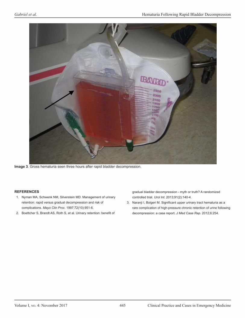
Volume I, no. 4: November 2017 445 Clinical Practice and Cases in Emergency Medicine
Gabriel et al. Hematuria Following Rapid Bladder Decompression
Image 3. Gross hematuria seen three hours after rapid bladder decompression.
REFERENCES1. Nyman MA, Schwenk NM, Silverstein MD. Management of urinary
retention: rapid versus gradual decompression and risk of complications. Mayo Clin Proc. 1997;72(10):951-6.
2. Boettcher S, Brandt AS, Roth S, et al. Urinary retention: benefit of
gradual bladder decompression - myth or truth? A randomized controlled trial. Urol Int. 2013;91(2):140-4.
3. Naranji I, Bolgeri M. Significant upper urinary tract hematuria as a rare complication of high-pressure chronic retention of urine following decompression: a case report. J Med Case Rep. 2012;6:254.

Clinical Practice and Cases in Emergency Medicine 446 Volume I, no. 4: November 2017
images in emergenCy mediCine
Post-Cholecystectomy Syndrome
Tami Moore, MDManish Amin, MD
Section Editor: Rick A. McPheeters, DO Submission history: Submitted December 13, 2016; Revision received January 26, 2017; Accepted June 27, 2017 Electronically published November 3, 2017 Full text available through open access at http://escholarship.org/uc/uciem_cpcem DOI: 10.5811/cpcem.2017.6.33321[Clin Pract Cases Emerg Med. 2017;1(4):446–447.]
CASE PRESENTATIONA 32-year-old female with a history of cholecystectomy
three years prior, presented to the emergency department with epigastric pain. Liver function tests (LFTs) were abnormal (total bilirubin: 1.4mg/dl, alkaline phosphatase: 117U/L, aspartate transaminase: 294U/L, alanine transaminase: 189U/L), however ultrasound (US) imaging was negative for gallbladder pathology and the patient was discharged home with normal vital signs and instructed to follow up in two days if symptoms persisted. At her follow
Kern Medical, Department of Emergency Medicine, Bakersfield, California
Image. Magnetic resonance cholangiopancreatography illustrating a persistent distal filling defect (arrow) of the common bile duct near the ampulla of vater suspicious for retained stone with slightly dilated common bile duct.
up visit, her LFTs worsened (total bilirubin: 3.1mg/dl, alkaline phosphatase: 172U/L, aspartate transaminase: 230U/L, alanine transaminase: 518U/L) and the patient underwent a magnetic resonance cholangiopancreatography (MRCP) which showed a dilated common bile duct (CBD) with filling defect suspicious of stone (Image). The patient subsequently underwent an endoscopic retrograde cholangio-pancreatography with removal of one stone and sphincterotomy. All symptoms improved, and the patient was discharged home with appropriate follow up.
CPC-EM Capsule What do we already know about this clinical entity? Magnetic resonance cholangiopancreatography (MRCP) identifies retained stones in patients with prior gallbladder surgery, however MRCP is not a tool readily available to emergency physicians.
What is the major impact of the image(s)? Given the wealth of information provided, perhaps MRCP imaging can be incorporated as part of a routine postcholecystectomy syndrome (PCS) workup and help to limit unnecessary hospital admissions. How might this improve emergency medicine practice?MRCP imaging can help to improve diagnostic capabilities for patients suffering from PCS presenting to the emergency department.

Volume I, no. 4: November 2017 447 Clinical Practice and Cases in Emergency Medicine
Moore et al. Post-Cholecystectomy Syndrome
DISCUSSIONApproximately 5% of patients who have undergone
cholecystectomy continue to have symptoms of abdominal pain, vomiting, dyspepsia, loose stool, and are thought to suffer from postcholecystectomy syndrome (PCS).1 The incidence of retained stone is as high as 10-15%.2 Patients with abnormal LFTs or an US showing a dilated CBD should be considered for a MRCP. It is the next appropriate step for patients with low to moderate risk of choledocolithiasis. Additionally, it is relatively non-invasive.1
CBD stones are a serious complication after cholecystectomy, therefore the diagnosis of PCS must always be considered in patients status post cholecystectomy with upper abdominal pain.2 MRCP is a useful but underused diagnostic tool and its routine use in the ED may significantly reduce morbidity and mortality.2 Although not a common diagnostic study in emergency medicine, it will likely become more prominent to avoid unnecessary admissions requiring emergency medicine providers to be more familiar with this tool.
Address for Correspondence: Manish Amin, MD, Kern Medical, 1700 Mount Vernon Avenue, Bakersfield, CA 93306. Email: [email protected].
Conflicts of Interest: By the CPC-EM article submission agreement, all authors are required to disclose all affiliations, funding sources and financial or management relationships that could be perceived as potential sources of bias. The authors disclosed none.
Copyright: © 2017 Amin et al. This is an open access article distributed in accordance with the terms of the Creative Commons Attribution (CC BY 4.0) License. See: http://creativecommons.org/licenses/by/4.0/
REFERENCES1. Schofer JM. Biliary causes of postcholecystectomy syndrome. J Emerg
Med. 2010;39(4):406-10.2. Shapey IM, Jaunoo SS, Arachchilage KM, et al. Bilary tract imaging for
retained calculi after laparoscopic cholecystectomy: is risk stratification useful? Surg Laparosc Endosc Percutan Tech. 2012;22:459-62.

Clinical Practice and Cases in Emergency Medicine 448 Volume I, no. 4: November 2017
images in emergenCy mediCine
Aortic Pseudo-dissection Karl Huesgen, MD Sarah Gul, MD Candice Norman, MD
Section Editor: Rick A. McPheeters, DO Submission history: Submitted July 12, 2017; Revision received August 19, 2017; Accepted September 6, 2017 Electronically published November 3, 2017 Full text available through open access at http://escholarship.org/uc/uciem_cpcem DOI: 10.5811/cpcem.2017.9.35658[Clin Pract Cases Emerg Med. 2017;1(4):448–449.]
CASE PRESENTATIONA 21-year-old female with a past medical history
significant for asthma and oral contraceptive use presented complaining of shortness of breath and wheezing. Symptoms started after contact with a dog. She came to the emergency department (ED) after home albuterol treatments failed to provide relief. Initial vital signs included a blood pressure of 145/49mmHg, pulse rate 127
University of Florida, Department of Emergency Medicine, Gainesville, Florida
beats/minute, respirations 32 breaths/minute, temperature 37.1°C (98.8°F), and oxygen saturation of 87% on room air. On auscultation, lung fields demonstrated bilateral wheezing and the expiratory phase was prolonged. She also had retractions and endorsed chest tightness. ED workup included an elevated D-dimer, and subsequent computed tomography (CT) pulmonary angiography indicated ascending aortic dissection instead (Image).
Image. Motion artifact suggesting luminal flap of aortic dissection (arrow).

Volume I, no. 4: November 2017 449 Clinical Practice and Cases in Emergency Medicine
Huesgen et al. Aortic Pseudo-dissection
CPC-EM Capsule
What do we already know about this clinical entity?An aortic dissection occurs when blood enters the medial layer of the aortic wall through a tear in the intima. An aortic pseudo-dissection on the other hand occurs due to aortic pulsation motion artifact on imaging.
What is the major impact of the image(s?)An inaccurate diagnosis of an aortic dissection might result in a patient undergoing unnecessary emergent surgery.
How might this improve emergency medicine practice?There is a risk for false-positive computed tomography (CT) results with ascending dissections. Using electrocardiography-gated CT is useful and may prevent unnecessary surgery. It is also important to take the patient’s history and presentation into account and not rely on imaging alone when making a final diagnosis.
DIAGNOSIS Aortic pseudo-dissection artifact. Emergent preoperative
transesophageal echocardiography disproved presence of intimal flap and dissection, so operative repair was aborted. The patient’s tachycardia after multiple beta agonist treatments produced a motion artifact concerning for aortic root dissection. Although CT imaging is highly sensitive and specific for aortic dissections, there is a potential for false-positive ascending dissections (Stanford type A).1,2 Such artifacts are frequently seen in the thoracic aorta due to its close proximity to the heart,3 and tachycardia correlates significantly with motion defects on CT.4 This problem can be overcome by use of electrocardiography-synchronized (ECG-gated) CT instead.5,6 The patient’s asthma exacerbation was treated as an inpatient and she was eventually discharged home. This case illustrates the importance of taking the clinical history along with the patient’s presentation into account when making a diagnosis.
Address for Correspondence: Candice Norman, MD, University of Florida, 1329 SW 16th Street, Gainesville, Florida 32610. Email: [email protected].
Conflicts of Interest: By the CPC-EM article submission agreement, all authors are required to disclose all affiliations, funding sources and financial or management relationships that could be perceived as potential sources of bias. The authors disclosed none.
Copyright: © 2017 Huesgen et al. This is an open access article distributed in accordance with the terms of the Creative Commons Attribution (CC BY 4.0) License. See: http://creativecommons.org/licenses/by/4.0/
REFERENCES1. Duvernoy O, Coulden R, Ytterberg C. Aortic motion: a potential pitfall
in CT imaging of dissection in the ascending aorta. J Comput Assist Tomogr. 1995;19(4):569-72.
2. Batra P, Bigoni B, Manning J, et al. Pitfalls in the diagnosis of thoracic aortic dissection at CT angiography. Radiographics. 2000;20(2):309-20.
3. Ko SF, Hsieh MJ, Chen MC, et al. Effects of heart rate on motion
artifacts of the aorta on non-ECG-assisted 0.5-sec thoracic MDCT. AJR Am J Roentgenol. 2005;184(4):1225-30.
4. Morgan-Hughes GJ, Owens PE, Marshall AJ, et al. Thoracic aorta at multi–detector row CT: motion artifact with various reconstruction windows. Radiology. 2003;228(2):583-8.
5. Raymond CE, Aggarwal B, Schoenhagen P, et al. Prevalence and factors associated with false positive suspicion of acute aortic syndrome: experience in a patient population transferred to a specialized aortic treatment center. Cardiovasc Diagn Ther. 2013;3(4):196-204.
6. Roos JE, Willmann JK, Weishaupt D, Lachat M, et al. Thoracic aorta: motion artifact reduction with retrospective and prospective electrocardiography-assisted multi-detector row CT. Radiology. 2002;222(1):271-7.

Volume I, no. 4: November 2017 vii Clinical Practice and Cases in Emergency Medicine
AAEM
RSA-
0517
-608
Join AAEM/RSA or Renew Your Membership! www.aaemrsa.org/renew | 800-884-2236
Join RSA or renew for the 2017-2018 year for continued academic and career planning bene-fits, outstanding opportunities for involvement, and premier education.
Are You Moving? Keep in Touch! If you are a graduating resident or medical student and your email address is changing, we recommend you update your address to one outside of your institution to ensure your benefits will continue without interruption. Log in to your members only account at aaemrsa.org/myrsa to list any changes to your name, mailing ad-dress, email address or phone number.
Join or Renew Today!
American Academy of Emergency Medicine Resident & Student Association

Emergency Medicine Fellowship Opportunities
The UC Irvine Department of Emergency Medicine is seeking HS Clinical Instructors for fellowships starting July 1, 2018. UC Irvine Medical Center is rated among the nation’s best hospitals by U.S. News & World Report 16 years in a row and is a 412-bed tertiary and quaternary care hospital with a nationally recognized three-year EM residency program since 1989. The progressive 35-bed ED sees more than 50,000 patients/year and serves as a Level I adult and Level II Pediatric Trauma Center with more than 5,000 annual trauma runs.
The hospital is also a Comprehensive Stroke & Cerebrovascular Center, Comprehensive Cancer Center, Cardiovascular receiving center and regional Burn centers, with Observation and an After Hours clinic in urban Orange County. Completion of an ACGME accredited EM Residency is required. Salary is commensurate with qualifications and proportion of clinical effort. For more information visit: http://www.emergencymed.uci.edu/fellowships.asp
1. Disaster Medicine Fellowship https://recruit.ap.uci.edu/apply/JPF04038
2. Research Fellowship in Population Health https://recruit.ap.uci.edu/apply/JPF04040
3. Medical Simulation Fellowship https://recruit.ap.uci.edu/apply/JPF04041
5. MDEdTech Fellowship https://recruit.ap.uci.edu/apply/JPF04037
6. Point-of-Care Ultrasound Fellowship https://recruit.ap.uci.edu/apply/JPF04042
7. Clinical Informatics Fellowship https://recruit.ap.uci.edu/apply/JPF04039
4. Wilderness Medicine Fellowship https://recruit.ap.uci.edu/apply/JPF04334
The University of California, Irvine is an Equal Opportunity/Affirmative Action Employer advancing inclusive excellence. All qualified applicants will receive consideration for employment without regard to race, color, religion, sex, sexual orientation, gender identity, national origin, disability, age, protected veteran status, or other protected categories covered by the UC nondiscrimination policy.

Medical Director of the Department of Emergency Medicine
The Department of Emergency Medicine at the UC Irvine School of Medicine is seeking applicants for a faculty position as Medical Director of Emergency Medicine. The faculty position will be in the H.S. Clinical Professor Series or Professor of Clinical Emergency Medicine (Clinical X) Series, at the Associate or Full Professor level. Requirements for the H.S. Clinical Professor Series: Qualified candidates must possess an M.D. or M.D./Ph.D. degree, be board certified/board eligible, and have a California license at the time of appointment. Candidates must have demonstrated capabilities for teaching, professional service, and research or creative work. The incumbent will participate in patient care, teach in clinical programs, and perform university and public service. Preference will be given to candidates who possess leadership experience and an M.B.A. degree. Requirements for the Professor of Clinical X Series: Qualified candidates must possess an M.D. or M.D./Ph.D. degree, be board certified/board eligible, and have a California license at the time of appointment. Candidates must have demonstrated capabilities for substantial research, excellent teaching, and professional service. The incumbent will participate in patient care, teach in clinical programs, and perform university and public service. Preference will be given to candidates who possess leadership experience and an M.B.A. degree. Medical Director Information: The ED Medical Director will help lead our care management initiatives using a patient-centered approach with outstanding customer service and clinical excellence, while optimizing ED flow. The Medical Director reports directly to the Vice Chair of Clinical Operations. The ED Medical Director is responsible for administering the bedside ED clinical operations. The candidate is expected to monitor operational data serve as a physician leader of EPIC, and adhere all clinical policies and protocols in lock-step with evidence based medicine. The candidate will also assist with regulatory affairs and Joint Commission compliance and address clinical issues as they arise. The ED Medical Director is expected to coordinate clinical operations with Nursing leadership by participating in their leadership meetings regarding operational issues and serve on the Unit Practice Council. The candidate will also collaborate with other clinical and ancillary departments to distribute information on existing and new operational initiatives, guidelines and protocols. Additionally, the candidate will oversee building and equipment maintenance and environmental services; and provide leadership and input on new equipment and products. The ED Medical Director will also serve as a Department representative at high level hospital and University administration meetings. The candidate will interact with other department clinical leaders, Chairs, and Deans within the UC Irvine Health System with regards to clinical operations and clinical department interactions. UCI Medical Center: The University of California, Irvine Medical Center is a 472-bed tertiary care hospital with all residencies. The ED is a progressive 35-bed Level I Trauma Center with 51,000 patients, in urban Orange County. Collegial relationships with all services. Excellent salary and benefits with incentive plan. Salary and rank will be commensurate with qualifications and experience. Application Procedure: Interested candidates should apply through UC Irvine’s Recruit system located at: https://recruit.ap.uci.edu/apply/JPF04308 The University of California, Irvine is an Equal Opportunity/Affirmative Action Employer advancing inclusive excellence. All qualified applicants will receive consideration for employment without regard to race, color, religion, sex, sexual orientation, gender identity, national origin, disability, age, protected veteran status, or other protected categories covered by the UC nondiscrimination policy.

Professor of Clinical X Series, Open Ranks & Health Sciences Clinical Professor Series, Open Ranks
Department of Emergency Medicine
The University of California, Irvine School of Medicine, Department of Emergency Medicine anticipates openings in the Professor of Clinical X Series, and Health Sciences Clinical Professor Series, Open Ranks.
Health Sciences Clinical Professor Series: Duties include resident and medical student teaching; direct patient care; research and/or creative work; and performing public and University service. Requirements for this series include certification in Emergency Medicine and fellowship or advanced degree, or both, strongly desired.
Professor of Clinical X Series: Duties include substantial research; resident and medical student teaching; performing public and University service; and optional clinical research. Requirements for this series include certification in Emergency Medicine, excellence in teaching, and fellowship or advanced degree, or both, strongly desired.
The University of California, Irvine Medical Center is a 472-bed tertiary care hospital with all residencies. The ED is a progressive 35-bed Level I Trauma Center with 50,000 patients, in urban Orange County. Collegial relationships with all services. Excellent salary and benefits with incentive plan.
Salary and rank will be commensurate with qualifications and experience.
Application Procedure: Interested candidates should apply through UC Irvine’s Recruit system located at: https://recruit.ap.uci.edu/apply/JPF04027
Applicants should complete an online application profile and upload the following application materials electronically to be considered for the position:
1. Curriculum Vitae2. Names and addresses of four references3. Statement of contributions to diversity
The University of California, Irvine is an Equal Opportunity/Affirmative Action Employer advancing inclusive excellence. All qualified applicants will receive consideration for employment without regard to race, color, religion, sex, sexual orientation, gender identity, national origin, disability, age, protected veteran status, or other protected categories covered by the UC nondiscrimination policy.

Post-Doctoral Scholar Position for Biostatistician-Epidemiology
The Department of Emergency Medicine at the UC Irvine School of Medicine is seeking applicants for a Post-Doctoral Scholar in the fields of Biostatistics and Epidemiology.
Candidates must possess a Ph.D. or equivalent doctoral degree in Biostatistics or in a related discipline with a strong quantitative background. Candidates must also be proficient with at least one statistical analysis language. Preference will be given to candidates with experience in the fields of Emergency Medicine and/or Population Based Health.
The Post-Doctoral Scholar is expected to perform statistical data analysis and consult on research designs for Emergency Medicine research projects, including research-statistical methods, sample size and data collection. In addition, the candidate will participate in writing and editing research reports with Principal Investigators. The candidate will interact closely with students, resident physicians, fellows, and faculty members within the Department, and is expected to manage multiple projects in parallel. Candidates must possess excellent writing and editing skills, and have experience working with large datasets. Preference will be given to candidates who are familiar with REDCap.
Minimum Qualifications: Required Ph.D. or equivalent doctoral degree in the field of Biostatistics or in a related discipline with a strong quantitative background.
Anticipated salary range: http://ap.uci.edu/wp-content/uploads/postdoc.pdf
Interested candidates should apply through UC Irvine’s RECRUIT system located at: https://recruit.ap.uci.edu/apply/JPF04350
The University of California, Irvine is an Equal Opportunity/Affirmative Action Employer advancing inclusive excellence. All qualified applicants will receive consideration for employment without regard to race, color, religion, sex, sexual orientation, gender identity, national origin, disability, age, protected veteran status, or other protected categories covered by the UC nondiscrimination policy.

Clinical Practice and Cases in Emergency Medicine viii Volume I, no. 4: November 2017
The University of California, Davis School of Medicine, Department of Emergency Medicine (EM) is conducting a faculty search for EM physicians in either a clinician/educator or clinician/researcher track. Candidates must be residency trained in EM with board certification/preparation and be eligible for licensure in California. At least one year of post-training clinical experience and/or fellowship training is preferred. Candidates are expected to enter at the Assistant/Associate level, commensurate with experience and credentials. EM faculty members at UC Davis who have preference fornight shifts work fewer clinical shifts each month.
The University of California, Davis, Medical Center, one of the nation’s “Top 50 Hospitals,” is a 613 bed academic medical center with approximately 80,000 emergency department visits annually, including approximately 20,000 pediatric visits.The emergency services facility opened in 2010 and is state-of-the-art. Our program provides comprehensive emergency services to a large local urban and referral population as a level 1 trauma center, paramedic base station and training center. The department also serves as the primary teaching site for a fully accredited EM residency program and eight different EM fellowship programs. Our residency training program began more than twenty years ago and currently has 48 residents. The Department has a separate area for the care of children and is one of the leading centers in the Pediatric Emergency Care Applied Research Network (PECARN).
Salary and benefits are competitive and commensurate with training and experience. Sacramento is located near the northern end of California's Central Valley, close to Lake Tahoe, San Francisco, and the "wine country" of the Napa and Sonoma Valleys.Sports enthusiasts will find Sacramento's climate and opportunities ideal.
Interested candidates should submit a letter outlining interests and experience, and curriculum vitae to: recruit.ucdavis.edu/apply/JPF01809
Erik Laurin MD, Professor and Search Committee Chair ([email protected])UC Davis Department of Emergency Medicine
2315 Stockton Blvd., PSSB 2100Sacramento, CA 95817
The University of California is an affirmative action/equal opportunity employer.

Join our 40TH ANNUAL EMERGENCY MEDICINE IN YOSEMITE
January 10 – 13, 2018
Registration is open online at
www.yosemitemef.org Early Bird c lo s e s November 22, 2017
Sponsored by Yosemite Medical Education Foundation (YMEF)
________________________________ Co-sponsored by California ACEP
American College of Emergency Physicians California Chapter
YOSEMITE
MEDICAL
EDUCATION
FOUNDATION
YosemiteMedicalEducationFoundationFacultyFaces:
Nidal Moukaddam, MD - Arian Nachat, MD - Claritza Rios, MD - David Schriger, MD - Name Withheld, MD - Ramin Tabatabai, MD - Christian Tomaszewski, MD - Veronica Tucci, MD
Paul Auerbach, MD - Graham Billingham, MD - Dan Imler, MD - Dara Kass, MD - Angela Lumba-Brown, MD - James Manning, MD - Tsuyoshi Mitarai, MD

Championing individual physician rights
and workplace fairness
JOIN
CAL/AAEM!
BENEFITS- WesternJournalofEmergencyMedicineSubscription- CAL/AAEM News Service email updates- Discounted AAEM pre-conference fees - And more!
CAL/AAEM NEWS SERVICE- Healthcare industry news- Public policy- Government issues- Legal cases and court decisions
Incollaborationwithourofficialjournal
FACEBOOK.COM/CALAAEM FOLLOW US @CALAAEM
HTTP://WWW.CALAAEM.ORG
AAEM-0217-733
Save the DateSave the DateNovember 4-8, 2017
Hyatt Regency, Denver, ColoradoNovember 4-8, 2017
Hyatt Regency, Denver, Colorado
Save the Date for ACOEP’s Flagship Conference
Visit www.acoep.org/scientific for more information!
Don’t miss out on:
• Keynote speaker Zubin Damania, MD, aka ZDogg
• Kickoff Party in downtown Denver presented by Island Medical Management
• Expanded Tachy Track
• Customizable experience through breakout lecture series
• Pre-Conference Tracks including: Wilderness Medicine, Resuscitation, Advanced Ultrasound, Advanced Airway
• 7th Annual FOEM Legacy Gala: Dinner and Awards Ceremony presented by US Acute Care Solutions
• …and much more!