Two isoforms of the Na +/K +/2Cl − cotransporter are expressed in the European eel ( Anguilla...
12
Two isoforms of the Na + /K + /2Cl cotransporter are expressed in the European eel (Anguilla anguilla) Christopher P. Cutler * , Gordon Cramb School of Biology, Bute Medical Buildings, University of St. Andrews, St. Andrews, Fife, Scotland, KY16 9TS, UK Received 1 July 2002; received in revised form 23 July 2002; accepted 4 September 2002 Abstract Two cDNA isoforms of the NKCC1 secretory cotransporter have been isolated from the European eel. The NKCC1a isoform exhibited mRNA expression in a wide range of tissues in a similar fashion to mammals, whereas NKCC1b was expressed primarily in the brain. The effect of freshwater (FW) to seawater (SW) transfer on NKCC1a expression was dependent on the developmental stage. In non-migratory yellow eels, NKCC1a mRNA expression in the gill was transiently up-regulated 4.3-fold after 2 days but also subsequently by 2.5 – 6-fold 3 weeks after SW transfer. Gill NKCC1a expression was localised mainly in branchial chloride cells of SW acclimated yellow eels. In contrast to yellow eels, NKCC1a mRNA abundance was not significantly different following SW acclimation in silver eel gill. NKCC1a mRNA abundance decreased in the kidney following SW acclimation and this may correlate with lower tubular ion/fluid secretion and urine flow rates in SW teleosts. Kidney NKCC1a mRNA expression in silver eels was also significantly lower than in yellow eels, suggesting some pre- acclimation of mRNA levels. NKCC1a mRNAwas expressed at similar low levels in the middle intestine of FW- and SW-acclimated yellow or silver eels, suggesting the presence of an ion secretory mechanism in this gut segment. D 2002 Elsevier Science B.V. All rights reserved. Keywords: Osmoregulation; Teleost fish; CCC; NKCC2; NCC; KCC; BSC1; BSC2 1. Introduction The mammalian chloride-cation-cotransporter (CCC) gene family currently comprises nine related membrane proteins with differing or unknown ion transporting specific- ities [1]. Within this family, the Na + /K + /2Cl cotransporters are found in a wide variety of tissues where they are engaged either in the control of cell volume or trans-epithelial ion transport [2,3]. The Na + /K + /2Cl cotransporter subfamily comprises two members, the secretory isoform known as NKCC1 and the absorptive isoform known as NKCC2 (also known as BSC2 and BSC1, respectively) which exist as several splice variants, and these isoforms are known to be primarily located in the respective basolateral or apical membrane compartments of transporting epithelial cells [1]. In teleost fish, Na + /K + /2Cl cotransporter activity has been associated with a number of ion secretory mechanisms. The cotransporter was initially identified as the likely entry pathway for Na + and Cl ions across the basolateral membrane of ‘chloride’ cells; the main cell type engaged in ion secretion in the operculum and gill of marine teleosts [4]. This assertion was based on earlier studies which used the loop-diuretic furosemide to inhibit cotransporter-like activity in the operculum of the killifish (Fundulus hetero- clitus [5,6]). The ion secretory system of chloride cells has been shown to be regulated by changes in plasma osmolality following acute transfer of killifish from freshwater (FW) to seawater (SW) [7]. In mammals, the activity of members of the CCC family is known to be regulated by various protein kinases [2]. Using an antibody which recognises a phosphorylated form of NKCC, Flemmer and Forbush [8] recently demonstrated the presence of a phosphorylated form of NKCC in bran- chial chloride cells of FW-acclimated killifish and showed a 3 – 4-fold increase in phosphorylation following transfer of fish to SW. The NKCC cotransporter has also been immu- nolocalised to branchial chloride cells in the mudskipper (Periophthalmodon schlosseri) [9], and in the Atlantic salmon (Salmo salar), where the abundance of cotransporter protein was shown to increase by 2.5-fold in the gill of parr following SW transfer [10]. However, in all of these studies 0005-2736/02/$ - see front matter D 2002 Elsevier Science B.V. All rights reserved. PII:S0005-2736(02)00596-5 * Corresponding author. Tel.: +44-1334-463531; fax: +44-1334- 463600. E-mail address: [email protected] (C.P. Cutler). www.bba-direct.com Biochimica et Biophysica Acta 1566 (2002) 92 – 103
-
Upload
st-andrews -
Category
Documents
-
view
0 -
download
0
Transcript of Two isoforms of the Na +/K +/2Cl − cotransporter are expressed in the European eel ( Anguilla...

Two isoforms of the Na+/K+/2Cl� cotransporter are expressed in the
European eel (Anguilla anguilla)
Christopher P. Cutler*, Gordon Cramb
School of Biology, Bute Medical Buildings, University of St. Andrews, St. Andrews, Fife, Scotland, KY16 9TS, UK
Received 1 July 2002; received in revised form 23 July 2002; accepted 4 September 2002
Abstract
Two cDNA isoforms of the NKCC1 secretory cotransporter have been isolated from the European eel. The NKCC1a isoform exhibited
mRNA expression in a wide range of tissues in a similar fashion to mammals, whereas NKCC1b was expressed primarily in the brain. The
effect of freshwater (FW) to seawater (SW) transfer on NKCC1a expression was dependent on the developmental stage. In non-migratory
yellow eels, NKCC1a mRNA expression in the gill was transiently up-regulated 4.3-fold after 2 days but also subsequently by 2.5–6-fold 3
weeks after SW transfer. Gill NKCC1a expression was localised mainly in branchial chloride cells of SW acclimated yellow eels. In contrast
to yellow eels, NKCC1a mRNA abundance was not significantly different following SW acclimation in silver eel gill. NKCC1a mRNA
abundance decreased in the kidney following SW acclimation and this may correlate with lower tubular ion/fluid secretion and urine flow
rates in SW teleosts. Kidney NKCC1a mRNA expression in silver eels was also significantly lower than in yellow eels, suggesting some pre-
acclimation of mRNA levels. NKCC1a mRNAwas expressed at similar low levels in the middle intestine of FW- and SW-acclimated yellow
or silver eels, suggesting the presence of an ion secretory mechanism in this gut segment.
D 2002 Elsevier Science B.V. All rights reserved.
Keywords: Osmoregulation; Teleost fish; CCC; NKCC2; NCC; KCC; BSC1; BSC2
1. Introduction
The mammalian chloride-cation-cotransporter (CCC)
gene family currently comprises nine related membrane
proteins with differing or unknown ion transporting specific-
ities [1]. Within this family, the Na+/K+/2Cl� cotransporters
are found in a wide variety of tissues where they are engaged
either in the control of cell volume or trans-epithelial ion
transport [2,3]. The Na+/K+/2Cl� cotransporter subfamily
comprises two members, the secretory isoform known as
NKCC1 and the absorptive isoform known as NKCC2 (also
known as BSC2 and BSC1, respectively) which exist as
several splice variants, and these isoforms are known to be
primarily located in the respective basolateral or apical
membrane compartments of transporting epithelial cells [1].
In teleost fish, Na+/K+/2Cl� cotransporter activity has
been associated with a number of ion secretory mechanisms.
The cotransporter was initially identified as the likely entry
pathway for Na+ and Cl� ions across the basolateral
membrane of ‘chloride’ cells; the main cell type engaged
in ion secretion in the operculum and gill of marine teleosts
[4]. This assertion was based on earlier studies which used
the loop-diuretic furosemide to inhibit cotransporter-like
activity in the operculum of the killifish (Fundulus hetero-
clitus [5,6]). The ion secretory system of chloride cells has
been shown to be regulated by changes in plasma osmolality
following acute transfer of killifish from freshwater (FW) to
seawater (SW) [7].
In mammals, the activity of members of the CCC family
is known to be regulated by various protein kinases [2].
Using an antibody which recognises a phosphorylated form
of NKCC, Flemmer and Forbush [8] recently demonstrated
the presence of a phosphorylated form of NKCC in bran-
chial chloride cells of FW-acclimated killifish and showed a
3–4-fold increase in phosphorylation following transfer of
fish to SW. The NKCC cotransporter has also been immu-
nolocalised to branchial chloride cells in the mudskipper
(Periophthalmodon schlosseri) [9], and in the Atlantic
salmon (Salmo salar), where the abundance of cotransporter
protein was shown to increase by 2.5-fold in the gill of parr
following SW transfer [10]. However, in all of these studies
0005-2736/02/$ - see front matter D 2002 Elsevier Science B.V. All rights reserved.
PII: S0005 -2736 (02 )00596 -5
* Corresponding author. Tel.: +44-1334-463531; fax: +44-1334-
463600.
E-mail address: [email protected] (C.P. Cutler).
www.bba-direct.com
Biochimica et Biophysica Acta 1566 (2002) 92–103

the isoform specificity of the antibodies for the NKCC
cotransporter isoform binding the antibodies was unknown.
The NKCC cotransporter has also been suggested to be
involved in ion secretion in the teleost intestine [11–13].
The intestine of marine teleosts is primarily involved in ion
absorption associated with the uptake of water from imbibed
SW. However, in fed SW killifish, the posterior region of the
intestine has been shown to be capable of secreting ions, by
a mechanism thought to involve a NKCC cotransporter [13].
Further components of the intestinal secretory mechanism
(as found in other vertebrates) such as the CFTR chloride
ion channel, are also known to be expressed in this segment
of the (killifish) intestine [14], as is the eKir potassium
channel, which is associated with ion secretory processes in
the eel [15]. Hormonal systems known to control intestinal
ion secretion in other vertebrates, such as the hormone
guanylin and isoforms of its receptor, GC-C, are also known
to be expressed in teleost intestine [16–18]. Together, this
data suggests that the teleost intestine should possess an ion
secretory mechanism similar to that found in mammals
which would include a NKCC1 secretory cotransporter [19].
In preliminary investigations, we have reported the
presence of NKCC isoforms in teleost fish including the
European eel (Anguilla anguilla), where the expression of
two homologous partial cDNA fragments of NKCC1 iso-
forms was described in a number of tissues including the gill
and intestine (previously called cot1 and cot3 [11,12]).
The object of this study was (1) to complete the cloning
and sequencing of both secretory NKCC1 cotransporter
isoforms expressed in the European eel, (2) determine their
distribution in eel tissues, (3) investigate changes in gene
expression associated with the process of acclimation from
FW to SW in both non-migratory ‘yellow’, and migratory
‘silver’ eels and, (4) to localise cotransporter protein expres-
sion within the branchial epithelium using immunohisto-
chemistry.
2. Materials and methods
2.1. Fish
Adult freshwater sexually immature ‘‘yellow’’ and sex-
ually maturing migratory ‘‘silver’’ eels (A. anguilla: 125–
1250 g weight) were obtained from local suppliers in
Inverness, Blairgowrie and Kelso and transferred to labo-
ratory aquaria at the Gatty Marine Laboratory where they
were maintained on a 12 h light/dark cycle. Yellow and
silver eels used in experiments were transferred to tanks
containing either FW or SW for a period of up to 21 days.
Fish were decapitated and pithed before removal of tissues.
2.2. Total RNA extraction
Total RNA was isolated from scrapes of gill arches
prepared with a single-sided razor blade. Total RNA
extracted from the intestine in the tissue distribution (Fig.
2) and gut segments (Fig. 5) experiments was prepared from
scrapings of the intestinal epithelium using a glass slide. Gill
and intestinal scrapes were homogenised using a syringe
and 16 gauge needle. Total RNA was otherwise prepared
from whole tissues homogenised using either a syringe and
16 gauge needle or using a Polytron blender (Kinematica).
In the gut segments experiment (Fig. 5), the intestine/rectum
was sectioned into three equal length parts, subsequently
referred to as the anterior and middle intestine and the
posterior intestine/rectum. Total RNA was extracted as
previously described [24].
2.3. cDNA cloning and sequencing
The original cDNA fragments amplified by reverse
transcriptase-polymerase chain reaction (RT-PCR) used
degenerate primers whose design was based on an amino
acid alignment of the NKCC1 cotransporter from shark
rectal gland [20] and the sodium chloride cotransporter
(NCC) from winter flounder bladder [21]. This was
because these were the only CCC sequences available at
the initiation of this project (see Table 1, [11]). These
original primers, amplified a 755 nucleotide (nt) fragment
(originally designated cot1) from 7-day SW-acclimated eel
gill mRNA (using RNA samples pooled from three fish)
and a 707 nt (originally designated cot3) fragment from 3-
week SW-acclimated eel brain total RNA (using tissue
samples pooled from 10 fish). For amplification and
cloning of the initial fragments, the RNA extraction, RT-
PCR, cloning and sequencing protocols were performed
essentially as described in Ref. [22]. Comparison of these
eel cDNA sequences with those from mammalian species
revealed that these two eel CCC isoforms represented
apparent duplicate copies of the NKCC1 isoform from
mammals [12].
Table 1
Degenerate CCC oligonucleotide primer sequences and their position within the derived encoded amino acid (aa) alignment in Fig. 1
Name and position in alignment Degenerate primer nucleotide sequences
Original sense at aa 810–821 GARGAYCAYGTXAARAAYTWYMGXCCXCARTGYYT
Original antisense at aa 1053–1064 AYRTCIATXGTXYYYTTXCCYTGYTTYTTYTGRAA
Modified sense at aa 810–820 GARGAYCAYRTXAARAAYTWYMGXCCXCARTG
Modified antisense at aa 1057–1067 ARCCAXYAXAYRTCXATXGTXYYYTTXCCYTG
3V End antisense at aa 1241–1250 ARTARAAXGTXARXACRYTYTBXTGRTT
The sequences use standard IUPAC codes except for I = inosine and X= inosine/cytosine wobbles (see Ref. [22]).
C.P. Cutler, G. Cramb / Biochimica et Biophysica Acta 1566 (2002) 92–103 93
C.P. Cutler, G. Cramb / Biochimica et Biophysica Acta 1566 (2002) 92–10394

C.P. Cutler, G. Cramb / Biochimica et Biophysica Acta 1566 (2002) 92–103 95

Fig. 1. Amino acid alignment of the eel duplicate NKCC1a and NKCC1b (gill spliceoform) cotransporters (EMBL Accession Nos. AJ486858 and AJ486859, respectively) in comparison to NKCC1 cotransporters
from an elasmobranch (EMBL Accession No. U05958 [Squalus acanthias]) and also mammalian (EMBL Accession Nos. U30246 [human], BTU70138 [bovine], AF051561 [rat] and NM 009194 [mouse])
species. ( – ) represent gaps where spaces have been added to improve alignment. (.) represent positions within the alignment which show complete conservation between species and isoforms. X represents
position of conserved difference; locations where the eel NKCC1 isoforms have the same aa as each other but where mammalian sequences have a different conserved aa. Text printed in 50% gray are locations of
putative N-glycosylation sites within the putative extracellular domains of eel NKCC1s (at positions 580 and 590 of the sequence alignment). Regions with a wavy underline are the putative locations of the
transmembrane domains. Amino acids with straight underlines are those within the eel NKCC1 sequences predicted to be potential phosphorylation sites for various cellular protein kinases as determined by
NetPhos 2.0 [23].
C.P.Cutler,
G.Cramb/Biochimica
etBiophysica
Acta
1566(2002)92–103
96

The remainder of both genes were amplified using vari-
ous PCR-based techniques essentially as described in Ref.
[24] with the exception of nested inverse PCR. The first of
these two genes is now re-designated NKCC1a here (pre-
viously cot1), in order to coincide with the nomenclature
now used in other species. The 5Vend of this isoform was
obtained using gene-specific primers and the rapid amplifi-
cation of cDNA ends (RACE) technique, the fragment
generated contained a 5Vuntranslated region (UTR) of 549
nt. At the 3Vend, an internal overlapping fragment was first
cloned using a gene-specific primer and a degenerate (3Vendantisense) primer and the remainder of the open reading
frame (ORF) was isolated using nested inverse PCR. Briefly,
the nested inverse PCR involved cutting eel gill cDNAwith a
DpnII restriction enzyme and ligating the ends of the frag-
ments generated together using a T4 DNA ligase reaction.
The ligated DpnII fragment containing the 3V end of
NKCC1a was then amplified using two pairs of gene-
specific nested primers facing away from each other in order
to amplify the region across the (DpnII cut) ligated ends.
This fragment then contains additional previously unknown
sequence, which in this case, included the ORF’s stop codon
and some 3VUTR. The final NKCC1a contiguous cDNA
sequence generated (EMBL Accession No. AJ486858) had a
3VUTR of 37 nt, although the mRNA clearly has a larger 3VUTR which accounts for its much greater size (see Fig. 2).
For the second gene, now designated NKCC1b (previ-
ously cot3) experiments using the original primers had
yielded a fragment of 707 nt from the brain (EMBL
Accession No. AJ487475). In subsequent experiments, a
further pair of degenerate primers were synthesized (modi-
fied sense and antisense, Table 1), which took into account
sequence information available from newly identified
CCCs. These new primers, when used with eel gill mRNA,
yielded a further fragment which encoded a larger putative
spliceoform of the NKCC1b ORF with an additional 54 nt
in comparison to the brain NKCC1b form (the additional aa
encoded are at positions 1013–1030 inclusive in the gill
spliceoform as shown in Fig. 1). Both 5V and 3V RACEtechniques were successfully used to generate a complete
ORF using gill RNA. Unfortunately, as a result of a homo-
polymer poly C region in the 3VUTR, only 23 nt of 3VUTRwas able to be sequenced. The 5VRACE experiments also
generated three fragments with differing UTR sequences
which are putative splice variants (EMBL Accession Nos.
AJ486860, AJ486861 and AJ486862).
2.4. Northern blot analysis
Northern blotting, nucleic hybridisation and quantifica-
tion were also performed as in Cutler et al. [24]. The original
fragments were used as cDNA probes for Northern blotting.
As the NKCC1a and NKCC1b probes did not cross-hybrid-
ise with each other’s mRNA on Northern blots (Fig. 2),
sufficiently high stringency conditions were used to produce
specific signals. Quantitative Northern blot data was trans-
formed using log10 to reduce the effects of heteroscedasticity,
tested for homogeneity using Bartlett’s test, and analysed
using ANOVA with Scheffe’s F or Fisher PLSD post hoc
testing of heterogeneous or homogeneous data, respectively.
2.5. Antibody production and immunohistochemistry
Antibodies were produced as outlined in Cutler et al. [24].
A peptide (NH2-DVKAPTQPLLKKDKK-COOH; extend-
ing from aa 1020–1034 in Fig. 1) was chosen from the
derived aa sequence of the original NKCC1a cDNA frag-
ment due to its high potential antigenicity [25]. The 15-mer
was manufactured as a multiple-antigen peptide (MAP;
Severn Biotech., Kidderminster, UK) and used to raise eel
NKCC1a polyclonal antibodies in sheep by the National
Diagnostics Scotland laboratory (Law Hospital, Carluke,
UK). A further isoform-specific antisera was raised against
the Na,K-ATPase a1 subunit peptide sequence (NH2-
HKNANSEESKHLLV-COOH; at aa positions 492–505;
see Ref. [22]) and this was used to identify chloride cells
within the branchial epithelium, as the Na,K-ATPase enzyme
is well-known to be expressed at high levels in these cells
[24]. Immuno-histochemistry was performed using a donkey
anti-sheep horseradish peroxidase (HRP)-conjugated secon-
dary antibody and visualised as described previously [24].
Serial cross-sections through the primary filaments and
secondary lamellae of the gill were stained with either the
NKCC1a antisera or Na,K-ATPase antisera (diluted 1:100).
Similarly diluted pre-immune serum was also used as a
control on sequential sections (data not shown).
3. Results
3.1. Sequence and structure of NKCC1a and NKCC1b
The derived amino acid sequences of both NKCC1a
and NKCC1b are highly homologous to NKCC1 sequen-
Fig. 2. Northern blots illustrating the tissue distribution of NKCC1a and
NKCC1b mRNA expression in total RNA samples (10 Ag) from the tissues
of SW-acclimated yellow eels, with the exception of samples from FW
yellow eels. Blots were exposed to film at � 80 jC for 9.6 h (NKCC1a)
and 27 h (NKCC1b).
C.P. Cutler, G. Cramb / Biochimica et Biophysica Acta 1566 (2002) 92–103 97

ces from other species (Fig. 1), although there are a
number of amino acid differences at the N-terminal. The
NKCC1a and NKCC1b isoforms are also highly homolo-
gous to each other with around 80% amino acid identity
over the entire sequence (Table 2). NKCC1a showed
greater similarity to mammalian NKCC1s, however, the
difference is only of the order of 1–2%. This is also
reflected in the increased similarity of NKCC1a to mam-
malian NKCC1s in comparison to shark NKCC1.
NKCC1b has approximately similar levels of homology
when compared to mammalian and shark NKCC1s.
After position 225 of the NKCC1 aa alignment, the
sequences of NKCC1a and NKCC1b show high levels of
conserved structure and homology including the predicted
12 transmembrane domains. In addition, both sequences
share two extracellular potential N-glycosylation sites
(NKCC1a has another three and NKCC1b another four
independent putative N-glycosylation sites) suggesting that
like mammalian and shark NKCC1s, eel NKCC1s are
glycosylated proteins.
3.2. Messenger RNA expression of eel NKCC1s
Gene-specific probes for NKCC1a and NKCC1b iso-
forms were used to determine the extent of mRNA expres-
sion in various tissues of the eel (Fig. 2). NKCC1a was
expressed in a wide range of tissues within the fish as a
single mRNA species of approximately 13 kilo bases (kb)
(initial size estimates for NKCC1a and NKCC1b mRNAs
Table 2
Percentage amino acid homology of the vertebrate NKCC1 cotransporter
sequences in comparison to eel NKCC1a and NKCC1b
Species/Isoform Eel NKCC1a
(cot1)
Eel NKCC1b
(cot3)
Eel NKCC1a (cot1) 100 80.4
Eel NKCC1b (cot3) 80.4 100
Shark NKCC1 68.9 68.5
Human NKCC1 71.5 68.1
Bovine NKCC1 70.8 68.0
Rat NKCC1 70.2 68.0
Mouse NKCC1 70.4 67.8
Homologies were calculated using the amino acid alignment in Fig. 1.
Fig. 3. (a) Northern blots of NKCC1a mRNA expression using total RNA (5 Ag) from the gill, kidney and intestine of FW or 3-week acclimated SW yellow or
silver eels. (b) Quantification of the levels of NKCC1a mRNA abundance on the Northern blots in panel a. Significant differences between FWand SW (yellow
or silver) eels are indicated for each tissue where ** indicated P< 0.01 and **** indicates P < 0.0001 using analysis of variance with Scheffe’s F post hoc test.
Error bars indicate standard error of the mean, where n= 6 fish.
C.P. Cutler, G. Cramb / Biochimica et Biophysica Acta 1566 (2002) 92–10398

were revised after the use of higher resolution gels [11]),
with highest abundance in pancreas, oesophagus, stomach
and gill, with intermediate levels in the eye and low levels in
the brain, heart, intestine and kidney. No sign of any
NKCC1a mRNA expression appeared to be present in
skeletal muscle and liver. NKCC1b expression exhibited a
different tissue distribution with a single 6 kb mRNA
species found only in the brain (Fig. 2). However, a further
blot using 5 Ag poly A+ mRNA (rather than total RNA)
from the gills of 7-day SW-acclimated eels indicated that
NKCC1b is expressed at extremely low levels in this tissue,
as multiple mRNA species with sizes of 3.6, 3.8, 6, 8 and
f 12 kb (data not shown).
The effect of SW acclimation on NKCC1a mRNA was
investigated in both yellow and silver eels (Fig. 3). In the gill
of yellow eels, a 5.9-fold increase in NKCC1a mRNA
abundance was found 3 weeks after transfer of fish to SW.
This was in marked contrast to the situation in silver eels
where an 80% decrease in expression was found, although
this was not sufficiently different enough to be significant
using the Scheffe’s F post hoc test. In the kidney, there was a
40% decrease in mRNA abundance following salinity accli-
mation in yellow eels, although the similar 60% decrease
found in SW-acclimated silver eels was not significant. In
addition, the overall levels of mRNA abundance were
significantly lower in silver eel when compared to yellow
eel kidney. The time course of changes in gill NKCC1a
mRNA abundance during salinity acclimation was further
examined in yellow eels (Fig. 4). After 21 days of SW
acclimation, the level of NKCC1a mRNA expression
showed a significant but lower level (f 2.5-fold) of increase
than in the previous experiment. However, there was also a
transient peak in NKCC1a mRNA expression, 2 days after
SW transfer where a f 2.5-fold increase over time-matched
control fish also occurred. However, over the first 4 days, the
actual changes in NKCC1a mRNA abundance were masked
to some extent by significant increases in expression in the
FW to FW-transferred time-matched control eels, which may
have been a result of a stress response associated with the
transfers. Consequently, the levels of NKCC1a mRNA
abundance was 4.3-fold higher in SW eels 2 days post-
transfer compared to FW fish at time zero.
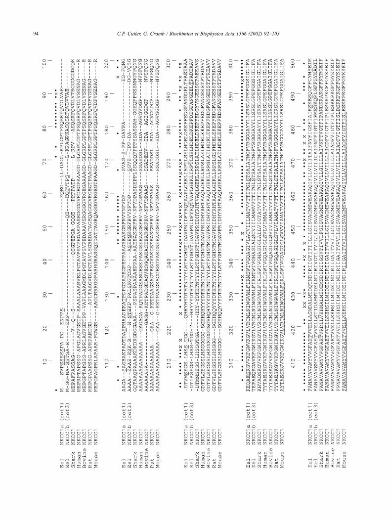
Initial experiments indicated low levels of expression of
NKCC1a mRNA in the intestine (Fig. 2). Subsequent experi-
ments determined that this expression was confined only to
the middle region of the gut with no mRNA detected in
anterior or posterior segments (Fig. 5). No significant
changes in NKCC1a mRNA expression were seen in midgut
sections from yellow or silver eels following SW acclima-
tion.
Fig. 4. Quantification of Northern blots showing the levels of branchial NKCC1a mRNA abundance during the time course of acclimation of yellow eels from
FW to SW and to control fish similarly transferred from FW to FW. Significant differences between FW/FW or FW/SW eels and fish at the initial time 0 point
are indicated where ** or yy indicates P< 0.01, yyy indicates P < 0.001, **** indicates P< 0.0001 using analysis of variance with Fishers PLSD post hoc test.
At each time point, the levels of NKCC1a mRNA abundance was significantly different to that in FW time-matched controls. Error bars indicate standard error
of the mean, where n= 6 fish.
C.P. Cutler, G. Cramb / Biochimica et Biophysica Acta 1566 (2002) 92–103 99
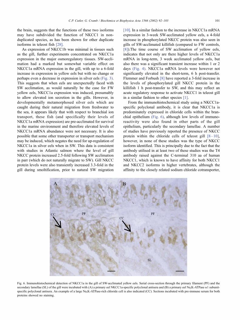
3.3. Branchial NKCC1a immunohistochemistry
The NKCC1a polyclonal antiserum was used in immu-
nohistochemistry to localise this protein in gill sections (Fig.
6; pre-immune serum gave no similar staining, data not
shown). In cross-sections through the gill of SW eels,
staining was seen in large cells within the primary filamental
epithelium. Similar sequential sections through the gill
filament, stained with the Na,K-ATPase a1 subunit poly-
clonal antibody also stained large cells at the same position
within the section. As Na,K-ATPase is well known to occur
at very high levels in chloride cells (for example see Ref.
[24]), staining in the same cell type as Na,K-ATPase with
the NKCC1a-specific antibody suggests that the expression
of the NKCC1a protein predominates within chloride cells.
In addition, there was also further general NKCC1a staining
of epithelial cells, particularly within the secondary lamel-
lae; staining with the Na,K-ATPase antibody in this region
was of much lower intensity, suggesting that the expression
of NKCC1a is somewhat more evenly distributed in gill
epithelial cells than is the expression of the Na,K-ATPase
a1 subunit.
4. Discussion
For some time, it has been acknowledged that the
genomes of certain teleost fish species underwent an ancient
duplication in comparison to the tetrapod genome [26,27],
with the result that around 20% of genes in zebrafish (Danio
rerio) and 10% of genes in pufferfish (Fugu rubripes) have
additional paralogues or isoforms without comparable
orthologues in higher vertebrates [28]. There is some
indication that this is a teleost-wide phenomenon which
includes the eel [12]. The presence of two copies of the
NKCC1 cotransporter therefore adds another small piece of
evidence in support of this theory. The availability of further
sequence data from diverse vertebrate groups such as the
eel, offers an opportunity to improve the identification of
conserved residues within the amino acid sequence, which
may be critical to the regulation or function of the protein
(Fig. 1). The relatively high homology of the eel NKCC1 aa
sequences in comparison to other species, however, limits
their usefulness in this regard (Table 2). The presence of the
two eel NKCC1 isoforms does at least allow an analysis of
unique motifs which are conserved between eel NKCC1
isoforms but are different to the conserved residues at the
equivalent positions in mammalian NKCC1s. On this basis,
there are 107 conserved aa differences between the eel
isoforms and mammalian sequences, but most of these are
single isolated residues. However, there are three motifs in
predicted intracellular domains which may have particular
significance. These are (1) the GLG motif at position 666 of
the alignment which is AFQ in mammalian sequences; (2)
the aa region FKDLA/TNDQV from position 861 of the
alignment which is MKEMSIDQA in mammalian sequen-
ces; and (3) the aa region MEQEAAERLKAE from position
1156 of the alignment which is KEQDIADKMKED in
mammalian sequences. These conserved differences may
well be responsible for structural, functional or regulatory
differences subsequently found between eel and mammalian
NKCC1s, and are therefore worthwhile targets for future
mutagenic or chimeric (eel/mammalian NKCC1 chimeras)
studies.
NKCC1a mRNA is expressed in a wide variety of eel
tissues, with high levels of abundance in the pancreas,
oesophagus, stomach and gill, with some also present in
kidney, intestine, heart and brain and this tissue distribution
is similar to mammalian NKCC1 (Fig. 2) [19,29]. One
obvious difference between eel NKCC1a and the expression
of NKCC1 in other species is the relatively large size of the
mRNAwhich in mammals, sharks and amphibians is around
6.5–7.5 kb [19,20,29,30] but in eels is estimated to be
around 13 kb. Most of the increased size of the eel NKCC1a
mRNA is likely to be due to a long 3V UTR, which may be
as large as 9 kb in eel NKCC1a. The NKCC1b brain
mRNA, which is estimated to be 6 kb, is much more similar
in size to NKCC1 counterparts in other species, but again
some of the minor NKCC1b transcripts found in eel gill are
estimated to be almost as large as eel NKCC1a (results not
shown). Five differently sized NKCC1b mRNAs were
isolated form the gill and the existence of such a large
number is so far unique to this isoform, although a second
smaller NKCC1 transcript has been reported in other species
(5.1–5.2 kb) [19,20]. Another difference between eel
NKCC1a and NKCC1 expression in other species is the
relatively low level of mRNA abundance in eel brain. This
of course may be explained by the presence of the second
isoform, NKCC1b, in this tissue (Fig. 2). The somewhat
offsetting level of expression of NKCC1a and NKCC1b in
Fig. 5. Quantification of Northern blots showing the levels of NKCC1a
mRNA abundance in the middle region of the intestine of FW or 3-week
acclimated SW yellow or silver eels. Error bars indicate standard error of
the mean, where n= 6 fish.
C.P. Cutler, G. Cramb / Biochimica et Biophysica Acta 1566 (2002) 92–103100
the brain, suggests that the functions of these two isoforms
may have subdivided the function of NKCC1 in non-
duplicated species, as has been shown for other duplicate
isoforms in teleost fish [28].
As expression of NKCC1b was minimal in tissues such
as the gill, further experiments concentrated on NKCC1a
expression in the major osmoregulatory tissues. SW-accli-
mation had a marked but somewhat variable effect on
NKCC1a mRNA expression in the gill, with up to a 6-fold
increase in expression in yellow eels but with no change or
perhaps even a decrease in expression in silver eels (Fig. 3).
This suggests that when eels are unexpectedly faced with
SW acclimation, as would naturally be the case for FW
yellow eels, NKCC1a expression was induced, presumably
to allow elevated ion secretion in the gills. However, in
developmentally metamorphosed silver eels which are
caught during their natural migration from freshwater to
the sea, it appears likely that with respect to branchial ion
transport, these fish (and specifically their levels of
NKCC1a mRNA expression) are pre-acclimated for survival
in the marine environment and therefore elevated levels of
NKCC1a mRNA abundance were not necessary. It is also
possible that some other transporter or transport mechanism
may be induced, which negates the need for up-regulation of
NKCC1a in silver eels when in SW. This data is consistent
with studies in Atlantic salmon where the level of gill
NKCC protein increased 2.5-fold following SW acclimation
in parr (which do not naturally migrate to SW). Gill NKCC
protein levels were also transiently increased 3.3-fold in the
gill during smoltification, prior to natural SW migration
[10]. In a similar fashion to the increase in NKCC1a mRNA
expression in 3-week SW-acclimated yellow eels, a 4-fold
increase in phosphorylated NKCC protein was also seen in
gills of SW-acclimated killifish (compared to FW controls,
[8]).The time course of SW acclimation of yellow eels,
indicates that not only are there higher levels of NKCC1a
mRNA in long-term, 3 week acclimated yellow eels, but
also there was a significant transient increase within 1 or 2
days (Fig. 4). NKCC1a mRNA levels were however not
significantly elevated in the short-term, 6 h post-transfer.
Flemmer and Forbush [8] have reported a 3-fold increase in
the levels of phosphorylated gill NKCC protein in the
killifish 1 h post-transfer to SW, and this may reflect an
acute regulatory response to activate NKCC1 in teleost gill
in a similar fashion to other species [1].
From the immunohistochemical study using a NKCC1a-
specific polyclonal antibody, it is clear that NKCC1a is
predominately expressed in chloride cells within the bran-
chial epithelium (Fig. 6), although low levels of immuno-
reactivity were also found in other parts of the gill
epithelium, particularly the secondary lamellae. A number
of studies have previously reported the presence of NKCC
protein within the chloride cells of teleost gill [8–10],
however, in none of these studies was the type of NKCC
isoform identified. This is principally due to the fact that the
antibody utilised in at least two of these studies was the T4
antibody raised against the C-terminal 310 aa of human
NKCC1, which is known to have affinity for both NKCC1
and NKCC2 isoforms in higher vertebrates, although the
affinity to the closely related sodium chloride cotransporter,
Fig. 6. Immunohistochemical detection of NKCC1a in the gill of SW-acclimated yellow eels. Serial cross-section through the primary filament (PF) and the
secondary lamellae (SL) of the gill were incubated with (A) a primary eel NKCC1a-specific polyclonal antisera and (B) a primary eel Na,K-ATPase a1 subunit-
specific polyclonal antisera. An example of a large Na,K-ATPase-rich chloride cell is also indicated (CC). Sections incubated with pre-immune serum for both
proteins showed no staining.
C.P. Cutler, G. Cramb / Biochimica et Biophysica Acta 1566 (2002) 92–103 101

NCC, was not determined [31]. Of the amino acids which
are conserved between human NKCC1 and NKCC2 within
the C-terminal fragment (which are likely to form the
epitope for this cross-reacting antibody), around 65% are
also conserved within NCC, suggesting that the T4 antibody
may also bind NCC isoforms. Within eel gill, as well as
NKCC1a and NKCC1b, an NCC isoform can also be
detected by RT-PCR (data not shown) and therefore it is
possible that the T4 antibody used across species may be
detecting one or a number of CCC isoforms in the teleost
gill. However, the relative abundance of CCC isoform
mRNAs in eel gill, together with the localisation experi-
ments in this study, suggest that studies using the T4
antibody on teleost gill sections are likely to be detecting
a NKCC1a-like isoform.
In the kidney, there was a decrease in NKCC1a mRNA
expression in both yellow and silver eels when acclimated
to SW (Fig. 3). In teleost kidney, a proportion of tubular
fluid is known to be produced by fluid secretion in the
proximal tubule using a similar ion transport mechanism to
that found in chloride cells [32]. Marine teleosts are known
to have a reduced number of relatively small glomeruli
with reduced urine flow rates compared to FW fish [33].
While there are a number of possible explanations, the
changes in eel kidney NKCC1a mRNA expression, sug-
gests that ion secretion involving the NKCC1a cotrans-
porter in eel kidney is reduced following SW acclimation
and this may be partly responsible for the lower urine flow
rates found in marine fish including euryhaline species
acclimated to SW.
In the intestine, only low amounts of NKCC1a mRNA
expression were found in yellow or silver eels and no
change in these levels were seen following SW acclimation
(Fig. 3). When expression was investigated in three seg-
ments of the intestine, detectable levels of NKCC1a mRNA
were only present in the middle region of the intestine, but
again, no change in expression was observed following SW
acclimation (Fig. 5). This suggests that NKCC1a is not
involved in the absorption of ions across the intestine
following the normal marine teleost osmoregulatory drink-
ing response. Ion absorption in this tissue is probably related
to the presence of NKCC2 isoforms [12]. The presence of
the NKCC1a secretory cotransporter in the middle segment
of the intestine suggests that this region may have an ion
secretory capacity, as has also been demonstrated in killifish
intestine [11–13]. However, the presence of NKCC1a
expression in the middle intestine of the eel apparently
contrasts with killifish, where fluid secretion occurred in
the posterior intestinal segment [13]. In anatomical terms, it
is likely that a portion of the eel middle intestinal section is
equivalent to the cephalad portion of the killifish posterior
intestinal segment (although the morphology of eel intestine
is somewhat variable). The localisation of NKCC to the
apical/brush border membranes rather than the basolateral
membranes of killifish intestinal surface epithelial cells
(again using the T4 antibody, [31]) probably relates to the
presence of NKCC2 absorptive cotransporter isoforms or
possibly an NCC isoform also present in this segment; at
least if killifish shows similar CCC expression patterns to
the eel [12]. Although the NKCC1 cotransporter can be
localised to apical membranes in specialised tissues such as
the choroid plexus, it has always otherwise been shown to
have a basolateral membrane cellular localisation [1].
Unfortunately, the NKCC1a-specific antibody was not sen-
sitive enough to detect the NKCC1a protein in eel intestinal
sections.
In summary, two cDNA isoforms of the NKCC1 secre-
tory cotransporter have been isolated from the European eel.
This again supports the possibility that an additional
genome duplication event occurred during the evolution of
the ray-finned fish lineage. The mRNA expression of the
NKCC1a isoform was widely distributed in the tissues of
the eel, in a similar fashion to mammals, whereas NKCC1b
was expressed mainly in the brain. Following SW acclima-
tion, NKCC1a gill mRNA expression was up-regulated
transiently after 2 days but also in the longer term in yellow
eels. NKCC1a expression was localised mainly to the
chloride cells within the branchial epithelium of SW accli-
mated yellow eels. Gill NKCC1a expression levels remain
unchanged or may even be reduced in silver eels prior to
SW migration. This suggests that either levels of protein are
pre-acclimated to those required for SW survival (negating
the need for a rise in NKCC1a mRNA levels) or that the
function of this isoform is taken over by some other
unknown transporter or transport system. NKCC1a mRNA
abundance was also decreased in the kidney following SW
acclimation and this may reflect reduced tubular ion/fluid
secretion concomitant with reduced urine flow rates, which
are known to occur following SW acclimation. NKCC1a is
also expressed at low levels in the middle intestine which
may indicate the presence of an ion secretion mechanism in
this segment of the gut.
Acknowledgements
The authors would like to acknowledge the contribution
of Mr. R. Sterling who performed the immunohistochemical
localisation studies utilised for this article. The authors
would also like to acknowledge the Natural Environment
Research Council for the provision of funds for this work.
References
[1] E. Delpire, D.B. Mount, Annu. Rev. Physiol. 64 (2002) 803–843.
[2] M. Haas , B. Forbush III, Annu. Rev. Physiol. 62 (2000) 515–534.
[3] J.M. Russell, Physiol. Rev. 80 (2000) 211–276.
[4] J.A. Zadunaisky, Fish Physiology, vol. 10B, Academic Press, London,
1984, pp. 129–176.
[5] K.J. Degnan, K.J. Karnaky Jr., J.A. Zadunaisky, J. Physiol. 271
(1977) 155–191.
[6] K.J. Karnaky Jr., K.J. Degnan, J.A. Zadunaisky, Science 195 (1977)
203–205.
C.P. Cutler, G. Cramb / Biochimica et Biophysica Acta 1566 (2002) 92–103102

[7] J.A. Zadunaisky, S. Cardona, L. Au, D.M. Roberts, E. Fisher, B.
Lowenstein, E.J. Cragoe Jr., K.R. Spring, J. Membr. Biol. 143
(1995) 207–217.
[8] A.W. Flemmer, B. Forbush III, FASEB J. 13 (1999) (Suppl. A399).
[9] J.M. Wilson, D.J. Randall, M. Donowitz, A.W. Vogl, A.K.-Y. IP, J.
Exp. Biol. 203 (2000) 2297–2310.
[10] R.M. Pelis, J. Zydlewski, S.D. McCormick, Am. J. Physiol. 280
(2001) R1844–R1852.
[11] C.P. Cutler, I.L. Sanders, G. Luke, N. Hazon, G. Cramb, Soc. Exp.
Biol., Seminar Series, vol. 58, Cambridge Univ. Press, Cambridge,
1996, pp. 43–74.
[12] C.P. Cutler, G. Cramb, Comp. Biochem. Physiol. 130A (2001)
551–564.
[13] W.S. Marshall, J.A. Howard, R.R.F. Cozzi, E.M. Lynch, J. Exp. Biol.
205 (2002) 745–758.
[14] T.D. Singer, S.J. Tucker, W.S. Marshall, C.F. Higgins, Am. J. Physiol.
274 (1998) C715–C723.
[15] Y. Suzuki, M. Itakura, M. Kashiwagi, N. Nakamura, T. Matsuki, H.
Sakuta, N. Naito, K. Takano, T. Fujita, S. Hirose, J. Biol. Chem. 274
(1999) 11376–11382.
[16] T. Mantoku, R. Muramatsu, M. Nakauchi, S. Yamagami, T. Kusa-
kabe, N. Suzuki, J. Biochem. 125 (1999) 476–486.
[17] M.M. Comrie, C.P. Cutler, G. Cramb, Comp. Biochem. Physiol. 129B
(2001) 575–586.
[18] M.M. Comrie, C.P. Cutler, G. Cramb, Biochem. Biophys. Res. Com-
mun. 281 (2001) 1078–1085.
[19] J.A. Payne, J.-C. Xu, M. Haas, C.Y. Lytle, D. Ward , B. Forbush III, J.
Biol. Chem. 270 (1995) 17977–17985.
[20] J.-C. Xu, C.Y. Lytle, T.T. Zhu, J.A. Payne, E. Benz Jr., B. Forbush III,
Proc. Natl. Acad. Sci. U. S. A. 91 (1994) 2201–2205.
[21] G. Gamba, S.N. Saltzberg, M. Lombardi, A. Miyanoshita, J. Lytton,
M.A. Hediger, B.M. Brenner, S.C. Hebert, Proc. Natl. Acad. Sci.
U. S. A. 90 (1993) 2749–2753.
[22] C.P. Cutler, I.L. Sanders, N. Hazon, G. Cramb, Comp. Biochem.
Physiol. 111B (1995) 567–573.
[23] N. Blom, S. Gammeltoft, S. Brunak, J. Mol. Biol. 294 (1999)
1351–1362.
[24] C.P. Cutler, S. Brezillon, S. Bekir, I.L. Sanders, N. Hazon, G. Cramb,
Am. J. Physiol. 279 (2000) R222–R229.
[25] G.W. Welling, W.J. Weijer, R. Van Der Zee, S. Welling-Wester, FEBS
Lett. 188 (1985) 215–218.
[26] J.H. Postlethwait, Y.L. Yan, M.A. Gates, S. Horne, A. Amores, A.
Brownlie, A. Donovan, E.S. Egan, A. Force, Z.Y. Gong, C. Goutel, A.
Fritz, R. Kelsh, E. Knapik, E. Liao, B. Paw, D. Ransom, A. Singer, M.
Thomson, T.S. Abduljabbar, P. Yelick, D. Beier, J.S. Joly, D. Larham-
mar, F. Rosa, M. Westerfield, L.I. Zon, S.L. Johnson, W.S. Talbot,
Nat. Genet. 18 (1998) 345–349.
[27] J. Wittbrodt, A. Meyer, M. Schartl, BioEssays 20 (1998) 511–515.
[28] M.C. Fishman, Science 294 (2001) 1290–1291.
[29] E. Delpire, M.I. Rauchman, S.C. Hebert, S.R. Gullans, J. Biol. Chem.
269 (1994) 25677–25683.
[30] D.I. Soybel, S.R. Gullans, F. Maxwell, E. Delpire, Am. J. Physiol.
269 (1995) C242–C249.
[31] C. Lytle, J.-C. Xu, D. Biemesderfer, B. Forbush III, Am. J. Physiol.
269 (1995) C1496–C1505.
[32] K.W. Bayenbach, M.D. Baustian, Structure and Function of the Kid-
ney, vol. 1, Karger, Basel, 1989, pp. 103–142.
[33] K.J. Karnaky Jr., Physiology of Fishes, CRC Press, Boca Raton, FL,
1998, pp. 157–176.
C.P. Cutler, G. Cramb / Biochimica et Biophysica Acta 1566 (2002) 92–103 103