Apoptin : oncogenic transformation & tumor-selective apoptosis
Upload
independentCategory
view
0download
0
2001;61:7339-7348. Cancer Res Xu Dong Zhang, Xi Yi Zhang, Christian P. Gray, et al. by Smac/DIABLO Release from MitochondriaLigand-induced Apoptosis of Human Melanoma Is Regulated Tumor Necrosis Factor-related Apoptosis-inducing
Updated version
http://cancerres.aacrjournals.org/content/61/19/7339
Access the most recent version of this article at:
Cited Articles
http://cancerres.aacrjournals.org/content/61/19/7339.full.html#ref-list-1
This article cites by 35 articles, 16 of which you can access for free at:
Citing articles
http://cancerres.aacrjournals.org/content/61/19/7339.full.html#related-urls
This article has been cited by 49 HighWire-hosted articles. Access the articles at:
E-mail alerts related to this article or journal.Sign up to receive free email-alerts
Subscriptions
Reprints and
To order reprints of this article or to subscribe to the journal, contact the AACR Publications
Permissions
To request permission to re-use all or part of this article, contact the AACR Publications
Research. on August 28, 2014. © 2001 American Association for Cancercancerres.aacrjournals.org Downloaded from
Research. on August 28, 2014. © 2001 American Association for Cancercancerres.aacrjournals.org Downloaded from
[CANCER RESEARCH 61, 7339–7348, October 1, 2001]
Tumor Necrosis Factor-related Apoptosis-inducing Ligand-induced Apoptosis ofHuman Melanoma Is Regulated by Smac/DIABLO Release from Mitochondria1
Xu Dong Zhang, Xi Yi Zhang, Christian P. Gray, Tam Nguyen, and Peter Hersey2
Immunology and Oncology Unit, Newcastle, New South Wales 2300, Australia
ABSTRACT
In previous studies we have shown that the sensitivity of melanoma celllines to tumor necrosis factor-related apoptosis-inducing ligand (TRAIL)-induced apoptosis was determined largely by the level of expression ofdeath receptor TRAIL receptor 2 on the cells. However, approximatelyone-third of melanoma cell lines were resistant to TRAIL, despite expres-sion of high levels of TRAIL receptor 2. The present studies show thatthese cell lines had similar levels of TRAIL-induced activated caspase-3 asthe TRAIL-sensitive lines, but the activated caspase-3 did not degradesubstrates downstream of caspase-3 [inhibitor of caspase-activated DNaseand poly(ADP-ribose) polymerase]. This appeared to be due to inhibitionof caspase-3 by X-linked inhibitor of apoptosis (XIAP) because XIAP wasbound to activated caspase-3, and transfection of XIAP into TRAIL-sensitive cell lines resulted in similar inhibition of TRAIL-induced apo-ptosis. Conversely, reduction of XIAP levels by overexpression of Smac/DIABLO in the TRAIL-resistant melanoma cells was associated with theappearance of catalytic activity by caspase-3 and increased TRAIL-induced apoptosis. TRAIL was shown to cause release of Smac/DIABLOfrom mitochondria, but this release was greater in TRAIL-sensitive celllines than in TRAIL-resistant cell lines and was associated with down-regulation of XIAP levels. Furthermore, inhibition of Smac/DIABLOrelease by overexpression of Bcl-2 inhibited down-regulation of XIAPlevels. These results suggest that Smac/DIABLO release from mitochon-dria and its binding to XIAP are an alternative pathway by which TRAILinduces apoptosis of melanoma, and this pathway is dependent on therelease of activated caspase-3 from inhibition by XIAP and possibly otherinhibitor of apoptosis family members.
INTRODUCTION
TRAIL3 is a member of the TNF family that, like TNF-� and Fasligand, is a type II membrane protein that can induce apoptotic celldeath in a variety of cell types (1–3). TRAIL appears to be particularlyimportant because it can induce apoptosis in a wide range of culturedmalignant cells but not in normal tissues (4–9), with the possibleexception of human liver cells (10). The potential importance ofTRAIL as an anticancer agent has been supported by studies in animalmodels showing selective toxicity to transplanted human tumors butnot to normal tissues (11–13). Induction of apoptosis by TRAIL isbelieved to be mediated by interaction with two death receptors oncells referred to as TRAIL-R1 and TRAIL-R2 (see review in Ref. 3for alternate nomenclature). Normal cells were postulated to be
protected from TRAIL-induced apoptosis by their expression ofTRAIL-R3 and TRAIL-R4, which lack cytoplasmic death domainsand act to sequester TRAIL (decoy receptors) or to mediate antiapo-ptotic signals (6, 14).
We have shown previously that TRAIL, but not other members ofthe TNF family, was able to induce varying degrees of apoptosis inapproximately two-thirds of the melanoma cell lines tested (4, 5).Sensitivity of melanoma cells to TRAIL-induced apoptosis showed anoverall correlation with the level of death receptors and TRAIL-R2expression but did not correlate with the level of expression of thedecoy receptors, TRAIL-R3 and TRAIL-R4. Resistance of some celllines to TRAIL was due to the absence of all receptors for TRAIL, butapproximately 30% of cell lines had no or low sensitivity to TRAIL-induced apoptosis despite the presence of death receptors on theirsurface (5).
These results would be consistent with abnormalities in thecaspases or inhibition of their activity by proteins known to act atvarious levels in the caspase pathway. These include the IAP family,e.g., c-IAP1 and c-IAP2, XIAP, and survivin, which can bind andinhibit caspase-3, caspase-7, and caspase-9 (15, 16). Melanoma cellsmay also express a protein called cellular FLIP, which preventsinteraction of caspase-8 with the adaptor protein Fas-associated deathdomain and therefore acts at an apical position in membrane-triggeredapoptosis (17, 18). The Bcl-2 family is a third group of proteins thatact at the level of the mitochondria to prevent release of mitochondrialintermembrane proteins, such as cytochrome c, and thereby inhibitapoptosis induced by changes in mitochondria. More recently, aprotein released from mitochondria has been described and referred toas Smac/DIABLO (second mitochondrial-derived activator caspase/direct IAP-binding protein with low pI), which promotes caspaseactivity by binding to IAPs (19, 20).
In the present study, we have examined the extent to which theseregulators of apoptosis may determine the sensitivity of human mel-anoma cells to TRAIL-induced apoptosis. We report that inhibition ofTRAIL-induced apoptosis in at least one-third of melanoma cell linesappears to be determined at the level of the effector caspase-3, and thismay be regulated by Smac/DIABLO release from mitochondria.
MATERIALS AND METHODS
Cell Lines. Human melanoma cell lines Me4405, Mel-FH, Mel-RM, Mel-CV, MM200, Me1007, Mel-RMu, IgR3, Mel-LT, Mel-AT, Me10538, andSK-Mel-28 have been described previously (4, 5). The cell lines were culturedin DMEM containing 5% FCS (Commonwealth Serum Laboratories, Mel-bourne, Australia).
Antibodies, Recombinant Proteins, and Other Reagents. Recombinanthuman TRAIL (lot 6321-19) was supplied by Immunex (Seattle, WA). Thepreparation was supplied as a leucine zipper fusion protein, which required nofurther cross-linking for maximal activity. The MAbs against TRAIL-R1(IgG2a hu TR1-M271; lot 7136-07), TRAIL-R2 (IgG1 hu TRAIL-R2-M413;lot 5274-96), TRAIL-R3 (IgG1 hu TR3-M430; lot 7313-217), and TRAIL-R4(IgG1 hu TR4-M444; lot 7136-15) were supplied by Immunex and have beendescribed previously (21). The rabbit antiserum against ICAD (Daffne) and thecontrol preimmune serum were a kind gift from Dr. S. L. Sabol (NationalCancer Institute, Bethesda, MD) and are described elsewhere (22). The rabbitMAb against activated caspase-3 and rabbit polyclonal Ab anti-caspase-3 werepurchased from PharMingen (Bioclone, Marrickville, Australia; catalogue
Received 4/24/01; accepted 7/26/01.The costs of publication of this article were defrayed in part by the payment of page
charges. This article must therefore be hereby marked advertisement in accordance with18 U.S.C. Section 1734 solely to indicate this fact.
1 Supported by the Melanoma and Skin Cancer Research Institute, Sydney, New SouthWales, the Hunter Melanoma Foundation, Newcastle, New South Wales, and the NewSouth Wales State Cancer Council, New South Wales, Australia.
2 To whom requests for reprints should be addressed, at Department of Oncology andImmunology, Room 443, David Maddison Building, Cnr. King & Watt Streets, NewcastleNSW 2300, Australia. Phone: 61-2-49236828; Fax: 61-2-49236184; E-mail: [email protected].
3 The abbreviations used are: TRAIL, tumor necrosis factor-related apoptosis-inducingligand; TNF, tumor necrosis factor; TRAIL-R, TRAIL receptor; IAP, inhibitor of apo-ptosis; c-IAP, cellular inhibitor of apoptosis; XIAP, X-linked IAP; ICAD, inhibitor ofcaspase-activated DNase; ICAD-L, long form of ICAD; ICAD-S, short form of ICAD;PARP, poly(ADP-ribose) polymerase; FLIP, FLICE/caspase-8-inhibitory protein; MAb,monoclonal antibody; Ab, antibody; PE, phycoerythrin; GFP, green fluorescence protein;CAD, caspase-activated DNase; Act-D, actinomycin D; CHX, cyclohexamide; FLICE,FADD-like interleukin-1 converting enzyme.
7339
Research. on August 28, 2014. © 2001 American Association for Cancercancerres.aacrjournals.org Downloaded from
numbers 68651G and 65906E, respectively). Rabbit polyclonal Abs againstcaspase-8 and Bid were also from PharMingen (catalogue numbers 69236Eand 68836E, respectively). Rabbit polyclonal Abs against c-IAP1 and c-IAP2were purchased from Santa Cruz Biotechnology (Santa Cruz, CA; cataloguenumbers sc-7943 and sc-7944, respectively). Mouse MAb against XIAP waspurchased from Transduction Laboratories (Lexington, KY; catalogue numberH62120). Anti-PARP p85 fragment was a rabbit polyclonal Ab from Promega(Madison, WI; catalogue number G7341). Rat polyclonal Ab against cellularFLIP was a kind gift from Dr. J. Tschopp (Institute of Biochemistry, Universityof Lausanne, Epalinges, Switzerland). Rabbit polyclonal Ab against humanSmac was a kind gift from Dr. Xiao Dong Wang (Howard Hughes MedicalInstitute, Dallas, TX), and rat polyclonal Ab against mouse DIABLO waskindly supplied by Dr. David Vaux (The Walter and Eliza Hall Institute ofMedical Research, Melbourne, Australia). Annexin V-PE conjugate was pur-chased from PharMingen (catalogue number 65875X). Isotype control MAbsused were the ID4.5 (mouse IgG2a) MAb against Salmonella typhi supplied byDr. L. Ashman (Institute for Medical and Veterinary Science, Adelaide,Australia), the 107.3 mouse IgG1 MAb purchased from PharMingen (SanDiego, CA), and rat IgG and rabbit IgG from Sigma Chemical Co. (Castle Hill,Australia; catalogue numbers 14131 and 15006, respectively).
Plasmid Vectors and Transfection. Expression constructs of pEF Bcl-2,pEF mouse DIABLO, pEF C-FLAG XIAP, and pEGFP were kind gifts fromDr. David Vaux (The Walter and Eliza Hall Institute of Medical Research).These constructs are PEF-puro vectors carrying human Bcl-2, the NH2-termi-nal 50 amino acids of mouse DIABLO, C-FLAG human XIAP, and GFP,respectively. Stable transfectants of Bcl-2 were established by electroporationin a gene pulser electroporator (Bio-Rad, Hercules, CA); pEF C-FLAG XIAPwas stably transfected into melanoma cells by cationic polymer transfectionreagent ExGen 500 (Fermentas, Hanover, MD). Twenty-four h after transfec-tion, cells were selected with puromycin (2 �g/ml) until stable coloniesappeared on the plates. Transient transfection was carried out for pEF mouseDIABLO, and the transfection efficiency was visualized by cotransfection withpEGFP at one-twentieth the concentration of DNA. The electroporation wascarried out in a gene pulser electroporator (Bio-Rad) with 30 �g of pEF mouseDIABLO and 1.5 �g of pEGFP. Cells were maintained in DMEM containing10% FCS. Forty-eight h later, cells were subjected to further analysis.
Flow Cytometry. Analysis was carried out using a Becton Dickinson(Mountain View, CA) FACScan flow cytometer, as described previously (5).The percentage of antigen-positive cells was calculated as the difference inpositive area between the positive and negative control histograms. The pos-itive area was that to the right of the intersection of the two curves (23).
Apoptosis. Apoptotic cells were determined by the propidium iodidemethod described elsewhere (4, 5). In some experiments, apoptosis was meas-ured by staining with PE-conjugated annexin V according to the manufactur-er’s instructions. In brief, cells with or without pretreatment with TRAIL werewashed twice with cold PBS and then resuspended in binding buffer at aconcentration of 1 � 106 cells/ml. One hundred �l of the resulting solution(1 � 105 cells) were transferred to a 5-ml culture tube, and 5 �l of annexinV-PE was added. After incubation at room temperature for 15 min in the dark,an additional 400 �l of binding buffer were added to each tube, and cells wereanalyzed by flow cytometry within 1 h. For measurement of apoptosis inDIABLO/Smac transfectants in which GFP was cotransfected, the percentageof annexin V-PE-positive cells was only calculated in the gated GFP-positivepopulation. For measurement of apoptosis in XIAP FLAG transfectant, cellswere also stained with the anti-FLAG M2 MAb (Sigma Chemical Co.), whichwas then labeled with a FITC-conjugated secondary Ab (Silenus; AmradBiotech, Boronia, Australia). The percentage of annexin V-PE-positive cellswas calculated in the gated green fluorescence-positive population.
Immunofluorescence Microscopy. Cells were seeded onto gelatin-coatedsterile glass coverslips in 24-well plates (Falcon 3047; Becton Dickinson, LaneCove, Australia) 16–24 h before fixation and treated with or without TRAIL(100 ng/ml) for the indicated time periods. The cells were washed in PBS,fixed in 2% paraformaldehyde for 5 min, and permeabilized with 0.1% saponinin PBS containing 10% human AB serum for 10 min. Cells were thenincubated with primary Abs diluted in PBS containing 1% human AB serumat 4°C for 45 min. After washing with PBS, cells were incubated with OregonGreen 488 goat antimouse IgG conjugate (Molecular Probes, Eugene, OR;catalogue number O-11000) or FITC-conjugated sheep antimouse (Silenus;catalogue number 985051020) secondary Abs at 4°C for 45 min. For double
labeling, cells were washed and incubated with cellular organelle-specificfluorescent reagents at 4°C for 30 min. Coverslips were mounted in Gel-Mount(Biomeda, Foster, CA) and examined using a Zeiss Axiophot microscope(Oberkochem, Germany).
Western Blot Analysis. Methods used were as described previously, withminor modification (5). Briefly, the protein content of cell extracts wasdetermined by the Bradford assay (Bio-Rad). A total of 20–30 �g of proteinwere electrophoresed on 10% SDS-PAGE gels and transferred to nitrocellulosemembranes. Membranes were blocked, incubated with primary Abs at theappropriate concentration, and subsequently incubated with horseradish per-oxidase-conjugated goat antirabbit IgG (1:3000 dilution; Bio-Rad; cataloguenumber 170-6518). Labeled bands were detected by Renaissance Western BlotChemiluminescence Regent (New England Nuclear Life Science Products,Boston, MA) and exposed on Hyper MP autoradiography film (Amersham).Whole cell extracts were obtained by lysing cells in a Triton X-100-based lysisbuffer [10% Triton X-100, 10% glycerol, 150 mM NaCl, 20 mM Tris (pH 7.5),2 mM EDTA, 1 mM phenylmethylsulfonyl fluoride, 10 �g/ml aprotinin, and 10�g/ml leupeptin].
Preparation of Mitochondrial and Cytosolic Fractions. Methods usedfor subcellular fraction were similar to the methods described by others (24).Adherent cells were removed by trypsinization in 0.25% trypsin at 37°C for 5min. When cells were pretreated with TRAIL, floating cells were also collectedin the same tubes, after being washed once with ice-cold PBS, the cell pelletwas suspended in 5 volumes of buffer A [20 mM HEPES-KOH (pH 7.5), 10mM KCl, 1.5 mM MgCl2, 1 mM Na-EDTA, 1 mM Na-EGTA, 1 mM DTT, and0.1 mM phenylmethylsulfonyl fluoride containing 250 mM sucrose] supple-mented with protease inhibitors (5 �g/ml pepstatin A, 10 �g/ml leupeptin, 2�g/ml aprotinin, and 25 �g/ml N-acetyl-leu-leu-norleucine (ALLN)). Afterincubation on ice for 15 min, the cells were disrupted by passing them 15 timesthrough a 22-gauge needle. After centrifugation twice at 750 � g for 10 minat 4°C, the supernatant was collected and centrifuged at 10,000 � g for 15 minat 4°C, and the resulting mitochondrial pellets were resuspended in buffer A.The supernatants of the 10,000 � g spin were further centrifuged at100,000 � g for 1 h at 4°C, and the resulting supernatants were designated asthe S-100 cytosolic fraction.
Immunoprecipitation. After cells were lysed for whole cell extracts, thenuclear debris and cellular debris were cleared by centrifugation. The resultinglysates (100 �l) were precleared by incubation with 50 �l of protein G-Sepharose-packed beads (Amersham) in a rotator at 4°C for 2 h, followed byincubation with 50 �l of freshly packed beads in a rotator at 4°C overnight.Anti-XIAP MAb (20 �g) was then added to the lysate, which was rotated at4°C for 2 h. The beads were then pelleted by centrifugation and washed fourtimes with ice-cold lysate buffer before elution of the proteins from the beadsin lysate buffer at room temperature for 1 h. The resulting immunoprecipitateswere then subjected to SDS-PAGE and Western blot analysis.
Immunodepletion of XIAP. An aliquot of 100 �l (250 �g/ml) of anti-XIAP MAb was incubated with 50 �l of protein G-agarose beads resuspendedin 100 �l of PBS in a rotator at 4°C for 3 h. The beads were collected bycentrifugation for 15 min in a microcentrifuge at 4°C. After removal of thesupernatant, the beads were washed once with 1 ml of cell lysis buffer andincubated with 1 ml of whole cell lysates for 6 h in a rotator at 4°C. The beadswere subsequently pelleted by centrifugation for 15 min in a microcentrifugeat 4°C. The supernatant was used as whole cell extracts immunodepleted ofXIAP.
RESULTS
TRAIL Induces Caspase-3 Activation in Death Receptor-expressing Melanoma Cells That Are Resistant to TRAIL-induced Apoptosis. To study the role of caspase-3 activation inTRAIL-induced apoptosis of melanoma cells, we measured activatedcaspase-3 in cells with or without exposure to TRAIL using a MAbthat specifically recognizes proteolytically cleaved caspase-3. Repre-sentative flow cytometric histograms for Me4405 and Mel-RMu cellsare shown in Fig. 1A. A summary of representative studies on at leasttwo occasions on a panel of melanoma cell lines is shown in Table 1.Caspase-3 activation induced by TRAIL is detected to a varyingdegree in all melanoma cell lines expressing the death receptors
7340
INHIBITION OF TRAIL-INDUCED APOPTOSIS OF MELANOMA BY XIAP
Research. on August 28, 2014. © 2001 American Association for Cancercancerres.aacrjournals.org Downloaded from
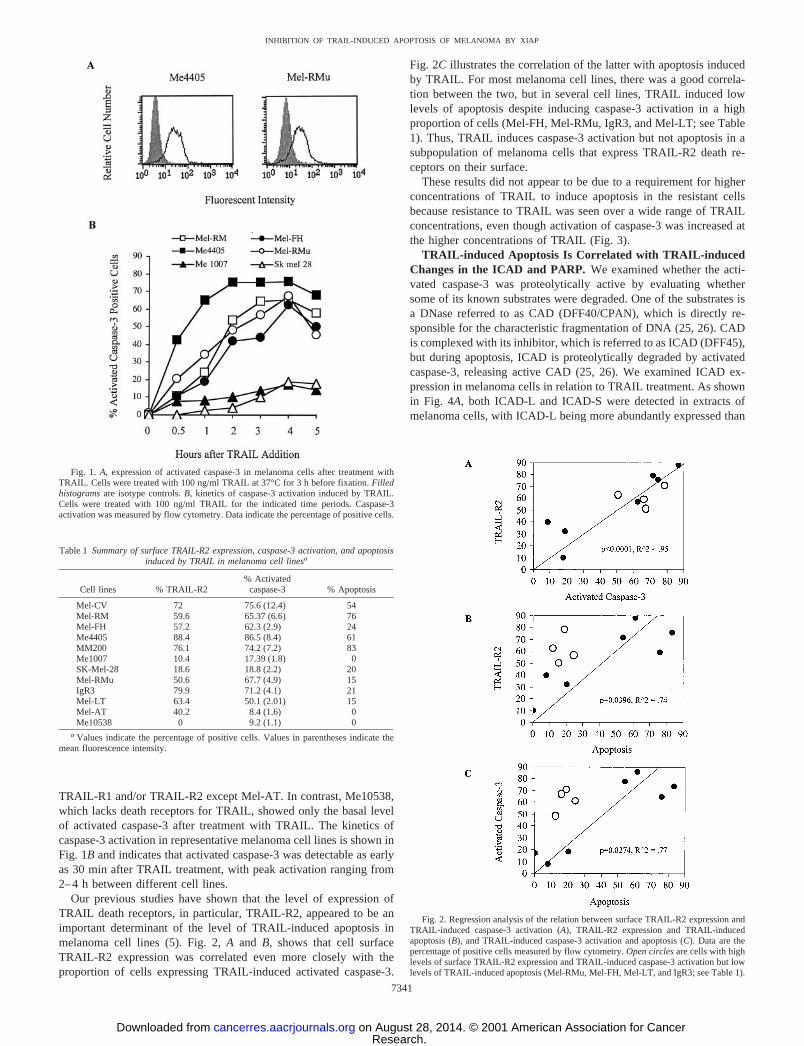
TRAIL-R1 and/or TRAIL-R2 except Mel-AT. In contrast, Me10538,which lacks death receptors for TRAIL, showed only the basal levelof activated caspase-3 after treatment with TRAIL. The kinetics ofcaspase-3 activation in representative melanoma cell lines is shown inFig. 1B and indicates that activated caspase-3 was detectable as earlyas 30 min after TRAIL treatment, with peak activation ranging from2–4 h between different cell lines.
Our previous studies have shown that the level of expression ofTRAIL death receptors, in particular, TRAIL-R2, appeared to be animportant determinant of the level of TRAIL-induced apoptosis inmelanoma cell lines (5). Fig. 2, A and B, shows that cell surfaceTRAIL-R2 expression was correlated even more closely with theproportion of cells expressing TRAIL-induced activated caspase-3.
Fig. 2C illustrates the correlation of the latter with apoptosis inducedby TRAIL. For most melanoma cell lines, there was a good correla-tion between the two, but in several cell lines, TRAIL induced lowlevels of apoptosis despite inducing caspase-3 activation in a highproportion of cells (Mel-FH, Mel-RMu, IgR3, and Mel-LT; see Table1). Thus, TRAIL induces caspase-3 activation but not apoptosis in asubpopulation of melanoma cells that express TRAIL-R2 death re-ceptors on their surface.
These results did not appear to be due to a requirement for higherconcentrations of TRAIL to induce apoptosis in the resistant cellsbecause resistance to TRAIL was seen over a wide range of TRAILconcentrations, even though activation of caspase-3 was increased atthe higher concentrations of TRAIL (Fig. 3).
TRAIL-induced Apoptosis Is Correlated with TRAIL-inducedChanges in the ICAD and PARP. We examined whether the acti-vated caspase-3 was proteolytically active by evaluating whethersome of its known substrates were degraded. One of the substrates isa DNase referred to as CAD (DFF40/CPAN), which is directly re-sponsible for the characteristic fragmentation of DNA (25, 26). CADis complexed with its inhibitor, which is referred to as ICAD (DFF45),but during apoptosis, ICAD is proteolytically degraded by activatedcaspase-3, releasing active CAD (25, 26). We examined ICAD ex-pression in melanoma cells in relation to TRAIL treatment. As shownin Fig. 4A, both ICAD-L and ICAD-S were detected in extracts ofmelanoma cells, with ICAD-L being more abundantly expressed than
Fig. 2. Regression analysis of the relation between surface TRAIL-R2 expression andTRAIL-induced caspase-3 activation (A), TRAIL-R2 expression and TRAIL-inducedapoptosis (B), and TRAIL-induced caspase-3 activation and apoptosis (C). Data are thepercentage of positive cells measured by flow cytometry. Open circles are cells with highlevels of surface TRAIL-R2 expression and TRAIL-induced caspase-3 activation but lowlevels of TRAIL-induced apoptosis (Mel-RMu, Mel-FH, Mel-LT, and IgR3; see Table 1).
Fig. 1. A, expression of activated caspase-3 in melanoma cells after treatment withTRAIL. Cells were treated with 100 ng/ml TRAIL at 37°C for 3 h before fixation. Filledhistograms are isotype controls. B, kinetics of caspase-3 activation induced by TRAIL.Cells were treated with 100 ng/ml TRAIL for the indicated time periods. Caspase-3activation was measured by flow cytometry. Data indicate the percentage of positive cells.
Table 1 Summary of surface TRAIL-R2 expression, caspase-3 activation, and apoptosisinduced by TRAIL in melanoma cell linesa
Cell lines % TRAIL-R2% Activated
caspase-3 % Apoptosis
Mel-CV 72 75.6 (12.4) 54Mel-RM 59.6 65.37 (6.6) 76Mel-FH 57.2 62.3 (2.9) 24Me4405 88.4 86.5 (8.4) 61MM200 76.1 74.2 (7.2) 83Me1007 10.4 17.39 (1.8) 0SK-Mel-28 18.6 18.8 (2.2) 20Mel-RMu 50.6 67.7 (4.9) 15IgR3 79.9 71.2 (4.1) 21Mel-LT 63.4 50.1 (2.01) 15Mel-AT 40.2 8.4 (1.6) 0Me10538 0 9.2 (1.1) 0
a Values indicate the percentage of positive cells. Values in parentheses indicate themean fluorescence intensity.
7341
INHIBITION OF TRAIL-INDUCED APOPTOSIS OF MELANOMA BY XIAP
Research. on August 28, 2014. © 2001 American Association for Cancercancerres.aacrjournals.org Downloaded from
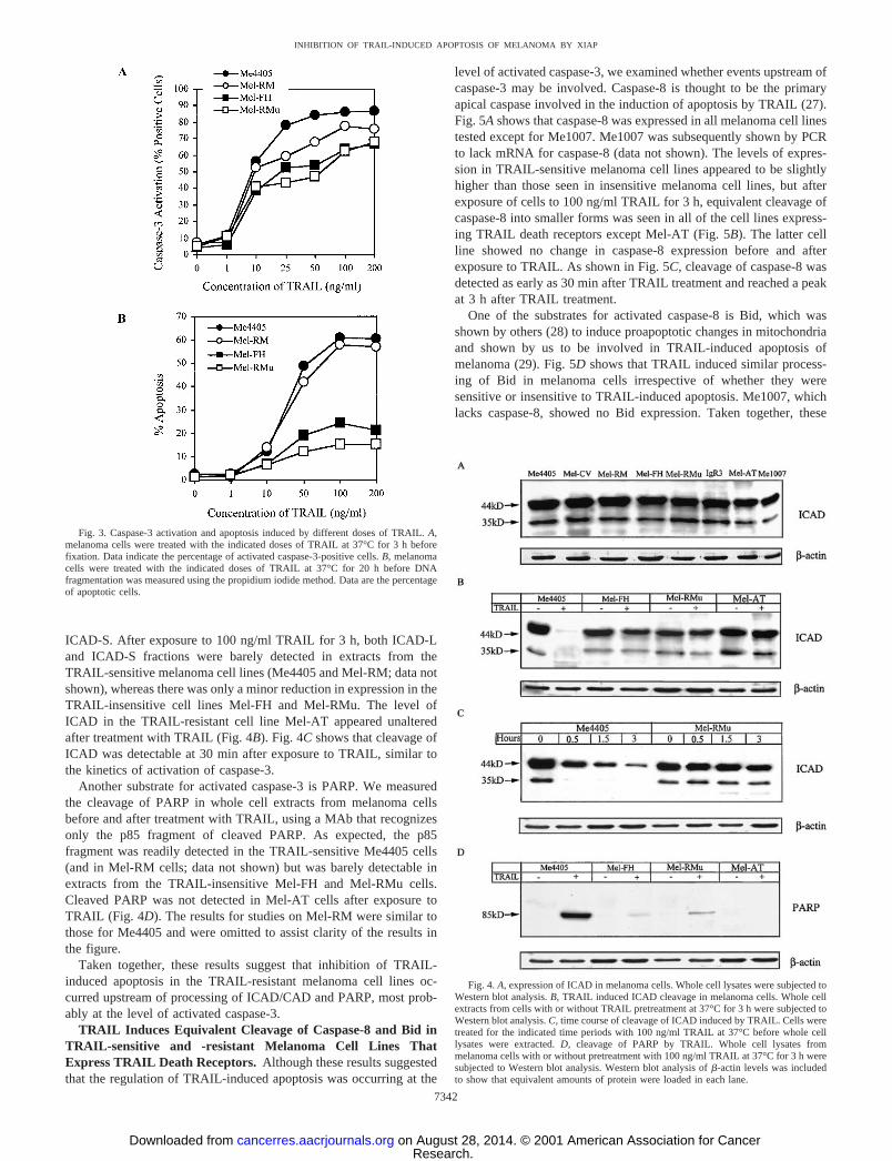
ICAD-S. After exposure to 100 ng/ml TRAIL for 3 h, both ICAD-Land ICAD-S fractions were barely detected in extracts from theTRAIL-sensitive melanoma cell lines (Me4405 and Mel-RM; data notshown), whereas there was only a minor reduction in expression in theTRAIL-insensitive cell lines Mel-FH and Mel-RMu. The level ofICAD in the TRAIL-resistant cell line Mel-AT appeared unalteredafter treatment with TRAIL (Fig. 4B). Fig. 4C shows that cleavage ofICAD was detectable at 30 min after exposure to TRAIL, similar tothe kinetics of activation of caspase-3.
Another substrate for activated caspase-3 is PARP. We measuredthe cleavage of PARP in whole cell extracts from melanoma cellsbefore and after treatment with TRAIL, using a MAb that recognizesonly the p85 fragment of cleaved PARP. As expected, the p85fragment was readily detected in the TRAIL-sensitive Me4405 cells(and in Mel-RM cells; data not shown) but was barely detectable inextracts from the TRAIL-insensitive Mel-FH and Mel-RMu cells.Cleaved PARP was not detected in Mel-AT cells after exposure toTRAIL (Fig. 4D). The results for studies on Mel-RM were similar tothose for Me4405 and were omitted to assist clarity of the results inthe figure.
Taken together, these results suggest that inhibition of TRAIL-induced apoptosis in the TRAIL-resistant melanoma cell lines oc-curred upstream of processing of ICAD/CAD and PARP, most prob-ably at the level of activated caspase-3.
TRAIL Induces Equivalent Cleavage of Caspase-8 and Bid inTRAIL-sensitive and -resistant Melanoma Cell Lines ThatExpress TRAIL Death Receptors. Although these results suggestedthat the regulation of TRAIL-induced apoptosis was occurring at the
level of activated caspase-3, we examined whether events upstream ofcaspase-3 may be involved. Caspase-8 is thought to be the primaryapical caspase involved in the induction of apoptosis by TRAIL (27).Fig. 5A shows that caspase-8 was expressed in all melanoma cell linestested except for Me1007. Me1007 was subsequently shown by PCRto lack mRNA for caspase-8 (data not shown). The levels of expres-sion in TRAIL-sensitive melanoma cell lines appeared to be slightlyhigher than those seen in insensitive melanoma cell lines, but afterexposure of cells to 100 ng/ml TRAIL for 3 h, equivalent cleavage ofcaspase-8 into smaller forms was seen in all of the cell lines express-ing TRAIL death receptors except Mel-AT (Fig. 5B). The latter cellline showed no change in caspase-8 expression before and afterexposure to TRAIL. As shown in Fig. 5C, cleavage of caspase-8 wasdetected as early as 30 min after TRAIL treatment and reached a peakat 3 h after TRAIL treatment.
One of the substrates for activated caspase-8 is Bid, which wasshown by others (28) to induce proapoptotic changes in mitochondriaand shown by us to be involved in TRAIL-induced apoptosis ofmelanoma (29). Fig. 5D shows that TRAIL induced similar process-ing of Bid in melanoma cells irrespective of whether they weresensitive or insensitive to TRAIL-induced apoptosis. Me1007, whichlacks caspase-8, showed no Bid expression. Taken together, these
Fig. 4. A, expression of ICAD in melanoma cells. Whole cell lysates were subjected toWestern blot analysis. B, TRAIL induced ICAD cleavage in melanoma cells. Whole cellextracts from cells with or without TRAIL pretreatment at 37°C for 3 h were subjected toWestern blot analysis. C, time course of cleavage of ICAD induced by TRAIL. Cells weretreated for the indicated time periods with 100 ng/ml TRAIL at 37°C before whole celllysates were extracted. D, cleavage of PARP by TRAIL. Whole cell lysates frommelanoma cells with or without pretreatment with 100 ng/ml TRAIL at 37°C for 3 h weresubjected to Western blot analysis. Western blot analysis of �-actin levels was includedto show that equivalent amounts of protein were loaded in each lane.
Fig. 3. Caspase-3 activation and apoptosis induced by different doses of TRAIL. A,melanoma cells were treated with the indicated doses of TRAIL at 37°C for 3 h beforefixation. Data indicate the percentage of activated caspase-3-positive cells. B, melanomacells were treated with the indicated doses of TRAIL at 37°C for 20 h before DNAfragmentation was measured using the propidium iodide method. Data are the percentageof apoptotic cells.
7342
INHIBITION OF TRAIL-INDUCED APOPTOSIS OF MELANOMA BY XIAP
Research. on August 28, 2014. © 2001 American Association for Cancercancerres.aacrjournals.org Downloaded from
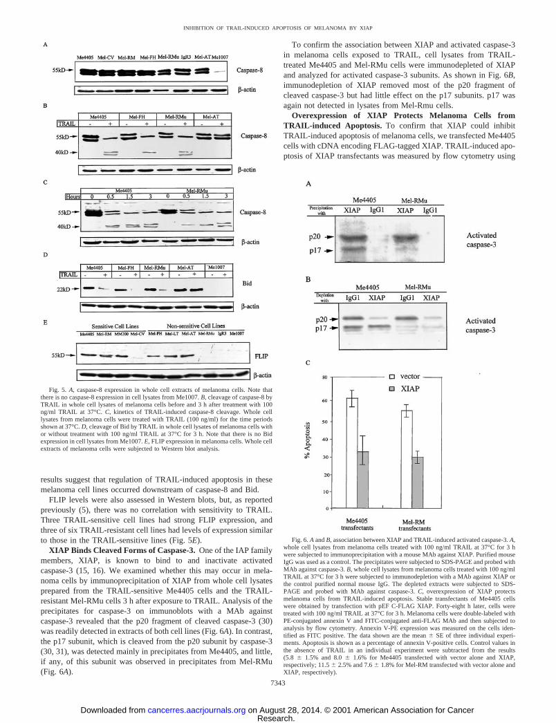
results suggest that regulation of TRAIL-induced apoptosis in thesemelanoma cell lines occurred downstream of caspase-8 and Bid.
FLIP levels were also assessed in Western blots, but, as reportedpreviously (5), there was no correlation with sensitivity to TRAIL.Three TRAIL-sensitive cell lines had strong FLIP expression, andthree of six TRAIL-resistant cell lines had levels of expression similarto those in the TRAIL-sensitive lines (Fig. 5E).
XIAP Binds Cleaved Forms of Caspase-3. One of the IAP familymembers, XIAP, is known to bind to and inactivate activatedcaspase-3 (15, 16). We examined whether this may occur in mela-noma cells by immunoprecipitation of XIAP from whole cell lysatesprepared from the TRAIL-sensitive Me4405 cells and the TRAIL-resistant Mel-RMu cells 3 h after exposure to TRAIL. Analysis of theprecipitates for caspase-3 on immunoblots with a MAb againstcaspase-3 revealed that the p20 fragment of cleaved caspase-3 (30)was readily detected in extracts of both cell lines (Fig. 6A). In contrast,the p17 subunit, which is cleaved from the p20 subunit by caspase-3(30, 31), was detected mainly in precipitates from Me4405, and little,if any, of this subunit was observed in precipitates from Mel-RMu(Fig. 6A).
To confirm the association between XIAP and activated caspase-3in melanoma cells exposed to TRAIL, cell lysates from TRAIL-treated Me4405 and Mel-RMu cells were immunodepleted of XIAPand analyzed for activated caspase-3 subunits. As shown in Fig. 6B,immunodepletion of XIAP removed most of the p20 fragment ofcleaved caspase-3 but had little effect on the p17 subunits. p17 wasagain not detected in lysates from Mel-Rmu cells.
Overexpression of XIAP Protects Melanoma Cells fromTRAIL-induced Apoptosis. To confirm that XIAP could inhibitTRAIL-induced apoptosis of melanoma cells, we transfected Me4405cells with cDNA encoding FLAG-tagged XIAP. TRAIL-induced apo-ptosis of XIAP transfectants was measured by flow cytometry using
Fig. 6. A and B, association between XIAP and TRAIL-induced activated caspase-3. A,whole cell lysates from melanoma cells treated with 100 ng/ml TRAIL at 37°C for 3 hwere subjected to immunoprecipitation with a mouse MAb against XIAP. Purified mouseIgG was used as a control. The precipitates were subjected to SDS-PAGE and probed withMAb against caspase-3. B, whole cell lysates from melanoma cells treated with 100 ng/mlTRAIL at 37°C for 3 h were subjected to immunodepletion with a MAb against XIAP orthe control purified normal mouse IgG. The depleted extracts were subjected to SDS-PAGE and probed with MAb against caspase-3. C, overexpression of XIAP protectsmelanoma cells from TRAIL-induced apoptosis. Stable transfectants of Me4405 cellswere obtained by transfection with pEF C-FLAG XIAP. Forty-eight h later, cells weretreated with 100 ng/ml TRAIL at 37°C for 3 h. Melanoma cells were double-labeled withPE-conjugated annexin V and FITC-conjugated anti-FLAG MAb and then subjected toanalysis by flow cytometry. Annexin V-PE expression was measured on the cells iden-tified as FITC positive. The data shown are the mean � SE of three individual experi-ments. Apoptosis is shown as a percentage of annexin V-positive cells. Control values inthe absence of TRAIL in an individual experiment were subtracted from the results(5.8 � 1.5% and 8.0 � 1.6% for Me4405 transfected with vector alone and XIAP,respectively; 11.5 � 2.5% and 7.6 � 1.8% for Mel-RM transfected with vector alone andXIAP, respectively).
Fig. 5. A, caspase-8 expression in whole cell extracts of melanoma cells. Note thatthere is no caspase-8 expression in cell lysates from Me1007. B, cleavage of caspase-8 byTRAIL in whole cell lysates of melanoma cells before and 3 h after treatment with 100ng/ml TRAIL at 37°C. C, kinetics of TRAIL-induced caspase-8 cleavage. Whole celllysates from melanoma cells were treated with TRAIL (100 ng/ml) for the time periodsshown at 37°C. D, cleavage of Bid by TRAIL in whole cell lysates of melanoma cells withor without treatment with 100 ng/ml TRAIL at 37°C for 3 h. Note that there is no Bidexpression in cell lysates from Me1007. E, FLIP expression in melanoma cells. Whole cellextracts of melanoma cells were subjected to Western blot analysis.
7343
INHIBITION OF TRAIL-INDUCED APOPTOSIS OF MELANOMA BY XIAP
Research. on August 28, 2014. © 2001 American Association for Cancercancerres.aacrjournals.org Downloaded from
dual staining with PE-conjugated annexin V and FITC-conjugatedanti-FLAG MAb. The levels of TRAIL-induced apoptosis in XIAPtransfectants were markedly decreased in comparison with those incells transfected with vector alone (Fig. 6C). To confirm that inhibi-tion of apoptosis in XIAP transfectants took place at the level ofcaspase-3 activation, XIAP was immunoprecipitated with anti-FLAGMAb coupled with agarose beads, and the immunoprecipitates wereexamined for the presence of activated caspase-3 by Western blottinganalysis. As before, activated caspase-3 was detected in the whole celllysate in two forms, the p20 and p17 fragments (data not shown).
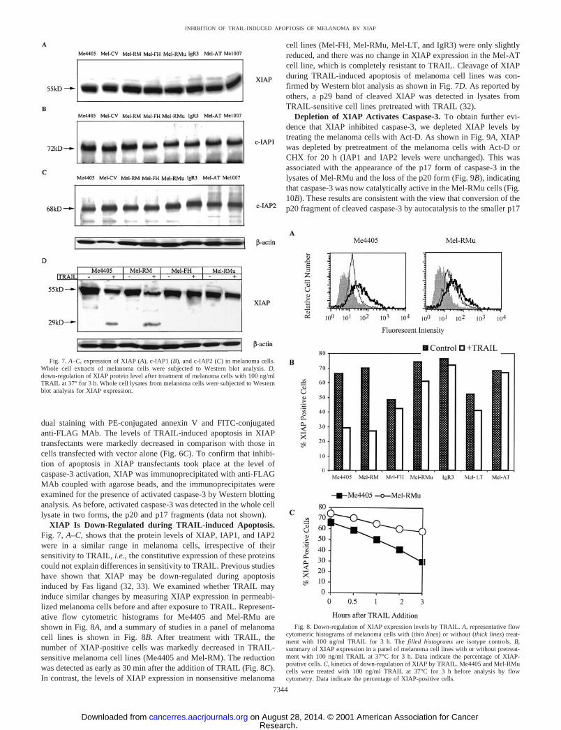
XIAP Is Down-Regulated during TRAIL-induced Apoptosis.Fig. 7, A–C, shows that the protein levels of XIAP, IAP1, and IAP2were in a similar range in melanoma cells, irrespective of theirsensitivity to TRAIL, i.e., the constitutive expression of these proteinscould not explain differences in sensitivity to TRAIL. Previous studieshave shown that XIAP may be down-regulated during apoptosisinduced by Fas ligand (32, 33). We examined whether TRAIL mayinduce similar changes by measuring XIAP expression in permeabi-lized melanoma cells before and after exposure to TRAIL. Represent-ative flow cytometric histograms for Me4405 and Mel-RMu areshown in Fig. 8A, and a summary of studies in a panel of melanomacell lines is shown in Fig. 8B. After treatment with TRAIL, thenumber of XIAP-positive cells was markedly decreased in TRAIL-sensitive melanoma cell lines (Me4405 and Mel-RM). The reductionwas detected as early as 30 min after the addition of TRAIL (Fig. 8C).In contrast, the levels of XIAP expression in nonsensitive melanoma
cell lines (Mel-FH, Mel-RMu, Mel-LT, and IgR3) were only slightlyreduced, and there was no change in XIAP expression in the Mel-ATcell line, which is completely resistant to TRAIL. Cleavage of XIAPduring TRAIL-induced apoptosis of melanoma cell lines was con-firmed by Western blot analysis as shown in Fig. 7D. As reported byothers, a p29 band of cleaved XIAP was detected in lysates fromTRAIL-sensitive cell lines pretreated with TRAIL (32).
Depletion of XIAP Activates Caspase-3. To obtain further evi-dence that XIAP inhibited caspase-3, we depleted XIAP levels bytreating the melanoma cells with Act-D. As shown in Fig. 9A, XIAPwas depleted by pretreatment of the melanoma cells with Act-D orCHX for 20 h (IAP1 and IAP2 levels were unchanged). This wasassociated with the appearance of the p17 form of caspase-3 in thelysates of Mel-RMu and the loss of the p20 form (Fig. 9B), indicatingthat caspase-3 was now catalytically active in the Mel-RMu cells (Fig.10B). These results are consistent with the view that conversion of thep20 fragment of cleaved caspase-3 by autocatalysis to the smaller p17
Fig. 8. Down-regulation of XIAP expression levels by TRAIL. A, representative flowcytometric histograms of melanoma cells with (thin lines) or without (thick lines) treat-ment with 100 ng/ml TRAIL for 3 h. The filled histograms are isotype controls. B,summary of XIAP expression in a panel of melanoma cell lines with or without pretreat-ment with 100 ng/ml TRAIL at 37°C for 3 h. Data indicate the percentage of XIAP-positive cells. C, kinetics of down-regulation of XIAP by TRAIL. Me4405 and Mel-RMucells were treated with 100 ng/ml TRAIL at 37°C for 3 h before analysis by flowcytometry. Data indicate the percentage of XIAP-positive cells.
Fig. 7. A–C, expression of XIAP (A), c-IAP1 (B), and c-IAP2 (C) in melanoma cells.Whole cell extracts of melanoma cells were subjected to Western blot analysis. D,down-regulation of XIAP protein level after treatment of melanoma cells with 100 ng/mlTRAIL at 37° for 3 h. Whole cell lysates from melanoma cells were subjected to Westernblot analysis for XIAP expression.
7344
INHIBITION OF TRAIL-INDUCED APOPTOSIS OF MELANOMA BY XIAP
Research. on August 28, 2014. © 2001 American Association for Cancercancerres.aacrjournals.org Downloaded from
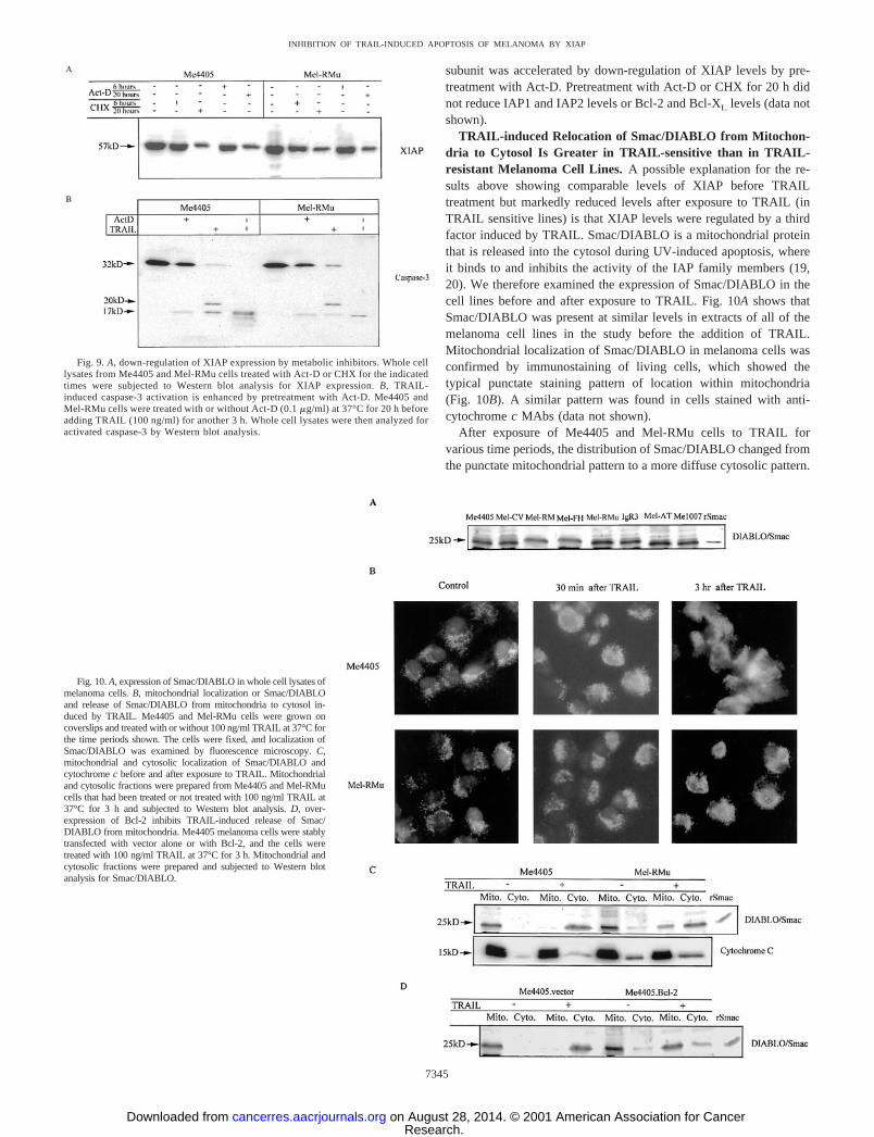
subunit was accelerated by down-regulation of XIAP levels by pre-treatment with Act-D. Pretreatment with Act-D or CHX for 20 h didnot reduce IAP1 and IAP2 levels or Bcl-2 and Bcl-XL levels (data notshown).
TRAIL-induced Relocation of Smac/DIABLO from Mitochon-dria to Cytosol Is Greater in TRAIL-sensitive than in TRAIL-resistant Melanoma Cell Lines. A possible explanation for the re-sults above showing comparable levels of XIAP before TRAILtreatment but markedly reduced levels after exposure to TRAIL (inTRAIL sensitive lines) is that XIAP levels were regulated by a thirdfactor induced by TRAIL. Smac/DIABLO is a mitochondrial proteinthat is released into the cytosol during UV-induced apoptosis, whereit binds to and inhibits the activity of the IAP family members (19,20). We therefore examined the expression of Smac/DIABLO in thecell lines before and after exposure to TRAIL. Fig. 10A shows thatSmac/DIABLO was present at similar levels in extracts of all of themelanoma cell lines in the study before the addition of TRAIL.Mitochondrial localization of Smac/DIABLO in melanoma cells wasconfirmed by immunostaining of living cells, which showed thetypical punctate staining pattern of location within mitochondria(Fig. 10B). A similar pattern was found in cells stained with anti-cytochrome c MAbs (data not shown).
After exposure of Me4405 and Mel-RMu cells to TRAIL forvarious time periods, the distribution of Smac/DIABLO changed fromthe punctate mitochondrial pattern to a more diffuse cytosolic pattern.
Fig. 9. A, down-regulation of XIAP expression by metabolic inhibitors. Whole celllysates from Me4405 and Mel-RMu cells treated with Act-D or CHX for the indicatedtimes were subjected to Western blot analysis for XIAP expression. B, TRAIL-induced caspase-3 activation is enhanced by pretreatment with Act-D. Me4405 andMel-RMu cells were treated with or without Act-D (0.1 �g/ml) at 37°C for 20 h beforeadding TRAIL (100 ng/ml) for another 3 h. Whole cell lysates were then analyzed foractivated caspase-3 by Western blot analysis.
Fig. 10. A, expression of Smac/DIABLO in whole cell lysates ofmelanoma cells. B, mitochondrial localization or Smac/DIABLOand release of Smac/DIABLO from mitochondria to cytosol in-duced by TRAIL. Me4405 and Mel-RMu cells were grown oncoverslips and treated with or without 100 ng/ml TRAIL at 37°C forthe time periods shown. The cells were fixed, and localization ofSmac/DIABLO was examined by fluorescence microscopy. C,mitochondrial and cytosolic localization of Smac/DIABLO andcytochrome c before and after exposure to TRAIL. Mitochondrialand cytosolic fractions were prepared from Me4405 and Mel-RMucells that had been treated or not treated with 100 ng/ml TRAIL at37°C for 3 h and subjected to Western blot analysis. D, over-expression of Bcl-2 inhibits TRAIL-induced release of Smac/DIABLO from mitochondria. Me4405 melanoma cells were stablytransfected with vector alone or with Bcl-2, and the cells weretreated with 100 ng/ml TRAIL at 37°C for 3 h. Mitochondrial andcytosolic fractions were prepared and subjected to Western blotanalysis for Smac/DIABLO.
7345
INHIBITION OF TRAIL-INDUCED APOPTOSIS OF MELANOMA BY XIAP
Research. on August 28, 2014. © 2001 American Association for Cancercancerres.aacrjournals.org Downloaded from
Redistribution of Smac/DIABLO from the mitochondria to the cyo-tosol was more prominent in the TRAIL-sensitive Me4405 cells thanin the TRAIL-resistant Mel-RMu cells (Fig. 10B). This was observed30 min after the addition of TRAIL and became more pronounced by2–3 h (Fig. 10B). Cytochrome c release was not detectable earlier than2 h after exposure to TRAIL (data not shown).
To further confirm the immunostaining results, we isolated mito-chondrial and cytosolic fractions from two TRAIL-sensitive cell lines,Me4405 and Mel-RM, and two TRAIL-resistant cell lines, Mel-RMuand Mel-FH. As shown in Fig. 10C, before exposure to TRAIL,Smac/DIABLO was localized exclusively in mitochondrial fractions.Three h after treatment with TRAIL, Smac/DIABLO was observed inthe cytosolic fraction, and there was a corresponding decrease in themitochondrial fraction. However, Smac/DIABLO was still retained inmitochondrial fractions from Mel-RMu and Mel-FH but not in frac-tions from Me4405 and Mel-RM, suggesting that release of Smac/DIABLO was incomplete from the mitochondria of Mel-RMu cells.The density of the bands was quantitated on a Macintosh computerusing the public domain NIH Image program, which is available onthe internet.4 When the fraction of Smac/DIABLO in the cytosol wasexpressed as a percentage of mitochondrial plus cytosol fractions, thepercentage of Smac/DIABLO released by TRAIL in Me4405 andMel-RM cells was 97% and 94%, respectively, as compared with 62%and 63%, respectively, in the TRAIL-resistant Mel-RMu and Mel-FHcells. Over the same time period, there was negligible TRAIL-mediated release of cytochrome c from the mitochondria of the twomelanoma cell lines (Fig. 10C). There was also no evidence foractivation of caspase-9 at this time, as assessed by MAbs against theactivated form of caspase-9 (data not shown).
Smac/DIABLO release was dependent on mitochondrial membranepermeability induced by TRAIL and was markedly reduced in mela-noma cells in which Bcl-2 was overexpressed. This is shown inFig. 10D, where the majority of the Smac/DIABLO was retained inthe mitochondrial fraction in cells transfected with Bcl-2, but TRAILinduced almost complete release of Smac/DIABLO in the nontrans-fected cells.
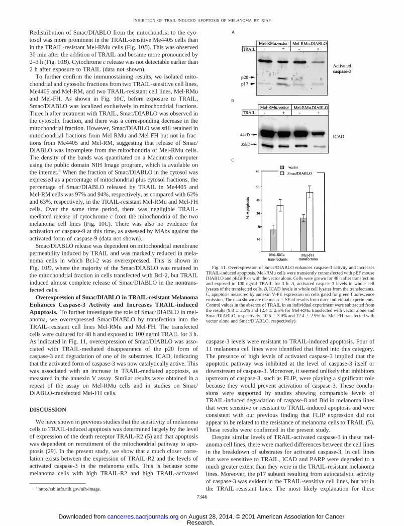
Overexpression of Smac/DIABLO in TRAIL-resistant MelanomaEnhances Caspase-3 Activity and Increases TRAIL-inducedApoptosis. To further investigate the role of Smac/DIABLO in mel-anoma, we overexpressed Smac/DIABLO by transfection into theTRAIL-resistant cell lines Mel-RMu and Mel-FH. The transfectedcells were cultured for 48 h and exposed to 100 ng/ml TRAIL for 3 h.As indicated in Fig. 11, overexpression of Smac/DIABLO was asso-ciated with TRAIL-mediated disappearance of the p20 form ofcaspase-3 and degradation of one of its substrates, ICAD, indicatingthat the activated form of caspase-3 was now catalytically active. Thiswas associated with an increase in TRAIL-mediated apoptosis, asmeasured in the annexin V assay. Similar results were obtained in arepeat of the assay on Mel-RMu cells and in studies on Smac/DIABLO-transfected Mel-FH cells.
DISCUSSION
We have shown in previous studies that the sensitivity of melanomacells to TRAIL-induced apoptosis was determined largely by the levelof expression of the death receptor TRAIL-R2 (5) and that apoptosiswas dependent on recruitment of the mitochondrial pathway to apo-ptosis (29). In the present study, we show that a much closer corre-lation exists between the expression of TRAIL-R2 and the levels ofactivated caspase-3 in the melanoma cells. This is because somemelanoma cells with high TRAIL-R2 and high TRAIL-activated
caspase-3 levels were resistant to TRAIL-induced apoptosis. Four of11 melanoma cell lines were identified that fitted into this category.The presence of high levels of activated caspase-3 implied that theapoptotic pathway was inhibited at the level of caspase-3 itself ordownstream of caspase-3. Moreover, it seemed unlikely that inhibitorsupstream of caspase-3, such as FLIP, were playing a significant rolebecause they would prevent activation of caspase-3. These conclu-sions were supported by studies showing comparable levels ofTRAIL-induced degradation of caspase-8 and Bid in melanoma linesthat were sensitive or resistant to TRAIL-induced apoptosis and wereconsistent with our previous finding that FLIP expression did notappear to be related to the resistance of melanoma cells to TRAIL (5).These results were confirmed in the present study.
Despite similar levels of TRAIL-activated caspase-3 in these mel-anoma cell lines, there were marked differences between the cell linesin the breakdown of substrates for activated caspase-3. In cell linesthat were sensitive to TRAIL, ICAD and PARP were degraded to amuch greater extent than they were in the TRAIL-resistant melanomalines. Moreover, the p17 subunit resulting from autocatalytic activityof caspase-3 was evident in the TRAIL-sensitive cell lines, but not inthe TRAIL-resistant lines. The most likely explanation for these4 http://rsb.info.nih.gov/nih-image.
Fig. 11. Overexpression of Smac/DIABLO enhances caspase-3 activity and increasesTRAIL-induced apoptosis. Mel-RMu cells were transiently cotransfected with pEF mouseDIABLO and pEGFP or with the vector alone. Cells were grown for 48 h after transfectionand exposed to 100 ng/ml TRAIL for 3 h. A, activated caspase-3 levels in whole celllysates of the transfected cells. B, ICAD levels in whole cell lysates from the transfectants.C, apoptosis measured by annexin V-PE expression on cells gated for green fluorescenceemission. The data shown are the mean � SE of results from three individual experiments.Control values in the absence of TRAIL in an individual experiment were subtracted fromthe results (9.8 � 2.5% and 12.4 � 2.6% for Mel-RMu transfected with vector alone andSmac/DIABLO, respectively; 10.6 � 3.0% and 12.4 � 2.9% for Mel-FH transfected withvector alone and Smac/DIABLO, respectively).
7346
INHIBITION OF TRAIL-INDUCED APOPTOSIS OF MELANOMA BY XIAP
Research. on August 28, 2014. © 2001 American Association for Cancercancerres.aacrjournals.org Downloaded from

results was that activated caspase-3 was inhibited by one of the IAPfamily members, which was reported by others to bind to and inhibitthe activity of caspase-3, as well as caspase-7 and caspase-9 (15, 16).This was supported by studies showing coimmunoprecipitation ofXIAP and activated caspase-3. It was particularly noticeable that onlythe small subunit of caspase-3 (p20) was immunoprecipitated from theTRAIL-resistant cell line, whereas immunoprecipitates from theTRAIL-sensitive cell line also contained the p17 unit, which resultswhen active caspase-3 cleaves its own prodomain (30, 31). Moreover,overexpression of XIAP in TRAIL-sensitive cell lines markedly sup-pressed TRAIL-mediated apoptosis.
To obtain further support for the role of XIAP as an inhibitor ofcaspase-3, we used two approaches to reduce its activity. One was tooverexpress Smac/DIABLO in TRAIL-resistant melanoma cell linesbecause studies by others have shown that Smac/DIABLO specifi-cally binds to and inactivates XIAP and other members of the IAPfamily (19, 20). These experiments showed that there was a markedincrease in TRAIL-induced apoptosis in the transfected cell lines, andthis was associated with an increase in autocatalytic activity bycaspase-3 and breakdown of one of its substrates, PARP. The secondapproach was to deplete XIAP levels using the inhibitor of transcrip-tion Act-D or the protein synthesis inhibitor CHX (34). Pretreatmentof TRAIL-resistant cell lines with these agents depleted XIAP (but notIAP1 and IAP2 levels or Bcl-2 and Bcl-XL) and resulted in an increaseof TRAIL-induced caspase-3 autocatalytic activity and apoptosis.These studies suggested that XIAP was mainly responsible for inhi-bition of caspase-3 but do not exclude the involvement of other IAPfamily members, particularly ML-IAP, which was not measured in thepresent study (35).
Although these studies clearly identified XIAP as a significantinhibitor of TRAIL-induced apoptosis, they did not explain the dif-ferences between cell lines that were sensitive to or resistant toTRAIL. This question was particularly relevant because Western blotstudies did not show marked differences in the constitutive levels ofXIAP (and IAP1 and IAP2) between the various melanoma cell lines.However, studies after exposure to TRAIL indicated that there was amarked decrease in XIAP levels in the TRAIL-sensitive cell lines, butnot in the TRAIL-resistant cell lines. We examined whether this maybe due to TRAIL-mediated release of Smac/DIABLO from mitochon-dria and found that TRAIL induced release of a much higher propor-tion of Smac/DIABLO from mitochondria of TRAIL-sensitive cellscompared with that seen in TRAIL-resistant melanoma cells. Further-more, the kinetics of Smac/DIABLO release was more consistent withthe kinetics of down-regulation of XIAP, both of which were apparentby 30 min, whereas cytochrome c was not detectable until 2 h afterthe addition of TRAIL and was maximal at approximately 6 h. Asexpected, overexpression of Bcl-2 was associated with inhibition ofSmac/DIABLO release, decreased autocatalytic activity of caspase-3,and TRAIL-mediated apoptosis.
These results are therefore consistent with the view that TRAILmediates apoptosis of melanoma by release of Smac/DIABLO frommitochondria, which in turn blocks the XIAP-mediated inhibition ofthe effector caspase-3. The mechanism involved in reduction of XIAPlevels may not be Smac/DIABLO itself but could be caspase-3 freedof the inhibitory effect of XIAP due to binding of Smac/DIABLO toXIAP (32, 33). It is conceivable that Smac/DIABLO release wouldincrease the mitochondrial pathway to apoptosis by reducing XIAP-mediated inhibition of caspase-9. We believe that this explanation isless likely because of the kinetics of cytochrome c release discussedabove and because the melanoma cells under study had high levels ofactivated caspase-3 induced by TRAIL. This would be unlikely tooccur if there was significant inhibition of caspase-9 by XIAP or otherIAP family members.
These studies indicate that TRAIL induces high levels of activatedcaspase-3 in approximately 36% of the melanoma cell lines in thepresent study. Whereas the results provide an explanation for theresistance of melanoma lines to TRAIL, they do not explain whyTRAIL should induce a higher proportion of Smac/DIABLO releasefrom mitochondria of some melanomas compared with others. Thisdoes not appear to be due to events upstream of the mitochondria, andcloser examination of the role of the Bcl-2 family proteins is nowneeded to see whether their activity correlates with Smac/DIABLOrelease. It is also apparent that a variety of defects (other thaninhibition by XIAP) exist in individual melanoma cell lines that makethem resistant to TRAIL. This includes complete loss of receptors (5)and, as shown above for Me1007, loss of caspase-8. The mechanismof resistance of Mel-AT remains under study. Loss of caspase-8 wasreported to be a common basis for resistance of neuroblastoma cells toTRAIL (36, 37).
ACKNOWLEDGMENTS
We thank Dr. David Vaux for supply of plasmid vectors for XIAP, DIA-BLO, Bcl-2, and Ab against DIABLO. We also thank Dr. Xiao Dong Wang forAb against Smac and recombinant Smac.
REFERENCES
1. Wiley, S. R., Schooley, K., Smolak, P. J., Din, W. S., Huang, C-P., Nicholl, J. K.,Sutherland, G. R., Smith, T. D., Rauch, C., Smith, C. A., and Goodwin, R. G.Identification and characterization of a new member of the TNF family that inducesapoptosis. Immunity, 3: 673–682, 1995.
2. Pitti, R. M., Marsters, S. A., Ruppert, S., Donahue, C. J., Moore, A., and Ashkenazi,A. Induction of apoptosis by Apo-2 ligand, a new member of the tumor necrosis factorcytokine family. J. Biol. Chem., 271: 12687–12690, 1996.
3. Griffith, T. S., and Lynch, D. H. TRAIL: a molecule with multiple receptors andcontrol mechanisms. Curr. Opin. Immunol., 10: 559–563, 1998.
4. Thomas, W. D., and Hersey, P. TNF-related apoptosis-inducing ligand (TRAIL)induces apoptosis in Fas ligand-resistant melanoma cells and mediates CD4 T cellkilling of target cells. J. Immunol., 161: 2195–2200, 1998.
5. Zhang, X. D., Franco, A., Myers, K., Gray, C., Nguyen, T., and Hersey, P. Relationof TNF-related apoptosis-inducing ligand (TRAIL) receptor and FLICE-inhibitoryprotein expression to TRAIL-induced apoptosis of melanoma. Cancer Res., 59:2747–2753, 1999.
6. Griffith, T. S., Chin, W. A., Jackson, G. C., Lynch, D. H., and Kubin, M. Z.Intracellular regulation of TRAIL-induced apoptosis in human melanoma cells.J. Immunol., 161: 2833–2840, 1998.
7. Rieger, J., Naumann, U., Glaser, T., Ashkenazi, A., and Weller, M. APO2 ligand: anovel lethal weapon against malignant glioma? FEBS Lett., 427: 124–128, 1998.
8. Phillips, T. A., Ni, J., Pan, G., Ruben, S. M., Wei, Y-F., Pace, J. L., and Hunt, J. S.TRAIL (Apo-2L) and TRAIL receptors in human placentas: implications for immuneprivilege. J. Immunol., 162: 6053–6059, 1999.
9. Zhang, X. D., Nguyen, T., Thomas, W. D., Sanders, J. E., and Hersey, P. Mechanismsof resistance of normal cells to TRAIL induced apoptosis vary between different celltypes. FEBS Lett., 482: 193–199, 2000.
10. Jo, M., Kim, T-H., Seol, D-W., Esplen, J. E., Dorko, K., Billiar, T. R., and Strom,S. C. Apoptosis induced in normal human hepatocytes by tumor necrosis factor-related apoptosis-inducing ligand. Nat. Med., 6: 564–567, 2000.
11. Walczak, H., Miller, R. E., Ariail, K., Gliniak, B., Griffith, T. S., Kubin, M., Chin,W., Jones, J., Woodward, A., Le, T., Smith, C., Smolak, P., Goodwin, R. G., Rauch,C. T., Schuh, J. C. L., and Lynch, D. H. Tumoricidal activity of tumor necrosisfactor-related apoptosis-inducing ligand in vivo. Nat. Med., 5: 157–163, 1999.
12. Ashkenazi, A., Pai, R. C., Fong, S., Leung, S., Lawrence, D. A., Marsters, S. A.,Blackie, C., Chang, L., McMurtrey, A. E., Hebert, A., DeForge, L., Koumenis, I. L.,Lewis, D., Harris, L., Bussiere, J., Koeppen, H., Shahrokh, Z., and Schwall, R. H.Safety and antitumor activity of recombinant soluble Apo2 ligand. J. Clin. Investig.,104: 155–162, 1999.
13. Gliniak, B., and Le, T. Tumor necrosis factor-related apoptosis-inducing ligand’santitumor activity in vivo is enhanced by the chemotherapeutic agent CPT-11. CancerRes., 59: 6153–6158, 1999.
14. Gura, T. How TRAIL kills cancer cells, but not normal cells. Cancer Res., 277: 768,1997.
15. Deveraux, Q. L., and Reed, J. C. IAP family proteins: suppressors of apoptosis. GenesDev., 13: 239–252, 1999.
16. Deveraux, Q. L., Roy, N., Stennicke, H. R., Van Arsdale, T., Zhou, Q., Srinivasula,S. M., Alnemri, E. S., Salvesen, G. S., and Reed, J. C. IAPs block apoptotic eventsinduced by caspase-8 and cytochrome c by direct inhibition of distinct caspases.EMBO J., 17: 2215–2223, 1998.
17. Irmler, M., Thome, M., Hahne, M., Schneider, P., Hofmann, K., Steiner, V., Bodmer,J-L., Schroter, M., Burns, K., Mattmann, C., Rimoldi, D., French, L. E., and Tschopp,
7347
INHIBITION OF TRAIL-INDUCED APOPTOSIS OF MELANOMA BY XIAP
Research. on August 28, 2014. © 2001 American Association for Cancercancerres.aacrjournals.org Downloaded from
J. Inhibition of death receptor signals by cellular FLIP. Nature (Lond.), 388: 190–195, 1997.
18. French, L. E., and Tschopp, J. Inhibition of death receptor signaling by FLICE-inhibitory protein as a mechanism for immune escape of tumors. J. Exp. Med., 190:891–893, 1999.
19. Du, C., Fang, M., Li, Y., Li, L., and Wang, X. Smac, a mitochondrial protein thatpromotes cytochrome c-dependent caspase activation by eliminating IAP inhibition.Cell, 102: 33–42, 2000.
20. Verhagen, A. M., Ekert, P. G., Pakusch, M., Silke, J., Connolly, L. M., Reid, G. E.,Moritz, R., Simpson, R. J., and Vaux, D. L. Identification of DIABLO, a mammalianprotein that promotes apoptosis by binding to and antagonizing IAP proteins. Cell,102: 43–53, 2000.
21. Griffith, T. S., Rauch, C., Smolak, P. J., Waugh, J. Y., Boiani, N., Lynch, D. H.,Smith, C. A., Goodwin, R. G., and Kubin, M. Z. Functional analysis of TRAILreceptors using monoclonal antibodies. J. Immunol., 162: 2597–2605, 1999.
22. Sabol, S. L., Li, R., Lee, T. Y., and Abdul-Khalek, R. Inhibition of apoptosis-associated DNA fragmentation activity in nonapoptotic cells: the role of DNAfragmentation factor-45 (DFF45/ICAD). Biochem. Biophys. Res. Commun., 253:151–158, 1998.
23. Sharrow, C. O. Analysis of flow cytometry data. In: J. E. Coligan, A. M. Kruisbeek,D. H. Margulies, E. M. Shevach, and W. Strober (eds.), Current Protocols inImmunology. New York: John Wiley & Sons, 1996.
24. Liu, X., Kim, C. N., Yang, J., Jemmerson, R., and Wang, X. Induction of apoptoticprogram in cell-free extracts: requirement for dATP and cytochrome c. Cell, 86:145–157, 1996.
25. Enari, M., Sakahira, H., Yokoyama, H., Okawa, K., Iwamatsu, A., and Nagata, S. Acaspase-activated DNase that degrades DNA during apoptosis, and its inhibitorICAD. Nature (Lond.), 391: 43–50, 1998.
26. Sakahira, H., Enari, M., and Nagata, S. Cleavage of CAD inhibitor in CAD activationand DNA degradation during apoptosis. Nature (Lond.), 391: 96–99, 1998.
27. Peter, M. E. The TRAIL discussion: it is FADD and caspase-8! Cell Death Differ., 7:759–760, 2000.
28. Yamada, H., Tada-Oikawa, S., Uchida, A., and Kawanishi, S. TRAIL causes cleavageof Bid by caspase-8 and loss of mitochondrial membrane potential resulting inapoptosis in BJAB cells. Biochem. Biophys. Res. Commun., 265: 130–133, 1999.
29. Thomas, W. D., Zhang, X. D., Franco, A. V., Nguyen, T., and Hersey, P. TNF-relatedapoptosis-inducing ligand-induced apoptosis of melanoma is associated with changesin mitochondrial membrane potential and perinuclear clustering of mitochondria.J. Immunol., 165: 5612–5620, 2000.
30. Fernandes-Alnemri, T., Armstrong, R. C., Krebs, J., Srinivasula, S. M., Wang, L.,Bullrich, F., Fritz, L. C., Trapani, J. A., Tomaselli, K. J., Litwack, G., and Alnemri,E. S. In vitro activation of CPP32 and Mch3 by Mch4, a novel human apoptoticcystein protease containing two FADD-like domains. Proc. Natl. Acad. Sci. USA, 93:7464–7469, 1996.
31. Nicholson, D. W., and Thornberry, N. A. Caspases: killer proteases. Trends Biochem.Sci., 22: 299–306, 1997.
32. Johnson, D. E., Gastman, B. R., Wieckowski, E., Wang, G. Q., Amoscato, A., Delach,S. M., and Rabinowich, H. Inhibitor of apoptosis protein hILP undergoes caspase-mediated cleavage during T lymphocyte apoptosis. Cancer Res., 60: 1818–1823,2000.
33. Deveraux, Q. L., Leo, E., Stennicke, H. R., Welsh, K., Salvesen, G. S., and Reed, J. C.Cleavage of human inhibitor of apoptosis protein XIAP results in fragments withdistinct specificities for caspases. EMBO J., 18: 5242–5251, 1999.
34. Fulda, S., Meyer, E., and Debatin, K. M. Metabolic inhibitors sensitize for CD95(APO-1/Fas)-induced apoptosis by down-regulating Fas-associated death domain-likeinterleukin 1-converting enzyme inhibitory protein expression. Cancer Res., 60:3947–3956, 2000.
35. Vucic, D., Stennicke, H. R., Pisabarro, M. T., Salvesen, G. S., and Dixit, V. M.ML-IAP, a novel inhibitor of apoptosis that is preferentially expressed in humanmelanomas. Curr. Biol., 10: 1359–1366, 2000.
36. Hopkins-Donaldson, S., Bodmer, J. L., Bourloud, K. B., Brognara, C. B., Tschopp, J.,and Gross, N. Loss of caspase-8 expression in neuroblastoma is related to malignancyand resistance to TRAIL-induced apoptosis. Med. Pediatr. Oncol., 35: 608–611,2000.
37. Eggert, A., Grotzer, M. A., Zuzak, T. J., Wiewrodt, B. R., Ho, R., Ikegaki, N., andBrodeur, G. M. Resistance to tumor necrosis factor-related apoptosis-inducing ligand(TRAIL)-induced apoptosis in neuroblastoma cells correlates with a loss of caspase-8expression. Cancer Res., 61: 1314–1319, 2001.
7348
INHIBITION OF TRAIL-INDUCED APOPTOSIS OF MELANOMA BY XIAP
Research. on August 28, 2014. © 2001 American Association for Cancercancerres.aacrjournals.org Downloaded from
Copyright © 2022 FDOKUMEN