Inhibiting HIV1 integrase by shifting its oligomerization equilibrium
Upload
independentCategory
view
0download
0
Journal of Cellular Biochemistry 58:248-259 (1 995)
Tumor Necrosis Factor-a Induces Changes in the Phosphorylation, Cellular Localization, and Oligomerization of Human hsp27, a Stress Protein That Confers Cellular Resistance to This Cytokine Patrick Mehlen, Anne Mehlen, Dominique Cuillet, Xavier Preville, and Andre-Patrick Arrigo
Laboratoire du Stress Cellulaire (P.M., D.G., X.P., A.-P.A.) and Laboratoire de Genetique des Thalassemies (A.M.), Centre de Genetique Moleculaire et Cellulaire, CNRS UMR-106, Universite Claude Bernard, LYON-I, 69622 Villeurbanne, France
Abstract The stress protein hsp27 is constitutively expressed in several human cells and shows a rapid phosphorylation following treatment with tumor necrosis factor-a (TNF-a). hsp27 usually displays native molecular mass ranging from 100 to 700 kDa. Here, we have analyzed the TNF-a-mediated changes in the phosphorylation, cellular localization, and structural organization of hsp27 in HeLa cells. We report that the TNF-a-mediated hsp27 phosphorylation is a long-lasting phenomenon that correlates with the cytostatic effect of this cytokine. Following TNF-a treatment, the rapid phosphorylation of hsp2 7 occurred concomitantly with complex changes in the intracellular distribution and structural organization of this protein. This resulted in the quantitative redistribution of hsp27 toward the soluble phase of the cytoplasm. In addition, during the first 2 h of TNF-a treatment, a transient increase in the native molecular mass of most hsp27 molecules ( I 700 kDa) occurred. Then, by 4 h of TNF-a treatment, the native size of this stress protein drastically regressed ( < 200 kDa). During this phenomenon, the phosphorylated isoforms of hsp27 remained concentrated in the small or medium-sized oligomers ( < 300 kDa) of this protein. We also analyzed the properties of human hsp27 in transfected murine L929 cell lines that constitutively express this protein. In these cells, TNF-a induced modifications in the phosphorylation, intracellular distribution, and oligomerization of human hsp27 similar to those observed in HeLa cells. Moreover, the expression of hsp27 in L929 cells was found to correlate with a reduced cytotoxicity of this cytokine. Hence, the complex changes in the phosphorylation, intracellular locale and structural organization of human hsp27 may be related to the protective activity of this protein against the deleterious effects induced by TNF-a. c 1995 Wtley-Liss, Inc
Key words: heat-shock protein, small stress protein, tumor necrosis factor-a, protein phosphorylation, oligomerization
In HeLa cells, as well as in several other human cells, low levels of the small stress pro- tein hsp27 are constitutively expressed "over, 1984; Lindquist, 1986; Morimoto et al., 1990; Ciocca et al., 1993; Arrigo and Landry, 19941. This protein, which is part of the heat-shock protein (hsp) family, has sequences similarities with a-crystallin [Ingolia and Craig, 19821. A similarity with an inhibitor of actin polymeriza- tion has also been reported [Miron et al., 19911.
Received April 25, 1994; revised September 26, 1994; ac- cepted October 21, 1994. Address reprint requests to Dr. Andre-Patrick Arrigo, Labo- ratoire du Stress Cellulaire, CNRS UMR 106, Centre de Genetique Moleculaire, Universite Claude Bernard, Lyon-I, 43, Bd du 11 Novembre 1918, 69622 Villeurbanne Cedex, France.
&. 1995 Wiley-Liss, Inc.
hsp27 and a-crystallin are oligomeric proteins [Arrigo et al., 1988; Seizen et al., 19781 that display complex structural organizations. De- pending on the physiology of the cell, the degree of hsp27 oligomerization varies, leading to the formation of structures with native molecular masses ranging from 100 to 700 kDa [Arrigo and Welch, 1987; Arrigo et al., 19881. For example, in starved cells, hsp27 forms small and dephos- phorylated aggregates that reorganize in large structures following serum stimulation [Mehlen and Arrigo, 19941. Moreover, during heat shock, hsp27 drastically increases its native molecular mass ( > 1,000 kDa) [Arrigo et al., 1988; Arrigo, 1990bl.
Another feature of hsp27 concerns its intense phosphorylation in different cellular conditions. Surprisingly, enhanced hsp27 phosphorylation
TNF-a Induces Changes in hsp27 Structural Organization 249
has been observed in conditions that have oppo- site effects on cell growth. These include inhibi- tory stimuli, such as heat shock, oxidative stress, tumor necrosis factor-a (TNF-a), interleukin-1 (IL-11, and differentiating agents as well as stimuli that induce the reverse effect and stimu- late cell growth, such as serum and mitogens [reviewed in Arrigo and Landry, 19941. Interest- ingly, thermal stress and mitogens induce the phosphorylation of similar hsp27 serine resi- dues [Gaestel et al., 1991; Landry et al., 19921, suggesting the involvement of the same kinase (hsp27 kinase) that may be activated by differ- ent signal transduction mechanisms [Landry et al., 1992; Guesdon et al., 19931.
TNF-a is a monocyteimacrophage-derived pro- inflammatory cytokine that causes in vivo solid tumors necrosis, fever, endothelial cell resorp- tion, and the acute-phase response [Beutler and Cerami, 19891. TNF-a does not induce the syn- thesis of stress proteins in spite of its cytostatici cytotoxic effect, but stimulates the transient phosphorylation of 28-kDa proteins [Robaye et al., 1989; Schutze et al., 1989; Kaur and Sak- latvala, 1988; Kaur et al., 19891 identified as hsp27 isoforms [Arrigo, 1990al. This phosphor- ylation is dependent on TNF-a concentration and has been observed in most mammalian cells that are responsive to this cytokine. In some cells, however, no phosphorylation of hsp27 has been reported, such as in the TNF-a hypersensi- tive L929 mouse fibroblasts [Robaye et al., 19891. In this particular case, this phenomenon is due to the absence of constitutively expressed endog- enous hsp27 in these cells [Mehlen et al., 19951. The kinase that phosphorylates hsp27 in TNF-a- treated cells is still unknown. On the other hand, it is possible that part of this phenomenon results in the inactivation of specific phosphata- ses [Gaestel et al., 1992; Guy et al., 19931. By using cells that overexpress the detoxifiant en- zyme glutathione peroxidase, we have recently observed that the increased levels of reactive oxygen intermediates (ROI) that accumulate in TNF-a-treated cells are probably key elements in the regulation of hsp27 phosphorylation (Mehlen et al., submitted).
It has been reported that a mild thermal stress that strongly induces hsps synthesis transiently enhanced the cellular resistance to various types of stress, including heat shock and the cytotoxic- ity mediated by TNF-a [Gerner and Schneider, 1975; Jaattela, 1989; Tomasovic et al., 1989; Kusher et al., 19901. This protective effect was
shown to be due, at least in part, to the expres- sion of the major stress protein, hsp70 [Jaattela et al., 1992; Jaattela, 19931. We and others have shown that small hsps were also able to enhance the cellular resistance to heat shock and drugs that increase intracellular ROI levels [Landry et al., 1989; Huot et al., 1991; Rollet et al., 1992; Lavoie et al., 1993a; Mehlen et al., 19931. We also observed that murine L929, NIH 3T3-T- antigen, and monkey COS cells transiently ex- pressing hsp27 from Drosophila were more resis- tant to TNF-a cytotoxicity (unpublished data).
We report here that in human HeLa cells, TNF-a induced drastic changes in the intracellu- lar distribution and oligomerization of the endog- enous, and constitutively expressed, hsp27. Since TNF-a in HeLa cells has only a cytostatic effect, the analysis was also performed in TNF-sensi- tive murine L929 cells following their stable transfection with a vector carrying human hsp27 gene. In these cells, we observed that the TNF-a- induced changes in hsp27 properties were simi- lar to those observed in HeLa cells. Moreover, in L929 cells, the expression of human hsp27 corre- lated with an enhanced resistance toward the cytotoxicity of this cytokine. Our results are discussed in view of a possible regulation of hsp27 protective activity against TNF-a by the phosphorylation, cellular redistribution, and structural reorganization of this stress protein.
MATERIALS AND METHODS Cell Cultures
HeLa cells and mouse tumorigenic L929 fibro- blasts were grown at 37°C in Dulbecco modified Eagle’s medium containing 5% bovine fetal se- rum (Gibco BRL) in the presence of 5% C02. L929 cell lines constitutively expressing human hsp27 were obtained following transfection with a psvK3 expression vector (Pharmacia, Uppsala, Sweden) containing human hsp27 gene under the control of the early promoter of SV40 virus (Briolay et al., in preparation). Co-transfection with SP65 plasmid bearing the hygromycin B resistance gene was used for selection. Hygromy- cin B-resistant clones were isolated, grown in the presence of the antibiotic, and screened for the expression of hsp27. Details of the transfec- tion and selection procedure will be published elsewhere [Melhen et al., 19951.
Reagents
TNF-a (murine recombinant) was from Boeh- ringer, Mannheim. Hygromycin B, actinomycin
250 Mehlen et al.
D, and crystal violet were from Sigma (St. Louis, MO). Human recombinant hsp27 polypeptide was from Stressgen-Tebu (France). The specific- ity of anti-hsp27 serum was already described [Arrigo et al., 19881. [3Hlthymidine (88 Ci/ mmol) was from Amersham Corp. (UK).
Cell Fractionation
Cells were washed with phosphate-buffered saline (PBS) (NaCl/Pi: 137 mM NaC1; 2.7 mM KCl; 8 mM Na2HP04; 1.5 mM KH2P04, pH 7.4) before being scraped from 35-mm Falcon dishes. Cells were pelleted at 2,OOOg for 5 min and lysed by vortexing and douncing at 4°C in a buffer containing 10 mM Tris, pH 7.4; 1 mM MgC12, 10 mM NaCl and 0.1% Triton x 100. Lysis was also performed in the absence of detergent. The ly- sates were first centrifuged at 2,OOOg for 10 min (P2 pellet). The resulting supernatants were then centrifuged at 20,OOOg for 10 min (P20 pellet and supernatant S). The different subcel- lular fractions were then boiled in Laemmli SDS buffer.
Gel Filtration Analysis
Cells were washed as above, and the lysates were centrifuged at 20,OOOg for 10 min. The supernatants were then applied to a Sepharose 6B gel filtration column (1 cm x 100 cm) (Phar- macia, Uppsala, Sweden), equilibrated, and de- veloped in lysis buffer devoid of Triton ~ 1 0 0 . The fractions eluting of the column were ana- lyzed by one- and/or two-dimensional Western blots. Molecular mass markers used to calibrate the gel filtration column included blue dextran ( > 2,000,000 Da), thyroglobulin (669,000 Da), apoferritin (440,000 Da), p-amylase (200,000 Da), and carbonic anhydrase (29,000 Da).
Gel Electrophoresis and lrnmunoblotting
One- or two-dimensional immunoblots using anti-hsp27 were performed as already described [Arrigo et al., 19881. The immunoblots were revealed with the ECL kit from Amersham Corp. ( UK) .
Indirect lmmunofluorescence Analysis
HeLa or L929 cells growing on glass cover slips were fixed directly with cold methanol (-20°C) for 90 sec. Staining with anti-hsp27 antibody was performed as previously described [Arrigo et al., 1988; Arrigo, 1990bl using goat antirabbit coupled with fluorescein isothiocya-
nate (FITC) (Organon Teknica-Cappel, Fresnes, France) as second antibody. The stained cells were then examined and photographed with a Zeiss Axioskop photomicroscope. Fluorescent im- ages were recorded onto Tri-X Pan (Eastman Kodak Co.).
Analysis of the Cytostatic Effect of TNF-a in HeLa Cells
HeLa cells were pulse-labeled for 15 min with 10 p,Ci/ml [3Hlthymidine. They were then washed with PBS and lysed in sodium dodecyl sulfate (SDS) as previously described [Spector et al., 1993,19941. Incorporated L3H1thymidine was counted on a Beckman scintillation counter LS6000 SC (Beckman, USA) after trichloroace- tic acid precipitation and filtration on glass mi- crofiber filters (GFC filters, Whatman).
Assay for TNF-a Cytotoxicity in Murine L929 Cells
L929 cell lines (lo4 per well) were grown in 96-well microtiter plates for 24 h before being analyzed for their resistance to TNF-a. Twofold serial dilutions of TNF-a were added to the cells. Incubation was for 24 h. Supernatants were discarded, and the remaining viable cells were stained for 15 min with 0.5% crystal violet in 50% methanol. Microtiter plates were rinsed and dried. A medium containing 0.1 M citrate sodium pH 5.4; 20% methanol was then added to solubilize the stained cells. The absorbance of each well was read at 570 nm with an MR5000 microelisa reader (Dynatech Laboratories, Chan- tilly, VA). Percentage of cell survival was de- fined as the relative absorbance of sample ver- sus control untreated cells.
RESULTS TNF-a Induces Changes in hsp27 lsoforms in
HeLa and Transfected Murine L929 Cells That Constitutively Express This Human
Stress Protein
The TNF-a-mediated changes in the isoform composition of human hsp27 were analyzed in HeLa cells and in transfected murine L929 cell lines that constitutively express this protein. As described in Materials and Methods, transfec- tion of L929 cells was performed with a psvK3 vector carrying the human hsp27 gene under the control of the constitutive early SV40 pro- moter. Several hygromycine-resistant clones were obtained that displayed various levels of
TNF-cx Induces Changes in hsp27 Structural Organization 251
hsp27 expression. Control experiments were per- formed that showed that the expression of hu- man hsp27 did not reduce the number of TNF-a receptors and/or alter the binding capacity of this cytokine to its receptors. Moreover, no changes in the pattern of protein synthesis, par- ticularly at the level of the endogenous murine hsps, were observed in L929 cells expressing human hsp27 [Mehlen et al., 19951.
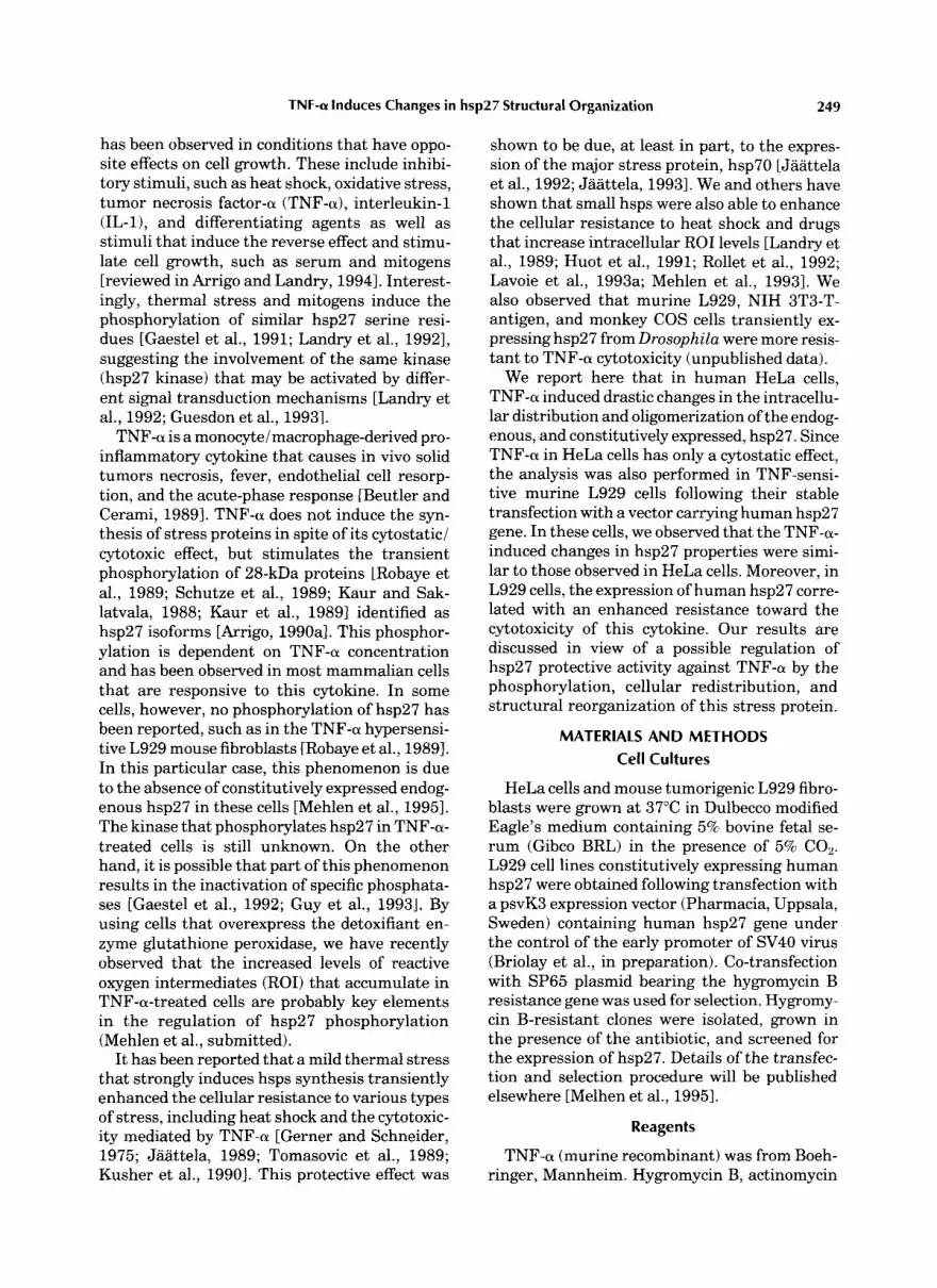
In previous reports [Arrigo, 1990a; Arrigo and Michel, 19911, we showed, by using short pulses of [32Plorthophosphate labeling of TNF-a-treated HeLa cells that the stimulation of hsp27 phos- phorylation peaked after 30 min of treatment and was no more observed 1 h later. hsp27 phosphorylation results in the accumulation of different phospho-isoforms denoted b, c, and d, that correspond to the protein phosphorylated at 1,2, and 3 serine sites, respectively [Landry et al., 19921. This is illustrated in the two-dimen- sional immunoblots presented in Figure 1A; as shown, a 1-h exposure of HeLa cells to 2,000 U/ml of TNF-a increased the level of the major hsp27 “b” phospho-isoform. A similar observa- tion was made in the murine L929-27-3 cell line that displays a high level of exogenous human hsp27 expression after exposure to 2,000 U/ml TNF-a (Fig. 1B). A quantitative analysis of this phenomenon in HeLa cells indicates that the level of hsp27 “b” phospho-isoform increased rapidly during the first hour of the treatment, until it reached about 40% of the total amount of hsp27 (Fig. 1C). The maximal accumulation (45%) was observed 2 h after the addition of the cytokine to the culture medium. By 4 h, the level of this isoform still represented 30% of the total amount of hsp27. Similar kinetics of accumula- tion of hsp27 “b” phospho-isoform were ob- served in TNF-treated L929-27-3 cells (not shown). These observations suggest a low turn- over of hsp27 phosphate residues in TNF-a- treated cells.
L929 cells are extremely sensitive to TNF-a [Wallach, 19841. By contrast, HeLa cells are resistant to the cytotoxicity mediated by this cytokine, displaying only transient growth ar- rest [Sugarman et al., 19851. A kinetic analysis of this phenomenon is presented in Figure 1D; as shown, a drastic 10-fold reduced L3H1thymi- dine incorporation was observed after treating HeLa cells with 2,000 U/ml of TNFa. By 4 hr, this cytostatic effect was still observed. Thus, in HeLa cells, the stimulation of hsp27 “b” phos- pho-isoforms accumulation by TNF-a is an early
phenomenon that occurs concomitantly with re- duced DNA synthesis.
In HeLa and Transfeded Murine L929 Cells, TNF-a Induces Drastic Changes in the
lntracellular Distribution, Cellular localization, and Oligomerization of Human hsp27
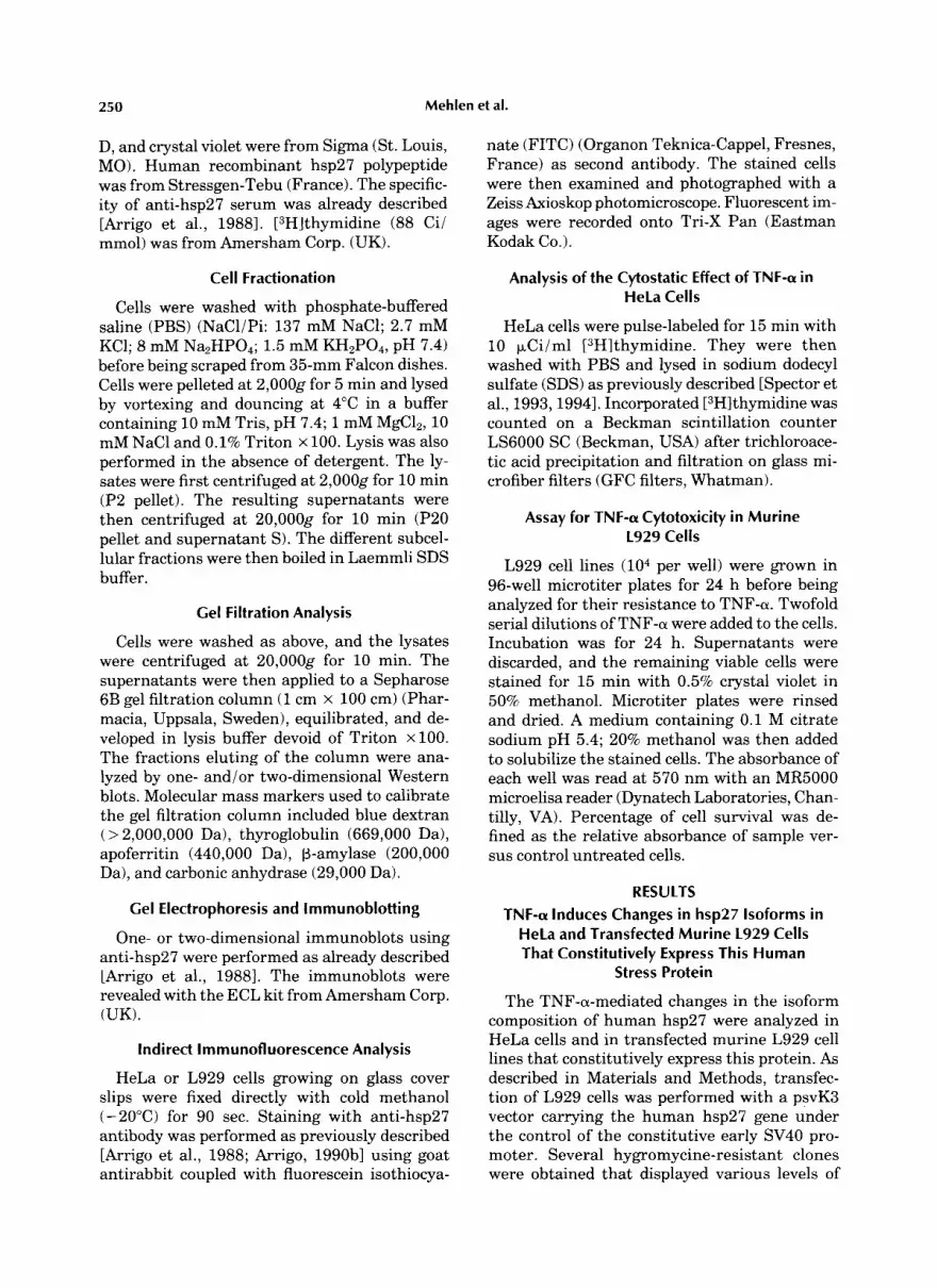
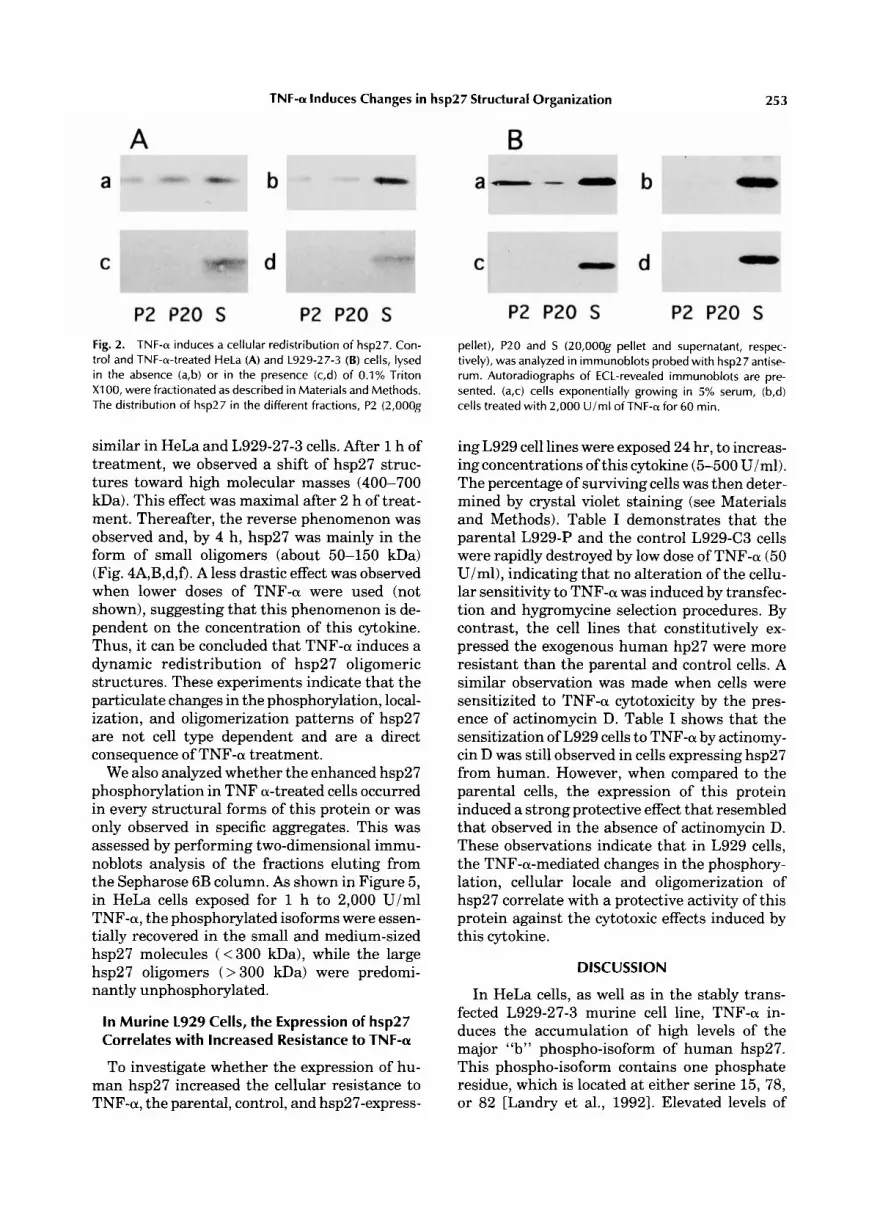
We next analyzed the intracellular distribu- tion of human hsp27 following TNF-a treat- ment. HeLa and L929-27-3 cells were lysed in the absence of detergent, fractionated, and hsp27 detected in immunoblots (see Materials and Methods). In exponentially growing HeLa cells, 50% of the cellular content of hsp27 was recov- ered in the 20,OOOg supernatant ( S ) , while the particulate fractions P2 (2,OOOg) and P20 (20,OOOg ) contained 15% and 35% of this pro- tein, respectively (Fig. 2A,a) [Arrigo et al., 1988; Mehlen and Arrigo, 19941. By contrast, in cells lysed after one hour exposure to 2,000 U/ml of TNF-a, hsp27 was quantitatively recovered in the 20,OOOg supernatant (S fraction) (Fig. 2A,b). In growing L929-27-3 cells, 45% of the cellular content of hsp27 was present in the 20,OOOg supernatant (S), while the P2 (2,OOOg) and P20 (20,OOOg) fractions contained 35% and 20% of the protein, respectively (Fig. 2B,a) while after one hour exposure to 2,000 U/ml of TNF-a, hsp27 was again quantitatively recovered in the S fraction (Fig. 2B,b). hsp27 was also found in the S fraction when HeLa and L929-27-3 cells were lyzed in the presence of 0.1% Triton X-100 (Fig. 2A,B,c,d). Hence, TNF-a induces an intra- cellular redistribution of human hsp27, which is observed in both HeLa and L929-27-3 cells.
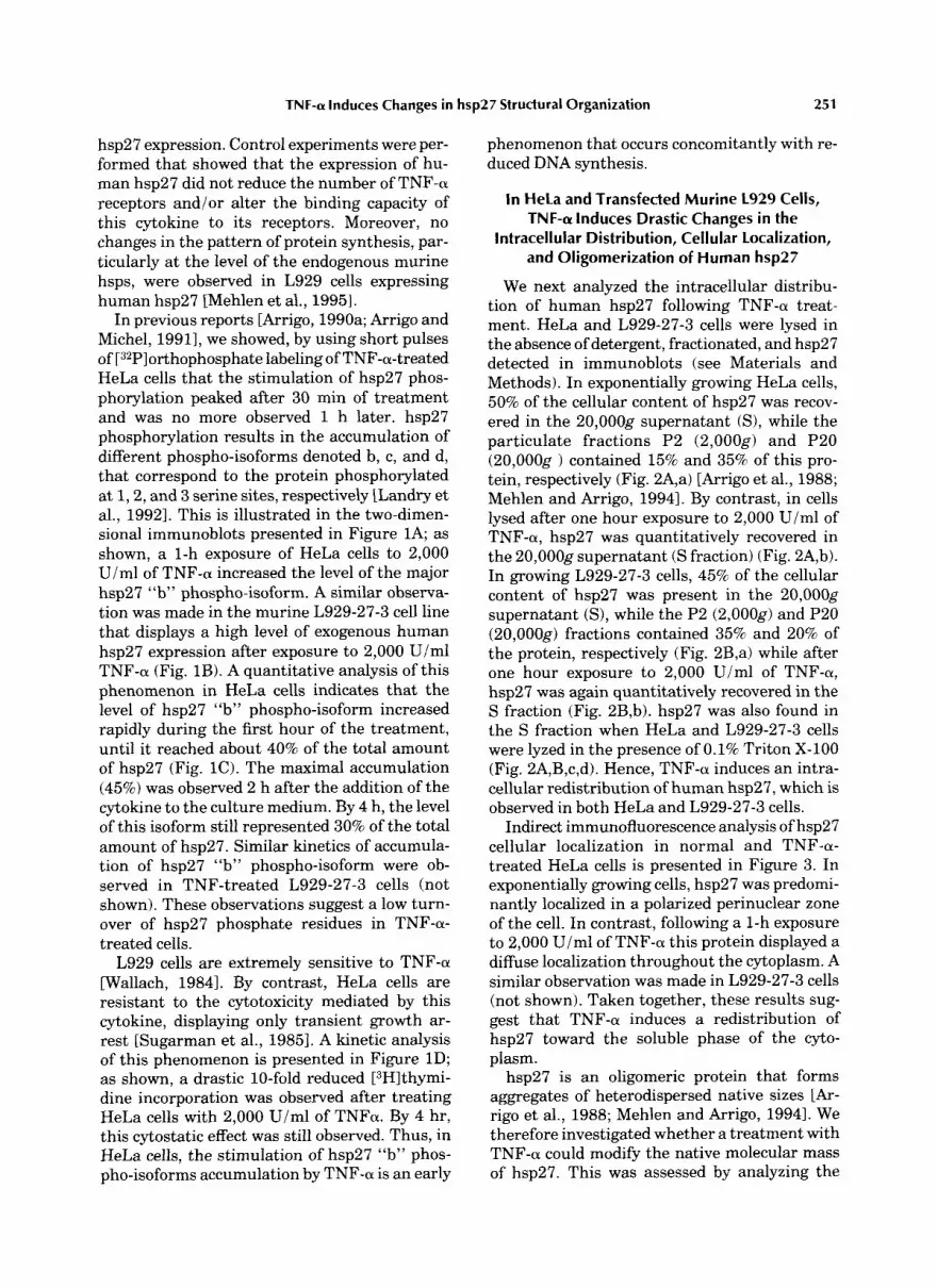
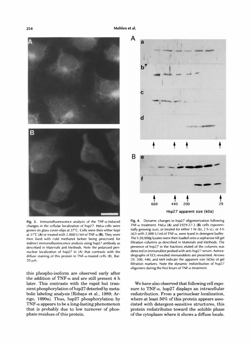
Indirect immunofluorescence analysis of hsp27 cellular localization in normal and TNF-a- treated HeLa cells is presented in Figure 3. In exponentially growing cells, hsp27 was predomi- nantly localized in a polarized perinuclear zone of the cell. In contrast, following a 1-h exposure to 2,000 U/ml of TNF-a this protein displayed a diffuse localization throughout the cytoplasm. A similar observation was made in L929-27-3 cells (not shown). Taken together, these results sug- gest that TNF-a induces a redistribution of hsp27 toward the soluble phase of the cyto- plasm.
hsp27 is an oligomeric protein that forms aggregates of heterodispersed native sizes [Ar- rig0 et al., 1988; Mehlen and Arrigo, 19941. We therefore investigated whether a treatment with TNF-a could modify the native molecular mass of hsp27. This was assessed by analyzing the
252 Mehlen et al.
A I
II
B
C
Fig. 1. Quantitativeanalysis of hsp27 isoforms followingTNF-a treatment; correlation with the inhibition of cellular prolifera- tion. Exponentially growing HeLa and L929-27-3 cells were either kept untreated or exposed for different times to 2,000 U im l of TNF-a. A,B: Two-dimensional immunoblots probed with anti-hsp27 serum of either untreated cells (I) or TNF-a- treated cells for 60 min (11) . A: HeLa cells. B: L929-27-3 cells. Autoradiographs of the ECL-revealed immunoblots showing hsp27 isoforms are presented. The acidic end is to the left. Arrowheads a, b, b', c indicate the different isoforms of hsp27. The "a" isoform represents the unphosphorylated form of the protein. C: Kinetics analysis of hsp27 "a and b" isoforms levels following TNF-a treatment. HeLa cells were kept at 37°C or treated for 30, 60, 120, or 240 min with 2,000 U/ml of TNF-a. Two-dimensional immunoblots probed with anti-hsp27 serum
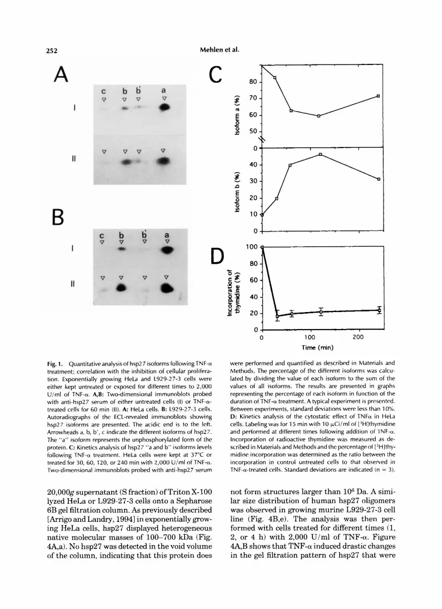
20,0009 supernatant (S fraction) of Triton X- 100 lyzed HeLa or L929-27-3 cells onto a Sepharose 6B gel filtration column. As previously described [Arrigo and Landry, 19941 in exponentially grow- ing HeLa cells, hsp27 displayed heterogeneous native molecular masses of 100-700 kDa (Fig. 4A,a). No hsp27 was detected in the void volume of the column, indicating that this protein does
0 1
0 , 0 100 200
Time (min)
were performed and quantified as described in Materials and Methods. The percentage of the different isoforms was calcu- lated by dividing the value of each isoform to the sum of the values of all isoforms. The results are presented in graphs representing the percentage of each isoform in function of the duration of TNF-a treatment. A typical experiment is presented. Between experiments, standard deviations were less than 10%. D: Kinetics analysis of the cytostatic effect of TNFa in HeLa cells. Labelingwas for 15 min with 10 FCi iml of [3H]thymidine and performed at different times following addition of TNF-a. Incorporation of radioactive thymidine was measured as de- scribed in Materials and Methods and the percentage of r3H]thy- midine incorporation was determined as the ratio between the incorporation in control untreated cells to that observed in TNF-a-treated cells. Standard deviations are indicated (n = 3).
not form structures larger than lo6 Da. A simi- lar size distribution of human hsp27 oligomers was observed in growing murine L929-27-3 cell line (Fig. 4B,e). The analysis was then per- formed with cells treated for different times (1, 2, or 4 h) with 2,000 U/ml of TNF-a. Figure 4A,B shows that TNF-a induced drastic changes in the gel filtration pattern of hsp27 that were
TNF-cu Induces Changes in hsp27 Structural Organization 253
Fig. 2. TNF-a induces a cellular redistribution of hsp27. Con- trol and TNF-a-treated HeLa (A) and L929-27-3 (B) cells, lysed in the absence (a,b) or in the presence (c,d) of 0.1% Triton XI 00, were fractionated as described in Materials and Methods. The distribution of hsp27 in the different fractions, P2 (2,OOOg
similar in HeLa and L929-27-3 cells. After 1 h of treatment, we observed a shift of hsp27 struc- tures toward high molecular masses (400-700 kDa). This effect was maximal after 2 h of treat- ment. Thereafter, the reverse phenomenon was observed and, by 4 h, hsp27 was mainly in the form of small oligomers (about 50-150 kDa) (Fig. 4A,B,d,f). A less drastic effect was observed when lower doses of TNF-a were used (not shown), suggesting that this phenomenon is de- pendent on the concentration of this cytokine. Thus, it can be concluded that TNF-a induces a dynamic redistribution of hsp27 oligomeric structures. These experiments indicate that the particulate changes in the phosphorylation, local- ization, and oligomerization patterns of hsp27 are not cell type dependent and are a direct consequence of TNF-a treatment.
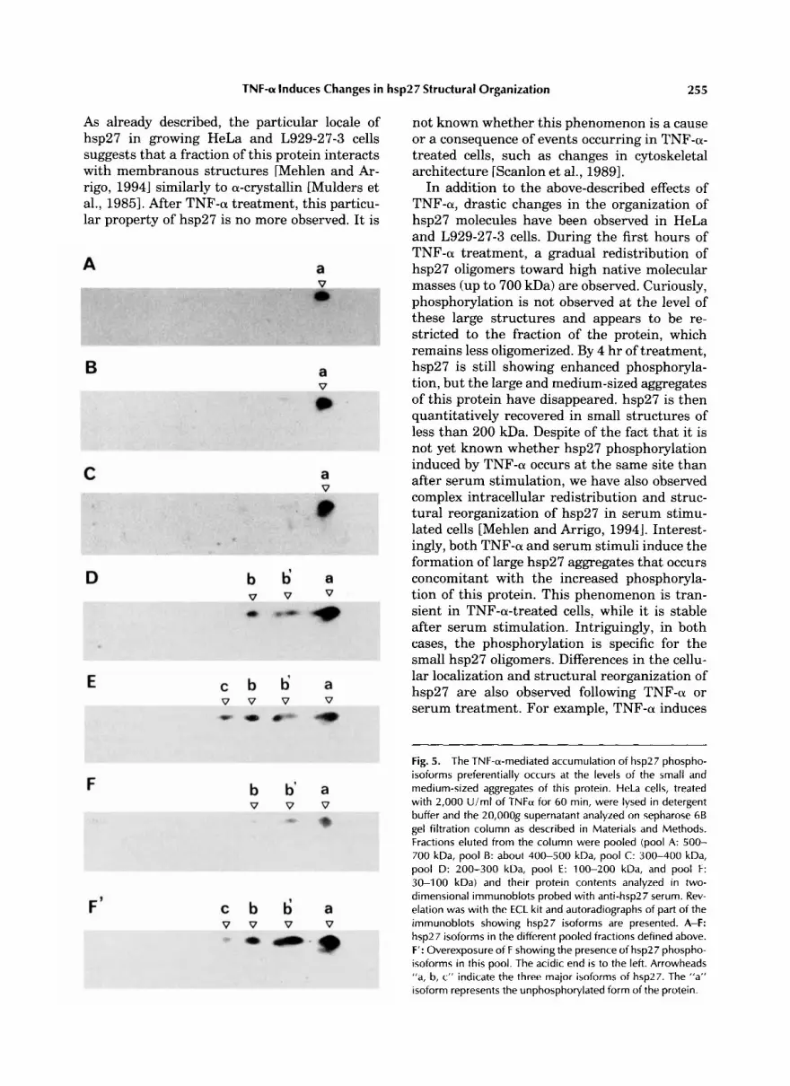
We also analyzed whether the enhanced hsp27 phosphorylation in TNF a-treated cells occurred in every structural forms of this protein or was only observed in specific aggregates. This was assessed by performing two-dimensional immu- noblots analysis of the fractions eluting from the Sepharose 6B column. As shown in Figure 5, in HeLa cells exposed for 1 h to 2,000 U/ml TNF-a, the phosphorylated isoforms were essen- tially recovered in the small and medium-sized hsp27 molecules ( < 300 m a ) , while the large hsp27 oligomers ( > 300 kDa) were predomi- nantly unphosphorylated.
In Murine L929 Cells, the Expression of hsp27 Correlates with Increased Resistance to TNF-a
To investigate whether the expression of hu- man hsp27 increased the cellular resistance to TNF-a, the parental, control, and hsp27-express-
pellet), P20 and S (20,OOOg pellet and supernatant, respec- tively), was analyzed in irnrnunoblots probed with hsp27 antise- rum. Autoradiographs of ECL-revealed irnmunoblots are pre- sented. (a,c) cells exponentially growing in 5% serum, (b,d) cells treated with 2,000 U/ml of TNF-a for 60 min.
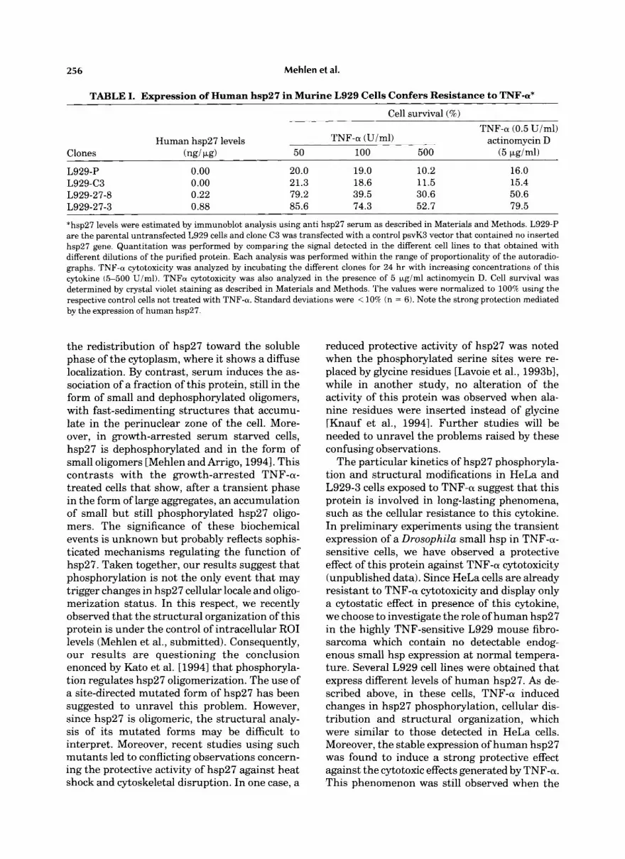
ing L929 cell lines were exposed 24 hr, to increas- ing concentrations of this cytolune (5-500 Uiml). The percentage of surviving cells was then deter- mined by crystal violet staining (see Materials and Methods). Table I demonstrates that the parental L929-P and the control L929-C3 cells were rapidly destroyed by low dose of TNF-a (50 U/ml), indicating that no alteration of the cellu- lar sensitivity to TNF-a was induced by transfec- tion and hygromycine selection procedures. By contrast, the cell lines that constitutively ex- pressed the exogenous human hp27 were more resistant than the parental and control cells. A similar observation was made when cells were sensitizited to TNF-a cytotoxicity by the pres- ence of actinomycin D. Table I shows that the sensitization of L929 cells to TNF-a by actinomy- cin D was still observed in cells expressing hsp27 from human. However, when compared to the parental cells, the expression of this protein induced a strong protective effect that resembled that observed in the absence of actinomycin D. These observations indicate that in L929 cells, the TNF-a-mediated changes in the phosphory- lation, cellular locale and oligomerization of hsp27 correlate with a protective activity of this protein against the cytotoxic effects induced by this cytokine.
DISCUSSION
In HeLa cells, as well as in the stably trans- fected L929-27-3 murine cell line, TNF-a in- duces the accumulation of high levels of the major “b” phospho-isoform of human hsp27. This phospho-isoform contains one phosphate residue, which is located at either serine 15, 78, or 82 [Landry et al., 19921. Elevated levels of
254 Mehlen et al.
A
B
Fig. 3. lmmunofluorescence analysis of the TNF-a-induced changes in the cellular localization of hsp27. HeLa cells were grown on glass cover-slips at 37°C. Cells were then either kept at 37°C (A) or treated with 2,000 U i m l of TNF-a (B). They were then fixed with cold methanol before being processed for indirect immunofluorescence analysis using hsp27 antibody as described in Materials and Methods. Note the polarized peri- nuclear localization of hsp27 in (A) that contrasts with the diffuse staining of this protein in TNF-a-treated cells (B). Bar: 20 Lm.
this phospho-isoform are observed early after the addition of TNF-a and are still present 4 h later. This contrasts with the rapid but tran- sient phosphorylation of hsp27 detected by meta- bolic labeling analysis [Robaye et al., 1989; Ar- rigo, 1990aI. Thus, hsp27 phosphorylation by TNF-a appears to be a long-lasting phenomenon that is probably due to low turnover of phos- phate residues of this protein.
Fig. 4. Dynamic changes in hsp27 oligornerization following TNF-a treatment. HeLa (A) and L929-27-3 (B) cells exponen- tially growing (a,e), or treated for either 1 hr (b), 2 h (c), or 4 h (d,f) with 2,000 U/ml of TNF-a, were lysed in detergent buffer. The S 20,OOOg lysates were then loaded onto a sepharose 68 gel filtration columns as described in Materials and Methods. The presence of hsp27 in the fractions eluted of the columns was detected in immunoblot probed with anti-hsp27 serum. Autora- diographs of ECL-revealed immunoblots are presented. Arrows 29, 200, 440, and 669 indicate the apparent size (kDa) of gel filtration markers. Note the dynamic redistribution of hsp27 oligomers during the first hours of TNF-a treatment.
We have also observed that following cell expo- sure to TNF-a, hsp27 displays an intracellular redistribution. From a perinuclear localization, where at least 50% of this protein appears asso- ciated with detergent-sensitive structures, this protein redistributes toward the soluble phase of the cytoplasm where it shows a diffuse locale.
TNF-a Induces Changes in hsp27 Structural Organization 255
As already described, the particular locale of hsp27 in growing HeLa and L929-27-3 cells suggests that a fraction of this protein interacts with membranous structures [Mehlen and Ar- rigo, 19941 similarly to a-crystallin [Mulders et al., 19851. After TNF-a treatment, this particu- lar property of hsp27 is no more observed. It is
not known whether this phenomenon is a cause or a consequence of events occurring in TNF-a- treated cells, such as changes in cytoskeletal architecture [Scanlon et al., 19891.
In addition to the above-described effects of TNF-a, drastic changes in the organization of hsp27 molecules have been observed in HeLa and L929-27-3 cells. During the first hours of TNF-a treatment, a gradual redistribution of hsp27 oligomers toward high native molecular masses (up to 700 kDa) are observed. Curiously, phosphorylation is not observed at the level of these large structures and appears to be re- stricted to the fraction of the protein, which remains less oligomerized. By 4 hr of treatment, hsp27 is still showing enhanced phosphoryla- tion, but the large and medium-sized aggregates of this protein have disappeared. hsp27 is then quantitatively recovered in small structures of less than 200 kDa. Despite of the fact that it is not yet known whether hsp27 phosphorylation induced by TNF-a occurs at the same site than after serum stimulation, we have also observed complex intracellular redistribution and struc- tural reorganization of hsp27 in serum stimu- lated cells [Mehlen and Arrigo, 19941. Interest- ingly, both TNF-a and serum stimuli induce the formation of large hsp27 aggregates that occurs concomitant with the increased phosphoryla- tion of this protein. This phenomenon is tran- sient in TNF-a-treated cells, while it is stable after serum stimulation. Intriguingly, in both cases, the phosphorylation is specific for the small hsp27 oligomers. Differences in the cellu- lar localization and structural reorganization of hsp27 are also observed following TNF-a or serum treatment. For example, TNF-a induces
Fig. 5. The TNF-a-mediated accumulation of hsp27 phospho- isoforms preferentially occurs at the levels of the small and medium-sized aggregates of this protein. HeLa cells, treated with 2,000 Uiml of TNFa for 60 min, were lysed in detergent buffer and the 20,OOOg supernatant analyzed on sepharose 6B gel filtration column as described in Materials and Methods. Fractions eluted from the column were pooled (pool A: 500- 700 kDa, pool B: about 400-500 kDa, pool C: 300-400 kDa, pool D: 200-300 kDa, pool E: 100-200 kDa, and pool F: 30-100 kDa) and their protein contents analyzed in two- dimensional immunoblots probed with anti-hsp27 serum. Rev- elation was with the ECL kit and autoradiographs of part of the immunoblots showing hsp27 isoforms are presented. A-F: hsp27 isoforms in the different pooled fractions defined above. F’: Overexposure of F showing the presence of hsp27 phospho- isoforms in this pool. The acidic end is to the left. Arrowheads “a, b, c” indicate the three major isoforms of hsp27. The “a” isoform represents the unphosphorylated form of the protein.
256 Mehlen et al.
TABLE I. Expression of Human hsp27 in Murine L929 Cells Confers Resistance to TNF-a*
Cell survival (%)
TNF-a (0.5 U/ml) Human hsp27 levels TNF-a (Uiml) actinomycin D
Clones (ngi PLg) 50 100 500 (5 Fgiml)
L929-P 0.00 20.0 19.0 10.2 16.0 L929-C3 0.00 21.3 18.6 11.5 15.4 L929-27-8 0.22 79.2 39.5 30.6 50.6 L929-27-3 0.88 85.6 74.3 52.7 79.5
*hsp27 levels were estimated by immunoblot analysis using anti hsp27 serum as described in Materials and Methods. L929-P are the parental untransfected L929 cells and clone C3 was transfected with a control psvK3 vector that contained no inserted hsp27 gene. Quantitation was performed by comparing the signal detected in the different cell lines to that obtained with different dilutions of the purified protein. Each analysis was performed within the range of proportionality of the autoradio- graphs. TNF-a cytotoxicity was analyzed by incubating the different clones for 24 hr with increasing concentrations of this cytokine (5-500 Uiml). TNFa cytotoxicity was also analyzed in the presence of 5 Fgiml actinomycin D. Cell survival was determined by crystal violet staining as described in Materials and Methods. The values were normalized to 100% using the respective control cells not treated with TNF-a. Standard deviations were < 10% (n = 6). Note the strong protection mediated by the expression of human hsp27
the redistribution of hsp27 toward the soluble phase of the cytoplasm, where it shows a diffuse localization. By contrast, serum induces the as- sociation of a fraction of this protein, still in the form of small and dephosphorylated oligomers, with fast-sedimenting structures that accumu- late in the perinuclear zone of the cell. More- over, in growth-arrested serum starved cells, hsp27 is dephosphorylated and in the form of small oligomers [Mehlen and Arrigo, 19941. This contrasts with the growth-arrested TNF-a- treated cells that show, after a transient phase in the form of large aggregates, an accumulation of small but still phosphorylated hsp27 oligo- mers. The significance of these biochemical events is unknown but probably reflects sophis- ticated mechanisms regulating the function of hsp27. Taken together, our results suggest that phosphorylation is not the only event that may trigger changes in hsp27 cellular locale and oligo- merization status. In this respect, we recently observed that the structural organization of this protein is under the control of intracellular ROI levels (Mehlen et al., submitted). Consequently, our results are questioning the conclusion enonced by Kato et al. [19941 that phosphoryla- tion regulates hsp27 oligomerization. The use of a site-directed mutated form of hsp27 has been suggested to unravel this problem. However, since hsp27 is oligomeric, the structural analy- sis of its mutated forms may be difficult to interpret. Moreover, recent studies using such mutants led to conflicting observations concern- ing the protective activity of hsp27 against heat shock and cytoskeletal disruption. In one case, a
reduced protective activity of hsp27 was noted when the phosphorylated serine sites were re- placed by glycine residues [Lavoie et al., 1993b1, while in another study, no alteration of the activity of this protein was observed when ala- nine residues were inserted instead of glycine [Knauf et al., 19941. Further studies will be needed to unravel the problems raised by these confusing observations.
The particular kinetics of hsp27 phosphoryla- tion and structural modifications in HeLa and L929-3 cells exposed to TNF-a suggest that this protein is involved in long-lasting phenomena, such as the cellular resistance to this cytokine. In preliminary experiments using the transient expression of a Drosophila small hsp in TNF-a- sensitive cells, we have observed a protective effect of this protein against TNF-a cytotoxicity (unpublished data). Since HeLa cells are already resistant to TNF-a cytotoxicity and display only a cytostatic effect in presence of this cytokine, we choose to investigate the role of human hsp27 in the highly TNF-sensitive L929 mouse fibro- sarcoma which contain no detectable endog- enous small hsp expression at normal tempera- ture. Several L929 cell lines were obtained that express different levels of human hsp27. As de- scribed above, in these cells, TNF-a induced changes in hsp27 phosphorylation, cellular dis- tribution and structural organization, which were similar to those detected in HeLa cells. Moreover, the stable expression of human hsp27 was found to induce a strong protective effect against the cytotoxic effects generated by TNF-a. This phenomenon was still observed when the
TNF-(U Induces Changes in hsp27 Structural Organization 257
cellular sensitivity to TNF-a was enhanced by actinomycin D. A similar observation was made in WEHI cells overexpressing human hsp27 [S. Tomasovic, personnal communication] and when Drosophila hsp27 or the related human ap- crystallin were stably expressed in L929 cell lines (Mehlen et al., submitted). In WHEI-S cells, however, the expression of human hsp27 was not reported to correlate with an enhanced cellular protection against TNF-a cytotoxicity [Jaattela et al., 19921. Several reasons may ex- plain this difference with our findings. First, Jaattela et al. used a highly TNF-sensitive sub- clone of WEHI cells that may have lost its poten- tiality to be protected by hsp27 during subclonal selection. Second, these authors did not quan- tify the level of hsp27 produced in their trans- fected cells.
TNF-sensitive murine L929 cells are origi- nally devoid of constitutively expressed endog- enous small hsps. By contrast, human HeLa cells that are resistant to this cytokine constitu- tively express the endogenous hsp27. Hence, it is possible that this protein and/or its phosphor- ylation and structural modifications may parti- cipe in the cellular resistance of HeLa cells against TNF-a cytotoxicity. The mechanisms regulating the protective activity of hsp27 are unknown. Concerning a putative role of hsp27 as an inhibitor of actin polymerization, we have recently suggested that the small and dephos- phorylated oligomers that accumulate in serum- starved cells may represent the active form of the protein [Mehlen and Arrigo, 19941. Interest- ingly, dissolution of actin microfilaments is an early phenomenon that characterizes the cyto- static/cytotoxic effects of TNF-a [Scanlon et al., 19891 and oxidative stress [Bellorno et al., 19901. Hence, it can be speculated that the dynamic reorganization of hsp27 in TNF-a-treated cells may abolish the putative function of this protein as an inhibitor of actin polymerization. This may result in a mechanism that could counter- act the TNF-a-mediated disruption of actin ar- chitecture. On the other hand, the large hsp27 oligomeric structures observed during the first hours of TNF-a treatment, may be functionally important. Indeed, it can be hypothesized that these structures interact with and/or sequester other proteins. This is of particular interest in light of recent reports suggesting that small hsps have in vitro protein chaperone activities [Horwitz, 1992; Jakob et al., 19931. As such, chaperones associate with and enhance the rena-
turation of denatured proteins [Sherman and Gold- berg, 1992; Jakob et al., 19931. Whether the tran- sient increase in hsp27 oligomerization in TNF-a- treated cells plays such a role remains to be shown.
ACKNOWLEDGMENTS
We thank C. Kretz-Remy for critical reading of the manuscript. P.M. and A.M. were sup- ported by doctoral fellowship funds from the Ecole Normale Superieure (AMN-ENS). This work was supported by grant 6011 from the Association pour la Recherche sur le Cancer, grant 930501 from the Institut National pour la Sante et la Recherche Medicale (INSERM), and CHRX-CT 93-0260 from the European Eco- nomic Community (Human Capital and Mobil- ity) (to A.-P.A.).
REFERENCES
Arrigo A-P (1990a): Tumor necrosis factor induces the rapid phosphorylation of the mammalian heat-shock protein hsp28. Mol Cell Biol 10:1276-1280.
Arrigo A-P (1990b): The monovalent ionophore monensin maintains the nuclear localization of the human stress protein hsp28 during heat shock recovery. J Cell Sci 96:419-427.
Arrigo A-P, Landry J (1994): Expression and function of the low molecular weight heat shock proteins. In Morimoto R, Tissieres A, Georgopoulos C (eds): “Heat Shock Proteins: Structure, Function and Regulation.” Cold Spring Har- bor, New York: Cold Spring Harbor Laboratory Press, pp 335-373.
Arrigo A-P, Michel MR (1991): Decreased heat- and tumor necrosis factor-mediated hsp28 phosphorylation in ther- motolerant HeLa cells. FEBS Lett 282:152-156.
Arrigo A-P, and Welch WJ (1987): Characterization and purification of the mammalian 28,000 dalton heat shock protein. J Biol Chem 262:15359-15369.
Arrigo A-P, Suhan J , Welch WJ (1988): Dynamic changes in the structure and locale of the mammalian low molecular weight heat shock protein. Mol Cell Biol8:5059-5071.
Bellomo G, Mirabelli F, Vairetti M, Malormi W (1990): Cytosqueleton as a target in menadione-induced oxidative stress in cultured mammalian cells. Biochemical and im- munocytochemical features. J Cell Physiol 143:118-128.
Beutler B, Cerami A (1989): The biology of cachectini TNF-A primary mediator of the host response. Annu Rev Immunol7:625-655.
Bitar KN, Kaminsh MS, Hailat N, Cease KB, Strahler JR (1991): Hsp27 is a mediator of sustained smooth muscle contraction in response to bombesin. Biochem Biophys Res Commun 181:1192-1200.
Ciocca DR, Oesterreich S, Chamness GC, McGuire WJ, Fuqua SAW (1993): Biological and clinical implications of heat shock protein 27000 (Hsp27)-A review. J Natl Cancer Inst 85:1558-1570.
Gaestel M, Schroder W, Benndorf R, Lippman C, Buchner K, Huchot F, Ermann VA, Bielka H (1991): Identification of the phosphorylation sites of the murine small heat shock protein 25. J Biol Chem 266: 14721-14724.
258 Mehlen et al.
Gaestel M, Benndorf R, Hayes K, Priemer E, Engel K (1992): Dephophorylation of the small heat shock protein hsp25 by calciumicalmodulin-dependent (type 2B) pro- tein phosphatase. J Biol Chem 267:21607-21611.
Gerner EW, Schneider MJ (1975): Induced thermal resis- tance in Hela cells. Nature 256:500-502.
Guesdon F, Freshney N, Waller RJ, Rawlinson L, Saklatvala J (1993): Interleukin-1 and tumor necrosis factor stimu- late two novel protein kinases that phosphorylate the heat shock protein hsp27 and beta-casein. J Biol Chem 268: 4236-4243.
Guy GR, Cairns J, Ng SB, Tan YH (1993): Inactivation of a redox-sensitive protein phosphatase during the early events of tumor necrosis factoriinterleukin-1 signal trans- duction. J Biol Chem 268:2141-2148.
Horwitz J (1992): Alpha-crystallin can function as a molecu- lar chaperone. Proc Natl Acad Sci USA 89:10449-10453.
Huot J , Roy G, Lambert H, Chretien P, Landry J (1991): Increased survival after treatments with anticancer agents of Chinese hamster cells expressing the human Mr 27,000 heat shock protein. Cancer Res 515245-5252.
Ingolia TD, Craig E (1982): Four small Drosophila heat shock proteins are related to each other and to mamma- lian A-crystallin. Proc Natl Acad Sci USA 79:2360-2364.
Jaattela M (1989): Heat shock protects WEHI-164 target cells from the cytolysis by tumor necrosis factor cx and p. Eur J Immunol19:1413-1417.
Jaattela M (1993): Over-expression of major heat shock protein hsp70 inhibits tumor necrosis factor-induced acti- vation of phospholipase A2. J Immunol 151:4286-4294.
Jaattela M, Wissing D, Bauer PA, Li CG (1992): Major heat shock protein hsp70 protects tumor cells from tumor necrosis factor cytotoxicity. EMBO J 11:3507-3512.
Jakob U, Gaestel M, Engel K, Buchner J (1993): Small heat shock proteins are molecular chaperones. J Biol Chem 268: 1517-1520.
Kato K, Hasegawa K, Goto S, Inaguma Y (1994): Dissocia- tion as a result of phosphorylation of an aggregated form of the small stress protein, hsp27. J Biol Chem 269: 11274- 11278.
Kaur P, Saklatvala J (1988): Interleukin 1 and tumor necro- sis factor increase phosphorylation of fibroblast proteins. FEBS Lett 241:6-10.
Kaur P, Welch WJ, Salklatvala J (1989): Interleukin 1 and tumor necrosis factor phosphorylation of the small heat shock protein. Effects in fibroblasts, HepG2 and U937 cells. FEBS Lett 58:269-273.
Knauf U, Bielka H, and Gaestel M (1992): Over-expression of the small heat shock protein, hsp25, inhibits growth of Ehrlich ascites tumor cells. FEBS Lett 308:297-302.
Knauf U, Jakob U, Engel K, Buchner J , Gaestel M (1994): Stress- and mitogen-induced phosphorylation of the small heat shock protein Hsp25 by MAPKAP kinase 2 is not essential for chaperone properties and cellular thermore- sistance. EMBO J 1354-60.
Kusher DL, Ware CF, Gooding LR (1990): Induction of the heat shock response protects cells from lysis by tumor necrosis factor. J Immunol 145:2925-2931.
,andry J, Chretien P, Lambert H, Hickey E, Weber LA (1989): Heat shock resistance conferred by expression of the human HSP27 gene in rodent cells. J Cell Biol109:7- 15.
,andry J , Lambert H, Zhou M, Lavoie JN, Hickey E, Weber LA, Anderson CW (1992): Human hsp27 is phosphory- lated at serine 78 and 82 by heat shock and mitogen-
activated kinases that reconise the same amino acid motif as S6 kinase 11. J Biol Chem 267:794-803.
Lavoie JN, Gingras-Breton G, Tanguay RM, Landry J (1993a): Induction of Chinese hamster HSP27 gene expres- sion in mouse cells confers resistance to heat shock. HSP27 stabilization of the microfilament organization. J Biol Chem 268:3420-3429.
Lavoie JN, Hickey E, Weber LA, Landry J (199313): Modula- tion of actin microfilament dynamics and fluid phase pinocytosis by phosphorylation of heat shock protein-27. J Biol Chem 268:24210-24214.
Lindquist S (1986): The heat shock response. Annu Rev Biochem 55: 115 1-1 191.
Mehlen P, Arrigo A-P (1994): The serum-induced phosphor- ylation of the mammalian small stress protein correlates with changes in its intracellular localization and levels of oligomerization. Eur J Biochem 221:321-334.
Mehlen P, Briolay J, Smith L, Diaz-Latoud C, Fabre N, Pauli D, Arrigo A-P (1993): Analysis of the resistance to heat and hydrogen peroxide stresses in COS cells tran- siently expressing wild type or deletion mutants of the DrosophiZa 27-kDa heat shock protein. Eur J Biochem 2 15:277-284.
Mehlen P, Preville X, Chareyron P, Briolay J , Klemenz R, Arrigo AP (1995): Constitutive expression of either hu- man hsp27, Drosophilia hsp27 or human a-p crystallin confers resistance to tumor cytotoxicity instably trans- fected murine L929 fibroblasts. J Immunol 154:363-374.
Morimoto RI, Tissieres A, Georgopoulos C (1990): “Stress Proteins in Biology and Medicine.” Cold Spring Harbor, New York: Cold Spring Harbor Laboratory Press.
Miron T, Vancompernolle K, Vanderkerckhove J , Wilchek M, Geiger B (1991): A 25-kD inhibitor of actin polymeriza- tion is a low molecular mass heat shock protein. J Cell Biol 114:255-261.
Mulders JWM, Stokkermans J, Leunissen JAM, Benedetti EL, Bloemendal H, De Jong WW (1985): Interaction of alpha-crystallin with lens plasma membranes. Affinity for MP26. Eur J Biochem 152:721-728.
Nover L (1984): “Heat Shock Response of Eukaryotic Cells.” Berlin: Springer-Verlag.
Regazzi R, Eppenberger U, Fabbro D (1988): The 27,000 dalton stress protein are phosphorylated by protein kinase C during the tumor promoter-mediated growth inhibition of human mammary carcinoma cells. Biochem Biophys Res Commun 15252-68.
Robaye B, Hepburn A, Lecocq R, Fiers W, Boeynaems JM, Dumont J E (1989): Tumor necrosis factor-cx induces the phosphorylation of 28 kDa stress proteins in endothelial cells: Possible role in protection against cytotoxicity? Bio- chem Biophys Res Commun 163:301-308.
Rollet E, Lavoie JN, Landry J , Tanguay RM (1992): Expres- sion ofDrosophiZa’s 27 kDa heat shock protein into rodent cells confers thermal resistance. Biochem Biophys Res Commun 185:116-120.
Scanlon M, Laster SM, Wood JG, Gooding LR (1989): Cytoly- sis by tumor necrosis factor is preceded by a rapid and specific dissolution of microfilaments. Proc Natl Acad Sci
Schutze S, Scheurich P, Pfizenmaier K, Kronke M (1989): Tumor necrosis factor signal transduction. Tissue-specific serine phosphorylation of a 26-kDa cytosolic protein. J Biol Chem 264:3562-3567.
USA 86:180-186.
TNF-(U Induces Changes in hsp27 Structural Organization 259
Seizen R J , Bindel JG, Hoenders HJ (1978): The quaternary structure of bovine alpha-crystallin. Eur J Biochem 91:
Semenzato G (1990): Tumor necrosis factor: Acytokine with multiple biological activities. Br J Cancer 61:354-361.
Sherman M Y , Goldberg AL (1992): Heat shock in Esch- erichia coli alters the protein-binding properties of the chaperonin groEL by inducing its phosphorylation. Na- ture 357:167-169.
Spector NL, Ryan C, Samson W, Nadler LM, Arrigo AP (1993): Heat shock protein is a unique marker of growth arrest during macrophage differentiation of HL-60 cells. J Cell Physiol 156:619-625.
Spector NL, Mehlen P, Ryan C, Hardy L, Samson W, Nadler
387-396.
LM, Fabre N, Arrigo AP (1994): Regulation of the 28 kDa heat shock protein by retinoic acid during differentiation of leukemic HL-60 cells. FEBS Lett 337:184-188.
Sugarman BJ, Aggarwal BB, Hass PE, Figari IS, Palladino MA, Shepard HM (1985): Recombinant human tumor necrosis factor-a: Effects on proliferation of normal and transformed cells in vitro. Science 230:943-945.
Tomasovic SP, Barta M, Klostergaard J (1989): Temporal dependence of hyperthermic augmentation of macrophage- TNF production and tumor cell TNF sensitization. Int J Hyperthermia 5:625-639.
Wallach D (1984): Preparation of lymphototoxin induce resistance to their own cytotoxic effects. J Immunol 132: 2664-2668.
Copyright © 2022 FDOKUMEN