Heat-shock protein 27 (Hsp27) as a target of methylglyoxal in gastrointestinal cancer
13
Heat-shock protein 27 (Hsp27) as a target of methylglyoxal in gastrointestinal cancer Tomoko Oya-Ito a,b, ⁎, Yuji Naito b , Tomohisa Takagi b , Osamu Handa b , Hirofumi Matsui c , Masaki Yamada d , Keisuke Shima d , Toshikazu Yoshikawa a,b a Department of Medical Proteomics, Kyoto Prefectural University of Medicine, 465 Kaji-i, Kyoto 602-8566, Japan b Department of Molecular Gastroenterology and Hepatology, Graduate School of Medical Science, Kyoto Prefectural University of Medicine, 465 Kaji-i, Kyoto 602-8566, Japan c Division of Gastroenterology, Graduate School of Comprehensive Human Sciences, University of Tsukuba, 1-1-1 Ten-nodai, Tsukuba 305-8575, Japan d Shimadzu Corporation, 1 Nishinokyo-kuwahara, Kyoto 604-8511, Japan abstract article info Article history: Received 27 October 2010 Received in revised form 25 February 2011 Accepted 23 March 2011 Available online xxxx Keywords: Posttranslational modification Proteomics Heat-shock protein 27 Methylglyoxal Apoptosis Cancer The molecular mechanisms underlying the posttranslational modification of proteins in gastrointestinal cancer are still unknown. Here, we investigated the role of methylglyoxal modifications in gastrointestinal tumors. Methylglyoxal is a reactive dicarbonyl compound produced from cellular glycolytic intermediates that reacts non-enzymatically with proteins. By using a monoclonal antibody to methylglyoxal-modified proteins, we found that murine heat-shock protein 25 and human heat-shock protein 27 were the major adducted proteins in rat gastric carcinoma mucosal cell line and human colon cancer cell line, respectively. Furthermore, we found that heat-shock protein 27 was modified by methylglyoxal in ascending colon and rectum of patients with cancer. However, methylglyoxal-modified heat-shock protein 25/heat-shock protein 27 was not detected in non cancerous cell lines or in normal subject. Matrix-associated laser desorption/ ionization mass spectrometry/mass spectrometry analysis of peptide fragments identified Arg-75, Arg-79, Arg-89, Arg-94, Arg-127, Arg-136, Arg-140, Arg-188, and Lys-123 as methylglyoxal modification sites in heat- shock protein 27 and in phosphorylated heat-shock protein 27. The transfer of methylglyoxal-modified heat- shock protein 27 into rat intestinal epithelial cell line RIE was even more effective in preventing apoptotic cell death than that of native control heat-shock protein 27. Furthermore, methylglyoxal modification of heat- shock protein 27 protected the cells against both the hydrogen peroxide- and cytochrome c-mediated caspase activation, and the hydrogen peroxide-induced production of intracellular reactive oxygen species. The levels of lactate converted from methylglyoxal were increased in carcinoma mucosal cell lines. Our results suggest that posttranslational modification of heat-shock protein 27 by methylglyoxal may have important implications for epithelial cell injury in gastrointestinal cancer. © 2011 Elsevier B.V. All rights reserved. 1. Introduction Most cancer cells produce energy by glycolysis rather than oxidative phosphorylation via the tricarboxylic acid (TCA) cycle, even in the presence of an adequate oxygen supply (Warburg effect) [1]. Extremely low glucose, coupled with high lactate and glycolytic intermediate concentrations, were found in tumor tissues obtained from 16 colon and 12 stomach cancer patients, indicating enhanced glycolysis, and thus confirming the Warburg effect [2]. Methylglyoxal (MG) modifies tissue proteins via the Maillard reaction, and in the glycolytic pathway, resulting in advanced glycation end products (AGEs) that can alter protein structure and function. In human non- small-cell lung cancer tissues, the AGEs Nε-(carboxymethyl) lysine (CML) and argpyrimidine have been detected by immunohistochem- istry [3]. Small heat-shock proteins, including murine heat-shock protein 25 (Hsp25) which is the murine homolog of human heat-shock protein 27 (Hsp27), human Hsp27, and alpha B-crystallin, are molecular chaperones constitutively expressed in several mammalian cells, particularly in pathological conditions [4]. Short-chain fatty acids, such as butyrate, induce a time- and concentration-dependent increase in Hsp25 protein expression in rat intestinal epithelial cells [5]. Small heat-shock proteins share functions as diverse as protection against toxicity mediated by aberrantly folded proteins, and oxida- tive-inflammation conditions [4,6]. In addition, these proteins share anti-apoptotic properties and are tumorigenic when expressed in cancer cells [4]. Hsp27 is phosphorylated at serines 15, 78 and 82 by mitogen-activated protein kinase associated protein kinases 2 and 3 Biochimica et Biophysica Acta xxx (2011) xxx–xxx Abbreviations: MG, methylglyoxal; Hsp25, heat-shock protein 25; Hsp27, heat- shock protein 27; AGE, advanced glycation end product; RGM, rat gastric mucosal; RIE, rat intestinal epithelial; YAMC, young adult mouse colon; MG-Hsp27, MG-modified Hsp27; ROS, reactive oxygen species; MALDI-MS, matrix-associated laser desorption/ ionization mass spectrometry; APF, aminophenyl fluorescein; FACS, fluorescence activated cell sorter ⁎ Corresponding author at: Department of Molecular Gastroenterology and Hepatology, Graduate School of Medical Science, Kyoto Prefectural University of Medicine, 465 Kaji-i, Kyoto 602-8566, Japan. Tel.: + 81 75 251 5519; fax: +81 75 251 0710. E-mail address: [email protected] (T. Oya-Ito). BBADIS-63273; No. of pages: 13; 4C: 0925-4439/$ – see front matter © 2011 Elsevier B.V. All rights reserved. doi:10.1016/j.bbadis.2011.03.017 Contents lists available at ScienceDirect Biochimica et Biophysica Acta journal homepage: www.elsevier.com/locate/bbadis Please cite this article as: T. Oya-Ito, et al., Heat-shock protein 27 (Hsp27) as a target of methylglyoxal in gastrointestinal cancer, Biochim. Biophys. Acta (2011), doi:10.1016/j.bbadis.2011.03.017
-
Upload
independent -
Category
Documents
-
view
3 -
download
0
Transcript of Heat-shock protein 27 (Hsp27) as a target of methylglyoxal in gastrointestinal cancer

Biochimica et Biophysica Acta xxx (2011) xxx–xxx
BBADIS-63273; No. of pages: 13; 4C:
Contents lists available at ScienceDirect
Biochimica et Biophysica Acta
j ourna l homepage: www.e lsev ie r.com/ locate /bbadis
Heat-shock protein 27 (Hsp27) as a target of methylglyoxal in gastrointestinal cancer
Tomoko Oya-Ito a,b,⁎, Yuji Naito b, Tomohisa Takagi b, Osamu Handa b, Hirofumi Matsui c, Masaki Yamada d,Keisuke Shima d, Toshikazu Yoshikawa a,b
a Department of Medical Proteomics, Kyoto Prefectural University of Medicine, 465 Kaji-i, Kyoto 602-8566, Japanb Department of Molecular Gastroenterology and Hepatology, Graduate School of Medical Science, Kyoto Prefectural University of Medicine, 465 Kaji-i, Kyoto 602-8566, Japanc Division of Gastroenterology, Graduate School of Comprehensive Human Sciences, University of Tsukuba, 1-1-1 Ten-nodai, Tsukuba 305-8575, Japand Shimadzu Corporation, 1 Nishinokyo-kuwahara, Kyoto 604-8511, Japan
Abbreviations: MG, methylglyoxal; Hsp25, heat-shshock protein 27; AGE, advanced glycation end product;rat intestinal epithelial; YAMC, young adult mouse coHsp27; ROS, reactive oxygen species; MALDI-MS, matrionization mass spectrometry; APF, aminophenyl fluactivated cell sorter⁎ Corresponding author at: Department of Molecular Ga
Graduate School of Medical Science, Kyoto Prefectural UnKyoto 602-8566, Japan. Tel.: +81 75 251 5519; fax: +81 7
E-mail address: [email protected] (T. Oya-It
0925-4439/$ – see front matter © 2011 Elsevier B.V. Aldoi:10.1016/j.bbadis.2011.03.017
Please cite this article as: T. Oya-Ito, et al.,Biophys. Acta (2011), doi:10.1016/j.bbadis
a b s t r a c t
a r t i c l e i n f oArticle history:Received 27 October 2010Received in revised form 25 February 2011Accepted 23 March 2011Available online xxxx
Keywords:Posttranslational modificationProteomicsHeat-shock protein 27MethylglyoxalApoptosisCancer
The molecular mechanisms underlying the posttranslational modification of proteins in gastrointestinalcancer are still unknown. Here, we investigated the role of methylglyoxal modifications in gastrointestinaltumors. Methylglyoxal is a reactive dicarbonyl compound produced from cellular glycolytic intermediatesthat reacts non-enzymatically with proteins. By using a monoclonal antibody to methylglyoxal-modifiedproteins, we found that murine heat-shock protein 25 and human heat-shock protein 27 were the majoradducted proteins in rat gastric carcinoma mucosal cell line and human colon cancer cell line, respectively.Furthermore, we found that heat-shock protein 27 was modified by methylglyoxal in ascending colon andrectum of patients with cancer. However, methylglyoxal-modified heat-shock protein 25/heat-shock protein27 was not detected in non cancerous cell lines or in normal subject. Matrix-associated laser desorption/ionization mass spectrometry/mass spectrometry analysis of peptide fragments identified Arg-75, Arg-79,Arg-89, Arg-94, Arg-127, Arg-136, Arg-140, Arg-188, and Lys-123 as methylglyoxal modification sites in heat-shock protein 27 and in phosphorylated heat-shock protein 27. The transfer of methylglyoxal-modified heat-shock protein 27 into rat intestinal epithelial cell line RIE was even more effective in preventing apoptotic celldeath than that of native control heat-shock protein 27. Furthermore, methylglyoxal modification of heat-shock protein 27 protected the cells against both the hydrogen peroxide- and cytochrome c-mediated caspaseactivation, and the hydrogen peroxide-induced production of intracellular reactive oxygen species. The levelsof lactate converted from methylglyoxal were increased in carcinoma mucosal cell lines. Our results suggestthat posttranslational modification of heat-shock protein 27 by methylglyoxal may have importantimplications for epithelial cell injury in gastrointestinal cancer.
ock protein 25; Hsp27, heat-RGM, rat gastric mucosal; RIE,lon; MG-Hsp27, MG-modifiedix-associated laser desorption/orescein; FACS, fluorescence
stroenterology and Hepatology,iversity of Medicine, 465 Kaji-i,5 251 0710.
o).
l rights reserved.
Heat-shock protein 27 (Hsp27) as a target of.2011.03.017
© 2011 Elsevier B.V. All rights reserved.
1. Introduction
Most cancer cells produce energy by glycolysis rather thanoxidative phosphorylation via the tricarboxylic acid (TCA) cycle,even in the presence of an adequate oxygen supply (Warburg effect)[1]. Extremely low glucose, coupled with high lactate and glycolyticintermediate concentrations, were found in tumor tissues obtainedfrom 16 colon and 12 stomach cancer patients, indicating enhancedglycolysis, and thus confirming the Warburg effect [2]. Methylglyoxal
(MG) modifies tissue proteins via the Maillard reaction, and in theglycolytic pathway, resulting in advanced glycation end products(AGEs) that can alter protein structure and function. In human non-small-cell lung cancer tissues, the AGEs Nε-(carboxymethyl) lysine(CML) and argpyrimidine have been detected by immunohistochem-istry [3].
Small heat-shock proteins, including murine heat-shock protein25 (Hsp25) which is the murine homolog of human heat-shockprotein 27 (Hsp27), human Hsp27, and alpha B-crystallin, aremolecular chaperones constitutively expressed in several mammaliancells, particularly in pathological conditions [4]. Short-chain fattyacids, such as butyrate, induce a time- and concentration-dependentincrease in Hsp25 protein expression in rat intestinal epithelial cells[5]. Small heat-shock proteins share functions as diverse as protectionagainst toxicity mediated by aberrantly folded proteins, and oxida-tive-inflammation conditions [4,6]. In addition, these proteins shareanti-apoptotic properties and are tumorigenic when expressed incancer cells [4]. Hsp27 is phosphorylated at serines 15, 78 and 82 bymitogen-activated protein kinase associated protein kinases 2 and 3
methylglyoxal in gastrointestinal cancer, Biochim.

2 T. Oya-Ito et al. / Biochimica et Biophysica Acta xxx (2011) xxx–xxx
(MAPKAP kinases 2 and 3), which are themselves activated byphosphorylation by MAP p38 protein kinase [7,8]. However, themechanism regulating Hsp27 action in cancer cells is still unknown. Ina previous study, we showed that Hsp27 has enhanced anti-apoptoticproperties after MG modification in lens epithelial cells [9]. Here, weexamine the effect of MG modification on the anti-apoptotic functionof Hsp27 in rat intestinal epithelial cell line.
2. Materials and methods
2.1. Reagents and cell culture
Human Hsp27 polyclonal and mouse Hsp25 polyclonal antibodieswere obtained from StressGen Biotechnologies Corp. (Victoria, BritishColumbia, Canada). Human phosho-Hsp27 (Ser-82) polyclonal andhuman Hsp27 monoclonal antibodies were obtained from CellSignaling Technology Japan, K.K. (Tokyo, Japan). Horseradish perox-idase (HRP)-linked anti-rabbit IgG was obtained from Cell SignalingTechnology, Inc. (Beverly, MA, United States). HRP-linked anti-mouseIgG, Deep Purple Total Protein Stain, Protein A Sepharose andenhanced chemiluminescence (ECL) Plus Western blotting detectionreagents were obtained from GE Healthcare UK Ltd. (Buckingham-shire, England). Staphylococcus aureus V8 protease (endoproteinaseGlu-C) was obtained from Wako Pure Chemical Industries, Ltd.(Osaka, Japan). D-Lactic acid, L-lactic acid, D-lactate dehydrogenase(D-LDH) (Lactobacillus leichmannii), L-lactate dehydrogenase (L-LDH)(bovine heart), glutamate-pyruvate transamirase, NAD+ and pyru-vate were purchased from J.K. International Inc. (Tokyo, Japan).Normal rat gastric mucosal cell line RGM-1 and rat gastric carcinomamucosal cell line RGK-1 [10] were donated by Dr. Hirofumi Matsui ofthe University of Tsukuba, Japan. RGM-1 cells, RGK-1 cells, humancolon cancer cell line HT-29, and rat intestinal epithelial cell line RIEwere grown in a 1:1 mixture of Dulbecco's modified Eagle mediumand Ham's F-12 medium (DMEM/F12; Cosmo Bio, Tokyo, Japan)supplementedwith 10% fetal calf serum (FCS; Gibco, Grand Island, NY,United States). Young adult mouse colon (YAMC) epithelial cells weregrown in RPMI with 5% fetal calf serum, 100 ng/μl IFN-γ, 2 mMglutamine, 50 μg/ml gentamicin, 100 U/ml penicillin, 100 μg/mlstreptomycin, and 1× insulin/transferrin/selenium. All cells weregrown at 37 °C in a humidified 5% CO2 atmosphere.
2.2. Sample preparation
Cells were harvested by centrifugation, rinsed in phosphate-buffered saline (PBS), and resuspended in homogenization buffer(8 M Urea, 4% 3-[(3-cholamidopropyl)dimethylammonio]-1-propa-nesulfonic acid (CHAPS), 40 mMTris) containing nuclease and proteininhibitors (GE Healthcare UK Ltd.) at a volume approximately equal tothat of the packed cells. The suspension was transferred to anultracentrifuge tube and the nuclei were removed by centrifugation(20 min at 20,000g, 25 °C). Lysates were precipitated using the PlusOne 2D Clean-up kit as recommended by the manufacturer (GEHealthcare UK Ltd.). The protein concentration in the supernatantfraction was determined using the Bradford assay, with bovine serumalbumin as the standard. Samples were solubilized in 8 M urea, 2%CHAPS, 20 mM dithiothreitol (DTT), 0.5% ZOOM Carrier Ampholytes3–10 (Invitrogen Japan K.K., Tokyo, Japan), and 0.002% bromophenolblue. Protein lysates (50 μg) were separated on two-dimensionalPAGE gels. IPG strips, pH 3–10 NL or pH 4–7 (Invitrogen Japan K.K.),were rehydrated overnight with the protein samples. Proteins wereseparated based on their isoelectric point by IEF using the ZOOM IPGRunner (Invitrogen Japan K.K.) with a maximal voltage of 2000 V and50 μA per gel. Following IEF, IPG strips were incubated in equilibrationbuffer I (6 M urea, 130 mM dithiothreitol, 30% glycerol, 45 mM Trisbase, 1.6% LDS, 0.002% bromophenol blue; Genomic Solutions) andonce in equilibration buffer II (6 M urea, 135 mM iodoacetamide, 30%
Please cite this article as: T. Oya-Ito, et al., Heat-shock protein 27 (Hsp2Biophys. Acta (2011), doi:10.1016/j.bbadis.2011.03.017
glycerol, 45 mM Tris base, 1.6% LDS, 0.002% bromophenol blue;Genomic Solutions) for 15 min each. Equilibrated IPG strips wereapplied to 4–12% Bis–Tris gradient gels (Invitrogen Japan K.K.), andthe proteins were separated in the second dimension based on theirmolecular size using NuPAGE MOPS buffer (Invitrogen Japan K.K.) at200 V for 55 min. Following electrophoresis, gels were transferredonto nitrocellulose and immunoblotted with the relevant antibodies.The blots were probed with anti-argpyrimidine monoclonal antibody(mAb6B), a kind gift from Prof. Koji Uchida (Nagoya University,Japan), diluted 1:5000 in 0.1% (v⁄v) tween 20 blocking solution in TBSfor 2.5 h at room temperature. Immunoreactivity was detected usingan HRP-labeled secondary antibodywith an ECL PlusWestern blottingdetection reagents system. Each immunoblot was repeated threetimes from independent experiments.
2.3. Immunoprecipitation
Cells were washed, collected, and resuspended in PBS. Cells werepelleted by centrifugation and lysed in immunoprecipitation buffer(Tris–HCl, pH 7.6, 150–400 mm NaCl, and 1% Nonidet P-40, 1 mmEDTA, and protein inhibitors). The lysates were cleared by centrifu-gation and were precleared by a 1-h incubation with 20 μl of a 50%slurry of Protein A Sepharose Fast Flow (GE Healthcare UK Ltd.). Thecleared lysates were then incubated overnight with Hsp27 or Hsp25polyclonal antibodies. Human Hsp27 polyclonal and mouse Hsp25polyclonal antibodies detect endogenous levels of total human Hsp27protein and mouse and rat Hsp25 protein, respectively. Theseantibodies do not cross-react with other heat-shock proteins. Theantibody-captured complexes were recovered with fresh Protein ASepharose Fast Flow beads (20 μl of original bead slurry/sample) byincubation with lysate/antibody mixture at 4 °C for 2 h. The beadswere then washed three times with the immunoprecipitation buffer.SDS Laemmli buffer was added to the beads, and the beads wereboiled to elute the proteins. After centrifugation, the immunopreci-pitated eluted proteins from the beads were subjected to SDS–PAGEand Western blotting.
2.4. Tissue samples
MG modification of Hsp27 was determined by immunoprecipita-tion and Western blot analysis in ascending colon and rectum of 6patients. This study was approved by the Ethics Committee of KyotoPrefectural University of Medicine (Kyoto, Japan) and informedconsent was obtained from all participants prior to enrollment. Sixspecimens from patients (2 males and 4 females) with a mean age of76±3 (range 73–80) years were clinically and pathologicallyexamined and diagnosed as adenoma and advanced cancer. Thetissue samples were immediately frozen in liquid nitrogen and storedat −80 °C until use.
2.5. Proteomics of MG-modified Hsp27
Recombinant human Hsp27 (0.1 mg/ml) was incubated withvarious concentrations of MG in 50 mM phosphate buffer (pH 7.4)at 37 °C for 24 h. The monomers of native control Hsp27 andMG-modified Hsp27 picked from the gels were rehydrated on icefor 45 min in 50 mM ammonium bicarbonate containing 12.5 μg/μlsequencing grade modified trypsin (Promega, Madison, WI). Diges-tion was carried out at 37 °C for 15 h. The resulting peptides weresequentially extracted for 20 min each in 50-μl aliquots of 20 mMammonium bicarbonate followed by 1% trifluoroacetic acid, 0.1%trifluoroacetic acid in 50% acetonitrile and, finally, 5% acetic acid in50% acetonitrile. Combined extracts were concentrated in a Speed Vacto ~5 μl, re-dissolved in 45 μl of 0.1 M acetic acid, and centrifuged for2 min at 14,000g. The supernatant fractions were carefully transferredinto a fresh tube and concentrated to ~5 μl. After evaporation, the
7) as a target of methylglyoxal in gastrointestinal cancer, Biochim.

3T. Oya-Ito et al. / Biochimica et Biophysica Acta xxx (2011) xxx–xxx
digests were analyzed by matrix-associated laser desorption/ioniza-tion mass spectrometry (MALDI-MS). MS analysis was performedusing an AXIMA Performance MALDI-TOF/TOF mass spectrometer(Shimadzu Co., Kyoto, Japan), and alpha-cyano-4-hydroxycinnamicacid (CHCA) solution was used as the MALDI matrix. MS/MS analysiswas performed using an AXIMA Resonance MALDI-QIT TOF massspectrometer (Shimadzu Co.), and 2,5-dihydroxybenzoic acid (DHB)solution was used as the MALDI matrix. On the other hand, prior toMS/MS analysis, the low molecular weight reactants were removedfrom the crude reaction mixture containing MG and Hsp27 bycentrifugal filtration (molecular weight cut-off of 10,000, Millipore).Hsp27 and MG-modified Hsp27 (25 μg) were digested with trypsin in0.2 ml of 100 mM Tris–HCl buffer (pH 8.0) at 37 °C for 24 h using anenzyme:substrate ratio of 1:50 (w/w), followed by V8 protease inTris–HCl buffer (pH 7.8) at 37 °C for 18 h using an enzyme:substrateratio of 1:50 (w/w). The resulting peptides were analyzed byLC-MALDI-TOF MS/MS using a Prominence nanoLC system and anAXIMA-Performance MALDI-TOF/TOF mass spectrometer (ShimadzuCo.). CHCA was used as the matrix.
2.6. Treatment of Hsp27 with BioPORTER
MG (0.5 mM)-modified, or native control Hsp27, was treated withBioPORTER according to the manufacturer's instructions (GeneTherapy Systems, Inc., San Diego, CA). Briefly, the BioPORTER reagentwas suspended in methanol and 15 μl aliquots were air dried in 0.5 mlEppendorf tubes. One hundred microliters of PBS, containing 7.5 μg ofprotein was added to each tube, thoroughly mixed by vortexing for15 s, and then incubated at room temperature for 5 min. RIE cells inDMEM/F12 containing 10% FCSwere cultured in six-well plates.Whencells reached 80% confluence, the adherent cells were transferred intonew plates containing one of the following: BioPORTER alone,BioPORTER with 7.5 μg of native control Hsp27, or BioPORTER with7.5 μg of MG-modified Hsp27, in serum-free DMEM/F12. Cells wereincubated for 4 h at 37 °C in a humidified 5% CO2 atmosphere, andthen washed with PBS. Introduced native control Hsp27 and MG-modified Hsp27 were quantitated by densitometric analysis ofWestern blots using CS Analyzer—Image Analysis Software (ATTOCorporation, Tokyo). Protein loading in all of the experiments wasnormalized by stripping the blots and then reprobing with anti-tubulin antibody. The ratio of introduced Hsp27 protein band densityto tubulin band density was calculated. Three independent experi-ments were performed.
2.7. Assay of cell viability
After the introduction of native control Hsp27 and MG-modifiedHsp27 (MG-Hsp27) using BioPORTER, the cell viability was measuredusing the 2-(2-methoxy-4-nitrophenyl)-3-(4-nitrophenyl)-5-(2,4-disulfophenyl)-2H-tetrazolium, monosodium salt (WST-8) in CellCounting Kit-8 (Wako Pure Chemical Industries, Ltd.). In brief, treatedRIE cells were cultured in 96-well plates for 24 and 48 h, followed byincubation with WST-8 for 4 h. Absorbance was measured at 450 nm,and cell viability was determined by comparing the ratio ofabsorbance for each experiment to that for the control experiments.
2.8. Apoptosis
To detect apoptotic cells, RIE cells were treated with hydrogenperoxide and then labeled with annexin V-FITC and propidium iodide(PI) for 15 min. Fluorescent cells were detected using FACSCalibur andanalyzed using the CellQuest analysis program (BD Biosciences,Tokyo, Japan). For measurement of reactive oxygen species (ROS)generation, RIE cells were incubated with aminophenyl fluorescein(APF), 2-[6-(4′-amino) phenoxy-3H-xanthen-3-on-9-yl] benzoic acid(Invitrogen Japan K.K.), for 30 min, and then treated with hydrogen
Please cite this article as: T. Oya-Ito, et al., Heat-shock protein 27 (Hsp2Biophys. Acta (2011), doi:10.1016/j.bbadis.2011.03.017
peroxide. The cells were scraped from the culture dishes, anddispersed by pipetting as gently as possible to avoid mechanicaldamage. Fluorescence intensity wasmeasured using a microtiter platereader (SpectraMax M2/M2e, Molecular Devices Corp. Tokyo, JAPAN),with an excitationwavelength of 488 nm and an emissionwavelengthof 515 nm. Data were expressed as mean±SEM of three independentmeasurements.
2.9. Activation of caspase-3 and caspase-9
Nuclei-free, mitochondria-free cytosolic extracts were prepared aspreviously described [9]. Cell-free extracts (100 μg) derived fromHsp27-introduced cells were incubated with 10 μM cytochrome cfrom bovine heart (Sigma) and 1 mM dATP (Sigma) for 30 min. Theseextracts were then incubated with 20 mM DEVD-AFC, or LEHD-AFC(Calbiochem, San Diego, CA), at 37 °C for 90 min. Protein lysates(200 μg) were incubated with 1.0 mM DEVD-AFC or LEHD-AFC at37 °C for 90 min after apoptosis induction by treatment withhydrogen peroxide. AFC released from the substrates was measuredusing a microtiter plate reader with an excitation wavelength of400 nm and an emission wavelength of 505 nm. Data were expressedas mean±SEM of three independent measurements.
2.10. Measurement of L-lactate and D-lactate
The determination of D-lactate and L-lactate in RGM-1 and RGK-1cells was performed by an enzymatic method based on the oxidationof L-lactate and D-lactate to pyruvate by NAD in the presence of L-LDHor D-LDH [11]. According to an instruction, in order to inactivate LDH,cell lysates were heated at 80 °C for 15 min.
2.11. Statistical analysis
All values are expressed as means±SEM. The data were comparedusing one-way analysis of variance (ANOVA) followed by Bonferroni'sMultiple Comparison Test. p-Valuesb0.05 were considered statisti-cally significant.
3. Results
3.1. MG-modified protein in gastrointestinal cancer cells
To search for protein modifications by MG, we used a proteomicapproach using two-dimensional (2D) gel electrophoresis and massspectrometry (MS) in rat gastric mucosal cell line RGM-1 and in ratgastric mucosal carcinoma cell line RGK-1. RGK-1 cells are anN-methyl-N′-nitro-N-nitrosoguanidine (MNNG)-induced mutant ofRGM-1 gastric epithelial cell line [10]. Themutant RGK-1 cell line formtumors in all the mice injected after 3 weeks [10]. Whole proteinextracts from both RGM-1 and RGK-1 cells were separated by 2Delectrophoresis, followed by immunoblot analysis with an anti-MG-modified protein monoclonal antibody. The monoclonal antibodyagainst MG-modified protein specifically recognizes argpyrimidine[12]. A large variety of proteins were stained with an anti-argpyrimidine antibody (Fig. 1A and D) because of endogenous MGmodification derived from glycolysis. However, only in RGK-1 cellsbut not in RGM-1 cells the immunoreactivity of a ~25 kDa protein wasidentified by an antibody to argpyrimidine, as indicated by arrow(Fig. 1D). It was expected that Hsp25 was modified by MG. Hsp25 inthe protein extracts obtained from RGM-1 and RGK-1 cells, wasconfirmed by immunoblotting using an anti-Hsp25 polyclonalantibody (Fig. 1B and E). This anti-Hsp25 polyclonal antibody hasbeen shown to react with both the phosphorylated and the non-phosphorylated forms of Hsp25 [13,14]. Hsp25 underwent variouspost-translational modifications including phosphorylation, as shownin Fig. 1B and E. The antibody against phosphorylated Hsp25/Hsp27
7) as a target of methylglyoxal in gastrointestinal cancer, Biochim.

Fig. 1. Two-dimensional electrophoresis and Western blot analysis of proteins obtained from RGM-1 cells and RGK-1 cells. Whole cell extracts from RGM-1 cells (A–C) and RGK-1cells (D–F) were separated by two-dimensional electrophoresis. MG-modified proteins were identified by immunoblot analysis using an anti-argpyrimidine antibody (A and D).Hsp25 (indicated by arrows) was identified by immunoblotting using an anti-Hsp25 antibody (B and E). Total protein was stained with Deep Purple Total Protein Stain (C and F).
4 T. Oya-Ito et al. / Biochimica et Biophysica Acta xxx (2011) xxx–xxx
(Ser82) reacted a large number of spots in 2D-PAGE followed byWestern blot analyses of RGM-1 cells and RGK-cells (data not shown).This phosphorylated Hsp25/Hsp27 (Ser82) antibody detects endog-enous Hsp25/Hsp27 only when phosphorylated at serine 82. Theantibody does not recognize other heat-shock proteins such as Hsp70and Hsp90. Taken together, these results suggest that Hsp25 wasphosphorylated in both RGM-1 and RGK-1 cells, and was modified byMG only in mutant RGK-1 cells but not in RGM-1 cells. In Fig. 2H,Hsp25 expression levels were confirmed byWestern blot analysis. Thesame level of Hsp25 expression was observed in RGM-1 and RGK-1cells. The modification of Hsp25 with MG was verified by immuno-precipitation study (Fig. 2Ha). Immunoprecipitated Hsp25 frommutant RGK-1 cells was recognized by argpyrimidine antibody.These findings were supported by immunoblot analysis in humancolon cancer cell line HT-29. Hsp27 (apparent molecular mass,27 kDa) is the human homolog of rodent protein Hsp25 [15]. Themodification of Hsp27 by MG was seen in HT-29 cells (Fig. 2A, Band H). In addition, by using polyclonal antibody against phosphor-ylated Hsp25/Hsp27 (Ser82) which is produced by immunizinganimals with a synthetic phospho-peptide corresponding to residuessurrounding Ser82 of human Hsp27, phosphorylation of Hsp27 wasalso seen in HT-29 cells (Fig. 2C). Oligomerized Hsp27 (approximately55 kDa)was also detected and this oligomerwas both phosphorylated
Please cite this article as: T. Oya-Ito, et al., Heat-shock protein 27 (Hsp2Biophys. Acta (2011), doi:10.1016/j.bbadis.2011.03.017
andMG-modified, as indicated by arrowhead (Fig. 2A, B and C). On theother hand, Hsp25 in rat intestinal epithelial cell line RIE, which wasforcibly expressed by treatment with butyrate, was not modified byMG (Fig. 2D and E). Moreover, young adult mouse colon epithelial cellline YAMC showed no modification of Hsp25 by MG (Fig. 2F and G).RIE cells and YAMC cells do not express Hsp25 protein, but short-chain fatty acids such as butyrate, propionate, and acetate in colonicfluid, are the major anions inducing a dose- and time-dependentincrease in Hsp25 expression [5]. These findings were confirmed byimmunoprecipitation assay using anti-rat Hsp25 polyclonal antibodywhich recognizes also mouse, bovine, dog, guinea pig and hamsterHsp25. Immunoprecipitated Hsp25 from the RIE cells and the YAMCcells were verified by using anti-Hsp25 monoclonal antibody and didnot show immunoreactivity to anti-argpyrimidine monoclonal anti-body (Fig. 2Ha and b). Thus, immunoprecipitation studies revealedthat MGmodifications of both Hsp25 in RGK-1 cells and Hsp27 in HT-29 cells are specific. Furthermore, in the ascending colon and rectumof patients with cancer Hsp27 was modified by MG. Immunopreci-pitated Hsp27 with polyclonal antibody from ascending colon andrectum of patient with adenoma and advanced cancer showed cross-reactivity with anti-argpyrimidine monoclonal antibody, whereasthat from normal subject showed no reactivity (Fig. 2Ha). The levels ofendogenous Hsp27 were confirmed by using monoclonal antibody
7) as a target of methylglyoxal in gastrointestinal cancer, Biochim.

Fig. 2. Detections of MG-modified proteins and Hsp25/Hsp27. Whole cell extracts from HT-29 cells (A–C), RIE cells (D and E), and YAMC cells (F and G) were separated by two-dimensional electrophoresis. MG-modified proteins were identified by immunoblot analysis using anti-argpyrimidine (A, D, and F). Hsp27 (B) and phosphorylated Hsp27 (C) wereidentified by immunoblot analysis using anti-Hsp27 and anti-pHsp27 (Ser-82), respectively. Forced expression of Hsp25 in RIE cells (D and E) and in YAMC cells (F and G) wereinduced by treatment with butyrate. Hsp25 (indicated by arrows) was identified by immunoblot analysis using an anti-Hsp25 antibody (E and G). MGmodifications of Hsp25/Hsp27were confirmed by immunoprecipitation assays with Hsp25/Hsp27 and argpyrimidine specific antibodies (H). MG modification in immunoprecipitated Hsp25/Hsp27 from wholecell extracts and from tissue extracts were identified by immunoblot analysis using anti-argpyrimidine antibody (a). The immunoprecipitated Hsp25/27 were verified byimmunoblot analyses using anti-Hsp25 antibody (RGM-1, RGK-1, RIE, and YAMC cells) and anti-Hsp27 antibody (HT-29 cells and tissue of patients) (b).
5T. Oya-Ito et al. / Biochimica et Biophysica Acta xxx (2011) xxx–xxx
Please cite this article as: T. Oya-Ito, et al., Heat-shock protein 27 (Hsp27) as a target of methylglyoxal in gastrointestinal cancer, Biochim.Biophys. Acta (2011), doi:10.1016/j.bbadis.2011.03.017

6 T. Oya-Ito et al. / Biochimica et Biophysica Acta xxx (2011) xxx–xxx
against Hsp27. There was no difference in immunoprecipitatedendogenous Hsp27 between cancer and normal. These results suggestthat MG modification of Hsp25/Hsp27 contributes to the alteredmetabolism of cancer. The expression level of Hsp25/Hsp27 was notinvolved in the pathogenesis of cancer.
3.2. MG modification sites on Hsp27
To identify MGmodification sites, human recombinant Hsp27 wasincubated with MG. Incubation of Hsp27 (0.1 mg/ml) and phosphor-ylated Hsp27 (pHsp27, 0.1 mg/ml) with up to 5 mM MG in 50 mMsodium phosphate buffer (pH 7.4) at 37 °C, resulted in a concentra-tion-dependent polymerization, which was associated with thecovalent binding of MG to protein (Fig. 3). Moreover, immunoreac-tivity with the anti-argpyrimidinemonoclonal antibody (mAb6B) wasobserved in polymerized Hsp27. Native Hsp27 was more easilypolymerized by MG than phosphorylated Hsp27. To identify the MGmodification sites on Hsp27, monomers of both the native andMG-treated Hsp27 in the gels were digested with trypsin. Theresulting peptides were subjected to matrix-assisted laser desorp-tion/ionization mass spectrometry (MALDI-MS). Sequence coverageof both the native, andMG-modified Hsp27 (MG-Hsp27) was 67% and63%, respectively. The MALDI-TOF MS spectra generated from nativeHsp27 and MG-Hsp27 are shown in Fig. 4A. To confirm the MGmodification sites, the MG-modified fragments were also analyzedusing a MALDI-QIT TOF mass spectrometer. The MS/MS spectrum ofthe [M+H]+ ion at m/z 2882.5 from the MG-modified fragment withthe sequence LATQSNEITIPVTFESRAQLGGPEAAK, plus the massaddition of 54 Da, is shown in Fig. 4B. In the MS/MS analysis, thesingly charged C-terminal product ions (y12–y13, y15, y17–y20, andy23) and the lysine loss ions (y12-K and y17-K) were seen. The singly
Fig. 3. Chemical modification of Hsp27 by MG. MG-modified Hsp27 and MG-modifiedphosphorylated Hsp27 were identified by immunoblot analysis using antibodies toargpyrimidine (A) or Hsp27 (B).
Please cite this article as: T. Oya-Ito, et al., Heat-shock protein 27 (Hsp2Biophys. Acta (2011), doi:10.1016/j.bbadis.2011.03.017
charged N-terminal product ions (b20, b22, and b24–b26) were alsoobserved, suggesting that the MGmodification site in this sequence ison Arg-188. This technique identified eleven peptides, whichcontainedMG adducts at Arg-136, Arg-188, and Lys-123. The peptidesidentified by MALDI analysis of enzymatic digests of MG-Hsp27 arelisted in Table 1. These results suggest that 5-hydro-5-methylimda-zolone, and carboxyethyllysine, are generated by MG modification ofHsp27.
To characterize the formation of argpyrimidine in MG-Hsp27,MG-treated Hsp27 was digested with trypsin and V8 protease, andthen analyzed by LC-MALDI-TOF MS/MS. The sequence coverage ofMG-Hsp27 was 93%. The MS/MS spectrum of the [M+H]+ ion at m/z1377.4 from the MG-modified fragment with the sequenceHGYISRCFTR, plus the mass addition of 80 Da, is shown in Fig. 4C.The singly charged N-terminal product ions (b1–b9), and the singlycharged C-terminal product ions (y1–y10), were observed. Fig. 4Dshows the MS/MS spectrum of the [M+H]+ ion at m/z 1265.3 withthe sequence SRAQLGGPEAAK, plus the mass addition of 80 Da. In theMS/MS analysis, single charged product ions (b1–b12 and y1–y12)were observed. These data indicate that both Arg-136 and Arg-188represent the MG modification sites due to the 80 Da increase in themass value, and are due to argpyrimidine modification. Peptidesidentified by LC-MALDI analysis of the enzymatic digests ofMG-Hsp27 are listed in Table 1. This technique identified elevenpeptides, which contained MG adducts at Arg-5, Arg-37, Arg-56, Arg-75, Arg-79, Arg-89, Arg-94, Arg-96, Arg-127, Arg-136, Arg-140, andArg-188. These results suggest that 5-hydro-5-methylimdazolone isassociatedwith Arg-5, Arg-37, Arg-56, Arg-75, Arg-79, Arg-89, Arg-94,Arg-96, Arg-127, Arg-136, Arg-140, and Arg-188. Moreover, dihy-droxyimidazolidine appears to associate with Arg-188.
The peptides identified by LC-MALDI analysis of enzymatic digestsof MG-modified phosphorylated Hsp27 (MG-pHsp27) are listed inTable 2. Sequence coverage of MG-pHsp27 digested with both trypsinand V8 protease was 72%. The MS/MS analyses showed that Hsp27was phosphorylated, and 5-hydro-5-methylimdazolone, dihydroxyi-midazolidine, and carboxyethyllysine were generated by MG modi-fication of phosphorylated Hsp27. This technique identified 18peptides, which contained MG adducts at Arg-75, Arg-89, Arg-94,Arg-127, Arg-136, Arg-140, Arg-188, Lys-114 and Lys-123. 5-Hydro-5-methylimdazolone was associated with Arg-75, Arg-89, Arg-94, Arg-127, Arg-136, Arg-140, and Arg-188. Dihydroxyimidazolidine wasgenerated at Arg-75 and Arg-188. The association of argpyrimidinewith pHsp27 was not detected.
To verify that recombinant Hsp27 reflects the situation in vivo,MG-Hsp27 and MG-pHsp27 were analyzed by 2D-PAGE followed byWestern blotting, and compared to Hsp27 both in HT-29 cells and ininflamed mucosa of patient obtained from colonic biopsy (unpub-lished data). It was difficult to detect MG-Hsp27 in the ascendingcolon and rectum of patients with cancer by 2D-PAGE followed byWestern blotting because of low protein concentrations. The results ofrecombinant MG-Hsp27 and MG-pHsp27 were similar to those inHT-29 cells. Recombinant MG-Hsp27 was detected as polymerizedandmigrated toward a more acidic pI form by 2D-PAGE. RecombinantMG-pHsp27 was mainly detected around 25 kDa and 55 kDa. Bothrecombinant MG-Hsp27 and MG-pHsp27 protein spots were alsodetected as same positions as those from inflamed mucosa (unpub-lished data).
3.3. Effect of MG modification on apoptosis
It is known that Hsp27 prevents apoptosis [16]. To investigate theeffect of MG modification on the anti-apoptotic properties of Hsp27,we compared the protective effects of native unmodified Hsp27, andMG-Hsp27, against hydrogen peroxide-induced apoptosis in RIE cells.RIE cells do not express Hsp27 protein. Native Hsp27 and MG-Hsp27were introduced into RIE cells using the BioPORTER reagent, which
7) as a target of methylglyoxal in gastrointestinal cancer, Biochim.

A B
C D
200 400 600 800 1000 1200 1400m/ z
1362
.34
795.
22
358.
72
1223
.16
192.
49
890.
32
1075
.77
1184
.77
819.
94
471.
96
1003
.99
583.
71
175.
41
937.
27
423.
93
540.
73
110.
29
276.
67
b1 y1 b2 y2 b3 y3 b4 y4b5
y5b6 y6 b7 y7 b8 y8 b9 y9 y10
0
10
20
30
40
50
60
70
80
90
100
%Int.
0
10
20
30
40
50
60
70
80
90
100
%Int.
200 400 600 800 1000 1200m/ z
1048
.88
751.
02
629.
88
942.
4897
7.82
1119
.94
743.
10
523.
60
694.
02
1223
.32
871.
23
572.
5451
5.57
854.
12
324.
56
395.
57
289.
57
426.
38
192.
48
111.
2482
.17
b1 y1 b2y2 b3y3 b4y4 b5y5 b6y6 b7y7 b8y8 b9y9 b
10y10
b11 y11
b12 y12
0
10
20
30
40
50
60
70
80
90
100%Int.
500 1000 1500 2000 2500 3000
m/ z
1238
.7 (y
12)
2867
.5
1812
.0 (y
17)
1220
.7
1110
.6 (y
12
–K
)
1793
.916
83.9
(y17
–
K)
2736
.4 (b
26)
1615
.8 (y
15)
1925
.0 (y
18)
2139
.1 (y
20)
2254
.2 (b
20)
2594
.3 (b
24)
2023
.0 (y
19)
1367
.7 (y
13)
2665
.2 (b
25)
2368
.3 (b
22)
2469
.3 (y
23)
0
20
40
60
80
100%Int.
2000 2200 2400 2600 2800 3000 3200 3400m/ z
2272
.1
3242
.6
2272
.222
08.1
2133
.2
2882
.5
2211
.1
3242
.6
3114
.5
3226
.6
2256
.2
3114
.6
2043
.0
3178
.6
MG-HSP27
HSP27
Fig. 4.MALDI-MS analyses of peptides from Hsp27 and MG-Hsp27. MS spectra of trypsin-digested native control Hsp27 and MG-modified Hsp27 (MG-Hsp27) peptides (A). MALDI-MS/MS spectrum of [M+H]+ ion atm/z 2882.5 from theMG-Hsp27 fragment digested with trypsin (B). MALDI-MS/MS spectrum of [M+H]+ ions atm/z 1377.4 from theMG-Hsp27fragment digested with both trypsin and V8 protease (C). MALDI-MS/MS spectrum of [M+H]+ ions atm/z 1265.3 from the MG-Hsp27 fragment digested with both trypsin and V8protease (D).
7T. Oya-Ito et al. / Biochimica et Biophysica Acta xxx (2011) xxx–xxx
fuses directly with the plasma membrane and delivers the capturedprotein into the cells by endocytosis; endosomes then release thecaptured protein into the cytoplasm. The cells were incubated for upto 48 h after introductions of native Hsp27 and MG-Hsp27, and cell
Table 1Peptides identified by MALDI-MS and LC-MALDI-TOF MS/MS analyses of enzymatic digests
Theoretical mass Start End Sequence
1041.25 5 12 R−VPFSLLR1556.76 28 40 LFDQAFGLPR−LPE2738.01 41 64 EWSQWLGGSSWPGYVR−PLPPAAIE1570.75 65 79 SPAVAAPAYSR−ALSR2412.61 65 87 SPAVAAPAYSR−ALSR−QLSSGVSE1318.44 88 96 IR−HTADR−WR2180.42 95 112 WR−VSLDVNHFAPDELTVK1769.91 113 127 TKDGVVEITGK−HEER2856.02 113 136 TKDGVVEITGK−HEERQDEHGYISR1540.63 115 127 DGVVEITGK−HEER2626.75 115 136 DGVVEITGK−HEERQDEHGYISR2680.79 115 136 DGVVEITGK−HEER−QDEHGYISR1709.73 124 136 HEER−QDEHGYISR2274.39 124 140 HEERQDEHGYISR−CFTR1722.84 128 140 QDEHGYISR−CFTR1350.5 131 140 HGYISR−CFTR1376.54 131 140 HGYISR−CFTR1532.72 131 141 HGYISR−CFTR−K2883.17 172 198 LATQSNEITIPVTFESR−AQLGGPEAAK1238.35 187 198 SR−AQLGGPEAAK1256.37 187 198 SR−AQLGGPEAAK1264.39 187 198 SR−AQLGGPEAAK
Please cite this article as: T. Oya-Ito, et al., Heat-shock protein 27 (Hsp2Biophys. Acta (2011), doi:10.1016/j.bbadis.2011.03.017
proliferation was measured by WST-8. The viability of RIE cells wasunaffected by the introduction of either native Hsp27 or MG-Hsp27(Fig. 5A). To determine the effects of native Hsp27 and MG-Hsp27 onapoptosis, the native unmodified Hsp27- and MG-Hsp27-introduced
of MG-Hsp27. Modification sites are underlined.
Modifications Enzyme
5-Hydro-5-methylimidazolone (R) Trypsin5-Hydro-5-methylimidazolone (R) Trypsin+V85-Hydro-5-methylimidazolone (R) Trypsin+V85-Hydro-5-methylimidazolone (R) Trypsin+V82× 5-Hydro-5-methylimidazolone (R) Trypsin+V82× 5-Hydro-5-methylimidazolone (R) Trypsin+V85-Hydro-5-methylimidazolone (R) TrypsinCarboxyethyllysine (K) TrypsinCarboxyethyllysine (K) TrypsinCarboxyethyllysine (K) TrypsinCarboxyethyllysine (K) Trypsin5-Hydro-5-methylimidazolone (R); carboxyethyllysine (K) Trypsin5-Hydro-5-methylimidazolone (R) Trypsin5-Hydro-5-methylimidazolone (R) Trypsin5-Hydro-5-methylimidazolone (R) Trypsin5-Hydro-5-methylimidazolone (R) Trypsin+V8Argpyrimidine (R) Trypsin+V82× 5-Hydro-5-methylimidazolone (R) Trypsin+V85-Hydro-5-methylimidazolone (R) Trypsin5-Hydro-5-methylimidazolone (R) Trypsin+V8Dihydroxyimidazolidine (R) Trypsin+V8Argpyrimidine (R) Trypsin+V8
7) as a target of methylglyoxal in gastrointestinal cancer, Biochim.

Table 2Peptides identified by MALDI-MS and LC-MALDI-TOF MS/MS analyses of enzymatic digests of MG-modified phosphorylated Hsp27. Modification sites are underlined.
Theoretical mass Start End Sequence Modifications Enzyme
1041.01 13 20 GPS−WDPFR Phospho (S) Trypsin+V81041.01 13 20 GPS−WDPFR Phospho (S) Trypsin1039.98 21 27 DWYPHS−R Phospho (S) Trypsin1169.18 65 75 SPAVAAPAYS−R Phospho (S) Trypsin+V81650.73 65 79 SPAVAAPAYSR−ALSR Phospho (S); 5-hydro-5-methylimidazolone (R) Trypsin+V81668.74 65 79 SPAVAAPAYS−R−ALSR Phospho (S); dihydroxyimidazolidine (R) Trypsin+V81155.15 80 89 QLS−SGVSEIR Phospho (S) Trypsin1789.8 80 94 QLS−SGVSEIR−HTADR Phospho (S); 5-hydro-5-methylimidazolone (R) Trypsin922 88 94 IR−HTADR 5-Hydro-5-methylimidazolone (R) Trypsin+V81318.44 88 96 IR−HTADR−WR 2× 5-Hydro-5-methylimidazolone (R) Trypsin+V8995.05 90 96 HTADR−WR 5-Hydro-5-methylimidazolone (R) Trypsin+V8995.05 90 96 HTADR−WR 5-Hydro-5-methylimidazolone (R) Trypsin1769.91 113 127 TKDGVVEITGK−HEER Carboxyethyllysine (K) Trypsin1841.97 113 127 TK−DGVVEITGK−HEER 2× Carboxyethyllysine (K) Trypsin1540.63 115 127 DGVVEITGK−HEER Carboxyethyllysine (K) Trypsin1041.12 120 127 ITGK−HEER Carboxyethyllysine (K) Trypsin+V81709.73 124 136 HEER−QDEHGYISR 5-Hydro-5-methylimidazolone (R) Trypsin1722.84 128 140 QDEHGYISR−CFTR 5-Hydro-5-methylimidazolone (R) Trypsin1350.5 131 140 HGYISR−CFTR 5-Hydro-5-methylimidazolone (R) Trypsin+V81532.72 131 141 HGYISR−CFTR−K 2× 5-Hydro-5-methylimidazolone (R) Trypsin+V8968.02 187 195 SR−AQLGGPE 5-Hydro-5-methylimidazolone (R) Trypsin+V81238.35 187 198 SR−AQLGGPEAAK 5-Hydro-5-methylimidazolone (R) Trypsin+V81256.37 187 198 SR−AQLGGPEAAK Dihydroxyimidazolidine (R) Trypsin+V8
8 T. Oya-Ito et al. / Biochimica et Biophysica Acta xxx (2011) xxx–xxx
cells were cultured in 6-well plates and then treated with H2O2 (0.1,0.5 and 1.0 mM). Exposure to 1.0 mM hydrogen peroxide led tonecrosis. The intracellular delivery of native Hsp27 and MG-Hsp27 toRIE cells was examined byWestern blot analysis (Fig. 5B). The densityof Hsp27 band was quantified by CS Analyzer—Image AnalysisSoftware. The ratio of band density of Hsp27 to band density oftubulin was calculated. Native Hsp27 and MG-Hsp27 intracellulardelivery to RIE cells were 1.77 and 1.53, respectively. The treatment ofHsp27- and MG-Hsp27-introduced cells with hydrogen peroxide didnot affect Hsp27 protein abundances. The same levels of Hsp27 andMG-Hsp27 were observed after treatment with hydrogen peroxide.The apoptotic RIE cells were assayed by flow cytometry using FITC-Annexin V binding and PI staining. Fig. 5C shows that 0.1 mMhydrogen peroxide treatment results in an increase in FITC-AnnexinV-positive cells. The introduction of native Hsp27 decreased thenumber of FITC-Annexin V positive cells induced by hydrogenperoxide. The transfer of MG-Hsp27 into the cells dramaticallyreduced the number of FITC-Annexin V positive cells. These resultssuggest that the introduction of native Hsp27 delays the induction ofapoptosis, and the MG modification of Hsp27 completely inhibits theinduction of apoptosis.
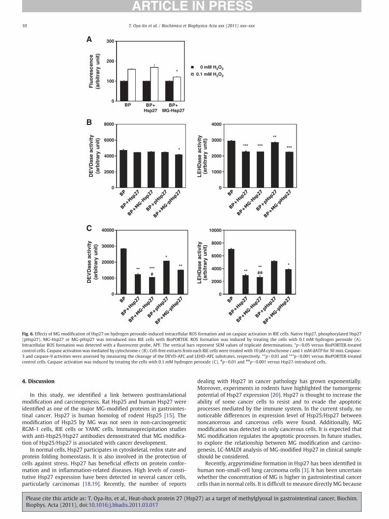
The efficacy of MG modification of Hsp27 on hydrogen peroxide-induced intracellular ROS formation was evaluated. Native Hsp27,pHsp27, MG-Hsp27 or MG-pHsp27 was introduced into RIE cells withBioPORTER. To detect ROS, we used 2-[6-(4′-amino)phenoxy-3H-xanthen-3-on-9-yl] benzoic acid (APF). APF can detect hydroxylradicals (·OH) and is completely resistant to autoxidation, both invitro and in vivo[17]. Hydrogen peroxide treatment caused an increasein the amount of intracellular ·OH of BioPORTER-treated control RIEcells in a concentration-dependentmanner (Fig. 6A). The introductionof MG-Hsp27 prevented ·OH generation in the cells treated with0.1 mM hydrogen peroxide compared with the introduction of nativeHsp27 or the BioPORTER-treated controls.
It is known that the formation of large oligomeric Hsp27 preventscytochrome c-mediated caspase activation [16], so we examinedwhetherMGmodification affected the regulation of caspase activationby Hsp27 using a cell-free system. The addition of cytochrome c anddATP to extracts from BioPORTER-treated control RIE cells resulted inthe activation of caspase-3 and caspase-9 (Fig. 6B). Extracts fromMG-pHsp27-introduced cells inhibited the activation of caspase-3,
Please cite this article as: T. Oya-Ito, et al., Heat-shock protein 27 (Hsp2Biophys. Acta (2011), doi:10.1016/j.bbadis.2011.03.017
whereas no inhibition was observed by using extracts from nativeHsp27- or pHsp27-introduced cells. Caspase-9 activation was inhib-ited by the introduction of all Hsp27 moieties (Hsp27, MG-Hsp27,pHsp27, and MG-pHsp27; pb0.01 versus BioPORTER-treated controlcells). MG modification did not affect the inhibition of caspase-9activation by Hsp27. The introduction of pHsp27 was less effective ininhibiting caspase-9 activation, but this inhibitory effect of pHsp27was enhanced by MG modification. These results suggested that MGmodification of pHsp27 potentiate the suppression of cytochrome c-mediated caspase activation.
We also determinedwhether Hsp27 introduction exerts a protectiveeffect against caspase activation during hydrogen peroxide-inducedapoptotic cell death. In BioPORTER-treated control RIE cells, bothcaspase-3 and caspase-9 were activated by treatment with 0.1 mMhydrogen peroxide. The introduction of Hsp27 into RIE cells preventedboth caspase-3 and caspase-9 activation, as shown in Fig. 6C. MGmodification enhanced the inhibitory effect of Hsp27 on caspase-3(pb0.01 versusHsp27-introduced cells) and caspase-9 (pb0.001 versusHsp27-introduced cells) activations. Phosphorylation of Hsp27 resultedin the loss of this effect. MG modification of pHsp27 increased theinhibitory effects of both caspase-3 and caspase-9 activations, but theoverall effect was still less than that seen with Hsp27. These resultsindicate that MG modification is essential for conferring protectionagainst caspase-mediated apoptosis induced by hydrogen peroxide.
3.4. Intracellular levels of lactate, the principal end-product of MGmetabolism
It is difficult to measure intracellular levels of MG directly becauseof its high reactivity. MG reacts rapidly with guanidine group such asarginine residue [12]. The glyoxalase system, comprising themetalloenzymes glyoxalase I and glyoxalase II, is an almost universalmetabolic pathway involved in the detoxification of the glycolyticbyproduct MG to D-lactate. Production of lactate in RGM-1 and RGK-1cells was measured by using D-lactate and L-lactate dehydrogenase-based assays (Fig. 8). The level of D-lactate in RGK-1 cells was found tobe significantly higher compared with that in RGM-1 cells (2-fold,pb0.05). The intracellular D-lactate contents of RGM-1 and RGK-1cells were 0.06±0.01 and 0.13±0.01 mM/mg of cell lysate protein,respectively. Moreover, the contents of L-lactate of RGM-1 and RGK-1
7) as a target of methylglyoxal in gastrointestinal cancer, Biochim.

Fig. 5. Effect ofMGmodificationofHsp27onhydrogenperoxide-induced apoptosis. NativeHsp27, orMG-Hsp27,was introduced intoRIE cellswithBioPORTER. After incubation for 24 and48 h at 37 °C, the effect of Hsp27 on cell viability was assessed by WST-8 (A). After a 4-h incubation, apoptosis was induced by treating the cells with hydrogen peroxide for 24 h. Theintroductions of native Hsp27 andMG-Hsp27were assessed byWestern blot analysis (B). After treating cells with hydrogen peroxide for 24 h, cell extracts from Hsp27- andMG-Hsp27-introduced cellswereexamined. Theextent of apoptosis inRIE cellswas assayed by FACS (FITC-AnnexinVbinding and PI staining) (a) BioPORTER-treated cells, (b)Hsp27-introduced cells,(c) MG-Hsp27-introduced cells, (d) apoptosis analyzed by FITC-Annexin V binding (red: BioPORTER, green: BioPORTER+Hsp27, orange: BioPORTER+MG-Hsp27) (C).
9T. Oya-Ito et al. / Biochimica et Biophysica Acta xxx (2011) xxx–xxx
cells were 0.32±0.01 and 0.77±0.01 mM/mg of cell lysate protein,respectively. The level of L-lactate in RGK-1 cells was 2.4 times higherthan that in RGM-1 cells (pb0.00001). In RGK-1 and RGM-1 cells,the concentration of D-lactate was one-sixth and one-fifth of
Please cite this article as: T. Oya-Ito, et al., Heat-shock protein 27 (Hsp2Biophys. Acta (2011), doi:10.1016/j.bbadis.2011.03.017
concentration of L-lactate, respectively. The total lactate content inRGK-1 cells was 0.90±0.08 mM/mg of cell lysate protein, while thatin RGM-1 cells was 0.38±0.07 mM/mg of cell lysate protein. Theseresults support high levels of lactate in cancer cells.
7) as a target of methylglyoxal in gastrointestinal cancer, Biochim.
0
2000
4000
6000
8000
10000
LE
HD
ase
acti
vity
(arb
itra
ry u
nit
)
****##
*
0
10000
20000
30000
40000
DE
VD
ase
acti
vity
(arb
itra
ry u
nit
)
**
*
***#
**
0
1000
2000
3000
4000
LE
HD
ase
acti
vity
(arb
itra
ry u
nit
)
*********
**
0
2000
4000
6000
8000
DE
VD
ase
acti
vity
(arb
itra
ry u
nit
)
*
*0 mM H2O2
0.1 mM H2O2
Flu
ore
scen
ce(a
rbit
rary
un
it)
BP BP+Hsp27
BP+MG-Hsp27
A
B
C
0
300
100
200
Fig. 6. Effects of MG modification of Hsp27 on hydrogen peroxide-induced intracellular ROS formation and on caspase activation in RIE cells. Native Hsp27, phosphorylated Hsp27(pHsp27), MG-Hsp27 or MG-pHsp27 was introduced into RIE cells with BioPORTER. ROS formation was induced by treating the cells with 0.1 mM hydrogen peroxide (A).Intracellular ROS formation was detected with a fluorescent probe, APF. The vertical bars represent SEM values of triplicate determinations. *pb0.05 versus BioPORTER-treatedcontrol cells. Caspase activation was mediated by cytochrome c (B). Cell-free extracts from each RIE cells were treated with 10 μM cytochrome c and 1 mM dATP for 30 min. Caspase-3 and caspase-9 activities were assessed by measuring the cleavage of the DEVD-AFC and LEHD-AFC substrates, respectively. **pb0.01 and ***pb0.001 versus BioPORTER-treatedcontrol cells. Caspase activation was induced by treating the cells with 0.1 mM hydrogen peroxide (C). #pb0.01 and ##pb0.001 versus Hsp27-introduced cells.
10 T. Oya-Ito et al. / Biochimica et Biophysica Acta xxx (2011) xxx–xxx
4. Discussion
In this study, we identified a link between posttranslationalmodification and carcinogenesis. Rat Hsp25 and human Hsp27 wereidentified as one of the major MG-modified proteins in gastrointes-tinal cancer. Hsp27 is human homolog of rodent Hsp25 [15]. Themodification of Hsp25 by MG was not seen in non-carcinogeneticRGM-1 cells, RIE cells or YAMC cells. Immunoprecipitation studieswith anti-Hsp25/Hsp27 antibodies demonstrated that MG modifica-tion of Hsp25/Hsp27 is associated with cancer development.
In normal cells, Hsp27 participates in cytoskeletal, redox state andprotein folding homeostasis. It is also involved in the protection ofcells against stress. Hsp27 has beneficial effects on protein confor-mation and in inflammation-related diseases. High levels of consti-tutive Hsp27 expression have been detected in several cancer cells,particularly carcinomas [18,19]. Recently, the number of reports
Please cite this article as: T. Oya-Ito, et al., Heat-shock protein 27 (Hsp2Biophys. Acta (2011), doi:10.1016/j.bbadis.2011.03.017
dealing with Hsp27 in cancer pathology has grown exponentially.Moreover, experiments in rodents have highlighted the tumorigenicpotential of Hsp27 expression [20]. Hsp27 is thought to increase theability of some cancer cells to resist and to evade the apoptoticprocesses mediated by the immune system. In the current study, nonoticeable differences in expression level of Hsp25/Hsp27 betweennoncancerous and cancerous cells were found. Additionally, MGmodification was detected in only cancerous cells. It is expected thatMG modification regulates the apoptotic processes. In future studies,to explore the relationship between MG modification and carcino-genesis, LC-MALDI analysis of MG-modified Hsp27 in clinical sampleshould be considered.
Recently, argpyrimidine formation in Hsp27 has been identified inhuman non-small-cell lung carcinoma cells [3]. It has been uncertainwhether the concentration of MG is higher in gastrointestinal cancercells than in normal cells. It is difficult to measure directly MG because
7) as a target of methylglyoxal in gastrointestinal cancer, Biochim.

gastrointestinal cancer cells
MG-pHsp27 MG-Hsp27
MG
Hsp27
pHsp27
Cytochrome cCaspase-9Caspase-3
Fig. 7. A proposed mechanism for prevention of caspase activation by Hsp27. MGmodification in gastrointestinal cancer cells promotes Hsp27 and pHsp27 properties.
11T. Oya-Ito et al. / Biochimica et Biophysica Acta xxx (2011) xxx–xxx
of its high reactivity. Gluconeogenesis occurs without ATP consump-tion through the MG pathway when used as a substrate of ketonebodies via the pathway: butyric acid (butyrate) → beta-hydroxybu-tyrate → acetoacetate → acetone → acetol → methylglyoxal →S-D-lactol-glutathione → D -lactate → pyruvate → D-lactate [21]. It haspreviously been reported that pyruvate concentration is significantlylower in colon tumors than in normal tissue, and slightly lower instomach tumors, whereas lactate concentration in tumor tissues ishigher in both tumor types than in normal tissue [2]. We measuredthe levels of intracellular lactate which is the end molecule of the MGpathway because MG is difficult to measure directly. Indeed, the totallactate content in MNNG-induced mutant of RGM-1 gastric epithelialcell line, which is named RGK-1 cells, was dramatically higher thanthat in RGM-1 cells (Fig. 8). This clearly indicates that cancer cells arehighly dependent on anaerobic breakdown of pyruvate. High lactatedehydrogenase-5 activity, and the resulting increase in conversion ofpyruvate to lactate has been identified in colon cancer [22,23]. Activeglycolysis also increases the cytosolic NADH/NAD+ ratio, therebyincreasing the activity of lactate dehydrogenase [24]. Enhancedpyruvate-to-lactate conversion may also be due to the activation ofthe pyruvate dehydrogenase kinase, isozyme 1 (PDK1), in cancer cells,which inactivates pyruvate dehydrogenase and leads to the inactiva-tion of the TCA cycle [25]. Therefore, it is likely that cancer cellspreferentially use glucose because of intrinsic metabolic characteris-tics, and micro-environmental factors such as hypoxia. Moreover, theMG pathway represents an energetically unfavorable bypass to theglycolytic reactions of the lower Embden–Meyerhof–Parnas pathwayusing pyruvate [26,27]. Activation of the MG pathway has beenconfirmed when the intracellular uptake of glucose 6-phosphate orother carbon substrates such as xylose, lactose, arabinose, glycerol orgluconate are deregulated by mutation and/or by addition of cAMP
0
0.2
0.4
0.6
0.8
1
D-lactate L-lactate
Lact
ate
(mM
/mg
of
cell
lysa
te p
rote
in)
RGK-1
RGM-1
*
**
Fig. 8. Contents of D-lactate and L-lactate in RGK-1 cells and RGM-1 cells. Intracellularlactate levels were estimated by D-lactate and L-lactate dehydrogenase-based assays.*pb0.05 and **pb0.00001 versus RGM-1 cells.
Please cite this article as: T. Oya-Ito, et al., Heat-shock protein 27 (Hsp2Biophys. Acta (2011), doi:10.1016/j.bbadis.2011.03.017
[28–30]. Moreover, the extremely low glucose content in tumortissues might result from both a poor blood supply and the highglucose consumption by cancer cells. In addition, the glyoxalasepathway catalyzes the formation of lactate fromMG in the presence ofglutathione. D-lactate is produced in the MG pathway as the end-product of glyoxalase II enzymatic reaction. The intracellular D-lactateconcentration in RGK-1 cells was twice as high as that in RGM-1 cells(pb0.05). There is an increase in the flux of MGmetabolized to lactatevia the glyoxalase pathway with high glucose concentrations [31].RGK-1 cells are the first MNNG-induced neoplastic mutant cellsderived from a noncancerous, nonembryonic gastric epithelial cellline. Ultrastructural results show homogenous, dense secretorygranules and deformed mitochondria in RGK-1 cells [10]. RGM-1cells have no secretory granules, normal-shaped mitochondria, andabundant polyribosomes. Moreover, mutant RGK-1 cells have showntumorigenic potential in all mice into which they have been injected[10]. We consider that the intracellular accumulation of MG isimplicated in the pathogenesis of cancer in RGK-1 cells. It is importantto determine whether high MG modification promotes the develop-ment of cancer. Because the intracellular MG concentration isregulated by glyoxalase, the experiments of overexpression orknockdown of glyoxalase should be done in the future.
The large oligomers of Hsp27, which bear chaperone-like activity,are also responsible for the tumorigenic activity of Hsp27 [32].Therefore, it cannot be excluded that large Hsp27 oligomers may actin a manner similar to Hsp90, and bind specific client proteins thatparticipate in the tumorigenic and metastatic processes. In this study,numerous post-translational modifications of Hsp25/Hsp27 in a largenumber of cancer cells were found. Recombinant Hsp27 andphosphorylated Hsp27 were polymerized during incubation withMG in vitro. Moreover, large MG-modified Hsp27 and MG-modifiedpHsp27 oligomers were detected in tumorigenic HT-29 cells. It issuggested that the polymerizations of Hsp27 and pHsp27 arepromoted by modification of MG. In Fig. 3, cross-linked Hsp27 andpHsp27 increased with increasing concentration of MG. Previousresearch has demonstrated that chaperone-like activity of Hsp27,even pHsp27, is greatly facilitated by MG modification [9]. Addition-ally, Hsp modification by MG enhances the hydrophobicity [33]. Thechaperone function of alpha-crystallin is attributed to hydrophobicregions of the protein, and modification by low concentration of MGincreases its hydrophobicity [33]. Hsp27 also has hydrophobic N-terminal region that contributes to its oligomerization [34]. Anincreased molecular chaperone function in cancer cells is likelyrelated to oligomerization of Hsp27 by MG modification and anincreased hydrophobicity. Future studies will investigate the effect ofMG modification on chaperone function in cancer cells.
The formation of argpyrimidine on Hsp27 has also been identifiedin endothelial cells [35], lens epithelial cells [9] and rat kidneymesangial cells [36], indicating that Hsp27 is a protein highlysusceptible to MGmodification. MALDI-MS/MS analysis of the peptidefragments derived from recombinant human Hsp27 showed MGmodification sites at Arg-5, Arg-96, Arg-127, Arg-136, Arg-188, andLys-123. For the first time, we confirmed the formation of argpyr-imidine on trypsin- and V8 protease-digested fragments of MG-modified Hsp27 by LC-MALDI-TOF MS/MS analysis. However, wefailed to detect argpyrimidine adducts in the peptide fragmentsderived from MG-modified phosphorylated Hsp27. It is unclearwhether MG is insensitive to pHsp27 or whether the peptidefragments derived fromMG-pHsp27 are beyond the limit of detection.The upper limit of detectable mass range is approximately 35,000 Da.At least immunoreactivity against argpyrimidine was observed withpolymerized MG-pHsp27 in our experiments. Meanwhile, it has beenshown that MG forms a number of adducts with lysine residues inpolymerized proteins. Nagaraj et al. [33] have described an imidazo-lium cross-link structure, imidazolysine. In this study, because theantibody does not recognize imidazolysine [12], this structure was not
7) as a target of methylglyoxal in gastrointestinal cancer, Biochim.

12 T. Oya-Ito et al. / Biochimica et Biophysica Acta xxx (2011) xxx–xxx
searched in the data base of LC-MALDI-TOF MS/MS analyses. It ispredicted that imidazolium cross-link structures are generated inMG-modified Hsp27.
As described previously, Hsp27 is expressed throughout the bodyand plays an important role in cellular functions such as apoptosis [9]and actin polymerization [37–39]. In stressed cells, increased levels ofHsp27 facilitate the repair, or destruction, of damaged proteins, thuspromoting cell recovery. In our previous study, we found that Hsp27shows increased anti-apoptotic effects in lens epithelial cells aftermodification by MG, which is due to multiple mechanisms includingthe enhancement of chaperone function, and inhibition of caspaseactivity [9,33,40]. The transfer of MG-modified Hsp27 significantlyinhibits staurosporine-induced apoptotic cell death and ROS produc-tion in a human lens epithelial cell line (HLE B-3) [9]. In this study, RIEcells also exhibited enhanced anti-apoptotic property by MGmodification of Hsp27. In particular, MG modification of Arg-188 inHsp27 is essential for inhibiting cytochrome c-mediated caspaseactivation in 293T cells transfected with mutant of Hsp27 (Arg-188 toglycine) [41]. MALDI-MS/MS analysis of the peptide fragmentsderived fromMG-modified Hsp27 showed that Arg-188 is susceptibleto modification. YAMC cells expressed Hsp25 by treatment withbutyrate were not resistant to hydrogen peroxide-induced apoptosis(data not shown). Additionally, we found that the expression levels ofHsp25 showed no difference between RGM-1 cells and mutant RGK-1cells. Our experiments, especially immunoprecipitation assays,showed that the expressed Hsp25 in RIE cells and YAMC cells bytreatment with butyrate was not modified by MG, whereas Hsp25 inmutant RGK-1 cells and Hsp27 in HT-29 cells and in ascending colonand rectum of patient with adenoma and advanced cancer were.These results suggest that MG modification of Arg-188 in Hsp25/Hsp27 plays a key role in the development of cancer. It was impossibleto detect MG modification of Arg-188 in protein extracts fromcultured cells and clinical samples by MALDI-MS analysis due to lowexpression level. Future studies are needed to confirm the effect ofArg-188 in Hsp25/Hsp27 in cancer cells.
It has been speculated that MG may protect cells against hypergly-cemia-induced damage in diabetes because MG modification of Hsp27enhances its chaperone function, as has been shown for alpha-crystallin[9,33]. However, protein-modifying sugars and ascorbic acid have nosuch effect and actually reduce chaperone function [33]. Thus, it isbelieved that MG modification of Hsp27 is different from otherglycation. Additionally, a study in human lens epithelial cells indicatedthat MG modification enhances the binding of Hsp27 to cytochrome cand, subsequently, the protection from caspase-3 dependent cell death[9]. Our experiments showed that the transfer of Hsp27 using a cationiclipid inhibits the hydrogen-peroxide-induced apoptosis in the RIE cellline and that MG modification of Hsp27 enhances this inhibition. Theintroduction of MG-Hsp27 reduced intracellular ROS generation also.MG-modified Hsp27 showed no difference in proliferation of ratintestinal epithelial cell line compared to other controls (Hsp27, vehicle,and BP-treatment). When apoptosis was mediated by the addition ofboth cytochrome c and ATP to the cell-free cytosol, the inhibition ofcaspase-3 activation by phosphorylated Hsp27, and of caspase-9activation by both native and phosphorylated Hsp27, was enhancedby MG modification. This shows that MG modification of pHsp27prevents the activations of both caspase-3 and caspase-9 induced by therelease of cytosolic cytochrome c from mitochondria. Moreover, MGmodification of native Hsp27 and phosphorylated Hsp27 inhibited theactivations of both caspase-3 and caspase-9 during oxidant-induced celldeath. These results suggest that Hsp27 reacts immediately with MGand serves protective functions against caspase activation in gastroin-testinal cancer cells (Fig. 7). The enhanced inhibitory effects of MGmodification on caspase activation are similar to those seen forchaperone activity [9]. We propose that MG modification of Hsp27can protect against caspase-3 and caspase-9 activations withoutaffecting the rate of cytochrome c release and promote cell survival.
Please cite this article as: T. Oya-Ito, et al., Heat-shock protein 27 (Hsp2Biophys. Acta (2011), doi:10.1016/j.bbadis.2011.03.017
Almost all phosphorylated Hsp27 were modified by MG in lensepithelial cells [9] and in HT-29 cells. Thus, while MG modification ofHsp27 can maintain transparency and protect against apoptosis in lenscells which has a slow turnover rate, this modification may be involvedin the pathogenesis of cancer in the gastrointestinal epithelium whichhas a rapid turnover rate.
In conclusion, the specific immunoreactivity against the MG-modified protein was observed in gastrointestinal carcinoma cell linesand in ascending colon and rectum of patients with cancer, but not innormal subjects. We identified this modified protein as Hsp25/Hsp27and showed for the first time that argpyrimidine adducts aregenerated at Arg-136 and Arg-188 of MG-modified Hsp27, which isthought to be necessary for its enhanced anti-apoptotic effects.Furthermore, we found that MG-modified Hsp27 effectively inhibitscaspase activation, ROS production, and the induction of apoptosis inhydrogen peroxide-treated cells. Therefore, our results suggest thatposttranslational modification of Hsp27 by MG may contribute to thedevelopment of gastrointestinal cancer.
5. Funding
This research was partially supported by FY 2009 and FY 2010Grant-in-Aid for Scientific Research for Young Scientists (B) to T. O.-I.from the Japan Society for the Promotion of Science, and by FY 2009Research for Promoting Technological Seeds A (discovery type) to T.O.-I. from the Independent Administrative Corporation Japan, Scienceand Technology Agency, and by the 170th Redox Life SciencesCommittee to T. Y. from the Japan Society for the Promotion ofScience.
Acknowledgments
We would like to thank Prof. Ram H. Nagaraj, of Case WesternReserve University, whose comments made an enormous contribu-tion to our work. We would also like to thank Prof. Etsuo Niki, of theNational Institute of Advanced Industrial Science and Technology, andProf. Koji Uchida, of Nagoya University, who gave us constructivecomments and kind encouragement.
References
[1] O. Warburg, On the origin of cancer cells, Science 123 (1956) 309–314.[2] A. Hirayama, K. Kami, M. Sugimoto, M. Sugawara, N. Toki, H. Onozuka, T. Kinoshita,
N. Saito, A. Ochiai, M. Tomita, H. Esumi, T. Soga, Quantitativemetabolome profilingof colon and stomach cancer microenvironment by capillary electrophoresis time-of-flight mass spectrometry, Cancer Res. 69 (2009) 4918–4925.
[3] J.W. van Heijst, H.W. Niessen, R.J. Musters, V.W. van Hinsbergh, K. Hoekman, C.G.Schalkwijk, Argpyrimidine-modified heat shock protein 27 in human non-smallcell lung cancer: a possible mechanism for evasion of apoptosis, Cancer Lett. 241(2006) 309–319.
[4] A.P. Arrigo, S. Simon, B. Gibert, C. Kretz-Remy, M. Nivon, A. Czekalla, D. Guillet, M.Moulin, C. Diaz-Latoud, P. Vicart, Hsp27 (HspB1) and alphaB-crystallin (HspB5) astherapeutic targets, FEBS Lett. 581 (2007) 3665–3674.
[5] H. Ren, M.W.Musch, K. Kojima, D. Boone, A. Ma, E.B. Chang, Short-chain fatty acidsinduce intestinal epithelial heat shock protein 25 expression in rats and IEC 18cells, Gastroenterology 121 (2001) 631–639.
[6] C. Decroos, Y. Li, G. Bertho, Y. Frapart, D. Mansuy, J.L. Boucher, Oxidative andreductive metabolism of tris(p-carboxyltetrathiaaryl)methyl radicals by livermicrosomes, Chem. Res. Toxicol. 22 (2009) 1342–1350.
[7] D. Stokoe, K. Engel, D.G. Campbell, P. Cohen, M. Gaestel, Identification of MAPKAPkinase 2 as a major enzyme responsible for the phosphorylation of the smallmammalian heat shock proteins, FEBS Lett. 313 (1992) 307–313.
[8] J. Rouse, P. Cohen, S. Trigon, M. Morange, A. Alonso-Llamazares, D. Zamanillo, T.Hunt, A.R. Nebreda, A novel kinase cascade triggered by stress and heat shock thatstimulates MAPKAP kinase-2 and phosphorylation of the small heat shockproteins, Cell 78 (1994) 1027–1037.
[9] T. Oya-Ito, B.F. Liu, R.H. Nagaraj, Effect of methylglyoxal modification andphosphorylation on the chaperone and anti-apoptotic properties of heat shockprotein 27, J. Cell. Biochem. 99 (2006) 279–291.
[10] O. Shimokawa, H. Matsui, Y. Nagano, T. Kaneko, T. Shibahara, A. Nakahara, I.Hyodo, A. Yanaka, H.J. Majima, Y. Nakamura, Y. Matsuzaki, Neoplastic transfor-mation and induction of H+, K+ −adenosine triphosphatase by N-methyl-N′-nitro-N-nitrosoguanidine in the gastric epithelial RGM-1 cell line, In Vitro Cell.Dev. Biol. Anim. 44 (2008) 26–30.
7) as a target of methylglyoxal in gastrointestinal cancer, Biochim.

13T. Oya-Ito et al. / Biochimica et Biophysica Acta xxx (2011) xxx–xxx
[11] R.B. Brandt, S.A. Siegel, M.G. Waters, M.H. Bloch, Spectrophotometric assay forD-(−)-lactate in plasma, Anal. Biochem. 102 (1980) 39–46.
[12] T. Oya, N. Hattori, Y. Mizuno, S. Miyata, S. Maeda, T. Osawa, K. Uchida,Methylglyoxal modification of protein. Chemical and immunochemical charac-terization of methylglyoxal–arginine adducts, J. Biol. Chem. 274 (1999)18492–18502.
[13] E. Muller, W. Neuhofer, A. Ohno, S. Rucker, K. Thurau, F.X. Beck, Heat shockproteins HSP25, HSP60, HSP72, HSP73 in isoosmotic cortex and hyperosmoticmedulla of rat kidney, Pflugers Arch. 431 (1996) 608–617.
[14] S. Gonin, N. Fabre-Jonca, C. Diaz-Latoud, J.P. Rouault, A.P. Arrigo, Transformationby T-antigen and other oncogenes delays Hsp25 accumulation in heat shockedNIH 3T3 fibroblasts, Cell Stress Chaperones 2 (1997) 238–251.
[15] T. Rogalla, M. Ehrnsperger, X. Preville, A. Kotlyarov, G. Lutsch, C. Ducasse, C. Paul, M.Wieske, A.P. Arrigo, J. Buchner, M. Gaestel, Regulation of Hsp27 oligomerization,chaperone function, and protective activity against oxidative stress/tumor necrosisfactor alpha by phosphorylation, J. Biol. Chem. 274 (1999) 18947–18956.
[16] H. Lambert, S.J. Charette, A.F. Bernier, A. Guimond, J. Landry, HSP27 multi-merization mediated by phosphorylation-sensitive intermolecular interactions atthe amino terminus, J. Biol. Chem. 274 (1999) 9378–9385.
[17] K. Setsukinai, Y. Urano, K. Kakinuma, H.J. Majima, T. Nagano, Development ofnovel fluorescence probes that can reliably detect reactive oxygen species anddistinguish specific species, J. Biol. Chem. 278 (2003) 3170–3175.
[18] D.R. Ciocca, S.K. Calderwood, Heat shock proteins in cancer: diagnostic, prognostic,predictive, and treatment implications, Cell Stress Chaperones 10 (2005) 86–103.
[19] S.K. Calderwood, M.A. Khaleque, D.B. Sawyer, D.R. Ciocca, Heat shock proteins incancer: chaperones of tumorigenesis, Trends Biochem. Sci. 31 (2006) 164–172.
[20] C. Garrido, A. Fromentin, B. Bonnotte, N. Favre, M. Moutet, A.P. Arrigo, P. Mehlen, E.Solary, Heat shock protein 27 enhances the tumorigenicity of immunogenic ratcolon carcinoma cell clones, Cancer Res. 58 (1998) 5495–5499.
[21] V.N. Titov, L.F. Dmitriev, V.A. Krylin, V.A. Dmitriev, Methylglyoxal—a test forimpaired biological functions of exotrophy and endoecology, low glucose level inthe cytosol and gluconeogenesis from fatty acids (a lecture), Klin. Lab. Diagn.(2010) 22–36.
[22] M.I. Koukourakis, A. Giatromanolaki, A. Polychronidis, C. Simopoulos, K.C. Gatter,A.L. Harris, E. Sivridis, Endogenous markers of hypoxia/anaerobic metabolism andanemia in primary colorectal cancer, Cancer Sci. 97 (2006) 582–588.
[23] R. Mazzanti, M. Solazzo, O. Fantappie, S. Elfering, P. Pantaleo, P. Bechi, F. Cianchi, A.Ettl, C. Giulivi, Differential expression proteomics of human colon cancer, Am. J.Physiol. Gastrointest. Liver Physiol. 290 (2006) G1329–G1338.
[24] J.M. Argiles, J. Azcon-Bieto, The metabolic environment of cancer, Mol. Cell.Biochem. 81 (1988) 3–17.
[25] J.G. Pan, T.W. Mak, Metabolic targeting as an anticancer strategy: dawn of a newera? Sci. STKE 2007 (2007) e14.
[26] R.A. Cooper, A. Anderson, The formation and catabolism of methylglyoxal duringglycolysis in Escherichia coli, FEBS Lett. 11 (1970) 273–276.
Please cite this article as: T. Oya-Ito, et al., Heat-shock protein 27 (Hsp2Biophys. Acta (2011), doi:10.1016/j.bbadis.2011.03.017
[27] Y. Kondoh, M. Kawase, Y. Kawakami, S. Ohmori, Concentrations of D-lactate and itsrelatedmetabolic intermediates in liver, blood, andmuscle of diabetic and starvedrats, Res. Exp. Med. (Berl.) 192 (1992) 407–414.
[28] R.S. Ackerman, N.R. Cozzarelli, W. Epstein, Accumulation of toxic concentrations ofmethylglyoxal by wild-type Escherichia coli K-12, J. Bacteriol. 119 (1974) 357–362.
[29] R. Puskas, N. Fredd, C. Gazdar, A. Peterkofsky, Methylglyoxal-mediated growthinhibition in an Escherichia coli cAMP receptor protein mutant, Arch. Biochem.Biophys. 223 (1983) 503–513.
[30] R.J. Kadner, G.P. Murphy, C.M. Stephens, Two mechanisms for growth inhibitionby elevated transport of sugar phosphates in Escherichia coli, J. Gen. Microbiol. 138(1992) 2007–2014.
[31] P.J. Thornalley, Modification of the glyoxalase system in human red blood cells byglucose in vitro, Biochem. J. 254 (1988) 751–755.
[32] J.M. Bruey, C. Paul, A. Fromentin, S. Hilpert, A.P. Arrigo, E. Solary, C. Garrido,Differential regulation of HSP27 oligomerization in tumor cells grown in vitro andin vivo, Oncogene 19 (2000) 4855–4863.
[33] R.H. Nagaraj, T. Oya-Ito, P.S. Padayatti, R. Kumar, S. Mehta, K. West, B. Levison, J.Sun, J.W. Crabb, A.K. Padival, Enhancement of chaperone function of alpha-crystallin by methylglyoxal modification, Biochemistry 42 (2003) 10746–10755.
[34] S. Dai, Y. Jia, S.L. Wu, J.S. Isenberg, L.A. Ridnour, R.W. Bandle, D.A. Wink, D.D.Roberts, B.L. Karger, Comprehensive characterization of heat shock protein 27phosphorylation in human endothelial cells stimulated by the microbial dithiolethiolutin, J. Proteome Res. 7 (2008) 4384–4395.
[35] C.G. Schalkwijk, J. van Bezu, R.C. van der Schors, K. Uchida, C.D. Stehouwer, V.W.van Hinsbergh, Heat-shock protein 27 is a major methylglyoxal-modified proteinin endothelial cells, FEBS Lett. 580 (2006) 1565–1570.
[36] A.K. Padival, J.W. Crabb, R.H. Nagaraj, Methylglyoxal modifies heat shock protein27 in glomerular mesangial cells, FEBS Lett. 551 (2003) 113–118.
[37] P. Negre-Aminou, R.E. van Leeuwen, G.C. van Thiel, P. van den Ijssel, W.W. de Jong,R.A. Quinlan, L.H. Cohen, Differential effect of simvastatin on activation of Rac(1)vs. activation of the heat shock protein 27-mediated pathway upon oxidativestress, in human smoothmuscle cells, Biochem. Pharmacol. 64 (2002) 1483–1491.
[38] J. Huot, F. Houle, S. Rousseau, R.G. Deschesnes, G.M. Shah, J. Landry, SAPK2/p38-dependent F-actin reorganization regulates early membrane blebbing duringstress-induced apoptosis, J. Cell Biol. 143 (1998) 1361–1373.
[39] J.H. Lee, D. Sun, K.J. Cho, M.S. Kim, M.H. Hong, I.K. Kim, J.S. Lee, Overexpression ofhuman 27 kDa heat shock protein in laryngeal cancer cells confers chemoresistanceassociated with cell growth delay, J. Cancer Res. Clin. Oncol. 133 (2007) 37–46.
[40] A. Biswas, A. Miller, T. Oya-Ito, P. Santhoshkumar, M. Bhat, R.H. Nagaraj, Effect ofsite-directed mutagenesis of methylglyoxal-modifiable arginine residues on thestructure and chaperone function of human alphaA-crystallin, Biochemistry 45(2006) 4569–4577.
[41] H. Sakamoto, T. Mashima, K. Yamamoto, T. Tsuruo, Modulation of heat-shockprotein 27 (Hsp27) anti-apoptotic activity by methylglyoxal modification, J. Biol.Chem. 277 (2002) 45770–45775.
7) as a target of methylglyoxal in gastrointestinal cancer, Biochim.