Microplastics Determination in Gastrointestinal Tracts ... - MDPI
Upload
khangminh22Category
view
4download
0
Citation: Koustas, E.; Trifylli, E.-M.;
Sarantis, P.; Papadopoulos, N.;
Karapedi, E.; Aloizos, G.; Damaskos,
C.; Garmpis, N.; Garmpi, A.;
Papavassiliou, K.A.; et al.
Immunotherapy as a Therapeutic
Strategy for Gastrointestinal
Cancer—Current Treatment Options
and Future Perspectives. Int. J. Mol.
Sci. 2022, 23, 6664. https://doi.org/
10.3390/ijms23126664
Academic Editor: Simona Gurzu
Received: 26 May 2022
Accepted: 14 June 2022
Published: 15 June 2022
Publisher’s Note: MDPI stays neutral
with regard to jurisdictional claims in
published maps and institutional affil-
iations.
Copyright: © 2022 by the authors.
Licensee MDPI, Basel, Switzerland.
This article is an open access article
distributed under the terms and
conditions of the Creative Commons
Attribution (CC BY) license (https://
creativecommons.org/licenses/by/
4.0/).
International Journal of
Molecular Sciences
Review
Immunotherapy as a Therapeutic Strategy for GastrointestinalCancer—Current Treatment Options and Future PerspectivesEvangelos Koustas 1,2, Eleni-Myrto Trifylli 1,2, Panagiotis Sarantis 1 , Nikolaos Papadopoulos 2 ,Eleni Karapedi 2, Georgios Aloizos 2, Christos Damaskos 3,4 , Nikolaos Garmpis 5,6 , Anna Garmpi 6,Kostas A. Papavassiliou 1 , Michalis V. Karamouzis 1,* and Athanasios G. Papavassiliou 1,*
1 Department of Biological Chemistry, Medical School, National and Kapodistrian University of Athens,11527 Athens, Greece; [email protected] (E.K.); [email protected] (E.-M.T.);[email protected] (P.S.); [email protected] (K.A.P.)
2 First Department of Internal Medicine, 417 Army Share Fund Hospital, 11521 Athens, Greece;[email protected] (N.P.); [email protected] (E.K.); [email protected] (G.A.)
3 ‘N.S. Christeas’ Laboratory of Experimental Surgery and Surgical Research, Medical School, National andKapodistrian University of Athens, 11527 Athens, Greece; [email protected]
4 Renal Transplantation Unit, ‘Laiko’ General Hospital, 11527 Athens, Greece5 Second Department of Propaedeutic Surgery, ‘Laiko’ General Hospital, Medical School, National and
Kapodistrian University of Athens, 11527 Athens, Greece; [email protected] First Department of Pathology, Medical School, National and Kapodistrian University of Athens,
11527 Athens, Greece; [email protected]* Correspondence: [email protected] (M.V.K.); [email protected] (A.G.P.)
Abstract: Gastrointestinal (GI) cancer constitutes a highly lethal entity among malignancies inthe last decades and is still a major challenge for cancer therapeutic options. Despite the currentcombinational treatment strategies, including chemotherapy, surgery, radiotherapy, and targetedtherapies, the survival rates remain notably low for patients with advanced disease. A betterknowledge of the molecular mechanisms that influence tumor progression and the development ofoptimal therapeutic strategies for GI malignancies are urgently needed. Currently, the developmentand the assessment of the efficacy of immunotherapeutic agents in GI cancer are in the spotlightof several clinical trials. Thus, several new modalities and combinational treatments with otheranti-neoplastic agents have been identified and evaluated for their efficiency in cancer management,including immune checkpoint inhibitors, adoptive cell transfer, chimeric antigen receptor (CAR)-Tcell therapy, cancer vaccines, and/or combinations thereof. Understanding the interrelation amongthe tumor microenvironment, cancer progression, and immune resistance is pivotal for the optimaltherapeutic management of all gastrointestinal solid tumors. This review will shed light on the recentadvances and future directions of immunotherapy for malignant tumors of the GI system.
Keywords: gastrointestinal tumors; cancer; immunotherapy; checkpoint inhibitors; cancer vaccine;tumor microenvironment
1. Introduction
There is a global trend of a continuously increasing incidence of gastrointestinal (GI)cancers with various epidemiological backgrounds and genetic and epigenetic aberrations,making them the most frequent cancers globally with generally high mortality rates. De-spite various conventional therapeutic options, such as chemotherapy, radiotherapy, andsurgical approaches, patients with end-stage disease present an unfavorable prognosis [1].As a result, novel anti-cancer therapies have been developed, such as immunotherapy.
Immunotherapy is considered a step-up strategy for the management of a wide spec-trum of malignancies, especially when those that have reached their end stage. Immunother-apeutic agents exhibit a favorable targeted effect on malignant cells, either promoting orinhibiting immune responses via interacting with immunogens presented on malignant
Int. J. Mol. Sci. 2022, 23, 6664. https://doi.org/10.3390/ijms23126664 https://www.mdpi.com/journal/ijms

Int. J. Mol. Sci. 2022, 23, 6664 2 of 26
cells. Importantly, they do not induce any detrimental effects on normal, non-cancerouscells, which makes them an optimal therapeutic option [2].
There is a wide range of immunotherapeutic modalities that are introduced in GI can-cer management, including immune checkpoint inhibitors, adoptive cell transfer, chimericantigen receptor (CAR)-T cell therapy, cancer vaccines, and/or combinations thereof. Theleast beneficial effect for cancer management is demonstrated by the newly developed can-cer vaccines [3], in comparison with immune checkpoint inhibitors that present remarkableeffects. Immune checkpoint blockade constitutes one of the most widely used immunother-apeutic modalities, aiming at three significant molecular targets: (i) the programmeddeath-ligand 1 (PD-L1), presented on the surface of cancer cells and antigen-presentingcells (APCs), (ii) the programmed cell death protein (PD-1) on the surface of lympho-cytes, and (iii) the cytotoxic T-lymphocyte associated protein-4 (CTLA-4) on the surfaceof regulatory T cells (Tregs). All of these molecular targets have a pivotal role in cancer-related immune responses, due to the fact that they induce the so-called tumor escapemechanism, avoiding the normal cellular apoptotic process and, therefore, leading to theimmortalization of cancer cells [4].
A better knowledge of the mechanisms that lead to tumor escape as well as a betterunderstanding of the impact of tumor microenvironment (TME) on the efficacy of im-munotherapy, are considered crucial for the optimal management of GI cancer. There isan important interplay between immune responses and TME, being highly investigatedespecially for gastric and colorectal malignancies [5], as it provides multiple potentialtherapeutic targets for anti-neoplastic treatment [6].
The immune response is considered a multiplex and multi-stepped procedure, com-prising three distinct steps: the first asymptomatic step includes the primary endeavor ofcancer cells for elimination, whereby innate immune cells recognize the malignant cellsand try to eliminate them via their cytotoxic effect and the production of antibodies againstimmunogens on the malignant cell surface through the adaptive immune response. Thisfirst step is followed by the step of balance, during which limitation of tumor progressionis unfeasible, since malignant tumors can escape immunosurveillance. The last step, thetumor escape step, is symptomatic and takes place when the tumor progresses despiteimmunotherapy [7–9]. This process is achieved by tumor cells as they escape from innateimmune system recognition. The restriction of antigen expression on cancer cell surfacesis considered the key mechanism for avoiding recognition by CD8+ T cells [10]. The in-duction of TME immunosuppression is attributed to many molecules secreted by cancercells, such as inhibitory checkpoints that lead to the recruitment of immune cells. Amongthese recruited immune cells are myeloid-derived suppressor cells (MDSCs), which areassociated with poor prognosis [11] and immune resistance [12,13]. Some other immuno-suppressive cells are T and B regulatory cells, as well as tumor-associated macrophages(TAMs), which also promote tumor progression and neoangiogenesis [14]. Specifically,Bregs impede the action of cytotoxic T cells and promote carcinogenesis mainly via thesecretion of Il-10, while Tregs express fork head box P3 (FOXP3) and they are associatedwith the downregulation or suppression of T-effector cells [12,15]. In Figure 1, we presentsome of the main components of TME.

Int. J. Mol. Sci. 2022, 23, 6664 3 of 26Int. J. Mol. Sci. 2022, 23, x FOR PEER REVIEW 3 of 27
Figure 1. Schematic presentation of TME elements that induce immunosuppression, tumor pro-
gression, and metastasis. TME constitutes a surrounding stroma with a wide variety of cells, such
as immune cells, fibroblasts, as well as many regulatory molecules, which are considered potential
druggable targets. MDSC, B and T regulatory cells, TAMs, and cancer-associated fibroblasts (CAFs)
have quite significant implications for cancer management, as they elicit an immunosuppressive
effect that limits the efficacy of immunotherapeutic agents. TME immunosuppression is attributed
to various molecules secreted by cancer cells, such as inhibitory checkpoints leading to the re-
cruitment of immune cells, including MDSCs, T regulatory cells, and TAMs. This figure was cre-
ated with BioRender.com, accessed on 14 May 2022 (agreement number UO23X0OEMQ).
The above phenomena imply the significant influence of TME on tumor progression,
invasion, metastasis, and development of therapy resistance attributed to the impedi-
ment of cytotoxic T cells’ action, as well as the secretion of immunosuppressive molecules
that interfere with immune anti-cancer responses [16,17].
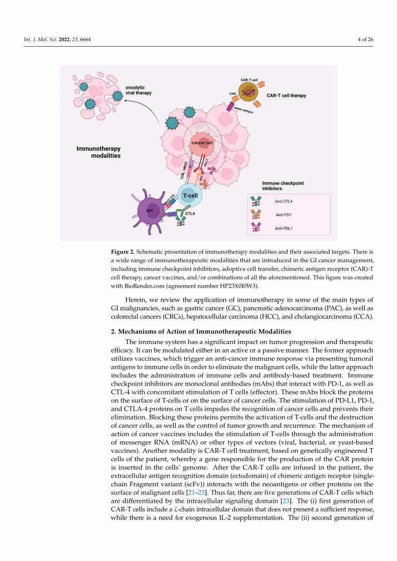
Additionally, the expression of inhibitory immune checkpoints enhances the im-
munosuppressive mechanism of tumor cells. Figure 2 depicts these checkpoints on tumor
cells and the action of PD1/PD-L1 and CTLA-1/B7, which constitute important therapeu-
tic targets. While under normal conditions, immune checkpoints on T cell surfaces act as
a brake for triggering an immune response, tumor cells hijack these molecules to promote
the tumor-escape phenomenon [18]. These immune checkpoints, including CTLA-4,
PD-1, and PD-L1, are involved in the biology of many malignant tumors [19]. However,
there are many reports on tumors displaying immunotherapy resistance, and as a result,
there is no evident improvement in survival rate [18,20].
Herein, we review the application of immunotherapy in some of the main types of
GI malignancies, such as gastric cancer (GC), pancreatic adenocarcinoma (PAC), as well
as colorectal cancers (CRCs), hepatocellular carcinoma (HCC), and cholangiocarcinoma
(CCA).
Figure 1. Schematic presentation of TME elements that induce immunosuppression, tumor progres-sion, and metastasis. TME constitutes a surrounding stroma with a wide variety of cells, such asimmune cells, fibroblasts, as well as many regulatory molecules, which are considered potentialdruggable targets. MDSC, B and T regulatory cells, TAMs, and cancer-associated fibroblasts (CAFs)have quite significant implications for cancer management, as they elicit an immunosuppressiveeffect that limits the efficacy of immunotherapeutic agents. TME immunosuppression is attributed tovarious molecules secreted by cancer cells, such as inhibitory checkpoints leading to the recruitmentof immune cells, including MDSCs, T regulatory cells, and TAMs. This figure was created withBioRender.com, accessed on 14 May 2022 (agreement number UO23X0OEMQ).
The above phenomena imply the significant influence of TME on tumor progression,invasion, metastasis, and development of therapy resistance attributed to the impedimentof cytotoxic T cells’ action, as well as the secretion of immunosuppressive molecules thatinterfere with immune anti-cancer responses [16,17].
Additionally, the expression of inhibitory immune checkpoints enhances the immuno-suppressive mechanism of tumor cells. Figure 2 depicts these checkpoints on tumor cellsand the action of PD1/PD-L1 and CTLA-1/B7, which constitute important therapeutictargets. While under normal conditions, immune checkpoints on T cell surfaces act as abrake for triggering an immune response, tumor cells hijack these molecules to promotethe tumor-escape phenomenon [18]. These immune checkpoints, including CTLA-4, PD-1,and PD-L1, are involved in the biology of many malignant tumors [19]. However, there aremany reports on tumors displaying immunotherapy resistance, and as a result, there is noevident improvement in survival rate [18,20].
Int. J. Mol. Sci. 2022, 23, 6664 4 of 26Int. J. Mol. Sci. 2022, 23, x FOR PEER REVIEW 4 of 27
Figure 2. Schematic presentation of immunotherapy modalities and their associated targets. There
is a wide range of immunotherapeutic modalities that are introduced in the GI cancer management,
including immune checkpoint inhibitors, adoptive cell transfer, chimeric antigen receptor (CAR)-T
cell therapy, cancer vaccines, and/or combinations of all the aforementioned. This figure was cre-
ated with BioRender.com (agreement number HP23X0I0W3).
2. Mechanisms of Action of Immunotherapeutic Modalities
The immune system has a significant impact on tumor progression and therapeutic
efficacy. It can be modulated either in an active or a passive manner. The former ap-
proach utilizes vaccines, which trigger an anti-cancer immune response via presenting
tumoral antigens to immune cells in order to eliminate the malignant cells, while the
latter approach includes the administration of immune cells and antibody-based treat-
ment. Immune checkpoint inhibitors are monoclonal antibodies (mAbs) that interact with
PD-1, as well as CTL-4 with concomitant stimulation of T cells (effector). These mAbs
block the proteins on the surface of T-cells or on the surface of cancer cells. The stimula-
tion of PD-L1, PD-1, and CTLA-4 proteins on T cells impedes the recognition of cancer
cells and prevents their elimination. Blocking these proteins permits the activation of
T-cells and the destruction of cancer cells, as well as the control of tumor growth and
recurrence. The mechanism of action of cancer vaccines includes the stimulation of
T-cells through the administration of messenger RNA (mRNA) or other types of vectors
(viral, bacterial, or yeast-based vaccines). Another modality is CAR-T cell treatment,
based on genetically engineered T cells of the patient, whereby a gene responsible for the
production of the CAR protein is inserted in the cells’ genome. After the CAR-T cells are
infused in the patient, the extracellular antigen recognition domain (ectodomain) of
chimeric antigen receptor (single-chain Fragment variant (scFv)) interacts with the neo-
antigens or other proteins on the surface of malignant cells [21–23]. Thus far, there are
five generations of CAR-T cells which are differentiated by the intracellular signaling
domain [23]. The (i) first generation of CAR-T cells include a ζ-chain intracellular domain
that does not present a sufficient response, while there is a need for exogenous IL-2 sup-
plementation. The (ii) second generation of CAR-T cells include OX-40, or CD28, and
4-1BB intracellular domains. The latter can be activated in the absence of the antigen, a
phenomenon called constitutive stimulation. The (iii) third generation combines various
costimulatory signaling domains with similar effects to the second generation. The (iv)
Figure 2. Schematic presentation of immunotherapy modalities and their associated targets. There isa wide range of immunotherapeutic modalities that are introduced in the GI cancer management,including immune checkpoint inhibitors, adoptive cell transfer, chimeric antigen receptor (CAR)-Tcell therapy, cancer vaccines, and/or combinations of all the aforementioned. This figure was createdwith BioRender.com (agreement number HP23X0I0W3).
Herein, we review the application of immunotherapy in some of the main types ofGI malignancies, such as gastric cancer (GC), pancreatic adenocarcinoma (PAC), as well ascolorectal cancers (CRCs), hepatocellular carcinoma (HCC), and cholangiocarcinoma (CCA).
2. Mechanisms of Action of Immunotherapeutic Modalities
The immune system has a significant impact on tumor progression and therapeuticefficacy. It can be modulated either in an active or a passive manner. The former approachutilizes vaccines, which trigger an anti-cancer immune response via presenting tumoralantigens to immune cells in order to eliminate the malignant cells, while the latter approachincludes the administration of immune cells and antibody-based treatment. Immunecheckpoint inhibitors are monoclonal antibodies (mAbs) that interact with PD-1, as well asCTL-4 with concomitant stimulation of T cells (effector). These mAbs block the proteinson the surface of T-cells or on the surface of cancer cells. The stimulation of PD-L1, PD-1,and CTLA-4 proteins on T cells impedes the recognition of cancer cells and prevents theirelimination. Blocking these proteins permits the activation of T-cells and the destructionof cancer cells, as well as the control of tumor growth and recurrence. The mechanism ofaction of cancer vaccines includes the stimulation of T-cells through the administrationof messenger RNA (mRNA) or other types of vectors (viral, bacterial, or yeast-basedvaccines). Another modality is CAR-T cell treatment, based on genetically engineered Tcells of the patient, whereby a gene responsible for the production of the CAR proteinis inserted in the cells’ genome. After the CAR-T cells are infused in the patient, theextracellular antigen recognition domain (ectodomain) of chimeric antigen receptor (single-chain Fragment variant (scFv)) interacts with the neoantigens or other proteins on thesurface of malignant cells [21–23]. Thus far, there are five generations of CAR-T cells whichare differentiated by the intracellular signaling domain [23]. The (i) first generation ofCAR-T cells include a ζ-chain intracellular domain that does not present a sufficient response,while there is a need for exogenous IL-2 supplementation. The (ii) second generation of

Int. J. Mol. Sci. 2022, 23, 6664 5 of 26
CAR-T cells include OX-40, or CD28, and 4-1BB intracellular domains. The latter can beactivated in the absence of the antigen, a phenomenon called constitutive stimulation. The(iii) third generation combines various costimulatory signaling domains with similar effectsto the second generation. The (iv) fourth generation comprises the so-called “TRUCK”T cells that include a cytokine, such as IL-12, which leads to a cytokine-mediated killingof cancer cells via the attraction and the activation of additional immune cells, while the(v) fifth generation includes IL-2 receptors that induce JAK/STAT pathway activation [24,25].Additionally, there are mAbs with a scFv that interact with T-cells (effector) and prevent thestimulation of cytokines, which either induces or suppresses the immunological response.Furthermore, the antibody-based therapy represents a promising therapeutic modalityfor the management of solid tumors. The introduction of bi- and tri-specific antibodies isconsidered a revolutionary approach. The former includes one arm specialized for CD3 anda second arm targets the neoantigen, while the stimulation of T cells is achieved without theinvolvement of MHC [21,26]. Finally, there is another immunotherapy modality based onthe depletion of regulatory T cells (Treg), for example daclizumab, a monoclonal antibodyagainst CD25 of Tregs [27].
3. Immunotherapy in Gastrointestinal Cancers
Conventionally, the mainstay treatment for GI cancers has been focused around radia-tion and/or chemotherapy, antiangiogenic therapy, surgical intervention, or a combinationof these treatments; nevertheless, the overall survival of patients with GI tumors remainspoor. Aiming to decrease morbidity and improve mortality outcomes in GI cancer patients,trends have shifted over the last couple of decades towards the use of new agents whichre-activate the immune system, namely, immunotherapy. For GI cancers, immunotherapyconsists mainly of ICI, cytokine, adoptive cell treatment, and vaccine therapies.
4. Immunotherapy in Pancreatic Adenocarcinoma
Pancreatic adenocarcinoma (PAC) is considered a lethal disease worldwide with adismal prognosis and low five year-survival rate (<10%) [28,29], while it constituted thethird most common cancer-related cause of death in the US in 2019. Diagnosis is com-mon in the late stages of cancer when tumor-resection is already unfeasible, due to earlydisseminated metastatic disease and the ineffective anti-neoplastic treatments for this ma-lignancy [30]. The risk of PAC occurrence is correlated with advanced age, with infrequentcases of patients below 40 years old [31]. Major risk factors for PAC development aretobacco abuse, chronic alcoholism, obesity (body mass index (BMI) value >30 kg/m2),long-lasting Diabetes mellitus (DM), increasing the risk of pancreatic carcinogenesis upto 2.4 times, as well as chronic pancreatitis, which doubles the risk for pancreatic carcino-genesis [32]. Reduced risk of PAC (25%) development is reported in some individuals thatpresent allergies, probably due to the overactive immune responses that contribute to ananti-cancer effect [33]. PAC exhibits many genetic and epigenetic aberrations, includingthe novel mutations of TP53, KRAS, the mutant ERBB2, CDKN2A, and BRCA2 genes, aswell as the deletion mutations of the DPC4 gene. Overall, the variety of gene mutationsand the combination of the upregulation of growth factors, such as interleukins 8, 6, and 1,vascular endothelial growth factor (VEGF), as well as Tumor necrosis factor (TNF), makepancreatic cancer remarkably unsusceptible to treatment [34]. The anti-tumor immuneresponse is elicited by the appearance of chronic inflammation, which is closely correlatedwith carcinogenesis. Promoters of cellular growth, such as Fibroblast activation protein(FAP), cancer-associated fibroblasts (CAFs), and fibronectin, induce the activation of themechanistic target of rapamycin (mTOR) and the secretion of cytokines, such as IL6 andCXCL12, which recruit T-cells CD4+ [35,36]. Immunotherapy is a recent breakthroughanti-neoplastic treatment strategy that was added to PAC management on the backboneof the conventional anti-cancer treatment strategies. The PAC management starts withthe surgical treatment, followed by chemoradiation, and then the immune modulationtherapy is applied [37]. Major obstacles to immunotherapy efficacy are the appearance of

Int. J. Mol. Sci. 2022, 23, 6664 6 of 26
stromal desmoplasia [38], a higher amount of collagen type I and IV fibers that impedestromal T-cells from interacting with malignant cells, and thus, promote PDAC cell growth,expansion, and immunotherapy resistance [39]. Moreover, the tumor microenvironment(TME) contains tumor-infiltrating lymphocytes (TILs) that are associated with immunother-apy resistance and poor prognosis [40]. Studies related to the efficacy of neoadjuvantor adjuvant immunotherapy show no significant variation in the OS, while neoadjuvantimmunotherapy proved beneficial. Particularly, neoadjuvant immunotherapy preventsrecurrence via limiting post-surgical immunosuppression [41].
4.1. Immune Checkpoints Inhibition in PAC
There are many clinical trials for assessing immune checkpoint inhibitors in PAC.However, the effectiveness of these agents proved limited based on phase I and II datadue to the lack of antigenicity [42]. The targets for these agents include CTLA-4, PD-1, and PD-L1, which are involved in many malignant tumors [43]. Based on phase Ib/II(NCT02305186), the combinational treatment consisting of Pembrolizumab, a PD-1 inhibitor(IV administration of 200 mg, every 3 weeks), radiotherapy, and capecitabine, a cytotoxicagent (oral administration of 825 mg/m2 1×2 per day) proved beneficial as a neoadjuvanttherapeutic strategy with a notable increase of overall survival (OS) compared to radia-tion alone [44]. For resectable PDAC, there is an ongoing phase II trial (NCT03727880)assessing the combination of Pembrolizumab with defactinib (a focal adhesion kinase in-hibitor) or Pembrolizumab as an adjuvant or neoadjuvant immunotherapeutic strategy forresectable tumors [45]. For advanced disease or metastatic PDAC, there is a phaseIb/II trial(NCT02331251) assessing the combinational treatment with gemcitabine, Pembrolizumab,and nab-paclitaxel. The doses were based on whether chemotherapy was previously ad-ministered or not. For the first category of patients, the doses were modified, includingnab-paclitaxel (100 mg/m2 every 21 days on days 1 and 8) andgemcitabine 800 mg/m2, incomparison with the groups of patients who did not have a history of previous chemother-apy, with the former being 125 mg/m2 every 21 days on days 1 and 8 and the latter1000 mg/m2. Meanwhile, the Pembrolizumab was given intravenously before chemother-apy at a dose of 2 mg/kg/every 21 days, administered for more than 30 min [46]. An-other phase II study (NCT02527434) of the combinational treatment of Tremelimumab,a CTLA-4 inhibitor (15 mg/kg), with gemcitabine (28 cycles of 1000 mg/m2 on days 1,8, and 15) [42,47] or durvalumab (a PD-1 inhibitor) demonstrated moderate favorableeffects in comparison to the single-agent treatment either with durvalumab or Tremeli-mumab. Furthermore, a phase III study (NCT03977272) compares the therapeutic effectsof mFOLFIRINOX as monotherapy with the combinational treatment of mFOLFIRINOXand camrelizumab (a PD-1 inhibitor) [48]. Some other ICIs that are currently studied aresintilimab (a PD-1 inhibitor) and Toripalimab (a PD-L1 inhibitor). The latter is studied ina phase Ib/II trial including the combination treatment of toripalimad, gemcitabine, andnab-paclitaxel [42]. Additionally, there are various clinical trials that assess the efficacy ofipilimumab, a CTL4-4 inhibitor, in advanced metastatic PDAC, such as the phase Ib trial(NCT01473940), which combines gemcitabine (1000 mg/m2) and ipilimumab (3 mg/kg),demonstrating that the combination of the two agents had similar effects to gemcitabinemonotherapy [49].
4.2. Oncolytic Viral Therapy in PAC
A combinational treatment of immune checkpoints blockade with viral cancer vac-cines in PAC, such as adeno-associated viruses (AVV) with an anti-PD-1 agent, suchaspembrolizumab, did not provide beneficial results. Some other viruses that are usedare Herpes Simplex Virus-1 and 2 (HSV-1 and HSV-2), HSV1716, R3616vaccinia virus,as well as rabbit-MYXV poxvirus [50]. There are multiple clinical trials that assess theoncolytic viral treatment in PAC based on HSV and Adenovirus, such as a phase I clinicaltrial (NCT00638612) that assesses the intratumoral administration of AdV-Tk in resectable,unresectable, or locally advanced tumors (LAPC), a phase I/II clinical trial (NCT03225989)
Int. J. Mol. Sci. 2022, 23, 6664 7 of 26
that assesses the intratumoral injection of LOAd703 in LAPC, as well as phase I trials forCAdVEC (NCT03740256) and Ad5-DS (NCT00638612, NCT00415454) for locally advancedtumors [51].
4.3. Cancer Vaccines in PAC
There are multiple combinational treatments of ICIs with different types of cancervaccines, such as Peptide vaccines, CRS-207, or GVAX in combination with nivolumaband ipilimumab. Antigen-based cancer vaccines are also used, such as CV301, whichtargets mucin-1 (MUC-1) and carcinoembryonic antigen (CEA) 2, two major antigens inmalignant pancreatic tumors [52]. Some other targets in pancreatic malignant cells includemKRAS, mesothelin, telomerase, and gastrin Mucin-1 protein, which participate in onco-genic signaling pathways that promote PAC progression and metastatic dissemination.Their inhibition could potentially induce a notable improvement in the overall survivalof advanced cases [53]. Moreover, GVAX is a vaccine that is used in patients who havealready undergone surgery in combination with chemoradiotherapy, while they are basedon whole-cancer cells, which are designed to exhibit granulocyte-monocyte-colony stimu-lating factor (GM-CSF) [54]. A phase II study of GVAX combined with ipilimumab as amaintenance treatment in metastatic PDAC was not proven to be superior to chemother-apy [54]. KrasG12D mutation is considered the most common mutation in PAC (90% of thecases) and is targeted by the mKRAS vaccine [55]. Furthermore, antigen-presenting cells(APCs)-based vaccines, such as dendritic cells, induce T-cell activation and an anti-cancerimmune reaction against a carcinoembryonic antigen as well as the telomerase reversetranscriptase [56]. Vaccines can also be based on bacteria, viruses, and yeasts as vectors,including vaccinia virus (VV), AVVs, alphaviruses, adenovirus (adV), bacilli Calmette–Guerin, L. monocytogenes that express mesothelin, such asthe pancreatic cells [57], and S.cerevisiae for GI-4000 vaccination, including four distinct vaccines [58].
4.4. Adoptive Cell Therapy in PAC
Chimeric antigen receptor (CAR) T cells constitute an adoptive cell treatment (ACT)designed to recognize specific antigens in malignant cells [59] and then induce their lysis.Some targetable antigens are mucin-1 (MUC1), mesothelin (MSLN), and CEA, while CARTcells against the tumor-specific antigen (TSA) are currently under study [60]. Particularly,there are ongoing studies, such as the phase I trial on animal models (murine) with theMSLN-targeted CAR T-cell that proved to be beneficial and well-tolerated for PDACpatients. Another phase I study assessing the CD133-targeted CAR-T cell therapy wasbeneficial for a limited number of patients. Moreover, HER-2 and EGFR-targeted CAR-Tcell therapies demonstrated toxicity not only for tumor cells but also for normal cells thatshared the antigen (on-Target, off-Tumor Toxicity). Unfortunately, CAR-T cells are currentlyapproved only for hematological malignancies [61].
4.5. Other Treatments That Target the TME Components
Inhibition of mTOR with agents such as SOM230, a somatostatin analog (pasireotide)or somatostatin (STT), or the use of PEGylated recombinant human hyaluronidase PH20(PEGPH20) could overcome immune resistance attributed to stromal desmoplasia. How-ever, SOM230 and STT proved unsuccessful [62,63]. PEGPH20, an inducer of hyaluronicacid breakdown, is associated with an aggressive phenotype, dismal prognosis, and inducesT-cell recruitment [64], while it also increases the drug amount that reaches the pancreaticstroma, leading to a significantly improved survival rate and a longer interval of clinicaldisease absence (5.7 months) [65]. The combination of PEGPH20 with either gemcitabine ornab-paclitaxel also shows beneficial results in PAC management [66], while FAK inhibitionwith or without gemcitabine limits the density of the pancreatic stroma and decreases PACinvasiveness and expansion [67].

Int. J. Mol. Sci. 2022, 23, 6664 8 of 26
4.6. Future Approaches for Treating PAC
TME cells secrete a variety of pro-inflammatory cytokines that interfere with anti-cancer immune response (VEGF), interleukins (IL-6, IL-12) [68], epidermal growth factor(EGF), as well as tumor necrosis factor-alpha (TNF-a) and indoleamine-2,3-dioxygenase(IDO) [69].
Based on recent pre-clinical trials, some experimental ICIs have been identified, suchas T cell immunoglobulin and mucin domain-containing protein 3 (TIM3), Indoleamine2,3-dioxygenase (IDO), and V-domain Ig suppressor of T cell activation (VISTA). TIM3is reported to have a role of a ‘checkpoint’ receptor, while its inhibition promotes theeffect of PD-1 blockade [70]. IDO constitutes an enzyme encoded by the IDO1 gene,which takes part in tryptophan metabolism and immune responses. Its action leads tothe production of ATP and nicotinamide adenine dinucleotide (NAD) that are involvedin tumor development and progression due to the activation of suppressor T cells andcytotoxic T-cell (CTL) suppression. Inactivation of the above enzyme is studied in clinicaltrials for disseminated metastatic PAC [71]. VISTA constitutes a newly identified immunecheckpoint protein, while its inhibition via anti-VISTA antibodies is considered a potentialtherapeutic strategy [72]. Furthermore, in patients with microsatellite stable (MSS) PAC whodo not respond to ICIs, there are studies using a continuous infusion (1 week) of AMD3100,a small-molecule inhibitor that acts as an inhibitor of CXCR4 and promotes the immuneresponses in the secondary lesions. This strategy is based on the fact that pancreatic cancercells exhibit a “coat” of CXCL12 which stimulates CXCR4 and modulates the immuneresponse [73]. Meanwhile, bispecific antibodies (BsAbs) are now in the spotlight as they caninteract either with two different epitopes of the same targeted antigen or with two distinctantigenic targets. Some of the main targets are EGFR, HER 2 and 3, as well as Angiopoietin-2 (ANG-2), prostate-specific membrane antigen (PSMA), and Delta Like Canonical NotchLigand 1 (DLL1). KN046 constitutes a novel bispecific antibody that inhibits CTLA-4, PD-1,as well as PD-L1. There is an ongoing phase II trial for non-resectable, metastatic, or LAPCthat assesses KN0465mpk (every 2 weeks) in combination with gemcitabine (1000 mg/m2,on days 1, 8, and 15 every 4 weeks) and nab-paclitaxel (125 mg/m2, on days 1, 8, and15, every 4 weeks) [74]. A phase I/II adoptive T cell trial assesses anti-EGFR x anti-CD3bispecific antibody (3–8 infusions), which constitutes a potent therapeutic strategy with abeneficial effect on overall survival [75].
5. Current Immunotherapy in Gastric Cancer
Gastric cancer (GC) is considered another highly lethal GI cancer worldwide, pre-senting a higher predominance in the male gender [76]. The amplification of GC cases isattributed to various risk factors, with the major being chronic infection of gastric mucosalcells with Helicobacter pylori (H. pylori), leading to non-cardia GC development, whereasgastroesophageal reflux disease (GERD) and obesity for cardia-type GC, as well as medi-cally induced causes, such as chronic use of Proton-pump inhibitors (PPIs) [77]. In addition,there are plenty of genetic alterations leading to gastric carcinogenesis, such as TP53 genemutation, which constitutes the most frequent aberration (40% of cases), ARID1A mutation,and BRCA2 mutation [76]. Cancer-associated inherited syndromes leading to the minorityof GC cases include Catenin Alpha 1 (CTNNA1) gene mutation and Cadherin 1 (CDH1) gene,as well as Glutathione S-Transferase Mu 1 (GSTM1)-null mutation, found in Diffuse GastricCancer (HDGC) and Lynch syndrome respectively, while there are also notable epigeneticmodifications that promote gastric carcinogenesis [78].
There are multiple types of GC based on a molecular basis, such as (i) positive GC forEpstein–Barr virus (EBV)(8% of the cases), which is because interferon-gamma (INFg) isproduced as a response to chronic viral infection, which also has an anti-cancer function;(ii) 22% of GC cases present microsatellite instability (MSI), exhibiting a high amount ofgenomic alterations and multiple cancer-associated antigens; and (iii) gastric tumors thatpresent chromosomal instability found in most of the GC cases (50%) [79]. The first twoare associated with a better prognosis in comparison with the last subgroup due to the
Int. J. Mol. Sci. 2022, 23, 6664 9 of 26
existence of more TILs in the TME or inside the tumor [80,81] and PD-L1 overexpression,and thus, providing a target for immunotherapy [82].
5.1. Immune Checkpoints Modulation in GC
The main target of immune checkpoint blockade is T-cell effector interaction withcancer cells. However, tumor antigenicity interferes with T-cell physiological function andmultiplication. Therefore, the blockade of molecular structures expressed by cancer cells,such as PD-1, CTL4, and PD-L1, could limit tumor cell progression [83,84].
GCs present with a better prognosis when Natural Killer (NK) cells and T-cells, includ-ing CD8+, memory, and CD3+, are abundant in the malignant gastric tumors [12]. HighPD-L1 expression in gastric tumors, which is associated with MSI- and EBV-positive GCmolecular subtypes, is considered a positive predictive biomarker for the effectiveness ofPD-L1 inhibitors, while its overexpression is correlated with lymph node disseminationand metastasis, leading to dismal prognosis [85–87] and reduced survival [88]. Moreover,PD-1 located on CD8+ T-cells in the malignant tumor binds to PD-L1/-L2, leading to T-celldysfunction and impaired proliferation that results in intratumoral immunosuppressionespecially when PD-L1 is overexpressed [89]. Based on phase III clinical trials, combinedtreatment of PD-L1 and PD-1 inhibitors with CTL-4 inhibitors did not show remarkable clin-ical benefit compared to other malignant tumors, [90]. There are multiple clinical trials, suchas KEYNOTE-590 and CHECKMATE-649, that assess the combination of pembrolizumabwith chemotherapy and the combination of nivolumab (a PD-L1 inhibitor) with chemother-apy, respectively. Moreover, based on phase III KEYNOTE-811 study (NCT03615326), Her2on tumor cells can be targeted with trastuzumab and combined with pembrolizumab(PD-1 inhibitor). There is a notable improvement with this combinational treatment, whichis considered first-line for Her2 positive tumors [91]. Moreover, based on the phase IIIstudyATTRACTION-02, nivolumab provided long-term benefits in the setting of advancedgastro-esophageal or gastric cancer. Lastly, pembrolizumab as monotherapy is considered apotential therapeutic strategy for advanced gastro-esophageal or gastric cancers that werepreviously treated [92,93].
5.2. Cancer Vaccines in GC
There is a polyclonal antibody stimulator (PAS)vaccination that targets gastrin, whichis related to tumor progression and growth and limits gastric tumor progression anddissemination in mice, when it is either combined or not with a PD-1 inhibitor [94]. Thereis a new phase II study that assesses the combinational treatment of G17DT with 5-FU andcisplatin, as well as a phase I/II targeting VEGFR-1 and VEGFR-2 combined with cisplatinand S-1, which demonstrated significant improvement in cases of advanced, relapsed GCpositive for HLA-A 2402 [95,96].
5.3. Future Approaches for Treating GC
CAR-T cells therapeutic strategies are in the spotlight for GC management. Some of themost significant targets for GC are: HER2, MUC1, and CEA, as well as other targets, such asmesothelin, epithelial cell adhesion molecule (EpCAM), folate receptor 1 (FOLR1), claudin18.2 (CLDN 18.2), and natural-killer receptor group 2, member D (NKG2D). However, theseCAR-T cell therapies may lead to unfavorable effects because malignant and physiologicaltissues share the same targeted molecules. Other novel therapeutic targets in the spotlightfor GC management include actin-related protein 2/3 (ARP 2/3), neoantigens such asCA19-9, CA-72-4, as well as B7H6, neuropilin-1, and anion-exchanger 1. These targetscould open the horizons for the development of novel therapeutic strategies in the future;however, further research is required [97].
6. Current Immunotherapy in Hepatocellular Carcinoma
Hepatocellular carcinoma (HCC) is considered one of the most frequent lethal cancersin the population and the primary cause of death in patients with cirrhosis [98]. Based

Int. J. Mol. Sci. 2022, 23, 6664 10 of 26
on global epidemiologic data, major risk factors for HCC are chronic viral infection withhepatitis C virus (HCV), hepatitis B virus (HBV) [99], as well as exposure to aflatoxins andchronic inflammatory diseases, such as cholestatic, alcoholic, and non-alcoholic steatohep-atitis (NASH), or other autoimmune disorders followed by cirrhosis that predisposes toHCC development [98]. In early stages, HCC management includes surgical resection,radio-ablation, as well as liver transplantation [100]; however, in advanced disease, whenHCC is inoperable, immunotherapy is considered a treatment option with a moderate im-provement of overall survival [101]. Some well-studied therapies for HCC are the two oralmulti-kinase inhibitors, sorafenib, which moderately increases overall survival [102,103],and lenvatinib, presenting non-inferiority compared to sorafenib in the REFLECT-phase IIIstudy [104,105]. The former used to be the first-line treatment since 2007 until the develop-ment of the latter in 2018. However, in recent years, immunotherapy gained ground as atherapeutic modality in HCC management [106].
6.1. Immune Checkpoint Blockade for HCC
CheckMate 040 (NCT01658878) is a clinical trial which assessed the combinationaltreatment of nivolumab (1 mg/kg) with ipilimumab (3 mg/kg) every 3 weeks and laternivolumab (240 mg every 2 weeks) for advanced HCC patients, who were already treatedwith Sorafenib [107]. Combinational treatment has proved superior to single-agent treat-ment with nivolumab [108]. KEYNOTE-224 constitutes another phase II trial that assessesthe use of pembrolizumab, a PD-1 inhibitor, presenting a favorable anti-cancer effect inend-stage cases. Pembrolizumab is another FDA-approved ICI for HCC, which is alsoconsidered a second-line systemic therapy for HCC [109]. Another phase II/III clinicaltrial is ORIENT-32, which assesses the combinational treatment of sintilimab (200 mg every3 weeks), another PD-1 inhibitor, with Bevacizumab biosimilar (IBI305, 15 mg/kg every3 weeks), an antibody against VEGF, for patients who were already treated with sorafenib.This combinational treatment could be a potential novel therapeutic strategy, significantlyimproving the OS and progression-free survival (PFS), in comparison with sorafenib [110].Inhibition of PD-L1 via atezolizumab in combination with bevacizumab, an anti-angiogenicagent that targets VEGF, showed a favorable effect on overall survival and prolonged thefree-of cancer period compared to sorafenib as monotherapy [111]. Based on researchon the combinational treatment of bevacizumab and atezolizumab in inoperable HCC,survival was significantly improved 84.8% for six months and 67.2% for 12 months, whilefor sorafenib it was 72.2% and 54.6%, respectively. At the same time, the progression-freeinterval was better (around 6.8 months) for the combinational therapy of atezolizumab–bevacizumab compared to monotherapy with Sorafenib (around 4.3 months) [112]. Lastly,the phase III study (RATIONALE-301) evaluated tislelizumab in comparison with sorafenib,as first-line systemic therapy, with promising anti-cancer effect for HCC, while anotherphase 3 (LEAP-002) study assessed the combinational treatment of Pembrolizumab (IVinfusion on day 1 of every 21-days cycle, for 24 months) and lenvatinib (oral administrationof 12 or 8 mg for body weight ≥60 kg or <60 kg, respectively) as first-line systemic therapyfor patients with advanced HCC [113]. Finally, there is the HIMALAYA phase III study thatassessed the combination of tremelimumab with durvalumab and the single-treatment withdurvalumab compared to sorafenib for advanced HCC patients, who did not receive anyprevious treatment, demonstrating favorable results for the combination of tremelimumabwith durvalumab, compared with sorafenib alone, while durvalumab monotherapy provednon-inferior to sorafenib [114].
In Table 1, we present a summary of the results from clinical trials that assess theutilization of ICIs in HCC management.

Int. J. Mol. Sci. 2022, 23, 6664 11 of 26
Table 1. Summary of the results from clinical trials in HCC with ICI.
Clinical Trial Drug Phase Results
Oriental SorafenibPhase III, randomized,double-blind,placebo-controlled
6.5 vs. 4.2 months OS2.8 vs. 1.4 months TTP
Sharp SorafenibPhase III Randomized,double-blind,Placebo-controlled
10.7 vs. 7.9 months OS5.5 vs. 2.8 months TTP43% vs. 32% DCR
Reflect Lenvatinib vs. sorafenib Phase III, open-label,multicenter, non-inferiority
13.6 vs. 12.3 months OS7.4 vs. 3.7 months TTP
CheckMate 459 Nivolumab vs. sorafenib Phase III, randomized,open-label 16.4 vs. 14.7 months OS
KEYNOTE-224 Pembrolizumab Phase II, non-randomized,multicenter, open-label
13.2 months OS4.8 months TTP61.5% DCR
IMbrave150 Atezolizumabplusbevacizumabvssorafenib Phase III study, randomized,open-label
19.2 vs. 13.4 months OS6.9 vs. 4.3 months PFS
6.2. Utilization of Oncolytic Viral Therapy in HCC
Oncolytic viral treatment has also demonstrated favorable anti-neoplastic effects forHCC. There are reports about this modality and the use of a viral vector, such as the Herpessimplex 1 (HSV-1) virus (the oncolytic ICP0-null virus (d0-GFP) such as in LCSOV, G47∆,and HSV-1-T-01 types. However, these are still at a preclinical phase, while adenovirus andvaccinia virus are also used. Additionally, many other treatments are reported, includingCNHK500, ONYX-015, AD, ZD55-IFN-β, and Smac/ZD55-TRAIL, which use adenovirusas a vector [112]. Meanwhile, vaccinia-based viral vectors are used in the JX-594 therapy(modified poxvirus), which is in phase II/III trials, as well as in CVV [112,115]. JX-594therapy (also called pexastimo gene devacirep vec (Pexa-Vec)), which is based on anoncolytic modified poxvirus, is under clinical trials for HCC management, while there isa phase III PHOCUS trial (NCT02562755) which assesses the application of Pexa-Vec incombination with sorafenib versus the monotherapy with sorafenib. Moreover, there is theTRAVERSE phase IIb trial that assesses the Pexa-Vec in cases of advanced HCC patientsafter the therapeutic failure of sorafenib [106,116,117].
6.3. Cancer Vaccine in HCC Management
There are multiple vaccine-based immunotherapeutic agents which have been as-sessed in the HEPAVAC project [118]. There are studies using dendritic cell vaccine (DCvaccine) combined with a PD-L1 or PD-1 inhibitor, such as nivolumab, expressing favor-able effects. Taking advantage of the presence of glypican-3 on malignant hepatocytes,another vaccine has been created, the so-called glypican-3 (GPC3) vaccine. Meanwhile, theutilization of the cancer vaccine with talimogene laherparepvec and the concomitant useof ipilimumab are under evaluation in current clinical trials for either HCC or metastaticsolid malignant tumors, where an intrahepatic injection is performed on the primary orsecondary lesions [119,120]. Based on the fact that Alpha-fetoprotein (AFP) is highly ex-pressed in HCC, it constitutes not only a specific biomarker for HCC diagnosis, but also atarget for immunotherapy, such as in the case of AFP-based HCC vaccines that provided aweak anti-tumor effect compared to optimized AFP gene vaccine, which demonstrated anincreased AFP-specific CD8 effectors’ response and it proved protective for mice againstAFP positive tumor cell challenge [111,121].
6.4. Adaptive Cell Therapy in HCC
In this particular therapeutic modality, multiple cell types are utilized, such as cytokine-induced killer cells (CIKS), adipose- or bone-derived mesenchymal stem cells (MSCs), as well

Int. J. Mol. Sci. 2022, 23, 6664 12 of 26
as CAR-T cells. The use of the former notably enhances overall survival, while the lattermodal-ity exhibits a beneficial effect on limiting the escape mechanism of the tumor [122–124]. Someof the potential therapeutic targets exist not only in the malignant tissue but also in thephysiological, such as Glypican-3 (GPC-3), which is overexpressed in HCC and infrequentlyin normal tissues, as well as AFP. Other targets that are present only in malignant tissuesare the melanoma antigen gene family (MAGE) and New York Esophageal Squamous CellCarcinoma-1 (NY-ESO-1). There are some other targets that are studied in basket trials,such as Claudin18.2, epithelial cell adhesion molecule (EpCAM), Epidermal growth factorreceptor variant III(EGFRvIII), as well as death receptor 5 (DR5). There are ongoing trialsof CAR-T cells in HCC, such as a phase I–II study (NCT03013712) that assesses EpCAM-CAR-T targeting EpCAM, aphase I study (NCT03198546) of GPC3/TGFβ-CAR-T targetingGPC3 and TGFβ in HCC, as well as a phase I study (NCT03884751) of CAR-GPC3 T Cellstargeting GPC3 in HCC. Moreover, there is a basket phase I–II study (NCT03941626) ofCAR-T/TCR-T cells that target EGFR vIII and DR5 [111,121].
6.5. Ongoing Clinical Trials and Future Approaches to HCC Management
There is an ongoing phase II study of the combinational treatment including a PD-1inhibitor, TSR-042 (dostarlimab), with an antibody TSR-022 (Cobolimab) against T-cellimmunoglobulin and mucin-domain containing-3 (TIM3) for patients with advanced HCC.TIM3 is a protein found on cytotoxic and effector T-cells that induces a defective phenotypein T cytotoxic cells. Meanwhile, there is another ongoing clinical trial using antibodiesagainst lymphocyte activation gene-3 (LAG-3), which binds to MCH II such asCD4, in-ducing a suppressive effect on T cells. Antibodies against LAG-3, combined with ICIs, areconsidered a potential novel therapeutic strategy. Finally, there is a phase I/Ib study ofNIS793 (antibody against TGF-β) plus PDR001 (spartalizumab, a PD-1 inhibitor) in patientswith advanced cancers, including HCC. This potential treatment is based on the fact thatTGF-β is Tregs and suppresses the effect of T-helper cells against cancer cells [106,125].
6.6. Predictors of Immunotherapy Response in HCC
Despite the notable clinical benefit of ICIs, only some patients actually benefit from thismodality, implying that the development of predictive biomarkers is crucial for the optimaltherapeutic management of HCC. There are recently reported predictive biomarkers ofresponse to immune checkpoint blockade treatment for cases of non-resectable HCC. Ex-amples include microsatellite instability (MSI), PD-L1, as well as tumor mutational burden(TMB). TMB constitutes the total amount of mutations that are presented in the DNA ofmalignant cells, which can be either 4–5 or over 10 mutations/megabases, while patients withTMB will most probably benefit from ICIs. Moreover, TMB is evaluated together with MSIas predictive biomarkers in HCC. However, only a small portion of HCC presents with highTMB and MSI-H in comparison with other malignancies, while in patients with high TMBand low MSI, the treatment response to nivolumab is optimal. The most frequently usedpredictive biomarker is PD-L1, which is closely associated with treatment response to eitherPD-L1 or PD-1 inhibition. High PD-L1 expression is associated with dismal outcomes, as wellas with reduced survival in cases of advanced disease [126–128]. The previously neglectedgut microbiome is in the spotlight, nowadays, due to the fact that is closely associatedwith multiple signaling and metabolic pathways, as well as with cancer development andinduction of immune responses. Disruption of the microbiome is closely associated withimmune resistance, which is mainly attributed to the overgrowth of Proteobacteria, whereaspatients who had an increased amount of Akkermansia muciniphila and Ruminococcus spp.modulation of the microbiome in cases of dysbiosis could optimize the response to im-munotherapy via fecal transplantation or concomitant administration of antibiotic therapywith ICIs, such as Vancomycin. Moreover, probiotic supplements that include Akkermansiamuciniphila, Clostridium IV, and XIVa, as well as E. faecalis could benefit patients due to thefact that they induce suppression of species that lead to dysbiosis and finally to HCC. Gutmicrobiome is closely interrelated with the functional state of the liver, which is described

Int. J. Mol. Sci. 2022, 23, 6664 13 of 26
by the gut–liver axis, via the portal circulation. The dysbiotic microbiome and the defectivegut barrier lead to the production of microbial metabolites that are released into the hepaticportal circulation and subsequently influence and modify the function of bile acids (BAs).The alteration in the function of BAs is closely associated with carcinogenesis of the liverand biliary tract [111,129].
7. Immunotherapy in Colorectal Cancer
Colorectal cancer (CRC) constitutes the third cancer-related cause of death worldwide,with a continuously increasing trend of an occurrence arising from colon epithelium andglands, while it is estimated that cases will be further increased (>50%) by 2030. Althoughthe mortality rate gradually decreases, the survival rate remains low and the prognosis isconsidered poor for disseminated metastatic disease [16].
Colorectal carcinogenesis is a multifactorial event caused by genome modifications,epigenetic aberrations, as well as the impact of environmental risk factors, which make themolecular-based therapeutic management of CRC a challenging task [130]. An importantmolecule that is expressed in CRCs and many other malignancies is the epidermal growthfactor receptor (EGFR) [131], which can be targeted with monoclonal antibodies, suchas Panitumumab and Cetuximab, without, however, showing remarkable improvementof survival and prognosis. This is a result of a gradually established resistance to thesetherapeutic agents correlated with certain genomic mutations of BRAF and RAS [131,132].High microsatellite instability (MSI-H) phenotype is also observed in malignant colorectaltumors closely related to BRAF proto-oncogene mutation via the methylation of cytosine atthe CpG islands of BRAF gene promoter [133]. The above phenotype is characterized byless aggressiveness and a less dismal prognosis in comparison with tumors that presentmicrosatellite stability, while they are also associated with highly immunogenic tumor-specific antigens, the so-called neoantigens that are related to the high number of TILs inthese neoplasms [134].
7.1. The Importance of the Tumor Microenvironment in CRC
Tumor microenvironment heterogeneity has a crucial role in CRC chemoresistance.It comprises a remarkable desmoplastic stroma that contains heterogeneous cells witha variable cellular differentiation and progression, fibroblasts, and many other immunecells, which are potential targets for anti-neoplastic agents. Cancer-Associated Fibroblasts(CAFs) are closely related to malignant tumor expansion and dissemination, while theyare considered factors that contribute to poor prognosis via the secretion of cytokines thatpromote the proliferation of colorectal malignant cells [16]. Neoangiogenesis constitutesa vital process for the survival of cancer cells, as they develop new vessels via the effectof VEGF, provided by stromal cells, such as CAFs [135]. Tumor-associated macrophages(TAMs) also contribute to tumor invasion and metastasis, while they are sub-classified intotwo types: (i) anti-tumorigenic M1 and (ii) pro-tumorigenic M2 [136]. Myeloid-derivedsuppressor cells (MDSCs) are also present in TME in high amounts, contributing to tumorinvasion and growth [137], while they are regulated by many tumor-derived substances,such as CCL5, as well as CCL2 [138].
7.2. Current Immune Checkpoint Inhibitors in CRC
The presence of Mismatch repair deficiency (dMMR) or Microsatellite instability-highMSI-H is associated with the deregulation of immune checkpoints molecules and withan increased mutational load. The utilization of ICIs is quite significant for this malig-nancy, especially for the tumors that are dMMR/MSI-H, in comparison with proficientMMR (pMMR) or microsatellite-stable (MSS) in which the response to ICIs is unfavor-able [139]. The ICIs currently approved by FDA for the management of advanced metastaticdMMR/MSI-H that had undergone prior treatment with chemotherapy are ipilimumab(a CTLA-4 inhibitor), pembrolizumab (a PD-1 inhibitor), as well as nivolumab (a PD-1inhibitor). Based on the Checkmate 142 trial, the combinational therapy of nivolumab with
Int. J. Mol. Sci. 2022, 23, 6664 14 of 26
ipilimumab (in low dose) is considered the first line for metastatic CRC with dMMR/MSI-H. This therapeutic strategy is well tolerated and provides long-term benefits for patientswho were already treated with chemotherapy including irinotecan, fluoropyrimidine, andoxaliplatin [140]. Meanwhile, in advanced metastatic CRCs, the use of pembrolizumabis approved with or without the existence of Mismatch repair deficiency (MMR) or Mi-crosatellite instability-high MSI-H (KEYNOTE 028 clinical trial) when chemotherapeuticagents have failed [141].
7.3. Ongoing Trials for Cancer Vaccines in CRC
The use of cancer vaccines in CRC is currently under research, such as the Talimogenelaherparepvec vaccine in cases of advanced metastatic CRC [142]. In this vaccine, a geneti-cally modified viral vector, the oncolytic Herpes virus type 1 (HSV-1), infects the malignanttumor cells and targets the GM-CSF gene, while the use of oncolytic viruses is consid-ered significantly potent as an anti-cancer therapy for solid malignant neoplasms [143]. Acombinational treatment, including immune checkpoint blockade and cancer vaccine, isalso under evaluation, such as the above vaccine with a PD-L1 inhibitor, atezolizumab,for end-stage CRC cases characterized by MSS [138]. There was a recent clinical trial(MASTERKEY-318) that evaluatedtalimogene laherparepvec vaccination as a monotherapyfor solid metastatic tumors, such as in the case of secondary hepatic lesions, where intrahep-atic administration is performed, while the combination of with talimogene laherparepvecvaccination treatment is also studied for HCC or metastatic hepatic disease [144].
There are various targeted antigens for CRC, such as MYB oncoproteins, which aretranscription factors overexpressed in many cancers, including CRC. There are ongoingtrials that assess the therapeutic or protective effects of the MYB-based DNA vaccines.Moreover, there are DC or peptide-based vaccines that are also studied in CRC. The lattercategory targets the tumor-associated antigens, such as MUC1, survivin, as well as CEAand signal transducer and activator of transcription 3 (STAT3); however, their applicationin CRC patients did not significantly increase their survival. Due to the fact that CEA iscommonly found in CRC patients, the development of a targeted CEA-specific anti-cancerresponse is in the spotlight of many clinical studies. Moreover, a stronger immune responseis induced by bacterial or viral antigen-based vaccines compared to DC and peptide-basedvaccines. However, the utilization of an infectious vector could potentially be pathogeneticor mutagenic. Some examples of such vaccines are TroVax and ALVAC vaccines, with theformer using vaccinia virus (attenuated strain) as a vector and the latter using a canarypoxvirus (non-replicating viral vector). It is reported that the combinational treatment withcytotoxic chemotherapy and ALVAC vaccine, which induces CEA- and B7-1-specific T-cellresponses, is considered safe for metastatic CRC patients [145,146].
7.4. Future Approaches for Treating CRC
Exosome-based vaccines are considered a potential therapeutic modality, which isbased on the fact that exosomes could be used as vehicles for the transport of many cargoes,such as micro RNAs (miRNAs), DNAs, and proteins, as well as messenger RNAs (mRNAs),towards the recipient cells. Exosomes constitute nanovesicles, which are usually producedby a wide variety of cells and they are closely associated with pathogenetic mechanisms, aswell as carcinogenesis. Moreover, exosomes exert various effects on recipient cells throughthe cargoes that they transport. It has been demonstrated that exosomes produced byCRC alter the behavior of the colonic mesenchymal stromal cells, a phenomenon that maylead to the overproduction of CEA, carcinogenesis with the development of CRC, andfinally metastasis. It is reported that exosomes that transport miR-20 as a cargo couldlead to the dissemination of cancer cells, although exosomes could potentially be used asvehicles for the transport of therapeutic molecules, such as Heat-shock protein-70 (Hsp70),which induces an increased expression of MHCII and T-cell responses that lead to theelimination of malignant cells in murine. This implies that Hsp70 exosomes could beutilized as a novel type of vaccine for the therapeutic management of CRC [146–149].

Int. J. Mol. Sci. 2022, 23, 6664 15 of 26
Lastly, there are ongoing clinical trials for CAR-T cell therapies in CRC that target MUC1(phase I/IIstudyNCT02617134), CEA (phase I study NCT03682744 and NCT02349724),NKG2DL (phase I study NCT03310008), as well as EGFR for metastatic CRC (phase I studyNCT03542799), HER2 (phase I/II study NCT02713984 and phase I study NCT03740256),and CD133 (phase I/II study NCT02541370) [150].
8. Immunotherapy in Cholangiocarcinoma
Cholangiocarcinoma (CCA) constitutes a highly lethal entity of epithelial tumorslocated in the biliary tract, including three distinct heterogeneous forms, which are anatom-ically classified into: (i) the extrahepatic and (ii) the intrahepatic (ICC) types. ICC-type isconsidered the second most frequent among the primary liver cancers, followed by HCC,even though it accounts for only 10% of them. There is another rare entity of CCA that isderived from trans-differentiated hepatocytes, the so-called (CHC-CCA), whereas extrahep-atic forms are the most commonly occurring, especially the perihilar type, followed by thedistal type [151]. There is a global trend of continuously increasing CCA incidence present-ing a notable increase in mortality rates [152–154]. CCA not only presents with disparitiesbetween the geographical regions, a phenomenon that is related to the environmentaldifferences among countries, but also racial and gender particularities, demonstrating aslight male predominance (1.5-fold). In Eastern countries, the most well-studied risk factoris the consumption of contaminated food with larvae of Clonorchissinesis and Opisthorchisviverrini, as well as the exposure to the family of aflatoxins produced by Aspergillusflavus and parasiticus. In Western countries, pathologies associated with chronic biliaryinflammation constitute the leading cause of CCA development, such as primary sclerosingcholangitis (PSC), non-alcoholic steatohepatitis (NASH), lithiasis of the biliary tree, as wellas hepatitis C and B infection. The combinational first-choice therapy for advanced cases ofCC includes cisplatin and gemcitabine [111,151–153]. Assessment of the effectiveness ofimmunotherapeutic modalities in CCA has beenin the spotlight inrecent years.
8.1. Immune Checkpoint Inhibitors in CCA
ICIs constitute a potential therapeutic strategy in many malignancies, including CCA.There are multiple clinical trials that assess the efficacy and safety of ICIs in CCA. Basedon phase IbKEYNOTE-028 and phase II KEYNOTE-158, clinical trials that assess pem-brolizumab monotherapy (a PD-1 inhibitor) for patients with advanced CCA provideda long-term anti-cancer response for cases that were non-eligible for other therapeuticoptions. In the former trial, pembrolizumab was given in a dose of 10 mg/kg, IV, on Day 1of every cycle (1 cycle included 2 weeks) up to 24 months, while in the latter, it was givena dose of 200 mg IV on Day 1 of every cycle (1 cycle included 3 weeks) up to 2 years oftreatment [155]. The utilization of PD-L1 inhibitors such as durvalumab is assessed inmany trials (phase III TOPAZ-1 (NCT03875235)), in which the combinational treatmentwith durvalumab, cisplatin, and gemcitabine is compared with cisplatin, gemcitabine,and placebo as a first-line therapy for non-resectable, advanced (locally), metastatic, orrecurrent CCA that was treatment-naive. The experimental treatment arm includes theIV administration of durvalumab 1500 mg every 3 weeks with gemcitabine (1000 mg/m2)plus cisplatin (25 mg/m2) on Days 1 and 8, every 3 weeks up to 8 cycles, followed bydurvalumab monotherapy, 1500 mg every 4 weeks until either the appearance of severetoxicity or disease advancement, while the placebo arm includes placebo IV infusion every3 weeks with gemcitabine (1000 mg/m2) plus cisplatin (25 mg/m2) on Days 1 and 8, every3 weeks up to 8 cycles, followed by (placebo) monotherapy every 4 weeks similarly untilthe appearance of discontinuation criteria or disease advancement [156].The results ofthe aforementioned trial for durvalumab as a single-agent treatment or in combinationare still expected. Moreover, there is a phase II (NCT02829918) study of nivolumab (aPD-L1) for advanced cases of biliary tract malignancy in an IV dose of 240 mg every2 weeks up to 16 weeks, followed by 480 mg every 4 weeks until disease advancement orunacceptable toxicity. In the aforementioned trial, nivolumab demonstrated a moderate

Int. J. Mol. Sci. 2022, 23, 6664 16 of 26
durable efficacy in refractory cases of biliary tract cancer [157]. Furthermore, there is aphase II trial IMbrave [151] that assesses atezolizumab with bevacizumab (VEGF inhibitor)on a chemotherapy backbone of Gemcitabine and Cisplatin for treating advanced CCA.Based on an ongoing phase I trial (NCT03101488), a partial response (PR) was observed fortwopatients with biliary tract malignancy that received the novel subcutaneously injectedanti-PD-L1 antibody, Envafolimab (KN035) [158]. In Table 2 we present a summary of theresults from clinical trials that assess the utilization of ICIs in CCA management.
Table 2. Summary of the results from clinical trials in CCA with ICI.
Clinical Trial Regimen Phase Results
MSB0011359C (M7824) inSubjects With Metastatic orLocally AdvancedSolid Tumors
Bintrafuspalfa Phase I, open-label trialexpansion cohort
12.7 months OS2.5 months PFS20% ORR
TOPAZ-1Durvalumab plus gemcitabineand cisplatin vs. gemcitabineand cisplatin
Phase III, randomized,double-blinded clinical trial
12.8 vs. 11.5 months OS7.2 vs. 5.7 months PFS26.7 vs. 18.7 months ORR
INTR@PID BTC 055 Bintrafuspalfa plusgemcitabine and cisplatin
Phase II, open-label,randomized, double-blinded 10.1% ORR
IMMUNOBIL PRODIGE 57
Durvalumab andtremelimumab vs. durvalum-abplustremelimumaband paclitaxel
Phase II, non-comparativerandomized
Raising safety concernsregarding co-administrationof paclitaxel with durvalumaband tremelimumab
KEYNOTE-158 Pembrolizumab Phase II, non-randomized,open-label
23.5 months OS4.1 months PFS34.3 ORR
A Phase 2 Clinical Trial ofEntinostat in CombinationWith Nivolumab for PatientsWith Previously TreatedUnresectable or MetastaticCholangiocarcinoma andPancreatic Adenocarcinoma
Entinostat plus nivolumab Phase II, open-label 6.4 months OS
A Randomized Phase 2 Studyof Atezolizumab inCombination WithCobimetinib VersusAtezolizumab Monotherapyin Participants WithUnresectableCholangiocarcinoma
Atezolizumab vs. Atezolizum-abpluscobimetinib
Phase II, open-labelrandomized 3.65 vs. 1.87 months PFS
CA209-538 Nivolumab and ipilimumab Phase II, non-randomized5.7 months OS2.9 months PFS23% ORR
Overall survival (OS); progressive-free survival (PFS); objective response rate (ORR).
8.2. Cell-Based Therapies and Cancer Vaccines for CCA
Another modality, based on the CAR-T cells against molecular targets on the cancercell surface, such as epidermal growth factor receptor (EGFR), and CD133 is still underassessment. Although they are promising as an anti-neoplastic modality, their use is limiteddue to the abundance of adverse effects. Similar to the HCC-cancer vaccines, there arevaccines also applied in CCA cases, such as peptide and DC-based vaccines; however, withlimited therapeutic effects. Some of the tumor-associated antigens (TAAs) that could bepotential targets for peptide vaccines in CCA are MUC1 and Wilms’ tumor protein 1 (WT1).Finally, there are ongoing studies of Tumor lysate-based DC vaccines, which demonstrated

Int. J. Mol. Sci. 2022, 23, 6664 17 of 26
an early invitro efficacy, while MUC1-loaded DC vaccine used as adjuvant treatment afterthe resection of biliary tract and pancreatic malignancies did not exert anti-MUC-1 antibodyresponses [159,160].
8.3. Future Approaches for Treating CCA
Future approaches for the optimal treatment of CCA include the identification ofpredictive biomarkers for durvalumab response in CCA, based on TMB, MSI/MMR, andPD-L1, such as in case of HCC [161]. Additionally, there is a promising clinical trial(NCT04910386) that has not yet recruited patients that is going to assess the combinationaltreatment of Envafolimab with gemcitabine and cisplatin, in comparison with gemcitabineand Cisplatin as a first-line therapy for cases of either metastatic or locally advancedCCA [162]. Toripalimab constitutes another PD-1/PD-L1 ICI, which will be assessed in anongoing trial (NCT03867370 study) as a neoadjuvant treatment combined with Lenvatinibfor resectable HCC, including the intrahepatic CCA [163].
Meanwhile, the recognition and development of personalized targets (tumor-associatedantigens) might improve the clinical efficacy of T-cell therapies or cancer vaccines in CCA,while ICI-centered combinations, as well as individualized therapy (TT) will be the focusof research for CCA management [164].
In Table 3, we provide a summarized table with immune checkpoint inhibitors, cancervaccines, oncolytic viral therapies, and adoptive cell therapies for gastrointestinal cancers.
Table 3. Summary of immunotherapy for gastrointestinal cancers.
Immunotherapy Modality. Agents
Immune checkpoint inhibitorsPancreatic cancerPD-1 inhibitors Nivolumab, Pembrolizumab
PD-L1 inhibitorsCTLA-4 Tremelimumab, Ipillimumab
Gastric cancerPD-1 inhibitors Nivolumab, Pembrolizumab
PD-L1 inhibitors AtezolizumabCTLA-4
Hepatocellular carcinomaPD-1 inhibitors Nivolumab, Pembrolizumab
PD-L1 inhibitors AtezolizumabCTLA-4
Colorectal cancerPD-1 inhibitors Nivolumab, Pembrolizumab
PD-L1 inhibitorsCTLA-4 Ipillimumab
CholangiocarcinomaPD-1 inhibitors Nivolumab, Pembrolizumab, Bintrafuspalfa
PD-L1 inhibitors DurvalumabCTLA-4 Tremelimumab, Ipillimumab
Cancer vaccinesPancreatic cancer Gvax, Peptide vaccines, mKras vaccine, CV301, GI-4000
Gastric cancer PAS-vaccination
Hepatocellular carcinoma HEPAVAC, dendritic cell vaccine (DC vaccine), glypican-3(GPC3) vaccine
Colorectal cancer Talimogene laherparepvec vaccineCholangiocarcinoma DC-based vaccines

Int. J. Mol. Sci. 2022, 23, 6664 18 of 26
Table 3. Cont.
Immunotherapy Modality. Agents
Oncolytic viral therapy
Pancreatic cancerAdeno-associated viruses (AVV), Herpes Simplex Virus-1 and 2
(HSV-1 and HSV-2), HSV1716, R3616, vaccinia virus,rabbit-MYXV poxvirus
Hepatocellular carcinomaHSV-1-based, adenovirus-based (CNHK500, ONYX-015, AD,
ZD55-IFN-β, Smac/ZD55-TRAIL), vaccinia-based (JX-594therapy)
Adaptive cell therapyPancreatic cancer Targets: MUC1, mesothelin, and CEA, FAP, HER2, PSCA, CD24,
Hepatocellular carcinoma cytokine-induced killer cells (CIKS), bone-derivedmesenchymal stem cells (MSCs), CAR-T cell treatment
Colorectal cancer CAR-T cells treatment
Cholangiocarcinoma CAR-T cells against epidermal growth factor receptor (EGFR),and CD133
9. The Pros and Cons of the Immunotherapeutic Modalities
Immunotherapy elicits an immune-inflammatory effect that has high accuracy andspecificity towards the targeted tissue. It provides a durable effect as it stimulates theimmune system of the patient to start killing the cancer cells in the long term. Moreover, itrestores the immune system of the patient and can potentially prevent cancer recurrence andmetastasis. On the other hand, one of the main disadvantages of immunotherapy is the factthat this modality needs to be precisely selected for the patients based on the type of theirtumors. Although ICIs provide favorable effects for many malignancies, they can inducevarious side effects; they are costly and sometimes lead to autoimmune diseases or evendeath, due to the aggravation of the general status of the patients. However, the adverseeffects of immunotherapy are still considered less in comparison with chemotherapy. Someadverse effects that manifest during the utilization of immunotherapeutic agents are skintoxicity rash and pruritus up to severe manifestations such as toxic epidermal necrolysis(TEN), Steven-Johnson syndrome, and Sweet syndrome in cases of ICIs toxicity. Thereare cases of PD-1/PD-L1 associated adverse effects, such as immune-related pneumonitis,as well as acute respiratory distress syndrome (ARDS) and diffuse alveolar damage. Themost common side effects of ICIs are gastrointestinal disruptions, such as enterocolitis afterCTLA-4 inhibitors, in comparison with PD-1 inhibitors, which usually induce acute colonicpseudo-obstruction. There are reports of disruption of the liver function tests during theuse of these agents. However, in every patient with hepatotoxicity, viral hepatitis mustbe necessarily excluded. Last but not least, there are cases of endocrinological side effects,such as thyroid dysfunction and even severe cases of hypophysitis after the utilization ofCTL4-inhibitors and de novo diabetes mellitus after PD-L1/PD-1 blockade [165,166].
10. Conclusions
A continuously increasing number of GI cancer cases constitutes a major cause ofcancer-related deaths globally. The multifactorial epidemiologic background of thesemalignancies and the wide variety of genomic and epigenetic modifications open newtherapeutic opportunities for their management. Immunotherapy is under the focus ofstudies, including a wide spectrum of agents such as cancer vaccines, ACT therapy, andimmune checkpoint inhibitors, which constitute alternative therapeutic strategies whenconventional chemotherapy has failed, or they take part in enhancing the anti-cancer effectof other methods. The specificity of this strategy includes its direct effect on cancer cells,which makes it less toxic compared to conventional treatments. Nevertheless, the appli-cation of immunotherapy is mainly limited to advanced cancer, when chemotherapeuticagents have proved inefficient, while its combination with other treatment modalitiesdemonstrates promising results with significantly improved survival and prognosis. How-
Int. J. Mol. Sci. 2022, 23, 6664 19 of 26
ever, further investigation is required to develop immunotherapeutic agents that couldbenefit patients as monotherapy or combinational treatment in early or advanced stages ofGI carcinogenesis.
Author Contributions: All authors, Evangelos Koustas (E.K.), E.-M.T., P.S., N.P., Eleni Karapedi(E.K.), G.A., C.D., N.G., A.G., K.A.P., M.V.K. and A.G.P., participated in the review. All authors haveread and agreed to the published version of the manuscript.
Funding: This research received no external funding.
Institutional Review Board Statement: Not applicable.
Informed Consent Statement: Not applicable.
Data Availability Statement: Not applicable.
Conflicts of Interest: The authors declare no conflict of interest.
Abbreviations
(CB1)/(CB2) cannabinoid receptor 1 and receptor2(ceRNA) competing endogenous RNA(CMA) chaperon-mediated autophagy(HCQ) hydroxychloroquine(HIF-1a) hypoxia-inducible factor-1(HMGB1) high-mobility group box 1(LAMP-2A) lysosomal membrane protein 2A(lncRNA) long noncoding RNA(microRNAs) small non-coding RNA molecules(MSI-H) microsatellite instability(mTOR) mechanistic target of rapamycin(PADC) pancreatic adenocarcinoma(PE) lipid phosphatidylethanolamine(PVT1) plasmacytoma variant translocation 1(Pygo2) pygopus2(RAGE) receptors for advanced glycation end products(ROS) reactive oxygen species(siRNA) small interfering RNA(TME) tumor microenvironment(UA) ursolic acid(ULK1) Unc-51-like kinase1 complex(VMP1) vacuole membrane protein 1
References1. Hazama, S.; Tamada, K.; Yamaguchi, Y.; Kawakami, Y.; Nagano, H. Current Status of Immunotherapy against Gastrointestinal
Cancers and Its Biomarkers: Perspective for Precision Immunotherapy. Ann. Gastroenterol. Surg. 2018, 2, 289–303. [CrossRef][PubMed]
2. Mellman, I.; Coukos, G.; Dranoff, G. Cancer Immunotherapy Comes of Age. Nature 2011, 480, 480–489. [CrossRef] [PubMed]3. Donninger, H.; Li, C.; Eaton, J.W.; Yaddanapudi, K. Cancer Vaccines: Promising Therapeutics or an Unattainable Dream. Vaccines
2021, 9, 668. [CrossRef] [PubMed]4. Dahiya, D.S.; Kichloo, A.; Singh, J.; Albosta, M.; Lekkala, M. Current Immunotherapy in Gastrointestinal Malignancies A Review.
J. Investig. Med. 2021, 69, 689–696. [CrossRef]5. Koi, M.; Carethers, J.M. The Colorectal Cancer Immune Microenvironment and Approach to Immunotherapies. Future Oncol.
2017, 13, 1633–1647. [CrossRef]6. Wang, D.-K.; Zuo, Q.; He, Q.-Y.; Li, B. Targeted Immunotherapies in Gastrointestinal Cancer: From Molecular Mechanisms to
Implications. Front. Immunol. 2021, 12, 705999. [CrossRef]7. Tang, S.; Ning, Q.; Yang, L.; Mo, Z.; Tang, S. Mechanisms of Immune Escape in the Cancer Immune Cycle. Int. Immunopharmacol.
2020, 86, 106700. [CrossRef]8. Jiang, X.; Wang, J.; Deng, X.; Xiong, F.; Ge, J.; Xiang, B.; Wu, X.; Ma, J.; Zhou, M.; Li, X.; et al. Role of the Tumor Microenvironment
in PD-L1/PD-1-Mediated Tumor Immune Escape. Mol. Cancer 2019, 18, 10. [CrossRef]
Int. J. Mol. Sci. 2022, 23, 6664 20 of 26
9. Pio, R.; Ajona, D.; Ortiz-Espinosa, S.; Mantovani, A.; Lambris, J.D. Complementing the Cancer-Immunity Cycle. Front. Immunol.2019, 10, 774. [CrossRef]
10. Dhatchinamoorthy, K.; Colbert, J.D.; Rock, K.L. Cancer Immune Evasion through Loss of MHC Class I Antigen Presentation.Front. Immunol. 2021, 12, 636568. [CrossRef]
11. Kumar, V.; Patel, S.; Tcyganov, E.; Gabrilovich, D.I. The Nature of Myeloid-Derived Suppressor Cells in the Tumor Microenviron-ment. Trends Immunol. 2016, 37, 208–220. [CrossRef] [PubMed]
12. Koustas, E.; Sarantis, P.; Papavassiliou, A.G.; Karamouzis, M.V. The Resistance Mechanisms of Checkpoint Inhibitors in SolidTumors. Biomolecules 2020, 10, 666. [CrossRef] [PubMed]
13. Rodriguez-Pascual, J.; Ayuso-Sacido, A.; Belda-Iniesta, C. Drug Resistance in Cancer Immunotherapy: New Strategies to ImproveCheckpoint Inhibitor Therapies. Cancer Drug Resist. 2019, 2, 980–993. [CrossRef] [PubMed]
14. Abdel-Gadir, A.; Massoud, A.H.; Chatila, T.A. Antigen-Specific Treg Cells in Immunological Tolerance: Implications for AllergicDiseases. F1000Research 2018, 7, 38. [CrossRef] [PubMed]
15. Kim, J.-H.; Kim, B.S.; Lee, S.-K. Regulatory T Cells in Tumor Microenvironment and Approach for Anticancer Immunotherapy.Immune Netw. 2020, 20, e4. [CrossRef]
16. Frankel, T.; Lanfranca, M.P.; Zou, W. The Role of Tumor Microenvironment in Cancer Immunotherapy. Adv. Exp. Med. Biol. 2017,1036, 51–64. [CrossRef]
17. Ohue, Y.; Nishikawa, H. Regulatory T (Treg) Cells in Cancer: Can Treg Cells Be a New Therapeutic Target? Cancer Sci. 2019, 110,2080–2089. [CrossRef]
18. Fares, C.M.; Van Allen, E.M.; Drake, C.G.; Allison, J.P.; Hu-Lieskovan, S. Mechanisms of Resistance to Immune CheckpointBlockade: Why Does Checkpoint Inhibitor Immunotherapy Not Work for All Patients? Am. Soc. Clin. Oncol. Educ. Book 2019, 39,147–164. [CrossRef]
19. Jacob, J.B.; Jacob, M.K.; Parajuli, P. Review of Immune Checkpoint Inhibitors in Immuno-Oncology. Adv. Pharmacol. 2021, 91,111–139. [CrossRef]
20. Young, K.; Hughes, D.J.; Cunningham, D.; Starling, N. Immunotherapy and Pancreatic Cancer: Unique Challenges and PotentialOpportunities. Ther. Adv. Med. Oncol. 2018, 10, 1758835918816281. [CrossRef]
21. Naran, K.; Nundalall, T.; Chetty, S.; Barth, S. Principles of Immunotherapy: Implications for Treatment Strategies in Cancer andInfectious Diseases. Front. Microbiol. 2018, 9, 3158. [CrossRef] [PubMed]
22. Dalgleish, A.G. Vaccines versus Immunotherapy: Overview of Approaches in Deciding between Options. Hum. Vaccin.Immunother. 2014, 10, 3369–3374. [CrossRef] [PubMed]
23. Zhang, C.; Liu, J.; Zhong, J.F.; Zhang, X. Engineering CAR-T Cells. Biomark. Res. 2017, 5, 22. [CrossRef]24. Chmielewski, M.; Abken, H. TRUCKs: The Fourth Generation of CARs. Expert Opin. Biol. Ther. 2015, 15, 1145–1154. [CrossRef]25. Mehrabadi, A.Z.; Ranjbar, R.; Farzanehpour, M.; Shahriary, A.; Dorostkar, R.; Hamidinejad, M.A.; Ghaleh, H.E.G. Therapeutic
Potential of CAR T Cell in Malignancies: A Scoping Review. Biomed. Pharmacother. 2022, 146, 112512. [CrossRef]26. Runcie, K.; Budman, D.R.; John, V.; Seetharamu, N. Bi-Specific and Tri-Specific Antibodies- the next Big Thing in Solid Tumor
Therapeutics. Mol. Med. 2018, 24, 50. [CrossRef]27. Ellis, G.I.; Riley, J.L. How to Kill Treg Cells for Immunotherapy. Nat Cancer 2020, 1, 1134–1135. [CrossRef]28. Miller, K.D.; Siegel, R.L.; Lin, C.C.; Mariotto, A.B.; Kramer, J.L.; Rowland, J.H.; Stein, K.D.; Alteri, R.; Jemal, A. Cancer Treatment
and Survivorship Statistics, 2016. CA Cancer J. Clin. 2016, 66, 271–289. [CrossRef]29. Siegel, R.L.; Miller, K.D.; Jemal, A. Cancer Statistics, 2018: Cancer Statistics, 2018. CA Cancer J. Clin. 2018, 68, 7–30. [CrossRef]30. Schizas, D.; Charalampakis, N.; Kole, C.; Economopoulou, P.; Koustas, E.; Gkotsis, E.; Ziogas, D.; Psyrri, A.; Karamouzis, M.V.
Immunotherapy for Pancreatic Cancer: A 2020 Update. Cancer Treat. Rev. 2020, 86, 102016. [CrossRef]31. Pourshams, A.; Sepanlou, S.G.; Ikuta, K.S.; Bisignano, C.; Safiri, S.; Roshandel, G.; Sharif, M.; Khatibian, M.; Fitzmaurice, C.;
Nixon, M.R.; et al. The Global, Regional, and National Burden of Pancreatic Cancer and Its Attributable Risk Factors in 195Countries and Territories, 1990–2017: A Systematic Analysis for the Global Burden of Disease Study 2017. Lancet Gastroenterol.Hepatol. 2019, 4, 934–947. [CrossRef]
32. Klein, A.P. Pancreatic Cancer Epidemiology: Understanding the Role of Lifestyle and Inherited Risk Factors. Nat. Rev. Gastroenterol.Hepatol. 2021, 18, 493–502. [CrossRef] [PubMed]
33. Gandini, S.; Lowenfels, A.B.; Jaffee, E.M.; Armstrong, T.D.; Maisonneuve, P. Allergies and the Risk of Pancreatic Cancer: AMeta-Analysis with Review of Epidemiology and Biological Mechanisms. Cancer Epidemiol. Biomark. Prev. 2005, 14, 1908–1916.[CrossRef] [PubMed]
34. Corbo, V.; Tortora, G.; Scarpa, A. Molecular Pathology of Pancreatic Cancer: From Bench-to-Bedside Translation. Curr. DrugTargets 2012, 13, 744–752. [CrossRef] [PubMed]
35. Shadhu, K.; Xi, C. Inflammation and Pancreatic Cancer: An Updated Review. Saudi J. Gastroenterol. 2019, 25, 3. [CrossRef]36. Manohar, M.; Verma, A.K.; Venkateshaiah, S.U.; Sanders, N.L.; Mishra, A. Pathogenic Mechanisms of Pancreatitis. World J.
Gastrointest. Pharmacol. Ther. 2017, 8, 10. [CrossRef]37. Hidalgo, M.; Álvarez, R.; Gallego, J.; Guillén-Ponce, C.; Laquente, B.; Macarulla, T.; Muñoz, A.; Salgado, M.; Vera, R.; Adeva, J.; et al.
Consensus Guidelines for Diagnosis, Treatment and Follow-up of Patients with Pancreatic Cancer in Spain. Clin. Transl. Oncol.2017, 19, 667–681. [CrossRef]
38. Thomas, D.; Radhakrishnan, P. Tumor-Stromal Crosstalk in Pancreatic Cancer and Tissue Fibrosis. Mol. Cancer 2019, 18. [CrossRef]
Int. J. Mol. Sci. 2022, 23, 6664 21 of 26
39. Hamada, S.; Masamune, A. Elucidating the Link between Collagen and Pancreatic Cancer: What’s next? Expert Rev. Gastroenterol.Hepatol. 2018, 12, 315–317. [CrossRef]
40. Orhan, A.; Vogelsang, R.P.; Andersen, M.B.; Madsen, M.T.; Hölmich, E.R.; Raskov, H.; Gögenur, I. The Prognostic Value ofTumour-Infiltrating Lymphocytes in Pancreatic Cancer: A Systematic Review and Meta-Analysis. Eur. J. Cancer 2020, 132, 71–84.[CrossRef]
41. Kole, C.; Charalampakis, N.; Tsakatikas, S.; Frountzas, M.; Apostolou, K.; Schizas, D. Immunotherapy in Combination withWell-Established Treatment Strategies in Pancreatic Cancer: Current Insights. Cancer Manag. Res. 2022, 14, 1043–1061. [CrossRef][PubMed]
42. Schmiechen, Z.C.; Stromnes, I.M. Mechanisms Governing Immunotherapy Resistance in Pancreatic Ductal Adenocarcinoma.Front. Immunol. 2021, 11. [CrossRef] [PubMed]
43. Rotte, A. Combination of CTLA-4 and PD-1 Blockers for Treatment of Cancer. J. Exp. Clin. Cancer Res. 2019, 38, 255. [CrossRef][PubMed]
44. Rahma, O.E.; Katz, M.H.G.; Wolpin, B.M.; Dias-Costa, A.; Nowak, J.; Rodig, S.J.; Dougan, S.; Bekaii-Saab, T.S.;Stucky, C.-C.H.; Elias, R.; et al. Randomized Multicenter Phase Ib/II Study of Neoadjuvant Chemoradiation Therapy(CRT) Alone or in Combination with Pembrolizumab in Patients with Resectable or Borderline Resectable Pancreatic Cancer. J.Clin. Oncol. 2021, 39, 4128. [CrossRef]
45. Davelaar, J.; Brown, Z.; Linden, S.; Rodriguez, C.; Elmadbouh, O.; Pachter, J.A.; Gong, J.; Hendifar, A.E.; Lo, S.; Gaddam, S.; et al.Trial in Progress: A Randomized Phase II Study of Pembrolizumab with or without Defactinib, a Focal Adhesion Kinase Inhibitor,Following Chemotherapy as a Neoadjuvant and Adjuvant Treatment for Resectable Pancreatic Ductal Adenocarcinoma (PDAC).J. Clin. Oncol. 2022, 40. [CrossRef]
46. Weiss, G.J.; Blaydorn, L.; Beck, J.; Bornemann-Kolatzki, K.; Urnovitz, H.; Schütz, E.; Khemka, V. Phase Ib/II Study of Gemcitabine,Nab-Paclitaxel, and Pembrolizumab in Metastatic Pancreatic Adenocarcinoma. Investig. New Drugs 2018, 36, 96–102. [CrossRef]
47. Aglietta, M.; Barone, C.; Sawyer, M.B.; Moore, M.J.; Miller, W.H., Jr.; Bagalà, C.; Colombi, F.; Cagnazzo, C.; Gioeni, L.; Wang, E.; et al. APhase I Dose Escalation Trial of Tremelimumab (CP-675,206) in Combination with Gemcitabine in Chemotherapy-Naive Patientswith Metastatic Pancreatic Cancer. Ann. Oncol. 2014, 25, 1750–1755. [CrossRef]
48. Kernel Networks Inc. Combination of Anti-PD-1 Antibody and Chemotherapy in Metastatic Pancreatic Cancer. Case Med. Res. 2019.[CrossRef]
49. Kamath, S.D.; Kalyan, A.; Kircher, S.; Nimeiri, H.; Fought, A.J.; Benson, A., III; Mulcahy, M. Ipilimumab and Gemcitabine forAdvanced Pancreatic Cancer: A Phase Ib Study. Oncologist 2020, 25, e808–e815. [CrossRef]
50. Fong, Y.; Ady, J.; Heffner, J.; Klein, E. Oncolytic Viral Therapy for Pancreatic Cancer: Current Research and Future Directions.Oncolytic Virother. 2014, 3, 35–46. [CrossRef]
51. Nisar, M.; Paracha, R.Z.; Adil, S.; Qureshi, S.N.; Janjua, H.A. An Extensive Review on Preclinical and Clinical Trials of OncolyticViruses Therapy for Pancreatic Cancer. Front. Oncol. 2022, 12, 875188. [CrossRef] [PubMed]
52. Bear, A.S.; Vonderheide, R.H.; O’Hara, M.H. Challenges and Opportunities for Pancreatic Cancer Immunotherapy. Cancer Cell2020, 38, 788–802. [CrossRef] [PubMed]
53. Wang, S.; You, L.; Dai, M.; Zhao, Y. Mucins in Pancreatic Cancer: A Well-Established but Promising Family for Diagnosis,Prognosis and Therapy. J. Cell. Mol. Med. 2020, 24, 10279–10289. [CrossRef] [PubMed]
54. Wu, A.A.; Bever, K.M.; Ho, W.J.; Fertig, E.J.; Niu, N.; Zheng, L.; Parkinson, R.M.; Durham, J.N.; Onners, B.; Ferguson, A.K.; et al. APhase II Study of Allogeneic GM-CSF–Transfected Pancreatic Tumor Vaccine (GVAX) with Ipilimumab as Maintenance Treatmentfor Metastatic Pancreatic Cancer. Clin. Cancer Res. 2020, 26, 5129–5139. [CrossRef]
55. Zhang, Y.; Ma, J.-A.; Zhang, H.-X.; Jiang, Y.-N.; Luo, W.-H. Cancer Vaccines: Targeting KRAS-Driven Cancers. Expert Rev. Vaccines2020, 19, 163–173. [CrossRef] [PubMed]
56. Dodson, L.F.; Hawkins, W.G.; Goedegebuure, P. Potential Targets for Pancreatic Cancer Immunotherapeutics. Immunotherapy2011, 3, 517–537. [CrossRef]
57. Luo, W.; Yang, G.; Luo, W.; Cao, Z.; Liu, Y.; Qiu, J.; Chen, G.; You, L.; Zhao, F.; Zheng, L.; et al. Novel Therapeutic Strategies andPerspectives for Metastatic Pancreatic Cancer: Vaccine Therapy Is More than Just a Theory. Cancer Cell Int. 2020, 20, 66. [CrossRef]
58. Hartley, M.L.; Bade, N.A.; Prins, P.A.; Ampie, L.; Marshall, J.L. Pancreatic Cancer, Treatment Options, and GI-4000. Hum. Vaccin.Immunother. 2014, 10, 3347–3353. [CrossRef]
59. Ali, A.I.; Oliver, A.J.; Samiei, T.; Chan, J.D.; Kershaw, M.H.; Slaney, C.Y. Genetic Redirection of T Cells for the Treatment ofPancreatic Cancer. Front. Oncol. 2019, 9, 56. [CrossRef]
60. Akce, M.; Zaidi, M.Y.; Waller, E.K.; El-Rayes, B.F.; Lesinski, G.B. The Potential of CAR T Cell Therapy in Pancreatic Cancer. Front.Immunol. 2018, 9, 2166. [CrossRef]
61. Yeo, D.; Giardina, C.; Saxena, P.; Rasko, J.E.J. The next Wave of Cellular Immunotherapies in Pancreatic Cancer. Mol. Ther.Oncolytics 2022, 24, 561–576. [CrossRef] [PubMed]
62. Wolpin, B.M.; Hezel, A.F.; Abrams, T.; Blaszkowsky, L.S.; Meyerhardt, J.A.; Chan, J.A.; Enzinger, P.C.; Allen, B.; Clark, J.W.;Ryan, D.P.; et al. Oral MTOR Inhibitor Everolimus in Patients with Gemcitabine-Refractory Metastatic Pancreatic Cancer. J. Clin.Oncol. 2009, 27, 193–198. [CrossRef] [PubMed]

Int. J. Mol. Sci. 2022, 23, 6664 22 of 26
63. Javle, M.M.; Shroff, R.T.; Xiong, H.; Varadhachary, G.A.; Fogelman, D.; Reddy, S.A.; Davis, D.; Zhang, Y.; Wolff, R.A.; Abbruzzese, J.L.Inhibition of the Mammalian Target of Rapamycin (MTOR) in Advanced Pancreatic Cancer: Results of Two Phase II Studies.BMC Cancer 2010, 10, 368. [CrossRef] [PubMed]
64. Fearon, D.T. The Carcinoma-Associated Fibroblast Expressing Fibroblast Activation Protein and Escape from Immune Surveillance.Cancer Immunol. Res. 2014, 2, 187–193. [CrossRef] [PubMed]
65. Wong, K.M.; Horton, K.J.; Coveler, A.L.; Hingorani, S.R.; Harris, W.P. Targeting the Tumor Stroma: The Biology and ClinicalDevelopment of Pegylated Recombinant Human Hyaluronidase (PEGPH20). Curr. Oncol. Rep. 2017, 19, 47. [CrossRef] [PubMed]
66. DuFort, C.C.; DelGiorno, K.E.; Hingorani, S.R. Mounting Pressure in the Microenvironment: Fluids, Solids, and Cells in PancreaticDuctal Adenocarcinoma. Gastroenterology 2016, 150, 1545–1557. [CrossRef]
67. Feig, C.; Jones, J.O.; Kraman, M.; Wells, R.J.B.; Deonarine, A.; Chan, D.S.; Connell, C.M.; Roberts, E.W.; Zhao, Q.; Caballero, O.L.; et al.Targeting CXCL12 from FAP-Expressing Carcinoma-Associated Fibroblasts Synergizes with Anti–PD-L1 Immunotherapy inPancreatic Cancer. Proc. Natl. Acad. Sci. USA 2013, 110, 20212–20217. [CrossRef]
68. Tamura, R.; Tanaka, T.; Akasaki, Y.; Murayama, Y.; Yoshida, K.; Sasaki, H. The Role of Vascular Endothelial Growth Factor in theHypoxic and Immunosuppressive Tumor Microenvironment: Perspectives for Therapeutic Implications. Med. Oncol. 2020, 37, 2.[CrossRef]
69. Munn, D.H.; Mellor, A.L. IDO in the Tumor Microenvironment: Inflammation, Counter-Regulation, and Tolerance. TrendsImmunol. 2016, 37, 193–207. [CrossRef]
70. Wolf, Y.; Anderson, A.C.; Kuchroo, V.K. TIM3 Comes of Age as an Inhibitory Receptor. Nat. Rev. Immunol. 2020, 20, 173–185.[CrossRef]
71. Meireson, A.; Devos, M.; Brochez, L. IDO Expression in Cancer: Different Compartment, Different Functionality? Front. Immunol.2020, 11, 531491. [CrossRef] [PubMed]
72. Hou, Z.; Pan, Y.; Fei, Q.; Lin, Y.; Zhou, Y.; Liu, Y.; Guan, H.; Yu, X.; Lin, X.; Lu, F.; et al. Prognostic Significance and TherapeuticPotential of the Immune Checkpoint VISTA in Pancreatic Cancer. J. Cancer Res. Clin. Oncol. 2021, 147, 517–531. [CrossRef][PubMed]
73. Biasci, D.; Smoragiewicz, M.; Connell, C.M.; Wang, Z.; Gao, Y.; Thaventhiran, J.E.D.; Basu, B.; Magiera, L.; Johnson, T.I.; Bax, L.; et al.CXCR4 Inhibition in Human Pancreatic and Colorectal Cancers Induces an Integrated Immune Response. Proc. Natl. Acad. Sci.USA 2020, 117, 28960–28970. [CrossRef] [PubMed]
74. Jin, G.; Guo, S.; Zhang, Y.; Ma, Y.; Guo, X.; Zhou, X.; Yu, Q. Efficacy and Safety of KN046 plus Nab-Paclitaxel/Gemcitabine asFirst-Line Treatment for Unresectable Locally Advanced or Metastatic Pancreatic Ductal Adenocarcinoma (PDAC). J. Clin. Oncol.2021, 39, 4138. [CrossRef]
75. Lum, L.G.; Thakur, A.; Choi, M.; Deol, A.; Kondadasula, V.; Schalk, D.; Fields, K.; Dufrense, M.; Philip, P.; Dyson, G.; et al. Clinicaland Immune Responses to Anti-CD3 x Anti-EGFR Bispecific Antibody Armed Activated T Cells (EGFR BATs) in PancreaticCancer Patients. Oncoimmunology 2020, 9, 1773201. [CrossRef]
76. Koustas, E.; Karamouzis, M.V.; Sarantis, P.; Schizas, D.; Papavassiliou, A.G. Inhibition of C-MET Increases the AntitumourActivity of PARP Inhibitors in Gastric Cancer Models. J. Cell. Mol. Med. 2020, 24, 10420–10431. [CrossRef]
77. Koustas, E.; Trifylli, E.-M.; Sarantis, P.; Kontolatis, N.I.; Damaskos, C.; Garmpis, N.; Vallilas, C.; Garmpi, A.; Papavassiliou, A.G.;Karamouzis, M.V. The Implication of Autophagy in Gastric Cancer Progression. Life 2021, 11, 1304. [CrossRef]
78. Setia, N.; Clark, J.W.; Duda, D.G.; Hong, T.S.; Kwak, E.L.; Mullen, J.T.; Lauwers, G.Y. Familial Gastric Cancers. Oncologist 2015, 20,1365–1377. [CrossRef]
79. Garattini, S.K.; Basile, D.; Cattaneo, M.; Fanotto, V.; Ongaro, E.; Bonotto, M.; Negri, F.V.; Berenato, R.; Ermacora, P.;Cardellino, G.G.; et al. Molecular Classifications of Gastric Cancers: Novel Insights and Possible Future Applications. World J.Gastrointest. Oncol. 2017, 9, 194. [CrossRef]
80. Zhang, D.; He, W.; Wu, C.; Tan, Y.; He, Y.; Xu, B.; Chen, L.; Li, Q.; Jiang, J. Scoring System for Tumor-Infiltrating Lymphocytes andIts Prognostic Value for Gastric Cancer. Front. Immunol. 2019, 10, 71. [CrossRef]
81. Tian, C.; Jing, H.; Wang, C.; Wang, W.; Cui, Y.; Chen, J.; Sha, D. Prognostic Role of Tumour-Infiltrating Lymphocytes Assessedby H&E-Stained Section in Gastric Cancer: A Systematic Review and Meta-Analysis. BMJ Open 2021, 11, e044163. [CrossRef][PubMed]
82. Liu, X.; Choi, M.G.; Kim, K.; Kim, K.-M.; Kim, S.T.; Park, S.H.; Cristescu, R.; Peter, S.; Lee, J. High PD-L1 Expression in GastricCancer (GC) Patients and Correlation with Molecular Features. Pathol. Res. Pract. 2020, 216, 152881. [CrossRef] [PubMed]
83. Gu, L.; Chen, M.; Guo, D.; Zhu, H.; Zhang, W.; Pan, J.; Zhong, X.; Li, X.; Qian, H.; Wang, X. PD-L1 and Gastric Cancer Prognosis:A Systematic Review and Meta-Analysis. PLoS ONE 2017, 12, e0182692. [CrossRef] [PubMed]
84. Kawazoe, A.; Shitara, K.; Boku, N.; Yoshikawa, T.; Terashima, M. Current Status of Immunotherapy for Advanced Gastric Cancer.Jpn. J. Clin. Oncol. 2021, 51, 20–27. [CrossRef]
85. Sasaki, S.; Nishikawa, J.; Sakai, K.; Iizasa, H.; Yoshiyama, H.; Yanagihara, M.; Shuto, T.; Shimokuri, K.; Kanda, T.; Suehiro, Y.; et al.EBV-Associated Gastric Cancer Evades T-Cell Immunity by PD-1/PD-L1 Interactions. Gastric Cancer 2019, 22, 486–496. [CrossRef]
86. Böger, C.; Behrens, H.-M.; Mathiak, M.; Krüger, S.; Kalthoff, H.; Röcken, C. PD-L1 Is an Independent Prognostic Predictor inGastric Cancer of Western Patients. Oncotarget 2016, 7, 24269–24283. [CrossRef]
Int. J. Mol. Sci. 2022, 23, 6664 23 of 26
87. Kawazoe, A.; Kuwata, T.; Kuboki, Y.; Shitara, K.; Nagatsuma, A.K.; Aizawa, M.; Yoshino, T.; Doi, T.; Ohtsu, A.; Ochiai, A.Clinicopathological Features of Programmed Death Ligand 1 Expression with Tumor-Infiltrating Lymphocyte, Mismatch Repair,and Epstein–Barr Virus Status in a Large Cohort of Gastric Cancer Patients. Gastric Cancer 2017, 20, 407–415. [CrossRef]
88. Thompson, E.D.; Zahurak, M.; Murphy, A.; Cornish, T.; Cuka, N.; Abdelfatah, E.; Yang, S.; Duncan, M.; Ahuja, N.; Taube, J.M.; et al.Patterns of PD-L1 Expression and CD8 T Cell Infiltration in Gastric Adenocarcinomas and Associated Immune Stroma. Gut 2017,66, 794–801. [CrossRef]
89. Coutzac, C.; Pernot, S.; Chaput, N.; Zaanan, A. Immunotherapy in Advanced Gastric Cancer, Is It the Future? Crit. Rev. Oncol.Hematol. 2019, 133, 25–32. [CrossRef]
90. Chen, C.; Zhang, F.; Zhou, N.; Gu, Y.-M.; Zhang, Y.-T.; He, Y.-D.; Wang, L.; Yang, L.-X.; Zhao, Y.; Li, Y.-M. Efficacy andSafety of Immune Checkpoint Inhibitors in Advanced Gastric or Gastroesophageal Junction Cancer: A Systematic Review andMeta-Analysis. Oncoimmunology 2019, 8, e1581547. [CrossRef]
91. Janjigian, Y.Y.; Kawazoe, A.; Yañez, P.; Li, N.; Lonardi, S.; Kolesnik, O.; Barajas, O.; Bai, Y.; Shen, L.; Tang, Y.; et al. TheKEYNOTE-811 Trial of Dual PD-1 and HER2 Blockade in HER2-Positive Gastric Cancer. Nature 2021, 600, 727–730. [CrossRef][PubMed]
92. Chen, L.-T.; Satoh, T.; Ryu, M.-H.; Chao, Y.; Kato, K.; Chung, H.C.; Chen, J.-S.; Muro, K.; Kang, W.K.; Yeh, K.-H.; et al. A Phase 3Study of Nivolumab in Previously Treated Advanced Gastric or Gastroesophageal Junction Cancer (ATTRACTION-2): 2-YearUpdate Data. Gastric Cancer 2020, 23, 510–519. [CrossRef] [PubMed]
93. Fuchs, C.S.; Doi, T.; Jang, R.W.; Muro, K.; Satoh, T.; Machado, M.; Sun, W.; Jalal, S.I.; Shah, M.A.; Metges, J.-P.; et al. Safety andEfficacy of Pembrolizumab Monotherapy in Patients with Previously Treated Advanced Gastric and Gastroesophageal JunctionCancer: Phase 2 Clinical KEYNOTE-059 Trial. JAMA Oncol. 2018, 4, e180013. [CrossRef] [PubMed]
94. Smith, J.P.; Cao, H.; Chen, W.; Mahmood, K.; Phillips, T.; Sutton, L.; Cato, A. Gastrin Vaccine Alone and in Combination withan Immune Checkpoint Antibody Inhibits Growth and Metastases of Gastric Cancer. Front. Oncol. 2021, 11, 788875. [CrossRef][PubMed]
95. Ajani, J.A.; Randolph Hecht, J.; Ho, L.; Baker, J.; Oortgiesen, M.; Eduljee, A.; Michaeli, D. An Open-Label, Multinational,Multicenter Study of G17DT Vaccination Combined with Cisplatin and 5-Fluorouracil in Patients with Untreated, AdvancedGastric or Gastroesophageal Cancer: The GC4 Study. Cancer 2006, 106, 1908–1916. [CrossRef] [PubMed]
96. Masuzawa, T.; Fujiwara, Y.; Okada, K.; Nakamura, A.; Takiguchi, S.; Nakajima, K.; Miyata, H.; Yamasaki, M.; Kurokawa, Y.;Osawa, R.; et al. Phase I/II Study of S-1 plus Cisplatin Combined with Peptide Vaccines for Human Vascular Endothelial GrowthFactor Receptor 1 and 2 in Patients with Advanced Gastric Cancer. Int. J. Oncol. 2012, 41, 1297–1304. [CrossRef]
97. Bebnowska, D.; Grywalska, E.; Niedzwiedzka-Rystwej, P.; Sosnowska-Pasiarska, B.; Smok-Kalwat, J.; Pasiarski, M.; Gózdz, S.;Rolinski, J.; Polkowski, W. CAR-T Cell Therapy—An Overview of Targets in Gastric Cancer. J. Clin. Med. 2020, 9, 1894. [CrossRef]
98. Giannitrapani, L.; Zerbo, M.; Amodeo, S.; Pipitone, E.; Galia, M.; Li Cavoli, T.V.; Minissale, M.G.; Licata, A.; Schiavone, C.;Brancatelli, G.; et al. The Changing Epidemiology of Hepatocellular Carcinoma: Experience of a Single Center. Biomed Res. Int.2020, 2020, 5309307. [CrossRef]
99. Petruzziello, A. Epidemiology of Hepatitis B Virus (HBV) and Hepatitis C Virus (HCV) Related Hepatocellular Carcinoma. OpenVirol. J. 2018, 12, 26–32. [CrossRef]
100. Crissien, A.M.; Frenette, C. Current Management of Hepatocellular Carcinoma. Gastroenterol. Hepatol. (N. Y.) 2014, 10, 153–161.101. Llovet, J.M.; Kelley, R.K.; Villanueva, A.; Singal, A.G.; Pikarsky, E.; Roayaie, S.; Lencioni, R.; Koike, K.; Zucman-Rossi, J.; Finn, R.S.
Hepatocellular Carcinoma. Nat. Rev. Dis. Primers 2021, 7, 6. [CrossRef] [PubMed]102. Cheng, A.-L.; Kang, Y.-K.; Chen, Z.; Tsao, C.-J.; Qin, S.; Kim, J.S.; Luo, R.; Feng, J.; Ye, S.; Yang, T.-S.; et al. Efficacy and Safety of
Sorafenib in Patients in the Asia-Pacific Region with Advanced Hepatocellular Carcinoma: A Phase III Randomised, Double-Blind,Placebo-Controlled Trial. Lancet Oncol. 2009, 10, 25–34. [CrossRef]
103. Llovet, J.M.; Ricci, S.; Mazzaferro, V.; Hilgard, P.; Gane, E.; Blanc, J.-F.; de Oliveira, A.C.; Santoro, A.; Raoul, J.-L.; Forner, A.; et al.Sorafenib in Advanced Hepatocellular Carcinoma. N. Engl. J. Med. 2008, 359, 378–390. [CrossRef] [PubMed]
104. Kudo, M.; Finn, R.S.; Qin, S.; Han, K.-H.; Ikeda, K.; Piscaglia, F.; Baron, A.; Park, J.-W.; Han, G.; Jassem, J.; et al. Lenvatinib versusSorafenib in First-Line Treatment of Patients with Unresectable Hepatocellular Carcinoma: A Randomised Phase 3 Non-InferiorityTrial. Lancet 2018, 391, 1163–1173. [CrossRef]
105. Vogel, A.; Qin, S.; Kudo, M.; Su, Y.; Hudgens, S.; Yamashita, T.; Yoon, J.-H.; Fartoux, L.; Simon, K.; López, C.; et al. Lenvatinibversus Sorafenib for First-Line Treatment of Unresectable Hepatocellular Carcinoma: Patient-Reported Outcomes from aRandomised, Open-Label, Non-Inferiority, Phase 3 Trial. Lancet Gastroenterol. Hepatol. 2021, 6, 649–658. [CrossRef]
106. Johnston, M.P.; Khakoo, S.I. Immunotherapy for Hepatocellular Carcinoma: Current and Future. World J. Gastroenterol. 2019, 25,2977–2989. [CrossRef]
107. Yau, T.; Park, J.-W.; Finn, R.S.; Cheng, A.-L.; Mathurin, P.; Edeline, J.; Kudo, M.; Harding, J.J.; Merle, P.; Rosmorduc, O.; et al.Nivolumab versus Sorafenib in Advanced Hepatocellular Carcinoma (CheckMate 459): A Randomised, Multicentre, Open-Label,Phase 3 Trial. Lancet Oncol. 2022, 23, 77–90. [CrossRef]
108. Yau, T.; Kang, Y.-K.; Kim, T.-Y.; El-Khoueiry, A.B.; Santoro, A.; Sangro, B.; Melero, I.; Kudo, M.; Hou, M.-M.; Matilla, A.; et al.Efficacy and Safety of Nivolumab plus Ipilimumab in Patients with Advanced Hepatocellular Carcinoma Previously Treated withSorafenib: The CheckMate 040 Randomized Clinical Trial. JAMA Oncol. 2020, 6, e204564. [CrossRef]

Int. J. Mol. Sci. 2022, 23, 6664 24 of 26
109. Finn, R.S.; Ryoo, B.-Y.; Merle, P.; Kudo, M.; Bouattour, M.; Lim, H.Y.; Breder, V.; Edeline, J.; Chao, Y.; Ogasawara, S.; et al.Pembrolizumab as Second-Line Therapy in Patients with Advanced Hepatocellular Carcinoma in KEYNOTE-240: A Randomized,Double-Blind, Phase III Trial. J. Clin. Oncol. 2020, 38, 193–202. [CrossRef]
110. Ren, Z.; Xu, J.; Bai, Y.; Xu, A.; Cang, S.; Du, C.; Li, Q.; Lu, Y.; Chen, Y.; Guo, Y.; et al. Sintilimab plus a Bevacizumab Biosimilar(IBI305) versus Sorafenib in Unresectable Hepatocellular Carcinoma (ORIENT-32): A Randomised, Open-Label, Phase 2–3 Study.Lancet Oncol. 2021, 22, 977–990. [CrossRef]
111. Trifylli, E.-M.; Koustas, E.; Papadopoulos, N.; Sarantis, P.; Aloizos, G.; Damaskos, C.; Garmpis, N.; Garmpi, A.; Karamouzis, M.V.An Insight into the Novel Immunotherapy and Targeted Therapeutic Strategies for Hepatocellular Carcinoma and Cholangiocar-cinoma. Life 2022, 12, 665. [CrossRef] [PubMed]
112. Li, Y.; Shen, Y.; Zhao, R.; Samudio, I.; Jia, W.; Bai, X.; Liang, T. Oncolytic Virotherapy in Hepato-bilio-pancreatic Cancer: The Keyto Breaking the Log Jam? Cancer Med. 2020, 9, 2943–2959. [CrossRef] [PubMed]
113. Llovet, J.M.; Kudo, M.; Cheng, A.-L.; Finn, R.S.; Galle, P.R.; Kaneko, S.; Meyer, T.; Qin, S.; Dutcus, C.E.; Chen, E.; et al. Lenvatinib(Len) plus Pembrolizumab (Pembro) for the First-Line Treatment of Patients (Pts) with Advanced Hepatocellular Carcinoma(HCC): Phase 3 LEAP-002 Study. J. Clin. Oncol. 2019, 37. [CrossRef]
114. Abou-Alfa, G.K.; Chan, S.L.; Kudo, M.; Lau, G.; Kelley, R.K.; Furuse, J.; Sukeepaisarnjaroen, W.; Kang, Y.-K.; Dao, T.V.;De Toni, E.N.; et al. Phase 3 Randomized, Open-Label, Multicenter Study of Tremelimumab (T) and Durvalumab (D) as First-LineTherapy in Patients (Pts) with Unresectable Hepatocellular Carcinoma (UHCC): HIMALAYA. J. Clin. Oncol. 2022, 40, 379.[CrossRef]
115. Guse, K.; Cerullo, V.; Hemminki, A. Oncolytic Vaccinia Virus for the Treatment of Cancer. Expert Opin. Biol. Ther. 2011, 11,595–608. [CrossRef]
116. Heo, J.; Breitbach, C.J.; Moon, A.; Kim, C.W.; Patt, R.; Kim, M.K.; Lee, Y.K.; Oh, S.Y.; Woo, H.Y.; Parato, K.; et al. SequentialTherapy with JX-594, A Targeted Oncolytic Poxvirus, Followed by Sorafenib in Hepatocellular Carcinoma: Preclinical and ClinicalDemonstration of Combination Efficacy. Mol. Ther. 2011, 19, 1170–1179. [CrossRef] [PubMed]
117. Moehler, M.; Heo, J.; Lee, H.C.; Tak, W.Y.; Chao, Y.; Paik, S.W.; Yim, H.J.; Byun, K.S.; Baron, A.; Ungerechts, G.; et al. Vaccinia-BasedOncolytic Immunotherapy Pexastimogene Devacirepvec in Patients with Advanced Hepatocellular Carcinoma after SorafenibFailure: A Randomized Multicenter Phase IIb Trial (TRAVERSE). Oncoimmunology 2019, 8, 1615817. [CrossRef] [PubMed]
118. Buonaguro, L.; Mayer-Mokler, A.; Accolla, R.; Ma, Y.T.; Heidenreich, R.; Avallone, A.; Simeone, E.; Koenigsrainer, A.;Loeffler, M.; Gouttefangeas, C.; et al. HepaVac-101 First-in-Man Therapeutic Cancer Vaccine Phase I/II Clinical Trial for Hepato-cellular Carcinoma Patients. J. Clin. Oncol. 2018, 36. [CrossRef]
119. Chesney, J.; Puzanov, I.; Collichio, F.; Singh, P.; Milhem, M.M.; Glaspy, J.; Hamid, O.; Ross, M.; Friedlander, P.; Garbe, C.; et al.Randomized, Open-Label Phase II Study Evaluating the Efficacy and Safety of Talimogene Laherparepvec in Combination withIpilimumab versus Ipilimumab Alone in Patients with Advanced, Unresectable Melanoma. J. Clin. Oncol. 2018, 36, 1658–1667.[CrossRef]
120. Puzanov, I.; Chesney, J.; Collichio, F.; Singh, P.; Milhem, M.; Glaspy, J.; Hamid, O.; Ross, M.; Friedlander, P.; Garbe, C.; et al. 433Talimogene Laherparepvec (T-VEC) in Combination with Ipilimumab (IPI) versus IPI Alone for Advanced Melanoma: 4-YearInterim Analysis of a Randomized, Open-Label, Phase 2 Trial. In Regular and Young Investigator Award Abstracts; BMJ PublishingGroup Ltd.: London, UK, 2020; Volume 8.
121. He, Y.; Hong, Y.; Mizejewski, G.J. Engineering α-Fetoprotein-Based Gene Vaccines to Prevent and Treat Hepatocellular Carcinoma:Review and Future Prospects. Immunotherapy 2014, 6, 725–736. [CrossRef]
122. Rochigneux, P.; Chanez, B.; De Rauglaudre, B.; Mitry, E.; Chabannon, C.; Gilabert, M. Adoptive Cell Therapy in HepatocellularCarcinoma: Biological Rationale and First Results in Early Phase Clinical Trials. Cancers 2021, 13, 271. [CrossRef] [PubMed]
123. Roddy, H.; Meyer, T.; Roddie, C. Novel Cellular Therapies for Hepatocellular Carcinoma. Cancers 2022, 14, 504. [CrossRef][PubMed]
124. Guo, J.; Tang, Q. Recent Updates on Chimeric Antigen Receptor T Cell Therapy for Hepatocellular Carcinoma. Cancer Gene Ther.2021, 28, 1075–1087. [CrossRef] [PubMed]
125. Lee, D.H. Update of Early Phase Clinical Trials in Cancer Immunotherapy. BMB Rep. 2021, 54, 70–88. [CrossRef] [PubMed]126. Rizzo, A.; Ricci, A.D. PD-L1, TMB, and Other Potential Predictors of Response to Immunotherapy for Hepatocellular Carcinoma:
How Can They Assist Drug Clinical Trials? Expert Opin. Investig. Drugs 2022, 31, 415–423. [CrossRef] [PubMed]127. Rizzo, A.; Ricci, A.D.; Di Federico, A.; Frega, G.; Palloni, A.; Tavolari, S.; Brandi, G. Predictive Biomarkers for Checkpoint
Inhibitor-Based Immunotherapy in Hepatocellular Carcinoma: Where Do We Stand? Front. Oncol. 2021, 11, 803133. [CrossRef][PubMed]
128. Rizzo, A.; Brandi, G. Biochemical Predictors of Response to Immune Checkpoint Inhibitors in Unresectable HepatocellularCarcinoma. Cancer Treat. Res. Commun. 2021, 27, 100328. [CrossRef]
129. Schwabe, R.F.; Greten, T.F. Gut Microbiome in HCC-Mechanisms, Diagnosis and Therapy. J. Hepatol. 2020, 72, 230–238. [CrossRef]130. Amaro, A.; Chiara, S.; Pfeffer, U. Molecular Evolution of Colorectal Cancer: From Multistep Carcinogenesis to the Big Bang.
Cancer Metastasis Rev. 2016, 35, 63–74. [CrossRef]131. Koustas, E.; Karamouzis, M.V.; Mihailidou, C.; Schizas, D.; Papavassiliou, A.G. Co-Targeting of EGFR and Autophagy Signaling
Is an Emerging Treatment Strategy in Metastatic Colorectal Cancer. Cancer Lett. 2017, 396, 94–102. [CrossRef]
Int. J. Mol. Sci. 2022, 23, 6664 25 of 26
132. Koustas, E.; Papavassiliou, A.G.; Karamouzis, M.V. The Role of Autophagy in the Treatment of BRAF Mutant ColorectalCarcinomas Differs Based on Microsatellite Instability Status. PLoS ONE 2018, 13, e0207227. [CrossRef]
133. Saridaki, Z.; Papadatos-Pastos, D.; Tzardi, M.; Mavroudis, D.; Bairaktari, E.; Arvanity, H.; Stathopoulos, E.; Georgoulias, V.;Souglakos, J. BRAF Mutations, Microsatellite Instability Status and Cyclin D1 Expression Predict Metastatic Colorectal Patients’Outcome. Br. J. Cancer 2010, 102, 1762–1768. [CrossRef] [PubMed]
134. Grassi, E.; Corbelli, J.; Papiani, G.; Barbera, M.A.; Gazzaneo, F.; Tamberi, S. Current Therapeutic Strategies in BRAF-MutantMetastatic Colorectal Cancer. Front. Oncol. 2021, 11, 601722. [CrossRef] [PubMed]
135. Wagner, S.; Mullins, C.S.; Linnebacher, M. Colorectal Cancer Vaccines: Tumor-Associated Antigens vs Neoantigens. World J.Gastroenterol. 2018, 24, 5418–5432. [CrossRef] [PubMed]
136. Wang, H.; Tian, T.; Zhang, J. Tumor-Associated Macrophages (TAMs) in Colorectal Cancer (CRC): From Mechanism to Therapyand Prognosis. Int. J. Mol. Sci. 2021, 22, 8470. [CrossRef] [PubMed]
137. Sieminska, I.; Baran, J. Myeloid-Derived Suppressor Cells in Colorectal Cancer. Front. Immunol. 2020, 11, 1526. [CrossRef][PubMed]
138. Liu, C.; Yao, Z.; Wang, J.; Zhang, W.; Yang, Y.; Zhang, Y.; Qu, X.; Zhu, Y.; Zou, J.; Peng, S.; et al. Correction: Macrophage-DerivedCCL5 Facilitates Immune Escape of Colorectal Cancer Cells via the P65/STAT3-CSN5-PD-L1 Pathway. Cell Death Differ. 2020, 27, 2293.[CrossRef]
139. Pyo, J.; Park, H.-J. Treatment Efficacy of Immune Checkpoint Inhibitors for Patients with Advanced or Metastatic ColorectalCancer: A Systematic Review and Meta-Analysis. J. Clin. Med. 2021, 10, 3599. [CrossRef]
140. Lenz, H.-J.; Van Cutsem, E.; Luisa Limon, M.; Wong, K.Y.M.; Hendlisz, A.; Aglietta, M.; García-Alfonso, P.; Neyns, B.;Luppi, G.; Cardin, D.B.; et al. First-Line Nivolumab plus Low-Dose Ipilimumab for Microsatellite Instability-High/MismatchRepair-Deficient Metastatic Colorectal Cancer: The Phase II CheckMate 142 Study. J. Clin. Oncol. 2022, 40, 161–170. [CrossRef]
141. Le, D.T.; Uram, J.N.; Wang, H.; Bartlett, B.R.; Kemberling, H.; Eyring, A.D.; Skora, A.D.; Luber, B.S.; Azad, N.S.; Laheru, D.; et al.PD-1 Blockade in Tumors with Mismatch-Repair Deficiency. N. Engl. J. Med. 2015, 372, 2509–2520. [CrossRef]
142. Azadi, A.; Golchini, A.; Delazar, S.; Abarghooi Kahaki, F.; Dehnavi, S.M.; Payandeh, Z.; Eyvazi, S. Recent Advances on ImmuneTargeted Therapy of Colorectal Cancer Using Bi-Specific Antibodies and Therapeutic Vaccines. Biol. Proced. Online 2021, 23, 13.[CrossRef] [PubMed]
143. Hamid, O.; Hoffner, B.; Gasal, E.; Hong, J.; Carvajal, R.D. Oncolytic Immunotherapy: Unlocking the Potential of Viruses to HelpTarget Cancer. Cancer Immunol. Immunother. 2017, 66, 1249–1264. [CrossRef] [PubMed]
144. Raman, S.S.; Hecht, J.R.; Chan, E. Talimogene Laherparepvec: Review of Its Mechanism of Action and Clinical Efficacy and Safety.Immunotherapy 2019, 11, 705–723. [CrossRef] [PubMed]
145. Kaufman, H.L.; Lenz, H.-J.; Marshall, J.; Singh, D.; Garett, C.; Cripps, C.; Moore, M.; von Mehren, M.; Dalfen, R.; Heim, W.J.; et al.Combination Chemotherapy and ALVAC-CEA/B7.1 Vaccine in Patients with Metastatic Colorectal Cancer. Clin. Cancer Res. 2008,14, 4843–4849. [CrossRef] [PubMed]
146. Sarvizadeh, M.; Ghasemi, F.; Tavakoli, F.; Sadat Khatami, S.; Razi, E.; Sharifi, H.; Biouki, N.M.; Taghizadeh, M. Vaccines forColorectal Cancer: An Update. J. Cell. Biochem. 2019, 120, 8815–8828. [CrossRef]
147. Lugini, L.; Valtieri, M.; Federici, C.; Cecchetti, S.; Meschini, S.; Condello, M.; Signore, M.; Fais, S. Exosomes from Human ColorectalCancer Induce a Tumor-like Behavior in Colonic Mesenchymal Stromal Cells. Oncotarget 2016, 7, 50086–50098. [CrossRef]
148. Huda, M.N.; Nurunnabi, M. Potential Application of Exosomes in Vaccine Development and Delivery. Pharm. Res. 2022, 1–37.[CrossRef]
149. Cho, J.-A.; Lee, Y.-S.; Kim, S.-H.; Ko, J.-K.; Kim, C.-W. MHC Independent Anti-Tumor Immune Responses Induced by Hsp70-Enriched Exosomes Generate Tumor Regression in Murine Models. Cancer Lett. 2009, 275, 256–265. [CrossRef]
150. Li, H.; Yang, C.; Cheng, H.; Huang, S.; Zheng, Y. CAR-T Cells for Colorectal Cancer: Target-Selection and Strategies for ImprovedActivity and Safety. J. Cancer 2021, 12, 1804–1814. [CrossRef]
151. Koustas, E.; Trifylli, E.-M.; Sarantis, P.; Papavassiliou, A.G.; Karamouzis, M.V. Role of Autophagy in Cholangiocarcinoma: AnAutophagy-Based Treatment Strategy. World J. Gastrointest. Oncol. 2021, 13, 1229–1243. [CrossRef]
152. Lendvai, G.; Szekerczés, T.; Illyés, I.; Dóra, R.; Kontsek, E.; Gógl, A.; Kiss, A.; Werling, K.; Kovalszky, I.; Schaff, Z.; et al.Cholangiocarcinoma: Classification, Histopathology and Molecular Carcinogenesis. Pathol. Oncol. Res. 2020, 26, 3–15. [CrossRef][PubMed]
153. Bridgewater, J.; Galle, P.R.; Khan, S.A.; Llovet, J.M.; Park, J.-W.; Patel, T.; Pawlik, T.M.; Gores, G.J. Guidelines for the Diagnosisand Management of Intrahepatic Cholangiocarcinoma. J. Hepatol. 2014, 60, 1268–1289. [CrossRef] [PubMed]
154. Banales, J.M.; Marin, J.J.G.; Lamarca, A.; Rodrigues, P.M.; Khan, S.A.; Roberts, L.R.; Cardinale, V.; Carpino, G.; Andersen, J.B.;Braconi, C.; et al. Cholangiocarcinoma 2020: The next Horizon in Mechanisms and Management. Nat. Rev. Gastroenterol. Hepatol.2020, 17, 557–588. [CrossRef] [PubMed]
155. Piha-Paul, S.A.; Oh, D.-Y.; Ueno, M.; Malka, D.; Chung, H.C.; Nagrial, A.; Kelley, R.K.; Ros, W.; Italiano, A.; Nakagawa, K.; et al.Efficacy and Safety of Pembrolizumab for the Treatment of Advanced Biliary Cancer: Results from the KEYNOTE -158 andKEYNOTE -028 Studies. Int. J. Cancer 2020, 147, 2190–2198. [CrossRef] [PubMed]
156. Oh, D.-Y.; He, A.R.; Qin, S.; Chen, L.-T.; Okusaka, T.; Vogel, A.; Kim, J.W.; Suksombooncharoen, T.; Lee, M.A.; Kitano, M.; et al. APhase 3 Randomized, Double-Blind, Placebo-Controlled Study of Durvalumab in Combination with Gemcitabine plus Cisplatin(GemCis) in Patients (Pts) with Advanced Biliary Tract Cancer (BTC): TOPAZ-1. J. Clin. Oncol. 2022, 40. [CrossRef]

Int. J. Mol. Sci. 2022, 23, 6664 26 of 26
157. Kim, R.D.; Chung, V.; Alese, O.B.; El-Rayes, B.F.; Li, D.; Al-Toubah, T.E.; Schell, M.J.; Zhou, J.-M.; Mahipal, A.; Kim, B.H.; et al. APhase 2 Multi-Institutional Study of Nivolumab for Patients with Advanced Refractory Biliary Tract Cancer. JAMA Oncol. 2020, 6, 888.[CrossRef]
158. Papadopoulos, K.P.; Harb, W.; Peer, C.J.; Hua, Q.; Xu, S.; Lu, H.; Lu, N.; He, Y.; Xu, T.; Dong, R.; et al. First-in-Human Phase IStudy of Envafolimab, a Novel Subcutaneous Single-Domain Anti-PD-L1 Antibody, in Patients with Advanced Solid Tumors.Oncologist 2021, 26, e1514–e1525. [CrossRef]
159. Fiste, O.; Ntanasis-Stathopoulos, I.; Gavriatopoulou, M.; Liontos, M.; Koutsoukos, K.; Dimopoulos, M.A.; Zagouri, F. TheEmerging Role of Immunotherapy in Intrahepatic Cholangiocarcinoma. Vaccines 2021, 9, 422. [CrossRef]
160. Han, S.; Lee, S.Y.; Wang, W.-W.; Tan, Y.B.; Sim, R.H.Z.; Cheong, R.; Tan, C.; Hopkins, R.; Connolly, J.; Shuen, W.H.; et al. APerspective on Cell Therapy and Cancer Vaccine in Biliary Tract Cancers (BTCs). Cancers 2020, 12, 3404. [CrossRef]
161. Rizzo, A.; Ricci, A.D.; Brandi, G. Durvalumab: An Investigational Anti-PD-L1 Antibody for the Treatment of Biliary Tract Cancer.Expert Opin. Investig. Drugs 2021, 30, 343–350. [CrossRef]
162. Gutiérrez-Larrañaga, M.; González-López, E.; Roa-Bautista, A.; Rodrigues, P.M.; Díaz-González, Á.; Banales, J.M.; López-Hoyos, M.;Santos-Laso, A.; Crespo, J. Immune Checkpoint Inhibitors: The Emerging Cornerstone in Cholangiocarcinoma Therapy? LiverCancer 2021, 10, 545–560. [CrossRef] [PubMed]
163. Akateh, C.; Ejaz, A.M.; Pawlik, T.M.; Cloyd, J.M. Neoadjuvant Treatment Strategies for Intrahepatic Cholangiocarcinoma. World J.Hepatol. 2020, 12, 693–708. [CrossRef] [PubMed]
164. Woods, E.; Le, D.; Jakka, B.K.; Manne, A. Changing Landscape of Systemic Therapy in Biliary Tract Cancer. Cancers 2022, 14, 2137.[CrossRef] [PubMed]
165. Tan, S.; Li, D.; Zhu, X. Cancer Immunotherapy: Pros, Cons and Beyond. Biomed. Pharmacother. 2020, 124, 109821. [CrossRef][PubMed]
166. Haanen, J.B.A.G.; Carbonnel, F.; Robert, C.; Kerr, K.M.; Peters, S.; Larkin, J.; Jordan, K.; ESMO Guidelines Committee. Managementof Toxicities from Immunotherapy: ESMO Clinical Practice Guidelines for Diagnosis, Treatment and Follow-Up. Ann. Oncol.2017, 28, iv119–iv142. [CrossRef]
Copyright © 2022 FDOKUMEN