Heat-shock protein 27 (Hsp27) as a target of methylglyoxal in gastrointestinal cancer

281
Cell Stress & Chaperones (2002) 7 (3), 281–296Q Cell Stress Society International 2002Article no. csac. 2002.351
Stress protection by a fluorescentHsp27 chimera that is independent ofnuclear translocation or multimericdissociationMichael J. Borrelli,1 Laura J. Bernock,1 Jacques Landry,2 Douglas R. Spitz,3 Lee A. Weber,4Eileen Hickey,4 Michael L. Freeman,5 and Peter M. Corry1
1Department of Radiation Oncology, William Beaumont Hospital, 3601 West Thirteen Mile Road, Royal Oak, MI 48073, USA2Laval University, Cancer Research Center, 1, Cote du Palais, Quebec G1R2J6, Canada3Free Radical and Radiation Biology Program, University of Iowa B-180 Medical Laboratories, Iowa City, IA 52242, USA4Department of Biology, University of Nevada, Reno, Reno, NV 89557, USA5Department of Radiation Oncology, Vanderbilt Medical Center, B-902 Vanderbilt Clinic, Nashville, TN 37232, USA
Abstract A chimeric protein consisting of enhanced green fluorescent protein (EGFP) fused to the N-terminus of humanHsp27 conferred stress protection in human A549 lung carcinoma and murine L929 cells that were stably transfectedto express the chimera constitutively. The resultant protection was comparable with that in the same cell lines whenthey were transfected to express corresponding levels of Hsp27. Unlike L929 cells, A549 cells exhibit endogenousHsp27 expression, whose expression was inhibited in proportion to the amount of fluorescent chimera expressed,suggesting that the A549 cells recognized the latter as Hsp27. Upregulation of Hsp27 or chimeric Hsp27 in all trans-fected cell lines (stable or transient transfection) caused no measurable change in cellular glutathione levels, indicatingthat glutathione played no role in the stress protection associated with either protein. Chimeric Hsp27 had a monomericmolecular weight of 55 kDa (that of Hsp27 plus EGFP) in both cell types and formed a 16-mer complex twice asmassive as that formed by Hsp27. Heat shock or sodium arsenite induced phosphorylation of both chimeric Hsp27and Hsp27, which resulted in the disaggregation of Hsp27 multimers in both cell types and disaggregation of 20% ofthe chimeric multimers in L929 cells. But chimeric Hsp27 multimers did not disaggregate after stress in A549 cells.Epifluorescence and confocal microscopy demonstrated that chimeric Hsp27 was restricted to the cytoplasm undernormal growth conditions and after heat shock in all cells. This study supports the conclusions that Hsp27 stressprotection requires neither its translocation into the nucleus nor the dissociation of its multimeric complex. Furthermore,it demonstrates that fluorescent chimeras of heat shock proteins can be functional and used to observe the protein’sdistribution within living cells.
INTRODUCTION
Exposing cells or tissues to elevated temperatures inducesan adaptive response known as heat shock response,which renders surviving cells resistant to the cytotoxicityof subsequent exposure to either heat shock (Lindquist1986; Henle 1987; Morimoto et al 1990) or to many otherstresses (Donaldson et al 1978; Dienel et al 1986; Lee andHahn 1988; Chopp et al 1989; Donati et al 1990; Borrelli
Received 17 October 2001; Revised 11 April 2002; Accepted 16 April 2002.Correspondence to: Michael J. Borrelli, Tel: 248 551-2564; Fax: 248 551-
2443; E-mail: [email protected].
et al 1998). The term thermotolerance has been used todenote this induced protective state because it was firstobserved as heat shock–induced protection to hyperther-mia (Gerner and Schneider 1975; Henle and Leeper 1976).But the term Tolerance will be used within this article toindicate the broader cross-protection that is evinced inthis phenomenon.
Many studies have implicated heat shock proteins(Hsps) as the purveyors of Tolerance (Landry et al 1982;Li and Werb 1982; Subjeck et al 1982), with overexpres-sion of Hsp27 (Landry et al 1989; Huot et al 1991), Hsp70(Angelidis et al 1991; Li et al 1992), Hsp90 (Yahara et al

Cell Stress & Chaperones (2002) 7 (3), 281–296
282 Borrelli et al
1986), or other Hsps (Laszlo and Li 1985) correlating di-rectly with increased resistance to hyperthermia and oth-er stresses. Many cellular stresses cause cellular proteinsto denature and aggregate (Roti Roti et al 1979; Lee andHahn 1988; Roti Roti and Laszlo 1988; Floyd 1990; Kam-pinga 1993; Lepock et al 1993; Caraceni et al 1997; Sen-isterra et al 1997; Borrelli et al 1998), and it has beenproposed that the Hsps confer stress protection by func-tioning as molecular chaperones to disaggregate or refoldthese proteins (or both). Recent studies have indicatedthat the major Hsps fall into 2 chaperone categories: Hspsthat bind to denatured or aggregated proteins and sta-bilize them in a refolding competent state and Hsps thatunfold and then refold proteins in the aggregates (Jakobet al 1993; Carver et al 1995; Freeman and Morimoto1996; Ehrnsperger et al 1997; Lee et al 1997).
Hsp27 forms a multimeric complex (Arrigo and Welch1987) within cells, and its primary chaperone function,like other small Hsps, appears to be the stabilization ofdenatured or aggregated proteins in a refolding compe-tent state (Ehrnsperger et al 1997; Lee et al 1997). Al-though this function of Hsp27 has been demonstratedconsistently in cell-free systems, it has not yet beenshown to occur within cells or tissues. A significant por-tion of the cellular Hsp27 becomes phosphorylated afterexposure to heat shock and other stresses (Chretien andLandry 1988; Crete and Landry 1990), which can resultin substantial dissociation of the multimeric complexes(Lavoie et al 1995; Chaufour et al 1996; Lambert et al1999). But there is still considerable controversy concern-ing whether or not Hsp27 phosphorylation or dissociation(or both) of the multimeric complexes influence its chap-erone function or its ability to confer stress protection(Knauf et al 1994; Lavoie et al 1995; Mehlen et al 1997).
Under normal conditions Hsp27 is cytoplasmic, butthere are reports of it translocating into the nucleus afterheat shock and other stresses (Arrigo et al 1988). Kam-pinga et al (1994) demonstrated that cells overexpressingHsp27 exhibited faster disaggregation of nuclear proteinsrendered insoluble (Borrelli et al 1996a) by heat shock.These observations suggest that stress-induced Hsp27translocation into the nucleus is required for the morerapid disaggregation of nuclear proteins that were de-natured and aggregated by hyperthermia. Because thetemporal integral of the heat-insolubilized nuclear pro-tein is proportional to heat killing (Kampinga et al 1989),it is plausible that facilitated recovery of heat denaturedor aggregated nuclear proteins is 1 mechanism by whichHsp27 confers stress protection. An ensuing corollary isthat stress-induced phosphorylation of Hsp27 is requiredto produce smaller complexes of Hsp27 that can trans-locate to the nucleus to interact with the denatured oraggregated nuclear proteins.
A chimeric protein consisting of enhanced green fluo-
rescent protein (EGFP) fused to the N-terminus of humanHsp27 was constructed to facilitate investigations into thefunctions of Hsp27 in mammalian cells. This chimeraproved equivalent to Hsp27 in conferring protectionagainst hyperthermic killing and permitted the visuali-zation of Hsp27 distribution within living cells. The latterproperty should prove useful in determining the com-partmentalization of Hsp27 and its interactions with oth-er cellular molecules, especially if fluorescence resonanceenergy transfer (FRET) is used to study and quantify in-teractions with other fluorescent molecules.
Forming the chimera affected Hsp27 phosphorylation,inhibited stress-induced dissociation of multimeric com-plexes, and prevented Hsp27 from translocating into thenucleus after stress. These properties of the chimera per-mitted experiments that provided definitive evidence thatdissociation of the Hsp27 multimer and its subsequenttranslocation into the nucleus are not a requisite for thestress protection conferred by upregulated Hsp27 ex-pression.
MATERIALS AND METHODS
Cell culture
A549 human lung carcinoma cells and L929 murine fi-broblasts were adapted to growth in 10% iron-supple-mented calf serum (Hyclone, Logan, UT, USA) in Dul-becco modified Eagle medium-F12. The medium for theA549 cells was also supplemented with minimum essen-tial medium (MEM) nonessential amino acids (Invitro-gen, Carlsbad, CA, USA), MEM vitamin solution (0.53recommended concentration; Life Technologies Inc), and1 mM L-glutamine. The adaptation process required 2weeks for the A549 cells, whereas it was immediate forthe L929 cells.
Both cell lines were grown as monolayers at 37.08C inhumidified incubators with 5% CO2 to maintain mediumpH at 7.4. For experimentation, cells were seeded into 25-cm2, 75-cm2, or 150-cm2 culture flasks to obtain cultureswith 80–85% confluency 48 hours later.
Chimera vector construction
The promoter was removed from the hsp27 gene, and theremainder of the genomic DNA was excised from the car-rier plasmid (Bluescript KS1, Stratagene Inc, La Jolla, CA,USA) using AatII and HindIII. Both ends of the DNAfragment were then blunted using T4 DNA polymerasebecause the DNA was to be cloned into several differentvectors, and the double-blunted fragment represented themost efficient means of accomplishing the desired clon-ings. This double-blunted DNA fragment was gel-isolatedand then cloned downstream of the egfp gene (egfp: stop

Cell Stress & Chaperones (2002) 7 (3), 281–296
Stress protection by a fluorescent Hsp27 chimera 283
codon removed) into the SmaI site of each of the followingEGFP fusion vectors: pEGFP-C1, pEGFP-C2, and pEGFP-C3 (Clontech Laboratories Inc, Palo Alto, CA, USA). Theresultant recombinants were first screened for the properorientation of the Hsp27 coding region and then theirability to produce the desired chimera within A549 cells(transient transfection with 30 mg of the plasmid, usingthe calcium phosphate technique). The hsp27 cloned intothe pEGFP-C3 produced the in-frame chimera, which wasconfirmed using Western blots to ensure that the chimerahad the expected molecular weight (55 kDa) and cross-reacted with antibodies against both Hsp27 and EGFP.
Establishing permanently transfected cell lines
Thirty micrograms of the pEGFP-C3 plasmid containingthe Hsp27 chimera were transfected into A549 and L929cells using the calcium phosphate technique. The cellswere then subcultured at a 1:4 dilution 24 hours after thetransfection. Addition of the selection drug G-418 (400mg/mL for A549 cells and 200 mg/mL for L929 cells) wasdelayed for 1 hour until the cells had first attached to thesubstrate. Forty-eight hours later the cells were seeded outat lower densities to permit clonal selection. All selectedclones required 1–2 rounds of subcloning to attain a cellline that maintained a constant level of chimera expres-sion.
Electrophoresis analyses
Western blot analyses were accomplished by running ly-sates of the permanently transfected cells on 12% sodiumdodecyl sulfate–polyacrylamide gel electrophoresis (SDS-PAGE) (Borrelli et al 1996b), electroblotting the proteinsonto nitrocellulose paper, and then probing the blots witha primary antibody to either EGFP (Clontech) or humanHsp27 (Stressgen, Victoria, BC, Canada). The labeled pro-teins were recorded on film using chemiluminescent de-tection (ECL system; Biosciences, Uppsala, Sweden).
Two-dimensional (2D) gel analyses were performed byfirst running cell lysates onto isoelectric-focusing tubegels containing 1.6%, pH 5–7, ampholine and 0.4%, pH3.5–10, ampholine. The extracted tube gel was then usedas the sample for running the second dimension on a 12%SDS-PAGE square plate gel. Two first- and second-di-mension gels were made for each sample. One of the re-sultant second-dimension gels was subjected to silverstaining, whereas the other was electroblotted onto nitro-cellulose paper. The resultant blots were treated for im-munolabeling with primary antibody to either EGFP orhuman Hsp27, and the labeled proteins were recordedon film using chemiluminescent detection.
Glutathione measurements
Cell monolayers were washed once with ice cold phos-phate buffered saline (PBS), scraped into cold PBS, andcentrifuged at 48C for 5 minutes at 400 3 g to obtain cellpellets which were then frozen at 2808C. Pellets werethen thawed and homogenized in 50 mM potassiumphosphate buffer (pH 7.8) containing 1.34 mM diethyl-enetriaminepenta-acetic acid. Protein assay and thiolanalyses were performed immediately.
Total glutathione content was determined by the meth-od of Anderson (1985). Reduced and oxidized glutathionewere distinguished by the addition of 2 mL 2-vinyl pyr-idine mixed 1:1 (v/v) with ethanol per 50 mL of samplefollowed by incubation for 1 hour and assay as previouslydescribed by Griffith (1980). All biochemical determina-tions were normalized to the protein content of wholehomogenates using the method of Lowry et al (1951).
Oligomer size determination by glycerol gradientcentrifugation
Hsp27 and chimeric Hsp27 oligomer size were deter-mined using glycerol gradients according to the proce-dure described previously by Lambert et al (1999). Briefly,0.5 mL of cell lysate was loaded atop a 12.6-mL lineargradient of glycerol (10–40%) made up in 25 mM N-2-hydroxythylpiperazine-N9-2-ethane-sulfonic acid buffer(pH 7.4) containing 1 mM ethylenediamine-tetraaceticacid and 1 mM dithiothreitol. The gradients were centri-fuged for 18 hours at 30 000 rpm in an SW40 rotor (Beck-man, Fullerton, CA, USA) at 4.08C. Each gradient wasfractionated into 44 fractions. Each fraction was then di-luted with Tris-glycine-SDS buffer (25 mM Tris, 192 mMglycine, 0.01% SDS), and then a sample was dot-blottedonto nitrocellulose membrane. The blotted nitrocellulosewas treated with a primary antibody raised againstHsp27, and the antibody-Hsp27 complex was detectedusing a secondary antibody (goat anti-rabbit IgG) labeledwith 125I. Detection and quantitation of the dots on themembrane were performed using a Storm imaging sys-tem (Molecular Dynamics Inc, Sunnyvale, CA, USA).Varying amounts of the supernatant and other standardswere used to confirm that the measured dots fell withinthe linear dynamic range of the detection system.
RESULTS
Characterization of the chimeric Hsp27 protein
The distribution of chimeric Hsp27 in stably transfectedA549 and L929 cells is presented in Figure 1 a–c. Thesemicrographs are of live cells that were grown and main-tained at 37.08C. In both cell lines chimeric Hsp27 wasrestricted to the cytoplasm, and the higher magnification

Cell Stress & Chaperones (2002) 7 (3), 281–296
284 Borrelli et al
Fig 1. Cellular distribution of chimeric Hsp27 and Hsp27. (a) Distribution of chimeric Hsp27 in live A549 cells. (b) Higher magnification oflive A549 cells to better illustrate the cytoplasmic distribution of chimeric Hsp27 and emphasize its absence within the nuclei. (c) Distributionof chimeric Hsp27 in live L929 cells. (d) Distribution of endogenous Hsp27 in fixed A549 cells, as visualized by indirect immunofluorescence.Chimeric Hsp27 was observed in live cells by means of its native fluorescence, which was present in the cytoplasm but not in the nucleusof both A549 and L929 cells. Indirect immunofluorescent labeling showed that the endogenous Hsp27 in A549 cells was also restricted tothe cytoplasm. The slight fluorescence seen in the nuclei of (d) was caused by nonspecific staining by the fluorescent secondary antibody(data not shown).
micrograph of A549 cells (Fig 1b) is provided to betterillustrate this point. The distribution of chimeric Hsp27in A549 and L929 cells was very similar to that of endog-enous Hsp27 in A549 cells (Fig 1d). The slight, back-ground fluorescence seen in the nuclei of Figure 1d wasattributed to the nonspecific staining by the fluorescentsecondary antibody (data not shown). A similar compar-ison could not be made for L929 cells because they lackendogenous Hsp27 and a-b crystalline (Lee et al 1992;Blackburn et al 1996).
Western blot analyses demonstrated that the chimerahad a monomeric molecular weight of approximately55 kDa in A549 cells, which reflects the combined mo-lecular weights of EGFP (28 kDa) and Hsp27 (27 kDa)(Fig 2). The molecular weight of chimeric Hsp27 wasalso 55 kDa in all transfected L929 clones (data notshown).
Constitutive expression of the chimeric Hsp27 in-creased the total Hsp27 content of A549 (chimeric Hsp27
plus endogenous Hsp27) by more than 2-fold in clone2.1c and by more than 4-fold in clone 1.5d. But express-ing chimeric Hsp27 proportionately reduced endoge-nous Hsp27 expression in A549 cells (Fig 2). This sug-gests that A549 cells recognized the chimera as Hsp27and downregulated the expression of endogenousHsp27 by the Hsp27 promoter accordingly, ostensiblycaused by feedback regulation of Hsp27. A similar re-duction in endogenous Hsp27 expression was observedin HeLa cells that were stably transfected to express chi-meric Hsp27 (data not shown) demonstrating that thisphenomenon was not unique to A549 cells and suggest-ing that it might be expected in all cell lines that expressHsp27.
Overexpression of either Hsp27 or chimeric Hsp27 hadno significant effect upon the steady-state levels ofHsp110, Hsp90, Hsp70, Hsc70, or Hsp40 in A549 or L929cells, or in a-b crystalline levels in A549 cells (data notshown), as determined from Western blots that were

Cell Stress & Chaperones (2002) 7 (3), 281–296
Stress protection by a fluorescent Hsp27 chimera 285
Fig 2. Western blot of chimeric and endogenous Hsp27 in A549 Cells. Western blot analyses of chimeric Hsp27 expression in 2 different,stably transfected A549 clones (2.1c and 1.5d). Recombinant Hsp27 and endogenous Hsp27 in control, untransfected A549 cells (C) wereincluded for comparison. As the level of chimeric Hsp27 expression increased, that of endogenous Hsp27 decreased proportionately. Theblot was counterprobed for actin to demonstrate equal protein loading per lane.
quantified using a scanning laser densitometer (Borrelliet al 1996b).
Stress protection by chimeric Hsp27
All stably transfected A549 and L929 that expressed chi-meric Hsp27 exhibited marked protection against hyper-thermic cell killing, as illustrated by the data presentedin Figures 3 and 4, respectively. In both cell lines chimericHsp27 provided protection equivalent to that observed incells expressing comparable levels of Hsp27, whereas ex-pressing similar levels of EGFP had no effect on cellularheat sensitivity (Figs 3 and 4). Adenovirus expression vec-tors were used to introduce EGFP and Hsp27 expressioncassettes into A549 cells. But EGFP and Hsp27 expressionin L929 cells was attained by stable transfection of thecells with the corresponding recombinant DNA because,
in our hands, L929 cells could not be infected with theadenovirus.
Cellular distribution of Hsp27 and chimeric Hsp27 afterheat shock
Epifluorescent and confocal microscopy were used to ob-serve the distribution of Hsp27 and chimeric Hsp27 incontrol and heat-shocked cells. Confocal microscopy wasused to unequivocally determine whether either proteinactually entered the nucleus after stress or merely col-lapsed tightly onto the nucleus. The latter could be mis-taken for nuclear translocation when viewed with con-ventional, epifluorescence microscopy. All confocal im-ages show slices taken approximately midway through atleast 1 of the pictured cells. Traditional, indirect immu-nohistochemistry was used to visualize Hsp27 in A549

Cell Stress & Chaperones (2002) 7 (3), 281–296
286 Borrelli et al
Fig 3. Effects of overexpressing chimeric or endogenous Hsp27on the heat shock survival of A549 cells. A549 cells constitutivelyexpressing chimeric Hsp27 (clone 1.5d: enhanced green fluorescentprotein-Hsp27) were markedly more resistant to heat killing at45.58C than were control cells. The level of heat protection was es-sentially identical to that of cells infected with adenovirus vectors toexpress an equivalent level of Hsp27. Adenovirus infection to ex-press an equivalent level of EGFP had no effect upon the cells’ heatsensitivity.
Fig 4. Effects of overexpressing chimeric or endogenous Hsp27on the heat shock survival of L929 cells. L929 cells constitutivelyexpressing chimeric Hsp27 (enhanced green fluorescent protein-Hsp27) were markedly more resistant to heat killing at 43.08C thanwere control cells. The level of heat protection was essentially iden-tical to that of cells expressing an equivalent level of Hsp27, whereasexpressing an equivalent level of EGFP had no effect upon the cells’heat sensitivity.
and L929 cells through the fluorescent chromophore at-tached to the secondary antibody, whereas chimericHsp27 was visualized through its native EGFP fluores-cence.
Figure 5 a,e shows the distribution of endogenousHsp27 in control A549 as observed with, respectively, epi-fluorescence and confocal microscopy. As shown in Fig-ure 1c the Hsp27 was restricted to the cytoplasm. Thelow fluorescence seen within the nuclei of Figure 5e wascaused by nonspecific staining with the fluorescent sec-ondary antibody, ie, background (data not shown). Morethan 80% of the Hsp27 was translocated into the nucleusimmediately after a 30-minute heat shock at 45.08C, withmaximal nuclear translocation observed 3 hours later (Fig5 b,f). Longer heat shocks produced no additional nucleartranslocation of Hsp27; however, the return of Hsp27 tothe cytoplasm was proportionately delayed.
No Hsp27 nuclear translocation was observed imme-diately or 3 hours after a mild, tolerance-inducing heatshock of 30 minutes at 43.08C (Fig 5 c,g). A subsequent,challenge heat shock of 45.08C still induced Hsp27 nucle-ar translocation, but approximately 50% of the Hsp27 re-mained cytoplasmic, albeit concentrated perinuclearly(Fig 5 d,h).
Chimeric Hsp27 was confined to the cytoplasm in con-trol A549 cells (Fig 6 a,e) and remained cytoplasmic afterall hyperthermic treatments up to and including 3 hoursat 45.08C (Fig 6 b–d, f–h). When the cells became roundedin response to more severe heat shocks (1–3 hours at45.08C) chimeric Hsp27 remained cytoplasmic eventhough it was concentrated tightly about the cell nuclei(Fig 6 c,d). Heat-shocked cells were observed every 0.5hour for 8 hours after heat shock (Fig 6 f–h) to determineif any chimeric Hsp27 nuclear translocation was merelydelayed relative to that of Hsp27. But chimeric Hsp27 nu-clear translocation was never observed within 8 hours af-ter heat shocks of 0.5–3 hours at 45.08C.
Fluorescent micrographs of L929 cells expressing chi-meric Hsp27 are presented in Figure 7. As with A549cells chimeric Hsp27 was restricted to the cytoplasm inboth control (Fig 7a) and heat-shocked (Fig 7 b–d) L929cells. Immediately after heat shock of 2 hours at 45.08Csome of the chimeric Hsp27 migrated to a more perinu-clear location, whereas the remainder took on a mottledappearance within the cytoplasm (Fig 7b). The mottleddistribution disappeared within 3 hours after the cellswere returned to 37.08C; although some cells rounded upduring this 3 hour period, no nuclear-translocated chi-meric Hsp27 was observed (Fig 7c). When cells weremade Tolerant (10 minutes at 43.08C followed by 3 hoursat 37.08C) before a challenge heat shock of 4 hours at45.08C the distribution of chimeric Hsp27 remained in-distinguishable from that in nonheated cells, despite theharsher challenge heat shock (Fig 7d).

Cell Stress & Chaperones (2002) 7 (3), 281–296
Stress protection by a fluorescent Hsp27 chimera 287
Fig 5. Epifluorescent and confocalimages of endogenous Hsp27 in heat-shocked A549 cells. A549 cells weresubjected to indirect immunofluores-cent labeling for Hsp27 after differentheat treatments at 45.08C, as indicated.The cells were then observed by epi-fluorescence (a–d) or confocal (e–h)microscopy. Some nuclear transloca-tion of Hsp27 was observed immedi-ately after heat shock but was maximal3 hours post heating (tp 5 3 hours)when all Hsp27 was either nuclear orperinuclear (b and f). Nuclear Hsp27translocation was not observed 3 hoursafter a Tolerance-inducing heat shock(TT: 43.08C for 30 minutes, tp 5 3hours) (g). Tolerant cells exhibited lessHsp27 nuclear translocation after achallenge heat shock (TT: h), with mostcells showing more perinuclear or evencytoplasmic Hsp27.

Cell Stress & Chaperones (2002) 7 (3), 281–296
288 Borrelli et al
Fig 6. Epifluorescent and confocalimages of chimeric Hsp27 in heat-shocked A549 cells. A549 cells weresubjected to different heat treatmentsat 45.08C, as indicated. The cells werethen observed by epifluorescence (a–d) or confocal (e–h) microscopy as afunction of time after heat shock (tp). Nonuclear translocation of chimericHsp27 was observed within an 8-hourtime span after heat shocks up to 3hours at 45.08C (tp: time post heat).
Cell Stress & Chaperones (2002) 7 (3), 281–296
Stress protection by a fluorescent Hsp27 chimera 289
Fig 7. Epifluorescent images of chimeric Hsp27 in heat-shocked L929 cells. L929 cells were subjected to different heat treatments at 45.08C,as indicated. The cells were then observed by epifluorescence microscopy as a function of time after heat shock. No nuclear translocationof chimeric Hsp27 was observed within an 8-hour time span after heat shocks up to 3 hours at 45.08C (tp: time post heat), and this remainedtrue for Tolerant cells (TT: 43.08C for 30 minutes, tp 5 3 hours followed by challenge heat shock) (d).
Phosphorylation of chimeric and wild-type Hsp27 inmammalian cells
It is plausible that the inability of chimeric Hsp27 totranslocate into the nucleus after heat shock occurred be-cause forming the chimera altered the phosphorylation oroligomerization properties (or both) of Hsp27. Conse-quently, the phosphorylation and oligomerization of chi-meric Hsp27 in A549 and L929 cells were investigatedand compared with those of Hsp27 expressed in thesesame cells.
Phosphorylation of Hsp27 and chimeric Hsp27 was ex-amined using 2D Western blots (isoelectric focusing thenmolecular weight separation: Lee et al 1992; Knauf et al1994) using a monoclonal antibody to Hsp27 to visualizethe proteins. Figure 8 a,b shows proteins from A549 cellsexpressing chimeric Hsp27. But the clone used for thesedata had sufficient, residual, endogenous Hsp27 for it toalso be visible on the 2D chemilumigraphs, and it servedas a reference for analyzing the chimeric Hsp27. To vi-sualize all the endogenous Hsp27 isoforms the scannersettings had to be set near their upper limits. This pro-
duced a visible interference pattern that did not obscureany relevant data.
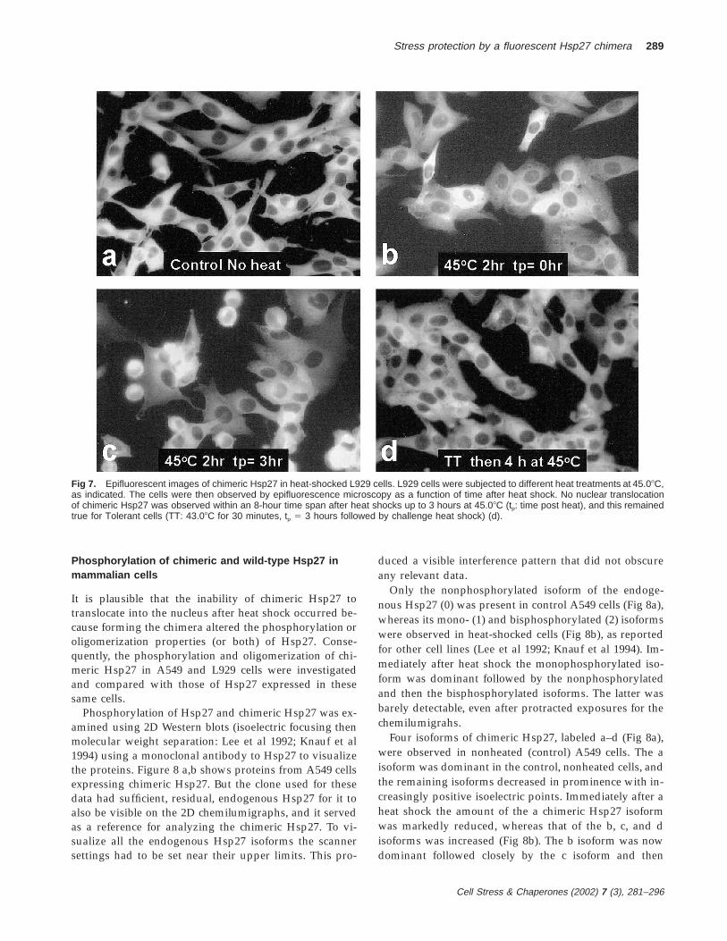
Only the nonphosphorylated isoform of the endoge-nous Hsp27 (0) was present in control A549 cells (Fig 8a),whereas its mono- (1) and bisphosphorylated (2) isoformswere observed in heat-shocked cells (Fig 8b), as reportedfor other cell lines (Lee et al 1992; Knauf et al 1994). Im-mediately after heat shock the monophosphorylated iso-form was dominant followed by the nonphosphorylatedand then the bisphosphorylated isoforms. The latter wasbarely detectable, even after protracted exposures for thechemilumigrahs.
Four isoforms of chimeric Hsp27, labeled a–d (Fig 8a),were observed in nonheated (control) A549 cells. The aisoform was dominant in the control, nonheated cells, andthe remaining isoforms decreased in prominence with in-creasingly positive isoelectric points. Immediately after aheat shock the amount of the a chimeric Hsp27 isoformwas markedly reduced, whereas that of the b, c, and disoforms was increased (Fig 8b). The b isoform was nowdominant followed closely by the c isoform and then
Cell Stress & Chaperones (2002) 7 (3), 281–296
290 Borrelli et al
Fig 8. Two-dimensional Western blot analysis of chimeric Hsp27 in A549 and L929 cells. Chimeric Hsp27 was visualized using a monoclonalantibody to Hsp27. The transfected A549 cells (clone 1.5d) had sufficient, residual, endogenous Hsp27 to be observed too. Only chimericHsp27 was present in transfected L929 cells. The arrows indicate proteins that were also visualized after the immonostaining, even innontransfected cells. (a) Four different isoforms of chimeric Hsp27 were present (a–d) in control A549 cells, but only the nonphosphorylatedisoform of Hsp27 (0) was observed. (b) Immediately after heat shock (43.08C, 2 hours) to A549 cells the mono- and bisphosphorylatedisoforms of Hsp27 were observed, and the b, c, and d isoforms of chimeric Hsp27 became more prominent, whereas the a isoform wasdiminished. (c) Only the a and b isoforms of chimeric Hsp27 were observed in L929 cells. (d) Immediately after heat shock (43.08C, 2 hours)4 isoforms (a–d) of chimeric Hsp27 were observed in L929, with the b isoform being dominant.
nearly equivalent amounts of the a and d isoforms (Fig8b). With reference to the endogenous Hsp27 the a and cisoforms of chimeric Hsp27 had isoelectric points similarto, respectively, the nonphosphorylated and monophos-phorylated forms of Hsp27, those of the b and d isoforms
fell midway between, respectively, the nonphosphorylat-ed and monophosphorylated, and the monophosphory-lated and bisphosphorylated isoforms of Hsp27.
Only the a and b isoforms of chimeric Hsp27 were vis-ible in control L929 cells (Fig 8c), but 4 isoforms were

Cell Stress & Chaperones (2002) 7 (3), 281–296
Stress protection by a fluorescent Hsp27 chimera 291
Fig 9. Two-dimensional Western blotanalysis of EGFP expressed in L929cells. EGFP constitutively expressed instably transfected L929 cells exhibiteda single isoform in nonheated cells (a)and immediately after heat shock (b: 1hour, 43.08C). A polyclonal antibody toEGFP was used to detect the protein,and the arrows indicate another proteinthat was also detected by this antibody.
present immediately after heat shock (Fig 8d). The rela-tive amounts of these 4 isoforms were similar to thoseobserved in heated A549 cells. Only the nonphosphory-lated isoform was present in control L929 cells transfectedto express Hsp27, with the mono- and bisphosphorylatedisoforms appearing after heat shock, as reported previ-ously (Lee et al 1992) (data not shown).
The arrows in Figure 8 indicate other proteins that pro-duced visible spots after staining with the primary andsecondary antibodies. These spots were present in cellsthat did not express chimeric Hsp27 or recombinant
Hsp27 (data not shown) and served as additional refer-ence points on the 2D Westerns.
The greater number of chimeric Hsp27 isoforms, theirshifted positions in 2D gels, and the fact that chimericHsp27 was phosphorylated in nonstressed cells wereclearly caused by the presence of EGFP in the chimera.But these changes were not necessarily the result of theEGFP moiety itself being phosphorylated, a point sup-ported by the fact that EGFP expressed in stably trans-fected L929 cells was not phosphorylated after heat shock(Fig 9 a,b).

Cell Stress & Chaperones (2002) 7 (3), 281–296
292 Borrelli et al
Fig 10. Glycerol gradient analyses of oligomer size for chimericHsp27 and Hsp27 in A549 (a) and L929 (b) cells. The data pointsshow the fraction of the total cellular Hsp27 or chimeric Hsp27 thatwas found in each gradient fraction as a function of the latter’s glyc-erol concentration. The arrows along the abscissa indicate the lo-calization of standard proteins within the gradient (molecular weightindicated in kilodaltons). In both cell lines aresenite treatment re-sulted in nearly complete dissociation of the 16-mer Hsp27 oligo-mers into dimers and tetramers. In contrast, arsenite caused no de-tectable dissociation of chimeric Hsp27 in A549 (Fig 10a) cells anddissociation of approximately 50% of the large oligomers in L929cells (Fig 10b).
The exact nature of chimeric Hsp27 phosphorylationwas not determined; also, it was not necessary for thepurposes of this study. These data were obtained solelyto determine whether chimeric Hsp27 phosphorylated inresponse to stress and whether such phosphorylation cor-responded to any stress-induced changes in oligomericsize.
Oligomeric status of chimeric and wild-type Hsp27 inmammalian cells
The size of the Hsp27 and chimeric Hsp27 oligomers wasdetermined by running cell lysates on linear glycerol gra-dients (Lambert et al 1999). Gradient fractions were sub-jected to slot blot analyses to quantitate Hsp27 content(Lambert et al 1999), whereas chimeric Hsp27 contentwas analyzed using a spectrofluorimeter.
In control A549 and L929 cells endogenous and recom-binant human Hsp27, respectively, was distributedamongst the gradient fractions containing between 13%and 25% glycerol (Fig 10 a,b). The distribution curves inboth cell lines were well defined, and the peak proteinamount occurred in the fraction containing 19% glycerol,corresponding to a molecular weight of approximately450 kDa (16-mer complex). After treatment with 200 mMsodium arsenite (2 hours at 37.08C) essentially all theHsp27 complexes in both cell lines disaggregated into ei-ther dimers or tetramers, as reported by Lambert et al(1999).
The size distribution of chimeric Hsp27 in control A549and L929 cells was broader than that of Hsp27, and itspeak occurred at 23.5% glycerol, which corresponded toa molecular weight of approximately 900 kDa (Fig 10 a,b).Thus, the chimeric Hsp27 maintained the same multi-meric size as Hsp27 in these cells. A smaller peak cen-tered about 350 kDa (6-mer) was observed consistently inA549 cells. Arsenite treatment had no significant effecton the size distribution of chimeric Hsp27 in A549 cells(Fig 10a). The same arsenite treatment caused approxi-mately 50% of the chimeric Hsp27 in L929 cells to dis-aggregate into smaller complexes with an average size of350 kDa (6-mer), whereas the remaining chimeric Hsp27maintained itself as 16-mer complexes (Fig 10b).
The multimer of human Hsp27 in both A549 and L929cells was smaller than the 600- to 700-kDa reported byLambert et al (1999) for the hamster Hsp27 in CCL39 and3T3 cells. When hamster Hsp27 was expressed in A549and L929 cells its size distribution was essentially iden-tical to that observed for human Hsp27, with its peak atapproximately 500 kDa (data not shown). Thus, the dif-ference between the Hsp27 multimeric size reported inthis study and by Lambert et al (1999) appears to be at-tributable to cell line variations.
Total cellular glutathione measurements
Other studies have suggested that overexpressing Hsp27increases total intracellular glutathione, which in turncontributes to cellular stress protection (Mehlen et al1995, 1997). This represents a potential stress protection

Cell Stress & Chaperones (2002) 7 (3), 281–296
Stress protection by a fluorescent Hsp27 chimera 293
Table 1 Cellular glutathione measurements
SampleGlutathione content
(nm/mg protein)
L929L929 1 Hsp27L929 1 Chimeric Hsp27L929 1 Lac repressorL929 1 Lac Hsp27 (not expressed)L929 1 Lac Hsp27 (expressed)
6.09 6 0.193.71 6 0.373.98 6 0.88
10.27 6 0.399.61 6 0.61
10.93 6 0.86
for cells overexpressing Hsp27 or chimeric Hsp27 that isindependent of Hsp27 chaperone activity. Consequently,glutathione levels were measured in the stably transfectedL929 cells that expressed Hsp27 or chimeric Hsp27,which were used throughout this study. Glutathione mea-surements were also made in L929 cells stably transfectedto express Hsp27 under control of the inducible Lac-Switch promoter (Stratagene Inc) to determine whetherany glutathione increase was short lived after a transientHsp27 upregulation. No glutathione measurements weremade in wild-type or transfected A549 cells because thesecells have a naturally high glutathione content (.500nM/mg protein) that could potentially mask smaller,Hsp27-induced increases in cellular glutathione.
Results of the glutathione measurements are presentedin Table 1. Expressing Hsp27 or chimeric Hsp27 (consti-tutively or transiently) consistently produced a slight re-duction in the glutathione content in L929 cells. A nearly2-fold increase in glutathione was observed in L929 cellsstably transfected to express the Lac repressor protein.But this was not associated with any increase in stressprotection or Hsp27 (data not shown), and no further in-crease in glutathione was observed after transfection withrecombinant Hsp27, even when the LacSwitch promoterwas activated with isopropyl-b-D-thiogalactoside to ex-press Hsp27.
DISCUSSION
No difficulties or problems were encountered while es-tablishing stable transfectants of A549 or L929 cells thatconstitutively expressed a fluorescent chimera containingEGFP fused directly to the N-terminus of Hsp27. Con-versely, establishing a stable transfectant of L929 cells thatconstitutively expressed EGFP was extremely difficultand time consuming, whereas all attempts to do so inA549 cells failed. The difficulty in establishing transfec-tants that constitutively expressed EGFP may have beenrelated to the toxicity of protracted EGFP expression thathas been reported by others (Liu et al 1999). One mightspeculate that the ease of producing and maintaining thetransfectants that expressed EGFP-Hsp27 was related tothe fact that EGFP was being expressed as a chimera witha chaperone protein and that the properties of the latter
somehow muted or obviated any EGFP toxicity. Regard-less, the chimeric Hsp27 provided a means for observingthe distribution of Hsp27 within living cells using fluo-rescence microscopy and permitted several specific con-clusions concerning Hsp27 stress protection. Further-more, the fluorescent chimera provides a means for in-vestigating chaperone function in living cells using quan-titative fluorescent measurements, as discussedsubsequently.
Forming the chimera did not affect the multimeric stateof Hsp27 or its ability to provide stress protection. Hence,it can be presumed that neither of these 2 properties ofthe Hsp27 multimer requires specific interactions be-tween free, Hsp27 N-termini. It is also clear that the mul-timeric complex did not have to be homogeneouslyHsp27 to provide stress protection. Half of the chimericHsp27 multimer consisted of the equimassive EGFP (aprotein that by itself provided no stress protection); yet,chimeric Hsp27 proved equally effective as did Hsp27 forstress protection.
The organization and orientation of chimeric Hsp27within the multimer was not determined; however, it isextremely unlikely that the oligomer formed with everyHsp27 moiety oriented into the complex’s central core,with the EGFP moieties projecting outward. In that casethe EGFP moieties would have served as a restrictive bar-rier preventing access of the Hsp27 moieties to any stress-damaged proteins or intracellular structures. This wouldhave ostensibly obviated any Hsp27 stress protection,which did not occur. The converse, ie, a multimer with anEGFP core and all Hsp27 moieties oriented outward, ispossible and could conceptually function identically to anHsp27 homomultimer because all the Hsp27 moietieswould be available for stress protection. Many interme-diate possibilities exist, and it is necessary that the exactorganization of the chimeric Hsp27 multimer be deter-mined.
Chimeric Hsp27 did not translocate into the nucleusafter stress, demonstrating that stress-induced nucleartranslocation is not a requisite for achieving the maxi-mum stress protection attainable with Hsp27 (Figs 3 and4). This does not exclude the possibility that Hsp27 trans-location into the nucleus provides some stress protection,eg, by stabilizing or facilitating recovery of denatured oraggregated proteins in the nucleus (Kampinga et al 1994),or that stress protection within the nucleus is not requiredto achieve the maximum protection attainable by over-expressing Hsp27. The results of this study merely dem-onstrate that it is not necessary for Hsp27 to enter thenucleus and directly perform a function therein to achieveits maximal level of stress protection. Ostensibly, over-expressed chimeric Hsp27 could stabilize denatured oraggregated proteins and subcellular structures within thecytoplasm of stressed cells and by so doing free up other

Cell Stress & Chaperones (2002) 7 (3), 281–296
294 Borrelli et al
Hsps. The latter could then translocate into the nucleusto protect the damage that would have been protected bynuclear-translocated Hsp27 were that expressed insteadof chimeric Hsp27. The possibility for such indirect pro-tection of the nucleus by chimeric Hsp27 is currently be-ing tested and represents a second phase of investigationsusing chimeric Hsp27.
A simple explanation for the inability of chimericHsp27 to translocate into the nucleus of either A549 orL929 cells is the failure of stress to reduce the size of thechimeric Hsp27 multimers. In A549 cells chimeric Hsp27multimers remained at 900 kDa, which, as in control cells,was too large to enter the nucleus. Although stress re-duced nearly 50% of chimeric Hsp27 multimers to 6-mers(350 kDa) in L929 cells, the latter were probably still toolarge to enter the nucleus. It is also plausible that mono-mers of the EGFP-Hsp27 chimera were incapable of nu-clear translocation. This was not tested because stress ex-posure (arsenite treatment) did not produce chimericHsp27 oligomers smaller than 6-mers in L929 cells, andproduction of mutant, chimeric Hsp27 that formed small-er multimers was not attempted. But the fact that Hsp27dimers and tetramers (produced after arsenite treatment;Lambert et al 1999) or EGFP dimers could enter the nu-cleus suggests that chimeric Hsp27 monomers would notbe excluded from the nucleus.
Forming the chimera altered the phosphorylation prop-erties of Hsp27 markedly. Hsp27 was not phosphorylatedin nonstressed A549 or L929 cells, whereas chimericHsp27 exhibited 3 and 1 phosphorylated isoforms, re-spectively. The shift in chimeric Hsp27 to more positiveisoforms in stressed cells was similar to that observed forHsp27, with the exception that the former exhibited 4rather than 3 isoforms. The positions of the 3 phosphor-ylation sites on chimeric Hsp27 were not determined, butit is reasonable to presume that 2 of these sites involvedphosphorylation of Ser78 and Ser82. The d isoform of thechimera may have required phosphorylation of a thirdresidue, and the identity of all phosphorylatable residueswill be determined subsequently.
Hsp27 and chimeric Hsp27 both formed 16-mers innonstressed cells despite the fact that chimeric Hsp27 ex-hibited 1 phosphorylated isomer in L929 cells and 3 inA549 cells. Increased phosphorylation after stress had noeffect on multimers in A549 cells and reduced 50% of the16-mers to 6-mers in L929 cells. These observations sug-gest the postulate that phosphorylation of Hsp27 is notsufficient for reducing its multimeric size after stress. Butthere are other plausible scenarios. It is possible that in-teractions between adjacent EGFP molecules or betweenadjacent EGFP and Hsp27 molecules (or both) providedsufficient binding force to hold the multimer intact de-spite any phosphorylation-induced reduction in multi-meric binding between Hsp27 molecules.
The data in Table 1 provide strong evidence that tran-sient or protracted overexpression of Hsp27 does not in-duce upregulation of intracellular glutathione in L929cells. In actuality, glutathione levels were markedly re-duced in L929 cells that constitutively expressed eitherchimeric Hsp27 or Hsp27. Expressing the Lac-repressorprotein did increase cellular glutathione levels by nearly2-fold, but this did not affect the heat sensitivity of thesecells. Consequently, the data in Table 1 support the con-clusions that that the protection against heat killing con-ferred by chimeric Hsp27 and Hsp27 did not involveupregulation of glutathione and that a 69% increase incellular glutathione levels (in Lac-repressor transfectedcells) did not alter cellular heat sensitivity. It is possiblethat the changes in glutathione levels observed in theseexperiments could alter cellular sensitivity to other stress-es, eg, reactive oxygen species (Mehlen et al 1997), butthis possibility was not tested in this study.
The success with the fluorescent Hsp27 chimera bodeswell for the potential to produce fluorescent chimeras ofother chaperones that retain, if not all, most of the func-tions of their chaperone moieties. Besides the obviousability to observe the distribution and redistribution offluorescent chimeras within, respectively, normal andstressed cells, fluorescent chimeras would present the op-portunity to perform very sophisticated experiments toelucidate the interaction of chaperones with each otherand with other proteins and structures within living cells.Fluorescence recovery after photobleaching could be usedto measure how heat shock and other stresses affectschaperone diffusion rates within cells and whether or notspecific chaperones become immobilized or migrate inconcert. FRET measurements could be used to determinewhich chaperones interact with each other during normalor stress conditions and how the chaperones interact withother fluorescently labeled structures within the cell, eg,the cytoskeleton, endoplasmic reticulum, or fluorescentlylabeled, denatured, and aggregated proteins. Some ofthese studies are currently underway using the fluores-cent EGFP-Hsp27 chimera described in this study.
ACKNOWLEDGMENT
This study was supported by NIH/NCI grant CA71650.
REFERENCES
Anderson ME. 1985. Tissue glutathione. In: CRC Handbook of Methodfor Oxygen Radical Research, ed Greenwald RA. CRC Press, BocaRaton, FL, 317–323.
Angelidis CE, Lazaridis I, Pagoulatos GN. 1991. Constitutive ex-pression of heat-shock protein 70 in mammalian cells confersthermoresistance. Eur J Biochem 199: 35–39.
Arrigo AP, Suhan JP, Welch W. 1988. Dynamic changes in the struc-

Cell Stress & Chaperones (2002) 7 (3), 281–296
Stress protection by a fluorescent Hsp27 chimera 295
ture and intracellular locale of the mammalian low–molecularweight heat shock protein. Mol Cell Biol 8: 5059–5071.
Arrigo AP, Welch WJ. 1987. Characterization and purification of thesmall 28,000-Dalton mammalian heat shock protein. J Biol Chem262: 15359–15369.
Blackburn R, Galoforo S, Berns CM, Ireland M, Cho JM, Corry PM,Lee YJ. 1996. Thermal response in murine L929 cells lackingaB-crystallin expression and aB-crystallin expressing L929transfectants. Mol Cell Biochem 155: 51–60.
Borrelli MJ, Lepock JR, Frey HE, Lee YJ, Corry PM. 1996a. Excessprotein in nuclei isolated from heat-shocked cells results fromreduced extractability of nuclear proteins. J Cell Physiol 167:369–379.
Borrelli MJ, Stafford DM, Karczewski LA, Rausch CM, Lee YJ, CorryPM. 1996b. Thermotolerance expression in mitotic cells withoutincreased translation of heat shock proteins. J Cell Physiol 169:420–428.
Borrelli MJ, Stafford DM, Rausch CM, Bernock LJ, Freeman ML, Le-pock JR, Corry PM. 1998. Diamide-induced cytoxicity and ther-motolerance in CHO cells. J Cell Physiol 177: 483–492.
Caraceni P, De Maria N, Ryu HS, et al. 1997. Proteins but not nucleicacids are molecular targets for the free radical attack duringreoxygenation of rat hepatocytes. Free Radic Biol Med 23: 339–344.
Carver JA, Guerreiro N, Nicholls KA, Truscott RJW. 1995. On theinteraction of a-crystallin with unfolded proteins. Biochim Bio-phys Acta 1252: 251–260.
Chaufour S, Mehlen P, Arrigo AP. 1996. Transient accumulation,phosphorylation and changes in the oligomerization of Hsp27during retinoic acid–induced differentiation of HL-60 cells: pos-sible role in the control of cellular growth and differentiation.Cell Stress Chaperones 1: 225–235.
Chopp M, Chen H, Ho K, Dereski M, Pulsinelli WA. 1989. Transienthyperthermia protects against subsequent forebrain ischemiccell damage in the rat. Neurology 39: 1396–1398.
Chretien P, Landry J. 1988. Enhanced constitutive expression of the27-kDa heat shock proteins in heat-resistant variants from Chi-nese hamster cells. J Cell Physiol 137: 157–166.
Crete P, Landry J. 1990. Induction of HSP27 phosphorylation andthermoresistance in Chinese hamster cells by arsenite, cyclo-heximide, A23187, and EGTA. Radiat Res 121: 320–327.
Dienel GA, Kiessling M, Jacewicz M, Pulsinelli WA. 1986. Synthesisof heat shock proteins in rat brain cortex after transient ische-mia. J Cereb Blood Flow Metab 6: 505–510.
Donaldson SS, Gordon LF, Hahn GM. 1978. Protective effect of hy-perthermia against the cytotoxicity of actinomycin D on Chi-nese hamster cells. Can Treat Rep 62: 1489–1495.
Donati YR, Slosman DO, Polla BS. 1990. Oxidative injury and theheat shock response. Biochem Pharmacol 40: 2571–2577.
Ehrnsperger M, Graber S, Gaestel M, Buchner J. 1997. Binding ofnon-native protein to HSP-25 during heat shock creates a res-ervoir of folding intermediates for reactivation. EMBO J 16: 221–229.
Floyd RA. 1990. Role of oxygen free radicals in carcinogenesis andbrain ischemia. FASEB 4: 2587–2597.
Freeman BC, Morimoto RI. 1996. The human cytosolic molecularchaperones hsp90, hsp70 (hsc70) and hdj-1 have distinct rolesin recognition of a non-native protein and protein folding.EMBO J 15: 2969–2979.
Gerner EW, Schneider MJ. 1975. Induced thermal resistance in HeLacells. Nature 256: 500–502.
Griffith OW. 1980. Determination of glutathione and glutathione di-sulfide using glutathione reductase and 2-vinylpyridine. AnalBiochem 106: 207–212.
Henle KJ. 1987. Thermotolerance in cultured mammalian cells. In:Thermotolerance: Thermotolerance and Thermophily, vol 1, ed HenleKJ. CRC Press, Boca Raton, FL, 13–71.
Henle KJ, Leeper DB. 1976. Interaction of hyperthermia and radia-tion CHO cells: recovery kinetics. Radiat Res 66: 505–518.
Huot J, Roy G, Lambert H, Chretien P, Landry J. 1991. Increasedsurvival after treatments with anticancer agents of Chinesehamster cells expressing the human Mr 27,000 heat shock pro-tein. Cancer Res 51: 5245–5252.
Jakob U, Gaestel M, Engel K, Buchner J. 1993. Small heat shock pro-teins are molecular chaperones. J Biol Chem 268: 1517–1520.
Kampinga HH. 1993. Thermotolerance in mammalian cells: proteindenaturation and aggregation, and stress proteins. J Cell Sci 104:11–17.
Kampinga HH, Brunsting JF, Stege GJ, Konings AW, Landry J. 1994.Cells overexpressing Hsp-27 show accelerated recovery fromheat-induced nuclear protein aggregation. Biochem Biophys ResCommun 204: 1170–1177.
Kampinga HH, Turkel-Uyger N, Roti Roti JL, Konings AWT. 1989.The relationship of increased protein content induced by hy-perthermia to killing of HeLa S3 cells. Radiat Res 117: 511–522.
Knauf U, Jakob U, Engel K, Buchner J, Gaestel M. 1994. Stress- andmitogen-induced phosphorylation of the small heat shock pro-tein Hsp25 by MAPKAP kinase 2 is not essential for chaperoneproperties and cellular thermoresistance. EMBO J 13: 54–60.
Lambert H, Charette SJ, Bernier AF, Guimond A, Landry J. 1999.HSP27 multimerization mediated by phosphorylation-sensitiveintermolecular interactions at the amino terminus. J Biol Chem274: 9378–9385.
Landry J, Bernier D, Chretien P, Nicole LM, Tanguay RM, MarceauN. 1982. Synthesis and degradation of heat shock proteins dur-ing development and decay of thermotolerance. Cancer Res 42:2457–2461.
Landry J, Chretien P, Lambert H, Hickey E, Weber LA. 1989. Heatshock resistance conferred by expression of the human HSP27gene in rodent cells. J Cell Biol 109: 7–15.
Laszlo A, Li GC. 1985. Heat resistant variants of Chinese hamsterfibroblasts altered in expression of heat shock protein. Proc NatlAcad Sci U S A 82: 8029–8033.
Lavoie JN, Lambert H, Hickey E, Weber LA, Landry J. 1995. Mod-ulation of cellular thermoresistance and actin filament stabilityaccompanies phosphorylation-induced changes in the oligo-meric structure of heat shock protein 27. Mol Cell Biol 15: 505–516.
Lee KJ, Hahn GM. 1988. Abnormal proteins as the trigger for theinduction of stress responses: heat, diamide, and sodium arse-nite. J Cell Physiol 136: 411–420.
Lee YJ, Hou ZZ, Curetty L, Borrelli MJ, Corry PM. 1992. Develop-ment of acute thermoresistance in L929 cells: lack of HSP28synthesis and phosphorylation. J Cell Physiol 152: 118–125.
Lee GJ, Roseman AM, Saibil HR, Vierling E. 1997. A small heatshock protein stably binds heat-denatured model substrates andcan maintain a substrate in a folding competent state. EMBO J16: 659–671.
Lepock JR, Frey HE, Ritchie KP. 1993. Protein denaturation in intacthepatocytes and isolated cellular organelles during heat shock.J Cell Biol 122: 1267–1276.
Li GC, Li L, Liu RY, Mak JY, Chen L, Lee WMF. 1992. Thermalresponse of rat fibroblasts stably transfected with the human70-kDa heat shock protein-encoding gene. Proc Natl Acad Sci US A 88: 1681–1685.
Li GC, Werb Z. 1982. Correlation between synthesis of heat shockproteins and development of thermotolerance in Chinese ham-ster fibroblasts. Proc Natl Acad Sci U S A 79: 3219–3222.

Cell Stress & Chaperones (2002) 7 (3), 281–296
296 Borrelli et al
Lindquist S. 1986. The heat shock response. Ann Rev Biochem 55:1151–1191.
Liu HS, Jan MS, Chou CK, Chen PH, Ke NJ. 1999. Is green fluores-cent protein toxic to the living cells? Biochem Biophys Res Com-mun 260: 712–717.
Lowry OH, Rosenbrough NJ, Farr AL, Randall RJ. 1951. Protein mea-surement using the folin phenol reagent. J Biol Chem 193: 265–275.
Mehlen P, Hickey E, Weber L, Arrigo, AP. 1997. Large unphosphor-ylated aggregates as the active form of hsp27 which controlsintracellular reactive oxygen species and glutathione levels andgenerates a protection against TNFa in NIH-3T3-ras cells. Bio-chem Biophys Res Commun 241: 187–192.
Mehlen P, Preville X, Chareyron P, Briolay J, Klemenz J, Arrigo AP.1995. Constitutive expression of human hsp27, Drosophila,hsp27, or human aB-crystallin confers resistance to TNF- andoxidative stress-induced cytotoxicity in stably transfected mu-rine L929 fibroblasts. J Immunol 154: 363–374.
Morimoto RI, Tissieres A, Georgopoulos C. 1990. The stress re-
sponse, function of the proteins, and perspectives. In: Stress Pro-teins in Biology and Medicine, ed Morimoto RI, Tissieres A, Geor-gopolous C. Cold Spring Harbor Press, NY, 1–36.
Roti Roti JL, Henle KJ, Winward RT. 1979. The kinetics of increasesin chromatin protein content in heated cells: a possible role incell killing. Radiat. Res 78: 522–531.
Roti Roti JL, Laszlo A. 1988. The effects of hyperthermia on cellularmacromolecules. In: Hyperthermia and Oncology, vol 1, ed UranoM, Douple EB. VNU Scientific, The Netherlands, 13–56.
Senisterra GA, Huntley SA, Escaravage M, Sekhar KR, Freeman ML,Borrelli MJ, Lepock JR. 1997. Destabilization of the Ca12-ATPaseof sarcoplasmic reticulum by thiol-specific, heat shock inducersresults in thermal denaturation at 37.08C. Biochemistry 36:11002–11011.
Subjeck JR, Sciandra J, Johnson RJ. 1982. Heat shock proteins andthermotolerance: comparison of induction kinetics. Br J Radiol55: 579–584.
Yahara I, Iida H, Koyasu S. 1986. A heat shock-resistant variant ofChinese hamster cell line constitutively expressing heat shockprotein of Mr 90,000 at high level. Cell Struct Funct 11: 65–73.
Copyright © 2022 FDOKUMEN


![Noninvasive Molecular Imaging of MYC mRNA Expression in Human Breast Cancer Xenografts with a [ 99m Tc]Peptide−Peptide Nucleic Acid−Peptide Chimera](https://static.fdokumen.com/doc/165x107/63214cddbc33ec48b20e4a4a/noninvasive-molecular-imaging-of-myc-mrna-expression-in-human-breast-cancer-xenografts.jpg)