optical control over monomeric and multimeric protein hybrids
133
-
Upload
khangminh22 -
Category
Documents
-
view
0 -
download
0
Transcript of optical control over monomeric and multimeric protein hybrids
OPTICAL CONTROL OVER MONOMERIC AND
MULTIMERIC PROTEIN HYBRIDS
Rindia Maharani Putri
Members of the committee:
Chairman: Prof. dr. H.H.J. ten Kate (University of Twente)
Promotors: Prof. dr. J.J.L.M. Cornelissen (University of Twente)
Prof. dr. N.H. Katsonis (University of Twente)
Members: Prof. dr. G.A. Woolley (University of Toronto)
Prof. dr. J.R. Castón (CSIC Madrid)
Dr. M.S.T. Koay (Sanofi-Aventis
Deutschland GmbH)
Prof. dr. M.M.A.E. Claessens (University of Twente)
Prof. dr. J. Huskens (University of Twente)
The research described in this thesis was performed within the laboratories of the
Biomolecular Nanotechnology (BNT) group, the MESA+ Institute for Nanotechnology, and
the Department of Science and Technology (TNW) of the University of Twente. This
research was supported by the Netherlands Organization for Scientific Research (NWO), the
European Research Council (ERC) and Indonesia Endowment Fund for Education (LPDP).
Optical control over monomeric and multimeric protein hybrids
Copyright © 2017, Rindia Maharani Putri, Enschede, The Netherlands.
All rights reserved. No part of this thesis may be reproduced or transmitted in any form, by
any means, electronic or mechanical without prior written permission of the author.
ISBN: 978-90-365-4365-1
DOI: 10.3990/1.9789036543651
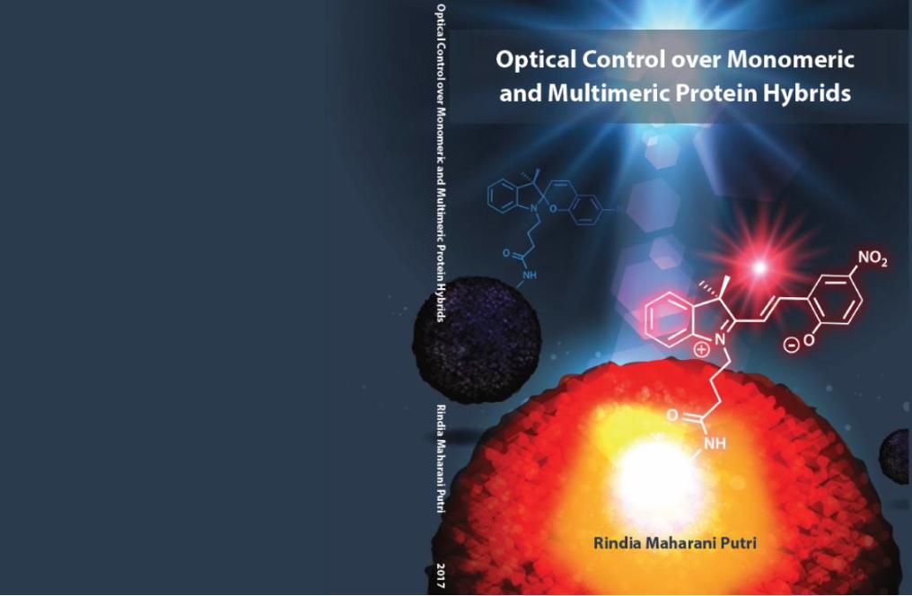
Cover art: Pichamon Graphic House and Rindia Maharani Putri
(also featured in the inside back cover of ChemPhysChem 12/2016)
Printed by: Gildeprint The Netherlands
OPTICAL CONTROL OVER MONOMERIC
AND MULTIMERIC PROTEIN HYBRIDS
DISSERTATION
to obtain
the degree of doctor at the University of Twente,
on the authority of rector magnificus
Prof. dr. T. T. M. Palstra,
on account of the decision of the graduation committee,
to be publicly defended
on Friday September 8, 2017 at 12.45 h
by
Rindia Maharani Putri
Born on May 28, 1991
in Padang, Indonesia

This dissertation has been approved by:
Promotors: Prof. dr. J.J.L.M. Cornelissen
Prof. dr. N.H. Katsonis

To my family

i
Table of contents
Chapter 1: General introduction 1
1.1 Introduction 1
1.2 Aim and outline 2
1.3 References 3
Chapter 2: Strategies to modulate allosteric regulation and
self-assembly of proteins using light
5
2.1 Introduction 6
2.2 Photo-activated allosteric regulation 8
2.2.1 Inclusion of photo-active protein domains 8
2.2.2 Inclusion of synthetic photo-switches 12
2.3 Photo-responsive protein assemblies of naturally occurring
structures
14
2.3.1 Photo-activated dimerization 15
2.3.2 Photo-responsive cage-like and filament proteins 19
2.4 Photo-responsive artificial protein-based systems and higher-
order structures
22
2.5 Perspectives 24
2.6 References 25
Chapter 3: Programming allostery in the human serum
albumin with light
31
3.1 Introduction 32
3.2 Results and discussion 33
3.2.1 Design and characterization of a photo-switchable
hybrid
33
3.2.2 Optical control over ligand binding to subdomain IB 37
3.2.3 Molecular dynamic simulations 41
3.3 Conclusions 44
3.4 Acknowledgements 44
ii
3.5 Materials and methods 45
3.5.1 General 45
3.5.2 Synthesis of the photo-switch 45
3.5.3 Synthesis and characterization of the hybrid system 48
3.5.4
3.5.5
3.5.6
3.5.7
3.5.8
Photo-responsive ligand binding analyses
PFGSE-NMR experiments
Determination of effective quenching constants
Analyses of protein global structure
Data and schematic representation
48
50
50
50
51
3.6 References 51
Chapter 4: Bacterial encapsulins as in vitro and in vivo
protein-based nanoplatforms
55
4.1 Introduction 56
4.2 Results and discussion 57
4.2.1 Characterization of encapsulin stability and structure 57
4.2.2
In vitro study of encapsulin as nanoreactors on
surface
65
4.2.3 In vivo study of encapsulin as an agent for cellular
infection
68
4.3
4.4
Conclusions
Acknowledgements
70
70
4.5 Materials and methods 71
4.5.1 General 71
4.5.2 Cryo-EM and image processing 71
4.5.3 Characterization of encapsulin stability 72
4.5.4
4.5.5
Immobilization of encapsulin and catalytic assay
Cell experiments
73
74
4.6 References 74
Chapter 5: Labeling encapsulin with light-switchable
fluorophores
77
5.1 Introduction 78
5.2 Results and discussion 79
iii
5.2.1 Covalent attachment of spiropyran to encapsulin
5.2.2 Photo-switchable fluorescence
5.2.3 Structural integrity of encapsulin particles
79
82
84
5.3
5.4
Conclusions
Acknowledgements
86
86
5.5 Materials and methods 86
5.5.1 General 86
5.5.2 Synthesis of spiropyran-succinimide 86
5.5.3
5.5.4
5.5.5
Covalent attachment of spiropyran and encapsulin
Photo-switchable fluorescence analysis
Characterization of encapsulin structure
89
89
90
5.6 References 90
Chapter 6: Light-fueled assembly of encapsulin and
chaperone into hybrid superstructures
93
6.1 Introduction 94
6.2 Results and discussion 95
6.2.1 Light-fueled assembly of hybrid superstructures 95
6.2.2 Structural changes of proteins upon assembly 101
6.2.3 Emergence of light-dependent order by inclusion of
photoacid in the superstructures
104
6.3
6.4
Conclusions
Acknowledgements
107
107
6.5 Materials and methods 108
6.5.1 General 108
6.5.2 Reversible formation of hybrid superstructures 108
6.5.3
6.5.4
6.5.5
Characterization of hybrid superstructures
Characterization of structural changes of proteins
Analysis of photoacid inclusion in the superstructures
109
109
109
6.6 References 110
Summary
Samenvatting
113
115

iv
Acknowledgements
About the author
117
121
Chapter 1
General introduction
Introduction
From naturally occurring to semi-synthetic systems, proteins dictate a plethora of
essential life processes, including catalysis,[1] structural support,[2] cell signaling[3] as well as
molecular transport and delivery.[4] Modulating the functioning of these proteins using an
external trigger would thus offer an effective means to remote control a range of complex
biological processes and networks of regulations. In this regard, light appears as an attractive
trigger, as it is precise, compatible with a wide range of condensed phases, clean, relatively
non-destructive towards proteins, and therefore it is envisioned that it can be adapted for in
vivo and in vitro applications. Moreover, light enables reversible and selective spatiotemporal
control, which in turn allows an on/off and dose-dependent regulation over complex
molecular processes.
Recent years have witnessed an increasing attempt to modulate biological processes by
photo-engineering some key proteins involved in cellular regulations. The photo-modulation
of biological processes has been reported,[5-9] including enzyme catalysis,[10] protein
folding,[11] the opening of channel proteins[12] as well as protein dimerization.[13] Among
biological regulations involving proteins, allostery and self-assembly of proteins are highly
dynamic, tunable processes[14, 15] that are essential to the functioning of cellular networks, yet
remain not entirely understood and therefore they are challenging to modulate in a controlled
manner. In this thesis, we address the challenge to understand the mechanisms of allostery
and hierarchical self-assembly of proteins and gain control over such cooperative and
dynamic actions to obtain a measurable output from individual switching events. We use pre-
existing biological systems as cooperative molecular media to amplify the motion of
molecular switches to generate collective behaviors of hybrid systems.

General introduction
2
Aim and outline
We engineer an allosteric transport protein[16] and a bacterial nanocompartment
encapsulin[17] into dynamic building blocks for light-responsive hybrid (semi-synthetic)
systems. Naturally, neither of these protein systems is responsive to light. Upon chemical
modification with artificial molecular photo-switches into their structures, their properties
and dynamics become reversibly controllable using irradiation with light.
The strategy involves modifying the proteins with photo-responsive spiropyran-based
switches,[18] which can be grafted covalently on the proteins (Chapter 3 and 5) or dispersed
in the medium that contains them (Chapter 6). In a reversible manner, the photo-switches
change in conformation and polarity as a response to light irradiation, therefore affecting the
proteins to which they are associated with. This strategy enables control over the dynamics
of allostery in transport protein (Chapter 3) and the chemical and self-assembly properties of
encapsulins (Chapter 5 and 6, respectively).
Chapter 2 describes recent strategies towards the modulation of protein functions using
light, with a special focus on allostery and self-assembly.
Chapter 3 describes a light-programmed allostery in the transport protein Human Serum
Albumin (HSA), that is engineered with a spiropyran photo-switch. The photo-switch is
incorporated into a specific binding site of HSA, and once it switches from closed, non-
charged spiropyran form to open, zwitter-ionic merocyanine form, we can demonstrate that
it is a neighboring binding site that responds to the environmental changes, mediated by
allostery.
In Chapter 4, the structural and functional basis of multimeric encapsulin from the
bacteria B. linens is presented. This chapter describes structural characterization of the cage-
like particles using cryo-electron microscopy (cryo-EM) reconstruction and analyses of
particle stability in front of environmental changes (i.e., pH, ionic strength and addition of
organic solvent). Furthermore, the functionality of encapsulin as nanoplatforms is
demonstrated both in vitro and in vivo.
Chapter 5 presents the labeling of B. linens encapsulin with photo-switchable
spiropyran-based fluorophores. Following structure- and function-based analyses in Chapter
Chapter 1
3
4, the covalent attachment of multiple spiropyran molecules at encapsulin exterior is
described in this chapter as well as the on/off photo-induced fluorescence in cycles.
Chapter 6 describes a light-fueled dynamic assembly of T. maritima encapsulin and E.
coli chaperone protein into transient giant superstructures. The assembly is mediated by a
spiropyran-based photoacid that plays two roles in the assembly event: 1) as a photo-active
building block for the assembly and 2) to reversibly and gradually control the pH of the
system to form ordered superstructures. We demonstrate that the resulting superstructures
depend on continuous irradiation to sustain their assembled state.
References
[1] S. J. Benkovic, S. Hammes-Schiffer, Science 2003, 301, 1196-1202.
[2] A. Akhmanova, M. O. Steinmetz, Nat. Rev. Mol. Cell Bio. 2015, 16, 711-726.
[3] C. J. Miller, L. A. Davidson, Nat. Rev. Genet. 2013, 14, 733-744.
[4] A. H. Futerman, Nature 2007, 449, 35-37.
[5] A. Gautier, C. Gauron, M. Volovitch, D. Bensimon, L. Jullien, S. Vriz, Nat. Chem. Biol.
2014, 10, 533-541.
[6] W. Szymanski, J. M. Beierle, H. A. Kistemaker, W. A. Velema, B. L. Feringa, Chem.
Rev. 2013, 113, 6114-6178.
[7] K. E. Brechun, K. M. Arndt, G. A. Woolley, Curr. Opin. Struct. Biol. 2016, 45, 53-58.
[8] G. Guglielmi, H. J. Falk, S. De Renzis, Trends Cell Biol. 2016, 26, 864-874.
[9] M. Baker, Nat. Methods 2012, 9, 443-446.
[10] C. W. Riggsbee, A. Deiters, Trends Biotechnol. 2010, 28, 468-475.
[11] U. T. Bornscheuer, Nature 2016, 540, 345-346.
[12] A. Kocer, M. Walko, W. Meijberg, B. L. Feringa, Science 2005, 309, 755-758.
[13] C. L. Tucker, J. D. Vrana, M. J. Kennedy, Curr. Protoc. Cell Biol. 2014, 64, 17 16 11-
20.
[14] S. R. Tzeng, C. G. Kalodimos, Nature 2009, 462, 368-372.
[15] J. J. McManus, P. Charbonneau, E. Zaccarelli, N. Asherie, Curr. Opin. Colloid Interface
Sci. 2016, 22, 73-79.
[16] X. M. He, D. C. Carter, Nature 1992, 358, 209-215.

General introduction
4
[17] M. Sutter, D. Boehringer, S. Gutmann, S. Guenther, D. Prangishvili, M. J. Loessner, K.
O. Stetter, E. Weber-Ban, N. Ban, Nat. Struct. Mol. Biol. 2008, 15, 939-947.
[18] R. Klajn, Chem. Soc. Rev. 2014, 43, 148-184.

Chapter 2
Strategies to modulate allosteric regulation
and self-assembly of proteins using light
Living cells and life processes rely on networks of interactions and
cellular dynamics that are orchestrated by protein assemblies. The early quest
was to understand how the assemblies are spontaneously put together and
controlled by Nature, leading to extensive research on protein assembly and
regulation. Among the mechanisms for r egulation exemplified by Nature,
allostery and self-assembly of proteins are particularly prominent as they not
only enable a dynamic and tunable control of naturally occurring systems, but
also inspire the design and generation of artificial protein -based hybrids.
Using light to stimulate and externally interfere with such mechanisms would
open up the possibility to control complex biological processes as well as
developing smart materials based on protein hybrids. Here , we provide a
concise review of recent advances on the strategies developed and used to
interfere with the allostery and self -assembly mechanisms in naturally
occurring systems and artificially designed systems.
Strategies to modulate allosteric regulation and self-assembly of proteins using light
6
2.1 Introduction
Supramolecular structures in Nature that are formed by protein assemblies play crucial
roles in cells, for instance as structural components and cellular machinery.[1] The skeleton
of the cell (cytoskeleton) is composed by self-assembled protein filaments that maintain the
structure and the shape of the cell. These filaments also provide platforms for vesicles and
organelles mobility inside the cell. Besides the structural support, major metabolic pathways
in cells are carried out by dozens of protein assemblies, for instance, ribosome (the translation
machinery) and spliceosome (the RNA-splicing machinery) are made up by multiple protein
subunits interacting with each other building complex catalytic protein clusters.[2-4]
Consequently, the cell itself can be viewed as a crowded and confined factory built up by
interlinking, highly-organized protein assemblies.[5,6]
Nature regulates the activity of protein assemblies using various mechanisms, including
allosteric and cooperative regulation,[7] tunable interactions and assembly of protein
subunits[8] as well as signal amplification and feedback loops in cascade systems.[9] Allostery
and self-assembly of proteins are highly dynamic, tunable processes and modulating such
mechanisms using external triggers would enable interfering with complex systems in
cellular networks. Light, in particular, is an attractive means to control protein assemblies as
it enables a precise spatiotemporal control and applicable in physiological conditions. The
use of light to modulate protein functionality in general has attracted attention in recent years,
leading to the success of photo-modulating various biological processes,[10-15] such as enzyme
catalysis,[16] protein folding/unfolding,[17] fluctuations in the activity of channel proteins[18]
as well as the dynamic process of protein dimerization.[19]
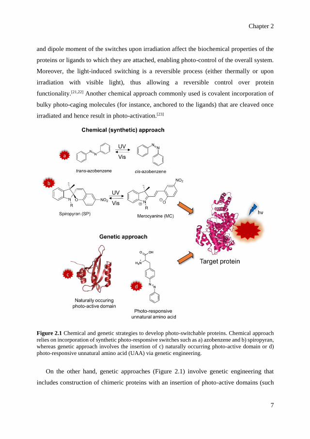
Naturally occurring, non-photoresponsive proteins can be engineered into photo-
switchable systems using chemical and/or genetic approaches (Figure 2.1). Chemical
approaches are based on covalent incorporation of synthetic photo-switchable molecules in
the structure of the proteins or in the ligands/substrates that bind the proteins.
Azobenzene[20,21] and spiropyran[22] switches are the most common examples of photo-
switches incorporated into protein or ligand structures. Upon UV light irradiation,
azobenzene switches from its trans form to cis form, while spiropyran photo-isomerization
involves a ring opening into merocyanine (Figure 2.1).[21,22] The changes in the conformation
Chapter 2
7
and dipole moment of the switches upon irradiation affect the biochemical properties of the
proteins or ligands to which they are attached, enabling photo-control of the overall system.
Moreover, the light-induced switching is a reversible process (either thermally or upon
irradiation with visible light), thus allowing a reversible control over protein
functionality.[21,22] Another chemical approach commonly used is covalent incorporation of
bulky photo-caging molecules (for instance, anchored to the ligands) that are cleaved once
irradiated and hence result in photo-activation.[23]
Figure 2.1 Chemical and genetic strategies to develop photo-switchable proteins. Chemical approach
relies on incorporation of synthetic photo-responsive switches such as a) azobenzene and b) spiropyran,
whereas genetic approach involves the insertion of c) naturally occurring photo-active domain or d)
photo-responsive unnatural amino acid (UAA) via genetic engineering.
On the other hand, genetic approaches (Figure 2.1) involve genetic engineering that
includes construction of chimeric proteins with an insertion of photo-active domains (such
Strategies to modulate allosteric regulation and self-assembly of proteins using light
8
as the light-oxygen-voltage-sensing protein domain, LOV)[24] or point mutation to
incorporate photo-responsive unnatural amino acids[25] (photo-caged amino acids or
azobenzene-modified amino acids). Here, we address both chemical/synthetic and genetic
strategies to modulate the phenomena of allosteric regulation and the assembly of proteins in
naturally occurring systems and artificial (designed) systems.
2.2 Photo-activated allosteric regulation
Nature assembles proteins into dimeric and multimeric forms to enhance cellular
efficiency, for instance mutations that are introduced in the genetic level will be amplified in
the assembled structure symmetrically.[8] Multimeric proteins often allow regulation via
cooperativity between protein subunits. The cooperative effect between subunits in a
multimeric protein is commonly known as a form of allostery. In general, allosteric
regulation enables communication between distinct sites in a protein and hence allows
indirect control over one site by influencing another.[7] As the phenomenon is central to life
yet not well understood and largely unresolved, allostery is referred to as the ‘second secret
of life’ (the first one is the genetic code).[26] Using allostery, Nature gains effective control
over various biological processes, ranging from molecular transport to enzyme catalysis.
To modulate allosteric regulation using light, photo-responsive switches can be
introduced either in the vicinity of allosteric binding sites or included in the chemical
structure of allosteric effectors (i.e., small molecules that bind to allosteric sites). In addition
to modifying naturally occurring allosteric proteins, photo-responsive allosteric proteins can
be generated from non-allosteric proteins by inserting photo-active protein domains (i.e., the
LOV) in a pre-determined position in the structure.
2.2.1 Inclusion of photo-active protein domains
Naturally, light is capable of stimulating certain processes in living cells due to the
presence of photo-active moieties embedded in the photo-active protein domains. A
superfamily of protein domains across the kingdoms of life that plays a fundamental role as
sensors for environmental stimuli in general is called the Per-ARNT-Sim (PAS) domains.[24]
PAS domains are responsible for signaling and adapting the cells to environmental changes
caused by external stimulations such as light irradiation, redox potential, oxygen and energy

Chapter 2
9
level. The mechanism of action of PAS domains usually involves cofactor binding and
mediating interactions between proteins.[24]
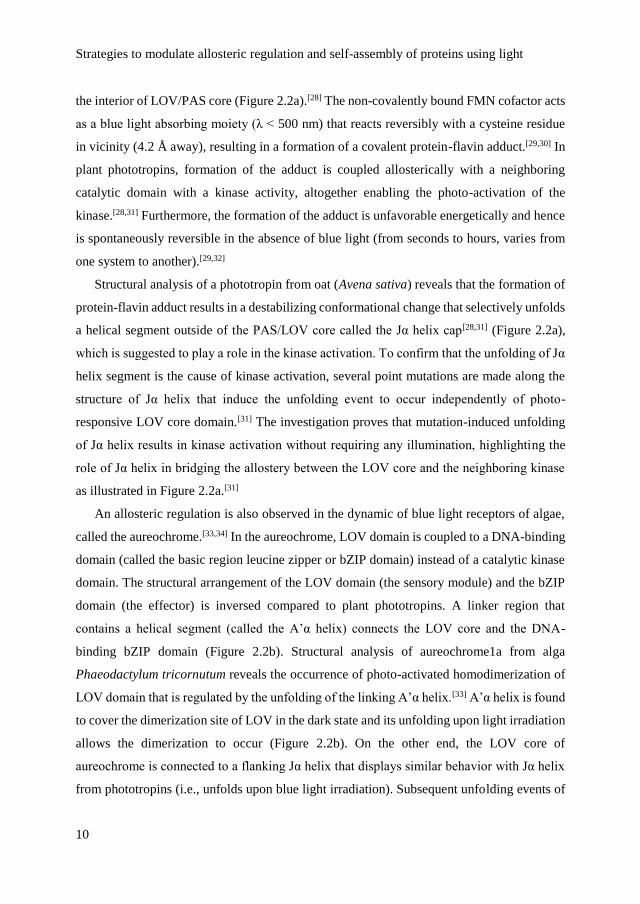
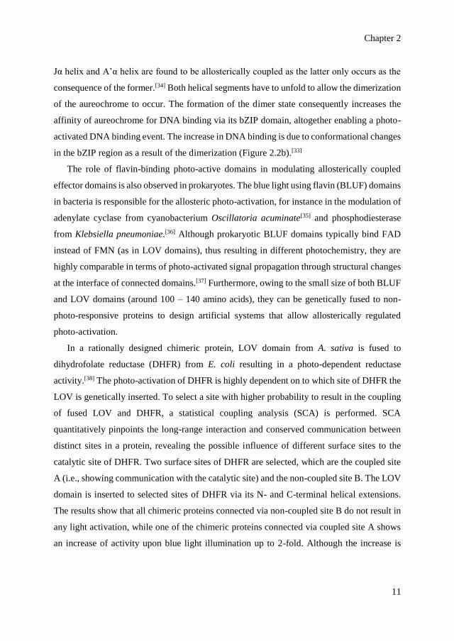
Figure 2.2 Modulation of allosteric regulation via photo-active protein domain. a) Structure of LOV
domain displaying the blue light-responsive FMN chromophore (red spheres) and the flanking Jα helix
cap. Reprinted with permission from ref [28]. Copyright © 2003, American Association for the
Advancement of Science. In plant phototropin, the unfolding of Jα helix (blue cylinder) allows
activation of the neighboring kinase domain (pink box) upon irradiation. The LOV is suggested to act
either as a kinase inhibitor in the dark state or kinase activator in the lit state. Reprinted with permission
from ref [31]. Copyright © 2004, American Chemical Society. b) Schematic representation of LOV in
algal aureochrome showing the A’α helix that blocks the dimerization site of LOV (in grey). Upon
irradiation, the A’α helix unfolds to allow the dimerization and promote the binding of double-stranded
(ds) DNA to DNA-binding domain (highlighted in red). Reprinted with permission from ref [33].
Copyright © 2016, Oxford University Press.
An example of PAS domains is the blue light-responsive domain found in photoreceptor
proteins in plants (i.e., the phototropins), which is referred to as the light-oxygen-voltage-
sensing (LOV) domain.[27] LOV domain is highly conserved and its response to light
originates from the presence of a chromophore, flavin mononucleotide cofactor (FMN), in
Strategies to modulate allosteric regulation and self-assembly of proteins using light
10
the interior of LOV/PAS core (Figure 2.2a).[28] The non-covalently bound FMN cofactor acts
as a blue light absorbing moiety (λ < 500 nm) that reacts reversibly with a cysteine residue
in vicinity (4.2 Å away), resulting in a formation of a covalent protein-flavin adduct.[29,30] In
plant phototropins, formation of the adduct is coupled allosterically with a neighboring
catalytic domain with a kinase activity, altogether enabling the photo-activation of the
kinase.[28,31] Furthermore, the formation of the adduct is unfavorable energetically and hence
is spontaneously reversible in the absence of blue light (from seconds to hours, varies from
one system to another).[29,32]
Structural analysis of a phototropin from oat (Avena sativa) reveals that the formation of
protein-flavin adduct results in a destabilizing conformational change that selectively unfolds
a helical segment outside of the PAS/LOV core called the Jα helix cap[28,31] (Figure 2.2a),
which is suggested to play a role in the kinase activation. To confirm that the unfolding of Jα
helix segment is the cause of kinase activation, several point mutations are made along the
structure of Jα helix that induce the unfolding event to occur independently of photo-
responsive LOV core domain.[31] The investigation proves that mutation-induced unfolding
of Jα helix results in kinase activation without requiring any illumination, highlighting the
role of Jα helix in bridging the allostery between the LOV core and the neighboring kinase
as illustrated in Figure 2.2a.[31]
An allosteric regulation is also observed in the dynamic of blue light receptors of algae,
called the aureochrome.[33,34] In the aureochrome, LOV domain is coupled to a DNA-binding
domain (called the basic region leucine zipper or bZIP domain) instead of a catalytic kinase
domain. The structural arrangement of the LOV domain (the sensory module) and the bZIP
domain (the effector) is inversed compared to plant phototropins. A linker region that
contains a helical segment (called the A’α helix) connects the LOV core and the DNA-
binding bZIP domain (Figure 2.2b). Structural analysis of aureochrome1a from alga
Phaeodactylum tricornutum reveals the occurrence of photo-activated homodimerization of
LOV domain that is regulated by the unfolding of the linking A’α helix.[33] A’α helix is found
to cover the dimerization site of LOV in the dark state and its unfolding upon light irradiation
allows the dimerization to occur (Figure 2.2b). On the other end, the LOV core of
aureochrome is connected to a flanking Jα helix that displays similar behavior with Jα helix
from phototropins (i.e., unfolds upon blue light irradiation). Subsequent unfolding events of
Chapter 2
11
Jα helix and A’α helix are found to be allosterically coupled as the latter only occurs as the
consequence of the former.[34] Both helical segments have to unfold to allow the dimerization
of the aureochrome to occur. The formation of the dimer state consequently increases the
affinity of aureochrome for DNA binding via its bZIP domain, altogether enabling a photo-
activated DNA binding event. The increase in DNA binding is due to conformational changes
in the bZIP region as a result of the dimerization (Figure 2.2b).[33]
The role of flavin-binding photo-active domains in modulating allosterically coupled
effector domains is also observed in prokaryotes. The blue light using flavin (BLUF) domains
in bacteria is responsible for the allosteric photo-activation, for instance in the modulation of
adenylate cyclase from cyanobacterium Oscillatoria acuminate[35] and phosphodiesterase
from Klebsiella pneumoniae.[36] Although prokaryotic BLUF domains typically bind FAD
instead of FMN (as in LOV domains), thus resulting in different photochemistry, they are
highly comparable in terms of photo-activated signal propagation through structural changes
at the interface of connected domains.[37] Furthermore, owing to the small size of both BLUF
and LOV domains (around 100 – 140 amino acids), they can be genetically fused to non-
photo-responsive proteins to design artificial systems that allow allosterically regulated
photo-activation.
In a rationally designed chimeric protein, LOV domain from A. sativa is fused to
dihydrofolate reductase (DHFR) from E. coli resulting in a photo-dependent reductase
activity.[38] The photo-activation of DHFR is highly dependent on to which site of DHFR the
LOV is genetically inserted. To select a site with higher probability to result in the coupling
of fused LOV and DHFR, a statistical coupling analysis (SCA) is performed. SCA
quantitatively pinpoints the long-range interaction and conserved communication between
distinct sites in a protein, revealing the possible influence of different surface sites to the
catalytic site of DHFR. Two surface sites of DHFR are selected, which are the coupled site
A (i.e., showing communication with the catalytic site) and the non-coupled site B. The LOV
domain is inserted to selected sites of DHFR via its N- and C-terminal helical extensions.
The results show that all chimeric proteins connected via non-coupled site B do not result in
any light activation, while one of the chimeric proteins connected via coupled site A shows
an increase of activity upon blue light illumination up to 2-fold. Although the increase is
Strategies to modulate allosteric regulation and self-assembly of proteins using light
12
modest, this study presents the proof-of-concept that photo-responsive allosteric proteins can
be rationally designed and built by fusing a photo-active domain to a non-allosteric protein.
The success of fusion approach heavily relies on whether the photo-active domain is
genetically inserted to the “right” region of the active protein that would result in maximum
perturbation and hence a measurable impact. This is corroborated by a rational design of a
chimeric protein of LOV and a viral K+ channel pore (Kcv).[39] In this study, LOV is fused
to various regions of Kcv that are important for channel gating in order to generate a light-
gated K+ channel. One variant in which the LOV is fused to the N-terminal of Kcv displays
a modest increase in conductance (ion flux) when irradiated with blue light (λ = 455 nm).
A similar concept with a different strategy is used in a rational design of a light-activated
DNA binding allosteric protein.[40] Instead of directly inserting the LOV domain to
allosterically coupled sites, a rigid α-helical domain linker is employed as the “allosteric lever
arm” to connect the LOV domain through its Jα helix extension to truncated N-terminal of
trp repressor from E. coli. As the two connected domains structurally share a helical arm in
between, it is assumed that the photo-activated unfolding of Jα helix of LOV will cause an
energetically unfavorable bending of the connecting arm that will be relieved through a
global shift in the conformation, affecting the functionality of the trp repressor. A series of
constructs to insert LOV domain to successive truncations of N-terminal helix of trp
repressor results in 12 protein fusions. Among the twelve, one fusion generated from
connecting the C-terminal of Jα helix to the middle of N-terminal helix of trp repressor shows
a significant difference in DNA binding affinities upon illumination at λ = 470 nm (dark state
is 5.6-fold higher than lit state).
2.2.2 Inclusion of synthetic photo-switches
Previous section elaborates how genetically fusing photo-active domains has been proven
successful to transform non-allosteric proteins into light-modulated allosteric proteins.
However, the fusion approach is not suitable in the cases where an on/off control is desired
and might be rather cumbersome to execute. A complementary approach is to incorporate
smaller, synthetic photo-switches into the protein structure or in the structure of the allosteric
effector. A reversible attachment of an agonist (i.e., an activating effector) to the allosteric
site of a protein via an azobenzene linker is demonstrated in the case of allosteric ionotropic
Chapter 2
13
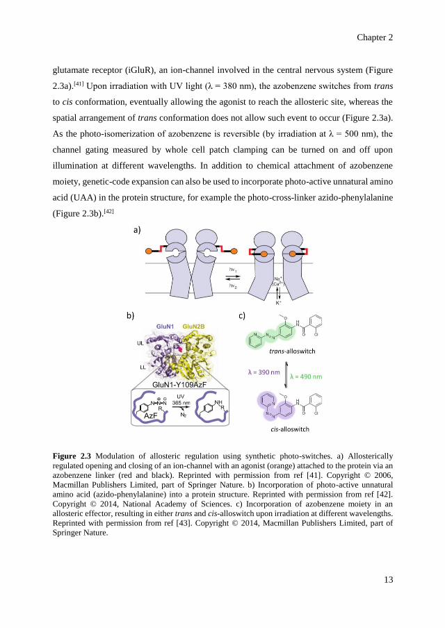
glutamate receptor (iGluR), an ion-channel involved in the central nervous system (Figure
2.3a).[41] Upon irradiation with UV light (λ = 380 nm), the azobenzene switches from trans
to cis conformation, eventually allowing the agonist to reach the allosteric site, whereas the
spatial arrangement of trans conformation does not allow such event to occur (Figure 2.3a).
As the photo-isomerization of azobenzene is reversible (by irradiation at λ = 500 nm), the
channel gating measured by whole cell patch clamping can be turned on and off upon
illumination at different wavelengths. In addition to chemical attachment of azobenzene
moiety, genetic-code expansion can also be used to incorporate photo-active unnatural amino
acid (UAA) in the protein structure, for example the photo-cross-linker azido-phenylalanine
(Figure 2.3b).[42]
Figure 2.3 Modulation of allosteric regulation using synthetic photo-switches. a) Allosterically
regulated opening and closing of an ion-channel with an agonist (orange) attached to the protein via an
azobenzene linker (red and black). Reprinted with permission from ref [41]. Copyright © 2006,
Macmillan Publishers Limited, part of Springer Nature. b) Incorporation of photo-active unnatural
amino acid (azido-phenylalanine) into a protein structure. Reprinted with permission from ref [42].
Copyright © 2014, National Academy of Sciences. c) Incorporation of azobenzene moiety in an
allosteric effector, resulting in either trans and cis-alloswitch upon irradiation at different wavelengths.
Reprinted with permission from ref [43]. Copyright © 2014, Macmillan Publishers Limited, part of
Springer Nature.

Strategies to modulate allosteric regulation and self-assembly of proteins using light
14
Furthermore, azobenzene switches can be incorporated into the chemical structure of the
allosteric effectors (drugs), resulting in an on/off photo-modulation of natural allosteric
proteins. In photopharmacology, this strategy is favored to avoid side effects as allosteric
drugs usually display higher selectivity in binding to receptors. The allosteric effectors are
modified with azobenzene in such a way so that one photo-isomer significantly favors
binding (on/active state) compared to the other (off/inactive state), owing to steric difference
between the isomers. The approach has been demonstrated to modulate G-protein-coupled
receptors (GPCRs) using light,[43] a class of transmembrane proteins responsible for signal
transduction and cellular response to stimuli in eukaryotic cells. Effectors that display
structural homology to Ar–N=N–Ar moiety of azobenzene are ideal target for modification
(Figure 2.3c). The N=N group specifically can be introduced, for instance, to replace the
amide bonds in the effectors. Remarkably, this strategy has been demonstrated to control the
motility of living cells of X. tropicalis tadpoles as a response to light stimulation.[43]
2.3 Photo-responsive protein assemblies of naturally
occurring structures
Self-assembly of protein subunits into dimers and multimers is crucial for their
functioning. A plethora of proteins that are responsible for essential, yet complex life
processes such as protein-synthesizing ribosomes and microtubules are active and functional
in their assembled forms.[2] Consequently, manipulating the self-assembly phenomenon
using external triggers would enable interfering with vital biological processes embedded in
the interlinking cellular networks. Comparable to modulating allosteric regulation with light,
self-assembly of proteins can be activated/deactivated upon illumination by introducing
photo-responsive moieties either in the structure of the protein subunits or the regulatory
molecules that chemically affect the process (e.g., inhibitors and activators of the assembly
event). Likewise, the rational design and the position to which the switches are chemically
or genetically introduced play a vital role in determining whether a strategy could result in
the desired effects. Modification with photo-switches has to introduce a significant change
in the structural dynamics of proteins to overcome the native behavior of assembly, whereas
Chapter 2
15
if the structural modification leads to excessive structural perturbations, the protein subunits
might lose their ability to assemble altogether.
2.3.1 Photo-activated dimerization
The simplest example of protein-based assemblies is the homodimers. Inside the cells,
homodimers (and further, homooligomers) are formed to reach structures with higher
stability as well as to minimize the genome size needed to synthesize functional proteins.
The formation of a dimer is more preferred instead of a multi-subunit single-chain protein
due to the ability to dissociate under certain conditions, which could be crucial in regard to
biological functions.[44] Although individually the intermolecular forces that support the
dimeric structure can be classified as relatively weak interactions, the collaborative networks
of interactions forming the dimers are among the strongest interactions found in Nature.
These collaborative networks mainly consist of non-covalent interactions, while stronger
individual interactions (i.e., covalent bonds as in disulfide bridges) are only present in a small
number at the dimeric interfaces.[45]
Despite the collectively strong forces that support protein dimers, interfering with
dimerization events using light can be achieved with the help of a class of activating
molecules called the dimerizers. Naturally, some of dimerization events require the presence
of natural products such as rapamycin and abscisic acid; such processes are referred to as the
chemically induced dimerization (CID).[46] Induced dimerization is employed by Nature to
control and direct the localization of proteins to specific cellular compartments and the
activation of signaling pathways. Optical control over CID can be achieved by modifying the
dimerizers with photo-caged molecules that are removed upon illumination. In the caged
state, the dimerizers cannot access the active site of the protein due to steric hindrance caused
by the caging moieties. Upon illumination, the steric hindrance is eliminated as the caging
moieties are irreversibly released (typically with UV light), hence resulting in photo-
activation. The time scales of such processes range from seconds to minutes. Photo-caging
dimerizers are relevant for photopharmacology as they enable dose-dependent photo-
activation of signaling pathways and cellular events.
Photo-caging molecules commonly used are the photo-cleavable nitrobenzyl derivatives
(Figure 2.4a).[23] As an example, the immunosuppressive drug rapamycin can be modified
Strategies to modulate allosteric regulation and self-assembly of proteins using light
16
into a photo-caged rapamycin dimer using nitrobenzyl derivatives to control the dimerization
of modular proteins FKBP12 and FRB, which regulate various processes including
transcription, protein localization and enzyme catalysis, using UV light (λ = 365 nm).[47]
Similar photo-caging approach using photo-removable 4,5-dimethoxy-2-nitrobenzyl is
demonstrated for a plant hormone, abscisic acid, to control the dimerization of the so-called
ABI and PYL proteins using UV light.[48] Such photo-caged systems enable photo-
modulation of regulatory processes in living cells such as signal transduction, translocation
of proteins and cytoskeletal remodeling. The concept of photo-caging has been expanded to
other dimerizers, including the phytohormone gibberellic acid[49] and antibiotic
trimethoprim.[50, 51] Moreover, the caging group can be used as a photo-cleavable linker that
acts as a bridge that connects two different proteins together. This concept is demonstrated
by covalently linking a HaloTag ligand chloroalkane and a SNAP-tag ligand O6-
benzylguanine together with a photo-labile methyl-6-nitroveratryl group in between (Figure
2.4a).[52] Upon irradiation with UV light, the linker is cleaved, liberating the ligands and
hence the dimer into two separate moieties, further allowing photo-modulation over protein
relocation. Although it is efficient to control cellular events, photo-caging approach typically
does not allow an inherent reversible control and therefore is not suitable if an on/off system
is desired. Such processes can be reversed for instance by adding a competitor binding.[46,53]
An option for reversible optical dimerizers is already exemplified by Nature in the form
of blue light-responsive LOV system. As elaborated in the previous section on allosteric
regulation, in some examples such as in algal aureochrome, the photo-switching of LOV
activates homodimerization that further enables binding event.[33,54] Similar mechanism is
observed in the marine bacterium E. litoralis in the activation of a transcription factor called
EL222.[55,56] Naturally occurring LOV further inspires the design of optical dimerizer tags
called the tunable light-inducible dimerization tags or TULIPs.[57] TULIPs harness the
interaction of LOV from plant phototropin (A. sativa) and an engineered PDZ domain as
binding partners (i.e., a protein domain in the signaling proteins across the kingdoms of life).

Chapter 2
17
Figure 2.4 Strategies to photo-modulate dimerization events. a) A photo-cleavable synthetic dimerizer
consisting of a photo-labile nitroveratryl group (blue) bridging two ligands: one ligand is a substrate
for a protein tag called SNAP-tag (red) and the other is a substrate for a protein tag called HaloTag
(green). Upon irradiation with UV light, the dimer disassociates as the linker (blue) is photo-cleaved.
Reprinted with permission from ref [52]. Copyright © 2014, Wiley-VCH Verlag GmbH & Co. b) Light-
inducible dimerization tag based on LOV-PDZ binding partners that are responsive to blue light.
Reprinted with permission from ref [57]. Copyright © 2012, Macmillan Publishers Limited, part of
Springer Nature. c) Plant phytochrome B (PhyB)-PIF binding partners that are responsive to red/far red
light. Reprinted with permission from ref [61]. Copyright © 2009, Macmillan Publishers Limited, part
of Springer Nature.
Strategies to modulate allosteric regulation and self-assembly of proteins using light
18
To generate light-inducible tags, the LOV domain (~125 residues) is fused through the
Jα helix to a specific peptide sequence that binds to PDZ (~194 residues). In the dark state,
the Jα helix physically blocks the peptide from binding to PDZ domain (Figure 2.4b).[57]
Upon irradiation with blue light, the Jα helix unfolds and exposes the peptide to allow the
interaction with PDZ domain, hence resulting in photo-activated dimerization of PDZ and
LOV. The tags have been employed to control protein localization using laser in living yeast.
However, the presence of PDZ domain and PDZ-binding peptide in the system raises concern
that the cross-talk with endogenous signaling pathways might occur. Therefore, to improve
orthogonality, PDZ as the binding partner can be replaced with a small adaptor protein called
SspB from E. coli.[58] In this case, a peptide tag that binds SspB called the SsrA peptide from
E. coli is fused to LOV domain. Coupled with point mutations that stabilize the Jα helix in
the dark (optimized from computational library screening), the tag leads to changes in
binding affinity for SspB over 50-fold upon blue light illumination. The photo-induced
binding occurs within seconds, while the reversion process occurs in the dark within minutes.
In addition to LOV-based systems, other light-inducible dimerizers are developed from plant
Arabidopsis thaliana, consisting of blue light-responsive flavoprotein cryptochrome 2 and
its interacting transcription factor CIB1[59] as well as UV light-responsive photoreceptor
UVR8 with its binding partner COP1.[60]
As an alternative to the blue light/UV light-responsive systems, dimerization tags that are
activated by red light irradiation (λ ≥ 650 nm) are derived from Arabidopsis thaliana
phytochrome B (PhyB) and its interacting factors (PIF3 and PIF6).[61-63] Structurally, PhyB
binds a chromophore namely phycocyanobilin (PCB) and is not capable of PIF binding in
the dark (Figure 2.4c). Upon red light illumination (λ = 650 nm), the chromophore undergoes
a conformational change that results in the structural change of PhyB, further allowing
binding to PIF (Figure 2.4c).[61] PhyB-PIF binding is reversible upon illumination with far-
red light (λ = 750 nm), which has been used to control protein localization in eukaryotic cells
such as yeast and mammalian cells. A set of comparative studies using yeast transcriptional
assay further reveals that PhyB/PIF3 dimerization tag allows a higher level of activation and
a lower background than TULIPs and PhyB/PIF6 systems.[64] This system therefore holds
promise for photopharmacology, also because the reversible activation using red light
wavelength enables better tissue penetration than blue or UV light.[65] Moreover, TULIPs
Chapter 2
19
system raises concern of toxicity when used for regulation of yeast transcription (observed
from cellular growth defects).[64]
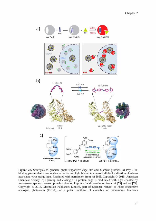
2.3.2 Photo-responsive cage-like and filament proteins
Expanding the concept of red/far-red responsive PhyB/PIF interaction beyond
dimerization of monomeric proteins enables a photo-control of higher-ordered protein
assemblies, for instance the viral protein cages. An adeno-associated virus is engineered with
PIF6 motifs at its exterior to enable light-dependent interaction with nuclear localization
sequence that is tagged with PhyB (Figure 2.5a).[66] Illumination with either red or far-red
light results in the changes of translocation of viruses into the host nucleus, allowing control
of gene delivery upon irradiation with different wavelengths (Figure 2.5a). Another approach
to modulate protein cages functionality using light is demonstrated by engineering metal-
storing ferritin cages to incorporate light-responsive manganese (Mn)-carbonyl moieties in
the interior of the cage.[67] Such metal complexes inherently dissociate and release carbon
monoxide upon irradiation with visible light,[68] allowing the generation of CO-releasing
ferritin that is modulated with light. Furthermore, naturally occurring photo-active moieties
can be used to trigger oligomerization and clustering of protein molecules. Such control is
achieved by incorporation of plant A. thaliana cryptochrome 2 that naturally assembles into
the so-called photobodies in plant cells when irradiated with blue light.[69,70] Genetically
incorporating cryptochrome 2 into signaling proteins such as RhoA (i.e., a GTPase that
mediates cellular tension and cytoskeletal contraction) enables photo-activation of signaling
pathways in mammalian cells.[70]
Nevertheless, incorporation of bulky photo-active domains is rather difficult to result in
a precise control over the assembly process of large and highly defined structures, such as
cage-like proteins and filaments. Attachment of synthetic photo-switches due to their smaller
size offers a safer approach in order to avoid excessive perturbations that might disfavor
assembly altogether. Owing to the complexity of higher-ordered structures and their natural
tendency to favor assembly, to be able to use light to interfere with such assemblies is a
challenging task. Photo-switches need to be carefully introduced into the structure of the
target protein to result in sufficient structural perturbations that allow photo-modulation,
without compromising the ability to assemble. Furthermore, highly-defined assemblies are

Strategies to modulate allosteric regulation and self-assembly of proteins using light
20
usually robust and modifications of the exterior of such structures, for instance bacterial
encapsulin nanocages[71] and filamentous M13 bacteriophage,[72] with photo-switchable
molecules (i.e., spiropyran and azobenzene, respectively) have been shown to not
compromise the stability of the assembled structures upon photo-switching.
A remarkable photo-control over the assembly behavior of a cage-like protein is
demonstrated in the case of homo-oligomeric cage-like group II chaperonin from M.
maripaludis.[73] This chaperonin naturally exists in two states: open and closed, depending
on the binding and hydrolysis of nucleotide (ATP) molecules. To control the opening/closing
of the chaperonin with light, azobenzene molecules are covalently placed at the interface of
the protein subunits, acting as a spacer crosslinking cysteine residues between the subunits
(Figure 2.5b).[73] Light-responsive isomerization of the trans and cis forms of azobenzene
crosslinkers results in a distance change between the subunits (i.e., 18 Å in the trans state
and 5 – 12 Å in cis state). Using computational screening, the cysteine residues to which the
azobenzene are attached are genetically introduced to match the distance of the cis and trans
forms of azobenzene. Upon irradiation with UV light (λ = 365 nm), the spacers are in their
cis form, resulting in the chaperonin cage favoring the closed state. Upon subsequent blue
light irradiation (λ = 450 nm), the spacers convert to their extended trans form, allowing the
opening of the chaperonin cage (Figure 2.5b).
Revealed by in silico investigation, the success of this approach relies on two important
aspects: first, the energy landscape, which cooperatively favors either of the two dominant
states (i.e., either open or closed)[74] and second, the strong allosteric coupling between the
orientation of the subunits (affected by the distance change from the photo-active azobenzene
spacers) with the structural rearrangements of the nucleotide-binding pocket.[75] The
allosteric coupling between the two events is mediated by a rigid-body rocking and rotation
of the subunits due to light-triggered distance change (Figure 2.5b).[74,75] The movement
subsequently results in the destabilization of protein-nucleotide interaction in the nucleotide-
binding pocket, leading to the opening of the cage.
A complementary approach to modification of protein structures is to incorporate photo-
switchable moieties into regulatory molecules that chemically affect the assembly process
(e.g., activators and inhibitors). This approach has been successfully used to activate or
inhibit the assembly of cellular filaments, the microtubules.
Chapter 2
21
Figure 2.5 Strategies to generate photo-responsive cage-like and filament proteins. a) PhyB-PIF
binding partner that is responsive to red/far red light is used to control cellular localization of adeno-
associated virus using light. Reprinted with permission from ref [66]. Copyright © 2015, American
Chemical Society. b) Opening and closing of a protein cage is modulated with light enabled by
azobenzene spacers between protein subunits. Reprinted with permission from ref [73] and ref [74].
Copyright © 2013, Macmillan Publishers Limited, part of Springer Nature. c) Photo-responsive
analogue, photostatin (PST-1), of a potent inhibitor of assembly of microtubule filaments
Strategies to modulate allosteric regulation and self-assembly of proteins using light
22
(combretastatin A-4). PST-1 is only active in its cis form, hence allowing photo-regulated inhibition of
microtubule assembly. Reprinted with permission from ref [78]. Copyright © 2015, Elsevier Inc.
In the case of photo-activation of microtubule assembly, the nucleotide substrate GTP
that is required for self-assembly is caged with a photo-cleavable nitrobenzyl derivative.[76]
Due to the steric hindrance of the caged form of GTP, the rate of microtubule assembly is
significantly reduced in the absence of illumination. Upon UV flash photolysis, the photo-
labile moiety is cleaved off and the GTP is released within milliseconds, resulting in the
activation of the microtubule assembly.
On the other hand, a class of active molecules called the combretastatin A-4 (CA4)
(Figure 2.5c) is identified as a potent inhibitor of microtubule assembly in its cis form, but
not in its trans form.[77] Replacing the C=C bond in the CA4 structure with N=N bond to
mimic azobenzene (Figure 2.5c) results in a photo-active derivative of CA4 called the
photostatins (PST).[78] PST switches from its inactive trans to active cis form upon irradiation
at λ = 390 – 430 nm, which is reversible upon irradiation at λ = 500 – 530 nm or by thermal
relaxation (in the dark, half-life = 6 min), allowing a fully reversible control over the drug
cytotoxicity. The activity of the trans form in the dark is about 250-fold less compared to the
cis form upon irradiation with blue light. This remarkable difference has been demonstrated
to photo-modulate the mictrotubule dynamic assembly in live cells of C. elegans embryo.[78]
2.4 Photo-responsive artificial protein-based systems and
higher-order structures
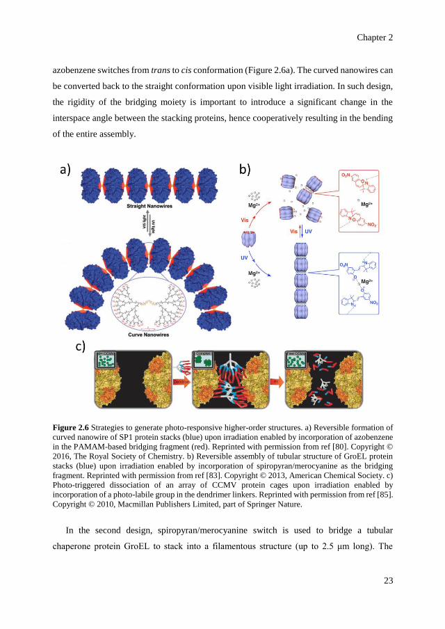
Synthetic photo-switches can be used to bridge proteins together to form artificial
extended nanostructures in which the assembly event is modulated with light. Here, we focus
on three examples of artificial higher-order structures mediated by three different photo-
switches: azobenzene, spiropyran and a photo-labile group. In the first design, azobenzene is
incorporated in the bridging fragment of a nanowire-like assembly of stable protein one
(SP1), a ring-shaped protein from plant Populus tremula (Figure 2.6a).[79,80] The stacking of
SP1 to form nanowires is structurally mediated by a globular poly(amido-amine) or PAMAM
dendrimer via electrostatic interaction.[80] Incorporation of azobenzene in the PAMAM
structure results in the bending of the overall structure upon UV light irradiation when the
Chapter 2
23
azobenzene switches from trans to cis conformation (Figure 2.6a). The curved nanowires can
be converted back to the straight conformation upon visible light irradiation. In such design,
the rigidity of the bridging moiety is important to introduce a significant change in the
interspace angle between the stacking proteins, hence cooperatively resulting in the bending
of the entire assembly.
Figure 2.6 Strategies to generate photo-responsive higher-order structures. a) Reversible formation of
curved nanowire of SP1 protein stacks (blue) upon irradiation enabled by incorporation of azobenzene
in the PAMAM-based bridging fragment (red). Reprinted with permission from ref [80]. Copyright ©
2016, The Royal Society of Chemistry. b) Reversible assembly of tubular structure of GroEL protein
stacks (blue) upon irradiation enabled by incorporation of spiropyran/merocyanine as the bridging
fragment. Reprinted with permission from ref [83]. Copyright © 2013, American Chemical Society. c)
Photo-triggered dissociation of an array of CCMV protein cages upon irradiation enabled by
incorporation of a photo-labile group in the dendrimer linkers. Reprinted with permission from ref [85].
Copyright © 2010, Macmillan Publishers Limited, part of Springer Nature.
In the second design, spiropyran/merocyanine switch is used to bridge a tubular
chaperone protein GroEL to stack into a filamentous structure (up to 2.5 μm long). The
Strategies to modulate allosteric regulation and self-assembly of proteins using light
24
opening of the cylindrical cavity of GroEL is genetically modified with 14 cysteine residues
to enable attachment of photo-switches bearing a maleimide moiety.[81, 82] Upon UV light
irradiation (λ = 280 nm) at room temperature, the spiropyran opens into its merocyanine
form, which enables electrostatic interaction with magnesium ions (Figure 2.6b).[83] Mg2+
ions mediate the interaction between the stacking GroEL proteins via merocyanine-Mg2+-
merocyanine bridges. As the closed spiropyran cannot interact with Mg2+, the assembled
structure collapses into shorter fragments upon irradiation with visible light (λ > 400 nm)
when the merocyanine form is converted back to spiropyran form.[83] Notably, the light-
induced reversibility is achieved only when a radical scavenger dithiothreitol (DTT) is
present in the system.[82,83] The authors postulated that radical species might have been
generated upon photoexcitation that could covalently cross-link the stacking GroEL
together.[83] Therefore, addition of a radical scavenger like DTT is necessary to prevent any
covalent cross-linking and ensure the reversibility of the system. Moreover, the cylindrical
cavity of the stacking GroEL is able to confine cargo molecules such as drugs, enabling the
design of photo-responsive nanocarriers.[84]
In the third design, a photo-responsive array of a viral capsid protein namely Cowpea
Chlorotic Mottle Virus (CCMV) is generated via electrostatic interaction between
negatively-charged exterior of the capsid and positively-charged dendrimers.[85] A photo-
labile nitrobenzyl linker is embedded in the structure of the bridging dendrimers (Figure
2.6c). Upon UV light irradiation (λ = 365 nm), the photo-labile moiety is cleaved, leading to
the collapse of the bridging structures. Consequently, the CCMV array dissociates into
individual particles upon UV light irradiation. A similar design can also be applied to other
protein cages, such as the magnetoferritin.[85]
2.5 Perspectives
Strategies on modulating naturally occurring proteins with photo-switchable moieties,
either chemically or genetically introduced, would enable interfering with complex
regulations that orchestrate cellular events, particularly in relation to photopharmacology.[86]
Even when a strategy does not result in a complete on/off photo-control but rather a modest
difference between the dark and lit-state, still a significant effect can be expected as the target
Chapter 2
25
proteins are usually involved in interlinking networks of cellular regulations. A fluctuation
triggered with light at one point in a cascade system would result in changes in the overall
dynamics of the process. Among the presented strategies, generating a library of photo-
responsive analogue drugs is a promising means to gain effective and reversible control over
a specific system. In terms of reversibility, the drugs ideally should undergo thermal
deactivation spontaneously, hence is only active upon irradiation at the desired site.
Nevertheless, the challenge remains in the stability of the designed drugs (especially against
spontaneous hydrolysis), orthogonality and specificity of action, as well as the means of
delivery of the designed drugs.
Moreover, most of the presented systems are active upon UV light illumination, which is
not the ideal option to apply in physiological processes due to the weak tissue penetration.
Red and far-red light with longer wavelengths are more suited to carry such task.[65] Nature
has presented an example of red light-responsive system in plant phytochrome B and its
interacting factors.[69] As a complementary approach, synthetic chemists have made attempts
to design and develop photo-switches that are responsive to red light illumination, such as
the azo-derivatives.[87-90] In the future, the application of such strategies based on red light-
responsive systems shall be expanded, particularly for in vivo studies. In a complementary
manner, artificially designed extended nanostructures are envisioned to stimulate the
development of photo-tunable arrays and carriers[91] and/or molecular labels for functional
assemblies and supramolecular platforms.[92-94]
2.6 References
[1] A. Typas, V. Sourjik, Nat. Rev. Microbiol. 2015, 13, 559-572.
[2] R. J. Arnold, J. P. Reilly, Anal. Biochem. 1999, 269, 105-112.
[3] E. Brody, J. Abelson, Science 1985, 228, 963-967.
[4] C. L. Will, R. Luhrmann, Curr. Opin. Cell Biol. 1997, 9, 320-328.
[5] B. Alberts, Cell 1998, 92, 291-294.
[6] T. Pawson, P. Nash, Science 2003, 300, 445-452.
[7] H. N. Motlagh, J. O. Wrabl, J. Li, V. J. Hilser, Nature 2014, 508, 331-339.
[8] M. H. Ali, B. Imperiali, Bioorg. Med. Chem. 2005, 13, 5013-5020.
Strategies to modulate allosteric regulation and self-assembly of proteins using light
26
[9] B. N. Kholodenko, Nat. Rev. Mol. Cell Bio. 2006, 7, 165-176.
[10] A. Gautier, C. Gauron, M. Volovitch, D. Bensimon, L. Jullien, S. Vriz, Nat. Chem. Biol.
2014, 10, 533-541.
[11] W. Szymanski, J. M. Beierle, H. A. V. Kistemaker, W. A. Velema, B. L. Feringa, Chem.
Rev. 2013, 113, 6114-6178.
[12] K. E. Brechun, K. M. Arndt, G. A. Woolley, Curr. Opin. Struct. Biol. 2016, 45, 53-58.
[13] G. Guglielmi, H. J. Falk, S. De Renzis, Trends Cell Biol. 2016, 26, 864-874.
[14] M. Baker, Nat. Methods 2012, 9, 443-446.
[15] C. W. Riggsbee, A. Deiters, Trends Biotechnol. 2010, 28, 468-475.
[16] U. T. Bornscheuer, Nature 2016, 540, 345-346.
[17] F. Z. Zhang, A. Zarrine-Afsar, M. S. Al-Abdul-Wahid, R. S. Prosser, A. R. Davidson,
G. A. Woolley, J. Am. Chem. Soc. 2009, 131, 2283-2289.
[18] A. Kocer, M. Walko, W. Meijberg, B. L. Feringa, Science 2005, 309, 755-758.
[19] C. L. Tucker, J. D. Vrana, M. J. Kennedy, Curr. Protoc. Cell Biol, 2014, 64, 17 16 11-
20.
[20] R. J. Mart, R. K. Allemann, Chem. Commun. 2016, 52, 12262-12277.
[21] A. A. Beharry, G. A. Woolley, Chem. Soc. Rev. 2011, 40, 4422-4437.
[22] R. Klajn, Chem. Soc. Rev. 2014, 43, 148-184.
[23] G. Mayer, A. Heckel, Angew. Chem. Int. Ed. 2006, 45, 4900-4921.
[24] B. L. Taylor, I. B. Zhulin, Microbiol. Mol. Biol. Rev. 1999, 63, 479-506.
[25] A. S. Baker, A. Deiters, ACS Chem. Biol 2014, 9, 1398-1407.
[26] A. W. Fenton, Trends Biochem. Sci. 2008, 33, 420-425.
[27] J. M. Christie, M. Salomon, K. Nozue, M. Wada, W. R. Briggs, Proc. Natl. Acad. Sci.
USA 1999, 96, 8779-8783.
[28] S. M. Harper, L. C. Neil, K. H. Gardner, Science 2003, 301, 1541-1544.
[29] S. Crosson, K. Moffat, Proc. Natl. Acad. Sci. USA 2001, 98, 2995-3000.
[30] M. Salomon, W. Eisenreich, H. Durr, E. Schleicher, E. Knieb, V. Massey, W. Rudiger,
F. Muller, A. Bacher, G. Richter, Proc. Natl. Acad. Sci. USA 2001, 98, 12357-12361.
[31] S. M. Harper, J. M. Christie, K. H. Gardner, Biochemistry 2004, 43, 16184-16192.
[32] F. Circolone, J. Granzin, K. Jentzsch, T. Drepper, K. E. Jaeger, D. Willbold, U. Krauss,
R. Batra-Safferling, J. Mol. Biol. 2012, 417, 362-374.
Chapter 2
27
[33] A. Banerjee, E. Herman, M. Serif, M. Maestre-Reyna, S. Hepp, R. Pokorny, P. G. Kroth,
L. O. Essen, T. Kottke, Nucleic Acids Res. 2016, 44, 5957-5970.
[34] E. Herman, T. Kottke, Biochemistry 2015, 54, 1484-1492.
[35] M. Ohki, K. Sugiyama, F. Kawai, H. Tanaka, Y. Nihei, S. Unzai, M. Takebe, S.
Matsunaga, S. Adachi, N. Shibayama, Z. W. Zhou, R. Koyama, Y. Ikegaya, T. Takahashi,
J. R. H. Tame, M. Iseki, S. Y. Park, Proc. Natl. Acad. Sci. USA 2016, 113, 6659-6664.
[36] A. Winkler, A. Udvarhelyi, E. Hartmann, J. Reinstein, A. Menzel, R. L. Shoeman, I.
Schlichting, J. Mol. Biol. 2014, 426, 853-868.
[37] J. M. Christie, J. Gawthorne, G. Young, N. J. Fraser, A. J. Roe, Mol. Plant 2012, 5, 533-
544.
[38] J. Lee, M. Natarajan, V. C. Nashine, M. Socolich, T. Vo, W. P. Russ, S. J. Benkovic, R.
Ranganathan, Science 2008, 322, 438-442.
[39] C. Cosentino, L. Alberio, S. Gazzarrini, M. Aquila, E. Romano, S. Cermenati, P.
Zuccolini, J. Petersen, M. Beltrame, J. L. Van Etten, J. M. Christie, G. Thiel, A. Moroni,
Science 2015, 348, 707-710.
[40] D. Strickland, K. Moffat, T. R. Sosnick, Proc. Natl. Acad. Sci. USA 2008, 105, 10709-
10714.
[41] M. Volgraf, P. Gorostiza, R. Numano, R. H. Kramer, E. Y. Isacoff, D. Trauner, Nat.
Chem. Biol. 2006, 2, 47-52.
[42] S. J. Zhu, M. Riou, C. A. Yao, S. Carvalho, P. C. Rodriguez, O. Bensaude, P. Paoletti,
S. X. Ye, Proc. Natl. Acad. Sci. USA 2014, 111, 6081-6086.
[43] I. Pittolo, X. Gomez-Santacana, K. Eckelt, X. Rovira, J. Dalton, C. Goudet, J. P. Pin, A.
Llobet, J. Giraldo, A. Llebaria, P. Gorostiza, Nat. Chem. Biol. 2014, 10, 813-815.
[44] N. J. Marianayagam, M. Sunde, J. M. Matthews, Trends Biochem. Sci. 2004, 29, 618-
625.
[45] S. Jones, J. M. Thornton, Proc. Natl. Acad. Sci. USA 1996, 93, 13-20.
[46] S. Voss, L. Klewer, Y. W. Wu, Curr. Opin. Chem. Biol. 2015, 28, 194-201.
[47] K. A. Brown, Y. Zou, D. Shirvanyants, J. Zhang, S. Samanta, P. K. Mantravadi, N. V.
Dokholyan, A. Deiters, Chem. Commun. 2015, 51, 5702-5705.
[48] C. W. Wright, Z. F. Guo, F. S. Liang, ChemBioChem 2015, 16, 254-261.
Strategies to modulate allosteric regulation and self-assembly of proteins using light
28
[49] K. M. Schelkle, T. Griesbaum, D. Ollech, S. Becht, T. Buckup, M. Hamburger, R.
Wombacher, Angew. Chem. Int. Ed. 2015, 54, 2825-2829.
[50] E. R. Ballister, C. Aonbangkhen, A. M. Mayo, M. A. Lampson, D. M. Chenoweth, Nat.
Commun. 2014, 5, 5475.
[51] E. R. Ballister, S. Ayloo, D. M. Chenoweth, M. A. Lampson, E. L. F. Holzbaur, Curr.
Biol. 2015, 25, R407-R408.
[52] M. Zimmermann, R. Cal, E. Janett, V. Hoffmann, C. G. Bochet, E. Constable, F.
Beaufils, M. P. Wymann, Angew. Chem. Int. Ed. 2014, 53, 4717-4720.
[53] P. Liu, A. Calderon, G. Konstantinidis, J. Hou, S. Voss, X. Chen, F. Li, S. Banerjee, J.
E. Hoffmann, C. Theiss, L. Dehmelt, Y. W. Wu, Angew. Chem. Int. Ed. 2014, 53, 10049-
10055.
[54] Y. Akiyama, Y. Nakasone, Y. Nakatani, O. Hisatomi, M. Terazima, J. Phys. Chem. B
2016, 120, 7360-7370.
[55] B. D. Zoltowski, L. B. Motta-Mena, K. H. Gardner, Biochemistry 2013, 52, 6653-6661.
[56] A. I. Nash, R. McNulty, M. E. Shillito, T. E. Swartz, R. A. Bogomolni, H. Luecke, K.
H. Gardner, Proc. Natl. Acad. Sci. USA 2011, 108, 9449-9454.
[57] D. Strickland, Y. Lin, E. Wagner, C. M. Hope, J. Zayner, C. Antoniou, T. R. Sosnick,
E. L. Weiss, M. Glotzer, Nat. Methods 2012, 9, 379-384.
[58] G. Guntas, R. A. Hallett, S. P. Zimmerman, T. Williams, H. Yumerefendi, J. E. Bear, B.
Kuhlman, Proc. Natl. Acad. Sci. USA 2015, 112, 112-117.
[59] M. J. Kennedy, R. M. Hughes, L. A. Peteya, J. W. Schwartz, M. D. Ehlers, C. L. Tucker,
Nat. Methods 2010, 7, 973-975.
[60] R. P. Crefcoeur, R. H. Yin, R. Ulm, T. D. Halazonetis, Nat. Commun. 2013, 4, 1779.
[61] A. Levskaya, O. D. Weiner, W. A. Lim, C. A. Voigt, Nature 2009, 461, 997-1001.
[62] G. G. Yu, H. Onodera, Y. Aono, F. Kawano, Y. Ueda, A. Furuya, H. Suzuki, M. Sato,
Sci. Rep. 2016, 6, 35777.
[63] W. E. Georgianna, A. Deiters, ChemBioChem 2010, 11, 301-303.
[64] G. P. Pathak, D. Strikland, J. D. Vrana, C. L. Tucker, ACS Synth. Biol. 2014, 3, 832-
838.
[65] R. R. Anderson, J. A. Parrish, J. Invest. Dermatol. 1981, 77, 13-19.
[66] E. J. Gomez, K. Gerhardt, J. Judd, J. J. Tabor, J. Suh, ACS Nano 2016, 10, 225-237.
Chapter 2
29
[67] K. Fujita, Y. Tanaka, S. Abe, T. Ueno, Angew. Chem. Int. Ed. 2016, 55, 1056-1060.
[68] R. Motterlini, J. E. Clark, R. Foresti, P. Sarathchandra, B. E. Mann, C. J. Green, Circ.
Res. 2002, 90, E17-E24.
[69] P. Mas, P. F. Devlin, S. Panda, S. A. Kay, Nature 2000, 408, 207-211.
[70] L. J. Bugaj, A. T. Choksi, C. K. Mesuda, R. S. Kane, D. V. Schaffer, Nat. Methods 2013,
10, 249-252.
[71] R. M. Putri, J. W. Fredy, J. J. L. M. Cornelissen, M. S. T. Koay, N. Katsonis,
ChemPhysChem 2016, 17, 1815-1818.
[72] M. Murugesan, G. Abbineni, S. L. Nimmo, B. R. Cao, C. B. Mao, Sci. Rep. 2013, 3,
1820.
[73] D. Hoersch, S. H. Roh, W. Chiu, T. Kortemme, Nat. Nanotechnol. 2013, 8, 928-932.
[74] G. A. Woolley, Nat. Nanotechnol. 2013, 8, 892-893.
[75] D. Hoersch, T. Kortemme, Structure 2016, 24, 576-584.
[76] A. Marx, A. Jagla, E. Mandelkow, Eur. Biophys. J. 1990, 19, 1-9.
[77] G. C. Tron, T. Pirali, G. Sorba, F. Pagliai, S. Busacca, A. A. Genazzani, J. Med. Chem.
2006, 49, 3033-3044.
[78] M. Borowiak, W. Nahaboo, M. Reynders, K. Nekolla, P. Jalinot, J. Hasserodt, M.
Rehberg, M. Delattre, S. Zahler, A. Vollmar, D. Trauner, O. Thorn-Seshold, Cell 2015,
162, 403-411.
[79] H. C. Sun, L. Miao, J. X. Li, S. Fu, G. An, C. Y. Si, Z. Y. Dong, Q. Luo, S. J. Yu, J. Y.
Xu, J. Q. Liu, ACS Nano 2015, 9, 5461-5469.
[80] H. C. Sun, L. L. Zhao, T. T. Wang, G. An, S. Fu, X. M. Li, X. L. Deng, J. Q. Liu, Chem.
Commun. 2016, 52, 6001-6004.
[81] S. Muramatsu, K. Kinbara, H. Taguchi, N. Ishii, T. Aida, J. Am. Chem. Soc. 2006, 128,
3764-3769.
[82] S. Biswas, K. Kinbara, N. Oya, N. Ishii, H. Taguchi, T. Aida, J. Am. Chem. Soc. 2009,
131, 7556-7557.
[83] T. Sendai, S. Biswas, T. Aida, J. Am. Chem. Soc. 2013, 135, 11509-11512.
[84] S. Biswas, K. Kinbara, T. Niwa, H. Taguchi, N. Ishii, S. Watanabe, K. Miyata, K.
Kataoka, T. Aida, Nat. Chem. 2013, 5, 613-620.
Strategies to modulate allosteric regulation and self-assembly of proteins using light
30
[85] M. A. Kostiainen, O. Kasyutich, J. J. L. M. Cornelissen, R. J. M. Nolte, Nat. Chem.
2010, 2, 394-399.
[86] W. A. Velema, W. Szymanski, B. L. Feringa, J. Am. Chem. Soc. 2014, 136, 2178-2191.
[87] A. A. Beharry, O. Sadovski, G. A. Woolley, J. Am. Chem. Soc. 2011, 133, 19684-19687.
[88] Y. Yang, R. P. Hughes, I. Aprahamian, J. Am. Chem. Soc. 2014, 136, 13190-13193.
[89] S. Samanta, A. Babalhavaeji, M. X. Dong, G. A. Woolley, Angew. Chem. Int. Ed. 2013,
52, 14127-14130.
[90] M. X. Dong, A. Babalhavaeji, S. Samanta, A. A. Beharry, G. A. Woolley, Acc. Chem.
Res. 2015, 48, 2662-2670.
[91] J. Olejniczak, C. J. Carling, A. Almutairi, J. Control. Release 2015, 219, 18-30.
[92] C. Grunwald, K. Schulze, A. Reichel, V. U. Weiss, D. Blaas, J. Piehler, K. H.
Wiesmuller, R. Tampe, Proc. Natl. Acad. Sci. USA 2010, 107, 6146-6151.
[93] N. Laboria, R. Wieneke, R. Tampe, Angew. Chem. Int. Ed. 2013, 52, 848-853.
[94] N. L. Weineisen, C. A. Hommersom, J. Voskuhl, S. Sankaran, A. M. Depauw, N.
Katsonis, P. Jonkheijm, J. J. L. M. Cornelissen, Chem. Commun. 2017, 53, 1896-1899.
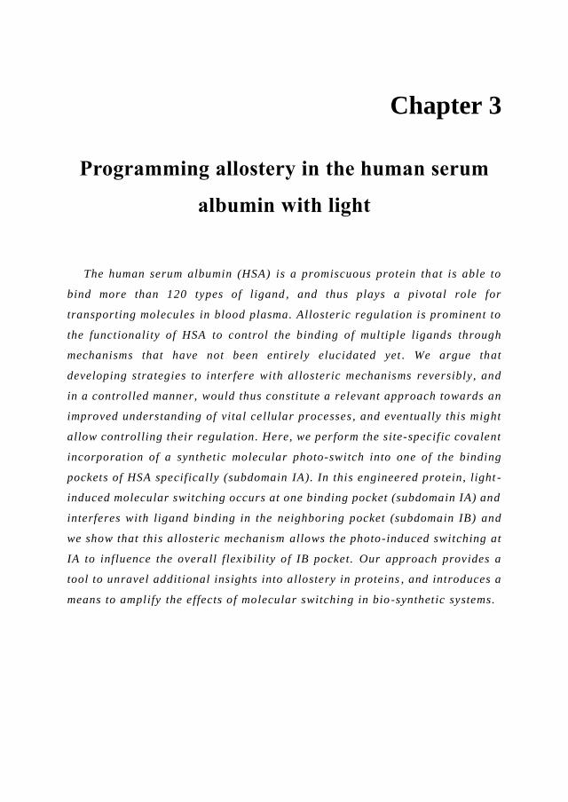
Chapter 3
Programming allostery in the human serum
albumin with light
The human serum albumin (HSA) is a promiscuous protein that is able to
bind more than 120 types of ligand , and thus plays a pivotal role for
transporting molecules in blood plasma. Allosteric regulation is prominent to
the functionality of HSA to control the binding of multiple ligands through
mechanisms that have not been entirely elucidated yet . We argue that
developing strategies to interfere with allosteric mechanisms reversibly, and
in a controlled manner, would thus constitute a relevant approach towards an
improved understanding of vital cellular processes , and eventually this might
allow controlling their regulation. Here, we perform the site-specific covalent
incorporation of a synthetic molecular photo-switch into one of the binding
pockets of HSA specifically (subdomain IA). In this engineered protein, light -
induced molecular switching occurs at one binding pocket (subdomain IA) and
interferes with ligand binding in the neighboring pocket (subdomain IB) and
we show that this allosteric mechanism allows the photo-induced switching at
IA to influence the overall flexibility of IB pocket. Our approach provides a
tool to unravel additional insights into allostery in proteins , and introduces a
means to amplify the effects of molecular switching in bio -synthetic systems.

Programming allostery in the human serum albumin with light
32
3.1 Introduction
In proteins with multiple binding pockets, binding of a ligand in one binding site often
influences the binding of other ligands to one or more binding sites, a process known as
allostery.[1-5] Allosteric regulation of protein activity is central to life and sometimes even
referred to as ‘the second secret of life’ - the first one being the genetic code.[2] However, the
mechanisms by which proteins undergo allosteric regulation remain largely unknown, and
these complex systems have already revealed a number of paradoxes. For example, tightly
packed, fully folded proteins can display a remarkable structural plasticity dictated by their
binding pockets. Also, it has been shown that a structural change in the protein backbone is
not always necessary for the binding sites to communicate with each other.[4]
Overall, it appears that allostery is facilitated in dynamic proteins, and in the state of our
understanding, allostery is not simply a shape change-induced phenomenon as understood in
early years,[6] but rather a thermodynamic process in an ensemble.[1-4] Pioneering work has
also highlighted the limitations of deducing mechanism from the static structure alone,[1,3]
which calls for tools to manipulate, investigate and eventually engineer the dynamics of
allosteric systems. A recent study reports on using temperature and pH control to increase
the local conformational entropy of fused proteins that communicate through allostery.[7] A
complementary approach towards allosteric engineering consists in implementing allosteric
behavior in wholly synthetic biomimetic systems to control ligand binding and catalysis.[8,9]
However, previous approaches lack of selectivity in space and time and for protein systems
operating at physiological conditions, environmental changes such as in pH and temperature
could lead to unfolding and loss of activity.
In contrast, light offers high spatiotemporal control while being relatively non-destructive
toward proteins and applicable in physiological conditions.[10-14] In order to make proteins
light-switchable, the challenge is to make sure that even in aqueous systems and in the protein
environment, the stability and photo-switching properties of molecular switches are
preserved. Light-modulated proteins have been developed to control biological activities,
such as directing the secondary structures of peptides/proteins,[15,16] enzymatic catalysis,[17,18]
protein-ligand binding,[19,20] modulation of protein channels[21,22] and receptors in neural
networks.[23] Recently, the emergence of photo-switchable allosteric modulators[24-26] as well
Chapter 3
33
as light-activated allosteric channel and DNA-binding protein[27,28] marks the era of
combining allosteric regulation with tailored optical control.
The human serum albumin (HSA) is characterized by its extraordinary capability to bind
over 120 types of ligands, despite its relatively simple monomeric structure.[29,30] Owing to
its promiscuity in ligand binding, HSA is the main carrier of a variety of compounds in blood
plasma. Structurally, HSA is a small protein (66 kDa) that is dominated by α-helices and
loops.[29-31] HSA has multiple binding sites (three primary ligand binding sites and seven sites
for fatty acid binding) that contribute to its capability of binding a broad range of ligands.[32]
Allostery in such a promiscuous protein remains a challenging phenomenon to decipher, not
only due to the inherent structural plasticity of the protein, but also due to the lack of
consensus in the type of cooperativity between binding pockets: it can be either a positive
cooperation, a negative cooperation, or no effect at all depending on the choice of ligands.[29]
Recent investigations by ultrafast time-resolved spectroscopy have suggested that the
allostery of HSA is likely to originate in a ballistic and anisotropic energy flow through
connecting helical structures.[32]
Here, we design and investigate a photo-responsive hybrid system that contributes to
unveiling the mechanistic aspects of cooperative actions between distinct pockets/sites of
HSA. We achieve an optical control over a specific binding site of HSA (i.e., a site known
as subdomain IB), by coupling a single spiropyran photo-switch to a neighboring site (i.e., a
cysteine residue at subdomain IA). In this way, we are able to design a photo-switchable
HSA where the photo-induced molecular switching occurs at IA site, while the effect is
propagated to IB site. This strategy allows to externally interfere with allostery using light as
a trigger.
3.2 Results and discussion
3.2.1 Design and synthesis of a photo-switchable hybrid
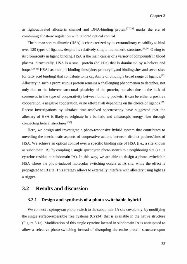
We connect a spiropyran photo-switch to the subdomain IA site covalently, by modifying
the single surface-accessible free cysteine (Cys34) that is available in the native structure
(Figure 3.1a). Modification of this single cysteine located in subdomain IA is anticipated to
allow a selective photo-switching instead of disrupting the entire protein structure upon
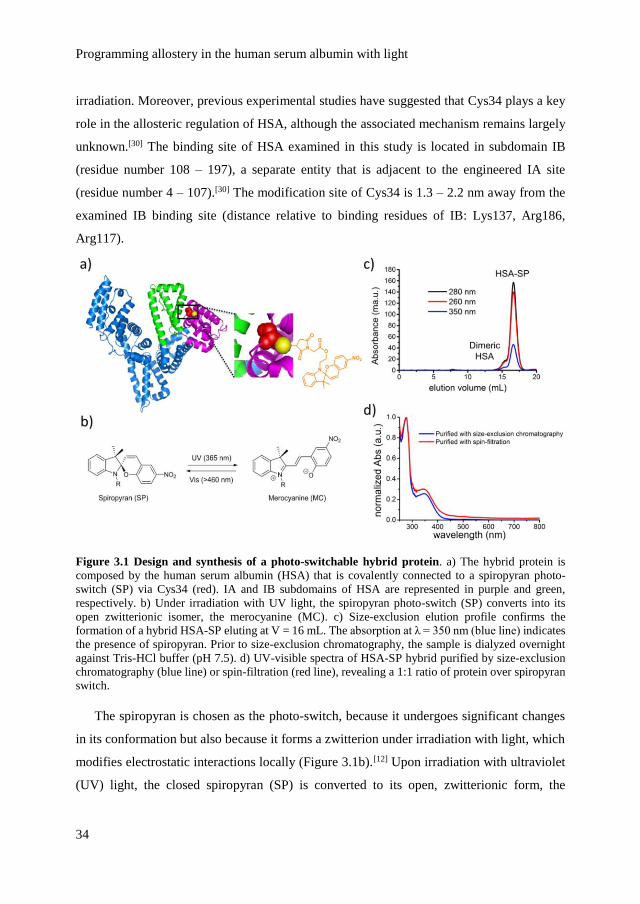
Programming allostery in the human serum albumin with light
34
irradiation. Moreover, previous experimental studies have suggested that Cys34 plays a key
role in the allosteric regulation of HSA, although the associated mechanism remains largely
unknown.[30] The binding site of HSA examined in this study is located in subdomain IB
(residue number 108 – 197), a separate entity that is adjacent to the engineered IA site
(residue number 4 – 107).[30] The modification site of Cys34 is 1.3 – 2.2 nm away from the
examined IB binding site (distance relative to binding residues of IB: Lys137, Arg186,
Arg117).
Figure 3.1 Design and synthesis of a photo-switchable hybrid protein. a) The hybrid protein is
composed by the human serum albumin (HSA) that is covalently connected to a spiropyran photo-
switch (SP) via Cys34 (red). IA and IB subdomains of HSA are represented in purple and green,
respectively. b) Under irradiation with UV light, the spiropyran photo-switch (SP) converts into its
open zwitterionic isomer, the merocyanine (MC). c) Size-exclusion elution profile confirms the
formation of a hybrid HSA-SP eluting at V = 16 mL. The absorption at λ = 350 nm (blue line) indicates
the presence of spiropyran. Prior to size-exclusion chromatography, the sample is dialyzed overnight
against Tris-HCl buffer (pH 7.5). d) UV-visible spectra of HSA-SP hybrid purified by size-exclusion
chromatography (blue line) or spin-filtration (red line), revealing a 1:1 ratio of protein over spiropyran
switch.
The spiropyran is chosen as the photo-switch, because it undergoes significant changes
in its conformation but also because it forms a zwitterion under irradiation with light, which
modifies electrostatic interactions locally (Figure 3.1b).[12] Upon irradiation with ultraviolet
(UV) light, the closed spiropyran (SP) is converted to its open, zwitterionic form, the
Chapter 3
35
merocyanine (MC). The conversion of SP to MC is reversible upon irradiation with visible
light (Figure 3.1b). The hybrid protein HSA-SP is prepared at room temperature, by coupling
the free cysteine of HSA to a nitro-spiropyran bearing a maleimide function, via Michael
addition in a Tris-HCl buffer pH 7.5 that also contains 20% DMSO as an organic solvent (to
solubilize the photo-switch). The nitro group ensures the stability of the photo-switch in a
water-based system by stabilizing the MC (i.e., stabilizing the –O-). It is important to work
at pH 7.5 as SP can spontaneously be converted into protonated MC in an acidic
environment.[12] Basic conditions also need to be avoided due to risk of alkaline hydrolysis
of the maleimide.[33]
The presence of SP is indicated by its characteristic absorption with λmax = 350 nm at pH
7.5. Furthermore, co-elution of both SP (indicated by the absorption at λ = 350 nm) and HSA
(indicated by the absorption at λ = 280 nm) around V = 16 mL by size-exclusion
chromatography indicates that the photo-switch and the protein are coupled covalently
(Figure 3.1c and d). We demonstrate that the coupling is quantitatively 1:1 based on
absorption values at λ = 280 nm and λ = 350 nm (Figure 3.1d). Prolonging the reaction time,
from one hour to up to 24 h does not have any effect on the coupling ratio.
Irradiating this albumin hybrid with UV light (λ = 365 nm, 5 min) triggers the appearance
of an absorption band at λmax = 550 nm, which indicates the transformation of colorless SP
form into the colored MC isomer, and demonstrates that the photochromic properties of the
switch are preserved after its covalent attachment to the protein (Figure 3.2a). NMR
spectroscopy allows determining the degree of photo-conversion from SP to MC at photo-
stationary state, which is around 40% under our experimental conditions (Figure 3.2b),
suggesting that a mixture of HSA-SP and HSA-MC is present at photostationary state.
Prolonged irradiation, however, is not recommended as the heat caused by irradiation can
disrupt the protein structure and even lead to partial aggregation.[34] Further, upon irradiation
with visible light (λ ≥ 420 nm, 5 min), the band at λmax = 550 nm gradually decreases,
indicating that some of the MC form switches back to the colorless SP form. UV-visible
spectra indicate that this transformation is not fully reversible (Figure 3.2a), which might be
related to the fact that the spiropyran also displays a weak absorption at λ ≥ 420 nm, and thus
a photostationary state is reached instead of full conversion. It is also known that the MC
form is favored and stabilized in a polar environment compared to non-polar environment.[12]
Programming allostery in the human serum albumin with light
36
Figure 3.2 Photo-switchable HSA-SP. a) UV-visible spectra of the hybrid system before any irradiation
(black line), after irradiation with UV light (blue line) and after irradiation with Vis light (red line). b)
NMR spectra of SP before (red line) and after UV light irradiation (cyan line). c) CD spectra of the
hybrid system before (black line), after irradiation with UV light (red line) and after irradiation with
Vis light (blue line). d) DLS-based size distribution of the hybrid system before (blue) and after UV
light irradiation (red).
We also analyze the effect of irradiation on the global structure of HSA-SP, in terms of
conformation and hydrodynamic size. Circular dichroism (CD) spectra in phosphate buffer
(pH 7.5) confirm that the HSA primarily consists of alpha helical and coiled segments (Figure
3.2c). Importantly, the CD spectra (Figure 3.2c) and the hydrodynamic size analysis (Figure
3.2d) do not reveal any significant structural perturbations after irradiation in the conditions
of the experiment, confirming that the overall structure of the HSA hybrid remains intact.
Nevertheless, the CD spectra indicate a slight decrease (around 2.9%) in the alpha helical
content upon irradiation (Figure 3.2c, a decrease in the negative CD signal at around 217–
228 nm). This decrease is likely associated to conformational changes occurring in the
vicinity of the Cys34 as a result of SP to MC conversion.

Chapter 3
37
3.2.2 Optical control over ligand binding to subdomain IB
We examine the behavior of the albumin hybrid, before and after irradiation with UV
light, in terms of binding to methyl orange (MO). Methyl orange is a commonly used dye,
that is known to bind primarily to subdomain IB,[35] with an association constant of 2.3 x 10⁵
M-1.[36] It is also known that the binding of dyes to native HSA is associated with
characteristic changes in their UV-visible spectra,[36,37] which, for example, have been used
to determine the concentration of albumin in blood serum.[38] Here, we monitor spectral
changes by UV-visible spectroscopy, in order to follow the binding/release of the ligand
(Figure 3.3). For all experiments, an excess of MO ligand (~4-equivalent) is added to HSA-
SP (3.7 μM) and, before each spectrum is measured, the unbound MO is removed by spin-
filtration. We exclude the possibility that this procedure modifies the equilibrium
significantly in view of the large values of association constants that characterize the binding
of ligands to HSA (typically larger than 104 M-1). Also, the optical signature of the material
is not modified significantly by spin-filtration (Figure 3.1d). Finally, from differences in UV-
vis spectra before and after ligand binding we estimate that concentration of bound MO is
4.7 μM (protein:ligand ratio is 1:1.3).
At a fixed concentration of ligand, the absorption of free MO in the visible region (λmax
= 460 nm) is lower than that of bound MO to HSA-SP (Figure 3.3a). Significantly, in the
absence of spiropyran, we observe that irradiation of the ligand-HSA complex does not yield
any significant spectral changes (Figure 3.3b). This blank experiment indicates that MO is
stable and not responsive to UV light in our experimental conditions, thus any spectral
modification induced by irradiation is associated to the photo-induced release/binding events
primarily.
The binding of the MO ligand to the HSA-SP hybrid does not interfere with the
photochromic properties of the photo-switch, as verified by UV-visible spectroscopy (Figure
3.3c). Further examination of the spectral contributions of MO shows a decrease of
absorption at λ = 460 nm after 5 min irradiation with UV light (Figure 3.3d, orange line). The
difference can be attributed to the release of MO from subdomain IB. The decrease in the
absorption band indicates that about (16 ± 2)% of the bound MO ligand is released upon
irradiation. Subsequently, about (8 ± 1)% of the released MO binds back after subsequent

Programming allostery in the human serum albumin with light
38
irradiation with visible light for 5 min; which means that the reversibility is moderate. Each
of the ligand release and re-binding experiments were repeated three times in-situ.
Figure 3.3 Photo-responsive binding of the methyl orange dye, to subdomain IB. a) MO spectra display
a positive shift in the absorption in the visible range when bound to HSA-SP (black line) compared to
the free dye (orange line) in Tris-HCl buffer pH 7.5. b) Spectra of HSA-bound MO (in the absence of
SP) before (black line) and after UV light irradiation (red line). c) Photo-switching of HSA-SP in the
presence of MO characterized with UV-visible spectroscopy, before any irradiation (red line), after UV
light irradiation (pink line) and after Vis light irradiation (blue line). The ligand binding does not hinder
the reversible conversion of SP to MC upon irradiation. d) Optical response of HSA-SP bound MO
(orange line) monitored at λ = 460 nm.
The photo-induced release of the dye from subdomain IB – whereas the spiropyran switch
is connected to subdomain IA, is also corroborated by investigating the diffusion behavior
before and after irradiation using Pulsed-Field Gradient Spin-Echo (PFGSE) NMR
experiments. The diffusion constant of methyl orange corresponding to the HSA-MO
complex raises from Dbefore = (4.11 ± 0.02) x 10-10 m2/s before irradiation, to Dafter = (4.28 ±
0.02) x 10-10 m2/s after irradiation. This light-induced increase in the diffusion constant
confirms qualitatively that the release of the ligand occurs, because free dyes are expected to
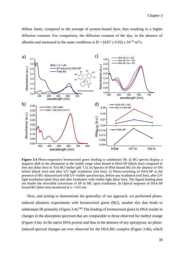
Chapter 3
39
diffuse faster, compared to the average of protein-bound dyes, thus resulting in a higher
diffusion constant. For comparison, the diffusion constant of the dye, in the absence of
albumin and measured in the same conditions is D = (4.67 ± 0.02) x 10-10 m2/s.
Figure 3.4 Photo-responsive bromocresol green binding to subdomain IB. a) BG spectra display a
negative shift in the absorption in the visible range when bound to HSA-SP (black line) compared to
free dye (blue line) in Tris-HCl buffer (pH 7.5). b) Spectra of HSA-bound BG (in the absence of SP)
before (black line) and after UV light irradiation (red line). c) Photo-switching of HSA-SP in the
presence of BG characterized with UV-visible spectroscopy, before any irradiation (red line), after UV
light irradiation (pink line) and after irradiation with visible light (blue line). The ligand binding does
not hinder the reversible conversion of SP to MC upon irradiation. d) Optical response of HSA-SP
bound BG (blue line) monitored at λ = 615 nm.
Next, and aiming to demonstrate the generality of our approach, we performed photo-
induced allosteric experiments with bromocresol green (BG), another dye that binds to
subdomain IB primarily (Figure 3.4).[38] The binding of bromocresol green to HSA results in
changes in the absorption spectrum that are comparable to those observed for methyl orange
(Figure 3.4a). In the native HSA protein and thus in the absence of any spiropyran, no photo-
induced spectral changes are ever observed for the HSA-BG complex (Figure 3.4b), which

Programming allostery in the human serum albumin with light
40
indicates that BG is stable against irradiation. In the hybrid system, the binding of BG does
not interfere with the photochromic properties of the spiropyran (Figure 3.4c). Upon
irradiation, an increase in absorption is observed at λ = 615 nm (Figure 3.4d, blue line), which
indicates release of HSA-SP bound BG that occurs upon conversion of the SP-form to the
MC-form. Based on similar examination, about (4.5 ± 0.5)% of the bound BG is released
upon photo conversion of the spiropyran. The lower percentage of BG that is released is
likely related to its binding affinity to HSA, which is 7-fold higher than the binding affinity
of MO to HSA.[36,39] Upon subsequent irradiation with visible light (λ ≥ 420 nm), about (89
± 11)% of the released ligand re-binds the protein during MC to SP conversion, which means
that BG displays a higher degree of reversibility than MO. The difference in the degree of
reversibility possibly originates in BG displaying a naturally higher binding constant.
To further investigate the release of MO from IB pocket, we proceed to determine the
binding affinity of MO to the engineered HSA by following the fluorescence of tryptophan
that is quenched upon binding of MO to IB pocket (Figure 3.5a, excitation at λ = 279 nm and
emission at λ = 340 nm). Notably, the only tryptophan residue in the entire HSA structure
(Trp214) is located at the connecting helix between subdomain IB and subdomain IIA
(Figure 3.5a, inset).[30] The observed quenching is attributed to energy transfer phenomenon
(FRET) between Trp (λemission = 300 – 400 nm) and methyl orange (λexcitation = 350 – 550 nm)
as the distance between Trp214 and IB binding site (3 – 4 nm) allows such mechanism to
occur.[40] Based on fitting with Stern-Volmer equation (Figure 3.5b), the protein-ligand
associations in the different systems reflected by their quenching constants are summarized
in Figure 3.5c. The effective quenching constants reveal a 2.7-fold decrease in MO binding
upon UV light irradiation.
Although the decrease in the ligand binding affinity is up to 3-fold at best, a significant
impact could still be expected as albumin is involved in complex cellular networks. Also,
this result compares positively to the magnitude in photo-controlling binding affinity
reported in literature, which was a 4-fold decrease of binding affinity in the catabolite
activator protein.[10,19]
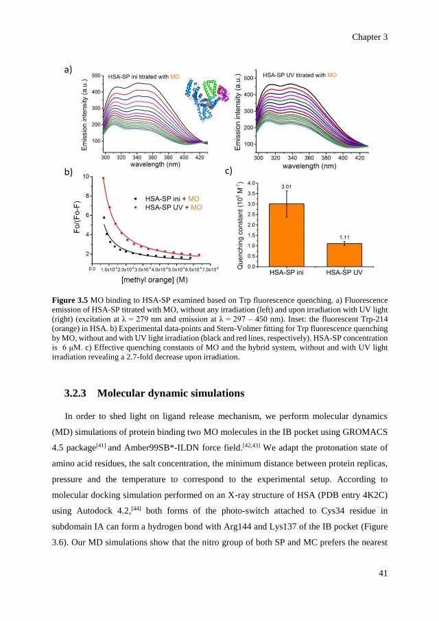
Chapter 3
41
Figure 3.5 MO binding to HSA-SP examined based on Trp fluorescence quenching. a) Fluorescence
emission of HSA-SP titrated with MO, without any irradiation (left) and upon irradiation with UV light
(right) (excitation at λ = 279 nm and emission at λ = 297 – 450 nm). Inset: the fluorescent Trp-214
(orange) in HSA. b) Experimental data-points and Stern-Volmer fitting for Trp fluorescence quenching
by MO, without and with UV light irradiation (black and red lines, respectively). HSA-SP concentration
is 6 μM. c) Effective quenching constants of MO and the hybrid system, without and with UV light
irradiation revealing a 2.7-fold decrease upon irradiation.
3.2.3 Molecular dynamic simulations
In order to shed light on ligand release mechanism, we perform molecular dynamics
(MD) simulations of protein binding two MO molecules in the IB pocket using GROMACS
4.5 package[41] and Amber99SB*-ILDN force field.[42,43] We adapt the protonation state of
amino acid residues, the salt concentration, the minimum distance between protein replicas,
pressure and the temperature to correspond to the experimental setup. According to
molecular docking simulation performed on an X-ray structure of HSA (PDB entry 4K2C)
using Autodock 4.2,[44] both forms of the photo-switch attached to Cys34 residue in
subdomain IA can form a hydrogen bond with Arg144 and Lys137 of the IB pocket (Figure
3.6). Our MD simulations show that the nitro group of both SP and MC prefers the nearest
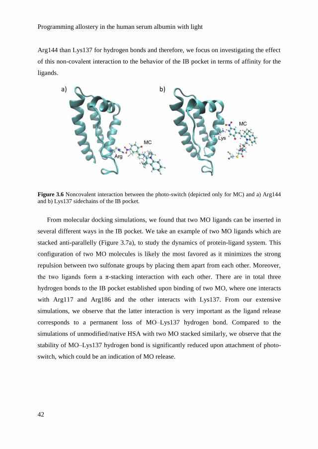
Programming allostery in the human serum albumin with light
42
Arg144 than Lys137 for hydrogen bonds and therefore, we focus on investigating the effect
of this non-covalent interaction to the behavior of the IB pocket in terms of affinity for the
ligands.
Figure 3.6 Noncovalent interaction between the photo-switch (depicted only for MC) and a) Arg144
and b) Lys137 sidechains of the IB pocket.
From molecular docking simulations, we found that two MO ligands can be inserted in
several different ways in the IB pocket. We take an example of two MO ligands which are
stacked anti-parallelly (Figure 3.7a), to study the dynamics of protein-ligand system. This
configuration of two MO molecules is likely the most favored as it minimizes the strong
repulsion between two sulfonate groups by placing them apart from each other. Moreover,
the two ligands form a π-stacking interaction with each other. There are in total three
hydrogen bonds to the IB pocket established upon binding of two MO, where one interacts
with Arg117 and Arg186 and the other interacts with Lys137. From our extensive
simulations, we observe that the latter interaction is very important as the ligand release
corresponds to a permanent loss of MO–Lys137 hydrogen bond. Compared to the
simulations of unmodified/native HSA with two MO stacked similarly, we observe that the
stability of MO–Lys137 hydrogen bond is significantly reduced upon attachment of photo-
switch, which could be an indication of MO release.
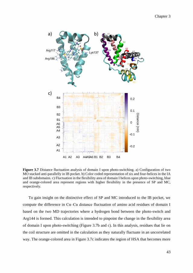
Chapter 3
43
Figure 3.7 Distance fluctuation analysis of domain I upon photo-switching. a) Configuration of two
MO stacked anti-parallelly in IB pocket. b) Color coded representation of six and four-helices in the IA
and IB subdomains. c) Fluctuation in the flexibility area of domain I helices upon photo-switching, blue
and orange-colored area represent regions with higher flexibility in the presence of SP and MC,
respectively.
To gain insight on the distinctive effect of SP and MC introduced to the IB pocket, we
compute the difference in Cα–Cα distance fluctuation of amino acid residues of domain I
based on the two MD trajectories where a hydrogen bond between the photo-switch and
Arg144 is formed. This calculation is intended to pinpoint the change in the flexibility area
of domain I upon photo-switching (Figure 3.7b and c). In this analysis, residues that lie on
the coil structure are omitted in the calculation as they naturally fluctuate in an uncorrelated
way. The orange-colored area in Figure 3.7c indicates the region of HSA that becomes more
Programming allostery in the human serum albumin with light
44
flexible in the presence of MC, while blue color indicates a higher flexibility in the HSA-SP
state. We observe that the IB pocket fluctuates more in the HSA-MC state as reflected by the
predominance of orange-colored region in both the diagonal B1–B4 vs B1–B4 and the off-
diagonal A1–A6 vs B1–B4 regions. The former region is related to an increased breathing of
IB pocket, while the latter indicates a higher cross-fluctuation between IA and IB subdomains
in the HSA-MC state. These findings altogether suggest that the IB pocket is more flexible
in the HSA-MC system compared to HSA-SP system.
3.3 Conclusions
We have demonstrated a photo-responsive hybrid system of the allosteric HSA that is
covalently modified with a single SP/MC switch at a specific site. Irradiation of the hybrid
system results in ligand release, while the overall structure of the protein is preserved.
Although the decrease in the ligand binding affinity is up to 3-fold at best, a significant impact
could still be expected as albumin is involved in complex cellular networks. The photo-
control of ligand binding is achieved in an allosteric manner as the switch is attached to a
neighboring site (subdomain IA) of the examined ligand binding site (subdomain IB).
Molecular dynamics simulations indicate that the covalent attachment of the switch in IA
reduces the stability of ligand binding to key residues of IB pocket. Moreover, IB pocket
evidently breathes and fluctuates more in the HSA-MC system compared to HSA-SP system.
The remote control of a binding pocket by switching a neighboring site opens new
opportunities for externally interfering with allosteric regulation of HSA. We envision that
similar engineering strategies could be extended to systems with higher-order structures and
further on to supramolecular protein-based hybrids and artificial biomimetic systems.
3.4 Acknowledgements
All molecular dynamic studies were performed by Habiburrahman Zulfikri and Prof.
Claudia Filippi. We thank Dr. Jean-Wilfried Fredy and Pichayut Tananchayakul for the
synthesis of the photo-switch and collaboration in ligand binding studies and Dr. Alberto
Juan for assistance with NMR analysis.

Chapter 3
45
3.5 Materials and methods
3.5.1 General
The human serum albumin (HSA) was purchased from Sigma-Aldrich. All experiments
are performed in a Tris-HCl buffer pH 7.5 (containing 150 mM NH4Cl). Chemicals were
purchased from Sigma-Aldrich and used without further purification, unless stated otherwise.
3.5.2 Synthesis of the photo-switch
The light-responsive maleimide-spiropyran switch was synthesized according to
described procedure (Figure 3.8).[45]
Figure 3.8 Synthesis of maleimide-bearing nitrospiropyran
1-(2-hydroxyethyl)-2,3,3-trimethyl-3H-indol-1-ium iodide (3) was synthesized
according to the literature method. 2,3,3-trimethyl-3H-indole 1 (4.96 g, 31.2 mmol, 1 eq)
was dissolved in dry acetonitrile (10 mL) and 2-iodoethanol (2.55 mL, 32.7 mmol, 1.1 eq)
was added and heated to reflux for 24 h under N2. The reaction mixture was cooled at room
temperature, the solid precipitate was filtered, washed with CH2Cl2 (30mL*3) to afford
purple solid 3 (8.44 g, 82%). MS(ESI): 204.1 [M-H]+;
Programming allostery in the human serum albumin with light
46
1H NMR (400 MHz, CD3CN) δ 7.79 – 7.75 (m, 1H), 7.74 – 7.70 (m, 1H), 7.66 – 7.58 (m,
2H), 4.59 – 4.48 (t, J = 5.0, 5.2 Hz, 2H), 4.00 (dd, J = 10.5, 5.5 Hz, 2H), 3.69 (t, J = 5.8 Hz,
1H), 2.79 (s, 3H), 1.57 (s, 6H);
13C NMR (101 MHz, CD3CN) δ 198.95, 142.89, 142.23, 130.81, 130.08, 124.38, 118.30,
116.48, 58.84, 55.66, 51.53, 22.83, 15.55.
9,9,9a-trimethyl-2,3,9,9a-tetrahydrooxazolo[3,2-a]indole (4) was synthesized
according to the literature method. A solution of 3 (8.35 g, 25.2 mmol, 1 eq), KOH (3.21 g,
57.2 mmol, 2.3 eq) in deionized water (40 mL) was stirred for 30 min at room temperature.
After extraction with diethylether (30mL*3), the organic layer was collected, dried with
MgSO4 and evaporated to afford 4 as a yellow oil.
1H NMR (400 MHz, CD3CN) δ 7.13 – 7.06 (m, 2H), 6.87 (m, 1H), 6.77 (d, J = 7.8 Hz,
1H), 3.75 (m, 2H), 3.52 (m, 1H), 3.40 – 3.32 (m, 1H), 1.35 (s, 3H), 1.32 (s, 3H), 1.12 (s, 3H);
13C NMR (101 MHz, CD3CN) δ 152.14, 141.00, 128.38, 123.26, 122.24, 112.79, 109.64,
63.53, 50.40, 47.54, 28.19, 20.94, 17.49.
2-(3',3'-dimethyl-6-nitrospiro[chromene-2,2'-indolin]-1'-yl)ethanol (5) was
synthesized according to the literature method. A solution of 5-nitrososalicylaldehyde (2.60
g, 15.6 mmol, 1.5 eq) in ethanol (20 mL) and the obtained crude 4 (2.11 g, 10.4 mmol, 1 eq
mmol) was refluxed for 3 h under N2. After cooling to room temperature, the mixture was
filtered, washed with EtOH and dried to afford 5 as a solid purple (1.07 g, 29%).
1H NMR (400 MHz, CD3CN) δ 8.07 (d, J = 2.8 Hz, 1H), 8.00 (dd, J = 9.0, 2.8 Hz, 1H),
7.18 – 7.08 (m, 2H), 7.03 (d, J = 10.4 Hz, 1H), 6.86 – 6.81 (m, J = 7.5, 0.9 Hz, 1H), 6.72 (d,
J = 9.0 Hz, 1H), 6.66 (d, J = 7.8 Hz, 1H), 5.99 (d, J = 10.4 Hz, 1H), 3.71 – 3.48 (m, 2H),
3.37 – 3.17 (m, 2H), 2.82 (s, 1H), 1.25 (s, 3H), 1.15 (s, 3H);
13C NMR (101 MHz, CD3CN) δ 160.44, 148.35, 142.10, 136.97, 128.81, 128.67, 126.57,
123.71, 123.26, 122.71, 120.37, 120.04, 116.25, 108.03, 107.77, 60.88, 53.49, 46.90, 26.15,
20.08.
2-(3',3'-dimethyl-6-nitrospiro[chromene-2,2'-indolin]-1'-yl)ethyl 2-bromoacetate
(6) was synthesized according to the literature method. A solution of 4 (0.25 g, 0.7 mmol, 1
Chapter 3
47
eq) was dissolved in dry toluene (6 mL). Pyridine (90 μL, 1.10 mmol, 1.5 eq) and
bromoacetylbromide (100 μL, 1.10 mmol, 1.5 eq) were added. The reaction was stirred at
room temperature, under N2. After stirring for 3 h, water (10 mL) was added and then was
extracted with dry diethylether (30mL*3). The organic layer was collected, dried over
MgSO4 and evaporated to afford 6 as a yellow brown (0.31 g, 91%).
1H NMR (400 MHz, CD3CN) δ 8.08 (d, J = 2.8 Hz, 1H), 8.00 (dd, J = 9.0, 2.8 Hz, 1H),
7.20 – 7.10 (m, 2H), 7.06 (d, J = 10.4 Hz, 1H), 6.87 (m, 1H), 6.71 (t, J = 8.3 Hz, 2H), 6.01
(d, J = 10.4 Hz, 1H), 4.33 (m, 1H), 4.28 – 4.19 (m, 1H), 3.94 – 3.81 (m, 2H), 3.52 (m, 1H),
3.40 (dt, J = 15.5, 5.1 Hz, 1H), 1.25 (s, 3H), 1.13 (s, 3H);
13C NMR (101 MHz, CD3CN) δ 168.21, 160.29, 147.74, 142.21, 136.97, 129.19, 128.76,
126.65, 123.77, 122.90, 122.87, 120.78, 119.97, 116.32, 107.81, 107.72, 64.89, 53.55, 43.07,
27.33, 26.13, 19.94.
2-(3',3'-dimethyl-6-nitrospiro[chromene-2,2'-indolin]-1'-yl)ethyl 2-iodoacetate (1)
was synthesized according to the literature method. A solution of 6 (0.31 g, 0.66 mmol, 1 eq)
was dissolved in acetone (6 mL) and added NaI (0.12 g, 0.79 mmol, 1.2 eq). After stirring at
room temperature for 2 h under Ar, the reaction mixture was filtered. Ethylacetate was added
to the mother liquor and the organic phase was washed with H2O and dried over MgSO4,
then evaporated and dried to afford 1 (0.23 g, 68%). MS(ESI): 521.1 [M-H]+;
1H NMR (400 MHz, CD3CN) δ 8.09 (d, J = 2.8 Hz, 1H), 8.00 (dd, J = 9.0, 2.8 Hz, 1H),
7.21 – 7.11 (m, 2H), 7.06 (d, J = 10.4 Hz, 1H), 6.87 (td, J = 7.5, 0.8 Hz, 1H), 6.72 (t, J = 8.6
Hz, 2H), 6.03 (d, J = 10.4 Hz, 1H), 4.33 – 4.24 (m, 1H), 4.23 – 4.16 (m, 1H), 3.70 – 3.67 (m,
2H), 3.55 – 3.46 (m, 1H), 3.43 – 3.34 (m, 1H), 1.25 (s, 3H), 1.14 (s, 3H);
13C NMR (101 MHz, CD3CN) δ 169.87, 160.30, 147.76, 142.20, 136.97, 129.19, 128.77,
126.65, 123.77, 122.97, 122.87, 120.76, 119.98, 116.33, 107.83, 107.74, 64.75, 53.57, 43.01,
26.19, 19.95, -4.18.
2-(3',3'-dimethyl-6-nitrospiro[chromene-2,2'-indolin]-1'-yl)ethyl 2-(2,5-dioxo-2,5-
dihydro-1H-pyrrol-1-yl)acetate (2) was synthesized according to the literature method. A
solution of 1 (22 mg, 0.047 mmol, 1 eq) in dry acetone (2 mL) was added maleimide (182
mg, 0.188 mmol, 4 eq) and K2CO3 (0.010 g, 0.071 mmol, 1.5 eq). After stirring for 4 h at

Programming allostery in the human serum albumin with light
48
room temperature under N2, ethylacetate was added to the reaction mixture and it was washed
with 0.1 M HCl and water. The organic phase was then evaporated and dried to afford 8 (0.02
g, 87%). MS(ESI): 489.9 [M-H]+;
1H NMR (400 MHz, CD3CN) δ 8.08 (d, J = 2.7 Hz, 1H), 8.00 (dd, J = 9.0, 2.8 Hz, 1H),
7.19 – 7.11 (m, 2H), 7.06 (d, J = 10.3 Hz, 1H), 6.89 – 6.84 (m, 1H), 6.81 (s, 1H), 6.73 (d, J
= 9.0 Hz, 1H), 6.69 (s, 1H), 6.65 (d, J = 7.8 Hz, 1H), 5.97 (d, J = 10.4 Hz, 1H), 4.34 (m, 1H),
4.20 (dd, J = 11.0, 5.6 Hz, 1H), 4.16 (s, 2H), 3.53 – 3.44 (m, 1H), 3.41 – 3.32 (m, 1H), 1.25
(s, 3H), 1.13 (s, 3H);
13C NMR (101 MHz, CD3CN) δ 171.04, 168.64, 160.20, 147.74, 142.13, 136.91, 136.01,
135.52, 129.14, 128.72, 126.58, 123.70, 122.77, 122.69, 120.70, 119.87, 116.25, 107.68,
107.62, 64.54, 53.41, 43.06, 39.32, 26.09, 19.86.
3.5.3 Synthesis and characterization of the hybrid system
A 10-fold excess of nitro-spiropyran bearing a maleimide functional group was added to
HSA (in 20 mM Tris-HCl buffer pH 7.5, with 20% DMSO) at room temperature and
incubated for an hour. The unbound spiropyran was removed by repeated spin-filtration
(Corning Spin-X UF 10k MWCO membrane filters), each for 20 min at 6,000 rpm
(Eppendorf centrifuge 5415R) or with the size-exclusion chromatography (preparative
column Superose 6 10/100 GL, GE Healthcare FPLC Äkta purifier 900 with a 24 mL bed
volume). Prior to injecting the samples into the column, all samples were dialyzed against
Tris-HCl buffer pH 7.5 to remove the DMSO using 12 – 14 kDa dialysis membranes
(Spectra/Por). The purified hybrid system was further characterized with UV-visible
spectrometer (Perkin Elmer Lambda 850).
Calculations for protein:photo-switch ratio based on UV-Vis spectra (Figure 3.1d):
𝐴350 = [𝐻𝑆𝐴] 𝑥 1,167 𝑀−1𝑐𝑚−1 𝑥 1 𝑐𝑚 + [𝑠𝑝𝑖𝑟𝑜𝑝𝑦𝑟𝑎𝑛] 𝑥 7,994 𝑀−1𝑐𝑚−1 𝑥 1 𝑐𝑚
𝐴280 = [𝐻𝑆𝐴] 𝑥 30,456 𝑀−1𝑐𝑚−1 𝑥 1 𝑐𝑚 + [𝑠𝑝𝑖𝑟𝑜𝑝𝑦𝑟𝑎𝑛] 𝑥 13,042 𝑀−1𝑐𝑚−1 𝑥 1 𝑐𝑚
3.5.4 Photo-responsive ligand binding analyses
A 4-fold excess of ligand (either methyl orange or bromocresol green) was added to the
hybrid system (3.7 μM in 20 mM Tris-HCl buffer) at room temperature and incubated for at
least 3 h. The unbound ligand was removed with repeated spin-filtration. The samples were
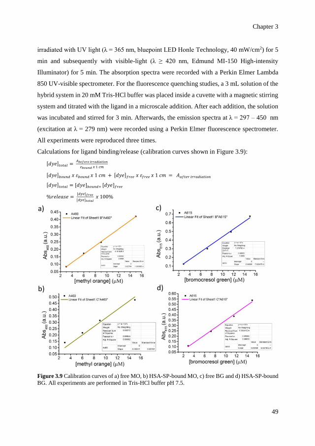
Chapter 3
49
irradiated with UV light (λ = 365 nm, bluepoint LED Honle Technology, 40 mW/cm2) for 5
min and subsequently with visible-light (λ ≥ 420 nm, Edmund MI-150 High-intensity
Illuminator) for 5 min. The absorption spectra were recorded with a Perkin Elmer Lambda
850 UV-visible spectrometer. For the fluorescence quenching studies, a 3 mL solution of the
hybrid system in 20 mM Tris-HCl buffer was placed inside a cuvette with a magnetic stirring
system and titrated with the ligand in a microscale addition. After each addition, the solution
was incubated and stirred for 3 min. Afterwards, the emission spectra at λ = 297 – 450 nm
(excitation at λ = 279 nm) were recorded using a Perkin Elmer fluorescence spectrometer.
All experiments were reproduced three times.
Calculations for ligand binding/release (calibration curves shown in Figure 3.9):
[𝑑𝑦𝑒]𝑡𝑜𝑡𝑎𝑙 = 𝐴𝑏𝑒𝑓𝑜𝑟𝑒 𝑖𝑟𝑟𝑎𝑑𝑖𝑎𝑡𝑖𝑜𝑛
𝜀𝑏𝑜𝑢𝑛𝑑 𝑥 1 𝑐𝑚
[𝑑𝑦𝑒]𝑏𝑜𝑢𝑛𝑑 𝑥 𝜀𝑏𝑜𝑢𝑛𝑑 𝑥 1 𝑐𝑚 + [𝑑𝑦𝑒]𝑓𝑟𝑒𝑒 𝑥 𝜀𝑓𝑟𝑒𝑒 𝑥 1 𝑐𝑚 = 𝐴𝑎𝑓𝑡𝑒𝑟 𝑖𝑟𝑟𝑎𝑑𝑖𝑎𝑡𝑖𝑜𝑛
[𝑑𝑦𝑒]𝑡𝑜𝑡𝑎𝑙 = [𝑑𝑦𝑒]𝑏𝑜𝑢𝑛𝑑+ [𝑑𝑦𝑒]𝑓𝑟𝑒𝑒
%𝑟𝑒𝑙𝑒𝑎𝑠𝑒 = [𝑑𝑦𝑒]𝑓𝑟𝑒𝑒
[𝑑𝑦𝑒]𝑡𝑜𝑡𝑎𝑙 𝑥 100%
Figure 3.9 Calibration curves of a) free MO, b) HSA-SP-bound MO, c) free BG and d) HSA-SP-bound
BG. All experiments are performed in Tris-HCl buffer pH 7.5.

Programming allostery in the human serum albumin with light
50
3.5.5 PFGSE-NMR experiments
All NMR experiments were measured using a Bruker Avance II NMR 600 MHz
spectrometer, equipped with a triple-nucleus TXI probe head with a z-gradient coil, and a
Great 3/10 gradient amplifier. All experiments were performed at 300 K using standard pulse
sequences from the Bruker library. Pulsed-Field Gradient Spin-Echo (PFGSE) NMR
experiments were carried out by using the pulse sequence of longitudinal-eddy-current delay
(LED) with bipolar-gradient pulses, with a diffusion delay () of 0.2 s and gradient length
() of 1200 μs and 5 ms of eddy-current delay. The gradient strength was varied from 5 to
95%. A total of 128 transients were collected for each of the 20 increment steps with a recycle
delay of 2 s. Pseudo-2D DOSY plots were processed with the standard Bruker software; the
diffusion coefficients were determined by the T1/T2 “vargrad” SimFit fitting routine.
Calibration of the field gradient strength was achieved by measuring the value of translational
diffusion coefficient (D) for the residual 1H signal in D2O (99.99%, 2H atom), D = 1.91×10⁻⁹
m2/s.
3.5.6 Determination of effective quenching constants
The formation of a protein-ligand complex is associated with static quenching of protein
fluorescence by the bound ligand. The quenching constant is obtained from the Stern-Volmer
equation:
𝐹𝑜
∆𝐹=
1
𝑓𝑎. 𝐾𝑎. [𝑄]+
1
𝑓𝑎
where ∆F is the difference in fluorescence emission with and without the quencher Q in the
system; Q represents the free quencher; fa represents the fraction of accessible fluorescence
and Ka is the effective quenching constant.[46,47]
Isothermal microcalorimetry is typically used to determine binding constants
experimentally. However, since the MC-form can be converted back to SP-form during the
automated measurement, this technique is not applicable here.
3.5.7 Analyses of protein global structure
The conformation of the protein (0.1 μM) before and after irradiation in 10 mM phosphate
buffer pH 7.5 was analyzed with a Jasco J-1500 circular dichroism spectrometer. The
Chapter 3
51
hydrodynamic size of the protein before and after irradiation in 20 mM Tris-HCl buffer pH
7.5 was determined with dynamic light scattering (Nanotrac Wave, Microtrac).
3.5.8 Data and schematic representation
All data plotting and mathematical fitting were performed with OriginPro 9.0 software.
For CD spectra, the raw data were smoothed using the Savitzky-Golay filter in OriginPro 9.0
software. Protein structures are rendered using PyMOL 1.3 software and chemical structures
are drawn using ChemBioDraw Ultra 12.0 software.
3.6 References
[1] H. N. Motlagh, J. O. Wrabl, J. Li, V. J. Hilser, Nature 2014, 508, 331-339.
[2] A. W. Fenton, Trends Biochem. Sci. 2008, 33, 420-425.
[3] D. Kern, E. R. P. Zuiderweg, Curr. Opin. Struc. Biol. 2003, 13, 748-757.
[4] C.-J. Tsai, A. del Sol, R. Nussinov, J. Mol. Biol. 2008, 378, 1-11.
[5] A. C. M. Ferreon, J. C. Ferreon, P. E. Wright, A. A. Deniz, Nature 2013, 498, 390-394.
[6] J.-P. Changeux, Nat. Rev. Mol. Cell. Biol. 2013, 14, 819-829.
[7] J. H. Choi, A. H. Laurent, V. J. Hilser, M. Ostermeier, Nat. Commun. 2015, 6, 6968.
[8] R. A. Brown, V. Diemer, S. J. Webb, J. Clayden, Nat. Chem. 2013, 5, 853-860.
[9] G. Y. Tonga, Y. Jeong, B. Duncan, T. Mizuhara, R. Mout, R. Das, S. T. Kim, Y.-C. Yeh,
B. Yan, S. Hou, V. M. Rotello, Nat. Chem. 2015, 7, 597-603.
[10] W. Szymański, J. M. Beierle, H. A. V. Kistemaker, W. A. Velema, B. L. Feringa, Chem.
Rev. 2013, 113, 6114-6178.
[11] A. A. Beharry, G. A. Woolley, Chem. Soc. Rev. 2011, 40, 4422-4437.
[12] R. Klajn, Chem. Soc. Rev. 2014, 43, 148-184.
[13] D. P. Nguyen, M. Mahesh, S. J. Elsässer, S. M. Hancock, C. Uttamapinant, J. W. Chin,
J. Am. Chem. Soc. 2014, 136, 2240-2243.
[14] J. Broichhagen, D. Trauner, Curr. Opin. Chem. Biol. 2014, 21, 121-127.
[15] J. R. Kumita, O. S. Smart, G. A. Woolley, Proc. Natl. Acad. Sci. USA 2000, 97, 3803-
3808.
[16] G. A. Woolley, Acc. Chem. Res. 2005, 38, 486-493.
Programming allostery in the human serum albumin with light
52
[17] B. Schierling, A.-J. Noël, W. Wende, L. T. Hien, E. Volkov, E. Kubareva, T. Oretskaya,
M. Kokkinidis, A. Römpp, B. Spengler, A. Pingoud, Proc. Natl. Acad. Sci. USA 2010,
107, 1361-1366.
[18] M. Aizawa, K. Namba, S. Suzuki, Arch. Biochem. Biophys. 1977, 182, 305-310.
[19] M. Bose, D. Groff, J. Xie, E. Brustad, P. G. Schultz, J. Am. Chem. Soc. 2006, 128, 388-
389.
[20] G. A. Woolley, A. S. I. Jaikaran, M. Berezovski, J. P. Calarco, S. N. Krylov, O. S. Smart,
J. R. Kumita, Biochemistry 2006, 45, 6075-6084.
[21] A. Koçer, M. Walko, W. Meijberg, B. L. Feringa, Science 2005, 309, 755-758.
[22] M. R. Banghart, M. Volgraf, D. Trauner, Biochemistry 2006, 45, 15129-15141.
[23] I. Tochitsky, M. R. Banghart, A. Mourot, J. Z. Yao, B. Gaub, R. H. Kramer, D. Trauner,
Nat. Chem. 2012, 4, 105-111.
[24] S. Pittolo, X. Gómez-Santacana, K. Eckelt, X. Rovira, J. Dalton, C. Goudet, J.-P. Pin,
A. Llobet, J. Giraldo, A. Llebaria, P. Gorostiza, Nat. Chem. Biol. 2014, 10, 813-815.
[25] M. Volgraf, P. Gorostiza, S. Szobota, M. R. Helix, E. Y. Isacoff, D. Trauner, J. Am.
Chem. Soc. 2007, 129, 260-261.
[26] M. Schönberger, D. Trauner, Angew. Chem. Int. Ed. 2014, 53, 3264-3267.
[27] M. Volgraf, P. Gorostiza, R. Numano, R. H. Kramer, E. Y. Isacoff, D. Trauner, Nat.
Chem. Biol. 2006, 2, 47-52.
[28] D. Strickland, K. Moffat, T. R. Sosnick, Proc. Natl. Acad. Sci. USA 2008, 105, 10709-
10714.
[29] P. Ascenzi, M. Fasano, Biophys. Chem. 2010, 148, 16-22.
[30] G. Fanali, A. di Masi, V. Trezza, M. Marino, M. Fasano, P. Ascenzi, Mol. Aspects Med.
2012, 33, 209-290.
[31] A. S. Pitek, S. A. Jameson, F. A. Veliz, S. Shukla, N. F. Steinmetz, Biomaterials 2016,
89, 89-97.
[32] G. F. Li, D. Magana, R. B. Dyer, Nat. Commun. 2014, 5, 3100.
[33] M. N. Khan, J. Pharm. Sci. 1984, 73, 1767-1771.
[34] M. J. Davies, Biochem. Biophys. Res. Commun. 2003, 305, 761-770.
[35] F. Zsila, Mol. Pharm. 2013, 10, 1668-1682.
Chapter 3
53
[36] R. Ambrosetti, R. Bianchini, S. Fisichella, M. Fichera, M. Zandomeneghi, Chem. Eur.
J. 1996, 2, 149-156.
[37] F. L. Rodkey, Clin. Chem. 1965, 11, 478-487.
[38] J. W. Keyser, Clin. Chim. Acta 1961, 6, 445-447.
[39] V. D. Trivedu, I. Saxena, M. U. Siddiqui, M. A. Qasim, IUBMB Life 1997, 43, 1-8.
[40] P. C. Ray, Z. Fan, R. A. Crouch, S. S. Sinhaa, A. Pramanika, Chem. Soc. Rev. 2014,
43, 6370-6404.
[41] D. van der Spoel, E. Lindahl, B. Hess, G. Groenhof, A. E. Mark, H. J. C. Berendsen, J.
Comput. Chem. 2005, 26, 1701-1718.
[42] R. B. Best, G. J. Hummer, Phys. Chem. B. 2009, 113, 9004-9015.
[43] K. Lindorff-Larsen, S. Piana, K. Palmo, P. Maragakis, J. L. Klepeis, R. O. Dror, D. E.
Shaw, Proteins 2010, 78, 1950-1958.
[44] G. M. Morris, R. Huey, W. Lindstrom, M. F. Sanner, R. K. Belew, D. S. Goodsell, A. J.
Olson, J. Comput. Chem. 2009, 16, 2785-2791.
[45] M. -Q. Zhu, T. Chen, G. -F. Zhang, C. Li, W. -L. Gong, Z. -Q. Chena, M. P. Aldreda,
Chem. Commun. 2014, 50, 2664-2666.
[46] Y. -J. Hu, Y. Liu, X. -H. Xiao, Biomacromolecules 2009, 10, 517-521.
[47] M. van de Weert, J. Fluoresc. 2010, 20, 625-629.

Programming allostery in the human serum albumin with light
54
Chapter 4
Bacterial encapsulins as in vitro and in vivo
protein-based nanoplatforms
Bacterial encapsulins are highly organized structures that are composed
of protein building blocks , making up icosahedral cage-like structures with
the size of 20 – 25 nm. The outer and inner surface of encapsulins can be
modified, either chemically or genetically, and the internal cavity can be used
to template, store and arrange molecular cargo within a defined space. Recent
years have witnessed the increasing interest on the application of bacterial
semi-organelle encapsulins for bio -nanotechnology. Here, we characterize the
native structure of B. linens encapsulins with both native and foreign cargo
using cryo-electron microscopy (cryo-EM). Furthermore, by harnessing the
confined enzyme (i.e., a peroxidase), we demonstrate the functionality of the
encapsulin for an in vitro surface-immobilized catalysis in a cascade pathway
with an additional enzyme, g lucose oxidase. We also demonstrate the in vivo
functionality of the encapsulin for cellular uptake using mammalian
macrophages. Unraveling both the structure and functionality of the
encapsulins paves the way towards transforming biological nanocompartmen ts
into functional systems.
Part of this chapter is published in R.M. Putri, J.J.L.M. Cornelissen, M.S.T. Koay,
ChemPhysChem 2015, 16, 911-918. Part of this chapter is included in a submission to ACS
Nano 2017.
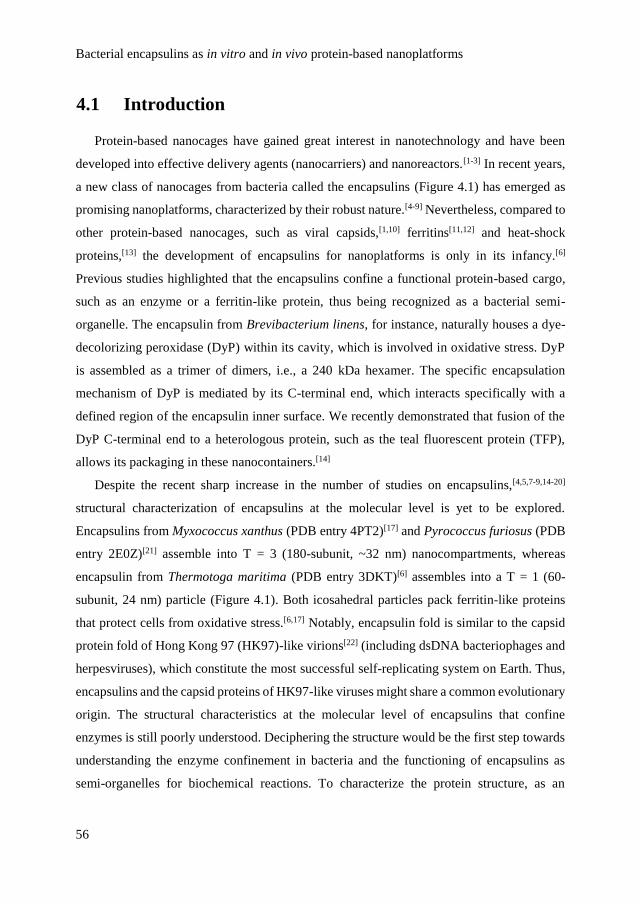
Bacterial encapsulins as in vitro and in vivo protein-based nanoplatforms
56
4.1 Introduction
Protein-based nanocages have gained great interest in nanotechnology and have been
developed into effective delivery agents (nanocarriers) and nanoreactors.[1-3] In recent years,
a new class of nanocages from bacteria called the encapsulins (Figure 4.1) has emerged as
promising nanoplatforms, characterized by their robust nature.[4-9] Nevertheless, compared to
other protein-based nanocages, such as viral capsids,[1,10] ferritins[11,12] and heat-shock
proteins,[13] the development of encapsulins for nanoplatforms is only in its infancy.[6]
Previous studies highlighted that the encapsulins confine a functional protein-based cargo,
such as an enzyme or a ferritin-like protein, thus being recognized as a bacterial semi-
organelle. The encapsulin from Brevibacterium linens, for instance, naturally houses a dye-
decolorizing peroxidase (DyP) within its cavity, which is involved in oxidative stress. DyP
is assembled as a trimer of dimers, i.e., a 240 kDa hexamer. The specific encapsulation
mechanism of DyP is mediated by its C-terminal end, which interacts specifically with a
defined region of the encapsulin inner surface. We recently demonstrated that fusion of the
DyP C-terminal end to a heterologous protein, such as the teal fluorescent protein (TFP),
allows its packaging in these nanocontainers.[14]
Despite the recent sharp increase in the number of studies on encapsulins,[4,5,7-9,14-20]
structural characterization of encapsulins at the molecular level is yet to be explored.
Encapsulins from Myxococcus xanthus (PDB entry 4PT2)[17] and Pyrococcus furiosus (PDB
entry 2E0Z)[21] assemble into T = 3 (180-subunit, ~32 nm) nanocompartments, whereas
encapsulin from Thermotoga maritima (PDB entry 3DKT)[6] assembles into a T = 1 (60-
subunit, 24 nm) particle (Figure 4.1). Both icosahedral particles pack ferritin-like proteins
that protect cells from oxidative stress.[6,17] Notably, encapsulin fold is similar to the capsid
protein fold of Hong Kong 97 (HK97)-like virions[22] (including dsDNA bacteriophages and
herpesviruses), which constitute the most successful self-replicating system on Earth. Thus,
encapsulins and the capsid proteins of HK97-like viruses might share a common evolutionary
origin. The structural characteristics at the molecular level of encapsulins that confine
enzymes is still poorly understood. Deciphering the structure would be the first step towards
understanding the enzyme confinement in bacteria and the functioning of encapsulins as
semi-organelles for biochemical reactions. To characterize the protein structure, as an

Chapter 4
57
alternative to the well-known protein crystallization, cryo-electron microscopy (cryo-EM)
recently emerges as a powerful technique.
Figure 4.1 Structure of encapsulin from the bacterium T. maritima (PDB entry 3DKT).[6] The protein
cage is composed of 12 pentamers, whereby the monomer (blue) and one pentamer (red) are shown.
The protein cargo (yellow) occupies the internal cavity as a result of interactions with a conserved
peptide sequence (purple).
In this chapter, the structural characterization of B. linens encapsulin with native DyP
cargo (DyP-E) and foreign TFP cargo (TFP-E) is presented using cryo-EM reconstruction.
Furthermore, the functionality of B. linens encapsulins as nanoplatforms for in vitro and in
vivo purposes is demonstrated. We harness the confined DyP in the native encapsulin to
perform a tandem catalysis in vitro on a surface in a cascade manner. For the in vivo study,
we use the modified encapsulin with a TFP cargo to investigate the particle uptake by murine
macrophages.
4.2 Results and discussion
Characterization of encapsulin stability and structure
The encapsulin from B. linens is recombinantly expressed in E. coli and mechanical lysis
(i.e., ultrasonication) is performed to disrupt the cell and retrieve the encapsulin particles.
The particles are purified with 38% sucrose cushion and 10 – 50% sucrose gradient to remove
other cellular proteins. The final step of purification involves a size-exclusion
chromatography (SEC) that indicates the presence of the encapsulin peak at V = 11 – 12 mL
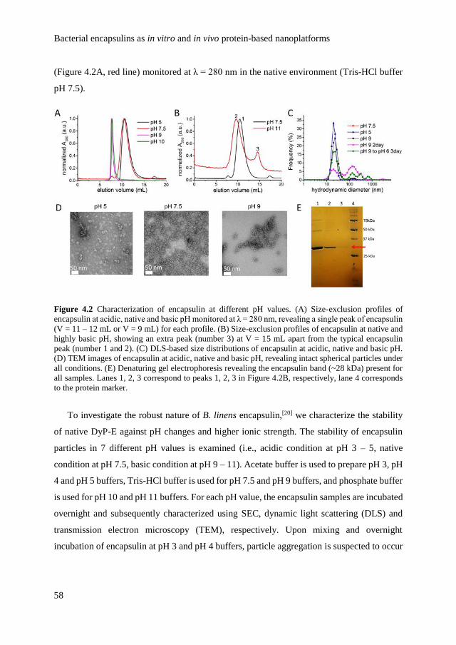
Bacterial encapsulins as in vitro and in vivo protein-based nanoplatforms
58
(Figure 4.2A, red line) monitored at λ = 280 nm in the native environment (Tris-HCl buffer
pH 7.5).
Figure 4.2 Characterization of encapsulin at different pH values. (A) Size-exclusion profiles of
encapsulin at acidic, native and basic pH monitored at λ = 280 nm, revealing a single peak of encapsulin
(V = 11 – 12 mL or V = 9 mL) for each profile. (B) Size-exclusion profiles of encapsulin at native and
highly basic pH, showing an extra peak (number 3) at V = 15 mL apart from the typical encapsulin
peak (number 1 and 2). (C) DLS-based size distributions of encapsulin at acidic, native and basic pH.
(D) TEM images of encapsulin at acidic, native and basic pH, revealing intact spherical particles under
all conditions. (E) Denaturing gel electrophoresis revealing the encapsulin band (~28 kDa) present for
all samples. Lanes 1, 2, 3 correspond to peaks 1, 2, 3 in Figure 4.2B, respectively, lane 4 corresponds
to the protein marker.
To investigate the robust nature of B. linens encapsulin,[20] we characterize the stability
of native DyP-E against pH changes and higher ionic strength. The stability of encapsulin
particles in 7 different pH values is examined (i.e., acidic condition at pH 3 – 5, native
condition at pH 7.5, basic condition at pH 9 – 11). Acetate buffer is used to prepare pH 3, pH
4 and pH 5 buffers, Tris-HCl buffer is used for pH 7.5 and pH 9 buffers, and phosphate buffer
is used for pH 10 and pH 11 buffers. For each pH value, the encapsulin samples are incubated
overnight and subsequently characterized using SEC, dynamic light scattering (DLS) and
transmission electron microscopy (TEM), respectively. Upon mixing and overnight
incubation of encapsulin at pH 3 and pH 4 buffers, particle aggregation is suspected to occur
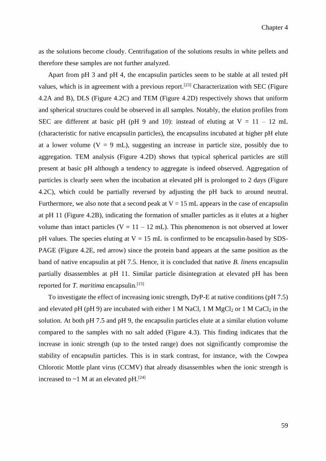
Chapter 4
59
as the solutions become cloudy. Centrifugation of the solutions results in white pellets and
therefore these samples are not further analyzed.
Apart from pH 3 and pH 4, the encapsulin particles seem to be stable at all tested pH
values, which is in agreement with a previous report.[23] Characterization with SEC (Figure
4.2A and B), DLS (Figure 4.2C) and TEM (Figure 4.2D) respectively shows that uniform
and spherical structures could be observed in all samples. Notably, the elution profiles from
SEC are different at basic pH (pH 9 and 10): instead of eluting at V = 11 – 12 mL
(characteristic for native encapsulin particles), the encapsulins incubated at higher pH elute
at a lower volume (V = 9 mL), suggesting an increase in particle size, possibly due to
aggregation. TEM analysis (Figure 4.2D) shows that typical spherical particles are still
present at basic pH although a tendency to aggregate is indeed observed. Aggregation of
particles is clearly seen when the incubation at elevated pH is prolonged to 2 days (Figure
4.2C), which could be partially reversed by adjusting the pH back to around neutral.
Furthermore, we also note that a second peak at V = 15 mL appears in the case of encapsulin
at pH 11 (Figure 4.2B), indicating the formation of smaller particles as it elutes at a higher
volume than intact particles (V = 11 – 12 mL). This phenomenon is not observed at lower
pH values. The species eluting at V = 15 mL is confirmed to be encapsulin-based by SDS-
PAGE (Figure 4.2E, red arrow) since the protein band appears at the same position as the
band of native encapsulin at pH 7.5. Hence, it is concluded that native B. linens encapsulin
partially disassembles at pH 11. Similar particle disintegration at elevated pH has been
reported for T. maritima encapsulin.[15]
To investigate the effect of increasing ionic strength, DyP-E at native conditions (pH 7.5)
and elevated pH (pH 9) are incubated with either 1 M NaCl, 1 M MgCl2 or 1 M CaCl2 in the
solution. At both pH 7.5 and pH 9, the encapsulin particles elute at a similar elution volume
compared to the samples with no salt added (Figure 4.3). This finding indicates that the
increase in ionic strength (up to the tested range) does not significantly compromise the
stability of encapsulin particles. This is in stark contrast, for instance, with the Cowpea
Chlorotic Mottle plant virus (CCMV) that already disassembles when the ionic strength is
increased to ~1 M at an elevated pH.[24]
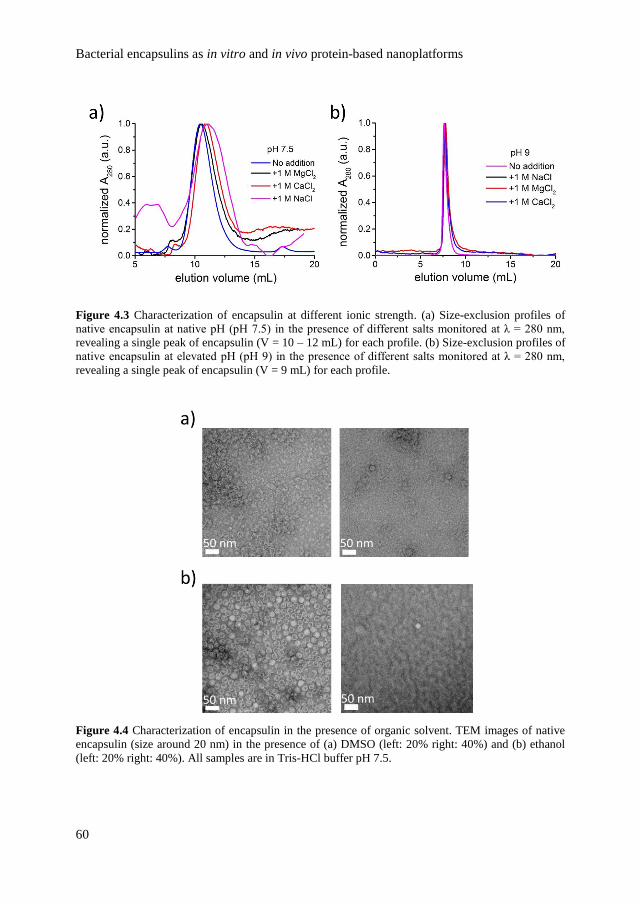
Bacterial encapsulins as in vitro and in vivo protein-based nanoplatforms
60
Figure 4.3 Characterization of encapsulin at different ionic strength. (a) Size-exclusion profiles of
native encapsulin at native pH (pH 7.5) in the presence of different salts monitored at λ = 280 nm,
revealing a single peak of encapsulin (V = 10 – 12 mL) for each profile. (b) Size-exclusion profiles of
native encapsulin at elevated pH (pH 9) in the presence of different salts monitored at λ = 280 nm,
revealing a single peak of encapsulin (V = 9 mL) for each profile.
Figure 4.4 Characterization of encapsulin in the presence of organic solvent. TEM images of native
encapsulin (size around 20 nm) in the presence of (a) DMSO (left: 20% right: 40%) and (b) ethanol
(left: 20% right: 40%). All samples are in Tris-HCl buffer pH 7.5.
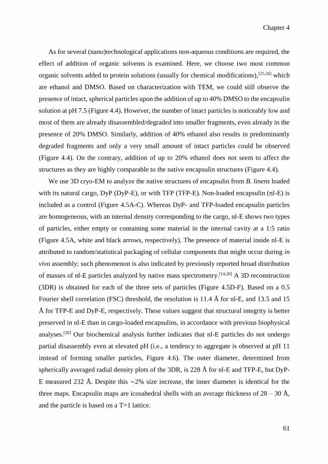
Chapter 4
61
As for several (nano)technological applications non-aqueous conditions are required, the
effect of addition of organic solvents is examined. Here, we choose two most common
organic solvents added to protein solutions (usually for chemical modifications),[25,26] which
are ethanol and DMSO. Based on characterization with TEM, we could still observe the
presence of intact, spherical particles upon the addition of up to 40% DMSO to the encapsulin
solution at pH 7.5 (Figure 4.4). However, the number of intact particles is noticeably low and
most of them are already disassembled/degraded into smaller fragments, even already in the
presence of 20% DMSO. Similarly, addition of 40% ethanol also results in predominantly
degraded fragments and only a very small amount of intact particles could be observed
(Figure 4.4). On the contrary, addition of up to 20% ethanol does not seem to affect the
structures as they are highly comparable to the native encapsulin structures (Figure 4.4).
We use 3D cryo-EM to analyze the native structures of encapsulin from B. linens loaded
with its natural cargo, DyP (DyP-E), or with TFP (TFP-E). Non-loaded encapsulin (nl-E) is
included as a control (Figure 4.5A-C). Whereas DyP- and TFP-loaded encapsulin particles
are homogeneous, with an internal density corresponding to the cargo, nl-E shows two types
of particles, either empty or containing some material in the internal cavity at a 1:5 ratio
(Figure 4.5A, white and black arrows, respectively). The presence of material inside nl-E is
attributed to random/statistical packaging of cellular components that might occur during in
vivo assembly; such phenomenon is also indicated by previously reported broad distribution
of masses of nl-E particles analyzed by native mass spectrometry.[14,20] A 3D reconstruction
(3DR) is obtained for each of the three sets of particles (Figure 4.5D-F). Based on a 0.5
Fourier shell correlation (FSC) threshold, the resolution is 11.4 Å for nl-E, and 13.5 and 15
Å for TFP-E and DyP-E, respectively. These values suggest that structural integrity is better
preserved in nl-E than in cargo-loaded encapsulins, in accordance with previous biophysical
analyses.[20] Our biochemical analysis further indicates that nl-E particles do not undergo
partial disassembly even at elevated pH (i.e., a tendency to aggregate is observed at pH 11
instead of forming smaller particles, Figure 4.6). The outer diameter, determined from
spherically averaged radial density plots of the 3DR, is 228 Å for nl-E and TFP-E, but DyP-
E measured 232 Å. Despite this ∼2% size increase, the inner diameter is identical for the
three maps. Encapsulin maps are icosahedral shells with an average thickness of 28 – 30 Å,
and the particle is based on a T=1 lattice.

Bacterial encapsulins as in vitro and in vivo protein-based nanoplatforms
62
Figure 4.5 Cryo-EM images and 3DR of non-loaded, DyP- and TFP-loaded encapsulin from B. linens.
(A–C) Cryo-electron micrographs of purified non-loaded encapsulin (nl-E) (A), DyP-loaded encapsulin
(DyP-E) (B), and TFP-loaded encapsulin (TFP-E) (C). In A, white and black arrows indicate empty nl-
E and nl-E with packed material, respectively. Bar, 500 Å. (D-F) 3DR with icosahedral symmetry of
nl-E (D), DyP-E (E) and TFP-E (F) viewed along a twofold axis of symmetry contoured at 2 σ above
the mean density. Arrows indicate pores on the outer surface. (G-I) 3DR with no symmetry of nl-E (G),
DyP-E (H) and TFP-E (I). Top row, the radially color-coded outer surfaces viewed along a twofold axis
of symmetry contoured at 2 σ above the mean density; bottom row, 3DR with the front half of the
protein shell removed. Cargo is yellow (nl-E), red (DyP-E) or green (TFP-E).

Chapter 4
63
Figure 4.6 Characterization of empty/non-loaded encapsulin. (a) Denaturing gel electrophoresis
revealing the encapsulin band (~28 kDa, green arrow) and the cargo DyP band (~50 kDa, red arrow).
Lane 1 corresponds to protein marker and lane 2 and 3 correspond to empty/non-loaded (nl-E) and
native (DyP) particles, respectively. (b) TEM image of nl-E at pH 7.5. (c) Size-exclusion profiles of nl-
E at pH 7.5 and pH 11, revealing only a single peak of encapsulin (V = 10 – 11 mL).
At the moderate resolutions achieved, some features are distinguished; the capsid is
formed by 12 slightly outward-protruding pentamers, each consisting of five curved,
elongated subunits. Particles have ~5 – 8 Å diameter holes that extend through the capsid.
These pores are located around the threefold axes (Figure 4.5D-F, arrows), and might serve
as channels through which solutes can be interchanged with the cytoplasm. To infer cargo
organization, 3DR of DyP-E, TFP-E and nl-E particles are calculated without imposing
icosahedral symmetry to avoid smearing cargo features (Figure 4.5G-I). Although the three
maps show smooth topography (average resolution ~24 Å), the cargo density exhibits distinct
features. Whereas DyP-related density is connected to the inner surface of DyP-E at one
three-fold axis, TFP-related density, which occupies a large part of the inner cavity, is clearly
connected at six locations, in addition to other possible connections at six other locations.
The internal density of nl-E capsids is much smaller, ~30 and 15% less compared to those of
DyP-E and TFP-E.
Encapsulin maps for B. linens are similar to the map for the T. maritima T=1 encapsulin
particle (230 – 240 Å in diameter). To evaluate this structural resemblance, and considering
a similar encapsulin fold, we perform docking analysis of the T. maritima encapsulin
crystallographic model (PDB entry 3DKT) into the cryo-EM density maps of non-loaded and
loaded T = 1 encapsulins. A total of 60 monomers of T. maritima encapsulin accounts well
for the protein shell of B. linens nl-E (Figure 4.7A). This model includes the C-terminal

Bacterial encapsulins as in vitro and in vivo protein-based nanoplatforms
64
extension of the packaged protein, a ferritin-like protein (Flp) from T. maritima (Figure 4.7A,
yellow). In addition, most pores in the T. maritima encapsulin shell co-localize with the pores
in B. linens nl-E (Figure 4.7B, dashed circles).
Figure 4.7 Pseudo-atomic models of nl-E, DyP-E and TFP-E. (A) nl-E inner surface viewed along a
threefold axis, with docked T. maritima encapsulin atomic coordinates. Encapsulin monomers at the
threefold axis are depicted in red, green and blue, and their corresponding Flp C-terminal ends in
yellow. (B) nl-E outer surface viewed along a threefold axis. There are numerous pores at and around
the threefold axis (dashed circles). The cargo density has been removed computationally. Black
symbols indicate icosahedral symmetry axes. (C) DyP-E map with C3 symmetry from inside, with
docked T. maritima encapsulin (blue) and DyP density (red). (D) DyP cryo-EM density extracted from
the map shown in C; arrows indicate connections to the inner encapsulin surface (left). DyP density
calculated from negative staining electron microscopy[6] (right). (E) TFP-E map from inside, with
docked T. maritima encapsulin (blue) and TFP density (green). The TFP atomic structure (PDB entry
2HQK) is shown at the same scale (top right).
The role of the DyP C-terminal extension is, as it is for Flp, to target the cargo protein to
the binding sites on the encapsulin interior. Considering that DyP is organized as a trimer of
dimers, we calculate a 3DR of DyP-E with C3 symmetry (Figure 4.7C). The map resolution
is slightly improved compared to the map calculated with no symmetry (25 Å vs 28 Å).
Furthermore, DyP is solved as a density with three connections that match with the location
of the Flp C-terminal extensions (Figure 4.7D, left). The B. linens DyP, expressed in the
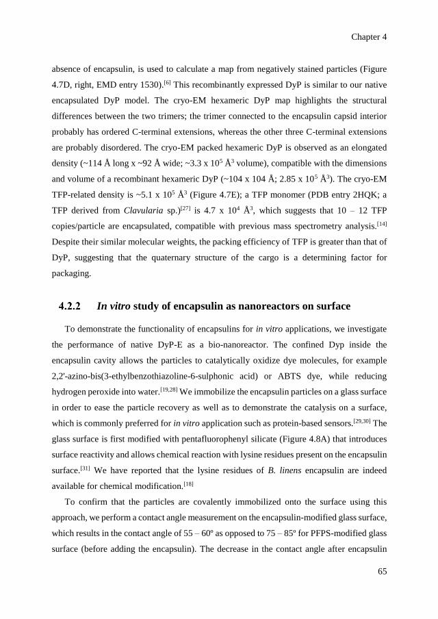
Chapter 4
65
absence of encapsulin, is used to calculate a map from negatively stained particles (Figure
4.7D, right, EMD entry 1530).[6] This recombinantly expressed DyP is similar to our native
encapsulated DyP model. The cryo-EM hexameric DyP map highlights the structural
differences between the two trimers; the trimer connected to the encapsulin capsid interior
probably has ordered C-terminal extensions, whereas the other three C-terminal extensions
are probably disordered. The cryo-EM packed hexameric DyP is observed as an elongated
density (~114 Å long x ~92 Å wide; ~3.3 x 10⁵ Å3 volume), compatible with the dimensions
and volume of a recombinant hexameric DyP (~104 x 104 Å; 2.85 x 10⁵ Å3). The cryo-EM
TFP-related density is ~5.1 x 10⁵ Å3 (Figure 4.7E); a TFP monomer (PDB entry 2HQK; a
TFP derived from Clavularia sp.)[27] is 4.7 x 10⁴ Å3, which suggests that 10 – 12 TFP
copies/particle are encapsulated, compatible with previous mass spectrometry analysis.[14]
Despite their similar molecular weights, the packing efficiency of TFP is greater than that of
DyP, suggesting that the quaternary structure of the cargo is a determining factor for
packaging.
In vitro study of encapsulin as nanoreactors on surface
To demonstrate the functionality of encapsulins for in vitro applications, we investigate
the performance of native DyP-E as a bio-nanoreactor. The confined Dyp inside the
encapsulin cavity allows the particles to catalytically oxidize dye molecules, for example
2,2'-azino-bis(3-ethylbenzothiazoline-6-sulphonic acid) or ABTS dye, while reducing
hydrogen peroxide into water.[19,28] We immobilize the encapsulin particles on a glass surface
in order to ease the particle recovery as well as to demonstrate the catalysis on a surface,
which is commonly preferred for in vitro application such as protein-based sensors.[29,30] The
glass surface is first modified with pentafluorophenyl silicate (Figure 4.8A) that introduces
surface reactivity and allows chemical reaction with lysine residues present on the encapsulin
surface.[31] We have reported that the lysine residues of B. linens encapsulin are indeed
available for chemical modification.[18]
To confirm that the particles are covalently immobilized onto the surface using this
approach, we perform a contact angle measurement on the encapsulin-modified glass surface,
which results in the contact angle of 55 – 60º as opposed to 75 – 85º for PFPS-modified glass
surface (before adding the encapsulin). The decrease in the contact angle after encapsulin

Bacterial encapsulins as in vitro and in vivo protein-based nanoplatforms
66
addition indicates a successful immobilization as the presence of encapsulin changes the
surface properties into a more polar state (in comparison to the surface with PFPS only).
Furthermore, as shown in Figure 4.8B, we are able to visualize the encapsulin particles on
the surface using scanning electron microscopy (SEM). Based on multiple images recorded
with SEM, we estimate the percentage of coverage area to be 35 ± 5% with up to 1.16 x 10¹¹
encapsulin particles immobilized on the surface (area: 1 x 1.5 cm2).
Figure 4.8 Surface-immobilized encapsulin as a bio-nanoreactor. (A) Immobilization strategy of
encapsulin particles on a glass surface using PFPS molecules as linkers. (B) SEM image of surface-
immobilized encapsulin showing the presence of nanometer-sized particles (~24 nm) on the surface.
(C) Schematic representation of a tandem system consisting of GOx and DyP-E. The initial substrate
glucose is oxidized by GOx while producing H2O2, which in turn is converted by Dyp into water while
producing green-colored ABTS radical. (D) Catalytic assay of the tandem system monitored at λ = 410
nm based on the absorption of the ABTS radical cation.
After surface attachment, we investigate the catalytic activity by carrying out a cascade
reaction on the surface. The encapsulin enzymatic activity is tested based on the oxidization
of ABTS dye while reducing H2O2 produced by an additional enzyme called glucose oxidase
(GOx) that is added into the solution (Figure 4.8C). We choose the well-studied tandem GOx-
peroxidase system as it has been proven useful as a biosensor for glucose detection,[28,32,33]
thus providing our system with a possible functionality. The oxidation of ABTS into its
green-colored radical form is monitored at λ = 410 nm for up to 6 min (Figure 4.8D) at
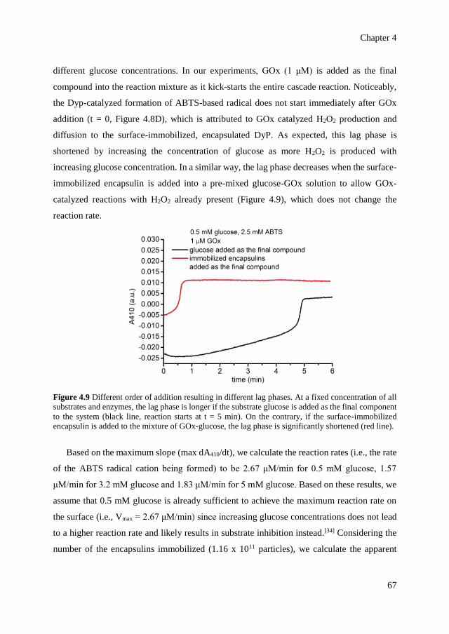
Chapter 4
67
different glucose concentrations. In our experiments, GOx (1 μM) is added as the final
compound into the reaction mixture as it kick-starts the entire cascade reaction. Noticeably,
the Dyp-catalyzed formation of ABTS-based radical does not start immediately after GOx
addition (t = 0, Figure 4.8D), which is attributed to GOx catalyzed H2O2 production and
diffusion to the surface-immobilized, encapsulated DyP. As expected, this lag phase is
shortened by increasing the concentration of glucose as more H2O2 is produced with
increasing glucose concentration. In a similar way, the lag phase decreases when the surface-
immobilized encapsulin is added into a pre-mixed glucose-GOx solution to allow GOx-
catalyzed reactions with H2O2 already present (Figure 4.9), which does not change the
reaction rate.
Figure 4.9 Different order of addition resulting in different lag phases. At a fixed concentration of all
substrates and enzymes, the lag phase is longer if the substrate glucose is added as the final component
to the system (black line, reaction starts at t = 5 min). On the contrary, if the surface-immobilized
encapsulin is added to the mixture of GOx-glucose, the lag phase is significantly shortened (red line).
Based on the maximum slope (max dA410/dt), we calculate the reaction rates (i.e., the rate
of the ABTS radical cation being formed) to be 2.67 μM/min for 0.5 mM glucose, 1.57
μM/min for 3.2 mM glucose and 1.83 μM/min for 5 mM glucose. Based on these results, we
assume that 0.5 mM glucose is already sufficient to achieve the maximum reaction rate on
the surface (i.e., Vmax = 2.67 μM/min) since increasing glucose concentrations does not lead
to a higher reaction rate and likely results in substrate inhibition instead.[34] Considering the
number of the encapsulins immobilized (1.16 x 10¹¹ particles), we calculate the apparent
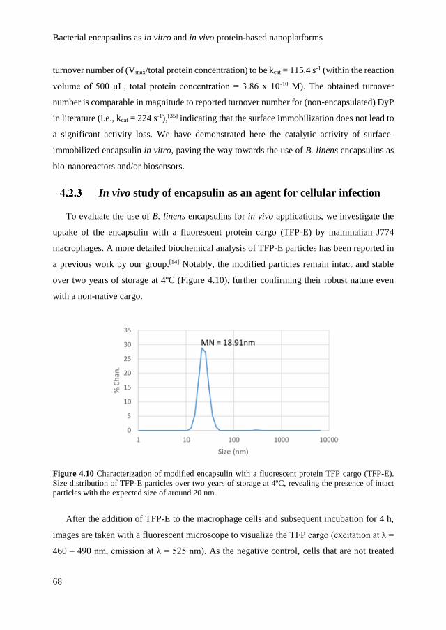
Bacterial encapsulins as in vitro and in vivo protein-based nanoplatforms
68
turnover number of (Vmax/total protein concentration) to be kcat = 115.4 s-1 (within the reaction
volume of 500 μL, total protein concentration = 3.86 x 10⁻¹⁰ M). The obtained turnover
number is comparable in magnitude to reported turnover number for (non-encapsulated) DyP
in literature (i.e., kcat = 224 s-1),[35] indicating that the surface immobilization does not lead to
a significant activity loss. We have demonstrated here the catalytic activity of surface-
immobilized encapsulin in vitro, paving the way towards the use of B. linens encapsulins as
bio-nanoreactors and/or biosensors.
In vivo study of encapsulin as an agent for cellular infection
To evaluate the use of B. linens encapsulins for in vivo applications, we investigate the
uptake of the encapsulin with a fluorescent protein cargo (TFP-E) by mammalian J774
macrophages. A more detailed biochemical analysis of TFP-E particles has been reported in
a previous work by our group.[14] Notably, the modified particles remain intact and stable
over two years of storage at 4ºC (Figure 4.10), further confirming their robust nature even
with a non-native cargo.
Figure 4.10 Characterization of modified encapsulin with a fluorescent protein TFP cargo (TFP-E).
Size distribution of TFP-E particles over two years of storage at 4ºC, revealing the presence of intact
particles with the expected size of around 20 nm.
After the addition of TFP-E to the macrophage cells and subsequent incubation for 4 h,
images are taken with a fluorescent microscope to visualize the TFP cargo (excitation at λ =
460 – 490 nm, emission at λ = 525 nm). As the negative control, cells that are not treated
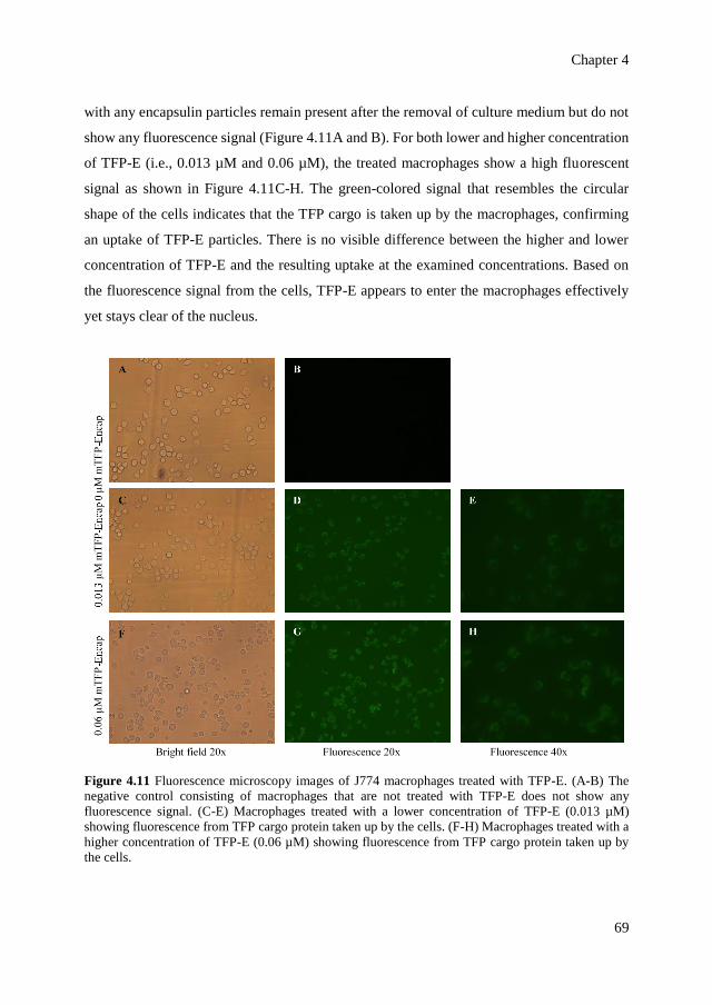
Chapter 4
69
with any encapsulin particles remain present after the removal of culture medium but do not
show any fluorescence signal (Figure 4.11A and B). For both lower and higher concentration
of TFP-E (i.e., 0.013 µM and 0.06 µM), the treated macrophages show a high fluorescent
signal as shown in Figure 4.11C-H. The green-colored signal that resembles the circular
shape of the cells indicates that the TFP cargo is taken up by the macrophages, confirming
an uptake of TFP-E particles. There is no visible difference between the higher and lower
concentration of TFP-E and the resulting uptake at the examined concentrations. Based on
the fluorescence signal from the cells, TFP-E appears to enter the macrophages effectively
yet stays clear of the nucleus.
Figure 4.11 Fluorescence microscopy images of J774 macrophages treated with TFP-E. (A-B) The
negative control consisting of macrophages that are not treated with TFP-E does not show any
fluorescence signal. (C-E) Macrophages treated with a lower concentration of TFP-E (0.013 µM)
showing fluorescence from TFP cargo protein taken up by the cells. (F-H) Macrophages treated with a
higher concentration of TFP-E (0.06 µM) showing fluorescence from TFP cargo protein taken up by
the cells.
Bacterial encapsulins as in vitro and in vivo protein-based nanoplatforms
70
This study shows that the encapsulins can be used to confine a foreign protein cargo and
preserve their structural integrity for a prolonged time in vitro, and the cargo can be taken up
by mammalian cells. Further studies are needed to identify whether the spherical particles
stay intact inside the cells as well as the uptake mechanism.
4.3 Conclusion
We have provided structural insight into the encapsulins from B. linens using cryo-EM,
confirming that the native icosahedral, cage-like assemblies confine a single peroxidase DyP
within its cavity. Using the same analysis, we also characterize the structure of modified
encapsulin particles with non-native cargo (i.e., a fluorescent protein). Compared to the
empty encapsulin (nl-E) the protein cages that contain a cargo are slightly larger, which is in
line with our previous analysis.[20] The data further confirms the directive role the C-terminal
fragments on the cargo proteins play in their inclusion,[6,14] allowing the design and
preparation of encapsulin containers with a variety of guest systems. To investigate the
functionality of encapsulins as a promising agent for nanoplatforms, we demonstrate a
tandem in vitro catalysis proving that the encapsulin is able to play a role as bio-nanoreactors
on surface as well as the in vivo cellular uptake of modified encapsulins by mammalian
macrophages. Our results provide both functional and structural basis of B. linens
encapsulins, paving the way towards the development of the encapsulins as functional
assemblies for nanotechnology, for instance as therapeutic agents and biosensors.
4.4 Acknowledgements
We thank Prof. Nenad Ban, Dr. Markus Sutter and Dr. W. Frederik Rurup for the donation
of plasmids. We acknowledge Aikaterini A. Rousou and Robin Klem for the collaboration
on the experiments regarding the stability of encapsulin and in vivo studies. Aijie Liu and Dr.
Christoph Traulsen are acknowledged for assistance with encapsulin immobilization. We
thank Prof. José R. Castón and team (Centro Nacional Biotecnología, Madrid, Spain) for
cryo-EM measurement and image reconstruction, Dr. Enrico G. Keim for assistance with
TEM imaging and Dr. Mark A. Smithers for assistance with SEM imaging.
Chapter 4
71
4.5 Materials and methods
4.5.1. General
B. linens encapsulin was recombinantly expressed in E. coli and purified based on
reported procedures.[14,18] Encapsulin samples were stored in “encapsulin storage buffer”
containing 20 mM Tris-HCl, 150 mM NH4Cl, 1 mM β-mercaptoethanol at pH 7.5. For pH
variation studies, 100 mM acetate buffer (CH3COONa-CH3COOH) was used to prepare pH
3, pH 4 and pH 5 buffers, 20 mM Tris-HCl buffer was used for pH 7.5 and pH 9 buffers, and
10 mM phosphate buffer (Na2HPO4-NaOH) was used for pH 10 and pH 11 buffers. All
chemicals were purchased from Sigma-Aldrich unless stated otherwise. All incubations and
reactions were conducted at room temperature unless stated otherwise.
For data processing and representation, all plotting and mathematical calculations were
performed with OriginPro 9.0 software. Protein structures were rendered using PyMOL 1.3
software and chemical structures were drawn using ChemBioDraw Ultra 12.0 software.
4.5.2. Cryo-EM and image processing
Non-loaded, DyP- and TFP-loaded B. linens encapsulin (5 μL) were applied to one side
of Quantifoil R 2/2 holey grids, blotted and plunged into liquid ethane in a Leica EM CPC
cryofixation unit. Samples were analyzed in a Tecnai G2 electron microscope equipped with
a field emission gun operating at 200kV, and images were recorded under low-dose
conditions with a FEI Eagle CCD at a detector magnification of 69,444X (2.16 Å/pixel
sampling rate). Image processing operations were performed using Xmipp[36] and Relion[37]
packages integrated in the Scipion platform.[38] Graphic representations were produced by
UCSF Chimera.[39] The Xmipp automatic picking routine was used to select 6,785, 19,591
and 29,680 individual particle images of non-loaded, DyP- and TFP-loaded encapsulin,
respectively. A 1.2 – 4.1 μm defocus range was determined for each image with CTFfind4.[40]
Particle images were extracted, normalized and downsampled to a factor of 2, with a final
sampling ratio of 4.32 Å/pixel. Using the Relion routine, a two-dimensional (2D)
classification was performed to discard low-quality particles, and 5,357, 15,941 and 19,779
isometric particles were selected for non-loaded, DyP- and TFP-loaded encapsulin,
respectively. A 3D classification was run using the structure of Thermotoga maritima

Bacterial encapsulins as in vitro and in vivo protein-based nanoplatforms
72
encapsulin (PDB entry 3DKT), low-pass filtered to 40 Å, as an initial model. When
icosahedral symmetry was imposed, a single class with 4,246, 12,833 and 15,976 particles
was obtained for non-loaded, DyP- and TFP-loaded encapsulin, respectively. When
assuming no symmetry for non-loaded, DyP- and TFP-loaded encapsulin, three classes were
obtained, but no significant differences were observed between them at the resolutions
achieved and particles were refined together. DyP-loaded encapsulin particles were analyzed
considering C3 symmetry and a single class with 9,930 particles was selected. These data
sets were used to obtain the final 3DR using the Relion autorefinement routine. Resolutions
of 3D with icosahedral symmetry were estimated from two independent half-datasets using
the 0.5 (or 0.3) criterion of the Fourier shell correlation (FSC), and the values for non-loaded,
DyP- and TFP-loaded encapsulin were 11.4 (10.7), 15.1 (13.5) and 13.5 (12) Å, respectively.
Similarly, resolution values for asymmetric 3DR were 27.3 (23.8), 28.3 (25) and 24.2 (22.7)
Å. For the DyP-loaded encapsulin with C3 symmetry, resolution was 24.7 (22.8) Å. The
Chimera fitting tool was used to dock the atomic crystallographic model T. maritima
encapsulin into our cryo-EM encapsulin maps.
4.5.3. Characterization of encapsulin stability
Size-exclusion profiles of encapsulin upon pH and ionic strength variation were obtained
by injecting and running 500 μL of each samples (~15 μM) into a Superose 6 preparative
column 10/100 GL (GE Healthcare FPLC Äkta purifier 900 with a 24 mL bed volume). The
hydrodynamic size distribution of the particles was determined using a particle analyzer
Nanotrac Wave (Microtrac). For particle imaging with TEM, 5 μL of a sample was applied
onto Formvar-carbon coated grids and the liquid was drained after 30 s. Afterwards, 5 μL of
a staining solution consisting of uranyl acetate (1% w/v) was added onto the grids and the
liquid was drained after 1 min. For protein characterization with denaturing gel
electrophoresis (SDS-PAGE), experiments using 12% polyacrylamide gel were conducted
based on procedures in literature[41] and Bio-Safe Coomassie (Bio-Rad) was used for
visualization of protein bands.
Chapter 4
73
4.5.4. Immobilization of encapsulin and catalytic assay
Substrates (glass or silicon) were rinsed with water and activated by immersion/cycling
piranha solution (H2SO4:H2O2 = 3:1) and rinsed with water and ethanol, then dried with a
steam of nitrogen. !Caution: Piranha solution reacts violently with organic material and has
to be treated with utmost care! Perfluorophenyl-11-(triethoxysilyl)undecanoate (PFPS) was
deposited by substrate immersion into a PFPS solution (DCM, 10 mM) for 24 h at room
temperature under argon atmosphere. The glass wafer was rinsed with DCM to remove
unreacted reagent and dried in a steam of N2. Encapsulin particles were deposited from
buffered solution (0.2 M phosphate buffer, pH 7.2). The particles were drop-coated on the
flat glass substrate overnight in a closed vessel to avoid solvent evaporation. Afterwards, the
glass was rinsed with buffer solution to remove non-immobilized particles.
For catalytic assay, 200 μL of glucose (3 different final concentrations: 0.5 mM, 3.2 mM,
5 mM) and 200 μL of ABTS (2.5 mM) in Tris-HCl buffer pH 7.5 was placed inside a cuvette
together with the glass surface with the immobilized encapsulin particles. Afterwards, 100
μL of GOx (1 μM) in PBS buffer pH 7.4 was added into the cuvette to start the reaction and
the absorption value at λ = 410 nm was immediately recorded for 6 min using a Perkin Elmer
Lambda 850 UV-visible spectrometer. As control experiments, a PFPS-modified glass
surface without encapsulin particles was used for similar kinetic studies.
The reaction rate based on radical ABTS production was calculated as follows:
𝑟𝑒𝑎𝑐𝑡𝑖𝑜𝑛 𝑟𝑎𝑡𝑒 = 𝑑 [𝑟𝑎𝑑𝑖𝑐𝑎𝑙 𝐴𝐵𝑇𝑆]
𝑑𝑡=
𝑑𝐴
𝑑𝑡 𝑥
1
ɛ 𝑥 𝑏
where A is the absorbance of radical ABTS at λ = 410 nm, ɛ is the extinction coefficient
of radical ABTS at λ = 410 nm (36000 M-1 cm-1) and b is the cuvette path length (1 cm).
dA/dt corresponds to the maximum slope of the kinetic plots in Figure 4.8D and Figure 4.9.
The apparent turnover number kcat was calculated based on Vmax/total protein concentration.
Although the particles were immobilized on the glass surface, the catalytic assay was
performed in solution (i.e., the modified surface was fully immersed inside a cuvette). The
total protein concentration was calculated based on the number of particles on the surface
and the volume of reaction.

Bacterial encapsulins as in vitro and in vivo protein-based nanoplatforms
74
4.5.5. Cell experiments
B. linens encapsulin containing mTFP (monomeric teal fluorescent protein) were
recombinantly produced in E. coli and purified using the same protocol established for native
B. linens encapsulin. The concentration of TFP-E was 0.70 µM. Murine macrophage cells
(J774) were cultivated in a DMEM. Following the cultivation, 100 µL of ~2000 cells were
plated per well on a 96-well plate and either 0, 5 or 10 µL of TFP-E was added into the well
(0, 0.013, 0.06 µM TFP-E). The treated cells were incubated at 37°C, 5% CO2 for 4 h to
allow the cells to take up the particles. Following the incubation, the medium was removed
and the cells were rinsed with phosphate buffer saline (10 mM PBS, pH 7.4) to further
remove non-absorbed species. For the visualization based on fluorescence, the cells were
imaged using a fluorescence microscope (Olympus TH4-200 with a X-Cite series 120pc Q
Laser from Lumen Dynamics, excitation at λ = 460 – 490 nm, emission at λ = 525 nm).
4.6 References
[1] Uchida, M.; Klem, M. T.; Allen, M.; Suci, P.; Flenniken, M.; Gillitzer, E.; Varpness,
Z.; Liepold, L. O.; Young, M.; Douglas, T. Adv. Mater. 2007, 19, 1025-1042.
[2] Peek, L. J.; Middaugh, C. R.; Berkland, C. Adv. Drug Deliv. Rev. 2008, 60, 915-928.
[3] Lee, E. J.; Lee, N. K.; Kim, I. S. Adv. Drug Deliv. Rev. 2016, 106, 157-171.
[4] Giessen, T. W. Curr. Opin. Chem. Biol. 2016, 34, 1-10.
[5] Choi, B.; Moon, H.; Hong, S. J.; Shin, C.; Do, Y.; Ryu, S.; Kang, S. ACS Nano 2016,
10, 7339-7350.
[6] Sutter, M.; Boehringer, D.; Gutmann, S.; Guenther, S.; Prangishvili, D.; Loessner, M.
J.; Stetter, K. O.; Weber-Ban, E.; Ban, N. Nat. Struct. Mol. Biol. 2008, 15, 939-947.
[7] Moon, H.; Lee, J.; Min, J.; Kang, S. Biomacromolecules 2014, 15, 3794-3801.
[8] Tamura, A.; Fukutani, Y.; Takami, T.; Fujii, M.; Nakaguchi, Y.; Murakami, Y.;
Noguchi, K.; Yohda, M.; Odaka, M. Biotechnol. Bioeng. 2015, 112, 13-20.
[9] Corchero, J. L.; Cedano, J. Microb. Cell. Fact. 2011, 10, 92.
[10] Kushnir, N.; Streatfield, S. J.; Yusibov, V. Vaccine 2012, 31, 58-83.
[11] Zhen, Z.; Tang, W.; Todd, T.; Xie, J. Expert Opin. Drug Deliv. 2014, 11, 1913-1922.
[12] Zeth, K.; Hoiczyk, E.; Okuda, M. Trends Biochem. Sci. 2016, 41, 190-203.
Chapter 4
75
[13] Choi, S. H.; Kwon, I. C.; Hwang, K. Y.; Kim, I. S.; Ahn, H. J. Biomacromolecules 2011,
12, 3099-3106.
[14] Rurup, W. F.; Snijder, J.; Koay, M. S. T.; Heck, A. J. R.; Cornelissen, J. J. L. M. J. Am.
Chem. Soc. 2014, 136, 3828-3832.
[15] Cassidy-Amstutz, C.; Oltrogge, L.; Going, C. C.; Lee, A.; Teng, P.; Quintanilla, D.;
East-Seletsky, A.; Williams, E. R.; Savage, D. F. Biochemistry 2016, 55, 3461-3468.
[16] Contreras, H.; Joens, M. S.; McMath, L. M.; Le, V. P.; Tullius, M. V.; Kimmey, J. M.;
Bionghi, N.; Horwitz, M. A.; Fitzpatrick, J. A.; Goulding, C. W. J. Biol. Chem. 2014,
289, 18279-18289.
[17] McHugh, C. A.; Fontana, J.; Nemecek, D.; Cheng, N. Q.; Aksyuk, A. A.; Heymann, J.
B.; Winkler, D. C.; Lam, A. S.; Wall, J. S.; Steven, A. C.; Hoiczyk, E. EMBO J. 2014,
33, 1896-1911.
[18] Putri, R. M.; Fredy, J. W.; Cornelissen, J. J. L. M.; Koay, M. S. T.; Katsonis, N.
ChemPhysChem 2016, 17, 1815-1818.
[19] Rahmanpour, R.; Bugg, T. D. H. FEBS J. 2013, 280, 2097-2104.
[20] Snijder, J.; Kononova, O.; Barbu, I. M.; Uetrecht, C.; Rurup, W. F.; Burnley, R. J.;
Koay, M. S. T.; Cornelissen, J. J. L. M.; Roos, W. H.; Barsegov, V.; Wuite, G. J.; Heck,
A. J. R. Biomacromolecules 2016, 17, 2522-2529.
[21] Akita, F.; Chong, K. T.; Tanaka, H.; Yamashita, E.; Miyazaki, N.; Nakaishi, Y.; Suzuki,
M.; Namba, K.; Ono, Y.; Tsukihara, T.; Nakagawa, A. J. Mol. Biol. 2007, 368, 1469-
1483.
[22] Abrescia, N. G.; Bamford, D. H.; Grimes, J. M.; Stuart, D. I. Annu. Rev. Biochem. 2012,
81, 795-822.
[23] Valdes-Stauber, N.; Scherer, S. Appl. Environ. Microbiol. 1994, 60, 3809-3814.
[24] Lavelle, L.; Michel, J. P.; Gingery, M. J. Virol. Methods. 2007, 146, 311-316.
[25] Teramoto, H.; Nakajima, K.; Takabayashi, C. Biomacromolecules 2004, 5, 1392-1398.
[26] Basle, E.; Joubert, N.; Pucheault, M. Chem. Biol. 2010, 17, 213-227.
[27] Ai, H. W.; Henderson, J. N.; Remington, S. J.; Campbell, R. E. Biochem. J. 2006, 400,
531-540.
[28] Lian, X.; Chen, Y.-P.; Liu, T.-F.; Zhou, H.-C. Chem. Sci. 2016, 7, 6969-6973.
[29] Sassolas, A.; Blum, L. J.; Leca-Bouvier, B. D. Biotechnol. Adv. 2012, 30, 489-511.
Bacterial encapsulins as in vitro and in vivo protein-based nanoplatforms
76
[30] Homaei, A. A.; Sariri, R.; Vianello, F.; Stevanato, R. J. Chem. Biol. 2013, 6, 185-205.
[31] Lockett, M. R.; Phillips, M. F.; Jarecki, J. L.; Peelen, D.; Smith, L. M. Langmuir 2008,
24, 69-75.
[32] Heller, A.; Feldman, B. Chem. Rev. 2008, 108, 2482-2505.
[33] Shlyahovsky, B.; Li, D.; Katz, E.; Willner, I. Biosens. Bioelectron. 2007, 22, 2570-
2576.
[34] Nicol, M. J.; Duke, F. R. J. Biol. Chem. 1966, 241, 4292-4293.
[35] Salvachúa, D.; Prieto, A.; Martínez, A. T.; Martínez, M. J. Appl. Environ. Microbiol.
2013, 79, 4316-4324.
[36] Marabini, R.; Masegosa, I. M.; San Martin, M. C.; Marco, S.; Fernandez, J. J.; de la
Fraga, L. G.; Vaquerizo, C.; Carazo, J. M. J. Struct. Biol. 1996, 116, 237-240.
[37] Scheres, S. H. J. Struct. Biol. 2012, 180, 519-530.
[38] de la Rosa-Trevin, J. M.; Quintana, A.; Del Cano, L.; Zaldivar, A.; Foche, I.; Gutierrez,
J.; Gomez-Blanco, J.; Burguet-Castell, J.; Cuenca-Alba, J.; Abrishami, V.; Vargas, J.;
Oton, J.; Sharov, G.; Vilas, J. L.; Navas, J.; Conesa, P.; Kazemi, M.; Marabini, R.;
Sorzano, C. O.; Carazo, J. M. J. Struct. Biol. 2016, 195, 93-99.
[39] Pettersen, E. F.; Goddard, T. D.; Huang, C. C.; Couch, G. S.; Greenblatt, D. M.; Meng,
E. C.; Ferrin, T. E. J. Comput. Chem. 2004, 25, 1605-1612.
[40] Rohou, A.; Grigorieff, N. J. Struct. Biol. 2015, 192, 216-221.
[41] Shapiro, A. L.; Vinuela, E.; Maizel, J. V. Biochem. Biophys. Res. Commun. 1967, 28,
815-820.
Chapter 5
Labeling encapsulin with
light-switchable fluorophores
The emergence of bacterial nanocages as nano-carriers to penetrate cells
calls for the development of tracking strategies that would enable in vivo
studies. Here, a strategy to label bacterial nanocages with photo -switchable
fluorophores is presented, to facilitate their imaging by super -resolution
microscopy. We demonstrate the functionalizatio n of the encapsulin from
Brevibacterium linens with a spiropyran photo-switch, which is not
fluorescent, by covalent attachment to the amine residues at the outer
encapsulin shell. Upon alternating irradiation with ultraviolet and visible
light, the spiropyran switches forth and back to its fluorescent merocyanine
photo-isomer and thus the fluorescence can be switched on and off, reversibly.
The bacterial compartments preserve their structural integrity upon covalent
modification and over at least five irra diation cycles.
This chapter is published in R.M. Putri, J.W. Fredy, J.J.L.M. Cornelissen, M.S.T. Koay and
N. Katsonis, ChemPhysChem 2016, 17, 1815-1818 (special issue on molecular machines),
and inside cover page.

Labeling encapsulin with light-switchable fluorophores
78
5.1 Introduction
The work presented in Chapter 4 has highlighted how encapsulins emerge as promising
building blocks for nanobiotechnology.[1-8] For in vivo applications, the ability of encapsulins
as nanoplatforms to penetrate cells while carrying functional molecules has been
demonstrated,[3,8] but further understanding of their in vivo behavior calls for the development
of non-invasive imaging and tracking strategies, while not compromising the self-assembly.
Overcoming the diffraction barrier for microscopy can be achieved in live cells by using
super-resolution fluorescence imaging,[9-15] by photo-switching fluorescence on and off to
statistically turn on a small fraction of fluorophores only.[9, 14-18] Such technique known as
Stochastic Optical Reconstruction Microscopy (STORM) involves the mapping of
fluorescent points by activating only a subset of fluorophores stochastically at any given time
while the rest are switched off (Figure 5.1).[9] Super-resolution images are then reconstructed
from multiple time-resolved fluorescence images at different areas. Moreover, the possibility
to switch fluorescence on or off might reveal useful to avoid overlapping signals in the
presence of various labeled structures. With this in mind, we set out to label bacterial
nanocages with photo-switchable fluorophores.
Figure 5.1 Principle of STORM imaging. An object is labeled with fluorophores (orange circles) that
can be switched between an off (dark) state and on (fluorescent) state by light irradiation at different
wavelengths, for instance, red laser to switch off and green laser to switch on (depicted by red and
green arrows). Initially, the fluorophores are all present in their dark state (black circles) and in each
cycle, a green laser is used to switch on only a fraction of the fluorophores. From multiple imaging
cycles, optically resolvable sets of active fluorophores are then obtained and used to reconstruct the
overall image. Reprinted with permission from [9]. Copyright © 2006, Macmillan Publishers Limited,
part of Springer Nature.
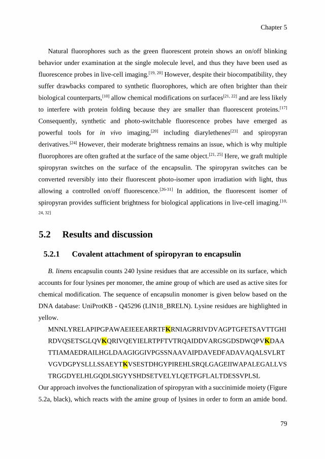
Chapter 5
79
Natural fluorophores such as the green fluorescent protein shows an on/off blinking
behavior under examination at the single molecule level, and thus they have been used as
fluorescence probes in live-cell imaging.[19, 20] However, despite their biocompatibility, they
suffer drawbacks compared to synthetic fluorophores, which are often brighter than their
biological counterparts,[10] allow chemical modifications on surfaces[21, 22] and are less likely
to interfere with protein folding because they are smaller than fluorescent proteins.[17]
Consequently, synthetic and photo-switchable fluorescence probes have emerged as
powerful tools for in vivo imaging,[20] including diarylethenes[23] and spiropyran
derivatives.[24] However, their moderate brightness remains an issue, which is why multiple
fluorophores are often grafted at the surface of the same object.[21, 25] Here, we graft multiple
spiropyran switches on the surface of the encapsulin. The spiropyran switches can be
converted reversibly into their fluorescent photo-isomer upon irradiation with light, thus
allowing a controlled on/off fluorescence.[26-31] In addition, the fluorescent isomer of
spiropyran provides sufficient brightness for biological applications in live-cell imaging.[10,
24, 32]
5.2 Results and discussion
5.2.1 Covalent attachment of spiropyran to encapsulin
B. linens encapsulin counts 240 lysine residues that are accessible on its surface, which
accounts for four lysines per monomer, the amine group of which are used as active sites for
chemical modification. The sequence of encapsulin monomer is given below based on the
DNA database: UniProtKB - Q45296 (LIN18_BRELN). Lysine residues are highlighted in
yellow.
MNNLYRELAPIPGPAWAEIEEEARRTFKRNIAGRRIVDVAGPTGFETSAVTTGHI
RDVQSETSGLQVKQRIVQEYIELRTPFTVTRQAIDDVARGSGDSDWQPVKDAA
TTIAMAEDRAILHGLDAAGIGGIVPGSSNAAVAIPDAVEDFADAVAQALSVLRT
VGVDGPYSLLLSSAEYTKVSESTDHGYPIREHLSRQLGAGEIIWAPALEGALLVS
TRGGDYELHLGQDLSIGYYSHDSETVELYLQETFGFLALTDESSVPLSL
Our approach involves the functionalization of spiropyran with a succinimide moiety (Figure
5.2a, black), which reacts with the amine group of lysines in order to form an amide bond.

Labeling encapsulin with light-switchable fluorophores
80
The spiropyran (closed, off-state) is then converted to the colored merocyanine form (open,
on-state) upon irradiation with UV light. Without UV irradiation, a spontaneous conversion
of spiropyran form to merocyanine form can also occur in the dark in aqueous environment
and is particularly favored in acidic environment, hence, all samples are kept under ambient
light (to stabilize the spiropyran form) and in a neutral solution (in PBS buffer pH 7.4).[27]
This conversion is reversible upon subsequent irradiation with visible light (Figure 5.2b).
The merocyanine also undergoes a slow thermal relaxation into spiropyran in the dark,
although the open (charged) form is more stabilized in a polar environment.[27]
Figure 5.2 Covalent attachment of spiropyran-succinimide onto encapsulin. a) Coupling of encapsulin
and spiropyran via amine-succinimide reaction. b) Photo-isomerization of non-fluorescent spiropyran
(blue) to fluorescent merocyanine (red).
Coupling the encapsulin nanocages to the spiropyran is achieved by adding 1000-times
excess of spiropyran to the purified encapsulin solution at pH 7.4, containing 10% DMSO.
After overnight incubation, the non-reacted spiropyran and DMSO are removed by dialysis
and size-exclusion chromatography (SEC). In this process, non-covalently bound spiropyran
molecules are removed by the combination of overnight dialysis and 48-times dilution
throughout the SEC column (a buffered saline is used as the eluent to avoid electrostatic
interactions).[33] The chromatogram reveals a protein peak at around V = 11 – 12 mL which
is characteristic for encapsulin particles (Figure 5.3a).

Chapter 5
81
Figure 5.3 Labeling encapsulin with a spiropyran photo-switch. a) Size-exclusion chromatogram
showing the characteristic encapsulin elution at V = 11 – 12 mL as well as the characteristic spiropyran
absorption at λ = 350 nm at the corresponding elution volume. b) UV-visible spectrum of encapsulin
labeled with spiropyran switches.
Encapsulin particles, owing to their sizes of 20 – 25 nm, always elute at around V = 11 –
12 mL, monitored at protein primary absorption at λ = 280 nm. When the encapsulins are
modified with spiropyran switches, another wavelength is monitored during the
chromatography (i.e., the characteristic absorption for spiropyran) that is at λ = 350 nm. The
modification with the switches does not change the size of encapsulin particles significantly
since the hybrid system still elutes at V = 11 – 12 mL during the chromatography (Figure
5.3a). The absorption at λ = 350 nm at the corresponding elution volume indicates covalent
coupling of the spiropyran to the encapsulin as non-covalently bound spiropyran has been
removed. The presence of spiropyran is further confirmed by UV-visible spectroscopy
(Figure 5.3b). The small absorption band observed at around λ = 520 nm prior to any
irradiation (Figure 5.3b) is likely due to a conversion of the spiropyran form into the
merocyanine form, which occurs spontaneously in water.
To quantify the labeling, the concentration of encapsulin is determined based on gel
densitometry, while the concentration of spiropyran could be determined based on the
absorption at λ = 350 nm in Figure 5.3b. Since the encapsulin does not absorb at this
wavelength, we are able to use the absorbance at λ = 350 nm of the hybrid system to
determine the spiropyran concentration in the hybrid system (ɛ = 7,994 M-1 cm-1), which
results in the value of 1.16 mM of spiropyran. On the other hand, the encapsulin monomer is
shown in SDS-PAGE gel (Figure 5.4), corresponding to the band at around 28 kDa (lanes 2,

Labeling encapsulin with light-switchable fluorophores
82
3, 4). Importantly, lane number 3 confirms the present of the encapsulin component in the
hybrid of encapsulin-spiropyran. By comparing the intensity of the encapsulin band in lane
3 and the intensity of the encapsulin standard in lane 4 (known concentration = 0.14 mM),
we could roughly estimate the concentration of the encapsulin in the hybrid system that is
0.01 mM. Furthermore, we could estimate the number of spiropyran molecules per one
encapsulin particle that is ~ (1.16 mM/0.01 mM) = 116 (~49% coverage).
Figure 5.4 Gel densitometry to determine encapsulin concentration. Denaturing gel electrophoresis
confirms the presence of encapsulin in the hybrid sample (lane 3) based on the characteristic encapsulin
band at around 28 kDa (the monomer of the encapsulin cage), which is comparable to the native
encapsulin (lane 2 and lane 4). The intensity of the encapsulin band in lane 4 is used as a standard
(known concentration = 0.14 mM) to estimate the concentration of the encapsulin in the hybrid system
(lane 3). The systematic comparison of band intensity (gel densitometry) is performed with Bio-Rad
Image Lab software.
5.2.2 Photo-switchable fluorescence of modified encapsulin
The photo-isomerization of labeled encapsulin in PBS (pH 7.4) is examined by irradiating
the sample with UV light for 2 min (λ = 365 nm) in solution. The photo-isomerization of
spiropyran into merocyanine is indicated by the appearance of an absorption band around λ
= 540 nm (Figure 5.5a and b). This conversion is complete after 2 min of irradiation and
prolonged irradiation does not significantly increase the merocyanine concentration. We also
observe the appearance of merocyanine fluorescence (Figure 5.5c and d) at λ = 615 nm upon
excitation at λ = 535 nm. The excitation with 535 nm light for fluorescence measurement is
unlikely to significantly revert the merocyanine form to spiropyran form because it occurs

Chapter 5
83
only for a short time (around 1 – 2 min of spectrum scanning), while the conversion back to
spiropyran form requires 7 min of continuous irradiation of visible light with considerable
intensity (flux density: 46 mW/cm2).
Figure 5.5 Photo-triggered on/off fluorescence of spiropyran-labeled encapsulin. a) UV-visible spectra
showing the characteristic absorption band of merocyanine at λ = 540 nm. b) Reversible photo-
switching of spiropyran to merocyanine monitored at λ = 540 nm. c) Emission spectra showing the
fluorescence of merocyanine at λ = 615 nm upon excitation at λ = 535 nm. d) Reversible conversion of
on-state merocyanine to off-state spiropyran monitored at λ = 615 nm.
We irradiate the encapsulin-merocyanine with visible light (λ ≥ 420 nm) to investigate
the reversibility. The disappearance of the absorption band at λ = 540 nm, after 7 min of
continuous irradiation, indicates that the merocyanine form is completely reverted to the
spiropyran form (Figure 5.5a). Similarly, we observe a decrease of the fluorescence of
merocyanine at λ = 615 nm after irradiation with visible light (Figure 5.5c). We further repeat
the ultraviolet light and visible light irradiation cycle up to five times. After the fifth cycle,
the merocyanine displays absorbance and fluorescence that amount to half their initial values
(Figure 5.5b and 5.5d). The alternating decrease and increase of the fluorescence during the
cycles confirms the photo-switching of the fluorophore (Figure 5.5d). The merocyanine form
attached to the protein also undergoes thermal relaxation into the spiropyran form in the dark,

Labeling encapsulin with light-switchable fluorophores
84
which can also be observed by the disappearance of its absorption band at λ = 540 – 550 nm
(Figure 5.6). However, this process is much slower in comparison to the conversion back
with continuous visible light irradiation.
Figure 5.6 Thermal relaxation of merocyanine into spiropyran in aqueous system (the switch is
attached to a model protein, human serum albumin). The relaxation is indicated by the disappearance
of absorption band at λ = 550 nm, which is much slower (within hours) in comparison to the light-
triggered conversion (within 7 min).
5.2.3 Structural integrity of encapsulin particles
To confirm that the encapsulin particles are stable upon covalent modification and photo-
switching, we characterize the hydrodynamic size of the particles before and after irradiation
by dynamic light scattering (DLS). DLS analysis reveals a diameter of about 20 – 25 nm,
which indicates that the functionalized encapsulins remain intact after irradiation with both
UV and visible light (Figure 5.7). The intact particles after irradiation are also visualized with
TEM (Figure 5.8a). Nevertheless, structures with sizes exceeding 100 nm are also observed
after the fifth cycle, which are likely aggregates forming upon the protein degradation caused
by prolonged irradiation (Figure 5.8b).[34]

Chapter 5
85
Figure 5.7 Structural integrity of labeled encapsulin measured by DLS. a) Size distribution of
encapsulin particles throughout the alternating irradiation with UV and visible light peaking at diameter
= 20 – 25 nm. b) Comparison of size distribution before any irradiation (black), after UV irradiation of
the first cycle (red) and after visible irradiation of the fifth cycle (blue) showing that the structural
integrity of the particles is preserved throughout the cycles.
Figure 5.8 Effect of irradiation on modified encapsulin. a) TEM image of encapsulin particles
(diameter = 20 – 25 nm) after irradiation with UV light (for 2 min), confirming that the majority of the
particles are still intact upon irradiation. b) Size distribution of encapsulin particles (diameter ~ 20 nm)
after 5 cycles of irradiation showing that the intact encapsulin particles are present but larger structures
also form (diameter >100 nm).
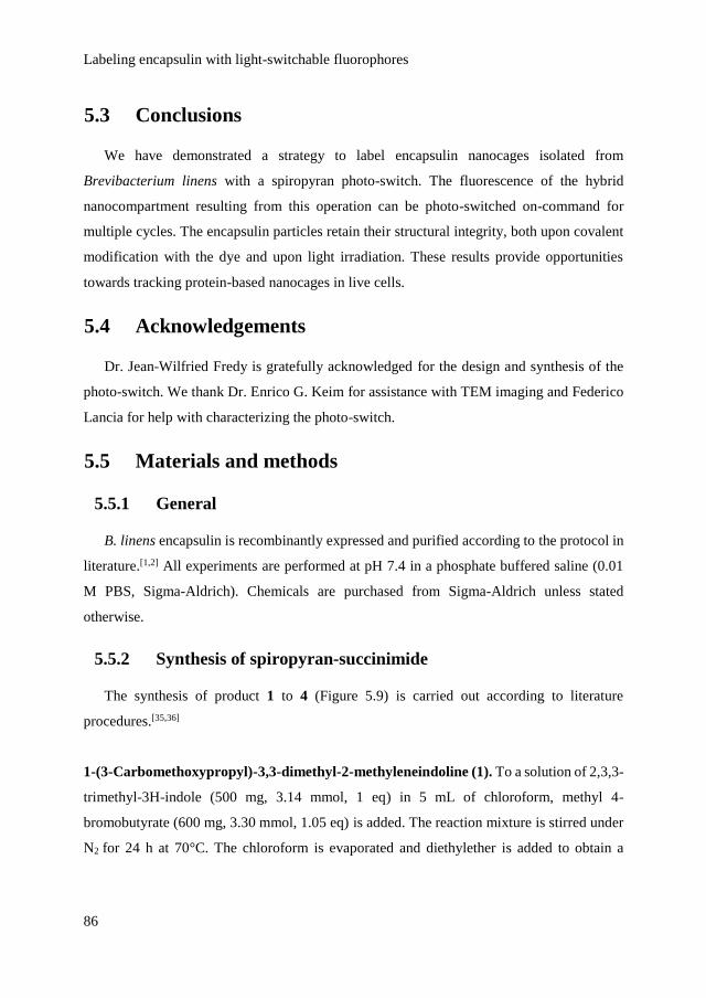
Labeling encapsulin with light-switchable fluorophores
86
5.3 Conclusions
We have demonstrated a strategy to label encapsulin nanocages isolated from
Brevibacterium linens with a spiropyran photo-switch. The fluorescence of the hybrid
nanocompartment resulting from this operation can be photo-switched on-command for
multiple cycles. The encapsulin particles retain their structural integrity, both upon covalent
modification with the dye and upon light irradiation. These results provide opportunities
towards tracking protein-based nanocages in live cells.
5.4 Acknowledgements
Dr. Jean-Wilfried Fredy is gratefully acknowledged for the design and synthesis of the
photo-switch. We thank Dr. Enrico G. Keim for assistance with TEM imaging and Federico
Lancia for help with characterizing the photo-switch.
5.5 Materials and methods
5.5.1 General
B. linens encapsulin is recombinantly expressed and purified according to the protocol in
literature.[1,2] All experiments are performed at pH 7.4 in a phosphate buffered saline (0.01
M PBS, Sigma-Aldrich). Chemicals are purchased from Sigma-Aldrich unless stated
otherwise.
5.5.2 Synthesis of spiropyran-succinimide
The synthesis of product 1 to 4 (Figure 5.9) is carried out according to literature
procedures.[35,36]
1-(3-Carbomethoxypropyl)-3,3-dimethyl-2-methyleneindoline (1). To a solution of 2,3,3-
trimethyl-3H-indole (500 mg, 3.14 mmol, 1 eq) in 5 mL of chloroform, methyl 4-
bromobutyrate (600 mg, 3.30 mmol, 1.05 eq) is added. The reaction mixture is stirred under
N2 for 24 h at 70°C. The chloroform is evaporated and diethylether is added to obtain a

Chapter 5
87
precipitate. The solid is extensively ished with diethylether. 490 mg is obtained as a purple
solid.
1H NMR (400 MHz, CD3CN) δ7.92 – 7.86 (m, 1H), 7.74 – 7.69 (m, 1H), 7.66 – 7.61 (m,
2H), 4.49 – 4.41 (m, 2H), 3.63 (s, 3H), 2.78 (s, 3H), 2.61 (dd, J = 8.6, 5.0 Hz, 2H), 2.23 –
2.13 (m, 3H), 1.55 (s, 6H).
13C NMR (101 MHz, CD3CN) δ 197.8, 173.9, 143.0, 130.9, 130.2, 124.4, 116.3, 55.6, 52.4,
48.3, 30.9, 23.4, 22.6, 14.9.
ESI MS (m/z) 260.02, calculated for [C16H22NO2]+ = 260.16.
1’-(3-Carbomethoxypropyl)-3’,3’-dimethyl-6-nitrospiro[2H-1]-benzopyran-2,2’-
indoline (2). To a solution of 5-nitrosalicylaldehyde (68 mg, 0.41 mmol, 1 eq) in 3 mL of
ethanol, the compound 1 (140 mg, 0.41 mmol, 1 eq) dissolved in 2 mL of ethanol is added
slowly. The reaction mixture is stirred at 80°C for 5 h. The solvent is evaporated and the
residue is purified by chromatography on silica gel to afford the product as a purple solid (58
mg, 56%)
1H NMR (400 MHz, CD3CN) δ 8.07 (d, J = 2.8 Hz, 1H), 8.00 (d, J = 2.8 Hz, 1H), 7.98 (d, J
= 2.8 Hz, 1H), 7.15 (td, J = 7.7, 1.3 Hz, 1H), 7.11 (dd, J = 7.3, 0.8 Hz, 1H), 7.02 (m, 1H),
6.87 – 6.81 (m, 1H), 6.71 (m, 1H), 6.66 (d, J = 7.8 Hz, 1H), 5.95 (d, J = 10.4 Hz, 1H), 3.57
(s, 3H), 3.26 – 3.10 (m, 2H), 2.32 (tt, J = 8.1, 4.1 Hz, 2H), 1.92 – 1.75 (m, 2H), 1.26 (s, 3H),
1.15 (s, 3H).
13C NMR (101 MHz, CD3CN) δ 174.44, 160.39, 148.13, 142.08, 137.11, 129.11, 128.72,
126.60, 123.73, 122.81, 122.74, 120.41, 119.97, 116.27, 107.93, 107.81, 53.36, 52.00, 43.58,
31.81, 26.27, 24.77, 20.00.
ESI MS (m/z) 409.28, calculated for [C23H24N2O5H]+ = 409.17.
1’-(3-Carboxypropyl)-3’,3’-dimethyl-6-nitrospiro[2H-1]benzopyran-2,2’-indoline (3).
To a solution of 2 (150 mg) in THF (3 mL), 1 mL of NaOH 10% is added. The reaction
mixture is stirred at room temperature for 18 h. Afterwards, it is acidified with a solution of
HCl (1 M). The aqueous phase is extracted with ethylacetate and concentrated to afford the
product as a yellow solid. (124 mg, 86%).

Labeling encapsulin with light-switchable fluorophores
88
1H NMR (400 MHz, CD3CN) δ 10.06 (d, J = 0.4 Hz, 1H), 8.70 (dd, J = 11.1, 2.9 Hz, 1H),
8.42 (dd, J = 9.2, 2.8 Hz, 1H), 8.10 (d, J = 2.8 Hz, 2H), 8.02 (dd, J = 9.0, 2.8 Hz, 2H), 7.21
– 7.12 (m, 5H), 7.05 (dd, J = 10.2, 4.3 Hz, 2H), 6.86 (td, J = 7.5, 0.9 Hz, 2H), 6.74 (d, J =
9.0 Hz, 2H), 6.70 (d, J = 7.8 Hz, 2H), 3.31 – 3.13 (m, 4H), 2.38 – 2.28 (m, 5H), 1.85 (dddd,
J = 15.4, 13.8, 10.7, 6.2 Hz, 6H), 1.27 (s, 8H), 1.18 (d, J = 6.1 Hz, 3H).
13C NMR (101 MHz, CD3CN) δ 197.6, 174.7, 166.8, 160.4, 148.2, 142.1, 137.1, 132.5, 130.9,
129.1, 128.7, 126.6, 123.7, 122.8, 122.7, 120.4, 120, 119.5, 116.3, 107.9, 107.8, 53.4, 43.64,
31.5, 26.3, 24.7, 20.0.
ESI MS (m/z) 395.09, calculated for [C22H22N2O5H]+ = 395.16.
Spiropyran-succinimide (4). Compound 3 (50 mg, 0.13 mmol, 1 eq) and N-
hydroxysuccinimide (15 mg, 0.13 mmol, 1 eq) are dissolved in 5 mL in dried acetonitrile.
DCC (26 mg, 0.13 mmol, 1 eq) is added and the reaction mixture is stirred at room
temperature for 18 h under nitrogen. The solvent is evaporated and the residue is purified by
chromatography over silica gel with hexane/ethylacetate to afford the product as a purple
solid (20 mg, 32%).
1H NMR (400 MHz, CD3CN) δ 8.07 (d, J = 2.7 Hz, 1H), 7.99 (dd, J = 9.0, 2.7 Hz, 1H), 7.17
– 7.07 (m, 2H), 7.03 (dd, J = 10.4, 6.2 Hz, 1H), 6.88 – 6.81 (m, 1H), 6.74 – 6.63 (m, 2H),
5.98 (dd, J = 10.4, 7.0 Hz, 1H), 3.32 – 3.11 (m, 2H), 2.76 (s, 2H), 2.66 (m, 2H), 2.59 – 2.56
(m, 2H), 2.36 – 2.26 (m, 2H), 1.25 (s, 3H), 1.15 (s, 3H).
13C NMR (101 MHz, CD3CN) δ 169.7, 168.2, 159.1, 146.8, 140.8, 136.0, 128.08, 127.8,
125.3, 122.4, 121.5, 121.4, 119.3, 118.7, 114.7, 106.5, 52.0, 42.3, 41.8, 27.9, 25.0, 23.4, 18.7
ESI MS (m/z) 491.94, calculated for [C26H25N3O7H]+ = 492.17.
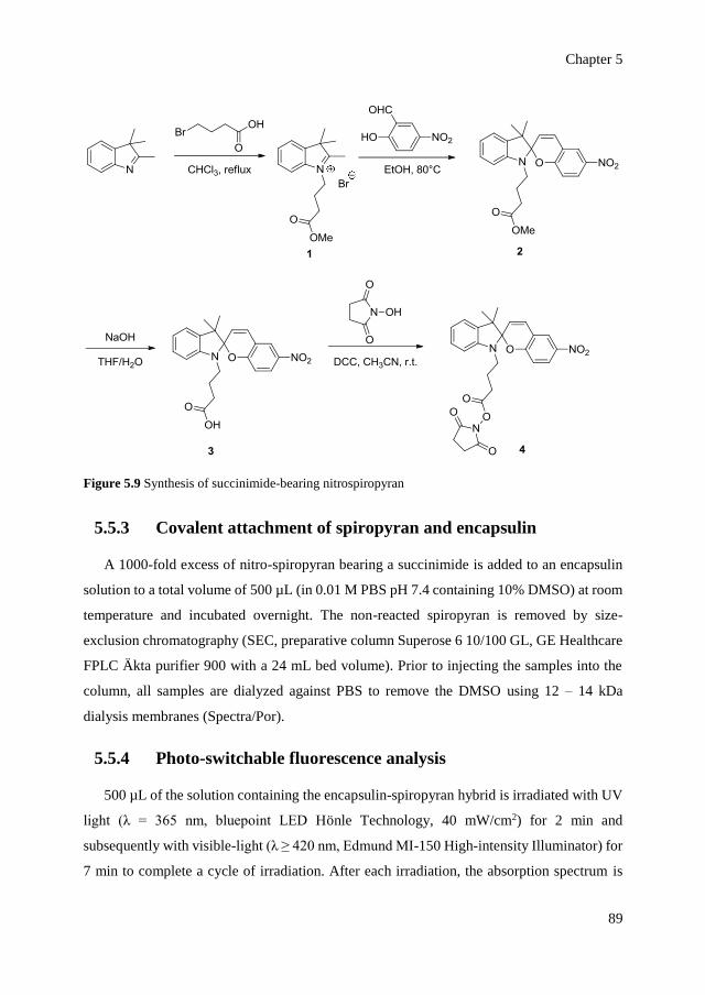
Chapter 5
89
Figure 5.9 Synthesis of succinimide-bearing nitrospiropyran
5.5.3 Covalent attachment of spiropyran and encapsulin
A 1000-fold excess of nitro-spiropyran bearing a succinimide is added to an encapsulin
solution to a total volume of 500 µL (in 0.01 M PBS pH 7.4 containing 10% DMSO) at room
temperature and incubated overnight. The non-reacted spiropyran is removed by size-
exclusion chromatography (SEC, preparative column Superose 6 10/100 GL, GE Healthcare
FPLC Äkta purifier 900 with a 24 mL bed volume). Prior to injecting the samples into the
column, all samples are dialyzed against PBS to remove the DMSO using 12 – 14 kDa
dialysis membranes (Spectra/Por).
5.5.4 Photo-switchable fluorescence analysis
500 µL of the solution containing the encapsulin-spiropyran hybrid is irradiated with UV
light (λ = 365 nm, bluepoint LED Hönle Technology, 40 mW/cm2) for 2 min and
subsequently with visible-light (λ ≥ 420 nm, Edmund MI-150 High-intensity Illuminator) for
7 min to complete a cycle of irradiation. After each irradiation, the absorption spectrum is
Labeling encapsulin with light-switchable fluorophores
90
recorded with a Perkin Elmer Lambda 850 UV-visible spectrometer and the emission
spectrum at λ = 555 – 670 nm (excitation at λ = 535 nm) is recorded using a Perkin Elmer
fluorescence spectrometer.
5.5.5 Characterization of encapsulin structure
The hydrodynamic size of the encapsulin solution (~500 µL) before and after irradiation
in 0.01 mM PBS (pH 7.4) is determined with dynamic light scattering (Nanotrac Wave,
Microtrac). In addition to DLS analysis, confirmation of the structural integrity is also
provided by TEM (300 kV acceleration voltage, Phillips CM 30) and SEC analysis.
5.6 References
[1] M. Sutter, D. Boehringer, S. Gutmann, S. Guenther, D. Prangishvili, M. J. Loessner, K.
O. Stetter, E. Weber-Ban, N. Ban, Nat. Struct. Mol. Biol. 2008, 15, 939-947.
[2] W. F. Rurup, J. Snijder, M. S. T. Koay, A. J. R. Heck, J. J. L. M. Cornelissen. J. Am.
Chem. Soc. 2014, 136, 3828-3832.
[3] H. Moon, J. Lee, J. Min, S. Kang, Biomacromolecules 2014, 15, 3794-3801.
[4] A. Tamura, Y. Fukutani, T. Takami, M. Fujii, Y. Nakaguchi, Y. Murakami, K. Noguchi,
M. Yohda, M. Odaka, Biotechnol. Bioeng. 2015, 112, 13-20.
[5] R. M. Putri, J. J. L. M. Cornelissen, M. S. T. Koay, ChemPhysChem 2015, 16, 911-918.
[6] L. Lavelle, J. P. Michel, M. Gingery, J. Virol. Methods. 2007, 146, 311-316.
[7] C. A. Hommersom, B. Matt, A. van der Ham, J. J. L. M. Cornelissen, N. Katsonis,
Org.Biomol. Chem. 2014, 12, 4065-4069.
[8] B. Choi, H. Moon, S. J. Hong, C. Shin, Y. Do, S. Ryu, S. Kang, ACS Nano 2016, 10,
7339-7350.
[9] M. J. Rust, M. Bates, X. W. Zhuang, Nat. Methods 2006, 3, 793-795.
[10] C. J. MacNevin, D. Gremyachinskiy, C. W. Hsu, L. Li, M. Rougie, T. T. Davis, K. M.
Hahn, Bioconjugate Chem. 2013, 24, 215-223.
[11] S. van de Linde, M. Heilemann, M. Sauer, Annu. Rev. Phys.Chem. 2012, 63, 519-540.
[12] S. N. Uno, M. Kamiya, T. Yoshihara, K. Sugawara, K. Okabe, M. C. Tarhan, H. Fujita,
T. Funatsu, Y. Okada, S. Tobita, Y. Urano, Nat. Chem. 2014, 6, 681-689.

Chapter 5
91
[13] S. W. Hell, J. Wichmann, Opt. Lett. 1994, 19, 780-782.
[14] E. Betzig, G. H. Patterson, R. Sougrat, O. W. Lindisser, S. Olenych, J. S. Bonifacino,
M. W. Davidson, J. Lippincott-Schwartz, H. F. Hess, Science 2006, 313, 1642-1645.
[15] S. T. Hess, T. P. K. Girirajan, M. D. Mason, Biophys. J. 2006, 91, 4258-4272.
[16] H. Bock, C. Geisler, C. A. Wurm, C. Von Middendorff, S. Jakobs, A. Schonle, A. Egner,
S. W. Hell, C. Eggeling, Appl. Phys. B. 2007, 88, 161-165.
[17] K. Hirabayashi, K. Hanaoka, M. Shimonishi, T. Terai, T. Komatsu, T. Ueno, T. Nagano,
Chem. Eur. J. 2011, 17, 14763-14771.
[18] S. W. Hell, Science 2007, 316, 1153-1158.
[19] R. M. Dickson, A. B. Cubitt, R. Y. Tsien, W. E. Moerner, Nature 1997, 388, 355-358.
[20] M. Fernandez-Suarez, A. Y. Ting, Nat. Rev. Mol. Cell Biol. 2008, 9, 929-943.
[21] T. Udayabhaskararao, P. K. Kundu, J. Ahrens, R. Klajn, ChemPhysChem 2016, 17,
1805-1809.
[22] N. Katsonis, M. Lubomska, M. M. Pollard, B. L. Feringa, P. Rudolf, Prog. Surf. Sci.
2007, 82, 407-434.
[23] Y. Zou, T. Yi, S. Z. Xiao, F. Y. Li, C. Y. Li, X. Gao, J. C. Wu, M. X. Yu, C. H. Huang,
J. Am. Chem. Soc. 2008, 130, 15750-15751.
[24] W. Wan, M. Q. Zhu, Z. Y. Tian, A. D. Q. Li, J. Am. Chem. Soc. 2015, 137, 4312-4315.
[25] A. Fihey, A. Perrier, W. R. Browne, D. Jacquemin, Chem. Soc. Rev. 2015, 44, 3719-
3759.
[26] Y. Kalisky, T. E. Orlowski, D. J. Williams, J. Phys. Chem. 1983, 87, 5333-5338.
[27] R. Klajn, Chem. Soc. Rev. 2014, 43, 148-184.
[28] M. Q. Zhu, L. Zhu, J. J. Han, W. Wu, J. K. Hurst, A. D. Q. Li, J. Am. Chem. Soc. 2006,
128, 4303-4309.
[29] W. Szymanski, J. M. Beierle, H. A. V. Kistemaker, W. A. Velema, B. L. Feringa, Chem.
Rev. 2013, 113, 6114-6178.
[30] Z. Y. Tian, A. D. Q. Li, Acc. Chem. Res. 2013, 46, 269-279.
[31] Z. Y. Tian, W. W. Wu, W. Wan, A. D. Q. Li, J. Am. Chem. Soc. 2009, 131, 4245-4252.
[32] E. Deniz, M. Tomasulo, R. A. DeFazio, B. D. Watson, F. M. Raymo, Phys. Chem. Chem.
Phys. 2010, 12, 11630-11634.

Labeling encapsulin with light-switchable fluorophores
92
[33] P. Hong, S. Koza, E. S. P. Bouvier, J. Liq. Chromatogr. Relat. Technol. 2012, 35, 2923-
2950.
[34] M. J. Davies, Biochem. Biophys. Res. Commun. 2003, 305, 761-770.
[35] P. Remon, M. Hammarson, S. M. Li, A. Kahnt, U. Pischel, J. Andreasson, Chem. Eur J.
2011, 17, 6492-6500.
[36] P. J. Wu, J. L. Chen, C. P. Chen, Y. H. Chan, Chem. Commun. 2013, 49, 898-900.

Chapter 6
Light-fueled assembly of encapsulin and
chaperone into hybrid superstructures
Life gains its sustainability from a continuous input of energy. Re -
engineering the principles underlying biological functionality into artificial
systems would allow developing new design paradigms for adaptive matt er.
Previously reported assemblies of semi-synthetic protein systems primarily
focus on integrative self-assembly under equilibrium conditions, while
extending the design to non-equilibrium dynamic order has proven challenging
due to the complexity. Here, we demonstrate that higher degrees of
organization emerge in self -assembled protein systems where proton is
released gradually under ir radiation with light, provided that proteins
undergo partial unfolding events. The giant superstructures are formed with
defined edges confining protein -based substructures. Their fueled formation
is mediated by the presence of a spiropyran -based photoacid, which releases
its proton upon irradiation, leading to partial unfolding and exposure of
hydrophobic segments of proteins and thus favoring self -association.
Simultaneously, the photoacid switches from a hydrophil ic state to a
hydrophobic state and thus becomes an active building block in the formation
of the giant assemblies. As the photo -induced switching and pH drop are
rapidly reversible in the absence of light, the superstructures spontaneously
disperse when the irradiation is stopped.

Light-fueled assembly of encapsulin and chaperone into hybrid superstructures
94
6.1 Introduction
In some cases, extended nanostructures and ordered states of protein assemblies in Nature
are sustained by a constant flow of energy or fuel. This is exemplified by the assembly of
cytoskeletal protein microtubules, where the consumption of a chemical fuel generates a
dynamic order that allows carrying complex tasks.[1,2] Drawing inspiration from the dynamic
interplay between fuel and matter in biological processes thus calls for strategies to move
beyond architectures under equilibrium conditions to a dynamic order of interacting
components.[3] The distinctive feature of a dynamic order is that it allows a highly
reconfigurable system. However, previous attempts on directing higher-order assembly of
proteins in vitro have relied primarily on the incorporation of bridging interactions (e.g.,
disulfide bonds,[4] dendrons,[5] or metal coordination[6]) that are encoded in the structure of
the proteins by design. Consequently, assembly of such systems occurs spontaneously as the
cue for the assembly is embedded in the structure, resulting in architectures under equilibrium
conditions. The disassembly would require addition of chemicals that can disrupt the
bridging moieties such as a reducing agent or a chelating agent.
Moving beyond spontaneously-assembled architectures, synthetic systems have
demonstrated a dynamic assembly that is sustained by a continuous input of a fuel, using
metal nanoparticles,[7] macrocycle[8] and chemical gelators as the building blocks.[9] To our
knowledge, such system that is based on protein building blocks has not been reported, while
protein architectures offer higher complexity, monodispersity and biocompatibility. In
multimeric self-assembled proteins, such as cage-like proteins of virus and bacteria,
environmental conditions such as pH, ionic strength and polarity of medium govern their
assembly behavior.[10] Dynamic self-association of such proteins can be achieved by
reversibly adjusting the pH to change the charges, conformations and properties of proteins
to induce self-association in a reversible manner.
In order to generate an energy-dependent dynamic order of proteins, the pH condition
that favors self-association should be achieved in a reversible manner and sustained upon an
input of energy. To enable this, we use a spiropyran-based photoacid (i.e., the open,
protonated merocyanine form, MCH+) that reversibly releases its proton upon irradiation
with visible light.[11] Using this molecule, previous reports demonstrated the decrease of pH
Chapter 6
95
up to 2 units within only 20 s of irradiation with visible light upon the conversion of MCH+
form to spiropyran (SP) form.[11,12] The release of the proton from the photoacid is rapidly
reversible as the MCH+ form is more stable than the SP form (half-life t1/2 ~70 s),[11] therefore
altogether allowing a rapidly reversible control over the assembly. The spontaneous and
immediate reversibility of pH control has enabled reversible photo-control of gold
nanoparticles assembly,[13] ring-opening polymerization,[14] formation and swelling of
supramolecular hydrogels,[15,16] photo-activation of antibiotic[17] and photo-activation of a
biological ion channel.[18]
6.2 Results and discussion
6.2.1 Light-fueled assembly of hybrid superstructures
Here, we use as building blocks the bacterial nanocompartment encapsulin from T.
maritima (abbreviated as TM-Enc) that is co-purified with GroEL chaperone upon
recombinant expression in E. coli (Figure 6.1a).[19] Both TM-Enc and GroEL are self-
assembled multimeric proteins with highly defined structures that favor self-association
under extreme pH values.[20] The native encapsulin particles adapt an icosahedral cage-like
structure, composed of 60 copies of identical protein subunits that self-assemble into a cage
with the size of around 20 – 25 nm (Figure 6.1b).[21] We recently reported that encapsulin
particles remain intact under irradiation with light.[22]
GroEL chaperone from E. coli, a tetradecameric cylindrical protein (~15 nm in diameter,
Figure 6.1b), is responsible for assisting the folding process of polypeptides into their native
protein conformation.[23-25] The so-called apical domain located at the opening of cylindrical
GroEL consists of hydrophobic surfaces that are responsible for polypeptide binding in the
GroEL functioning.[23,24] Furthermore, GroEL molecules have a tendency to stack into
fibrillar assembly upon agitation or in the presence of surfactant such as sodium dodecyl
sulfate (SDS).[20,26] Recently, a design of photo-responsive stacking of GroEL into fibrillar
structures is reported based on chemical incorporation of a spiropyran photoswitch that is
anchored in the apical domain of GroEL.[27-29] The closed SP form is converted to the open
MC form upon irradiation with UV light.[28,29] In the designed system, the stacking of GroEL
relies on the formation of MC–Mg2+–MC bridges while the SP form does not accommodate
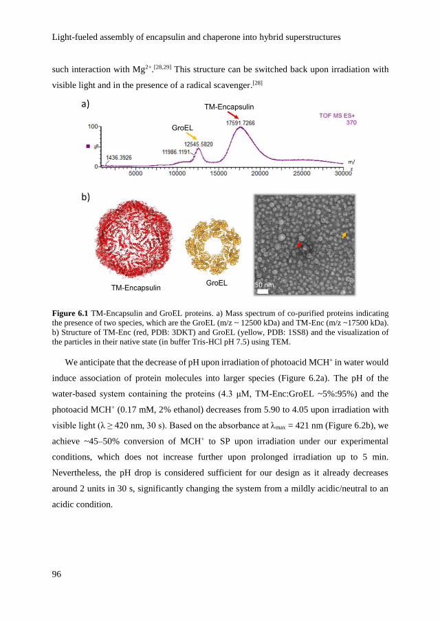
Light-fueled assembly of encapsulin and chaperone into hybrid superstructures
96
such interaction with Mg2+.[28,29] This structure can be switched back upon irradiation with
visible light and in the presence of a radical scavenger.[28]
Figure 6.1 TM-Encapsulin and GroEL proteins. a) Mass spectrum of co-purified proteins indicating
the presence of two species, which are the GroEL (m/z ~ 12500 kDa) and TM-Enc (m/z ~17500 kDa).
b) Structure of TM-Enc (red, PDB: 3DKT) and GroEL (yellow, PDB: 1SS8) and the visualization of
the particles in their native state (in buffer Tris-HCl pH 7.5) using TEM.
We anticipate that the decrease of pH upon irradiation of photoacid MCH+ in water would
induce association of protein molecules into larger species (Figure 6.2a). The pH of the
water-based system containing the proteins (4.3 μM, TM-Enc:GroEL ~5%:95%) and the
photoacid MCH+ (0.17 mM, 2% ethanol) decreases from 5.90 to 4.05 upon irradiation with
visible light (λ ≥ 420 nm, 30 s). Based on the absorbance at λmax = 421 nm (Figure 6.2b), we
achieve ~45–50% conversion of MCH+ to SP upon irradiation under our experimental
conditions, which does not increase further upon prolonged irradiation up to 5 min.
Nevertheless, the pH drop is considered sufficient for our design as it already decreases
around 2 units in 30 s, significantly changing the system from a mildly acidic/neutral to an
acidic condition.

Chapter 6
97
Figure 6.2 Formation of protein-based superstructures in the presence of photoacid. a) Schematic of
reversible assembly of TM-Enc (red arrow) and GroEL (yellow arrow) into the giant superstructures in
the presence of photoacid MCH+. The particles are visualized in TEM images. Upon irradiation with
visible light for 30 s, the MCH+ form releases its proton and switches to the SP form. The process is
reversible in the absence of light source (i.e., incubation for 30 min in the dark). b) Chemical structures
of MCH+ and SP and the corresponding UV-visible spectra demonstrating the reversible photo-
conversion of photoacid monitored at λmax = 421 nm.
Figure 6.3 Photo-switching of photoacid in the presence of proteins. a) UV-visible spectra of proteins
only (Prot only) and proteins-photoacid (ProtMCH) before and after irradiation with visible light,
showing a decrease of absorbance at λmax = 421 nm upon irradiation. b) Reversion of the absorbance of
photoacid to its initial state upon incubation in the dark monitored at λmax = 421 nm for 300 s.

Light-fueled assembly of encapsulin and chaperone into hybrid superstructures
98
Importantly, the presence of the proteins in solution does not significantly affect the
photo-switching of MCH+ to SP (Figure 6.3a). Furthermore, the back process from SP to
MCH+ occurs rapidly in the dark (Figure 6.3b), leading to a complete reversibility in 30 min
(Figure 6.2b).
Figure 6.4 Emergence of higher organization in a hybrid system, under irradiation with light.
Formation of larger assemblies upon irradiation, based on size distribution profiles from a) dynamic
light scattering and b) TEM analyses. The superstructures with defined edges (black arrow) confining
smaller substructures (blue arrow), shown by c) SEM and d) TEM images. e) TEM images of dispersed
superstructures upon subsequent incubation in the dark for 1 h, showing the TM-Enc (red arrow) and
GroEL fragments (yellow arrow).
In the presence of photoacid, irradiation with visible light (λ ≥ 420 nm) leads to the
formation of larger and finite structures in solution, revealed by dynamic light scattering

Chapter 6
99
(Figure 6.4a). Examination with negative-stained electron microscopy confirms the presence
of large, organized, spherical superstructures with a broad distribution of sizes, ranging from
100 nm – 1.2 μm (Figures 6.2a, 6.4b-d). Based on TEM analysis, most of the circular
structures are of 100 – 300 nm in diameter (Figure 6.4b). Examination with SEM reveals the
presence of defined edges of the superstructures (Figure 6.4c). Such defined edges are found
in unilamellar vesicles consisting of lipid bilayers.[30] In our case, based on TEM analysis,
the edges seem to consist of densely associated protein materials (Figure 6.4d). Furthermore,
TEM also reveals the presence of confined smaller protein-based substructures inside the
assemblies (in average ≤ 12 nm, see Figure 6.4d), which are less dense than the edges. The
superstructures spontaneously disperse when the irradiation is stopped (Figure 6.4e). These
results confirm the light-dependent, reversible formation of the giant superstructures as
illustrated in Figure 6.5.
Figure 6.5 Schematic of light-fueled assembly of hybrid superstructures.
Notably, in the absence of photoacid, the protein system remains intact and does not show
any response to light irradiation (λ ≥ 420 nm, 30 s), confirmed by analyses with dynamic
light scattering and fluorescence spectroscopy (Figure 6.6). In the absence of photoacid,
when the pH is decreased from 7 to 4 using an acid salt (i.e., potassium hydrogen phthalate),
the protein system in water visibly forms large aggregate (Figure 6.7). pH 4 is relatively close
to the isoelectric points of both TM-Enc and GroEL (i.e., 4.89 and 4.76, respectively)[31], at
which the proteins would carry zero net charge overall. Measurement of zeta potential reveals
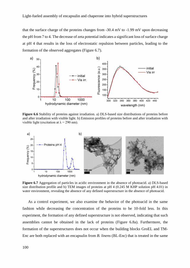
Light-fueled assembly of encapsulin and chaperone into hybrid superstructures
100
that the surface charge of the proteins changes from -30.4 mV to -1.99 mV upon decreasing
the pH from 7 to 4. The decrease of zeta potential indicates a significant loss of surface charge
at pH 4 that results in the loss of electrostatic repulsion between particles, leading to the
formation of the observed aggregates (Figure 6.7).
Figure 6.6 Stability of proteins against irradiation. a) DLS-based size distributions of proteins before
and after irradiation with visible light. b) Emission profiles of proteins before and after irradiation with
visible light (excitation at λ = 290 nm).
Figure 6.7 Aggregation of particles in acidic environment in the absence of photoacid. a) DLS-based
size distribution profile and b) TEM images of proteins at pH 4 (0.245 M KHP solution pH 4.01) in
water environment, revealing the absence of any defined superstructure in the absence of photoacid.
As a control experiment, we also examine the behavior of the photoacid in the same
fashion while decreasing the concentration of the proteins to be 10-fold less. In this
experiment, the formation of any defined superstructure is not observed, indicating that such
assemblies cannot be obtained in the lack of proteins (Figure 6.8a). Furthermore, the
formation of the superstructures does not occur when the building blocks GroEL and TM-
Enc are both replaced with an encapsulin from B. linens (BL-Enc) that is treated in the same

Chapter 6
101
fashion (Figure 6.8b). BL-Enc is structurally comparable with TM-Enc in terms of symmetry
and number of subunits (i.e., 60 subunits), however they are constituted of entirely different
amino acid sequences and appear to differ in terms of stability in the presence of photoacid
(Figure 6.8b).
Figure 6.8 Aggregation of particles in the lack or absence of GroEL/TM-Enc system. TEM images of
a) highly diluted GroEL/TM-Enc system and b) BL-Enc system after irradiation with visible light in
the presence of photoacid, revealing the absence of any defined superstructure.
6.2.2 Structural changes of proteins upon assembly
TEM images indicate that the proteins already undergo structural changes upon the
addition of photoacid, even before any irradiation starts: the spherical, cage-like structure of
TM-Enc is largely retained but seems to be more irregular in shape (Figure 6.2a) in
comparison to the native state in buffer Tris-HCl pH 7.5 (Figure 6.1b). On the other hand,
the circular structure of GroEL in the native state (Figure 6.1b) is disrupted into smaller
fragments (Figure 6.2a). Using circular dichroism (CD) spectroscopy, we attempt to
investigate whether the proteins undergo further conformational changes upon the drop in

Light-fueled assembly of encapsulin and chaperone into hybrid superstructures
102
pH that follows irradiation. The CD spectrum of the proteins-photoacid system at λ = 240–
290 nm is modified significantly upon irradiation (Figure 6.9). This modification is reversible
upon incubation in the dark (Figure 6.9). However, as the photoacid on its own also raises
CD signal in the same range of wavelength, it is difficult to infer the conformation of the
proteins based on CD spectra.
Figure 6.9 CD spectra of protein (Prot), photoacid (MCH) and photoacid-proteins (ProtMCH) before
irradiation, after irradiation (for 30 s) and after subsequent incubation in the dark (for 30 min).
Figure 6.10 TEM images of GroEL/TM-Enc system after heating treatment at 80ºC for 1.5 h to
denature the GroEL fragments. Images are recorded after irradiation with visible light in the presence
of photoacid, revealing the absence of any defined superstructure.
Nevertheless, as the protein component is predominantly composed of GroEL (~95%),
we could anticipate that the behavior of the protein component is governed by the response
of GroEL to the changes in the system. With this in mind, we proceed to denature the GroEL
by heating at 80ºC for 1.5 h (GroEL denatures at 65 – 70ºC),[32,33] whereas TM-Enc is
previously reported to tolerate such treatment.[19] In this case, addition of photoacid and

Chapter 6
103
subsequent irradiation with visible light does not result in the formation of the superstructures
(Figure 6.10). As the thermally treated GroEL fails to result in any superstructure even in the
presence of TM-Enc in the system, we postulate that the GroEL fragments might be the key
component for the assembly.
Figure 6.11 Emission profiles of photoacid-proteins after subtraction by emission profiles of photoacid
only monitored at λmax = 340 nm (excitation at λ = 290 nm) before irradiation, after irradiation (30 s)
and after subsequent incubation in the dark (30 min).
The confined protein substructures in Figure 6.4d are smaller than the structure of cage-
like TM-Enc, hinting at possible conformational changes of TM-Enc upon assembly of the
superstructures. Since GroEL does not have any tryptophan residue (Trp) while TM-Enc
consists of 5 Trp per monomer, we use fluorescence spectroscopy to examine whether the
conformation of TM-Enc remains unchanged upon the assembly. Trp fluorescence intensity
directly represents whether Trp residues are buried in the core of the protein, which indicates
a folded conformation, or exposed to water environment in the case of (partially) unfolded
proteins. Upon excitation at λ = 290 nm, we observe a 23% decrease in the emission at λ =
330 – 350 nm (Figure 6.11) as the response to irradiation with visible light (λ ≥ 420 nm),
which indicates a partial unfolding of TM-Enc that accompanies the assembly into the
superstructures. Subsequent incubation in the dark for 30 min increases the emission at λ =
330 – 350 nm back to its initial state (Figure 6.11), demonstrating the reversibility of TM-
Enc partial unfolding when the superstructures disperse in the dark. The result suggests that
TM-Enc does undergo structural or conformational changes accompanying the assembly of
the superstructures. This could be attributed to the fact that our experimental condition upon

Light-fueled assembly of encapsulin and chaperone into hybrid superstructures
104
irradiation with visible light (i.e., pH 4) is relatively close to the isoelectric pH of TM-Enc
(i.e., pH 4.89), which could result in the reduced stability of the structure.
6.2.3 Emergence of light-dependent order by inclusion of photoacid
in the superstructures
Although both partial unfolding and loss of charge of protein molecules are likely to
induce self-association upon light irradiation, those phenomena fail to explain why the
assembled superstructures adopt a curvature that leads to large circular bodies. It is evident
from the control experiment in the absence of the photoacid (Figure 6.7) that the photoacid
is the origin of the difference in the obtained structures. The chemical characteristics of the
photoacid MCH+ and its photoisomer SP (Figure 6.2b) prompt us to postulate that the
irradiation with visible light triggers two events to occur simultaneously: first, the expected
release of the proton from MCH+ that results in the pH decrease from 5.9 to 4.0; and second,
the significant increase in the hydrophobicity of the system as the closed SP form is much
more hydrophobic compared to the open, protonated MCH+ form. The SP form could be the
part of the hydrophobic tail while the sulfonate group at the end of the alkyl chain could be
the hydrophilic head (Figure 6.2b). Moreover, the photoacid is likely to have interacted with
the protein molecules to form ordered superstructures because on its own it cannot form any
defined superstructure, as evident from control experiments summarized in Figure 6.8.
In the previously described results, the proteins are first added with the photoacid MCH+
(initial pH = 5.9) and then the mixture is irradiated with visible light that results in the
formation of organized superstructures (Figure 6.4). To corroborate the possible interaction
between the photoacid and the protein molecules, we switch the order of light irradiation vs
addition of the components: the photoacid MCH+ is first irradiated with visible light leading
to the formation of SP and decreasing the pH to 4; then, the mixture of proteins is added into
the formed SP. In this control experiment, we observe the formation of aggregates (Figure
6.12) that are comparable to the aggregates shown in Figure 6.7 (i.e., control experiment
using an acid salt instead of the photoacid). Based on this experiment, while the proteins do
aggregate as their natural response to acidic condition (i.e., pH 4), the pre-mixing of proteins
and photoacid seems to be necessary to obtain the ordered structures. This result suggests
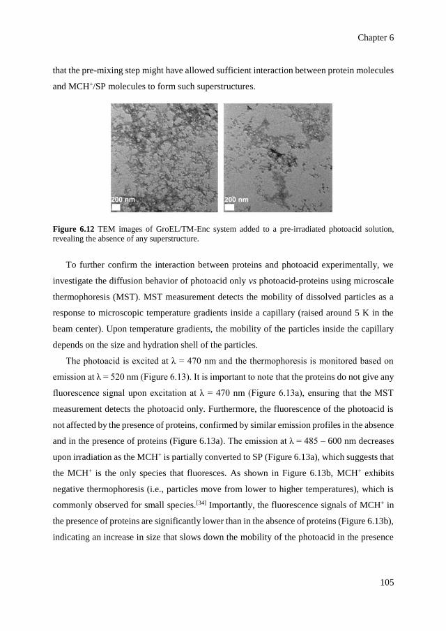
Chapter 6
105
that the pre-mixing step might have allowed sufficient interaction between protein molecules
and MCH+/SP molecules to form such superstructures.
Figure 6.12 TEM images of GroEL/TM-Enc system added to a pre-irradiated photoacid solution,
revealing the absence of any superstructure.
To further confirm the interaction between proteins and photoacid experimentally, we
investigate the diffusion behavior of photoacid only vs photoacid-proteins using microscale
thermophoresis (MST). MST measurement detects the mobility of dissolved particles as a
response to microscopic temperature gradients inside a capillary (raised around 5 K in the
beam center). Upon temperature gradients, the mobility of the particles inside the capillary
depends on the size and hydration shell of the particles.
The photoacid is excited at λ = 470 nm and the thermophoresis is monitored based on
emission at λ = 520 nm (Figure 6.13). It is important to note that the proteins do not give any
fluorescence signal upon excitation at λ = 470 nm (Figure 6.13a), ensuring that the MST
measurement detects the photoacid only. Furthermore, the fluorescence of the photoacid is
not affected by the presence of proteins, confirmed by similar emission profiles in the absence
and in the presence of proteins (Figure 6.13a). The emission at λ = 485 – 600 nm decreases
upon irradiation as the MCH+ is partially converted to SP (Figure 6.13a), which suggests that
the MCH+ is the only species that fluoresces. As shown in Figure 6.13b, MCH+ exhibits
negative thermophoresis (i.e., particles move from lower to higher temperatures), which is
commonly observed for small species.[34] Importantly, the fluorescence signals of MCH+ in
the presence of proteins are significantly lower than in the absence of proteins (Figure 6.13b),
indicating an increase in size that slows down the mobility of the photoacid in the presence
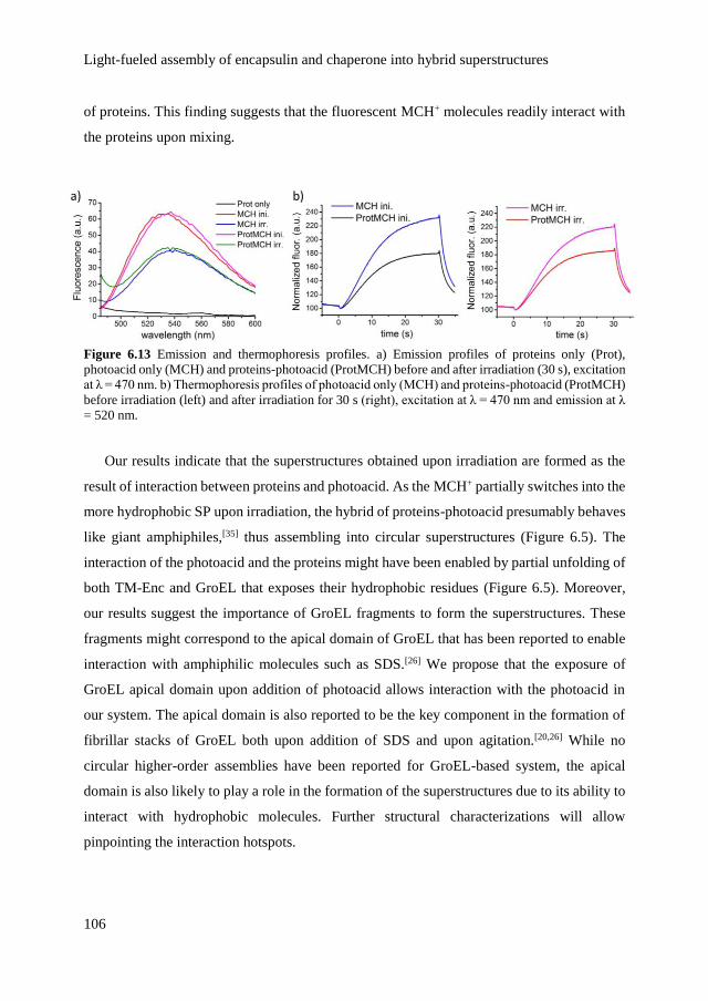
Light-fueled assembly of encapsulin and chaperone into hybrid superstructures
106
of proteins. This finding suggests that the fluorescent MCH+ molecules readily interact with
the proteins upon mixing.
Figure 6.13 Emission and thermophoresis profiles. a) Emission profiles of proteins only (Prot),
photoacid only (MCH) and proteins-photoacid (ProtMCH) before and after irradiation (30 s), excitation
at λ = 470 nm. b) Thermophoresis profiles of photoacid only (MCH) and proteins-photoacid (ProtMCH)
before irradiation (left) and after irradiation for 30 s (right), excitation at λ = 470 nm and emission at λ
= 520 nm.
Our results indicate that the superstructures obtained upon irradiation are formed as the
result of interaction between proteins and photoacid. As the MCH+ partially switches into the
more hydrophobic SP upon irradiation, the hybrid of proteins-photoacid presumably behaves
like giant amphiphiles,[35] thus assembling into circular superstructures (Figure 6.5). The
interaction of the photoacid and the proteins might have been enabled by partial unfolding of
both TM-Enc and GroEL that exposes their hydrophobic residues (Figure 6.5). Moreover,
our results suggest the importance of GroEL fragments to form the superstructures. These
fragments might correspond to the apical domain of GroEL that has been reported to enable
interaction with amphiphilic molecules such as SDS.[26] We propose that the exposure of
GroEL apical domain upon addition of photoacid allows interaction with the photoacid in
our system. The apical domain is also reported to be the key component in the formation of
fibrillar stacks of GroEL both upon addition of SDS and upon agitation.[20,26] While no
circular higher-order assemblies have been reported for GroEL-based system, the apical
domain is also likely to play a role in the formation of the superstructures due to its ability to
interact with hydrophobic molecules. Further structural characterizations will allow
pinpointing the interaction hotspots.

Chapter 6
107
The obtained assemblies that confine smaller substructures are arguably comparable to
the naturally occurring casein micelles.[36] Structural similarities can be drawn for the
building blocks as casein also possesses hydrophobic residues and flexible conformations
like GroEL chaperone. Previous studies have even reported the ability of casein to function
as a molecular chaperone.[36] Both proteins also have the tendency to form fibrillar structures,
although naturally casein prefers to form amorphous circular bodies (around 300 nm in
diameter) confining high density nanoclusters of calcium phosphate along with channels and
water-filled cavities.[36-38]
6.3 Conclusions
We have demonstrated a dynamic order upon fueling hybrid bio-synthetic system with
light. The system consists of T. maritima encapsulin and GroEL chaperone that are partially
unfolded to accommodate the assembly process. The dynamic assembly into organized
superstructures is enabled by a spiropyran-based photoacid that undergoes reversible
deprotonation and structural change upon irradiation. Moreover, the photoacid molecules are
likely to have interacted with the proteins to form such structures. Hence, the photo-switching
of the photoacid plays a role beyond pH control by increasing the hydrophobicity of the
system. In view of GroEL functioning as a chaperone and TM-Enc as a natural
nanoconfinement system, the presented assemblies hold promise as a transient vessel to
entrap, accumulate and store functional materials, which could be released and dispersed on-
command. The on-command release feature is enabled in our system owing to the
spontaneous disassembly of superstructures in the absence of energy source.
6.4 Acknowledgements
We thank Federico Lancia and Dr. Tibor Kudernac for fruitful brainstorming and
discussions. We acknowledge Dr. Enrico G. Keim for assistance with TEM imaging and Dr.
Joost Snijder and Prof. Albert Heck (Utrecht University) for assistance with mass
spectrometry analysis and Agustin Enciso Martinez and Dr. Cees Otto for assistance with
SEM analysis.

Light-fueled assembly of encapsulin and chaperone into hybrid superstructures
108
6.5 Materials and methods
6.5.1 General
TM-Enc and GroEL were recombinantly co-expressed in E. coli and co-purified
according to the procedure in literature.[21,22] As a control system, BL-Enc was separately
expressed in E. coli and purified according to similar procedure. The proteins were stored in
Tris-HCl buffer pH 7.5 (containing 150 mM NH4Cl) at 4 ºC and dialyzed against MilliQ
water (12 – 14 kDa dialysis membrane, Spectra/Por) at room temperature prior to all
experiments. The spiropyran-based photoacid MCH+ was purchased from Syncom
(Groningen, The Netherlands) and then dissolved and stored in ethanol at room temperature.
All experiments were performed in MilliQ (containing 150 mM NaCl and 2% ethanol). All
chemicals were purchased from Sigma-Aldrich unless stated otherwise. All experiments
were performed at room temperature unless stated otherwise.
6.5.2 Reversible formation of hybrid superstructures
500 μL of a sample solution containing proteins (4.3 μM) and photoacid (0.17 mM) in
MilliQ (containing 150 mM NaCl and 2% ethanol) was prepared as the initial condition (prior
to irradiation). Afterwards, the mixture was irradiated with visible light (λ ≥ 420 nm, Edmund
MI-150 High-intensity Illuminator, 2 mW/cm2) for 30 s. The absorption spectra were
recorded with a Perkin Elmer Lambda 850 UV-visible spectrometer and the photo-switching
was monitored at λmax = 421 nm. For the reversion, the mixture after irradiation was incubated
in the dark for 30 – 60 min and the UV-visible spectrum was recorded following the
incubation. For control experiments, solutions containing photoacid only and proteins only
were separately prepared and treated in the same fashion. Due to the absence of photoacid,
the pH of the solution containing proteins only was decreased using a commercial acid salt,
potassium hydrogen phthalate (0.245 M KHP solution pH 4.01, Certipur®). The pH of all
system was measured using a Sartorius™ pH meter (Thermo Fisher Scientific). Zeta potential
of proteins was measured using a Zetasizer Nano (Malvern).
Chapter 6
109
6.5.3 Characterization of hybrid superstructures
The assembled superstructures were characterized using dynamic light scattering (DLS),
negative-stained transmission electron microscopy (TEM) and scanning electron microscopy
(SEM). For DLS analyses, 500 μL of a proteins-photoacid sample was prepared and
measured using a Nanotrac Wave, Microtrac particle analyzer. For control experiments, a
solution containing only the proteins was prepared and treated in the same fashion. For
electron microscopy analyses, samples (5 μL) were applied onto Formvar‐carbon coated
grids. After 30 s, the excess of liquid was drained. Uranyl acetate (5 μL, 1% w/v) was added
for negative staining and the excess of liquid was drained after 1 min and air-dried for 1 h at
room temperature. For TEM and SEM respectively, the samples were examined using FEG-
TEM (Phillips CM 30) and JSM-6610LV-SEM (JEOL). For SEM analysis, the grid was
placed on top of a stainless steel substrate (µRIM) prior to measurement.
6.5.4 Characterization of structural changes of proteins
The structure of proteins was analyzed based on examination of negative-stained TEM
images, circular dichroism (CD) spectra and fluorescence spectra. The CD spectra were
recorded for 500 μL of sample using a Jasco J-1500 circular dichroism spectrometer at λ =
190 – 350 nm. The emission spectra of 500 μL of sample at λ = 300 – 440 nm (excitation at
λ = 290 nm) were recorded using a Perkin Elmer fluorescence spectrometer. For control
experiments, solutions containing photoacid only and proteins only were separately prepared
and treated in the same fashion.
6.5.5 Analysis of photoacid inclusion in the superstructures
The interaction between photoacid and proteins was inferred based on fluorescence
spectroscopy and microscale thermophoresis (MST) analyses. The emission spectra of 500
μL of sample at λ = 485 – 600 nm (excitation at λ = 470 nm) were recorded using a Perkin
Elmer fluorescence spectrometer. For control experiments, solutions containing photoacid
only and proteins only were separately prepared and treated in the same fashion. For MST
analyses, around 3 – 5 μL of each sample was loaded into a hydrophilic glass capillary and
the fluorescence-based thermophoresis profiles for all samples were recorded using a

Light-fueled assembly of encapsulin and chaperone into hybrid superstructures
110
Monolith NT.115 Blue/Green Nanotemper (excitation at λ = 470 nm, emission at λ = 520
nm). The microscopic gradient of temperature inside the capillary was generated by a built-
in IR-laser module (λ = 1475 nm at 120 mW laser power). For all measurements, the IR laser
was switched on at t = 5 and switched off at t = 30 s, corresponding to Figure 6.13b. All
measurements were performed at room temperature of approximately 20°C.
6.6 References
[1] C. Conde, A. Caceres, Nat. Rev. Neurosci. 2009, 10, 319-332.
[2] H. Hess, J. L. Ross, Chem. Soc. Rev. 2017.
[3] B. A. Grzybowski, W. T. Huck, Nat. Nanotechnol. 2016, 11, 585-592.
[4] M. Uchida, B. LaFrance, C. C. Broomell, P. E. Prevelige, Jr., T. Douglas, Small 2015,
11, 1562-1570.
[5] M. A. Kostiainen, O. Kasyutich, J. J. L. M. Cornelissen, R. J. M. Nolte, Nat. Chem. 2010,
2, 394-399.
[6] C. C. Broomell, H. Birkedal, C. L. P. Oliveira, J. S. Pedersen, J. A. Gertenbach, M.
Young, T. Douglas, Soft Matter 2010, 6, 3167-3171.
[7] R. Klajn, K. J. Bishop, B. A. Grzybowski, Proc. Natl. Acad. Sci. USA 2007, 104, 10305-
10309.
[8] G. Ragazzon, M. Baroncini, S. Silvi, M. Venturi, A. Credi, Nat. Nanotechnol. 2015, 10,
70-75.
[9] J. Boekhoven, W. E. Hendriksen, G. J. M. Koper, R. Eelkema, J. H. van Esch, Science
2015, 349, 1075-1079.
[10] R. M. Putri, J. J. L. M. Cornelissen, M. S. T. Koay, ChemPhysChem 2015, 16, 911-918.
[11] Z. Shi, P. Peng, D. Strohecker, Y. Liao, J. Am. Chem. Soc. 2011, 133, 14699-14703.
[12] L. Florea, K. Wagner, P. Wagner, G. G. Wallace, F. Benito-Lopez, D. L. Officer, D.
Diamond, Adv. Mater. 2014, 26, 7339-7345.
[13] P. K. Kundu, D. Samanta, R. Leizrowice, B. Margulis, H. Zhao, M. Borner, T.
Udayabhaskararao, D. Manna, R. Klajn, Nat. Chem. 2015, 7, 646-652.
[14] C. Fu, J. Xu, C. Boyer, Chem. Commun. 2016, 52, 7126-7129.
[15] C. Maity, W. E. Hendriksen, J. H. van Esch, R. Eelkema, Angew. Chem. Int. Ed. 2015,
54, 998-1001.
Chapter 6
111
[16] T. Satoh, K. Sumaru, T. Takagi, T. Kanamori, Soft Matter 2011, 7, 8030-8034.
[17] Y. Luo, C. M. Wang, P. Peng, M. Hossain, T. L. Jiang, W. Fu, Y. Liao, M. Su, J. Mater.
Chem. B 2013, 1, 997-1001.
[18] O. S. Shafaat, J. R. Winkler, H. B. Gray, D. A. Dougherty, ChemBioChem 2016, 17,
1323-1327.
[19] C. Cassidy-Amstutz, L. Oltrogge, C. C. Going, A. Lee, P. Teng, D. Quintanilla, A. East-
Seletsky, E. R. Williams, D. F. Savage, Biochemistry 2016, 55, 3461-3468.
[20] J. Chen, H. Yagi, P. Sormanni, M. Vendruscolo, K. Makabe, T. Nakamura, Y. Goto, K.
Kuwajima, FEBS Lett. 2012, 586, 1120-1127.
[21] M. Sutter, D. Boehringer, S. Gutmann, S. Gunther, D. Prangishvili, M. J. Loessner, K.
O. Stetter, E. Weber-Ban, N. Ban, Nat. Struct. Mol. Biol. 2008, 15, 939-947.
[22] R. M. Putri, J. W. Fredy, J. J. L. M. Cornelissen, M. S. T. Koay, N. Katsonis,
ChemPhysChem 2016, 17, 1815-1818.
[23] A. L. Horwich, G. W. Farr, W. A. Fenton, Chem. Rev. 2006, 106, 1917-1930.
[24] M. Mayhew, A. C. da Silva, J. Martin, H. Erdjument-Bromage, P. Tempst, F. U. Hartl,
Nature 1996, 379, 420-426.
[25] M. Hayer-Hartl, A. Bracher, F. U. Hartl, Trends Biochem. Sci. 2016, 41, 62-76.
[26] J. Chen, H. Yagi, Y. Furutani, T. Nakamura, A. Inaguma, H. Guo, Y. Kong, Y. Goto,
Sci. Rep. 2014, 4, 5614.
[27] S. Biswas, K. Kinbara, T. Niwa, H. Taguchi, N. Ishii, S. Watanabe, K. Miyata, K.
Kataoka, T. Aida, Nat. Chem. 2013, 5, 613-620.
[28] T. Sendai, S. Biswas, T. Aida, J. Am. Chem. Soc. 2013, 135, 11509-11512.
[29] S. Biswas, K. Kinbara, N. Oya, N. Ishii, H. Taguchi, T. Aida, J. Am. Chem. Soc. 2009,
131, 7556-7557.
[30] G. Basanez, M. B. Ruiz-Arguello, A. Alonso, F. M. Goni, G. Karlsson, K. Edwards,
Biophys. J. 1997, 72, 2630-2637.
[31] S. K. Molugu, J. Li, R. A. Bernal, J. Chromatogr. B 2015, 1007, 93-99.
[32] J. Martin, A. L. Horwich, F. U. Hartl, Science 1992, 258, 995-998.
[33] B. Sot, S. Banuelos, J. M. Valpuesta, A. Muga, J. Biol. Chem. 2003, 278, 32083-32090.
[34] B. Gotsmann, U. Durig, Appl. Phys. Lett. 2005, 87.
[35] Q. Luo, C. Hou, Y. Bai, R. Wang, J. Liu, Chem. Rev. 2016, 116, 13571-13632.

Light-fueled assembly of encapsulin and chaperone into hybrid superstructures
112
[36] C. Holt, J. A. Carver, H. Ecroyd, D. C. Thorn, J. Dairy Sci. 2013, 96, 6127-6146.
[37] R. Trejo, T. Dokland, J. Jurat-Fuentes, F. Harte, J. Dairy Sci. 2011, 94, 5770-5775.
[38] D. G. Dalgleish, P. A. Spagnuolo, H. D. Goff, Int. Dairy J. 2004, 14, 1025-1031.
Summary
Living materials are based on proteins that adapt and change in structure and function
continuously when in use; cellular microtubules, ATP synthases and ribosomes are but a few
examples. Breathing life into semi-synthetic materials would allow improved understanding
over protein regulation and dynamics, and how their integration into complex systems can
lead to emergent functions across length scales. The focus of my research is to achieve optical
control over functional proteins by connecting them to artificial molecular switches, with the
aim to amplify molecular switching events across length scales and gain understanding over
cooperative and systematic regulations of proteins. We choose light because it offers
spatiotemporal selectivity, is compatible with a wide range of phases and relatively non-
destructive towards protein systems. Interfering with mechanisms such as allosteric
communication or hierarchical self-assembly not only paves the way towards an improved
understanding of cooperative or collective effects in living matter, but it is also associated
with the generation of new classes of smart bio-hybrids. The works presented in this thesis
involve diverse functional proteins, ranging from an allosteric transport protein, the human
serum albumin (Chapter 3), to proteins forming cages (Chapter 4, 5, 6) all the way to
chaperone proteins that are known to assist cellular protein folding (Chapter 6).
The work focuses on the use of spiropyran derivatives as photo-switches, because their
irradiation induces not only a change in conformation, but also it yields a zwitterionic form
that is bound to influence a protein environment. Chapter 3 and 5 demonstrate covalent
incorporation of spiropyran switches into a cysteine and lysines that are present in the studied
proteins. The covalent coupling involves reacting maleimide or succinimide moieties
(attached to the photo-switchable units) with the proteins via thiol-Michael addition or
amine-NHS ester reaction, respectively. Chapter 3 describes how such structural
modification allows photo-controlling the natural allosteric regulation of the human serum
albumin (HSA), and thus to modulate ligand binding by irradiation with light. On the other
hand, modification of a bacterial encapsulin, a self-assembled protein cage that confines
Summary
114
smaller proteins in its cavity (Chapter 4 and 5), yields local modifications only, and does not
compromise the structural integrity and the behavior of the entire assembly.
Chapter 4 and 5 further confirm and demonstrate that bacterial encapsulins are robust,
catalytically active and biocompatible. They can also be labeled with light-switchable
fluorophores on their lysines via reaction with succinimide-bearing spiropyran switches, and
thus they are good candidates as smart nanoplatforms for in vitro and in vivo research
purposes.
Chapter 6 demonstrates the light-fueled assembly of proteins by adding photo-switches
into a medium containing the encapsulin and GroEL chaperone. This strategy enables
addition of a large amount of photo-switches, which, in this context, results in the changes
of the medium upon irradiation, i.e., pH and hydrophobicity. Mediated by a spiroyran-based
photoacid, the described approach leads to an assembly of transient superstructures that
depend on light irradiation to prevent disassembly. This assembly thus presents a dynamic
and reversible order of a protein system that is sustained by a continuous input of energy,
and this mechanism is reminiscent of the GTP-fueled assembly of cellular microtubules.
Overall, we have developed photo-responsive protein-based hybrids in which dynamic
regulations such as allostery and self-assembly are prominent to protein functionality. Via
incorporation of photo-switches, optical control has been demonstrated across length scales,
from a monomeric structure to highly defined multimeric architectures made of proteins. The
presented research provides a means to interfere with dynamic regulations of proteins and
supports strategies towards the development of biocompatible and smart materials.

Samenvatting
Levende materialen zijn gebaseerd op eiwitten die zich continu aanpassen en van
structuur veranderen wanneer ze actief zijn; cellulaire microtubuli, ATP synthases en
ribosomen zijn slechts enkele voorbeelden. Het door mensen gemaakte materialen levend
maken kan zorgen voor een verbeterd begrip van eiwitregulatie en -dynamiek en meer kennis
over hoe de integratie van complexe systemen kan leiden tot nieuwe functies over lengte
schalen. De focus van mijn onderzoek is om optische controle te verkrijgen over functionele
eiwitten door ze te verbinden met artificiële moleculaire schakelaars. Het doel is om
moleculaire schakelingen te versterken over lengte schalen en meer kennis te vergaren over
de coöperatieve en systemische regulering van eiwitten. We hebben gekozen voor licht
omdat het spatio-temporele selectiviteit biedt, compatibel is met een groot scala aan fases en
het relatief non-destructief is met betrekking tot eiwitten. Het interfereren met mechanismen
zoals allosterische communicatie of hiërarchische zelfassemblage zet de deur open voor een
verbeterd begrip van coöperatieve of collectieve effecten in levende materialen, maar het is
ook geassocieerd met de vorming van nieuwe klassen van slimme bio-hybriden. Het werk
dat in deze thesis wordt gepresenteerd betreft diverse functionele eiwitten, reikend van
allosterische transport eiwitten, albumine van humaan serum (Hoofdstuk 3), via eiwitten die
kooien vormen (Hoofdstuk 4, 5, 6), tot chaperonne-eiwitten die bekend staan te assisteren in
cellulaire eiwit vouwing (Hoofdstuk 6).
Dit werk focust op het gebruik van spiropyran derivaten als foto-schakelaars, omdat de
bestraling ervan niet alleen een verandering in conformatie induceert, maar ook omdat het
een zwitterion oplevert dat de eiwit omgeving zal beïnvloeden. Hoofdstuk 3 en 5
demonsteren covalente incorporatie van spiropyran schakelaars met een cysteïne en lysines
die aanwezig zijn in de bestudeerde eiwitten. De covalente koppeling werkt door maleimide
of succinimide groepen (die verbonden zijn met de foto-schakelaars) te laten reageren met
eiwitten via thiol-Michael additie of een amine-NHS ester reactie, respectievelijk.
Hoofdstuk 3 beschrijft hoe dergelijke structurele modificaties foto-controle van de
natuurlijke allosterische regulering van albumine uit humaan serum (HSA) toestaan en

Samenvatting
116
zodoende ligand binding moduleren via bestraling met licht. Anderzijds, modificatie van
bacteriële encapsulines, zelfassemblerende eiwit kooien die een kleiner eiwit inkapselen
(Hoofdstuk 4 en 5), resulteert slechts in lokale modificaties en heeft geen invloed op de
structurele integriteit en het gedrag van het gehele construct.
Hoofdstuk 4 en 5 bevestigen en demonstreren verder dat bacteriële encapsulines robuust,
katalytisch actief en biocompatibel zijn. Ze kunnen ook gelabeld worden met licht-
geschakelde fluoroforen die zijn gebonden aan lysines door middel van een reactie met
spiropyran schakelaars die een succinimide-groep hebben. Zodoende zijn het geschikte
kandidaten om te dienen als slimme nanoplatformen voor in vitro en in vivo onderzoek.
Hoofdstuk 6 demonstreert de licht-gedreven assemblage van eiwitten via foto-
schakelaars in een medium dat encapsulines en de chaperonne GroEL bevat. Deze strategie
laat de toevoeging van grote hoeveelheden foto-schakelaar toe, welke, in deze context,
resulteert in veranderingen in het medium na bestraling, zoals veranderingen in pH en
hydrofobiciteit. Terwijl het gemedieerd wordt door een spiropyran-gebaseerde fotozuur, leidt
de beschreven methode tot een assemblage van vesikel-achtige superconstructen die
afhankelijk zijn van bestraling met licht om te voorkomen dat ze uiteen vallen. Deze vorm
van assemblage presenteert een dynamische en reversibele orde van een eiwit systeem dat
wordt onderhouden door een continue invoer van energie, en dit mechanisme doet denken
aan de GTP-gevoede assemblage van cellulaire microtubuli.
Concluderend hebben we licht-responsieve eiwit-gebaseerde hybriden gemaakt waarin
dynamische regulatie zoals allosterie en autoassemblage prominent zijn over eiwit
functionaliteit. Door middel van de incorporatie van foto-schakelaars, optische controle is
aangetoond over lengte schalen, van een monomerische structuur tot zeer gedefinieerde
multimerische structuren die bestaan uit eiwitten. Het hier gepresenteerde onderzoek geeft
een manier om te interfereren met de dynamische regulatie van eiwitten en het ondersteunt
strategieën voor de ontwikkeling van biocompatibele en slimme materialen.

Acknowledgements
With the completion of this dissertation, I would like to take the opportunity to thank
everyone who has supported me through my PhD study and my stay in The Netherlands.
First of all, I am grateful to my supervisors, Jeroen and Nathalie, for welcoming me into
their research groups, providing guidance and facilities to conduct scientific works and
develop my skills and giving me the opportunities to join a lot of international conferences.
Jeroen, I was a Master student in your group first and afterwards a PhD student, it was a great
pleasure to have been a part of the BNT group in these past five years. Looking back, I am
grateful that all this time I have been given immense opportunity and research experiences
that have undoubtedly shaped my scientific life and helped me become a better researcher
and scientist. Nathalie, thank you for introducing me to the fascinating world of molecular
switches, it was a new field to me and I am happy to have learned a lot in the past years and
to have conducted interesting projects on the hybrids of proteins and photo-switches. I greatly
appreciate that you welcomed me into your research group and gave me a lot of opportunities,
guidance and support during my study. I would also like to express my sincere gratitude to
Melissa for your guidance, support and advice. It was a great pleasure to have worked with
you and thank you for participating in my graduation committee. Furthermore, I would like
to sincerely thank all my committee members: Prof. Andrew Woolley from the University
of Toronto, Prof. Pepe Castón from CSIC Madrid, Prof. Mireille Claessens, Prof.
Jurriaan Huskens and Prof. Herman ten Kate from the University of Twente for reading
my manuscript and the opportunity of having my PhD defense under your consideration.
I would like to express my gratitude to the staff of MnF/BNT cluster: Tibor, Jurriaan,
Pascal, Jos and Wim for scientific advice, suggestions and discussions during the
colloquium and meetings. Tibor, thank you for your suggestions and fruitful discussion
regarding the final chapter of my thesis. I would like to thank Nicole and Izabel for all your
kind help regarding the many arrangements of schedules, conferences and especially
regarding my scholarship and administration as an international student that might have been
complicated at times. Marcel, Regine, Richard and Bianca, thank you for all your kind help
and support in the labs and the nice chats that we had.
Acknowledgements
118
Furthermore, I am very grateful to my scholarship provider, Indonesia Endowment
Fund of Education or Lembaga Pengelola Dana Pendidikan (LPDP) who has supported my
PhD study throughout these four years and to Prof. Zeily Nurachman from Institut
Teknologi Bandung (ITB), Indonesia for your recommendation and scientific training that I
received while performing my Bachelor and Master study at ITB under your supervision.
The accomplishment of this dissertation would have not been possible without the fruitful
collaborations with the research group of Prof. Pepe Castón from CSIC Madrid,
Habiburrahman and Prof. Claudia Filippi from MESA+ University of Twente, Agustin
and Dr. Cees Otto from MIRA University of Twente. Thank you and it was an honor to have
the opportunity to collaborate with your teams. Furthermore, I would like to thank fellow
students and postdocs in the BNT/MnF cluster who have worked together and collaborated
with me on my projects: Wilfried, Federico, Robin, Aijie, Christoph, Mark de R., Alberto
and my Master student, Katerina. I am very thankful for your help, suggestions and
discussion and I wish you all the best with your study and future plans. Robin, thank you for
your help with my Samenvatting. I would like to thank Dr. Rico Keim for your help with
TEM measurements and the nice chats about Indonesia as your family used to live in my
hometown.
Aijie and Wilfried, thank you for being my paranymphs and for all your support during
my PhD, thank you for not only being scientifically involved in my projects, but more so, for
being such good friends to me. Aijie, we started about the same time and I am happy that we
could finish our study also about the same time. In these past four years, suffice to say that
we have been through a lot together and that we have helped each other out throughout. I
sincerely wish you all the best towards the completion of your PhD and for your future plans.
Wilfried, thank you for always helping me out and cheering me up, you really are a funny
and supportive friend, in addition to a smart scientist. I wish you all the best with your future
plans and I am sure you will succeed (I see it..!) I would also like to sincerely thank Meow
(Supa), Xiu, Yao, Jenny (Shuqin), Liulin, He and Wang Yi for your help, suggestions,
funny and nice chats, laughter and jokes, and of course nice foods! I cherish the moments we
shared together in Enschede (and beyond!) and I sincerely hope that we could meet again

119
someday, perhaps in a different part of the world I wish you all the best with your study
and your future plans. Yao, special thanks to you for giving me a Chinese name!
Melanie, you were the first person that I met from the group and I am grateful for all the
time we worked together and your supervision during my Master study. Supitch, thank you
for being such a good friend and for your help and advice during my stay in Enschede.
Rianne and Stan, thank you for your many suggestions and feedback during colloquium and
meetings, I really appreciate it, and also thank you for the nice chats outside the labs.
Gülistan, I admire your hard work, thank you for the nice chats that we had and I wish you
all the best for your study and research. Wouter, we went to the Global Young Scientists
Summit in Singapore together and it was a nice experience, thank you for the nice chats that
we had and I wish you all the best for your study. I would like to thank all the members and
former members of BNT/MnF cluster who have made my stay in Twente really enjoyable
and for your help in so many different ways: Raquel, Emanuela, Sarah, Rik, Alexis, Ben,
Tushar, Shirish, Jenny, Bettina, Janneke, Jasper, Milou, Roberto, Alejandro, Laura,
Pichayut, Fern, Anne-Déborah, Alexander R., Mark V., Maike, Almudena, Daniele,
Jacopo, Sara, Andrea, Erik, Alexander M., Nico, Gaurav, Pramod, Nicole, Pia, Ines,
Sander, Dhanya, Salmeen and fellow students in the BNT/MnF cluster.
Being far away from home can be difficult at times, but the Indonesian community in
Enschede has truly provided me a second home. I am very grateful to my Indonesian friends
who have supported me throughout, as it would have been difficult had I gone through this
journey alone. I have met a lot of people from Indonesia in these past five years in Enschede,
I forever cherish the moment we shared together and I am thankful to have made new friends
and family half the world away from home. For those who are still in the middle of their
study, I sincerely wish you all the very best towards the finish line and remember to never
give up.
Finally and especially, I would like to thank my beloved family and friends, without
whom I would not have made it this far. I am sincerely grateful for your constant support and
never-ending encouragement. Mami, Papi, Pranata dan Tiwi, alhamdulillahirabbil’alamin,
akhirnya selesai sudah perjuangan selama lima tahun ini, terimakasih atas dukungan,

Acknowledgements
120
semangat dan do’a yang mengalir tak henti-hentinya. Semoga ilmu ini kiranya berkah dan
kita semua selalu di dalam rahmat dan lindungan Allah SWT.
Enschede, September 2017
Rindia Maharani Putri
About the author
Rindia Maharani Putri was born on May 28th 1991 in Padang, Indonesia. She pursued her
bachelor degree in Chemistry at Institut Teknologi Bandung, Indonesia, and graduated in
July 2011. During her bachelor project, she studied the isolation and characterization of
lipase under the supervision of Prof. Zeily Nurachman. Afterwards, in January and April
2013, she received her master degrees in Chemical Engineering at the University of Twente
and in Chemistry at Institut Teknologi Bandung, respectively. Her master research, entitled
“Synergistic Biocatalysts inside Cowpea Chlorotic Mottle Virus-based Nanoreactors” was
carried out in the Biomolecular Nanotechnology group, University of Twente under the
supervision of Prof. Jeroen Cornelissen, Dr. Melissa Koay and Dr. Melanie Brasch.
Since September 2013 she started her PhD program in the Biomolecular Nanotechnology
group under the supervision of Prof. Jeroen Cornelissen, Prof. Nathalie Katsonis and Dr.
Melissa Koay. The aim of her project is to engineer proteins into dynamic building blocks
for light-responsive hybrid (semi-synthetic) systems. The results of this project are described
in this thesis.
122
List of publications
1. Rindia M. Putri, Jeroen J.L.M. Cornelissen and Melissa S.T. Koay. Self-assembled
Cage-like Protein Structures, ChemPhysChem 2015, 16, 911-918.
2. Rindia M. Putri, Jean Wilfried Fredy, Jeroen J.L.M. Cornelissen, Melissa S.T.
Koay and Nathalie Katsonis. Labelling Bacterial Nanocages with Photo-switchable
Fluorophores, ChemPhysChem 2016, 17, 1815-1818.
3. Liulin Yang, Aijie Liu, Shuqin Cao, Rindia M. Putri, Pascal Jonkheijm, Jeroen
J.L.M. Cornelissen. Self-assembly of Proteins: Towards Supramolecular Materials,
Chem. Eur. J. 2016, 22, 15570–15582.
4. Rindia M. Putri, Mark V. de Ruiter, Jeroen J.L.M. Cornelissen. CCMV-like
Particles as Enzymatic Nanoreactors, in press. "Virus-derived Nanoparticles for
Advanced Technologies", in the lab protocol series 'Methods in Molecular Biology',
G. Lomonossoff and C. Wege, Eds.; Humana Press: Springer Science + Business
Media New York, USA.
5. Melanie Brasch, Rindia M. Putri, Mark V. de Ruiter, Daniel Luque, Melissa S.T.
Koay, José R. Castón and Jeroen J.L.M. Cornelissen. Assembling Enzymatic
Cascade Pathways Inside Virus-based Nanocompartments Using Dual-tasking
Nucleic Acid, J. Am. Chem. Soc. 2017, 139, 1512-1519.
Publications in preparation
6. Rindia M. Putri, Daniel Luque, Robin Klem, Aikaterini A. Rousou, Aijie Liu,
Christoph H.-H. Traulsen, W. Frederik Rurup, Melissa S.T. Koay, José R. Castón
and Jeroen J.L.M. Cornelissen. Structural Characterization of Native and Modified
Encapsulins as Nanoplatforms for Catalysis and Cellular Uptake, submitted to ACS
Nano.
7. Rindia M. Putri, Federico Lancia, Agustin Enciso Martinez, Cees Otto, Melissa
S.T. Koay, Tibor Kudernac, Jeroen J.L.M. Cornelissen and Nathalie Katsonis.
123
Opportunistic Behavior of Artificial Switches in Protein Environment, in
preparation.
8. Rindia M. Putri, Habiburrahman, Jean Wilfried Fredy, Pichayut Tananchayakul,
Jeroen J.L.M. Cornelissen, Melissa S.T. Koay, Claudia Filippi and Nathalie
Katsonis. Light-programmed Allostery in an Engineered Human Serum Albumin
and Mechanistic Insight from Molecular Dynamics, in preparation.
9. Rindia M. Putri, Jeroen J.L.M. Cornelissen and Nathalie Katsonis. Strategies to
Modulate Allosteric Regulation and Self-assembly of Proteins using Light, in
preparation.
10. Aijie Liu, Wei Zhu, Mark V. de Ruiter, Stan J. Maassen, Rindia M. Putri, Liulin
Yang, C. Jeffrey Brinker and Jeroen J.L.M. Cornelissen. Interfacial Crosslinking of
Viral Protein Cage-inspired Free-standing Thin Films with Tunable Functionalities,
in preparation.