Comparison of backbone dynamics of monomeric and domain-swapped stefin A
Upload
independentCategory
view
2download
0
Pathogenic role of (S)IgA in IgA
nephropathy
Beatrijs D. Oortwijn


Pathogenic role of (S)IgA in IgA
nephropathy
Proefschrift
ter verkrijging van
de graad van Doctor aan de Universiteit Leiden,
op gezag van de Rector Magnificus Dr. D.D. Breimer,
hoogleraar in de faculteit der Wiskunde en
Natuurwetenschappen en die der Geneeskunde,
volgens besluit van het College voor Promoties
te verdedigen op woensdag 17 januari 2007
klokke 15.00 uur
door
Beatrijs Dorinda Oortwijn
geboren te Purmerend in 1979

PROMOTIECOMMISSIE
Promoter Prof. Dr. M.R. Daha
Copromoter Dr. C. van Kooten
Referent Prof. Dr. J.J. Weening (Academisch Medisch Centrum, Amsterdam)
Overige Leden Prof. Dr. A.M. DeelderDr. M. van Egmond (Vrije Universiteit Medisch Centrum, Amsterdam)Dr. A. GorterProf. Dr. P.S. HiemstraProf. Dr. G.J. Navis (Universitair Medisch Centrum Groningen, Groningen)
The research described in the present thesis was performed at the Department ofNephrology of the Leiden Unversity Medical Centre and was financed by a grant from theDutch Kidney foundation (C99.1822)Financial support for printing of this thesis from 3A-out foundation, the Dutch KidneyFoundation and the Jurriaanse stichting is gratefully acknowledged
Used by permission: Chapter 2 - © 2007 Elsevier, Chapter 4& 5 - © 2006 LippincottWilliams & Wilkins, Chapter 6 - © 2006 Nature Publishing Group
ISBN: 90-9021342-2, 978-90-9021342-2
Coverdesign: Beatrijs D. Oortwijn Origin of the picture: Maria Pia Rastaldi
Printed by: Gildeprint Drukkerijen B.V., Enschede, The Netherlands.
© 2007 Beatrijs D. Oortwijn

In the middle of every difficulty lies opportunity'
-Albert Einstein


CONTENTS
Chapter 1 General Introduction 9
Chapter 2: Monomeric and polymeric IgA show a similar association 27with the myeloid FcαRI/CD89. Mol. Immunol. 44:966-973, 2007
Chapter 3: Comparable binding of monomeric and polymeric IgA 41to the novel IgA Fc receptor, Fcα/µR.
Chapter 4: Glomerular activation of the lectin pathway of complement in 55IgA nephropathy is associated with more severe renal diseaseJ. Am. Soc. Nephrol. Jun;17(6):1724-1734, 2006
Chapter 5: Differential glycosylation of polymeric and monomeric IgA: 73a possible role in glomerular inflammation in IgA nephropathyJ. Am. Soc. Nephrol. in press
Chapter 6: A pathogenic role for secretory IgA in IgA nephropathy 93Kidney Int. 69:1131-1138, 2006
Chapter 7: Demonstration of secretory IgA in kidneys of patients with 109IgA nephropathySubmitted
Chapter 8: General Discussion and Summary 117
Samenvatting 131Dankwoord 135Curriculum Vitae 136Publications 137Color Figures 139

ABBREVIATIONS
Aa amino acidASGPR asialoglycoprotein receptorBSA bovine serum albuminCHO chinese hamster ovaryCTB cholera toxin BDC dendritic cellsDig digoxygenindIgA dimeric IgAEDTA ethylenediaminetetraacetic acidELISA enzyme-linked immunosorbent assayFACS fluorescence-activated cell sorterFCS fetal calf serumHMW high molecular weightIgA Immunoglobulin AIgAN IgA nephropathyIL interleukinkDa kilo daltonKLH keyhole limpet hemocyanin mAb monoclonal antibodyMAC membrane attack complexMASP MBL-associated serine proteaseMBL mannose-binding lectinMCP-1 monocyte chemoattractant protein-1MFI mean fluorescence intensityMIF macrophage migration inhibitory factormIgA monomeric IgANHMC normal human mesangial cellsNHS normal human serum pIgA polymeric IgApIgR polymeric Ig receptorRT-PCR reverse transcriptase polymerase chain reactionSC secretory componentSDS-PAGE sodium dodecyl sulphate- poly acrylamide gel eletrophoresisSIgA secretory IgASNP single nucleotide polymorphismSPR surface plasmon resonanceTGF transforming growth factorTNF tumor necrosis factor

General Introduction CHAPTER
1

Clinical presentation of IgA nephropathy
Primary IgA nephropathy (IgAN) is the most common form of primary glomeru-lonephritis. The disease shows a broad spectrum of clinical presentations, leadingto progressive renal failure in a substantial proportion of patients. The hallmark ofthis disease is deposition of IgA1 in the glomerular mesangium (1-3). In theglomeruli of patients with IgAN mostly high molecular weight IgA1 is detected,sometimes together with IgM and/or C3 (4,5). After renal transplantation recurrentmesangial IgA deposition is observed in 50 % of the patients (6). Case reports haveshown that IgA deposits disappear after transplantation of a kidney with IgA depositsinto a non-IgAN patient (7). These results strongly suggest that IgAN is not only adisease of the kidney, but also dependent on systemic factors.
Interestingly, patients with IgAN often present macroscopic hematuria followingupper respiratory tract infections. Mucosal immune challenge leads to an increasedproduction of IgA in the systemic compartment, probably based on the migration ofB cells (the mucosa-bone marrow axis) (8). This mucosa-bone marrow traffic hasbeen confirmed by challenging healthy individuals intranasally with the neoantigencholera toxin subunit B (CTB) (9). In patients with IgAN a mucosal IgA hypo-response to mucosal immunization with this neoantigen was observed (9).
Since differences in circulating IgA together with IgA binding to mesangial cellshave been proposed to play an important role in the pathogenesis of IgA nephropa-thy, detailed analysis of circulating IgA and its interactions with cellular receptors isimportant.
Histopathology of IgA nephropathy
The most common histological lesion seen in renal biopsies from patients withIgAN are focal or diffuse mesangial proliferative glomerulonephritis (10). The initialphase of IgAN is characterized by increase in mesangial matrix but no segmentalsclerosis. Focal proliferative lesions comprise the largest subgroup of IgA nephropa-thy. The histologic changes range from focal and segmental mesangial proliferativeglomerulonephritis to focal glomerulonephritis with segmental endocapillary cell pro-liferation, with or without crescent formation. Associated with these variable expres-sions of glomerular pathology are variable degrees of tubular atrophy, interstitialfibrosis, and interstitial inflammation comprised of lymphocytes,monocytes/macrophages, and plasma cells. The number of macrophages in theglomeruli correlates with the presence of crescents and proteinuria (11).Furthermore, more monocytes and T-cells were found in biopsies of patients withactive disease as compared to those without disease activity (12).
Immunohistochemistry has revealed that IgA deposits mainly consist of the IgA1subclass (13,14), and commonly occur with co-deposits of C3, IgG, and, less com-mon, IgM (15). The predominance of IgA1 deposits and the specific hinge region ofIgA1 with potential O-linked glycosylation sites have initiated a directed search foralterations in glycosylation. Indeed, in the eluate of renal deposits, a specific reduc-tion of O-linked galactosylation has been observed (16,17). Furthermore, with sizefractionation of eluted proteins from kidney sections, it was shown that deposited
Chapter 1
10

IgA is mostly high molecular weight of nature (18). Additional evidence for the dep-osition of high molecular weight IgA was obtained from immunohistochemical analy-sis of renal tissue (14,19,20).
Composition of IgA
IgA is the most abundant class of immunoglobulin synthesized in humans, with66 mg of IgA/kg of body weight produced daily compared to 34 mg of IgG and 8 mgof IgM. The half life of IgA in the circulation is 6 days. Almost all circulating IgA (2mg/ml) is produced in the bone marrow and the liver is involved in the catabolism ofthe circulating IgA. Only negligible amounts (1 mg/ kg bodyweight/ day) of the totalIgA produced in the bone marrow, spleen and lymph nodes (20 mg/ kg bodyweigth/day) reach the external secretions (21). The other part of the IgA (46 mg/ kg body-weight/ day) is produced at the mucosal sites and is secreted efficiently as secreto-ry IgA (SIgA).
General compositionHuman IgA exists as two isotypes, IgA1 and IgA2, with IgA2 having two allotyp-
ic variants: IgA2m(1) and IgA2m(2). The human IgA subclasses differ at 14 aminoacid (aa) positions in the α-chain sequence. α-Chains have three constant regiondomains Cα1 to Cα3 (Figure 1A). IgA2 of the A2m(2) allotype differs from A2m(1)and IgA1 in 6 positions; 2 in Cα1 and 4 in Cα3. The Cα3 domain is the same forIgA1 and IgA2m(1). The Cα2 domain is the same for both A2 allotypes and differsfrom IgA1.
A major difference between IgA1 and IgA2 occurs in the hinge region. IgA2 mol-ecules lack a 13-aa segment found in the hinge region of IgA1 molecules, whichcontain 5 potential O-linked carbohydrate sites.
Serum IgAHuman IgA in serum exists with an IgA1: IgA2 ratio of about 9:1 (22,23) and is
found in different molecular forms: monomeric IgA (mIgA), composed of two heavyand two light chains; dimeric IgA (dIgA), consists of two IgA molecules linked with ajoining (J-) chain. Finally, in serum also additional high molecular weight forms of IgAcan be recognized, generally described as polymeric IgA (pIgA). The composition ofhuman serum polymeric IgA is diverse and may include CD89/IgA complexes, IgAimmune complexes and IgA-fibronectin complexes (24). In humans, circulating IgAprimarily consists of monomeric IgA (mIgA), and only 10-20% of the IgA is found inhigh molecular weight IgA (dIgA and pIgA) forms. In contrast, in rodents IgA is most-ly present in a high molecular weight form (25).
Secretory IgASecretory IgA (SIgA) is the major immunoglobulin responsible for protecting the
mucosal surfaces. To generate SIgA, dimeric IgA with the attached J-chain is pro-duced in plasma cells close to the epithelium. The epithelial cells express on thebasolateral side the polymeric Ig receptor (pIgR) that binds to dIgA; this complex istranslocated through the epithelial cell (transcytosis). During transcytosis the extra-
Introduction
11

cellular part of the pIgR, the secretory component (SC), is covalently linked to dIgA.At the mucosal surface the secretory component is cleaved from the pIgR andsecretory IgA is secreted. Besides the presence of SIgA in the mucosa, low levels(10 µg/ ml) of SIgA can be detected in serum (Figure 1B) (26,27).
Chapter 1
12
Figure 1: proposed domain structure of human monomeric IgAA) The α heavy chain contains 3 constant domains Cα1, Cα2 and Cα3 and 1 variabledomain VH. The light chain contains 1 constant domain CL and 1 variable domainVL. Positions of disulfide bonds (S), N- (N) and O-(O) linked glycosylation sites areindicated. (adapted from (30)). B) Pathway of pIgR through an epithelial cell (adaptedfrom (104))
Cα2Cα3 s
s
s
s
N
N
N
NCα1
CL
VL
VH
o o ooo
oooo o
SS
SS
ss s
sCα2
Cα3 s
s
s
s
N
N
N
NCα1
CL
VL
VH
o o oooo o oooo o ooo
oooo ooooo ooooo o
SS
SSSS
ss s
s
Cα2Cα3 s
s
s
s
N
N
N
N
SS
N
NCα1
CL
VL
VH
SS
N
N
sss
s
Cα2Cα3 s
s
s
s
N
N
N
N
SSSS
N
NCα1
CL
VL
VH
SS
Cα1CL
VL
VH
SSSS
N
N
sss
s
IgA1
IgA2
A
B
Basolateral
Apical
Golgi
BasolateralDelivery
SIgA
Endocytosis
EndosomeEndosome
Transcytosis
pIgR
dIgA
N

Glycosylation of serum IgA
Glycans contribute 6 to 7 % of the total molecular mass of IgA1 and 8 to 10% ofthe total mass of IgA2 proteins. The higher carbohydrate content in IgA2 proteins isthe result of additional N-linked oligosaccharide side chains (28). Human IgA1 con-tains two conserved N-glycosylation sites in each α-chain (Asn263 and Asn459),while the IgA2 subclass contains an additional two (IgA2m(1)) or three (IgA2m(2))conserved N-glycans (29). The number, the type and the terminal sugar residuesvary between proteins of IgA subclasses but also within one subclass (Figure 2)(30). Serum IgA contains complex type N-linked carbohydrate moieties. Biantennarystructures accounted for 86 % of the N-linked glycans on IgA whereas 14 % of theoligosaccharides were multiantennary or extended (31).
IgA1 is one of the few serum proteins and unique among circulating immunoglob-ulins in having O-glycosylation as well as N-glycosylation sites. These O-glycosyla-tion sites are restricted to the hinge region of IgA1, which contains four to five shortchains. The O-glycans are relatively simple sugars in which N-acetylgalactosamine(GalNAc) is O-linked to a serine or threonine residue. The glycan is completed witha terminal galactose (Gal) with or without additional sialic acid residues (NeuNAc)(Figure 2).
Glycosylation of SIgA
The glycosylation of SIgA is different compared to that of serum IgA in severalaspects (Figure 2). Modelling of SIgA suggests that the N-glycans on the heavychain can be masked by the SC (32). This may also result in a different exposure ofthe O-glycans. Moreover, specific analysis of the glycosylation of the IgA heavychain present in SIgA, demonstrated different N-glycan structures compared to thatof serum IgA. Specifically, terminal GlcNAc residues are present on the majority ofthe N-glycans of SIgA (32). The O-glycans on the hinge region of the heavy chainof SIgA1 presented a wide range of glycan structures, of which the major part is nowcharacterized (32). Finally, also SC itself is heavily glycosylated.
The J chain (16 kDa) contains a single carbohydrate side chain linked toasparagine. This N-linked glycan is approximately 8 % of the molecular mass of Jchain. This chain consists of fucose, mannose, galactose, N-acetylglucosamine andsialic acid. The N-glycan appears to be critical to polymer formation between J chainand IgA monomer subunits (Figure 2) (33).
Free secretory component (SC) was isolated from mucosal secretions as well asassociated with SIgA. SC (70 kDa) consists of five immunoglobulin-like domainswith approximately 22 % of the total molecular mass of SC contributed by carbohy-drates. The 5 to 7 site chains contain N-acetylglucosamine, fucose, mannose,galactose and sialic acid, N-glycosidic linked to the protein backbone (Figure 2)(34).
Introduction
13

Effector functions of IgA
IgA plays an important role in providing protection at mucosal surfaces. Passiveprotection by SIgA, secreted by the mucosal immune system, plays a central role inthe protection of mucosal surfaces in general. Mechanisms of protection by SIgA atmucosal surfaces are: inhibition of adherence (SIgA appears to surround a microbeand other particulate antigens with a hydrophilic shell that repels attachment to amucosal surface), agglutination, mucus trapping (SIgA diffuses freely throughmucus (35)), neutralization of enzymes and toxins, and interaction with innateantimicrobial factors. On the other site there is increasing evidence that serum IgAis able to trigger effector functions that have the potential to destroy micro-organ-
Chapter 1
14
Figure 2: prototype structures of O- and N-linkedcarbohydrates identified on the h eavy chain ofserum and secretory IgA, the J chain andsecretory component (30,32) . ± indicates that somechains terminate at the site of the preceding sugar
Serum IgA heavy chain
Ser/Thrβ1,3± α2,3
± α2,6
Ser/Thrβ1,3± α2,3
± α2,6
O-glycans
N-glycans
ASNβ1,4β1,4
α1,3
α1,6
α1,6
α1,3ASN
β1,4β1,4α1,3
α1,6
α1,6
α1,3
ASNβ1,4β1,4
α1,3
α1,6
β1,4
β1,2
β1,2± β1,4
± β1,4± α2,3
± α2,3
± α1,6
ASNβ1,4β1,4
α1,3
α1,6
β1,4
β1,2
β1,2± β1,4
± β1,4± α2,3
± α2,3
± α1,6
ASNβ1,4
β1,4
α1,3
α1,6
± β1,4
β1,2
β1,2
± α1,6
ASNβ1,4
β1,4
α1,3
α1,6
± β1,4
β1,2
β1,2
± α1,6
Ser/Thr
β1,4
± α1,3
β1,6
β1,3
± α1,3β1,4β1,4
Ser/Thr
β1,4
± α1,3
β1,6
β1,3
± α1,3β1,4β1,4
Secretory IgA heavy chain
ASNβ1,4β1,4
α1,3
α1,6
β1,2
β1,2± ß1,4
± β1,4± α2,3
± α2,3
± α1,6
ASNβ1,4β1,4
α1,3
α1,6
β1,2
β1,2± ß1,4
± β1,4± α2,3
± α2,3
± α1,6
ASNβ1,4β1,4
α1,3
α1,6
β1,2
β1,2± β1,4
± β1,4± α2,3
± α2,3
± α1,6
± β1,3
± β1,3
ASNβ1,4β1,4
α1,3
α1,6
β1,2
β1,2± β1,4
± β1,4± α2,3
± α2,3
± α1,6
± β1,3
± β1,3
Secretory componentJ chain
ASNβ1,4β1,4
α1,3
α1,6
β1,2
β1,2
± β1,4
± α1,6
±α2,3
ASNβ1,4β1,4
α1,3
α1,6
β1,2
β1,2
± β1,4
± α1,6
±α2,3
Mannose (Man)
Fucose (Fuc)
N-acetylgalactosamine (GalNAc)
N-acetylglucosamine (GlcNAc)
Galactose (Gal)
Sialic acid (NeuNAc)

isms, including: interaction with the complement pathway (although the level of acti-vation differs between isotypes), interaction with Fc receptors on leukocytes, andepithelial cells (Figure 3).
Complement activationThe complement system is a key component of our innate immune system and
is comprised of a complex of at least 30 proteins and regulators. The liver is themain source of complement synthesis. The complement molecules constituteapproximately 5 % of the total serum proteins. Three principle pathways areinvolved in complement activation, the classical pathway, the alternative pathwayand the lectin pathway, each with their own recognition mechanism. These path-ways converge at the central component of the complement system, C3. The finalcommon pathway leads to the formation of a protein complex on a complement-acti-vating surface, named the membrane attack complex (MAC) (Figure 4). IgA can acti-vate complement via the alternative pathway and the lectin pathway (36,37). Thelectin pathway can be activated via the recognition molecules mannose-bindinglectin (MBL), H-ficolin and L-ficolin. IgA can interact with MBL and thereby activatethe lectin pathway as demonstrated by activation and deposition of C4 (37), where-as for the ficolins no IgA binding data are available yet.
Introduction
15
Figure 3: Biological consequences of the interaction of IgA with various celltypes (adapted from (30)).Cells of the myeloid lineage (neutrophils, eosinophils, monocytes, and macrophages) express CD89through which these cells can be activated by IgA. B cells produce IgA, whereas T cells areimportant for the regulation of the IgA production. The interaction of IgA with Natural Killer (NK) cellsmay be mediated by lectin-like receptors for carbohydrate determinants. Epithelial cells transportdIgA to the apical surface where it release SIgA. Hepatocytes are important in the clearance of IgAfrom the circulation.
T cellsRegulation of IgA synthesis
B cellsIgA production
NK cellsCytotoxity �(IgA2>IgA1)
EosiniphilsDegranulationKilling of schistosomes
Macrophages and neutrophilsRemoval of ICOpsonizationDepression of C-dependent IgG mediated opsonization
MonocytesRelease of inflammatory cytokines�Production of IL-1 receptor antagonist �
Epithelial cellsTransport of pIgAElimination of Immune complexesIntraepithelial virus neutralization
HepatocytesSelective removal of ICCatabolism of IgA

IgA receptors Different IgA receptors are described in literature (Table 1). These receptors
belong to two major families of receptors, namely the Ig superfamily and the lectinfamily. The known receptors for IgA in the Ig superfamily are the FcαRI (CD89), theFcα/µR and the polymeric Ig receptor. The polymeric Ig receptor is present onepithelial cells and is important for the transcytosis of IgA to the mucosal surfaces(38). The known receptors for IgA in the lectin family are the asialoglycoproteinreceptor (ASGPR) and the mannose receptor. The ASGPR is present on hepato-cytes and is important for clearance of IgA (39). The ligand specificity for ASGPR isterminal galactose. The other IgA receptor in the lectin family, the mannose recep-tor, is present on dendritic cells and macrophages, and can bind and internalizeSIgA without inducing maturation in dendritic cells (40). This binding of SIgA to themannose receptor is sugar dependent and can be blocked with mannose, fucoseand N-acetylglucosamine.
FcαRI (CD89)FcαRI (CD89) is an IgA receptor, which is constitutively expressed on polymor-
phonuclear leukocytes (PMN), monocytes, eosinophils, and selected macrophages(41). CD89 is also expressed on Kupffer cells in the liver. It was suggested thatCD89 on Kupffer cells provides a second line of defence in mucosal immunity (42).Initially it has been suggested that CD89 might be an IgA receptor at the surface ofmesangial cells (43), however it is now widely accepted that CD89 is not expressedby mesangial cells (44-47). CD89 consists of two extracellular Ig-like domains withpotential N- and O-linked glycosylation sites, followed by a stretch of hydrophobic
Chapter 1
16
Classical pathway Lectin pathway Alternative pathwayImmune complexes,
IgG, IgMCarbohydrates,
IgABacterial surfaces,
LPS, IgA
C1qMBLFicolins
C3H2O
C2C4
BDProperdin
C3C4b2a C3bBb
C3b
C5C6C7C8C9
C5b-9 (membrane attack complex)
Terminal sequence
Figure 4: The threecomplement pathways ofcomplement activation(MAC: membrane attack complex)

amino acids representing the predicted transmembrane domain, with a positivelycharged arginine which is essential for association of CD89 with the FcR γ-chainhomodimer-signalling subunit (48), and a short cytoplasmic tail devoid of recognitionsignalling motifs (Figure 5). The protein core of CD89 has a predicted molecularmass of 30 kDa with differential glycosylation at six potential N-linked sites, and theprobability of additional O-glycosylation contributing to the variable size observed forthe mature receptor, 55-75 kDa on monocytes and neutrophils, 70-100 kDa oneosinophils. The site of interaction between CD89 and IgA was identified in the firstextracellular domain of CD89 (49,50) and in the Cα2/Cα3 junction of IgA (51,52).
CD89 participates in different aspects in host defence. CD89 induces phagocy-tosis of IgA complexed antigens (53), initiates antibody-dependent cellular cytotoxi-ty (54) and CD89 is important for the clearance of IgA from the circulation (48). Uponactivation, a soluble form of CD89 is released from the surface of monocytes andmonocytic cell lines (55). These soluble CD89 molecules circulate in a complex,covalently linked with IgA, in the high molecular weight fractions of serum IgA (56).Binding studies with different molecular forms of IgA have shown that pIgA bindsbetter to CD89 than mIgA (54,57,58). Furthermore one study suggested that SIgAcan only interact with CD89 if MAC-1 (CD11b/ CD18) is present (59).
To study the role of the IgA-CD89 interaction mouse models were used. AlthoughCD89 is described on human myeloid cells, no murine homolog has yet beendefined. Therefore transgenic mouse models have been created, including a modelin which the CD11b promoter was used (60). In this model human CD89 was high-ly expressed on macrophages/ monocytes. These transgenic mice develop sponta-neously massive mesangial IgA deposits after 12 weeks, suggesting a role for CD89in IgA nephropathy (60).
Fcα/µRThe Fcα/µR, located on chromosome 1, is a newly identified receptor for IgA.
Transcription of the receptor is demonstrated in several tissues including thymus,spleen (B cells and macrophages, but not on granulocytes, T cells or NK cells), liver,kidney, small and large intestines, testis and placenta (61,62). Furthermore, tran-scription of the Fcα/µR was described on mesangial cells and was upregulated afterstimulation of mesangial cells with IL-1 (63).
The Fcα/µR is a type 1 transmembrane protein with a 32-aa leader sequence, a
Introduction
17
Table 1: Receptors with IgA binding capacities Receptor Ligand Cellular distribution Ig family FcαRI (CD89)
pIgA> mIgA Myeloid cells
Fcα/µR IgA/ IgM B cells,Macrophages,Mesangial cells pIgR dIgA Epithelial cells C-type ASGPR IgA Hepatocytes lectins Mannose receptor (CD205) SIgA Macrophages,Dendritic cells
Transferrin receptor (CD71) IgA1 Mesangial cells

423-aa extracellular domain, a 20-aa transmembrane domain and a 60-aa cytoplas-mic region. The extracellular domain has four potential sites for NH2-linked glycosy-lation (61), leading to a mature protein of 60- 70 kDa. In the extracellular domain ofthe receptor cysteine residues are identified and it is flanked by the consensussequence for immunoglobulin-like domains, indicating that this molecule is a mem-ber of the immunoglobulin super family. The Fcα/µR shows no significant homologywith other proteins. However, in the immunoglobulin like domain there is a motif thatis conserved in the first immunoglobulin like domain of the polymeric Ig receptor(Figure 5) (61).
The Fcα/µR mediates endocytosis of immune complex composed ofStaphylococcus aureus and IgM anti-S. aureus antibody by primary B lymphocytes(61). The underlying mechanism of this internalization is not yet known. However,experiments with Fcα/µR mutants suggest that the di-leucine motif is important inthis process (61). Furthermore, the Fcα/µR acquires the ability to bind IgM and IgAantibodies after stimulation of B cells.
IgA receptors and mesangial cells
The binding of high molecular weight IgA is better to mesangial cells than that ofmonomeric IgA. However the specific mechanism for the binding and retention ofIgA1 remains uncertain. Several findings point to an IgA-specific receptor(s) onmesangial cells (41,64). However none of the known IgA receptors (CD89, ASGPR,pIgR) is expressed on mesangial cells (45,47). Two other receptors have recentlyemerged as candidate receptors for binding IgA; CD71 (transferrin receptor) and theFcα/µR (63,65,66). CD71 expression is enhanced in the glomeruli of IgA nephropa-thy patients and co-localizes with IgA1 deposits. Mesangial cells bind both IgA sub-
Chapter 1
18
Figure 5: structure of Fc αα /µR and of CD89 with the γγ-chain.The extracellular, membrane and cytoplasmic domains of the Fcα/µR andCD89 as well as the γ-chain with its signalling motifs are depicted.
membrane
out
in
N-terminus
C-terminus
S
S
out
in
N-terminus
C-terminus
S
S
Fcα/µR
out
in
membrane
ITAM
ITAM
- -
S S
γ γ
N-terminus
C-terminus
+
S
S
S
S
EC-1
EC-2
FcαRI/CD89
out
in
membrane
ITAM
ITAM
ITAM
ITAM
-- --
S S
γ γ
N-terminus
C-terminus
+
S
S
S
S
EC-1
EC-2
FcαRI/CD89
N-terminus
C-terminus
++
S
S
S
S
EC-1
EC-2
FcαRI/CD89

classes, whereas CD71 binds only IgA1, suggesting involvement of an additionalreceptor. Theoretically, it may be the Fcα/µR that can be transcribed by mesangialcells in vitro (63). In contrast, another group found that polymeric IgA1 from IgAnephropathy patients induces macrophage migration inhibitory factor (MIF) andTNF-α in mesangial cells. This induction is probably through an unidentified IgAreceptor, as shown by failure to suppress IgA-induced MIF synthesis by blocking IgAreceptors with specific antibodies or various ligands to IgA receptors (67).
IgA in IgA nephropathy
Several studies using lectin interactions (Table 2) and fluorophore-assisted car-bohydrate electrophoresis (FACE) focused on the analysis of IgA glycosylation,showing aberrant O-glycosylation in circulating IgA from IgAN patients, resulting inincreased Tn antigen (GalNAcβ1-Ser/Thr) residues (68-71). This undergalactosylat-ed IgA1 may lead to recognition by IgG antibodies and generation of circulating IgG-IgA1 complexes (72). Furthermore, altered interaction with mesangial cells hasbeen described (73). This aberrantly O-glycosylated IgA is suggested to be depend-ent on a hampered function of the β1-3 galactosyltransferase (74). Furthermore, itis suggested that downregulation of the β1-3 galactosyltransferase chaperone(Cosmc) is important for the aberrant O-glycosylation in patients with IgAN (75).
Levels of plasma IgA1 are elevated in about half of the patients with IgAN (76-78), which appears to be the result of an increased production of this isotype by thebone marrow (79-82) and by a low elimination rate by the liver. Mucosal pIgA plas-ma cell numbers are normal or even reduced in IgAN (83,84), whereas pIgA anti-body levels in mucosal secretions are not elevated and are sometimes lower thancontrols (9). Furthermore, systemic antigen challenge results in increased titers ofcirculating pIgA1 antibodies (85,86) with normal levels in mucosal secretions (87).
IgA and mesangial cells
Functional studies with purified IgA from IgAN patients showed that IgA fromIgAN patients binds better to mesangial cells than IgA healthy individuals (73),
Introduction
19
Table 2: Reactivity of lectins used for analysis of IgA glycosylation Name Abbreviation Sugar specificity Artocarpus integrifolia Jacalin Galβ1-3GalNAc Helix aspersa HAA GalNAc Vicia villosa VV GalNAc Helix pomatia HPO GalNAc Erythrina crystagalli ECL Galβ1-4GlcNAc Ricinus communis RCA βGal Sambucus nigra SNA NeuNAc Peanut agglutinin PNA Galβ1-3GalNAc

although this is still controversial (88). The binding of polymeric and aggregated IgAto mesangial cells was stronger as compared to monomeric IgA. Moreover, polymer-ic IgA with the highest net negative charge is superior in binding to mesangial cells(73). In IgAN circulating aberrantly glycosylated IgA1 has been described. To mimicthis IgA, IgA1 was purified with Jacalin and in vitro degalactosylated. The removalof galactose residues from IgA1 isolated with Jacalin increases binding to mesan-gial cells in vitro (89).
The activation of mesangial cells by IgA1 immune complexes is considered theinitiating event in the pathogenesis of IgA nephropathy. Mesangial cell activationwas observed in vitro in many instances (90-93). Exposure of mesangial cells to IgAis capable of initiating a proinflammatory cascade involving mesangial cell secretionof IL-1β, TNF-α , IL-6, TGF-β and MIF and the release of the chemokines MCP-1(CCL2), IL-8, and IP-10 (91,94-97). After stimulation of mesangial cells withdegalactosylated IgA the production of these factors is higher as compared to con-trol IgA. In vivo, urinary IL-6 (98), the tubular and interstitial expression of intercellu-lar adhesion molecule type 1 (99), and the intrarenal expression of proinflammato-ry cytokines and chemokines (100) correlated with renal injury and may have prog-nostic value.
IgA is also capable of altering mesangial cell-matrix interactions by modulatingintegrin expression, and this could have an important role in remodeling of themesangium following glomerular injury (101). There is also evidence that activationof mesangial cells by co-deposited IgG could synergistically contribute to the devel-opment of a proinflammatory mesangial cell phenotype and thereby influence thedegree of glomerular injury (102). It is not yet clear which specific physicochemicalproperties of mesangial IgA affect mesangial cell activation; however, there is somein vitro evidence that undergalactosylated IgA glycoforms from patients with IgANreduce proliferation, increase nitric oxide synthesis and the rate of apoptosis, andenhance integrin synthesis in cultured mesangial cells (101,103). This, together withthe overrepresentation of aberrantly glycosylated IgA1 in mesangial IgA, suggeststhat IgA1 O-glycosylation plays a role in both the deposition of IgA and the subse-quent injury.
Scope of this thesis
For a better understanding of the role of IgA and mesangial cells in IgA nephropa-thy, we focused on different questions in the course of the disease. In chapter 2 and3 we focused on the possible receptor mechanisms underlying mesangial IgA dep-osition. Therefore we studied the interaction of IgA with CD89 in different bindingassays. We showed a similar association to CD89 for monomeric and polymeric IgA(chapter 2). CD89 is described not to be present on mesangial cells, whereas therecently identified Fcα/µR is suggested to be expressed by mesangial cells. Toinvestigate the role of the Fcα/µR in IgA nephropathy we produced fusion proteinsof this receptor and used these fusion proteins for IgA binding studies (chapter 3).
Because it is suggested that IgAN is not only a disease of the kidney, but alsodependent on systemic factors we investigated in chapter 4 to chapter 8 whichchanges in IgA lead to the deposition of IgA in the glomeruli. Therefore, we investi-
Chapter 1
20

gated in chapter 4 the activation of the lectin pathway of complement via IgA inglomeruli of IgAN patients. We show that activation of the lectin pathway of comple-ment in the glomeruli of patients with IgAN is associated with more severe renal dis-ease. In chapter 5 we isolated IgA from patients and controls and separated this IgAin monomeric and polymeric IgA. With these IgA preparations we investigated thedifferences between monomeric and polymeric IgA between patients and controlsincluding the interaction with lectins and mesangial cells. In this study we observedclear differences between monomeric and polymeric IgA for lectin and mesangialcell interactions, but there were no differences between patients and controls.However, the concentration of SIgA in the polymeric IgA preparations was signifi-cantly higher in patients as compared to controls. This suggests that only a minorpart of the IgA from patients might be different from controls. Finally, we demonstrat-ed that SIgA is able to bind stronger to mesangial cells than serum IgA, and thatSIgA is present in glomerular IgA deposits (chapter 6). To confirm the presence ofSIgA in the glomerular IgA deposits we studied the presence of SIgA in biopsiesfrom IgAN patients. We showed in chapter 7 that in 15 % of the cases SIgA isdetectable in the glomerular IgA deposits. Finally, chapter 8 summarizes the studiesdescribed in this thesis and discusses the relevance of these new findings.
REFERENCES
1. Berger J, Hinglais N: Les depots intercapillaires d'IgA-IgG. J Urol Nephrol (Paris) 74:694-695,
1968
2. Donadio JV, Grande JP: IgA nephropathy. N Engl J Med 347:738-748, 2002
3. Feehally J: IgA nephropathy--a disorder of IgA production? QJM 90:387-390, 1997
4. Floege J, Feehally J: IgA nephropathy: recent developments. J Am Soc Nephrol 11:2395-
2403, 2000
5. Van Es LA, de Fijter JW, Daha MR: Pathogenesis of IgA nephropathy. Nephrology 3:3-12, 1997
6. Sanfilippo F, Croker BP, Bollinger RR: Fate of four cadaveric donor renal allografts with mesan
gial IgA deposits. Transplantation 33:370-376, 1982
7. Silva FG, Chander P, Pirani CL et al.: Disappearance of glomerular mesangial IgA deposits
after renal allograft transplantation. Transplantation 33:241-246, 1982
8. Russell MW, Lue C, van den Wall Bake AW et al.: Molecular heterogeneity of human IgA anti
bodies during an immune response. Clin Exp Immunol 87:1-6, 1992
9. de Fijter JW, Eijgenraam JW, Braam CA et al.: Deficient IgA1 immune response to nasal
cholera toxin subunit B in primary IgA nephropathy. Kidney Int 50:952-961, 1996
10. Haas M: Histology and immunohistology of IgA nephropathy. J Nephrol 18:676-680, 2005
11. Arima S, Nakayama M, Naito M et al.: Significance of mononuclear phagocytes in IgA
nephropathy. Kidney Int 39:684-692, 1991
12. Li HL, Hancock WW, Hooke DH et al.: Mononuclear cell activation and decreased renal func
tion in IgA nephropathy with crescents. Kidney Int 37:1552-1556, 1990
13. Conley ME, Cooper MD, Michael AF: Selective deposition of immunoglobulin A1 in
immunoglobulin A nephropathy, anaphylactoid purpura nephritis, and systemic lupus erythe
matosus. J Clin Invest 66:1432-1436, 1980
14. Valentijn RM, Radl J, Haaijman JJ et al.: Circulating and mesangial secretory component-bind
ing IgA-1 in primary IgA nephropathy. Kidney Int 26:760-766, 1984
15. Russell MW, Mestecky J, Julian BA et al.: IgA-associated renal diseases: antibodies to envi
Introduction
21

ronmental antigens in sera and deposition of immunoglobulins and antigens in glomeruli. J Clin
Immunol 6:74-86, 1986
16. Allen AC, Bailey EM, Brenchley PE et al.: Mesangial IgA1 in IgA nephropathy exhibits aberrant
O-glycosylation: observations in three patients. Kidney Int 60:969-973, 2001
17. Hiki Y, Odani H, Takahashi M et al.: Mass spectrometry proves under-O-glycosylation of
glomerular IgA1 in IgA nephropathy. Kidney Int 59:1077-1085, 2001
18. Monteiro RC, Halbwachs-Mecarelli L, Roque-Barreira MC et al.: Charge and size of mesangial
IgA in IgA nephropathy. Kidney Int 28:666-671, 1985
19. Egido J, Sancho J, Mampaso F et al.: A possible common pathogenesis of the mesangial IgA
glomerulonephritis in patients with Berger's disease and Schonlein-Henoch syndrome. Proc
Eur Dial Transplant Assoc 17:660-666, 1980
20. Komatsu N, Nagura H, Watanabe K et al.: Mesangial deposition of J chain-linked polymeric IgA
in IgA nephropathy. Nephron 33:61-64, 1983
21. Conley ME, Delacroix DL: Intravascular and mucosal immunoglobulin A: two separate but relat
ed systems of immune defense? Ann Intern Med 106:892-899, 1987
22. Delacroix DL, Dive C, Rambaud JC et al.: IgA subclasses in various secretions and in serum.
Immunology 47:383-385, 1982
23. Conley ME, Koopman WJ: Serum IgA1 and IgA2 in normal adults and patients with systemic
lupus erythematosus and hepatic disease. Clin Immunol Immunopathol 26:390-397, 1983
24. van der Boog PJ, van Kooten C, de Fijter JW et al.: Role of macromolecular IgA in IgA
nephropathy. Kidney Int 67:813-821, 2005
25. Endo T, Radl J, Mestecky J: Structural differences among serum IgA proteins of chim
panzee, rhesus monkey and rat origin. Mol Immunol 34:557-565, 1997
26. Delacroix DL, Vaerman JP: A solid phase, direct competition, radioimmunoassay for quantita
tion of secretory IgA in human serum. J Immunol Methods 40:345-358, 1981
27. Thompson RA, Asquith P, Cooke WT: Secretory IgA in the serum. Lancet 2:517-519, 1969
28. Endo T, Mestecky J, Kulhavy R et al.: Carbohydrate heterogeneity of human myeloma proteins
of the IgA1 and IgA2 subclasses. Mol Immunol 31:1415-1422, 1994
29. Mattu TS, Pleass RJ, Willis AC et al.: The glycosylation and structure of human serum IgA1,
Fab, and Fc regions and the role of N-glycosylation on Fc alpha recep tor interactions. J Biol
Chem 273:2260-2272, 1998
30. Mestecky J, Moro B, Underdown BJ: Mucosal Immunoglobulins, chap. 9, in Mucosal
Immunology, 2 ed., edited by Ogra PL, Mestecky J, Lamm ME, Strober W, Bienenstock J,
McGhee JR, San Diego, Academic Press, 1999, pp 133-152
31. Field MC, Amatayakul-Chantler S, Rademacher TW et al.: Structural analysis of the N-glycans
from human immunoglobulin A1: comparison of normal human serum immunoglobulin A1
with that isolated from patients with rheumatoid arthritis. Biochem J 299 ( Pt 1):261-275, 1994
32. Royle L, Roos A, Harvey DJ et al.: Secretory IgA N- and O-Glycans Provide a Link between
the Innate and Adaptive Immune Systems. J Biol Chem 278:20140-20153, 2003
33. Krugmann S, Pleass RJ, Atkin JD et al.: Structural requirements for assembly of dimeric IgA
probed by site-directed mutagenesis of J chain and a cysteine residue of the alpha-chain CH2
domain. J Immunol 159:244-249, 1997
34. Mizoguchi A, Mizuochi T, Kobata A: Structures of the carbohydrate moieties of secretory com
ponent purified from human milk. J Biol Chem 257:9612-9621, 1982
35. Saltzman WM, Radomsky ML, Whaley KJ et al.: Antibody diffusion in human cervical mucus.
Biophys J 66:508-515, 1994
36. Hiemstra PS, Gorter A, Stuurman ME et al.: Activation of the alternative pathway of comple
Chapter 1
22
ment by human serum IgA. Eur J Immunol 17:321-326, 1987
37. Roos A, Bouwman LH, Gijlswijk-Janssen DJ et al.: Human IgA activates the complement sys
tem via the mannan-binding lectin pathway. J Immunol 167:2861-2868, 2001
38. Kaetzel CS, Robinson JK, Chintalacharuvu KR et al.: The polymeric immunoglobulin receptor
(secretory component) mediates transport of immune complexes across epithelial cells: a local
defense function for IgA. Proc Natl Acad Sci U S A 88:8796-8800, 1991
39. Stockert RJ: The asialoglycoprotein receptor: relationships between structure, function, and
expression. Physiol Rev 75:591-609, 1995
40. Heystek HC, Moulon C, Woltman AM et al.: Human immature dendritic cells efficiently bind and
take up secretory IgA without the induction of maturation. J Immunol 168:102-107, 2002
41. Monteiro RC, van de Winkel JG: IgA Fc Receptors. Annu Rev Immunol 21:177-204, 2003
42. van Egmond M, van Garderen E, van Spriel AB et al.: FcalphaRI-positive liver Kupffer cells:
reappraisal of the function of immunoglobulin A in immunity. Nat Med 6:680-685, 2000
43. Gomez-Guerrero C, Gonzalez E, Egido J: Evidence for a specific IgA receptor in rat and human
mesangial cells. J Immunol 151:7172-7181, 1993
44. Barratt J, Greer MR, Pawluczyk IZ et al.: Identification of a novel Fcalpha receptor expressed
by human mesangial cells. Kidney Int 57:1936-1948, 2000
45. Westerhuis R, van Zandbergen G, Verhagen NA et al.: Human mesangial cells in culture and i
in kidney sections fail to express Fc alpha receptor (CD89). J Am Soc Nephrol 10:770-778,
1999
46. Diven SC, Caflisch CR, Hammond DK et al.: IgA induced activation of human mesangial cells:
independent of FcalphaR1 (CD 89). Kidney Int 54:837-847, 1998
47. Leung JC, Tsang AW, Chan DT et al.: Absence of CD89, polymeric immunoglobulin receptor,
and asialoglycoprotein receptor on human mesangial cells. J Am Soc Nephrol 11:241-249,
2000
48. Morton HC, van den Herik-Oudijk IE, Vossebeld P et al.: Functional association between the
human myeloid immunoglobulin A Fc receptor (CD89) and FcR gamma chain. Molecular basis
for CD89/FcR gamma chain association. J Biol Chem 270:29781-29787, 1995
49. Wines BD, Hulett MD, Jamieson GP et al.: Identification of residues in the first domain of
human Fc alpha receptor essential for interaction with IgA. J Immunol 162:2146-2153, 1999
50. Morton HC, van Zandbergen G, van Kooten C et al.: Immunoglobulin-binding sites of human
FcalphaRI (CD89) and bovine Fcgamma2R are located in their membrane-distal extracellular
domains. J Exp Med 189:1715-1722, 1999
51. Carayannopoulos L, Hexham JM, Capra JD: Localization of the binding site for the monocyte
immunoglobulin (Ig) A-Fc receptor (CD89) to the domain boundary between Calpha2 and
Calpha3 in human IgA1. J Exp Med 183:1579-1586, 1996
52. Pleass RJ, Dunlop JI, Anderson CM et al.: Identification of residues in the CH2/CH3 domain
interface of IgA essential for interaction with the human fcalpha receptor (FcalphaR) CD89. J
Biol Chem 274:23508-23514, 1999
53. Gorter A, Hiemstra PS, Leijh PC et al.: IgA- and secretory IgA-opsonized S. aureus induce a
respiratory burst and phagocytosis by polymorphonuclear leucocytes. Immunology 61:303-
309, 1987
54. Fanger MW, Shen L, Pugh J et al.: Subpopulations of human peripheral granulocyes and
monocytes express receptors for IgA. Proc Natl Acad Sci U S A 77:3640-3644, 1980
55. van Zandbergen G, Westerhuis R, Mohamad NK et al.: Crosslinking of the human Fc recep
tor for IgA (FcalphaRI/CD89) triggers FcR gamma-chain-dependent shedding of soluble CD89.
J Immunol 163:5806-5812, 1999
Introduction
23

56. van der Boog PJ, van Zandbergen G, de Fijter JW et al.: Fc alpha RI/CD89 circu lates in
human serum covalently linked to IgA in a polymeric state. J Immunol 168:1252-1258, 2002
57. Reterink TJ, van Zandbergen G, van Egmond M et al.: Size-dependent effect of IgA on the IgA
Fc receptor (CD89). Eur J Immunol 27:2219-2224, 1997
58. van Zandbergen G, van Kooten C, Mohamad NK et al.: Reduced binding of immunoglobulin A
(IgA) from patients with primary IgA nephropathy to the myeloid IgA Fc-receptor, CD89.
Nephrol Dial Transplant 13:3058-3064, 1998
59. van Spriel AB, Leusen JH, Vile H et al.: Mac-1 (CD11b/CD18) as Accessory Molecule for
FcalphaR (CD89) Binding of IgA. J Immunol 169:3831-3836, 2002
60. Launay P, Grossetete B, Arcos-Fajardo M et al.: Fcalpha receptor (CD89) mediates the devel
opment of immunoglobulin A (IgA) nephropathy (Berger's disease). Evidence for pathogenic
soluble receptor-Iga complexes in patients and CD89 transgenic mice. J Exp Med 191:1999-
2009, 2000
61. Shibuya A, Sakamoto N, Shimizu Y et al.: Fc alpha/mu receptor mediates endocytosis of IgM-
coated microbes. Nat Immunol 1:441-446, 2000
62. Sakamoto N, Shibuya K, Shimizu Y et al.: A novel Fc receptor for IgA and IgM is expressed on
both hematopoietic and non-hematopoietic tissues. Eur J Immunol 31:1310-1316, 2001
63. McDonald KJ, Cameron AJ, Allen JM et al.: Expression of Fc alpha/mu receptor by human
mesangial cells: a candidate receptor for immune complex deposition in IgA nephropathy.
Biochem Biophys Res Commun 290:438-442, 2002
64. Novak J, Julian BA, Tomana M et al.: Progress in molecular and genetic studies of IgA
nephropathy. J Clin Immunol 21:310-327, 2001
65. Moura IC, Centelles MN, Arcos-Fajardo M et al.: Identification of the transferrin receptor as a
novel immunoglobulin (Ig)A1 receptor and its enhanced expression on mesangial cells in IgA
nephropathy. J Exp Med 194:417-425, 2001
66. Haddad E, Moura IC, Arcos-Fajardo M et al.: Enhanced Expression of the CD71 Mesangial
IgA1 Receptor in Berger Disease and Henoch-Schonlein Nephritis: Association between CD71
Expression and IgA Deposits. J Am Soc Nephrol 14:327-337, 2003
67. Leung JC, Tang SC, Chan LY et al.: Polymeric IgA increases the synthesis of macrophage
migration inhibitory factor by human mesangial cells in IgA nephropathy. Nephrol Dial
Transplant 18:36-45, 2003
68. Allen AC, Bailey EM, Barratt J et al.: Analysis of IgA1 O-glycans in IgA nephropathy by fluo
rophore-assisted carbohydrate electrophoresis. J Am Soc Nephrol 10:1763-1771, 1999
69. Allen AC: Methodological approaches to the analysis of IgA1 O-glycosylation in IgA nephropa
thy. J Nephrol 12:76-84, 1999
70. Hiki Y, Tanaka A, Kokubo T et al.: Analyses of IgA1 hinge glycopeptides in IgA nephropathy by
matrix-assisted laser desorption/ionization time-of-flight mass spectrometry. J Am Soc Nephrol
9:577-582, 1998
71. Coppo R, Amore A: Aberrant glycosylation in IgA nephropathy (IgAN). Kidney Int 65:1544-
1547, 2004
72. Tomana M, Novak J, Julian BA et al.: Circulating immune complexes in IgA nephropathy con
sist of IgA1 with galactose-deficient hinge region and antiglycan antibodies. J Clin Invest
104:73-81, 1999
73. Leung JC, Tang SC, Lam MF et al.: Charge-dependent binding of polymeric IgA1 to human
mesangial cells in IgA nephropathy. Kidney Int 59:277-285, 2001
74. Allen AC, Topham PS, Harper SJ et al.: Leucocyte beta 1,3 galactosyltransferase activity in IgA
nephropathy. Nephrol Dial Transplant 12:701-706, 1997
Chapter 1
24

75. Qin W, Zhou Q, Yang LC et al.: Peripheral B lymphocyte beta1,3-galactosyltrans ferase and
chaperone expression in immunoglobulin A nephropathy. J Intern Med 258:467-477, 2005
76. van den Wall Bake AW, Daha MR, van der AA et al.: Serum levels and in vitro production of
IgA subclasses in patients with primary IgA nephropathy. Clin Exp Immunol 74:115-120, 1988
77. Delacroix DL, Elkom KB, Geubel AP et al.: Changes in size, subclass, and metabolic proper
ties of serum immunoglobulin A in liver diseases and in other diseases with high serum
immunoglobulin A. J Clin Invest 71:358-367, 1983
78. Peterman JH, Julian BA, Kirk KA et al.: Selective elevation of monomeric IgA1 in IgA
nephropathy patients with normal renal function. Am J Kidney Dis 18:313-319, 1991
79. Harper SJ, Allen AC, Layward L et al.: Increased immunoglobulin A and immunoglobulin A1
cells in bone marrow trephine biopsy specimens in immunoglobulin A nephropathy. Am J
Kidney Dis 24:888-892, 1994
80. van den Wall Bake AW, Daha MR, Evers-Schouten J et al.: Serum IgA and the production of
IgA by peripheral blood and bone marrow lymphocytes in patients with primary IgA nephropa
thy: evidence for the bone marrow as the source of mesangial IgA. Am J Kidney Dis 12:410-
414, 1988
81. van den Wall Bake AW, Daha MR, Radl J et al.: The bone marrow as production site of the IgA
deposited in the kidneys of patients with IgA nephropathy. Clin Exp Immunol 72:321-325, 1988
82. van den Wall Bake AW, Daha MR, Haaijman JJ et al.: Elevated production of polymeric and
monomeric IgA1 by the bone marrow in IgA nephropathy. Kidney Int 35:1400-1404, 1989
83. Westberg NG, Baklien K, Schmekel B et al.: Quantitation of immunoglobulin-producing cells in
small intestinal mucosa of patients with IgA nephropathy. Clin Immunol Immunopathol
26:442-445, 1983
84. Harper SJ, Pringle JH, Wicks AC et al.: Expression of J chain mRNA in duodenal IgA plasma
cells in IgA nephropathy. Kidney Int 45:836-844, 1994
85. van den Wall Bake AW, Beyer WE, Evers-Schouten JH et al.: Humoral immune response to
influenza vaccination in patients with primary immunoglobulin A nephropathy. An analysis of
isotype distribution and size of the influenza-specific antibodies. J Clin Invest 84:1070-1075,
1989
86. Layward L, Allen AC, Harper SJ et al.: Increased and prolonged production of specific polymer
ic IgA after systemic immunization with tetanus toxoid in IgA nephropathy. Clin Exp Immunol
88:394-398, 1992
87. Layward L, Finnemore AM, Allen AC et al.: Systemic and mucosal IgA responses to systemic
antigen challenge in IgA nephropathy. Clin Immunol Immunopathol 69:306-313, 1993
88. Wang Y, Zhao MH, Zhang YK et al.: Binding capacity and pathophysiological effects of IgA1
from patients with IgA nephropathy on human glomerular mesangial cells. Clin Exp Immunol
136:168-175, 2004
89. Novak J, Vu HL, Novak L et al.: Interactions of human mesangial cells with IgA and IgA-con
taining immune complexes. Kidney Int 62:465-475, 2002
90. Chen A, Chen WP, Sheu LF et al. : Pathogenesis of IgA nephropathy: in vitro activation of
human mesangial cells by IgA immune complex leads to cytokine secretion. J Pathol 173:119-
126, 1994
91. Duque N, Gomez-Guerrero C, Egido J: Interaction of IgA with Fc alpha receptors of human
mesangial cells activates transcription factor nuclear factor-kappa B and induces expression
and synthesis of monocyte chemoattractant protein-1, IL-8, and IFN-inducible protein 10. J
Immunol 159:3474-3482, 1997
92. Fujii K, Muller KD, Clarkson AR et al.: The effect of IgA immune complexes on the proliferation
Introduction
25

of cultured human mesangial cells. Am J Kidney Dis 16:207-210, 1990
93. Haas CS, Schocklmann HO, Lang S et al.: Regulatory mechanism in glomerular mesangial
cell proliferation. J Nephrol 12:405-415, 1999
94. Monteiro RC, Moura IC, Launay P et al.: Pathogenic significance of IgA receptor interactions
in IgA nephropathy. Trends Mol Med 8:464-468, 2002
95. van den Dobbelsteen ME, van der Woude FJ, Schroeijers WE et al.: Binding of dimeric and
polymeric IgA to rat renal mesangial cells enhances the release of interleukin 6. Kidney Int
46:512-519, 1994
96. Gomez-Guerrero C, Lopez-Armada MJ, Gonzalez E et al.: Soluble IgA and IgG aggregates are
catabolized by cultured rat mesangial cells and induce production of TNF-alpha and IL-6, and
proliferation. J Immunol 153:5247-5255, 1994
97. Lai KN, Tang SC, Guh JY et al.: Polymeric IgA1 from Patients with IgA Nephropathy
Upregulates Transforming Growth Factor-beta Synthesis and Signal Transduction in Human
Mesangial Cells via the Renin-Angiotensin System. J Am Soc Nephrol 14:3127-3137, 2003
98. Harada K, Akai Y, Kurumatani N et al.: Prognostic value of urinary interleukin 6 in patients with
IgA nephropathy: an 8-year follow-up study. Nephron 92:824-826, 2002
99. Arrizabalaga P, Sole M, Abellana R et al.: Tubular and Interstitial Expression of ICAM-1 as a
Marker of Renal Injury in IgA Nephropathy. Am J Nephrol121-128, 2003
100. Lim CS, Yoon HJ, Kim YS et al. : Clinicopathological correlation of intrarenal cytokines and
chemokines in IgA nephropathy. Nephrology (Carlton ) 8:21-27, 2003
101. Peruzzi L, Amore A, Cirina P et al.: Integrin expression and IgA nephropathy: in vitro modula
tion by IgA with altered glycosylation and macromolecular IgA. Kidney Int 58:2331-2340, 2000
102. van Dixhoorn MG, Sato T, Muizert Y et al.: Combined glomerular deposition of polymeric rat IgA
and IgG aggravates renal inflammation. Kidney Int 58:90-99, 2000
103. Amore A, Cirina P, Conti G et al.: Glycosylation of circulating IgA in patients with IgA nephropa
thy modulates proliferation and apoptosis of mesangial cells. J Am Soc Nephrol 12:1862-1871,
2001
104. Mostov KE, Kaetzel CS: Immunoglobulin transport and the polymeric immunoglobulin recep
tor, chap.12, in Mucosal Immunology, 2 ed., edited by Ogra PL, Mestecky J, Lamm ME,
Strober W, Bienenstock J, McGhee JR, San Diego, Academic Press, 1999, pp 181-211
Chapter 1
26

Monomeric and polymeric IgA show a similar asso-ciation with the myeloid Fc ααRI/CD89
Beatrijs D. Oortwijn1, Anja Roos1, Paul J. M. van der Boog1, Ngaisah Klar-Mohamad1, Alexandra van Remoortere2, André M. Deelder2, Mohamed R.Daha1, Cees van Kooten1
1Dept. Nephrology, C3-P, Albinusdreef 2, 2333 ZA, Leiden University Medical Center,Leiden, The Netherlands 2Dept. Parasitology, L4-Q, Albinusdreef 2, 2333 ZA, LeidenUniversity Medical Center, Leiden, The Netherlands
SummaryIgA is found in both mucosal secretions and serum and is the dominantimmunoglobulin isotype produced in humans. It exists in different molecularforms, namely monomeric IgA, dimeric IgA, polymeric IgA and secretory IgA,all exhibiting interactions with FcaRI/CD89 to some extent. CD89 is an acti-vating, γ-chain associated, Fc receptor for IgA expressed on myeloid cells.Here, we investigated the interaction of monomeric and polymeric IgA purifiedfrom human serum with CD89 using surface plasmon resonance. The resultsdemonstrate a similar association for monomeric and polymeric IgA withCD89. In contrast, monomeric IgA dissociated more rapidly from CD89 thanpolymeric IgA. Removal of N-glycans from mIgA resulted is an increasedassociation with CD89, whereas the dissociation was more rapid, resulting inbinding comparable to that of untreated monomeric IgA. We conclude thatthe initial interaction of monomeric and polymeric IgA with CD89 is similar,whereas monomeric IgA dissociates more rapidly from CD89. In view of thelarge excess of monomeric IgA in serum, monomeric IgA will compete forCD89 interaction with polymeric IgA, thereby preventing cell activation initiat-ed by receptor aggregation contributing to the anti-inflammatory role of IgA.
Mol. Immunol. 44: 966, 2007
CHAPTER
2

INTRODUCTION
Immunoglobulin A (IgA) is the predominant immunoglobulin isotype, and plays acritical role in protecting the host against environmental pathogens and antigensencountered at mucosal surfaces (1). In secretions, secretory IgA (SIgA) is generat-ed during transcytosis of dimeric IgA (dIgA) by epithelial cells, ultimately leading toits association with the extracellular part of the polymeric Ig receptor (secretory com-ponent) (2). In humans, IgA in the circulation primarily consists of monomeric IgA(mIgA), but about 10-20% of the IgA is found in dimeric or polymeric IgA (pIgA)forms, while in rodents IgA is mostly present in a polymeric form (3). Furthermore,IgA consists of two subclasses namely IgA1 and IgA2. IgA1 contains ten potentialO-glycosylation sites and two N-glycosylation sites and IgA2 does not contain O-gly-cosylation sites but contains two or three additional N-glycosylation sites. In vitrodeglycosylation of IgA leads to self-aggregation, suggesting that underglycosylationof IgA may contribute to generation of high molecular weight IgA (4).
The transmembrane molecule FcαRI (CD89) has been identified as receptor forthe Fc portion of human IgA (5). This receptor is constitutively expressed on poly-morphonuclear leukocytes (PMN), monocytes, eosinophils, dendritic cells and asubset of macrophages (5-7), as well as on Kupffer cells in the liver, where it hasbeen suggested to provide a second line of defence (8). CD89 consists of two extra-cellular Ig-like domains followed by a stretch of hydrophobic amino acids represent-ing the predicted transmembrane domain, with a positively charged arginine whichis essential for association of CD89 with the FcR γ-chain homodimeric signallingsubunit (9), and a short cytoplasmic tail devoid of recognition signalling motifs. Theprotein core of CD89 has a predicted molecular mass of 30 kDa with differential gly-cosylation at six potential N-linked sites, and the probability of additional O-glycosy-lation contributing to the variable size observed for the mature receptor (55-110 kDa)(10). CD89 has been reported to bind both IgA1 and IgA2 with similar affinity (Ka~106 M-1). The site of interaction between CD89 and IgA was identified in the firstextracellular domain of CD89 (11,12) and in the Cα2/Cα3 junction of IgA (13,14).
CD89 participates in different aspects in host defence. CD89 induce phagocyto-sis of IgA complexed antigens (15), initiates antibody dependent cellular cytotoxity(16) and CD89 is important for the clearance of IgA from the circulation (9). Bindingstudies with different molecular forms of IgA have shown that pIgA binds stronger toCD89 than mIgA (16-18).
Furthermore, one study suggested that secretory IgA (SIgA) can only interactwith CD89 if MAC-1 is present (19). For most of these binding studies total IgA waspurified from serum as a source for mIgA followed by production of pIgA by an arti-ficial aggregation process (17). Alternatively, mIgA was generated from SIgA via achemical treatment (16).
The purpose of present study was to study the interaction of mIgA and pIgA intheir physiological conformation with CD89 in a quantitative manner. Therefore,these two biological forms of IgA were directly isolated from serum and their inter-action with CD89 was studied by ELISA and surface plasmon resonance.Furthermore, we studied a potential role for IgA glycosylation in the differential inter-action of the molecular forms of IgA with CD89. The results indicate that the initial
Chapter 2
28

interaction of mIgA and pIgA with CD89 is comparable, but since mIgA dissociatesmore rapidly from CD89, the final outcome is that pIgA remains associated withCD89 for an extended time, thereby potentially increasing its signalling potency.
MATERIAL AND METHODS
IgA purificationSerum from 6 individuals was used for IgA purification, according to methods described
before (20). In brief, serum was applied to an anti-IgA (HisA 43, kindly provided by dr J. vanden Born, Free University Medical Center, Amsterdam) affinity column. The column waswashed with 0.5 x PBS and 0.3 M NaCl, and subsequently IgA was eluted with 0.1 M glycine/0.3 M NaCl (pH 2.8). Directly after elution the fractions were neutralized with 1 M Tris pH 8.0.The eluted protein fractions that contained IgA, as assessed by ELISA (21), were pooled anddialysed against PBS containing 2 mM EDTA.
Isolated IgA was size-separated with a HiLoadTM 16/60 HR200 Superdex prep gradegelfiltration column (120 ml, Amersham Pharmacia, Roosendaal, The Netherlands) into poly-meric IgA and monomeric IgA. These pools were analysed for total IgA content using ELISA(21).
Secretory IgA that was used in some experiments was obtained from Sigma.
ELISABinding of IgA to Fc-(CD89)2 was analysed by coating ninety-six well Nunc Maxisorb
microtitre plates (Gibco/Invitrogen, Carlsbad, CA) with Fc-(CD89)2 (2 µg/ml) in carbonatebuffer (pH 9.6) (100 µl/well) overnight at room temperature. After coating, the plates werewashed with PBS/ 0.05 % Tween. Plates were incubated with different concentrations of IgAin PBS/ 1 % BSA/ 0.05 % Tween for one hour at 37 ºC. After washing, bound IgA was detect-ed using mouse anti-human IgA (4E8), followed by HRP-conjugated goat anti-mouse IgG(Dako, Heverlee, Belgium), both diluted in PBS/ 1 % BSA/ 0.05 % Tween and incubated for1 hour at 37 ºC. Enzyme activity of HRP was developed using ABTS (2, 2'-azino-bis (3-ethylbenzathioline-6-sulphonic acid)) (Sigma, St. Louis, MO). The O.D. at 415 nm was measuredusing a microplate biokinetics reader (EL312e, Biotek Instruments, Winooski, Vermont, USA).
Surface plasmon resonance analysisSurface plasmon resonance (SPR) analysis was carried out using a BIAcore instrument
(Biacore AB, Uppsala, Sweden). A CM5 sensor chip (BIAcore AB) was coupled with recom-binant CD89 (10000 response units (RU)), chimeric Fc-(CD89)2 (10000 RU), and BSA(10000 RU), following manufacturers' instructions. Binding assays were performed at flowrates of 5 µl/min using HBS EP buffer (0.01 M HEPES, pH 7.4, 0.15 M NaCl, 3 mM EDTA and0.005% (v/v) surfactant P20). A 10 µl aliquot of analytes was injected and subsequentlyallowed to dissociate for 10 minutes. Subsequently, the surfaces were regenerated with 0.1M glycine/ 0.3 M NaCl, pH 2.8. Early binding was defined as the response units at 450 sec-onds, and late binding was defined as the response units at 1100 seconds.
Another CM5 sensor chip was coupled with BSA (200 RU) and chimeric Fc-(CD89)2 (250RU). Kinetic assays were performed at flow rate of 30 µl/min and 180 µl IgA was injected.Data collected for each experiment were analyzed in a bivalent model using biaevaluationsoftware (mIgA 180 kDa and pIgA 360 kDa).
Similar association of mIgA and pIgA with CD89
29

Glycosidase treatment of IgAMonomeric IgA (800 µg) was first digested with N-glycosidase F (PNGase F), cloned from
flavobacterium meningosepticum and expressed in E. coli (6 U/ 200 µg IgA, Roche) in 0.02M sodium phosphate buffer pH 7.2 with 2 mM EDTA. The first sample, to be used as control,was IgA with incubation buffer (mock IgA); the second sample was IgA with enzymes (treat-ed IgA). After 72 hours of incubation at 37ºC this IgA was further digested with Neuraminidasefrom Arthrobacter ureafaciens (6 mU/200 µg IgA, Roche, Mannheim, Germany),β (1-3,4,6)galactosidase isolated from a cloned gene expressed in E. coli (7 mU/ 200 µg IgA, Prozyme),or combinations of the enzymes. This deglycosylation step was performed in 50 mM sodiumacetate pH 5.6 for 72 hours at 37ºC. After treatment, samples (10 µg) were analysed by load-ing on a 10 % reduced SDS-PAGE gel and Coomassie stained.
HAA ELISATo confirm deglycosylation, the deglycosylated IgA was assessed for binding to biotinylat-
ed Helix Aspersa (HAA, Sigma) lectin, known to recognize terminal GalNAc, was performed.Ninety-six well NUNC Maxisorp microtitre plates were coated with 2 µg/ml IgA in carbonatebuffer (pH 9.6) (100 µl/well), overnight at room temperature. After washing with PBS/ 0.05 %Tween and blocking for one hour 37 ºC with PBS/ 1 % BSA, the plate was incubated with 5µg/ml biotinylated HAA in PBS/ 1 % BSA/ 0.05 % Tween. Binding of HAA to IgA was detect-ed with HRP-conjugated streptavidin (Zymed). Enzyme activity of HRP was developed usingABTS (Sigma). The O.D. was measured at 415 nm.
Western blot analysisIgA preparations (3 µg) were subjected to 10 % SDS-PAGE under reducing conditions,
followed by semi-dry blotting to PVDF (immobilin-P, Millipore, Bedford, MA). Blots wereblocked for 2 hours in PBS/ 0.1 % Tween/ 5 % BSA. Blots were subsequently incubated with2 µg/ml biotinylated HAA in PBS/ 0.1 % Tween/ 2.5 % BSA overnight at 4ºC. After washingwith PBS/ 0.1 % Tween, blots were incubated with HRP-conjugated streptavidin (Zymed) for2 hours at room temperature. After extensive washing bands were visualized withSupersignal (Pierce Chemical Co., Rockford, IL) and exposure to HyperfilmTM films(Amersham Pharmacia).
Statistical analysisStatistical analysis was performed using the Wilcoxon signed rank test. Differences were
considered statistically significant when p values were less than 0.05.
RESULTS
Dose dependent binding of IgA to Fc-(CD89) 2 in ELISATo investigate the interaction of different molecular forms of IgA with CD89, IgA
was purified from serum using a monoclonal anti-IgA affinity column and the eluatewas size fractionated by gel filtration (Figure 1A). Pools containing mIgA or pIgAwere obtained and assessed for the binding to Fc-(CD89)2 in ELISA (Figure 1B).The binding of IgA was dose-dependent over a wide range of concentrations.Binding of pIgA to CD89 was much stronger than that of mIgA, the latter requiring a
Chapter 2
30

50-fold higher concentration to reach the same level of binding. We have reportedearlier that in serum low concentrations of SIgA are present (22). Therefore we alsotested the binding of SIgA to CD89. The binding of SIgA to CD89 was similar to thatof pIgA, whereas it was much stronger than the binding of mIgA.
Using the same procedure, mIgA and pIgA were isolated from serum of 6 differ-ent individuals and tested for binding to Fc-(CD89)2 in ELISA at a fixed concentra-tion (Figure 1C). In all cases, pIgA bound stronger to Fc-(CD89)2 compared to mIgA.
Difference in IgA binding is determined by dissociation from immobilizedCD89 and not by association
The interaction of mIgA or pIgA with CD89 was evaluated using a biosensor. Twochannels of a CM-5 sensor chip were coupled with recombinant CD89 and chimericFc-(CD89)2. A third control channel was coupled with BSA. Monomeric IgA andpolymeric IgA exhibited strong binding to both CD89 (not shown) and Fc-(CD89)2
Similar association of mIgA and pIgA with CD89
31
35 45 55 650.0
0.3
0.6
0.9protein
IgA
0
50
100
150
ml
IgA
(O
D 4
15 n
m) P
rotein (mA
U)
00
1
2
3
BSA
mIgApIgA
SIgA
0.1 1 10 100
Concentration protein (µg/ml)
CD
89 b
indi
ng(O
D 4
15 n
m)
mIgA pIgA0
1
2
3
CD
89 b
indi
ng(O
D 4
15 n
m)
A
B C
Figure 1: Binding of pIgA and SIgA to CD89 in ELISA is strongerthan that of mIgA.A) IgA was affinity purified and size-fractionated on a HiLoadTM 16/60 HR200Superdex prep grade gelfiltration column. All fractions were measured for totalprotein and the presence of total IgA by ELISA. B) Isolated IgA was pooled in pIgAfraction (39-47.5ml) and mIgA fraction (47.5-57ml). Binding of mIgA, pIgA andSIgA to immobilized Fc-(CD89)2 was measured in ELISA. C) From 6 individuals,
IgA was purified and size fractionated as above. The binding of IgA (10 µg/ml) toFc-(CD89)2 was measured in ELISA.

(Figure 2). The association of pIgA and mIgA to Fc-(CD89)2, was similar while thedissociation was more rapid for mIgA compared to pIgA which led to a higher latebinding level for pIgA (Figure 2A). SIgA binding was assessed as well and it wasfound that the association of SIgA to CD89 was lower compared with mIgA andpIgA. The binding of SIgA remained stable, comparable to that of pIgA (Figure 2A).
Similar results were obtained from IgA preparations from 6 different individuals(Figure 2B). The profiles of the binding of IgA isolated from 6 different individuals toFc-(CD89)2 were also analysed for the early (450 s) and late binding (1100 s).Monomeric and polymeric IgA, applied at identical concentrations, showed similarearly binding to Fc-(CD89)2 (Figure 2B, C). The late binding assessed by SPR, inagreement with the measurements in ELISA, showed a stronger binding of pIgA toCD89 compared to mIgA (Figure 2B, C).
Chapter 2
32
250 500 750 1000
0
200
400
600
800 mIgA
Time (s)
Bin
ding
(R
U)
250 500 750 1000
0
200
400
600
800 pIgA
Time (s)250 500 750 1000
0
200
400
600
800 SIgA
Time (s)
early late0
250
500
750
mIg
A b
indi
ng F
c-(C
D89
)2
(RU
)
A
B
early late0
250
500
750
pIgA
bin
ding
Fc-
(CD
89)
2
(RU
)
C
Figure 2: Real time binding of IgA to CD89.A) Binding profile of mIgA (200 µg/ml), pIgA (200 µg/ml), isolated from serum, and SIgA (200 µg/ml)to Fc-(CD89)2 measured with SPR. From 6 individuals, IgA was purified and size fractionated as
above. The binding of mIgA B); (200 µg/ml) and pIgA C); (200 µg/ml) to Fc-(CD89)2 was measured
with SPR at two different time points (early (450 s) and late (1100 s) binding). Results representresponse units of binding to Fc-(CD89)2 after subtraction of response units obtained with BSA.

To determine the binding constants of the interaction of IgA with CD89, anotherCM-5 sensor chip was coupled with a low concentration of Fc-(CD89)2 and BSA,and the binding of different concentrations of mIgA and pIgA was measured. Toinvestigate the dissociation constants (kd) for mIgA and pIgA we made use of a biva-lent model with a molecular mass of 180 kDa for mIgA and 360 kDa for pIgA. As pre-sented above the dissociation of mIgA was more rapid compared with pIgA. Kineticanalysis showed a lower affinity for mIgA (kd = 3.9x10⁻⁴) compared to pIgA (kd =2.6x10⁻⁴). These dissociation constants explain why pIgA ultimately remains asso-ciated with CD89 for a longer period, and also provide a rationale for the higher bind-ing of pIgA as compared to mIgA to CD89 in ELISA.
Binding of deglycosylated IgA to CD89 is not changed while association isincreased
Results presented above demonstrate that the interaction between pIgA andCD89 is more stable than the interaction between mIgA and CD89. Since previous-ly published results suggest that underglycosylation promotes the production ofpolymeric IgA (4), and since the glycosylation of IgA has been implicated in its inter-action with CD89 (23), we postulated that the glycosylation differences betweenmonomeric and polymeric IgA could account for their differential interaction withCD89. Furthermore, underglycosylated IgA has been shown in serum of IgAnephropathy patients (24,25). This patient-derived IgA shows enhanced exposure ofterminal GalNAc on O-linked glycans, which can be recognized by a GalNAc-spe-cific lectin, Helix Aspersa (HAA). Previously, it has been reported that HAA mightspecifically bind to high molecular weight proteins in human serum (26), but interac-tion with pIgA has not been studied. The binding of HAA lectin to immobilized high-ly purified monomeric and polymeric IgA was studied by ELISA. We observed adose-dependent binding of both mIgA and pIgA with HAA, with mIgA exhibiting sig-nificantly less binding to HAA as compared to pIgA (Figure 3A). These results sug-gest that pIgA, compared with mIgA, has smaller O-linked glycans with increasedexposure of GalNAc.
To investigate whether exposure of terminal O-linked GalNAc might lead to anincreased interaction of IgA with CD89, we treated mIgA with neuraminidase andgalactosidase. Furthermore, to evaluate a possible role of N-glycans in the bindingof IgA to CD89, mIgA was treated with PNGase F to remove N-linked sugars. Theseenzymatic treatments resulted in a clearly reduced MW of the IgA heavy chain asshown by SDS-PAGE, without affecting the light chain which is known to be non-gly-cosylated (Figure 3B). Increased exposure of terminal GalNAc after treatment ofmIgA with neuraminidase and galactosidase was confirmed by a lectin-blot usingbiotinylated HAA (Figure 3B, C) and by using an ELISA system (Figure 3D). Asexpected, removal of N-glycans with PNGase F did not result in increased exposureof GalNAc (Figure 3B-D). Interestingly we observed that treatment of mIgA with neu-raminidase alone already increased HAA reactivity, suggesting the presence ofundergalactosylated O-linked glycans on mIgA.
To investigate whether exposure of terminal GalNAc or removal of N-glycansmight enhance the interaction between IgA and CD89, we tested these well charac-
Similar association of mIgA and pIgA with CD89
33

Chapter 2
34
mIgA pIgA0.0
0.5
1.0
1.5 p= 0.0313H
AA
bin
ding
(OD
415
nm
)
PNGase F Neur.+ Gal.0
10
20
30
40
50
60 (mock) IgAtreated IgA
Blo
t/gel
den
sity
0.00
0.25
0.50
(mock) IgAtreated IgA
+ + +_
+ ++ ++
___
PNGase FGalactosidaseNeuraminidase
HA
A b
indi
ng(O
D 4
15 n
m)
A
C
D
B
Figure 3: Binding of HAA toenzymatically treated mIgA.A) Purified and size-fractionated IgAfrom 6 individuals was coated (2µg/ml) and HAA binding wasdetected. The horizontal dashed linerepresents the detection limit. B) IgAwas loaded on a 10 % reducedSDS-PAGE gel and was Coomassiestained or after Western blottingHAA binding was detected. C)Quantification of HAA binding onWestern blot. Intensity of bands onWestern blot divided by intensity onSDS-PAGE gel (Neur:neuraminidase, Gal: galactosidase)D) IgA (2 µg/ml) treated with differentenzyme combinations were coatedon an ELISA plate and HAA reactivitywas detected. Shown is the mean ±SD of 3 experiments. The horizontaldashed line represents the detectionlimit.

terized deglycosylated IgA preparations for binding to Fc-(CD89)2 using ELISA andSPR. Treatment of mIgA with combinations of neuraminidase, galactosidase and/orPNGase F did not change the binding of IgA to CD89 in ELISA (Figure 4A) and thiswas confirmed by showing a similar level of binding to CD89 in the late phase ofSPR (Figure 4B, C). However, the early binding of mIgA after removing the N-gly-cans was increased compared to mock treated IgA or IgA treated with otherenzymes (Figure 4B, D). These results suggest that N-glycans negatively affects theinteraction of IgA with CD89.
Similar association of mIgA and pIgA with CD89
35
0.10
0.5
1.0
1.5
2.0
2.5
mockPNGase FNeur+ Gal
PNGase F+ Neur+ GalPNGase F+ Neur
0.1 1 10concentration IgA (µg/ml)
CD
89 b
indi
ng(O
D 4
15 n
m)
A B
C
0
100
200
late
bin
ding
(% o
f moc
k tre
ated
)
0
100
200
+ + +_
+ ++ ++
___
PNGase FGalactosidaseNeuraminidase
earl
y bi
ndin
g(%
of m
ock
trea
ted)
250 500 750 1000-100
0
100
200
300
400
500treatedmockBSA
Time (s)
Bin
ding
(R
U)
D
Figure 4: Interaction ofdeglycosylated IgA with CD89.A) Dose-dependent binding of enzymetreated IgA to Fc-(CD89)2 detected with
ELISA. B) Binding profile of mock-treatedmIgA (200 µg/ml), and PNGase F andneuraminidase-treated mIgA (200 µg/ml)measured with SPR. Results representresponse units of binding to Fc-(CD89)2
after subtraction of values obtained withBSA. C) Late binding of enzyme-digestedIgA (200 µg/ml) to Fc-(CD89)2 measured
with SPR. Depicted is the percentage ofearly binding compared with mock-treatedIgA of a representative experiment of 3experiments. D) Early binding ofenzyme-digested IgA (200 µg/ml) toFc-(CD89)2 measured with SPR. Depicted is
the percentage of early binding comparedwith mock-treated IgA of a representativeexperiment of 3 experiments.

DISCUSSION
The present study provides evidence that the initial binding of mIgA and pIgA toCD89 by SPR were similar. However the dissociation of mIgA is more rapid than thatof pIgA leading to a more sustained binding of the latter to CD89. Furthermore weshow that SIgA is able to bind to CD89 without the presence of accessory mole-cules. It seems that N-glycans are important for the initial interaction of mIgA toCD89 because removal of the N-glycans by enzymatic treatment of mIgA resultedin enhanced association of IgA with CD89, whereas the final binding level remainedthe same as that for mock treated IgA. Taken together, these data provide moreinsight in the interaction of different molecular forms of IgA with CD89.
Binding studies with IgA and CD89 using mutagenesis and chemical modificationshowed that R82 and H85 in the FG loop of the first domain of CD89 are essentialfor the binding activity of CD89 (11). In a model (27), based on the KIR structure, itwas suggested that the ligand binding site of CD89 is located in the first domain andnot in the second domain, as described for other Fc receptors. The crystal structureof the IgA/CD89 recently confirmed these findings (28). The binding site for CD89on IgA seems to overlap with that of bacterial IgA-binding proteins, suggestinginvolvement of the Cα2/Cα3 interdomain regions (29). Furthermore, using recombi-nant IgA molecules it has been shown that the presence of certain regions in boththe Cα2 and Cα3 domains of IgA are required for efficient interaction with CD89 andfor triggering of this receptor (14). Such a model (27) is consistent with a study thatrevealed a 2:1 stoichiometry for the CD89-IgAFc interaction (30,31). Furthermore,the crystal structure of the IgA1Fc-CD89 complex also showed that IgA could be incomplex with two CD89 molecules (30). Our data indicate that mIgA molecules areable to interact similar with CD89 as pIgA, in contradiction with earlier suggestions(16,17).
In the circulation a large excess of mIgA is present and therefore CD89 might bepermanently occupied with mIgA, thereby competing for CD89 binding by pIgA andprevents cell activation. The interaction of mIgA with CD89 might also be sufficientfor internalization of mIgA via CD89, thereby creating intracellular CD89-IgA com-plexes which then can be secreted in the circulation (20).
In IgA nephropathy (IgAN), the most common primary glomerulonephritis withIgA deposits in the glomeruli, an altered O-glycosylation of the IgA1 molecule isdescribed (24,32-34). Patient IgA1 contains smaller O-linked glycans with more ter-minal GalNAc and this could play a role in the deposition of IgA1 in the mesangialarea of these patients (24,25,35). IgA treated with neuraminidase and galactosidaseresembles the underglycosylated IgA as described in IgA nephropathy (24). In ourstudies we did not find a difference in binding of IgA treated with neuraminidase andgalactosidase to CD89 as compared to control IgA. However after treatment of IgAwith PNGase F, to remove the N-glycans, the initial interaction of mIgA with CD89was enhanced, suggesting an important role for the N-glycans on IgA in the initialinteraction with CD89. Several different N-glycans are described to be present onIgA, among which high mannose type N-linked glycan chains (36). These high man-nose N-glycans may be likely candidates for the interaction of IgA with MBL (37).Recent studies have suggested the presence of MBL in association with IgA in the
Chapter 2
36

mesangial area of patients with IgAN (38,39). In literature it is still controversial whether N-glycans present on IgA do influence
the interaction with CD89. One study showed that IgA with a mutation in ASN-263in IgA, which lacks the Cα2 N-glycan, led to loss of binding to CD89 (23). However,another study showed a normal binding of the IgA1 mutant N263A to CD89 (32). Inthe first study mutant IgA was expressed in insect cells. This could lead to differ-ences in the glycan structure (23). In the present study we show that deglycosyla-tion of IgA could lead to an increase in early binding and therefore more interactionin the circulation, however this will not lead to an increase in late binding, which isin agreement with the data observed with mutant N263A. In the crystal structure ofthe IgA-CD89 complex (28), the observed N-glycans on IgA approach within 8 Å, butdo not directly contact, the receptor (28). In CD89, a mutation in an amino acid thatis not directly in contact with IgA resulted in an 11-fold loss of affinity (28). Becauseof the presence of the N-glycans on IgA close to these binding sites it could be thatthe N-glycans hamper the IgA/ CD89 interaction, but more research is needed toconfirm this.
In summary we have shown that mIgA is able to interact with CD89 in a similarway as pIgA but that the interaction is more stable for pIgA than for mIgA.Furthermore we show that the N-glycans of IgA are important for the initial interac-tion with CD89. This binding of mIgA to CD89 could explain the observed presenceof IgA-CD89 complexes in serum.
Acknowledgements
The authors would like to thank D.J. van Gijlswijk-Janssen and M.C. Faber-Krol(Dept. of Nephrology, Leiden University Medical Center, The Netherlands) for excel-lent technical assistance, and dr. J. van der Born (Free University Medical Center)for the supply of HISA 43. This work was financially supported by the Dutch KidneyFoundation (BDO: C99.1822; AR: PC95)
REFERENCES
1. Kerr MA: The structure and function of human IgA. Biochem J 271:285-296, 1990
2. Mostov KE, Kaetzel CS: Immunoglobulin transport and the polymeric immunoglobulin recep
tor, chap. 12 , in Mucosal Immunology, 2 ed., edited by Ogra PL, Mestecky J, Lamm ME,
Strober W, Bienenstock J, McGhee JR, San Diego, Academic Press, 1999, pp 181-211
3. Endo T, Radl J, Mestecky J: Structural differences among serum IgA proteins of chim
panzee, rhesus monkey and rat origin. Mol Immunol 34:557-565, 1997
4. Kokubo T, Hiki Y, Iwase H et al. : Protective role of IgA1 glycans against IgA1 self-aggregation
and adhesion to extracellular matrix proteins. J Am Soc Nephrol 9:2048-2054, 1998
5. Morton HC, van Egmond M, van de Winkel JG: Structure and function of human IgA Fc recep
tors (Fc alpha R). Crit Rev Immunol 16:423-440, 1996
6. Heystek HC, Moulon C, Woltman AM et al.: Human immature dendritic cells efficiently bind and
take up secretory IgA without the induction of maturation. J Immunol 168:102-107, 2002
7. Monteiro RC, van de Winkel JG: IgA Fc Receptors. Annu Rev Immunol 21:177-204, 2003
8. van Egmond M, van Garderen E, van Spriel AB et al.: FcalphaRI-positive liver Kupffer cells:
reappraisal of the function of immunoglobulin A in immunity. Nat Med 6:680-685, 2000
Similar association of mIgA and pIgA with CD89
37

9. Morton HC, van den Herik-Oudijk IE, Vossebeld P et al.: Functional association between the
human myeloid immunoglobulin A Fc receptor (CD89) and FcR gamma chain. Molecular basis
for CD89/FcR gamma chain association. J Biol Chem 270:29781-29787, 1995
10. Morton HC, Brandtzaeg P: CD89: the human myeloid IgA Fc receptor. Arch Immunol Ther Exp
(Warsz ) 49:217-229, 2001
11. Wines BD, Hulett MD, Jamieson GP et al.: Identification of residues in the first domain of
human Fc alpha receptor essential for interaction with IgA. J Immunol 162:2146-2153, 1999
12. Morton HC, van Zandbergen G, van Kooten C et al.: Immunoglobulin-binding sites of human
FcalphaRI (CD89) and bovine Fcgamma2R are located in their membrane-distal extracellular
domains. J Exp Med 189:1715-1722, 1999
13. Carayannopoulos L, Hexham JM, Capra JD: Localization of the binding site for the monocyte
immunoglobulin (Ig) A-Fc receptor (CD89) to the domain boundary between Calpha2 and
Calpha3 in human IgA1. J Exp Med 183:1579-1586, 1996
14. Pleass RJ, Dunlop JI, Anderson CM et al.: Identification of residues in the CH2/CH3 domain
interface of IgA essential for interaction with the human fcalpha receptor (FcalphaR) CD89. J
Biol Chem 274:23508-23514, 1999
15. Gorter A, Hiemstra PS, Leijh PC et al.: IgA- and secretory IgA-opsonized S. aureus induce a
respiratory burst and phagocytosis by polymorphonuclear leucocytes. Immunology 61:303-
309, 1987
16. Fanger MW, Shen L, Pugh J et al.: Subpopulations of human peripheral granulocyes and
monocytes express receptors for IgA. Proc Natl Acad Sci U S A 77:3640-3644, 1980
17. Reterink TJ, van Zandbergen G, van Egmond M et al.: Size-dependent effect of IgA on the IgA
Fc receptor (CD89). Eur J Immunol 27:2219-2224, 1997
18. van Zandbergen G, van Kooten C, Mohamad NK et al.: Reduced binding of immunoglobulin A
(IgA) from patients with primary IgA nephropathy to the myeloid IgA Fc-receptor, CD89.
Nephrol Dial Transplant 13:3058-3064, 1998
19. van Spriel AB, Leusen JH, Vile H et al.: Mac-1 (CD11b/CD18) as Accessory Molecule for
FcalphaR (CD89) Binding of IgA. J Immunol 169:3831-3836, 2002
20. van der Boog PJ, van Zandbergen G, de Fijter JW et al.: Fc alpha RI/CD89 circu lates in
human serum covalently linked to IgA in a polymeric state. J Immunol 168:1252-1258, 2002
21. de Fijter JW, van den Wall Bake AW, Braam CA et al.: Immunoglobulin A subclass measure
ment in serum and saliva: sensitivity of detection of dimeric IgA2 in ELISA depends on the anti
body used. J Immunol Methods 187:221-232, 1995
22. Oortwijn BD, van der Boog PJ, Roos A et al.: A pathogenic role for secretory IgA in IgA
nephropathy. Kidney Int 69:1131-1138, 2006
23. Carayannopoulos L, Max EE, Capra JD: Recombinant human IgA expressed in insect cells.
Proc Natl Acad Sci U S A 91:8348-8352, 1994
24. Allen AC, Bailey EM, Barratt J et al.: Analysis of IgA1 O-glycans in IgA nephropathy by fluo
rophore-assisted carbohydrate electrophoresis. J Am Soc Nephrol 10:1763-1771, 1999
25. Tomana M, Matousovic K, Julian BA et al.: Galactose-deficient IgA1 in sera of IgA nephropa
thy patients is present in complexes with IgG. Kidney Int 52:509-516, 1997
26. Novak J, Vu HL, Novak L et al.: Interactions of human mesangial cells with IgA and IgA-con
taining immune complexes. Kidney Int 62:465-475, 2002
27. Wines BD, Sardjono CT, Trist HH et al.: The interaction of Fc alpha RI with IgA and its implica
tions for ligand binding by immunoreceptors of the leukocyte receptor cluster. J Immunol
166:1781-1789, 2001
28. Herr AB, Ballister ER, Bjorkman PJ: Insights into IgA-mediated immune responses from the
Chapter 2
38

crystal structures of human FcalphaRI and its complex with IgA1-Fc. Nature 423:614-620,
2003
29. Pleass RJ, Areschoug T, Lindahl G et al.: Streptococcal IgA-binding proteins bind in the Calpha
2-Calpha 3 interdomain region and inhibit binding of IgA to human CD89. J Biol Chem
276:8197-8204, 2001
30. Herr AB, White CL, Milburn C et al.: Bivalent binding of IgA1 to FcalphaRI suggests a mecha
nism for cytokine activation of IgA phagocytosis. J Mol Biol 327:645-657, 2003
31. Boehm MK, Woof JM, Kerr MA et al.: The Fab and Fc fragments of IgA1 exhibit a different
arrangement from that in IgG: a study by X-ray and neutron solution scattering and homology
modelling. J Mol Biol 286:1421-1447, 1999
32. Mattu TS, Pleass RJ, Willis AC et al.: The glycosylation and structure of human serum IgA1,
Fab, and Fc regions and the role of N-glycosylation on Fc alpha recep tor interactions. J Biol
Chem 273:2260-2272, 1998
33. Coppo R, Amore A: Aberrant glycosylation in IgA nephropathy (IgAN). Kidney Int 65:1544-
1547, 2004
34. Odani H, Hiki Y, Takahashi M et al.: Direct evidence for decreased sialylation and galactosyla
tion of human serum IgA1 Fc O-glycosylated hinge peptides in IgA nephropathy by mass spec
trometry. Biochem Biophys Res Commun 271:268-274, 2000
35. Allen AC, Bailey EM, Brenchley PE et al.: Mesangial IgA1 in IgA nephropathy exhibits aberrant
O-glycosylation: observations in three patients. Kidney Int 60:969-973, 2001
36. Endo T, Mestecky J, Kulhavy R et al.: Carbohydrate heterogeneity of human myeloma proteins
of the IgA1 and IgA2 subclasses. Mol Immunol 31:1415-1422, 1994
37. Roos A, Bouwman LH, Gijlswijk-Janssen DJ et al.: Human IgA activates the complement sys
tem via the mannan-binding lectin pathway. J Immunol 167:2861-2868, 2001
38. Endo M, Ohi H, Ohsawa I et al.: Glomerular deposition of mannose-binding lectin (MBL) indi
cates a novel mechanism of complement activation in IgA nephropathy. Nephrol Dial
Transplant 13:1984-1990, 1998
39. Matsuda M, Shikata K, Wada J et al.: Deposition of mannan binding protein and mannan
binding protein-mediated complement activation in the glomeruli of patients with IgA nephropa
thy. Nephron 80:408-413, 1998
Similar association of mIgA and pIgA with CD89
39

Chapter 2
40

Comparable binding of monomeric and polymericIgA to the novel IgA Fc receptor, Fc αα/µR.
Beatrijs D. Oortwijn1, Reinier N. van der Geest1, Anja Roos1, Kenneth J.McDonald2, Mohamed R. Daha1, Cees van Kooten1
1Dept. Nephrology, C3-P, Albinusdreef 2, 2333 ZA, Leiden University Medical Center,Leiden, The Netherlands 2Renal Unit and University Department of Medicine &Therapeutics, Western Infirmary, Glasgow, United Kingdom
SummaryAberrant interaction between IgA and IgA-receptors has been implicated as animportant pathogenic factor in IgA nephropathy (IgAN). Several IgA receptorshave been characterized but most of these receptors are not present onmesangial cells. Recently a novel Fc receptor for IgA (Fcα/µR) has been iden-tified in B cells and macrophages. It has been shown that Fcα/µR is abundant-ly expressed in the kidney. In this study we produced recombinant humanFcα/µR proteins and investigated their interaction with human IgA and IgM.Using RT-PCR we showed that human renal mesangial cells express Fcα/µRand that the expression is upregulated following IL-1 stimulation of the mesan-gial cells. Human Fcα/µR chimeric proteins (Fcα/µRIg), consisting of the extra-cellular domain of Fcα/µR and the human IgG1 Fc-tail, were generated andproduced in CHO cells. The interaction of Fcα/µRIg with IgA was tested withtwo different assays. First the binding of different forms of IgA and IgM toFcα/µRIg was measured directly by ELISA. In a second assay the competitionwith binding of labelled mIgA to Fcα/µRIg by different forms of IgA and IgMwere investigated. All forms of IgA and IgM showed a dose dependent bindingto Fcα/µRIg. The binding of monomeric and polymeric IgA to Fcα/µRIg werecomparable. Enzymatically deglycosylated IgA obtained by PNGase F andneuraminidase treatment showed a reduced interaction with the Fcα/µRIg. Inconclusion, using recombinant chimeric proteins, we showed that IgA binds toFcα/µR and that the interaction of IgA with Fcα/µR is partially dependent onglycosylation of IgA. This study provides additional insight in the interaction ofIgA with Fcα/µR.
CHAPTER
3

INTRODUCTION
Receptors for the Fc portion of immunoglobulins are present on different celltypes of the immune system and play important roles in a wide array of immuneresponses, like antibody dependent cellular cytotoxity and phagocytosis. IgA is thepredominant isotype produced and it plays a critical role in the protection of the hostagainst environmental pathogens at mucosal surfaces (1).
IgA nephropathy (IgAN) is the most common type of primary glomerulonephritisworldwide. The disease presents a spectrum of clinical symptoms, leading to pro-gressive renal failure in a substantial proportion of patients. The hallmark of the dis-ease is deposition of IgA in the glomerular mesangium (2-4). In glomeruli of patientswith IgAN mostly high molecular weight IgA1 is detected, sometimes together withIgM and/or C3 (5-7). The deposition of IgA in the mesangium is associated withrenal inflammation, however extensive studies still have not revealed a specificmechanism of deposition.
Binding of IgA to mesangial cells has been studied before (8,9) and has revealedthat the binding of polymeric IgA to mesangial cells is stronger than that ofmonomeric IgA. Moreover, recently our group showed that secretory IgA, present inlow concentrations in the circulation, has the best binding to mesangial cells in vitroas compared to serum IgA (10). Several studies have shown that aberrant O-glyco-sylation of circulating IgA from IgAN patients, may result in increased Tn antigen(GalNAcβ1-Ser/Thr) expression (11-13) and that binding of IgA from patients withIgAN to mesangial cells is better as compared to that of IgA from healthy individu-als (9). However, this finding is still controversial (14).
Several IgA receptors have been described in literature. The best-known recep-tors, the polymeric Ig receptor (pIgR), the asialoglycoprotein receptor and CD89have already been described to be absent on mesangial cells (15-17). Recently anovel IgA receptor, Fcα/µR, located on chromosome 1, has been identified.Expression of transcripts of this receptor has been demonstrated in several tissuesincluding kidney, small intestine, lymph node and appendix (18,19). Furthermore,transcription of the Fcα/µR was demonstrated in mesangial cells, and shown to beupregulated by IL-1, a cytokine implicated in the pathogenesis of IgAN (20). Studiesin mice have shown surface expression of Fcα/µR in mature but not in immature Blymphocytes. Furthermore, stimulated B lymphocytes were able to bind IgA and IgM(19). Of interest is the finding that rat IgA and both human and rat IgM binds tomouse Fcα/µR (18). However, the interaction of human Fcα/µR with human IgA andIgM has not been reported until now.
Therefore, the aim of the present study was to analyse the interaction of humanIgA and IgM to recombinant human Fcα/µR. We show that two different recombinantFcα/µRs react with IgM and with different molecular forms of IgA in ELISA and thatmonomeric IgA has similar binding as polymeric IgA to Fcα/µR. Furthermore, thebinding of monomeric IgA to Fcα/µR is affected by its glycosylation.
Chapter 3
42

MATERIAL AND METHODS
Production of Fc α/µRIgTwo forms of Fcα/µRIg fusion protein were produced following cloning of the complete
extracellular part of the receptor into the pME-Neo Fc vector (long Fcα/µRIg) (21) or of the N-terminal Ig domain without the long stalk (short Fcα/µRIg). The XHO-1 restriction site in thelinker between the fusion product and the human IgG1 Fc tail and Eco-RI at the beginning ofthe extracellular part of the receptor were used. PCR was used to amplify the extracellularpart of the Fcα/µR using a pcDNA3.1/ His/ TOPO vector containing the whole receptor (20)(Table 1) and PWO polymerase (5U/µl, Roche). The purified product obtained after agarosegel electrophoresis was subcloned in the pME-Neo Fc vector previously used for the produc-tion of Fc-(CD89)2 (21) and the identity and orientation were confirmed by sequencing andrestriction digestion. The vector was subsequently introduced into Chinese hamster ovary(CHO) cells using electroporation and selected for neomycin resistance with G418 (400µg/ml, Gibco). Single clones with stable expression were obtained, and clones with the high-est production as determined by ELISA were selected (long Fcα/µRIg: 4 µg/ml, shortFcα/µRIg 1.7 µg/ml). Supernatants of CHO cells producing long or short Fcα/µRIg wereapplied to a prot G or HiTrap Mabselect SuRe columns and after washing the column withPBS, bound proteins were eluted with 0.1M citric acid pH 3.0. Fractions were neutralizedimmediately with 1M Tris-HCl pH 10, and tested for human IgG content by ELISA.
Cytospin preparationsCells were harvested by trypsin digestion and subsequently incubated with 1 %
paraformaldehyde for 30 min at 4 ºC. After washing with PBS, the cells were centrifuged ontoglass slides and air dried. After blocking with PBS/ 1 % BSA for 1 hour RT, the cytospin prepa-rations were incubated with 10 µg/ml monoclonal anti human IgG (HB43, ATCC) for 1 hourRT. Binding of HB43 was visualized with Oregon Green conjugated goat anti mouse IgG(Molecular probes, Leiden, The Netherlands).
IgG ELISAIn order to determine the chimeric protein concentrations we used an IgG ELISA as
described before (22). Plates were coated with rabbit anti human IgG (Jackson, West Grove,PA, USA) in carbonate buffer (pH 9.6) (100 µl/ well) overnight at RT. Subsequently, the plateswere washed with PBS/ 0.05 % Tween. Plates were incubated with chimeric protein or IgG inPBS/ 1 % BSA/ 0.05 % Tween for one hour at 37 ºC. After washing, bound IgG was detect-ed with goat anti human IgG biotin, followed by streptavidin conjugated to HRP (Zymed).
Interaction of IgA with Fcα/µR
43
Table 1: Primer sequences Sequence (5’-3’) Long Fcα/µRIg Forward CTT CGA ATT CAT GCC CCT CTT CCT CAT ACT GTG CC Reverse TCA ACT CGA GCC GAG AGC TGC TTT CAT CTT CTG G
Short Fcα/µRIg Forward CTT CGA ATT CAT GCC CCT CTT CCT CAT ACT GTG CC Reverse ATT ACC TCG AGC CCA GCA GCT GGA GTG GCT GT Fcα/µR Forward GAC AAC TAC CAA GGC TGA TAG G Reverse TCT GTC CCT CAG GGT CCT GGA T

Enzyme activity of HRP was developed using ABTS (Sigma). The O.D. at 415 nm was meas-ured.
RNA extraction and RT-PCR Total RNA was extracted from mesangial cells using RNeasy mini kit (Qiagen, Valencia,
CA). OD260/280 ratio was measured to determine the quantity and purity of RNA prepara-tions. Fixed amounts of total cellular RNA (1 µg) were reverse transcribed into cDNA byoligo(dT) priming, using M-MLV reverse transcriptase (Gibco/Life Technologies, Breda, TheNetherlands). PCR to detect the human Fcα/µR was performed with specific primers (Table1) using AmpliTaq DNA polymerase (Applied Biosystems, Roche, Mannheim, Germany). ThePCR reaction was performed as described before (20). The cDNA samples were also sub-jected to PCR for GAPDH as an internal control (23). PCR products were resolved on 1 %agarose gels and bands were visualized by ethidium bromide staining.
Purification of IgA, IgG and IgM Serum was used as a starting source for IgA purification, using methods described before
(24). In brief, serum was applied to an anti-IgA (clone HisA 43, kindly provided by dr J. vanden Born, Free University Medical Center, Amsterdam) affinity column. The column waswashed sequential with 0.5 x PBS and 1 M NaCl, and subsequently bound IgA was elutedwith 0.1 M glycine/ 0.3 M NaCl (pH 2.8). Directly after elution the fractions were neutralizedwith 1 M Tris pH 8.0. The eluted protein fractions that contained IgA, as assessed by ELISA(25), were pooled and dialysed against PBS containing 2 mM EDTA.
Purified IgA was size-separated into monomeric IgA (mIgA) and polymeric IgA (pIgA)using a HiLoadTM 16/60 HR200 Superdex prep grade gelfiltration column (120 ml,Amersham Pharmacia, Roosendaal, The Netherlands). Pools of monomeric and polymericIgA were made and analysed for total IgA content using ELISA (25).
Secretory IgA (SIgA) was obtained from Sigma. Human IgG and IgM, free of IgA, wereisolated as described before (26).
Assessment of immunoglobulin binding to Fc αα/µRIgBinding of IgA and IgM to Fcα/µRIg constructs was analysed by ELISA. Ninety-six well
Nunc Maxisorb microtitre plates (Gibco/Invitrogen, Carlsbad, CA) were coated overnight withlong Fcα/µRIg (5 µg/ml) in carbonate buffer (pH 9.6) (100 µl/well) at room temperature.Subsequently, the plates were washed with PBS/ 0.05 % Tween and incubated with differentconcentrations of IgA or IgM in PBS/ 1 % BSA/ 0.05 % Tween for one hour at 37 ºC. Afterwashing, bound IgA was detected with biotin labelled monoclonal anti human IgA (4E8),bound IgM with biotin labelled monoclonal anti human IgM (HB57). After incubation of onehour the plates were washed and bound conjugates were detected with streptavidin conjugat-ed to HRP. Enzyme activity of HRP was developed using ABTS (2, 2'-azino-bis (3-ethyl ben-zathioline-6-sulphonic acid)) (Sigma, St. Louis, MO). The O.D. at 415 nm was measuredusing a microplate biokinetics reader (EL312e, Biotek Instruments, Winooski, Vermont, USA).
In order to compare the interaction of different immunoglobulins to the Fcα/µRIg con-structs we set up a competition ELISA. For this purpose, the dose dependent binding of diglabelled mIgA was tested to immobilized long and short Fcα/µRIg. Subsequently, plates werecoated with long Fcα/µRIg (5 µg/ml) in carbonate buffer (pH 9.6) (100 µl/well) overnight atroom temperature and after extensive washing with PBS/ 0.05 % Tween, incubated with IgA,
Chapter 3
44

IgM or IgG in PBS/ 1 % BSA/ 0.05 % Tween for one hour at 37 ºC. After one hour, dig-conju-gated mIgA (final concentration 1 µg/ml) was added to the plate. The amount of mIgA-digbound was detected subsequently using F(ab)2 anti-Dig Abs conjugated to HRP.
Glycosidase treatment of IgAIn order to investigate whether the IgA glycosylation is important for the interaction with
Fcα/µR, mIgA was enzymatically deglycosylated. Therefore, monomeric IgA (800 µg) wasfirst digested with N-glycosidase F (PNGase F), cloned from flavobacterium meningosep-ticum and expressed in E. coli (6 U/ 200 µg IgA, Roche) in 0.02 M sodium phosphate bufferpH 7.2 with 2 mM EDTA. The first sample, to be used as control, was IgA with incubationbuffer (mock IgA (800 µg)); the second sample was IgA with enzymes (treated IgA (800 µg)).After 72 hours of incubation at 37 ºC this IgA was further digested with neuraminidase fromArthrobacter ureafaciens (6 mU/200 µg IgA, Roche, Mannheim, Germany), β (1-3,4,6) galac-tosidase isolated from a cloned gene expressed in E. coli (7 mU/ 200 µg IgA, Prozyme), orcombinations of the enzymes. This deglycosylation step was performed in 50 mM sodiumacetate pH 5.6 for 72 hours at 37 ºC. After treatment, samples (10 µg) were analysed by load-ing on a 10 % reduced SDS-PAGE gel and Coomassie stained.
Flow cytometryNHMC (normal human mesangial cells, Cambrex, USA) were expanded according to the
protocol provided by the manufacturer. Experiments with NHMC were performed in RPMI with10 % FCS, 1 % non-essential amino acids, 0.5 % transferrin/insulin/selenium, 1 % sodiumpyruvate, 1 % L-glutamine (all purchased at Gibco/ life Technologies, Paisley, Scotland). Cellswere harvested by trypsinization (0.02 % (w/v) EDTA/ 0.05 % (w/v) trypsin in PBS from Sigma(St. Louis, MO)), and washed with FACS buffer (0.5 x PBS containing 1 % BSA/ 2.8 % glu-cose/ 0.01% NaN3) and incubated with monomeric and polymeric serum IgA, and SIgA(Sigma). Following incubation for 1 hour at 4 ºC, cells were washed and incubated for onehour at 4 ºC with monoclonal anti-IgA Ab 4E8 (IgG1) (25). IgA binding was visualized usingPE-conjugated goat anti-mouse IgG1 (Southern Biotechnology, Birmingham, AL) andassessed for fluorescence intensity by flow cytometry (FACSCalibur, Cell quest software; BDBiosciences). Dead cells, identified by propidium iodide uptake, were excluded from analysis.
Binding of IgA to mesangial cells was also determined following activation of mesan-gial cells for 48 hours with IL-1 (10 ng/ml).
RESULTS
Construction of chimeric forms of Fc αα/µRIn order to investigate the interaction of different molecular forms of IgA with
Fcα/µR, two different Fcα/µRIg fusion proteins were generated. Long Fcα/µRIg wasgenerated by fusion of the complete extracellular part of the receptor with the humanIgG1 Fc tail, whereas short Fcα/µRIg was generated by fusion of the extracellularpart containing the Immunoglobulin-like domain with the human IgG1 Fc tail (Figure1A). Recombinant soluble long and short Fcα/µRIg were produced in CHO cells.Because no specific reagents were available for detection of human Fcα/µR, wetested the cells (Figure 1B) and supernatants for the presence of the IgG Fc-tail.Untransfected cells were negative for the human IgG1 Fc tail, whereas both fusion
Interaction of IgA with Fcα/µR
45

proteins were produced by a number of cell lines. Supernatants of the two transfected cell lines were applied to protein G columns
and after acid elution one peak each was collected and tested in the human IgGELISA (Figure 1C). The human IgG ELISA was positive for both human IgG and thechimeric proteins.
Chapter 3
46
- Fcα/µRIg Human IgG0.0
0.5
1.0
1.5
2.0
2.5
OD
415
nm
A
B
CFigure 1: Fc αα /µRIg fusionproteins.A) Schematic representation ofthe long and short Fcα/µRIg. B)IgG staining of transfected cellsafter cytospin centrifugation. C)IgG ELISA to detect the fusionproteins.
Fcα/µR ec Human IgG1 Fc
Short Fcα/µRIg
s
s
s s
s
s98 655bp
cca gct gct ggg ctc gag ccc aaa
Eco-RI Xho-1
P A A G L E P K
Fcα/µR ec Human IgG1 FcFcα/µR ec Human IgG1 Fc
Short Fcα/µRIg
s
s
s s
s
ss
s
s
s
s ss s
s
s
s
s98 655bp
cca gct gct ggg ctc gag ccc aaa
Eco-RI Xho-1
P A A G L E P KP A A G L E P K
Fcα/µR ec Human IgG1 Fc
Long Fcα/µRIg
s
s
s
s
s s
98 1447bp
agc agc tct cgg ctc gag ccc aaa
Eco-RI Xho-1
S S S R L E P K
Fcα/µR ec Human IgG1 FcFcα/ µR ec Human IgG1 Fc
Long Fcα/µRIg
s
s
s
s
s s
s
s
s
s
s
s
s
s
s s
98 1447bp
agc agc tct cgg ctc gag ccc aaa
Eco-RI Xho-1
S S S R L E P KS S S R L E P K

Dose dependent binding of IgA and IgM to Fc αα/µR in ELISATo investigate the interaction of different molecular forms of IgA with Fcα/µR,
long Fcα/µRIg was coated in ELISA plates and binding of IgA and IgM wasassessed. The binding of IgA was dose-dependent over a wide range of concentra-tions (Figure 2A). Binding of monomeric and polymeric IgA to Fcα/µR were similar.We have reported earlier that in serum low concentrations of SIgA are present (10).Therefore we also tested the binding of SIgA to Fcα/µR. Binding of purified serumIgA to Fcα/µR was stronger than that of SIgA. Also IgM exhibited a dose dependentbinding to Fcα/µR (Figure 2B).
Inhibition of binding of mIgA-Dig to Fc αα/µR by monomeric and polymeric IgAResults presented above demonstrated that several forms of IgA and IgM bind to
Fcα/µR. However, the detection antibody for binding of IgA and IgM were differentand therefore it was not possible to compare the relative amounts of binding of IgAand IgM to Fcα/µR. To obtain more quantitative comparison of the binding of IgA andIgM to Fcα/µR, a competition ELISA was set up. First, the dose dependent bindingof mIgA-Dig to Fcα/µR was tested (Figure 3A). Binding of mIgA-Dig to Fcα/µR wasdose dependent and the binding of mIgA-Dig to Fcα/µR was similar for longFcα/µRIg and short Fcα/µRIg. Short Fcα/µRIg contains the immunoglobulin domainof the Fcα/µR without the long extracellular stalk, whereas long Fcα/µRIg consistsof the whole extracellular part of the Fcα/µR. The similar binding of mIgA-dig to longFcα/µRIg and short Fcα/µRIg, suggests the involvement of the immunoglobulin likedomain of Fcα/µR for binding to mIgA.
Based on the results, we then selected a dose of 1 µg/ml mIgA-Dig for subse-quent competition experiments with different forms of IgA and IgM (Figure 3B). Thebinding of mIgA-Dig to Fcα/µR was inhibited with mIgA and pIgA in a dose depend-ent manner, while IgG was unable to influence the binding of mIgA-Dig to Fcα/µR.Furthermore, SIgA showed a reduced capacity to inhibit mIgA-Dig binding to
Interaction of IgA with Fcα/µR
47
0.01 0.1 1 10 1000.0
0.5
1.0
1.5
2.0
2.5mIgApIgA
SIgA
Concentration (µg/ml)
OD
415
nm
0.01 0.1 1 10 1000.0
0.5
1.0IgM
Concentration (µg/ml)
OD
415
nm
BA
Figure 2: Binding of IgA and IgM to Fc αα /µR in ELISA.A) Dose dependent binding of IgA to immobilized long Fcα/µRIg (5 µg/ml). IgA binding wasdetected with a monoclonal anti-human IgA antibody B) Dose dependent binding of IgM toimmobilized long Fcα/µRIg (5 µg/ml). IgM binding was detected with a monoclonal anti-humanIgM antibody. Dotted line represents the negative control.

Fcα/µR. Using IgM as a competitor; we did find some inhibition even at relatively lowconcentrations. However, IgM inhibited the mIgA-Dig binding with a maximum of 30%, indicating that SIgA and IgM have a much lower affinity for Fcα/µR than mIgA orpIgA.
Deglycosylation of mIgA is associated with reduced binding to Fc αα/µRSince previously published results suggest that glycosylation of IgA can be
important for the interaction with its receptors (27,28), we investigated whether gly-cosylation differences of IgA affect its interaction with Fcα/µR. Therefore, IgA wastreated with neuraminidase, galactosidase, PNGase F or with combinations of theseenzymes (28), and tested this treated IgA for binding to Fcα/µR (Figure 4).Treatment of mIgA with a combination of galactosidase and neuraminidase did notalter the binding of IgA to Fcα/µR (Figure 4). Removal of the N-glycans of IgA bytreatment of IgA with PNGase F resulted in reduced inhibition of the binding of mIgA-Dig to Fcα/µR. This reduction in binding of IgA to Fcα/µR was even more pro-nounced after treatment of IgA with a combination of PNGase F and neuraminidase.These results suggest that N-glycans and sialic acids positively affect the interactionof IgA with Fcα/µR.
No difference in interaction of IgA with stimulated mesangial cellsWe hypothesized that after IL-1 stimulation the IgA binding to mesangial cells
was increased. Therefore, we first analyzed the IgA binding to mesangial cells with-
Chapter 3
48
0.1 1 10 100 10000
1
2
3 short Fcα/µRIglong Fcα/µRIg
Concentration (µg/ml)
mIg
A-b
indi
ng(O
D 4
15nm
)
0.01 0.1 1 10 100 10000
25
50
75
100
mIgApIgA
SIgA
IgG
Concentration Ig (µg/ml)
Bin
ding
mIg
A-D
IG (
%)
0.01 0.1 1 10 100 10000
25
50
75
100
IgGIgM
Concentration Ig (µg/ml)
Bin
ding
mIg
A-D
IG (
%)
Figure 3: Inhibition of mIgA-Digbinding to Fc αα /µRIg by mIgA.A) Dose dependent binding of mIgA-Dig toimmobilized long and short Fcα/µRIg (5µg/ml) B/C) After preincubation ofimmobilized long Fcα/µRIg with differentconcentration of IgA, IgM or IgG mIgA-Digbinding (2 µg/ml) was examined. Depictedis the percentage of mIgA-Dig binding.
A
CB

out stimulation of the mesangial cells (Figure 5A). After incubation of different molec-ular forms of IgA with mesangial cells the binding was examined by FACS. Bindingof mIgA to mesangial cells was very low. In contrast, pIgA and SIgA showed a clearbinding to mesangial cells.
The results presented above indicate IgA binding to mesangial cells. It has beensuggested that Fcα/µR is present on mesangial cells, and thereby could play a rolein IgAN (20). Therefore, we investigated whether Fcα/µR was detectable in mesan-gial cells (Figure 5B). The Fcα/µR transcript was present in mesangial cells and wasup-regulated after stimulation of mesangial cells with IL-1, as described before (20).
The binding of IgA to mesangial cells was examined after mesangial cell stimu-lation with IL-1. Although the transcript of Fcα/µR is up-regulated in mesangial cellsafter IL-1 stimulation, there was no increase in IgA binding after stimulation ofmesangial cells (Figure 5C). This was observed with all different molecular forms ofIgA.
DISCUSSION
To our knowledge this is the first time that the interaction of human IgA andhuman IgM to human Fcα/µR was studied directly. In literature it has been suggest-ed that the binding of mouse IgM to mouse Fcα/µR was stronger as compared tothe binding of mouse IgA (18). In the present study we cannot confirm the mousestudy in the human situation, in that the interaction of human IgA and human IgMwith human Fcα/µR was strongest for IgA. Furthermore, this interaction of IgA withFcα/µR was affected by the glycosylation of IgA. The binding of IgA and IgM wassimilar for both fusion proteins investigated, suggesting an important role for theimmunoglobulin-like domain of the receptor in the interaction with IgA and IgM.
The mouse Fcα/µR is homologous to the human Fcα/µR (18). Strong binding ofrodent IgM and IgA to mouse Fcα/µR was observed, but in contrast to rodents inhumans 90 % of circulating IgA is monomeric (29). In this study we found that theinteraction of human Fcα/µR was similar for monomeric and polymeric IgA. This
Interaction of IgA with Fcα/µR
49
0
50
100
(mock) IgA
treated IgA
+ + +_
+ ++ ++
___
PNGase FGalactosidaseNeuraminidase
Bin
ding
mIg
A-D
IG (
%)
Figure 4: Interaction ofdeglycosylated IgA with Fc αα /µR.A) Inhibition of mIgA-Dig binding (2µg/ml) with mock treated IgA (10 µg/ml)or deglycosylated IgA (10 µg/ml) toimmobilized long Fcα/µRIg, depicted isthe percentage of mIgA-Dig inhibition B)mIgA-Dig binding after preincubation ofimmobilized long Fcα/µRIg with differentconcentration mock treated or PNGaseF and neuraminidase treated IgA.Depicted is the percentage of mIgA-Digbinding.

interaction of IgA with Fcα/µR is different as compared to the interaction of IgA withanother IgA receptor of the Ig superfamily, the FcαRI/CD89 (28,30-32).
Fcα/µR has been described to be present on mesangial cells and was suggest-ed to play a role in IgAN. With size fractionation of eluted proteins from kidney sec-tions, it was shown that deposited IgA was mostly of high molecular weight nature(5). Furthermore, elution of deposits from glomeruli from a patient with IgAnephropathy, revealed a high accumulation of SIgA in the IgA deposits (10).Moreover, in vitro binding studies showed that high molecular weight IgA bound bet-ter to mesangial cells than monomeric IgA. In the present study we show that theFcα/µR interacts similar with monomeric and polymeric IgA. Furthermore weshowed an up-regulation of Fcα/µR transcript in mesangial cells after IL-1 stimula-tion. However, high molecular weight IgA binds better to mesangial cells as com-pared to monomeric IgA and we could not detect differences in IgA binding to IL-1stimulated mesangial cells. Altogether this suggests an additional receptor onmesangial cells.
In IgAN an aberrantly O-glycosylated IgA1 molecule has been described. Withenzymatic treatment of IgA we investigated whether the interaction of IgA with
Chapter 3
50
control mIgA pIgA SIgA0
25
50
75
100
125-IL1
MF
I
HMC-F HMC-H
kidney
500
IL-10 IL-10 dH2O
HMC-F HMC-H
kidney
HMC-F HMC-H
kidney
500
IL-10 IL-10 dH2O
Log fluorescence intensity
rela
tive
cell
num
ber
mIgA
rela
tive
cell
num
ber
mIgA pIgApIgA SIgASIgA
A
B C
Figure 5: Interaction of IgA with stimulated mesangial cells.A) mesangial cells were incubated with different molecular forms of IgA (200µg/ml) and assessedfor IgA binding by flow cytometry. Filled histograms represent the binding of IgA, the openhistograms represent the control staining with secondary antibodies B) mRNA expression ofFcα/µR in mesangial cells with and without IL-1 stimulation. Kidney was used as positive controland H2O as negative control. C) FACS analysis with IL-1 stimulated mesangial cells and different
molecular forms of IgA. Depicted is the mean fluorescence intensity (MFI).

Fcα/µR was glycosylation dependent. After treatment of IgA with neuraminidase andgalactosidase, to generate a glycan structure as described to be present on IgA fromIgAN patients, no difference in the interaction of IgA with Fcα/µR was observed.However, after removal of the sialic acid and N-glycan residues a clear reduction inbinding of IgA to Fcα/µR was observed, suggesting that the charge of the sialic acidresidues together with the N-glycan structure is important for the IgA- Fcα/µR inter-action. However, it is not clear whether the carbohydrates itself or the possiblechange in confirmation as a result of the treatment play a role in this process.
Mouse Fcα/µR can mediate endocytosis of immune-complexes composed ofIgM and S. aureus (18). The underlying mechanism of this internalization is notknown yet, but with receptor mutants it is suggested that the di-leucine motif isimportant in this process (18). For human Fcα/µR it is not known whether the recep-tor is able to internalize immune-complexes with IgA or IgM and if so then whichmechanism could be of importance for this process. Therefore it would be very inter-esting to investigate, whether human Fcα/µR is also able to mediate endocytosis ofIgA immune-complexes.
The Fcα/µR immunoglobulin like domain has the same motif as the first extracel-lular immunoglobulin like domain of the polymeric Ig receptor (pIgR) (18). IgA andIgM are also able to bind to pIgR, and this binding is described to be on the firstextracellular domain of the pIgR (33). The binding of IgA and IgM to pIgR is restrict-ed to IgA (dimeric IgA (dIgA)) and IgM containing the J chain (34). The binding ofIgA and IgM was similar for the long Fcα/µRIg and the short Fcα/µRIg, suggestingan important role for the immunoglobulin like domain of the Fcα/µR for the interac-tion with IgA and IgM. As mentioned before the binding of IgA to the pIgR is restrict-ed to dIgA (34), whereas monomeric IgA has the best interaction with Fcα/µR, sug-gesting another role for the Fcα/µR as compared to the role of pIgR.
In conclusion, we showed that IgA and IgM are able to interact with Fcα/µR. Asimilar binding of monomeric and polymeric IgA to Fcα/µR was observed.Furthermore, we showed that this interaction is affected by the glycosylation of theIgA molecule. The mechanism of interaction of IgA with Fcα/µR is still not clear how-ever our data suggest an important role for the immunoglobulin like domain of theextracellular part of the Fcα/µR. This study provides more insights in the interactionof IgA with Fcα/µR.
AcknowledgementsThis work was financially supported by the Dutch Kidney Foundation (BDO:
C99.1822; AR: PC95)
REFERENCES
1. Mestecky J, Moro B, Underdown BJ: Mucosal Immunoglobulins, chap. 9, in Mucosal
Immunology, 2 ed., edited by Ogra PL, Mestecky J, Lamm ME, Strober W, Bienenstock J,
McGhee JR, San Diego, Academic Press, 1999, pp 133-152
2. Berger J, Hinglais N: Les depots intercapillaires d'IgA-IgG. J Urol Nephrol (Paris) 74:694-695,
1968
3. Donadio JV, Grande JP: IgA nephropathy. N Engl J Med 347:738-748, 2002
4. Feehally J: IgA nephropathy--a disorder of IgA production? QJM 90:387-390, 1997
Interaction of IgA with Fcα/µR
51

5. Monteiro RC, Halbwachs-Mecarelli L, Roque-Barreira MC et al.: Charge and size of mesangial
IgA in IgA nephropathy. Kidney Int 28:666-671, 1985
6. Floege J, Feehally J: IgA nephropathy: recent developments. J Am Soc Nephrol 11:2395-
2403, 2000
7. Van Es LA, de Fijter JW, Daha MR: Pathogenesis of IgA nephropathy. Nephrology 3:3-12, 1997
8. Novak J, Vu HL, Novak L et al.: Interactions of human mesangial cells with IgA andIgA-contain
ing immune complexes. Kidney Int 62:465-475, 2002
9. Leung JC, Tang SC, Lam MF et al. : Charge-dependent binding of polymeric IgA1 to human
mesangial cells in IgA nephropathy. Kidney Int 59:277-285, 2001
10. Oortwijn BD, van der Boog PJ, Roos A et al.: A pathogenic role for secretory IgA in IgA
nephropathy. Kidney Int 69:1131-1138, 2006
11. Allen AC, Bailey EM, Barratt J et al.: Analysis of IgA1 O-glycans in IgA nephropathy by fluo
rophore-assisted carbohydrate electrophoresis. J Am Soc Nephrol 10:1763-1771, 1999
12. Coppo R, Amore A: Aberrant glycosylation in IgA nephropathy (IgAN). Kidney Int 65:1544-
1547, 2004
13. Hiki Y, Tanaka A, Kokubo T et al.: Analyses of IgA1 hinge glycopeptides in IgA nephropathy by
matrix-assisted laser desorption/ionization time-of-flight mass spectrometry. J Am Soc Nephrol
9:577-582, 1998
14. Wang Y, Zhao MH, Zhang YK et al.: Binding capacity and pathophysiological effects of IgA1
from patients with IgA nephropathy on human glomerular mesangial cells. Clin Exp Immunol
136:168-175, 2004
15. Barratt J, Greer MR, Pawluczyk IZ et al.: Identification of a novel Fcalpha receptor expressed
by human mesangial cells. Kidney Int 57:1936-1948, 2000
16. Leung JC, Tsang AW, Chan DT et al.: Absence of CD89, polymeric immunoglobulin receptor,
and asialoglycoprotein receptor on human mesangial cells. J Am Soc Nephrol 11:241-249,
2000
17. Westerhuis R, van Zandbergen G, Verhagen NA et al.: Human mesangial cells in culture and
in kidney sections fail to express Fc alpha receptor (CD89). J Am Soc Nephrol 10:770-778,
1999
18. Shibuya A, Sakamoto N, Shimizu Y et al.: Fc alpha/mu receptor mediates endocytosis of IgM-
coated microbes. Nat Immunol 1:441-446, 2000
19. Sakamoto N, Shibuya K, Shimizu Y et al.: A novel Fc receptor for IgA and IgM is expressed on
both hematopoietic and non-hematopoietic tissues. Eur J Immunol 31:1310-1316, 2001
20. McDonald KJ, Cameron AJ, Allen JM et al.: Expression of Fc alpha/mu receptor by human
mesangial cells: a candidate receptor for immune complex deposition in IgA nephropathy.
Biochem Biophys Res Commun 290:438-442, 2002
21. van der Boog PJ, van Kooten C, van Zandbergen G et al.: Injection of recombinant
Fc{alpha}RI/CD89 in mice does not induce mesangial IgA deposition. Nephrol Dial
Transplant 19:2729-2736, 2004
22. de Fijter JW, van Nisselrooij NI, Schroeijers WE et al.: Decreased cytokine-induced IgA sub
class production by CD40-ligated circulating B cells in primary IgA nephropathy. Nephrol
Dial Transplant 13:285-292, 1998
23. Nauta AJ, De Haij S, Bottazzi B et al.: Human renal epithelial cells produce the long pentraxin
PTX3. Kidney Int 67:543-553, 2005
24. van der Boog PJ, van Zandbergen G, de Fijter JW et al.: Fc alpha RI/CD89 circu lates in
human serum covalently linked to IgA in a polymeric state. J Immunol 168:1252-1258, 2002
25. de Fijter JW, van den Wall Bake AW, Braam CA et al.: Immunoglobulin A subclass measure
Chapter 3
52

ment in serum and saliva: sensitivity of detection of dimeric IgA2 in ELISA depends on the anti
body used. J Immunol Methods 187:221-232, 1995
26. Roos A, Essers M, Gijlswijk-Janssen D et al.: Both IgG and IgM anti-pig antibodies induce com
plement activation and cytotoxicity. Xenotransplantation 8:3-14, 2001
27. Carayannopoulos L, Max EE, Capra JD: Recombinant human IgA expressed in insect cells.
Proc Natl Acad Sci U S A 91:8348-8352, 1994
28. Oortwijn BD, Roos A, van der Boog PJ et al.: Monomeric and polymeric IgA show a similar
association with the myeloid Fc?RI/CD89. Mol Immunol 44:966-973, 2007
29. Endo T, Radl J, Mestecky J: Structural differences among serum IgA proteins of chim
panzee, rhesus monkey and rat origin. Mol Immunol 34:557-565, 1997
30. Reterink TJ, van Zandbergen G, van Egmond M et al.: Size-dependent effect of IgA on the IgA
Fc receptor (CD89). Eur J Immunol 27:2219-2224, 1997
31. van Zandbergen G, van Kooten C, Mohamad NK et al.: Reduced binding of immunoglobulin A
(IgA) from patients with primary IgA nephropathy to the myeloid IgA Fc-receptor, CD89.
Nephrol Dial Transplant 13:3058-3064, 1998
32. Fanger MW, Shen L, Pugh J et al.: Subpopulations of human peripheral granulocyes and
monocytes express receptors for IgA. Proc Natl Acad Sci U S A 77:3640-3644, 1980
33. Beale D: Cyanogen bromide cleavage of bovine secretory component and its tryptic fragments.
Int J Biochem 20:873-879, 1988
34. Hendrickson BA, Rindisbacher L, Corthesy B et al.: Lack of association of secretory compo
nent with IgA in J chain-deficient mice. J Immunol 157:750-754, 1996
Interaction of IgA with Fcα/µR
53

Chapter 3
54

Glomerular activation of the lectin pathway of com-plement in IgA nephropathy is associated with moresevere renal disease
Anja Roos1, Maria Pia Rastaldi2, Novella Calvaresi2, Beatrijs D. Oortwijn1,Nicole Schlagwein1, Danielle J. van Gijlswijk-Janssen1, Gregory L. Stahl3,Misao Matsushita4, Teizo Fujita5, Cees van Kooten1 and Mohamed R. Daha1
1Department of Nephrology, Leiden University Medical Center, Leiden, theNetherlands 2Renal Immunopathology Laboratory, Fondazione D'Amico per la Ricercasulle Malattie Renali, Associazione Nuova Nefrologia, c/o San Carlo BorromeoHospital, Milano, Italy 3Center for Experimental Therapeutics and Reperfusion Injury,Brigham and Women's Hospital, Harvard Medical School, Boston, MA 4Institute ofGlycotechnology and Department of Applied Biochemistry, Tokai University, Hiratsuka,Japan 5Department of Immunology, Fukushima Medical University, Fukushima, Japan
SummaryIgA nephropathy (IgAN) is characterized by glomerular co-deposition of IgAand complement components. Earlier studies have shown that IgA activatesthe alternative pathway of complement, whereas more recent data also indi-cate activation of the lectin pathway. The lectin pathway can be activated bybinding of mannose-binding lectin (MBL) and ficolins to carbohydrate ligands,followed by activation of MBL-associated serine proteases (MASPs) and C4.We studied the potential role of the lectin pathway in IgAN.Renal biopsies of IgAN patients (N = 60) showed mesangial deposition ofIgA1 but not IgA2. Glomerular deposition of MBL was observed in 15 out of60 cases with IgAN (25 %) and showed a mesangial pattern. All MBL-positivecases, but none of the MBL-negative cases, showed glomerular co-depositionof L-ficolin, MASPs and C4d. Glomerular deposition of MBL and L-ficolin wasassociated with more pronounced histological damage, as evidenced byincreased mesangial proliferation, extracapillary proliferation, glomerular scle-rosis and interstitial infiltration, as well as with significantly more proteinuria.IgAN patients with or without glomerular MBL deposition did not show signifi-cant differences in serum levels of MBL, L-ficolin or IgA, nor in the size distri-bution of circulating IgA. Furthermore, in vitro experiments showed clear bind-ing of MBL to polymeric but not monomeric patient IgA, without a significantdifference between both groups. Together, these findings strongly point to a role for the lectin pathway of com-plement in glomerular complement activation in IgAN, and suggest a contribu-tion for both MBL and L-ficolin in the progression of the disease.
J. Am. Soc. Nephrol 17:1724, 2006
CHAPTER
4

INTRODUCTION
IgA nephropathy (IgAN) is a common renal disease primarily characterized bymesangial deposition of IgA (reviewed in (1,2)). Mesangial IgA deposition inducesan inflammatory process, involving mesangial proliferation, interstitial damage andproteinuria, which slowly progresses to sclerosis and end-stage renal failure inabout one third of cases. Several effector mechanisms are likely to be involved inthe induction of renal inflammation and damage, including direct interactionsbetween IgA and mesangial cells (3-5) and IgA-mediated complement activation(1,6,7).
IgA deposition in IgAN is commonly associated with deposition of complementfactors, most often C3, the terminal complex C5b-9, and properdin (8,9).Furthermore, increased levels of soluble split products of activated C3 have beenobserved in the circulation of IgAN patients (10-12). These findings suggest involve-ment of the alternative pathway. Indeed, in vitro studies with IgA from humans androdents, as well as in vivo studies in rats, have indicated that IgA can directly acti-vate the alternative pathway of complement (6,13-15).
The complement system can be activated via three pathways, the classical path-way, the alternative pathway, and the lectin pathway. These pathways converge atthe cleavage of C3, followed by activation of the common terminal pathway and for-mation of C5b-9. The classical pathway involves binding of C1q to e.g. immunecomplexes, leading to generation of the C3 convertase C4b2a. The more recentlydiscovered lectin pathway of complement is activated following an interaction of theplasma lectins mannose-binding lectin (MBL), L-ficolin or H-ficolin with their carbo-hydrate ligands (16-19). This leads to activation of MASPs (MBL-associated serineproteases) present in a pro-enzymatic complex with these lectins. Activated MASP-2 generates C4b2a, followed by C3 cleavage. In contrast to the classical pathwayand the lectin pathway, the alternative pathway leads to activation of C3 in a C4-independent way, involving factors B and D and properdin.
Until now, the mechanism of complement activation in IgAN is still incompletelydefined. Next to the presence of factors indicative for alternative pathway activation,several studies have shown glomerular deposition of C4, as well as circulating C4activation products in a subpopulation of IgAN patients (10,20). Since in vitro stud-ies indicated that IgA can not activate the classical complement pathway, and evi-dence for C4 activation in IgAN was observed in the absence of C1q deposition,activation of the lectin pathway of complement was hypothesized. Deposition ofMBL in association with IgA, as a marker for lectin pathway activation, was report-ed in a subpopulation of IgAN patients by several authors (21-23), but these findingswere questioned by others (24). Furthermore, the relation of glomerular MBL posi-tivity with parameters of renal damage and complement activation via the lectinpathway was inconsistent between the different studies.
Recently, we reported that human polymeric serum IgA can bind to human MBL(25). This interaction involves the lectin domain of MBL, and results into activationof C4 and C3. Therefore, we hypothesize that C4 activation in IgAN may result frombinding of serum MBL-MASP-2 complexes to IgA deposited in the renal mesangium.In the present study we have analyzed the possible contribution of the lectin path-
Chapter 4
56

way of complement to activation of C4 and induction of renal damage in a well-defined set of renal biopsies of IgAN patients. Next to MBL, also deposition of thefunctionally related molecule L-ficolin was studied. The results indicate activation ofthe lectin pathway, involving both MBL and L-ficolin, in a subpopulation of IgANpatients, which is strongly associated with markers of disease progression.
MATERIAL AND METHODS
Patients and biopsiesRenal biopsies were selected from patients with IgA nephropathy of whom a renal biopsy
was taken between January 2001 and December 2003. Patients were selected if adequatetissue was obtained for diagnostics (at least 8 glomeruli in light microscopy sections; com-plete immunohistology and electron microscopy examination), and if sufficient frozen materi-al was available for additional staining after immunodiagnosis (at least 6 glomeruli in at least15 (5 m thick) tissue sections). Cases with Henoch-Schönlein purpura, systemic lupus ery-thematosus, liver cirrhosis or other systemic diseases were excluded. In total, 60 biopsieswere selected for evaluation.
Among selected patients, 69 % were males and 31 % females. Serum creatinine levelsranged from 44 to 972 µmol/l at the time of renal biopsy. Creatinine clearance was calculat-ed according to the Cockroft formula (range 10-160 ml/min). Presentation at time of renalbiopsy was as following: 58 % urinary abnormalities, 32 % chronic renal failure, and 10 %acute renal failure. Chronic renal failure was defined as serum creatinine value >124 µmol/lor creatinine clearance <80 ml/min in at least three determinations prior to renal biopsy andfurther confirmed during hospitalization. Acute renal failure was defined as an abrupt declinein glomerular filtration rate detected at patient admission, without previous altered examina-tions and without ultrasonographic signs of chronic renal injury.
Urine samples (24 hour collection) and serum samples were obtained at time of renalbiopsy.
For additional immune fluorescence studies, a more recent series of renal biopsies wascollected from IgAN patients (N = 25) using the same selection criteria as described above.From these patients, detailed clinical and histological information has not been collected.
Serological analysisSerum IgA concentration was measured by standard nephelometric immunoassay. MBL
complex activity was assessed by ELISA using coated mannan as a ligand and detecting theactivation of purified C4, as described before (26) with some modifications (27). MBL com-plex activity was expressed in arbitrary units per ml based on the activity of a normal humanpool serum. MBL concentrations were assessed by sandwich ELISA as described previously(25). Concentrations of L-ficolin were assessed using a similar protocol. In brief, plates(Maxisorb, Nunc, Roskilde, Denmark) were coated with mAb GN5 (5 µg/ml; mouse mAbagainst human L-ficolin) or mAb 3E7 (5 µg/ml; mouse IgG1 anti-human MBL) in coating buffer(100 mM Na2CO3/ NaHCO3, pH 9.6). After each step, plates were washed with PBS contain-ing 0.05 % Tween 20. Residual binding sites were blocked by incubation with PBS contain-ing 1 % BSA. Serum samples and subsequent detection antibodies were diluted in PBS con-taining 1 % BSA and 0.05 % Tween 20, and incubated for one hour at 37 °C. Primary anti-bodies were mAb 3E7 and mAb GN5, respectively, conjugated to digoxygenin (dig) (from
Lectin pathway of complement in IgAN
57

Roche Diagnostics, Almere, The Netherlands). Binding of mAb was detected using HRP-con-jugated sheep anti-dig antibodies (Fab fragments, from Roche). Concentrations of MBL andL-ficolin were calculated using serial dilutions of a standard serum with a known concentra-tion of MBL (kindly provided by Dr. P. Garred, Kopenhagen, Denmark) or L-ficolin (kindly pro-vided by Dr. D.C. Kilpatrick, Edinburgh, UK), respectively.
Purification of serum IgA and assessment of MBL binding to IgAIgA was purified starting from 350 µl of patient serum (N = 12). The serum was directly
applied to an IgA affinity column (Sepharose beads conjugated to mAb HisA43 anti-humanIgA, kindly provided by Dr. J. van den Born, Amsterdam, the Netherlands), using Veronal-buffered saline containing 2 mM EDTA as a running buffer. IgA was eluted using 0.1 M Glycin/ 0.3 M NaCl (pH 2.8) and fractions were immediately neutralized by 1 M Tris pH 8.0.Fractions were assessed for IgA content and the IgA peak was pooled and concentrated untilapproximately 350 µl. These samples were further purified on a Sephadex 200 gel filtrationcolumn (25 ml, from GE Healthcare, Munich, Germany), using the same running buffer.Fractions of 0.3 ml were collected.
On basis of the protein profile, fractions were selected that contain polymeric, dimeric andmonomeric IgA, respectively. IgA concentration in these fractions was quantified by IgA sand-wich ELISA. Increasing concentrations of IgA were coated on ELISA plates and binding ofpurified human MBL was assessed as described previously (25) using dig-conjugated mAb3E7 for detection. In parallel, the amount of immobilized IgA was detected using biotinylatedgoat anti-human IgA (Dako, Glostrup, Denmark), followed by HRP-conjugated streptavidin(Zymed, Invitrogen, Breda, The Netherlands).
The relative amounts of polymeric, dimeric, and monomeric serum IgA were quantified onbasis of the protein profile obtained following gel filtration of purified IgA, using calculation ofthe area under the curve.
Immunohistology and immunofluorescenceFor immunofluorescence and immunoperoxidase stainings, the unfixed renal tissue was
embedded in OCT compound (Sakura Tissue-tek, Bayer, Leverkusen, Germany), snap-frozen in a mixture of isopentane and dry-ice and stored at -80 °C. Subsequently, 5 µm sec-tions were placed on slides and stored at -20 °C until immunostaining.
We used mouse monoclonal antibodies directed against the following molecules: MBL(mAb 3E7 (28), mAb 1C10 and mAb 2A9 (29)), L-ficolin (mAb GN4, mAb GN5 (30)), MASP-1/3 heavy chain (mAb 1E2 and mAb 4C2 (31,32)), C4 binding protein (mAb from Quidel, SanDiego, CA, USA), C5b-9 (aE11, from Abcam, Cambridge, UK), IgA1 (mAb NI69-11 fromNordic Tilburg, The Netherlands (33)) and IgA2 (mAb 14-3-26 from Becton Dickinson,Erembodegem, Belgium and mAb NI512 from Nordic (33)). Rabbit polyclonal antibodieswere applied for detection of C3 (FITC-labeled anti-human C3c, from Dako), C4d (fromBiomedica, Vienna, Austria (34)) and MASP-2 (a MASP-2-specific antibody generated in thelaboratory of Nephrology using the recombinant protease domain of MASP-2 ( (35,36)) kind-ly provided by Dr. P. Gal, Institute of Enzymology, Budapest, Hungary; to be described else-where).
For indirect immunofluorescence, after fixation in cold acetone, tissues were incubatedsequentially with the primary antibody and the proper fluorescently labeled secondary anti-body (Alexa Fluor 488-conjugated goat anti-mouse Ig or goat anti-rabbit Ig, Molecular
Chapter 4
58

Probes). Slides were finally mounted with an anti-fading aqueous mounting medium(Fluorsave, Calbiochem).
Immunoperoxidase staining of MBL was performed as previously described (37). Briefly,after incubation with 0.5 % avidin (Sigma Chimica, Milan, Italy) and 0.01 % biotin (Sigma), tosuppress endogenous avidin-binding activity, tissue sections were fixed in a methanol-H2O2solution to block endogenous peroxidase. After washing, sections were incubated with pri-mary antibody, followed by biotinylated anti-mouse antibody (Zymed), and peroxidase-labeled streptavidin (Zymed). Peroxidase activity was detected with 3,5-diaminobenzidine(DAB, from Sigma), sections were counterstained with Harry's hematoxylin, dehydrated, andmounted in Permount.
In both immunofluorescence and immunoperoxidase methods, specificity of antibodylabeling was demonstrated concurrently using proper control immunoglobulins (Zymed).
Evaluation of renal tissue Evaluation of renal tissue was performed blindly by two independent observers. For
immunostaining, tissues were scored as negative (0) or positive (1), according to the detec-tion of staining in the majority of glomeruli, in at least 3 tubular cross-sections per field, andin vessel endothelium.
For histology, sections were stained using standard periodic-acid Schiff (PAS), periodic-acid-silver methenamine (PASM), and/or Trichrome techniques. Mesangial proliferation wasscored as 1+ when mild-moderate (i.e. between 4 and 6 cells per mesangial area) and 2+when intense (more than 6 cells per mesangial area). Extracapillary proliferation, global scle-rosis, and segmental sclerosis were calculated as percentage of the total number ofglomeruli. Interstitial infiltration and fibrosis were scored 0 when absent, 1+ when mild (involv-ing < 30 % of the interstitium), 2+ when moderate (30-60 % of the interstitium involved), or 3+when intense (when present in > 60 % of the renal interstitium). Hyalinosis of the vessel wallwas indicated when absent (0) or present (1).
Statistical analysisData were compared between IgAN patients showing positive and negative glomerular
staining for MBL, respectively. Frequency analysis was performed using Chi-square test.Other comparisons were evaluated using the Mann Whitney U test. The Spearman Rank cor-relation coefficient was used to analyze correlation. Differences were considered statisticallysignificant when P was below 0.05.
RESULTS
Evidence for glomerular lectin pathway activation in IgA nephropathyThe presence of MBL was examined in 60 renal biopsies from IgAN patients.
Glomerular staining for MBL was observed in a predominantly mesangial pattern in15 biopsies (25 %, Figure 1A, C) whereas glomeruli in 45 IgAN biopsies stainednegative for MBL (Figure 1D). A similar positive staining was observed using threedifferent mAb directed against MBL, whereas staining with an isotype control mAbwas completely negative (Figure 1B).
Next we examined the presence of other molecules involved in the lectin path-way of complement. All renal biopsies with positive glomerular staining for MBL also
Lectin pathway of complement in IgAN
59

showed glomerular staining for L-ficolin, as shown by two different mAb, whereasglomerular L-ficolin staining was negative in all MBL-negative cases (Table 1, Figure2). Similarly, all cases positive for MBL and L-ficolin, but none of the negative cases,showed positive mesangial staining for the common heavy chain of MASP-1 andMASP-3, using two mAb, and for C4d and C4 binding protein (Table 1, Figure 2).The majority of biopsies in both MBL-negative and MBL-positive groups showeddeposition of C3, whereas C1q was negative (Table 1).
All IgAN biopsies showed a typical mesangial deposition of IgA1 (Table 1, Figure2). In contrast, we were unable to show a positive staining for IgA2, using two differ-ent IgA2-specific mAb (Table 1, Figure 2), although these antibodies showed clearpositive staining on renal biopsies from lupus nephritis patients (data not shown).
In order to further extend the analysis of complement activation in IgAN, deposi-tion of MASP-2, being the C4-cleaving enzyme of the lectin pathway, and C5b-9, asthe final product of the terminal pathway of complement, was evaluated. Due to ashortage of renal tissue, an additional series of 25 renal biopsies was used for thesestudies. From these biopsies, 6 showed positive glomerular staining for MBL (24 %),confirming the results presented above in an independent study. All MBL-positivebiopsies showed positive glomerular staining for MASP-2 (Figure 2), whereasMASP-2 was negative in all other biopsies. Staining for C5b-9 was clearly positivein a mesangial pattern in all IgAN biopsies. However, in MBL-positive biopsies,staining was observed with a high intensity (Figure 2), whereas intensity showed amarked variation in MBL-negative biopsies (not shown).
Glomerular lectin pathway activation in IgA nephropathy is associated withmarkers of disease progression
Data indicated above show that IgAN patients can be divided in two groups, with(25 %) and without (75 %) evidence for glomerular activation of the lectin pathwayof complement. These two patient groups were further characterized.
Chapter 4
60
Figure 1. GlomerularMBL deposition in IgANpatients. Biopsies frompatients with IgAN werestained with mAb 1C10 (A),mAb 3E7 (C, D) or an isotypecontrol mAb (B). Figures A, C,and D are derived fromdifferent patients who showedpositive (A, C) or negative (D)staining for MBL. Figures Aand B are from the samepatient. Please note tubularand vascular staining for MBLin C, in addition to glomerularstaining.

MBL-positive and negative cases had a similar male/female distribution and asimilar age at the time of the renal biopsy (Table 2). However, patients with glomeru-lar MBL-positivity presented twice as often with renal failure at the time of renal biop-sy, mostly chronic renal failure (P < 0.0001, Table 2). Furthermore, MBL-positivecases showed significantly more proteinuria than MBL-negative IgAN patients,whereas, in contrast, less MBL-positive IgAN patients suffered from episodes ofmacroscopic hematuria (Table 2, Figure 3A, B). In our IgAN patient population,macroscopic hematuria seems to be associated with a more benign disease course,
Lectin pathway of complement in IgAN
61
Figure 2. Lectin pathway activation in IgAN.Renal tissue from IgAN patients was stained for the presence of MBL (mAb 3E7), L-ficolin (mAb GN4),MASP-1/3 (mAb 1E2), MASP-2, C4d, C4 binding protein, C5b-9, IgA1 and IgA2 (mAb NI512), asindicated. Representative images are shown.
Table 1. Molecular composition of glomerular deposition in IgA nephropathy*
MBL staining L-
ficolin MASP-1/3 C4d C4 binding
protein C1q C3 IgA1 IgA2
Neg ( N = 45) 0% 0% 0% 0% 0% 82% 100% 0%
Pos (N = 15) 100% 100% 100% 100% 0% 60% 100% 0%
*The data indicates the percentage of biopsies with positive staining for a certain marker. Positive staining means clear positivity in the majority of glomeruli. Neg: negative, Pos: positive

since patients with macroscopic hematuria showed significantly less proteinuria thanpatients without macroscopic hematuria, also when MBL-negative cases wereanalysed only (P = 0.04, not shown). MBL-positive cases also showed a higherserum creatinine level than MBL-negative IgAN patients, although the creatinineclearance was not significantly different (Table 2). However, the latter parametercould be analyzed for only part of the patients in our study.
Data presented above suggest that glomerular MBL-positivity in IgAN is associ-ated with a more severe disease. This is strongly supported by semi-quantitativehistological evaluation of renal damage, demonstrating that biopsies from IgANpatients with positive glomerular staining for MBL had more intense mesangial pro-liferation as well as significantly more extracapillary proliferation, global sclerosisand interstitial infiltration than MBL-negative cases (Figure 3C-F, Table 3). Increasedglomerular and tubulointerstitial damage in MBL-positive cases, as compared toMBL-negative cases, is illustrated in Figure 4.
No difference between the two groups was found for the presence of vascularlesions. Glomerular staining for MBL was significantly associated with staining forMBL in vessels and tubuli (Table 3 and Figure 1C). However, tubular and vascularstaining for MBL was only weakly associated with mesangial proliferation (P = 0.03),and no association was found with proteinuria or other parameters of disease activ-ity. Furthermore, tubulointerstitial staining for MBL was not clearly associated withmarkers of complement activation (not shown).
Chapter 4
62
Table 2. Clinical and laboratory data from IgA nephropathy patients
Parameter MBL-
negative N MBL-
positive N P
Age at renal biopsy (yr;median) 35 36 27 12 0.28 Female gender (%) 36 36 17 12 0.21
Renal failure (% chronic) 33 (73 % chronic) 45
67 (80 % chronic) 15 <0.0001
Proteinuria (gram/24 hr;median) 0.7 45 2.5 15 0.004 Macroscopic hematuria present (%) 46 39 13 15 0.025 Serum creatinine (µmol/l;median) 97 45 133 15 0.042 Creatinine clearance (ml/min;median) 92 25 61 10 0.43 Serum MBL (ng/ml;median) 1299 28 873 12 0.8 MBL complex activity (U/ml;median) 738 29 865 12 0.27 Serum L-ficolin (µg/ml;median) 2.6 28 1.82 12 0.15 Serum IgA (mg/ml;median) 2.54 36 2.28 15 0.37 Monomeric serum IgA (%;median) 73 6 73 6 0.7 Dimeric serum IgA (%;median) 23 6 20 6 0.39
Polymeric serum IgA (%;median) 4 6 5.4 6 0.09 MBL-negative cases and MBL-positive cases are defined on basis of glomerular staining. The size distribution of purified serum IgA is expressed as % of total IgA. All data were obtained at the time of the renal biopsy.

Lectin pathway of complement in IgAN
63
MBL-neg MBL-pos0
10
20
30
40
50
60 P = 0.025
% w
ith m
acro
scop
ic h
emat
uria
MBL-neg MBL-pos0
2
4
6
8 P = 0.004
Pro
tein
uria
(g/
24 h
r)
MBL-neg MBL-pos0
10
20
30
40
50
60 P = 0.003
% w
ith in
tens
em
esan
gial
pro
lifer
atio
n
MBL-neg MBL-pos0
20
40
60
80 P = 0.031
Glo
bal s
cler
osis
(% o
f glo
mer
uli)
MBL-neg MBL-pos0
10
20
30
40
50
60 P = 0.046
% w
ith e
xtra
capi
llary
prol
ifera
tion
MBL-neg MBL-pos0
1
2
3P = 0.017
Inte
rstit
alin
filtra
tion
(sco
re)
A
B
C
D
E
F
Figure 3. MBL positivity is associated with markers of renal damage.IgAN patients were divided in two groups on basis of glomerular positivity for MBL. Clinical (A, B)and histological data (C-F) presented are scored as explained in Materials and Methods. Horizontallines in B, D, and F represent the median. Statistical analysis was performed as described in Tables2 and 3.
Table 3. Histological data from IgA nephropathy patients Parameter MBL-neg MBL-pos P (N = 45) (N = 15)
Intense mesangial proliferation (% of cases) 20 60 0.003 Extracapillary proliferation present (% of cases) 31 60 0.046 Global sclerosis (% of glomeruli;median) 10 25 0.031 Segmental sclerosis (% of glomeruli;median) 0 8 0.069 Interstitial infiltration (0-3 scale scoring;median) 1 2 0.017 Interstitial fibrosis (0-3 scale scoring;median) 1 2 0.12 Vessel lesions present (% of cases) 33 40 0.64 Vascular MBL staining positive (% of cases) 36 73 0.01 Tubular MBL staining positive (% of cases) 22 80 0.0001 MBL-negative cases and MBL-positive cases are defined on basis of glomerular staining

Characterization of circulating MBL, L-ficolin and IgAWe further examined whether the observed dichotomy in IgAN patients with
respect to glomerular lectin pathway activation could be associated with certainproperties of the lectin pathway of complement and /or the IgA in the circulation.
Serum levels of MBL are genetically determined and highly variable in the humanpopulation (27). A similar variability was observed for IgAN patients in our study,without a significant difference between cases showing positive and negative stain-ing for MBL (Figure 5A and Table 2). Serum levels of L-ficolin showed less variabil-ity, and also did not distinguish between both groups (Figure 5B and Table 2). As afunctional parameter, we assessed MBL complex activity, being a measure of theability of the MBL-MASP-2 complex to activate C4. MBL complex activity showed asimilar distribution in both groups (Figure 5C and Table 2), and was strongly corre-lated to MBL serum concentration (Figure 5D). As expected, three patients with clearMBL deficiency (Figure 5D) showed negative glomerular staining for MBL.
Since these data exclude the simple explanation that lectin pathway deficiencyexplains the lack of glomerular lectin pathway activation in most IgAN patients, wefurther studied whether properties of serum IgA may explain the observed dichoto-my. The concentration of serum IgA was not significantly different between bothgroups (Figure 5E and Table 2). To address the question more in depth, serum IgAwas purified from serum from IgAN patients. From patients with and withoutglomerular MBL deposition, we selected 6 patients in each group. Patients with a(possible) MBL deficiency were excluded. IgA was purified from serum and subject-ed to gel filtration. The protein profile clearly reveals the presence of polymeric,
Chapter 4
64
Figure 4. Renal histological damage in IgAN patients.Renal histology from three patients w ithout glomerular MBL deposition (A-C) show s glomeruli affectedby mild mesangial proliferation and mild mesangial matrix expansion. No major tubulo-interstitial lesionsare visible. Images D-F represent patients w ith glomerular MBL deposition (N = 3). Glomerular injury ischaracterized by intense mesangial proliferation and mesangial matrix expansion (D), segmental andglobal sclerosis (E), and extracapillary proliferation (F), w hereas tubular dilations, interstitial inf iltrationand f ibrosis are evident in the interstitium (D-F). Sections w ere stained w ith PAS (A, B), PASM (C, F)or trichrome (D, E) techniques.

dimeric and monomeric IgA (Figure 6A). The size distribution of serum IgA in IgANpatients with and without glomerular MBL deposition showed a low variability with-out any significant differences (Figure 6B and Table 2).
Subsequently, we studied the binding of purified MBL to patient IgA of differentsizes. Therefore, increasing concentrations of monomeric, dimeric, and polymericIgA were coated on ELISA plates, and binding of MBL was assessed. The resultsshowed a high variability in the MBL binding properties of IgA from different donors,although similar amounts of IgA were immobilized on the ELISA plates (Figure 7A).Monomeric IgA did not show MBL binding, whereas strong but donor-dependentMBL binding could be observed to polymeric IgA (Figure 7B). MBL binding to dimer-ic IgA was significantly less than that to polymeric IgA (P < 0.01). Quantification ofthe binding of MBL per unit immobilized IgA did not reveal a significant differencebetween patients with and without glomerular MBL positivity, respectively (Figure7B). However, two patients whose IgA showed strong MBL binding both showedpositive deposition of MBL in the glomerulus.
Lectin pathway of complement in IgAN
65
MBL-neg MBL-pos10
100
1000
10000
MB
L (n
g/m
l)
MBL-neg MBL-pos0
1
2
3
4
5
6
L-fic
olin
(µg
/ml)
MBL-neg MBL-pos10
100
1000
10000M
BL
com
plex
act
ivity
(U
/ml)
10 100 1000 1000010
100
1000
10000 R = 0.79P < 0.0001
MBL (ng/ml)
MB
L co
mpl
ex a
ctiv
ity(U
/ml)
MBL-neg MBL-pos0
2
4
6
IgA
(m
g/m
l)
A B C
D E
Figure 5. Circulating MBL, L-ficolin, and IgA in IgAN patients. Concentrations of MBL(A), L-ficolin (B) and IgA (E), as well as MBL complex activity (C) were quantified in serum of IgANpatients as described in Materials and Methods. IgAN patients were divided in two groups on basis ofglomerular positivity for MBL. Horizontal solid lines represent the median, and dashed lines indicatethe detection limit. Correlation between MBL concentration and MBL complex activity is shown in D.

DISCUSSION
Data from the present study indicate that IgAN patients can be divided in twogroups on the basis of the pattern of complement activation. About 75 % of IgANpatients show negative glomerular staining for MBL, L-ficolin, MASP, C4d and C4-binding protein, indicating that C3 and C5b-9 activation in these patients most like-ly occurs via the alternative pathway. In contrast, 25 % of IgAN patients showglomerular deposition of MBL, L-ficolin, MASP, C4d but not C1q, which is stronglyindicative of activation of complement via the lectin pathway of complement.Importantly, the clinical and histological data clearly indicate that activation of thelectin pathway of complement is associated with more severe renal damage.
Using a well-defined polyclonal antibody to C4d, which is now frequently used inthe diagnostic evaluation of renal transplant rejection (34), we observed depositionof glomerular C4d in IgAN exclusively in association with molecules of the lectinpathway. An identical pattern was observed for deposition of C4-binding protein, aregulatory protein that was also previously reported as a sensitive marker for thepresence of C4 (20). Activated C4 is most likely generated by MASP-2, the key com-plement-activating enzyme of the lectin pathway, which was shown to be present inthe mesangial area in association with MBL.
Next to strongly suggesting activation of C4 via the lectin pathway in a subpop-ulation of IgAN patients, our data indicate glomerular activation of C3 and C5b-9 viathe alternative pathway in the majority of IgAN patients, independently of C4. Thelatter aspect is in agreement with a number of in vitro studies showing alternativepathway activation by IgA from human and rodent species (6,13,15).
We assume that mesangial MBL and L-ficolin are derived from the circulation aspreformed complexes with MASPs, although local production of molecules of thelectin pathway can not be excluded (38). Our previous in vitro studies showed thatMBL is able to interact via its lectin domain with purified polymeric serum IgA (25).
Chapter 4
66
7 8 9 10 11 12 130
10
20
30
40
50
60
Elution volume (ml)
Pro
tein
det
ectio
n (
AU
)
A
neg pos neg pos neg pos0
25
50
75
100
mIgA dIgA pIgA
Glom. MBL
% o
f tot
al I
gA
B
Figure 6. Size distribution of circulating IgA. A. Typical example of a protein profileobtained after gel filtration of purified IgA. Position of polymeric, dimeric and monomeric IgA isindicated. B. Quantification of IgA size distribution of IgA from 12 IgAN patients showingpositive or negative glomerular staining for MBL.

Further support for an interaction between MBL and IgA is provided by studies inHenoch Schönlein Purpura nephritis (39,40). In the present study, we show thatMBL binds to polymeric IgA from IgAN patients. It is possible that MBL binds to gly-cans present on the IgA heavy chains. IgA consists of two subclasses, IgA1 andIgA2, of which the former dominates in the circulation (90 % IgA1). However, mucos-al IgA may contain up to 65 % IgA2 (7). Interestingly, circulating IgA1 from IgANpatients was reported to have aberrant glycosylation of O-linked glycans, potential-ly involved in recognition by lectins (41).
Although it is generally believed that IgA deposits in IgAN consist almost exclu-sively of IgA1 (1,2,7,33,42,43), biopsies from IgAN patients may also contain IgA2(44). A recent study in this field by Hisano et al. (23) showed that about 50 % of a
Lectin pathway of complement in IgAN
67
0
1
2
3
MB
L bi
ndin
g (O
D41
5)
0
1
2
3
polymeric IgAdimeric IgAmonomeric IgA
IgA
det
ectio
n(O
D41
5)
00
1
2
3
0.0010.01 0.1 12
IgA (µg/ml)
MB
L bi
ndin
g (O
D41
5)
00
1
2
3
0.0010.01 0.1 12IgA (µg/ml)
IgA
det
ectio
n (
OD
415)
neg pos neg pos neg pos
0
100
200
300
400
500
mIgA dIgA pIgA
Glom. MBL
MB
L bi
ndin
g pe
r u
nit I
gA (
AU
)
A
B
MBL binding IgA detection
Highresponder
Lowresponder
Figure 7. Binding of MBL topolymeric circulating IgA fromIgAN patients. A. IgA of differentsizes was coated, followed byassessment of MBL binding and, inparallel, detection of immobilized IgA, asindicated. Two representative examplesare shown for IgA samples that show ahigh or low binding of MBL. B.Quantification of MBL-binding propertiesof patient IgA of different sizes, on basisof a calibration line and corrected for theamount of immobilized IgA.

Japanese population of IgAN patients showed deposition of IgA2 next to IgA1,whereas glomerular MBL deposition was exclusively observed in these IgA2-posi-tive patients. Similar results were recently reported in Henoch-Schönlein purpuranephritis (40). Stimulated by these striking observations, we investigated theglomerular presence of IgA2 in our patient population, using two different IgA2-spe-cific mAb, including mAb 14-3-26 that was also used by Hisano et al. (23). However,we were not able to detect any glomerular positivity for IgA2 in IgAN biopsies. Thisdifference might be due to ethnical differences between patient populations.Furthermore, although we tried several amplification methods to acquire a signal forIgA2, it could still be that IgA2 is present in tiny amounts and/or masked by othermolecules, thereby hampering detection.
IgA deposited in the renal mesangium of IgAN patients is classically accepted asbeing polymeric IgA (45). Polymeric IgA contains several other molecular compo-nents, which are currently only partially identified (45,46). It can not be excluded thatMBL binds to a glycosylated molecule associated with polymeric IgA and present inthe mesangial deposits of IgAN patients. An alternative explanation would be thatMBL binds to injured tissue in the glomerulus, which is in line with the binding ofMBL to apoptotic and necrotic cells (47).
To our best knowledge, data from the present study are the first indication for arole of L-ficolin in disease. In vitro data indicate that ligand specificities of MBL andL-ficolin do only partially overlap (16,17,48). However, recent experiments in ourgroup indicate that L-ficolin also binds to polymeric IgA (Roos et al. manuscript inpreparation). MBL and ficolins have an important role in innate immune defense.However, evidence is increasing that MBL and the lectin pathway of complementcan also be harmful for the host as mediators of inflammation. In this respect, MBLhas been proposed to be involved in ischemia/reperfusion injury (49), diabeticnephropathy (50) and ulcerative colitis (51). In the present study, we observed thatIgAN patients with glomerular MBL deposition and lectin pathway activation showedsignificantly more renal damage, as evidenced by increased mesangial proliferation,extracapillary proliferation, glomerular sclerosis, interstitial infiltration, and protein-uria. An association of glomerular MBL positivity with decreased renal function andincreased mesangial proliferation and proteinuria was also reported by Matsuda etal. (21) but could not be confirmed by studies from Endo et al. (22) and Hisano etal. (23). In Henoch-Schönlein purpura nephritis, MBL deposition was associatedwith increased progression of renal disease (40). Increased disease progression inassociation with glomerular lectin pathway activation may possibly be ascribed topro-inflammatory activation products of the complement system, including C5a andC5b-9, and potentially also to a direct effect of MBL.
Similar to several other studies (21-23), we observed lectin pathway activationonly in a subpopulation of IgAN patients. The majority of IgAN patients withoutmesangial MBL deposition did not show any evidence for deficiency of MBL or L-ficolin. Interestingly, serum concentrations of MBL in this population of IgAN patients(N= 41, median = 1213 ng/ml) were significantly higher than those in a populationof Caucasian healthy controls (N = 190; median = 677 ng/ml, P = 0.002, data notshown). This difference could be explained by genetic differences and/or by condi-tions associated with renal disease.
Chapter 4
68

We hypothesize that the difference between IgAN patients with and withoutmesangial MBL deposition is based on differential availability of the ligand, poten-tially related to differences in IgA glycosylation. In order to further examine thishypothesis, we investigated the binding of purified MBL to polymeric, dimeric andmonomeric IgA from IgAN patients being either positive or negative for glomerularMBL deposition. This analysis revealed binding of MBL to polymeric serum IgA witha large inter-individual variation, but the degree of MBL binding could not be direct-ly related to glomerular lectin pathway activation. However, mesangial IgA is notnecessarily similar to serum IgA, and furthermore, it could be that glomerular IgA isdifferentially accessible in IgAN patients. We assume that the observed dichotomyin IgAN patients is due to several factors acting in combination.
In conclusion, the current study shows that MBL and the lectin pathway of com-plement are involved in complement activation in a subpopulation of patients withIgAN, which is highly likely to play an adverse role in the disease. In vitro data pointto a role for polymeric IgA in lectin pathway activation. Precise identification of theligand for MBL and L-ficolin in the mesangium, which is presumably present and/oraccessible in only part of the patients with IgAN, will provide novel insight in thepathogenesis of IgAN and may provide novel therapeutic options to treat diseaseprogression.
Acknowledgments
The financial support from the Dutch Kidney Foundation (PC95 and C99-1822)is gratefully acknowledged. Parts of these studies were also financially supported bythe European Community (QLG1 CT 2002-01215 and QLG1-CT-2001-01039). Wethank Dr. P. Gal (Budapest, Hungary), Dr. P. Garred (Copenhagen, Denmark), Dr.D.C. Kilpatrick (Edinburgh, UK), and Dr. J. van den Born (Amsterdam, TheNetherlands) for providing valuable reagents for our studies.
REFERENCES
1. Floege J, Feehally J: IgA nephropathy: recent developments. J Am Soc Nephrol 11:2395-
2403, 2000
2. Donadio JV, Grande JP: IgA nephropathy. N Engl J Med 347:738-748, 2002
3. van den Dobbelsteen ME, van der Woude FJ, Schroeijers WE et al.: Binding of dimeric and
polymeric IgA to rat renal mesangial cells enhances the release of interleukin 6. Kidney Int
46:512-519, 1994
4. Duque N, Gomez-Guerrero C, Egido J: Interaction of IgA with Fc alpha receptors of human
mesangial cells activates transcription factor nuclear factor-kappa B and induces expression
and synthesis of monocyte chemoattractant protein-1, IL-8, and IFN-inducible protein 10. J
Immunol 159:3474-3482, 1997
5. Lai KN, Tang SC, Guh JY et al.: Polymeric IgA1 from Patients with IgA Nephropathy
Upregulates Transforming Growth Factor-beta Synthesis and Signal Transduction in Human
Mesangial Cells via the Renin-Angiotensin System. J Am Soc Nephrol 14:3127-3137, 2003
6. Bogers WM, Stad RK, Van Es LA et al.: Immunoglobulin A: interaction with complement,
phagocytic cells and endothelial cells. Complement Inflamm 8:347-358, 1991
7. Van Es LA, de Fijter JW, Daha MR: Pathogenesis of IgA nephropathy. Nephrology 3:3-12, 1997
Lectin pathway of complement in IgAN
69

8. Bene MC, Faure GC: Composition of mesangial deposits in IgA nephropathy: complement fac
tors. Nephron 46:219, 1987
9. Rauterberg EW, Lieberknecht HM, Wingen AM et al.: Complement membrane attack (MAC) in
idiopathic IgA-glomerulonephritis. Kidney Int 31:820-829, 1987
10. Wyatt RJ, Kanayama Y, Julian BA et al.: Complement activation in IgA nephropathy. Kidney Int
31:1019-1023, 1987
11. Abou-Ragheb HH, Williams AJ, Brown CB et al.: Plasma levels of the anaphylatoxins C3a and
C4a in patients with IgA nephropathy/Henoch-Schonlein nephritis. Nephron 62:22-26, 1992
12. Zwirner J, Burg M, Schulze M et al.: Activated complement C3: a potentially novel predictor of
progressive IgA nephropathy. Kidney Int 51:1257-1264, 1997
13. Hiemstra PS, Gorter A, Stuurman ME et al.: Activation of the alternative pathway of comple
ment by human serum IgA. Eur J Immunol 17:321-326, 1987
14. Stad RK, Gijlswijk-Janssen DJ, Van Es LA et al.: Complement depletion abolishes IgA-mediat
ed glomerular inflammation in rats. Exp Nephrol 2:182-189, 1994
15. Janoff EN, Fasching C, Orenstein JM et al.: Killing of Streptococcus pneumoniae by capsular
polysaccharide-specific polymeric IgA, complement, and phagocytes. J Clin Invest 104:1139-
1147, 1999
16. Petersen SV, Thiel S, Jensenius JC: The mannan-binding lectin pathway of complement acti
vation: biology and disease association. Mol Immunol 38:133-149, 2001
17. Holmskov U, Thiel S, Jensenius JC: Collections and ficolins: humoral lectins of the innate
immune defense. Annu Rev Immunol 21:547-578, 2003
18. Matsushita M, Endo Y, Fujita T: Cutting edge: complement-activating complex of ficolin and
mannose-binding lectin-associated serine protease. J Immunol 164:2281-2284, 2000
19. Matsushita M, Kuraya M, Hamasaki N et al.: Activation of the lectin complement pathway by
H-ficolin (Hakata antigen). J Immunol 168:3502-3506, 2002
20. Miyazaki R, Kuroda M, Akiyama T et al.: Glomerular deposition and serum levels of comple
ment control proteins in patients with IgA nephropathy. Clin Nephrol 21:335-340, 1984
21. Matsuda M, Shikata K, Wada J et al.: Deposition of mannan binding protein and mannan
binding protein-mediated complement activation in the glomeruli of patients with IgA nephropa
thy. Nephron 80:408-413, 1998
22. Endo M, Ohi H, Ohsawa I et al.: Glomerular deposition of mannose-binding lectin (MBL) indi
cates a novel mechanism of complement activation in IgA nephropathy. Nephrol Dial
Transplant 13:1984-1990, 1998
23. Hisano S, Matsushita M, Fujita T et al.: Mesangial IgA2 deposits and lectin pathway-mediated
complement activation in IgA glomerulonephritis. Am J Kidney Dis 38:1082-1088, 2001
24. Lhotta K, Wurzner R, Konig P: Glomerular deposition of mannose-binding lectin in human
glomerulonephritis. Nephrol Dial Transplant 14:881-886, 1999
25. Roos A, Bouwman LH, Gijlswijk-Janssen DJ et al.: Human IgA activates the complement sys
tem via the mannan-binding lectin pathway. J Immunol 167:2861-2868,2001
26. Petersen SV, Thiel S, Jensen L et al.: An assay for the mannan-binding lectin pathway of com
plement activation. J Immunol Methods 257:107-116, 2001
27. Roos A, Garred P, Wildenberg ME et al.: Antibody-mediated activation of the classical pathway
of complement may compensate for mannose-binding lectin deficiency. Eur J Immunol
34:2589-2598, 2004
28. Matsushita M, Takahashi A, Hatsuse H et al.: Human mannose-binding protein is identical to a
component of Ra-reactive factor. Biochem Biophys Res Commun 183:645-651, 1992
29. Collard CD, Vakeva A, Morrissey MA et al.: Complement activation after oxidative stress: role
Chapter 4
70

of the lectin complement pathway. Am J Pathol 156:1549-1556, 2000
30. Kilpatrick DC, Fujita T, Matsushita M: P35, an opsonic lectin of the ficolin family, in human blood
from neonates, normal adults, and recurrent miscarriage patients. Immunol Lett 67:109-112,
1999
31. Terai I, Kobayashi K, Matsushita M et al.: Human serum mannose-binding lectin (MBL)-asso
ciated serine protease-1 (MASP-1): determination of levels in body fluids and identification of
two forms in serum. Clin Exp Immunol 110:317-323, 1997
32. Kuraya M, Matsushita M, Endo Y et al.: Expression of H-ficolin/Hakata antigen, mannose-bind
ing lectin-associated serine protease (MASP)-1 and MASP-3 by human glioma cell line T98G.
Int Immunol 15:109-117, 2003
33. Valentijn RM, Radl J, Haaijman JJ et al.: Circulating and mesangial secretory component-bind
ing IgA-1 in primary IgA nephropathy. Kidney Int 26:760-766, 1984
34. Bohmig GA, Exner M, Habicht A et al.: Capillary C4d deposition in kidney allografts: a specific
marker of alloantibody-dependent graft injury. J Am Soc Nephrol 13:1091-1099, 2002
35. Harmat V, Gal P, Kardos J et al.: The structure of MBL-associated serine protease-2 reveals
that identical substrate specificities of C1s and MASP-2 are realized through different sets of
enzyme-substrate interactions. J Mol Biol 342:1533-1546, 2004
36. Ambrus G, Gal P, Kojima M et al.: Natural substrates and inhibitors of mannan-binding lectin-
associated serine protease-1 and -2: a study on recombinant catalytic fragments. J Immunol
170:1374-1382, 2003
37. Rastaldi MP, Ferrario F, Tunesi S et al.: Intraglomerular and interstitial leukocyte infiltration,
adhesion molecules, and interleukin-1 alpha expression in 15 cases of anti neutrophil cytoplas
mic autoantibody-associated renal vasculitis. Am J Kidney Dis 27:48-57, 1996
38. Kuncewicz T, Sheta EA, Goldknopf IL et al.: Proteomic analysis of S-nitrosylated proteins in
mesangial cells. Mol Cell Proteomics 2:156-163, 2003
39. Endo M, Ohi H, Ohsawa I et al.: Complement activation through the lectin pathway in patients
with Henoch-Schonlein purpura nephritis. Am J Kidney Dis 35:401-407, 2000
40. Hisano S, Matsushita M, Fujita T et al.: Activation of the lectin complement pathway in Henoch-
Schonlein purpura nephritis. Am J Kidney Dis 45:295-302, 2005
41. Coppo R, Amore A: Aberrant glycosylation in IgA nephropathy (IgAN). Kidney Int 65:1544-
1547, 2004
42. Conley ME, Cooper MD, Michael AF: Selective deposition of immunoglobulin A1 in
immunoglobulin A nephropathy, anaphylactoid purpura nephritis, and systemic lupus erythe
matosus. J Clin Invest 66:1432-1436, 1980
43. Lomax-Smith JD, Zabrowarny LA, Howarth GS et al.: The immunochemical characterization of
mesangial IgA deposits. Am J Pathol 113:359-364, 1983
44. Andre C, Berthoux FC, Andre F et al.: Prevalence of IgA2 deposits in IgA nephropathies: a clue
to their pathogenesis. N Engl J Med 303:1343-1346, 1980
45. van der Boog PJ, van Kooten C, de Fijter JW et al.: Role of macromolecular IgA in IgA
nephropathy. Kidney Int 67:813-821, 2005
46. van der Boog PJ, van Zandbergen G, de Fijter JW et al.: Fc alpha RI/CD89 circu lates in
human serum covalently linked to IgA in a polymeric state. J Immunol 168:1252-1258, 2002
47. Nauta AJ, Raaschou-Jensen N, Roos A et al.: Mannose-binding lectin engagement with late
apoptotic and necrotic cells. Eur J Immunol 33:2853-2863, 2003
48. Le Y, Lee SH, Kon OL et al.: Human L-ficolin: plasma levels, sugar specificity, and assignment
of its lectin activity to the fibrinogen-like (FBG) domain. FEBS Lett 425:367-370, 1998
49. Walsh MC, Bourcier T, Takahashi K et al.: Mannose-binding lectin is a regulator of inflamma
Lectin pathway of complement in IgAN
71

tion that accompanies myocardial ischemia and reperfusion injury. J Immunol 175:541-546,
2005
50. Hansen TK, Tarnow L, Thiel S et al.: Association between mannose-binding lectin and vascul
ar complications in type 1 diabetes. Diabetes 53:1570-1576, 2004
51. Rector A, Lemey P, Laffut W et al.: Mannan-binding lectin (MBL) gene polymorphisms in ulcer
ative colitis and Crohn's disease. Genes Immun 2:323-328, 2001
Chapter 4
72

Differential glycosylation of polymeric andmonomeric IgA: a possible role in glomerularinflammation in IgA nephropathy
Beatrijs D. Oortwijn1*, Anja Roos1*, Louise Royle2, Daniëlle J. van Gijlswijk-Janssen1, Maria C. Faber-Krol1, Jan-Willem Eijgenraam1, Raymond A.Dwek2, Mohamed R. Daha1, Pauline M. Rudd2, and Cees van Kooten1
* Both authors contributed equally to this study 1Department of Nephrology, Leiden University Medical Center, Leiden, theNetherlands. 2Glycobiology Institute, Department of Biochemistry, Oxford University,Oxford OX1 3QU, United Kingdom.
SummaryIgA nephropathy (IgAN) is characterized by mesangial deposition of polymer-ic IgA1 and complement. Complement activation via MBL and the lectin path-way is associated with disease progression. Furthermore, recent studies haveindicated a possible role for secretory IgA. IgAN is associated with abnormal-ities in circulating IgA, including aberrant O-linked glycosylation. The presentstudy characterized and compared functional properties and N-linked glycosy-lation of highly purified monomeric and polymeric IgA from IgAN patients andcontrols. Total serum IgA was affinity-purified from patients (n= 11) and con-trols (n= 11) followed by size-separation. Polymeric but not monomeric IgAcontained secretory IgA, and its concentration was significantly higher in IgANpatients than in controls. Both in IgAN patients and in controls, IgA binding tothe GalNAc-specific lectin HAA and to MBL was much stronger for polymericthan for monomeric IgA. Furthermore, binding of IgA to mesangial cells waslargely restricted to polymeric IgA. Binding of polymeric IgA to mesangial cellsresulted in increased production of IL-8, predominantly with IgA from IgANpatients. Quantitative analysis of N-linked glycosylation of IgA heavy chainsshowed significant differences in glycan composition between monomeric andpolymeric IgA, including the presence of oligomannose exclusively on poly-meric IgA. In conclusion, binding and activation of mesangial cells, as well aslectin pathway activation is a predominant characteristic of polymeric IgA asopposed to monomeric IgA. Furthermore, polymeric IgA has different N-gly-cans, which may recruit lectins of the inflammatory pathway. These resultsunderscore the role of polymeric IgA in glomerular inflammation in IgAN.
J. Am. Soc. Nephrol. in press
CHAPTER
5

INTRODUCTION
Primary IgA nephropathy (IgAN) is the most common form of primary glomeru-lonephritis. The disease leads to progressive renal failure in a substantial proportionof patients. The hallmark of this disease is deposition of IgA in the glomerularmesangium, together with markers of complement activation (1,2). It is generallythought that this mesangial IgA mainly consists of IgA1 and is mostly polymeric (3).The composition of polymeric forms of IgA in serum is diverse and may includedimeric IgA, secretory IgA (SIgA), CD89 (FcαRI) /IgA complexes, IgA immune com-plexes and IgA-fibronectin complexes (4-6). Serum dimeric IgA consists of two IgAmolecules linked with J chain, whereas SIgA in addition contains secretory compo-nent, derived from the mucosal epithelium.
Deposition of circulating IgA in the mesangium leads to renal inflammation,potentially involving direct interactions of IgA with resident and infiltrating cells in theglomerulus, as well as complement activation. The inflammatory process results inrenal injury. Although the mechanism of IgA deposition in the renal mesangium ofIgAN patients has been a subject of intensive research during the last decades, thepathogenesis of IgAN is still incompletely characterized. A number of studies provid-ed evidence for a mesangial IgA receptor, which is involved in mesangial cell acti-vation by IgA in vitro (7-9).
Glomerular IgA deposition is associated with activation of the complement sys-tem (5), involving the alternative pathway and the lectin pathway of complement(10). Recent studies indicate deposition of MBL, a major recognition molecule of thelectin pathway of complement, in a subpopulation of patients in association with amore severe renal injury (10,11), whereas in vitro studies demonstrated binding ofMBL to polymeric serum IgA (12). Glomerular complement activation can enhancerenal injury via the pro-inflammatory effects of the complement activation cascade.
Studies in IgAN patients who received a renal transplant showed recurrence ofmesangial IgA deposition in a high number of cases (13). Vice versa, the accidentaltransplantation of a kidney with mesangial IgA deposition into a recipient withoutIgAN resulted in spontaneous disappearance of IgA deposits following transplanta-tion (14). These studies strongly suggest that IgAN is a systemic disease rather thana disease of the kidney.
On basis of these data, abnormalities in IgA are hypothesized to be involved inthe pathogenesis of IgAN. Therefore, circulating IgA from IgAN patients has beenextensively studied. Serum from IgAN patients contains higher concentrations of IgA(15,16). Recently, our group showed low concentrations of circulating secretory IgAin IgAN patients and controls, whereas IgAN patients with a high serum concentra-tion of SIgA showed more hematuria (17). Furthermore, SIgA accumulated inglomerular IgA deposits, suggesting a pathogenic role for SIgA in IgAN (17). Severalstudies also focused on IgA glycosylation, showing aberrant O-glycosylation on cir-culating IgA from IgAN patients, resulting in increased Tn antigen (GalNAcα1-Ser/Thr) residues (18,19). This undergalactosylated IgA1 may lead generation ofcirculating IgG-IgA1 complexes (20). O-linked glycans are present on IgA1 but notIgA2, whereas IgA1 and IgA2 heavy chains both contain several N-glycosylationsites (21). The galactosylation of the N-glycans is not different between IgAN
Chapter 5
74

patients and controls (22). However, the complete structure of N-linked glycans onIgA has not been studied in IgAN.
Functional studies with purified IgA from IgAN patients suggested an increasedinteraction of IgA from IgAN patients with mesangial cells as compared to IgA fromhealthy individuals (23), although this is still controversial (24). Furthermore, afterstimulation of mesangial cells with IgA from IgAN patients, the production of proin-flammatory cytokines and chemokines was shown to be increased (25), possiblyinvolving (26,27) undergalactosylation of IgAN IgA (28).
The aim of the present study was to characterize and compare the molecularcomposition and functional properties of monomeric and polymeric serum IgA fromIgAN patients and controls. Therefore, we analysed highly purified total serum IgAfrom patients and controls in a number of aspects that are potentially important inthe pathogenesis of IgA nephropathy, including interaction with lectins and mesan-gial cells. The results show clear functional differences between naturally occurringpolymeric and monomeric serum IgA both for patients and controls. The most obvi-ous difference that was noted between IgA isolated from IgAN patients and fromcontrols was an increased fraction of SIgA in polymeric IgA from IgAN patients.Furthermore, we demonstrate that polymeric IgA differs from monomeric IgA in itscomposition of N-linked glycans.
MATERIAL AND METHODS
Human subjectsIn the present study, we obtained serum from 11 healthy volunteers and 11 patients with
primary IgAN (Table 1). All patients had biopsy-proven IgA nephropathy. None of thesepatients had clinical or laboratory evidence of Henoch Schönlein purpura, systemic lupus ery-thematosus or liver disease, or received immunosuppressive therapy. A healthy control groupwas selected and matched for gender. The mean age of the controls was somewhat lower;however we had no indications that this affects the biochemical properties of IgA. Renal func-tion was non stable in 5 of the 11 patients with serum creatinine ranging from 203 to 366µmol/L. The study was approved by the ethical committee of the Leiden University MedicalCenter. All individuals gave informed consent.
Differential glycosylation mIgA and pIgA
75
Table 1 Clinical characteristics of the patients w ith IgA nephropathy and control persons
IgAN patients controls
Number 11 11 Male/ female 8/3 9/2
Mean age (years)
48 33 Mean serum creatinine (µmol/L; range) 190 (82-366) ND
Proteinuria (0 to 3+; median) 1+ ND
Erythrocyturia (0 to 5+; median) 1+ ND

IgA purification IgA was precipitated from serum using (NH4)2SO4 at 50 % saturation, followed by affini-
ty chromatography using HisA43 (mAb against human IgA, kindly provided by dr J. van denBorn, Free University Medical Center, Amsterdam) coupled to CNBR-activated Sepharose4FF (Amersham). A column of 15 ml was loaded with gamma globulin precipitate correspon-ding to 10 ml serum, using 0.5 x PBS as a running buffer, followed by washing with 90 ml 0.5x PBS. Fractions of 3 ml were collected. To remove non-specifically bound proteins, the col-umn was washed with 70 ml 1 M NaCl. Finally, bound IgA was eluted with 100 ml 0.1 Mglycine/ 0.3 M NaCl (pH 2.8). Fractions were neutralized with 1 M Tris (pH 8.0). Fractions thatcontained IgA, as assessed by ELISA (29), were pooled, dialysed against PBS containing 2mM EDTA, and applied to a mixture of protein G/ anti-human IgM (HB57)-Biogel A5 to removeresidual contaminating IgG and IgM, followed by concentration and size-separation with aHiLoadTM 16/60 HR200 Superdex prep grade gel filtration column (120 ml, AmershamPharmacia, Roosendaal, The Netherlands), run in 50 mM NH4HCO3. Fractions wereassessed for the presence of IgA and total protein. On basis of the protein profile, IgA-con-taining fractions were pooled into polymeric IgA (eluted at 44-50 ml) and monomeric IgA (elut-ed at 50-60 ml). These pools were analysed for total IgA, IgA1 and IgA2 content using ELISA(29). The percentage of polymeric IgA was quantified using calculation of the area under thecurve on basis of the gel filtration profile.
MBL binding ELISAMBL was purified from pooled plasma obtained from healthy human donors, as described
before (12), resulting in a preparation of MBL in complex with its associated serine proteas-es (MASPs). MBL binding was studied by ELISA, in which 5 µg/ml IgA, or human serum albu-min (HSA) as a control, was coated , followed by blocking with PBS/BSA, incubation withMBL (2 µg/ml) and detection of MBL binding as described (12). For inhibition experiments,MBL was pre-incubated with MgEGTA (10 mM), D-mannose or L-mannose (100 mM, fromSigma).
Activation of C4 via the lectin pathwayActivation of C4 by MBL-MASP complexes was measured as described before (12). In
brief, incubation of MBL-MASP-complexes on coated IgA was followed by incubation withpurified C4 and detection of C4 binding.
HAA bindingIgA was assessed for binding to biotinylated Helix Aspersa (HAA, Sigma) lectin, known to
recognize terminal GalNAc. NUNC Maxisorp plates were coated with 5 µg/ml IgA or HSA asa control, in carbonate buffer (pH 9.6), overnight at room temperature. After washing withPBS/ Tween and blocking for one hour with PBS/ 1 % BSA, wells were incubated with 5 µg/mlbiotinylated HAA in PBS/ 1 % BSA/ 0.05 % Tween. Binding of HAA was detected with HRP-conjugated streptavidin (Zymed). Enzyme activity of HRP was developed using ABTS(Sigma). The O.D. at 415 nm was measured.
SIgA ELISAIn order to quantify SIgA levels in isolated IgA, a sandwich ELISA specific for SIgA was
used as described before (17). Briefly, plates were coated with a mAb against secretory com-
Chapter 5
76

ponent (NI194-4), followed by incubation with IgA and detection of IgA binding.
Glycosidase treatment of IgADetection of undergalactosylated IgA with lectins could be hampered by the presence of
sialic acids. To get a clear and full picture of the galactosylation, we treated IgA with neu-raminidase and checked for binding to HAA and MBL. IgA (5 µg/ml) and HSA were coated,followed by blocking, and subsequent incubation with 100 mM sodium acetate pH 5.0, withor without 10 mU/ml Neuraminidase from Arthrobacter ureafaciens (Roche, Mannheim,Germany), for 3 hours at 37 ºC. Subsequently HAA and MBL binding were assessed asdescribed above.
Cell culture Normal human mesangial cells (NHMC, Cambrex, USA) were expanded according to the
protocol provided by the manufacturer in mesangial cell basal medium with supplements(Cambrex, USA). Experiments with NHMC were performed in RPMI with 10 % FCS, 1 % non-essential amino acids, 0.5 % transferrin/insulin/selenium, 1 % sodium pyruvate, and 1 % L-glutamine (all purchased at Gibco/ life Technologies, Paisley, Scotland). AMC11, a sponta-neously growing mesangial cell line of adult human origin (kindly provided by Prof. Holthofer,Helsinki), was cultured in DMEM with 10 % FCS. Cells were harvested by trypsinization.
Differential glycosylation mIgA and pIgA
77
0 50 100 150 200 250 3000
40
80
120
IgAProtein
0
1
2
Flow through 0.5 M NaCl Elution pH 2.8
120
1370
Volume (ml)
Pro
tein
(A
U)
IgA (O
D415)
A
40 45 50 55 60 650
10
20
30
40
50 Protein
0
1
2
3
IgA
pIgA
mIgA
Volume (ml)
Pro
tein
(A
U) IgA
(OD
415)B
Figure 1. Purification ofmonomeric and polymeric IgA.A) Affinity purification of IgA with an anti-IgAcolumn. First peak is flow through (protein,no IgA), the second peak is after washingwith 0.5 M NaCl (protein, no IgA), and thethird peak is after acid elution. This peakcontains IgA as detected with ELISA. B)Size-fractionation of IgA on a HiLoadTM16/60 HR 200 Superdex prep gradegelfiltration column. All fractions weremeasured for total protein and the presenceof total IgA by ELISA. IgA was pooled inpIgA (44-50 ml) and mIgA (50-60 ml) asindicated.

Flow cytometryCells were washed with FACS buffer (0.5 x PBS containing 1 % BSA/ 2.8 % glucose/ 0.01
% NaN3) and incubated with monomeric and polymeric IgA. Following incubation for 1 hourat 4 º C, cells were washed and incubated for one hour at 4 º C with monoclonal anti-IgA mAb4E8 (IgG1) (29). IgA binding was visualized using PE-conjugated goat anti-mouse IgG1(Southern Biotechnology, Birmingham, AL) and fluorescence intensity was assessed by flowcytometry (FACSCalibur, Cell quest software; BD Biosciences). Dead cells, identified by pro-pidium iodide uptake, were excluded from analysis.
Cytokine analysisProduction of IL-8 and MCP-1 was measured in supernatants of cultured mesangial cells.
Prior to stimulation, cells were transferred to 96-wells plates (Costar, Corning, NY) at a den-
Chapter 5
78
control IgAN control IgAN0
5
10
15
20
25
monomeric polymeric
IgA
bin
ding
(ΔΔ
MF
I)
control IgAN control IgAN0.0
0.5
1.0
1.545
monomeric polymeric
IL8
(ng/
ml)
0 10 20 300.0
0.5
1.045 R = 0.645
P = 0.0038
IgA binding ( ΔMFI)
IL8
(ng/
ml)
A B
C
Figure 2. Increased binding and stimulation of mesangial cells with polymeric IgA.A) Normal human mesangial cells w ere incubated w ith different molecular forms of IgA (200 µg/ml) fromIgAN patients and controls, and assessed for IgA binding by flow cytometry. Depicted is the meanfluorescence intensity after subtracting the isotype control. B) Human mesangial cells (15 x 103cells/well) were stimulated with different molecular forms of IgA (200 µg/ml). After 72 hours supernatantswere harvested and tested for IL-8. Horizontal lines indicate the median, the dotted line represents thedetection limit. IL-8 was undetectable in cultures without IgA. Polymeric IgA versus monomeric IgA (A,B): P < 0.01. C) Correlation between production of IL-8 after stimulation of mesangial cells with IgA andthe binding of IgA to mesangial cells.
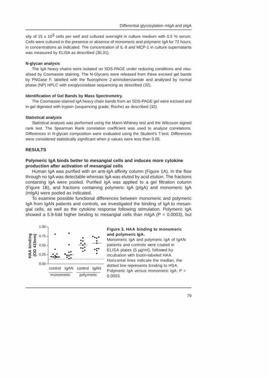
sity of 15 x 103 cells per well and cultured overnight in culture medium with 0.5 % serum.Cells were cultured in the presence or absence of monomeric and polymeric IgA for 72 hours,in concentrations as indicated. The concentration of IL-8 and MCP-1 in culture supernatantswas measured by ELISA as described (30,31).
N-glycan analysisThe IgA heavy chains were isolated on SDS-PAGE under reducing conditions and visu-
alised by Coomassie staining. The N-Glycans were released from these excised gel bandsby PNGase F, labelled with the fluorophore 2-aminobenzamide and analysed by normalphase (NP) HPLC with exoglycosidase sequencing as described (32).
Identification of Gel Bands by Mass Spectrometry.The Coomassie-stained IgA heavy chain bands from an SDS-PAGE gel were excised and
in-gel digested with trypsin (sequencing grade; Roche) as described (32).
Statistical analysisStatistical analysis was performed using the Mann-Whitney test and the Wilcoxon signed
rank test. The Spearman Rank correlation coefficient was used to analyze correlations.Differences in N-glycan composition were evaluated using the Student's T-test. Differenceswere considered statistically significant when p values were less than 0.05.
RESULTS
Polymeric IgA binds better to mesangial cells and induces more cytokineproduction after activation of mesangial cells
Human IgA was purified with an anti-IgA affinity column (Figure 1A). In the flowthrough no IgA was detectable whereas IgA was eluted by acid elution. The fractionscontaining IgA were pooled. Purified IgA was applied to a gel filtration column(Figure 1B), and fractions containing polymeric IgA (pIgA) and monomeric IgA(mIgA) were pooled as indicated.
To examine possible functional differences between monomeric and polymericIgA from IgAN patients and controls, we investigated the binding of IgA to mesan-gial cells, as well as the cytokine response following stimulation. Polymeric IgAshowed a 5.9-fold higher binding to mesangial cells than mIgA (P = 0.0003), but
Differential glycosylation mIgA and pIgA
79
control IgAN control IgAN0.00
0.25
0.50
0.75
1.00
monomeric polymeric
HA
A b
indi
ng (
OD
415
nm)
Figure 3. HAA binding to monomericand polymeric IgA.Monomeric IgA and polymeric IgA of IgANpatients and controls were coated inELISA plates (5 µg/ml), followed byincubation with biotin-labeled HAA.Horizontal lines indicate the median, thedotted line represents binding to HSA.Polymeric IgA versus monomeric IgA: P =0.0003.

there was no difference between IgAN patients and controls (Figure 2A). Supernatants of the cells stimulated with IgA for 72 hours were tested for produc-
tion of the chemokines IL-8 and MCP-1. The IL-8 production was significantly high-er after stimulation of mesangial cells with polymeric IgA than after stimulation withmIgA (P = 0.010), whereas IL-8 production was undetectable in cultures without IgA(Figure 2B). Furthermore, IL-8 production tended to be higher upon stimulation withpolymeric IgA from IgAN patients compared with that from controls (P = 0.077). Asignificant correlation was observed between binding of pIgA to mesangial cells andIL-8 production after co-culture (Figure 2C; R = 0.6450, P = 0.0038). Furthermore,stimulation with IgA also clearly enhanced production of MCP-1, and MCP-1 produc-tion correlated with IL-8 production after stimulation with different IgA samples (R =0.59, P = 0.01). These functional data indicate intrinsic differences betweenmonomeric and polymeric IgA.
Chapter 5
80
control IgAN control IgAN
0.0
0.5
1.0
1.5
2.0
monomeric polymeric
MB
L bi
ndin
g (O
D41
5)
control IgAN control IgAN0.00
0.25
0.50
0.75
1.00 control
MgEGTAD-mannose
L-mannose
monomeric polymeric
MB
Lbin
ding
(O
D41
5)
A B
control IgAN control IgAN
0.0
0.5
1.0
1.5
2.0
2.5
monomeric polymeric
C4
depo
sitio
n (O
D41
5)
C
Figure 4. MBL binding to polymeric IgA from IgAN patients and controls.Monomeric and polymeric IgA were coated in ELISA plates, followed by incubation with purifiedMBL-MASP complexes. A) Detection of MBL binding. B) MBL was pre-incubated with MgEGTA (10mM), D-mannose (100 mM) or L-mannose (100 mM) before incubation on wells coated with IgA asindicated. MBL binding was detected. The horizontal dashed line represents the negative control C)Detection of C4 activation after incubation with purified C4. For each IgA sample, blanc values,obtained after incubation with C4 and/or detection antibodies without MBL, were subtracted. Dashedlines indicate OD values obtained with coating of HSA. Polymeric IgA versus monomeric IgA (A, C):P = 0.0001.

Interaction of polymeric IgA with HAA lectinIt has been reported that O-glycans of patients with IgAN contain more Tn anti-
gen (GalNAc-Ser/Thr) compared to controls (18,33). Terminal GalNAc can bedetected by specific lectins, including Helix Aspersa (HAA). We investigated thebinding of HAA to IgA by ELISA (Figure 3). The binding of HAA to IgA from healthyindividuals and IgAN patients was 4 fold higher for polymeric IgA than for monomer-ic IgA (P = 0.0003). However, we could not observe a difference in HAA bindingbetween patients and controls.
MBL exclusively binds to polymeric IgA, resulting in C4 activationWe and others showed that IgAN is associated with complement activation via
the lectin pathway (10,34). Therefore, the binding of MBL to purified IgA frompatients with IgAN and controls was studied, showing that MBL binds to pIgA but notto mIgA with a high inter-individual variation, both for IgAN patients and controls(Figure 4A). Using parallel detection of immobilized IgA on plates, we confirmed thatequal amounts of IgA were present in coated wells, indicating that differences incoating could not explain the observed differences (not shown). Binding of MBL toIgA was completely inhibitable by D-mannose and MgEGTA but not by L-mannose(Figure 4B), confirming that the C-type lectin domain of MBL was involved in bind-ing to IgA.
Binding of MBL to pIgA from IgAN patients and from controls resulted in activa-tion of purified C4 (Figure 4C), presumably involving C4 cleavage by MBL-associat-ed MASP-2. This activation of C4 showed a strong correlation with MBL binding (R=0.98, P < 0.0001 for pIgA).
Differential glycosylation mIgA and pIgA
81
1
control IgAN control IgAN0.0
2.5
5.0
7.5
10.0
1
** ** ** **1020
monomeric polymeric
effe
ct o
f neu
ram
inid
ase
on
HA
A b
indi
ng (
ratio
)
1
control IgAN control IgAN0.5
1.5
2.0
2.5
1
* *
monomeric polymeric
effe
ct o
f neu
ram
inid
ase
on
MB
L bi
ndin
g (r
atio
)
A B
Figure 5: Increased interaction with HAA after treatment of IgA withneuraminidase.IgA (5 µg/ml) was coated in ELISA. After treatment with neuraminidase HAA binding (A) and MBLbinding (B) were detected. Depicted is the ratio of non-treated and treated IgA after subtraction ofbackground values, and the dotted line represents the non-treated IgA. * P < 0.05, ** P <0.01.

Treatment of IgA with neuraminidase enhances its interaction with HAATo examine whether sialic acids, commonly present on N-linked and O-linked
glycans of IgA, might hamper the interaction of IgA with HAA and/or MBL we treat-ed immobilized IgA with neuraminidase. After treatment of IgA the interaction withHAA increased significantly (3.8-fold for control mIgA, 3.1-fold for IgAN mIgA, 3.2-fold for control pIgA, 3.0-fold for IgAN pIgA; P < 0.004) (Figure 5A), suggesting thepresence of sialylated Tn antigen, as the removal of the sialic acid exposes theGalNAc (Tn) epitope. In contrast, the binding of MBL to IgA was hardly affected byneuraminidase, only showing a minor increase after treatment of polymeric IgA (1.1-fold) (Figure 5B), consistent with the known specificity of MBL for glycans present-ing 3,4 cis hydroxyls such as mannose to which sialic acids do not attach.
Molecular composition of monomeric and polymeric IgA from IgAN patientsand controls
The results presented above indicate major functional differences betweenmonomeric and polymeric IgA. We therefore investigated the molecular compositionof monomeric and polymeric IgA from IgAN patients and controls. The size distribu-tion of IgA from IgAN patients and controls was similar (mean % polymeric IgA, con-trols: 18.9 %, patients: 18.8%; P = 0.89). The IgA preparations were assessed forIgA1 and IgA2 content by ELISA. As described before (35), IgA2 is a minor con-stituent of human serum IgA. However, the relative amount of IgA2 was significant-ly higher in polymeric IgA (20 ± 4.1 %) as compared to monomeric IgA (9.2 ± 4.7 %)(Figure 6A) (P< 0.0001), suggesting that circulating IgA2 is more likely to be pro-duced as polymeric complexes than circulating IgA1. The relative amount of IgA2was similar in IgA from patients and controls.
Subsequently, we measured SIgA in monomeric and polymeric IgA from IgANpatients and controls. In agreement with the molecular size of SIgA, SIgA is exclu-
Chapter 5
82
control IgAN control IgAN0
100
200
300
400
monomeric polymeric
IgA
2 (µ
g/m
g Ig
A)
control IgAN control IgAN0
5
10
*
monomeric polymeric
SIg
A (
µg/m
g Ig
A)
A B
Figure 6. Increased concentration of SIgA and IgA2 in polymeric IgA.The relative content of IgA2 (A) and SIgA (B) in monomeric and polymeric IgA as assessed byELISA. Horizontal lines indicate the median. Polymeric IgA versus monomeric IgA (A, B): P <0.0001. SIgA content in pIgA of IgAN patients versus controls: * P = 0.0152.

sively present in polymeric IgA (Figure 6B). SIgA comprised less than 1 % of totalpolymeric serum IgA. However, the proportion of SIgA present in polymeric IgA fromIgAN patients is 2.5 times higher than in polymeric IgA from controls (P= 0.0152).
Polymeric IgA shows a different composition of N-linked glycans comparedto monomeric IgA
Recent studies showed that MBL is able to bind to N-linked glycans of IgG (36)and IgM (37). Based on the known structures of N-linked and O-linked glycans onIgA, it is more likely that MBL would bind to N-linked glycans than to the O-linkedglycans. Furthermore, information on N-linked glycosylation of IgA in IgAN is notavailable. Therefore, we characterized the N-glycans of 6 monomeric and 6 poly-meric IgA preparations in detail.
Heavy chains and light chains of monomeric and polymeric IgA were separatedby SDS-PAGE (Figure 7). N-linked glycans were released via an in-gel digestion ofthe heavy chain and light chain bands of IgA using PNGase F. Isolated glycan sam-ples were labeled with 2-aminobenzamide and run on normal phase HPLC.Consistent with earlier data (32), light chains of IgA were found not to be glycosylat-ed (not shown). The elution pattern of heavy chains is shown in Figure 8A. The mostprominent peaks, present between glucose units 8 and 10, represent complex gly-cans that are sialylated, as was demonstrated by a neuraminidase digestion (abs;Figure 8B). No obvious differences could be observed between N-glycans frompatients and from controls. However, upon comparison of glycans from monomericand polymeric IgA, a single peak at GU 6.2 was present in all polymeric IgA sam-ples but absent in monomeric IgA. Digestion with mannosidase (jbm, Figure 8B)demonstrated that this is an oligomannose structure (Man 5), as schematicallydrawn in Figure 8A.
Using sequential enzyme digestions (Figure 8B), the glycan structures on these
Differential glycosylation mIgA and pIgA
83
Figure 7. SDS-PAGE analysisof monomeric and polymericIgA.Separation of monomeric andpolymeric IgA with 10% SDS-PAGEunder reducing conditions showsheavy and light chains after stainingwith Coomassie. IgA heavy chainsrun as a doublet (upper and lowerheavy chains).

Chapter 5
84
A
B
Minutes Minutes
6 7 8 9 10GU
6 7 8 9 10GU
6 7 8 9 10GU
80 85 90 95 100 105 110 115 12080 85 90 95 100 105 110 115 120 80 85 90 95 100 105 110 115 12080 85 90 95 100 105 110 115 120
monomeric polymeric
ctrl
ctrl
ctrl
IgAN
IgAN
IgAN
Undig
abs
absjbm
absbtg
abs btgbkf
abs btgbkf guh
Minutes60 70 80 90 100 110 120 130
Minutes60 70 80 90 100 110 120 130
4 765 1098 1211 GU4 765 1098 1211 GU
Figure 8. Analysis of N-linked glycans on IgA heavy chains.A) Analysis of N-glycans of the heavy chain of polymeric IgA from controls and from patients with IgANby NP-HPLC. The figures show the individual elution profiles following fluorescence detection.Retention times are standardized to a glucose oligomer ladder to give glucose units (GU). The boxedpeaks are only present in polymeric IgA and are identified as oligomannose (Man 5), as indicated. B)Digestion of the N-glycans with A. ureafaciens sialidase (abs), jack bean α-mannosidase (jbm),bovine testes β-galactosidase (btg), bovine kidney α-fucosidase (bkf) and Streptococcuspneumoniae N-acetyl β-glucosaminidase (guh), followed by separation with NP-HPLC.

Differential glycosylation m
IgAand pIgA85
Table 2. Analysis of N -glycans of IgA heavy chains of monomeric and polymeric IgA
monomeric polymeric Major structure *
GU Control IgAN Mean Control IgAN Mean P value
A2B 5.80 1.53 2.07 2.66 1.85 1.71 2.25 2.01 1.32 2.55 2.49 1.63 1.60 1.88 1.91 0.4469 Man 5 6.17 0.00 0.00 0.00 0.00 0.00 0.00 0.00 4.51 3.60 2.24 3.71 3.06 2.48 3.27 0.0002 A2G1[3] A2BG1[6]
6.52 0.66 1.02 0.84 1.03 1.21 1.67 1.07 0.95 1.72 1.39 1.11 1.14 1.24 1.26 0.3214
A2BG1[3] FA2G1[6]
6.61 1.48 1.68 1.89 1.21 1.67 2.28 1.70 1.23 1.09 1.88 1.06 1.16 1.66 1.35 0.0187
FA2G1[3] FA2BG1[6] FA2BG1[3]
6.92 0.00 0.38 0.46 0.46 1.07 0.60 0.50 1.69 2.04 0.66 0.88 1.73 1.02 1.34 0.0264
A2G2 7.20 2.10 1.89 2.23 1.87 1.37 2.08 1.92 2.16 2.65 2.75 1.32 2.09 1.09 2.01 0.7806 A2BG2 7.37 3.17 3.62 4.22 2.53 2.04 2.73 3.05 3.36 4.72 4.17 2.59 3.77 4.42 3.84 0.0663 FA2G2 A3G2
7.65
FA2BG2 7.73 8.20 8.03 8.57 5.58 7.01 6.75 7.36 9.56 10.34 7.55 7.13 10.84 10.60 9.33 0.0448
A3BG2 7.94 2.51 1.52 2.28 1.66 1.61 1.12 1.78 2.00 2.32 3.64 1.69 2.74 2.96 2.56 0.0816 A2G2S1 8.13 20.22 21.15 20.65 21.64 16.24 20.75 20.11 17.43 17.28 19.15 21.81 15.89 17.83 18.23 0.0338 A3G3 A2BG2S1
8.35 7.81 7.20 5.56 5.66 6.65 6.29 6.53 8.40 8.64 6.50 6.44 7.64 8.04 7.61 0.0017
FA2G2S1 8.50 A3BG3 8.52
9.87 7.80 10.95 11.67 11.34 10.99 10.44 10.06 7.85 9.25 11.27 10.11 9.65 9.70 0.0709
FA2BG2S1 8.72 6.88 5.15 6.26 5.84 8.59 6.29 6.50 8.67 6.87 8.34 6.34 9.31 7.56 7.85 0.0034 A2G2S2 9.03 5.90 5.76 5.05 5.62 4.58 4.61 5.25 7.84 7.57 9.32 8.94 6.67 7.92 8.04 0.0010 A2BG2S2 9.19 2.80 2.28 2.06 3.03 2.51 2.55 2.54 1.96 2.14 1.79 1.93 1.67 2.68 2.03 0.0493 FA2G2S2 9.40 14.69 16.27 13.92 18.02 15.56 15.77 15.70 10.32 9.67 10.05 14.55 10.84 9.69 10.85 0.0002 FA2BG2S2 9.48 12.17 14.17 12.40 12.34 16.84 13.27 13.53 8.55 8.93 8.83 7.59 9.77 9.27 8.82 0.0003
N-glycans were identified on IgA heavy chains of monomeric and polymeri c IgA from six individuals (3 IgAN patients and 3 controls , as indicated), using sequential exoglycosidase digestion as indicated in F ig. 8 . For each structure, the percentage of total N -glycans is indicated. Differences between monomeric and polymeric IgA are evaluated by paired t -test. *Structural abbreviations: all N -glycans have two core GlcNAcs; F, core fucose linked αα1-6 to inner G lcNAc; Man(x), number (x) of mannose on core GlcNAcs; A, number ( x) of antenna (GlcNAc) on trimannosyl core; B, bisecting GlcNac linked ββ1-4 to inner mannose; Gx, number (x) of galactose on anntenne; Sx, number (x ) of sialic acids on anntenne.

samples were identified and quantified (Table 2). Comparison of monomeric andpolymeric IgA revealed that 3.3 % of the total glycan pool contained Man5 in poly-meric IgA, whereas this structure is absent in monomeric IgA. Furthermore, the dou-ble sialylated glycans are under-represented in polymeric IgA as compared tomonomeric IgA (29 % and 37 %, respectively, P = 0.001), resulting in a shift to small-er glycan structures on polymeric IgA. There were no significant differencesbetween patients and controls. Together, the results indicate that N-linked glycosy-lation of the IgA heavy chain is significantly different between monomeric and poly-meric IgA (Table 2).
DISCUSSION
Deposition of IgA in the renal mesangium is the primary characteristic of IgAN,responsible for glomerular inflammation and finally the development of renal failure.Based on earlier observations of mesangial IgA, this IgA is believed to be largelypolymeric IgA1. In the present study we show that polymeric IgA, as opposed tomonomeric IgA, from IgAN patients and from healthy controls shows increased bind-ing to and activation of mesangial cells, and has a superior capacity to bind the com-plement-activating lectin MBL. These aspects of polymeric IgA are most likely to beinvolved in induction of glomerular deposition and inflammation. Furthermore, weprovide evidence that polymeric IgA is differently glycosylated from monomeric IgA,as suggested by lectin binding studies and demonstrated by a direct identification ofN-linked glycans. Noteworthy, the only obvious difference that was observedbetween IgA from IgAN patients and controls was a substantial increase in the frac-tion of SIgA in polymeric IgA from IgAN patients.
Comparisons between IgA isolated from patients and from healthy controls werereported previously (19,22,28,38-40). Most experiments were performed with eitherfractionated total serum without an IgA purification step (28,39,40) or with pooledserum IgA purified with Jacalin, a lectin that binds Gal 1-3GalNAc (39-41). In ourstudy we purified IgA from individual patients and controls with an anti-IgA mAb.With this method we prepared monomeric and polymeric IgA that contains total andhighly pure serum IgA without a preceding selection for certain IgA glycoforms. Incontrast to methods using Jacalin, this method also enabled us to isolate the IgA2present in serum.
Previous studies described that the binding of polymeric IgA to mesangial cellswas higher than that of monomeric IgA (23,28) although this could not be repro-duced by others (24). Moreover, the binding of patient IgA and that of in vitrodegalactosylated IgA was higher than that of control IgA (24). In the present study,we confirm a prominent increase in mesangial cell binding of polymeric overmonomeric IgA, but we did not detect a difference between patient and control IgA.Our studies further establish the pro-inflammatory properties of polymeric IgA, anddemonstrate that IgA-induced chemokine production is correlates with the interac-tion of IgA with the mesangial cell surface. It is most likely that next to the increasedbinding of pIgA, also a more efficient receptor crosslinking will contribute to its proin-flammatory action. In addition, also the observed biochemical properties of pIgAmight contribute to this process. However, unlike previous investigations (27), this
Chapter 5
86

property could not be specifically attributed to IgA derived from IgAN patients. Next to the direct effects of IgA on mesangial cells, activation products of the
complement system, involving both the alternative pathway and the lectin pathway,are likely to drive the local inflammatory process. Activation of the lectin pathway ofcomplement via an interaction between MBL and IgA has been shown before (12),and in the present study we confirm and extend these data by showing that bindingof MBL is a common feature of polymeric but not monomeric IgA isolated from dif-ferent donors. Since ligand recognition by MBL requires multiply presented carbo-hydrates, MBL binding could be favored by the structure of polymeric IgA. Bindingof MBL leads to activation of C4 presumably via activation of the C4-cleavingenzyme MASP-2. In a healthy situation, the binding of MBL to polymeric IgA couldbe involved in host defense. However, in IgAN, lectin pathway activation via poly-meric IgA is unfavorable (10).
Many studies on IgA from IgAN patients focused on glycosylation. IgA is exten-sively glycosylated, both via N-linkages (IgA1 and IgA2) and O-linkages (IgA1) (21).It was consistently observed that serum IgA from IgAN patients contains smaller O-linked glycans, with less sialylation and galactosylation, than IgA from healthy con-trols (18). Previous investigations suggested that this was predominantly the casefor monomeric IgA (40). Our experiments using HAA, a lectin that is commonly usedto detect terminal GalNAc present on non-galactosylated O-linked glycans, suggest-ed the presence of terminal GalNAc (Tn antigen) predominantly on polymeric IgA,both from patients and controls. This is in agreement with a previous study showingreactivity of HAA with high molecular weight serum proteins (28) and with data pro-vided by Leung et al. (40). Binding of HAA was strongly increased by neuraminidasetreatment, suggesting a high frequency of non-galactosylated O-linked glycans onIgA, that expose terminal GalNAc after enzymatic removal of sialic acid.
A detailed quantitative analysis of N-linked glycans on IgA heavy chains ofmonomeric and polymeric IgA revealed several significant differences betweenthese molecular forms of IgA. In this respect, polymeric IgA consistently containedan oligomannose structure that was undetectable on all monomeric IgA prepara-tions, and showed significantly less glycans with two terminal sialic acid residues.Recent studies showed that MBL can bind to certain glycoforms of human IgM,involving GlcNAc-terminated glycans and oligomannose structures on the IgMheavy chains (42). Therefore, the presence of specific glycans on polymeric but notmonomeric IgA might also be involved in its recognition by MBL. At present it isunknown whether a specific glycosylation pattern of the heavy chain of polymericIgA is involved in the polymerization of IgA, and/or whether this merely is related tothe conditions present during production of the different forms of IgA. Earlier stud-ies from our group indicated that polymeric serum IgA contains dimeric IgA linkedwith J chain, as well as complexes of monomeric IgA linked via other mechanisms(43). In the first case, polymerization takes place in the B cell, and the presence ofoligomannose, which is a premature glycan structure, might suggest the ER as apossible location for polymerization, which prevents further synthesis of the glycanstructure by steric hindrance. In the latter case, polymerization might take place out-side the B cell.
Part of the observed differences in glycosylation between monomeric and poly-
Differential glycosylation mIgA and pIgA
87

meric IgA might also be explained by the presence of SIgA in the polymeric fractionof serum IgA, since the SIgA heavy chain N-glycosylation is very different from thatof monomeric serum IgA (32). SIgA has about 8 % oligomannose structures and 60% glycans with exposed GlcNAc with less than 15 % for all glycans sialylated (32),compared to monomeric serum IgA where most of the glycans are sialylated.However, SIgA comprises only less than 1 % of total polymeric serum IgA.
We observed that polymeric serum IgA contains more IgA2 than monomeric IgA.This could be because IgA2 polymerises more easily than IgA1, or that IgA2-pro-ducing B-cells preferentially secrete polymeric IgA. Bone-marrow-derived IgA aspresent in serum is largely monomeric and of the IgA1 subclass, whereas mucosalIgA is largely polymeric, containing J chain and secretory component (44), and con-tains a substantial fraction of IgA2 (35). Therefore, an increased fraction of IgA2 andthe presence of SIgA in circulating polymeric IgA may suggest its production by themucosal immune system. Quantitative measurement of the presence of SIgA inpolymeric serum IgA suggests that only a minor part of polymeric IgA containssecretory component. We hypothesize that this SIgA is derived from the mucosalsurface. Circulating dimeric IgA without secretory component could be partially pro-duced in mucosal lymphoid tissue and directly transported towards the circulation.
Although SIgA requires transepithelial transport for the attachment of secretorycomponent to dimeric IgA, the presence of low concentrations of circulating SIgAhas been described before (17,45,46). Moreover, increased serum levels of SIgAhave been reported in various diseases (17,47-49). In contrast to previous studies,we now determined the SIgA concentration in highly purified polymeric serum IgA.Our data demonstrate a clear relative increase in SIgA in polymeric IgA from patientswith IgAN compared with controls. We recently reported a preferential interaction ofSIgA with mesangial cells, and showed glomerular accumulation of SIgA in IgAN(17). Therefore, our current results further support a role for SIgA in the pathogene-sis of IgAN.
Taken together, the presented data suggest that a part of circulating polymericIgA has a mucosal origin. There is accumulating evidence that the pathogenesis ofIgAN is related to aberrant production of IgA. In this respect, in vivo studies indicat-ed that IgAN patients have a disturbed mucosal immune response, which wasrestricted to production of antibodies of the IgA1 subclass (50). Our present obser-vation that secretory IgA is increased in the polymeric IgA fraction of IgAN patientsfurther supports a role for abnormal mucosal immunity. Since IgAN is a slowly pro-gressive disease, it is well conceivable that only a minor subfraction of circulatingIgA in IgAN patients is abnormal, and that this IgA gradually accumulates in themesangial area. We hypothesize that this abnormal IgA is at least partially derivedfrom the mucosal immune system. Since our data strongly indicate that large-sizedIgA is especially able to interact with mesangial cells, and to induce complementactivation, the gradual deposition of such pro-inflammatory IgA may eventually leadto renal disease.
Acknowledgements
Chapter 5
88

The authors thank A. de Wilde (Leiden, The Netherlands) for technical assis-tance, Dr. J. van der Born (Amsterdam, the Netherlands) for the supply of valuablereagents, and E. Shillington (Oxford, UK) for assistance with glycan sequencing.This work was financially supported by the Dutch Kidney Foundation (BDO:C99.1822; AR: PC95)
REFERENCES
1. Berger J, Hinglais N: Les depots intercapillaires d'IgA-IgG. J Urol Nephrol (Paris) 74:694-695,
1968
2. Donadio JV, Grande JP: IgA nephropathy. N Engl J Med 347:738-748, 2002
3. Tomino Y, Sakai H, Miura M et al.: Detection of polymeric IgA in glomeruli from patients with
IgA nephropathy. Clin Exp Immunol 49:419-425, 1982
4. Novak J, Julian BA, Tomana M et al.: Progress in molecular and genetic studies of IgA
nephropathy. J Clin Immunol 21:310-327, 2001
5. Floege J, Feehally J: IgA nephropathy: recent developments. J Am Soc Nephrol 11:2395-
2403, 2000
6. van der Boog PJ, van Kooten C, de Fijter JW et al.: Role of macromolecular IgA in IgA
nephropathy. Kidney Int 67:813-821, 2005
7. Moura IC, Centelles MN, Arcos-Fajardo M et al.: Identification of the transferrin receptor as a
novel immunoglobulin (Ig)A1 receptor and its enhanced expression on mesangial cells in IgA
nephropathy. J Exp Med 194:417-425, 2001
8. McDonald KJ, Cameron AJ, Allen JM et al.: Expression of Fc alpha/mu receptor by human
mesangial cells: a candidate receptor for immune complex deposition in IgA nephropathy.
Biochem Biophys Res Commun 290:438-442, 2002
9. van den Dobbelsteen ME, van der Woude FJ, Schroeijers WE et al.: Binding of dimeric and
polymeric IgA to rat renal mesangial cells enhances the release of interleukin 6. Kidney Int
46:512-519, 1994
10. Roos A, Rastaldi MP, Calvaresi N et al.: Glomerular activation of the lectin pathway of comple
ment in IgA nephropathy is associated with more severe renal disease. J Am Soc Nephrol
17:1724-1734, 2006
11. Endo M, Ohi H, Ohsawa I et al.: Glomerular deposition of mannose-binding lectin (MBL) indi
cates a novel mechanism of complement activation in IgA nephropathy. Nephrol Dial
Transplant 13:1984-1990, 1998
12. Roos A, Bouwman LH, Gijlswijk-Janssen DJ et al.: Human IgA activates the complement sys
tem via the mannan-binding lectin pathway. J Immunol 167:2861-2868, 2001
13. Berger J, Yaneva H, Nabarra B et al.: Recurrence of mesangial deposition of IgA after renal
transplantation. Kidney Int 7:232-241, 1975
14. Sanfilippo F, Croker BP, Bollinger RR: Fate of four cadaveric donor renal allografts with mesan
gial IgA deposits. Transplantation 33:370-376, 1982
15. Schena FP, Pastore A, Ludovico N et al.: Increased serum levels of IgA1-IgG immune com
plexes and anti-F(ab')2 antibodies in patients with primary IgA nephropathy. Clin Exp Immunol
77:15-20, 1989
16. van der Boog PJ, van Kooten C, van Seggelen A et al.: An increased polymeric IgA level is not
a prognostic marker for progressive IgA nephropathy. Nephrol Dial Transplant 19:2487-2493,
2004
17. Oortwijn BD, van der Boog PJ, Roos A et al.: A pathogenic role for secretory IgA in IgA
Differential glycosylation mIgA and pIgA
89

nephropathy. Kidney Int 69:1131-1138, 2006
18. Allen AC, Bailey EM, Barratt J et al.: Analysis of IgA1 O-glycans in IgA nephropathy by fluo
rophore-assisted carbohydrate electrophoresis. J Am Soc Nephrol 10:1763-1771, 1999
19. Coppo R, Amore A: Aberrant glycosylation in IgA nephropathy (IgAN). Kidney Int 65:1544-
1547, 2004
20. Tomana M, Novak J, Julian BA et al.: Circulating immune complexes in IgA nephropathy con
sist of IgA1 with galactose-deficient hinge region and antiglycan antibodies. J Clin Invest
104:73-81, 1999
21. Mestecky J, Moro B, Underdown BJ: Mucosal Immunoglobulins, chap. 9, in Mucosal
Immunology, 2 ed., edited by Ogra PL, Mestecky J, Lamm ME, Strober W, Bienenstock J,
McGhee JR, San Diego, Academic Press, 1999, pp 133-152
22. Allen AC, Harper SJ, Feehally J: Galactosylation of N- and O-linked carbohydrate moieties of
IgA1 and IgG in IgA nephropathy. Clin Exp Immunol 100:470-474, 1995
23. Leung JC, Tang SC, Lam MF et al.: Charge-dependent binding of polymeric IgA1 to human
mesangial cells in IgA nephropathy. Kidney Int 59:277-285, 2001
24. Wang Y, Zhao MH, Zhang YK et al.: Binding capacity and pathophysiological effects of IgA1
from patients with IgA nephropathy on human glomerular mesangial cells. Clin Exp Immunol
136:168-175, 2004
25. Gomez-Guerrero C, Lopez-Armada MJ, Gonzalez E et al.: Soluble IgA and IgG aggregates are
catabolized by cultured rat mesangial cells and induce production of TNF-alpha and IL-6, and
proliferation. J Immunol 153:5247-5255, 1994
26. Duque N, Gomez-Guerrero C, Egido J: Interaction of IgA with Fc alpha receptors of human
mesangial cells activates transcription factor nuclear factor-kappa B and induces expression
and synthesis of monocyte chemoattractant protein-1, IL-8, and IFN-inducible protein 10. J
Immunol 159:3474-3482, 1997
27. Leung JC, Tang SC, Chan LY et al.: Polymeric IgA increases the synthesis of macrophage
migration inhibitory factor by human mesangial cells in IgA nephropathy. Nephrol Dial
Transplant 18:36-45, 2003
28. Novak J, Vu HL, Novak L et al.: Interactions of human mesangial cells with IgA and IgA-con
taining immune complexes. Kidney Int 62:465-475, 2002
29. de Fijter JW, van den Wall Bake AW, Braam CA et al.: Immunoglobulin A subclass measure
ment in serum and saliva: sensitivity of detection of dimeric IgA2 in ELISA depends on the anti
body used. J Immunol Methods 187:221-232, 1995
30. van Kooten C, Gerritsma JS, Paape ME et al.: Possible role for CD40-CD40L in the regulation
of interstitial infiltration in the kidney. Kidney Int 51:711-721, 1997
31. Taekema-Roelvink ME, Kooten C, Kooij SV et al.: Proteinase 3 enhances endothelial mono
cyte chemoattractant protein-1 production and induces increased adhesion of neutrophils to
endothelial cells by upregulating intercellular cell adhesion molecule-1. J Am Soc Nephrol
12:932-940, 2001
32. Royle L, Roos A, Harvey DJ et al.: Secretory IgA N- and O-Glycans Provide a Link between
the Innate and Adaptive Immune Systems. J Biol Chem 278:20140-20153, 2003
33. Odani H, Hiki Y, Takahashi M et al.: Direct evidence for decreased sialylation and galactosyla
tion of human serum IgA1 Fc O-glycosylated hinge peptides in IgA nephropathy by mass spec
trometry. Biochem Biophys Res Commun 271:268-274, 2000
34. Matsuda M, Shikata K, Wada J et al.: Deposition of mannan binding protein and mannan
binding protein-mediated complement activation in the glomeruli of patients with IgA nephropa
thy. Nephron 80:408-413, 1998
Chapter 5
90

35. Van Es LA, de Fijter JW, Daha MR: Pathogenesis of IgA nephropathy. Nephrology 3:3-12, 1997
36. Malhotra R, Wormald MR, Rudd PM et al.: Glycosylation changes of IgG associated with
rheumatoid arthritis can activate complement via the mannose-binding protein. Nat Med 1:237-
243, 1995
37. Arnold JN, Wormald MR, Suter DM et al.: Human serum IgM glycosylation: identification of gly
coforms that can bind to mannan-binding lectin. J Biol Chem 280:29080-29087, 2005
38. Julian BA, Novak J: IgA nephropathy: an update. Curr Opin Nephrol Hypertens 13:171-179,
2004
39. Amore A, Cirina P, Conti G et al.: Glycosylation of circulating IgA in patients with IgA nephropa
thy modulates proliferation and apoptosis of mesangial cells. J Am Soc Nephrol 12:1862-1871,
2001
40. Leung JC, Poon PY, Lai KN: Increased sialylation of polymeric immunoglobulin A1: mechanism
of selective glomerular deposition in immunoglobulin A nephropathy? J Lab Clin Med 133:152-
160, 1999
41. Kabir S: Jacalin: a jackfruit (Artocarpus heterophyllus) seed-derived lectin of versatile applica
tions in immunobiological research. J Immunol Methods 212:193-211, 1998
42. Arnold JN, Wormald MR, Suter DM et al.: Human serum IgM glycosylation: identification of gly
coforms that can bind to mannan-binding lectin. J Biol Chem 280:29080-29087, 2005
43. van der Boog PJ, van Zandbergen G, de Fijter JW et al.: Fc alpha RI/CD89 circulates in human
serum covalently linked to IgA in a polymeric state. J Immunol 168:1252-1258, 2002
44. Delacroix DL, Dive C, Rambaud JC et al.: IgA subclasses in various secretions and in serum.
Immunology 47:383-385, 1982
45. Thompson RA, Asquith P, Cooke WT: Secretory IgA in the serum. Lancet 2:517-519, 1969
46. Delacroix DL, Vaerman JP: A solid phase, direct competition, radioimmunoassay for quantita
tion of secretory IgA in human serum. J Immunol Methods 40:345-358, 1981
47. Mole CM, Renoult E, Bene MC et al.: Serum secretory IgA and IgM and free secretory compo
nent in IgA nephropathy. Nephron 71:75-78, 1995
48. Seilles E, Rossel M, Vuitton DA et al.: Serum secretory IgA and secretory component in
patients with non-cirrhotic alcoholic liver diseases. J Hepatol 22:278-285, 1995
49. Rostoker G, Terzidis H, Petit-Phar M et al.: Secretory IgA are elevated in both saliva and serum
of patients with various types of primary glomerulonephritis. Clin Exp Immunol 90:305-311,
1992
50. de Fijter JW, Eijgenraam JW, Braam CA et al.: Deficient IgA1 immune response to nasal
cholera toxin subunit B in primary IgA nephropathy. Kidney Int 50:952-961, 1996
Differential glycosylation mIgA and pIgA
91

Chapter 5
92

A pathogenic role for Secretory IgA in IgANephropathy.
Beatrijs D. Oortwijn, Paul J.M. van der Boog, Anja Roos, Reinier N. van derGeest, Johan W. de Fijter, Mohamed R. Daha, Cees van Kooten.
Dept. Nephrology, Leiden University Medical Center, Leiden, The Netherlands
SummaryIgA nephropathy (IgAN) is characterized by deposits of IgA in the renalmesangium. It is thought that deposits of IgA mainly involve high molecularweight (HMW) IgA1. However there is limited information on the exact compo-sition of HMW IgA in these deposits. In this study we investigated the presenceof secretory IgA (SIgA) in human serum and in the glomerular deposits of apatient with IgAN. Furthermore, we analysed the interaction of SIgA withmesangial cells. With ELISA SIgA concentrations in serum of IgAN patientsand healthy controls was measured. Patients and controls both have circulat-ing SIgA that was restricted to the HMW fractions. Patients tend to have high-er levels of SIgA, but this difference was not significant. However, in patientswith IgAN, high serum SIgA concentrations were associated with hematuria.Binding of size fractionated purified serum IgA and secretory IgA to mesangialcells was investigated with flow cytometry. These studies with mesangial cellsshowed stronger binding of SIgA to primary mesangial cells, compared to bind-ing of serum IgA. Importantly, after isolation and elution of glomeruli from anephrectomized transplanted kidney from a patient with recurrent IgAN, wedemonstrated a 120-fold accumulation of SIgA compared to IgA1 in the eluate.In conclusion we have demonstrated that SIgA strongly binds to humanmesangial cells, and is present in significant amounts in serum. Furthermore,we showed that SIgA is accumulated in the glomeruli of an IgAN patient. Thesedata suggest an important role for SIgA in the pathogenesis of IgA nephropa-thy.
Kidney Int. 69: 1131, 2006
CHAPTER
6

INTRODUCTION
Primary IgA nephropathy (IgAN) is the most common form of primary glomeru-lonephritis. The disease shows a spectrum of clinical presentations, leading to pro-gressive renal failure in a substantial proportion of patients. The hallmark of this dis-ease is deposition of IgA in the glomerular mesangium (1-3). It is generally thoughtthat deposits of IgA mainly involve IgA1 and for a large part consist of high molecu-lar weight (HMW) IgA (4). The composition of HMW forms of IgA in serum is diverseand may include dimeric IgA, CD89/IgA complexes, IgA immune complexes andIgA-fibronectin complexes (5-9).
Several reports have shown that the glycosylation of IgA1 in patients is differentfrom that in controls. Patient IgA1 contains more terminal GalNAc and this couldplay a role in the deposition of IgA1 in the mesangium (10-12). IgA from theglomeruli of IgAN patients consists at least partly of HMW IgA (13) and is under-O-glycosylated (10,14). In these studies no data was presented concerning the pres-ence of secretory IgA (SIgA). When deposited in the kidney, the IgA1-containingcomplexes are linked to inflammation. Stimulation of mesangial cells with HMW IgAleads to enhanced production of cytokines and chemokines, including IL-6, TGF-β,TNF-α, MCP-1, IL-8 and MIF (15-18). These cytokines and chemokines might playa role in the development and progression of renal injury in IgAN patients.
SIgA is the dominant immunoglobulin in external mucosal secretions like in oral,respiratory, and intestinal cavities, and is often characterized as a component of theimmune systems "first-line defence" against pathogenic micro-organisms (19). TheSIgA molecule is composed of two IgA monomers, linked by a junction peptidecalled J chain, and the secretory component (SC) that wraps around the dimer (20).Next to its presence in mucosal secretions, small amounts of SIgA can also be foundin human serum (21,22). Moreover, increased serum levels of SIgA have beenreported in various diseases (23-25), indicating that SIgA may be a marker of clini-cal interest. A previous study has suggested that the serum concentrations of SIgAare not different in IgAN patients compared to healthy controls (23). The physiolog-ical roles of serum IgA and SIgA are quite different, and the presence of the highlyglycosylated SC can have major effects on the biological functions of SIgA (26,27).In literature it is still controversial if SIgA is able to bind to mesangial cells (28,29).One study showed that SIgA is able to bind to mesangial cells (28), however thiscould not be confirmed in another study (29).
In the present study we investigated the presence of SIgA in sera of IgANpatients and healthy controls, and examined the binding of different molecular formsof IgA to human mesangial cells with special interest for SIgA. Finally we investigat-ed the presence of SIgA in the glomerular eluate of an IgAN patient.
MATERIAL AND METHODS
Human subjectsIn this study, we included 19 healthy volunteers and 47 patients with primary IgAN (30).
The latter were defined by mesangial deposits of IgA. None of these patients had clinical orlaboratory evidence of Henoch Schoenlein purpura, systemic lupus erythematosus, liver dis-ease or received immunosuppressive therapy. Patients were included in the study between
Chapter 6
94

October 1998 and February 1999. Blood and urine samples were collected, clinical charac-teristics and laboratory data of the patient group were obtained (Table 1) retrospectively(observation time 5.9 ± 0.5 years) and prospectively (follow-up time 3.7 ± 0.2 years).
Renal cortex was obtained from a transplant nephrectomy from a male patient (1977),with biopsy-proven recurrent IgAN. He was diagnosed with macroscopic hematuria (serumcreatinine 146 µmol/l) in 1993. The renal biopsy of 1996 showed characteristic features ofprogressive IgAN, and dialysis was started. In April 2002 he received a cadaveric renal trans-plantation. Three months later a decline in renal function was observed and a renal biopsyshowed the presence of interstitial nephritis and recurrent IgAN. In a biopsy taken 6 monthsfollowing transplantation characteristics of the interstitial nephritis were disappeared, but IgAwas still present. Due to decreased patient compliance there were two episodes of acuterejection in 2003 and 2004, which eventually led to graft loss. Informed consent was obtainedfrom all subjects.
IgA purificationSerum from healthy controls was used for IgA purification, according to methods
described before (9). In brief, serum was applied to an anti-IgA (HisA 43, kindly provided bydr J. van den Born, Free University Medical Center, Amsterdam) affinity column. IgA was elut-ed with 0.1 M glycine/ 0.3 M NaCl (pH 2.8). The eluted protein fractions containing IgA, asassessed by ELISA (31), were pooled and dialysed. The IgA was size-separated with aHiLoadTM 16/60 HR200 Superdex prep grade gelfiltration column (120 ml, AmershamPharmacia, Roosendaal, The Netherlands) into pIgA and mIgA.
Cell culture NHMC (normal human mesangial cells, Cambrex, USA) were expanded according to the
protocol provided by the manufacturer. Experiments with NHMC were performed in RPMI with10 % FCS, 1 % non-essential amino acids, 0.5 % transferrin/insulin/selenium, 1 % sodiumpyruvate, 1 % L-glutamine (all purchased at Gibco/ life Technologies, Paisley, Scotland).AMC11, a spontaneously growing adult human mesangial cell line (kindly provided by Prof.Holthofer, Helsinki), was cultured in DMEM with 10 % FCS. Cells were cultured in cultureflasks (Greiner, Frickenhausen, Germany) at 37 ºC in a humidified incubator with 5 % CO2/95 % air. For passage the cells were harvested by trypsinization (0.02 % (w/v) EDTA/ 0.05 %(w/v) trypsin in PBS from Sigma (St. Louis, MO)).
Monocyte-derived dendritic cells were generated as described before (32).
Role for SIgA in IgAN
95
Table 1: Clinical characteristics of the patients with IgAN at the point of ser um SIgA measurement.
Number of patients Median Range
Male/ female 38/ 9 ACE-inhibitor/AII antagonist 24/ 3 Age (years) 47 19-69 Systolic blood pressure (mmHg) 130 100-160 Diastolic blood pressure (mmHg) 80 55-100 Proteinuria (g/day) 0.6 0.1-5.2 Glomerular filtration rate (ml/min) 71 15-137

Chapter 6
96
Flow cytometryCells were harvested, washed with FACS buffer (0.5 x PBS containing 1 % BSA/ 2.8 %
glucose/ 0.01 % NaN3) and incubated with monomeric and polymeric serum IgA, and SIgA(Sigma). Following incubation for 1 hour at 4 ºC, cells were washed and incubated for onehour at 4 ºC with monoclonal anti-IgA Ab 4E8 (IgG1) (31). IgA binding was visualized usingPE-conjugated goat anti-mouse IgG1 (Southern Biotechnology, Birmingham, AL) andassessed for fluorescence intensity by flow cytometry (FACSCalibur, Cell quest software; BDBiosciences). Dead cells, identified by propidium iodide uptake, were excluded from analysis.
For analysing the presence of the mannose receptor, anti-mannose receptor antibody(D547.3; kindly provided by F. Koning, Leiden University Medical Center, Leiden, TheNetherlands) was used followed by PE-conjugated goat anti-mouse Ig (Dako, Glostrup,Denmark). For the detection of the two chains of MAC-1, anti-human CD11b (leu-15-PE, BDBiosciences) and anti-CD18 (IB4, ATCC) were used.
For inhibition of the binding of SIgA to mesangial cells, the cells were pre-incubated withpurified free SC (100 µg/ml) (33), 10 mM EDTA or 10 mM CaCl2 for 1 hour at 4 ºC.Subsequently, without washing, SIgA was added and this binding was visualized as describedabove. Inhibition of binding of SIgA was also detected with a pre-incubation of IgA for onehour at 4 ºC; subsequently Alexa-conjugated SIgA was added. After one hour the binding ofAlexa-conjugated SIgA was measured.
Cytokine analysisProduction of IL-6 was measured in supernatants of mesangial cells after 72 hr stimula-
tion. Prior to stimulation, cells were transferred to 48-wells plates (Costar, Corning, NY) at adensity of 25 x 103 cells per well and cultured overnight in culture medium with 0.5 % serum.Cells were stimulated with IL-1, serum IgA, or SIgA. The concentration of IL-6 in culturesupernatants was measured by specific ELISA as described previously (34).
RNA extraction and RT-PCR Total RNA was extracted from mesangial cells using RNeasy mini kit (Qiagen, Valencia,
CA). OD260/280 ratio was measured to determine the quantity and purity of RNA prepara-tions. Fixed amounts of total cellular RNA (1 µg) were reverse transcribed into cDNA byoligo(dT) priming, using M-MLV reverse transcriptase (Gibco/Life Technologies, Breda, TheNetherlands). PCR to detect the human mannose receptor was performed with specificprimers (sense 5'-TTG AGT GGA GTG ATG GGA CC-3'; antisense 5'-TTT CTG GAC CTTGGC TTC GT-3') using AmpliTaq DNA polymerase (Applied Biosystems, Roche, Mannheim,Germany). The PCR reaction was performed under standard conditions (35). The cDNA sam-ples were also subjected to PCR for GAPDH as an internal control (35), PCR products wereresolved on 1 % agarose gels and bands were visualized by ethidium bromide staining.
Preparation of glomerular eluateGlomeruli were isolated from a nephrectomized kidney from a transplanted IgAN patient
with recurrent disease. For this purpose the renal cortex was separated from the medulla.After slicing the cortex in little pieces, the glomeruli were collected on a 150-mesh sieve andstored at -70ºC with protease inhibitors (Complete, Mini, and Roche). The glomeruli werewashed with PBS and the final pellet was resuspended in 5 ml of elution buffer (2 M KSCNin 0.01 M phosphate buffer, pH 7.6) as described before (36). After stirring at room tempera-

ture (RT) for 60 min, the suspension was centrifuged at 8,000 g for 15 min at 4ºC in a high-speed centrifuge (Beckman, Avanti J25-1). The supernatant was collected and dialysedovernight against PBS. The precipitate that was formed during dialysis was removed by cen-trifugation for 15 min at 17,000 g and was negative for immunoglobulins. The remainingsupernatant was concentrated to one-third of the original volume.
ELISA for human SIgA, IgA1, IgA2, IgG and IgMTo test the specificity of the antibodies used for the SIgA ELISA, Ninety-six well Nunc
Maxisorp microtitre plate (Gibco/Invitrogen) was coated with 2 µg/ml IgA and BSA in carbon-ate buffer (pH 9.6) overnight at RT. After washing, the plate was incubated with monoclonalantibodies (2 µg/ml) specific for secretory component: NI194-4 (IgG1-k; Nordic Immunology,Tilburg, The Netherlands) (37) or 3F8 (kindly provided by dr R.M. Goldblum, University ofTexas Medical Branch, Galveston) (38) in PBS/ 1 % BSA/ 0.05 % Tween for one hour at 37ºC. Bound antibody was detected with goat anti-mouse Ig HRP (Dako). Enzyme activity ofHRP was developed using ABTS (Sigma). The O.D. at 415 nm was measured using amicroplate biokinetics reader (EL312e, Biotek Instruments, Winooski, Vermont, USA).
In order to quantify SIgA levels in isolated IgA and serum, a sandwich ELISA specific forSIgA was developed. Plates were coated with 2 µg/ml monoclonal antibody specific for secre-tory component (NI194-4; 3F8) in carbonate buffer (pH 9.6) (100 µl/well) overnight at RT.Subsequently, the plates were washed with PBS/ 0.05 % Tween. Plates were incubated withIgA or with serum from patients and controls in PBS/ 1 % BSA/ 0.05 % Tween for one hourat 37 ºC. After washing, bound IgA was detected using mouse anti-human IgA (4E8) conju-gated to digoxigenin (Dig), followed by F(ab)2 anti-Dig antibodies conjugated to HRP(Roche). Enzyme activity of HRP was developed using ABTS (Sigma). The O.D. at 415 nmwas measured. A calibration line was produced using purified SIgA (Sigma).
Concentrations of IgA1, IgA2, IgG and IgM in sera and glomerular eluate were determinedusing specific ELISAs (39).
Western blot analysisIgA preparations were subjected to 10% SDS-PAGE under reducing conditions, followed
by semi-dry blotting to PVDF (immobilin-P, Millipore, Bedford, MA). Blots were blocked for 2hours at RT in TBS/ 0.1 % Tween/ 5 % skim milk powder (Fluka, Buchs, Switzerland). Blotswere subsequently incubated with 2 µg/ml monoclonal antibody specific for secretory compo-nent (NI194-4) in TBS/ 0.1 % Tween/ 2.5 % skim milk powder overnight at 4 ºC. After wash-ing with TBS/ 0.1 % Tween, blots were incubated with HRP conjugated goat anti-mouseimmunoglobulin (Dako) for 2 hours at RT. After washing bands were visualized withSupersignal (Pierce Chemical Co., Rockford, IL) and exposure to HyperfilmTM films(Amersham Pharmacia).
Statistical analysisStatistical analysis was performed using the Mann-Whitney test. Differences were consid-
ered statistically significant when p values are less than 0.05.
Role for SIgA in IgAN
97

RESULTS
Specific detection of SIgA in human serumAn ELISA system was developed to specifically measure the amounts of secre-
tory IgA in serum. In this system anti-SC antibodies were coated, samples wereapplied and SIgA was detected with anti-IgA antibodies. Purified SIgA was readilydetected by this ELISA with a detection limit of 100 ng/ml (Figure 1A). In contrast,purified monomeric serum IgA is not recognized in the ELISA, even when applied athigh concentrations (Figure 1A). In accordance with previous publications (23), aspecific signal for SIgA could be detected in serum of healthy individuals (Figure1B). The specificity of this assay is especially dependent on the specificity of theanti-SC antibody. Using Western blot, we showed that this monoclonal antibody onlyrecognized the 75 kDa SC (Figure 2A). Furthermore comparison of the NI194-4 anti-body with another anti-SC antibody 3F8 (38) showed specificity for SIgA both in adirect ELISA (Figure 2B) as well as in a sandwich ELISA (Figure 2C).
To determine the molecular size of SIgA in serum, IgA was isolated from serumusing affinity chromatography. Size-fractionation revealed that SIgA was specifical-ly present in the HMW fractions (Figure 3A). Using the same procedure, IgA was iso-lated from 8 healthy controls followed by gel filtration. Pools containing pIgA andmIgA, respectively, were obtained and assessed for the amount of SIgA (Figure 3B).In all cases, SIgA was demonstrated exclusively in the pIgA pool.
Next we assessed concentrations of SIgA in serum of 47 IgAN patients and 19healthy controls (Figure 4A). Both in controls and in patients significant serum con-centrations of SIgA were detected. There was no significant difference (p= 0.159)in the SIgA concentrations in serum of patients (3.3 ± 3.0 µg/ml) compared to con-trols (2.2 ± 1.2 µg/ml). After comparison the SIgA concentration in serum of IgANpatients with different clinical parameters there were no correlations found. Howeverconcentrations of SIgA in IgAN patients were more heterogeneous than in controls.Therefore we divided the IgAN group in patients with elevated levels of SIgA (> 5µg/ml, mean ± 2 SD of control sera), and patients with normal levels (<5 µg/ml).
Chapter 6
98
00
1
2
3
mIgA
SIgA
0.01 0.1 1 10 100 1000
IgA (µg/ml)
SIg
A (
OD
415
nm)
00.00
0.25
0.50
0.75
1.00
NHS 1NHS 2NHS 3NHS 4
0.01 0.1 1 10
Serum (%)
SIg
A (
OD
145n
m)
Figure 1: Specific detection of SIgA in human serum.A) Purified SIgA and monomeric serum IgA were measured in a sandwich ELISA, as detailed inthe Material and Methods. B) Measurement of SIgA in serum from healthy individuals in differentserum dilutions. NHS, normal human serum.
A B

Role for SIgA in IgAN
99
NI194-4 3F80
1
2
3 BSAmIgApIgASIgA
Bin
ding
(O
D 4
15nm
)
194.41 3F80.0
0.5
1.0
1.5
2.0
2.5 BSAmIgApIgASIgA
SIg
A (
OD
415
nm)
B
C
A1µg 0.2 µg
SIgA
250150
100
75
50
1µg 0.2 µg
SIgA
250150
100
75
50
Figure 2: Specific detection of SIgA and secretory component.A) Detection of secretory component in SIgA, 1 and 0.2 µg SIgA were loaded on 10 % SDS-PAGEgel under reducing conditions after blotting secretory component was detected with 2µg/mlNI194-4. B) Detection of secretory component with NI194-4 and 3F8 (2 µg/ml) in different formsof IgA (2 µg/ml) coated on ninety-six well Nunc maxisorp microtitre plate. C) Different forms ofIgA measured with sandwich ELISA using two different coating antibodies (NI194-4 and 3F8).
35 45 55 650
50
100
150
SIgA
protein
IgA
0.25
0.50
0.75
335 kDa 160 kDa
ml
Pro
tein
(m
AU
)
IgA/S
IgA (O
D 415nm
)
pIgA mIgA0
5
10
15
SIg
A (
µg/m
g Ig
A)
BA
Figure 3: SIgA is present in high molecular weight fractions of serum IgA.A) IgA was affinity purified and size-fractionated on a HiLoadTM 16/60 HR200 Superdex prep gradegelfiltration column. All fractions were measured for total protein and the presence of total IgA andSIgA by ELISA. B) From 8 healthy controls, IgA was purified and size fractionated as above. IgA waspooled in pIgA fraction (39-47.5ml) and mIgA fraction (47.5-57ml). Both IgA and SIgA concentrationswere determined, and depicted is the amount of SIgA corrected for the amount of total IgA Thehorizontal dashed line represents the detection limit. The horizontal solid lines indicate the median.

There were no significant correlations with the degree of proteinuria or creatinineclearance and the serum concentration of SIgA (Figure 4B). However, there was sig-nificantly more pronounced hematuria in the group with higher levels of SIgA (p=0.04) (Figure 4B).
SIgA binds to mesangial cells and induces cell activationAfter demonstrating the presence of SIgA in the circulation, we investigated its
capacity to interact with mesangial cells. After incubation of the mesangial cell-lineAMC11 or normal human mesangial cells (NHMC) with 200 µg/ml of different molec-ular forms of IgA, the binding was examined by FACS analysis. Binding ofmonomeric serum IgA to NHMC is very low (Figure 5A). In contrast, the polymericform of serum IgA showed a clear binding to mesangial cells. However the bestbinding was observed with similar concentrations of SIgA, which occurred in a dose-dependent fashion, present over a wide range of concentrations (Figure 5B).Although the mean fluorescence intensity of IgA binding to NHMC, compared with
Chapter 6
100
IgAN control0
5
10
15
20p= 0.159
SIg
A (
µg/m
l)
< 5 < 50
1
2
3
4
5
6 NS
SIgA (µg/ml)
Pro
tein
uria
< 5 > 50
50
100
150 NS
SIgA (µg/ml)
Cle
aran
ce (
ml/m
in)
< 5 > 50
1
2
3
4
5
6p= 0.0428
SIgA (µg/ml)
Ery
thro
cytu
ria
A
B
Figure 4: Measurement of SIgA in serum of IgAN patients and controls andrelation to clinical parameters.A) The concentrations of SIgA were determined in serum from patients (n= 47) and controls (n=19).The horizontal dashed line represents the detection limit and the horizontal solid lines indicate themedian B) Based on SIgA concentrations IgAN patients were divided in two groups: normal SIgAlevels (<5µg/ml; mean ± 2 SD of control sera) and increased SIgA levels (≥ 5µg/ml). These groupswere analyzed for proteinuria, creatinin clearance (700* creatinin in urine (mmol/ 24 hours)/ creatininin serum (µmol/l) and erythrocyturia at the time of sampling. The horizontal solid lines indicate themedian. Statistics were performed using the Mann-Whitney test (ns= not significant)

AMC11, was overall higher for all forms of IgA, the relative differences between thedifferent forms of IgA were the same.
The different molecular forms of IgA were compared for their capacity to induceIL-6 production by mesangial cells. Stimulation of NHMC for 72 hours with IgAresulted in an increased IL-6 production. This increase was most prominent follow-ing stimulation with SIgA (Figure 6A). The induction of IL-6 production by mesangialcells was dose dependent: a 17-fold increase was observed compared to the nega-tive control upon stimulation with 200 µg/ml SIgA (Figure 6B).
Mesangial cells do not express the mannose receptor (CD206) or MAC-1(CD11b/CD18)
Recently we showed that dendritic cells are able to bind SIgA via the mannosereceptor (CD206) (40). Therefore we investigated the presence of the mannosereceptor as a potential SIgA receptor on mesangium cells. However both usingFACS analysis (Figure 7A) and RT-PCR (Figure 7B) we were not able to demon-strate the presence of the mannose receptor on mesangial cells. In both cases, DCserved as a positive control. Similarly, we were not able to demonstrate the pres-ence of CD11b/CD18 (Figure 7C), recently identified as a co-receptor for SIgA bind-ing (41).
Binding of SIgA to mesangial cells is not inhibited by secretory component,EDTA and calcium
To investigate in more detail the mechanism of binding of SIgA to mesangial
Role for SIgA in IgAN
101
0 100 200 3000
10
20
30
40
50
60SIgApIgA
Concentration IgA (µg/ml)
Bin
ding
(M
FI)
Figure 5: Binding of SIgA to mesangial cells.A) Normal human mesangial cells (NHMC) wereincubated with different molecular forms of IgA(200µg/ml) and assessed for IgA binding by flowcytometry. Filled histograms represent the binding ofIgA, the open histograms represent the control stainingwith secondary antibodies B) FACS analysis withAMC11 was performed using increasing doses of SIgA(Sigma) and pIgA (isolated from a healthy individual).Depicted is the mean fluorescence intensity (MFI)
A
B
Rel
ativ
ece
llnu
mbe
r SIgA
Log fluorescence intensity
pIgAmIgA

cells, the cells were pre-incubated with free SC. After pre-incubation with SC thebinding of SIgA to mesangial cells was not affected (Figure 8A). Binding of SIgA wasslightly inhibited with SIgA and pIgA but not with mIgA (Figure 8A), suggesting inter-action with the IgA part of the molecule.
To investigate the potential contribution of C-type lectins, a family of cell surfacemolecules including the mannose receptor, the role of calcium in SIgA binding tohuman mesangial cells was examined. However, neither the addition of extra calci-um, nor the removal of calcium using EDTA showed a significant effect on the bind-ing of SIgA to mesangial cells (Figure 8B).
SIgA is present in glomerular eluate from a kidney of an IgAN patient.To determine the potential role of SIgA in the pathogenesis of IgAN, we had the
unique opportunity to analyse the glomerular eluate of a nephrectomized specimenderived from a patient with recurrent IgAN. After elution, concentrations of specificimmunoglobulin isotypes were determined. In the glomerular eluate, allimmunoglobulin classes measured were detectable (Table 2), including SIgA in aconcentration of 2 µg/ml. To exclude that the immunoglobulins in the eluate were theresult of aspecific trapping from the circulation, serum immunoglobulin levels weredetermined in the serum of this patient at the time of nephrectomy (Table 2). Theratio of the immunoglobulin concentrations in the eluate and the serum can be usedas a measure of the specific accumulation in the glomerular deposit (Figure 9). Inthis analysis we observed a ratio for SIgA which was 120 fold higher that the ratiofor IgA1.
DISCUSSION
This is the first study to support a role for SIgA in the pathogenesis of IgAN. Weshow that SIgA is present in low concentrations in serum of healthy individuals aswell as in IgAN patients. In patients with higher SIgA serum concentrations hema-turia is more pronounced. Furthermore we show that SIgA exhibits the strongest
Chapter 6
102
- mIgA pIgA SIgA0.0
2.5
5.0
7.5
IL-6
(ng
/ml)
00
1
2
3SIgA
1 10 100
IgA (µg/ml)
IL6
(ng/
ml)
A B
Figure 6: SIgA increase IL-6 production by mesangial cells.A) Normal human mesangial cells (NHMC) (25x103 cells/well) were stimulated with differentmolecular forms of IgA (200µg/ml). After 72 hours supernatants were harvested and tested forIL-6 using ELISA. Depicted is the mean ± SD B) NHMC was stimulated with differentconcentrations of SIgA (Sigma) as described above and IL-6 production was assessed.

binding to mesangial cells compared to serum IgA. Finally, in the eluate of glomerulifrom a kidney of an IgAN patient a strong accumulation of SIgA was detected. Takentogether these data suggest an important role for SIgA in the pathogenesis of IgAN.
The high incidence of IgAN recurrence after renal transplantation, and the disap-pearance of deposits of IgA from accidentally transplanted kidneys clearly suggeststhat intrinsic alterations and/or structural characteristics of IgA contribute to theprocess of deposition (42,43). The predominance of IgA1 deposits and the specifichinge region of IgA1 with potential O-linked glycosylation sites, has initiated a direct-ed search for alterations in glycosylation. Indeed, both in serum but, more important-ly, also in the eluate of renal deposits (14), a specific reduction of O-linked galacto-sylation has been observed (10,12,14). Furthermore, with size fractionation of elut-ed proteins from kidney sections, it was shown that deposited IgA was mostly HMWof nature (13). In addition based on different staining methods, it has been proposedthat a large part of the deposited IgA is high molecular weight of nature (44-46).
The results from all these methods provide indirect indications for the composi-tion of the IgA1 deposits. We now show by eluting glomeruli, that glomeruli show astrong and specific accumulation of SIgA compared to other serum immunoglobu-lins. However, this technique can only be applied in limited cases of situations. Wehave tried to demonstrate the presence of SIgA deposits using traditional immuno-fluorescense on cryosections. Until now, we were not able to show SIgA in renal
Role for SIgA in IgAN
103
MR (CD206)
Rel
ativ
e ce
ll nu
mbe
r
DCLog fluorescence intensity
AMC11
A
MAC-1 (CD11b/CD18)
Log fluorescence intensity
DC
Rel
ativ
e ce
ll nu
mbe
r
AMC11
C
AM
C11
NH
MC
DC
H2O
GAPDH
MR
AM
C11
NH
MC
DC
H2O
GAPDH
MR
B
Figure 7: Mannose r eceptor and MAC-1 are not present on mesangial cells.A) FACS analysis of mannose receptor on DC and AMC11. Filled histograms represent expressionof mannose receptor; open histograms represent the control staining with secondary antibodies B)Mannose receptor mRNA expression was analyzed by RT-PCR as described in the material andmethods C) Presence of MAC-1 on mesangial cells and DC was tested with FACS analysis. Filledhistograms represent expression of MAC-1. Open histograms represent the control staining withsecondary antibodies.

sections, even not in cryosections of the kidney used for our elution study. Thismight be due to inappropriate reagents or conformational changes of the depositedSIgA, thereby masking the SC epitope. Therefore it will be necessary to generateother reagents for the detection of deposited SIgA, and a more thorough analysis ofrenal biopsies.
As described above, it is assumed that glycosylation of IgA is an important fac-tor in IgAN. Previous studies have shown the role of the glycosylation of IgA on theactivation of mesangial cells (47,48), which could be partially explained by alteredinteraction with mesangial cells (49). The glycosylation of SIgA is different comparedto that of serum IgA in several aspects. First, SIgA is a tetra molecular complex con-sisting of two IgA molecules, a J chain and the SC wrapped around the H chain.Modelling of SIgA suggests that the N-glycans on the heavy chain can be maskedby the SC (50). This may also result in a different exposure of the O-glycans.Moreover, specific analysis of the glycosylation of the IgA heavy chain present inSIgA, demonstrated different N-glycan structures compared to that of serum IgA,with terminal GlcNAc residues on the majority of the N-glycans (50). The O-glycanson the hinge region of the heavy chain of SIgA1 presented a wide range of glycanstructures, of which the major part is now characterized (50). Finally, also the SCitself is heavily glycosylated. However we were not able to inhibit the binding of SIgAto mesangial cells with SC, suggesting that the SC part is not important for interac-tion with mesangial cells. It will be a major challenge to isolate SIgA from serum ofIgAN patients and to determine specific alterations in glycosylation.
Having shown that SIgA strongly binds to mesangial cells, an important questionis which receptor is involved in this binding and whether this receptor is differentfrom that of serum IgA. Several IgA receptors have been described in the literature.The best known receptors, the polymeric Ig receptor (pIgR), the asialoglycoproteinreceptor and CD89, have already been described to be absent on mesangial cells(28,51,52). The transferrin receptor is described as an IgA receptor present on
Chapter 6
104
- mIgA pIgA SIgA SC0
50
100
150
SIg
A b
indi
ng (
%)
- CaCl2 DETA0
10
20
30 controlSIgA
MF
I
A B
Figure 8: Binding of SIgA to mesangial cells is not affect by free secretorycomponent or calcium but is affected by IgA.A) Mesangial cells were pre-incubated with mIgA, pIgA, SIgA (400 µg/ml) or free secretorycomponent (SC) (100 µg/ml). After one hour SIgA (200 µg/ml) was added and the binding of SIgAto mesangial cells was examined with flow cytometry. Depicted is the percentage of SIgA bindingof a representative experiment of 2 experiments. B) Cells were incubated with SIgA (200 µg/ml)in the presence of absence of EDTA (10 mM) or calcium (10 mM) followed by detection of SIgAbinding as described in the Material and Methods.

mesangial cells but it has been reported that this receptor is not able to bind SIgA(29). We have previously reported the binding of SIgA to the mannose receptor ondendritic cells (40), but we were not able to demonstrate the presence of the man-nose receptor on mesangial cells. Similarly we were not able to demonstrate thepresence of CD11b/CD18 (MAC1), a co-receptor for CD89 specifically involved inrecognizing SIgA or free SC (41). Therefore until now we have no indication for themesangial IgA receptor involved in SIgA binding. Our inhibition experiments suggestthat IgA rather than SC is recognized and that no C-type lectin is involved.Importantly we found that the putative receptor is able to transmit proinflammatorysignals, since SIgA induced a strong dose-dependent increase in IL-6 production bymesangial cells. This seems in contrast with the proposed anti-inflammatory role ofSIgA (53).
Generation of SIgA, i.e. production of dIgA followed by transcytosis using thepIgR, is a specific process taking place at mucosal surfaces (54). Interestingly, IgANpatients often present macroscopic hematuria following upper respiratory tract infec-tions. Mucosal challenge also leads to an increased production of IgA in the sys-temic compartment, probably based on the migration of B cells (the mucosa-bonemarrow axis) (55). This mucosa-bone marrow traffic has been confirmed by chal-lenging healthy individuals intranasally with the neoantigen cholera toxin subunit B(CTB) (56). In patients with IgAN we observed a reduced mucosal IgA response tomucosal immunization with CTB (56). At present it is not clear whether a mucosalchallenge also regulates levels of circulating SIgA. Still our finding of glomerularaccumulation of SIgA provides a link between the mucosal immune system andrenal deposits.
In summary, we have shown that SIgA is able to bind to mesangial cells in a
Role for SIgA in IgAN
105
Table 2: Immunoglobulin concentrations in serum and glomerular eluate of IgAN patient. IgA1 IgA2 SIgA IgM IgG
Serum (mg/ml) 5.9 0.59 0.016 0.74 10
Glomerular eluate (µg/ml) 6.1 0.65 2 2.2 6.8
IgA1 IgA2 SIgA IgM IgG0.00
0.05
0.10
0.15
elut
ion/
seru
m
Figure 9: SIgA is accumulated in the glomerular eluate of an IgAN kidneyTo make an estimate for the specific accumulation in the glomerular deposit,immunoglobulin concentrations in the eluate and serum were compared. Depicted is the ratioof concentration of different immunoglobulins in the eluate and the serum.

dose-dependent manner and that this binding is calcium-independent and cannot beinhibited with free SC. Furthermore low concentrations of SIgA can be detected inserum. Further research is needed to determine to which receptor SIgA can bindand what the mechanism of cell activation induced by SIgA is. SIgA is strongly accu-mulated in the glomeruli of a kidney from an IgAN patient. Therefore we concludethat our data support a role for SIgA in the pathogenesis of IgAN, and furtherresearch to define such a pathogenic role is warranted.
Acknowledgements
The authors would like to thank D.J. van Gijlswijk-Janssen, M.C. Faber-Krol andN. Klar-Mohamad (Dept. of Nephrology, LUMC, The Netherlands) for excellent tech-nical assistance. This work was financially supported by the Dutch KidneyFoundation (BO: C99.1822; AR: PC95)
REFERENCES1. Berger J, Hinglais N: Les depots intercapillaires d'IgA-IgG. J Urol Nephrol (Paris) 74:694-695,
1968
2. Donadio JV, Grande JP: IgA nephropathy. N Engl J Med 347:738-748, 2002
3. Feehally J: IgA nephropathy--a disorder of IgA production? QJM 90:387-390, 1997
4. Tomino Y, Sakai H, Miura M et al.: Detection of polymeric IgA in glomeruli from patients with
IgA nephropathy. Clin Exp Immunol 49:419-425, 1982
5. Barratt J, Feehally J, Smith AC: Pathogenesis of IgA nephropathy. Semin Nephrol 24:197-217,
2004
6. Floege J, Feehally J: IgA nephropathy: recent developments. J Am Soc Nephrol 11:2395-
2403, 2000
7. Novak J, Julian BA, Tomana M et al.: Progress in molecular and genetic studies of IgA
nephropathy. J Clin Immunol 21:310-327, 2001
8. van der Boog PJ, van Kooten C, de Fijter JW et al.: Role of macromolecular IgA in IgA
nephropathy. Kidney Int 67:813-821, 2005
9. van der Boog PJ, van Zandbergen G, de Fijter JW et al.: Fc alpha RI/CD89 circu lates in
human serum covalently linked to IgA in a polymeric state. J Immunol 168:1252-1258, 2002
10. Allen AC, Bailey EM, Brenchley PE et al.: Mesangial IgA1 in IgA nephropathy exhibits aberrant
O-glycosylation: observations in three patients. Kidney Int 60:969-973, 2001
11. Allen AC, Bailey EM, Barratt J et al.: Analysis of IgA1 O-glycans in IgA nephropathy by fluo
rophore-assisted carbohydrate electrophoresis. J Am Soc Nephrol 10:1763-1771, 1999
12. Tomana M, Matousovic K, Julian BA et al.: Galactose-deficient IgA1 in sera of IgA nephropa
thy patients is present in complexes with IgG. Kidney Int 52:509-516, 1997
13. Monteiro RC, Halbwachs-Mecarelli L, Roque-Barreira MC et al.: Charge and size of mesangial
IgA in IgA nephropathy. Kidney Int 28:666-671, 1985
14. Hiki Y, Odani H, Takahashi M et al.: Mass spectrometry proves under-O-glycosylation of
glomerular IgA1 in IgA nephropathy. Kidney Int 59:1077-1085, 2001
15. van den Dobbelsteen ME, van der Woude FJ, Schroeijers WE et al.: Binding of dimeric and
polymeric IgA to rat renal mesangial cells enhances the release of interleukin 6. Kidney Int
46:512-519, 1994
16. Gomez-Guerrero C, Lopez-Armada MJ, Gonzalez E et al.: Soluble IgA and IgG aggregates are
catabolized by cultured rat mesangial cells and induce production of TNF-alpha and IL-6, and
Chapter 6
106

proliferation. J Immunol 153:5247-5255, 1994
17. Duque N, Gomez-Guerrero C, Egido J: Interaction of IgA with Fc alpha receptors of human
mesangial cells activates transcription factor nuclear factor-kappa B and induces expression
and synthesis of monocyte chemoattractant protein-1, IL-8, and IFN-inducible protein 10. J
Immunol 159:3474-3482, 1997
18. Lai KN, Tang SC, Guh JY et al.: Polymeric IgA1 from Patients with IgA Nephropathy
Upregulates Transforming Growth Factor-beta Synthesis and Signal Transduction in Human
Mesangial Cells via the Renin-Angiotensin System. J Am Soc Nephrol 14:3127-3137, 2003
19. Kerr MA: The structure and function of human IgA. Biochem J 271:285-296, 1990
20. Johansen FE, Braathen R, Brandtzaeg P: The J chain is essential for polymeric Ig receptor-
mediated epithelial transport of IgA. J Immunol 167:5185-5192, 2001
21. Thompson RA, Asquith P, Cooke WT: Secretory IgA in the serum. Lancet 2:517-519, 1969
22. Delacroix DL, Vaerman JP: A solid phase, direct competition, radioimmunoassay for quantita
tion of secretory IgA in human serum. J Immunol Methods 40:345-358, 1981
23. Mole CM, Renoult E, Bene MC et al.: Serum secretory IgA and IgM and free secretory compo
nent in IgA nephropathy. Nephron 71:75-78, 1995
24. Seilles E, Rossel M, Vuitton DA et al.: Serum secretory IgA and secretory component in
patients with non-cirrhotic alcoholic liver diseases. J Hepatol 22:278-285, 1995
25. Rostoker G, Terzidis H, Petit-Phar M et al.: Secretory IgA are elevated in both saliva and serum
of patients with various types of primary glomerulonephritis. Clin Exp Immunol 90:305-311,
1992
26. Norderhaug IN, Johansen FE, Schjerven H et al.: Regulation of the formation and external
transport of secretory immunoglobulins. Crit Rev Immunol 19:481-508, 1999
27. Hughes GJ, Reason AJ, Savoy L et al.: Carbohydrate moieties in human secretory component.
Biochim Biophys Acta 1434:86-93, 1999
28. Barratt J, Greer MR, Pawluczyk IZ et al.: Identification of a novel Fcalpha receptor expressed
by human mesangial cells. Kidney Int 57:1936-1948, 2000
29. Moura IC, Arcos-Fajardo M, Sadaka C et al.: Glycosylation and size of IgA1 are essential for
interaction with mesangial transferrin receptor in IgA nephropathy. J Am Soc Nephrol 15:622-
634, 2004
30. van der Boog PJ, van Kooten C, van Seggelen A et al.: An increased polymeric IgA level is not
a prognostic marker for progressive IgA nephropathy. Nephrol Dial Transplant 19:2487-2493,
2004
31. de Fijter JW, van den Wall Bake AW, Braam CA et al.: Immunoglobulin A subclass measure
ment in serum and saliva: sensitivity of detection of dimeric IgA2 in ELISA depends on the anti
body used. J Immunol Methods 187:221-232, 1995
32. Woltman AM, de Fijter JW, Kamerling SW et al.: The effect of calcineurin inhibitors and corti
costeroids on the differentiation of human dendritic cells. Eur J Immunol 30:1807-1812, 2000
33. Hiemstra PS, Baldwin WM, van der Voort EA et al.: Polymeric IgA antibody response to rabbit
antithymocyte globulin in renal transplant recipients. Transplantation 45:701-705, 1988
34. van Kooten C, Gerritsma JS, Paape ME et al.: Possible role for CD40-CD40L in the regulation
of interstitial infiltration in the kidney. Kidney Int 51:711-721, 1997
35. Nauta AJ, De Haij S, Bottazzi B et al.: Human renal epithelial cells produce the long pentraxin
PTX3. Kidney Int 67:543-553, 2005
36. de Heer E, Daha MR, Van Es LA: Lymph node cells from rats with Heymann's nephritis pro
duce in vitro autoantibodies directed against purified renal tubular antigen. Immunology
52:743-752, 1984
Role for SIgA in IgAN
107

108
Chapter 6
37. Mestecky J, Hamilton RG, Magnusson CG et al.: Evaluation of monoclonal antibodies with
specificity for human IgA, IgA subclasses and allotypes and secretory component. Results of
an IUIS/WHO collaborative study. J Immunol Methods 193:103-148, 1996
38. Woodard CS, Splawski JB, Goldblum RM et al.: Characterization of epitopes of human
secretory component on free secretory component, secretory IgA, and membrane-associated
secretory component. J Immunol 133:2116-2125, 1984
39. de Fijter JW, van Nisselrooij NI, Schroeijers WE et al.: Decreased cytokine-induced IgA sub
class production by CD40-ligated circulating B cells in primary IgA nephropathy. Nephrol Dial
Transplant 13:285-292, 1998
40. Heystek HC, Moulon C, Woltman AM et al.: Human immature dendritic cells efficiently bind and
take up secretory IgA without the induction of maturation. J Immunol 168:102-107, 2002
41. van Spriel AB, Leusen JH, Vile H et al.: Mac-1 (CD11b/CD18) as Accessory Molecule for
FcalphaR (CD89) Binding of IgA. J Immunol 169:3831-3836, 2002
42. Silva FG, Chander P, Pirani CL et al.: Disappearance of glomerular mesangial IgA deposits
after renal allograft transplantation. Transplantation 33:241-246, 1982
43. Sanfilippo F, Croker BP, Bollinger RR: Fate of four cadaveric donor renal allografts with mesan
gial IgA deposits. Transplantation 33:370-376, 1982
44. Egido J, Sancho J, Mampaso F et al.: A possible common pathogenesis of the mesangial IgA
glomerulonephritis in patients with Berger's disease and Schonlein-Henoch syndrome. Proc
Eur Dial Transplant Assoc 17:660-666, 1980
45. Komatsu N, Nagura H, Watanabe K et al.: Mesangial deposition of J chain-linked polymeric
IgA in IgA nephropathy. Nephron 33:61-64, 1983
46. Valentijn RM, Radl J, Haaijman JJ et al.: Circulating and mesangial secretory component-bind
ing IgA-1 in primary IgA nephropathy. Kidney Int 26:760-766, 1984
47. Amore A, Cirina P, Conti G et al.: Glycosylation of circulating IgA in patients with IgA nephropa
thy modulates proliferation and apoptosis of mesangial cells. J Am Soc Nephrol 12:1862-1871,
2001
48. Amore A, Conti G, Cirina P et al.: Aberrantly glycosylated IgA molecules downregulate the syn
thesis and secretion of vascular endothelial growth factor in human mesangial cells. Am J
Kidney Dis 36:1242-1252, 2000
49. Novak J, Tomana M, Matousovic K et al.: IgA1-containing immune complexes in IgA nephropa
thy differentially affect proliferation of mesangial cells. Kidney Int 67:504-513, 2005
50. Royle L, Roos A, Harvey DJ et al.: Secretory IgA N- and O-Glycans Provide a Link between
the Innate and Adaptive Immune Systems. J Biol Chem 278:20140-20153, 2003
51. Leung JC, Tsang AW, Chan DT et al.: Absence of CD89, polymeric immunoglobulin receptor,
and asialoglycoprotein receptor on human mesangial cells. J Am Soc Nephrol 11:241-249,
2000
52. Westerhuis R, van Zandbergen G, Verhagen NA et al.: Human mesangial cells in culture and i
n kidney sections fail to express Fc alpha receptor (CD89). J Am Soc Nephrol 10:770-778,
1999
53. van Egmond M, van Garderen E, van Spriel AB et al.: FcalphaRI-positive liver Kupffer cells:
reappraisal of the function of immunoglobulin A in immunity. Nat Med 6:680-685, 2000
54. Mostov KE: Transepithelial transport of immunoglobulins. Annu Rev Immunol 12:63-84, 1994
55. Russell MW, Lue C, van den Wall Bake AW et al.: Molecular heterogeneity of human IgA anti
bodies during an immune response. Clin Exp Immunol 87:1-6, 1992
56. de Fijter JW, Eijgenraam JW, Braam CA et al.: Deficient IgA1 immune response to nasal
cholera toxin subunit B in primary IgA nephropathy. Kidney Int 50:952-961, 1996

CHAPTER
Demonstration of secretory IgA in kidneys ofpatients with IgA nephropathy
Beatrijs D. Oortwijn1, Maria Pia Rastaldi3, Anja Roos1,2, Deborah Mattinzoli3,Mohamed R. Daha1, Cees van Kooten1
1Department of Nephrology and 2Clinical Chemistry, Leiden University Medical Center,Leiden, The Netherlands 3Renal Immunopathology Laboratory. Fondazione D'Amicoper la Ricerca sulle Malattie Renali, Associazione Nuova Nefrologia, c/o San CarloBorromeo Hospital, Milano, Italy
Summary
Recently we reported a possible role for secretory IgA (SIgA) in IgA nephropa-thy (IgAN), as suggested by increased serum levels in patients with active dis-ease and accumulation of SIgA in a glomerular eluate. Therefore, we attempt-ed to find support for these findings by analysis of the presence of SIgA inbiopsies of IgAN patients. Renal biopsies of 26 patients with biopsy-provenIgAN were analyzed for the presence of SIgA. In 15 % clear deposition of SIgAwas demonstrable. The presence of SIgA in these biopsies showed a strongcorrelation with deposition of MBL and C4d. It has been previously document-ed that patients with MBL deposits have more severe renal injury. Therefore,these data provide additional evidence for a pathogenic role for SIgA in IgAnephropathy.
Submitted
CHAPTER
7

INTRODUCTION
Primary IgA nephropathy (IgAN) is the most common form of primary glomeru-lonephritis worldwide. The hallmark of this disease is deposition of IgA in theglomerular mesangium, together with markers of complement activation (1-3). It isgenerally thought that deposits of IgA consist of IgA1 which is mostly polymeric (4).The composition of polymeric IgA in serum is highly diverse and may includeCD89/IgA complexes, dimeric IgA, IgA immune complexes and secretory IgA (SIgA)(5-7).
SIgA is the dominant immunoglobulin in external mucosal secretions like oral,respiratory and intestinal cavities, and is often characterized as a component of theimmune systems' first line defense against pathogens (8). Next to its presence inmucosal secretions, small amounts of SIgA can also be found in human serum(7,9,10). Noteworthy, increased serum levels of SIgA have been reported in variousdiseases (11-13), and are associated with more hematuria in IgAN patients (7).Moreover, polymeric serum IgA of patients with IgAN contains higher SIgA concen-trations as compared to healthy controls (14).
Glomerular IgA deposition is associated with activation of the complement sys-tem (15), involving the alternative pathway and the lectin pathway of complement(16). Recent studies indicate that deposition of MBL, one of the recognition mole-cules of the lectin pathway of complement, in a subpopulation of IgAN patients isassociated with a more severe renal injury (16,17), compatible with the observationthat MBL bind to polymeric IgA (18).
The aim of the present study was to investigate whether SIgA can be demon-strated in biopsies from patients with IgA nephropathy. The results show that SIgAcan be found in a subpopulation of IgAN patients, and that presence of SIgA is asso-ciated with the presence of MBL and C4d.
MATERIAL AND METHODS
Patients and biopsies Renal biopsies were selected from patients with IgA nephropathy of whom a renal biopsy
was taken between January 2001 and December 2003. Patients were selected when ade-quate tissue was obtained for diagnostics (at least 8 glomeruli in light microscopy sections;complete immunohistology and electron microscopy examination), and when sufficient frozenmaterial was available for additional staining after immunodiagnosis (at least 6 glomeruli in atleast 15 (5µm thick) tissue sections). Cases with Henoch-Schönlein purpura, systemic lupuserythematosus, liver cirrhosis or other systemic diseases were excluded. In total, 26 biopsieswere selected for evaluation.
Among selected patients, 77 % were male and 23 % females. Creatinine clearance wascalculated according to the Cockroft formula (range 16-130 ml/min).
ImmunofluorescenceFor immunofluorescence stainings, unfixed renal tissue was embedded in OCT com-
pound (Sakura Tissue-tek, Bayer), snap-frozen in a mixture of isopentane and dry-ice andstored at -80°C. Subsequently, 5µm sections were placed on slides and stored at -20°C until
Chapter 7
110

immunostaining.We used mouse monoclonal antibodies directed against the following molecules: MBL
(mAb 3E7, kindly provided by Prof. Fujita, Fukushima, Japan (27)), and secretory component(SC) (mAb NI194-4 from Nordic (7)). Rabbit polyclonal antibodies were applied for detectionof IgA (FITC-labeled anti-human IgA, Dako), C3 (FITC-labeled anti-human C3c, Dako) andC4d (Biomedica (28)). For indirect immunofluorescence, after fixation in cold acetone, tissueswere incubated sequentially with the primary antibody and the proper fluorescently labeledsecondary antibody (Alexa Fluor 488-conjugated goat anti-mouse Ig or Alexa Fluor 546-con-jugated goat anti-mouse Ig, Molecular probes). Slides were finally mounted with anti-fadingaqueous mounting medium (Fluorsave, Calbiochem).
Statistical analysisData were compared between IgAN patients showing positive and negative glomerular
staining of SIgA, respectively. Frequency analysis was performed using the Fisher exact test.Other comparisons were evaluated using the Mann Whitney U test. Differences were consid-ered statistically significant when P was below 0.05.
RESULTS
Immunofluorescence staining for SIgA was performed in renal biopsies from 26IgAN patients. Glomerular SIgA positivity was observed in a mesangial pattern in 4biopsies (15 %, Figure 1), whereas glomeruli in 22 biopsies were negative.
Next we examined the presence of molecules of the complement system (Table1). In line with our previous study we observed that 19 % were positive for MBL andC4d (16). We observed a strong association between the presence of SIgA and thepresence of MBL (P = 0.0003) and C4d, respectively (P = 0.0003).
Based on the presence (15 %) or absence (85 %) of glomerular SIgA Two IgAN
SIgA present in kidney
111
Table 1: Histological data from IgA nephropathy patients
Parameter SIgA neg
(N= 22) SIgA pos
(N= 4) P
Intense mesangial proliferation (% of cases) 32 75 0.264 Extracapillary proliferation present (% of cases) 27 50 0.543 Global sclerosis (% of glomeruli ; median) 13 23 0.943 Segmental sclerosis (% of glomeruli ; median) 3.5 8 0.972 Interstitial infiltration (0-3 scale scoring: median) 1 2 0.319 Interstitial fibrosis (0-3 scale scoring: median) 1 1 0.831 Vessel lesions present (% of cases) 45 25 0.614 C3 present (% of cases) 91 50 0.0987 C4d present (% of cases) 4.5 100 0.0003 Glomerular MBL staining present (% of cases) 4.5 100 0.0003 SIgA-negative cases and SIgA-positive cases are defined on basis of glomerular staining.

patient groups were defined and further characterized. SIgA-positive and negativecases had a similar male/female distribution and no difference in renal function(Table 2). There was a clear trend towards a younger age at time of renal biopsy forpatients with positive glomerular SIgA staining as compared to negative SIgA stain-ing (p= 0.0596, Table 2). There was also a trend towards more severe mesangialproliferation in SIgA- positive biopsies as compared to SIgA-negative biopsies(Table 1). This is in line with our previous observation that deposition of MBL is asso-ciated with more severe renal injury, characterized by more intense mesangial andextracapillary proliferation, glomerular sclerosis and interstitial damage (16).
DISCUSSION
The present study provides further evidence for a possible pathogenic role forSIgA in IgAN. We demonstrate glomerular deposition of SIgA in biopsies of a sub-population of IgAN patients. Furthermore, there was a strong association betweenglomerular SIgA staining and the presence of MBL and C4d, suggesting activationof the lectin pathway of complement in cases with SIgA deposition.
Deposition of IgA in the mesangial area is the hallmark of patients with IgAN andit is generally thought that this deposition drives a local inflammatory response.Previous research has concentrated on quantitative and qualitative differences ofIgA deposited in the kidney. It has been proposed that IgA in renal deposits is most-ly high MW of nature and might contain differences in glycosylation which mightaffect receptor interaction or effector functions. We now show that in a subset ofpatients SIgA can be demonstrated in the renal biopsies, in line with our previousinvestigation of a renal eluate (7). Deposition of SIgA in patients with IgAN has notbeen widely documented, although it was observed in a recent study (19).
Generation of secretory IgA (SIgA) is a specific process taking place at mucosalsurfaces (20). It has been reported that SIgA adheres selectively to microfold (M)cells irrespective of their antigen-binding specificity (21,22), followed by its transportacross the epithelium and targeting of dendritic cells (DC) (23,24). In vitro it hasbeen demonstrated that DC can bind and endocytose SIgA (25). Probably not all
Chapter 7
112
Table 2: Clinical and laboratory data from IgA nephropathy patients
Parameter SIgA neg (N= 22)
SIgA pos (N= 4) P
Age at renal biopsy (Years; median)(range) 33 (21-57) 26.5 (24-31) Female gender (%) 27 0 0.5425 Proteinuria (gram/24hr; median) 1.4 2.1 0.2864 Macroscopic hematuria present (% of cases) 37 25 0.6603 Serum creatinine (ì mol/l; median) 1.3 1.4 0.4553 Creatinine clearance (ml/min; median) 53 71.5 0.9151 Serum IgA (mg/ml) 2.4 2.49 0.6698 SIgA-negative cases and SIgA-positive cases are defined on basis of glomerular staining. All data were obtained at the time of renal biopsy

SIgA is internalized by DC and this SIgA may end up in the circulation. Indeed, smallamounts of SIgA have also been found in human serum (9,10). Moreover, increasedserum levels of SIgA have been reported in various diseases (11-13) indicating thatSIgA may be a marker of clinical interest. We hypothesize that after mucosal chal-lenge the production of SIgA at mucosal sites is increased. This could potentiallylead to increased serum SIgA concentrations (7) and thereby, via a presently unde-fined mechanism, lead to glomerular deposition of SIgA.
Earlier studies have shown that the carbohydrate moieties on SIgA are differentas compared to serum IgA (26). Furthermore, it was shown that MBL can interactwith SIgA upon conformational change under acid conditions. This suggests thatdisruption of the non-covalent interactions between secretory component and theIgA heavy chain can lead to MBL binding and subsequently complement activationvia the lectin pathway. The data from the present study suggest that in the glomeruliof a subpopulation of IgAN patients, the deposition of SIgA may lead to unmasking
SIgA present in kidney
113
Figure 1. Glomerular SIgA deposition in IgAN patients.Renal tissue from patients with IgAN was stained for the presence of SIgA (A, B and C), MBL (D, Eand F), IgA (G), C4d (H) and C3 (I). Representative images are shown. Renal tissues are derivedfrom different patients who showed positive (A and B) and negative staining (C) for SIgA andpositive (D and E) and negative staining (F) for MBL. All patients were positive for IgA (G). Asubpopulation of patients was positive for C4d (H) and C3 (I).

the heavy chain of IgA of SIgA leading to MBL binding and complement activation.Recently, it has been described that MBL deposition in glomeruli is associated
with more severe renal disease (16). In the present study we show a strong co-dep-osition of SIgA and MBL, suggesting SIgA as a strong cofactor. Furthermore, morehematuria was observed in patients with higher concentrations of SIgA in serum (7).Moreover, after elution of isolated glomeruli from a patient with IgAN a 120-foldaccumulation of SIgA in the glomeruli was observed. In the present study we pro-vide additional evidence for a pathogenic role of SIgA in a subpopulation of patientswith IgA nephropathy.
Acknowledgements
The authors thank Prof. Fujita (Fukushima, Japan) for the supply of valuablereagents. This work was financially supported by the Dutch Kidney Foundation(BDO: C99.1822; AR: PC95).
REFERENCES
1. Berger J, Hinglais N: Les depots intercapillaires d'IgA-IgG. J Urol Nephrol (Paris) 74:694-695,
1968
2. Donadio JV, Grande JP: IgA nephropathy. N Engl J Med 347:738-748, 2002
3. Feehally J: IgA nephropathy--a disorder of IgA production? QJM 90:387-390, 1997
4. Tomino Y, Sakai H, Miura M et al.: Detection of polymeric IgA in glomeruli from patients with
IgA nephropathy. Clin Exp Immunol 49:419-425, 1982
5. Novak J, Julian BA, Tomana M et al.: Progress in molecular and genetic studies of IgA
nephropathy. J Clin Immunol 21:310-327, 2001
6. van der Boog PJ, van Kooten C, de Fijter JW et al.: Role of macromolecular IgA in IgA
nephropathy. Kidney Int 67:813-821, 2005
7. Oortwijn BD, van der Boog PJ, Roos A et al.: A pathogenic role for secretory IgA in IgA
nephropathy. Kidney Int 69:1131-1138, 2006
8. Kerr MA: The structure and function of human IgA. Biochem J 271:285-296, 1990
9. Thompson RA, Asquith P, Cooke WT: Secretory IgA in the serum. Lancet 2:517-519, 1969
10. Delacroix DL, Vaerman JP: A solid phase, direct competition, radioimmunoassay for quantita
tion of secretory IgA in human serum. J Immunol Methods 40:345-358, 1981
11. Mole CM, Renoult E, Bene MC et al.: Serum secretory IgA and IgM and free secretory compo
nent in IgA nephropathy. Nephron 71:75-78, 1995
12. Seilles E, Rossel M, Vuitton DA et al.: Serum secretory IgA and secretory component in
patients with non-cirrhotic alcoholic liver diseases. J Hepatol 22:278-285, 1995
13. Rostoker G, Terzidis H, Petit-Phar M et al.: Secretory IgA are elevated in both saliva and serum
of patients with various types of primary glomerulonephritis. Clin Exp Immunol 90:305-311,
1992
14. Oortwijn BD, Roos A, Royle L et al. Differential glycosylation of polymeric and monomeric IgA:
a possible role in glomerular inflammation in IgA nephropathy. J Am Soc Nephrol . 2006 in
press
15. Floege J, Feehally J: IgA nephropathy: recent developments. J Am Soc Nephrol 11:2395-
2403, 2000
16. Roos A, Rastaldi MP, Calvaresi N et al.: Glomerular activation of the lectin pathway of comple
Chapter 7
114

ment in IgA nephropathy is associated with more severe renal disease. J Am Soc Nephrol
17:1724-1734, 2006
17. Matsuda M, Shikata K, Wada J et al.: Deposition of mannan binding protein and mannan
binding protein-mediated complement activation in the glomeruli of patients with IgA nephropa
thy. Nephron 80:408-413, 1998
18. Roos A, Bouwman LH, Gijlswijk-Janssen DJ et al.: Human IgA activates the complement sys
tem via the mannan-binding lectin pathway. J Immunol 167:2861-2868, 2001
19. Obara W, Iida A, Suzuki Y et al.: Association of single-nucleotide polymorphisms in the poly
meric immunoglobulin receptor gene with immunoglobulin A nephropathy (IgAN) in Japanese
patients. J Hum Genet 48:293-299, 2003
20. Mostov KE: Transepithelial transport of immunoglobulins. Annu Rev Immunol 12:63-84, 1994
21. Mantis NJ, Cheung MC, Chintalacharuvu KR et al.: Selective adherence of IgA to murine
Peyer's patch M cells: evidence for a novel IgA receptor. J Immunol 169:1844-1851, 2002
22. Weltzin R, Lucia-Jandris P, Michetti P et al.: Binding and transepithelial transport of
immunoglobulins by intestinal M cells: demonstration using monoclonal IgA antibodies against
enteric viral proteins. J Cell Biol 108:1673-1685, 1989
23. Neutra MR: Current concepts in mucosal immunity. V Role of M cells in transepithelial trans
port of antigens and pathogens to the mucosal immune system. Am J Physiol 274:G785-G791,
1998
24. Favre L, Spertini F, Corthesy B: Secretory IgA possesses intrinsic modulatory properties stim
ulating mucosal and systemic immune responses. J Immunol 175:2793-2800, 2005
25. Heystek HC, Moulon C, Woltman AM et al.: Human immature dendritic cells efficiently bind and
take up secretory IgA without the induction of maturation. J Immunol 168:102-107, 2002
26. Royle L, Roos A, Harvey DJ et al.: Secretory IgA N- and O-Glycans Provide a Link between
the Innate and Adaptive Immune Systems. J Biol Chem 278:20140-20153, 2003
27. Matsushita M, Takahashi A, Hatsuse H et al.: Human mannose-binding protein is identical to a
component of Ra-reactive factor. Biochem Biophys Res Commun 183:645-651, 1992
28. Bohmig GA, Exner M, Habicht A et al.: Capillary C4d deposition in kidney allografts: a specific
marker of alloantibody-dependent graft injury. J Am Soc Nephrol 13:1091-1099, 2002
SIgA present in kidney
115

Chapter 7
116

General Discussion and Summary CHAPTER
8

Introduction
Primary IgA nephropathy (IgAN) is the most common form of primary glomeru-lonephritis. The disease is characterized by mesangial deposits of IgA (1-3). Thesedeposits are thought to be composed mostly of high molecular weight IgA1, some-times together with IgM or complement components like C3 (4,5). The diseaseshows a broad spectrum of clinical presentations, leading to progressive renal fail-ure in a substantial proportion of patients. Case reports have shown that IgAdeposits disappear after transplantation of a kidney with IgA deposits into a non-IgAN patient (6). Furthermore, after renal transplantation recurrent mesangial IgAdeposition is observed in about 50 % of the patients (7). These results strongly sug-gest that the basic abnormality of the disease lies within the IgA immune systemrather than in the kidney.
In serum of IgAN patients, elevated levels of IgA and IgA immune complexeswere observed (8,9). Furthermore, aberrantly glycosylated IgA, due to a reducedactivity of the β1-3 galactosyltransferase, has been described in patients with IgAN.This aberrantly glycosylated IgA is also demonstrated in glomerular IgA deposits,suggesting a better binding of this IgA as compared to normal glycosylated IgA.
Another important factor is the binding of IgA to mesangial cells. Several findingspoint to an IgA receptor on mesangial cells (10,11). For most of the IgA receptors ithas already been demonstrated that they are not present on mesangial cells(12,13). There are some new candidates like Fcα/µR (14) or transferrin receptor(15) but, there is only limited information available about these receptors and furtherresearch has to confirm their possible role in IgAN.
The present thesis focuses on different aspects of IgAN, namely IgA and thebinding of IgA to its receptors. In the first part of this thesis (chapter 2,3) we focusedon possible receptor-mediated mechanisms underlying mesangial IgA deposition. Inthe second part of the thesis (chapter 4 to 7) we focused our attention on the char-acterization of IgA and associated molecules present in the mesangium of IgANpatients and on the characterization of serum IgA from patients and controls.
Binding of IgA to IgA receptors
Several IgA receptors have been identified and characterized. One of thesereceptors is FcαRI/CD89. A number of studies already showed that CD89 is notpresent on mesangial cells. Still, there are suggestions for a role of this receptor inIgAN. Human CD89 transgenic mice spontaneously develop at old age renal dis-ease with characteristics of IgAN (16). On the other hand, our group showed thatinjection of soluble human CD89 into mice does not induce deposition of IgA in kid-neys (17). It remains unclear how human CD89 could affect IgA deposition, sincewe and others showed that human CD89 cannot react with murine IgA (17,18).
Complexes of CD89 with monomeric IgA are present in the circulation (19).However, it is also shown that especially high molecular weight IgA can interact withCD89 (20-22). Therefore, we investigated the binding of different molecular forms ofIgA with CD89 with different methods. In chapter 2 we confirm data from literature(20-22), that high molecular weight IgA can interact with CD89. We however, using
Chapter 8
118

surface plasmon resonance (SPR), a method in which the interaction in time can bemeasured, found that the initial interaction of monomeric IgA and CD89 is compara-ble to polymeric IgA-CD89 interactions. However, the interaction of polymeric IgAwith CD89 is more stable as compared to monomeric IgA ultimately resulting in ahigher final binding of polymeric IgA to CD89 than monomeric IgA. A recently pub-lished crystal structure of the IgA1Fc-CD89 complex showed that one IgA moleculeis in complex with two CD89 molecules, confirming interactions of mIgA with CD89(23). Furthermore, the initial interaction of mIgA with CD89 might also be sufficientfor internalization of mIgA via CD89, thereby creating intracellular CD89-IgA com-plexes which then can be secreted in the circulation (19).
After cleavage of the N-glycans present on IgA we demonstrated enhancedassociation of IgA to CD89, suggesting an important role for the N-glycans on IgA inthe initial interaction. In literature, it is still controversial whether N-glycans presenton IgA do influence the interaction with CD89 (24,25). In the crystal structure of theIgA1Fc-CD89 complexes the observed N-glycans are close but do not directly con-tact the receptor (23). Some amino acid mutations in the CD89 molecule, which arenot in direct contact with IgA, also hamper the binding of IgA-CD89. Because of thepresence of the N-glycans close to the contact sites it could be that the N-glycanshamper the IgA/CD89 interaction.
Another recently identified IgA receptor is Fcα/µR. This receptor has beendescribed to be present on B cells and macrophages (26). Furthermore, highexpression in the kidney is observed (26). The Fcα/µR is also suggested to be pres-ent on mesangial cells, and thereby could play a role in IgAN. However there are nosuitable reagents available to study this receptor. Therefore we made chimeric pro-teins to study IgA and IgM binding to this receptor. We demonstrated that IgA bindsto Fcα/µR, likely through the proximal immunoglobulin like domain. The binding ofIgA to Fcα/µR was equal for monomeric and polymeric IgA and was glycan depend-ent. Therefore these findings do not fit in the observations that it is mostly pIgA thatis present in the renal mesangium of IgAN patients (27). Moreover, the glycan com-position of IgA described in patients with IgAN (28-30), does not have any effect onthe interaction of IgA with Fcα/µR. After removing the N-glycans and the sialic acidsfrom the IgA molecule, the interaction with the Fcα/µR is hampered, whereas deg-lycosylation of IgA leads to an increase in binding to mesangial cells (31). We con-firmed the presence of the Fcα/µR on mesangial cells. Furthermore, we showed anupregulation of Fcα/µR transcript in mesangial cells after IL-1 stimulation. However,high molecular weight IgA binds better to mesangial cells as compared to monomer-ic IgA and we could not detect differences in IgA binding to IL-1 stimulated mesan-gial cells. In conclusion, the Fcα/µR could play a role in the clearance of IgA in IgAN,but its in vitro characteristics do not favor a significant role for this receptor in IgAN.
Complement in IgA nephropathy
In IgAN deposits of IgA are commonly associated with complement components,most often C3, the membrane attack complex (C5b-9) and properdin (32,33).Furthermore, increased levels of split products of activated C3 have been observedin the circulation of IgAN patients (34-36). These findings suggest involvement of the
General Discussion
119

alternative pathway in IgAN. Indeed, in vitro as well as in vivo studies, have indicat-ed that IgA can directly activate the alternative pathway of complement (37-39).
Recently, the lectin pathway of complement, with their recognition moleculesMBL, H-ficolin and L-ficolin, has been described (40-43). MBL is able to bind direct-ly to a number of microorganisms, via carbohydrates expressed on their surface(44,45). Upon binding, MBL activates the complement cascade via the lectin path-way, which plays a critical role in the first line of host defense against thesepathogens. Furthermore, genetic mutations in the MBL gene, leading to low serumMBL levels and non functional MBL, have a negative impact on several chronic dis-eases (46-48). However evidence is increasing that MBL and the lectin pathway ofcomplement can also be unfavorable for disease progression. This is proposed forrheumatoid arthritis, and is suggested by the presence of MBL in renal biopsies frompatients with IgAN (49-51). Furthermore, it was shown that MBL is able to bind topolymeric IgA which leads to the activation of the lectin pathway in vitro (52).
In chapter 4 we investigated the presence of MBL in glomerular IgA deposits ofIgA nephropathy patients. We showed that in 25 % of the cases co-deposition of IgAwith MBL was present (Figure 1). Furthermore, patients with MBL deposition suf-fered from more severe renal injury as compared to the MBL negative cases, sug-gesting an important role for MBL in disease progression. In a recent Japanesestudy it has been suggested that MBL deposition was restricted to biopsies withIgA2 deposition (53). However, in the biopsies that we investigated we were not ableto detect any IgA2. The difference with an earlier report might be due to ethnical dif-ferences of the patients.
In chapter 5 we investigated the differences between monomeric and polymericIgA purified from patient and control. We confirmed that specifically polymeric IgA isable to bind to MBL and induce C4 activation. However, no differences betweenpatients and controls were observed. Furthermore, we showed that on the heavychain of polymeric IgA oligomannose structures are present which are absent onmonomeric IgA. The carbohydrate recognition domain of MBL is able to bind in acalcium-dependent way to a number of saccharides, such as D-mannose, L-fucose,and N-acetylglucosamine (GlcNAc) (44). Therefore, the oligomannose structurepresent on the heavy chain of IgA could be a likely candidate to bind MBL, but thisneeds further confirmation
Serum IgA in IgA nephropathy
In IgAN, the glycosylation of the IgA1 molecule plays an important role. It hasbeen described that the IgA1 molecule is aberrantly glycosylated leading toincreased Tn antigen (GalNAcα1-Ser/Thr) residues (28,30). Allen et al. have report-ed that the β1-3 galactosyltransferase synthesis activity was remarkably lower inperipheral B lymphocytes of IgAN patients as compared to healthy controls (54). Theβ1-3 galactosyltransferase activity is dependent on the coexistence of a specificchaperone, Cosmc (core 1 β1-3-Gal-T-specific molecular chaperone) (55), and inIgAN patients it is suggested that downregulation of Cosmc is important for the aber-rant O-glycosylation of IgA1 (56).
Serum IgA from IgAN patients was shown to have an increased binding to
Chapter 8
120

mesangial cells (57), however this could not be confirmed by others (58).Furthermore, increased production of cytokines and chemokines was shown afterstimulation of mesangial cells with serum IgA from IgAN patients (59-61). To mimicthis aberrantly glycosylated IgA, IgA was treated with enzymes. In vitro studies withdeglycosylated IgA suggested that the undergalactosylated IgA in IgAN patientsmight be involved in increased interaction with mesangial cells (31). Noteworthy,these in vitro studies made use of the lectin Jacalin (which specifically recognizesGalβ1-3GalNAc) for IgA purification, possibly leading to a selection of only a sub-population of serum IgA1.
In chapter 5 we purified IgA from serum of patients and controls with an anti-human IgA affinity column and separated these IgA preparations in monomeric andpolymeric IgA. With this method we did not select for typical IgA glycosylation struc-tures. With these highly purified monomeric and polymeric IgA preparations we stud-ied the interaction with mesangial cells and lectins. The interaction with mesangialcells, and the induction of IL-8 production was better for polymeric IgA than formonomeric IgA, but there was no difference between patients and controls.Stimulation of mesangial cells with IgA and IgA binding to mesangial cells has beenstudied before (31,60,62,63), but this is one of the first studies that a correlationbetween binding of IgA to mesangial cells and chemokine production after stimula-tion of mesangial cells with the same IgA preparations shows.
Another interesting finding in this chapter is the binding of Helix aspersa (HAA)to the IgA preparations. HAA is a lectin which recognizes terminal GalNAc. In an ear-lier report, size fractionation of total serum of an IgAN patient showed HAA reactiv-ity in the high molecular weight fractions suggesting interaction with high molecularweight IgA (31). In our study, we confirmed this better binding of polymeric IgA toHAA as compared to monomeric IgA. Binding of HAA was strongly increased byneuraminidase treatment, suggesting a high frequency of non-galactosylated O-linked glycans on IgA, that expose terminal GalNAc after enzymatic removal of sial-ic acid.
Polymeric serum IgA contains more IgA2 as compared to monomeric serum IgA(chapter 5). Mucosal IgA is largely polymeric IgA whereas serum IgA contains main-ly monomeric IgA. Furthermore, the IgA subclass distribution varies with differentmucosal locations (5) depending on the site, the total IgA amount consists of 15 to65 % IgA2. Therefore, an increased polymeric IgA2 fraction may suggest its produc-tion at the mucosal sites. Another explanation could be the glycosylation of the IgAmolecule. IgA2 contains additional N-glycans as compared to IgA1. At present it isunknown whether the glycans present on IgA are important for the polymerizationprocess. The observation of a difference in N-glycosylation of polymeric IgA versusmonomeric IgA, together with the presence of two to three additional N-glycans onIgA2 as compared to IgA1, may suggest a more efficient polymerization of IgA2, andtherefore could explain that more IgA2 is found in the polymeric IgA fraction.
Secretory IgA in IgA nephropathy
About 40% of patients with IgAN have recurrent episodes of macroscopic hema-turia frequently preceded one or two days earlier by infections. Upper respiratory
General Discussion
121

tract infections occur most frequently (64), but also occasionally other infectionshave been implicated, including gastrointestinal and urinary tract infections (65,66).Furthermore, mucosal immunization with a neo-antigen in healthy individuals leadsto an antigen-specific immune response in plasma, suggesting a relation betweenmucosa and bone marrow. However, this response is reduced after immunization ofpatients with IgAN (67). SIgA is the major immunoglobulin responsible for protectingthe mucosal surfaces. However, at present it is not clear whether a mucosal chal-lenge also regulates levels of circulating SIgA.
Generation of secretory IgA (SIgA) is a specific process taking place at mucosalsurfaces (68), after binding of dimeric IgA (dIgA) to the polymeric Ig receptor (pIgR)followed by a process of transcytosis. In addition also a process of transepithelialtransport has been described in which SIgA adheres selectively to microfold (M)cells irrespective of their antigen-binding specificity (69,70), and that SIgA is subse-quently transported across the epithelium and targeted to dendritic cells (DC)(71,72). In vitro it has been demonstrated that DC can bind and endocytose SIgA(73). It could be hypothesized that not all SIgA is internalized by DC and this SIgAmay end up in the circulation. Indeed, small amounts of SIgA can also be found inhuman serum (74,75). Moreover, increased serum levels of SIgA have been report-ed in various diseases (76-78) indicating that SIgA may be a marker of clinical inter-est.
In the final section of this thesis we addressed the role of SIgA in IgA nephropa-thy (Figure 1). First, we investigated the presence of SIgA in serum (chapter 6) andin purified IgA preparations (chapter 5). We showed that in purified IgA preparationsSIgA is restricted to the polymeric IgA fractions and that the relative concentration ofSIgA is higher in patients with IgAN as compared to controls. In serum we measuredlow concentrations of SIgA, but we could not observe differences in SIgA concen-trations between patients and controls. However, we did find a relation betweenhematuria in patients with IgAN and the SIgA concentration in serum.
Tonsils in IgA nephropathyTonsils are located at the gateway of the respiratory and alimentary tract and
belong to the mucosa-associated lymphoid tissue. The major function of the tonsilsis a first line of defence against viral, bacterial and food antigens. Tonsils of IgANpatients contain more IgA secreting B cells as compared to other diseases (79). Thisincrease is paralleled by an increase of the number of dimeric IgA secreting cells(79). Furthermore, SIgA can adhere to M cells and transported into the lymphoid tis-sues. Stimulation, by ultra short wave, of the tonsils of IgAN patients leads to dete-rioration in renal function as compared to patients with other kidney diseases (80).In addition, previous episodes of gross hematuria following upper respiratory tractinfections and the level of serum secretory IgA were higher in IgAN patients withdeterioration of renal function after tonsil stimulation that those without deterioration(80). Tonsillar IgA1 from IgAN patients contains more asialo IgA1 as compared toserum IgA1 (81,82), suggesting that underglycosylated tonsillar IgA is produced inthis lymphoid organ. This abnormally produced IgA reach the peripheral blood, andbecause of their size or composition, fails to be properly filtered by the kidneys butrather accumulate in the mesangium. In this respect the role of SIgA in this process
Chapter 8
122

was not studied. Tonsillectomy can improve the renal function and keep the renalfunction stable in some IgAN patients (83,84). Tonsillectomy had a favorable effecton long term renal survival in IgAN patients (85).
General Discussion
123
Figure 1: Hypothetical overview of different factors involved in IgA Nephropathy.In IgAN three compartments are important, the mucosa, the circulation and the kidney. After upperrespiratory tract infection 40 % of the IgAN patients have recurrent episodes of hematuria, suggestinga role for the mucosal compartment, whereby may be some SIgA is leaking in the circulation.Furthermore, the B lymphocytes present at the mucosal surface will produce dIgA which will be presentin the circulation and via transcytosis in the epithelial cells secreted as SIgA in the mucosa. Via thecirculation the SIgA is in contact with the mesangial cells to which it can bind. At the same time, thebone marrow is producing increasing amounts and aberrantly glycosylated IgA which is also present inthe circulation. The binding of IgA to mesangial cells will lead to activation of the mesangial cells andthe complement pathways. This mesangial cell activation will attract immune cells, and therebyinducing inflammation leading to renal injury.
Mucosa
Circulation
SIgA
Bone marrow
Kidney
Inflammation
Proinflammatory cytokines/ chemokines
IgA
Mesangial cells
Complement activation
Epithelial cells
Renal injury
Upper respiratory tract infectionIncreased SIgA production
Increased IgA productionAberrant glycosylation IgA
Circulating immune complexesImpaired IgA clearanceHigher SIgA concentration
Mesangial IgA depositionInitiation glomerular infiltrationComplement activationActivation of mesangial cells
MBL
B lymphocytes
dIgA

SIgA and mesangial cellsThe activation of mesangial cells by IgA1 immune complexes is considered as
an initiating event in the pathogenesis of IgA nephropathy (Figure 1). Therefore, weanalyzed the interaction of serum IgA and SIgA with mesangial cells (chapter 6). Wedemonstrated a higher binding of SIgA to mesangial cells as compared to the bind-ing of serum IgA. Passive protection by SIgA, secreted by the mucosal immune sys-tem, plays a central role in the protection of mucosal surfaces in general. However,after stimulation of mesangial cells with SIgA, the mesangial cells start to produceeven more IL-6 than after exposure to serum IgA. Finally, we demonstrate in chap-ter 6 that immunoglobulins eluted from glomeruli of a patient show a strong accumu-lation of SIgA in the renal mesangium as compared to other immunoglobulins.These data suggest a pathogenic role for SIgA in IgA nephropathy.
SIgA glycosylation in IgANAs mentioned before, it is assumed that glycosylation of IgA is an important fac-
tor in IgAN (Figure 1). Previous studies have shown a role for glycosylation of IgAand its subsequent interaction with mesangial cells (31,60,62,63). The glycosylationof SIgA is different as compared to serum IgA. Secretory IgA consists of two IgA mol-ecules together with the J chain and the secretory component. Modeling SIgA hasshown that secretory component masks the heavy chain of IgA and thereby pre-vents interaction with glycan structures (86). Furthermore, it was shown that N-gly-cans present on SIgA contain other structures as compared to serum IgA (86). InIgAN, especially the O-glycosylation of IgA was studied. The O-glycan structure onthe hinge region of SIgA1 contains many different carbohydrate structures. Thesestructures might be differently exposed because of the presence of secretory com-ponent. Taken together, this may lead to increased interaction of SIgA with mesan-gial cells. In this respect, the specific alterations in glycosylation of SIgA from IgANpatients would be very interesting to study.
SIgA in IgAN biopsiesTo confirm the presence of SIgA in glomeruli of a patient with IgAN, we stained
kidney biopsies from IgA nephropathy patients for SIgA (chapter 7). In 15 % of thecases positive staining of SIgA in biopsies was observed. In a Japanese study allIgAN biopsies studied were positive for secretory component, whereas normal kid-neys were negative (87). However, the relation with the presence of secretory com-ponent and other molecules in the glomeruli or clinical parameters of the patientswere not studied. Interestingly, this study (87) showed an association with singlenucleotide polymorphisms (SNPs) in the pIgR and the presence of IgAN.
In chapter 7 we demonstrate a clear correlation between the presence of SIgAand MBL in renal biopsies. Earlier studies had already demonstrated an interactionof SIgA with MBL (86). Royle et al. suggested that disruption of the non-covalentinteractions between secretory component and the IgA heavy chain may, forinstance at low pH, lead to MBL binding and subsequent complement activation viathe lectin pathway (86). The presence of SIgA with MBL in glomeruli of a subpopu-lation of IgAN patients suggests unmasking of the IgA heavy chain leading to MBLdeposition. However, MBL was found in 20 % of the cases in the glomeruli, where-
Chapter 8
124

as glomerular SIgA is detected in 15 % of the cases. This indicates that MBL mostprobably is not only binding to deposited SIgA but also to other high molecularweight forms of IgA.
Immunization studies in IgAN patientsSeveral immunization studies, both after systemic and oral secondary immuniza-
tion, showed conflicting results with respect to the specific serum and mucosal IgAconcentrations in IgAN patients (88-94). After mucosal immunization with CTB, IgANpatients exhibit a decreased mucosal and systemic antigen specific IgA response,whereas simultaneous systemic immunization with KLH did not lead to a differencein antigen specific IgA response between patients and controls (67). Unpublishedresults from our group showed the size distribution of the antigen specific IgAresponse in serum and nasal washes after immunization with CTB and KLH. Thenasal washes contain mainly SIgA whereas serum IgA showed a typical serum IgAprofile (mainly mIgA). Furthermore, SIgA is restricted to fractions containing highmolecular weight IgA. The antigen specific IgA response is mostly of high molecularweight. Unfortunately, with the available reagents, we were not able to detect anti-gen specific SIgA. However, the partly overlapping profile would be in line with a rolefor antigen specific SIgA.
The systemic antigen specific IgA response we found was mainly associated withhigh molecular weight IgA. We hypothesize that this IgA may be partly derived frommucosal sites via production of dimeric IgA by mucosal B cells and leakage of SIgAfrom the mucosa (Figure 1). However, also in the bone marrow antigen specific IgA1antibody producing cells were demonstrated (67). IgA from the circulation is mainlyproduced in the bone marrow and is monomeric, whereas the response we testedwas mainly high molecular weight IgA. Therefore it would be very interesting toinvestigate the size distribution of bone marrow synthesized antigen specific IgA. Inliterature, up-regulation of pIgA synthesis in the bone marrow and an increase ofpIgA-producing plasma cells have been described in IgAN patients (79,95).However, after intramuscular immunization with inactivated influenza virus mainly amonomeric IgA response is observed (88). It seems that the route of immunizationis important for the size distribution of the antigen specific IgA response. Therefore,it would be very interesting to investigate the size of the antigen specific IgA afterdifferent immunization routes.
Conclusion
In summary, we have demonstrated in the first part of this thesis that monomer-ic IgA is able to interact with different IgA receptors and that this interaction is glyco-sylation dependent. Furthermore, we investigated the binding of IgA to a recentlyidentified IgA receptor, thereby showing clear IgA binding to this receptor. However,the binding of monomeric and polymeric IgA were similar, suggesting that this recep-tor is probably not the mesangial receptor responsible for the deposition of IgA in themesangium of IgAN patients.
In the second part of this thesis we showed clear differences between polymer-ic and monomeric IgA, which could finally lead to deposition of IgA (Figure 1). We
General Discussion
125

demonstrated a better interaction of polymeric IgA with mesangial cells than that ofmonomeric IgA. Furthermore, polymeric IgA is able to interact with MBL and there-by activates the lectin pathway of complement, in addition to the alternative path-way. We also showed that binding of MBL to IgA can lead to MBL deposition in theglomeruli of a subpopulation of IgAN patients. Moreover, we showed that this depo-sition of MBL is associated with more severe renal injury (Figure 1).
IgAN patients often have macroscopic hematuria following upper respiratory tractinfections. Although, we could not observe differences in SIgA concentrations inserum of IgAN patients and controls, we showed a clear relation with the SIgA con-centration in serum and hematuria. Furthermore, significantly more SIgA was pres-ent in affinity purified IgA preparations from IgAN patients as compared to controls.Moreover, we showed a strong accumulation of SIgA, as compared to otherimmunoglobulins, in the glomeruli, and this was in agreement with the positive SIgAstaining in kidney biopsies of a subpopulation of IgAN patients (Figure 1). Altogether,the data presented in this thesis support a role for SIgA in the pathogenesis of a sub-population of IgAN patients. Further research to define such a pathogenic role iswarranted.
REFERENCES
1. Berger J, Hinglais N: Les depots intercapillaires d'IgA-IgG. J Urol Nephrol (Paris) 74:694-695,
1968
2. Donadio JV, Grande JP: IgA nephropathy. N Engl J Med 347:738-748, 2002
3. Feehally J: IgA nephropathy--a disorder of IgA production? QJM 90:387-390, 1997
4. Floege J, Feehally J: IgA nephropathy: recent developments. J Am Soc Nephrol 11:2395-
2403, 2000
5. Van Es LA, de Fijter JW, Daha MR: Pathogenesis of IgA nephropathy. Nephrology 3:3-12, 1997
6. Silva FG, Chander P, Pirani CL et al.: Disappearance of glomerular mesangial IgA deposits
after renal allograft transplantation. Transplantation 33:241-246, 1982
7. Sanfilippo F, Croker BP, Bollinger RR: Fate of four cadaveric donor renal allografts with mesan
gial IgA deposits. Transplantation 33:370-376, 1982
8. van der Boog PJ, van Kooten C, de Fijter JW et al.: Role of macromolecular IgA in IgA
nephropathy. Kidney Int 67:813-821, 2005
9. van den Wall Bake AW, Daha MR, van der AA et al.: Serum levels and in vitro production of
IgA subclasses in patients with primary IgA nephropathy. Clin Exp Immunol 74:115-120, 1988
10. Novak J, Julian BA, Tomana M et al.: Progress in molecular and genetic studies of IgA
nephropathy. J Clin Immunol 21:310-327, 2001
11. Monteiro RC, van de Winkel JG: IgA Fc Receptors. Annu Rev Immunol 21:177-204, 2003
12. Leung JC, Tsang AW, Chan DT et al.: Absence of CD89, polymeric immunoglobulin receptor,
and asialoglycoprotein receptor on human mesangial cells. J Am Soc Nephrol 11:241-249,
2000
13. Westerhuis R, van Zandbergen G, Verhagen NA et al.: Human mesangial cells in culture and
in kidney sections fail to express Fc alpha receptor (CD89). J Am Soc Nephrol 10:770-778,
1999
14. McDonald KJ, Cameron AJ, Allen JM et al.: Expression of Fc alpha/mu receptor by human
mesangial cells: a candidate receptor for immune complex deposition in IgA nephropathy.
Biochem Biophys Res Commun 290:438-442, 2002
Chapter 8
126

15. Moura IC, Centelles MN, Arcos-Fajardo M et al.: Identification of the transferrin receptor as a
novel immunoglobulin (Ig)A1 receptor and its enhanced expression on mesangial cells in IgA
nephropathy. J Exp Med 194:417-425, 2001
16. Launay P, Grossetete B, Arcos-Fajardo M et al.: Fcalpha receptor (CD89) mediates the devel
opment of immunoglobulin A (IgA) nephropathy (Berger's disease). Evidence for pathogenic
soluble receptor-Iga complexes in patients and CD89 transgenic mice. J Exp Med 191:1999-
2009, 2000
17. van der Boog PJ, van Kooten C, van Zandbergen G et al.: Injection of recombinant
Fc{alpha}RI/CD89 in mice does not induce mesangial IgA deposition. Nephrol Dial Transplant
19:2729-2736, 2004
18. Pleass RJ, Dunlop JI, Anderson CM et al.: Identification of residues in the CH2/CH3 domain
interface of IgA essential for interaction with the human fcalpha receptor (FcalphaR) CD89. J
Biol Chem 274:23508-23514, 1999
19. van der Boog PJ, van Zandbergen G, de Fijter JW et al.: Fc alpha RI/CD89 circulates in human
serum covalently linked to IgA in a polymeric state. J Immunol 168:1252-1258, 2002
20. van Zandbergen G, van Kooten C, Mohamad NK et al.: Reduced binding of immunoglobulin A
(IgA) from patients with primary IgA nephropathy to the myeloid IgA Fc-receptor, CD89.
Nephrol Dial Transplant 13:3058-3064, 1998
21. Reterink TJ, van Zandbergen G, van Egmond M et al.: Size-dependent effect of IgA on the IgA
Fc receptor (CD89). Eur J Immunol 27:2219-2224, 1997
22. Fanger MW, Shen L, Pugh J et al.: Subpopulations of human peripheral granulocyes and
monocytes express receptors for IgA. Proc Natl Acad Sci U S A 77:3640-3644, 1980
23. Herr AB, Ballister ER, Bjorkman PJ: Insights into IgA-mediated immune responses from the
crystal structures of human FcalphaRI and its complex with IgA1-Fc. Nature 423:614-620,
2003
24. Carayannopoulos L, Max EE, Capra JD: Recombinant human IgA expressed in insect cells.
Proc Natl Acad Sci U S A 91:8348-8352, 1994
25. Mattu TS, Pleass RJ, Willis AC et al.: The glycosylation and structure of human serum IgA1,
Fab, and Fc regions and the role of N-glycosylation on Fc alpha recep tor interactions. J Biol
Chem 273:2260-2272, 1998
26. Shibuya A, Sakamoto N, Shimizu Y et al. : Fc alpha/mu receptor mediates endocytosis of IgM-
coated microbes. Nat Immunol 1:441-446, 2000
27. Tomino Y, Sakai H, Miura M et al.: Detection of polymeric IgA in glomeruli from patients with
IgA nephropathy. Clin Exp Immunol 49:419-425, 1982
28. Allen AC, Bailey EM, Barratt J et al.: Analysis of IgA1 O-glycans in IgA nephropathy by fluo
rophore-assisted carbohydrate electrophoresis. J Am Soc Nephrol 10:1763-1771, 1999
29. Hiki Y, Tanaka A, Kokubo T et al.: Analyses of IgA1 hinge glycopeptides in IgA nephropathy by
matrix-assisted laser desorption/ionization time-of-flight mass spectrometry. J Am Soc Nephrol
9:577-582, 1998
30. Coppo R, Amore A: Aberrant glycosylation in IgA nephropathy (IgAN). Kidney Int 65:1544-
1547, 2004
31. Novak J, Vu HL, Novak L et al.: Interactions of human mesangial cells with IgA and IgA-con
taining immune complexes. Kidney Int 62:465-475, 2002
32. Bene MC, Faure GC: Composition of mesangial deposits in IgA nephropathy: complement fac
tors. Nephron 46:219, 1987
33. Rauterberg EW, Lieberknecht HM, Wingen AM et al.: Complement membrane attack (MAC) in
idiopathic IgA-glomerulonephritis. Kidney Int 31:820-829, 1987
General Discussion
127

34. Wyatt RJ, Kanayama Y, Julian BA et al.: Complement activation in IgA nephropathy. Kidney Int
31:1019-1023, 1987
35. Abou-Ragheb HH, Williams AJ, Brown CB et al.: Plasma levels of the anaphylatoxins C3a and
C4a in patients with IgA nephropathy/Henoch-Schonlein nephritis. Nephron 62:22-26, 1992
36. Zwirner J, Burg M, Schulze M et al.: Activated complement C3: a potentially novel predictor of
progressive IgA nephropathy. Kidney Int 51:1257-1264, 1997
37. Hiemstra PS, Gorter A, Stuurman ME et al.: Activation of the alternative pathway of comple
ment by human serum IgA. Eur J Immunol 17:321-326, 1987
38. Bogers WM, Stad RK, Van Es LA et al.: Immunoglobulin A: interaction with complement,
phagocytic cells and endothelial cells. Complement Inflamm 8:347-358, 1991
39. Stad RK, Gijlswijk-Janssen DJ, Van Es LA et al.: Complement depletion abolishes IgA-mediat
ed glomerular inflammation in rats. Exp Nephrol 2:182-189, 1994
40. Petersen SV, Thiel S, Jensenius JC: The mannan-binding lectin pathway of complement acti
vation: biology and disease association. Mol Immunol 38:133-149, 2001
41. Holmskov U, Thiel S, Jensenius JC: Collections and ficolins: humoral lectins of the innate
immune defense. Annu Rev Immunol 21:547-578, 2003
42. Matsushita M, Endo Y, Fujita T: Cutting edge: complement-activating complex of ficolin and
mannose-binding lectin-associated serine protease. J Immunol 164:2281-2284, 2000
43. Matsushita M, Kuraya M, Hamasaki N et al.: Activation of the lectin complement pathway by
H-ficolin (Hakata antigen). J Immunol 168:3502-3506, 2002
44. Turner MW: Mannose-binding lectin: the pluripotent molecule of the innate immune system.
Immunol Today 17:532-540, 1996
45. Neth O, Jack DL, Dodds AW et al.: Mannose-binding lectin binds to a range of clinically rele
vant microorganisms and promotes complement deposition. Infect Immun 68:688-693, 2000
46. Garred P, Madsen HO, Halberg P et al.: Mannose-binding lectin polymorphisms and suscepti
bility to infection in systemic lupus erythematosus. Arthritis Rheum 42:2145-2152, 1999
47. Garred P, Pressler T, Madsen HO et al.: Association of mannose-binding lectin gene hetero
geneity with severity of lung disease and survival in cystic fibrosis. J Clin Invest 104:431-437,
1999
48. Graudal NA, Madsen HO, Tarp U et al.: The association of variant mannose-binding lectin
genotypes with radiographic outcome in rheumatoid arthritis. Arthritis Rheum 43:515-521, 2000
49. Endo M, Ohi H, Ohsawa I et al.: Glomerular deposition of mannose-binding lectin (MBL) indi
cates a novel mechanism of complement activation in IgA nephropathy. Nephrol Dial
Transplant 13:1984-1990, 1998
50. Matsuda M, Shikata K, Wada J et al.: Deposition of mannan binding protein and mannan
binding protein-mediated complement activation in the glomeruli of patients with IgA nephropa
thy. Nephron 80:408-413, 1998
51. Malhotra R, Wormald MR, Rudd PM et al.: Glycosylation changes of IgG associated with
rheumatoid arthritis can activate complement via the mannose-binding protein. Nat Med 1:237-
243, 1995
52. Roos A, Bouwman LH, Gijlswijk-Janssen DJ et al.: Human IgA activates the complement sys
tem via the mannan-binding lectin pathway. J Immunol 167:2861-2868, 2001
53. Hisano S, Matsushita M, Fujita T et al. : Mesangial IgA2 deposits and lectin pathway-mediated
complement activation in IgA glomerulonephritis. Am J Kidney Dis 38:1082-1088, 2001
54. Allen AC, Topham PS, Harper SJ et al.: Leucocyte beta 1,3 galactosyltransferase activity in IgA
nephropathy. Nephrol Dial Transplant 12:701-706, 1997
55. Ju T, Cummings RD: A unique molecular chaperone Cosmc required for activity of the mam
Chapter 8
128

malian core 1 beta 3-galactosyltransferase. Proc Natl Acad Sci U S A 99:16613-16618, 2002
56. Qin W, Zhou Q, Yang LC et al.: Peripheral B lymphocyte beta1,3-galactosyltransferase and
chaperone expression in immunoglobulin A nephropathy. J Intern Med 258:467-477, 2005
57. Leung JC, Tang SC, Lam MF et al.: Charge-dependent binding of polymeric IgA1 to human
mesangial cells in IgA nephropathy. Kidney Int 59:277-285, 2001
58. Wang Y, Zhao MH, Zhang YK et al.: Binding capacity and pathophysiological effects of IgA1
from patients with IgA nephropathy on human glomerular mesangial cells. Clin Exp Immunol
136:168-175, 2004
59. Gomez-Guerrero C, Lopez-Armada MJ, Gonzalez E et al.: Soluble IgA and IgG aggregates are
catabolized by cultured rat mesangial cells and induce production of TNF-alpha and IL-6, and
proliferation. J Immunol 153:5247-5255, 1994
60. Duque N, Gomez-Guerrero C, Egido J: Interaction of IgA with Fc alpha receptors of human
mesangial cells activates transcription factor nuclear factor-kappa B and induces expression
and synthesis of monocyte chemoattractant protein-1, IL-8, and IFN-inducible protein 10. J
Immunol 159:3474-3482, 1997
61. Leung JC, Tang SC, Chan LY et al.: Polymeric IgA increases the synthesis of macrophage
migration inhibitory factor by human mesangial cells in IgA nephropathy. Nephrol Dial
Transplant 18:36-45, 2003
62. Chen A, Chen WP, Sheu LF et al.: Pathogenesis of IgA nephropathy: in vitro activation of
human mesangial cells by IgA immune complex leads to cytokine secretion. J Pathol 173:119-
126, 1994
63. Fujii K, Muller KD, Clarkson AR et al.: The effect of IgA immune complexes on the proliferation
of cultured human mesangial cells. Am J Kidney Dis 16:207-210, 1990
64. Nicholls KM, Fairley KF, Dowling JP et al.: The clinical course of mesangial IgA associated
nephropathy in adults. Q J Med 53:227-250, 1984
65. Thomas M, Ibels LS, Abbot N: IgA nephropathy associated with mastitis and haematuria. Br
Med J (Clin Res Ed) 291:867-868, 1985
66. Carter JE, Cimolai N: IgA nephropathy associated with Campylobacter jejuni enteritis. Nephron
58:101-102, 1991
67. de Fijter JW, Eijgenraam JW, Braam CA et al.: Deficient IgA1 immune response to nasal
cholera toxin subunit B in primary IgA nephropathy. Kidney Int 50:952-961, 1996
68. Mostov KE: Transepithelial transport of immunoglobulins. Annu Rev Immunol 12:63-84, 1994
69. Mantis NJ, Cheung MC, Chintalacharuvu KR et al.: Selective adherence of IgA to murine
Peyer's patch M cells: evidence for a novel IgA receptor. J Immunol 169:1844-1851, 2002
70. Weltzin R, Lucia-Jandris P, Michetti P et al.: Binding and transepithelial transport of
immunoglobulins by intestinal M cells: demonstration using monoclonal IgA antibodies against
enteric viral proteins. J Cell Biol 108:1673-1685, 1989
71. Neutra MR: Current concepts in mucosal immunity. V Role of M cells in transepithelial trans
port of antigens and pathogens to the mucosal immune system. Am J Physiol 274:G785-
G791, 1998
72. Favre L, Spertini F, Corthesy B: Secretory IgA possesses intrinsic modulatory properties stim
ulating mucosal and systemic immune responses. J Immunol 175:2793-2800, 2005
73. Heystek HC, Moulon C, Woltman AM et al. : Human immature dendritic cells efficiently bind
and take up secretory IgA without the induction of maturation. J Immunol 168:102-107, 2002
74. Thompson RA, Asquith P, Cooke WT: Secretory IgA in the serum. Lancet 2 :517-519, 1969
75. Delacroix DL, Vaerman JP: A solid phase, direct competition, radioimmunoassay for quantita
tion of secretory IgA in human serum. J Immunol Methods 40:345-358, 1981
General Discussion
129

76. Mole CM, Renoult E, Bene MC et al.: Serum secretory IgA and IgM and free secretory compo
nent in IgA nephropathy. Nephron 71:75-78, 1995
77. Rostoker G, Terzidis H, Petit-Phar M et al.: Secretory IgA are elevated in both saliva and serum
of patients with various types of primary glomerulonephritis. Clin Exp Immunol 90:305-311,
1992
78. Seilles E, Rossel M, Vuitton DA et al.: Serum secretory IgA and secretory component in
patients with non-cirrhotic alcoholic liver diseases. J Hepatol 22:278-285, 1995
79. Harper SJ, Allen AC, Pringle JH et al.: Increased dimeric IgA producing B cells in the bone mar
row in IgA nephropathy determined by in situ hybridisation for J chain mRNA. J Clin Pathol
49:38-42, 1996
80. Yamabe H, Sugawara T, Nakamura M et al. : Involvement of tonsils in IgA nephropathy. Acta
Otolaryngol Suppl54-57, 2004
81. Itoh A, Iwase H, Takatani T et al.: Tonsillar IgA1 as a possible source of hypoglycosylated IgA1
in the serum of IgA nephropathy patients. Nephrol Dial Transplant 18:1108-1114, 2003
82. Horie A, Hiki Y, Odani H et al.: IgA1 molecules produced by tonsillar lymphocytes are under-
O-glycosylated in IgA nephropathy. Am J Kidney Dis 42:486-496, 2003
83. Akagi H, Kosaka M, Hattori K et al.: Long-term results of tonsillectomy as a treatment for IgA
nephropathy. Acta Otolaryngol Suppl38-42, 2004
84. Bene MC, Faure GC, de Ligny BH et al.: Clinical involvement of the tonsillar immune system
in IgA nephropathy. Acta Otolaryngol Suppl10-14, 2004
85. Xie Y, Nishi S, Ueno M et al.: The efficacy of tonsillectomy on long-term renal survival in
patients with IgA nephropathy. Kidney Int 63:1861-1867, 2003
86. Royle L, Roos A, Harvey DJ et al.: Secretory IgA N- and O-Glycans Provide a Link between
the Innate and Adaptive Immune Systems. J Biol Chem 278:20140-20153, 2003
87. Obara W, Iida A, Suzuki Y et al.: Association of single-nucleotide polymorphisms in the poly
meric immunoglobulin receptor gene with immunoglobulin A nephropathy (IgAN) in Japanese
patients. J Hum Genet 48:293-299, 2003
88. van den Wall Bake AW, Beyer WE, Evers-Schouten JH et al.: Humoral immune response to
influenza vaccination in patients with primary immunoglobulin A nephropathy. An analysis of
isotype distribution and size of the influenza-specific antibodies. J Clin Invest 84:1070-1075,
1989
89. Layward L, Finnemore AM, Allen AC et al.: Systemic and mucosal IgA responses to systemic
antigen challenge in IgA nephropathy. Clin Immunol Immunopathol 69:306-313, 1993
90. Waldo FB, Cochran AM: Systemic immune response to oral polio immunization in patients with
IgA nephropathy. J Clin Lab Immunol 28:109-114, 1989
91. Waldo FB: Systemic immune response after mucosal immunization in patients with IgA
nephropathy. J Clin Immunol 12:21-26, 1992
92. Leinikki PO, Mustonen J, Pasternack A: Immune response to oral polio vaccine in patients with
IgA glomerulonephritis. Clin Exp Immunol 68:33-38, 1987
93. Endoh M, Suga T, Miura M et al.: In vivo alteration of antibody production in patients with IgA
nephropathy. Clin Exp Immunol 57:564-570, 1984
94. Pasternack A, Mustonen J, Leinikki P: Humoral immune response in patients with IgA and IgM
glomerulonephritis. Clin Exp Immunol 63:228-233, 1986
95. van den Wall Bake AW, Daha MR, Evers-Schouten J et al.: Serum IgA and the production of
IgA by peripheral blood and bone marrow lymphocytes in patients with primary IgA nephropa
thy: evidence for the bone marrow as the source of mesangial IgA. Am J Kidney Dis 12:410-
414, 1988
Chapter 8
130

Nederlandse Samenvatting

IgA nefropathiePrimaire IgA nefropathie (IgAN) is een veel voorkomende nierziekte, waarbij IgA
neerslaat in het mesangium van de glomeruli (zeeflichaampjes) van de nier. Doorde binding van IgA aan mesangium cellen kan dit leiden tot een lokale ontstek-ingsreactie. Deze ontsteking veroorzaakt schade met als gevolg uitscheiding vanbloed en eiwit in de urine en uiteindelijk, in een groot deel van de patiënten tot nier-functieverlies. De mechanismen die IgAN veroorzaken zijn nog onduidelijk. Er zijnaanwijzingen dat het primaire probleem niet de nier is, maar het IgA. Bijvoorbeeld;na transplantatie van een nier in een IgAN patiënt is er 50 % kans dat er opnieuwIgA neerslaat in de nier. Ook is er aangetoond dat als een nier van een IgAN patiëntin een niet IgAN patiënt wordt geplaatst de IgA neerslag verdwijnt. Deze data sug-gereren dat in IgAN niet alleen de nier belangrijk is maar dat de circulatie ook eenrol speelt.
Immunoglobuline A (IgA) is een van de moleculen van het afweersysteem. Zodraer vreemde eiwitten (antigenen) in het lichaam aanwezig zijn, zullen er verschillendeeiwitten binden aan deze antigenen waardoor de antigenen worden opgeruimd. Ineerste instantie zullen dat niet specifieke eiwitten zijn waaronder eiwitten van hetcomplement systeem. Daarnaast worden er specifieke antistoffen gemaakt enzullen er verschillende antistoffen aan deze antigenen binden, waaronder IgA. IgAis in hoge concentraties aanwezig in de slijmvliezen van bijvoorbeeld longen en dar-men, maar is ook aanwezig in het bloed.
Er zijn verschillende vormen van IgA beschreven, waaronder monomeer IgA(mIgA)(1 IgA molecuul) en dimeer IgA (dIgA)(twee IgA moleculen die bij elkaar wor-den gehouden door een ander eiwit (J chain)). Polymeer IgA (pIgA) bestaat uit ver-schillende IgA moleculen aan elkaar, maar ook bijvoorbeeld complexen van IgA metandere moleculen. In het menselijke lichaam is vooral monomeer IgA aanwezig inhet bloed terwijl in de slijmvliezen voornamelijk SIgA aanwezig is. Om SIgA tegenereren, wordt dIgA door cellen van het weefsel dicht bij de slijmvliezen getrans-porteerd. Tijdens dit transport blijft het extracellulaire deel van de receptor (secretoircomponent) dat het dIgA bindt, aan het dIgA zitten. Het complex van dIgA metsecretoir component, wordt SIgA genoemd.
Verder zijn er ook twee verschillende isotypen beschreven, IgA1 en IgA2. Eenbelangrijk verschil tussen IgA1 en IgA2 zijn de suikergroepen (glycosylering) dieaanwezig zijn op het eiwit. IgA1 bevat namelijk O-glycanen die niet aanwezig zijn opIgA2, terwijl IgA2 meer N-glycanen bevat dan IgA1. In de literatuur is beschrevendat in patiënten met IgAN de O-glycosylering van IgA1 veranderd is en dat dit IgAeen belangrijke rol in IgAN speelt.
Het mechanisme van IgA binding aan mesangiumcellen en welke IgA vormeneen belangrijke rol spelen in IgAN is nog onduidelijk. Daarom hebben we in ditproefschrift gekeken naar IgA receptoren die een rol zouden kunnen spelen in IgANen hebben we vervolgens de verschillende vormen van IgA bestudeerd in relatiemet IgAN.
Binding van IgA aan IgA receptorenIn IgAN bindt IgA aan een IgA receptor op de mesangiumcellen, waardoor het
IgA neerslaat in de nier. Verschillende IgA receptoren zijn beschreven, maar van de
Samenvatting
132

meest beschreven IgA receptoren is al aangetoond dat deze niet aanwezig zijn opde mesangiumcellen. In hoofdstuk 2 is de binding van verschillende vormen van IgAaan een bekende IgA receptor (FcαRI/CD89) met verschillende methodesbestudeerd. CD89 is een IgA receptor dat beschreven is op myeloide cellen enwaarvan onze groep al eerder heeft aangetoond dat deze receptor niet aanwezig isop mesangiumcellen. Toch kan deze receptor wel belangrijk zijn in IgAN, doordat deklaring van het IgA via de IgA receptor verminderd kan zijn. Dit zou een hogere con-centratie van het IgA in de circulatie tot gevolg kunnen hebben wat kan leiden totbinding van IgA aan mesangiumcellen. In dit hoofdstuk worden de binding ver-schillen, zoals beschreven in de literatuur, tussen monomeer IgA en polymeer IgAaan CD89 bevestigd. In dit hoofdstuk is gebruikt gemaakt van twee verschillendemethodes om de binding van IgA aan CD89 te meten. Met deze methodes laten wezien dat er geen verschillen waren in de initiële fase van de binding van monomeeren polymeer IgA aan CD89, maar dat de uiteindelijke binding van polymeer IgA aanCD89 sterker was dan de binding van monomeer IgA aan CD89. Verder werdaangetoond dat de N-glycanen aanwezig op IgA van belang waren voor de bindingvan IgA met CD89, terwijl de O-glycosylering van IgA geen rol lijkt te spelen.
Recent is een nieuwe IgA receptor beschreven (Fcα/µR) die wel aanwezig zouzijn op mesangium cellen en ook verhoogd tot expressie wordt gebracht na stimu-latie van de mesangiumcellen. Omdat er nog geen reagentia aanwezig zijn om dezereceptor in detail te bestuderen hebben we een recombinante vorm van de recep-tor gemaakt (hoofdstuk 3). Deze recombinante receptor was in staat om IgA en IgMdosis afhankelijk te binden. De binding van monomeer en polymeer IgA aan Fcα/µRwaren gelijk terwijl de binding van IgM aan Fcα/µR lager dan de binding vanmonomeer en polymeer IgA was. Ook hier waren de N-glycanen van belang voor debinding aan de receptor. Een ander aspect van de receptor, bestudeerd in dit hoofd-stuk, was de expressie van de receptor in mesangiumcellen in relatie met de IgAbinding aan mesangiumcellen. De receptor expressie was hoger na stimulatie vanmesangium cellen terwijl er geen verschil in binding van IgA aan de mesangium-cellen was na stimulatie. Deze resultaten suggereren dat de Fcα/µR op mesangium-cellen geen directe rol speelt in IgAN.
Serum IgA in IgA nefropathieEr zijn aanwijzingen in de literatuur dat er verschillen zijn tussen IgA van IgAN
patiënten en controles, voornamelijk met betrekking tot de glycosylering van IgA. Deneerslag van IgA in de nier van patiënten is door verschillende onderzoeksgroepenbestudeerd. De resultaten laten zien dat het IgA in de nier voornamelijk bestaat uithoog moleculair IgA met een abnormale glycosylering. Verder is al aangetoond datIgA in de nier het complement systeem kan activeren. In hoofdstuk 4 is de neerslagvan IgA met moleculen van het complementsysteem in de nier bestudeerd. Hierinwerd duidelijk aangetoond dat de aanwezigheid van MBL, een molecuul van de lec-tine route van het complement systeem, in de nierneerslag leidt tot ernstigerenierontsteking.
In de literatuur zijn er ook glycosylerings verschillen beschreven tussen hetserum IgA van patiënten en controles, In hoofdstuk 5 hebben we IgA geïsoleerd uitserum van patiënten en controles en bestudeerd op verschillende aspecten, zoals
Samenvatting
133

134
Samenvatting
interactie met lectines en mesangiumcellen. Verder hebben we ook gekeken of erverschillen in IgA2 en SIgA concentratie tussen IgA van patiënten en controleswaren te detecteren. Het opvallende van deze studie was dat er grote functioneleen moleculaire verschillen zijn tussen monomeer en polymeer IgA maar niet tussenpatiënten en controles. Het enige verschil tussen patiënten en controles was eenverhoogde concentratie SIgA in patiënten.
SIgA in IgA nefropathieNaast allerlei andere functionele en moleculaire veranderingen is de SIgA con-
centratie in de gezuiverde IgA preparaten van patiënten en controles ook gemeten.Zoals verwacht was SIgA alleen aanwezig in polymeer IgA, daarbij was er een sig-nificante verhoging van de SIgA concentratie in de patiënten ten opzichte van decontroles. Deze verhoging van SIgA in patiënten was niet te meten in serum vanpatiënten en controles (hoofdstuk 6), hoewel patiënten met een hogere SIgA con-centratie in het serum ook meer hematurie (bloed in urine) hadden. Verder is er inhoofdstuk 6 ook aangetoond dat SIgA kan binden aan mesangiumcellen en datdeze binding in vergelijking met serum IgA superieur is. Voor SIgA is beschreven dathet in de slijmvliezen een bescherming biedt tegen antigenen zonder een ontstek-ingsreactie te initiëren. Na stimulatie van mesangiumcellen met SIgA een verhogingvan de IL-6 productie (een cytokine dat belangrijk is voor een ontstekingsreactie)was gemeten, suggererend dat er na binding van SIgA aan mesangiumcellen weleen ontstekingsreactie kan ontstaan. Om aan te tonen dat SIgA ook daadwerkelijkaanwezig is in een nier van een IgAN patiënt hebben we de glomeruli uit de nier vandeze patiënt geïsoleerd. Na elutie van de eiwitten uit deze glomeruli was er eenduidelijke accumulatie van SIgA in de neerslag aanwezig. Dit suggereert dat SIgAaanwezig kan zijn in de IgA neerslag van patiënten met IgAN. Deze neerslag is inhoofdstuk 7 bevestigd met kleuringen van nierweefsel van IgAN patiënten en con-troles. In 15% van de patiënten was SIgA aanwezig terwijl alle controles negatiefvoor SIgA waren.
ConclusieBij IgAN patiënten is er vaak een verergering van de ziekte, met bloed in de
urine, na een ontsteking van de bovenste luchtwegen. Een activatie op de sli-jmvliezen zal resulteren in een verhoogde IgA productie en met name het genererenvan SIgA. In dit proefschrift laten we zien dat als patiënten bloed in de urine hebbener ook meer SIgA in het bloed aanwezig is. Verder laten we zien dat SIgA in staat isaan mesangiumcellen te binden en er dan voor te zorgen dat er IL-6 door demesangiumcellen wordt geproduceerd. Uiteindelijk laten we zien dat er een accu-mulatie van SIgA in de nier is en dit wordt bevestigd met een kleuring van SIgA innierweefsel. Alles bij elkaar suggereren bovenstaande data dat SIgA een rol speeltin de pathogenese bij een subpopulatie van de IgAN patiënten. Verder onderzoek isnodig om de rol van SIgA in IgAN te bevestigen.

135
Dankwoord
DANKWOORD
Hier dank ik graag iedereen die in welke vorm dan ook een bijdrage heeftgeleverd bij het tot stand komen van dit proefschrift. In de eerste plaats zijn datmijn collega's van de D3-vloer. In het bijzonder wil ik graag iedereen van laborato-rium Nierziekten noemen die mij een zeer leerzame en gezellige tijd hebbenbezorgd. Reinier, bedankt voor het geduld dat jij met mij hebt gehad tijdens onzesamenwerking. Ik ben blij dat jij mij als paranimf nu ook nog wil bijstaan. Mijnandere paranimf, Annelein, met jou als mede-aio heb ik een gezellige tijd achterde rug. Zonder mijn kamergenoten zouden de afgelopen vier jaar een stuk moeil-ijker en minder leuk zijn geweest. Ik wil jullie graag bedanken voor het beantwoor-den van al mijn vragen en jullie aanwezigheid.Een deel van mijn onderzoek heb ik gedaan met hulp van de afdelingParasitologie, mijn dank daarvoor en dan wil ik in het bijzonder Alexandra en Crinanoemen. Mijn familie en vrienden, dank ik voor hun zorg, steun, het aanhoren van mijn ver-halen en om mij er op te wijzen dat er meer in het leven is dan onderzoek.Tenslotte wil ik Pieter hier noemen, ik ben jou zeer dankbaar voor jou liefde,begrip, geduld, steun, en vertrouwen in mij.

136
Curriculum vitae
CURRICULUM VITAE
De auteur van dit proefschrift werd op 21 juli 1979 geboren in Purmerend. In 1997behaalde zij het VWO diploma aan het Jan van Egmond College te Purmerend.Aansluitend begon zij aan de studie medische biologie aan de Vrije Universiteit(VU) te Amsterdam. De wetenschappelijke stages werden gelopen op de afdelingMedische Chemie van de Vrije Universiteit (dr I.M. van Die) en de afdelingParasitologie van het Leids Universitair Medisch Centrum (LUMC) (dr. A. vanRemoortere). Na het behalen van het doctoraal examen in 2002, trad zij in dienstbij de afdeling Nierziekten van het LUMC als assistent-in-opleiding. Hier werkte zijonder begeleiding van dr C. van Kooten en Prof. dr. M.R. Daha aan het in ditproefschrift beschreven onderzoek. Vanaf oktober 2006 is zij werkzaam als post-doc bij de afdeling Klinische Chemie en Hematologie van het Universitair MedischCentrum Utrecht (dr. P.J. Lenting).

137
Publications
PUBLICATIONS
P.J.M. van der Boog, C. van Kooten, G. van Zandbergen, N. Klar-Mohamad,B.D. Oortwijn , N.A. Bos, A. van Remoortere, C.H. Hokke, J.W. de Fijter, M.R.Daha. Injection of recombinant FcαRI/CD89 in mice does not induce mesangialIgA deposition. 2004. Nephrology Dialysis Transplantation 19:2729-36.
B.D. Oortwijn , P.J.M. van der Boog, A. Roos, R.N. van der Geest, J.W. deFijter, M.R. Daha, C. van Kooten. A pathogenic role for secretory IgA in IgAnephropathy. 2006. Kidney international 69:1131-1138.
A. Roos, M.P. Rastaldi, N. Calvaresi, B.D. Oortwijn , N. Schlagwein, D.J. vanGijlswijk-Janssen, G.L. Stahl, M. Matsushita, T. Fujita, C. van Kooten, M.R.Daha. Glomerular activation of the lectin pathway of complement in IgA nephropa-thy is associated with more severe renal disease. 2006. Journal of the AmericanSociety of Nephrology 17: 1724-1734.
B.D. Oortwijn , A. Roos, P.J.M. van der Boog, N. Klar-Mohamad, A. vanRemoortere, A.M. Deelder, M.R. Daha, C. van Kooten. Monomeric and polymer-ic IgA show a similar association with the myeloid FcαRI/CD89. 2007. MolecularImmunology 44:966-973
B.D. Oortwijn , A. Roos, D.J. van Gijlswijk-Janssen, M.C. Faber-Krol, J.W.Eijgenraam, M.R. Daha, C van Kooten. Differential glycosylation of polymericand monomeric IgA: a possible role in glomerular inflammation in IgA nephropathy.Journal of the American Society of Nephrology in press
B.D. Oortwijn , M.P. Rastaldi, A. Roos, D. Mattinzoli, M.R. Daha, C. vanKooten. Demonstration of secretory IgA in kidneys of patients with IgA nephropa-thy. Submitted
J.W. Eijgenraam, B.D. Oortwijn , S.W.A. Kamerling, J.W. de Fijter, A.W.L. vanden Wall Bake, M.R. Daha and C. van Kooten. Antigen specific IgA responseafter mucosal and systemic immunisation with neoantigens is restricted to poly-meric IgA. In preparation

138

COLOR FIGURES
139

140
Chapter 3 Figure 1b page 46
Chapter 4 Figure 1 page 60
Chapter 4 Figure 2 page 61

Chapter 4 Figure 4 page 64
141
Chapter 7 Figure 1 page 113

Chapter 3, Figure 1B, page 46
Fcαα/µRIg fusion proteins.B) IgG staining of transfected cells after cytospin centrifugation.
Chapter 4, Figure 1, page 60
Glomerular MBL deposition in IgAN patients.Biopsies from patients with IgAN were stained with mAb 1C10 (A), mAb 3E7 (C,D) or an isotype control mAb (B). Figures A, C, and D are derived from differentpatients who showed positive (A, C) or negative (D) staining for MBL. Figures Aand B are from the same patient. Please note tubular and vascular staining forMBL in C, in addition to glomerular staining.
Chapter 4, Figure 2, page 61
Lectin pathway activation in IgAN.Renal tissue from IgAN patients was stained for the presence of MBL (mAb 3E7),L-ficolin (mAb GN4), MASP-1/3 (mAb 1E2), MASP-2, C4d, C4 binding protein,C5b-9, IgA1 and IgA2 (mAb NI512), as indicated. Representative images areshown.
Chapter 4, Figure 4, page 63
Renal histological damage in IgAN patients.Renal histology from three patients without glomerular MBL deposition (A-C)shows glomeruli affected by mild mesangial proliferation and mild mesangialmatrix expansion. No major tubulo-interstitial lesions are visible. Images D-F rep-resent patients with glomerular MBL deposition (N = 3). Glomerular injury is char-acterized by intense mesangial proliferation and mesangial matrix expansion (D),segmental and global sclerosis (E), and extracapillary proliferation (F), whereastubular dilations, interstitial infiltration and fibrosis are evident in the interstitium (D-F). Sections were stained with PAS (A, B), PASM (C, F) or trichrome (D, E) tech-niques.
142

Chapter 7, Figure 1, page 113
Glomerular SIgA deposition in IgAN patients.Renal tissue from patients with IgAN was stained for the presence of SIgA (A, Band C), MBL (D, E and F), IgA (G), C4d (H) and C3 (I). Representative images areshown. Renal tissues are derived from different patients who showed positive (Aand B) and negative staining (C) for SIgA and positive (D and E) and negativestaining (F) for MBL. All patients were positive for IgA (G). A subpopulation ofpatients was positive for C4d (H) and C3 (I).
143

Copyright © 2022 FDOKUMEN